©2006 FASEB The FASEB Journal express article 10.1096/fj.05-5316fje. Published online February 8, 2006. Real-time diagnostic imaging of tumors and metastases by use of a replication-competent herpes vector to facilitate minimally invasive oncological surgery Prasad S. Adusumilli,* Brendon M. Stiles,* Mei-Ki Chan,* David P. Eisenberg,* Zhenkun Yu,* Stephen F. Stanziale,* Rumana Huq, † Richard J. Wong,* Valerie W. Rusch,* and Yuman Fong* *Departments of Surgery, and † Molecular Cytology Core Facility; Memorial Sloan–Kettering Cancer Center, New York, New York Corresponding author: Yuman Fong, Murray F. Brennan Chair in Surgery, Department of Surgery, Memorial Sloan-Kettering Cancer Center 1275 York Avenue New York, New York 10021. E-mail: [email protected] ABSTRACT Current efforts on expanding minimally invasive techniques into the realm of oncological surgery are hindered by lack of accurate visualization of tumor margins and failure to detect micro metastases in real time. We used a systemic delivery of a herpes viral vector with cancer- selective infection and replication to precisely differentiate between normal and malignant tissue. NV1066 is a genetically modified, replication-competent herpes simplex virus carrying a transgene for enhanced green fluorescent protein (GFP). We tested the potential of NV1066 in delineating tumor tissue in vitro and in vivo in a wide range of cancers and whether NV1066- induced GFP expression can detect small foci of tumors and metastases in in vivo models using an operating endoscope with fluorescent filters. Our findings indicate that NV1066 can be used for real-time intraoperative imaging and enhanced detection of early cancers and metastases. We demonstrate that a single dose of NV1066, administered either locally (intratumoral or intracavitary) or systemically, will detect loco-regional and distant disease throughout the body. Such cancer selectivity is confirmed in 110 types of cancer cells from 16 different primary organs. Fluorescence-aided minimally invasive endoscopy revealed microscopic tumor deposits unrecognized by conventional laparoscopy/thoracoscopy. Furthermore, NV1066 ability to transit and infect tumor and metastases is proven in syngenic and transplanted tumors in different animal models, both immunocompetent and immunodeficient. Cancer-selective GFP expression is confirmed by histology, immunohistochemistry, and qRT-PCR. These studies form the basis for real-time, intraoperative diagnostic imaging of tumor and metastases by minimally invasive endoscopic technology. Key words: oncolytic viral therapy • gene therapy • HSV • endoscopy ith the advances in imaging technology, many cancers are being diagnosed in their early stages. These patients are excellent candidates for minimally invasive oncologic surgery provided the tumor tissue could be defined in its entirety in real time for W Page 1 of 20 (page number not for citation purposes)

Welcome message from author
This document is posted to help you gain knowledge. Please leave a comment to let me know what you think about it! Share it to your friends and learn new things together.
Transcript
©2006 FASEB The FASEB Journal express article 10.1096/fj.05-5316fje. Published online February 8, 2006. Real-time diagnostic imaging of tumors and metastases by use of a replication-competent herpes vector to facilitate minimally invasive oncological surgery Prasad S. Adusumilli,* Brendon M. Stiles,* Mei-Ki Chan,* David P. Eisenberg,* Zhenkun Yu,* Stephen F. Stanziale,* Rumana Huq,† Richard J. Wong,* Valerie W. Rusch,* and Yuman Fong* *Departments of Surgery, and †Molecular Cytology Core Facility; Memorial Sloan–Kettering Cancer Center, New York, New York Corresponding author: Yuman Fong, Murray F. Brennan Chair in Surgery, Department of Surgery, Memorial Sloan-Kettering Cancer Center 1275 York Avenue New York, New York 10021. E-mail: [email protected]
ABSTRACT
Current efforts on expanding minimally invasive techniques into the realm of oncological surgery are hindered by lack of accurate visualization of tumor margins and failure to detect micro metastases in real time. We used a systemic delivery of a herpes viral vector with cancer-selective infection and replication to precisely differentiate between normal and malignant tissue. NV1066 is a genetically modified, replication-competent herpes simplex virus carrying a transgene for enhanced green fluorescent protein (GFP). We tested the potential of NV1066 in delineating tumor tissue in vitro and in vivo in a wide range of cancers and whether NV1066-induced GFP expression can detect small foci of tumors and metastases in in vivo models using an operating endoscope with fluorescent filters. Our findings indicate that NV1066 can be used for real-time intraoperative imaging and enhanced detection of early cancers and metastases. We demonstrate that a single dose of NV1066, administered either locally (intratumoral or intracavitary) or systemically, will detect loco-regional and distant disease throughout the body. Such cancer selectivity is confirmed in 110 types of cancer cells from 16 different primary organs. Fluorescence-aided minimally invasive endoscopy revealed microscopic tumor deposits unrecognized by conventional laparoscopy/thoracoscopy. Furthermore, NV1066 ability to transit and infect tumor and metastases is proven in syngenic and transplanted tumors in different animal models, both immunocompetent and immunodeficient. Cancer-selective GFP expression is confirmed by histology, immunohistochemistry, and qRT-PCR. These studies form the basis for real-time, intraoperative diagnostic imaging of tumor and metastases by minimally invasive endoscopic technology.
Key words: oncolytic viral therapy • gene therapy • HSV • endoscopy
ith the advances in imaging technology, many cancers are being diagnosed in their early stages. These patients are excellent candidates for minimally invasive oncologic surgery provided the tumor tissue could be defined in its entirety in real time for W
Page 1 of 20(page number not for citation purposes)
complete resection. In addition, endoscopy and minimally invasive techniques are being used increasingly to provide more accurate cancer staging information. Despite improvements in imaging and preoperative staging, diagnostic laparoscopy or thoracoscopy detected new disease in more than 20–30% of patients (1–3). The advancement of minimally invasive surgical techniques has a need for enhanced intraoperative imaging to accurately distinguish between benign and malignant tissue.
Currently, minimally invasive surgical techniques are guided mainly by conventional visual inspection. Such conventional optical imaging is limited by the strong absorbance and scattering of light by the surrounding tissue. Previous investigators have attempted to use gene therapy to enhance visualization but have been limited by the efficiency of tumor cell transduction in vivo (4, 5). Having an agent that specifically identifies tumor cells in vivo would increase the accuracy of resection and better identify those patients with extensive disease. This paradigm requires an easily administered agent, with high specificity for many cancer types, and with low affinity for non-cancerous tissues.
Oncolytic herpes simplex viral (HSV) therapy has been shown to be highly specific for infection of tumor cells while sparing normal cells and is effective against multiple tumor types, including lung, esophageal, gastric, colorectal, and pancreatic cancer (6–11). Because oncolytic HSV selectively replicates within cancer cells, we sought to determine whether a strain of HSV-1, NV1066, could delineate tumor tissue from normal tissue in vivo. NV1066 carries a transgene for an enhanced green fluorescent protein (GFP), which is constitutively expressed 2–6 h following viral entry into cells (12). Previously, we have shown that NV1066-induced GFP expression can be used as a marker of viral infection and can be used to identify cancer (8, 10, 13). In this study we seek to determine its appropriateness in a wide range of cancers and to see whether NV1066-induced GFP expression can detect small foci of tumor in in vivo models.
Our findings indicate that oncolytic HSV strains can be used for real-time intraoperative imaging and enhanced detection of early cancers and metastases. We demonstrate that a single dose of NV1066, administered either locally or systemically, will detect loco-regional and distant disease throughout the body. We believe that this development will expand the efficacy of minimally invasive oncolytic surgery.
MATERIALS AND METHODS
Herpes simplex virus, NV1066
NV1066 is a novel, attenuated oncolytic herpes vector that expresses enhanced green fluorescent protein (GFP) (12). The created virus is highly specific for the infection of cancer cells and constitutively expresses the marker gene for GFP. NV1066 has been studied in therapeutic and imaging applications and is described in detail in previously published studies from our laboratory (6–13).
Cancer cell lines
A total of 111 human cancer cell lines from 16 different primary organs were used in this study. The human cancer cell lines were obtained from the American Type Culture Collection (ATCC®,
Page 2 of 20(page number not for citation purposes)
Rockville, MD) and other independent investigators. All the cells were maintained in appropriate media and were incubated in a humidified incubator supplied with 5% carbon dioxide at 37°C.
Flow cytometry
Standard flow cytometry was performed in accordance with the guidelines outlined in the 1995 U.S.–Canadian consensus conference. Data acquisition analyses were performed on a FACScan flow cytometer (BD Biosciences, San Jose, CA). CellQuest software (Becton Dickinson Immunocytometry Systems, San Jose, CA) was used for data analysis. Nonviable cells were identified by using 7-amino actinomycin D (7-AAD). Matched isotype controls were used in all flow cytometry panels. The mean intensity of the GFP-positive cells was compared with the mean intensity of the GFP-negative uninfected cells. All the flow cytometry experiments were repeated by two independent investigators to ensure reproducibility. Each experiment was repeated at least three times.
Imaging
Zeiss LSM 510 confocal laser scanning microscope (Carl Zeiss, Inc., Oberkochen, Germany) and MetaMorph (Imaging System, Downingtown, PA) were used to visualize GFP expressing cancer cells. Imaging was performed in both bright-field and fluorescent modes. Live cells were identified by Hoechst staining, examined under a DAPI filter, and GFP expression was identified after placement of both excitation and emission filters to detect GFP. The excitation filter was fixed at passage 470 ± 40 nm wavelength of light as GFP has a minor excitation peak at 475 nm. The emission filter was fixed at 500 nm, to accommodate the emission peak of GFP at 509 nm. The image-capture system consisted of a Retiga EX digital CCD camera (Qimaging, BC, Canada). Mathematical algorithms are used in the computer deconvolution to improve image quality by decreasing the “out-of-focus” fluorescence.
Correlation of GFP expression to HSV infection
Gastric cancer cells OCUM-2MD3 (1×10⁶) were grown and infected with NV1066 at MOIs of 0.01, 0.1, and 1. Cells were harvested at 6, 12, and 24 h after infection. GFP-expressing cells were separated from non-GFP-expressing cells by fluorescence-activated cell sorting (MoFlo; Dako Cytomation, Fort Collins, CO) and fixed on slides for immunohistochemistry. Uninfected cells served as negative controls. Slides were stained by the improved biotin-streptavidin amplified method (BioGenex Supersensitive Detection System, BioGenex, San Ramon, CA) using a polyclonal antibody to HSV-1. Slides were examined by using a Zeiss Axiovert 200 microscope (Carl Zeiss, Inc.) for the presence of herpes and for GFP expression. Overlay images were created of the same field viewed under routine light microscopy and the GFP microscopy modes. Each condition was performed in triplicate.
Animal models
All animal procedures were performed under the guidelines approved by the Memorial Sloan-Kettering Institutional Animal Care and Use Committee. Athymic mice, 8–10 weeks old (National Cancer Institute, Bethesda, MD), were housed in a temperature- and light-controlled animal facility. Food and water were permitted ad libitum. Animals were anesthetized with
Page 3 of 20(page number not for citation purposes)
inhalational methoxyflurane for all experimental manipulations and were killed by CO2 inhalation at the termination of the experiment.
Peritoneal carcinomatosis (PC) was established by injection of 1 × 10⁷ OCUM-2MD3 cancer cells suspended in 500 μl PBS into the peritoneal cavity of athymic mice (n=8). One week or twelve days following implantation of tumor cells, animals were treated with a single intraperitoneal injection of 5 × 10⁶ plaque forming units (PFU) of NV1066 in 100 μl PBS. Forty-eight hours or one week following viral administration, laparoscopy was performed to identify intraperitoneal tumors. Similar models were also developed using the pancreatic cancer cell line HTB-147, the colorectal cancer cell line HT-29, and the peritoneal mesothelioma cell line MSTO-211H. Similarly, intramucosal early gastric cancers were developed by injection of 1 × 10⁵ OCUM-2MD3 cells into the subserosal layer of the stomach and received viral treatment one week later. Upper gastrointestinal endoscopy was performed to identify early lesions using NV1066-guided fluorescence.
Pleural models were developed by injection of 1 × 10⁷ MSTO-211H mesothelioma cells into the thoracic cavities of mice. Animals were treated with 5 × 10⁶ PFU of intrapleural NV1066 at separate time points. For imaging of gross pleural disease, mice were treated 10 days after cell injection. Mice were examined 48 to 72 h and one week later by using a fluorescent thoracoscopic system and by fluorescent stereomicroscopy. Similar models were also developed by instillation of the lung cancer cells A549 or H1299 and the malignant mesothelioma cells VAMT or JMN.
To develop metastatic lymph node models, pre-auricular tumors, intrathoracic tumors, and mammary fat pad tumors were developed by injecting mesothelioma (VAMT or JMN), lung (H-1299), and breast cancer (435LN) cells into their respective sites. Most of these animal models developed micro- or macroscopic lymph node metastases 3 to 8 weeks following tumor cell inoculation. NV1066 was then injected into the primary tumor, and the lymphatic stations were examined after 48 to 96 h.
To develop spontaneous tumors in immunocompetent animals, the skin of SENCAR mice and the cheek pouches of hamsters were painted with 10 µg of DMBA (dimethyl benzanthracene) in 0.2 ml of acetone twice weekly for 12 to 18 weeks. The control group was painted with acetone twice a week for a similar time period. One group of mice painted with DMBA received treatment with 5 × 10⁶ PFU NV1066. At three days following treatment with NV1066, mice were killed, and green fluorescent lesions and non-fluorescent lesions were harvested. All the lesions were examined for macroscopic and microscopic evidence of cancer. The presence of HSV was confirmed by immunohistochemistry.
In vivo fluorescent imaging
The peritoneal and pleural cavities of all animals were systematically examined by either bright-field and fluorescent laparoscopy or thoracoscopy. We used an endoscopic system developed in concert with Olympus America, Inc. (Olympus America, Inc., Scientific Equipment Division, Melville, NY) that images in both bright-field and fluorescent modes permitting the detection of GFP. The light source is derived from the Olympus Visera CLV-U40 model (Olympus America, Inc.) adapted with an interchangeable excitation filter set at 470 ± 20 nm to accommodate the
Page 4 of 20(page number not for citation purposes)
minor excitation peak of GFP at 475 nm and an emission filter fixed at 500 nm to accommodate the emission peak of GFP at 509 nm. The camera processor was an Olympus Visera OTV-S7V with an emission filter set at 510 nm (Olympus America, Inc.). A control button incorporated directly into the camera head enables rapid exchange between bright-field and fluorescent modes. GFP images were taken with minimal background illumination to illustrate the surrounding organs.
Using this system, each mouse was individually and systematically examined for the presence of metastatic disease as determined by the presence or absence of green fluorescence. Animals with disease treated by PBS and animals without disease treated by NV1066 (negative controls) were also observed in a similar fashion as described above.
Confirmation of viral specificity for tumor
Samples of tissue emitting GFP under stereomicroscopy were frozen in Tissue-Tek embedding medium (Sakura Finetek, Torrance, CA) and sectioned by cryotome for histological examination. Following paraformaldehyde fixation, slides were first examined under fluorescent microscopy for GFP expression, then stained with hematoxylin and eosin (H&E) to determine whether GFP expression localized to foci of cancer. To confirm that GFP expression was localizing virus, serial sections that expressed GFP were stained with rabbit polyclonal HSV-1 antibody using a Histomouse-SP Bulk Staining Kit (Zymed Laboratories Inc., San Francisco, CA). A biotinylated secondary antibody was added and visualized with streptavidin-labeled horseradish peroxidase and chromogen solutions (Super Sensitive Ready-to-Use Detection System, Biogenex). Counterstaining with Harris hematoxylin was performed. All the slides were compared for the correlation of cancerous tissue with GFP expression and HSV localization. An institutional animal pathologist confirmed all of the results.
Additional random tissue samples were harvested and snap-frozen in liquid nitrogen. Genomic DNA was isolated using standard protocols (Wizard Genomic DNA Isolation Kit, Promega, Madison, WI). Real-time quantitative PCR was performed using an ABI Prism 7900HT Sequence Detection System (Applied Biosystems, Foster City, CA). Forward (5′-ATGTTTCCCGTCTGGTCCAC-3′) and reverse (5′-CCCTGTCGCCTTACGTGAA-3′) primers and a dual-labeled fluorescent TaqMan probe (5′-FAM-CCCCGTCTCCATGTCCAGGATGG-TAMRA-3′) were designed to amplify and detect the 111-bp fragment of the herpes simplex virus immediate-early gene ICP0. Forward (5′-CGCCTACCACATCCAAGGAA-3′) and reverse (5′-GCTGGAATTACCGCGGCT-3′) primers and a dual-labeled fluorescent TaqMan probe (5′-VIC-TGCTGGCACCAGCTTGCCCTC-TAMRA-3′) were also designed for the 87-bp coding sequence of 18 s rRNA to normalize for the amount of total DNA present. The PCR conditions were as follows: stage 1: 50°C for 2 min; stage 2: 95°C for 10 min; stage 3 (35 cycles): 95°C for 15 s and 60°C for 1 min; and stage 4, 25°C soak.
RESULTS
NV1066 infects and expresses fluorescence in a wide range of cancer cell lines
In vitro, NV1066 infected and expressed GFP in more than 100 cancer cell lines from 16 different primary organs. The ability to infect and express fluorescence is present regardless of
Page 5 of 20(page number not for citation purposes)
the phenotypic or pathological variability of the cancer cells. NV1066 expressed GFP at all MOI’s (MOI: multiplicity of infection, ratio of viral PFU to cancer cells) starting as early as 2 to 4 h after administration. Even at lower MOIs of 0.1 and 0.01, NV1066 can replicate and express GFP in 100% of cells by day 6 (Fig. 1A). The GFP is expressed intracellularly and can be detected by fluorescent microscopy (Fig. 1B) and by flow cytometry. Molecular confirmation was obtained using immunohistochemistry to show that the intracellular green fluorescence is due to viral infection (Fig. 1C–D). The intensity of GFP expression is directly related to the cellular proliferation rate with the highest degree of fluorescence found within rapidly dividing cells (Fig. 1E). Because NV1066 replicates within cancer cells, an initial low dose of virus still results in adequate green fluorescence. NV1066 replication within cancer cells is demonstrated by the 5- to 3000-fold increase in viral titers as measured by viral plaque assay.
Fluorescence-guided minimally invasive detection of abdominal tumor and metastases
NV1066 could infect all the cancer tissue even when a single low dose of virus (2.5 or 5×10⁶ PFU) is injected into the abdominal cavity (Fig. 2C–D). This ability to infect and express fluorescence delineating cancer tissue is retained when multiple discrete tumor foci are present (Fig. 2A, B, F–I). Selective biopsy of the fluorescent lesions facilitated accurate diagnosis of small tumor deposits that were missed on routine bright-field laparoscopic examination. Under fluorescent laparoscopy, tiny cancerous nodules are clearly identified and delineated from autofluorescence at multiple sites in the mesentery (Fig. 2A, B, G), on intestinal loops (Fig. 2F), in the pararectal space (Fig. 2H) and on the peritoneum (Fig. 2I). Endoluminal evaluation by upper gastrointestinal endoscopy also detected tumors (Fig. 2J). The efficacy of NV1066 is maintained regardless of the method of administration, whether it is intratumoral, intracavitary, or systemic tail-vein injection.
Fluorescent thoracoscopy enhances microscopic tumor detection
A single dose of NV1066 instilled into the thoracic cavity could spread, infect, and express green fluorescence in tumor tissue while the normal lung and heart tissue is spared (Fig. 3A). Fluorescent expression facilitated clear identification of tumor nodules in between the fissures of the lung lobes (Fig. 3A), on the diaphragm (Fig. 3B), and on the pleura (Fig. 3C). NV1066-guided fluorescence enhanced the identification of microscopic nodules that were not evident with bright-field examination (Fig. 3C).
NV1066-directed fluorescence reveals the location of metastases
Injection of NV1066 into the primary tumor results in dissemination of virus to metastatic sites to allow identification of metastatic disease with endoscopic examination. A single dose of NV1066 injected into the primary breast tumor resulted in the infection and fluorescence of multiple metastatic lung tumors (Fig. 4A). In addition, disease in the axillary and mediastinal lymph nodes was found (not shown). A single-dose, systemic injection of NV1066 for esophageal cancer revealed selective infection and fluorescence of liver metastases (Fig. 4B). Splenic metastases from pleural mesothelioma were identified after infection of NV1066 into the chest cavity (Fig. 4C). Similarly, systemic injection of NV1066 clearly delineated adrenal and renal metastases in a primary lung cancer model (Fig. 4D–E). Thus, administration of diagnostic
Page 6 of 20(page number not for citation purposes)
virus does not need to be at each site of metastatic disease for use in identification of disseminated cancer.
Minimally invasive detection of lymph node metastases
Administration of NV1066 into the primary tumor resulted in spread by the lymphatics, revealing metastases in both the regional stations and in distant nodes. In a model for advanced malignant pleural mesothelioma, intrathoracic administration of NV1066 led to selective infection and fluorescence of otherwise inconspicuous nodal metastases in the paraaortic region as well as adrenal metastases (Fig. 5A). NV1066-guided fluorescence delineated cervical (Fig. 5B) and axillary (Fig. 5C) lymph nodes disease after viral administration into the primary cancer that is distant from the identified lymph nodes. NV1066 injection into primary breast tumors in the mammary fat pad delineates mediastinal lymph nodes from mediastinal fat based on the intensity of green fluorescence (Fig. 5D). Similarly, celiac lymph node metastases were clearly identified by fluorescence guidance in a gastric cancer model (Fig. 5E).
GFP-fluorescence enables detection of micrometastatic tumors
Green fluorescence identified tumor deposits, localizing even microscopic collections of tumor cells (<1 mm) not apparent under bright-field endoscopy (Figs. 2B, F–H and 3C–D). Non-tumor bearing thoracic and abdominal organs did not fluoresce in vivo when examined through the GFP filter and were easily distinguished from infected tumor deposits (Fig. 6A–B). The presence of tumor cells was confirmed by histology and immunohistochemistry. Topographic maps of GFP expression were generated from the digital images obtained with the fluorescent microscopic or endoscopic systems (Fig. 6C). This technology enables real-time identification and quantification of in vivo fluorescent intensity. GFP-induced fluorescence is several logs higher than the background autofluorescence.
A single dose of NV1066 can spread across cavities and express GFP in tumor and metastases
To confirm that NV1066-guided fluorescence detection of tumors and metastases is not isolated to the body cavity of viral injection, we created advanced tumor models with widespread dissemination of metastases. When a single dose of NV1066 (5×10⁶ PFU) is injected into either the chest or abdominal cavity, virus spreads to the other cavity and infects metastases. In mice with pleural mesothelioma (Fig. 7A), a single intrapleural injection of NV1066 identified tumor deposits in the paraaortic lymph nodes. In mice with extensive pleural cancer (Fig. 7B), NV1066 injected into the chest cavity infected identified adrenal metastases. Injection of virus into the pleural cavity also identified splenic metastases.
NV1066 specificity targeting tumor tissue and metastases is confirmed by histology, immunohistochemistry, and real-time PCR for HSV-1
Following pleural administration of NV1066, strong GFP expression was noted in the pathological sections of tumor nodules. All sections that expressed GFP were found to have tumor cell infiltrates corresponding to areas of expression (Fig. 3D). H&E staining confirmed that GFP expression localized to foci of cancer (Fig. 3D). Staining for polyclonal HSV-1
Page 7 of 20(page number not for citation purposes)
antibody also corresponded to areas of GFP expression by histology (Fig. 3D). No viral staining was evident in tissues that did not express GFP. Similar immunohistochemical studies confirmed viral specificity to tumor in other peritoneal animal models. Solid organs, serum, and tumor nodules were analyzed for the presence of viral gene by PCR. No viral gene presence was detected in the normal organs and serum three days after viral inoculation. In tumor tissue, viral gene presence amplified from 7- to 47-fold confirming viral infection and replication. The level of viral gene amplification correlates with the length of time after viral injection.
NV1066 specifically targets spontaneous tumors and spares normal tissue
NV1066 cancer-selective infection and GFP expression is demonstrated in several models in immunodeficient mice. To further prove that NV1066 infection is not isolated to immunodeficient animal models, murine SCC VII syngenic subcutaneous tumors were grown in C3H/HeJ immunocompetent mice. Injection of NV1066 into the primary tumor selectively infected only cancerous tissue while sparing normal tissue (data not shown).
NV1066 tumor specificity was further demonstrated in two spontaneous tumor models in immunocompetent animals: SENCAR mice and hamsters. In both animal models, carcinogen application (DMBA) resulted in spontaneous cancer development 8 to 16 weeks after exposure. These tumor models were selected because their spontaneous tumor development most closely mimics the physiological progression of cancer. NV1066 tumor specificity is confirmed with fluorescence imaging (Fig. 8) and immunohistochemistry. The ability of NV1066 to infect and replicate within cancer cells is present even when the virus is administered at sites distant from the tumor. These results clearly indicate that the tumor targeting specificity of NV1066 is not species-dependent and can function in hosts with intact immune systems.
DISCUSSION
Minimally invasive surgery is being increasingly performed for benign diseases as it offers many advantages over traditional surgical intervention. Possible benefits include decreased postoperative pain, shorter lengths of stay, reduced immune response, and minimal scar formation (14–16). However, there has been reluctance to apply minimally invasive principles to oncology because curative surgery requires the identification and removal of all malignant tissue. Herpes viral vectors present an opportunity to insert GFP production into cancer tissue, allowing for more accurate visualization during minimally invasive surgery. The herpes virus vectors are replication-competent and selectively infect tumor cells while sparing normal tissue. Previous studies have shown that HSV can specifically infect, replicate within, and lyse a number of malignancies (6–13). By incorporating a strain of HSV that contains the marker gene for GFP, we hope to expand the role of viral gene therapy from one that is purely therapeutic to one that is also diagnostic and can be used in surgical applications. This study also demonstrates that the administered virus can travel along natural paths of spread of cancer, including intracavity, vascular and lymphatic, to reach and identify metastatic disease.
The use of GFP and its derivative proteins has been previously explored for use as independent reporters/markers (17, 18). However, the ability of GFP to detect macroscopically invisible tumor nodules in vivo is an emerging technology (19). GFP was selected because it provides rapid, accurate identification using fluorescent light filters. Fixation, proteins, substrate, or the
Page 8 of 20(page number not for citation purposes)
use of other cofactors are not needed. Additionally, enhanced GFP expresses 35-fold stronger fluorescence than wild-type GFP, providing for better visualization of GFP-expressing tissue (20, 21). The enhanced GFP has a single excitation peak at 490 nm, which differs significantly from the excitation-emission peaks found in the naturally fluorescent compounds in inflammatory cells: tryptophan peak (290 excitation, 330 nm emission), NAD(P)H peak (350/450 nm), FAD peak (450/530 nm), fat (322/440 nm), myofiber (290/332 nm). The inherent difference in the peaks reduces the risk that autofluorescence will provide false-positive results. GFP is quite sensitive; our in vitro data suggest that as few as 10 clustered cells may be identified, a limit of detection significantly lower than that of other imaging techniques (CT, MR, PET scans cannot detect tumor foci smaller than 3–4 mm in size, or ~1×10⁷ cells) (22).
Many tissue/tumor-specific promoters have been cloned, which allow transgene expression specifically in tumor tissues. Consequently, the locations of primary tumors and previously unknown metastases in animals are revealed in vivo (23–25). Other investigators have attempted to use different viral vectors to produce GFP (17, 23–25). Specifically, studies have examined the use of adenoviral vectors for tumor detection and in vivo imaging (26). However, these recombinant vectors cause widespread viral infection resulting in GFP expression that is found outside of rapidly dividing tumor cells. Additionally, the adenoviral vectors are found at high levels in the liver, which raises concerns about potential hepatotoxicity and non-specificity (27). In contrast, herpes vectors target only tumor cells and no viral activity is seen in the liver, heart, lung, kidney, and testis tissues. In this study, we are exploiting the tumor specificity of HSV to induce fluorescence that is restricted to cancer cells.
The diagnostic potential of HSV guided fluorescence stems from its ability to specifically infect and replicate within tumor cells. This results in cancer-specific expression of GFP that can identify even small foci of malignant tissue. Primary tumor deposits are evident as are nodal and distant metastases. Because HSV is replication-competent, a small initial dose of virus spreads to other cells eventually turning the entire tumor green. This results in an increase in fluorescence over time. A previous study using a murine flank tumor model demonstrated that infected tumors were clearly distinguishable from non-infected tumors by 24 to 72 h following administration of virus. This study expands the use of HSV guided fluorescence from solid tumors and into its use within body cavities. Furthermore, in our experience we noticed that NV1066 spreads and expresses fluorescence on the surface of tumors in body cavities at a faster rate as is evident from Figs. 2, 3, and 6. This ability is important when HSV is used solely for diagnostic staging purpose, where a low dose of virus can identify cancerous tissue. The diagnostic potential of HSV to detect microscopic cancer is not limited by the route of administration. Biodistribution studies from our laboratory using radiolabeled NV1066 have demonstrated that tumor targeted delivery of NV1066 is not affected by the route of delivery. Both regional and systemic delivery achieved similar peak values, although regional delivery of the virus achieved earlier peak viral infiltration compared with systemic delivery. Where feasible, HSV can be delivered into body cavities by a simple cavitary injection into the body cavity the day before.
HSV guided fluorescent detection is both sensitive and specific. The use of virally guided fluorescence significantly improved the detection of macroscopically unidentifiable tumor nodules by 20–30%. In addition, HSV specificity for tumor was confirmed at both a histological level and at a molecular level using RT-PCR and immunohistochemistry.
Page 9 of 20(page number not for citation purposes)
Despite advances in preoperative imaging, precise assessment of the extent of local, regional, and distant disease is inadequate. The development of a real-time intraoperative detection system that is sensitive and specific for tumor will improve surgical performance and expand the applicability of minimally invasive oncological surgery. Improved tumor visualization will help ensure the completeness of tumor resection, reducing the risk of loco-regional recurrence. Additionally, improved visualization will increase the accuracy of endoscopic staging. Because preoperative imaging remains imprecise, patients frequently undergo laparoscopic examination to identify unresectable disease before being subjected to the morbidity and mortality of major open surgery. While endoscopic assessment is superior to preoperative imaging, 25% of patients thought to be resectable after laparoscopy are still found to be unresectable after open surgery is initiated. The use of HSV guided fluorescent detection of tumor will improve the completeness of surgical resection and better identify patients who require neoadjuvant rather than operative intervention.
Furthermore, HSV administration can be therapeutic in addition to being diagnostic. Oncolytic HSV are highly effective in the treatment of multiple cancer types, including breast, prostate, lung, gastric, colorectal, esophageal and hepatobiliary cancers. Potentially, HSV used to detect disease might also eliminate microscopic foci of cells that are too small to visualize and resect.
This study presents a unique approach to the challenge of accurately defining the extent and margins of macroscopic and the identification of microscopic tumor foci for optimal resection and staging. The data demonstrate that both systemic and locoregional administration of NV1066 significantly improves real-time tumor visualization. Furthermore, because NV1066 is an oncolytic virus, it can be used as a therapeutic vector following surgical resection.
ACKNOWLEDGMENTS
The authors thank Brian Horsburgh, Ph.D., and Medigene, Inc. for constructing and providing us with the NV1066 virus. We thank Liza Marsh of the Department of Surgery at Memorial Sloan-Kettering Cancer Center for her editorial assistance. Supported in part by AACR-AstraZeneca Cancer Research and Prevention fellowship (P.S.A), grants RO1 CA 75416 and RO1 CA/DK80982 (Y.F.) from the National Institutes of Health, grant BC024118 from the U.S. Army (Y.F.), grant IMG0402501 from the Susan G. Komen Foundation (Y.F. and P.S.A), grant 032047 from Flight Attendant Medical Research Institute (Y.F. and P.S.A), and Mr. William H. Goodwin and Mrs. Alice Goodwin and the Commonwealth Foundation for Cancer Research grant—The Experimental Therapeutics Center of Memorial Sloan-Kettering Cancer Center (Y.F).
REFERENCES
1. D'Angelica, M., Fong, Y., Weber, S., Gonen, M., DeMatteo, R. P., Conlon, K., Blumgart, L. H., and Jarnagin, W. R. (2003) The role of staging laparoscopy in hepatobiliary malignancy: prospective analysis of 401 cases. Ann. Surg. Oncol. 10, 183–189
2. Paraskeva, P. A., Purkayastha, S., and Darzi, A. (2004) Laparoscopy for malignancy: current status. Semin. Laparosc. Surg. 11, 27–36
Page 10 of 20(page number not for citation purposes)
3. Weitz, J., D'Angelica, M., Jarnagin, W., Gonen, M., Fong, Y., Blumgart, L., and DeMatteo, R. (2004) Selective use of diagnostic laparoscopy prior to planned hepatectomy for patients with hepatocellular carcinoma. Surgery 135, 273–281
4. Akporiaye, E. T., and Hersh, E. (1999) Clinical aspects of intratumoral gene therapy. Curr. Opin. Mol. Ther. 1, 443–453
5. Green, N. K., and Seymour, L. W. (2002) Adenoviral vectors: systemic delivery and tumor targeting. Cancer Gene Ther. 9, 1036–1042
6. Bennett, J. J., Tjuvajev, J., Johnson, P., Doubrovin, M., Akhurst, T., Malholtra, S., Hackman, T., Balatoni, J., Finn, R., Larson, S. M., et al. (2001) Positron emission tomography imaging for herpes virus infection: Implications for oncolytic viral treatments of cancer. Nat. Med. 7, 859–863
7. Kooby, D. A., Carew, J. F., Halterman, M. W., Mack, J. E., Bertino, J. R., Blumgart, L. H., Federoff, H. J., and Fong, Y. (1999) Oncolytic viral therapy for human colorectal cancer and liver metastases using a multi-mutated herpes simplex virus type-1 (G207). FASEB J. 13, 1325–1334
8. Stiles, B. M., Bhargava, A., Adusumilli, P. S., Stanziale, S. F., Kim, T. H., Rusch, V. W., and Fong, Y. (2003) The replication-competent oncolytic herpes simplex mutant virus NV1066 is effective in the treatment of esophageal cancer. Surgery 134, 357–364
9. Bennett, J. J., Adusumilli, P., Petrowsky, H., Burt, B. M., Roberts, G., Delman, K. A., Zager, J. S., Chou, T. C., and Fong, Y. (2004) Up-regulation of GADD34 mediates the synergistic anticancer activity of mitomycin C and a gamma134.5 deleted oncolytic herpes virus (G207). FASEB J. 18, 1001–1003
10. Stiles, B., Adusumilli, P., Bhargava, A., Stanziale, S., Kim, T., Chan, M., Huq, R., Wong, R., Rusch, V., and Fong, Y. (2006) Minimally invasive localization of oncolytic herpes simplex viral therapy of micrometastatic pleural cancer. Cancer Gene Therapy 13(1), 53−64
11. Adusumilli, P. S., Stiles, B. M., Chan, M. K., Chou, T. C., Wong, R. J., Rusch, V. W., and Fong, Y. (2005) Radiation therapy potentiates effective oncolytic viral therapy in the treatment of lung cancer. Ann. Thorac. Surg. 80, 409–416
12. Wong, R. J., Joe, J. K., Kim, S. H., Shah, J. P., Horsburgh, B., and Fong, Y. (2002) Oncolytic herpesvirus effectively treats murine squamous cell carcinoma and spreads by natural lymphatics to treat sites of lymphatic metastases. Hum. Gene Ther. 13, 1213–1223
13. Stanziale, S. F., Stiles, B. M., Bhargava, A., Kerns, S. A., Kalakonda, N., and Fong, Y. (2004) Oncolytic herpes simplex virus-1 mutant expressing green fluorescent protein can detect and treat peritoneal cancer. Hum. Gene Ther. 15, 609–618
14. Buunen, M., Gholghesaei, M., Veldkamp, R., Meijer, D. W., Bonjer, H. J., and Bouvy, N. D. (2004) Stress response to laparoscopic surgery: a review. Surg. Endosc. 18, 1022–1028
Page 11 of 20(page number not for citation purposes)
15. Sylla, P., Kirman, I., and Whelan, R. L. (2005) Immunological advantages of advanced laparoscopy. Surg. Clin. North Am. 85, 1–18
16. Lal, D. R., Pellegrini, C. A., and Oelschlager, B. K. (2005) Laparoscopic repair of paraesophageal hernia. Surg. Clin. North Am. 85, 105–118
17. Paquin, A., Jaalouk, D. E., and Galipeau, J. (2001) Retrovector encoding a green fluorescent protein-herpes simplex virus thymidine kinase fusion protein serves as a versatile suicide/reporter for cell and gene therapy applications. Hum. Gene Ther. 12, 13–23
18. Hoffman, R. M. (2005) The multiple uses of fluorescent proteins to visualize cancer in vivo. Nat. Rev. Cancer 5, 796–806
19. Yang, M., Baranov, E., Moossa, A. R., Penman, S., and Hoffman, R. M. (2000) Visualizing gene expression by whole-body fluorescence imaging. Proc. Natl. Acad. Sci. USA 97, 12278–12282
20. Heim, R., Cubitt, A. B., and Tsien, R. Y. (1995) Improved green fluorescence. Nature 373, 663–664
21. Cubitt, A. B., Heim, R., Adams, S. R., Boyd, A. E., Gross, L. A., and Tsien, R. Y. (1995) Understanding, improving and using green fluorescent proteins. Trends Biochem. Sci. 20, 448–455
22. Tsien, R. Y. (2003) Imagining imaging's future. Nature Reviews Molecular Cell Biology Suppl 4, SS16–SS21
23. Yu, Y. A., Shabahang, S., Timiryasova, T. M., Zhang, Q., Beltz, R., Gentschev, I., Goebel, W., and Szalay, A. A. (2004) Visualization of tumors and metastases in live animals with bacteria and vaccinia virus encoding light-emitting proteins. Nat. Biotechnol. 22, 313–320
24. Yu, Y. A., Timiryasova, T., Zhang, Q., Beltz, R., and Szalay, A. A. (2003) Optical imaging: bacteria, viruses, and mammalian cells encoding light-emitting proteins reveal the locations of primary tumors and metastases in animals. Anal. Bioanal. Chem. 377, 964–972
25. Umeoka, T., Kawashima, T., Kagawa, S., Teraishi, F., Taki, M., Nishizaki, M., Kyo, S., Nagai, K., Urata, Y., Tanaka, N., et al. (2004) Visualization of intrathoracically disseminated solid tumors in mice with optical imaging by telomerase-specific amplification of a transferred green fluorescent protein gene. Cancer Res. 64, 6259–6265
26. Adams, J. Y., Johnson, M., Sato, M., Berger, F., Gambhir, S. S., Carey, M., Iruela-Arispe, M. L., and Wu, L. (2002) Visualization of advanced human prostate cancer lesions in living mice by a targeted gene transfer vector and optical imaging. Nat. Med. 8, 891–897
27. Everts, M., and Curiel, D. T. (2004) Transductional targeting of adenoviral cancer gene therapy. Curr. Gene Ther. 4, 337–346
Received October 24, 2005; accepted December 8, 2005.
Page 12 of 20(page number not for citation purposes)
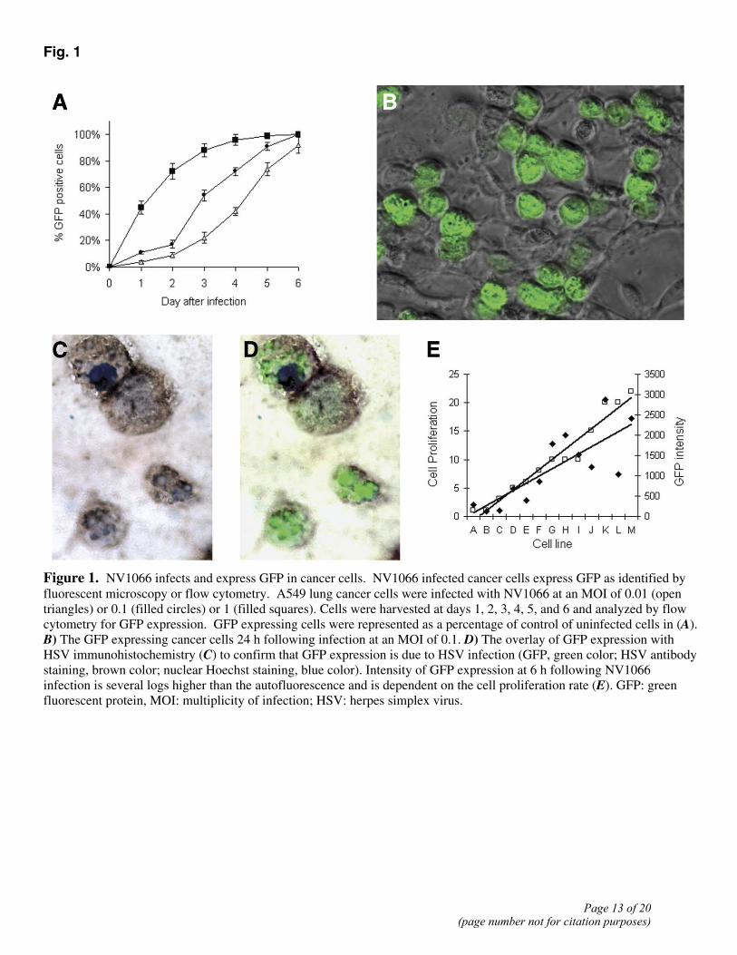
Fig. 1
Figure 1. NV1066 infects and express GFP in cancer cells. NV1066 infected cancer cells express GFP as identified by fluorescent microscopy or flow cytometry. A549 lung cancer cells were infected with NV1066 at an MOI of 0.01 (open triangles) or 0.1 (filled circles) or 1 (filled squares). Cells were harvested at days 1, 2, 3, 4, 5, and 6 and analyzed by flow cytometry for GFP expression. GFP expressing cells were represented as a percentage of control of uninfected cells in (A). B) The GFP expressing cancer cells 24 h following infection at an MOI of 0.1. D) The overlay of GFP expression with HSV immunohistochemistry (C) to confirm that GFP expression is due to HSV infection (GFP, green color; HSV antibody staining, brown color; nuclear Hoechst staining, blue color). Intensity of GFP expression at 6 h following NV1066 infection is several logs higher than the autofluorescence and is dependent on the cell proliferation rate (E). GFP: green fluorescent protein, MOI: multiplicity of infection; HSV: herpes simplex virus.
Page 13 of 20(page number not for citation purposes)
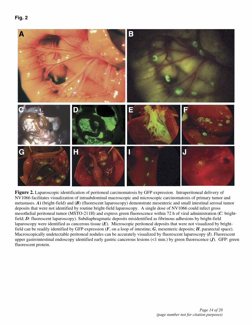
Fig. 2
Figure 2. Laparoscopic identification of peritoneal carcinomatosis by GFP expression. Intraperitoneal delivery of NV1066 facilitates visualization of intraabdominal macroscopic and microscopic carcinomatosis of primary tumor and metastases. A) (bright-field) and (B) (fluorescent laparoscopy) demonstrate mesenteric and small intestinal serosal tumor deposits that were not identified by routine bright-field laparoscopy. A single dose of NV1066 could infect gross mesothelial peritoneal tumor (MSTO-211H) and express green fluorescence within 72 h of viral administration (C: bright-field; D: fluorescent laparoscopy). Subdiaphragmatic deposits misidentified as fibrinous adhesions by bright-field laparoscopy were identified as cancerous tissue (E). Microscopic peritoneal deposits that were not visualized by bright-field can be readily identified by GFP expression (F, on a loop of intestine; G, mesenteric deposits; H, pararectal space). Macroscopically undetectable peritoneal nodules can be accurately visualized by fluorescent laparoscopy (I). Fluorescent upper gastrointestinal endoscopy identified early gastric cancerous lesions (<1 mm.) by green fluorescence (J). GFP: green fluorescent protein.
Page 14 of 20(page number not for citation purposes)
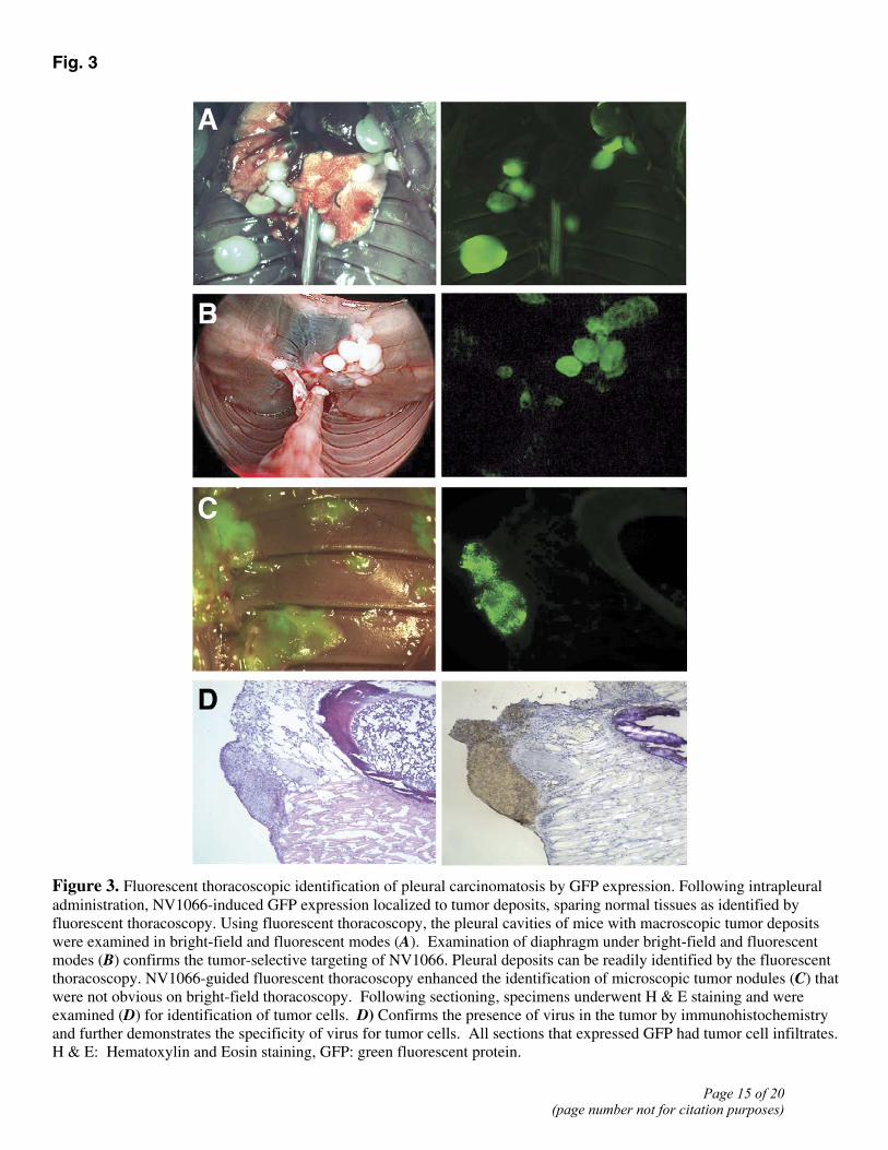
Fig. 3
Figure 3. Fluorescent thoracoscopic identification of pleural carcinomatosis by GFP expression. Following intrapleural administration, NV1066-induced GFP expression localized to tumor deposits, sparing normal tissues as identified by fluorescent thoracoscopy. Using fluorescent thoracoscopy, the pleural cavities of mice with macroscopic tumor deposits were examined in bright-field and fluorescent modes (A). Examination of diaphragm under bright-field and fluorescent modes (B) confirms the tumor-selective targeting of NV1066. Pleural deposits can be readily identified by the fluorescent thoracoscopy. NV1066-guided fluorescent thoracoscopy enhanced the identification of microscopic tumor nodules (C) that were not obvious on bright-field thoracoscopy. Following sectioning, specimens underwent H & E staining and were examined (D) for identification of tumor cells. D) Confirms the presence of virus in the tumor by immunohistochemistry and further demonstrates the specificity of virus for tumor cells. All sections that expressed GFP had tumor cell infiltrates. H & E: Hematoxylin and Eosin staining, GFP: green fluorescent protein.
Page 15 of 20(page number not for citation purposes)
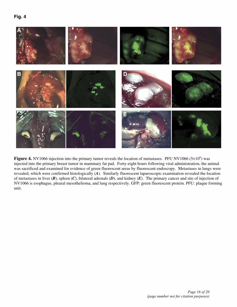
Fig. 4
Figure 4. NV1066 injection into the primary tumor reveals the location of metastases. PFU NV1066 (5×10⁶) was injected into the primary breast tumor in mammary fat pad. Forty-eight hours following viral administration, the animal was sacrificed and examined for evidence of green fluorescent areas by fluorescent endoscopy. Metastases in lungs were revealed, which were confirmed histologically (A). Similarly fluorescent laparoscopic examination revealed the location of metastases in liver (B), spleen (C), bilateral adrenals (D), and kidney (E). The primary cancer and site of injection of NV1066 is esophagus, pleural mesothelioma, and lung respectively. GFP: green fluorescent protein. PFU: plaque forming unit.
Page 16 of 20(page number not for citation purposes)
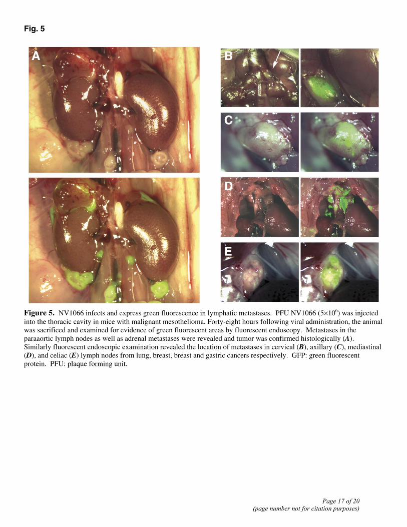
Fig. 5
Figure 5. NV1066 infects and express green fluorescence in lymphatic metastases. PFU NV1066 (5×10⁶) was injected into the thoracic cavity in mice with malignant mesothelioma. Forty-eight hours following viral administration, the animal was sacrificed and examined for evidence of green fluorescent areas by fluorescent endoscopy. Metastases in the paraaortic lymph nodes as well as adrenal metastases were revealed and tumor was confirmed histologically (A). Similarly fluorescent endoscopic examination revealed the location of metastases in cervical (B), axillary (C), mediastinal (D), and celiac (E) lymph nodes from lung, breast, breast and gastric cancers respectively. GFP: green fluorescent protein. PFU: plaque forming unit.
Page 17 of 20(page number not for citation purposes)
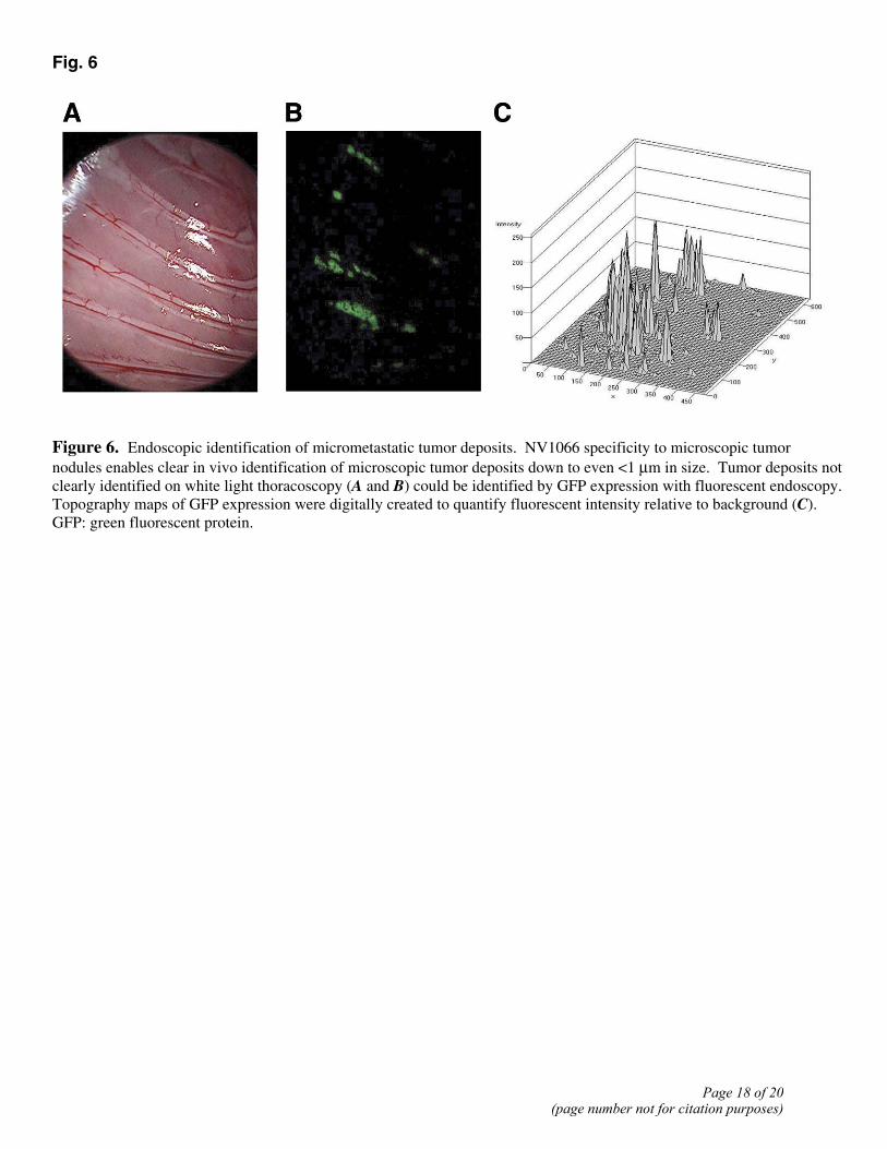
Fig. 6
Figure 6. Endoscopic identification of micrometastatic tumor deposits. NV1066 specificity to microscopic tumor nodules enables clear in vivo identification of microscopic tumor deposits down to even <1 µm in size. Tumor deposits not clearly identified on white light thoracoscopy (A and B) could be identified by GFP expression with fluorescent endoscopy. Topography maps of GFP expression were digitally created to quantify fluorescent intensity relative to background (C). GFP: green fluorescent protein.
Page 18 of 20(page number not for citation purposes)
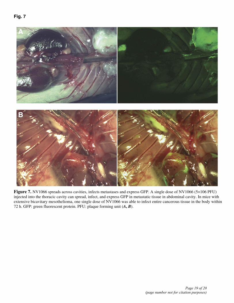
Fig. 7
Figure 7. NV1066 spreads across cavities, infects metastases and express GFP. A single dose of NV1066 (5×10⁶ PFU) injected into the thoracic cavity can spread, infect, and express GFP in metastatic tissue in abdominal cavity. In mice with extensive bicavitary mesothelioma, one single dose of NV1066 was able to infect entire cancerous tissue in the body within 72 h. GFP: green fluorescent protein. PFU: plaque forming unit (A, B).
Page 19 of 20(page number not for citation purposes)
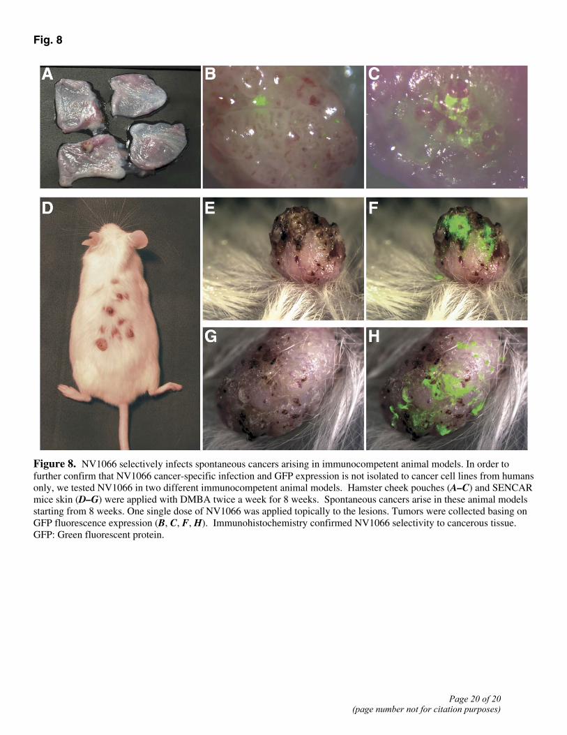
Fig. 8
Figure 8. NV1066 selectively infects spontaneous cancers arising in immunocompetent animal models. In order to further confirm that NV1066 cancer-specific infection and GFP expression is not isolated to cancer cell lines from humans only, we tested NV1066 in two different immunocompetent animal models. Hamster cheek pouches (A–C) and SENCAR mice skin (D–G) were applied with DMBA twice a week for 8 weeks. Spontaneous cancers arise in these animal models starting from 8 weeks. One single dose of NV1066 was applied topically to the lesions. Tumors were collected basing on GFP fluorescence expression (B, C, F, H). Immunohistochemistry confirmed NV1066 selectivity to cancerous tissue. GFP: Green fluorescent protein.
Page 20 of 20(page number not for citation purposes)
Related Documents


![Case Report Prostatic Carcinosarcoma with Lung Metastases · 2019. 7. 31. · Case Reports in Oncological Medicine sarcomatoid carcinoma [ [] C. G. Rogers, A. Parwani, A. Tekes, M.](https://static.cupdf.com/doc/110x72/6106208aa9c710750f2e2726/case-report-prostatic-carcinosarcoma-with-lung-metastases-2019-7-31-case-reports.jpg)


