Real-time analysis of the effects of toxic, therapeutic and sub-therapeutic concentrations of digitoxin on lung cancer cells R. Eldawud a , T.A. Stueckle b,d , S. Manivannan a , H. Elbaz c , M. Chen d , Y. Rojanasakul d,e,n , C.Z. Dinu a,d,e,nn a Department of Chemical Engineering, West Virginia University, WV, United States b Health Effects Laboratory Division, National Institute for Occupational Safety and Health, WV, United States c Wayne State University School of Medicine, MI, United States d Department of Basic Pharmaceutical Sciences, West Virginia University, WV, United States e Mary Babb Randolph Cancer Center Allen Lung Program, West Virginia University, WV, United States article info Article history: Received 10 January 2014 Received in revised form 9 March 2014 Accepted 12 March 2014 Available online 26 March 2014 Keywords: Digitoxin Cell-based sensing Cell adhesion Anti-cancer abstract Digitoxin belongs to a naturally occurring class of cardiac glycosides (CG); digitoxin is clinically approved for heart failure and known for its anti-cancer effects against non-small lung cancer cells (NSCLC). However, concerns associated with its narrow therapeutic index and its concentration-dependent mechanism of action are rising. Thus, before digitoxin implementation in designing and developing safer and more effective CG-based anti-cancer therapies, its pharmacological and safety profiles need to be fully elucidated. In this research we used a combinatorial approach to evaluate the anti-cancer mechanisms of digitoxin in real-time. Our approach employed a non-invasive electric cell impedance sensing technique as a proxy to monitor NSCLC behavior post-exposure to toxic, therapeutic and sub- therapeutic concentrations of the drug. By developing structure–function combinatorial relations we showed that digitoxin targets cancer cells in a time and dose-dependant manner by activating pro- apoptotic and anti-proliferative signaling cascades that results in strengthening cellular adhesion and sequestration of key regulatory proliferation protein from the nucleus. & 2014 Elsevier B.V. All rights reserved. 1. Introduction Digitoxin is a natural occurring cardiac glycoside (CG) (Elbaz et al., 2012a) with a prolonged half-life, a well-established clinical profile, a narrow therapeutic window (Elbaz et al., 2012a), and increased ability to readily cross both the blood brain and the placental barriers (Storstein et al., 1979). Laboratory investigations suggested that digitoxin exhibits high selectivity towards cancer cells when compared to healthy cells (Elbaz et al., 2012a; López- Lázaro et al., 2006) making the drug a viable chemotherapeutic alternative against several types of cancer from leukemia, to pancreatic and lung cancers (López-Lázaro, 2007; Winnicka et al., 2006). The anti-cancer mechanisms induced by digitoxin have been extensively studied and are mainly associated with the drug's ability to manipulate intracellular ion hemostasis which led to a downstream signaling cascade eventually inducing apoptosis and cell cycle arrest (Elbaz et al., 2012b). In particular, in vitro studies showed that exposure of non-small lung cells, renal, pancreatic and breast cells to micromolar concentrations of digitoxin (0.5– 5 mM) inhibits Na þ /K þ -ATPase pump activity (López-Lázaro, 2007; Newman et al., 2008), induces calcium-dependent activa- tion of caspases and other hydrolytic enzymes (Einbond et al., 2008; Elbaz et al., 2012a), causes generation of reactive oxygen species (Prassas et al., 2011), activates the cell-cycle inhibitor p21Cip1 (Prassas et al., 2011), directs the inhibition of topoisome- rase activity and hypoxia-inducible factor1a synthesis (Sun et al., 2013), and ultimately reduces viability and cell proliferation (Elbaz et al., 2012a; Menger et al., 2013). Complementary, cellular exposure to nanomolar concentrations of digitoxin (10–100 nM) leads to inhibition of (HIF-1) and topoisomerase II synthesis (Prassas et al., 2011), activation of phospholipase C (Elbaz et al., 2012a; Ho et al., 1987), phosphatidylinositol-3-kinase (PI3K) Contents lists available at ScienceDirect journal homepage: www.elsevier.com/locate/bios Biosensors and Bioelectronics http://dx.doi.org/10.1016/j.bios.2014.03.030 0956-5663/& 2014 Elsevier B.V. All rights reserved. n Corresponding author at: Department of Basic Pharmaceutical Sciences, West Virginia University, PO Box 9530, Morgantown, WV 26505, USA. Tel.: þ1 304 293 1476; fax: þ1 304 293 2576. nn Corresponding author at: Department of Chemical Engineering, Benjamin M. Statler College of Engineering and Mineral Resources, West Virginia University, PO Box 6102, Morgantown, WV 26506, USA. Tel.: þ1 304 293 9338; fax: þ1 304 293 4139. E-mail addresses: [email protected] (Y. Rojanasakul), [email protected] (C.Z. Dinu). Biosensors and Bioelectronics 59 (2014) 192–199

Welcome message from author
This document is posted to help you gain knowledge. Please leave a comment to let me know what you think about it! Share it to your friends and learn new things together.
Transcript
Real-time analysis of the effects of toxic, therapeuticand sub-therapeutic concentrations of digitoxin on lung cancer cells
R. Eldawud a, T.A. Stueckle b,d, S. Manivannan a, H. Elbaz c, M. Chen d, Y. Rojanasakul d,e,n,C.Z. Dinu a,d,e,nn
a Department of Chemical Engineering, West Virginia University, WV, United Statesb Health Effects Laboratory Division, National Institute for Occupational Safety and Health, WV, United Statesc Wayne State University School of Medicine, MI, United Statesd Department of Basic Pharmaceutical Sciences, West Virginia University, WV, United Statese Mary Babb Randolph Cancer Center Allen Lung Program, West Virginia University, WV, United States
a r t i c l e i n f o
Article history:Received 10 January 2014Received in revised form9 March 2014Accepted 12 March 2014Available online 26 March 2014
Keywords:DigitoxinCell-based sensingCell adhesionAnti-cancer
a b s t r a c t
Digitoxin belongs to a naturally occurring class of cardiac glycosides (CG); digitoxin is clinically approvedfor heart failure and known for its anti-cancer effects against non-small lung cancer cells (NSCLC).However, concerns associated with its narrow therapeutic index and its concentration-dependentmechanism of action are rising. Thus, before digitoxin implementation in designing and developingsafer and more effective CG-based anti-cancer therapies, its pharmacological and safety profiles need tobe fully elucidated. In this research we used a combinatorial approach to evaluate the anti-cancermechanisms of digitoxin in real-time. Our approach employed a non-invasive electric cell impedancesensing technique as a proxy to monitor NSCLC behavior post-exposure to toxic, therapeutic and sub-therapeutic concentrations of the drug. By developing structure–function combinatorial relations weshowed that digitoxin targets cancer cells in a time and dose-dependant manner by activating pro-apoptotic and anti-proliferative signaling cascades that results in strengthening cellular adhesion andsequestration of key regulatory proliferation protein from the nucleus.
& 2014 Elsevier B.V. All rights reserved.
1. Introduction
Digitoxin is a natural occurring cardiac glycoside (CG) (Elbazet al., 2012a) with a prolonged half-life, a well-established clinicalprofile, a narrow therapeutic window (Elbaz et al., 2012a), andincreased ability to readily cross both the blood brain and theplacental barriers (Storstein et al., 1979). Laboratory investigationssuggested that digitoxin exhibits high selectivity towards cancercells when compared to healthy cells (Elbaz et al., 2012a; López-Lázaro et al., 2006) making the drug a viable chemotherapeuticalternative against several types of cancer from leukemia, to
pancreatic and lung cancers (López-Lázaro, 2007; Winnickaet al., 2006).
The anti-cancer mechanisms induced by digitoxin have beenextensively studied and are mainly associated with the drug'sability to manipulate intracellular ion hemostasis which led to adownstream signaling cascade eventually inducing apoptosis andcell cycle arrest (Elbaz et al., 2012b). In particular, in vitro studiesshowed that exposure of non-small lung cells, renal, pancreaticand breast cells to micromolar concentrations of digitoxin (0.5–5 mM) inhibits Naþ/Kþ-ATPase pump activity (López-Lázaro,2007; Newman et al., 2008), induces calcium-dependent activa-tion of caspases and other hydrolytic enzymes (Einbond et al.,2008; Elbaz et al., 2012a), causes generation of reactive oxygenspecies (Prassas et al., 2011), activates the cell-cycle inhibitorp21Cip1 (Prassas et al., 2011), directs the inhibition of topoisome-rase activity and hypoxia-inducible factor1a synthesis (Sun et al.,2013), and ultimately reduces viability and cell proliferation (Elbazet al., 2012a; Menger et al., 2013). Complementary, cellularexposure to nanomolar concentrations of digitoxin (10–100 nM)leads to inhibition of (HIF-1) and topoisomerase II synthesis(Prassas et al., 2011), activation of phospholipase C (Elbaz et al.,2012a; Ho et al., 1987), phosphatidylinositol-3-kinase (PI3K)
Contents lists available at ScienceDirect
journal homepage: www.elsevier.com/locate/bios
Biosensors and Bioelectronics
http://dx.doi.org/10.1016/j.bios.2014.03.0300956-5663/& 2014 Elsevier B.V. All rights reserved.
n Corresponding author at: Department of Basic Pharmaceutical Sciences, WestVirginia University, PO Box 9530, Morgantown, WV 26505, USA.Tel.: þ1 304 293 1476; fax: þ1 304 293 2576.
nn Corresponding author at: Department of Chemical Engineering, Benjamin M.Statler College of Engineering and Mineral Resources, West Virginia University, POBox 6102, Morgantown, WV 26506, USA.Tel.: þ1 304 293 9338; fax: þ1 304 293 4139.
E-mail addresses: [email protected] (Y. Rojanasakul),[email protected] (C.Z. Dinu).
Biosensors and Bioelectronics 59 (2014) 192–199
(Ho et al., 1987), tyrosine kinase (Src) (Elbaz et al., 2012a; Jagielskaet al., 2009), mitogen-activated protein kinase (MAPK) (Prassaset al., 2011), affects cell cycle and anoikis (Pongrakhananon et al.,2014) inducing alternations in membrane fluidity (Larre et al.,2010; Xie and Cai, 2003), ultimately leading to cell apoptosis(Lopez-Lazaro et al., 2005). However, the nature of these in vitrostudies only allowed for discrete time points monitoring andlimited analysis of the cellular functions upon exposure, all afterinvasive or destructive preparation of the samples, as well as laborintensive and time consuming analysis. Therefore, further studiesare needed to better characterize the pharmacological and safetyprofiles of digitoxin before its chemotherapeutic implementation,especially considering that therapeutic concentrations of digitoxinvaries according to the age and weight of the patient, generallyranging from 26 to 46 nM (Wu et al., 2001). Such studies do notonly have to account for the narrow therapeutic window or drug'sdose-dependent and selective apoptosis to cancer cells (Newmanet al., 2008; Xie and Cai, 2003), but also need to allow monitoringof the cellular systems without lag time between sample collectionand data analysis.
Electric cell–substrate impedance sensing (ECIS) is a non-invasive and quantitative form of cell-based sensing that utilizesidentical small gold-film electrodes deposited on the bottom ofcell culture dishes to measure the cellular resistance and itsalteration upon changes in cell morphology, spreading, attach-ment and migration (Arndt et al., 2004; Spegel et al., 2008), all as afunction of applied frequencies and in real-time (Giaever andKeese, 1984; Wegener et al., 2000a). The applicability of ECIS wasextended to inhibition assays for cytochalasin-D (cytoskeletalinhibitor) (Sapper et al., 2006), prostaglandin E2 (inflammatorymediator) (Wang et al., 1995), bacterial protease (Itagaki et al.,2011), or platelet-activating factors that affect in cellular adhesion(Melnikova et al., 2009).
By combining standard in vitro cell viability and cell-basedcytotoxicity assays with ECIS, we aim to investigate the anti-proliferative and pro-apoptotic mechanisms of non-small lungcancer cells (NSCLC) exposed to different concentrations of digi-toxin in real-time. NCI-H460 cells were chosen as model NSCLCbased on their sensitivity to digitoxin (López-Lázaro, 2007; Wanget al., 2010) and increased resistance to chemotherapy (Mijatovicet al., 2006a,b). We hypothesized that NCI-H460 exposure to toxic(80 nM), therapeutic (40 nM) and sub-therapeutic (25 and 10 nM)concentrations of digitoxin is associated with changes in thecellular viability that can be monitored using an individual cellas a primary transducer and recording its real-time alterations inmorphology and adhesion profile. Further, based on the key role ofthe cyclin-dependent kinase-4 (CDK4) in regulation of cell pro-liferation and cell cycle (Gulappa et al., 2013; Si and Liu, 2001), wehypothesized that exposure to digitoxin targets cellular adhesionpathways in a dose and time-dependent manner by sequesteringCDK4 in the cytoplasm and thus reducing its nuclear levels.Further, CDK4 association with viable candidates responsible forcellular junctions formation can be monitored in real-time as achange in the cellular attachment profile. Our findings underscorethe potential of digitoxin to be used as the next generation ofchemotherapeutic drugs that target cell adhesion and cell cycleprofiles for improved anti-cancer activity.
2. Materials and methods
2.1. Cell culture and treatment
Human lung cancer epithelial cells (NCI-H460; ATCC, VA)were cultured in Roswell Park Memorial Institute-1640 medium(RPMI-1640; Sigma Chemicals, MO) supplemented with 10% fetal
bovine serum (FBS; Atlanta Biologicals, GA), 2 mM L-glutamine and100-units/mL penicillin/streptomycin (Sigma Chemicals, MO) andmaintained in a humidified atmosphere at 37 1C under 5% CO2.Cells were passaged regularly using 0.25% (w/v) trypsin (MolecularProbles, OR) with 1.5 mM ethylene diaminetetracetic acid (EDTA;Molecular Probles, OR). Stock concentrations of digitoxin (SigmaChemicals, MO) were made in dimethyl sulfoxide (DMSO; SigmaChemicals, MO) and diluted to 1000x exposure concentrations aspreviously described (Elbaz et al., 2012b). Digitoxin exposure wasperformed in a medium containing 1% FBS, 2 mM L-glutamine and100-units/mL penicillin/streptomycin. The concentration of FBSwas reduced due to existing concerns regarding digitoxin's inter-action with serum proteins (Baggot and Davis, 1973) and to betterapproximate the minimum concentrations and times that wererequired to achieve drugs’ activity in vitro (Elbaz et al., 2012b).
2.2. Apoptosis assay
Cells were seeded overnight in 12-well plates (Fisher, PA) at2�105 cell/mL and treated with 0, 10, 25, 40 and 80 nM digitoxinfor 24, 48 and 72 h respectively. After treatment, the cells wereincubated with 10 μg/mL Hoechst-33342 (Molecular Probles, OR)for 30 min. The percentage of cells having intensely condensedchromatin and/or fragmented nuclei was scored using fluores-cence microscope (Leica Microsystems, IL). Approximately 1000nuclei from ten random fields were analyzed for each sample. Theapoptotic percentage was calculated as the percentage of cellswith apoptotic nuclei over the total number of cells per fieldof view.
2.3. Western blot analysis
Cells were seeded overnight in 6-well plates at a density of6�105 cell/well, and treated with 0, 10 or 25 nM digitoxin in 0.1%DMSO for 24 h. Subsequently, cells were placed on ice and lysedfor 30 min in a lysis buffer containing 2% Triton X-100, 1% sodiumdodecyle sulfate (SDS), 100 mM sodium chloride (NaCl), 10 mMtris-hydrochloric acid (HCl), complete mini cocktail proteaseinhibitors (all reagents are purchased from Roche, IN) and 1 mMEDTA insoluble cellular debris was pelleted by centrifugation at4 1C and 16,000� g for 15 min. The supernatant was collected andused to determine the total protein content using standardBicinchoninic Acid Assay (BCA, Thermo Scientific, IL). Briefly,working reagent was prepared according to the manufacturerinstructions by mixing 50 parts of reagent A with 1 part of reagentB (reagents included with kit). Two μL of each sample was addedto a 96-well plate and incubated with 200 μL of the workingreagent at 37 1C for 30 min; experiments were performed induplicates. Control calibration curves were prepared using serialdilutions of standard bovine serum albumin (BSA). Absorbanceat 562 nm was recorded on a BioTek 96-plate reader (BioTek,Winooski, VT).
The supernatant was separated by a 10% SDS–PAGE gel andtransferred to polyvinylidene fluoride (PVDF) membranes usingthe iBlots Dry Blotting System (Invitrogen, CA). Membranes wereblocked in 5% skim milk in Tris-buffered saline (TBST, 25 mM Tris–HCl, 125 mM NaCl, and 0.1% Tween-20; Sigma Chemicals, MO) for1 h at room temperature, and subsequently incubated with anti-CDK4 primary antibody (Cell Signaling, MA) at 4 1C overnight. Themembranes were subsequently washed three times in phosphatebuffer saline (PBS; Lonza, MD) containing 1% Tween-20 for 10 mineach, incubated with horseradish peroxidase-conjugated second-ary antibody (Cell Signaling, MA) for 1 h at room temperature,then washed again for three more times each for 10 min inTBST. Finally, the samples were analyzed by chemiluminescence
R. Eldawud et al. / Biosensors and Bioelectronics 59 (2014) 192–199 193
(Supersignal West Pico, IL). Band quantification via densitometrywas performed using ImageJ software, version 10.2.
2.4. Trypan-blue exclusion assay
NCI-H460 cells were seeded overnight in 12-well plates at adensity of 2�105 cell/mL, and treated with 0, 10, 25, 40 or 80 nMdigitoxin for 24, 48, and 72 h, respectively. Cells were subsequentlywashed with PBS, trypsinized (0.25%), suspended in 10% mediaand stained with 0.4% trypan-blue (Invitrogen, CA) at 1:1 volumeratio and analyzed using Countess automated cell counter(Invitrogen, CA).
2.5. Electric cell–substrate impedance sensing (ECIS)
Real-time quantification of cellular behavior was conductedusing an electric cell impedance sensing instrument (ECIS-ZΘ,Applied Biophysics, NY). In one set of experiments, two ECIS arrays(8W10Eþ), each containing 8-wells with 40 gold electrodes, weresimultaneously employed to provide concomitant measurementsof 16 samples at multiple frequencies. Prior to any experiment, thegold electrodes were stabilized for 3 h in 400 ml RPMI media toaccount for electrode variances and to create a reference lineassociated with free electrodes; subsequently, the array holder wasplaced in a humidified incubator at 37 1C and 5% CO2 to provideoptimal conditions for cellular growth. NCI-H460 cells were addedat a density of 2�105 cell/mL in a volume of 400 ml/well. Cells wereallowed to settle and grow over the gold electrodes and form aconfluent monolayer for 24 h. The formation of the cellular mono-layer was indicated as a settlement in the resistance value withminor fluctuation caused by cellular micromotion (Arndt et al.,2004; Giaever and Keese, 1991; Wegener et al., 2000a). Uponmonolayer formation cells were treated with 0, 10, 25, 40 or80 nM digitoxin and their cellular behavior was monitored for48 h post-exposure.
2.6. Statistical analysis
Results are presented as mean7standard deviation. Experi-ments (viability, apoptosis and Western blot) were performed induplicates and repeated at least 3 times. ECIS experiments wereperformed in duplicates and repeated at least four times, for a totalof minimum 8 replicates per dose. Changes in the behavior of thecells (i.e., resistance) were recorded every 180 s for the duration ofthe experiments with each time point being an average of 16replicates (2 arrays with 8 wells each). Two-way analysis ofvariance and unpaired two-tailed Student's t-test were performedusing JMP 8.0 (SAS Institute) and SigmaPlot 10.0 (Systat SoftwareInc.). Results were considered significant for npo0.05.
3. Results
3.1. Digitoxin induced apoptosis in a time and dose-dependentmanner
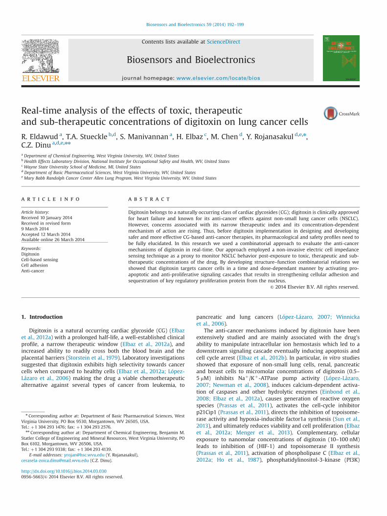
To evaluate the effects of digitoxin on cellular apoptosis, confl-uent monolayers of human lung cancer (NCI-H460) cells wereexposed to toxic, therapeutic and sub-therapeutic concentrationsof digitoxin for 24, 48 and 72 h respectively. Visual inspection ofthe 24 h exposed cells showed minor differences in their nuclearmorphology for the cells treated with 10 nM (5%) and 25 nM (15%)digitoxin when compared to the untreated cells (Fig. 1A). Incontrast, the nuclei of the cells treated with 40 and 80 nMdigitoxin showed major changes (Z50%) in their morphologies,i.e., rounding, swelling, condensation and fragmentation. Such
changes are considered early indicators of cellular apoptosis(Furukawa et al., 2007; Yuan et al., 2007).
The percentage of apoptotic cells at 24, 48 and 72 h post-exposure to toxic, therapeutic and sub-therapeutic concentrationsof digitoxin is shown in Fig. 1B. Analysis of variance showed thatthe NCI-H460 apoptosis was both dose and time-dependent.In particular, 24 h after exposure, the cells treated with 10 nMshowed no significant changes in their apoptotic percentage;however, increased apoptosis was noticed after 48 and 72 h ofdigitoxin exposure. Cells exposed to 25, 40 and 80 nM digitoxinexhibited higher apoptosis within first 24 h with a significantincrease after 48 and 72 h, respectively. Values for the IC50 (half-inhibition concentration) derivated using a three-parameter sig-moid regression analysis were 43.4–36.6 nM and 31.6 nM at 24, 48and 72 h post-exposure respectively.
3.2. Digitoxin inhibited cellular proliferation
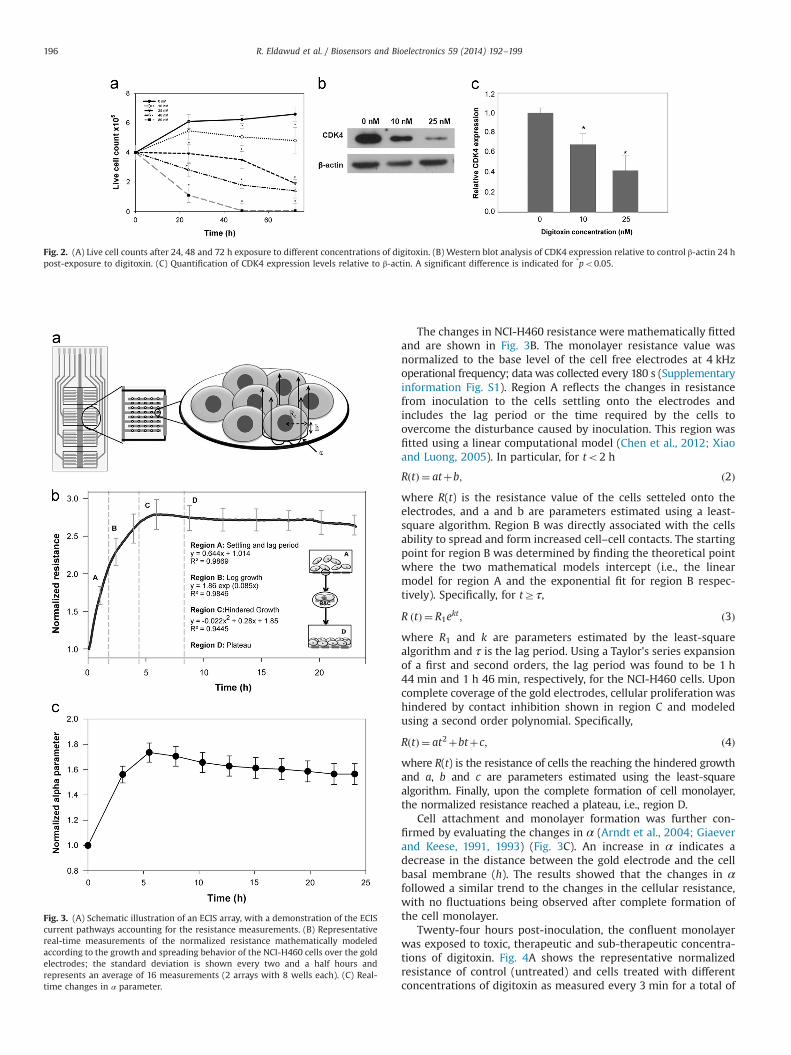
To study the effects of toxic, therapeutic and sub-therapeuticconcentrations of digitoxin on the NCI-H460 cell proliferation, alive cell exclusion assay was performed. The results showed thatexposure to digitoxin caused inhibition of cellular proliferation in adose and time-dependent manner (Fig. 2A). Specifically, no differ-ences relative to controls were noticed 24 h post-exposure for thecells treated with 10 nM digitoxin; however, significant inhibitoryeffects were observed after 48 (18%) and 72 h (27%) respectively.For the cells treated with 25, 40 and 80 nM digitoxin, the decreasein the live cell counts was significant within the first 24 h with35%, 54% and 82% respectively. This subsequently increased to 43%,70% and 99% after 48 h exposure and 70%, 85% and 100% after 72 h,respectively.
Since inhibition of cell proliferation was the major effect 24 hpost-exposure to sub-therapeutic concentrations of digitoxin, wealso examined the expression of cyclin-dependent kinases-4(CDK4), a key regulatory protein that controls cellular proliferationand cell cycle progression at the G1/S phase (Gulappa et al., 2013; Siand Liu, 2001). Western blot analysis showed significant decrease inthe expression of CDK4 after exposure to both 10 and 25 nM, with a35% and 60% reduction in the CDK4 expression relative to expres-sion of control β-actin (Fig. 2B and C).
3.3. Real-time cell monitoring using electric cell impedance sensing
Real-time analysis from the time of cell inoculation into thewells, to the formation of a confluent monolayer, and subsequently48 h post-exposure to toxic, therapeutic and sub-therapeuticconcentrations of digitoxin were performed using electric cellimpedance sensing (ECIS). Previous studies showed that cellsimmobilized onto gold electrodes have similar behaviors withtheir counterparts immobilized onto polystyrene surfaces (Giaeverand Keese, 1991; Wegener et al., 2000b). Fig. 3A shows an eightwell ECIS array with inter-finger-like arrangement and the currentpathways contributing to the resistance measurements. Once thecells spread onto the gold electrodes, their insulating nature of theplasma membrane constricted the current flow in the spacesbeneath the basal membrane and the electrode surface as wellas in the paracellular spaces between adjacent cells, leading to therecorded resistance (Giaever and Keese, 1991, 1993). Alpha is afunction of the distance between the basal membrane and cellularradius according to the following equation:
α¼ Rcðρ=hÞ0:5; ð1Þwhere ρ represents the specific resistivity of the electrolyte (i.e.,culture media) underneath the cells, h represents the distance atwhich the cells hover above the electrode, and Rc is the cellularradius.
R. Eldawud et al. / Biosensors and Bioelectronics 59 (2014) 192–199194
Fig. 1. (A) Fluorescent images of Hoechst stained NCI-H460 cells 24 h post-exposure to digitoxin. Cells treated with 40 and 80 nM digitoxin showed major changes in theirnuclei morphologies (white circles) relative to controls or cells treated with 10 and 25 nM digitoxin. (B) Percentage of apoptotic cells upon exposure to digitoxin is dose andtime-dependent. Difference is considered significant for *po0.05.
R. Eldawud et al. / Biosensors and Bioelectronics 59 (2014) 192–199 195
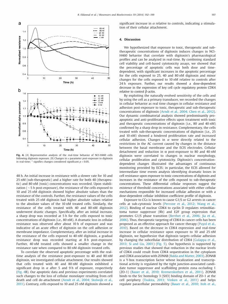
The changes in NCI-H460 resistance were mathematically fittedand are shown in Fig. 3B. The monolayer resistance value wasnormalized to the base level of the cell free electrodes at 4 kHzoperational frequency; data was collected every 180 s (Supplementaryinformation Fig. S1). Region A reflects the changes in resistancefrom inoculation to the cells settling onto the electrodes andincludes the lag period or the time required by the cells toovercome the disturbance caused by inoculation. This region wasfitted using a linear computational model (Chen et al., 2012; Xiaoand Luong, 2005). In particular, for to2 h
RðtÞ ¼ atþb; ð2Þwhere R(t) is the resistance value of the cells setteled onto theelectrodes, and a and b are parameters estimated using a least-square algorithm. Region B was directly associated with the cellsability to spread and form increased cell–cell contacts. The startingpoint for region B was determined by finding the theoretical pointwhere the two mathematical models intercept (i.e., the linearmodel for region A and the exponential fit for region B respec-tively). Specifically, for tZτ,
R ðtÞ ¼ R1ekt ; ð3Þwhere R1 and k are parameters estimated by the least-squarealgorithm and τ is the lag period. Using a Taylor's series expansionof a first and second orders, the lag period was found to be 1 h44 min and 1 h 46 min, respectively, for the NCI-H460 cells. Uponcomplete coverage of the gold electrodes, cellular proliferation washindered by contact inhibition shown in region C and modeledusing a second order polynomial. Specifically,
RðtÞ ¼ at2þbtþc; ð4Þwhere R(t) is the resistance of cells the reaching the hindered growthand a, b and c are parameters estimated using the least-squarealgorithm. Finally, upon the complete formation of cell monolayer,the normalized resistance reached a plateau, i.e., region D.
Cell attachment and monolayer formation was further con-firmed by evaluating the changes in α (Arndt et al., 2004; Giaeverand Keese, 1991, 1993) (Fig. 3C). An increase in α indicates adecrease in the distance between the gold electrode and the cellbasal membrane (h). The results showed that the changes in αfollowed a similar trend to the changes in the cellular resistance,with no fluctuations being observed after complete formation ofthe cell monolayer.
Twenty-four hours post-inoculation, the confluent monolayerwas exposed to toxic, therapeutic and sub-therapeutic concentra-tions of digitoxin. Fig. 4A shows the representative normalizedresistance of control (untreated) and cells treated with differentconcentrations of digitoxin as measured every 3 min for a total of
Fig. 2. (A) Live cell counts after 24, 48 and 72 h exposure to different concentrations of digitoxin. (B) Western blot analysis of CDK4 expression relative to control β-actin 24 hpost-exposure to digitoxin. (C) Quantification of CDK4 expression levels relative to β-actin. A significant difference is indicated for *po0.05.
Fig. 3. (A) Schematic illustration of an ECIS array, with a demonstration of the ECIScurrent pathways accounting for the resistance measurements. (B) Representativereal-time measurements of the normalized resistance mathematically modeledaccording to the growth and spreading behavior of the NCI-H460 cells over the goldelectrodes; the standard deviation is shown every two and a half hours andrepresents an average of 16 measurements (2 arrays with 8 wells each). (C) Real-time changes in α parameter.
R. Eldawud et al. / Biosensors and Bioelectronics 59 (2014) 192–199196
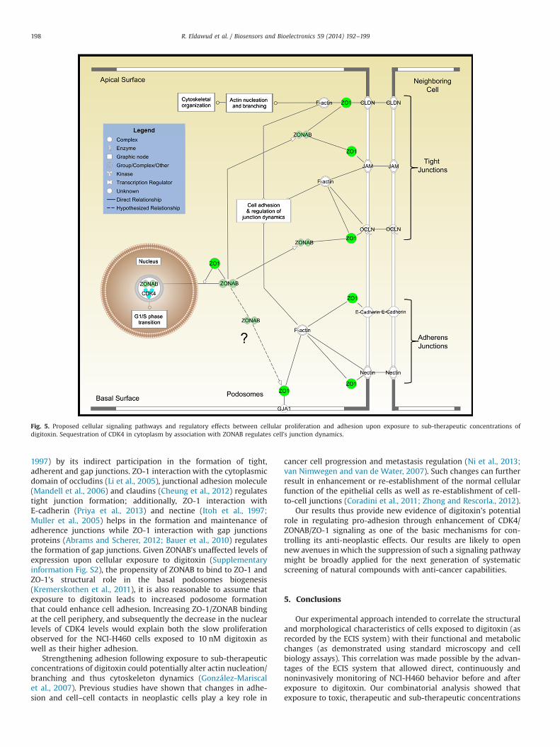
48 h. An initial increase in resistance with a slower rate for 10 and25 nM (sub-therapeutic) and a higher rate for both 40 (therapeu-tic) and 80 nM (toxic) concentrations was recorded. Upon stabili-zation (�5 h post-exposure), the resistance of the cells exposed to10 and 25 nM digitoxin showed higher absolute values than theresistance of the controls. Further, the resistance values of the cellstreated with 25 nM digitoxin had higher absolute values relativeto the absolute values of the 10 nM treated cells. Similarly, theresistance of the cells treated with 40 and 80 nM digitoxinunderwent drastic changes. Specifically, after an initial increase,a sharp drop was recorded at 5 h for the cells exposed to toxicconcentrations of digitoxin (i.e., 80 nM). A dramatic loss in cellularresistance was observed after about 10 h of exposure and wasindicative of an acute effect of digitoxin on the cell adhesion ormembrane impedance. Complementary, after an initial increase inthe resistance of the cells exposed to 40 nM digitoxin, a gradualresistance drop was recorded starting at 8 h post-exposure.Further, 40 nM treated cells showed a smaller change in theresistance rate when compared to 80 nM digitoxin treated cells.
To correlate the observed apoptotic percentage with the real-time analysis of the resistance post-exposure to 40 and 80 nMdigitoxin, we investigated cellular attachment. Our results showedthat cells treated with 40 and 80 nM digitoxin exhibited asignificant drop in α after 5 and 9 h of exposure, respectively(Fig. 4B). Our apoptotic data and previous experiments correlatedsuch changes to the loss of cellular monolayer resulting from celldeath and cell de-attachment (Arndt et al., 2004; Stolwijk et al.,2011). Contrary, cells exposed to 10 and 25 nM digitoxin showed a
significant increase in α relative to controls, indicating a stimula-tion of their cellular attachment.
4. Discussion
We hypothesized that exposure to toxic, therapeutic and sub-therapeutic concentrations of digitoxin induces changes in NCI-H460 behavior that correlate with digitoxin's pharmacologicalprofiles and can be analyzed in real-time. By combining standardcell viability and cell-based cytotoxicity assays, we showed thatthe percentage of apoptotic cells was both dose and time-dependent, with significant increases in the apoptotic percentagefor the cells exposed to 25, 40 and 80 nM digitoxin and minorchanges for the cells exposed to 10 nM relative to controls after24 h exposure. Further, our results showed a dose-dependentdecrease in the expression of key cell cycle regulatory protein CDK4relative to control β-actin.
By exploiting the naturally evolved sensitivity of the cells andby using the cell as a primary transducer, we recorded the changesin cellular behavior as real time changes in cellular resistance andadhesion post-exposure to toxic, therapeutic and sub-therapeuticconcentrations of digitoxin (Arndt et al., 2004; Chen et al., 2012).Our dynamic combinatorial analysis showed predominantly pro-apoptotic and anti-proliferative effects upon treatment with toxicand therapeutic concentrations of digitoxin (i.e., 80 and 40 nM)confirmed by a sharp drop in resistance. Complementary, the cellstreated with sub-therapeutic concentrations of digitoxin (i.e., 25and 10 nM) showed a hindered proliferation rate and increasedcellular adhesion. Changes in α were directly related to therestrictions in the AC current caused by changes in the distancebetween the basal membrane and the ECIS electrodes. Cellulardetachment and reduction in α post-exposure to 80 and 40 nMdigitoxin were correlated to changes in nuclear morphology,cellular proliferation and cytotoxicity. Digitoxin's concentration-dependent changes illustrated the advantages of continuousmonitoring provided by ECIS; in particular, the ECIS allowed forintermediate time events analysis identifying dramatic losses incell resistance upon exposure to toxic concentrations of digitoxin andincreases in the resistance of the cells exposed to sub-therapeuticconcentrations. These differential cellular responses suggest theexistence of threshold concentrations associated with either cellularmechanisms responsible for increased cellular adhesion or with adose-dependent cellular inhibition coefficient profile of digitoxin.
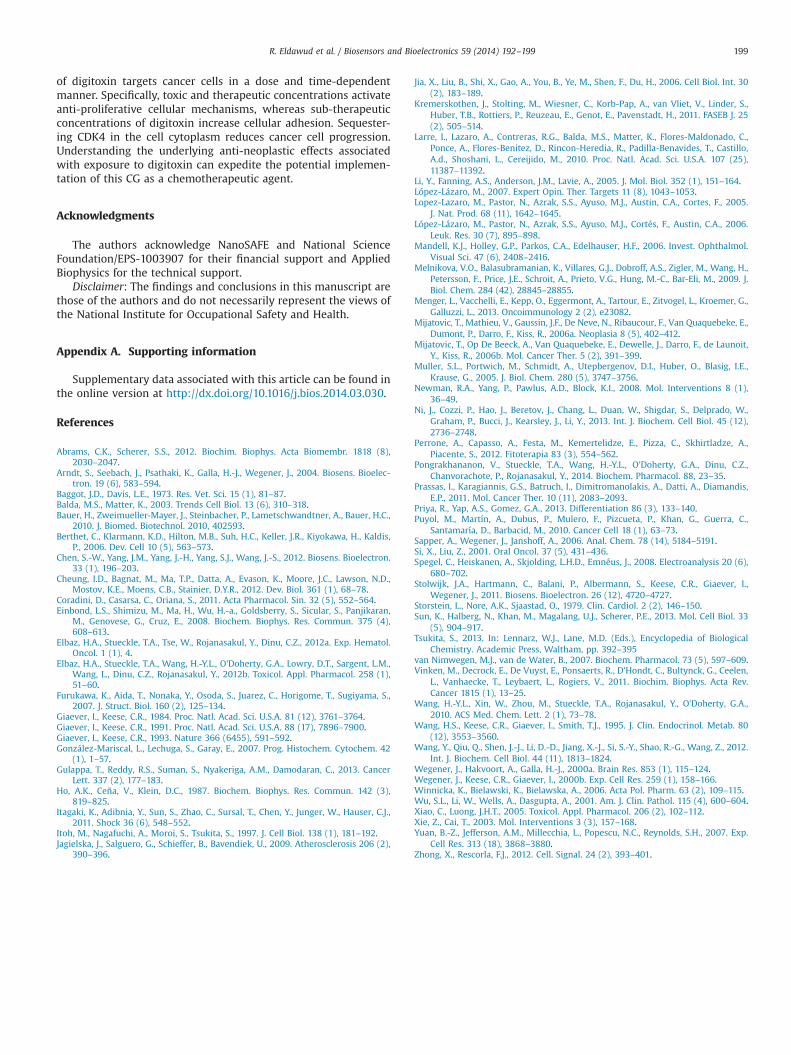
Exposure to CGs is known to cause G1/S or G2 arrests in cancercells at sub-cytotoxic levels (Perrone et al., 2012; Wang et al.,2012). Binding of nuclear CDK4 to cyclin D regulates retinoblas-toma tumor suppressor (Rb) and E2F group expression thatpromotes G1/S phase transition (Berthet et al., 2006; Jia et al.,2006). Thus, therapeutic targeting of CDK4 in cancer cells has beenproposed as an effective approach for cancer therapy (Puyol et al.,2010). Based on the decrease in CDK4 expression and real-timeincrease in cellular resistance upon exposure to 10 and 25 nMdigitoxin, we hypothesize that digitoxin targets cellular adhesionby changing the subcellular distribution of CDK4 (Gulappa et al.,2013; Si and Liu, 2001) (Fig. 5). Our hypothesis is supported byprevious studies that showed that reduction in the nuclear levelsof CDK4 could result from CDK4 sequestration in the cytoplasmand CDK4 associationwith ZONAB (Balda andMatter, 2003). ZONABis a Y-box transcription factor whose localization and transcrip-tional activity is regulated by the tight junction-associated candi-date tumor suppressor and scaffolding protein zona occluding 1(ZO-1) (Bauer et al., 2010; Kremerskothen et al., 2011). ZONABbinds to the Src homology 3 (SH3) binding domain of ZO-1 at thecell periphery (Tsukita, 2013; Vinken et al., 2011) and helpsregulate paracellular permeability (Bauer et al., 2010; Itoh et al.,
Fig. 4. (A) Representative analysis of the real-time behavior of NCI-H460 cellsfollowing digitoxin exposure. (B) Changes in α parameter post-exposure to digitoxinin real-time; * signifies changes considered significant po0.05.
R. Eldawud et al. / Biosensors and Bioelectronics 59 (2014) 192–199 197
1997) by its indirect participation in the formation of tight,adherent and gap junctions. ZO-1 interaction with the cytoplasmicdomain of occludins (Li et al., 2005), junctional adhesion molecule(Mandell et al., 2006) and claudins (Cheung et al., 2012) regulatestight junction formation; additionally, ZO-1 interaction withE-cadherin (Priya et al., 2013) and nectine (Itoh et al., 1997;Muller et al., 2005) helps in the formation and maintenance ofadherence junctions while ZO-1 interaction with gap junctionsproteins (Abrams and Scherer, 2012; Bauer et al., 2010) regulatesthe formation of gap junctions. Given ZONAB's unaffected levels ofexpression upon cellular exposure to digitoxin (Supplementaryinformation Fig. S2), the propensity of ZONAB to bind to ZO-1 andZO-1's structural role in the basal podosomes biogenesis(Kremerskothen et al., 2011), it is also reasonable to assume thatexposure to digitoxin leads to increased podosome formationthat could enhance cell adhesion. Increasing ZO-1/ZONAB bindingat the cell periphery, and subsequently the decrease in the nuclearlevels of CDK4 levels would explain both the slow proliferationobserved for the NCI-H460 cells exposed to 10 nM digitoxin aswell as their higher adhesion.
Strengthening adhesion following exposure to sub-therapeuticconcentrations of digitoxin could potentially alter actin nucleation/branching and thus cytoskeleton dynamics (González-Mariscalet al., 2007). Previous studies have shown that changes in adhe-sion and cell–cell contacts in neoplastic cells play a key role in
cancer cell progression and metastasis regulation (Ni et al., 2013;van Nimwegen and van de Water, 2007). Such changes can furtherresult in enhancement or re-establishment of the normal cellularfunction of the epithelial cells as well as re-establishment of cell-to-cell junctions (Coradini et al., 2011; Zhong and Rescorla., 2012).
Our results thus provide new evidence of digitoxin's potentialrole in regulating pro-adhesion through enhancement of CDK4/ZONAB/ZO-1 signaling as one of the basic mechanisms for con-trolling its anti-neoplastic effects. Our results are likely to opennew avenues in which the suppression of such a signaling pathwaymight be broadly applied for the next generation of systematicscreening of natural compounds with anti-cancer capabilities.
5. Conclusions
Our experimental approach intended to correlate the structuraland morphological characteristics of cells exposed to digitoxin (asrecorded by the ECIS system) with their functional and metabolicchanges (as demonstrated using standard microscopy and cellbiology assays). This correlation was made possible by the advan-tages of the ECIS system that allowed direct, continuously andnoninvasively monitoring of NCI-H460 behavior before and afterexposure to digitoxin. Our combinatorial analysis showed thatexposure to toxic, therapeutic and sub-therapeutic concentrations
Fig. 5. Proposed cellular signaling pathways and regulatory effects between cellular proliferation and adhesion upon exposure to sub-therapeutic concentrations ofdigitoxin. Sequestration of CDK4 in cytoplasm by association with ZONAB regulates cell's junction dynamics.
R. Eldawud et al. / Biosensors and Bioelectronics 59 (2014) 192–199198
of digitoxin targets cancer cells in a dose and time-dependentmanner. Specifically, toxic and therapeutic concentrations activateanti-proliferative cellular mechanisms, whereas sub-therapeuticconcentrations of digitoxin increase cellular adhesion. Sequester-ing CDK4 in the cell cytoplasm reduces cancer cell progression.Understanding the underlying anti-neoplastic effects associatedwith exposure to digitoxin can expedite the potential implemen-tation of this CG as a chemotherapeutic agent.
Acknowledgments
The authors acknowledge NanoSAFE and National ScienceFoundation/EPS-1003907 for their financial support and AppliedBiophysics for the technical support.
Disclaimer: The findings and conclusions in this manuscript arethose of the authors and do not necessarily represent the views ofthe National Institute for Occupational Safety and Health.
Appendix A. Supporting information
Supplementary data associated with this article can be found inthe online version at http://dx.doi.org/10.1016/j.bios.2014.03.030.
References
Abrams, C.K., Scherer, S.S., 2012. Biochim. Biophys. Acta Biomembr. 1818 (8),2030–2047.
Arndt, S., Seebach, J., Psathaki, K., Galla, H.-J., Wegener, J., 2004. Biosens. Bioelec-tron. 19 (6), 583–594.
Baggot, J.D., Davis, L.E., 1973. Res. Vet. Sci. 15 (1), 81–87.Balda, M.S., Matter, K., 2003. Trends Cell Biol. 13 (6), 310–318.Bauer, H., Zweimueller-Mayer, J., Steinbacher, P., Lametschwandtner, A., Bauer, H.C.,
2010. J. Biomed. Biotechnol. 2010, 402593.Berthet, C., Klarmann, K.D., Hilton, M.B., Suh, H.C., Keller, J.R., Kiyokawa, H., Kaldis,
P., 2006. Dev. Cell 10 (5), 563–573.Chen, S.-W., Yang, J.M., Yang, J.-H., Yang, S.J., Wang, J.-S., 2012. Biosens. Bioelectron.
33 (1), 196–203.Cheung, I.D., Bagnat, M., Ma, T.P., Datta, A., Evason, K., Moore, J.C., Lawson, N.D.,
Mostov, K.E., Moens, C.B., Stainier, D.Y.R., 2012. Dev. Biol. 361 (1), 68–78.Coradini, D., Casarsa, C., Oriana, S., 2011. Acta Pharmacol. Sin. 32 (5), 552–564.Einbond, L.S., Shimizu, M., Ma, H., Wu, H.-a., Goldsberry, S., Sicular, S., Panjikaran,
M., Genovese, G., Cruz, E., 2008. Biochem. Biophys. Res. Commun. 375 (4),608–613.
Elbaz, H.A., Stueckle, T.A., Tse, W., Rojanasakul, Y., Dinu, C.Z., 2012a. Exp. Hematol.Oncol. 1 (1), 4.
Elbaz, H.A., Stueckle, T.A., Wang, H.-Y.L., O'Doherty, G.A., Lowry, D.T., Sargent, L.M.,Wang, L., Dinu, C.Z., Rojanasakul, Y., 2012b. Toxicol. Appl. Pharmacol. 258 (1),51–60.
Furukawa, K., Aida, T., Nonaka, Y., Osoda, S., Juarez, C., Horigome, T., Sugiyama, S.,2007. J. Struct. Biol. 160 (2), 125–134.
Giaever, I., Keese, C.R., 1984. Proc. Natl. Acad. Sci. U.S.A. 81 (12), 3761–3764.Giaever, I., Keese, C.R., 1991. Proc. Natl. Acad. Sci. U.S.A. 88 (17), 7896–7900.Giaever, I., Keese, C.R., 1993. Nature 366 (6455), 591–592.González-Mariscal, L., Lechuga, S., Garay, E., 2007. Prog. Histochem. Cytochem. 42
(1), 1–57.Gulappa, T., Reddy, R.S., Suman, S., Nyakeriga, A.M., Damodaran, C., 2013. Cancer
Lett. 337 (2), 177–183.Ho, A.K., Ceña, V., Klein, D.C., 1987. Biochem. Biophys. Res. Commun. 142 (3),
819–825.Itagaki, K., Adibnia, Y., Sun, S., Zhao, C., Sursal, T., Chen, Y., Junger, W., Hauser, C.J.,
2011. Shock 36 (6), 548–552.Itoh, M., Nagafuchi, A., Moroi, S., Tsukita, S., 1997. J. Cell Biol. 138 (1), 181–192.Jagielska, J., Salguero, G., Schieffer, B., Bavendiek, U., 2009. Atherosclerosis 206 (2),
390–396.
Jia, X., Liu, B., Shi, X., Gao, A., You, B., Ye, M., Shen, F., Du, H., 2006. Cell Biol. Int. 30(2), 183–189.
Kremerskothen, J., Stolting, M., Wiesner, C., Korb-Pap, A., van Vliet, V., Linder, S.,Huber, T.B., Rottiers, P., Reuzeau, E., Genot, E., Pavenstadt, H., 2011. FASEB J. 25(2), 505–514.
Larre, I., Lazaro, A., Contreras, R.G., Balda, M.S., Matter, K., Flores-Maldonado, C.,Ponce, A., Flores-Benitez, D., Rincon-Heredia, R., Padilla-Benavides, T., Castillo,A.d., Shoshani, L., Cereijido, M., 2010. Proc. Natl. Acad. Sci. U.S.A. 107 (25),11387–11392.
Li, Y., Fanning, A.S., Anderson, J.M., Lavie, A., 2005. J. Mol. Biol. 352 (1), 151–164.López-Lázaro, M., 2007. Expert Opin. Ther. Targets 11 (8), 1043–1053.Lopez-Lazaro, M., Pastor, N., Azrak, S.S., Ayuso, M.J., Austin, C.A., Cortes, F., 2005.
J. Nat. Prod. 68 (11), 1642–1645.López-Lázaro, M., Pastor, N., Azrak, S.S., Ayuso, M.J., Cortés, F., Austin, C.A., 2006.
Leuk. Res. 30 (7), 895–898.Mandell, K.J., Holley, G.P., Parkos, C.A., Edelhauser, H.F., 2006. Invest. Ophthalmol.
Visual Sci. 47 (6), 2408–2416.Melnikova, V.O., Balasubramanian, K., Villares, G.J., Dobroff, A.S., Zigler, M., Wang, H.,
Petersson, F., Price, J.E., Schroit, A., Prieto, V.G., Hung, M.-C., Bar-Eli, M., 2009. J.Biol. Chem. 284 (42), 28845–28855.
Menger, L., Vacchelli, E., Kepp, O., Eggermont, A., Tartour, E., Zitvogel, L., Kroemer, G.,Galluzzi, L., 2013. Oncoimmunology 2 (2), e23082.
Mijatovic, T., Mathieu, V., Gaussin, J.F., De Neve, N., Ribaucour, F., Van Quaquebeke, E.,Dumont, P., Darro, F., Kiss, R., 2006a. Neoplasia 8 (5), 402–412.
Mijatovic, T., Op De Beeck, A., Van Quaquebeke, E., Dewelle, J., Darro, F., de Launoit,Y., Kiss, R., 2006b. Mol. Cancer Ther. 5 (2), 391–399.
Muller, S.L., Portwich, M., Schmidt, A., Utepbergenov, D.I., Huber, O., Blasig, I.E.,Krause, G., 2005. J. Biol. Chem. 280 (5), 3747–3756.
Newman, R.A., Yang, P., Pawlus, A.D., Block, K.I., 2008. Mol. Interventions 8 (1),36–49.
Ni, J., Cozzi, P., Hao, J., Beretov, J., Chang, L., Duan, W., Shigdar, S., Delprado, W.,Graham, P., Bucci, J., Kearsley, J., Li, Y., 2013. Int. J. Biochem. Cell Biol. 45 (12),2736–2748.
Perrone, A., Capasso, A., Festa, M., Kemertelidze, E., Pizza, C., Skhirtladze, A.,Piacente, S., 2012. Fitoterapia 83 (3), 554–562.
Pongrakhananon, V., Stueckle, T.A., Wang, H.-Y.L., O'Doherty, G.A., Dinu, C.Z.,Chanvorachote, P., Rojanasakul, Y., 2014. Biochem. Pharmacol. 88, 23–35.
Prassas, I., Karagiannis, G.S., Batruch, I., Dimitromanolakis, A., Datti, A., Diamandis,E.P., 2011. Mol. Cancer Ther. 10 (11), 2083–2093.
Priya, R., Yap, A.S., Gomez, G.A., 2013. Differentiation 86 (3), 133–140.Puyol, M., Martín, A., Dubus, P., Mulero, F., Pizcueta, P., Khan, G., Guerra, C.,
Santamaría, D., Barbacid, M., 2010. Cancer Cell 18 (1), 63–73.Sapper, A., Wegener, J., Janshoff, A., 2006. Anal. Chem. 78 (14), 5184–5191.Si, X., Liu, Z., 2001. Oral Oncol. 37 (5), 431–436.Spegel, C., Heiskanen, A., Skjolding, L.H.D., Emnéus, J., 2008. Electroanalysis 20 (6),
680–702.Stolwijk, J.A., Hartmann, C., Balani, P., Albermann, S., Keese, C.R., Giaever, I.,
Wegener, J., 2011. Biosens. Bioelectron. 26 (12), 4720–4727.Storstein, L., Nore, A.K., Sjaastad, O., 1979. Clin. Cardiol. 2 (2), 146–150.Sun, K., Halberg, N., Khan, M., Magalang, U.J., Scherer, P.E., 2013. Mol. Cell Biol. 33
(5), 904–917.Tsukita, S., 2013. In: Lennarz, W.J., Lane, M.D. (Eds.), Encyclopedia of Biological
Chemistry. Academic Press, Waltham, pp. 392–395van Nimwegen, M.J., van de Water, B., 2007. Biochem. Pharmacol. 73 (5), 597–609.Vinken, M., Decrock, E., De Vuyst, E., Ponsaerts, R., D'Hondt, C., Bultynck, G., Ceelen,
L., Vanhaecke, T., Leybaert, L., Rogiers, V., 2011. Biochim. Biophys. Acta Rev.Cancer 1815 (1), 13–25.
Wang, H.-Y.L., Xin, W., Zhou, M., Stueckle, T.A., Rojanasakul, Y., O'Doherty, G.A.,2010. ACS Med. Chem. Lett. 2 (1), 73–78.
Wang, H.S., Keese, C.R., Giaever, I., Smith, T.J., 1995. J. Clin. Endocrinol. Metab. 80(12), 3553–3560.
Wang, Y., Qiu, Q., Shen, J.-J., Li, D.-D., Jiang, X.-J., Si, S.-Y., Shao, R.-G., Wang, Z., 2012.Int. J. Biochem. Cell Biol. 44 (11), 1813–1824.
Wegener, J., Hakvoort, A., Galla, H.-J., 2000a. Brain Res. 853 (1), 115–124.Wegener, J., Keese, C.R., Giaever, I., 2000b. Exp. Cell Res. 259 (1), 158–166.Winnicka, K., Bielawski, K., Bielawska, A., 2006. Acta Pol. Pharm. 63 (2), 109–115.Wu, S.L., Li, W., Wells, A., Dasgupta, A., 2001. Am. J. Clin. Pathol. 115 (4), 600–604.Xiao, C., Luong, J.H.T., 2005. Toxicol. Appl. Pharmacol. 206 (2), 102–112.Xie, Z., Cai, T., 2003. Mol. Interventions 3 (3), 157–168.Yuan, B.-Z., Jefferson, A.M., Millecchia, L., Popescu, N.C., Reynolds, S.H., 2007. Exp.
Cell Res. 313 (18), 3868–3880.Zhong, X., Rescorla, F.J., 2012. Cell. Signal. 24 (2), 393–401.
R. Eldawud et al. / Biosensors and Bioelectronics 59 (2014) 192–199 199
Related Documents