BioOne sees sustainable scholarly publishing as an inherently collaborative enterprise connecting authors, nonprofit publishers, academic institutions, research libraries, and research funders in the common goal of maximizing access to critical research. Reading the Leaves: A Comparison of Leaf Rank and Automated Areole Measurement for Quantifying Aspects of Leaf Venation Author(s): Walton A. Green, Stefan A. Little, Charles A. Price, Scott L. Wing, Selena Y. Smith, Benjamin Kotrc, and Gabriela Doria Source: Applications in Plant Sciences, 2(8) 2014. Published By: Botanical Society of America DOI: http://dx.doi.org/10.3732/apps.1400006 URL: http://www.bioone.org/doi/full/10.3732/apps.1400006 BioOne (www.bioone.org ) is a nonprofit, online aggregation of core research in the biological, ecological, and environmental sciences. BioOne provides a sustainable online platform for over 170 journals and books published by nonprofit societies, associations, museums, institutions, and presses. Your use of this PDF, the BioOne Web site, and all posted and associated content indicates your acceptance of BioOne’s Terms of Use, available at www.bioone.org/page/terms_of_use . Usage of BioOne content is strictly limited to personal, educational, and non-commercial use. Commercial inquiries or rights and permissions requests should be directed to the individual publisher as copyright holder.

Welcome message from author
This document is posted to help you gain knowledge. Please leave a comment to let me know what you think about it! Share it to your friends and learn new things together.
Transcript

BioOne sees sustainable scholarly publishing as an inherently collaborative enterprise connecting authors, nonprofit publishers, academic institutions, researchlibraries, and research funders in the common goal of maximizing access to critical research.
Reading the Leaves: A Comparison of Leaf Rank and Automated AreoleMeasurement for Quantifying Aspects of Leaf VenationAuthor(s): Walton A. Green, Stefan A. Little, Charles A. Price, Scott L. Wing, Selena Y. Smith,Benjamin Kotrc, and Gabriela DoriaSource: Applications in Plant Sciences, 2(8) 2014.Published By: Botanical Society of AmericaDOI: http://dx.doi.org/10.3732/apps.1400006URL: http://www.bioone.org/doi/full/10.3732/apps.1400006
BioOne (www.bioone.org) is a nonprofit, online aggregation of core research in the biological, ecological, andenvironmental sciences. BioOne provides a sustainable online platform for over 170 journals and books publishedby nonprofit societies, associations, museums, institutions, and presses.
Your use of this PDF, the BioOne Web site, and all posted and associated content indicates your acceptance ofBioOne’s Terms of Use, available at www.bioone.org/page/terms_of_use.
Usage of BioOne content is strictly limited to personal, educational, and non-commercial use. Commercial inquiriesor rights and permissions requests should be directed to the individual publisher as copyright holder.

1 of 14
Applications in Plant Sciences 2014 2 ( 8 ): 1400006ApApplicatitionsonsinin Pl Plant t ScienSciencesces
Applications in Plant Sciences 2014 2 ( 8 ): 1400006; http://www.bioone.org/loi/apps © 2014 Green et al. Published by the Botanical Society of America. This article is a U.S. Government work and is in the public domain in the USA.
“Leaves are all my darker mood.” –Robert Frost, “Leaves Compared with Flowers”
The veins in vascular plant leaves distribute water and nutri-ents ( Sack and Holbrook, 2006 ), transport photosynthate ( Giaquinta, 1978 ), and provide structural support and reinforce-ment of photosynthetic tissue ( Givnish, 1978 ), under the con-straints of existing developmental pathways and phylogenetic history. Leaves are among the most commonly fossilized plant
organs ( DiMichele and Wing, 1988 ; Behrensmeyer et al., 1992 ), and they often preserve vein networks in some detail. Venation patterns are therefore potentially useful for taxonomic identifi -cation, for examination of evolutionary change through time, and for inferring the physiology and habitat of both ancient and modern plants.
There are several independent groups of plant scientists in-terested in leaf venation: systematists using venation patterns to identify plants and ascertain their phylogenetic relationships; ecologists and ecophysiologists focusing on the functional role of veins in transport, structural support, and photosynthesis; and developmental biologists examining the pathways and sig-naling that cause vascular differentiation. This paper is not an attempt to unify these approaches; the classifi cation, quantifi ca-tion, and terminology employed by each of these three groups will probably continue to differ. Here we present an attempt to look at leaf venation from a descriptive but quantitative frame-work, without taking a position on causality or the importance of the characteristics being quantifi ed. Subsequent study will focus on the ecological and phylogenetic signals that we have observed in the data presented here; in this paper, we focus on methodological issues and try to take no a priori stance on the biological signifi cance or objective reality of the patterns we observe.
1 Manuscript received 16 January 2014; revision accepted 1 July 2014. The late Leo J. Hickey does not appear as a co-author of this paper only
because we feel that despite his contributions he would not have wanted to be credited as the author of a manuscript that he did not have a chance to edit. He contributed some of the data and many helpful ideas that are reported here. We deeply regret that he did not survive to see the out-come of this investigation, and we would not be comfortable publishing it without emphasizing his contribution. J. Weitz also made signifi cant, practical contributions to the areole measurements discussed here. Part of the work was supported by a Smithsonian Institution postdoctoral fellowship to W.A.G. Thanks also to two anonymous reviewers for their input.
9 Author for correspondence: [email protected]
doi:10.3732/apps.1400006
REVIEW ARTICLE
READING THE LEAVES: A COMPARISON OF LEAF RANK AND AUTOMATED AREOLE MEASUREMENT FOR QUANTIFYING ASPECTS
OF LEAF VENATION 1
WALTON A. GREEN 2,9 , STEFAN A. LITTLE 3 , CHARLES A. PRICE 4 , SCOTT L. WING 5 , SELENA Y. SMITH 6 , BENJAMIN KOTRC 7 , AND GABRIELA DORIA 8
2 Department of Organismic and Evolutionary Biology, Harvard University, 26 Oxford Street, Cambridge, Massachusetts 02138 USA; 3 Department of Plant Sciences, University of California, Davis, One Shields Avenue, Davis, California 95616 USA; 4 School of Plant Biology, University of Western Australia (M084), 35 Stirling Highway, Crawley, Western Australia 6009,
Australia; 5 Department of Paleobiology, Natural Museum of Natural History, Smithsonian Institution, P.O. Box 37012 MRC 121, Washington, D.C. 20013-7012 USA; 6 Department of Earth and Environmental Sciences and Museum of Paleontology,
University of Michigan, 2534 CC Little Bldg., 1100 North University Ave., Ann Arbor, Michigan 48109-1005 USA; 7 Massachusetts Institute of Technology, 77 Massachusetts Avenue, Cambridge, Massachusetts 02139 USA; and 8 School of
Forestry and Environmental Studies, Yale University, 195 Prospect Street, New Haven, Connecticut 06511 USA
The reticulate venation that is characteristic of a dicot leaf has excited interest from systematists for more than a century, and from physiological and developmental botanists for decades. The tools of digital image acquisition and computer image analysis, how-ever, are only now approaching the sophistication needed to quantify aspects of the venation network found in real leaves quickly, easily, accurately, and reliably enough to produce biologically meaningful data. In this paper, we examine 120 leaves distributed across vascular plants (representing 118 genera and 80 families) using two approaches: a semiquantitative scoring system called “leaf ranking,” devised by the late Leo Hickey, and an automated image-analysis protocol. In the process of comparing these ap-proaches, we review some methodological issues that arise in trying to quantify a vein network, and discuss the strengths and weak-nesses of automatic data collection and human pattern recognition. We conclude that subjective leaf rank provides a relatively consistent, semiquantitative measure of areole size among other variables; that modal areole size is generally consistent across large sections of a leaf lamina; and that both approaches—semiquantitative, subjective scoring; and fully quantitative, automated mea-surement—have appropriate places in the study of leaf venation.
K e y words: automated image quantifi cation; leaf architecture; leaf rank; leaf venation; repeatability.

2 of 14
Applications in Plant Sciences 2014 2 ( 8 ): 1400006 Green et al.—Quantifying aspects of leaf venation doi:10.3732/apps.1400006
http://www.bioone.org/loi/apps
and parenchymatous bundle sheath cells be included in the cir-cumscription? In this paper, we take a simple operational view: a vein is a vascular bundle that stains signifi cantly darker than the surrounding ground tissue with standard histological stains like safranin or phloroglucinol. Typically a vein includes tra-cheids, vessels, associated fi bers, and some bundle sheath cells, but this operational defi nition sometimes includes fi brous bun-dles that stain dark while lacking vascular tissue or excludes thin vascular strands that cannot be distinguished from sur-rounding parenchyma except under high magnifi cation. Our defi nition seems to be approximately accurate in a majority of woody dicot leaves, but it may be more biased in the case of “problematic leaves”—that is, leaves with thick or lignifi ed cu-ticle or hairs, weak vascularization, extremely large or small size, or atypical lobation or compounding. All of these charac-teristics make it diffi cult to obtain reasonably clear images of vein networks. Because these features are not randomly distrib-uted across plants, any collection of specimens or images and the statistics associated with such collections may be biased by nonrandom sampling. In particular, because of the apparent self-similarity of a branching network, aggregate characteristics like vein length will be heavily infl uenced by the very smallest veins. Capturing images that record vein detail at a cellular level—a scale at which vein geometry clearly infl uences water transport and photosynthesis—is itself a signifi cant challenge. Full evaluation of these biases and their biological signifi cance is not possible here, but it should be borne in mind both that a quantitative description is only as good as the image from which it is produced and that the description may be biased by choices made in the course of image processing ( Price et al., 2014 ).
In theory, the whole vein system of a leaf can be repre-sented by a mathematical abstraction—a typical reticulate or anastomosing vein network from an angiosperm as a simple cyclic connected graph , or the dichotomizing venation like that in most ferns as a simple acyclic connected graph or mathematical tree ( Mileyko et al., 2012 ). The internodes or segments in such a graph can be named by their connectivity, with the largest vein (midrib) considered primary and higher orders increasing away from the petiole, or alternatively with the fi nest segments considered primary and connections counted up toward the petiole, as is done by geomorphologists classi-fying streams ( Strahler, 1957 ). Measurements of the course and diameter of each vein segment (width, thickness, stout-ness, and gauge are all synonyms for diameter in this context) should in theory fully characterize a vein network (in either two or three dimensions), although it is not clear that all of the information in such a “complete” characterization is biologi-cally relevant or if such a representation captures everything of biological interest.
In dicot leaves, this sort of “complete” description has not been attempted until recently; for even a small, simple leaf, the time and labor required for full measurement would be prohibitive without automation. This fact was relied on by printers of the early continental currency in colonial North America, who used leaf prints on the reverse sides of bills as an antiforgery device ( Newman, 1964 ). Instead of a com-plete quantitative description, the most frequently used de-scriptive terminology for angiosperm leaves is based on what is called vein order. This is a venerable but still some-what vague concept that was developed to distinguish large from small segments in an anastomosing vein system. Up to fi ve orders of venation in certain leaves have been recognized
PRIOR WORK
The vast majority of prior work on leaf venation has been essentially qualitative ( von Ettingshausen, 1861 ; Hickey, 1971 , 1973 , 1974 , 1977 ; Hickey and Wolfe, 1975 ; Doyle and Hickey, 1976 ; Melville, 1976 ; Hickey and Doyle, 1977 ), and the ap-proach taken by plant taxonomists and systematic paleobota-nists continues to be primarily descriptive ( Ash et al., 1999 ; Ellis et al., 2009 ). When appropriate, we will employ the termi-nology in the most recent version of the Manual of Leaf Archi-tecture ( Ellis et al., 2009 ), which provides a brief discussion of the history of the qualitative approach to leaf description. The description of leaves by reference to their venation goes back at least to Linnaeus, who described, for instance, “nervis laterali-bus” ( Linnaeus, 1753 , p. 2) in the net-veined monocot Curcuma L. (“lateral veins”; probably actinodromous secondary veins according to Ellis et al. [2009] ), and “nervis transversalibus” ( Linnaeus, 1753 , p. 208) in the dicot Cerbera L. (“transverse veins”; which seem to correspond to opposite percurrent ter-tiary veins in Ellis et al. [2009] ). Terminology has changed through time, so what Sargent (1905) describes as “midribs and primary veins” ( Sargent, 1905 , p. 512) would now be consid-ered primary and secondary veins ( Ellis et al., 2009 ).
Quantifying venation is particularly important for func-tional or ecophysiological examination ( Price et al., 2012 ; Sack and Scoffoni, 2013 ). As has been recognized for some time, evidence as to function or adaptation, such as the co-variation of phenotype and environment, can be particularly unpersuasive when unsupported by quantitative data ( Gould and Lewontin, 1979 ). Tools for phylogenetic reconstruction also increasingly employ measures of similarity—genetic or morphological—rather than discrete, descriptive characters. For instance, the presence of what is typically called “multi-ple orders of leaf venation” has long been one of most effec-tive practical diagnostic vegetative characters of angiosperms, and the associated high vein density only observed among an-giosperms has recently been implicated in their evolution-ary success via a physiological argument ( Feild et al., 2004 , 2009 ; Boyce et al., 2009 ; Brodribb et al., 2010 ). Recent ad-vances ( Katifori and Magnasco, 2012 ; Mileyko et al., 2012 ; Price et al., 2013 ) build on approaches developed for describ-ing river networks, such as the Horton-Strahler method ( Horton, 1945 ; Strahler, 1957 ). These approaches, however, require a complete description of the network to be employed, and can result in orderings that differ from those obtained using tradi-tional approaches. For example, what would be considered a single fi rst-order vein (primary vein or midrib) under tradi-tional approaches may be composed of several orders using a Horton-Strahler type approach. Moreover, such approaches may be impractical for many situations (e.g., working with leaf fragments or in the fi eld). Thus, it remains nontrivial to defi ne the precise meaning of “multiple orders of leaf vena-tion,” although it is a feature recognized by all botanists. Thus there remain considerable challenges associated with measuring aspects of a vein network accurately, and even greater challenges in characterizing the relationships between measurable variables and their associated functions or phylo-genetic signifi cances.
Even defi ning what a vein consists of can be controversial. It is generally accepted that all veins include some vascular tissue (xylem), but there is more to a vein than just the presence of conducting tissue ( Wylie, 1952 ). Can a single tracheid be con-sidered a vein, or must phloem also be present? Should fi bers

Applications in Plant Sciences 2014 2 ( 8 ): 1400006 Green et al.—Quantifying aspects of leaf venation doi:10.3732/apps.1400006
3 of 14http://www.bioone.org/loi/apps
there have also been prior attempts to combine quantitative leaf description with a biological discussion. For instance, Rolland-Lagan et al. (2009) use automated description of young Arabidopsis (DC.) Heynh. leaves to make a limited but intere sting developmental argument, and Roth-Nebelsick et al. (2001) provide a general review of the literature on the characterization of leaf venation, along with some prelimi-nary hydraulic modeling results. Price et al. (2012) also pro-vide a useful review of the recent literature along with some conclusions on the scaling properties of leaf vein networks. In this paper, we are concerned not with the choice of for-malism used to represent a network mathematically, but with the practical concerns faced in quantifying the venation of real leaves.
LEAF RANKING
The practice of assigning leaf ranks ( Hickey, 1971 , 1977 ; Doyle and Hickey, 1976 ; Ash et al., 1999 ) provides a sub-jective but relatively repeatable method of recognizing levels of organization in leaves: a leaf with few orders of venation and irregular vein courses is considered less organized than a leaf with highly regular veins falling into distinct size classes.
We will not provide a full description here of the practice of leaf ranking, but instead refer the reader to Hickey (1971) , Doyle and Hickey (1976) , Hickey (1977) , and Ash et al. (1999) . In brief, leaf ranking involves assigning leaves to one of four arbitrary levels or ranks along a continuum of increasing orga-nization. These four ranks (abbreviated 1r, 2r, 3r, and 4r) can be subdivided into informal, hitherto unpublished, subdivisions (1r1, 1r2, 1r3, [2r0], 2r1, 2r2, 2r3, [3r0], 3r1, 3r2, 3r3, [4r0], 4r1, 4r2, 4r3), where the “nought” ranks in square brackets are transitional between the four main ranks and their use optional. This gives 12 to 15 ordered categories by which leaf organiza-tion can be subjectively but semiquantitatively described. In this paper, we treat the full 15-level scale as an ordered factor, or (equivalently) as an integer measure of organization running from 1 to 15. In original conception, the four ranks were associ-ated with organization (regularity of course, regularity of size, regularity of spacing) appearing at the fi rst four orders of veins, so fi rst-rank leaves had regular midribs (but disorganization at higher vein orders); second-rank leaves had regular midribs and secondary veins (but disorganized tertiaries and above); etc. Problems have been observed with this scheme, as when regu-larity is absent in the primary or secondary veins, but present in the higher-order venation.
Organization as measured by leaf rank, however, does seem to correlate with both phylogeny and ecology. For in-stance, Hickey (1974) argued that many taxonomic groups show higher organization in their more-derived species, and that the fossil record in general shows a gradient of increas-ing organization through the Cretaceous. Part of this trend may be due to local environmental and taphonomic bias (fa-cies control), as fossil localities are predominantly from low-land fl oodplains and will show less variance than modern samples from a broader range of environments. Nonetheless, there does seem to be current consensus that a real evolution-ary trend of increasing vascularization is associated with the rise of the angiosperms ( Feild et al., 2004 , 2009 ; Boyce et al., 2009 ).
for some time ( von Ettingshausen, 1861 ). Recent authorities recognize “between four and seven orders” ( Ash et al., 1999 ) of venation in most angiosperm leaves according to the fol-lowing system: fi rst, all vein widths are measured at their basal junction (i.e., thickest end). The thickest (largest diam-eter) vein is the main primary vein. The second-largest basal or approximately basal vein is considered primary if its di-ameter is greater than or equal to 3/4 the size of the main primary vein. If there are two or more primary veins, then other basal or approximately basal veins are considered pri-mary if their diameters are greater than or equal to 1/4 the size of the main primary vein. Secondary veins are structural (branching) elements that do not meet the criteria for recog-nizing primary or tertiary veins. Tertiaries are the “widest veins that fi ll the fi eld of the leaf” ( Ash et al., 1999 , p. 26). They “fi ll intercostal areas” and “cover the leaf lamina” or “form the vein fi eld mesh or fabric of the leaf” ( Ellis et al., 2009 , p. 53). For orders higher than tertiary, the same 3/4 rule at the basal junction applies. That is, where a vein branches symmetrically (the smaller branch is greater than or equal to 3/4 the size of the larger branch), no higher order is recognized; where a vein branches asymmetrically (the smaller branch is less than 3/4 the size of the larger), a higher order is recognized. Higher orders are only recognized in anastomosing veins; all veins that branch strictly dichoto-mously are lumped into the highest order.
While this prescription has often proved to be repeatable by trained observers, it admittedly has a “somewhat arbitrary as-pect” ( Ellis et al., 2009 , p. 47), and some of the authors of this paper would argue that there exists scant empirical evidence for the presence of more than three consistently recognizable vein orders. Three orders is also the number suggested by traditional developmental theory: primary and secondary venation are laid down respectively by apical and marginal meristems while in-tercalary (plate meristematic) growth accounts for tertiary and any higher orders. In general terms, this three-level hierarchy has been supported by recent developmental work ( Poethig, 1984 ; Nelson and Dengler, 1997 ; Tsiantis and Langdale, 1998 ; Piazza et al., 2005 ).
No classifi cation of veins on a purely functional basis seems to have been attempted (but see Wylie [1938 , 1939 , 1943 , 1946 , 1949 , 1951 , 1952 , 1954 ]). Physiological experiments and mod-els, however, have begun to explore the relationship between vein density (in units of Vein Length per lamina Area, or VLA) and water use effi ciency ( Feild et al., 2004 , 2009 ; Sack and Holbrook, 2006 ; Noblin et al., 2008 ; Boyce et al., 2009 ). A careful examination of the measurement and signifi cance of vein density is beyond the scope of this paper, but has been be-gun by Price et al. (2014) and may form the basis of future work. Another variable that could be examined in more detail is total vein length (as opposed to vein segment or internode length), used by Hill (1980) to distinguish secondary veins from “intersecondaries” using the product of their length and width. Defi ning total vein length, however, is not a trivial problem, and its solution may require more precise defi nition of vein order than is currently available.
There has also been recent work on the mathematic de-scription of leaf venation in contexts that are not explicitly bo-tanical ( Prusinkiewicz and Lindenmayer, 1990 ; Bohn et al., 2002 ; Couder et al., 2002 ; Runions et al., 2005 ; Dimitrov and Zucker, 2006 ; Katifori et al., 2010 ; Katifori and Magnasco, 2012 ; Mileyko et al., 2012 ). Of more relevance to our examination,
4 of 14
Applications in Plant Sciences 2014 2 ( 8 ): 1400006 Green et al.—Quantifying aspects of leaf venation doi:10.3732/apps.1400006
http://www.bioone.org/loi/apps
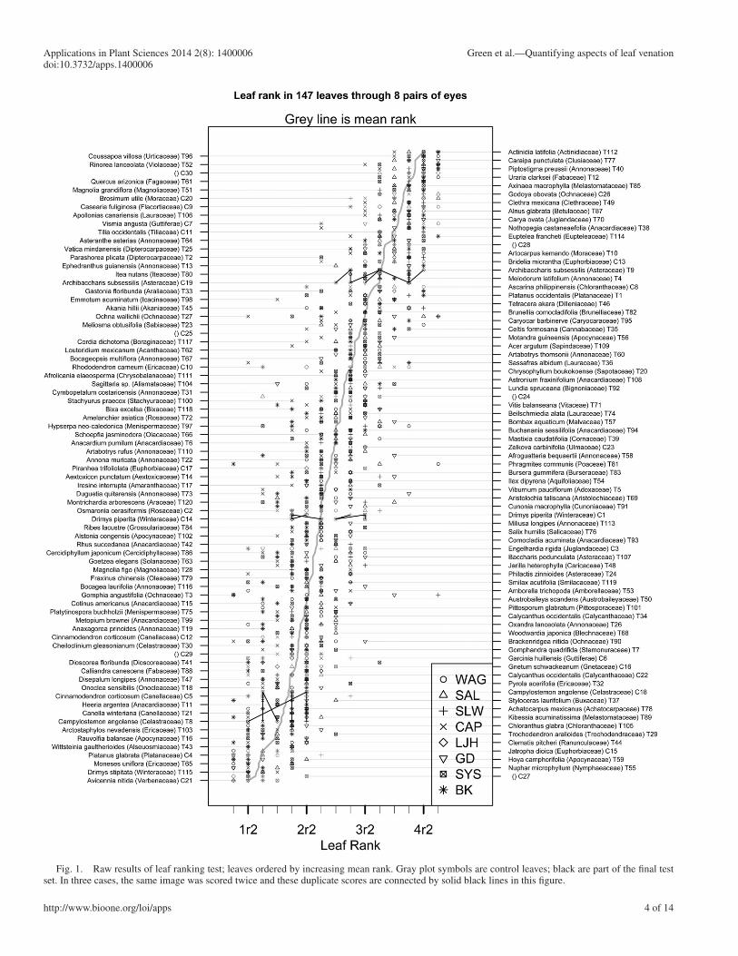
Fig. 1. Raw results of leaf ranking test; leaves ordered by increasing mean rank. Gray plot symbols are control leaves; black are part of the fi nal test set. In three cases, the same image was scored twice and these duplicate scores are connected by solid black lines in this fi gure.

Applications in Plant Sciences 2014 2 ( 8 ): 1400006 Green et al.—Quantifying aspects of leaf venation doi:10.3732/apps.1400006
5 of 14http://www.bioone.org/loi/apps
cluster about the lower ranks with the intermediate and advanced families ranked higher, respectively … most families fall within the third rank. Similar, though smaller, differences in rank occur between the primitive and ad-vanced genera of the better known families (Theaceae).
The original intent of leaf rank was primarily to describe the phylogenetic and ecological distribution of leaf organization in angiosperm leaves. The general conclusions reached were that:
“families tend to show one predominating level of organiza-tion. Families considered primitive from other evidence
Fig. 2. Comparisons between coders. This is a generalized pairs plot showing, below the diagonal, all the pairwise scatter plots comparing scores of each pair of coders. Each plotted point represents a specimen; the control images are in gray and the fi nal test set of 120 leaves are in black. Above the diagonal are parametric and nonparametric correlation statistics (Spearman’s ρ , Kendall’s τ , Pearson’s product-moment correlation r ) and the signifi cance of the least-squares regression ( P value). Correlations are signifi cant by all tests and range from about 0.6 to 0.9. Note that the vertical white lines in the scatter plots associated with scores of S.L.W. refl ect the omission of the transitional “nought” ranks. See Emerson et al. (2013) for further discussion of the generalized pairs plot. L.J.H. only completed the control sets.
6 of 14
Applications in Plant Sciences 2014 2 ( 8 ): 1400006 Green et al.—Quantifying aspects of leaf venation doi:10.3732/apps.1400006
http://www.bioone.org/loi/apps
AUTOMATED IMAGE ANALYSIS
Relatively recent advances in computer modeling and frac-tal geometry have stimulated interest in synthesizing leaf-like networks relying only on simple physical or mathematical models (e.g., Prusinkiewicz and Lindenmayer, 1990 ; Bohn et al., 2002 ; Couder et al., 2002 ; Runions et al., 2005 ; Dimitrov and Zucker, 2006 ). From a botanical perspective, the problem with such approaches is that they focus on the type of general-ity that may be physically or mathematically interesting but tends to demonstrate biological truisms. To the best of our knowledge, no study to date has provided a body of measure-ments of enough leaves to evaluate the ecological or phyloge-netic signal in different measurements, although the study by Wilf et al. (2012) represents a large-scale attempt currently under way to use automated image analysis to identify leaves taxonomically; and the database discussed by Das et al. (2014) could be so examined.
Using photographs of modern cleared leaves and an auto-mated image analysis protocol, we measured a number of at-tributes of the vein network of 120 leaves, representing most major angiosperm groups and a few ferns. The measurements related to the areoles—the largest regions of leaf lamina
The fossil record of the angiosperms also shows a gradual increase in the rank of leaf assemblages from the fi rst to the third ranks during Early Cretaceous time…. Finally, leaves of plants native to arid, arctic, or alpine environments frequently show a regression below the organizational level characteristic of the family.” ( Hickey, 1977 , p. 159)
Leaf ranking has not been very broadly applied, perhaps because it is a coarse measure, reliant heavily on the learned recognition of Gestalt categories. Therefore, it was only con-sidered possible to learn how to apply the system by calibrat-ing one’s own perceptions of organization against a database of leaves with established ranks, which has not until now been available in published form. In this paper, we provide rank scores for 120 leaves along with some data on the ex-pected variation among different coders. Little et al. (2014) and subsequent papers will focus on the systematic and eco-logical distribution of leaf rank; here we are concerned with establishing its repeatability, expected variation, and how it corresponds to automatic measurements. In the process, we also hope to demystify leaf ranking, which has been consid-ered a sort of black magic, as our results suggest that exten-sive training is not necessary for reliable application of the method.
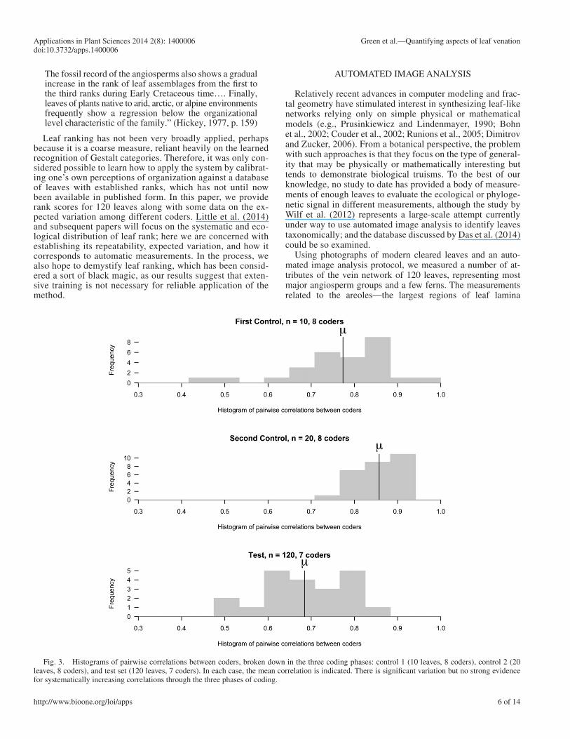
Fig. 3. Histograms of pairwise correlations between coders, broken down in the three coding phases: control 1 (10 leaves, 8 coders), control 2 (20 leaves, 8 coders), and test set (120 leaves, 7 coders). In each case, the mean correlation is indicated. There is signifi cant variation but no strong evidence for systematically increasing correlations through the three phases of coding.
Applications in Plant Sciences 2014 2 ( 8 ): 1400006 Green et al.—Quantifying aspects of leaf venation doi:10.3732/apps.1400006
7 of 14http://www.bioone.org/loi/apps
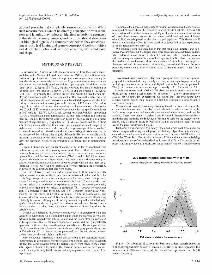
To evaluate the expected magnitude of random variation introduced, we also resampled 30 leaves from the complete control-plus-test set (150 leaves) 200 times and found a similar random spread. Figure 4 shows the actual distributions of correlations between coders for test leaves (solid line) and control leaves (dotted line) superimposed on the bootstrapped replicates. This shows that if there is any learning effect, it is too small to be identifi ed in this experiment, given the random errors observed.
We conclude from this examination that leaf rank is an imperfect and sub-jective measurement, but it is largely rank-order consistent across different coders, who tend to show correlations of about 0.7 with each other. Thus, leaf rank is a decent semiquantitative estimator of leaf organization. It is also very quick—the fi nal test set took most coders only a matter of a few hours to complete. Because leaf rank is determined subjectively, it remains diffi cult to be sure precisely what measurable characteristics of the vein network leaf rank describes.
Automated image analysis — The same group of 120 leaves was photo-graphed for automated image analysis using a macrophotographic setup including a macro lens, bellows, and digital camera back on a copy stand. The (true) image size was set to approximately 1.2 × 7 cm with a 2.4 × 1.6-cm image sensor with 3008 × 2000 pixels (about 4 × optical magnifica-tion), giving a true pixel dimension of about 4.4 μ m or approximately 50,000 pixels/mm 2 . By experiment, we found that this procedure gave clearer, flatter images than the use of a flat-bed scanner or a photographic stereomicroscope.
Where it was possible, two images were obtained for each leaf: one in the center of the lamina, intersected by the midrib, and the other wherever on the leaf lamina the primary and secondary network of major veins could best be avoided. These two images (labeled a and b ) should, therefore, respectively maximize and minimize the infl uence of the major veins on the intercostal ve-nation. The off-midrib image ( b ) was also used as the detailed image of each leaf in the rank test described above.
Each image was converted to a binary, black-and-white mask (black vein on white background) using an adaptive thresholding algorithm, automatically cleaned, and each connected white region measured using a MATLAB script (The MathWorks Inc., Natick, Massachusetts, USA) with the same underlying functionality as the software described by Price et al. (2011) . The details of this processing are provided as a MATLAB script (matlab_code.txt; available from
(ground parenchyma ) completely surrounded by veins. While such measurements cannot be directly converted to vein diam-eters and lengths, they refl ect an identical underlying geometry in thresholded (binary) images, and therefore should show sim-ilar biological signals. As demonstrated below, they are consis-tent across a leaf lamina and seem to correspond well to intuitive and descriptive notions of vein organization, like areole size and shape.
METHODS AND RESULTS
Leaf ranking — Our set of 120 species was chosen from the cleared leaves available in the National Cleared Leaf Collection (NCLC) at the Smithsonian Institution. Specimens were chosen to represent most major clades among the vascular plants, and were otherwise selected by grab sampling among the avail-able leaves in suffi ciently good condition to photograph. In addition to the “test” set of 120 leaves (T1–T120), we also collected two smaller training or “control” sets—the fi rst of 10 leaves (C1–C10) and the second of 20 leaves (C11–C30)—to evaluate the learning process that would be faced when em-ploying a new technique. After scoring each of the two small control sets, the coders compared results and discussed any substantial disagreements about the coding of each leaf before moving on to the fi nal set of 120 species. The coders ranged in experience from no prior experience with examination of leaf vena-tion (C.A.P., B.K.) to over a decade of experience with the specifi c techniques of leaf ranking (S.L.W., L.J.H.). All scoring was semiblinded: one author (W.A.G.) randomized and renumbered all the leaf images before redistributing them for coding. Three leaves were seen twice by each coder to get a direct measure of repeatability, giving a total of 147 distinct leaves and 150 distinct sets of rank scores. After the fi rst control set, a second, detailed image of the higher-order venation was provided, as well as a photograph of the whole leaf. In general, we seldom differed about the relative ranking of two leaves, but of-ten interpreted the ranking rules slightly differently. This was especially true in the cases of atypical leaves: those with palmate primary veins, very many or very few secondary veins, or the presence of a feature like an intramarginal vein.
Figure 1 shows the raw results of coding with the leaves reordered from bottom to top in order of increasing mean rank. For the three leaves scored twice (unbeknownst to most of the coders), the two occurrences are connected by solid lines, and the fi rst two control sets (C1–C10 and C11–C30) are shown in gray. Although we initially expected there to be more variation among the control leaves and more consistency between coders with the fi nal test set (a “learning” effect), we found no dramatic difference between the consistency with which the control and test sets were coded.
Note the relatively good rank-order consistency of all the scores, slightly higher consistency within the scores from an individual coder, and the rela-tively large range of variation among coders for some leaves. In general, scores for a single leaf tended to range over a full rank (four subranks), and the only effect of extensive prior experience seemed to be a weak propensity to avoid very high and low ranks. In particular, T81 ( Phragmites communis Trin.), a parallel-veined monocot, and T3 ( Gomphia angustifolia Vahl) showed the full range of possible variation. Two ferns ( Onoclea L. and Woodwardia Sm.) and a leaf of Gnetum L. were also included, all showing relatively low ranks, although leaf ranking was not originally intended to be applied outside the dicots. Figure 1 also shows, as had been observed anec-dotally in the past, that there were small systematic biases introduced by certain coders.
Despite the substantial differences among coders in experience with leaf venation in general and with leaf ranking in particular, the pairwise correlations between coders ranged from 0.6 to 0.9 and did not seem strongly correlated with experience—that is, the more experienced coders did not systematically agree more with each other than the inexperienced coders. This is apparent from Fig. 2 , where the control leaves are again shown as the gray points; the test set of 120 in black. All parametric and nonparametric tests for correlation between coders were positive and highly signifi cant.
Also, somewhat surprisingly, there did not seem to be signifi cant overall improvement in consistency over the course of the control and test sets despite the fact that some obvious errors by certain coders were made in the control sets. Figure 3 shows histograms of the pairwise correlations between coders, which vary signifi cantly but do not consistently improve over the course of the experiment.
Fig. 4. Distributions of correlations between coders, superimposed on 200 bootstrapped distributions of size n = 30. The solid line represents the fi nal test set (120 leaves, 7 coders); the dashed line represents controls (30 leaves, 8 coders).

8 of 14
Applications in Plant Sciences 2014 2 ( 8 ): 1400006 Green et al.—Quantifying aspects of leaf venation doi:10.3732/apps.1400006
http://www.bioone.org/loi/apps
Fig. 5. Images with distributions of areole measurements. For six images of three leaf specimens (on midrib and off midrib), the fi gure gives the dis-tributions of areole areas (in pixels) and areole eccentricities (dimensionless number between 0 and 1). Y -axes vary and are not meaningful because of the different number of areoles in each image; therefore, they are omitted from this fi gure. Note the small, narrow range of areole sizes and low eccentricity of areoles in the fi rst two images; the similar-sized but more eccentric areoles in the middle pair; and the larger, more variable areoles in the bottom two images.
the Dryad Digital Repository: http://dx.doi.org/10.5061/dryad.8h022; Green et al., 2014 ). The variables measured included:
Area: number of pixels in each region Convex Area: number of pixels in the convex hull of a region
Eccentricity: eccentricity of the ellipse that has the same second-moments as the region
Solidity: proportion of the pixels in the convex hull that are also in the re-gion. Computed as Area/Convex Area.

Applications in Plant Sciences 2014 2 ( 8 ): 1400006 Green et al.—Quantifying aspects of leaf venation doi:10.3732/apps.1400006
9 of 14http://www.bioone.org/loi/apps
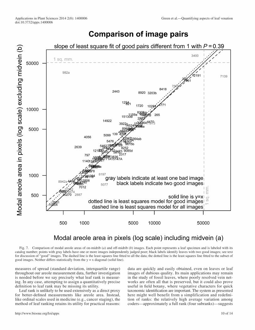
on-midrib ( a ) images directly with the off-midrib ( b ) summaries. Figure 7 shows this comparison, with each point representing a leaf labeled with its NCLC catalog number. Note the close correspondence between the images; whether or not potentially problematic images are excluded, the least squares fi tted line does not differ signifi cantly ( P = 0.39) from the y = x diagonal.
DISCUSSION
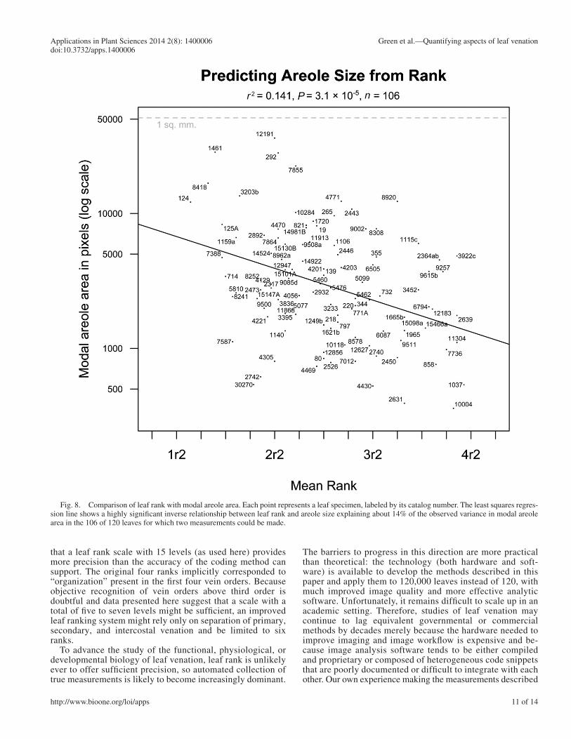
Figure 8 provides a direct comparison between the rank test results shown in Fig. 1 and the areole measurements shown in Fig. 6 . This reveals a statistically signifi cant but noisy relation-ship. As expected, leaf rank provides an approximate method for estimating modal areole area, explaining about 14% of the variance in the areole size measurements (using the squared correlation as a metric). Correlation tests using nonparametric measures of correlation are also signifi cant: P < 0.001 for both Kendall’s τ and Spearman’s ρ .
Comparing mean rank with the other measured quantities substantiates the idea that rank is not just a proxy for areole size. Of the six measured areole characteristics shown in Fig. 9 , leaf rank actually correlates best with areole standard deviation ( r = 0.59). Superfi cially, this suggests that our perceived value of leaf rank corresponds best to low variability in are-ole size. This is a plausible conclusion, but the data may not support it conclusively. Because there are signifi cant correla-tions between measures of center (mean, median, mode) and
Perimeter: distance around the boundary of each region Equivalent Diameter: diameter of a circle with the same area as the region Major Axis Length: length (in pixels) of the major axis of the ellipse that has
the same normalized second central moments as the region Minor Axis Length: length (in pixels) of the minor axis of the ellipse that has
the same normalized second central moments as the region We measured a total of 208,253 regions (areoles) in 230 images, of which
206 images seemed “good,” i.e., the masks seemed to refl ect the general pattern of veins and areoles observed in the original leaves accurately. This subjec-tive assessment of whether a binary mask accurately refl ects a vein network is not easily avoidable. As with many biological measurements, there is no gold standard for what constitutes accurate measurement of a feature of the real world; so our own human perception has to serve as ground truth ( Martin et al., 2004 ).
Figure 5 shows six images of three leaves. The fi rst column provides the grayscale image, the second column the binary mask, the third a distribution of the areole sizes, and the fourth a distribution of the areole eccentricities; be-cause the distribution shapes but not the absolute densities are relevant, the y -axis labels and ticks are omitted for clarity.
A complete representation of the raw areole area measurements is shown in Fig. 6 . Each measured areole is shown as a gray dot, the superimposed black lines give the interquartile ranges of each image, and the monotonically increasing line con-nects the modes of the areole area distributions of each leaf. Several features of in-terest can be observed from this representation: the pattern of right-skewed distributions of areole sizes with a mode on the lower end of the interquartile range is quite general; the mode, therefore, seems to be the best estimate of the central tendency of areole size in each image. The cropping at the bottom of the graph is due to the image cleaning protocol, which removes regions under a given size (in this case, 300 pixels), but notice that this cropping does not affect the modes.
Because most of the raw image measurements are paired (two images per leaf), a useful check is provided by comparing the summary statistics of the
Fig. 6. Raw areole measurements of 230 images representing 120 leaves. Gray dots correspond to raw areole size measurements; black vertical lines are the interquartile range for each image; the monotonically increasing line connects the modal areas for each image. Images are arranged from left to right in order of increasing modal areole size. Note cropping at the bottom caused by the image cleaning protocol and the right skew of the distributions (modes near the bottom of the interquartile ranges).
10 of 14
Applications in Plant Sciences 2014 2 ( 8 ): 1400006 Green et al.—Quantifying aspects of leaf venation doi:10.3732/apps.1400006
http://www.bioone.org/loi/apps
data are quickly and easily obtained, even on leaves or leaf images of dubious quality. Its main applications may remain in the study of fossil leaves, where poorly resolved vein net-works are often all that is preserved, but it could also prove useful in fi eld botany, where vegetative characters for quick taxonomic identifi cation are important. The system as presented here might well benefit from a simplification and redefi ni-tion of ranks: the relatively high average variation among coders—approximately a full rank (four subranks)—suggests
measures of spread (standard deviation, interquartile range) throughout our areole measurement data, further investigation is needed before we say precisely what leaf rank is measur-ing. In any case, attempting to assign a quantitatively precise defi nition to leaf rank may be missing its utility.
Leaf rank is unlikely to be used extensively as a direct proxy for better-defi ned measurements like areole area. Instead, like ordinal scales used in medicine (e.g., cancer staging), the method of leaf ranking retains its utility for practical reasons:
Fig. 7. Comparison of modal areole areas of on-midrib ( a ) and off-midrib ( b ) images. Each point represents a leaf specimen and is labeled with its catalog number; points with gray labels have one or more images independently judged poor; black labels identify leaves with two good images; see text for discussion of “good” images. The dashed line is the least squares line fi tted to all the data; the dotted line is the least squares line fi tted to the subset of good images. Neither differs statistically from the y = x diagonal (solid line).
Applications in Plant Sciences 2014 2 ( 8 ): 1400006 Green et al.—Quantifying aspects of leaf venation doi:10.3732/apps.1400006
11 of 14http://www.bioone.org/loi/apps
The barriers to progress in this direction are more practical than theoretical: the technology (both hardware and soft-ware) is available to develop the methods described in this paper and apply them to 120,000 leaves instead of 120, with much improved image quality and more effective analytic software. Unfortunately, it remains diffi cult to scale up in an academic setting. Therefore, studies of leaf venation may continue to lag equivalent governmental or commercial methods by decades merely because the hardware needed to improve imaging and image workfl ow is expensive and be-cause image analysis software tends to be either compiled and proprietary or composed of heterogeneous code snippets that are poorly documented or diffi cult to integrate with each other. Our own experience making the measurements described
that a leaf rank scale with 15 levels (as used here) provides more precision than the accuracy of the coding method can support. The original four ranks implicitly corresponded to “organization” present in the fi rst four vein orders. Because objective recognition of vein orders above third order is doubtful and data presented here suggest that a scale with a total of fi ve to seven levels might be suffi cient, an improved leaf ranking system might rely only on separation of primary, secondary, and intercostal venation and be limited to six ranks.
To advance the study of the functional, physiological, or developmental biology of leaf venation, leaf rank is unlikely ever to offer suffi cient precision, so automated collection of true measurements is likely to become increasingly dominant.
Fig. 8. Comparison of leaf rank with modal areole area. Each point represents a leaf specimen, labeled by its catalog number. The least squares regres-sion line shows a highly signifi cant inverse relationship between leaf rank and areole size explaining about 14% of the observed variance in modal areole area in the 106 of 120 leaves for which two measurements could be made.

12 of 14
Applications in Plant Sciences 2014 2 ( 8 ): 1400006 Green et al.—Quantifying aspects of leaf venation doi:10.3732/apps.1400006
http://www.bioone.org/loi/apps
Fig. 9. Generalized pairs plot comparing leaf rank with several other measured areole characteristics. The top left scatter plot is the same plot as Fig. 8 . Each point represents a leaf specimen and the other scatter plots below the diagonal show covariation among some of the other measured quantities. Note in particular that the correlation between mean leaf rank and areole standard deviation is substantially higher than the correlation between mean rank and areole size.
here provides a good example of the diffi culties that can be encountered. We relied on the underlying image analysis code in the commercial package MATLAB and, there fore, some of our analyses can only be documented by refer-ence to compiled code. The way forward that we would ad-vocate follows the open-source model: public repositories of images and commented source code, documented in sufficient detail for comparatively small contributions to build on each other. Attempts have been made to provide this sort of facility (e.g., http://clearedleavesdb.org/, http://www.
plant-image-analysis.org/), but many more publications in this arena describe new software than provide source code for it. In an attempt to provide an example of the procedure that we advocate, we have included in the supplemental ar-chive associated with this paper (available from the Dryad Digital Repository: http://dx.doi.org/10.5061/dryad.8h022; Green et al., 2014 ) all the images and code used. Because even the simplest data table would run to several pages when printed out, we have not included paper data appendices, but instead refer readers to the supplemental archive, which

Applications in Plant Sciences 2014 2 ( 8 ): 1400006 Green et al.—Quantifying aspects of leaf venation doi:10.3732/apps.1400006
13 of 14http://www.bioone.org/loi/apps
includes far more information than can be discussed in the text of the paper.
Finally, it should be re-emphasized that automation does not provide objectivity: throughout the process of automatic anal-ysis, choices are made and parameters are set that can bias the measurements in ways that may be unpredictable without ex-tensive sensitivity analysis (e.g., Price et al., 2014 ). Automation provides data of a sort that would be otherwise unobtainable, but its application also carries the need for thorough documen-tation of elaborate, complex procedures.
CONCLUSIONS
From this examination, we conclude that leaf rank can be quickly and repeatably scored by different people without signifi cant training or experience, with a consistency (correlation between pairs of coders) of about 0.7.
It also appears that leaf rank is inversely correlated to areole size and related to other measurable attributes of leaf areoles. Specifi cally, leaf rank seems to explain about 14% of the variance when used as a predictor of modal areole area in a set of 120 angiosperm leaves. It may be even more closely associated with measures of within-leaf areole variability.
Areole size is generally consistent across the leaf lamina and can be reliably measured on an area as small as 1 cm 2 , provided images of suffi cient quality are available. The barriers to quan-titative description of leaf venation tend to be practical: the need for images that can be accurately thresholded and the dif-fi culty of completely automating the process of image cleaning and analysis.
Thus, leaf rank remains a useful semi-quantitative proxy for leaf organization when fully quantitative measurements cannot be made, as in the case of most fossilized material. Automated measurements, which will become easier with every advance in digital image acquisition and analysis, provide hitherto unob-tainable data with which to investigate functional, physiologi-cal, or developmental attributes of modern leaves. It is essential for the progress of such studies to build up public databases of identifi ed, high-quality images and commented source code, documenting analytic choices in full detail.
LITERATURE CITED
ASH , A. , B. ELLIS , L. J. HICKEY , K. JOHNSON , P. WILF , AND S. WING . 1999 . Manual of leaf architecture. Smithsonian Institution, Washington, D.C., USA.
BEHRENSMEYER , A. K. , J. D. DAMUTH , W. A. DIMICHELE , R. POTTS , H.-D. SUES , AND S. L. WING . 1992 . Terrestrial ecosystems through time. University of Chicago Press, Chicago, Illinois, USA.
BOHN , S. , B. ANDREOTTI , S. DOUADY , J. MUNZINGER , AND Y. COUDER . 2002 . Constitutive property of the local organization of leaf venation networks. Physical Review E: Statistical, Nonlinear, and Soft Matter Physics 65 : 061914 .
BOYCE , C. K. , T. J. BRODRIBB , T. S. FIELD , AND M. A. ZWIENIECKI . 2009 . Angiosperm leaf vein evolution was physiologically and environmentally transformative. Proceedings of the Royal Society. Series B, Biological Sciences 276 : 1771 – 1776 .
BRODRIBB , T. J. , T. FEILD , AND L. SACK . 2010 . Viewing leaf structure and evolution from a hydraulic perspective. Functional Plant Biology 37 : 488 – 498 .
COUDER , Y. , L. PAUCHARD , C. ALLAIN , M. ADDA-BEDIA , AND S. DOUADY . 2002 . The leaf venation as formed in a tensorial fi eld. European Physical Journal B 28 : 135 – 138 .
DAS , A. , A. BUCKSCH , C. A. PRICE , AND J. S. WEITZ . 2014 . ClearedLeavesDB: An online database of cleared plant leaf images. Plant Methods 10 : 8 .
DIMICHELE , W. A. , AND S. L. WING [eds.]. 1988 . Methods and applications of plant paleoecology. Paleontological Society Special Publications 3. Published for the Paleontological Society at the Department of Geological Sciences, University of Tennessee-Knoxville, Knoxville, Tennessee, USA.
DIMITROV , P. , AND S. W. ZUCKER . 2006 . A constant production hypothesis guides leaf venation patterning. Proceedings of the National Academy of Sciences, USA 103 : 9363 – 9368 .
DOYLE , J. A. , AND L. J. HICKEY . 1976 . Pollen and leaves from the mid-Cretaceous Potomac Group and their bearing on early angiosperm evo-lution. In C. B. Beck [ed.], Origin and early evolution of angiosperms, 139–206. Columbia University Press, New York, New York, USA.
ELLIS , B. , D. C. DALY , L. J. HICKEY , K. R. JOHNSON , J. D. MITCHELL , P. WILF , AND S. L. WING . 2009 . Manual of leaf architecture. Cornell University Press, Ithaca, New York, USA.
EMERSON , J. W. , W. A. GREEN , B. SCHLOERKE , J. CROWLEY , D. COOK , H. HOFMANN , AND H. WICKHAM . 2013 . The Generalized Pairs Plot. Journal of Computational and Graphical Statistics 22 : 79 – 91 .
FEILD , T. S. , N. C. ARENS , J. A. DOYLE , T. E. DAWSON , AND M. J. DONOGHUE . 2004 . Dark and disturbed: A new image of early angiosperm ecology. Paleobiology 30 : 82 – 107 .
FEILD , T. S. , D. S. CHATELET , AND T. J. BRODRIBB . 2009 . Ancestral xerophobia: A hypothesis on the whole plant ecophysiology of early angiosperms. Geobiology 7 : 237 – 264 .
GIAQUINTA , R. 1978 . Source and sink leaf metabolism in relation to phloem translocation. Plant Physiology 61 : 380 – 385 .
GIVNISH , T. J. 1978 . Ecological aspects of plant morphology: Leaf form in relation to environment . In R. Sattler [ed.], Theoretical plant morphol-ogy, 83–142. Folia Biotheoretica, Supplement 7 to Acta Biotheoretica 27. Leiden University Press, The Hague, The Netherlands.
GOULD , S. J. , AND R. C. LEWONTIN . 1979 . The spandrels of San Marco and the panglossian paradigm: A critique of the adaptationist programme. Proceedings of the Royal Society. Series B, Biological Sciences 205 : 581 – 598 .
GREEN , W. A. , S. A. LITTLE , C. A. PRICE , S. L. WING , S. Y. SMITH , B. KOTRC , AND G. DORIA . 2014 . Data from: Reading the leaves: A comparison of leaf rank and automated areole measurement for quantifying aspects of leaf venation. Dryad Digital Repository. http://dx.doi.org/10.5061/dryad.8h022
HICKEY , L. J. 1971 . Evolutionary signifi cance of leaf architectural features in the woody dicots [abstract] . American Journal of Botany 58 : 469 .
HICKEY , L. J. 1973 . Classifi cation of the architecture of dicotyledonous leaves. American Journal of Botany 60 : 17 – 33 .
HICKEY , L. J. 1974 . A revised classifi cation of the architecture of dicotyle-donous leaves. In C. R. Metcalfe and L. Chalk [eds.], Anatomy of the dicotyledons, 25–39, vol. 1, 2nd ed. Clarendon Press, Oxford, United Kingdom.
HICKEY , L. J. 1977 . Stratigraphy and paleobotany of the Golden Valley Formation (Early Tertiary) of western North Dakota. Geological Society of America Memoir No. 150. Geological Society of America, Boulder, Colorado, USA.
HICKEY , L. J. , AND J. A. DOYLE . 1977 . Early Cretaceous fossil evidence for angiosperm evolution. Botanical Review 43 : 3 – 104 .
HICKEY , L. J. , AND J. A. WOLFE . 1975 . The bases of angiosperm phy-logeny: Vegetative morphology. Annals of the Missouri Botanical Garden 62 : 538 – 589 .
HILL , R. S. 1980 . A numerical taxonomic approach to the study of angio-sperm leaves. Botanical Gazette (Chicago, Ill.) 141 : 213 – 229 .
HORTON , R. E. 1945 . Erosional development of streams and their drain-age basins: Hydrophysical approach to quantitative morphology. Geological Society of America Bulletin 56 : 275 – 370 .
KATIFORI , E. , G. J. SZÖLLŐSI , AND M. O. MAGNASCO . 2010 . Damage and fl uctuations induce loops in optimal transport networks. Physical Review Letters 104 : 048704 .
KATIFORI , E. , AND M. O. MAGNASCO . 2012 . Quantifying loopy network architectures. PLoS ONE 7 : e37994 .
LINNAEUS , C. 1753 . Species Plantarum. Holmius, Uppsala, Sweden.

14 of 14
Applications in Plant Sciences 2014 2 ( 8 ): 1400006 Green et al.—Quantifying aspects of leaf venation doi:10.3732/apps.1400006
http://www.bioone.org/loi/apps
LITTLE , S. A. , W. A. GREEN , S. L. WING , AND P. WILF . 2014 . Reinvestigation of leaf rank, an underappreciated component of Leo Hickey’s legacy . Bulletin of the Peabody Museum of Natural History 55 : in press.
MARTIN , D. R. , C. C. FOWLKES , AND J. MALIK . 2004 . Learning to detect nat-ural image boundaries using local brightness, color, and texture cues. IEEE Transactions on Pattern Analysis and Machine Intelligence 26 : 530 – 549 .
MELVILLE , R. 1976 . The terminology of leaf architecture. Taxon 25 : 549 – 561 .
MILEYKO , Y. , H. EDELSBRUNNER , C. A. PRICE , AND J. S. WEITZ . 2012 . Hierarchical ordering of reticular networks. PLoS ONE 7 : e36715 .
NELSON , T. , AND N. DENGLER . 1997 . Leaf vascular pattern formation. Plant Cell 9 : 1121 – 1135 .
NEWMAN , E. P. 1964 . Newly discovered Franklin invention: Nature printing on colonial and continental currency. Numismatist 77 : 147 – 154 .
NOBLIN , X. , L. MAHADEVAN , I. A. COOMARASWAMY , D. A. WEITZ , N. M. HOLBROOK , AND M. A. ZWIENIECKI . 2008 . Optimal vein density in artifi cial and real leaves. Proceedings of the National Academy of Sciences, USA 105 : 9140 – 9144 .
PIAZZA , P. , S. JASINSKI , AND M. TSIANTIS . 2005 . Evolution of leaf develop-mental programmes. New Phytologist 167 : 693 – 710 .
POETHIG , S. 1984 . Cellular parameters of leaf morphogenesis in maize and tobacco. In R. A. White and W. C. Dickison [eds.], Contemporary problems in plant anatomy, 235–259. Academic Press, Orlando, Florida, USA.
PRICE , C. A. , O. SYMONOVA , Y. MILEYKO , T. HILLEY , AND J. S. WEITZ . 2011 . Leaf extraction and analysis framework graphical user interface: Segmenting and analyzing the structure of leaf veins and areoles. Plant Physiology 155 : 236 – 245 .
PRICE , C. A. , S. WING , AND J. S. WEITZ . 2012 . Scaling and struc-ture of dicotyledonous leaf venation networks. Ecology Letters 15 : 87 – 95 .
PRICE , C. A. , S.-J. C. KNOX , AND T. J. BRODRIBB . 2013 . The infl uence of branch order on optimal leaf vein geometries: Murray’s Law and area preserving branching. PLoS ONE 8 : e85420 .
PRICE , C. A. , P. R. T. MUNRO , AND J. S. WEITZ . 2014 . Estimates of leaf vein density are scale dependent. Plant Physiology 164 : 173 – 180 .
PRUSINKIEWICZ , P. , AND A. LINDENMAYER . 1990 . The algorithmic beauty of plants. Springer, New York, New York, USA.
ROLLAND-LAGAN , A.-G. , M. AMIN , AND M. PAKULSKA . 2009 . Quantifying leaf venation patterns: Two-dimensional maps. Plant Journal 57 : 195 – 205 .
ROTH-NEBELSICK , A. , D. UHL , V. MOSBRUGGER , AND H. KERP . 2001 . Evolution and function of leaf venation architecture: A review. Annals of Botany 87 : 553 – 566 .
RUNIONS , A. , M. FUHRER , B. LANE , P. FEDERL , A.-G. ROLLAND-LAGAN , AND P. PRUSINKIEWICZ . 2005 . Modeling and visualization of leaf venation patterns. ACM Transactions on Graphics 24 : 702 – 711 .
SACK , L. , AND N. M. HOLBROOK . 2006 . Leaf hydraulics. Annual Review of Plant Biology 57 : 361 – 381 .
SACK , L. , AND C. SCOFFONI . 2013 . Leaf venation: Structure, function, development, evolution, ecology and applications in the past, present and future. New Phytologist 198 : 983 – 1000 .
SARGENT , C. S. 1905 . Manual of the trees of North America. Houghton, Miffl in and Co., Boston, Massachusetts, USA.
STRAHLER , A. N. 1957 . Quantitative analysis of watershed geomorphol-ogy. American Geophysical Union Transactions 38 : 913 – 920 .
TSIANTIS , M. , AND J. A. LANGDALE . 1998 . The formation of leaves. Current Opinion in Plant Biology 1 : 43 – 48 .
VON ETTINGSHAUSEN , C. R. 1861 . Die Blatt-Skelete der Dikotyledonen. Druck und Verlag der Kais, Kön. Hof- und Staatdruckerei, Vienna.
WILF , P. , S. CHIKKERUR , S. A. LITTLE , AND T. SERRE . 2012 . Computer vi-sion cracks the leaf code [abstract], 201-7. Geological Society of America Annual Meeting, Charlotte, North Carolina, USA.
WYLIE , R. B. 1938 . Concerning the conductive capacity of the mi-nor veins of foliage leaves. American Journal of Botany 25 : 567 – 572 .
WYLIE , R. B. 1939 . Relations between tissue organization and vein distribution in dicotyledon leaves. American Journal of Botany 26 : 219 – 225 .
WYLIE , R. B. 1943 . The role of the epidermis in foliar organization and its relations to the minor venation. American Journal of Botany 30 : 273 – 280 .
WYLIE , R. B. 1946 . Relations between tissue organization and vascu-larization in leaves of certain tropical and subtropical dicotyledons. American Journal of Botany 33 : 721 – 726 .
WYLIE , R. B. 1949 . Variations in leaf structure among Adiantum peda-tum plants growing in a rock cavern. American Journal of Botany 36 : 282 – 287 .
WYLIE , R. B. 1951 . Principles of foliar organization shown by sun-shade leaves from ten species of deciduous dicotyledonous trees. American Journal of Botany 38 : 355 – 361 .
WYLIE , R. B. 1952 . The bundle sheath extension in leaves of dicotyle-dons. American Journal of Botany 39 : 645 – 651 .
WYLIE , R. B. 1954 . Leaf organization of some woody dicotyledons from New Zealand. American Journal of Botany 41 : 186 – 191 .
Related Documents