DOES HUMAN NEUROGLOBIN ACT ONLY AS A SCAVENGER? REACTIVITY AND ENDOGENOUS MODIFICATION BY NITRITE AND HYDROGEN PEROXIDE * Stefania Nicolis * , Enrico Monzani * , Chiara Ciaccio † , Paolo Ascenzi ‡ , Luc Moens § , and Luigi Casella *1 * Dipartimento di Chimica Generale, Università di Pavia, Via Taramelli 12, 27100 Pavia, Italy, † Dipartimento di Medicina Sperimentale e Scienze Biochimiche, Università di Roma-Tor Vergata, Via Montpellier 1, 00133 Roma, Italy, ‡ Dipartimento di Biologia, Università “Roma Tre”, Viale Guglielmo Marconi 446, 00146, Roma, Italy, § Department of Biomedical Sciences, University of Antwerp, Universiteit 1, B-2610 Antwerp, Belgium 1 To whom correspondence should be addressed: Prof. Luigi Casella, Dipartimento di Chimica Generale, Università di Pavia, Via Taramelli 12, 27100 Pavia, Italy. Tel. +39-0382-507331; Fax: +39-0382-528544; E-Mail: [email protected] Running Title: Activation of Nitrite by Neuroglobin and Hydrogen Peroxide Biochemical Journal Immediate Publication. Published on 29 Jun 2007 as manuscript BJ20070372 © 2007 The Authors Journal compilation © 2007 Biochemical Society

Welcome message from author
This document is posted to help you gain knowledge. Please leave a comment to let me know what you think about it! Share it to your friends and learn new things together.
Transcript
DOES HUMAN NEUROGLOBIN ACT ONLY AS A SCAVENGER? REACTIVITY
AND ENDOGENOUS MODIFICATION BY NITRITE AND HYDROGEN PEROXIDE*
Stefania Nicolis*, Enrico Monzani*, Chiara Ciaccio†, Paolo Ascenzi‡, Luc Moens§, and Luigi
Casella*1
*Dipartimento di Chimica Generale, Università di Pavia, Via Taramelli 12, 27100 Pavia, Italy,†Dipartimento di Medicina Sperimentale e Scienze Biochimiche, Università di Roma-Tor
Vergata, Via Montpellier 1, 00133 Roma, Italy, ‡Dipartimento di Biologia, Università “Roma
Tre”, Viale Guglielmo Marconi 446, 00146, Roma, Italy, §Department of Biomedical Sciences,
University of Antwerp, Universiteit 1, B-2610 Antwerp, Belgium
1 To whom correspondence should be addressed: Prof. Luigi Casella, Dipartimento di Chimica
Generale, Università di Pavia, Via Taramelli 12, 27100 Pavia, Italy. Tel. +39-0382-507331;
Fax: +39-0382-528544; E-Mail: [email protected]
Running Title: Activation of Nitrite by Neuroglobin and Hydrogen Peroxide
Biochemical Journal Immediate Publication. Published on 29 Jun 2007 as manuscript BJ20070372
© 2007 The Authors Journal compilation © 2007 Biochemical Society

Abbreviations used: NGB, human neuroglobin; Mb, myoblobin; Hb, hemoglobin; ROS,
reactive oxygen species; RNS, reactive nitrogen species; NGBS-S and NGBSH, the two forms of
NGB with and without the internal disulfide bond, respectively; 4-PDS, 4,4′-dithiodipyridine;
DTT, dithiothreitol; EDTA, ethylenediaminetetraacetic acid; 4-TP, 4-thiopyridone; HPA, 3-(4-
hydroxyphenyl)-propionic acid; HMb, human myoglobin; LPO, lactoperoxidase; p-NGBs,
modified NGBs at low nitrite and hydrogen peroxide concentrations; h-NGBs, modified NGBs at
high nitrite and hydrogen peroxide concentrations; p′-NGBSH, modified NGBSH at low hydrogen
peroxide concentrations; h′-NGBSH, modified NGBSH at high hydrogen peroxide concentrations;
lpo-NGBs, modified NGBs in the presence of LPO, nitrite and hydrogen peroxide; EIC,
extracted ion current; ABTS, 2,2’-azino-bis(3-ethylbenz-thiazoline-6-sulfonic acid); HPLC-ESI-
MS/MS, high performance liquid chromatography-electrospray ionization-tandem mass
spectrometry; TFA, trifluoroacetic acid; CID, collision induced dissociation; amu, atomic mass
unit; Gdn-HCl, guanidinium hydrochloride.
2
Biochemical Journal Immediate Publication. Published on 29 Jun 2007 as manuscript BJ20070372
© 2007 The Authors Journal compilation © 2007 Biochemical Society

ABSTRACT
Human neuroglobin (NGB), a recently discovered heme protein of the globin family containing a
six-coordinated heme, is expressed in the nervous tissue with a yet unclear physiological
function. Besides being involved in neuronal oxygen homeostasis, NGB is thought to act as a
scavenger of reactive species. Herein we report on the reactivity of metNGB, which can
accumulate in vivo by reaction of oxyNGB with nitric oxide, towards nitrite and H2O2. Nitrite
coordination to the heme accounts for the activity of metNGB in the nitration of phenolic
substrates. The two metNGB forms, with and without the internal disulfide bond between
Cys(46)CD7 and Cys(55)D5, exhibit different reactivity, the former being more efficient in
activating NO2-. The kinetics of the reactions, the nitrite-binding studies, and the analysis of the
nitrated products from different substrates all support the hypothesis that metNGB is able to
generate, at pathophysiological concentration of nitrite and H2O2, an active species with the
chemical properties of peroxynitrite.
Without external substrates, the targets of the reactive species generated by the
metNGB/NO2-/H2O2 system are endogenous tyrosine (transformed into 3-nitrotyrosine) and
cysteine (oxidized to sulfinic and sulfonic acids) residues. The endogenous modifications were
characterized by HPLC-MS/MS analysis of metNGB reacted with nitrite and H2O2 in various
conditions. The internal S-S bond affects the functional properties of the protein. Therefore,
metNGB acts not only as scavenger of toxic species, but also as target of the self-generated
reactive species: the protein self-modification may be related or impair its postulated
neuroprotective activity.
KEY WORDS: human neuroglobin, peroxynitrite, heme proteins, tyrosine nitration, cysteine
oxidation, oxidative stress
3
Biochemical Journal Immediate Publication. Published on 29 Jun 2007 as manuscript BJ20070372
© 2007 The Authors Journal compilation © 2007 Biochemical Society
INTRODUCTION
Human neuroglobin (NGB) was first identified in 2000 as a third globin type protein,
besides myoglobin (Mb) and hemoglobin (Hb) [1]. NGB is expressed at low levels (in the µM
range) in various regions of the brain, but at much higher levels (∼100 µM) in retinal cells [1,2].
NGB consists of 151 amino acids and shares high sequence similarity with mouse neuroglobin
(94 % identity) but little homology with vertebrate Mbs and Hbs (<21 % and <25 % identity,
respectively) [1]. It differs from the other vertebrate globins also for the six-coordinated heme
(with His(96)F8 and His(64)E7 acting as the fifth/proximal and sixth/distal ligands,
respectively), in both the ferrous and ferric forms [3,4] (Figure 1). The presence of a six-
coordinated heme reduces the affinity of the protein for ligands with respect to the expected
values in the absence of the competitive coordination by the distal histidine [3].
= Figure 1 =
The function of NGB in the human brain remains unclear. In analogy to other globins,
several possible physiological roles have been considered [5]; among these, much indirect
evidence such as the correlation between NGB expression and O2 consumption, suggests a role
of NGB in neuronal oxygen homeostasis [6]. The oxygen affinity of NGB depends on several
factors: the relative binding rates at the heme sixth position of O2 and the competing distal
histidine [3,7], and the redox state of the cell which controls the formation or cleavage of an
internal disulfide bond between Cys(46)CD7 and Cys(55)D5 [8,9]. The presence of the S-S bond
could perturb the three-dimensional structure of the CD-D region and affect the location of the
neighboring E-helix, thus modulating the binding of the endogenous His(64)E7 ligand to the
heme [9,10]. As a consequence, in ferrous NGB the distal histidine dissociation rate increases by
a factor of 10 with respect to the protein form without the disulfide bond [9,11], thus leading to
an effective increase in O2 affinity by the same factor [12] (Figure 1).
O2 storage or diffusion to or from the heme of NGB may be assisted by the presence of a
large protein matrix cavity (about 120 Å3), which is located between the heme distal site and the
EF interhelical hinge and connected to the protein surface [10,13]. The cavity may be reshaped
upon ligand binding, which may allow trapping of harmful reactive oxygen or nitrogen species
(ROS and RNS, respectively) [14-17]. In particular, the rise of NO concentration up to the low µ
M range occurring under ischemic conditions [18], may be contrasted by its reaction with
4
Biochemical Journal Immediate Publication. Published on 29 Jun 2007 as manuscript BJ20070372
© 2007 The Authors Journal compilation © 2007 Biochemical Society
oxyNGB, yielding metNGB (i.e. the ferric form of NGB) and NO3-, through a heme-bound
peroxynitrite intermediate [15]:
NGBFeIIO2 + NO → NGBFeIIIOONO- → NGBFeIII + NO3- (1)
Since in the brain a metNGB reductase system has not been identified yet [15], the in vivo
formation of metNGB through this mechanism may have physiological relevance. Also metNGB
reacts with NO [19], generating the stable NGBFeIINO species by reductive nitrosylation; this
form of NGB is a good scavenger of peroxynitrite, which oxidizes the protein back to metNGB,
according to the following reactions [19]:
NGBFeIII → NO NGBFeIINO (2)
NGBFeIINO →−ONOO NGBFeIIINO (3)
NGBFeIIINO → NGBFeIII + NO (4)
In contrast to Mb and Hb, the protein does not generate the ferryl form upon reaction with H2O2
or ONOO- [20]. No reaction was either observed upon addition of nitrite [19].
Thus, even if the FeIII center of metNGB seems to be protected from attack of oxidizing
species by the coordinated distal His, the competitive coordination of exogenous ligands could
account for the activation of alternative reactions. Herein we report on the reactivity of human
metNGB in the presence of nitrite and hydrogen peroxide, whose concentrations in vivo increase
under conditions of oxidative stress [21]. The activation of NO2-/H2O2 by metNGB was assayed
in the nitration of phenolic substrates [22-24], obtaining an unexpectedly high reactivity.
Moreover, the two forms of the protein, with and without the internal disulfide bond (metNGBS-S
and metNGBSH, respectively), exhibit significantly different efficiency in the activation of NO2-
and H2O2. The problem of the endogenous modification of metNGB by reaction with NO2- and
H2O2, even at pathophysiological concentrations, in the absence of external substrates was also
addressed, since the identification of protein residues modified by ROS and RNS is essential for
the understanding of the mechanisms of development of various pathologies [25-29] and the
postulated neuroprotective function of NGB. The endogenous targets of the reactive species
generated by the protein are tyrosine and cysteine residues, with a more extensive pattern of
modifications than previously observed with HMb [24].
5
Biochemical Journal Immediate Publication. Published on 29 Jun 2007 as manuscript BJ20070372
© 2007 The Authors Journal compilation © 2007 Biochemical Society

EXPERIMENTAL
Reagents - All the buffer solutions were prepared with deionized Milli-Q water.
Hydrogen peroxide (30 % solution), 3-(4-hydroxyphenyl)-propionic acid (HPA), 4-
hydroxybenzonitrile and phenylacetic acid were obtained from Aldrich, whereas trypsin was
from Sigma. The other reagents were obtained at the best grade available. The concentration of
hydrogen peroxide solutions was controlled by monitoring the formation of the ABTS radical
cation according to a standard enzymatic method [30]. Recombinant human NGB was expressed
and purified as previously reported [12]. In order to obtain a homogeneous sulfur-bridged protein
(i.e. NGBS-S), the protein solution was dialyzed overnight at 4 °C against 50 mM phosphate
buffer pH 7.5. To reduce the intramolecular disulfide bond (obtaining NGBSH), the NGB solution
was dialyzed at 37 °C against 1 mg/ml DTT dissolved in degassed 50 mM Tris-HCl/0.5 mM
EDTA buffer, pH 7.5, under anaerobic conditions. After 30 min reduction, three exchanges of
the same degassed buffer (50 mM Tris-HCl/0.5 mM EDTA, pH 7.5) were made to eliminate
DTT. Protein dialysis was always performed at 4 °C. In all the experiments described below,
when not explicitally stated, the proteins (NGBS-S and NGBSH) were utilized in their met form.
All spectrophotometric measurements were performed on a Hewlett Packard HP 8452A diode
array spectrophotometer.
Kinetic Studies of Phenol Nitration Catalyzed by NGBS-S and NGBSH - The kinetic
experiments were carried out in 200 mM phosphate buffer pH 7.5 using a quartz cuvette with
path length of 1 cm, thermostatated at 25.0 ± 0.1 °C and equipped with a magnetic stirrer. The
initial solution containing protein and variable substrate and nitrite concentrations (final volume
1600 µL) was obtained by mixing solutions of appropriate concentration of the reagents in the
buffer. The reaction was started by the addition of the H2O2 solution and was followed by
monitoring the absorbance change at 450 nm during the initial 10-15 s. The conversion of the
rate data from absorbance s-1 into [nitrophenol] s-1 was done by using the extinction coefficient of
3-(4-hydroxy-3-nitrophenyl)-propionic acid at 450 nm, ε = 3350 M -1 cm-1 [22]. The kinetic
parameters were obtained from fitting the plots of experimental rates at different substrate/nitrite
concentrations to the appropriate equations (see Results).
For each substrate, the rate dependence on the various reactant concentrations was
studied through three series of steps: (1) finding a suitable [H2O2] maximizing the rate but
avoiding unwanted excess of the oxidant, and then using this [H2O2] (2) study the dependence of
6
Biochemical Journal Immediate Publication. Published on 29 Jun 2007 as manuscript BJ20070372
© 2007 The Authors Journal compilation © 2007 Biochemical Society

the rate vs. [HPA], and (3) study the dependence of the rate vs. [NO2-], following the iterative
procedure previously described in detail [23]. The protein (NGBS-S or NGBSH) concentration was
1 µM in all the reactions, while the concentration of the other reactants were as follows: (1)
optimization of the peroxide concentration: using NGBS-S, [HPA] = 1 mM, [NO2-] = 0.3 M,
[H2O2] = 0.09–0.8 M; using NGBSH, [HPA] = 0.4 mM, [NO2-] = 1.0 M, [H2O2] = 0.09–0.8 M; (2)
dependence of the rate vs. HPA concentration: using NGBS-S, [H2O2] = 0.6 M, [NO2-] = 0.3 M,
[HPA] = 0.031-1.2 mM; using NGBSH, [H2O2] = 0.6 M, [NO2-] = 1.0 M, [HPA] = 0.0096-0.96
mM; (3) dependence of the rate vs. nitrite concentration: using NGBS-S, [H2O2] = 0.6 M, [HPA] =
0.4 mM, [NO2-] = 0.0062-0.44 M; using NGBSH, [H2O2] = 0.6 M, [HPA] = 0.3 mM, [NO2
-] =
0.037-2.5 M.
The reaction rates observed in the absence of the protein (noncatalytic reaction) or
without hydrogen peroxide are completely negligible.
Binding of Nitrite - To a solution of NGBS-S (4.8 µM, 1600 µL) or NGBSH (5.2 µM, 1600
µL) in 200 mM phosphate buffer pH 7.5, increasing quantities of a concentrated nitrite solution
in the same buffer (final concentration from 0 to 0.42 M for NGBS-S and from 0 to 0.64 M for
NGBSH) were added in a quartz cuvette with path length of 1 cm, thermostated at 25.0 ± 0.1 °C,
and UV-Vis spectra were recorded after each addition. Blank spectra were recorded in the same
way but in the absence of protein. After subtracting the corresponding blank to each spectrum,
the resulting spectra were corrected for dilution and then transformed into difference spectra by
subtracting the native protein spectrum. A plot was constructed with the difference between the
absorbance changes at 414 nm and at 434 nm, the wavelengths at which the difference spectra
exhibit the maximum variations, vs. the ligand concentration.
For the binding of NO2- to NGBS-S, the constants K1 and K2 were obtained by
interpolation of the absorbance data with the binding isotherm for low affinity binding of two
consecutive ligands: ∆A = (∆A∞,1×K1×[NO2-]tot+∆A∞,2×K1×K2×[NO2
-]tot2)/(1+K1×[NO2
-]tot+K1×K2
×[NO2-]tot
2) (where ∆A∞,1 and ∆A∞,2 represent, respectively, the absorbance changes due to the
binding of one and two ligand molecules, and [NO2-]tot the total nitrite concentration, free plus
bound). For the binding of NO2- to NGBSH, the constant KB was obtained by interpolation of the
absorbance data with the binding isotherm for low affinity binding of a single ligand: ∆A = ∆A∞
×KB×[NO2-]tot/(1+KB×[NO2
-]tot) (where ∆A∞ represents the absorbance change consequent to the
binding of NO2-, and [NO2
-]tot the total nitrite concentration).
7
Biochemical Journal Immediate Publication. Published on 29 Jun 2007 as manuscript BJ20070372
© 2007 The Authors Journal compilation © 2007 Biochemical Society

HPLC Analysis of the Nitration Products of 4-Hydroxybenzonitrile and Phenylacetic Acid - The
product mixtures derived from the nitration of 4-hydroxybenzonitrile and phenylacetic acid
promoted by the NGB/NO2-/H2O2 system, were analyzed by HPLC using a Jasco MD-1510
instrument with diode array detection equipped with a SupelcosilTM LC18 reverse-phase
semipreparative column (5 µm, 250×10 mm). Elution was carried out using 0.1 % TFA in
distilled water (solvent A) and 0.1 % TFA in acetonitrile (solvent B), with a flow rate of 5
ml/min. Elution started with 100 % solvent A for 5 min, followed by a linear gradient from 100
% A to 100 % B in 25 min. Spectrophotometric detection of the eluate was performed in the
range 200-600 nm.
The catalytic nitrations of 4-hydroxybenzonitrile and phenylacetic acid were studied
under the following experimental conditions: [NGBS-S] = 1 µM, [substrate] = 1 mM, [H2O2] = 0.6
M, [NO2-] = 0.01, 0.05, or 0.15 M, in 200 mM phosphate buffer pH 7.5 The reaction mixtures
were allowed to react for 10 min at room temperature, and then were analyzed by HPLC as
described above. The retention time of 4-hydroxybenzonitrile was 15.7 min, while the
corresponding nitration product, 4-hydroxy-3-nitrobenzonitrile, was absent in all the conditions.
Its retention time (17.4 min) was established through the LPO-catalyzed nitration carried out in
the following conditions: [LPO] = 80 nM, [phenol] = 1 mM, [H2O2] = 1 mM, [NO2-] = 0.01 M, in
200 mM phosphate buffer pH 7.5. The HPLC chromatograms of the mixtures obtained from the
nitration of phenylacetic acid showed, besides unreacted substrate at 16.8 min, two main peaks at
16.0 and 16.2 min and two minor peaks at 14.5 and 14.7 min (obtained all both at low and high
NO2- concentrations).
HPLC analysis showed that no modification of 4-hydroxybenzonitrile and phenylacetic
acid occurred in the absence of the protein or without hydrogen peroxide.
Modification of NGB in the Presence of NO2- and H2O2 - Samples of modified NGBS-S
and NGBSH proteins were prepared by adding to the proteins solutions (60 µM, in 50 mM
phosphate buffer pH 7.5 for NGBS-S, and in 50 mM Tris-HCl/0.5 mM EDTA buffer pH 7.5 for
NGBSH) sodium nitrite and hydrogen peroxide to the concentrations described below. a)
pathophysiological conditions [21]: for p-NGBS-S and p-NGBSH, 0.1 mM NO2- and 0.15 mM
H2O2 (divided in 5 aliquots of 30 µM each); b) harsh conditions: for h-NGBS-S and h-NGBSH, 0.1
M NO2- and 1.0 mM H2O2 (divided in 5 aliquots); c) modifications without nitrite: for p′-NGBSH
and h′-NGBSH 0.15 mM H2O2 (divided in 5 aliquots of 30 µM each) and 1.0 mM H2O2 (divided
8
Biochemical Journal Immediate Publication. Published on 29 Jun 2007 as manuscript BJ20070372
© 2007 The Authors Journal compilation © 2007 Biochemical Society

in 5 aliquots), respectively (no nitrite); d) LPO catalyzed modification: for lpo-NGBS-S and lpo-
NGBSH, 0.25 M NO2- and 0.15 mM H2O2 (divided in 5 aliquots), and in addition a solution of
LPO (80 nM) was added.
The proteins were allowed to react at 20 °C for 10 min. Excess nitrite and oxidant were
removed by overnight dialysis against 20 mM phosphate buffer pH 7.5 for NGBS-S derivatives
and degassed 20 mM Tris-HCl/0.2 mM EDTA buffer pH 7.5 for NGBSH derivatives.
Analysis of Protein Fragments and Heme Modification - For the analysis of protein
fragments, a portion of each sample of the unmodified proteins NGBS-S and NGBSH, and the
derivatives p-, h-, and lpo-NGBS-S, and p-, h-, p′-, h′, and lpo-NGBSH (about 0.5 mg) was
transformed into the apo-protein by standard hydrochloric acid/2-butanone method [31] and
subsequently hydrolyzed by trypsin. Digestion was performed with 1:50 (w/w) trypsin at 37 °C
for 3 h in 20 mM ammonium bicarbonate buffer pH 8.0. Prior to the HPLC-MS/MS analysis, the
samples were reacted with 5 mM DTT for 15 min at 37 °C since Cys containing peptides
generally couple with one other during digestion. The reduction of the disulfide bonds was
necessary for the quantification of the unreacted cysteine residues in NGB derivatives.
The heme modification was studied by direct HPLC-MS/MS analysis on solutions of
NGB derivatives in 20 mM ammonium bicarbonate buffer pH 8.0 after acidification with HCl to
pH ∼ 1.
LC-MS and LC-MS/MS data were obtained using an LCQ ADV MAX ion trap mass
spectrometer equipped with an ESI ion source and controlled by Xcalibur software 1.3 (Thermo-
Finnigan, San Jose, CA, USA). ESI experiments were carried out in positive ion mode under the
following constant instrumental conditions: source voltage 5.0 kV, capillary voltage 46 V,
capillary temperature 210 °C, tube lens voltage 55 V. The system was run in automated LC-
MS/MS mode and using a Surveyor HPLC system (Thermo Finnigan, San Jose, CA, USA)
equipped with a BioBasicTM C18 column, 5 µm, 150×2.1 mm. The elution was performed using
0.1 % HCOOH in distilled water (solvent A) and 0.1 % HCOOH in acetonitrile (solvent B), with
a flow rate of 0.2 ml/min. Elution started with 98 % solvent A for 5 min, followed by a linear
gradient from 98 % to 55 % A in 65 min for the analysis of tryptic digests, and by a two-steps
linear gradient (from 98 % to 50 % A in 5 min, and then from 50 % to 20 % A in 40 min) for the
analysis of the non-digested protein solutions. MS/MS spectra obtained by CID were performed
9
Biochemical Journal Immediate Publication. Published on 29 Jun 2007 as manuscript BJ20070372
© 2007 The Authors Journal compilation © 2007 Biochemical Society
with an isolation width of 2 Th (m/z), the activation amplitude was around 35 % of ejection RF
amplitude of the instrument.
For the analysis of protein fragments derived from NGB derivatives the mass
spectrometer was set such that one full MS scan was followed by zoomscan and MS/MS scan on
the most intense ion from the MS spectrum. To identify the modified residues, the acquired
MS/MS spectra were automatically searched against protein database for human neuroglobin
using the SEQUEST® algorithm incorporated into Bioworks 3.1 (ThermoFinnigan, San Jose,
CA).
Guanidinium hydrochloride denaturation assay – The stability to denaturation of NGBSH,
p-NGBSH, and h-NGBSH was determined by monitoring the absorbance variation of the Soret
band of the protein (about 7 µM) upon addition of increasing amounts of a 8 M guanidinium
hydrochloride (Gdn-HCl) solution (up to 4.5 M) in degassed 50 mM Tris-HCl/0.5 mM EDTA
buffer, pH 6.0, to the protein solution in the same buffer. Data were corrected for dilution by the
Gdn-HCl addition. The Gdn-HCl concentration at 50 % unfolding of the proteins was evaluated
from the curves of absorbance versus denaturant concentration according to a standard method
[32].
RESULTS
The Two Forms of NGB, with and without the Disulfide Bond - The purification of human
NGB provides the protein in its met form, since in vitro the oxygenated form of NGB is unstable
and rapidly autoxidizes to the ferric form (t1/2 = 11 min) [7]. This is confirmed by the UV-Vis
spectrum, which is characteristic of metNGB (Figure 2) and clearly distinguishable in the visible
region from the spectrum of the FeII-O2 form (for metNGB, λmax = 532 nm with a shoulder at 554
nm; for oxyNGB, λmax = 543, 576 nm) [7,12]. The molar extinction coefficient of the Soret band
of metNGB in the disulfide-bridged form (metNGBS-S), obtained from the standard pyridine
hemochrome assay [33] and used in the determination of the protein concentration, was 1.29 ×
105 M-1 cm-1 at 414 nm (in phosphate buffer pH 7.5). The phosphate buffer used in our
experiments is believed to promote the disulfide bond formation (Figure 1) [12]. Actually, the
number of accessible thiol groups per heme measured by the 4,4′-dithiodipyridine (4-PDS) assay
[12,34] was slightly lower than 1.0. This result is consistent with the presence of the single
Cys(120)G19 in the reduced form and with the almost quantitative oxidation of the other two
10
Biochemical Journal Immediate Publication. Published on 29 Jun 2007 as manuscript BJ20070372
© 2007 The Authors Journal compilation © 2007 Biochemical Society

cysteines to S-S bond [9]. These disulfide bonds are thought to play a significant role in the
protein [9,35].
= Figure 2 =
Treatment of NGBS-S with DTT in anaerobic conditions leads to the partial reduction of
the disulfide bridge; NGBSH is stable to oxidation when kept in degassed Tris-EDTA buffer at 4
°C. Different experimental conditions (temperature and reaction time) were examined to
maximize the S-S bond reduction, obtaining at most 2.4 cysteines per heme titrated in the protein
reacted with DTT for 30 min at 37 °C. It should be noted that the 4-PDS assay underestimates
the number of thiol groups, since the initial fast reduction of 4-PDS to 4-thiopyridone by the
CysSH groups overlaps with the subsequent slow formation of the latter product by reaction with
some reducing species that can be present in solution; therefore, it can be assumed that in NGBSH
almost all Cys residues are in the reduced form.
The UV-Vis spectra of the met forms of NGBS-S and NGBSH are identical (λmax = 414, 532
and 554sh nm) and characteristic of a six-coordinated ferric heme (Figure 2). Actually, even if
the ratio between the high-spin and low-spin forms increases upon formation of the disulfide
bridge, the amount of high-spin form remains low (less than 5 % at pH 7.5 [36], and less than 10
% at pH 5 [37]): the only species detectable in the UV-Vis spectra is the predominant six-
coordinated species, for both NGBS-S and NGBSH.
Phenol Oxidation - In the presence of hydrogen peroxide, heme proteins such as Mb in
their met form exhibit peroxidase-like activity towards phenolic substrates, oxidizing them to
phenoxy radicals, which give rise to dimers in solution [38,39]. The formation of these species
can be followed spectrophotometrically through the characteristic absorption around 300 nm.
Moreover, a protein ferryl intermediate (named compound II) can be detected in the reactions of
both Mb and peroxidases upon addition of the oxidant. This is characterized by a red-shift of the
protein Soret band, from 408 to 424 nm for Mb [40], 403 to 420 nm for horseradish peroxidase
[41], and 412 to 422 nm for LPO [42]. In contrast, no spectral change was observed upon mixing
NGB (1 µM) with H2O2 (up to 100 mM) in the absence or presence of a phenol like HPA.
Indeed, also Herold et al. reported that, in contrast to Mb and Hb, NGB does not generate the
ferryl form of the protein, since its reactivity is strongly limited by the coordinated distal
histidine [19]. In fact, when NGB is reacted with H2O2 and HPA, the UV-Vis spectral features
11
Biochemical Journal Immediate Publication. Published on 29 Jun 2007 as manuscript BJ20070372
© 2007 The Authors Journal compilation © 2007 Biochemical Society

characteristic of phenolic dimers do not develop, indicating that the reactivity of NGB with
hydrogen peroxide is negligible.
Phenol Nitration Catalyzed by NGBS-S and NGBSH - Surprisingly, NGB activates nitrite in
the presence of H2O2 and nitrates the phenolic substrate HPA in ortho position to the hydroxyl
group. An analogous catalytic activity was reported for peroxidases [21,22,43,44] and the heme
proteins Mb [23,24,45,46] and Hb [46]. The investigation of the kinetics of phenol nitration
catalyzed by NGB (both the NGBS-S and NGBSH forms) is not simple, since the reaction rate
depends on the concentration of all the reagents. In order to get insight into the mechanism of
nitration by the metNGB/NO2-/H2O2 system, it was necessary to investigate the reaction varying
the concentration of the reagents in a large range, beyond the values that can be encountered in
vivo. Following the approximations and procedures that we previously followed for the
enzymatic nitration reactions [22] it was possible to obtain the kinetic parameters kcat, OHPhMK − ,
and kcat/ OHPhMK − , from the rate dependence on the HPA concentration, through the following
equation:
[ ] [ ][ ]HPAK
HPANGBkrate OHPhM
cat
+⋅⋅
= − (5)
and kcat, nitriteMK , and kcat/ nitrite
MK from the rate dependence on [NO2-], through the following
equation:
[ ] [ ][ ]−
−
+⋅⋅
=2
2
NOKNONGBk
rate nitriteM
cat (6)
The data are collected in Table 1; for comparison purposes, also the corresponding data
for the nitration of HPA catalyzed by human myoglobin (HMb) are reported [24]. The rate
constant kcat represents the maximum turnover number of the proteins, OHPhMK − and nitrite
MK give an
indication of the dissociation constants of HPA and NO2-, respectively, from the complexes with
the proteins, and kcat/ OHPhMK − and kcat/ nitrite
MK give the dependence of the reaction rates on HPA
and NO2-, respectively, at low reagent concentrations.
It is worth noting that the overall catalytic efficiency of NGB (expressed by kcat and both
kcat/ OHPhMK − and kcat/ nitrite
MK ) is only slightly lower than that of Mb. This is surprising, considering
that NGB contains a six-coordinate heme, because the reactivity of the protein in the nitration
12
Biochemical Journal Immediate Publication. Published on 29 Jun 2007 as manuscript BJ20070372
© 2007 The Authors Journal compilation © 2007 Biochemical Society
reaction was expected to be negligible, as found for the phenol oxidation. Since the fraction of
five-coordinated high-spin metNGB is less than 5 %, its intrinsic reactivity must be very high,
much more than that of Mb. The interpretation of the kinetic parameters is complicated by the
presence of the heme-distal His dissociation equilibrium, since the reported value of rate constant
for His release, k-H ∼ 1 s-1 [36], is of the same order of magnitude of the NGB turnover rate, kcat.
It follows that the high-spin/low-spin equilibrium is neither slow nor fast, so that, during the
kinetic studies, the steady state is not reached in the initial phase and the predominant form of the
protein that is present in solution tends to change with time.
The most interesting information emerges from the catalytic activity studied as a function
of nitrite concentration, since the disulfide-bridged NGBS-S exhibits a higher affinity for NO2-
with respect to the thiol form NGBSH ( nitriteMK = 43 and 220 mM, respectively). For ferrous
NGBSH, the distal histidine dissociation rate is nearly one order of magnitude smaller than that of
NGBS-S, thus leading to an effective decrease in O2 affinity [9]. A similar effect of the S-S bond
can be considered for the metNGBs on the iron affinity towards exogenous ligands such as
nitrite. Therefore, the larger nitriteMK value observed for NGBSH is in agreement with the
requirement of nitrite coordination to the iron of NGB in order to promote nitrating activity.
The catalytic efficiency at low [NO2-] (i.e. kcat/ nitrite
MK ) is similar for the two proteins
NGBS-S and HMb, since the lower turnover rate of the former is compensated by its higher
affinity for nitrite.
Binding of Nitrite to NGBS-S and NGBSH - Small but significant changes can be observed
in the spectrum of NGBS-S upon addition of excess nitrite to a solution of the protein. Difference
spectra from optical titration clearly show both a smoothing and a slight shift towards higher
energy of the Soret band during the first part of the binding process (at [NO2-] below 50 mM),
and a slight decrease in the intensity of the band in the second part of the titration (at [NO2-]
above 50 mM). The fitting of the changes of the Soret band as a function of [NO2-] is consistent
with low affinity binding of two ligands, yielding the constants K1 = (51 ± 15) M-1 and K2 = (5 ±
2) M-1 (Figure 3). The former constant probably refers to an electrostatic interaction of a nitrite
ion with charged amino acid residues of the protein and the latter constant to the coordination of
a nitrite ion to the iron center. As nitrite has to replace the distal histidine from the sixth
13
Biochemical Journal Immediate Publication. Published on 29 Jun 2007 as manuscript BJ20070372
© 2007 The Authors Journal compilation © 2007 Biochemical Society

coordination site, the affinity of NGBS-S for nitrite results about one order of magnitude lower
with respect to that previously obtained for HMb in the same conditions (KB = 76 M-1) [24].
The nitrite binding experiment performed with NGBSH shows a different behavior with
respect to NGBS-S, since only the initial spectral changes are similar for the two proteins. Indeed,
the data could be fitted with a single binding isotherm, giving the association constant KB = (48 ±
5) M-1, very similar to the K1 value obtained with NGBS-S (Figure 3). By analogy, also in this
case the process can be associated to the electrostatic interaction of nitrite to NGBSH. It was
impossible for NGBSH to obtain a reliable binding constant for the direct coordination of nitrite to
the heme iron, thus confirming the lower affinity of the thiol form of the protein for exogenous
ligands with respect to the disulfide-bridged NGB, as it emerged also from the kinetic studies of
phenol nitration.
= Figure 3 =
The interaction of NGB with nitrite was also investigated by the group of Herold, but at
the low [NO2-] employed (below 1 mM) the binding of nitrite was not observed [19]. Since
metNGB is active in promoting the nitration reaction also at low nitrite concentration
(pathophysiological values), a small and hardly detectable fraction of the heme iron must be
accessible to the ligand.
Nitration of 4-Hydroxybenzonitrile and Phenylacetic Acid - The substrates phenylacetic
acid and 4-hydroxybenzonitrile were used as mechanistic probes for the nitration catalyzed by
the system NGB/NO2-/H2O2. The reaction could proceed through the formation of the same
nitrating species involved in the reactions promoted by peroxidases and Mb, i.e. nitrogen dioxide
(NO2) or peroxynitrite (ONOO-), depending on nitrite concentration [22-24]. Phenylacetic acid
reacts with ONOO- generating nitrated and/or hydroxylated compounds, while it is unreactive to
NO2; hence it is a good probe for peroxynitrite. The reactivity of 4-hydroxybenzonitrile is totally
different, since it reacts with NO2 generating 4-hydroxy-3-nitrobenzonitrile, while ONOO- is a
poor nitrating agent for this substrate [22] (Figure 4).
= Figure 4 =
4-Hydroxybenzonitrile was then reacted with NGB and H2O2 at different nitrite
concentrations (from 10 to 150 mM) and the reaction mixtures were analyzed by HPLC. The
nitrated product was absent in all the conditions, indicating that NO2 was not formed, or formed
in negligible amounts, by NGB in these conditions. In contrast, NGB/NO2-/H2O2 induced the
14
Biochemical Journal Immediate Publication. Published on 29 Jun 2007 as manuscript BJ20070372
© 2007 The Authors Journal compilation © 2007 Biochemical Society
modification of phenylacetic acid and by HPLC it was possible to separate four different
products, independently of NO2- concentration. These derivatives shares spectral features and
retention times with the products obtained in the reaction of phenylacetic acid with peroxynitrite
[22]. These results suggest that the nitration catalyzed by NGB in the presence of nitrite and
H2O2 proceeds through the formation of a reactive species with the chemical properties of
peroxynitrite (Figure 4).
Tandem Mass Analysis of NGBS-S and NGBSH Modified by H2O2 and NO2- - To investigate
the endogenous modifications undergone by NGB in the presence of nitrite and hydrogen
peroxide, the protein (both in the NGBS-S and NGBSH forms) was reacted with various amounts of
NO2- and H2O2 in the absence of external substrate. Depending on the concentration of the
reagents, a series of protein derivatives was prepared: i) under very mild conditions, similar to
those that can occur under pathophysiological conditions [21], NGBS-S and NGBSH were reacted
with 0.1 mM NO2- and 0.15 mM H2O2, obtaining p-NGBS-S and p-NGBSH, respectively; ii) under
more harsh conditions, NGBS-S and NGBSH were reacted with 100 mM NO2- and 1 mM H2O2,
obtaining h-NGBS-S and h-NGBSH, respectively; iii) the reactivity of the cysteine residues in the
presence of hydrogen peroxide alone was studied by reacting NGBSH with H2O2 at low (0.15
mM) and high (1 mM) oxidant concentration, obtaining p′-NGBSH and h′-NGBSH, respectively.
Moreover, the derivatization of NGB by NO2- and H2O2 was performed in the presence of
LPO as an external source of the nitrating agent. The protein derivatives obtained in these
conditions were labeled lpo-NGBS-S and lpo-NGBSH.
The UV-Vis spectra of the p-NGB derivatives are basically indistinguishable from that of
native metNGB, while for the h-NGB and lpo-NGB derivatives both a slight broadening of the
Soret band and the appearance of a shoulder around 440-450 nm are observed. These spectral
features reflect the color change of the protein solutions from brown to greenish-brown
accompanying the formation of h-NGBS-S, h-NGBSH, lpo-NGBS-S and lpo-NGBSH derivatives and
are associated with heme nitration [24]. All the protein derivatives were analyzed by HPLC-ESI-
MS/MS with the aim of investigating the endogenous modifications; the results are reported in
Tables 2 and 3. The potential targets of the oxidizing and nitrating species are, besides the heme
prosthetic group, the protein tyrosine residues (Tyr(44)CD3, Tyr88, Tyr(115)G14, and
Tyr(137)H12) and cysteine residues (Cys(46)CD7, Cys(55)D5, and Cys(120)G19 in NGBSH;
only Cys(120)G19 in NGBS-S) (Figure 5).
15
Biochemical Journal Immediate Publication. Published on 29 Jun 2007 as manuscript BJ20070372
© 2007 The Authors Journal compilation © 2007 Biochemical Society

= Figure 5 =
The modification of the prosthetic group can be detected by direct HPLC-MS/MS
analysis of the acidified protein solution, since the heme group is released from the protein. The
modified hemin was detected only in the NGB derivatives obtained upon reaction with high
levels of NO2- and H2O2, i.e. h-NGBS-S and h-NGBSH. In particular, the ions with m/z 616 and
661, corresponding to free hemin and hemin modified with a NO2 group [23,24], respectively,
were identified. The modification probably occurs at one of the heme vinyl groups, in analogy to
the heme nitration observed upon treatment Mb with a large excess of nitrite at pH 5.5 [47].
Integration of the peaks in the EIC chromatograms indicated that hemin nitration occurred with
38 % yield in h-NGBS-S, 14 % in h-NGBSH, 25 % in lpo-NGBS-S, and 27 % in lpo-NGBSH (Table
2).
The modifications at the cysteine and tyrosine residues were characterized on the
polypeptide fragments resulting from tryptic digestion of the apoNGB derivatives. The HPLC-
ESI-MS/MS analysis showed the presence of many modified peptide fragments (the molecular
weights and m/z values as bicharged ions are reported in Supplementary Table S1). The analysis
of the MS/MS spectra with the SEQUEST® algorithm allowed the assignment of the
modifications to the nitration of the tyrosines to 3-nitrotyrosine [23] and the oxidation of the
cysteines to sulfinic (RSO2H) and sulfonic (RSO3H) acids. As an example of MS results, the
MS/MS spectrum of the 68-94 peptide nitrated at Tyr88 obtained by the analysis of h-NGBS-S, is
shown in Figure 6 (with the y and b ion series).
= Figure 6 =
The percent of residue modifications is reported in Tables 2 and 3, and was obtained by
comparing, in the EIC chromatograms, the areas of the peaks corresponding to the derivatized
peptides with those of the corresponding peptides in the starting proteins. The greater efficiency
of NGBS-S with respect to NGBSH in promoting self-nitration of both the heme and the tyrosine
residues by NO2- and H2O2 clearly appears from the amount of endogenous modifications
obtained for h-NGBS-S and h-NGBSH (Table 2). This is in agreement with the kinetic parameters
for the phenol nitration (Table 1), since NGBS-S exhibits a significantly higher catalytic efficiency
in terms of kcat/ nitriteMK with respect to NGBSH.
A complete picture of the cysteine derivatizations is provided by the modification of
NGBSH under different conditions, giving rise to the derivatives h-, p-, h′- and p′-NGBSH (Table
16
Biochemical Journal Immediate Publication. Published on 29 Jun 2007 as manuscript BJ20070372
© 2007 The Authors Journal compilation © 2007 Biochemical Society
3). All the cysteine residues were oxidized to sulfinic and sulfonic acids; the amount of the
modification induced by NO2- and H2O2 increases with the concentration of the reagents
(compare p-NGBSH with h-NGBSH) and is significantly lower in the absence of nitrite (h′-NGBSH
vs. p′-NGBSH), thus indicating that the NGB/NO2-/H2O2 system generates a distinctive oxidant
species.
The mechanism of protein modification was explored by inducing the derivatization in
the presence of an external catalyst. The enzyme LPO was chosen, since it reacts with H2O2 and
NO2- generating nitrating species with a rate two orders of magnitude larger with respect to NGB
[22]. Therefore, when NGB is reacted with nitrite at low peroxide concentration in the presence
of LPO, the reactive species is generated by the enzyme, making negligible the NGB self-
promoted derivatization. This is confirmed by the similarity in the extent of nitration (both at the
heme and the tyrosine residues) obtained for the LPO-promoted modification of NGBS-S and
NGBSH (Table 2).
From the analysis of the derivatives lpo-NGBS-S and lpo-NGBSH, it emerges that the
peroxidase promoted NGB tyrosine nitration and cysteine oxidation occur to a lower extent with
respect to the modifications self-promoted by NGB. In contrast, for both lpo-NGBs the heme is
the favored modification site (Tables 2 and 3). These results indicate that the prosthetic group is
the most accessible target for the LPO-generated reactive species, while the NGB-generated
reactive species exhibits a preference for intramolecular rather than intermolecular reactivity.
Guanidinium hydrochloride denaturation assay – The unfolding midpoint [D0] in the
presence of Gdn-HCl for NGBSH, p-NGBSH, and h-NGBSH, obtained from the curves of
absorbance vs. denaturant concentration, was estimated to be in the range of 4.1-4.2 M for both
the proteins. These values can be compared with those obtained in the same conditions for sperm
whale Mb, [D0] = 1.46 M [32], and horse heart Mb, [D0] = 1.32 M (unpublished data). Therefore,
NGB exhibits a considerably higher stability to Gdn-HCl than myoglobins. Moreover, the
derivatization of the protein residues (nitration of tyrosines and oxidation of cysteines) in the h-
NGBSH derivative has very little effect on the stability of the protein. Treatment of NGB with
pathophysiological nitrite and H2O2 concentrations, yielding p-NGBSH, induces completely
negligible effects on protein stability to Gdn-HCl denaturation. A similar negligible effect
toward denaturation is observed for horse heart Mb upon nitration of the tyrosine residues in the
presence of NO2- and H2O2 ([D0] = 1.35 M vs. 1.32 M for the native protein). A more detailed
17
Biochemical Journal Immediate Publication. Published on 29 Jun 2007 as manuscript BJ20070372
© 2007 The Authors Journal compilation © 2007 Biochemical Society
characterization of the unfolding properties of NGB and its derivatives will be carried out
separately.
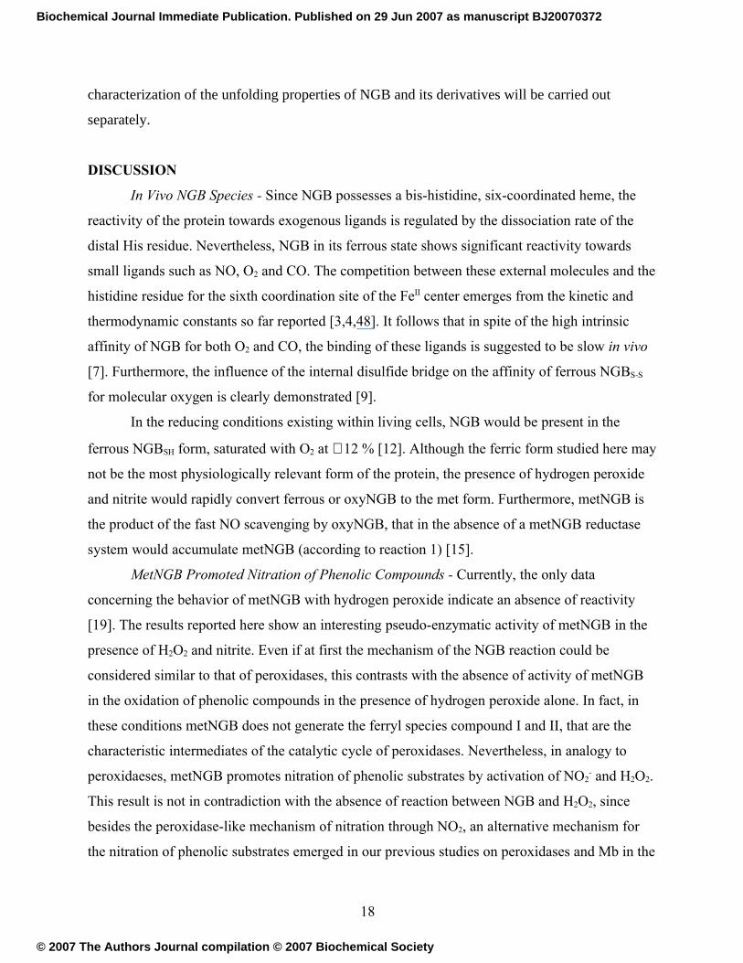
DISCUSSION
In Vivo NGB Species - Since NGB possesses a bis-histidine, six-coordinated heme, the
reactivity of the protein towards exogenous ligands is regulated by the dissociation rate of the
distal His residue. Nevertheless, NGB in its ferrous state shows significant reactivity towards
small ligands such as NO, O2 and CO. The competition between these external molecules and the
histidine residue for the sixth coordination site of the FeII center emerges from the kinetic and
thermodynamic constants so far reported [3,4,48]. It follows that in spite of the high intrinsic
affinity of NGB for both O2 and CO, the binding of these ligands is suggested to be slow in vivo
[7]. Furthermore, the influence of the internal disulfide bridge on the affinity of ferrous NGBS-S
for molecular oxygen is clearly demonstrated [9].
In the reducing conditions existing within living cells, NGB would be present in the
ferrous NGBSH form, saturated with O2 at ∼ 12 % [12]. Although the ferric form studied here may
not be the most physiologically relevant form of the protein, the presence of hydrogen peroxide
and nitrite would rapidly convert ferrous or oxyNGB to the met form. Furthermore, metNGB is
the product of the fast NO scavenging by oxyNGB, that in the absence of a metNGB reductase
system would accumulate metNGB (according to reaction 1) [15].
MetNGB Promoted Nitration of Phenolic Compounds - Currently, the only data
concerning the behavior of metNGB with hydrogen peroxide indicate an absence of reactivity
[19]. The results reported here show an interesting pseudo-enzymatic activity of metNGB in the
presence of H2O2 and nitrite. Even if at first the mechanism of the NGB reaction could be
considered similar to that of peroxidases, this contrasts with the absence of activity of metNGB
in the oxidation of phenolic compounds in the presence of hydrogen peroxide alone. In fact, in
these conditions metNGB does not generate the ferryl species compound I and II, that are the
characteristic intermediates of the catalytic cycle of peroxidases. Nevertheless, in analogy to
peroxidaeses, metNGB promotes nitration of phenolic substrates by activation of NO2- and H2O2.
This result is not in contradiction with the absence of reaction between NGB and H2O2, since
besides the peroxidase-like mechanism of nitration through NO2, an alternative mechanism for
the nitration of phenolic substrates emerged in our previous studies on peroxidases and Mb in the
18
Biochemical Journal Immediate Publication. Published on 29 Jun 2007 as manuscript BJ20070372
© 2007 The Authors Journal compilation © 2007 Biochemical Society

presence of NO2- and H2O2 [22,23]. This mechanism involves the initial binding of nitrite to the
heme iron, followed by the reaction of hydrogen peroxide with the coordinated NO2-, to produce
an iron-bound peroxynitrite (NGBFeIII-N(O)OO), which acts as the nitrating active species. In
the case of peroxidases and Mbs, the ONOO- pathway becomes important only at relatively high
nitrite concentrations [22-24], while with NGB our results indicate that this is the only
mechanism active in the nitration of exogenous and endogenous phenolic groups (i.e. tyrosine
residues), even in the pathophysiological concentration range of NO2- and H2O2 (Figure 7).
= Figure 7 =
The Peroxynitrite Pathway - The kinetic parameters for HPA nitration reported in Table 1
show the importance of nitrite in discriminating the reactivity of the two forms metNGBS-S and
metNGBSH. As expected from the influence of the internal S-S bond on the binding rate of
exogenous ligands to the iron center, the kinetic parameters for the two forms of the protein in
the rate dependence on [NO2-] are significantly different, supporting the hypothesis of direct
coordination of NO2- to the FeIII center to promote the catalysis. In particular, metNGBS-S exhibits
larger values both for nitrite affinity extrapolated from kinetic data (1/ nitriteMK ) and reactivity at
low [NO2-] (kcat/ nitrite
MK ) with respect to metNGBSH, thus indicating that the interaction of NO2-
with the catalytic site regulates the efficiency of nitration at low concentrations of the reagent, in
conditions that can occur in vivo. The higher affinity of nitrite for metNGBS-S with respect to
metNGBSH was confirmed by nitrite binding studies (Figure 3).
The peroxynitrite pathway proposed for the nitrating activity of metNGB gains an
important support by the reactions with the substrates 4-hydroxybenzonitrile and phenylacetic
acid, that are mechanistic probes of the nitrating species NO2 and peroxynitrite, respectively. The
lack of reactivity of hydroxybenzonitrile on one hand, and the presence of several products
deriving from nitration and/or hydroxylation of phenylacetic acid on the other hand, are in
agreement with the formation of an active species with the chemical properties of ONOO- by
metNGB (Figure 4). It follows that the formation of a protein-bound peroxynitrite intermediate is
a reasonable step for the nitration catalyzed by metNGB in the presence of NO2- and H2O2 even
at rather low concentration of the reagents.
NGB Endogenous Modifications – Further support to the generation and activity of the
oxidant species generated by metNGB were extrapolated by the pattern of protein modifications
obtained with LPO as an external catalyst, in comparison with the metNGB-promoted
19
Biochemical Journal Immediate Publication. Published on 29 Jun 2007 as manuscript BJ20070372
© 2007 The Authors Journal compilation © 2007 Biochemical Society

endogenous modifications (Tables 2 and 3). At the high nitrite concentration (250 mM)
employed for the preparation of the lpo-NGBS-S and lpo-NGBSH derivatives, the LPO promoted
nitration proceeds through the formation of the peroxynitrite active species [22]. The differences
in the pattern of modified residues by the LPO-generated peroxynitrite in comparison with those
self induced by NGB, indicate that in the latter case peroxynitrite reacts intramolecularly.
Moreover, the peroxynitrite reactive species is probably responsible for the oxidation of
cysteinyl residues to the corresponding sulfinic and sulfonic acids: not only it can promote this
type of modifications, as observed for instance in the oxidations of bovine serum albumin and
HMb [24,49], but the high reactivity of this species also accounts for the lower extent of
derivatization observed in the h′-NGBSH and p′-NGBSH derivatives (obtained by H2O2 only) in
comparison with the h-NGBSH and p-NGBSH derivatives, respectively. The “harsh” conditions
employed in the preparation of the h-NGBSH and h-NGBS-S derivatives, forcing the extent of
endogenous modifications, allow the investigation of the competitive reactivity of the protein
residues. Both the presence of three reactive cysteines (which may scavenge the RNS) in NGBSH
and its poorer catalytic efficiency in the nitration of phenolic compounds with respect to NGBS-S,
may account for the lower degree of tyrosine nitration in h-NGBSH with respect to h-NGBS-S.
From the point of view of the physiological relevance of NGB endogenous modifications,
of particular interest is the analysis of p-NGBSH, since this was obtained from NGBSH, presumed
to be the predominant form in vivo [12], with low concentrations of NO2- and H2O2. Among the
protein residues, cysteine residues showed the highest reactivity towards the peroxynitrite active
species, and their reaction prevents nitration of tyrosine residues and the heme prosthetic group.
It is possible to calculate that ~36% of H2O2 oxidizing equivalents introduced were used for
oxidation of cysteines to sulfinic and sulfonic acids. Oxidation of cysteines to sulfinic and
sulfonic acid derivatives has been often observed [50,51], together with other types of oxidations
that can be reversed by reaction with biological thiols [52]. It is worth noting that, among the
tyrosine residues, the only one undergoing nitration in both p-NGBS-S and p-NGBSH is Tyr88, the
phenolic OH group of which points toward the large NGB cavity connected to the heme distal
site [10]. This may indicate that the peroxynitrite reactive species endogenously generated at the
heme distal site can easily diffuse through the cavity and nitrate Tyr88.
Does Human NGB Act Only as a Scavenger? – In the globins, cysteine residues often
play specific roles, modulated by the formation of intra- or intermolecular disulfide bonds [9]. In
20
Biochemical Journal Immediate Publication. Published on 29 Jun 2007 as manuscript BJ20070372
© 2007 The Authors Journal compilation © 2007 Biochemical Society
the case of NGB, the oxidation state of Cys46 and Cys55 is relevant because it is linked to the
peculiar six-coordination of the heme. The present study confirms the importance of the internal
S-S bond in controlling the reactivity of metNGB towards nitrite and hydrogen peroxide. It also
shows that the protein can be involved in other activities. In fact, NGB is thought to act as
scavenger of toxic species generated in vivo under conditions of oxidative stress. But the ability
of the metNGB form generated by this activity to activate NO2- and H2O2 means that, besides
acting as a scavenger, NGB can be the source of RNS reactive towards protein residues, thus
affecting its physiological activity. A significant fraction of the nitrating and oxidizing species
(~36% in the conditions used to prepare p-NGBSH) does not escape from the protein but is
consumed in the self-modification of NGB. The easy oxidation of cysteines to sulfinic and
sulfonic acids may be one way used by NGB to scavenge part of the generated RNS. To gain a
better understanding of the physiological relevance of these reactions, it would be important to
analyze the state of endogenous modification of samples of the protein isolated after in vivo
exposure to oxidative stress conditions.
ACKNOWLEDGMENTS
This work was supported by funds from PRIN (Progetto di Ricerca di Interesse Nazionale) and
FIRB projects of the Italian MIUR. The University of Pavia, the European COST D21 Action,
and CIRCMSB are also gratefully acknowledged for support.
REFERENCES
1. Burmester, T., Weich, B., Reinhardt, S., and Hankeln, T. (2000) A vertebrate globin
expressed in the brain. Nature 407, 520-523
2. Schmidt, M., Giessl, A., Laufs, T., Hankeln, T., Wolfrum, U., and Burmester, T. (2003)
How does the eye breathe? Evidence for neuroglobin-mediated oxygen supply in the
mammalian retina. J. Biol. Chem. 278, 1932-1935
3. Trent, J. T. III, Watts, R. A., and Hargrove, M. S. (2001) Human neuroglobin, a
hexacoordinate hemoglobin that reversibly binds oxygen. J. Biol. Chem. 276, 30106-30110
21
Biochemical Journal Immediate Publication. Published on 29 Jun 2007 as manuscript BJ20070372
© 2007 The Authors Journal compilation © 2007 Biochemical Society

4. Uno, T., Ryu, D., Tsutsumi, H., Tomisugi, Y., Ishikawa, Y., Wilkinson, A. J., Sato, H., and
Hayashi, T. (2004) Residues in the distal heme pocket of neuroglobin. Implications for the
multiple ligand binding steps. J. Biol. Chem. 279, 5886-5893
5. Hankeln, T., Ebner, B., Fuchs, C., Gerlach, F., Haberkamp, M., Laufs, T. L., Roesner, A.,
Schmidt, M., Weich, B., Wystub, S., Saaler-Reinhardt, S., Reuss, S., Bolognesi, M., De
Sanctis, D., Marden, M. C., Kiger, L., Moens, L., Dewilde, S., Nevo, E., Avivi, A., Weber,
R. E., Fago, A., and Burmester, T. (2005) Neuroglobin and cytoglobin in search of their
role in the vertebrate globin family. J. Inorg. Biochem. 99, 110-119
6. Garry, D. J., and Mammen, P. P. A. (2003) Neuroprotection and the role of neuroglobin.
The Lancet 362, 342-343
7. Dewilde, S., Kiger, L., Burmester, T., Hankeln, T., Baudin-Creuza, V., Aerts, T., Marden,
M. C., Caubergs, R., and Moens, L. (2001) Biochemical characterization and ligand
binding properties of neuroglobin, a novel member of the globin family. J. Biol. Chem.
276, 38949-38955
8. Hamdane, D., Kiger, L., Dewilde, S., Green, B. N., Pesce, A., Uzan, J., Burmester, T.,
Hankeln, T., Bolognesi, M., Moens, L., and Marden, M. C. (2004) Coupling of the heme
and an internal disulfide bond in human neuroglobin. Micron 35, 59-62
9. Hamdane, D., Kiger, L., Dewilde, S., Green, B. N., Pesce, A., Uzan, J., Burmester, T.,
Hankeln, T., Bolognesi, M., Moens, L., and Marden, M. C. (2003) The redox state of the
cell regulates the ligand binding affinity of human neuroglobin and cytoglobin. J. Biol.
Chem. 278, 51713-51721
10. Pesce, A., Dewilde, S., Nardini, M., Moens, L., Ascenzi, P., Hankeln, T., Burmester, T.,
and Bolognesi, M. (2003) Human brain neuroglobin structure reveals a distinct mode of
controlling oxygen affinity. Structure 11, 1087-1095
11. Smagghe, B. J., Sarath, G., Ross, E., Hilbert, J-L., and Hargrove, M. S. (2006) Slow ligand
binding kinetics dominate ferrous hexacoordinate hemoglobin reactivities and reveal
differences between plants and other species. Biochemistry 45, 561-570
12. Fago, A., Hundahl, C., Dewilde, S., Gilany, K., Moens, L., and Weber, R. E. (2004)
Allosteric regulation and temperature dependence of oxygen binding in human neuroglobin
and cytoglobin. Molecular mechanisms and physiological significance. J. Biol. Chem. 279,
44417-44426
22
Biochemical Journal Immediate Publication. Published on 29 Jun 2007 as manuscript BJ20070372
© 2007 The Authors Journal compilation © 2007 Biochemical Society

13. Pesce, A., Dewilde, S., Nardini, M., Moens, L., Ascenzi, P., Hankeln, T., Burmester, T.,
and Bolognesi, M. (2004) The human brain hexacoordinated neuroglobin three-
dimensional structure. Micron 35, 63-65
14. Vallone, B., Nienhaus, K., Matthes, A., Brunori, M., and Nienhaus, G. U. (2004) The
structure of carbonmonoxy neuroglobin reveals a heme-sliding mechanism for control of
ligand affinity. Proc. Natl. Acad. Sci., U.S.A. 101, 17351-17356
15. Brunori, M., Giuffrè, A., Nienhaus, K., Nienhaus, G. U., Scandurra, F. M., and Vallone, B.
(2005) Neuroglobin, nitric oxide, and oxygen: functional pathways and conformational
changes. Proc. Natl. Acad. Sci., U.S.A. 102, 8483-8488
16. Sun, Y., Jin, K., Peel, A., Mao, X. O., Xie, L., and Greenberg, D. A. (2003) Neuroglobin
protects the brain from experimental stroke in vivo. Proc. Natl. Acad. Sci., U.S.A. 100,
3497-3500
17. Sun, Y., Jin, K., Mao, X. O., Zhu, Y., and Greenberg, D. A. (2001) Neuroglobin is up-
regulated by and protects neurons from hypoxic-ischemic injury. Proc. Natl. Acad. Sci.,
U.S.A. 98, 15306-15311
18. Lipton, P. (1999) Ischemic cell death in brain neurons. Physiol. Rev. 79, 1431-1568
19. Herold, S., Fago, A., Weber, R. E., Dewilde, S., and Moens, L. (2004) Reactivity studies of
the Fe(III) and Fe(II)NO forms of human neuroglobin reveal a potential role against
oxidative stress. J. Biol. Chem. 279, 22841-22847
20. Herold, S., and Fago, A. (2005) Reactions of peroxynitrite with globin proteins and their
possible physiological role. Comp. Biochem. Physiol., Part A 142, 124-129
21. Van der Vliet, A., Eiserich, J. P., Halliwell, B., and Cross, C. E. (1997) Formation of
reactive nitrogen species during peroxidase-catalyzed oxidation of nitrite. A potential
additional mechanism of nitric oxide-dependent toxicity. J. Biol. Chem. 272, 7617-7625
22. Monzani, E., Roncone, R., Casella, L., Galliano, M., and Koppenol, W. H. (2004)
Mechanistic insight into the peroxidase catalyzed nitration of tyrosine derivatives by nitrite
and hydrogen peroxide. Eur. J. Biochem. 271, 895-906
23. Nicolis, S., Monzani, E., Roncone, R., Gianelli, L., and Casella, L. (2004) Metmyoglobin-
catalyzed exogenous and endogenous tyrosine nitration by nitrite and hydrogen peroxide.
Chem. Eur. J. 10, 2281-2290
23
Biochemical Journal Immediate Publication. Published on 29 Jun 2007 as manuscript BJ20070372
© 2007 The Authors Journal compilation © 2007 Biochemical Society

24. Nicolis, S., Pennati, A., Perani, E., Monzani, E., Sanangelantoni, A. M., and Casella, L.
(2006) Easy oxidation and nitration of human myoglobin by nitrite and hydrogen peroxide.
Chem. Eur. J. 12, 749-757
25. Sacksteder, C. A., Qian, W-J., Knyushko, T. V., Wang, H., Chin, M. H., Lacan, G.,
Melega, W. P., Camp, D. G. II, Smith, R. D., Smith, D. J., Squier, T. C., and Bigelow, D. J.
(2006) Endogenously nitrated proteins in mouse brain: links to neurodegenerative disease.
Biochemistry 45, 8009-8022
26. Turko, I. V., and Murad, F. (2002) Protein nitration in cardiovascular diseases. Pharmacol.
Rev. 54, 619-634
27. Hunt, J., Byrns, R. E., Ignarro, L. J., and Gaston, B. (1995) Condensed expirate nitrite as a
home marker for acute asthma. Lancet 346, 1235-1236
28. Torre, D., Ferrario, G., Speranza, F., Orani, A., Fiori, G. P., and Zeroli, C. (1996) Serum
concentrations of nitrite in patients with HIV-1 infection. J. Clin. Pathol. (Lond.) 49, 574-
576
29. Pryor, W. A., and Squadrito, G. L. (1995) The chemistry of peroxynitrite: a product from
the reaction of nitric oxide with superoxide. Am. J. Physiol. 268, L699-L722
30. Casella, L., De Gioia, L., Frontoso Silvestri, G., Monzani, E., Redaelli, C., Roncone, R.,
and Santagostini, L. (2000) Covalently modified microperoxidases as heme-peptide
models for peroxidases. J. Inorg. Biochem. 79, 31-39
31. Antonini, E., and Brunori, M. (1971) Hemoglobin and Myoglobin in Their Reactions with
Ligands, North-Holland Publishing Company, Amsterdam, The Netherlands
32. Roncone, R., Monzani, E., Labò, S., Sanangelantoni, A. M., and Casella, L. (2005)
Catalytic activity, stability, unfolding, and degradation pathways of engineered and
reconstituted myoglobins. J. Biol. Inorg. Chem. 10, 11-24
33. Fuhrhop, J. H., and Smith, K. M. (1975) Laboratory methods in porphyrin and
metalloporphyrin research, Elsevier, Amsterdam, The Nederland
34. Grassetti, D. R., and Murray, J. F. (1967) Determination of sulfhydryl groups with 2,2'- or
4,4'-dithiodipyridine. Arch. Biochem. Biophys. 119, 41-49
35. Hogg, P. J. (2003) Disulfide bonds as switches for protein function. Trends Biochem. Sci.
28, 210-214
24
Biochemical Journal Immediate Publication. Published on 29 Jun 2007 as manuscript BJ20070372
© 2007 The Authors Journal compilation © 2007 Biochemical Society

36. Du, W., Syvitski, R., Dewilde, S., Moens, L., and La Mar, G. N. (2003) Solution 1H NMR
characterization of equilibrium heme orientational disorder with functional consequences
in mouse neuroglobin. J. Am. Chem. Soc. 125, 8080-8081
37. Nistor, S. V., Goovaerts, E., Van Doorslaer, S., Dewilde, S., and Moens, L. (2002) EPR-
spectroscopic evidence of a dominant His-FeIII-His coordination in ferric neuroglobin.
Chem. Phys. Lett. 361, 355-361
38. Watanabe, Y., and Ueno, T. (2003) Introduction of P450, peroxidase, and catalase
activities into myoglobin by site-directed mutagenesis: diverse reactivities of compound I.
Bull. Chem. Soc. Jpn. 76, 1309-1322
39. Roncone, R., Monzani, E., Nicolis, S., and Casella, L., (2004) Engineering and prosthetic
group modification of myoglobin: peroxidase activity, chemical stability and unfolding
properties. Eur. J. Inorg. Chem., 2203-2213
40. Hayashi, T., Hitomi, Y., Ando, T., Mizutani, T., Hisaeda, Y., Kitagawa, S., and Ogoshi, H.
(1999) Peroxidase activity of myoglobin is enhanced by chemical mutation of heme-
propionates. J. Am. Chem. Soc. 121, 7747-7750
41. Dunford, H. B. (1999) Heme Peroxidases, Wiley-VCH, New York, U.S.A.
42. Monzani, E., Gatti, A. L., Profumo, A., Casella, L., and Gullotti, M. (1997) Oxidation of
phenolic compounds by lactoperoxidase. Evidence for the presence of a low-potential
compound II during catalytic turnover. Biochemistry 36, 1918-1926
43. Sampson, J. B., Ye, Y. Z., Rosen, H., and Beckman, J. S. (1998) Myeloperoxidase and
horseradish peroxidase catalyze tyrosine nitration in proteins from nitrite and hydrogen
peroxide. Arch. Biochem. Biophys. 356, 207-213
44. Eiserich, J. P., Hristova, M., Cross, C. E., Jones, A. D., Freeman, B. A., Halliwell, B., and
van der Vliet, A. (1998) Formation of nitric oxide-derived inflammatory oxidants by
myeloperoxidase in neutrophils. Nature 391, 393-397
45. Herold, S., and Rehmann, F. -J. K. (2001) Kinetic and mechanistic studies of the reactions
of nitrogen monoxide and nitrite with ferryl myoglobin. J. Biol. Inorg. Chem. 6, 543-555
46. Casella, L., Monzani, E., Roncone, R., Nicolis, S., Sala, A., and De Riso, A. (2002)
Formation of reactive nitrogen species at biologic heme centers: a potential mechanism of
nitric oxide-dependent toxicity. Environ. Health Perspect. 110, 709-711
25
Biochemical Journal Immediate Publication. Published on 29 Jun 2007 as manuscript BJ20070372
© 2007 The Authors Journal compilation © 2007 Biochemical Society

47. Bondoc, L. L., and Timkovich, R. (1989) Structural characterization of nitrimyoglobin. J.
Biol. Chem. 264, 6134-6145
48. Fago, A., Mathews, A. J., Dewilde, S., Moens, L., and Brittain, T. (2006) The reactions of
neuroglobin with CO: evidence for two forms of the ferrous protein. J. Inorg. Biochem.
100, 1339-1343
49. Radi, R., Beckman, J. S., Bush, K. M., and Freeman, B. A. (1991) Peroxynitrite oxidation
of sulfhydryls. The cytotoxic potential of superoxide and nitric oxide. J. Biol. Chem. 266,
4244-4250
50. Hamann, M., Zhang, T., Hendrich, S., and Thomas, J. A. (2002) Quantitation of protein
sulfinic and sulfonic acid, irreversibly oxidized protein cysteine sites in cellular proteins.
Methods Enzymol. 348, 146-156
51. Woo, H. A., Jeong, W., Chang, T.-S., Park, K. J., Park, S. J., Yang, J. S., and Rhee, S. G.
(2005) Reduction of cysteine sulfinic acid by sulfiredoxin is specific to 2-Cys
peroxiredoxins. J. Biol. Chem. 280, 3125-3128
52. Jacob, C., Holme, A. L., and Fry, F. H. (2004) The sulfinic acid switch in proteins. Org.
Biomol. Chem. 2, 1953-1956
26
Biochemical Journal Immediate Publication. Published on 29 Jun 2007 as manuscript BJ20070372
© 2007 The Authors Journal compilation © 2007 Biochemical Society
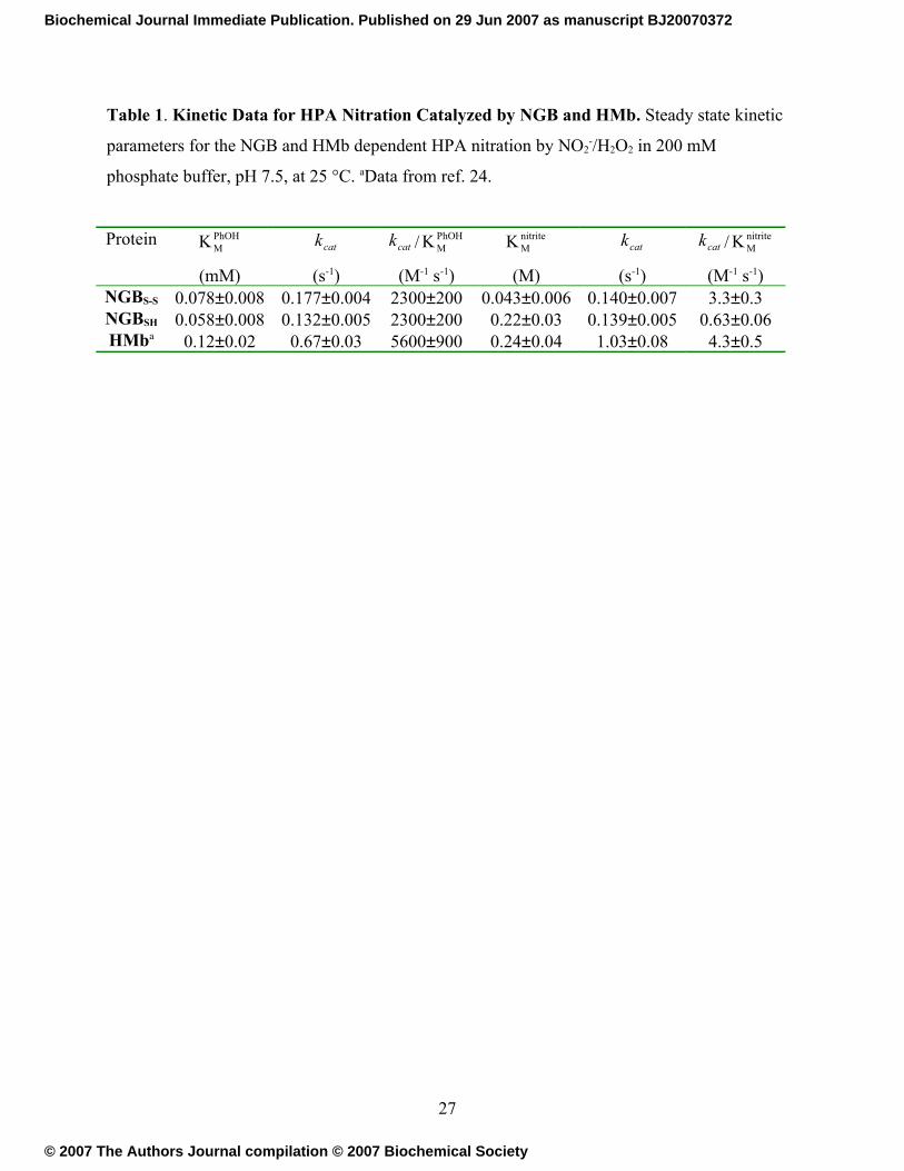
Table 1. Kinetic Data for HPA Nitration Catalyzed by NGB and HMb. Steady state kinetic
parameters for the NGB and HMb dependent HPA nitration by NO2-/H2O2 in 200 mM
phosphate buffer, pH 7.5, at 25 °C. aData from ref. 24.
Protein PhOHMK
(mM)
catk
(s-1)
catk / PhOHMK
(M-1 s-1)
nitriteMK
(M)
catk
(s-1)
catk / nitriteMK
(M-1 s-1)NGBS-S 0.078±0.008 0.177±0.004 2300±200 0.043±0.006 0.140±0.007 3.3±0.3NGBSH 0.058±0.008 0.132±0.005 2300±200 0.22±0.03 0.139±0.005 0.63±0.06HMba 0.12±0.02 0.67±0.03 5600±900 0.24±0.04 1.03±0.08 4.3±0.5
27
Biochemical Journal Immediate Publication. Published on 29 Jun 2007 as manuscript BJ20070372
© 2007 The Authors Journal compilation © 2007 Biochemical Society
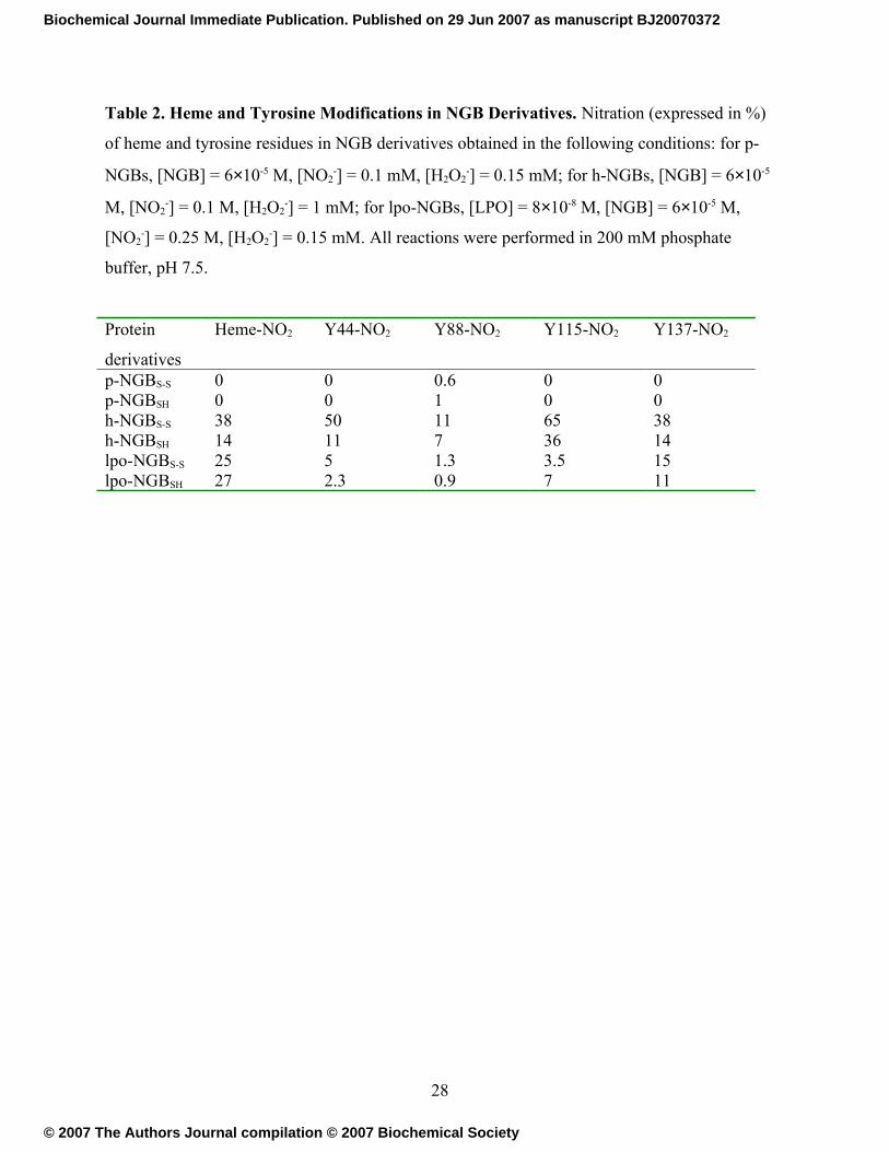
Table 2. Heme and Tyrosine Modifications in NGB Derivatives. Nitration (expressed in %)
of heme and tyrosine residues in NGB derivatives obtained in the following conditions: for p-
NGBs, [NGB] = 6×10⁻⁵ M, [NO2-] = 0.1 mM, [H2O2
-] = 0.15 mM; for h-NGBs, [NGB] = 6×10⁻⁵
M, [NO2-] = 0.1 M, [H2O2
-] = 1 mM; for lpo-NGBs, [LPO] = 8×10⁻⁸ M, [NGB] = 6×10⁻⁵ M,
[NO2-] = 0.25 M, [H2O2
-] = 0.15 mM. All reactions were performed in 200 mM phosphate
buffer, pH 7.5.
Protein
derivatives
Heme-NO2 Y44-NO2 Y88-NO2 Y115-NO2 Y137-NO2
p-NGBS-S 0 0 0.6 0 0p-NGBSH 0 0 1 0 0h-NGBS-S 38 50 11 65 38h-NGBSH 14 11 7 36 14lpo-NGBS-S 25 5 1.3 3.5 15lpo-NGBSH 27 2.3 0.9 7 11
28
Biochemical Journal Immediate Publication. Published on 29 Jun 2007 as manuscript BJ20070372
© 2007 The Authors Journal compilation © 2007 Biochemical Society

Table 3. Modification of Cysteines in NGB Derivatives. Oxidation (expressed in %) of
cysteine residues in NGB derivatives obtained by protein modification in the following
conditions: for p-NGBs, [NGB] = 6×10⁻⁵ M, [NO2-] = 0.1 mM, [H2O2
-] = 0.15 mM; for h-
NGBs, [NGB] = 6×10⁻⁵ M, [NO2-] = 0.1 M, [H2O2
-] = 1 mM; for lpo-NGBs, [LPO] = 8×10⁻⁸ M,
[NGB] = 6×10⁻⁵ M, [NO2-] = 0.25 M, [H2O2
-] = 0.15 mM; for p′-NGBSH, [NGB] = 6×10⁻⁵ M and
[H2O2-] = 0.15 mM; for h′-NGBs, [NGB] = 6×10⁻⁵ M and [H2O2
-] = 1 mM. All reactions were
performed in 200 mM phosphate buffer, pH 7.5. aThe residues C46 and C55 are involved in the
S-S bond.
Protein
derivatives
C46-SO2H C46-SO3H C55-SO2H C55-SO3H C120-SO2H C120-SO3H
NGBS-Sa a a a 1.7 1.5
NGBSH 0.3 0.9 0.6 1.7 2 2p-NGBS-S
a a a a 2 2p-NGBSH 2.3 4.5 2.5 7 10 9p′-NGBSH 1 2 0.5 2 2 1.5h-NGBS-S
a a a a 5 8h-NGBSH 21 14 4 14 18 16h′-NGBSH 0.9 4 0.7 4 5 6lpo-NGBS-S
a a a a 6 7lpo-NGBSH 22 8 3 7 10 5
29
Biochemical Journal Immediate Publication. Published on 29 Jun 2007 as manuscript BJ20070372
© 2007 The Authors Journal compilation © 2007 Biochemical Society

FIGURE LEGENDS
Figure 1. Cysteine-oxidation states and coordination equilibria in metNGB. Schematic
representation of the low-spin/high-spin equilibria for metNGBS-S (on the left) and metNGBSH
(on the right). The disposition of the side chains of the proximal and distal histidine residues
(His(96)F8 and His(64)E7, respectively) is shown. The length of the vertical arrows indicates
that even if the coordination equilibrium is always shifted towards the six-coordinated species,
in the case of metNGBS-S the amount of high-spin form is larger with respect to metNGBSH.
Figure 2. UV-Vis spectra of NGBS-S and NGBSH. UV-Vis spectra of metNGBS-S (6.7 µM,
solid line) and metNGBSH (6.2 µM, dashed line) in 0.2 M phosphate buffer, pH 7.5. The
wavelength maxima are shown.
Figure 3. Binding of Nitrite to NGBS-S and NGBSH. Plot of the difference between the
absorbance changes at 414 and 434 nm in the UV-Vis spectra of NGBS-S and NGBSH upon
addition of nitrite, vs. the ligand concentration. The absorbance data were fitted with the
binding isotherm for low affinity binding of two ligands for NGBS-S and a single ligand for
NGBSH.
Figure 4. Reactivity of 4-hydroxybenzonitrile and phenylacetic acid with RNS. Schematic
representation of the reactivity of 4-hydroxybenzonitrile and phenylacetic acid with the
nitrating agents NO2 and ONOO-, and the catalytic system metNGB/NO2-/H2O2.
Figure 5. Structure of NGB. Structure of the C46G/C55S/C120S mutant of NGB (according
to ref. 10). The disposition of the side chains of the tyrosines (Y44, Y88, Y115, Y137), and the
cysteines (C46, C55, and C120) present in the wild type protein are shown.
Figure 6. Collision-induced dissociation spectrum of the modified 68-94 peptide. MS/MS
spectrum of the m/z 1477.6 peak (mass of 2953.3 amu) assigned to the 68-94 peptide in a
double-charged state and with the residue of Tyr88 modified to 3-nitrotyrosine. The assignment
of the y and b ion series is shown. Above the spectrum, the sequence of the 68-94 peptide, with
30
Biochemical Journal Immediate Publication. Published on 29 Jun 2007 as manuscript BJ20070372
© 2007 The Authors Journal compilation © 2007 Biochemical Society
the modified residue in bold and with the summary of the y and b ions found in the spectrum, is
reported.
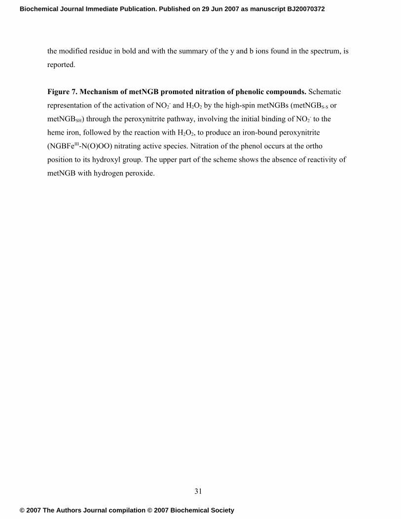
Figure 7. Mechanism of metNGB promoted nitration of phenolic compounds. Schematic
representation of the activation of NO2- and H2O2 by the high-spin metNGBs (metNGBS-S or
metNGBSH) through the peroxynitrite pathway, involving the initial binding of NO2- to the
heme iron, followed by the reaction with H2O2, to produce an iron-bound peroxynitrite
(NGBFeIII-N(O)OO) nitrating active species. Nitration of the phenol occurs at the ortho
position to its hydroxyl group. The upper part of the scheme shows the absence of reactivity of
metNGB with hydrogen peroxide.
31
Biochemical Journal Immediate Publication. Published on 29 Jun 2007 as manuscript BJ20070372
© 2007 The Authors Journal compilation © 2007 Biochemical Society

Figure 1
32
Biochemical Journal Immediate Publication. Published on 29 Jun 2007 as manuscript BJ20070372
© 2007 The Authors Journal compilation © 2007 Biochemical Society
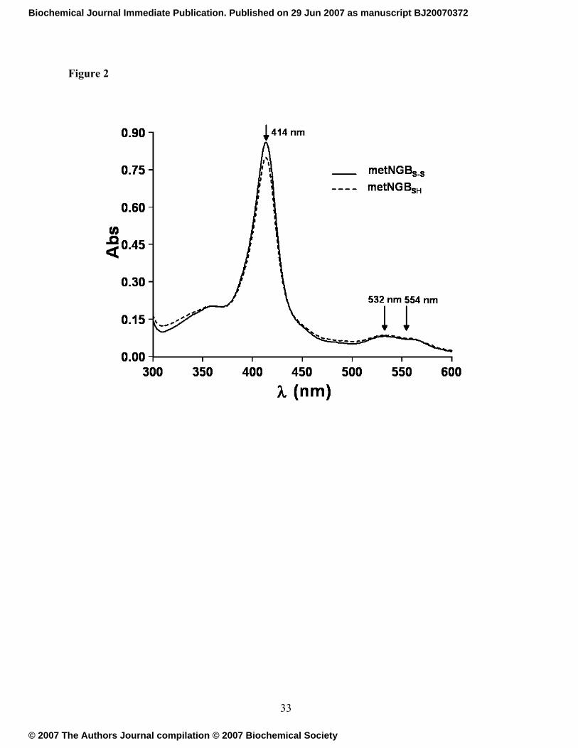
Figure 2
33
Biochemical Journal Immediate Publication. Published on 29 Jun 2007 as manuscript BJ20070372
© 2007 The Authors Journal compilation © 2007 Biochemical Society
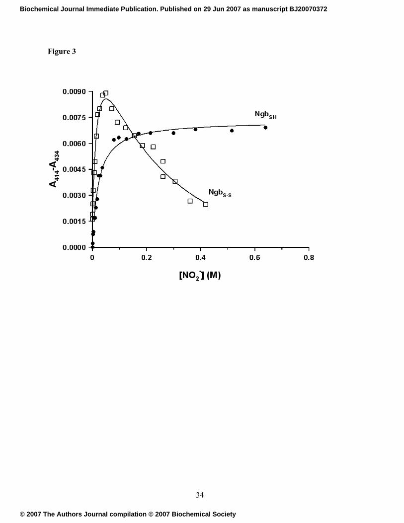
Figure 3
34
Biochemical Journal Immediate Publication. Published on 29 Jun 2007 as manuscript BJ20070372
© 2007 The Authors Journal compilation © 2007 Biochemical Society
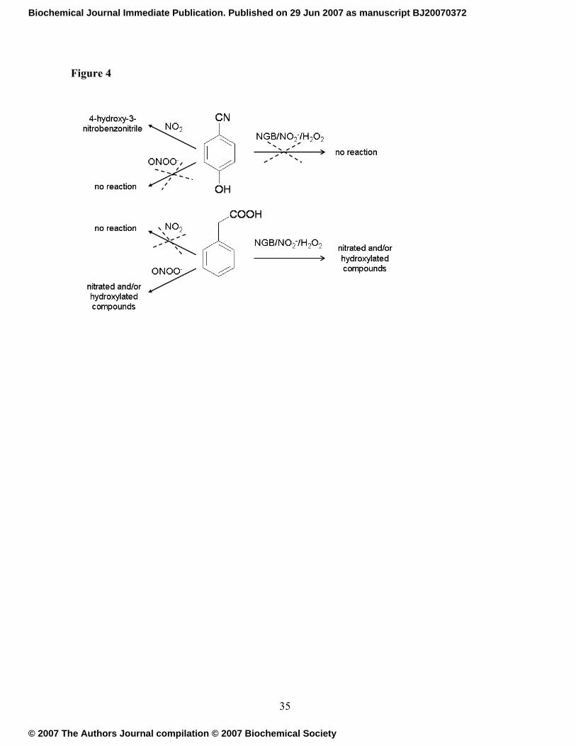
Figure 4
35
Biochemical Journal Immediate Publication. Published on 29 Jun 2007 as manuscript BJ20070372
© 2007 The Authors Journal compilation © 2007 Biochemical Society
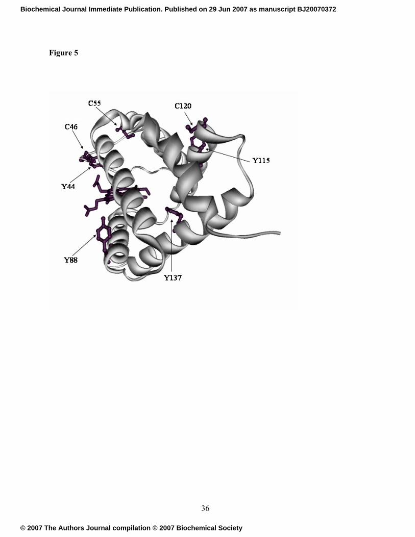
Figure 5
36
Biochemical Journal Immediate Publication. Published on 29 Jun 2007 as manuscript BJ20070372
© 2007 The Authors Journal compilation © 2007 Biochemical Society
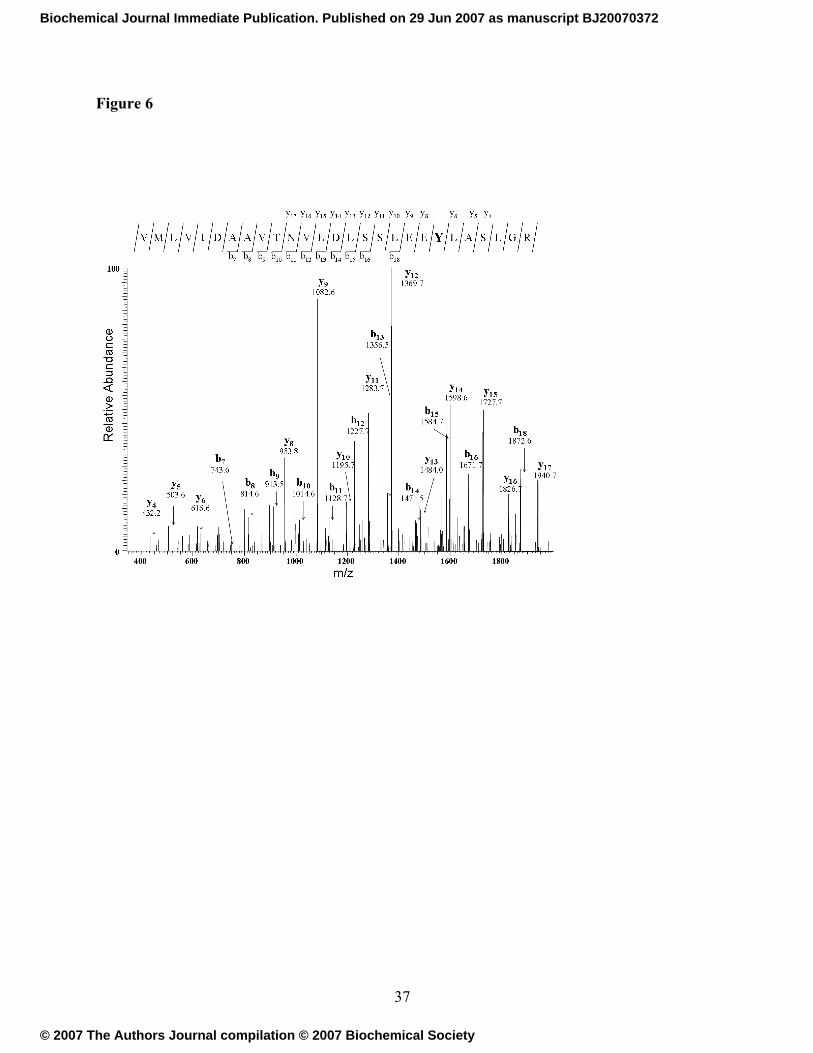
Figure 6
37
Biochemical Journal Immediate Publication. Published on 29 Jun 2007 as manuscript BJ20070372
© 2007 The Authors Journal compilation © 2007 Biochemical Society
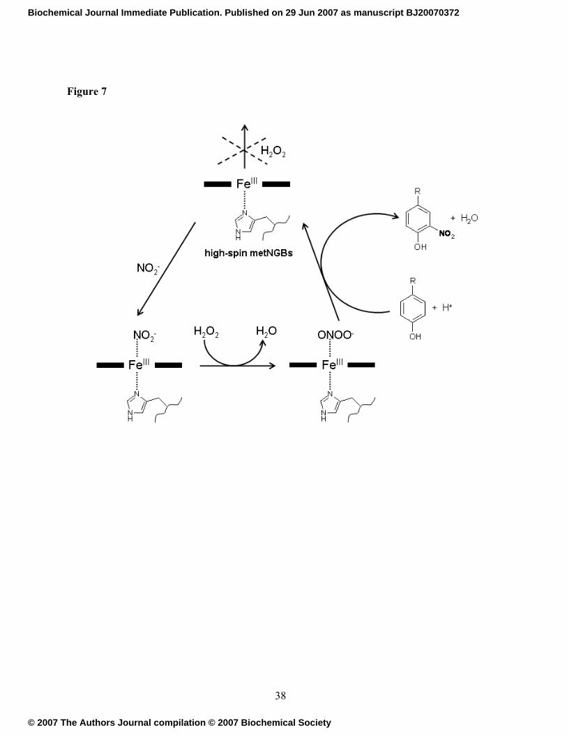
Figure 7
38
Biochemical Journal Immediate Publication. Published on 29 Jun 2007 as manuscript BJ20070372
© 2007 The Authors Journal compilation © 2007 Biochemical Society
Related Documents