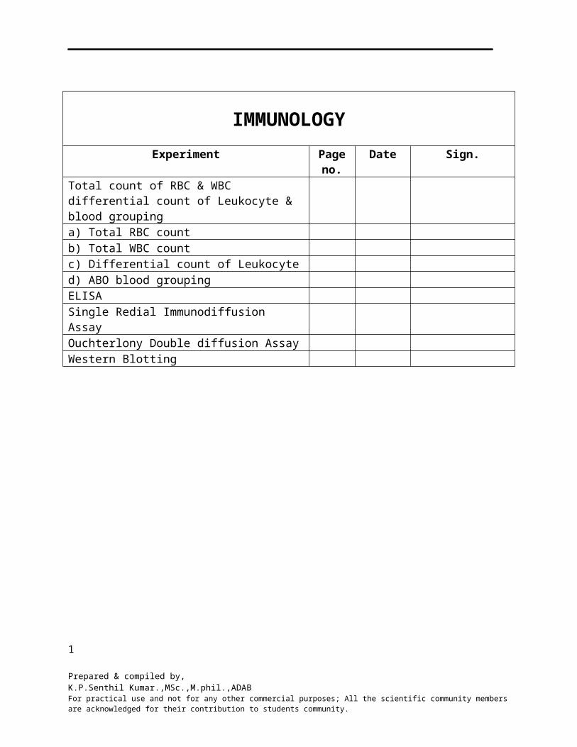
IMMUNOLOGY Experiment Page no. Date Sign. Total count of RBC & WBC differential count of Leukocyte & blood grouping a) Total RBC count b) Total WBC count c) Differential count of Leukocyte d) ABO blood grouping ELISA Single Redial Immunodiffusion Assay Ouchterlony Double diffusion Assay Western Blotting 1 Prepared & compiled by, K.P.Senthil Kumar.,MSc.,M.phil.,ADAB For practical use and not for any other commercial purposes; All the scientific community members are acknowledged for their contribution to students community.

Rbc,wbc count
May 10, 2015
Immunology practicals for MSc BT Semester II
Welcome message from author
This document is posted to help you gain knowledge. Please leave a comment to let me know what you think about it! Share it to your friends and learn new things together.
Transcript
IMMUNOLOGY
Experiment Page no.
Date Sign.
Total count of RBC & WBC differential count of Leukocyte & blood grouping a) Total RBC countb) Total WBC countc) Differential count of Leukocyted) ABO blood groupingELISASingle Redial Immunodiffusion AssayOuchterlony Double diffusion AssayWestern Blotting
1
Prepared & compiled by,K.P.Senthil Kumar.,MSc.,M.phil.,ADABFor practical use and not for any other commercial purposes; All the scientific community members are acknowledged for their contribution to students community.
Experiment: 1 Date:
TOTAL COUNT OF RBC & WBC, DIFFERENTIAL COUNT OF LEUKOCYTES & BLOOD GROUPING
INTRODUCTION:
Hematology is the branch of science concerned with the study of blood, blood forming tissues & the disorders associated with them. It includes,
1) Analysis of the concentration, structure and functions of the cells and their precursors in the bone marrow.
2) Analysis of chemical constituents of plasma or serum intimately, linked with blood cell structure and functions.
3) Study of functions of the platelets and proteins involved in blood coagulation.
The hematology laboratory deals with routine determination of total number of cells in circulation, hemoglobin concentration, and differential count of leukocytes based on the study of the stained blood smear.
Blood:
Blood may be described as the specialized liquid connective tissue, which circulates in a closed system of blood vessels. The circulating blood consists of suspension of formed elements such as erythrocytes, leukocytes and platelets in a pale yellow colored fluid called “plasma”. In adults, the total volume of blood comprises about 8% of the body weight. The formed elements accounts for about 46% of total blood volume. Under normal conditions, the production of blood cells is carried out by the bone marrow.
Functions of blood:
1. Respiration: Transport of oxygen from the lungs to the tissues and of carbon dioxide from tissues to the lungs.
2. Excretion: Transport of metabolic wastes to the lungs, kidneys, skin and intestine for removal.3. Maintenance of normal acid-base balance i.e. pH regulation.4. Nutrition: Transport of absorbed fatty acids, monosaccharides and amino acids.5. Regulation of water balance.6. Regulation of body temperature.7. Transport of hormones, vitamins, salts and metabolites.8. Defense against infection by the white blood cells & the antibodies.9. Coagulation of blood: To stop bleeding by clotting.
2
Prepared & compiled by,K.P.Senthil Kumar.,MSc.,M.phil.,ADABFor practical use and not for any other commercial purposes; All the scientific community members are acknowledged for their contribution to students community.
Physical features of blood:
1. Blood is more viscous & denser than water and feels slightly sticky.2. Temperature of blood is 38oC, about 10oC higher than oral or rectal body temperature.3. It has slightly alkaline pH ranging from 7.35 to 7.45
Experiment: 1(a) Date:
DETERMINATION OF TOTAL COUNT OF (RBC) RED BLOOD CELLS
AIM: To determine total count of RBC in the given blood sample.
INTRODUCTION:
Erythrocytes are elastic, non-nucleated biconcave discs. The cell possesses on its surface a number of antigens responsible for the blood groups. The function of the red cell is to transport oxygen through hemoglobin in a functional state. Life span of red blood cell is about 120days. The erythrocyte count is performed to determine whether a person has anemia or polycythaemia and to judge their degree.
Clinical Significance:As birth the total erythrocyte count varies from 6.5millions -7.25millions per cubic mm (mm3). There is steady decline after a few hours and at the end of 15 days to one month there is a slow rise to normal adult levels. An increase in total erythrocyte count is observed in conditions such as haemoconcentration due to burns, cholera etc. In central cyanotic states as seen in chronic heart disease, condition of decreased lung functions such as emphysema and polycythemia. Decrease in erythrocyte count is observed in (a) Old age
(b) In pregnancy
(c) In group of diseases classified under anemia
Normal values: Male: 4.5 to 6.0 x 10⁶cells /mm3 Female: 4.0 to 4.5 x 10⁶cells/mm3
Specimens: EDTA blood or Capillary blood
3
Prepared & compiled by,K.P.Senthil Kumar.,MSc.,M.phil.,ADABFor practical use and not for any other commercial purposes; All the scientific community members are acknowledged for their contribution to students community.
PRINCIPLE:
The blood specimen is diluted 1:200 with the RBC diluting fluid and cells are counted under high (40x objective) by using a counting chamber. The number of cells in undiluted blood are calculated and reported as the total number of cells per cubic mm of whole blood.
REQUIREMENTS:
1. Neubauer’s Haemocytometer Slide 2. RBC pipette
3. RBC diluting fluid 4. Microscope
Description of the Apparatus:It is a special glass slide. It has two central platforms separated by a horizontal groove
which joins a vertical groove at each of its ends, thus an ‘H’ shaped grooved area is seen on the slide. Outer to vertical groove i.e. on each side of ’H’, there is a slide pillar; both the slide pillars are 1/10mm (0.1mm) lighter than the two central platforms.
On each platform lies a ruled area of counting chamber so that two preparations may be set simultaneously. Each chamber is a square of the counting chamber i.e. 1sq mm area is marked ‘R’. This 1 sq mm area is divided into 25 small squares by triple lines i.e. each of these small squares has an area of 1/25 sq mm which is bounded by triple lines. Each of these small squares i.e. as in area 1/25 sq mm is further divided into 16 smaller squares each of which has an area
= 1/25 X 1/16
= 1/400
= 0.0025 sq mm
For WBC each of the 4 corner square at the counting chamber i.e. 1 sq mm area marked ‘W’ and is divided into small squares, which are WBC counting chambers. Each small square has an area of 1/16 sq mm.
4
Prepared & compiled by,K.P.Senthil Kumar.,MSc.,M.phil.,ADABFor practical use and not for any other commercial purposes; All the scientific community members are acknowledged for their contribution to students community.
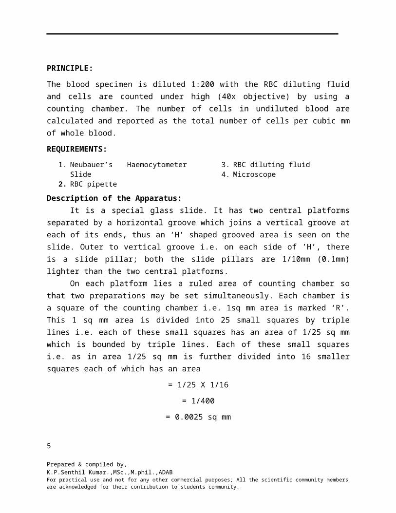
Figure 1.1: Hemocytometer gridred square = 1 mm2,
green square = 0.0625 mm2,
yellow square = 0.04 mm2,
blue square = 0.0025 mm2,
at a depth of 0.1 mm
RBC PIPETTE (Thoma glass pipette):
It consists of capillary tube, central tube and three graduation marks. Capillary tube is opened at both ends. One end of the tube is narrow while the other end is broad. For sucking a rubber tube fixed to the broad end. The bubble of the pipette has a red glass bead inside. This is a bulb pipette having a long stem with a capillary bore and a pointed tip. The bulb contains a red bead inside. A small rubber tube provided with a mouth piece is connected to the small narrow portion above the bulb for sucking blood and fluid into the pipette. The pipette has three markings on in it, ‘0.5’ mark in the middle of the stem, ‘1’ mark at the junction between stem and bulb, and ‘101’ mark above the bulb. The total volume of the pipette is 101 parts, of which one part is in the stem and 100 parts in the bulb. Blood is drawn to ‘0.5’ mark and then diluting fluid is drawn up to the mark ‘101’. The dilution of blood is 1:200.
RBC diluting fluid:
Hayem’s fluid is isotonic with blood. Its composition is
1. Sodium chloride (NaCl) - 0.5 2. Sodium Sulphate (Na2SO4) - 2.5 g3. Mercuric Chloride (HgCl2) - 0.25 g4. Distilled water - 100 ml
5
Prepared & compiled by,K.P.Senthil Kumar.,MSc.,M.phil.,ADABFor practical use and not for any other commercial purposes; All the scientific community members are acknowledged for their contribution to students community.
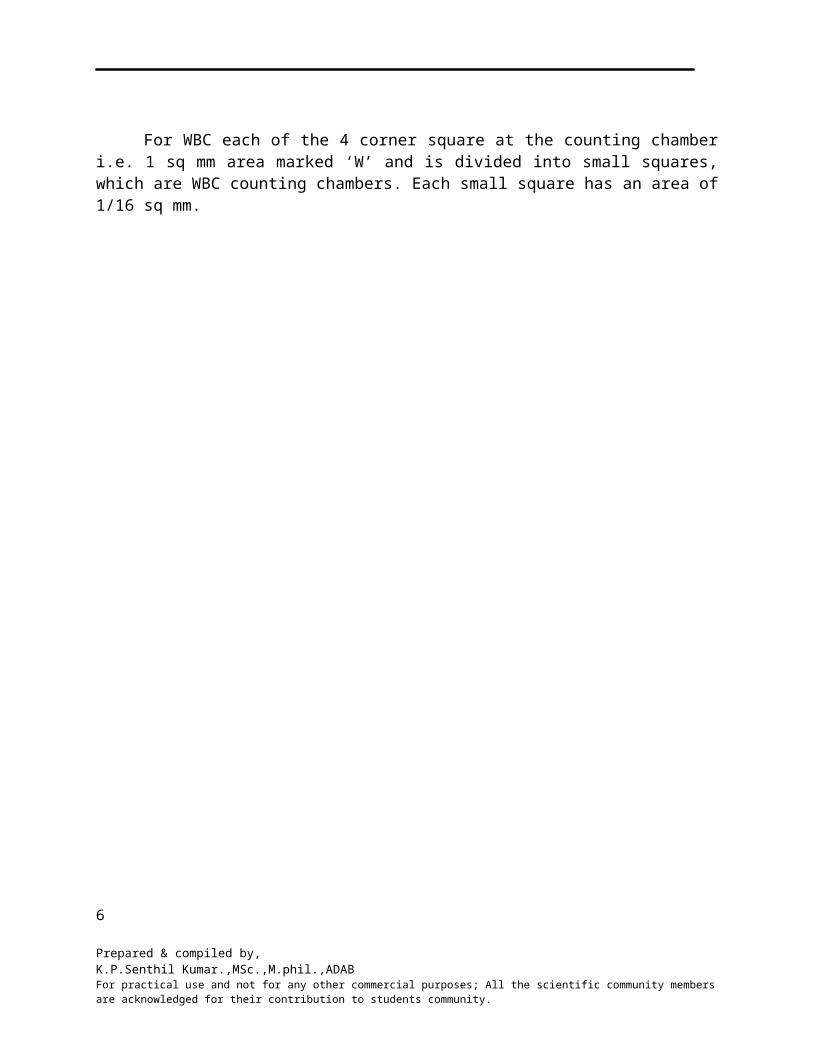
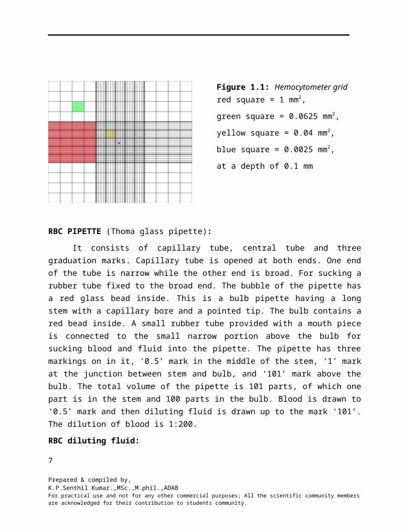
Figure 1.2: Neubauer’s Haemocytometer Slide, RBC pipette, WBC pipette.
Sodium chloride and sodium sulphate together keeps the isotonicity of fluid. Sodium sulphate also prevents clumping of red cells. Mercuric per chloride fixes the cells and acts as a preservative, so diluting fluid helps in preventing hemolysis, coagulation of blood and fungal and bacterial growth.
Cover slip of Haemocytometer slide:
It is thicker than the ordinary cover slip used in laboratory. When it is placed on the two counting chambers the space enclosed between the lower surfaces of the two platforms is 1/10 mm deep. This means that the volume of the fluid enclosed in each of the smaller squares having an area of 1/400 sq mm will be =1/400 X 1/10= 1/4000 mm3.
6
Prepared & compiled by,K.P.Senthil Kumar.,MSc.,M.phil.,ADABFor practical use and not for any other commercial purposes; All the scientific community members are acknowledged for their contribution to students community.
Precautions:
1. Counting chamber and pipette should be clean and dry.
2. Fingertip and pricking lancet should be sterile.
3. Blood should freely come out without squeezing.
4. Be careful to prevent clotting of blood inside the pipette.
5. While filling the pipette and charging (loading) the counting chamber, no air bubble should enter.
6. Blood should be taken only up to the ‘0.5’ mark and diluting fluid sucked only up to ‘101’ mark.
7. Blood should be properly mixed with the diluting fluid.
8. While charging the counting chamber, over filling and spilling should be avoided.
9. Cells should be settled down and more or less evenly distributed before counting.
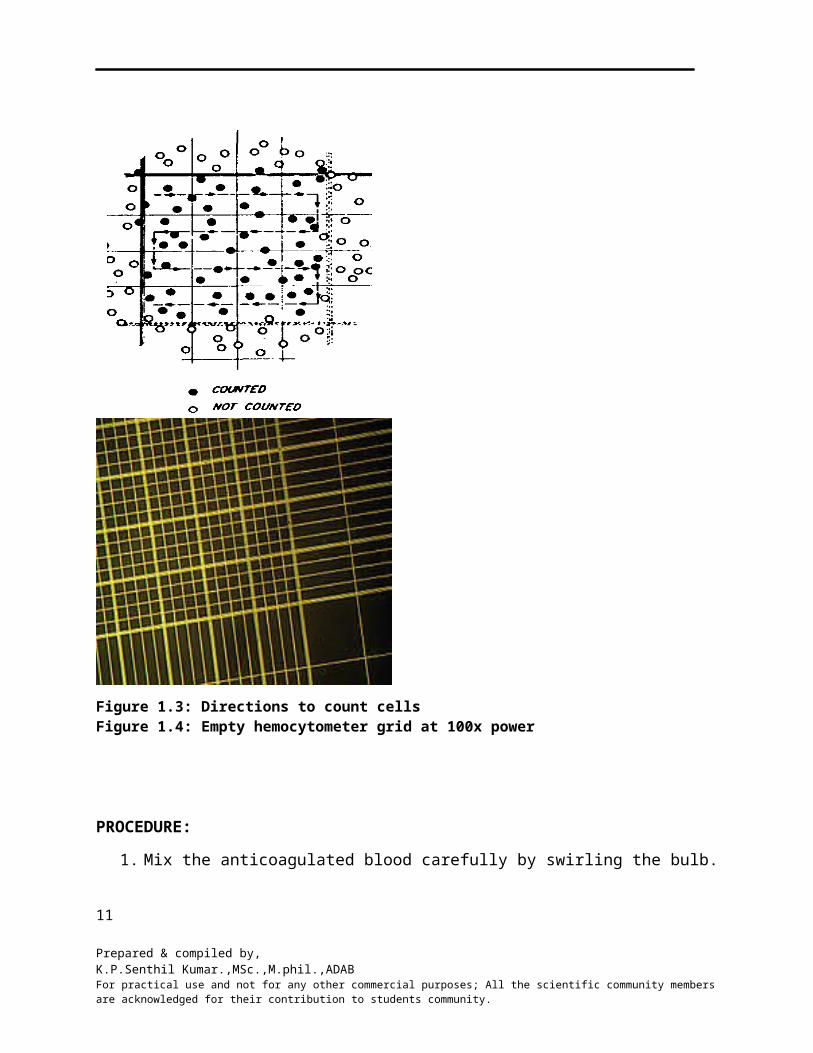
10. Count from Left to Right (fig 1) and avoid counting of the same cell.
Figure 1.3: Directions to count cells Figure 1.4: Empty hemocytometer grid at 100x power
7
Prepared & compiled by,K.P.Senthil Kumar.,MSc.,M.phil.,ADABFor practical use and not for any other commercial purposes; All the scientific community members are acknowledged for their contribution to students community.
PROCEDURE:
1. Mix the anticoagulated blood carefully by swirling the bulb.2. In the case of capillary blood the lancet stab should be sufficiently deep to allow free
flow of blood. It is drawn quickly in the RBC pipette.3. Draw blood up to ‘0.5’ mark.4. Carefully wipe the excess blood outside the pipette by using cotton or a gauze.5. Draw diluting fluid up to ‘101’ mark.6. The pipette in rotated rapidly by keeping it horizontal during mixing.7. After two minute, by discarding few drops from the pipette and holding it slightly
inclined small volume of the fluid is introduced under the cover slip which is placed on the counting chamber.
8. Allow the cells to settle for 2 to 3 minutes.9. Place the counting chamber on the stage of the microscope.10. Switch to low power (10 X) objective. Adjust light and locate the large square in the
center with 25 small squares.11. Now switch to high power (40 X) objective.12. The red blood cells in the four corners and central Square of ‘R’ are counted.
Calculation of red blood cells:
Total RBC/mm3 = Number of RBC counted × Dilution factor × Depth factor
No. of chambers counted
RESULT:
Total RBC count of the given blood sample is ______________ million/mm3
8
Prepared & compiled by,K.P.Senthil Kumar.,MSc.,M.phil.,ADABFor practical use and not for any other commercial purposes; All the scientific community members are acknowledged for their contribution to students community.
Experiment: 1(b) Date:
DETERMINATION OF TOTAL COUNT OF WHITE BLOOD CELLS (WBC)
AIM: To determine total count of WBC in the given blood sample.
INTRODUCTION:
The leucocytes or white blood cells are nucleated corpuscles. The various types of white blood cells are neutrophils, eosinophils, basophils, lymphocyte & monocytes. Leucocytes are needed by the body for its defense against invading organisms like bacteria, viruses and cancer cell.
Clinical significance:
Increase in total leucocytes count of more than 10,000/mm3 is known as leucocytosis and decrease less than 3,000/mm3 as leukopenia
Causes of leucocytosis:
1. Pathological:
It is common for a transient period in infection. The degree of rise in leucocytes depends on the type and severity of the infection and response of the body. The infection may be Bacterial, Viral, Protozoan (Malaria), and Parasitic (filarial) hookworm infection. Leucocytosis is also observed in severe hemorrhage and in leukemia
2. Physiological:
a) Age: At birth total leucocytes count is about 18,000/mm3, later it drops to adult level.b) Pregnancy: At “full term” the total count tends to be about 12,000-15000/mm3. It rises
soon after delivery and then gradually returns to normal.c) High temperature d) Severe pain e) Muscular Exercise
Causes of leucopenia:
Certain viral and bacterial infection (typhoid) leads to leukopenia rather than leucocytosis.
Infection
a) Bacterial (Typhoid, Paratyphoid, Tuberculosis etc.)b) Viral (Hepatitis, Influenza, Measles etc)c) Protozoan (Malaria)
9
Prepared & compiled by,K.P.Senthil Kumar.,MSc.,M.phil.,ADABFor practical use and not for any other commercial purposes; All the scientific community members are acknowledged for their contribution to students community.
Some cases of leukemia:
1. Primary bone marrow depressions (Aplastic anemia)2. Secondary bone marrow depression (due to drugs, radiation etc)3. Anemia (iron deficiency, Megaloblastic etc)
Normal Values:
Adult : 4000-10,000/mm3
At birth : 10,000-25,000/mm3
1 to 3 years : 6000-18,000/mm3
4 to 7 years : 6,000-15,000/mm3
8-12 years : 4,500-13,500/mm3
Specimens:
a) Double oxalated or EDTA blood or
b) Capillary blood
REQUIREMENTS:
1. Neubauer Haemocytometer slide with cover slip2. WBC pipette3. WBC diluting fluid4. Microscope
WBC Pipette:
This is a bulb pipette having a long stem with a capillary bore and a pointed tip. The bulb contains a red bead inside. A small rubber tube provided with a mouth piece is connected to the small narrow portion above the bulb for sucking blood and fluid into the pipette.WBC pipette is like RBC pipette but here the bulb has a white glass bead in place of red and the third graduation mark which lies just above the bulb is ‘11’ instead of ‘101’, this means the WBC pipette has 11 volumes from the tip up to ‘11’ mark.
WBC diluting fluid (Truck’s fluid):
1. 1ml of 3% acetic acid, 1% gelatin violet- 1 or 2 drops or methylene blue. 2. The function of diluting fluid is to destroy RBC and stain the nucleus of WBC to increase
the visibility.
PROCEDURE:
1. The blood sample is taken up to the ‘0.5’ mark, then the diluting fluid is taken up to the ‘11’ mark, this gives 1:20 dilution.
2. The mixing was done well for one minute.10
Prepared & compiled by,K.P.Senthil Kumar.,MSc.,M.phil.,ADABFor practical use and not for any other commercial purposes; All the scientific community members are acknowledged for their contribution to students community.
3. The counting chamber and cover glass placed on central platform were cleaned thoroughly before use.
4. A drop of diluted blood was discharged into the chamber. 5. The counting to be done using low power objective lens of the microscope.
The four ‘W’ corner squares of the both the chambers were counted. The number of WBC present give the value of, n-number of WBC counted. Then the total number of WBC per mm 3 is obtained by the formulae;
Total WBC/mm3 = Number of WBC counted × dilution factor × depth factor
No. of chambers counted
RESULT:
Total WBC count of the given blood sample is ___________ /mm3.
Experiment: 1(c) Date:
11
Prepared & compiled by,K.P.Senthil Kumar.,MSc.,M.phil.,ADABFor practical use and not for any other commercial purposes; All the scientific community members are acknowledged for their contribution to students community.
STUDY OF BLOOD SMEAR FOR DIFFERENTIAL LEUKOCYTE COUNT
AIM: To determine the differential count of Leukocytes by studying the blood smear.
INTRODUCTION:
Differential count is the percent distribution of various white cells in the peripheral blood. It is determined from a blood smear stained with a polychromatic stain and after examination of the stained smear by using oil immersion objective (total magnification 1000x). The number of each type of white cell is then expressed as a percentage of the total number of cells. The stained blood smear also helps to study abnormal morphology of leucocytes and red cells. Study of blood smear helps in the diagnosis of various anaemias, leukaemias and detection of blood parasites. Three major steps involved in differential count are (a) preparation of smear (b) staining of smear and (c) microscopic examination of the stained smear. The leucocytes are the most active and motile constituents of blood. The different types of leucocytes seen in a normal peripheral blood film may be divided in to two broad groups.
1. Granulocytes:
Granulocytes are the cells containing granules, and a polymorphic nucleus in the cytoplasm. There are three types of granulocytes which derive their names from the staining reaction of the granules present in the cytoplasm. These cells are
(a) Neutrophils or polymorphonuclears
(b) Eosinophils and
(c) Basophils
Neutrophil (polymorphonuclears):
The average diameter is 10 to 12 µm. The nucleus is usually divided into 2-7 lobes. The cell derives its name from the color of the granules. Neutrophil stains when exposed to acidic, as well as basic dyes. The number of lobes gives an indication of the age of the cell. Neutrophil are mainly responsible for the protection against infection.
Eosinophil:
The diameter is about the same as the neutrophil. The cytoplasm contains large, oval or round red-orange (eosinophilic) granules that are stained bright red with eosin, an acid dye. The nucleus shows fewer lobes, on an average only two. The eosinophils are not markedly motile and only slightly phagocytic. They play important role in detoxification.
Basophil:
12
Prepared & compiled by,K.P.Senthil Kumar.,MSc.,M.phil.,ADABFor practical use and not for any other commercial purposes; All the scientific community members are acknowledged for their contribution to students community.
The diameter is 8 to 10 µm. The nucleus has two to three lobes. It contains fewer coarse granules, which can be stained with basic dyes, e.g. methylene blue. Basophils are significant in allergic reactions.
2. Agranulocytes:
Agranulocytes are leucocytes that lack granules and consist of two types of cells:
a) Lymphocytes and b) Monocytes
Monocyte:
The diameter is about 16 to 22 μm. The nucleus is kidney shaped or horse shoe shaped. Sometimes it may be round or oval. It stains pale violet and has fine chromatic arrangement. The cytoplasm is plentiful and stains pale grayish blue.
Lymphocyte: Two forms observed are
a) Large lymphocyte b) Small lymphocyte.
(a) Large lymphocytes: it is about 12 to 15 µm in diameter. This has abundant clear pale blue cytoplasm and a large round or slightly indented nucleus with dense chromatin.
(b) Small lymphocyte: These are small round cells about 10 to12 µm in diameter. It has very little blue cytoplasm and often more than just a rim around the nucleus. The nucleus is dark, round and sometimes indented.
Clinical significance:
Differential count is useful to identify changes in the distribution of white cells which may be related to specific type of disorders. It also gives idea regarding the severity of the disease and the degree of response of the body.
Neutrophilia:
Increase in the percentage of neutropils is called neutrophilia. All the physiological causes that produce leukocytosis give rise to neutrophilia. The commonest pathological cause is phylogenic bacterial infection. Decrease in neutrophil (Neutropaenia) is observed in infections such as bacterial (typhoid), viral (Measles, influenza etc.) and in other conditions such as anaemias (aplastic, megaloblastic, iron deficiency) and in suppression of bone marrow by various drugs and radiation.
Lymphocytosis: It may be relative or absolute
13
Prepared & compiled by,K.P.Senthil Kumar.,MSc.,M.phil.,ADABFor practical use and not for any other commercial purposes; All the scientific community members are acknowledged for their contribution to students community.
Relative lymphocytosis:
In this condition the actual number of lymphocytes is unchanged but due to decrease in neutrophils mainly the differential count shows an increase in lymphocytes.
Absolute lymphocytosis:
It is observed in
1. Children
2. Certain infections such as
a. Mumps,
b. Cough,
c. Measles,
d. Influenza,
e. Syphilis,
f. Tuberculosis,
g. Typhoid and other chronic infections
3. Infectious mononucleosis
4. Chronic lymphatic leukemia.
Lymphopaenia: In this condition the actual number of lymphocytes is decreased. It is observed in acute stage of infections and in excess irradiation.
Eosinophilia: It is defined as an increase in peripheral blood eosinophilic leukocytes to more than 0.45×10⁹/L (450/μl). It is observed in asthma, hypersensitivity reactions, and parasitic infections and in chronic inflammatory disease. Monocytosis: It is an increase in the number of monocytes circulating in the blood. It is observed in tuberculosis, malaria, subacute bacterial endocarditis, typhoid and in kala Azar. Basophilia: It is a condition where the basophil quantity is abnormally elevated (more than 1010 basophils per liter of blood). It is usually observed in chronic myeloid leukaemia. Specimen:
The blood smears should be preferably prepared immediately after skin puncture or venipuncture before mixing with anticoagulant. If EDTA blood is used the smears should be prepared within1 to 2 hours after blood drawing. Other anticoagulants do not give satisfactory results. The blood smears should be immediately fixed in methanol.
14
Prepared & compiled by,K.P.Senthil Kumar.,MSc.,M.phil.,ADABFor practical use and not for any other commercial purposes; All the scientific community members are acknowledged for their contribution to students community.
PRINCIPLE:
The polychromic staining solutions (Wright, Leishman’s, and Giemsa) contain methylene blue and eosin. These basic and acidic dyes induce multiple colors when applied to cells, methanol acts as fixative and also as solvent. The fixative does not allow any further change in the cells and makes them adhere to the glass slide. The basic component of white blood cells (i.e. cytoplasm) is stained by acidic dye and they are described as eosinophilic. The acidic components (e.g. nucleus with nucleic acid) take blue to purple shades by the basic dye and they are called basophilic. The neutral components of the cell are stained by both the dyes.
REQUIREMENTS:
1. Blood sample2. Microscope slides3. Cedar wood oil4. Glass Spreader 5. Staining reagent6. Methanol7. Distilled water and Miscellaneous. 8. Microscope
Figure 1c.1: Differential leukocyte staining
15
Prepared & compiled by,K.P.Senthil Kumar.,MSc.,M.phil.,ADABFor practical use and not for any other commercial purposes; All the scientific community members are acknowledged for their contribution to students community.
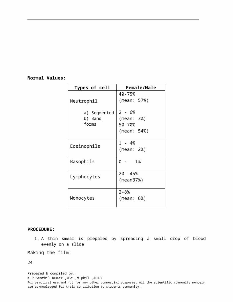
Normal Values:
Types of cell Female/MaleNeutrophil
a) Segmentedb) Band forms
40-75% (mean: 57%)
2 - 6% (mean: 3%)50-70% (mean: 54%)
Eosinophils 1 - 4% (mean: 2%)
Basophils 0 - 1%
Lymphocytes 20 –45% (mean37%)
Monocytes2-8% (mean: 6%)
PROCEDURE:
1. A thin smear is prepared by spreading a small drop of blood evenly on a slide
Making the film:
a. Take clean, dry (grease free) slide.b. Transfer a small drop of blood near the edge of the slide.c. Place the spreader slide at an angle of 300 to 350. Pull back the spreader until touches the
drop of blood. d. Let the blood run along the edge of the spreader.e. Push the spreader forward to the end of the slide with a smooth movement.f. Dry the blood smear at room temperature. g. Adequate drying is essential to preserve quality of the film.
2. Fixing the Smear:a. The slide should be stained after making the smear. b. Methanol present in the stain fixed the smear. c. If the staining is to be done later, the blood smear must be fixed with methanol for 2 to 3
minutes to prevent distortion of cells.
Field Stain:
For the rapid screening of blood smears and also for the screening of thick films for malarial parasites, field stain is used. The blood smears are fixed with methanol for 2 to 3 minutes before the staining. Following staining solutions are used.
16
Prepared & compiled by,K.P.Senthil Kumar.,MSc.,M.phil.,ADABFor practical use and not for any other commercial purposes; All the scientific community members are acknowledged for their contribution to students community.
SOLUTION –A
Azrure-I- 0.5g Disodiumhydrogen phosphate
(anhydrous) - 5.0g
Potassium dihydrogen phosphate (anhydrous) - 6.25g
Methylene blue - 0.8g Distilled water-500
SOLUTION – B
Eosin (Yellow, Water soluble) -1.0g Disodium hydrogen phosphate
(anhydrous) -5.0g
Potassium dihydrogen phosphate (anhydrous)-6.25g
Distilled water-500ml
Preparation of the solution:
1. The phosphate salts are dissolved first in distilled water and afterwards the stains are added.
2. Azrure I ground in a mortar with phosphate solution. 3. Keep the solutions for at least 24 hours and filter before use. 4. Store it in coping jars.
Staining Procedure:
1. Dip the fixed smear in Field Stain ‘B’ for 5 seconds. 2. Wash in the tap water.3. Dip now in Field Stain ‘A’ for 5 seconds. 4. Wash in the tap water.5. Drain the water. 6. Place it vertically against a rack.7. Examine the dry smear under oil immersion objective exactly in the same way as
described earlier (Figure 1c.1).
RESULT:
CONCLUSION:
17
Prepared & compiled by,K.P.Senthil Kumar.,MSc.,M.phil.,ADABFor practical use and not for any other commercial purposes; All the scientific community members are acknowledged for their contribution to students community.
Experiment: 1(d) Date:
A B O - BLOOD GROUPING AND Rh TYPING
AIM: To determine the ABO blood group and Rh typing of the given blood sample.
INTRODUCTION:
Immunohaematology is an application of the principles of immunology to the study of red cell antigens and their corresponding antibodies in blood. Karl Landsteiner in 1900 discovered the fundamental principles of blood grouping. He discovered ABO-blood group system in human beings. All people of ABO system can be divided into 4 major groups. They are ‘A’ group, ‘AB’ group, ‘B’ group and ‘O’ group. This depends on the presence of antigen on the RBCs. A person with blood group "A" will have antigen "A" and person with blood group "B" will have antigen "B" and with “O” group neither “A” or “B” antigen. There are nearly 300 blood group systems so far discovered, such as Rh, Diego, Lewis, MNs, Luthera " Kell, Duffy etc.,.
A B O Blood group system:
The A B O antigens are detectable early in fetal life but are not well developed until about 3 years of age. A unique feature of this blood group system is the fact that antibodies to these A &/or B antigens arise naturally in virtually all individuals. People with only ‘A’ antigens on their cells naturally have anti-B in their serum. People with ‘B’ Ag on their cells have anti-A in their serum, those of group O have both anti-A and anti-B and those of group AB have neither antibody. These facts are known as ‘Landsteiner’s rule’.
It is found that the production of free antibodies is an acquired characteristic i.e. the body must be antigenically stimulated before it will release the corresponding antibody from the B lymphocytes. It so happens that the glycoprotein specificities A & B found on red cell membranes are also widely distributed in nature from bacteria to pollens and animal denders.
Rh Typing:
Rhesus (Rh) blood group system is clinically the second most important blood group system in humans after ABO blood group system. It was discovered in 1940 by Landsteiner & Wiener. Rh antibodies are not naturally occurring but develop only after exposure to Rh antigen. These are of IgG type.
PRINCIPLE:
To detect the group an antiserum having a high concentration of antibodies against the specific antigen is employed. This antiserum when mixed with whole blood shows agglutination if the RBCs possess the specific antigen in its surface e.g. antiserum A will show agglutination 18
Prepared & compiled by,K.P.Senthil Kumar.,MSc.,M.phil.,ADABFor practical use and not for any other commercial purposes; All the scientific community members are acknowledged for their contribution to students community.
with RBCs having A antigen and hence person's blood group will be indicated as "A". There are two methods of blood grouping.
TILE TEST (SLIDE TEST):
REQUIREMENTS:
1. Glass slides 2. Match sticks 3. Lancet4. Anti-A, Anti-B and Anti-D Monoclonal agglutinating antisera.
PROCEDURE:
1. Surfaces sterilize the finger for pricking.2. Prick the finger and put 2 drops of blood on 1 slide & 1 drop on the other slide.3. Add 1 drop of antisera ‘A’, antisera ‘B’ & antisera ‘D’ separately on drop of blood.4. Mix well with pricking stick individually and observe for agglutination after few seconds/
minute. 5. Blood group is indicated as agglutination of RBCs with specific antisera. 6. Agglutination of patient is as shown in figure.
RESULT: The ABO blood group of the given sample is _____________.The Rh typing of the given blood sample is ___________.
19
Prepared & compiled by,K.P.Senthil Kumar.,MSc.,M.phil.,ADABFor practical use and not for any other commercial purposes; All the scientific community members are acknowledged for their contribution to students community.
Experiment: 2 Date:
WESTERN BLOTTINGAIM: To perform western blotting for the given protein sample.INTRODUCTION:
Western blotting method was devised by Tocobin in 1979 to find out the protein encoded by the cloned gene in transformed cells. Western blotting is a method that takes advantage of the specificity of antibodies to specifically stain one protein in complex biological samples. The blot is a membrane, almost always of nitrocellulose or PVDF (polyvinylidene fluoride). The gel is placed next to the membrane and application of an electrical current induces the proteins in the gel to move to the membrane where they adhere. The membrane is then a replica of the gel’s protein pattern, and is subsequently stained with an antibody. Proteins are first separated by electrophoresis (SDS-PAGE in the current experiment). After the gel has been run, the separated proteins are transferred to a "filter". The filter is "stained" for the protein of interest by incubation with a specific antibody that is conjugated to a "tag" followed by localization of the tag on the filter.
In this experiment we will run samples of Glutathione-S-Transferase (GST) fusion protein on our SDS gel along with a standard protein marker on a polyacrylamide gel. Following electrophoresis, the lysate and marker will be stained to know the electrophoretic mobility of the GST fusion protein, while the other electrophoresed lysate sample will be transferred by electroblotting onto nitrocellulose membrane. The electroblotted sample will then be detected using anti- GST IgG as primary antibody and secondary antibody labeled with Horse Radish Peroxidase (HRP). HRP is then detected using hydrogen peroxide as a substrate and Tetramethylbenzidine (TMB) as a chromogen. HRP acts on hydrogen peroxide to release oxygen, which oxidizes the TMB to TMB oxide. The TMB oxide is deposited wherever enzyme is present and appears as a blue band on the NC membrane.OBJECTIVE:
To learn the technique of Western Blotting this involves the following experiments Electrophoresis of the protein (SDS-PAGE) Electro transfer of protein onto nitrocellulose membrane (Western Blotting) Immunodetection of the transferred protein (Blot development)
20
Prepared & compiled by,K.P.Senthil Kumar.,MSc.,M.phil.,ADABFor practical use and not for any other commercial purposes; All the scientific community members are acknowledged for their contribution to students community.
PRINCIPLE:Western blotting (also known as protein blotting or immunoblotting) is a rapid &
sensitive assay for detection & characterization of proteins. Western Blotting technique exploits the inherent specific antigens by polyclonal or monoclonal antibodies.SDS-PAGE:
Sodium Dodecyl Sulphate- Polyacrylamide Gel Electrophoresis (SDS-PAGE) is carried out in discontinuous buffer system wherein the reservoir buffer is of a different pH & ionic strength from the buffer used to cast the gel. The SDS-polypeptide complexes in the sample applied to the gel are swept along by a moving boundary created when an electric current is passed between the electrodes. After migrating through the stacking gel of high porosity, complexes get deposited in a very thin zone on the surface of the resolving gel. On further electrophoresis, polypeptides get resolved based on their size in the resolving gel. WESTERN BLOTTING:
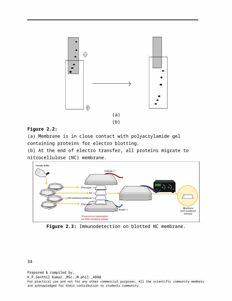
Blotting is transfer of resolved proteins from the gel onto a surface of a suitable membrane, done commonly by electrophoresis and referred to as electro blotting. The gel is placed in contact with nitrocellulose membrane which is then sandwiched between filter paper, two porous pads and two plastic supports. The entire set up is then placed in an electrophoretic tank containing blotting buffer. The proteins get transferred to the corresponding position on the membrane as resolved on the polyacrylamide gel, forming a mirror image of the gel. Protein of interest on the membrane is further located by immunodetection.
The band on the nitrocellulose membrane indicates the GST protein detected by the antibody (anti-GST)
The position of the band on the membrane indicates its electrophoretic mobility during electrophoresis.
The transferred proteins bound to the surface of nitrocellulose membrane are detected using immunological reagents. This process is known as immunodetection. All the unoccupied sites on the membrane are first blocked with an inert protein, a detergent or any other suitable blocking agent. The membrane is then identified using an enzyme-labeled secondary antibody and a suitable substrate to the enzyme, which results in a colored band on the nitrocellulose membrane.
21
Prepared & compiled by,K.P.Senthil Kumar.,MSc.,M.phil.,ADABFor practical use and not for any other commercial purposes; All the scientific community members are acknowledged for their contribution to students community.
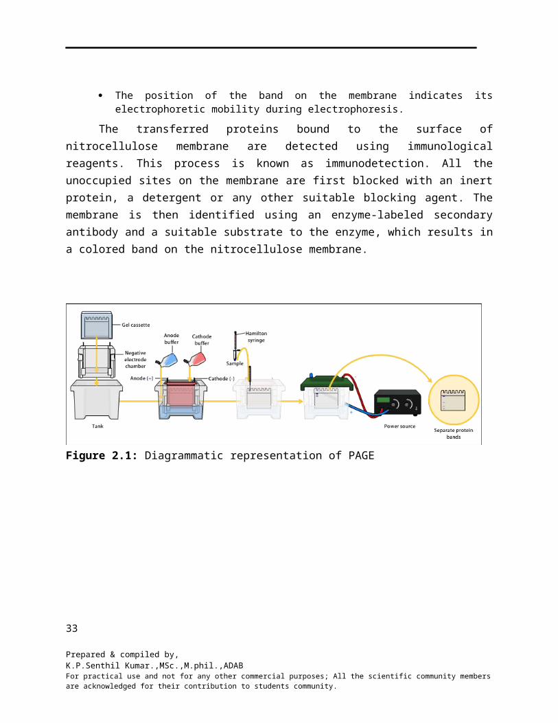
Figure 2.1: Diagrammatic representation of PAGE
(a) (b)Figure 2.2:(a) Membrane is in close contact with polyacrylamide gel containing proteins for electro blotting.(b) At the end of electro transfer, all proteins migrate to nitrocellulose (NC) membrane.
22
Prepared & compiled by,K.P.Senthil Kumar.,MSc.,M.phil.,ADABFor practical use and not for any other commercial purposes; All the scientific community members are acknowledged for their contribution to students community.
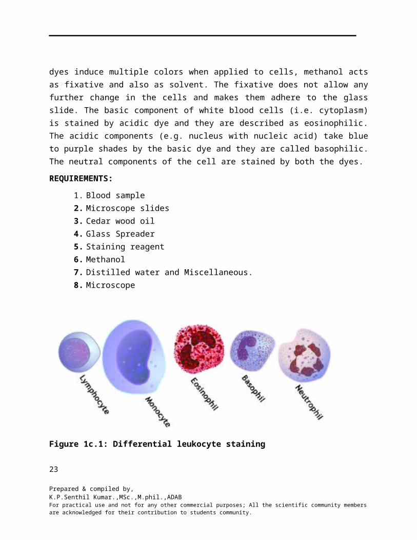
Figure 2.3: Immunodetection on blotted NC membrane.
Figure 2.4: Notch Base plate & Spacers Assembled for casting gel
IMMUNODETECTION:The transferred proteins bound to the surface of nitrocellulose membrane are detected
using immunological reagents. This process is known as immunodetection. All the unoccupied sites on the membrane are first blocked with an inert protein, a detergent or any other suitable blocking agent. The membrane is then probed with a primary antibody specific to the protein of interest. The Ag-Ab complex formed on the membrane is then identified using an enzyme-labeled secondary antibody and a suitable substrate to the enzyme, which results in a colored band on the nitrocellulose membrane, referred to as blot development.
23
Prepared & compiled by,K.P.Senthil Kumar.,MSc.,M.phil.,ADABFor practical use and not for any other commercial purposes; All the scientific community members are acknowledged for their contribution to students community.
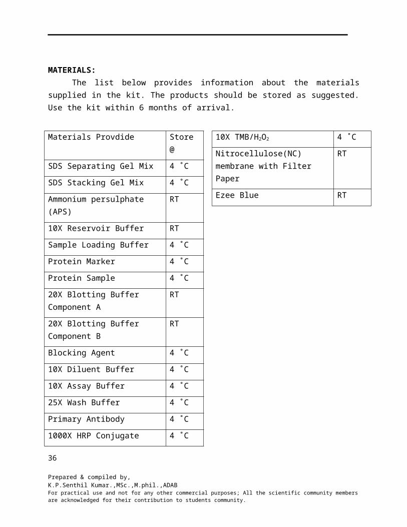
MATERIALS:The list below provides information about the materials supplied in the kit. The products
should be stored as suggested. Use the kit within 6 months of arrival.
Materials Provdide Store @
SDS Separating Gel Mix 4 ˚C
SDS Stacking Gel Mix 4 ˚C
Ammonium persulphate (APS) RT
10X Reservoir Buffer RT
Sample Loading Buffer 4 ˚C
Protein Marker 4 ˚C
Protein Sample 4 ˚C
20X Blotting Buffer Component A RT
20X Blotting Buffer Component B RT
Blocking Agent 4 ˚C
10X Diluent Buffer 4 ˚C
10X Assay Buffer 4 ˚C
25X Wash Buffer 4 ˚C
Primary Antibody 4 ˚C
1000X HRP Conjugate 4 ˚C
10X TMB/H2O2 4 ˚C
Nitrocellulose(NC) membrane with Filter Paper
RT
Ezee Blue RT
24
Prepared & compiled by,K.P.Senthil Kumar.,MSc.,M.phil.,ADABFor practical use and not for any other commercial purposes; All the scientific community members are acknowledged for their contribution to students community.
Materials required:
1. Equipment: Gel rocker (optional)2. Glassware: Conical flasks, Measuring cylinder, Petri dishes, staining tray.3. Distilled water4. Other equipment: Micropipettes, Tips, Water bath.
Note:• Read the entire procedure before starting the experiment.• Wear gloves while handling the gel and membrane.
• Prepare 1X TMB/H2O2, 1X secondary antibody, blocking buffer just before use.
• Resuspend an aliquot of APS in 1 ml of distilled water. Store at 4°C. Use within 2 months.
• Destaining step is not required on staining the gel with Ezee blue stainer (Coommassie brilliant blue).
Prepare the reagents as indicated below before starting each experiment:
Preparation of 1X Assay Buffer: To 2 ml of 10X assay buffer add 18 ml of distilled water to get 20 ml of 1X assay buffer.
Preparation of Protein Sample: Resuspend protein sample in 25 μl of distilled water.
Preparation of Primary Antibody: Resuspend an aliquot of primary antibody in 1 ml of 1X assay buffer. Transfer to a test tube and make up the volume to 10 ml with 1X assay buffer.
Preparation of Blotting Buffer: Mix 25 ml each of blotting buffer components A and B with 450 ml of distilled water.
Preparation of 1X Reservoir Buffer: To 25 ml of 10X Reservoir Buffer add 225 ml of distilled water to get 250 ml of 1X reservoir buffer.
Preparation of 1X Secondary Antibody: Pipette 10 μl of 1000X HRP conjugate and add 9.90 ml of 1X assay buffer. Mix thoroughly.
Preparation of Diluent Buffer: Dilute 1 ml of 10X diluents buffer to 10 ml with distilled water just before use.
Preparation of Blocking Buffer: Weigh 300 mg of blocking agent, suspend in 10 ml of 1X diluent buffer.
Kit description:
In this kit, bacterial lysate having Glutathione-S-Transferase (GST) fusion protein is provided, which will be electrophoresed in duplicates along with a standard protein marker on a 25
Prepared & compiled by,K.P.Senthil Kumar.,MSc.,M.phil.,ADABFor practical use and not for any other commercial purposes; All the scientific community members are acknowledged for their contribution to students community.
polyacrylamide gel. Following electrophoresis, the lysate and marker will be stained to know the electrophoretic mobility of the GST fusion protein, while the other electrophoresed lysate sample will be transferred by electro blotting onto nitrocellulose membrane. The electro blotted sample will then be detected using anti-GST lgG as primary antibody and secondary antibody labeled with Horse Radish Peroxidase (HRP). HRP is then detected using hydrogen peroxide as a substrate & Tetramethylbenzidine (TMB) as chromogen. HRP acts on hydrogen peroxide to release oxygen, which oxidizes the TMB to TMB oxide. The TMB oxide is deposited wherever enzyme is present & appears as a blue band on the NC membrane.
26
Prepared & compiled by,K.P.Senthil Kumar.,MSc.,M.phil.,ADABFor practical use and not for any other commercial purposes; All the scientific community members are acknowledged for their contribution to students community.
PROCEDURE:
DAY-1
SDS – PAGE
1. Assemble the plates for casting gel:2. Clamp the assembly of plates to fix it in a gel casting apparatus. Ensure the assembly is leak
proof by filling water between the plates. Silicon grease can be applied to spacer to make a water-tight seal.
3. Add 50µl of APS solution to 5ml of SDS separating gel mix & pour the gel solution between the plates till the level is 2cm below the top edge of notched plate.
4. Add 200 to 250ml of water to make the surface even.5. After the gel is set (approximately 20-30 min.), was the top of the separating gel with distilled
water & drain off the water completely.6. Add 20 µl of APS solution to 2ml of stacking gel mix and pour directly onto the polymerized
separating gel. 7. Insert the comb into the gel solution carefully without trapping any bubbles, about 1cm above the
separating gel. The stacking gel will set in approximately 10min.8. Add 25 µl of Sample Loading Buffer to Protein sample.9. Add 25 µl of Sample Loading Buffer to 25 µl of protein marker.10. Place it in a boiling water bath for 5 minutes.11. After the stacking Gel has set, carefully remove the comb and the bottom spacer. Wash the wells
immediately with distilled water to remove non-polymerized acryl amide. Fill the bottom reservoir with 1X Reservoir Buffer and carefully fix the plate to the apparatus without trapping any air bubbles between the buffer and the bottom of the gel. Fix the plates to PAGE apparatus. Fill the top reservoir with 1X Reservoir Buffer.
12. Load 30 µl protein marker in well 1, 40 µl of protein sample in well 2 & 10 µl of protein sample in well 4. Note down the order of loading. Connect the cords to the power supply according to the convention red: anode, black: cathode.
13. Set voltage at 100 V & switch on the power supply.14. When the dye front comes to 0.5 cm above the bottom of the gel, turn off the power. This will
take approximately 1 to 1.5 hours.15. Remove the gel plates and gently pry the plates apart using a spatula or similar tool, not at the
notch.16. Transfer the gel to a tray containing water; wash the gel for 1-2 minutes at room temperature.17. Decant water, cut the gel along lane 3.18. Transfer lane 4 i.e., Protein Sample in 10ml of blotting buffer taken in a Petri dish. Keep at room
temperature for 10 minutes. Following incubation, proceed for electro blotting as described in step 22.
19. To the gel piece (lanes 1&2) add minimum of 20ml water.20. Decant the water; add minimum 20ml of Ezee Blue Stain. Stain at room temperature for
1-2hours.
Note: For uniform staining & washing, place the tray on a rocker or shake intermittently every 10 to 15 minutes.
21. Decant the staining solution add minimum quantity of water cover the gel.
27
Prepared & compiled by,K.P.Senthil Kumar.,MSc.,M.phil.,ADABFor practical use and not for any other commercial purposes; All the scientific community members are acknowledged for their contribution to students community.
Note: Cover the tray & leave it overnight at room temperature.
ELECTRO BLOTTING
22. Assemble the blotting sandwich within the blotting cassette as shown in the figure. Take care to avoid air bubbles between the gel and NC membrane.
23. Insert the cassette into the apparatus filled with blotting buffer and connect blotting unit to power supply as per the convention.
24. Red: anode, black: cathode 25. Electrophoreses the sample at 50 V for 2 hours for blotting to occur.26. Remove the NC membrane gently from the cassette and place the membrane in 10ml of freshly
prepared blocking buffer taken in a Petri dish. Leave it overnight at 4oC.
DAY: 2
IMMUNODETECTION:
27. Discard Blocking Buffer.28. Immerse blot in 10ml of Primary Antibody Solution & mix gently for 30 minutes. Discard the
Primary Antibody Solution 29. Wash the blot by immersing in 10ml Wash Buffer for 3-5 minutes. Repeat the wash two times.
Discard the buffer each time.30. Immerse the blot in 10ml of 1X HRP labeled Antibody. Mix gently for 30 minutes. Discard the
HRP labeled Antibody.31. Wash the blot by immersing in 10ml Wash Buffer for 3-5 minutes. Repeat the wash process four
to five times. Discarding the buffer each time.32. Immerse the washed blot in 10ml of Substrate Solution, mix gently for 5-10 minutes, within this
time colored band will appear.33. Remove the blot; wash with distilled water, discard & dry.Note:
Although the colored band fades with time, the rate of color loss can be retarded if the blots are kept in dark.
34. Compare the SDS-Polyacrylamide gel with the developed NC membrane.
OBSERVATION:
35. On staining SDS-Polyacrylamide gel, different proteins will appear as dark blue bands against a light blue background.
36. On immunodetection, a single blue band will be observed on NC membrane.
INTERPRETATION:
On electrophoresis of bacterial lysate on SDS-PAGE, many bands indicating the different proteins present in the crude sample are seen. A predominant band among these is that of GST fusion protein corresponding to 26 kD protein of the marker. Following transfer & immunodetection, one observes a predominat band corresponding to GST protein bound to anti-
28
Prepared & compiled by,K.P.Senthil Kumar.,MSc.,M.phil.,ADABFor practical use and not for any other commercial purposes; All the scientific community members are acknowledged for their contribution to students community.
GST antibody. However, few light bands may be seen indicating the proteins with which anti-GST antibody cross reacts.
RESULT:
CONCLUSION:
29
Prepared & compiled by,K.P.Senthil Kumar.,MSc.,M.phil.,ADABFor practical use and not for any other commercial purposes; All the scientific community members are acknowledged for their contribution to students community.
Experiment: 3 Date:
SINGLE RADIAL IMMUNODIFFUSION ASSAY
AIM: To perform the single radial immunodiffusion assay.
INTRODUCTION:
Single Radial Immunodiffusion (RID) is used extensively for the quantitative estimation of antigens. RID technique used for estimating the concentration of plasma protein like immunoglobulin and also for the estimation of antibody concentration in sera samples.
PRINCIPLE:
When antigen diffuses radially forming a concentration gradient disk in gel containing uniformly distributed antibody, the Ag-Ab complex precipitates in the gel where the diffusing antigen reaches equivalence with the antibody concentration presenting the gel. The precipitate formed in the gel appears as an opaque ring. The ring diameter is directly proportional to the antigen concentration.
The Technique:
In this method antibody is incorporated before the gel is made. Thus, the antiserum is uniformly distributed throughout the agarose gel. Antigen is then allowed to diffuse from wells cut into the gel. Initially, as the antigen diffuses out of the well, its concentration is relatively high and it forms soluble antigen-antibody adducts. However, as it diffuses farther and farther from the well, its concentration decreases. When its concentration becomes equivalent to that of the antibody in the gel; antigen-antibody precipitates and a precipitin ring is formed. Greater the concentration of antigen greater is the diameter of the precipitin ring. Thus, by running a range of known antigen concentration on the gel and by measuring the diameter of their precipitin rings, a calibration graph can be constructed. The antigen concentration of unknown samples run on the same gel can then be determined by simple interpolation, having measured the diameters of the respective precipitin rings.
REQUIREMENTS:
1. Agarose2. 10X assay Buffer (To be diluted 10
times before use.)3. Standard Antigens.4. Test Antigen5. Antiserum6. Gel Punch with syringe
7. Glass Plates8. Template9. Semi log Graph Sheet10. Distilled Water11. Micropipette.12. A box to keep the gel plate in moist
chamber
30
Prepared & compiled by,K.P.Senthil Kumar.,MSc.,M.phil.,ADABFor practical use and not for any other commercial purposes; All the scientific community members are acknowledged for their contribution to students community.
Precautions:
The glass plate should be wiped grease free with cotton for the even spreading of agarose.
The wells should be cut neatly without rugged margins to get a perfect ring of precipitation.
The antiserum should be added to the agar only after it cools to 550C. Higher temperature will inactivate the antibody.
After the addition of antiserum to the agar proper mixing is essential for uniform distribution of antibody
Reconstitutions:
Reconstitute each vial containing lyophilized antigen and antisera with 1ml of 1X assay buffer. Mix it well and allow it to stand for 30 minutes.
PROCEDURE:
1. 1gm Agarose is dissolved in 100 ml of 1X assay buffer by heating.2. The solution is then allowed to cool to 550C and 120 µl of antiserum mixed with 6ml of
the solution.3. The agarose solution containing the antiserum is poured onto a grease free glass plate that
had previously been set on a horizontal level surface; Cool to form a gel.4. Using a gel punch, wells of 4mm diameter are cut into the gel as show figure1.5. Add 20µl of the given standard antigens and samples to the wells as shown in figure (two
test samples provided with the kit); Avoid overflow. 6. The gel plate is left in a box containing wet cotton and incubated overnight at room
temperature.7. The diameter of the disk can be measured by marking the edges of the circle.8. A standard graph is constructed by plotting the diameters of the disk against the
concentration of antigen in a semi log graph sheet as shown in figure 2. The concentration of unknown is then determined by reading the concentration against the ring diameter.
31
Prepared & compiled by,K.P.Senthil Kumar.,MSc.,M.phil.,ADABFor practical use and not for any other commercial purposes; All the scientific community members are acknowledged for their contribution to students community.
1 2
3 4 5 6
Figure 3.1: Pattern of wells for Antigen and tests.
Standard Antigen A-0.25mg/ml
Standard Antigen B-0.5mg/ml
Standard Antigen C-1.0mg/ml
Standard Antigen D-2.0mg/ml
Test Antigen –1
Test Antigen –2
Observation table:
RESULT:
CONCLUSION:
Experiment: 4 Date:
OUCHTERLONY DOUBLE DIFFUSION ASSAY
AIM: To determine the titter value of the antiserum using ouchterlony double diffusion assay.
INTRODUCTION:32
Prepared & compiled by,K.P.Senthil Kumar.,MSc.,M.phil.,ADABFor practical use and not for any other commercial purposes; All the scientific community members are acknowledged for their contribution to students community.
Sr. No. Std Conc. mg/ml Ring Diameter mm Test Conc. mg/ml.
1 0.25 6
2 0.5 8
3 1.0 10
4 2.0 12
5 Test-1 ?
6 Test-2 ?
Interaction between antigen (Ag) and antibody (Ab) in the molecular level forms the basis for several techniques that are useful in modern day scientific studies and in routine clinical diagnosis. These techniques are either based on use of labeled reagents, a tracer or by immunoprecipitation. Ouchterlony double diffusion (ODD) is one of the simplest technique extensively used to check antisera for the presence and specificity of antibodies for a particular Ag. This technique is also known as double immunodiffusion
PRINCIPLE:
The technique is based on the phenomenon that when Ag and Ab mix in optimal proportion, the Ag-Ab complex precipitates. The precipitate formed in agarose gel appears as an opaque line.
The Technique:
In this technique, solutions of Ag and Ab placed in adjacent wells cut in agarose gel diffuse radially. Initially as the Ag and Ab diffuse out of the respective wells, the concentrations are relatively high nearer to the well. However as they diffuse further from the wells, the concentration decreases. At one point their concentrations become equivalent and Ag-Ab complex precipitates to form a precipitin line at this point. The relative position of the precipitin line yields an estimate of Ag (or Ab) concentration, i.e. more concentrated the immunoreactions solution, the precipitin line formed is farther from the well.
REQUIREMENTS:
1. Agarose
2. 10X Assay buffer
3. Antigen
4. Test antisera
5. Glass plates
6. Gel punch with syringe
7. Template
8. Micropipettes
9. Tubes
10. Distilled water
11. A box to keep the gel plate in moist
chamber.
33
Prepared & compiled by,K.P.Senthil Kumar.,MSc.,M.phil.,ADABFor practical use and not for any other commercial purposes; All the scientific community members are acknowledged for their contribution to students community.
PRECAUTIONS:
1. The glass plates should be wiped grease free with cotton for the even spreading of the
agarose.
2. Ensure that the moist chamber has enough wet cotton to keep the atmosphere humid.
RECONSTITUTIONS:
Reconstitute each vial containing lyophilized antigen and antisera with 1 ml of 1 X
assay buffer. Mix it well and allow it to stand for 30 minutes.
PROCEDURE:
1. Prepare 1 % agarose solution in 1 X assay buffer by heating to dissolve the agarose
completely.
2. Cool the solution to about 55-60°C and pour 5ml of the solution onto grease free glass
plate that had previously been set on horizontal level surface. Allow the gel to set for
20-30 minutes.
3. Serially dilute the test antisera up to 1:32 dilution. Take 20 μl of 1 X assay buffer in
five eppendorf tubes. To the first tube add 20 μl of test antiserum mix well. The
dilution of antiserum in the first tube is 1:2. From the first tube take out 20 μl of 1:2
diluted antiserum and add to the second tube. The dilution in the second tube is 1:4;
repeat the dilution up to the fifth tube as shown in Figure 1. Add 20μl test antiserum
in first well.
4. Keep the gel plate on the template provided. Punch wells in the gel with the help of a
gel punch corresponding to the marking on the template with gentle suction so that the
wells are cleanly formed as the resultant agarose plugs are sucked out.
5. Add 20 μl of the Ag into the centre well and 20 μl of each of neat, test serum, 1 :2,
1:4, 1:8, 1:16, 1:32, dilutions into the surrounding wells as shown in the following
figure.
34
Prepared & compiled by,K.P.Senthil Kumar.,MSc.,M.phil.,ADABFor practical use and not for any other commercial purposes; All the scientific community members are acknowledged for their contribution to students community.
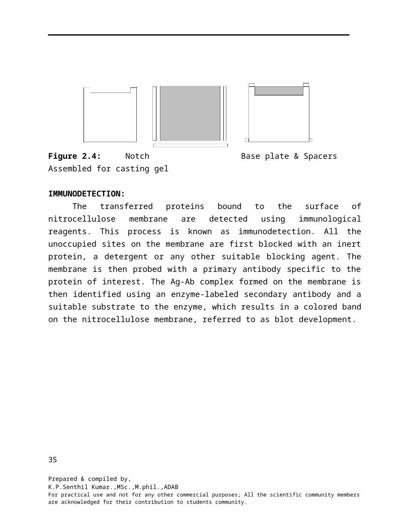
Figure 4.1: The pattern of wells and additions of Ag and test serum dilutions
6. Keep the plate in a moist chamber overnight at room temperature.
7. After incubation observe for opaque precipitation lines between the Ag and antisera
wells.
INTERPRETATION:
1. Precipitin line indicates presence of Ab in the antiserum to the Ag added.
2. The titter of the antiserum is the highest dilution showing the precipitin line. For example
if the line of precipitation is seen up to 1:16 dilution, the titter of the antiserum is considered
as 1:16.
RESULT:
DISCUSSION:
35
Prepared & compiled by,K.P.Senthil Kumar.,MSc.,M.phil.,ADABFor practical use and not for any other commercial purposes; All the scientific community members are acknowledged for their contribution to students community.
Experiment: 5 Date:
ENZYME LINKED IMMUNOSORBENT ASSAY (DOT ELISA)
AIM: To perform sandwich Dot ELISA test for antigen.
INTRODUCTION:
ELISA is being extensively used as a tool in research as well as analytical/diagnostic
laboratories. The specificity, sensitivity and ease to perform these techniques have made
these methods popular. It is therefore necessary to have hands on experience of this technique
along with the theoretical knowledge. The kit is designed to understand the principle and
working of a direct sandwich ELISA test using human IgG as a model. Such methods can be
used for estimating any type of multivalent antigen using the appropriate antibodies. The kit
is safe to handle, it does not involve handling of any type of hazardous materials. It is easy to
perform the test and understand the reactions.
PRINCIPLE:
In direct sandwich Dot-ELISA the antigen is sandwiched directly between two
antibodies. For this to happen, the two antibodies used must be able to react (bind) with two
different epitope on the same antigen. One antibody is immobilized on a solid support and the
other one is linked to an enzyme. Then the bound antigen is allowed to react with the second
enzyme linked antibody. Then the substrate for the linked enzyme is added. The enzyme
linked conjugate oxidizes the substrate. The oxidized substrate gives a blue color spot (hence
the name Dot-ELISA). The enzyme activity (color intensity of the spot) is directly
proportional to the antigen concentration.
TMB (Tetramethyl benzidine) / H2O2 (horseradish peroxidase) is used for the
detection of HRP enzyme linked conjugate. Here H2O2 is the substrate and TMB is a
chromogen. HRP act on H2O2 to release oxygen, which oxidizes the TMB to TMB oxide.
This TMB oxide is deposited whenever enzyme is present and appears as a blue spot. That is
36
Prepared & compiled by,K.P.Senthil Kumar.,MSc.,M.phil.,ADABFor practical use and not for any other commercial purposes; All the scientific community members are acknowledged for their contribution to students community.
If the test sample does not contain the antigen specific to the antibody, there will be
no enzyme reaction and no spot develops.
REQUIREMENT:
1. ELISA strip for human IgG test. 2. Assay buffer at 10x concentration. 3. Goat anti human IgG peroxidase conjugate (antibody-HRP)4. TMB/H2O2 10 x concentration (substrate)5. Serum samples for human IgG test 3 x 0.5ml.6. Micropipette.7. Distilled water.8. Test tubes of size 10 x 75 mm or 1.5 ml eppendorf tubes.
PRECAUTIONS:
Reconstitute test samples with 0.5 ml distilled water and store at 4 - 8˚c.
Prepare 1x assay buffer by diluting 10x assay buffer 10 times with distilled water.
Use the diluted buffer on the same day.
Do not contaminate reagents with each other.
Dilute only required amount of reagents.
Do not leave the reagents at room temperature.
Ensure all three zones on strip are immersed in solution.
PROCEDURE:
1. To one drop (50μl) of the sample add 1ml of 1x assay buffer and insert an ELISA strip.
Keep for 20 minutes at room temperature
2. Wash the strip by dipping it in 1 ml of 1x assay buffer for about 5 minutes and replace the
buffer. Repeat two more times.
3. In 1ml 1x assay buffer add 10μl antibody-HRP conjugate and dip the strip. Keep for 20
minutes.
4. Wash the strip as in step 2.
5. In a fresh tube take 0.1 ml TMB/H2O2, 10X concentrate, 0.9 ml distilled water and dip the
strip.
6. Observe the strip in 10-20 minutes for the appearance of blue/gray spot on the strip.
37
Prepared & compiled by,K.P.Senthil Kumar.,MSc.,M.phil.,ADABFor practical use and not for any other commercial purposes; All the scientific community members are acknowledged for their contribution to students community.
7. Rinse the strip with distilled water.
INTERPRETATION:
1. A spot on the lower portion and no spot on the upper portion indicate the proper
performance of the test.
2. A spot in middle portion indicates presence of the antigen in the sample.
3. Intensity of the spot is proportional to antigen concentration in the sample, i.e. human
IgG.
RESULT:
CONCLUSION:
38
Prepared & compiled by,K.P.Senthil Kumar.,MSc.,M.phil.,ADABFor practical use and not for any other commercial purposes; All the scientific community members are acknowledged for their contribution to students community.
Related Documents


