ORIGINAL ARTICLE Raw starch–degrading a-amylase from Bacillus aquimaris MKSC 6.2: isolation and expression of the gene, bioinformatics and biochemical characterization of the recombinant enzyme F. Puspasari 1,2 , O.K. Radjasa 3 , A.S. Noer 1 , Z. Nurachman 1 , Y.M. Syah 1 , M. van der Maarel 4 , L. Dijkhuizen 4 , S. Janecek 5,6 and D. Natalia 1 1 Biochemistry Research Division, Faculty of Mathematics and Natural Sciences, Institut Teknologi Bandung, Bandung, Indonesia 2 School of Industrial Technology and Pharmacy (STTIF) Bogor, Bogor, Indonesia 3 Center for Tropical Coastal and Marine Studies, Diponegoro University, Semarang, Indonesia 4 Microbial Physiology, Groningen Biomolecular Sciences and Biotechnology Institute (GBB), University of Groningen, Haren, Groningen, The Netherlands 5 Laboratory of Protein Evolution, Institute of Molecular Biology, Slovak Academy of Sciences, Bratislava, Slovakia 6 Department of Biotechnology, Faculty of Natural Sciences, University of SS. Cyril and Methodius, Trnava, Slovakia Keywords B. aquimaris MKSC 6.2, GH13 subfamily, raw starch degrading, a-amylase. Correspondence Dessy Natalia, Biochemistry Division, Faculty of Mathematics and Natural Sciences, Bandung Institute of Technology, Jl. Ganesha 10, Bandung 40132, Indonesia. E-mail: [email protected] and Stefan Janecek. Laboratory of Protein Evolution, Institute of Molecular Biology, Slovak Academy of Sciences, Dubravska cesta 21, SK-84551 Bratislava, Slovakia. E-mail: stefan. [email protected] Dedicated to the memory of Achmad Saifudin Noer who passed away in 2010. 2012/0724: received 21 April 2012, revised 18 August 2012 and accepted 29 August 2012 doi:10.1111/jam.12025 Abstract Aims: The aims were to isolate a raw starch–degrading a-amylase gene baqA from Bacillus aquimaris MKSC 6.2, and to characterize the gene product through in silico study and its expression in Escherichia coli. Methods and Results: A 1539 complete open reading frame of a starch– degrading a-amylase gene baqA from B. aquimaris MKSC 62 has been determined by employing PCR and inverse PCR techniques. Bioinformatics analysis revealed that B. aquimaris MKSC 6.2 a-amylase (BaqA) has no starch- binding domain, and together with a few putative a-amylases from bacilli may establish a novel GH13 subfamily most closely related to GH13_1. Two consecutive tryptophans (Trp201 and Trp202, BaqA numbering) were identified as a sequence fingerprint of this novel GH13 subfamily. Escherichia coli cells produced the recombinant BaqA protein as inclusion bodies. The refolded recombinant BaqA protein degraded raw cassava and corn starches, but exhibited no activity with soluble starch. Conclusions: A novel raw starch–degrading B. aquimaris MKSC 6.2 a-amylase BaqA is proposed to be a member of new GH13 subfamily. Significance and Impact of the Study: This study has contributed to the overall knowledge and understanding of amylolytic enzymes that are able to bind and digest raw starch directly. Introduction Starch-degrading enzymes cleave glycosidic bonds in starch either from the nonreducing end (exo-acting enzymes) or in the interior (endo-acting enzymes) of the polymer. a-Amylases (EC 3.2.1.1) are endo-acting enzymes cleaving a-1,4-glycosidic bonds in starch to form various maltooligosaccharides and maltodextrin (MacGregor et al. 2001; van der Maarel et al. 2002). a-Amylases are mainly classified into glycoside hydro- lase (GH) family 13 (Henrissat 1991). The GH13 family covers enzymes with several reaction specificities, but they share common conserved sequence regions (CSRs) with a catalytic triad (Asp, Glu, Asp), employ an a-retaining mechanism, and adopt a Triosephosphate IsoMerase (TIM)-barrel fold (Janecek 2002). A few years ago, Stam et al. (2006) divided 1691 different members of the GH13 family into 35 subfamilies based on their amino Journal of Applied Microbiology © 2012 The Society for Applied Microbiology 1 Journal of Applied Microbiology ISSN 1364-5072

Welcome message from author
This document is posted to help you gain knowledge. Please leave a comment to let me know what you think about it! Share it to your friends and learn new things together.
Transcript
ORIGINAL ARTICLE
Raw starch–degrading a-amylase from Bacillus aquimarisMKSC 6.2: isolation and expression of the gene,bioinformatics and biochemical characterization of therecombinant enzymeF. Puspasari1,2, O.K. Radjasa3, A.S. Noer1, Z. Nurachman1, Y.M. Syah1, M. van der Maarel4,L. Dijkhuizen4, S. Janecek5,6 and D. Natalia1
1 Biochemistry Research Division, Faculty of Mathematics and Natural Sciences, Institut Teknologi Bandung, Bandung, Indonesia
2 School of Industrial Technology and Pharmacy (STTIF) Bogor, Bogor, Indonesia
3 Center for Tropical Coastal and Marine Studies, Diponegoro University, Semarang, Indonesia
4 Microbial Physiology, Groningen Biomolecular Sciences and Biotechnology Institute (GBB), University of Groningen, Haren, Groningen,
The Netherlands
5 Laboratory of Protein Evolution, Institute of Molecular Biology, Slovak Academy of Sciences, Bratislava, Slovakia
6 Department of Biotechnology, Faculty of Natural Sciences, University of SS. Cyril and Methodius, Trnava, Slovakia
Keywords
B. aquimaris MKSC 6.2, GH13 subfamily, raw
starch degrading, a-amylase.
Correspondence
Dessy Natalia, Biochemistry Division, Faculty
of Mathematics and Natural Sciences,
Bandung Institute of Technology, Jl. Ganesha
10, Bandung 40132, Indonesia. E-mail:
[email protected] and Stefan Janecek.
Laboratory of Protein Evolution, Institute of
Molecular Biology, Slovak Academy of
Sciences, Dubravska cesta 21, SK-84551
Bratislava, Slovakia. E-mail: stefan.
Dedicated to the memory of Achmad Saifudin
Noer who passed away in 2010.
2012/0724: received 21 April 2012, revised
18 August 2012 and accepted 29 August
2012
doi:10.1111/jam.12025
Abstract
Aims: The aims were to isolate a raw starch–degrading a-amylase gene baqA
from Bacillus aquimaris MKSC 6.2, and to characterize the gene product
through in silico study and its expression in Escherichia coli.
Methods and Results: A 1539 complete open reading frame of a starch–degrading a-amylase gene baqA from B. aquimaris MKSC 6�2 has been
determined by employing PCR and inverse PCR techniques. Bioinformatics
analysis revealed that B. aquimaris MKSC 6.2 a-amylase (BaqA) has no starch-
binding domain, and together with a few putative a-amylases from bacilli may
establish a novel GH13 subfamily most closely related to GH13_1. Two
consecutive tryptophans (Trp201 and Trp202, BaqA numbering) were
identified as a sequence fingerprint of this novel GH13 subfamily. Escherichia
coli cells produced the recombinant BaqA protein as inclusion bodies. The
refolded recombinant BaqA protein degraded raw cassava and corn starches,
but exhibited no activity with soluble starch.
Conclusions: A novel raw starch–degrading B. aquimaris MKSC 6.2 a-amylase
BaqA is proposed to be a member of new GH13 subfamily.
Significance and Impact of the Study: This study has contributed to the
overall knowledge and understanding of amylolytic enzymes that are able to
bind and digest raw starch directly.
Introduction
Starch-degrading enzymes cleave glycosidic bonds in
starch either from the nonreducing end (exo-acting
enzymes) or in the interior (endo-acting enzymes) of the
polymer. a-Amylases (EC 3.2.1.1) are endo-acting
enzymes cleaving a-1,4-glycosidic bonds in starch to form
various maltooligosaccharides and maltodextrin
(MacGregor et al. 2001; van der Maarel et al. 2002).
a-Amylases are mainly classified into glycoside hydro-
lase (GH) family 13 (Henrissat 1991). The GH13 family
covers enzymes with several reaction specificities, but they
share common conserved sequence regions (CSRs) with a
catalytic triad (Asp, Glu, Asp), employ an a-retainingmechanism, and adopt a Triosephosphate IsoMerase
(TIM)-barrel fold (Janecek 2002). A few years ago, Stam
et al. (2006) divided 1691 different members of the
GH13 family into 35 subfamilies based on their amino
Journal of Applied Microbiology © 2012 The Society for Applied Microbiology 1
Journal of Applied Microbiology ISSN 1364-5072

acid sequence similarities. Most subfamilies are mono-
functional, while the rest contain polyspecific enzymes
with strongly related substrate and/or product specifici-
ties. Nowadays (August 2012), 37 subfamilies from more
than 12 000 different members of this family can be
found at the CAZy website (http://www.cazy.org/), that
is, a database specialized in classifying the carbohydrate-
active enzymes that build and degrade complex carbohy-
drates (Cantarel et al. 2009). Bacterial a-amylases are
classified into GH13 subfamilies 5, 27, 28, 32, 36 and 37.
Subfamily GH13_36 contains the so-called intermediary
enzymes that exhibit a mixed enzyme specificity of
a-amylase and an additional GH13 specificity (Oslancova
and Janecek 2002). Various novel putative a-amylases
identified in genome projects have not been classified yet
into any subfamily (Cantarel et al. 2009).
a-Amylases from microbes are applied widely in the
starch industry and in food processing (Gupta et al.
2003). Bacteria from the genus Bacillus are well known as
a-amylase producers. Some bacilli were reported to pos-
sess raw starch–degrading enzyme activities (Demirkan
et al. 2005; Goyal et al. 2005; Mitsuiki et al. 2005). A
marine Bacillus associated with soft coral Sinularia sp.,
Bacillus aquimaris MKSC 6.2, also produced raw starch–degrading amylolytic enzymes (Puspasari et al. 2011).
Raw starch–degrading amylases are of great interest in
view of their ability to hydrolyze raw starch at moderate
temperature, which is the key to reduce production cost
in starch-processing industry (Leveque et al. 2000). Most
raw starch–degrading enzymes possess a starch-binding
domain (SBD) responsible for binding and raw starch
degradation (Machovic and Janecek 2006; Janecek et al.
2011), localized usually at the C-terminus of bacterial
and fungal amylases (Janecek and Sevcik 1999). Such a
distinct SBD is present in approximately 10% of all amy-
lolytic enzymes (Machovic and Janecek 2006), but amy-
lases without such a specialized domain able to bind and
digest raw starch also exist (Søgaard et al. 1993; Hostino-
va et al. 2003). Usually tryptophan (or in a wider sense
aromatic) residues are responsible for these so-called sur-
face-binding sites (Gibson and Svensson 1987; Gyemant
et al. 2009) that, for example, in the barley a-amylase iso-
zyme AMY1 and Saccharomycopsis fibuligera glucoamylase
are arranged as so-called ‘sugar tongs’, that is, a sugar
molecule entrapped by a tyrosine (Robert et al. 2003;
Sevcik et al. 2006).
In this study, we report the nucleotide sequence of the
B. aquimaris MKSC 6.
Two a-amylase gene (baqA), its expression in Escheri-
chia coli and bioinformatics analysis together with bio-
chemical characterization of the recombinant enzyme.
Sequence and phylogenetic analyses of BaqA and some
other novel putative a-amylases from genus Bacillus
revealed that these a-amylases, grouped separately in a
novel cluster in the evolutionary tree, may define a new
GH13 subfamily.
Materials and methods
Strains and plasmids
Bacillus aquimaris MKSC 6.2 (Bacaq) was obtained from
the culture collection at the Center for Tropical Coastal
and Marine Studies, Diponegoro University, Semarang,
Indonesia. The bacterium was isolated from a soft coral
Sinularia sp. obtained from the sea around Merak Kecil
Island, Banten, Indonesia. Escherichia coli strain TOP10F0
(Invitrogen) was used for gene manipulation. The host
strain for heterologous recombinant expression was
E. coli BL21 (DE3) (Novagen, Darmstadt, Germany).
Plasmid pGEM-T (Promega, Madison, WI) was used to
clone PCR products, while pET30a (+) (Novagen) was
used as an expression vector.
Isolation of the a-amylase gene
Isolation of Bacaq a-amylase gene (baqA) was conducted
through several PCR steps using degenerate primers
(degPCR) and inverse PCR (invPCR) (Weber-Arden
et al. 1996). The nucleotide sequences of degenerate
primers were 5′-GAYTTYRTYGTSAATCAYGTYGG-3′(degFA), 5′-GATGGRTAYCGTCTRGATACYG-3′ (degFB),5′-AATCGWMYCATATCATGGTTATC-3′ (degR2) and
5′-CAGATCCRTAGTAAASAATSGG-3′ (degR4). The first
invPCR used primers InvF1 5′-TGACCCACAGAAGA-TAGCAG-3′ and InvR1 5′-ATGGCGCACAGTATCCA-GAC-3′ with circularized ClaI cut Bacaq chromosomal
DNA as template. The second invPCR was performed with
circularized HindIII cut Bacaq chromosomal DNA as tem-
plate using InvF2 5′-TACACGACTCCGGGAATACC-3′and InvR1. The last one used primers InvF3
5′-AGCGGGATATGAAAGTCGTC-3′ and InvR3 5′-GAG-TCAGCCATATGGAAGTG-3′ with circularized HpaI cut
Bacaq chromosomal DNA as template. Purified PCR prod-
ucts were cloned into plasmid pGEM-T and sequenced
through dideoxy Sanger method using dye terminator
(Macrogen, Seoul, Korea). The nucleotide sequence of
baqA has been deposited in Genetic sequence database at
the National Center for Biotechnical Information (NCBI)
(GenBank ID: JN797599).
Cloning of baqA gene
The whole baqA gene without putative signal peptide
encoding region was amplified by PCR using primers
baqA-F 5′-GATATCGAAGAACGAAAGTGGCAG-3′ and
2 Journal of Applied Microbiology © 2012 The Society for Applied Microbiology
Expression and bioinformatics of B. aquimaris a-amylase gene F. Puspasari et al.
baqA-R 5′-GAATTCGATTTGCGGTTTTTTCTTCCG-3′.Forward and reverse primers carried EcoRI and EcoRV
restriction sites, respectively. The amplified gene was
cloned in pGEM-T (Promega), and the sequence was ver-
ified. It was then subcloned into pET30a(+) (Novagen) toconstruct the recombinant plasmid pET30-baqA.
Production and refolding of recombinant BaqA
Escherichia coli BL21 (DE3) carrying pET30-baqA was
grown in 50-ml LB medium [0�5% (w/v) yeast extract;
1% (w/v) tryptone; 0�5% (b/v) NaCl] containing
30 lg ml�1 kanamycin in a rotary shaker (37°C, 150 rev
min�1) to an optical density of 0�4–0�6 (600 nm).
Expression of recombinant baqA was induced by
0�5 mmol l�1 isopropyl-b-D-thiogalactopyranoside (IPTG)
37°C for 4 h and analyzed by SDS-PAGE. Cells were har-
vested by centrifugation (13 000 g, 30 min) and then dis-
rupted by sonication. Inclusion bodies were pelleted by
centrifugation (13 000 g, 10 min) and dissolved by addi-
tion of sarkosyl [final concentration was 0�3% (w/v)] as
described by Marshak et al. (1996). After centrifugation
(13 000 g, 10 min), the solubilized protein was diluted
tenfold with 20 mmol l�1 maleate buffer (pH 6�5) and
dialyzed against 20 mmol l�1 maleate buffer (pH 6�5) for20–24 h to remove the detergent. Protein was subjected
to SDS-PAGE (Laemmli 1970). This refolded recombi-
nant BaqA a-amylase was used for biochemical studies.
a-Amylase activity toward soluble starch
a-Amylase activity was determined by measuring the
amount of reducing sugars formed using a modification
of the dinitrosalicylic acid (DNS) method (Miller 1959).
The reaction was performed in a reaction mixture con-
taining 25 ll of 1% soluble starch (E-Merck, Darmstadt,
Germany) and various amounts of enzyme in
50 mmol l�1 maleate buffer (pH 6�5) at 37 and 50°C for
various time intervals up to 24 h. The reaction was
stopped by addition of 50 ll of DNS solution and incu-
bated in boiling water bath for 10 min. The reaction mix-
ture was cooled down to room temperature, and the
absorbance at 500 nm was measured. The amount of
reducing sugar was then determined using a glucose stan-
dard curve. One unit of amylase activity equals the
amount of the enzyme producing 1 lmol of reducing
sugars per min under the assay conditions. All assays
were performed in triplicates.
Raw starch digestion
To determine the digestion of raw starch by recombinant
BaqA, reaction mixtures containing 50 lg of the enzyme
and 10% (w/v) raw starch (corn or cassava) to a final
volume of 0�2 ml in 50 mmol l�1 universal buffer
(pH 6�5) were incubated at 37°C with shaking for 24 h.
After centrifugation, the reducing sugar produced in
supernatant was determined using a modification of the
DNS method. The degree of hydrolysis (DH) was calcu-
lated by the following equation: DH (%) = (H1/
H0) 9 100, where H1 was reducing sugar produced by
enzyme hydrolysis, and H0 was reducing sugar produced
by acid hydrolysis. Acid hydrolysis was carried out by
treating raw starch with 1 mol l�1 HCl at 100°C for 2 h
(Wang et al. 1995).
Raw starch adsorbability
Affinity of the enzyme towards raw corn and cassava
starch was studied by incubating 24 lg of the enzyme
with 5–350 mg of raw starch at 4°C for 1 h. After centri-
fugation, the amount of free enzyme in the supernatant
was determined. The bound protein was the difference
between original protein amount and the free protein
amount in the supernatant after binding. Total protein
concentration was determined by Bradford method (Brad-
ford 1976) using bovine serum albumin as a standard.
Scanning electron microscopy and end products
determination
A mixture of 1% (w/v) of raw corn or cassava starch and
800 mg of recombinant BaqA to a final volume of 0�2 ml
in 50 mmol l�1 maleate buffer (pH 6�5) was incubated at
37°C for 48 h. After centrifugation, the pellet was washed
with 95% ethanol and then dried at 35°C. The treated
starch granules were coated with Au-Pd (80–20) using
Ion Sputter JFC-110 at 1�2 KV and 6 mA for 4 min and
photographed using scanning electron microscopy (SEM)
(JSM-35C). Supernatant was tested for end products
determination. End product of raw starch degradation by
recombinant BaqA treatment was analyzed by thin layer
chromatography using a stationary phase of 10-cm silica
gel plate (E-Merck) and a solvent system containing
butanol:ethanol:water (5:5:3). End product spots were
visualized by spraying the plate with 50% (v/v) of sul-
phuric acid in methanol and heated at 110°C for 10 min.
Bioinformatics
Nucleotide sequences of PCR products were used as the
query sequence in the BLAST searches (http://blast.ncbi.
nlm.nih.gov/Blast.cgi) using the BLASTX program (Altsc-
hul et al. 1997). The type of family and domain in the
deduced protein were searched against the Pfam database
(http://pfam.sanger.ac.uk/; Finn et al. 2010). Prediction of
Journal of Applied Microbiology © 2012 The Society for Applied Microbiology 3
F. Puspasari et al. Expression and bioinformatics of B. aquimaris a-amylase gene
a signal peptidase cleavage site was performed by the
web-based search tools SIGNALP (http://www.cbs.dtu.dk/
services/; Bendtsen et al. 2004).
The amino acid sequence of BaqA deduced from the
nucleotide sequence was compared with those of other
a-amylases retrieved from the Universal Protein
Resources Knowledgebase (UniProt; Apweiler et al. 2011)
and GenBank (Benson et al. 2011) databases. The set cov-
ered all GH13 subfamilies (Stam et al. 2006; Cantarel
et al. 2009) with well-defined (GH13 subfamilies 1, 5, 6,
7, 15, 24, 27, 28, 32 and 37) and supposed (GH13_36)
a-amylase activity (EC 3.2.1.1) completed with represen-
tatives of the bacterial subfamily GH13_19 (closely related
sequences with mostly maltohexaose-forming amylase
specificity). The aligned amino acid sequence segments
spanned the region (approximately 300 residues) of the
catalytic (b/a)8-barrel from b1 to b8 strands including
domain B (Da Lage et al. 2004; Hostinova et al. 2010)
and containing all the seven CSRs of the a-amylase family
(Janecek 2002). The alignment was performed using the
CLUSTALX program (Jeanmougin et al. 1998), and the evo-
lutionary tree was calculated from the manually adjusted
alignment as a PHYLIP-tree type with the neighbour-join-
ing clustering method (Saitou and Nei 1987) and the
bootstrapping procedure (Felsenstein 1985; the number
of bootstrap trials used was 1000) implemented in the
CLUSTALX package. The tree was displayed with the TREE-
VIEW program (Page 1996).
A BaqA three-dimensional structure model was
obtained with the PHYRE (Protein Homology/AnalogY
Recognition Engine) fold recognition server (http://www.
sbg.bio.ic.ac.uk/phyre/; Kelley and Sternberg 2009). The
structure of the BaqA model was compared with the
experimentally determined structure of barley a-amylase
low pI isozyme AMY1, a raw starch–degrading a-amylase
without an SBD (Robert et al. 2003), and that of Asper-
gillus niger a-amylase solved in the complex with a malt-
ose molecule bound outside the active site (Vujicic-Zagar
and Dijkstra 2006). The structures of these barley and
Aspergillus a-amylases were retrieved from the Protein
Data bank (PDB; Rose et al. 2011) under the codes
1P6W and 2GVY, respectively. The structures were over-
lapped with the program MULTIPROT (http://bioinfo3d.cs.
tau.ac.il/MultiProt/; Shatsky et al. 2004).
Results
Nucleotide sequence of baqA
The combined nucleotide sequences from all degPCR and
invPCR products resulted in an open reading frame (ORF)
of baqA consisting of 1539 bp. The BaqA polypeptide
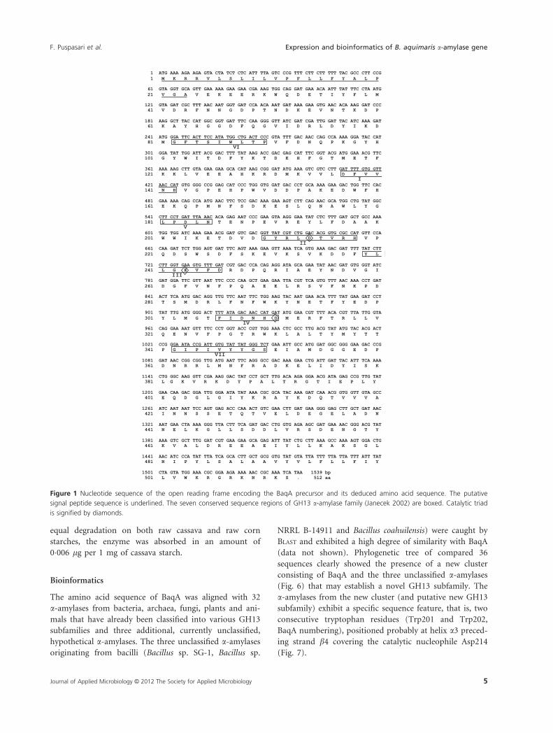
deduced was 512 amino acid residues in length, including
the first 23 residues that formed a putative signal peptide
(Fig. 1). Seven CSRs of the GH13 a-amylase family
(Janecek 2002) and the catalytic triad (MacGregor et al.
2001) were identified in BaqA. The catalytic triad consists
of Asp214 (catalytic nucleophile at b4), Glu243 (proton
donor at b5) and Asp311 (transition-state stabilizer at b7).To characterize the protein encoded by baqA, the baqA
fragment encoding for 27–512 amino acid residues was
PCR amplified, cloned into pET30 vector and then
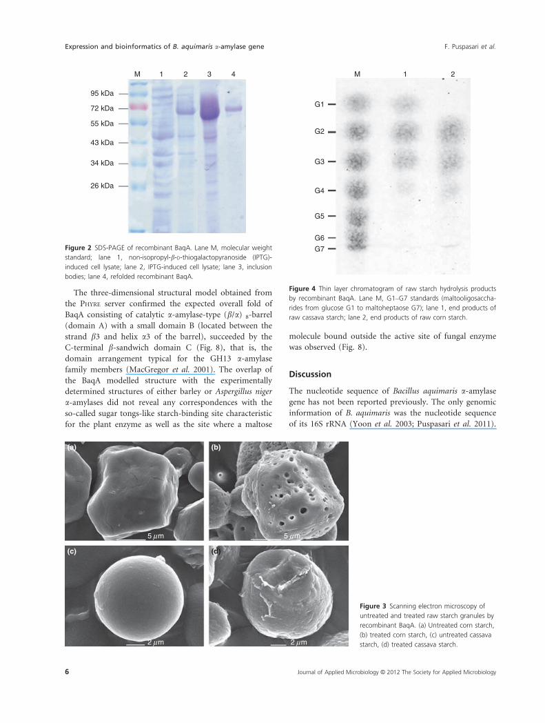
expressed in Escherichia coli BL21 (DE3). Intracellular pro-
duction of BaqA in E. coli resulted in the formation of
inclusion bodies (Fig. 2, lane 3) which required solubiliza-
tion and refolding to generate an active recombinant BaqA.
The baqA expression cassette encoded for 553 amino acid
residues consisting of BaqA without its signal sequences
and N and C terminal His-tag fusions. SDS-PAGE analysis
showed that the mobility of recombinant BaqA was
c. 70 kDa, which is somewhat higher than the expected
64�4 kDa. This appears to reflect an aberrant electropho-
retic mobility of recombinant BaqA on the SDS/PAGE gel.
Biochemical properties of recombinant BaqA
Recombinant BaqA could not degrade soluble starch. No
detectable degradation of soluble starch was measured in
all variation of enzyme amount (up to 150 lg of enzyme)
nor variation of time intervals at 37 and 50°C. The refoldedrecombinant BaqA can degrade raw cassava and corn
starch. At the experiment conditions, recombinant BaqA
degraded raw cassava starch and produced 8 mmol l�1
reducing-end sugars; the observation which was propor-
tional to 1�5% DH. Raw corn starch was degraded to pro-
duce 7�5 mmol l�1 reducing-end sugars (proportional to
1�4% DH).
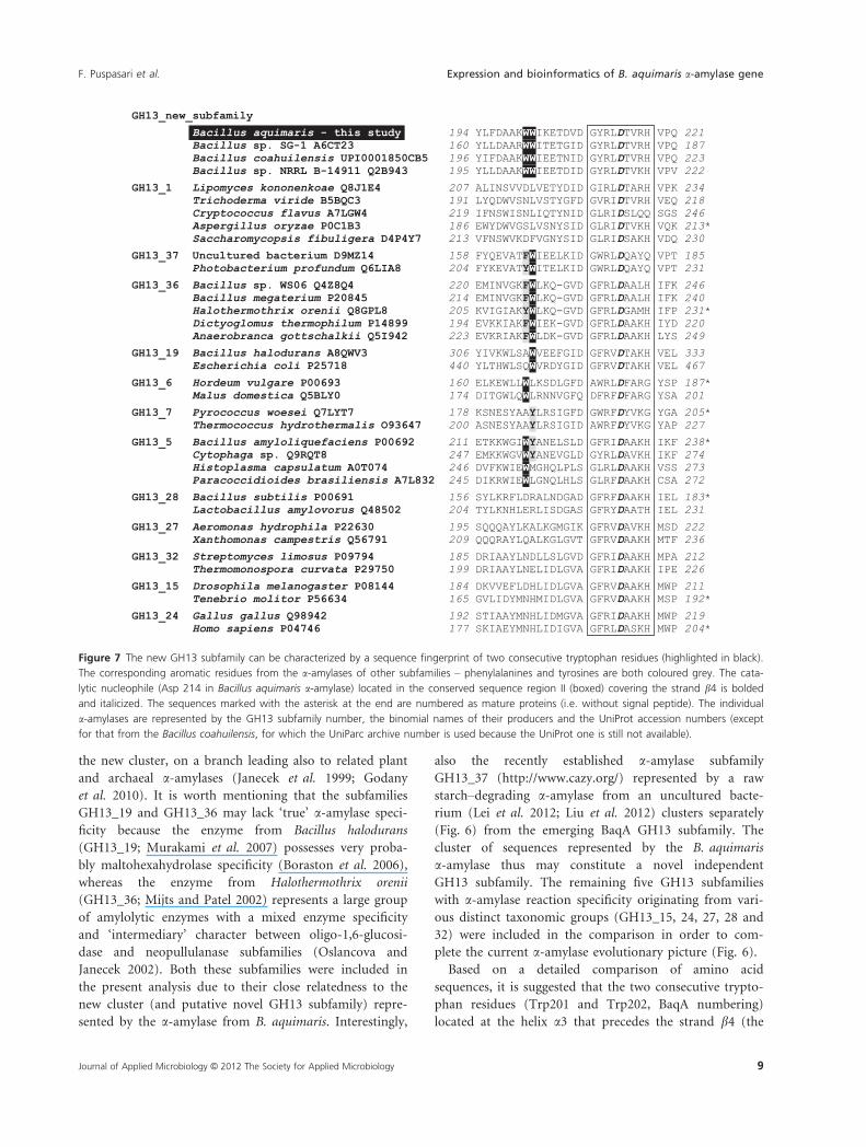
The ability of recombinant BaqA to hydrolyze raw
starch was confirmed by SEM. Recombinant BaqA
attacked raw corn starch with result of forming the holes
in starch granule surface, while it peeled away the surface
of raw cassava starch (Fig. 3).
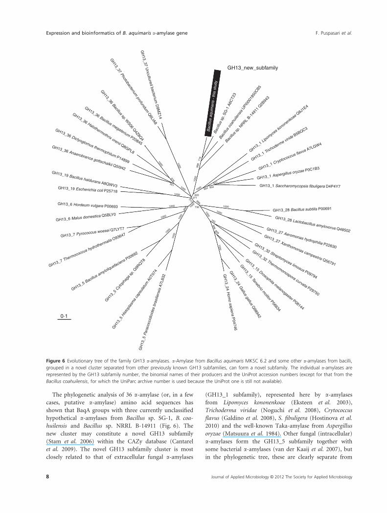
The end products of recombinant BaqA action on raw
corn starch were different from those on raw cassava
starch. Degradation of raw cassava starch by recombinant
BaqA released glucose, maltose, maltotriose and maltotet-
raose, while degrading raw corn starch, only maltose,
maltotriose and maltotetraose were released (Fig. 4).
Adsorption of recombinant BaqA towards raw corn
and cassava starch was assayed at various amount of
starch. The slope of the curve on bound protein (lg) vs.
raw starch (mg) graphic indicated the amount of recom-
binant BaqA bound per mg raw starch. The curve would
reach a plateau when all protein was bound to raw starch
(100% bound protein). Figure 5 shows that 0�06 lg BaqA
was absorbed to 1 mg raw corn starch. Despite their
4 Journal of Applied Microbiology © 2012 The Society for Applied Microbiology
Expression and bioinformatics of B. aquimaris a-amylase gene F. Puspasari et al.
equal degradation on both raw cassava and raw corn
starches, the enzyme was absorbed in an amount of
0�006 lg per 1 mg of cassava starch.
Bioinformatics
The amino acid sequence of BaqA was aligned with 32
a-amylases from bacteria, archaea, fungi, plants and ani-
mals that have already been classified into various GH13
subfamilies and three additional, currently unclassified,
hypothetical a-amylases. The three unclassified a-amylases
originating from bacilli (Bacillus sp. SG-1, Bacillus sp.
NRRL B-14911 and Bacillus coahuilensis) were caught by
BLAST and exhibited a high degree of similarity with BaqA
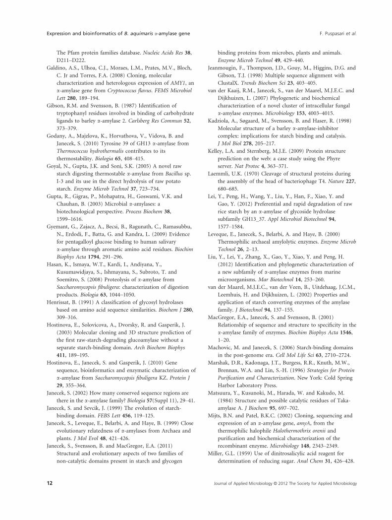
(data not shown). Phylogenetic tree of compared 36
sequences clearly showed the presence of a new cluster
consisting of BaqA and the three unclassified a-amylases
(Fig. 6) that may establish a novel GH13 subfamily. The
a-amylases from the new cluster (and putative new GH13
subfamily) exhibit a specific sequence feature, that is, two
consecutive tryptophan residues (Trp201 and Trp202,
BaqA numbering), positioned probably at helix a3 preced-
ing strand b4 covering the catalytic nucleophile Asp214
(Fig. 7).
Figure 1 Nucleotide sequence of the open reading frame encoding the BaqA precursor and its deduced amino acid sequence. The putative
signal peptide sequence is underlined. The seven conserved sequence regions of GH13 a-amylase family (Janecek 2002) are boxed. Catalytic triad
is signified by diamonds.
Journal of Applied Microbiology © 2012 The Society for Applied Microbiology 5
F. Puspasari et al. Expression and bioinformatics of B. aquimaris a-amylase gene
The three-dimensional structural model obtained from
the PHYRE server confirmed the expected overall fold of
BaqA consisting of catalytic a-amylase-type (b/a) 8-barrel
(domain A) with a small domain B (located between the
strand b3 and helix a3 of the barrel), succeeded by the
C-terminal b-sandwich domain C (Fig. 8), that is, the
domain arrangement typical for the GH13 a-amylase
family members (MacGregor et al. 2001). The overlap of
the BaqA modelled structure with the experimentally
determined structures of either barley or Aspergillus niger
a-amylases did not reveal any correspondences with the
so-called sugar tongs-like starch-binding site characteristic
for the plant enzyme as well as the site where a maltose
molecule bound outside the active site of fungal enzyme
was observed (Fig. 8).
Discussion
The nucleotide sequence of Bacillus aquimaris a-amylase
gene has not been reported previously. The only genomic
information of B. aquimaris was the nucleotide sequence
of its 16S rRNA (Yoon et al. 2003; Puspasari et al. 2011).
(a) (b)
(c) (d)
5 µm 5 µm
2 µm 2 µm
Figure 3 Scanning electron microscopy of
untreated and treated raw starch granules by
recombinant BaqA. (a) Untreated corn starch,
(b) treated corn starch, (c) untreated cassava
starch, (d) treated cassava starch.
M 1 2 3 4
95 kDa
72 kDa
55 kDa
43 kDa
34 kDa
26 kDa
Figure 2 SDS-PAGE of recombinant BaqA. Lane M, molecular weight
standard; lane 1, non-isopropyl-b-D-thiogalactopyranoside (IPTG)-
induced cell lysate; lane 2, IPTG-induced cell lysate; lane 3, inclusion
bodies; lane 4, refolded recombinant BaqA.
M 1 2
G3
G4
G5
G6
G7
G1
G2
Figure 4 Thin layer chromatogram of raw starch hydrolysis products
by recombinant BaqA. Lane M, G1–G7 standards (maltooligosaccha-
rides from glucose G1 to maltoheptaose G7); lane 1, end products of
raw cassava starch; lane 2, end products of raw corn starch.
6 Journal of Applied Microbiology © 2012 The Society for Applied Microbiology
Expression and bioinformatics of B. aquimaris a-amylase gene F. Puspasari et al.

A first attempt to clone the a-amylase gene of B. aquim-
aris MKSC 6.2 employedits partial genomic library; how-
ever, the gene could not be pulled out (F. Puspasari and
D. Natalia, unpublished data). Nevertheless, BLASTX analy-
sis of the nucleotide sequences obtained from several
clones of the partial genomic library revealed that some
proteins of Bacaq exhibited the highest degree of sequence
similarity (data not shown) with their counterparts from
Bacillus sp. SG-1, B. coahuilensis and Bacillus weihenste-
phanensis. The degenerate primers for degPCR were then
designed based on the alignment of putative a-amylase
genes of the three above-mentioned bacilli. The primer
degFA covered the CSR I, while primers degFB, degR2
and degR4 covered consecutively on CSRs II, IV and VII;
conserved regions as proposed by Janecek (2002).
Further nucleotide sequence analysis upstream of the
baqA ORF showed that the a-amylase gene was part of
an operon together with at least one other gene encoding
maltosaccharide ABC transporter, permease gene
upstream with a 37-nucleotide long gap (data not
shown). The polypeptide chain of BaqA exhibited the
highest identity (66%) with putative a-amylases from
Bacillus sp. NRRL B-14911 (UniProt ID: A6CT23; with-
out gaps) and Bacillus sp. SG-1 (UniProt ID: Q2B943;
with 7% of gaps). No distinct SBD sequence was found
within BaqA (Fig. 1).
The recombinant BaqA protein expressed in Escherichia
coli BL21 (DE3) was produced in inclusion bodies, which
upon refolding showed ability to degrade raw corn and
cassava starch; however, it was inactive toward soluble
starch. Most raw starch–degrading enzymes degrade solu-
ble starch as well, however one of three amylases of Pop-
lar (Populus canadensis) wood lacked activity with soluble
starch, instead it attacked starch granules with product
characteristics of endoamylase (Witt and Sauter 1995).
With our current knowledge, it is difficult to explain this
property of BaqA because the amino acid sequence and
structure of this Poplar wood amylase have not been
reported yet. Speculatively, the presence of N and C ter-
minal His tags may negatively affect accessibility of solu-
ble starch into the BaqA active site and thus its activity.
In future work, it will be of interest to generate a recom-
binant BaqA without His tags, and/or to express it as an
extracellular protein in another expression system, such
as Bacillus megaterium. Other approaches, for example,
coexpression of BaqA with a chaperone to improve its
solubility, could also be considered. Nevertheless, our
results clearly show that the refolding strategy employed
resulted in soluble recombinant BaqA competent to
interact with and then to digest the raw starch.
Scanning electron microscope analysis showed that the
recombinant BaqA action on raw corn starch introduced
holes in the granule surface, while it caused surface peel-
ing of raw cassava starch (Fig. 3). The same pattern of
hydrolysis toward raw corn and cassava starch was also
observed on partially purified nonrecombinant Bacaq
a-amylase (Puspasari et al. 2011) and some other a-amy-
lases from marine Bacillus (Vidilaseris et al. 2009;
Nurachman et al. 2010). Recently, Sarian et al. (2012)
reported characteristics of a Microbacterium aurum strain
B8.A a-amylase attacking a range of starch granules from
diverse plant sources by initially introducing holes, fol-
lowed by complete degradation. Furthermore, a new sub-
family GH13_37 a-amylase from a marine metagenomic
library showed a preferential raw rice starch degradation
forming deep holes in the granule surface (Lei et al.
2012). These various enzyme mechanisms for granule
degradation maybe correlated with differences in crystal-
linity and morphology of the various plant granules used.
Raw cassava starch hydrolyzed by recombinant BaqA
yielded glucose, maltose, maltotriose and maltotetraose,
while hydrolysis of raw corn starch yielded the same oli-
gosaccharides but without glucose (Fig. 4). These results
indicated that recombinant BaqA is a saccharifying or liq-
uefying a-amylase depending on the type of raw starch
used as substrate.
The adsorbability of recombinant BaqA (Fig. 5) on raw
starch was found to be relatively low, for example, com-
pared with that of raw starch–degrading a-amylase of Lac-
tobacillus amylovorus (an a-amylase having a SBD), in
which 30-lg a-amylase was absorbed per 1-mg raw corn
starch (Rodriguez-Sanoja et al. 2000). On the other hand,
a-amylase from yeast Saccharomycopsis fibuligera showed
raw starch–degrading activity but no enzyme adsorption
onto raw starch (Hasan et al. 2008; Hostinova et al. 2010);
it is important to note that the a-amylase of S. fibuligera
does not contain any SBD. It, thus, appears that the pres-
ence of SBD is not essential for degradation of raw starch
by an amylase, but it obviously results in better enzyme
adsorption onto raw starch (Christiansen et al. 2009).
20
15
10
5
00 100 200 300 400
0
20
40
60
80
y = 0·0057×
y = 0·0549×
% B
ound
pro
tein
Bou
nd p
rote
in (
µ g)
Raw starch (mg)
Figure 5 Adsorption of recombinant BaqA on raw corn (♦) and
cassava (□) starch. The slope indicates the amount of recombinant
BaqA bound per mg raw starch.
Journal of Applied Microbiology © 2012 The Society for Applied Microbiology 7
F. Puspasari et al. Expression and bioinformatics of B. aquimaris a-amylase gene
The phylogenetic analysis of 36 a-amylase (or, in a few
cases, putative a-amylase) amino acid sequences has
shown that BaqA groups with three currently unclassified
hypothetical a-amylases from Bacillus sp. SG-1, B. coa-
huilensis and Bacillus sp. NRRL B-14911 (Fig. 6). The
new cluster may constitute a novel GH13 subfamily
(Stam et al. 2006) within the CAZy database (Cantarel
et al. 2009). The novel GH13 subfamily cluster is most
closely related to that of extracellular fungal a-amylases
(GH13_1 subfamily), represented here by a-amylases
from Lipomyces kononenkoae (Eksteen et al. 2003),
Trichoderma viridae (Noguchi et al. 2008), Crytococcus
flavus (Galdino et al. 2008), S. fibuligera (Hostinova et al.
2010) and the well-known Taka-amylase from Aspergillus
oryzae (Matsuura et al. 1984). Other fungal (intracellular)
a-amylases form the GH13_5 subfamily together with
some bacterial a-amylases (van der Kaaij et al. 2007), but
in the phylogenetic tree, these are clearly separate from
GH13_6 Malus domestica Q5BLY0
GH13_7 Pyrococcus woesei Q7LYT7
GH13_7 Thermococcus hydrothermalis O93647
GH13_5 Bacillus amyloliquefaciens P00692
GH13_5
Cyto
phag
a sp
. Q9R
QT8
GH
13_5
His
topl
asm
a ca
psul
atum
A0T
074
GH
13_5
Par
acoc
cidi
oide
s br
asili
ensi
s A
7L83
2
GH13_6 Hordeum vulgare P00693
GH13_19 Escherichia coli P25718
GH13_19 Bacillus haldurans A8QWV3
GH13_36 Anaerobranca gottschalkii Q5I942
GH13_36 Dictyoglomus thermophilum P14899
GH13_36 Halothermothrix orenii Q8GPL8
GH13_36 Bacillus megaterium P20845
GH13_36 Bacillus sp. W
S06 Q4Z8Q
4
GH
13_37 Photobacterium profundum
Q6LIA8
GH
13_37 Uncultured bacterium
D9M
Z14
Bac
illus
aqu
imar
is -
this
stu
dyB
acill
us s
p. S
G-1
A6C
T23
Bacil
lus
coah
uile
nsis
UPI00
0185
0CB5
Bacillu
s sp.
NRRL B-1
4911
Q2B
943
GH13_1 Lipomyces k
ononenkoae Q
8J1E4
GH13_1 Trichoderme virid
e B5BQC3
GH13_1 Cryptococcus flavus A7LGW4
GH13_1 Aspergillus oryzae P0C1B3
GH13_1 Saccharomycopsis fibuligera D4P4Y7
GH13_28 Bacillus subtilis P00691
GH13_28 Lactobacillus amylovorus Q48502GH13_27 Aeromonas hydrophilia P22630
GH13_27 Xanthomonas campestris Q56791
GH13_32 Streptomyces limosus P09794
GH13_32 Thermomonospora curvata P29750
GH13_15 Drosophila melanogaster P08144
GH13_15 Tenebrio m
olitor P56634
GH
13_24 Gallus gallus Q
98942
GH
13_24 Hom
o sapiens P04746
0·1
GH13_new_subfamily
1000
1000
1000
1000
1000
1000
10001000
1000
1000
1000
1000
1000
1000
1000
1000
958
551
976
998
719
312271
600
999
730
954 954611
763
999
809
Figure 6 Evolutionary tree of the family GH13 a-amylases. a-Amylase from Bacillus aquimaris MKSC 6.2 and some other a-amylases from bacilli,
grouped in a novel cluster separated from other previously known GH13 subfamilies, can form a novel subfamily. The individual a-amylases are
represented by the GH13 subfamily number, the binomial names of their producers and the UniProt accession numbers (except for that from the
Bacillus coahuilensis, for which the UniParc archive number is used because the UniProt one is still not available).
8 Journal of Applied Microbiology © 2012 The Society for Applied Microbiology
Expression and bioinformatics of B. aquimaris a-amylase gene F. Puspasari et al.
the new cluster, on a branch leading also to related plant
and archaeal a-amylases (Janecek et al. 1999; Godany
et al. 2010). It is worth mentioning that the subfamilies
GH13_19 and GH13_36 may lack ‘true’ a-amylase speci-
ficity because the enzyme from Bacillus halodurans
(GH13_19; Murakami et al. 2007) possesses very proba-
bly maltohexahydrolase specificity (Boraston et al. 2006),
whereas the enzyme from Halothermothrix orenii
(GH13_36; Mijts and Patel 2002) represents a large group
of amylolytic enzymes with a mixed enzyme specificity
and ‘intermediary’ character between oligo-1,6-glucosi-
dase and neopullulanase subfamilies (Oslancova and
Janecek 2002). Both these subfamilies were included in
the present analysis due to their close relatedness to the
new cluster (and putative novel GH13 subfamily) repre-
sented by the a-amylase from B. aquimaris. Interestingly,
also the recently established a-amylase subfamily
GH13_37 (http://www.cazy.org/) represented by a raw
starch–degrading a-amylase from an uncultured bacte-
rium (Lei et al. 2012; Liu et al. 2012) clusters separately
(Fig. 6) from the emerging BaqA GH13 subfamily. The
cluster of sequences represented by the B. aquimaris
a-amylase thus may constitute a novel independent
GH13 subfamily. The remaining five GH13 subfamilies
with a-amylase reaction specificity originating from vari-
ous distinct taxonomic groups (GH13_15, 24, 27, 28 and
32) were included in the comparison in order to com-
plete the current a-amylase evolutionary picture (Fig. 6).
Based on a detailed comparison of amino acid
sequences, it is suggested that the two consecutive trypto-
phan residues (Trp201 and Trp202, BaqA numbering)
located at the helix a3 that precedes the strand b4 (the
Figure 7 The new GH13 subfamily can be characterized by a sequence fingerprint of two consecutive tryptophan residues (highlighted in black).
The corresponding aromatic residues from the a-amylases of other subfamilies – phenylalanines and tyrosines are both coloured grey. The cata-
lytic nucleophile (Asp 214 in Bacillus aquimaris a-amylase) located in the conserved sequence region II (boxed) covering the strand b4 is bolded
and italicized. The sequences marked with the asterisk at the end are numbered as mature proteins (i.e. without signal peptide). The individual
a-amylases are represented by the GH13 subfamily number, the binomial names of their producers and the UniProt accession numbers (except
for that from the Bacillus coahuilensis, for which the UniParc archive number is used because the UniProt one is still not available).
Journal of Applied Microbiology © 2012 The Society for Applied Microbiology 9
F. Puspasari et al. Expression and bioinformatics of B. aquimaris a-amylase gene

CSR II) of the catalytic (b/a)8-barrel domain (Fig. 7)
represent a sequence fingerprint of this new GH13
a-amylase subfamily. It is important to note that such
two consecutive tryptophans implicated in sugar recogni-
tion and eventually also binding are not unique for the
newly proposed GH13 subfamily. For example, in barley
a-amylases, two tryptophans (Trp276 and Trp277, isozyme
AMY2 numbering) were shown to form a well-defined sec-
ondary sugar-binding site (Gibson and Svensson 1987;
Søgaard et al. 1993), and in mammalian counterparts, also
two consecutive tryptophans (Trp58 and Trp59; human
salivary a-amylase numbering) are important for sugar
recognition (Ramasubbu et al. 1996; Gyemant et al. 2009).
Inspection of the superimposed tertiary structures of bar-
ley a-amylase isozyme AMY2 (Kadziola et al. 1998) and
human salivary a-amylase (Ramasubbu et al. 1996) with
the predicted model of BaqA with focus on the above-
mentioned tryptophan pairs confirmed that the two con-
secutive tryptophans are located in different parts of the
structure of each of the three a-amylases (not shown).
This observation supports the idea that Trp201 and
Trp202 of BaqA may be the unique sequence-structural
feature of the new GH13 a-amylase subfamily.
Mammalian a-amylases, moreover, together with some
bacterial homologues, for example, that from Pseudoaltero-
monas haloplanktis (Aghajari et al. 2002), are activated by (or
dependent on) a chloride ion (D’Amico et al. 2000). The
BaqA sequence does not contain an arginine or lysine residue
corresponding with the Arg300 of P. haloplanktis a-amylase
found to be crucial for interaction with the chloride anion
(Aghajari et al. 2002). In addition, the proposed GH13 sub-
family represented by BaqA appears to be most closely related
to the subfamily GH13_1 (fungal enzymes, e.g. Taka-amylase
A), which is not known to be activated by (or dependent on)
chloride ion (Matsuura et al. 1984; van der Kaaij et al. 2007).
On the other hand, the chloride-dependent a-amylases
(D’Amico et al. 2000; Cipolla et al. 2012) are members of
the subfamilies (cf. Fig. 6) GH13_24 (mammals; vertebrates),
GH13_15 (insects) and eventually GH13_32 (actinomycetes).
The best studied bacterial chloride-dependent a-amylase
from P. haloplanktis has not been classified into any GH13
subfamily as yet (http://www.cazy.org/), although it is evident
that it is related to the large cluster of the three GH13
subfamilies 24, 15 and 32 (Da Lage et al. 2004).
The best a-amylase template for modelling was the
Taka-amylase (Matsuura et al. 1984). No SBD, that is, a
separate carbohydrate-binding molecule is seen (Fig. 8A),
that usually is found in raw starch–degrading amylolytic
enzymes (Machovic and Janecek 2006; Janecek et al.
2011). a-Amylases able to degrade raw starch without a
separate SBD are rare; examples are barley isozyme
AMY1 from plants (Tibbot et al. 2002; Robert et al.
(a) (b)
(d)
(c)
Figure 8 (a) Overall fold of the three-dimensional structure model of Bacillus aquimaris a-amylase. The enzyme consists of three domains A (cata-
lytic TIM-barrel; blue), B (magenta) and C (cyan), characteristic of the a-amylase family GH13, without a distinct SBD. (b) The side chains of two
consecutive tryptophans (Trp201 and Trp202) positioned on the helix a3 of domain A are shown to be oriented outside the space occupied by
the catalytic triad. (c) Overlap of the BaqA model (blue) with the GH13_1 a-amylase from Aspergillus niger Protein Data bank (PDB: 2GVY; red)
and GH13_6 barley AMY1 (1P6W; green) supporting the closer relatedness of BaqA (novel GH13 subfamily) to GH13_1 (anticipated also from
the evolutionary tree). (d) A maltose molecule (on the left) bound by Tyr382 and Trp385 (red) outside the active site in the A. niger a-amylase
structure with corresponding residues in BaqA (blue) and barley AMY1 (green), as well as the 4I, 4II, 4III-trithiomaltotetraoside (on the right) with
Tyr380 (green) from the sugar tongs-like binding site of barley AMY1 without any correspondences in both BaqA and A. niger a-amylase.
10 Journal of Applied Microbiology © 2012 The Society for Applied Microbiology
Expression and bioinformatics of B. aquimaris a-amylase gene F. Puspasari et al.

2005) and the yeast a-amylase from S. fibuligera KZ
(Hostinova et al. 2010). The two above-mentioned try-
ptophans (Trp210 and Trp202) of BaqA may contribute
to its raw starch–degrading ability, that is, to act as bind-
ing residues enabling the stacking interactions with glu-
cose molecules because their side chains are positioned
outside the space occupied by the catalytic triad
(Fig. 8B). A higher homology, expected also from the
evolutionary tree (Fig. 6), is evident also from the struc-
tural comparison (Fig. 8C) of BaqA with GH13_1 Asper-
gillus niger a-amylase (root-mean-square deviation 1�32 A
for 420 corresponding Ca atoms) and GH13_6 barley
AMY1 (1�83 A for 271 Ca atoms). The A. niger a-amylase
was chosen because it is 100% identical in sequence with
TAKA-amylase and a maltose molecule bound outside
the active site was found in its structure (Vujicic-Zagar
and Dijkstra 2006). Based on the overlap, the two aro-
matic residues Tyr382 and Trp385 responsible for bind-
ing the maltose in the A. niger a-amylase structure have
no proper (i.e. aromatic) counterparts in the BaqA model
(Thr395 and Pro398, respectively) and the Tyr380 acting
in the barley AMY1 sugar tongs-like binding site has no
corresponding residue in BaqA at all (Fig. 8D). Concern-
ing the starch granule binding surface site on catalytic
domain A of the barley AMY1 (Trp278 and Trp279),
which is also present in the isozyme AMY2 (Kadziola
et al. 1998), there are no counterpart residues again in
the corresponding region of the modelled BaqA structure
(not shown) and the two BaqA signature trytophanes
(Trp201 and Trp202) are located elsewhere in domain A.
The results suggest that the amino acid residues responsi-
ble for raw starch binding in BaqA are probably unique
for the new GH13 subfamily proposed in this study.
In conclusion, this is the first report of the amino acid
sequence of a raw starch–degrading a-amylase from a soft
coral associated B. aquimaris MKSC 6.2. The BaqA may
form a new cluster of glycoside hydrolase subfamily
GH13 together with several putative Bacillus a-amylases.
Acknowledgements
This work was funded to D.N. by the International
Research Grant ITB 2007. S.J. thanks the Slovak Grant
agency VEGA for the grant no. 2/0148/11. L.D. acknowl-
edges financial support from the Carbohydrate Compe-
tence Center CCC.
References
Aghajari, N., Feller, G., Gerday, C. and Haser, R. (2002)
Structural basis of a-amylase activation by chloride.
Protein Sci 11, 1435–1441.
Altschul, S.F., Madden, T.L., Schaffer, A.A., Zhang, J., Zhang,
Z., Miller, W. and Lipman, D.J. (1997) Gapped BLAST
and PSI-BLAST: a new generation of protein database
search programs. Nucleic Acids Res 25, 3389–3402.
Apweiler, R., Martin, M.J., O’Donovan, C., Magrane, M.,
Alam-Faruque, Y., Antunes, R., Barrell, D., Bely, B. et al.
(2011) Ongoing and future developments at the Universal
Protein Resource. Nucleic Acids Res 39, D214–D219.
Bendtsen, J.D., Nielsen, H., von Heijne, G. and Brunak, S.
(2004) Improved prediction of signal peptides-SignalP 3.0.
J Mol Biol 340, 783–795.
Benson, D.A., Karsch-Mizrachi, I., Lipman, D.J., Ostell, J. and
Sayers, E.W. (2011) GenBank. Nucleic Acids Res 39,
D32–D37.
Boraston, A.B., Healey, M., Klassen, J., Ficko-Blean, E.,
Lammerts van Bueren, A. and Law, V. (2006) A structural
and functional analysis of a-glucan recognition by family
25 and 26 carbohydrate-binding modules reveals a
conserved mode of starch recognition. J Biol Chem 281,
587–598.
Bradford, M.M. (1976) A rapid and sensitive method for the
quantitation of microgram quantities of protein utilizing
the principle of protein-dye-binding. Anal Biochem 72,
248–254.
Cantarel, B.L., Coutinho, P.M., Rancurel, C., Bernard, T.,
Lombard, V. and Henrissat, B. (2009) The carbohydrate-
active enzymes database (CAZy): an expert resource for
glycogenomics. Nucleic Acids Res 37, D233–D238.
Christiansen, C., Abou Hachem, M., Janecek, S., Viksø-
Nielsen, A., Blennow, A. and Svensson, B. (2009) The
carbohydrate-binding module family 20–diversity,
structure, and function. FEBS J 276, 5006–5029.
Cipolla, A., Delbrassine, F., Da Lage, J.L. and Feller, G. (2012)
Temperature adaptations in psychrophilic, mesophilic and
thermophilic chloride-dependent a-amylases. Biochimie 94,
1943–1950.
Da Lage, J.L., Feller, G. and Janecek, S. (2004) Horizontal gene
transfer from Euckarya to Bacteria and domain shuffling:
a-amylase model. Cell Mol Life Sci 61, 97–109.
D’Amico, S., Gerday, C. and Feller, G. (2000) Structural
similarities and evolutionary relationships in chloride-
dependent a-amylases. Gene 253, 95–105.
Demirkan, E.S., Mikami, B., Adachi, M., Higasa, T. and
Utsami, S. (2005) a-Amylase from B. amyloliquefaciens:
purification, characterization, raw starch degradation and
expression in E. coli. Process Biochem 40, 2629–2636.
Eksteen, J.M., Steyn, A.J., van Rensburg, P., Cordero Otero, R.
R. and Pretorius, I.S. (2003) Cloning and characterization
of a second a-amylase gene (LKA2) from Lipomyces
kononenkoae IGC4052B and its expression in
Saccharomyces cerevisiae. Yeast 20, 69–78.
Felsenstein, J. (1985) Confidence limits on phylogenies: an
approach using the bootstrap. Evolution 39, 783–791.
Finn, R.D., Mistry, J., Tate, J., Coggill, P., Heger, A.,
Pollington, J.E., Gavin, O.L., Gunasekaran, P. et al. (2010)
Journal of Applied Microbiology © 2012 The Society for Applied Microbiology 11
F. Puspasari et al. Expression and bioinformatics of B. aquimaris a-amylase gene
The Pfam protein families database. Nucleic Acids Res 38,
D211–D222.
Galdino, A.S., Ulhoa, C.J., Moraes, L.M., Prates, M.V., Bloch,
C. Jr and Torres, F.A. (2008) Cloning, molecular
characterization and heterologous expression of AMY1, an
a-amylase gene from Cryptococcus flavus. FEMS Microbiol
Lett 280, 189–194.
Gibson, R.M. and Svensson, B. (1987) Identification of
tryptophanyl residues involved in binding of carbohydrate
ligands to barley a-amylase 2. Carlsberg Res Commun 52,
373–379.
Godany, A., Majzlova, K., Horvathova, V., Vidova, B. and
Janecek, S. (2010) Tyrosine 39 of GH13 a-amylase from
Thermococcus hydrothermalis contributes to its
thermostability. Biologia 65, 408–415.
Goyal, N., Gupta, J.K. and Soni, S.K. (2005) A novel raw
starch digesting thermostable a-amylase from Bacillus sp.
I-3 and its use in the direct hydrolysis of raw potato
starch. Enzyme Microb Technol 37, 723–734.
Gupta, R., Gigras, P., Mohapatra, H., Goswami, V.K. and
Chauhan, B. (2003) Microbial a-amylases: a
biotechnological perspective. Process Biochem 38,
1599–1616.
Gyemant, G., Zajacz, A., Becsi, B., Ragunath, C., Ramasubbu,
N., Erdodi, F., Batta, G. and Kandra, L. (2009) Evidence
for pentagalloyl glucose binding to human salivary
a-amylase through aromatic amino acid residues. Biochim
Biophys Acta 1794, 291–296.
Hasan, K., Ismaya, W.T., Kardi, I., Andiyana, Y.,
Kusumawidjaya, S., Ishmayana, S., Subroto, T. and
Soemitro, S. (2008) Proteolysis of a-amylase from
Saccharomycopsis fibuligera: characterization of digestion
products. Biologia 63, 1044–1050.
Henrissat, B. (1991) A classification of glycosyl hydrolases
based on amino acid sequence similarities. Biochem J 280,
309–316.
Hostinova, E., Solovicova, A., Dvorsky, R. and Gasperik, J.
(2003) Molecular cloning and 3D structure prediction of
the first raw-starch-degrading glucoamylase without a
separate starch-binding domain. Arch Biochem Biophys
411, 189–195.
Hostinova, E., Janecek, S. and Gasperik, J. (2010) Gene
sequence, bioinformatics and enzymatic characterization of
a-amylase from Saccharomycopsis fibuligera KZ. Protein J
29, 355–364.
Janecek, S. (2002) How many conserved sequence regions are
there in the a-amylase family? Biologia 57(Suppl 11), 29–41.
Janecek, S. and Sevcik, J. (1999) The evolution of starch-
binding domain. FEBS Lett 456, 119–125.
Janecek, S., Leveque, E., Belarbi, A. and Haye, B. (1999) Close
evolutionary relatedness of a-amylases from Archaea and
plants. J Mol Evol 48, 421–426.
Janecek, S., Svensson, B. and MacGregor, E.A. (2011)
Structural and evolutionary aspects of two families of
non-catalytic domains present in starch and glycogen
binding proteins from microbes, plants and animals.
Enzyme Microb Technol 49, 429–440.
Jeanmougin, F., Thompson, J.D., Gouy, M., Higgins, D.G. and
Gibson, T.J. (1998) Multiple sequence alignment with
ClustalX. Trends Biochem Sci 23, 403–405.
van der Kaaij, R.M., Janecek, S., van der Maarel, M.J.E.C. and
Dijkhuizen, L. (2007) Phylogenetic and biochemical
characterization of a novel cluster of intracellular fungal
a-amylase enzymes. Microbiology 153, 4003–4015.
Kadziola, A., Søgaard, M., Svensson, B. and Haser, R. (1998)
Molecular structure of a barley a-amylase-inhibitor
complex: implications for starch binding and catalysis.
J Mol Biol 278, 205–217.
Kelley, L.A. and Sternberg, M.J.E. (2009) Protein structure
prediction on the web: a case study using the Phyre
server. Nat Protoc 4, 363–371.
Laemmli, U.K. (1970) Cleavage of structural proteins during
the assembly of the head of bacteriophage T4. Nature 227,
680–685.
Lei, Y., Peng, H., Wang, Y., Liu, Y., Han, F., Xiao, Y. and
Gao, Y. (2012) Preferential and rapid degradation of raw
rice starch by an a-amylase of glycoside hydrolase
subfamily GH13_37. Appl Microbiol Biotechnol 94,
1577–1584.
Leveque, E., Janecek, S., Belarbi, A. and Haye, B. (2000)
Thermophilic archaeal amylolytic enzymes. Enzyme Microb
Technol 26, 2–13.
Liu, Y., Lei, Y., Zhang, X., Gao, Y., Xiao, Y. and Peng, H.
(2012) Identification and phylogenetic characterization of
a new subfamily of a-amylase enzymes from marine
microorganisms. Mar Biotechnol 14, 253–260.
van der Maarel, M.J.E.C., van der Veen, B., Uitdehaag, J.C.M.,
Leemhuis, H. and Dijkhuizen, L. (2002) Properties and
application of starch converting enzymes of the amylase
family. J Biotechnol 94, 137–155.
MacGregor, E.A., Janecek, S. and Svensson, B. (2001)
Relationship of sequence and structure to specificity in the
a-amylase family of enzymes. Biochim Biophys Acta 1546,
1–20.
Machovic, M. and Janecek, S. (2006) Starch-binding domains
in the post-genome era. Cell Mol Life Sci 63, 2710–2724.
Marshak, D.R., Kadonaga, J.T., Burgess, R.R., Knuth, M.W.,
Brennan, W.A. and Lin, S.-H. (1996) Strategies for Protein
Purification and Characterization. New York: Cold Spring
Harbor Laboratory Press.
Matsuura, Y., Kusunoki, M., Harada, W. and Kakudo, M.
(1984) Structure and possible catalytic residues of Taka-
amylase A. J Biochem 95, 697–702.
Mijts, B.N. and Patel, B.K.C. (2002) Cloning, sequencing and
expression of an a-amylase gene, amyA, from the
thermophilic halophile Halothermothrix orenii and
purification and biochemical characterization of the
recombinant enzyme. Microbiology 148, 2343–2349.
Miller, G.L. (1959) Use of dinitrosalicylic acid reagent for
determination of reducing sugar. Anal Chem 31, 426–428.
12 Journal of Applied Microbiology © 2012 The Society for Applied Microbiology
Expression and bioinformatics of B. aquimaris a-amylase gene F. Puspasari et al.
Mitsuiki, S., Mukaea, K., Sakai, M. and Goto, M. (2005)
Comparative characterization of raw starch hydrolyzing
a-amylases from various Bacillus strains. Enzyme Microb
Technol 37, 410–416.
Murakami, S., Nishimoto, H., Toyama, Y., Shimamoto, E.,
Takenaka, S., Kaulpiboon, J., Prousoontorn, M.,
Limpaseni, T. et al. (2007) Purification and
characterization of two alkaline, thermotolerant
a-amylases from Bacillus halodurans 38C-2-1 and
expression of the cloned gene in Escherichia coli. Biosci
Biotechnol Biochem 71, 2393–2401.
Noguchi, A., Inohara-Ochiai, M., Ishibashi, N., Fukami, H.,
Nakayama, T. and Nakao, M. (2008) A novel glucosylation
enzyme: molecular cloning, expression, and
characterization of Trichoderma viride JCM22452 a-amylase
and enzymatic synthesis of some flavonoid monoglucosides
and oligoglucosides. J Agric Food Chem 56, 12016–12024.
Nurachman, Z., Kono, A., Radjasa, O.K. and Natalia, D.
(2010) Identification a novel raw-starch-degrading-
a-amylase from a tropical marine bacterium. Am J
Biochem Biotechnol 6, 300–306.
Oslancova, A. and Janecek, S. (2002) Oligo-1,6-glucosidase
and neopullulanase enzyme subfamilies from the
a-amylase family defined by the fifth conserved sequence
region. Cell Mol Life Sci 59, 1945–1959.
Page, R.D. (1996) TreeView: an application to display
phylogenetic trees on personal computer. Comput Appl
Biosci 12, 357–358.
Puspasari, F., Nurachman, Z., Noer, A.S., Radjasa, O.K., van
der Maarel, M.J.E.C. and Natalia, D. (2011) Characteristics
of raw starch degrading a-amylase from Bacillus aquimaris
MKSC 6.2 associated with soft coral Sinularia sp. Starch/
Staerke 63, 461–467.
Ramasubbu, N., Paloth, V., Luo, Y., Brayer, G.D. and Levine,
M.J. (1996) Structure of human salivary a-amylase at 1.6
A resolution: implications for its role in the oral cavity.
Acta Crystallogr D Biol Crystallogr 52, 435–446.
Robert, X., Haser, R., Gottschalk, T.E., Ratajczak, F., Driguez,
H., Svensson, B. and Aghajari, N. (2003) The structure of
barley a-amylase isozyme 1 reveals a novel role of domain
C in substrate recognition and binding: a pair of sugar
tongs. Structure 11, 973–984.
Robert, X., Haser, R., Mori, H., Svensson, B. and Aghajari, N.
(2005) Oligosaccharide binding to barley a-amylase 1.
J Biol Chem 280, 32968–32978.
Rodriguez-Sanoja, R., Morlon-Guyot, J., Jore, J., Pintado, J.,
Juge, N. and Guyot, P. (2000) Comparative
characterization of complete and truncated forms of
Lactobacillus amylovorus a-amylase and role of the
C-terminal direct repeats in raw-starch binding. Appl
Environ Microbiol 66, 3350–3356.
Rose, P.W., Beran, B., Bi, C., Bluhm, W.F., Dimitropoulos, D.,
Goodsell, D.S., Prlic, A., Quesada, M. et al. (2011) The
RCSB Protein Data Bank: redesigned web site and web
services. Nucleic Acids Res 39, D392–D401.
Saitou, N. and Nei, M. (1987) The neighbor-joining method: a
new method for reconstructing phylogenetic trees. Mol
Biol Evol 4, 406–425.
Sarian, F.D., van der Kaaij, R.M., Kralj, S., Wijbenga, D.J.,
Binnema, D., van der Maarel, M.J.E.C. and Dijkhuizen, L.
(2012) Enzymatic degradation of granular potato starch by
Microbacterium aurum strain B8.A. Appl Microbiol
Biotechnol 93, 645–654.
Sevcik, J., Hostinova, E., Solovicova, A., Gasperik, J., Dauter,
Z. and Wilson, K.S. (2006) Structure of the complex of a
yeast glucoamylase with acarbose reveals the presence of a
raw starch binding site on the catalytic domain. FEBS J
273, 2161–2171.
Shatsky, M., Nussinov, R. and Wolfson, H.J. (2004) A method
for simultaneous alignment of multiple protein structures.
Proteins 56, 143–156.
Søgaard, M., Kadziola, A., Haser, R. and Svensson, B. (1993)
Site-directed mutagenesis of histidine 93, aspartic acid
180, glutamic acid 205, histidine 290, and aspartic acid
291 at the active site and tryptophan 279 at the raw starch
binding site in barley a-amylase 1. J Biol Chem 268,
22480–22484.
Stam, M.R., Danchin, E.G.J., Rancurel, C., Coutinho, P.M. and
Henrissat, B. (2006) Dividing the large glycoside hydrolase
family 13 into subfamilies: towards improved functional
annotations of a-amylase-related proteins. Protein Eng Des
Sel 19, 555–562.
Tibbot, B.K., Wong, D.W.S. and Robertson, G.H. (2002)
Studies on the C-terminal region of barley a-amylase 1
with emphasis on raw starch-binding. Biologia 57(Suppl
11), 229–238.
Vidilaseris, K., Hidayat, K., Retnoningrum, D.S., Nurachman,
Z., Noer, A.S. and Natalia, D. (2009) Biochemical
characterization of a raw starch degrading a-amylase from
the Indonesian marine bacterium Bacillus sp. ALSHL3.
Biologia 64, 1047–1052.
Vujicic-Zagar, A. and Dijkstra, B.W. (2006) Monoclinic crystal
form of Aspergillus niger a-amylase in complex with
maltose at 1.8 A resolution. Acta Crystallogr Sect F Struct
Biol Cryst Commun 62, 716–721.
Wang, W.J., Powell, A.D. and Oates, C.G. (1995) Pattern of
enzymes hydrolysis in raw sago starch: effects of
processing history. Carbohydr Polym 26, 91–97.
Weber-Arden, J., Wilbert, O.M., Kabelitz, D. and Arden, B.
(1996) Inverse PCR amplification of low-abundancy
message of cd T cell receptor genes. J Immunol Methods
197, 187–192.
Witt, W. and Sauter, J.J. (1995) In vitro degradation of starch
grain by phosphorylases and amylases from Poplar wood.
J Plant Physiol 146, 35–40.
Yoon, J.H., Kim, I.G., Kang, K.H., Oh, T.K. and Park, Y.H.
(2003) Bacillus marisflavi sp. nov. and Bacillus aquimaris
sp. nov., isolated from sea water of a tidal flat of the
Yellow Sea in Korea. Int J Syst Evol Microbiol 53,
1297–1303.
Journal of Applied Microbiology © 2012 The Society for Applied Microbiology 13
F. Puspasari et al. Expression and bioinformatics of B. aquimaris a-amylase gene
Related Documents


