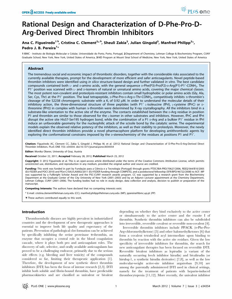
Rational Design and Characterization of D-Phe-Pro-D- Arg-Derived Direct Thrombin Inhibitors Ana C. Figueiredo 1. , Cristina C. Clement 2 * . , Sheuli Zakia 2 , Julian Gingold 3 , Manfred Philipp 2 *, Pedro J. B. Pereira 1 * 1 IBMC - Instituto de Biologia Molecular e Celular, Universidade do Porto, Porto, Portugal, 2 Department of Chemistry, Lehman College & Biochemistry Program, CUNY Graduate School, New York, New York, United States of America, 3 MD Program at Mount Sinai School of Medicine, New York, New York, United States of America Abstract The tremendous social and economic impact of thrombotic disorders, together with the considerable risks associated to the currently available therapies, prompt for the development of more efficient and safer anticoagulants. Novel peptide-based thrombin inhibitors were identified using in silico structure-based design and further validated in vitro. The best candidate compounds contained both L- and D-amino acids, with the general sequence D-Phe(P3)-Pro(P2)-D-Arg(P1)-P19-CONH 2 . The P19 position was scanned with L- and D-isomers of natural or unnatural amino acids, covering the major chemical classes. The most potent non-covalent and proteolysis-resistant inhibitors contain small hydrophobic or polar amino acids (Gly, Ala, Ser, Cys, Thr) at the P19 position. The lead tetrapeptide, D-Phe-Pro-D-Arg-D-Thr-CONH 2 , competitively inhibits a-thrombin’s cleavage of the S2238 chromogenic substrate with a K i of 0.92 mM. In order to understand the molecular details of their inhibitory action, the three-dimensional structure of three peptides (with P19 L-isoleucine (fPrI), L-cysteine (fPrC) or D- threonine (fPrt)) in complex with human a-thrombin were determined by X-ray crystallography. All the inhibitors bind in a substrate-like orientation to the active site of the enzyme. The contacts established between the D-Arg residue in position P1 and thrombin are similar to those observed for the L-isomer in other substrates and inhibitors. However, fPrC and fPrt disrupt the active site His57-Ser195 hydrogen bond, while the combination of a P1 D-Arg and a bulkier P19 residue in fPrI induce an unfavorable geometry for the nucleophilic attack of the scissile bond by the catalytic serine. The experimental models explain the observed relative potency of the inhibitors, as well as their stability to proteolysis. Moreover, the newly identified direct thrombin inhibitors provide a novel pharmacophore platform for developing antithrombotic agents by exploring the conformational constrains imposed by the D-stereochemistry of the residues at positions P1 and P19. Citation: Figueiredo AC, Clement CC, Zakia S, Gingold J, Philipp M, et al. (2012) Rational Design and Characterization of D-Phe-Pro-D-Arg-Derived Direct Thrombin Inhibitors. PLoS ONE 7(3): e34354. doi:10.1371/journal.pone.0034354 Editor: Monika Oberer, University of Graz, Austria Received October 22, 2011; Accepted February 28, 2012; Published March 23, 2012 Copyright: ß 2012 Figueiredo et al. This is an open-access article distributed under the terms of the Creative Commons Attribution License, which permits unrestricted use, distribution, and reproduction in any medium, provided the original author and source are credited. Funding: This work was funded in part by Fundac ¸a ˜o para a Cie ˆ ncia e a Tecnologia (Portugal) through grants PTDC/BIA-PRO/70627/2006, REEQ/564/B10/2005 (EU-FEDER and POCI 2010) and PEst-C/SAU/LA0002/2011 (EU-FEDER funding through COMPETE), and a postdoctoral fellowship (SFR/BPD/46722/2008) to ACF. MP was supported by a Fullbright Scholar Award and the PSC-CUNY research awards program. CC was supported by a research grant from the Biochemistry Department at the Graduate Center of the City University of New York (2001–2003) and by an Adjunct Lecturer appointment at the Chemistry Department, Lehman College, CUNY (2001–2006). The funding bodies had no role in study design, data collection and analysis, decision to publish or preparation of the manuscript. Competing Interests: The authors have declared that no competing interests exist. * E-mail: [email protected] (CC); [email protected] (MP); [email protected] (PP) . These authors contributed equally to this work. Introduction Thromboembolic diseases are highly prevalent in industrialized countries and the development of new therapeutic approaches is essential to improve both life quality and expectancy of the patients. Prevention of pathological clot formation can be achieved by specifically inhibiting the serine proteinase a-thrombin, an enzyme that occupies a central role in the blood coagulation cascade, where it plays both pro and anticoagulant roles. The discovery of safe, selective, and orally available anticoagulants has proved to be a challenging endeavor, primarily due to the serious side effects (e.g. bleeding and liver toxicity) of the compounds considered so far, limiting their therapeutic application [1]. Therefore, the development of new synthetic direct thrombin inhibitors (DTI) has been the focus of intense research [2,3]. DTI inhibit both soluble and fibrin-bound thrombin, have predictable pharmacokinetics and are classified as univalent or bivalent depending on whether they bind exclusively to the active center or simultaneously to the active center and the exosite I of thrombin. Synthetic thrombin inhibitors can also be subdivided into irreversible, reversible covalent or reversible non-covalent [4]. Irreversible thrombin inhibitors include PPACK (D-Phe-Pro- Arg-chloromethylketone) [5] and other halomethylketones [6] that form a covalent tetrahedral acyl intermediate upon binding to thrombin by reaction with the active site residues. Given the low specificity of irreversible inhibitors for thrombin, the search for new anticoagulant therapies has been focused on reversible DTI. Reversible bivalent inhibitors as lepirudin (a variant of the naturally occurring leech inhibitor hirudin) and bivalirudin (or hirulog-1, a synthetic hirudin derivative) [7,8], as well as the low molecular-weight active-site inhibitor argatroban [9,10] are among the parentally administered DTI used in clinical settings, namely for the treatment of patients with heparin-induced thrombocytopenia [11,12]. More recently, the univalent inhibitor PLoS ONE | www.plosone.org 1 March 2012 | Volume 7 | Issue 3 | e34354

Welcome message from author
This document is posted to help you gain knowledge. Please leave a comment to let me know what you think about it! Share it to your friends and learn new things together.
Transcript
Rational Design and Characterization of D-Phe-Pro-D-Arg-Derived Direct Thrombin InhibitorsAna C. Figueiredo1., Cristina C. Clement2*., Sheuli Zakia2, Julian Gingold3, Manfred Philipp2*,
Pedro J. B. Pereira1*
1 IBMC - Instituto de Biologia Molecular e Celular, Universidade do Porto, Porto, Portugal, 2 Department of Chemistry, Lehman College & Biochemistry Program, CUNY
Graduate School, New York, New York, United States of America, 3 MD Program at Mount Sinai School of Medicine, New York, New York, United States of America
Abstract
The tremendous social and economic impact of thrombotic disorders, together with the considerable risks associated to thecurrently available therapies, prompt for the development of more efficient and safer anticoagulants. Novel peptide-basedthrombin inhibitors were identified using in silico structure-based design and further validated in vitro. The best candidatecompounds contained both L- and D-amino acids, with the general sequence D-Phe(P3)-Pro(P2)-D-Arg(P1)-P19-CONH2. TheP19 position was scanned with L- and D-isomers of natural or unnatural amino acids, covering the major chemical classes.The most potent non-covalent and proteolysis-resistant inhibitors contain small hydrophobic or polar amino acids (Gly, Ala,Ser, Cys, Thr) at the P19 position. The lead tetrapeptide, D-Phe-Pro-D-Arg-D-Thr-CONH2, competitively inhibits a-thrombin’scleavage of the S2238 chromogenic substrate with a Ki of 0.92 mM. In order to understand the molecular details of theirinhibitory action, the three-dimensional structure of three peptides (with P19 L-isoleucine (fPrI), L-cysteine (fPrC) or D-threonine (fPrt)) in complex with human a-thrombin were determined by X-ray crystallography. All the inhibitors bind in asubstrate-like orientation to the active site of the enzyme. The contacts established between the D-Arg residue in positionP1 and thrombin are similar to those observed for the L-isomer in other substrates and inhibitors. However, fPrC and fPrtdisrupt the active site His57-Ser195 hydrogen bond, while the combination of a P1 D-Arg and a bulkier P19 residue in fPrIinduce an unfavorable geometry for the nucleophilic attack of the scissile bond by the catalytic serine. The experimentalmodels explain the observed relative potency of the inhibitors, as well as their stability to proteolysis. Moreover, the newlyidentified direct thrombin inhibitors provide a novel pharmacophore platform for developing antithrombotic agents byexploring the conformational constrains imposed by the D-stereochemistry of the residues at positions P1 and P19.
Citation: Figueiredo AC, Clement CC, Zakia S, Gingold J, Philipp M, et al. (2012) Rational Design and Characterization of D-Phe-Pro-D-Arg-Derived DirectThrombin Inhibitors. PLoS ONE 7(3): e34354. doi:10.1371/journal.pone.0034354
Editor: Monika Oberer, University of Graz, Austria
Received October 22, 2011; Accepted February 28, 2012; Published March 23, 2012
Copyright: � 2012 Figueiredo et al. This is an open-access article distributed under the terms of the Creative Commons Attribution License, which permitsunrestricted use, distribution, and reproduction in any medium, provided the original author and source are credited.
Funding: This work was funded in part by Fundacao para a Ciencia e a Tecnologia (Portugal) through grants PTDC/BIA-PRO/70627/2006, REEQ/564/B10/2005(EU-FEDER and POCI 2010) and PEst-C/SAU/LA0002/2011 (EU-FEDER funding through COMPETE), and a postdoctoral fellowship (SFR/BPD/46722/2008) to ACF. MPwas supported by a Fullbright Scholar Award and the PSC-CUNY research awards program. CC was supported by a research grant from the BiochemistryDepartment at the Graduate Center of the City University of New York (2001–2003) and by an Adjunct Lecturer appointment at the Chemistry Department,Lehman College, CUNY (2001–2006). The funding bodies had no role in study design, data collection and analysis, decision to publish or preparation of themanuscript.
Competing Interests: The authors have declared that no competing interests exist.
* E-mail: [email protected] (CC); [email protected] (MP); [email protected] (PP)
. These authors contributed equally to this work.
Introduction
Thromboembolic diseases are highly prevalent in industrialized
countries and the development of new therapeutic approaches is
essential to improve both life quality and expectancy of the
patients. Prevention of pathological clot formation can be achieved
by specifically inhibiting the serine proteinase a-thrombin, an
enzyme that occupies a central role in the blood coagulation
cascade, where it plays both pro and anticoagulant roles. The
discovery of safe, selective, and orally available anticoagulants has
proved to be a challenging endeavor, primarily due to the serious
side effects (e.g. bleeding and liver toxicity) of the compounds
considered so far, limiting their therapeutic application [1].
Therefore, the development of new synthetic direct thrombin
inhibitors (DTI) has been the focus of intense research [2,3]. DTI
inhibit both soluble and fibrin-bound thrombin, have predictable
pharmacokinetics and are classified as univalent or bivalent
depending on whether they bind exclusively to the active center
or simultaneously to the active center and the exosite I of
thrombin. Synthetic thrombin inhibitors can also be subdivided
into irreversible, reversible covalent or reversible non-covalent [4].
Irreversible thrombin inhibitors include PPACK (D-Phe-Pro-
Arg-chloromethylketone) [5] and other halomethylketones [6] that
form a covalent tetrahedral acyl intermediate upon binding to
thrombin by reaction with the active site residues. Given the low
specificity of irreversible inhibitors for thrombin, the search for
new anticoagulant therapies has been focused on reversible DTI.
Reversible bivalent inhibitors as lepirudin (a variant of the
naturally occurring leech inhibitor hirudin) and bivalirudin (or
hirulog-1, a synthetic hirudin derivative) [7,8], as well as the low
molecular-weight active-site inhibitor argatroban [9,10] are
among the parentally administered DTI used in clinical settings,
namely for the treatment of patients with heparin-induced
thrombocytopenia [11,12]. More recently, the univalent inhibitor
PLoS ONE | www.plosone.org 1 March 2012 | Volume 7 | Issue 3 | e34354
dabigatran etexilate, an oral anticoagulant that is rapidly absorbed
and converted to its active form dabigatran [13], revealed being a
promising alternative to the traditional use of warfarin in stroke
prevention in atrial fibrillation [14]. This drug was designed by a
structure-driven approach based on a peptidic DTI structure in
complex with bovine thrombin [15].
The interest on the development of small peptidomimetic
inhibitors (,500 Da), dating back to 1982 with the dipeptide-
derived D-Phe-Pro-Agamantin [16], was mostly driven by their
potential to traverse the gastrointestinal epithelium and enter the
blood circulation. More recent approaches include the study of
several synthetic analogs of the angiotensin converting enzyme
breakdown product of bradykinin (RPPGF) [17,18,19,20] as well
as 1,3,5-trisubstituted benzenes [21] as potent thrombin inhibitors.
Virtual screening by ligand docking is a common approach in
modern drug design [22]. In the case of anticoagulant compounds,
this is particularly appealing given the abundant experimental
structural information on thrombin-inhibitor complexes currently
available. This information, together with modern structure-based
drug design (SBDD) methods, can be used to shorten the discovery
and design phases of new drugs [23].
In this work a SBDD approach was used for designing novel
peptidic DTI derived from the D-Phe-Pro-D-Arg-P19-CONH2 (fPr-
P19) tetrapeptide scaffold. As peptides containing D amino acids
are less susceptible to proteolytic cleavage [24], the replacement of
L-Arg by D-Arg at position P1 was therefore hypothesized to confer
resistance to proteolysis, allowing these sequences to function as
thrombin inhibitors. The crystallographic structures of the a-
thrombin complexes of three lead compounds having L-isoleucine
(fPrI), L-cysteine (fPrC) and D-threonine (fPrt) at the P19 position
are reported. The experimental models revealed substrate-like
binding of the inhibitors to the active site of the enzyme and
provided a structural explanation for their resistance to proteolysis.
Furthermore, the extent of the observed interactions established by
the residue at position P19 in the three-dimensional structures of
the complexes, correlated well with the experimental binding
affinities for all three lead peptidic DTI. Thus, the newly identified
sequence D-Phe(P3)-Pro(P2)-D-Arg(P1)-P19-CONH2 emerges as a
potential antithrombotic scaffold which can be further optimized
at the P3, P2 and P19 positions to improve its potency, selectivity,
bioavailability, and anticoagulant properties.
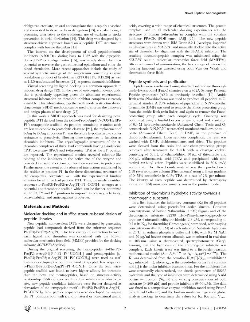
Materials and Methods
Molecular docking and in silico structure-based design ofpeptide libraries
New peptidic non-covalent DTIs were designed by generating
peptide lead compounds derived from the substrate sequence
Phe(P3)-Pro(P2)-Arg(P1). The free energy of interaction between
each ligand and thrombin was calculated with the built-in
molecular mechanics force field (MMFF) provided by the docking
software SCULPT (Accelrys).
During the original screening, the hexapeptides [D-Phe(P3)-
Pro(P2)-D-Arg(P1)-P19-P29-P39-CONH2] and pentapeptides [D-
Phe(P3)-Pro(P2)-D-Arg(P1)-P19-P29-CONH2] were used as scaf-
folds for developing the optimized final tetrapeptide lead sequence,
D-Phe(P3)-Pro(P2)-D-Arg(P1)-P19-CONH2. Once the lead tetra-
peptide scaffold was found to have higher affinity for thrombin
than the hexa and pentapeptides, based on structure-activity
relationship (SAR) studies on thrombin inhibition conducted in
vitro, new peptide candidate inhibitors were further designed as
derivatives of the tetrapeptide motif D-Phe(P3)-Pro(P2)-D-Arg(P1)-
P19-CONH2. New peptide sequences were developed by varying
the P19 positions both with L and D natural or non-natural amino
acids, covering a wide range of chemical structures. The protein
template used in all molecular docking experiments was the
structure of human a-thrombin in complex with the covalent
inhibitor PPACK (PDB entry 1ABJ [25]). The new peptide
structures were drawn with ISIS Draw 2.2.1 (Accelrys), imported
as 3D-structures in SCULPT, and manually docked into the active
site of thrombin by alignment with the PPACK inhibitor. The
resulting thrombin:peptide complex was minimized using the
SCULPT built-in molecular mechanics force field (MMFF94).
After each round of minimization, the free energy of interaction
(scoring function) was assessed using both Van der Waals and
electrostatic force fields.
Peptide synthesis and purificationPeptides were synthesized using standard solid-phase fluorenyl-
methyloxycarbonyl (Fmoc) chemistry on a 432A Synergy Personal
Peptide synthesizer (ABI) as previously described [19]. Amide
Rink resin (Novabiochem) was used to produce all peptides as C-
terminal amides. A 20% solution of piperidine in N,N9-dimethyl
formamide (DMF) was used to remove the Fmoc protecting group
from the amide Rink resin linker, and again to remove the Fmoc-
protecting group after each coupling cycle. Coupling was
performed using a fourfold excess of amino acid and a solution
of 0.4 M hydroxybenzotriazole (Advanced Chem Tech) and O-
benzotriazole-N,N,N9,N9-tetramethyl-uroniumhexafluoro-phos-
phate (Advanced Chem Tech) in DMF, in the presence of
diisopropylethylamine. Upon synthesis completion, the resin was
washed with DMF, dichloromethane, and dried. The peptides
were cleaved from the resin and side-chain-protecting groups
removed after treatment for 3–4 h with a cleavage cocktail
consisting of 50 mL of ethanedithiol, 50 mL of thioanisole and
900 mL trifluoroacetic acid (TFA) and precipitated with cold
methyl tert-butyl ether. Peptides were solubilized in 50% (v/v)
acetonitrile. The filtered crude material was then purified on a
C18 reversed-phase column (Phenomenex) using a linear gradient
of 0–75% acetonitrile in 0.1% TFA, at a rate of 2% per minute.
The identity of each peptide was determined by electrospray
ionization (ESI) mass spectrometry run in the positive mode.
Inhibition of thrombin’s hydrolytic activity towards achromogenic substrate
In a first instance, the inhibitory constants (Ki) for all peptides
were determined using pseudo-first order kinetics. Constant
concentrations of bovine a-thrombin (5 nM; Sigma) and of the
chromogenic substrate S2238 (H-D-Phenylalanyl-L-pipecolyl-L-
arginine 4-nitroanilidedihydrochloride; 2.8 mM, corresponding to
0.76 its KM for thrombin; Chromogenix) were used, with varying
concentrations (0–100 mM) of each inhibitor. Substrate hydrolysis
at 25uC, in sodium phosphate buffer pH 7.46, with 0.2 M NaCl
and 50 mg/ml bovine serum albumin was monitored in real-time
at 405 nm using a thermostated spectrophotometer (Cary),
assuring that the hydrolysis of the chromogenic substrate was
complete. Each kinetic trace was fitted to a first-order reaction
mathematical model (At = A0*e2kt or At = A0+a*(12e2kt)). The
Ki was determined from the equation Ki = [I]/(kobs uninhibited/
kobs inhibited21), where kobs is the pseudo-first order rate constant
and [I] is the molar inhibitor concentration. For the inhibitors that
were structurally characterized, the kinetic parameters of S2238
hydrolysis and the type of inhibition were determined using 5 nM
bovine a-thrombin (Sigma) and varying concentrations of both
substrate (0–200 mM) and peptide inhibitors (0–50 mM). The data
was fitted to a competitive enzyme inhibition model using Prism5
(GraphPad Software) and the built-in nonlinear regression global
analysis package to determine the values for Ki, KM and Vmax.
Novel Peptidic Anticoagulants
PLoS ONE | www.plosone.org 2 March 2012 | Volume 7 | Issue 3 | e34354
The goodness of fit was assessed using the built-in statistical
analysis package, which enabled the calculation of R2 values.
Inhibitory activity towards factor Xa and trypsinFactor Xa (15 nM; American Diagnostica) or trypsin (25 nM;
Sigma) was incubated for 15 min at room temperature with 20–
1600 mM of fPrI, fPrC or fPrt in 50 mM Tris-HCl pH 8.0 and
150 mM NaCl (factor Xa) or 50 mM Tris-HCl pH 8.0, 150 mM
NaCl and 160 mM CaCl2 prior to substrate addition. The
chromogenic substrates were S2222 (Na-Benzoyl-L-isoleucyl-L-
glutamyl-glycyl-L-arginine-4-nitroanilide hydrochloride; American
Diagnostica) for factor Xa or BAPNA (Na-Benzoyl-L-arginine-4-
nitroanilide hydrochloride; Sigma) for trypsin. Substrate hydrolysis
was monitored at 405 (S2222) or 410 nm (BAPNA) using a
thermostated spectrophotometer (Cary). IC50 values were deter-
mined using the built in equation provided by Prism5 (GraphPad
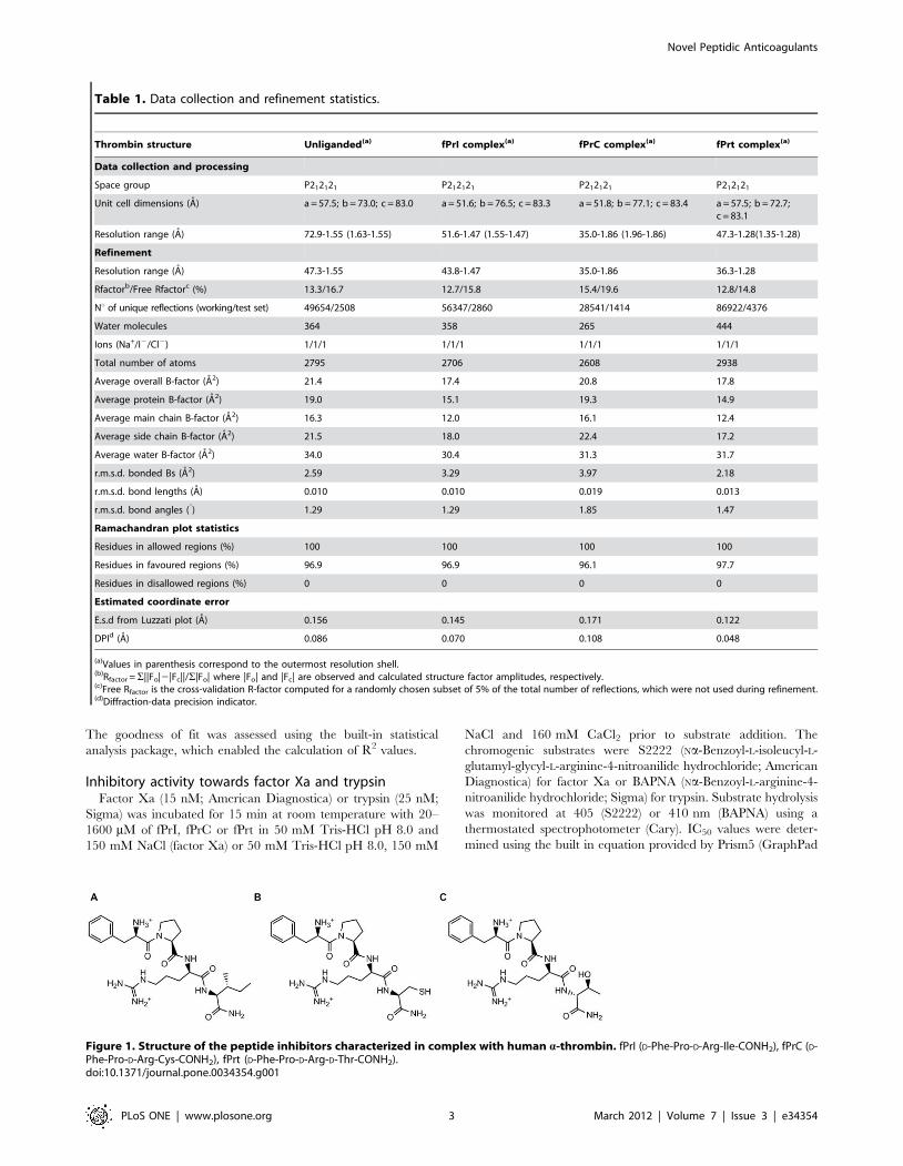
Table 1. Data collection and refinement statistics.
Thrombin structure Unliganded(a) fPrI complex(a) fPrC complex(a) fPrt complex(a)
Data collection and processing
Space group P212121 P212121 P212121 P212121
Unit cell dimensions (A) a = 57.5; b = 73.0; c = 83.0 a = 51.6; b = 76.5; c = 83.3 a = 51.8; b = 77.1; c = 83.4 a = 57.5; b = 72.7;c = 83.1
Resolution range (A) 72.9-1.55 (1.63-1.55) 51.6-1.47 (1.55-1.47) 35.0-1.86 (1.96-1.86) 47.3-1.28(1.35-1.28)
Refinement
Resolution range (A) 47.3-1.55 43.8-1.47 35.0-1.86 36.3-1.28
Rfactorb/Free Rfactorc (%) 13.3/16.7 12.7/15.8 15.4/19.6 12.8/14.8
Nu of unique reflections (working/test set) 49654/2508 56347/2860 28541/1414 86922/4376
Water molecules 364 358 265 444
Ions (Na+/I2/Cl2) 1/1/1 1/1/1 1/1/1 1/1/1
Total number of atoms 2795 2706 2608 2938
Average overall B-factor (A2) 21.4 17.4 20.8 17.8
Average protein B-factor (A2) 19.0 15.1 19.3 14.9
Average main chain B-factor (A2) 16.3 12.0 16.1 12.4
Average side chain B-factor (A2) 21.5 18.0 22.4 17.2
Average water B-factor (A2) 34.0 30.4 31.3 31.7
r.m.s.d. bonded Bs (A2) 2.59 3.29 3.97 2.18
r.m.s.d. bond lengths (A) 0.010 0.010 0.019 0.013
r.m.s.d. bond angles (u) 1.29 1.29 1.85 1.47
Ramachandran plot statistics
Residues in allowed regions (%) 100 100 100 100
Residues in favoured regions (%) 96.9 96.9 96.1 97.7
Residues in disallowed regions (%) 0 0 0 0
Estimated coordinate error
E.s.d from Luzzati plot (A) 0.156 0.145 0.171 0.122
DPId (A) 0.086 0.070 0.108 0.048
(a)Values in parenthesis correspond to the outermost resolution shell.(b)Rfactor =S||Fo|2|Fc||/S|Fo| where |Fo| and |Fc| are observed and calculated structure factor amplitudes, respectively.(c)Free Rfactor is the cross-validation R-factor computed for a randomly chosen subset of 5% of the total number of reflections, which were not used during refinement.(d)Diffraction-data precision indicator.
Figure 1. Structure of the peptide inhibitors characterized in complex with human a-thrombin. fPrI (D-Phe-Pro-D-Arg-Ile-CONH2), fPrC (D-Phe-Pro-D-Arg-Cys-CONH2), fPrt (D-Phe-Pro-D-Arg-D-Thr-CONH2).doi:10.1371/journal.pone.0034354.g001
Novel Peptidic Anticoagulants
PLoS ONE | www.plosone.org 3 March 2012 | Volume 7 | Issue 3 | e34354
Software). Values for the inhibition constant (Ki) were calculated
assuming a competitive mechanism of inhibition according to the
equation Ki = IC50/(1+S/KM), where S is the substrate concen-
tration (0.18 mM for S2222 and 1.84 mM for BAPNA) and KM is
the Michaelis constant of the substrate (0.3 mM for S2222 and
0.95 mM for BAPNA).
Thrombin time (TT) assaysHuman plasma (800 ml) was mixed with 200 ml of 0–1 mM
(final concentration) peptide solutions in 20 mM Tris pH 8.0,
100 mM NaCl. The thrombin time was measured at BM Analises
Clınicas following standard protocols.
Inhibitor resistance to proteolysisEach peptide inhibitor (20 mM) was incubated with a-thrombin
(20 mM) at room temperature in phosphate buffer pH 7.46 with
0.2 M NaCl. After 24 h incubation, aliquots were removed and
the reaction was quenched with 0.1% TFA. The samples were
analyzed on an ESI spectrometer run in the positive mode. As the
expected molecular mass of the tripeptide resulting from the
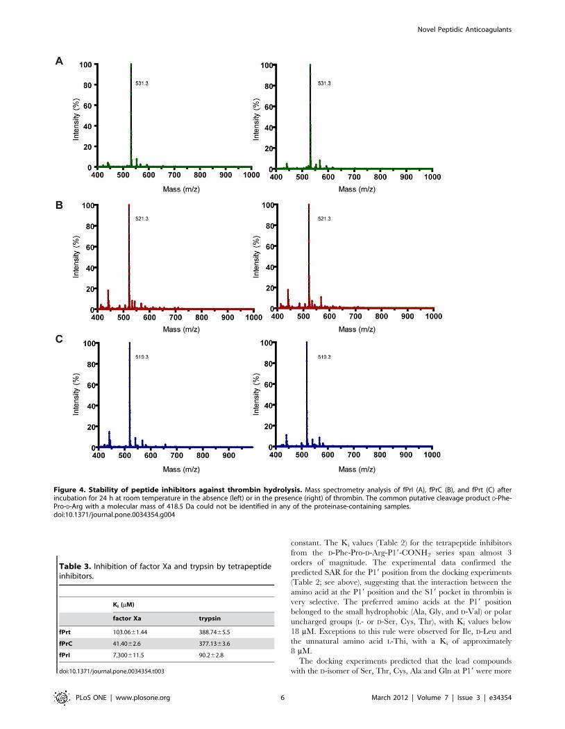
hydrolysis of the D-Arg-P19 peptide bond is 418.5 Da, spectra
were acquired in the 400–1000 Da window.
Crystallization and data collectionSample preparation, crystallization, and X-ray diffraction data
collection were performed as previously described [26].
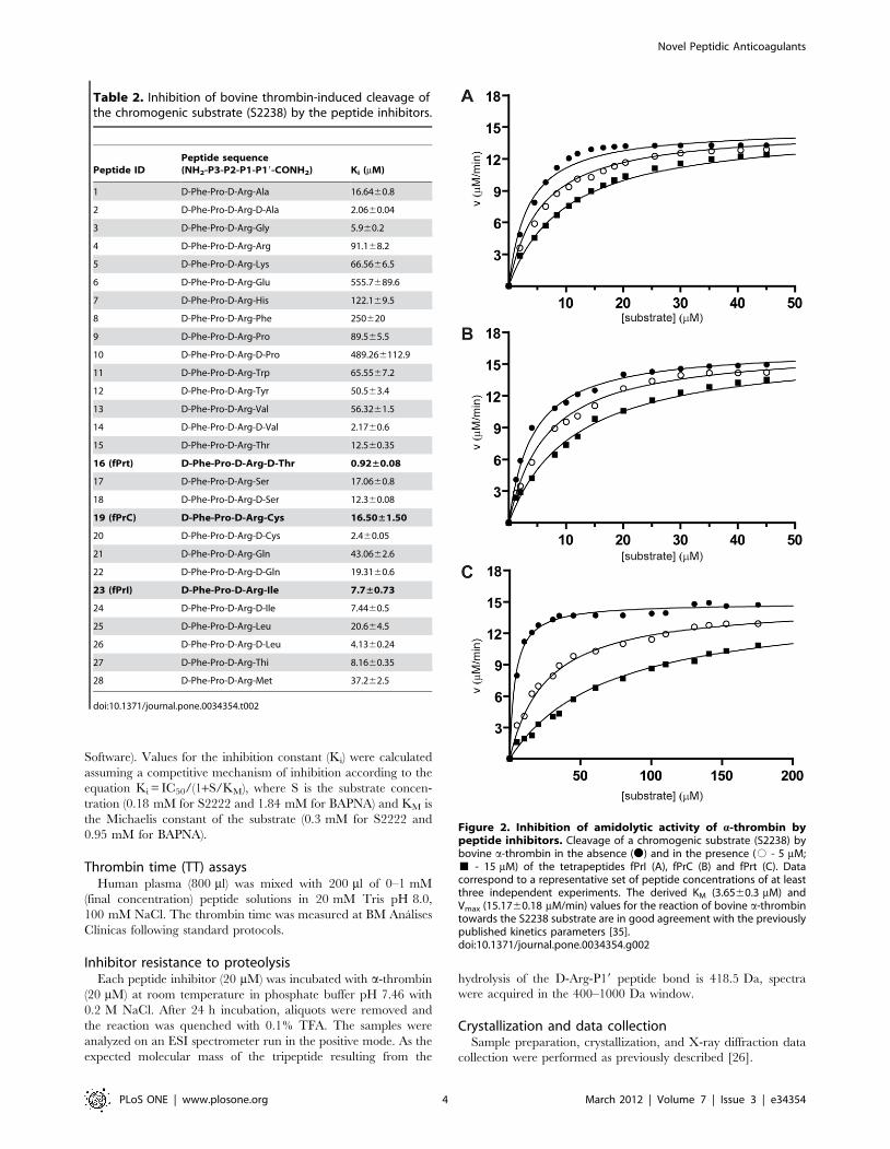
Table 2. Inhibition of bovine thrombin-induced cleavage ofthe chromogenic substrate (S2238) by the peptide inhibitors.
Peptide IDPeptide sequence(NH2-P3-P2-P1-P19-CONH2) Ki (mM)
1 D-Phe-Pro-D-Arg-Ala 16.6460.8
2 D-Phe-Pro-D-Arg-D-Ala 2.0660.04
3 D-Phe-Pro-D-Arg-Gly 5.960.2
4 D-Phe-Pro-D-Arg-Arg 91.168.2
5 D-Phe-Pro-D-Arg-Lys 66.5666.5
6 D-Phe-Pro-D-Arg-Glu 555.7689.6
7 D-Phe-Pro-D-Arg-His 122.169.5
8 D-Phe-Pro-D-Arg-Phe 250620
9 D-Phe-Pro-D-Arg-Pro 89.565.5
10 D-Phe-Pro-D-Arg-D-Pro 489.266112.9
11 D-Phe-Pro-D-Arg-Trp 65.5567.2
12 D-Phe-Pro-D-Arg-Tyr 50.563.4
13 D-Phe-Pro-D-Arg-Val 56.3261.5
14 D-Phe-Pro-D-Arg-D-Val 2.1760.6
15 D-Phe-Pro-D-Arg-Thr 12.560.35
16 (fPrt) D-Phe-Pro-D-Arg-D-Thr 0.92±0.08
17 D-Phe-Pro-D-Arg-Ser 17.0660.8
18 D-Phe-Pro-D-Arg-D-Ser 12.360.08
19 (fPrC) D-Phe-Pro-D-Arg-Cys 16.50±1.50
20 D-Phe-Pro-D-Arg-D-Cys 2.460.05
21 D-Phe-Pro-D-Arg-Gln 43.0662.6
22 D-Phe-Pro-D-Arg-D-Gln 19.3160.6
23 (fPrI) D-Phe-Pro-D-Arg-Ile 7.7±0.73
24 D-Phe-Pro-D-Arg-D-Ile 7.4460.5
25 D-Phe-Pro-D-Arg-Leu 20.664.5
26 D-Phe-Pro-D-Arg-D-Leu 4.1360.24
27 D-Phe-Pro-D-Arg-Thi 8.1660.35
28 D-Phe-Pro-D-Arg-Met 37.262.5
doi:10.1371/journal.pone.0034354.t002
Figure 2. Inhibition of amidolytic activity of a-thrombin bypeptide inhibitors. Cleavage of a chromogenic substrate (S2238) bybovine a-thrombin in the absence (N) and in the presence (# - 5 mM;& - 15 mM) of the tetrapeptides fPrI (A), fPrC (B) and fPrt (C). Datacorrespond to a representative set of peptide concentrations of at leastthree independent experiments. The derived KM (3.6560.3 mM) andVmax (15.1760.18 mM/min) values for the reaction of bovine a-thrombintowards the S2238 substrate are in good agreement with the previouslypublished kinetics parameters [35].doi:10.1371/journal.pone.0034354.g002
Novel Peptidic Anticoagulants
PLoS ONE | www.plosone.org 4 March 2012 | Volume 7 | Issue 3 | e34354
Structure Determination and RefinementThe structure of unliganded human a-thrombin was solved by
molecular replacement with Phaser [27] using the coordinates
from PDB entry 1VZQ [28] as search model. The refined model
of unliganded human a-thrombin was subsequently used as search
model in the structural determination of the fPrI, fPrC and fPrt
thrombin complexes. The initial electron density difference maps
showed interpretable density for all inhibitors.
Cycles of manual model building with Coot [29], alternating
with cycles of crystallographic refinement with PHENIX [30],
were performed until completion of the models. The models were
initially subjected to positional simulated annealing, followed by
refinement of TLS parameters (determined using the TLS Motion
Determination software [31], as implemented in the TLSMD
server [32]) and individual atomic displacement parameters.
When most of the solvent structure was built, the inhibitor was
fitted to the electron density maps. A sodium cation (in the sodium
binding loop), and a chloride and an iodide anion, as well as 2-
methyl-2,4-pentanediol (MPD) molecules from the crystallization
buffer were also located. An ordered N-acetyl-glucosamine moiety
was also modeled, bound to Asn60G. In the final cycles,
occupancy of the ions and sugar groups was refined. Individual
anisotropic ADP refinement was carried out for the light and
heavy chains of thrombin for the higher resolution models
(unliganded thrombin, fPrI and fPrt complexes).
To correctly assign the bound halide anions, from the iodide,
chloride and bromide present in the crystallization buffer, two
datasets for the same crystal were collected at different energies
(12 keV and 14 keV). Inspection of the peaks at the anion
positions in the anomalous difference maps allowed the unambig-
uous identification of the bound ions as chloride and iodide.
The final models of unliganded thrombin and of the
thrombin:fPrt complex comprise residues Ile16 to Glu247 and
Ala1B to Arg15 of one thrombin molecule (chains H and L,
respectively). The model for the thrombin:fPrI complex comprises
residues Ile16 to Glu247 and Ala1B to Ile14K, and that for the
thrombin:fPrC complex comprises residues Ile16 to Phe245 and
Ala1B to Ile14K. The models contain one each of sodium, iodide
and chloride ions. An N-acetyl-glucosamine sugar moiety is
attached to Asn60G in chain H. Three, two or one molecule of
MPD from the crystallization buffer were observed in the models
of unliganded thrombin, fPrI/fPrt and fPrC, respectively. Residues
of loop 148 (Thr147 to Lys149E) were not well defined in the
electron density maps and were not included on the final models.
Refinement statistics are summarized in Table 1. The refined
coordinates and structure factors were deposited at the PDB with
accession numbers 3U69, 3U8R, 3U8T and 3U8O.
Results and Discussion
Structure-based design of peptide libraries as potentialdirect thrombin inhibitors
In an attempt to discover new anticoagulants with lower risk of
bleeding, a new generation of peptidic DTI derived from the D-
Phe-Pro-D-Arg tripeptide scaffold was developed (Figure 1).
Besides the optimal D-Phe-Pro dipeptide at positions P3-P2 [5],
the D-isomer of arginine was selected for position P1 in order to
improve resistance to proteolytic degradation by thrombin. Given
the peptidic nature of the compounds, they would be best suited
for intravenous delivery (e.g. in the treatment of acute thrombotic
events and in surgical settings) therefore minimizing the possible
impact in bioavailability of a basic P1 moiety, as in other similar
cases [33]. The peptide libraries generated contained different L-
and D-isomers of natural (and some non-natural) amino acids at
positions P19, P19 and P29 or P19, P29 and P39, thus generating
peptide inhibitors with formulae ranging from D-Phe-Pro-D-Arg-
P19-CONH2 to D-Phe-Pro-D-Arg-P19-P29-P39-CONH2.
Initial docking experiments allowed a fast screening of the
structural fitness between thrombin and the peptide ligands, based
on the Van der Waals force field included in the MMFF94 package.
The hits were ranked after improvement of the initial predicted
relative free energy of interaction-based main scoring function by
inclusion of the electrostatic force field. From the very beginning it
was clear that the peptides with P29 or P29 and P39 occupancy scored
much lower than the tetrapeptidic compounds, and their screening
was truncated. One hundred and twenty virtual lead compounds
with a predicted protein-peptide ligand binding energy of less than
230.0 kcal/mol (corresponding to low mM to low nM Ki values) and
reasonable fit to thrombin’s active site (as judged by visual inspection)
were selected from the more than 1000 virtually screened peptide
sequences. The top-ranking 28 compounds (Table 2), which were
synthesized for further experimental validation (see below), all
contained the D-Phe-Pro-D-Arg-P19-CONH2 sequence, differing
only in the chemical nature of the residue at position P19.
Virtual screening data supported a model of interaction between
thrombin and peptide ligand in which the amino acid at position
P19 would make a relatively significant contribution to the free
energy of interaction. Among the selected lead tetrapeptides
(Table 2), the calculated free energy of interaction suggested tighter
binding for those compounds with either the D-isomer of some of the
polar uncharged amino acids (D-Gln, D-Cys, and D-Ser) or the
somewhat unexpected L-Met or L-Thienylalanine (L-Thi) in P19. An
intermediate group of compounds comprised those containing the
polar uncharged D-Thr, L-Ser or L-Gln as the terminal residue,
while charged (L-Glu and L-Arg) and bulky (L-Ile, L-Phe, L-Trp, L-
His) P19 moieties ranked closely in a third group of putative binders.
Considering the wide chemical space covered by the P19 residues in
these lead peptides, the only general SAR trend that could be
observed is that, whenever both isomers of a given amino acid were
present at this position, the D-isoform was predicted to have higher
affinity for thrombin than the L counterpart (Table 2).
Kinetics of thrombin inhibition by the synthetic peptidesThe inhibitory efficiency of the designed peptides against
bovine thrombin was evaluated by determining their inhibitory
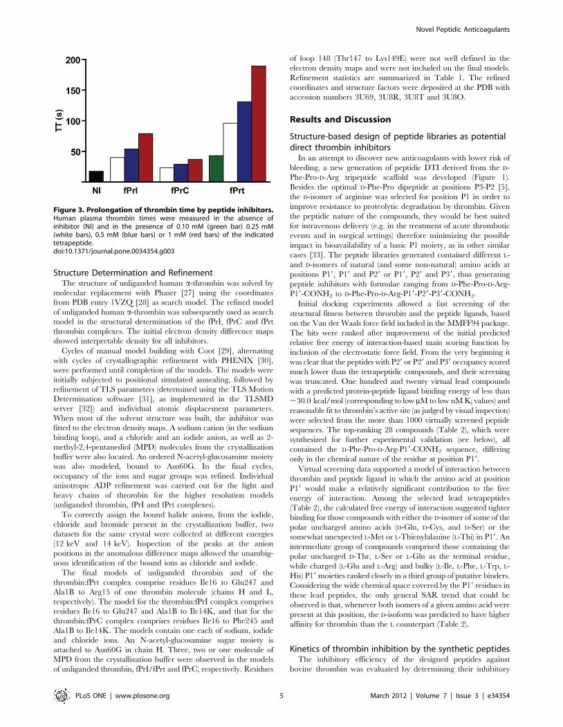
Figure 3. Prolongation of thrombin time by peptide inhibitors.Human plasma thrombin times were measured in the absence ofinhibitor (NI) and in the presence of 0.10 mM (green bar) 0.25 mM(white bars), 0.5 mM (blue bars) or 1 mM (red bars) of the indicatedtetrapeptide.doi:10.1371/journal.pone.0034354.g003
Novel Peptidic Anticoagulants
PLoS ONE | www.plosone.org 5 March 2012 | Volume 7 | Issue 3 | e34354
constant. The Ki values (Table 2) for the tetrapeptide inhibitors
from the D-Phe-Pro-D-Arg-P19-CONH2 series span almost 3
orders of magnitude. The experimental data confirmed the
predicted SAR for the P19 position from the docking experiments
(Table 2; see above), suggesting that the interaction between the
amino acid at the P19 position and the S19 pocket in thrombin is
very selective. The preferred amino acids at the P19 position
belonged to the small hydrophobic (Ala, Gly, and D-Val) or polar
uncharged groups (L- or D-Ser, Cys, Thr), with Ki values below
18 mM. Exceptions to this rule were observed for Ile, D-Leu and
the unnatural amino acid L-Thi, with a Ki of approximately
8 mM.
The docking experiments predicted that the lead compounds
with the D-isomer of Ser, Thr, Cys, Ala and Gln at P19 were more
Table 3. Inhibition of factor Xa and trypsin by tetrapeptideinhibitors.
Ki (mM)
factor Xa trypsin
fPrt 103.0661.44 388.7465.5
fPrC 41.4062.6 377.1363.6
fPrI 7,300611.5 90.262.8
doi:10.1371/journal.pone.0034354.t003
Figure 4. Stability of peptide inhibitors against thrombin hydrolysis. Mass spectrometry analysis of fPrI (A), fPrC (B), and fPrt (C) afterincubation for 24 h at room temperature in the absence (left) or in the presence (right) of thrombin. The common putative cleavage product D-Phe-Pro-D-Arg with a molecular mass of 418.5 Da could not be identified in any of the proteinase-containing samples.doi:10.1371/journal.pone.0034354.g004
Novel Peptidic Anticoagulants
PLoS ONE | www.plosone.org 6 March 2012 | Volume 7 | Issue 3 | e34354
efficient inhibitors that their L-amino acid-containing variants.
This was verified experimentally, reaching its maximum expres-
sion in the case of Thr, where the D-isomer displayed a nearly 15-
fold lower Ki than its L- counterpart (Table 2).
The three lead tetrapeptides (fPrI, fPrC and fPrt; Figures 1 and
2) that were also characterized structurally were found to be potent
competitive thrombin inhibitors in vitro (Table 2). Furthermore,
these peptides prolonged thrombin time (TT) in a dose-dependent
manner (Figure 3), with relative activities that correlated well with
their observed inhibition efficiency towards thrombin.
Resistance to proteolytic cleavageThe three structurally characterized inhibitors were found to be
stable to cleavage by thrombin, as no proteolytic fragments could
be identified by mass spectrometry upon 24 h incubation with the
enzyme at room temperature (Figure 4), in good agreement with
their observed binding mode in the experimental crystallographic
structures (see below).
Selectivity for thrombinThe three structurally characterized peptide inhibitors display a
higher selectivity for a-thrombin than for factor Xa or trypsin
(Table 3). The best thrombin inhibitor, fPrt, is 420-fold and 110-
fold more selective for thrombin than for trypsin or factor Xa,
respectively. While fPrI is essentially unable to inhibit factor Xa in
vitro, it displays a considerably more modest selectivity for
thrombin versus trypsin (12-fold). Of the three tetrapeptides, fPrC
was found to be the least selective, displaying only 3- or 20-fold
selectivity towards both factor Xa or trypsin, respectively.
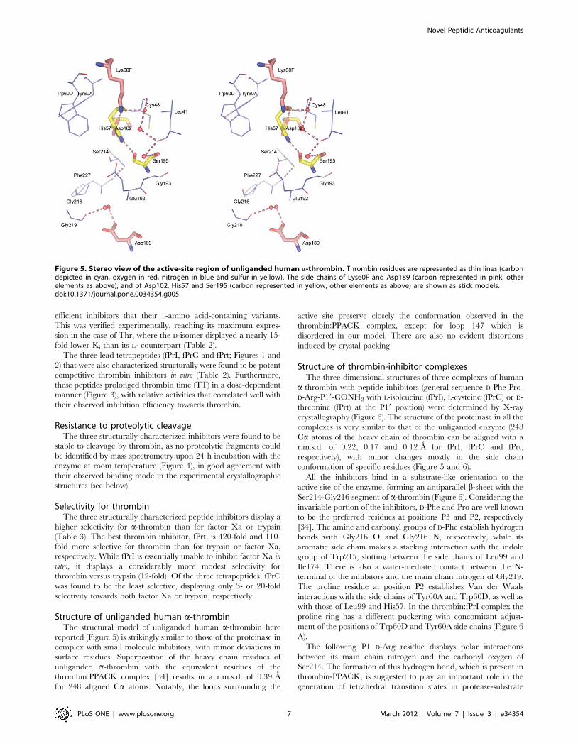
Structure of unliganded human a-thrombinThe structural model of unliganded human a-thrombin here
reported (Figure 5) is strikingly similar to those of the proteinase in
complex with small molecule inhibitors, with minor deviations in
surface residues. Superposition of the heavy chain residues of
unliganded a-thrombin with the equivalent residues of the
thrombin:PPACK complex [34] results in a r.m.s.d. of 0.39 A
for 248 aligned Ca atoms. Notably, the loops surrounding the
active site preserve closely the conformation observed in the
thrombin:PPACK complex, except for loop 147 which is
disordered in our model. There are also no evident distortions
induced by crystal packing.
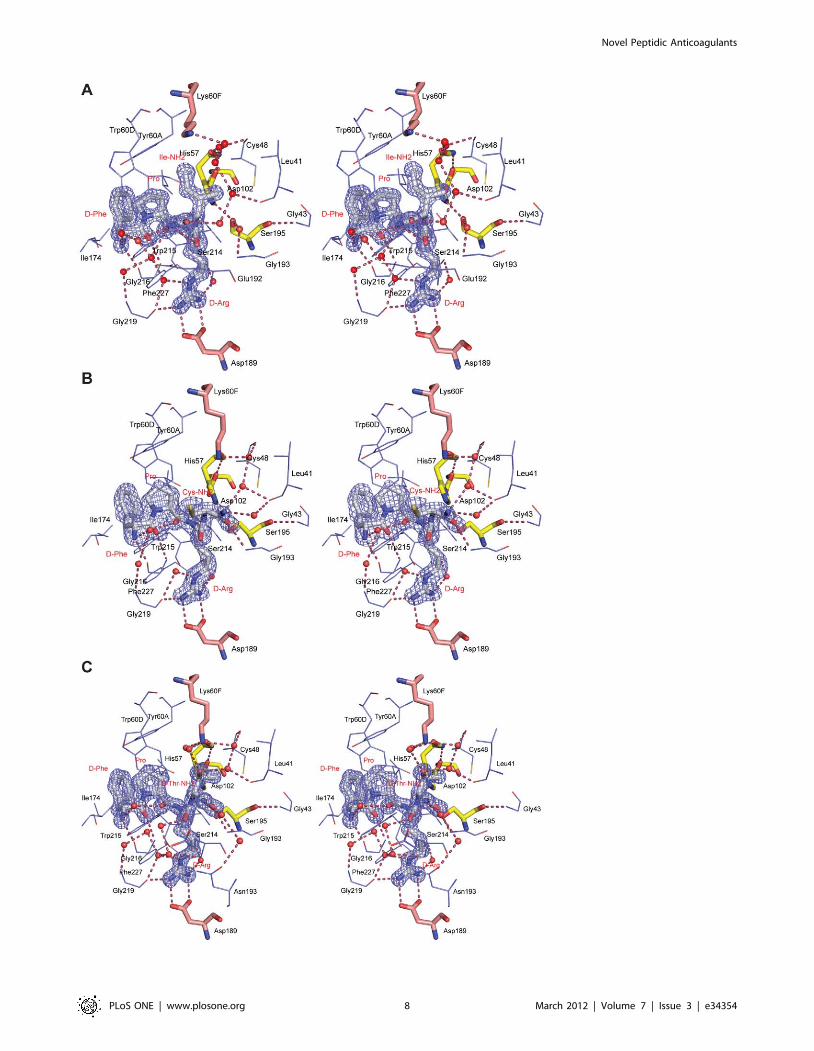
Structure of thrombin-inhibitor complexesThe three-dimensional structures of three complexes of human
a-thrombin with peptide inhibitors (general sequence D-Phe-Pro-
D-Arg-P19-CONH2 with L-isoleucine (fPrI), L-cysteine (fPrC) or D-
threonine (fPrt) at the P19 position) were determined by X-ray
crystallography (Figure 6). The structure of the proteinase in all the
complexes is very similar to that of the unliganded enzyme (248
Ca atoms of the heavy chain of thrombin can be aligned with a
r.m.s.d. of 0.22, 0.17 and 0.12 A for fPrI, fPrC and fPrt,
respectively), with minor changes mostly in the side chain
conformation of specific residues (Figure 5 and 6).
All the inhibitors bind in a substrate-like orientation to the
active site of the enzyme, forming an antiparallel b-sheet with the
Ser214-Gly216 segment of a-thrombin (Figure 6). Considering the
invariable portion of the inhibitors, D-Phe and Pro are well known
to be the preferred residues at positions P3 and P2, respectively
[34]. The amine and carbonyl groups of D-Phe establish hydrogen
bonds with Gly216 O and Gly216 N, respectively, while its
aromatic side chain makes a stacking interaction with the indole
group of Trp215, slotting between the side chains of Leu99 and
Ile174. There is also a water-mediated contact between the N-
terminal of the inhibitors and the main chain nitrogen of Gly219.
The proline residue at position P2 establishes Van der Waals
interactions with the side chains of Tyr60A and Trp60D, as well as
with those of Leu99 and His57. In the thrombin:fPrI complex the
proline ring has a different puckering with concomitant adjust-
ment of the positions of Trp60D and Tyr60A side chains (Figure 6
A).
The following P1 D-Arg residue displays polar interactions
between its main chain nitrogen and the carbonyl oxygen of
Ser214. The formation of this hydrogen bond, which is present in
thrombin-PPACK, is suggested to play an important role in the
generation of tetrahedral transition states in protease-substrate
Figure 5. Stereo view of the active-site region of unliganded human a-thrombin. Thrombin residues are represented as thin lines (carbondepicted in cyan, oxygen in red, nitrogen in blue and sulfur in yellow). The side chains of Lys60F and Asp189 (carbon represented in pink, otherelements as above), and of Asp102, His57 and Ser195 (carbon represented in yellow, other elements as above) are shown as stick models.doi:10.1371/journal.pone.0034354.g005
Novel Peptidic Anticoagulants
PLoS ONE | www.plosone.org 7 March 2012 | Volume 7 | Issue 3 | e34354
Novel Peptidic Anticoagulants
PLoS ONE | www.plosone.org 8 March 2012 | Volume 7 | Issue 3 | e34354
complexes [34]. Furthermore, the D-Arg side chain extends into
the S1 pocket and the guanidinium group is hydrogen bonded to
Asp189 (P1 NH2 - Asp189 OD1, P1 NH1 - Asp189 OD2) and to
Gly219 (P1 NH1 - Gly219 O). There are also water-mediated
hydrogen bonds connecting D-Arg NE and NH2 to the carbonyl
oxygen of Gly219 and Phe227, respectively. In the fPrt complex,
the carbonyl oxygen of D-Arg (P1) establishes water-mediated
interactions with thrombin residues Glu192 (Glu192 N) and
Gly219 (Gly219 O; Figure 6 C). These interactions are absent
both in fPrI and fPrC complexes.
The contacts established by the D-Arg residue in position P1
with the enzyme backbone are similar to those observed for the L-
isomer in other substrates and inhibitors (e.g. PPACK [34], MD-
805/argotraban [9] and SDZ229-357 [35]. However, the
presence of D-Arg at this position also results in the upstream
residues sitting deeper in the S2 and S3 pockets, thereby increasing
the distance between the Ser195 side chain hydroxyl and the P1
carbonyl carbon of the inhibitors (3.69 A, 2.85 A and 2.96 A for
fPrI, fPrC and fPrt, respectively; Table 4). Replacing the P1
residue with other variants of arginine such as b-homo-arginine (in
hirulog3 [25]) or N-a-methyl-arginine (in I-11 [36]) was shown to
improve resistance to thrombin hydrolysis. In all cases, the
putative scissile bond becomes less accessible to the active site
nucleophile. The TH146 (rOicPGF) [18] and FM19 (rOicPaF(p-
Me)) [17] analogs of RPPGF, the angiotensin-converting enzyme
breakdown product of bradykinin, inhibit thrombin in a retro-
binding orientation inserting a D-Arg residue in the S1 specificity
site in a similar way to that observed in our complexes, although
without occupying the P19 subsite.
Thrombin’s Ser195 side chain occupies its canonical position
(similar to that of the unliganded enzyme) in the fPrI complex,
retaining the hydrogen bond between its OG and the side chain
NE2 of His57 (Figure 6 A). However, in the fPrC and fPrt
complexes, the Ser195 side chain is rotated away from the catalytic
histidine residue, therefore disrupting the canonical hydrogen
bond (Figure 6 B, C; Table 5), and interacts instead with the
carbonyl oxygen of the P19 residue. Hydrogen bonds between the
inhibitor P19 and thrombin Ser195 residues were observed in
complexes of thrombin with non-covalent inhibitors Eoc-D-Phe-
Pro-Abh and Cbz-Pro-Abh (P19 N - Ser195 OG) [37] or CVS995
(P19 O - Ser195 OG). We suggest that the catalytic triad disruption
in thrombin:fPrC and thrombin:fPrt impairs the enzyme’s
hydrolytic ability, as proton abstraction by His57 NE2 and
activation of Ser195 OG is no longer possible.
Substrate cleavage by thrombin is dependent on the formation
of a tetrahedral transition state intermediate. Comparison of the
interactions that thrombin establishes with fPrt, fPrC and fPrI with
transition-state mimetic inhibitors can help explaining the
resilience of the former compounds to proteolytic cleavage.
Electrophilic carbonyl thrombin inhibitors such as PPACK [34]
and APPA [38] form covalent transition-state-like analogs in
complex with thrombin. The carbonyl carbon of their P1 residue
(Arg in PPACK, aminophenylpyruvate in APPA) is in a
tetrahedral configuration and covalently bound to Ser195 OG
(Table 4). In these complexes the side chain of Ser195 is
considerably rotated when compared to unliganded thrombin,
however without disrupting the Ser195 OG - His57 NE2
hydrogen bond. The divalent potent thrombin inhibitor
CVS995 is a competitive reversible inhibitor that displays an a-
keto amide group at P19 and forms a tetrahedral transition state
with the active site serine and histidine residues [39]. Halo-
methylketones also form a covalent bond to His57 [6,34].
The carbonyl oxygen atom of APPA and PPACK, located in
the oxyanion hole, and the keto oxygen of CVS995 establish
hydrogen bonds with the amide nitrogen atoms of Gly193 and
Ser195 (Table 6). The interaction with Gly193 is present in the
complexes thrombin:fPrC and thrombin:fPrt, whereas the latter
interaction is absent. In the case of the thrombin:fPrI complex
Gly193N does not interact with the inhibitor, being instead
indirectly bonded to Ser195 OG.
The P19 Cys of fPrC establishes Van der Waals contacts with
Trp60D and is hydrogen bonded to Gly193 N and Ser195 OG
through its carbonyl oxygen. Its terminal amide nitrogen (N2) also
Figure 6. Stereo view of the active-site region of human a-thrombin in complex with the peptide inhibitors fPrI (A), fPrC (B) and fPrt(C). Thrombin residues establishing hydrogen bonds (red dashed lines) or hydrophobic contacts with the inhibitor are represented as thin lines(carbon depicted in cyan, oxygen in red, nitrogen in blue and sulfur in yellow). The side chains of Lys60F and Asp189 (carbon represented in pink,other elements as above), of Asp102, His57 and Ser195 (carbon represented in yellow, other elements as above) and the inhibitors (carbon is depictedin white, other elements as above) are shown as stick models. The electron density map (2Fobs-Fcalc) of the inhibitors is contoured at 1.5s for fPrIand fPrt and at 1.0s for fPrC. Inhibitor residues and selected thrombin side chains are labeled in red and in black, respectively.doi:10.1371/journal.pone.0034354.g006
Table 4. Distance between the catalytic Ser195 residue ofthrombin and the P1 residue carbonyl carbon in thrombin-inhibitor complexes.
Thrombincomplex
Ser195 OG - P1C distance (A) Comments and References
Thrombin :PPACK
1.42 electrophilic carbonyl inhibitor (P1 = Arg) -irreversible [34]
Thrombin :APPA
1.44 electrophilic carbonyl inhibitor(P1 = aminophenylpyruvate) - reversible [38]
Thrombin :CVS95
1.74 bivalent inhibitor (P1 = Arg, P19 =a-ketoamide) - transition state analogue - potentinhibitor (Ki = 1.01610212 M) [39]
Thrombin :fPrI
3.69 This work
Thrombin :fPrC
2.85 This work
Thrombin :fPrt
2.96 This work
doi:10.1371/journal.pone.0034354.t004
Table 5. Hydrogen bond distances between the catalytic His57 NE2 and Ser195 OG in thrombin complexes.
Thrombin complex Distance (A) Reference
Unliganded thrombin 2.80 This work
Thrombin : APPA 3.19 [38]
Thrombin : PPACK 2.90 [34]
Thrombin : fPrt N.P. This work
Thrombin : fPrC N.P. This work
Thrombin : fPrI 2.97 This work
N.P. - not present.doi:10.1371/journal.pone.0034354.t005
Novel Peptidic Anticoagulants
PLoS ONE | www.plosone.org 9 March 2012 | Volume 7 | Issue 3 | e34354
establishes solvent-mediated interactions with Leu41 O, Cys58 O
and Lys60F NZ.
When D-Thr is found in the P19 position (fPrt), the inhibitor is
stabilized by water-mediated hydrogen bonds between its OG and
Lys60F NZ, Leu41 O and Cys58 O and between its terminal
amide nitrogen and Asn143 OD1 and Trp141 O. Additionally,
fPrt interacts directly with thrombin through hydrogen bonds
between its D-Thr carbonyl oxygen and both Gly193 N and the
hydroxyl group of the catalytic Ser195. A D-Thr N - His57 NE2
interaction is observed as well, resembling that found in complexes
of thrombin with active site inhibitors with benzothiazole [40] and
ketothiazole [41] or with bivalent inhibitors with a-keto-amide
[39] or 2-(4-aminobutyl)-hydrazyde (Abh) [37] at the P19 position.
Furthermore, D-Thr makes hydrophobic contacts with Trp60D,
His57 and with the Cys42-Cys58 disulfide bond. All these
interactions could be maintained in the case of D-Val at position
P19, except for the solvent-mediated contacts established by the
side chain hydroxyl group of threonine, possibly accounting for the
2-fold lower Ki displayed by fPrt (Table 2). Finally, in the fPrI
complex, the carbonyl oxygen of the isoleucine residue in position
P19 establishes water-mediated hydrogen bonds with Leu41 O and
Cys48 O, while its terminal amide nitrogen interacts directly with
the carbonyl oxygen of the P2 proline residue and the Glu192
OE2 via a solvent molecule. Furthermore, the P19 isoleucine side
chain also makes hydrophobic contacts with Trp60D and His57. It
is conceivable that the similar-sized D-Leu side chain could
establish equivalent contacts with the proteinase, as indicated by
the comparable Ki determined for both peptides (Table 2).
The side chain of Lys60F was proposed to limit the size of
thrombin’s S19 pocket contributing to the frequent occurrence of
small residues (such as glycine and serine in thrombin-activated
platelet receptors) at this position in natural protein substrates. In
the thrombin:fPrt and thrombin:fPrC complexes, the side chain of
Lys60F is found in an extended conformation similar to that
observed in the thrombin-PPACK model [34] and in the
unliganded enzyme. However, in the thrombin-fPrI complex,
accommodation of the isoleucine in position P19 implies the
displacement of Lys60F, which is found in a different conforma-
tion similar to that observed in complexes of thrombin with
inhibitors containing benzothiazole groups [40,41], or bulky
residues (Nle or Thi) at the P19 position [42]. In addition, the
terminal amide nitrogen of Ile points towards Trp60D, which
moves further away from the catalytic site.
The atomic detail of the experimental three-dimensional
structures provides a molecular explanation for the relative
binding affinity and resistance to proteolysis of the designed
compounds. The now identified peptidic direct thrombin
inhibitors represent a novel pharmacophore scaffold for develop-
ing new antithrombotic agents by exploring the conformations
imposed by the D-stereochemistry of the amino acids at positions
P1 and P19.
Acknowledgments
We acknowledge the ESRF (Grenoble, France) for the provision of
synchrotron-radiation facilities and the ESRF staff for assistance in using
beamline ID14-EH1.
Author Contributions
Conceived and designed the experiments: ACF CCC MP PJBP. Performed
the experiments: ACF CCC SZ JG MP PJBP. Analyzed the data: ACF
CCC MP PJBP. Contributed reagents/materials/analysis tools: CCC MP
PJBP. Wrote the paper: ACF CCC MP PJBP.
References
1. Mousa SA (2010) Novel anticoagulant therapy: principle and practice. Methods
Mol Biol 663: 157–179.
2. Steffel J, Braunwald E (2011) Novel oral anticoagulants: focus on stroke
prevention and treatment of venous thrombo-embolism. Eur Heart J 32:
1968–1976.
3. Steffel J, Luscher TF (2009) Novel anticoagulants in clinical development: focus
on factor Xa and direct thrombin inhibitors. J Cardiovasc Med (Hagerstown) 10:
616–623.
4. Srivastava S, Goswami LN, Dikshit DK (2005) Progress in the design of low
molecular weight thrombin inhibitors. Med Res Rev 25: 66–92.
5. Bode W, Mayr I, Baumann U, Huber R, Stone SR, et al. (1989) The refined
1.9 A crystal structure of human alpha-thrombin: interaction with D-Phe-Pro-
Arg chloromethylketone and significance of the Tyr-Pro-Pro-Trp insertion
segment. EMBO J 8: 3467–3475.
6. Neises B, Tarnus C, Broersma RJ, Bald C, Remy JM, et al. (1993) The synthesis
and anticoagulant activity of novel peptidylfluoroalkanes. Adv Exp Med Biol
340: 185–188.
7. Maraganore JM, Bourdon P, Jablonski J, Ramachandran KL, Fenton JW, 2nd
(1990) Design and characterization of hirulogs: a novel class of bivalent peptide
inhibitors of thrombin. Biochemistry 29: 7095–7101.
8. Skrzypczak-Jankun E, Carperos VE, Ravichandran KG, Tulinsky A,
Westbrook M, et al. (1991) Structure of the hirugen and hirulog 1 complexes
of alpha-thrombin. J Mol Biol 221: 1379–1393.
9. Banner DW, Hadvary P (1991) Crystallographic analysis at 3.0-A resolution of
the binding to human thrombin of four active site-directed inhibitors. J Biol
Chem 266: 20085–20093.
10. Okamoto S, Hijikata A, Kikumoto R, Tonomura S, Hara H, et al. (1981) Potent
inhibition of thrombin by the newly synthesized arginine derivative No. 805.
Table 6. Hydrogen bond distances between thrombin Gly193 N and Ser195 N and bound inhibitors.
Thrombin complex Thrombin Atom Inhibitor Atom Distance (A) Reference
Thrombin : APPA Gly193 N O2 2.95 [38]
Gly193 N O1 3.23
Ser 195 N O1 2.89
Thrombin : PPACK Gly193 N P1 O 3.16 [34]
Ser 195 N P1 O 3.31
Thrombin : CVS995 Gly193 N P1 O 2.70 [39]
Thrombin : fPrt Gly193 N P19 O 2.83 This work
Thrombin : fPrC Gly193 N P19 O 3.24 This work
Thrombin : fPrI Gly193 N P19 O N.P. This work
N.P. - not present.doi:10.1371/journal.pone.0034354.t006
Novel Peptidic Anticoagulants
PLoS ONE | www.plosone.org 10 March 2012 | Volume 7 | Issue 3 | e34354
The importance of stereo-structure of its hydrophobic carboxamide portion.
Biochem Biophys Res Commun 101: 440–446.11. Cuker A (2011) Heparin-induced thrombocytopenia: present and future.
J Thromb Thrombolysis 31: 353–366.
12. Babuin L, Pengo V (2010) Argatroban in the management of heparin-inducedthrombocytopenia. Vasc Health Risk Manag 6: 813–819.
13. Stangier J (2008) Clinical pharmacokinetics and pharmacodynamics of the oraldirect thrombin inhibitor dabigatran etexilate. Clin Pharmacokinet 47: 285–295.
14. Ezekowitz M, Nagarakanti R (2011) Dabigatran in atrial fibrillation:
pharmacology and clinical trials. J Interv Card Electrophysiol 32: 173–180.15. Hauel NH, Nar H, Priepke H, Ries U, Stassen J-M, et al. (2002) Structure-based
design of novel potent nonpeptide thrombin inhibitors. J Med Chem 45:1757–1766.
16. Bajusz S, Szell E, Barabas E, Bagdy D (1982) Design and synthesis of peptideinhibitors of blood coagulations. Folia Haematol Int Mag Klin Morphol
Blutforsch 109: 16–21.
17. Nieman MT, Burke F, Warnock M, Zhou Y, Sweigart J, et al. (2008)Thrombostatin FM compounds: direct thrombin inhibitors – mechanism of
action in vitro and in vivo. J Thromb Haemost 6: 837–845.18. Nieman MT, Warnock M, Hasan AAK, Mahdi F, Lucchesi BR, et al. (2004)
The preparation and characterization of novel peptide antagonists to thrombin
and factor VIIa and activation of protease-activated receptor 1. J PharmacolExp Ther 311: 492–501.
19. Burke FM, Warnock M, Schmaier AH, Mosberg HI (2006) Synthesis of novelpeptide inhibitors of thrombin-induced platelet activation. Chem Biol Drug Des
68: 235–238.20. Girnys EA, Sobczyk-Kojiro K, Mosberg HI (2010) Structure-based design of
residue 1 analogs of the direct thrombin inhibitor pentapeptide FM 19. Chem
Biol Drug Des 75: 35–39.21. Isaacs RCA, Newton CL, Cutrona KJ, Mercer SP, Payne LS, et al. (2011)
Design, synthesis and SAR of a series of 1,3,5-trisubstituted benzenes asthrombin inhibitors. Bioorg Med Chem Lett 21: 1536–1540.
22. Andricopulo AD, Salum LB, Abraham DJ (2009) Structure-based drug design
strategies in medicinal chemistry. Curr Top Med Chem 9: 771–790.23. Talele TT, Khedkar SA, Rigby AC (2010) Successful applications of computer
aided drug discovery: moving drugs from concept to the clinic. Curr Top MedChem 10: 127–141.
24. Guichard G, Benkirane N, Zeder-Lutz G, van Regenmortel MH, Briand JP,et al. (1994) Antigenic mimicry of natural L-peptides with retro-inverso-
peptidomimetics. Proc Natl Acad Sci U S A 91: 9765–9769.
25. Qiu X, Padmanabhan KP, Carperos VE, Tulinsky A, Kline T, et al. (1992)Structure of the hirulog 3-thrombin complex and nature of the S9 subsites of
substrates and inhibitors. Biochemistry 31: 11689–11697.26. Figueiredo AC, Clement CC, Philipp M, Pereira PJB (2011) Crystallization and
preliminary crystallographic characterization of three peptidic inhibitors in
complex with a-thrombin. Acta Crystallogr Sect F Struct Biol Cryst Commun67: 54–58.
27. McCoy AJ, Grosse-Kunstleve RW, Adams PD, Winn MD, Storoni LC, et al.(2007) Phaser crystallographic software. J Appl Crystallogr 40: 658–674.
28. Scharer K, Morgenthaler M, Seiler P, Diederich F, Banner D, et al. (2004)
Enantiomerically pure thrombin inhibitors for exploring the molecular-
recognition features of the oxyanion hole. Helv Chim Acta 87: 2517–2538.
29. Emsley P, Cowtan K (2004) Coot: model-building tools for molecular graphics.
Acta Crystallogr D Biol Crystallogr 60: 2126–2132.
30. Adams PD, Afonine PV, Bunkoczi G, Chen VB, Davis IW, et al. (2010)
PHENIX: a comprehensive Python-based system for macromolecular structure
solution. Acta Crystallogr D Biol Crystallogr 66: 213–221.
31. Painter J, Merritt EA (2006) Optimal description of a protein structure in terms
of multiple groups undergoing TLS motion. Acta Crystallogr D Biol Crystallogr
62: 439–450.
32. Painter J, Merritt EA (2006) TLSMD web server for the generation of multi-
group TLS models. J Appl Cryst 39: 109–111.
33. Sinauridze EI, Romanov AN, Gribkova IV, Kondakova OA, Surov SS, et al.
(2011) New synthetic thrombin inhibitors: molecular design and experimental
verification. PLoS ONE 6: e19969.
34. Bode W, Turk D, Karshikov A (1992) The refined 1.9-A X-ray crystal structure
of D-Phe-Pro-Arg chloromethylketone-inhibited human alpha-thrombin: struc-
ture analysis, overall structure, electrostatic properties, detailed active-site
geometry, and structure-function relationships. Protein Sci 1: 426–471.
35. Wagner J, Kallen J, Ehrhardt C, Evenou J-P, Wagner D (1998) Rational design,
synthesis, and X-ray structure of selective noncovalent thrombin inhibitors.
J Med Chem 41: 3664–3674.
36. Friedrich R, Steinmetzer T, Huber R, Sturzebecher J, Bode W (2002) The
methyl group of N[alpha](Me)Arg-containing peptides disturbs the active-site
geometry of thrombin, impairing efficient cleavage. J Mol Biol 316: 869–874.
37. De Simone G, Balliano G, Milla P, Gallina C, Giordano C, et al. (1997) Human
alpha-thrombin inhibition by the highly selective compounds N-ethoxycarbonyl-
D-Phe-Pro-alpha-azaLys p-nitrophenyl ester and N-carbobenzoxy-Pro-alpha-
azaLys p-nitrophenyl ester: A kinetic, thermodynamic and X-ray crystallo-
graphic study. J Mol Biol 269: 558–569.
38. Chen ZG, Li Y, Mulichak AM, Lewis SD, Shafer JA (1995) Crystal structure of
human alpha-thrombin complexed with hirugen and p-amidinophenylpyruvate
at 1.6 A resolution. Arch Biochem Biophys 322: 198–203.
39. Krishnan R, Tulinsky A, Vlasuk GP, Pearson D, Vallar P, et al. (1996) Synthesis,
structure, and structure-activity relationships of divalent thrombin inhibitors
containing an alpha-keto-amide transition-state mimetic. Protein Sci 5:
422–433.
40. Matthews JH, Krishnan R, Costanzo MJ, Maryanoff BE, Tulinsky A (1996)
Crystal structures of thrombin with thiazole-containing inhibitors: probes of the
S19 binding site. Biophys J 71: 2830–2839.
41. St Charles R, Matthews JH, Zhang E, Tulinsky A (1999) Bound structures of
novel P3–P19 beta-strand mimetic inhibitors of thrombin. J Med Chem 42:
1376–1383.
42. Slon-Usakiewicz JJ, Sivaraman J, Li Y, Cygler M, Konishi Y (2000) Design of
P19 and P39 residues of trivalent thrombin inhibitors and their crystal structures.
Biochemistry 39: 2384–2391.
Novel Peptidic Anticoagulants
PLoS ONE | www.plosone.org 11 March 2012 | Volume 7 | Issue 3 | e34354
Related Documents