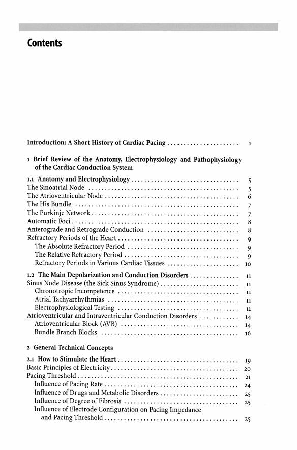
Rate - Interval Conversion Table Rate Cycle length Rate Cycle length (BPM) (ms) (BPM) (ms) 20 3000 215 279 25 2400 220 273 30 2000 225 267 35 1714 230 261 40 1500 235 255 45 1333 240 250 50 1200 245 245 55 1091 250 240 60 1000 255 235 65 923 260 231 70 857 265 226 75 800 270 222 80 750 275 218 85 706 280 214 90 667 285 211 95 632 290 207 100 600 295 203 105 571 300 200 110 545 305 197 115 522 310 194 120 500 315 190 125 480 320 188 130 462 325 185 135 444 330 182 140 429 335 179 145 414 340 176 150 400 345 174 155 387 350 171 160 375 355 169 165 364 360 167 170 353 365 164 175 343 370 162 180 333 375 160 185 324 380 158 190 316 385 156 195 308 390 154 200 300 395 152 205 293 400 150 210 286 Rate (BPM or m i n 1 ) = Cycle length (ms) = 60,000 Cycle length (ms) 60,000 Rate (BPM or m i n 1 )

Welcome message from author
This document is posted to help you gain knowledge. Please leave a comment to let me know what you think about it! Share it to your friends and learn new things together.
Transcript
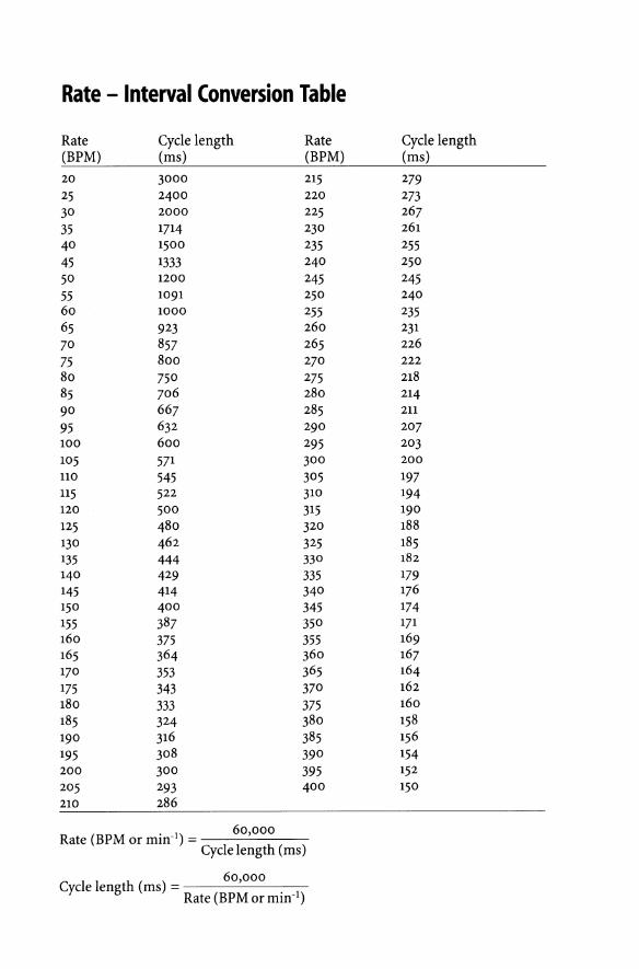
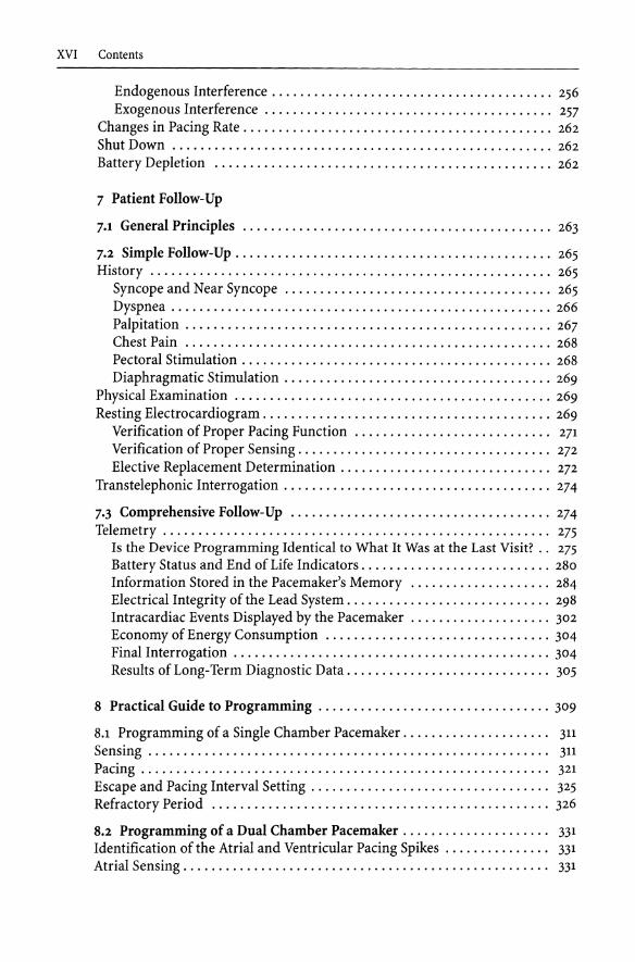
Rate - Interval Conversion Table
Rate Cycle length Rate Cycle length (BPM) (ms) (BPM) (ms) 20 3000 215 279 25 2400 220 273 30 2000 225 267 35 1714 230 261 40 1500 235 255 45 1333 240 250 50 1200 245 245 55 1091 250 240 60 1000 255 235 65 923 260 231 70 857 265 226 75 800 270 222 80 750 275 218 85 706 280 214 90 667 285 211 95 632 290 207 100 600 295 203 105 571 300 200 110 545 305 197 115 522 310 194 120 500 315 190 125 480 320 188 130 462 325 185 135 444 330 182 140 429 335 179 145 414 340 176 150 400 345 174 155 387 350 171 160 375 355 169 165 364 360 167 170 353 365 164 175 343 370 162 180 333 375 160 185 324 380 158 190 316 385 156 195 308 390 154 200 300 395 152 205 293 400 150 210 286
Rate (BPM or m i n 1 ) =
Cycle length (ms) =
60,000 Cycle length (ms)
60,000
Rate (BPM or m i n 1 )
W. Fischer • Ph. Ritter, Cardiac Pacing in Clinical Practice

Springer-Verlag Berlin Heidelberg GmbH

W. Fischer • Ph. Ritter
Cardiac Pacing in Clinical Practice With a Foreword by D. Hayes
With 324 Figures in 547 Parts, 8 Tables, and Glossary
Springer
Dr. med. Wilhelm Fischer Innere Abteilung, Krankenhaus Peißenberg Hauptstraße 55-57, D-82380 Peißenberg
Dr. med. Philippe Ritter Dept. de Stimulation Cardiaque d'Electrophysiologie Centre Chirurgical Val d'Or, F-92210 Saint Cloud
Translator: Rodolphe Ruffy
Translation of the Second German Edition 1997, and of the French Edition 1997 W. Fischer, Ph. Ritter: Praxis der Herzschrittmacher-Therapie 2. Auflage 1997, ISBN 3-540-60264-X Ph. Ritter, W. Fischer: Pratique de la Stimulation cardiaque, Edition 1997 I S B N 978-3-642-63741-4
Library of Congress Cataloging-in-Publication Data Fischer, Wilhelm. 1949- [Praxis der Herzschrittmachertherapie. English] Cardiac pacing in clinical practice / W. Fischer, Ph. Ritter; with a foreword by David Hayes ; [translator, Rodolphe Ruffy]. - - ist ed. p. cm. "Translation of the second German edition 1997" - T.p. verso Includes bibliographical references and index. I S B N 978-3-642-63741-4 I S B N 978-3-642-58810-5 (eBook) DOI 10.1007/978-3-642-58810-5 1. Cardiac pacing. I. Ritter, Ph., 1949- . II. Title. [ D N L M : 1. Pacemaker, Artificial. 2. Cardiac Pacing, Artificial. WG 26 F529P 1998a] RC684.P3F5513 1998 617.4 120645--dcZT D N L M / D L C for Library of Congress 98-12136 CiP
This work is subject to copyright. A l l rights are reserved, whether the whole or part of the material is concerned, specifically the rights of translation, reprinting, reuse of illustrations, recitation, broadcasting, reproduction on microfilm or in other way, and storage in data banks. Duplication of this publication or parts thereof is permitted only under the provisions of the German Copyright Law of September 9,1965, in its current version, and permission for use must always be obtained from Springer-Verlag. Violations are liable for prosecution under the German Copyright Law.
© Springer-Verlag Berlin Heidelberg 1998 Originally published by Springer-Verlag Berlin Heidelberg New York in 1998
The use of general descriptive names, registered names, trademarks, etc. in this publication does not imply, even in the absence of a specific statement, that such names are exempt from the relevant protective laws and regulations and therefore free for general use.
Product liability: The publisher cannot assume any legal responsibility for given data, especially as far as directions for the use and the handling of chemicals and technical devices are concerned. This information can be obtained from the instructions on safe laboratory practice and from the manufacturers of chemical and laboratory equipment.
Cover Design: E Steinen-Broo, eSTUDIO C A L A M A R , E-17494 Pau Typesetting: FotoSatz Pfeifer GmbH, D-82166 Gräfelfing
SPIN: 10769266 22/3111 - 5 4 3 2 1 - Printed on acid-free paper

Dedicated to our families

Foreword
Pacemaker technology has evolved rapidly over the nearly 40 year history of the device. On several occasions over the past decade, I have heard individuals involved in some aspect of cardiac pacing, state that pacemaker technology had reached the end of it's developmental stage and no further improvements should be anticipated. Conversely, I've also heard many far-sighted individuals discuss potential pacemaker features and applications that seemed far-fetched. The latter group have been vindicated as pacemaker technology continues to advance. In such a dynamic field, it is crucial that state-of-the-art information exists and that it is provided in an understandable format.
In the course of our medical library acquisitions, many of us have purchased a medical textbook based upon the title that promises "state-of-the-art" information, only to be disappointed when the text fails to adequately deliver the expected information. Anyone who reads "Cardiac Pacing in Clinical Practice<~ whether cover-to-cover or used as a reference for management of clinical pacing problems, will find that the text fulfills all expectations. Drs. Ritter and Fischer have provided a comprehensive, understandable, state-of-the-art guide to clinical management of the pacemaker patient that can be appreciated by both physician and allied professional.
Preparation of such a comprehensive text by only two authors is an arduous task. However, the benefits of limited authorship is evident as one reads this book. There is a consistent style throughout in both text and graphics. This avoids redundancy and facilitates comprehension. In addition, the consistent writing style allows the authors to build on complexity from the beginning to end of each chapter. For example, the description of pacemaker system implantation encompasses the most basic portions, such as attaching the lead to the pulse generator, as well as less commonly encountered aspects such as pacemaker implantation in the cardiac transplant patient.
The extensive clinical experience of the authors is clearly evident. Every chapter is complete and up-to-date, from the description of the newer combipolar pacing configuration to new indications for pacing, and the extensive glossary and index makes it easy to seek answers to specific clinical questions. Readers who are involved in the day-to-day care of the pacemaker patient will find several chapters particularly helpful. The extensive discussion of pacing modes and their application allows a logical approach to individualizing pacemaker pre-

VIII Foreword
scriptions. An exhaustive guide to patient follow-up and programming is provided. Thorough knowledge and understanding of this text should allow providers to avoid complications in their practice. However, in the event that a patient with a complication is referred for treatment, practical management guidelines for everything from hematoma formation to AccufixTM lead management is included.
It is highly likely that this text will prove to be an enduring source of information in the field of cardiac pacing, providing an inclusive guide with an international perspective. Those of us dedicated to providing expert care in the arena of cardiac pacing, are hopeful that many countries will eventually develop and apply standards of care for the paced patient. Ritter and Fischer provide a work that could serve as a basis for such standards.
David L. Hayes, MD
Acknowledgements
This book is the result of an intensive collaboration between scientists, engineers, and physicians from separate institutions, cities, countries, and continents.
Ms. Monika Schrimpf of Springer-Verlag deserves a great deal of credit for her assistance and organization and Ms. Ursula Appl, Dipl.-Ing., for the excellent and expert translation she provided the authors with.
Furthermore, we gratefully acknowledge the invaluable assistance of Prof. Werner Irnich; both his counsel and his review of the whole book with regard to its physics and correct use of terminology are much appreciated.
We owe many thanks to Ulf H. Knabe, MD, for his helpful input and proofreading of the sections on surgery and to Martin R. Locher for his valuable editorial contributions to the German edition during its extensive revision.
Thanks are also due to Kaoru Kunisada, MD, Japan, for his fine cooperative efforts.
Special thanks also go to Bernard Dodinot, MD, Editor in Chief of Stimucoeur Stimulography, for giving us permission to reproduce a great number of the figures included in this book; to Rodolfo Ruffy, MD, Cardioscript International, for the very professional translation of this book from the German and French, and for his corrections and advice; to David Hayes, MD, for kindly writing the foreword; and to Jacques Mugica, MD, and Prof. Claude Daubert, MD, for their great help in making this work possible.
Philippe Ritter Wilhelm Fischer st. Cloud Peissenberg
Contents
Introduction: A Short History of Cardiac Pacing. . . . . . . . . . . . . . . . . . . . . . 1
1 Brief Review of the Anatomy, Electrophysiology and Pathophysiology of the Cardiac Conduction System
1.1 Anatomy and Electrophysiology. . . . . . . . . . . . . . . . . . . . . . . . . . . . . . . . . 5 The Sinoatrial Node .............................................. 5 The Atrioventricular Node. . . . . . . . . . . . . . . . . . . . . . . . . . . . . . . . . . . . . . . . . 6 The His Bundle .................................................. 7 The Purkinje Network. . . . . . . . . . . . . . . . . . . . . . . . . . . . . . . . . . . . . . . . . . . . . 7 Automatic Foci. . . . . . . . . . . . . . . . . . . . . . . . . . . . . . . . . . . . . . . . . . . . . . . . . . . 8 Anterograde and Retrograde Conduction ............................ 8 Refractory Periods of the Heart. . . . . . . . . . . . . . . . . . . . . . . . . . . . . . . . . . . . . 9
The Absolute Refractory Period .................................. 9 The Relative Refractory Period . . . . . . . . . . . . . . . . . . . . . . . . . . . . . . . . . . . 9 Refractory Periods in Various Cardiac Tissues . . . . . . . . . . . . . . . . . . . . .. 10
1.2 The Main Depolarization and Conduction Disorders. . . . . . . . . . . . . . . 11
Sinus Node Disease (the Sick Sinus Syndrome) . . . . . . . . . . . . . . . . . . . . . . . . 11
Chronotropic Incompetence ..................................... 11
Atrial Tachyarrhythmias ........................................ 11
Electrophysiological Testing ..................................... 11
Atrioventricular and Intraventricular Conduction Disorders ............ 14
Atrioventricular Block (AVB) .................................... 14
Bundle Branch Blocks .......................................... 16
2 General Technical Concepts
2.1 How to Stimulate the Heart. . . . . . . . . . . . . . . . . . . . . . . . . . . . . . . . . . . .. 19
Basic Principles of Electricity. . . . . . . . . . . . . . . . . . . . . . . . . . . . . . . . . . . . . .. 20
Pacing Threshold. . . . . . . . . . . . . . . . . . . . . . . . . . . . . . . . . . . . . . . . . . . . . . . .. 21
Influence of Pacing Rate. . . . . . . . . . . . . . . . . . . . . . . . . . . . . . . . . . . . . . . .. 24 Influence of Drugs and Metabolic Disorders. . . . . . . . . . . . . . . . . . . . . . .. 25 Influence of Degree of Fibrosis ................................... 25 Influence of Electrode Configuration on Pacing Impedance
and Pacing Threshold. . . . . . . . . . . . . . . . . . . . . . . . . . . . . . . . . . . . . . . .. 25

XII Contents
Influence of Polarity . . . . . . . . . . . . . . . . . . . . . . . . . . . . . . . . . . . . . . . . . . .. 26 Influence of the Pacing Stimulus Waveform ........................ 26
Sensing......................................................... 27 The Frequency Spectrum. . . . . . . . . . . . . . . . . . . . . . . . . . . . . . . . . . . . . . .. 27 The Slew Rate ................................................. 28
Signal Amplitude .............................................. 30
2.2 The Pulse Generator .......................................... 33 Sources of Pacing Power . . . . . . . . . . . . . . . . . . . . . . . . . . . . . . . . . . . . . . . . . .. 33 The Pacemaker Components . . . . . . . . . . . . . . . . . . . . . . . . . . . . . . . . . . . . . .. 36 Pacemaker Longevity ............................................. 38
2.3 Programming and Telemetry . . . . . . . . . . . . . . . . . . . . . . . . . . . . . . . . . .. 40 Programming. . . . . . . . . . . . . . . . . . . . . . . . . . . . . . . . . . . . . . . . . . . . . . . . . . .. 40 Telemetry . . . . . . . . . . . . . . . . . . . . . . . . . . . . . . . . . . . . . . . . . . . . . . . . . . . . . .. 43
2.4 The Pacing Lead. . . . . . . . . . . . . . . . . . . . . . . . . . . . . . . . . . . . . . . . . . . . .. 44 The Connector . . . . . . . . . . . . . . . . . . . . . . . . . . . . . . . . . . . . . . . . . . . . . . . . . .. 44 The Conductor. . . . . . . . . . . . . . . . . . . . . . . . . . . . . . . . . . . . . . . . . . . . . . . . . .. 45 The Insulation ................................................... 45 The Electrode . . . . . . . . . . . . . . . . . . . . . . . . . . . . . . . . . . . . . . . . . . . . . . . . . . .. 46 Fixation Methods. . . . . . . . . . . . . . . . . . . . . . . . . . . . . . . . . . . . . . . . . . . . . . . .. 47 Unipolarity Versus Bipolarity. . . . . . . . . . . . . . . . . . . . . . . . . . . . . . . . . . . . . .. 48
Combipolar Concept. . . . . . . . . . . . . . . . . . . . . . . . . . . . . . . . . . . . . . . . . . .. 53
3 The Pacing Modes
J.1 The International Code . . . . . . . . . . . . . . . . . . . . . . . . . . . . . . . . . . . . . . .. 54
3.2 The Various Types of Pacemakers and Their Programmable Functions. . . . . . . . . . . . . . . . . . . . . . . . . . . . . . . . . . . . . . . . . . . . . . . . . . .. 57
Terminology. . . . . . . . . . . . . . . . . . . . . . . . . . . . . . . . . . . . . . . . . . . . . . . . . . . .. 59 Demand Versus Fixed Rate Pacing . . . . . . . . . . . . . . . . . . . . . . . . . . . . . . .. 59 Rate Versus Pacing Cycle and Versus Escape Interval ................ 61
Single Chamber Pacemakers ....................................... 62 SSI Mode Without Rate Responsiveness. . . . . . . . . . . . . . . . . . . . . . . . . . .. 62 The SST Mode . . . . . . . . . . . . . . . . . . . . . . . . . . . . . . . . . . . . . . . . . . . . . . . .. 75 The Asynchronous (SOO) Mode. . . . . . . . . . . . . . . . . . . . . . . . . . . . . . . . . .. 78
Dual Chamber Pacemakers ........................................ 78 The DDD Mode. . . . . . . . . . . . . . . . . . . . . . . . . . . . . . . . . . . . . . . . . . . . . . .. 79 Automatic Mode Switch AAI/DDD ................................ 106
The DDI Mode. . . . . . . . . . . . . . . . . . . . . . . . . . . . . . . . . . . . . . . . . . . . . . . .. 106
The DOO Mode. . . . . . . . . . . . . . . . . . . . . . . . . . . . . . . . . . . . . . . . . . . . . . . .. 112
Other Modes of Dual Chamber Pacing ............................ , 112
The VDD(R) Mode . . . . . . . . . . . . . . . . . . . . . . . . . . . . . . . . . . . . . . . . . . . .. 116
Rate Responsive Pacemakers ....................................... 120
The Various Sensors . . . . . . . . . . . . . . . . . . . . . . . . . . . . . . . . . . . . . . . . . . .. 121 Dual Sensor Pacemakers ........................................ 138
Contents XIII
Integration of Rate Responsiveness in Single and Dual Chamber Pacemakers ................................................... 139
Antitachycardia Pacing . . . . . . . . . . . . . . . . . . . . . . . . . . . . . . . . . . . . . . . . . . .. 144 Atrial Tachyarrhythmias Prevention. . . . . . . . . . . . . . . . . . . . . . . . . . . . . .. 144
Protection Against Sustained Atrial Tachyarrhythmias . . . . . . . . . . . . . .. 146 Multimode Switch .............................................. 155 Role of Antitachycardia Pacing in Permanent Cardiac Pacing ......... 155
4 Indications for Permanent Pacing and Choice of Pacemaker. . . . . . . . .. 166
4.1 History and Investigation ................•..................... 167 Clinical History ... . . . . . . . . . . . . . . . . . . . . . . . . . . . . . . . . . . . . . . . . . . . . . .. 167 Physical Examination . . . . . . . . . . . . . . . . . . . . . . . . . . . . . . . . . . . . . . . . . . . .. 168
Resting and Ambulatory Electrocardiogram. . . . . . . . . . . . . . . . . . . . . . .. 168 Exercise Testing. . . . . . . . . . . . . . . . . . . . . . . . . . . . . . . . . . . . . . . . . . . . . . .. 168 Atropin Test . . . . . . . . . . . . . . . . . . . . . . . . . . . . . . . . . . . . . . . . . . . . . . . . . .. 168 Carotid Sinus Massage . . . . . . . . . . . . . . . . . . . . . . . . . . . . . . . . . . . . . . . . .. 169 Upright Tilt-Table Testing ....................................... 169 Invasive Testing. . . . . . . . . . . . . . . . . . . . . . . . . . . . . . . . . . . . . . . . . . . . . . .. 169
4.2 Temporary Cardiac Pacing . . . . . . . . . . . . . . . . . . . . . . . . . . . . . . . . . . . .. 170 Indications ...... . . . . . . . . . . . . . . . . . . . . . . . . . . . . . . . . . . . . . . . . . . . . . . .. 170 Techniques of Temporary Pacing. . . . . . . . . . . . . . . . . . . . . . . . . . . . . . . . . . .. 172
Mechanical Stimulation ... . . . . . . . . . . . . . . . . . . . . . . . . . . . . . . . . . . . . .. 172 Transcutaneous Electrical Stimulation. . . . . . . . . . . . . . . . . . . . . . . . . . . .. 172 Transesophageal Stimulation. . . . . . . . . . . . . . . . . . . . . . . . . . . . . . . . . . . .. 173
Endocardial Stimulation ........................................ 173 Transthoracic Pacing ........................................... 174
4.3 Permanent Cardiac Pacing . . . . . . . . . . . . . . . . . . . . . . . . . . . . . . . . . . . .. 175 Pacing Indications Based on Abnormal Electrocardiogram. . . . . . . . . . . . .. 175
Sino-Atrial Dysfunction ......................................... 175 Atrioventricular Block .... . . . . . . . . . . . . . . . . . . . . . . . . . . . . . . . . . . . . .. 176 Bi - and Trifascicular Blocks . . . . . . . . . . . . . . . . . . . . . . . . . . . . . . . . . . . . .. 178 Bradyarrhythmia in the Presence of Atrial Fibrillation ... . . . . . . . . . . .. 178
Indications for Permanent Pacing in the Pediatric Patient. . . . . . . . . . . . . .. 179 Indications for Implantation of a Permanent Pacemaker for Sino-Atrial and
Intraventricular Conduction Disturbances After Acute Myocardial Infarction. . . . . . . . . . . . . . . . . . . . . . . . . . . . . . . . . . . . . . . . . . . . . . . . . .. 179
Indications for Permanent Pacing in Cases of Syncope with Normal or Near Normal Electrocardiogram . . . . . . . . . . . . . . . . . . . . . . . . . . . .. 180
Intrahisian Block. . . . . . . . . . . . . . . . . . . . . . . . . . . . . . . . . . . . . . . . . . . . . .. 180 Carotid Sinus Syndrome ........................................ 180 Vaso-Vagal Syndrome. . . . . . . . . . . . . . . . . . . . . . . . . . . . . . . . . . . . . . . . . .. 181
Unexplained Syncope ............................................. 181
Bradyarrhythmias Following Heart Surgery .......................... 182 New Indications .................................................. 183
XIV Contents
Complete Heart Block Induced by His Bundle Ablation .............. 183
Obstructive Hypertrophic Cardiomyopathy ........................ 183
Prevention of Atypical Atrial Flutter .............................. 183
Non-Obstructive, Dilated Cardiomyopathy ......................... 184
4.4 Choice of Pacemaker Model . . . . . . . . . . . . . . . . . . . . . . . . . . . . . . . . . . .. 185
Physiology of the Paced Patient . . . . . . . . . . . . . . . . . . . . . . . . . . . . . . . . . . . .. 185
Heart Rate Increase with Exercise. . . . . . . . . . . . . . . . . . . . . . . . . . . . . . . .. 186
Atrioventricular Synchrony . . . . . . . . . . . . . . . . . . . . . . . . . . . . . . . . . . . . .. 186
Optimization of Atrioventricular Synchrony. . . . . . . . . . . . . . . . . . . . . . .. 189
Ventricular Contraction Synchrony ... . . . . . . . . . . . . . . . . . . . . . . . . . . .. 192
Atrial Resynchronization ........................................ 195
Advantages and Disadvantages of Various Pacing Modes ............... 196
Pure Atrial Modes. . . . . . . . . . . . . . . . . . . . . . . . . . . . . . . . . . . . . . . . . . . . .. 196
Pure Ventricular Modes ..... . . . . . . . . . . . . . . . . . . . . . . . . . . . . . . . . . . .. 198
The Dual Chamber Modes. . . . . . . . . . . . . . . . . . . . . . . . . . . . . . . . . . . . . .. 199
Pacing Mode Selection ............................................ 200
For Pure Sinus Node Dysfunction ................................. 200
For Isolated Atrioventricular Block ............................... 201
For Binodal Disease (Sinus Node Dysfunction Plus at Least First Degree AV Block) . . . . . . . . . . . . . . . . . . . . . . . . . . . . . . . . . . . . . . . . . . . . . . . . . .. 201
For Chronic Atrial Fibrillation with Ventricular Bradyarrhythmia ..... 201
For Carotid Sinus Syndrome or Vaso-Vagal Syndrome ............... 201
5 Implantation Techniques
5.1 Preparation of the Patient . . . . . . . . . . . . . . . . . . . . . . . . . . . . . . . . . . . . .. 203
Patient Information. . . . . . . . . . . . . . . . . . . . . . . . . . . . . . . . . . . . . . . . . . . . . .. 203
Requirements of the Procedure ..... . . . . . . . . . . . . . . . . . . . . . . . . . . . . . . .. 203
Implant Facilities ................................................. 204
The Implantation Site ............................................. 204
Preoperative Preparation .......................................... 204
Choice of Implant Instrumentation. . . . . . . . . . . . . . . . . . . . . . . . . . . . . . . . .. 205
Anesthesia ...................................................... 206
5.2 System Implantation .......................................... 206
Venous Access ................................................... 206
Lead Placement .................................................. 211
Intraoperative Measurements ...................................... 218
Sensing. . . . . . . . . . . . . . . . . . . . . . . . . . . . . . . . . . . . . . . . . . . . . . . . . . . . . .. 219
Pacing ........................................................ 222
Verification of Absence of Diaphragmatic Pacing and Lead Stability . . . . .. 222
Attachment and Connections of the Leads. . . . . . . . . . . . . . . . . . . . . . . . . . .. 223
The Post-Implant Period. . . . . . . . . . . . . . . . . . . . . . . . . . . . . . . . . . . . . . . . . .. 227
5.3 Pacemaker Implant in the Pediatric Patient. . . . . . . . . . . . . . . . . . . . . .. 228
5.4 Pacemaker Implantation in the Cardiac Transplant Patient ......... 229
Contents XV
5.5 Pacemaker Implantation in Special Cases. . . . . . . . . . . . . . . . . . . . . . . .. 230
Presence of a Left Superior Vena Cava ............................... 230
Right Heart Chambers Dilatation ................................... 230
Tricuspid Annuloplasty or Prosthetic Tissue Valves .... . . . . . . . . . . . . . . .. 231
Planned Cardiac Surgery .......................................... 231 Biatrial Pacing ................................................... 231
Left Ventricular Pacing. . . . . . . . . . . . . . . . . . . . . . . . . . . . . . . . . . . . . . . . .. 234
6 Complications 6.1 Intra- and Postoperative Complications. . . . . . . . . . . . . . . . . . . . . . . . .. 237
Complications at the Implant Site .. . . . . . . . . . . . . . . . . . . . . . . . . . . . . . . . .. 238
Hematoma . . . . . . . . . . . . . . . . . . . . . . . . . . . . . . . . . . . . . . . . . . . . . . . . . . .. 238 Pectoral Stimulation . . . . . . . . . . . . . . . . . . . . . . . . . . . . . . . . . . . . . . . . . . .. 238
Infection. . . . . . . . . . . . . . . . . . . . . . . . . . . . . . . . . . . . . . . . . . . . . . . . . . . . .. 239 Complications Related to Vascular Access ............................ 239
Lead Implantation in a Left -Sided Chamber . . . . . . . . . . . . . . . . . . . . . . .. 239 Pneumothorax, Hemothorax and Hemomediastinum . . . . . . . . . . . . . . . . 240 Air Embolism ................................................. 241 Venous Thrombosis ............................................ 241
Cardiac Complications ............................................ 241 Rhythm Disturbances. . . . . . . . . . . . . . . . . . . . . . . . . . . . . . . . . . . . . . . . . .. 241
Asystole ...................................................... 242 Myocardial Perforation and Tamponnade . . . . . . . . . . . . . . . . . . . . . . . . .. 242
Complications Related to the Pacing System .......................... 243
Diaphragmatic Pacing .......................................... 243 Lead Dislodgment. . . . . . . . . . . . . . . . . . . . . . . . . . . . . . . . . . . . . . . . . . . . .. 243
Pacing and Sensing Threshold Rise ............................... 245
Postoperative Autonomic Dysregulation ............................. 246
6.2 Late Complications ........................................... 246 Local or Regional Complications Related to the Site of Implantation ...... 246
Infection. . . . . . . . . . . . . . . . . . . . . . . . . . . . . . . . . . . . . . . . . . . . . . . . . . . . . . 246 Migration of the Pacemaker Can. . . . . . . . . . . . . . . . . . . . . . . . . . . . . . . . .. 247 Pacemaker Pocket Erosion. . . . . . . . . . . . . . . . . . . . . . . . . . . . . . . . . . . . . .. 247 Venous Thrombosis ............................................ 248
Lead-Related Complications ....................................... 249 Secondary Dislodgment. . . . . . . . . . . . . . . . . . . . . . . . . . . . . . . . . . . . . . . .. 249 Changes in Pacing and Sensing Threshold ......................... 249
Insulation Failure .............................................. 250 Interruption of Electrical Continuity . . . . . . . . . . . . . . . . . . . . . . . . . . . . .. 252
Mechanical Complication Related to the Lead ...................... 255 Recommendations Relative to the Accufix and Encor Leads. . . . . . . . . .. 255
6.3 Complications Related to the Pulse Generator or the Programmed Mode. . . . . . . . . . . . . . . . . . . . . . . . . . . . . . . . . . . . .. 256
Sources of Interference . . . . . . . . . . . . . . . . . . . . . . . . . . . . . . . . . . . . . . . . . . .. 256
XVI Contents
Endogenous Interference. . . . . . . . . . . . . . . . . . . . . . . . . . . . . . . . . . . . . . .. 256 Exogenous Interfurence ......................................... 257
Changes in Pacing Rate. . . . . . . . . . . . . . . . . . . . . . . . . . . . . . . . . . . . . . . . . . .. 262 Shut Down ...................................................... 262 Battery Depletion ................................................ 262
7 Patient Follow-Up
7.1 General Principles
7.2 Simple Follow-Up . . . . . . . . . . . . . . . . . . . . . . . . . . . . . . . . . . . . . . . . . . . .. 265 History ......................................................... 265
Syncope and Near Syncope ...................................... 265 Dyspnea . . . . . . . . . . . . . . . . . . . . . . . . . . . . . . . . . . . . . . . . . . . . . . . . . . . . . . 266 Palpitation .................................................... 267 Chest Pain .................................................... 268 Pectoral Stimulation . . . . . . . . . . . . . . . . . . . . . . . . . . . . . . . . . . . . . . . . . . . . 268 Diaphragmatic Stimulation ...................................... 269
Physical Examination ............................................. 269 Resting Electrocardiogram. . . . . . . . . . . . . . . . . . . . . . . . . . . . . . . . . . . . . . . . . 269
Verification of Proper Pacing Function ............................ 271 Verification of Proper Sensing. . . . . . . . . . . . . . . . . . . . . . . . . . . . . . . . . . .. 272 Elective Replacement Determination. . . . . . . . . . . . . . . . . . . . . . . . . . . . .. 272
Transtelephonic Interrogation ...................................... 274
7.3 Comprehensive Follow-Up ..................................... 274
Telemetry .. . . . . . . . . . . . . . . . . . . . . . . . . . . . . . . . . . . . . . . . . . . . . . . . . . . . .. 275 Is the Device Programming Identical to What It Was at the Last Visit? .. 275 Battery Status and End of Life Indicators. . . . . . . . . . . . . . . . . . . . . . . . . . . 280 Information Stored in the Pacemaker's Memory .................... 284 Electrical Integrity of the Lead System. . . . . . . . . . . . . . . . . . . . . . . . . . . .. 298 Intracardiac Events Displayed by the Pacemaker .................... 302 Economy of Energy Consumption ................................ 304 Final Interrogation . . . . . . . . . . . . . . . . . . . . . . . . . . . . . . . . . . . . . . . . . . . . . 304 Results ofLong-Term Diagnostic Data ............................. 305
8 Practical Guide to Programming . . . . . . . . . . . . . . . . . . . . . . . . . . . . . . . . . 309
8.1 Programming of a Single Chamber Pacemaker. . . . . . . . . . . . . . . . . . . .. 311 Sensing . . . . . . . . . . . . . . . . . . . . . . . . . . . . . . . . . . . . . . . . . . . . . . . . . . . . . . . .. 311 Pacing .......................................................... 321 Escape and Pacing Interval Setting . . . . . . . . . . . . . . . . . . . . . . . . . . . . . . . . .. 325 Refractory Period ................................................ 326
8.2 Programming of a Dual Chamber Pacemaker . . . . . . . . . . . . . . . . . . . .. 331
Identification of the Atrial and Ventricular Pacing Spikes . . . . . . . . . . . . . .. 331 Atrial Sensing. . . . . . . . . . . . . . . . . . . . . . . . . . . . . . . . . . . . . . . . . . . . . . . . . . .. 331

Contents XVII
Ventricular Pacing. . . . . . . . . . . . . . . . . . . . . . . . . . . . . . . . . . . . . . . . . . . . . . .. 337 Atrial Pacing. . . . . . . . . . . . . . . . . . . . . . . . . . . . . . . . . . . . . . . . . . . . . . . . . . . .. 337 Ventricular Sensing ............................................... 344 Atrioventricular Crosstalk ......................................... 349 Ventriculo-Atrial Crosstalk ........................................ 349 AVD After Paced Versus Sensed P Wave (AV Hysteresis) ................ 349 Atrioventricular Delay (AVD) ...................................... 356 The Postventricular Atrial Refractory Period (PVARP) ................. 359 Other Parameters. . . . . . . . . . . . . . . . . . . . . . . . . . . . . . . . . . . . . . . . . . . . . . . .. 362
9 Pulse Generator and/or Lead Replacement
9.1 Reasons for Replacement of the Implanted Material. . . . . . . . . . . . . . .. 368 Indications for Replacement of the Pulse Generator . . . . . . . . . . . . . . . . . . .. 368 Indications for Lead Replacement . . . . . . . . . . . . . . . . . . . . . . . . . . . . . . . . . .. 369 Grounds for Changing the Pacing Mode .............................. 369
9.2 Reintervention Techniques ..................................... 370 Preparation. . . . . . . . . . . . . . . . . . . . . . . . . . . . . . . . . . . . . . . . . . . . . . . . . . . . .. 370 The Procedure ................................................... 370 Connection Problems . . . . . . . . . . . . . . . . . . . . . . . . . . . . . . . . . . . . . . . . . . . .. 373 Reinterventions in Presence ofInfection ............................. 375
General Strategy . . . . . . . . . . . . . . . . . . . . . . . . . . . . . . . . . . . . . . . . . . . . . .. 375 Lead Extraction . . . . . . . . . . . . . . . . . . . . . . . . . . . . . . . . . . . . . . . . . . . . . . . . .. 377
10 Conclusions .................................................. 379
Appendix ................... " ........... " ..................... 381
General The Magnet Modes ............................................... 381 Medical Emergencies in Paced Patients .............................. 381 Questions Asked by Paced Patients . . . . . . . . . . . . . . . . . . . . . . . . . . . . . . . . .. 382
The Simple Questions. . . . . . . . . . . . . . . . . . . . . . . . . . . . . . . . . . . . . . . . . .. 382 Environmental Interference. . . . . . . . . . . . . . . . . . . . . . . . . . . . . . . . . . . . .. 385
Appendix to Chapter 2
Additional Theoretical Notions of Electricity ......................... 388 Additional Facts on Stimulating the Heart. . . . . . . . . . . . . . . . . . . . . . . . . . .. 390
Appendix to Chapter 3 Special Non-Responsive SSI Mode Functions that Control the Escape Rate .................................................. 393
Bradycardia Diagnostic Functions ................................ 393 Rate Smoothing. . . . . . . . . . . . . . . . . . . . . . . . . . . . . . . . . . . . . . . . . . . . . . .. 393 Dynamic Overdrive ............................................ 394 Sleep Rate. . . . . . . . . . . . . . . . . . . . . . . . . . . . . . . . . . . . . . . . . . . . . . . . . . . .. 394
Special Non-Responsive DDD Mode Functions that Control the Escape Rate .................................................. 394
XVIII Contents
Automatic Search of Spontaneous QRS . . . . . . . . . . . . . . . . . . . . . . . . . . . . . .. 397 The Vitatron and Pacesetter Systems (AV Search Hysteresis,
AVD Scanning) . . . . . . . . . . . . . . . . . . . . . . . . . . . . . . . . . . . . . . . . . . . . .. 397 Automatic Switching from AAI to DDD; the ELA System ............. 398
Separate Programming of the Upper P Wave Synchronous Rate and of the Maximal Sensor Rate in DDDR Pacemakers ...................... 399
The Maximal Sensor Rate (MSR) is Programmed Below the Upper P Wave Synchronous Rate (UR) ............................... , 399
The Maximal Sensor Rate Is Programmed at the Upper P Wave Synchronous Rate. . . . . . . . . . . . . . . . . . . . . . . . . . . . . . . . . . . . . . . . . . .. 399
The Maximal Sensor Rate Is Programmed Above the Upper P Wave Synchronous Rate. . . . . . . . . . . . . . . . . . . . . . . . . . . . . . . . . . . . . . . . . . .. 401
Protection Algorithms Against Atrial Arrhythmias .................... 402 Lower Rate Timing of the Pacemaker . . . . . . . . . . . . . . . . . . . . . . . . . . . . . . .. 415
The European Pacemaker Cards and Codes 1998 . . . . . . . . . . . . . . . . . . . . .. 417
Further Reading ................................................. 419
Sources of Figures ............................................... , 421
Some Internet Addresses . . . . . . . . . . . . . . . . . . . . . . . . . . . . . . . . . . . . . . . . .. 422
Glossary . . . . . . . . . . . . . . . . . . . . . . . . . . . . . . . . . . . . . . . . . . . . . . . . . . . . . . .. 424
Subject Index . . . . . . . . . . . . . . . . . . . . . . . . . . . . . . . . . . . . . . . . . . . . . . . . . . .. 445

Abbreviations 1
A A wave (in atrial electrogram); activa- ERT Elective replacement time tion of the atrium ES Extrasystole
AC Alternating current H Signal of His bundle (His electrogram) Ah Ampere-hour HOCM Hypertrophic obstructive cardiomyo-AH Interval between the atrial electrogram pathy
(A wave) and activation of the His bun- HV Interval between the His bundle (His die (His electrogram) electrogram) and the ventricular elec-
AICD Automatic implantable cardioverterl trogram defibrillator Hz Hertz
AMC Automatic mode conversion IC Integrated circuit AV Atrioventricular ICD Implantable cardioverter/defibrillator AVD AV delay; AV interval ICHD Intersociety Commission for Heart Dis-BBB Bundle branch block ease Resources BOL Begin oflife IEC International Electricotechnical Com-BOS Begin of service mission BPEG British pacing and electrophysiology INR International normalized ratio
group IPG Implantable pulse generator BPM Beats per minute IS-l International standard no. 1
BTS Bradycardia-tachycardia syndrome ISO International Standards Organization CPU Central processing unit I.U. International unit CPX Cardiopulmonary stress testing i.v. Intravenous CSM Carotid sinus massage Jill Kiloohm CSNRT Corrected sinus node recovery time kV Kilovolt CVTL Conditional ventricular tracking limit LAH Left anterior hemiblock CWS Chest wall stimulation LBBB Left bundle-branch block DAB Diagonal atrial bipolar LPH Left posterior hemiblock DC Direct current rnA Milliampere ECG Electrocardiogram min-1 Per minute: unit for rate ELT Endless loop tachycardia ms Millisecond EMI Electromagnetic interference MRI Nuclear magnetic resonance imager EOL End oflife MSNRT Maximal sinus node recovery time EOS End of service MSR Maximal sensor rate EP Evoked potential MSRI Minimal sensor rate interval (according ER Evoked response to MSR) ERI Elective replacement indicator mT Millitesla
1 For abbreviations of the International Pacemaker Code, see foldout table at back of book. ECG uses the signals P - U in accordance with Einthoven; these abbreviations are not listed here. The symbols A, P, V, R are used in the terminology of cardiac pacing to distinguish the following: A atrial stimulus; P atrial spontaneous event; V ventricular stimulus; R ventricular spontaneous event
XX Abbreviations
mV Millivolt PVC Premature ventricular contraction MV Minute ventilation RAM Random access memory
/lA Microampere RBBB Right bundle-branch block
/lJ Microjoule ROM Read only memory
/IT Microtesla RRT Recommended replacement time NASPE North American Society of Pacing SACT Sinoatrial conduction time
and Electrophysiology SNRT Sinus node recovery time NBGcode NASPE/BPEG generic pacemaker SR Sinus rhythm
code SSS Sick sinus syndrome O2 Oxygen SVT Supraventricular tachycardia n Ohm TARP Total atrial refractory period PAC Premature atrial contraction UR Upper rate PMT Pacemaker mediated tachycardia URI Upper rate interval PPM Pulses per minute V Volt PSA Pacer system analyzer VA Ventriculo-atrial PTT Partial thromboplastin time YES Ventricular extrasystole PVARP Postventricular atrial refractory VS-l Voluntary standard NO.1
period WPW Wolff-Parkinson -White syndrome PVB Premature ventricular beat syndrome
Related Documents


![[Product Monograph Template - Standard]€¦ · Heart Rate Decrease and PR Interval Prolongation: XELJANZ caused a decrease in heart rate and a prolongation of the PR interval (see](https://static.cupdf.com/doc/110x72/60218ad625455276f66652ee/product-monograph-template-standard-heart-rate-decrease-and-pr-interval-prolongation.jpg)


![2 Average Rate of Change of f over [a, b]: Difference Quotient The average rate of change of the function f over the interval [a, b] is Average rate of.](https://static.cupdf.com/doc/110x72/56649d6d5503460f94a4cf3b/2-average-rate-of-change-of-f-over-a-b-difference-quotient-the-average.jpg)


