Ras-mediated modulation of pyruvate dehydrogenase activity regulates mitochondrial reserve capacity and contributes to glioblastoma tumorigenesis Antony Prabhu, Bhaswati Sarcar, C. Ryan Miller, Sung-Hak Kim, Ichiro Nakano, Peter Forsyth, and Prakash Chinnaiyan Radiation Oncology, H. Lee Moffitt Cancer Centerand Research Institute, Tampa, Florida (A.P., B.S., P.C.); Chemical Biology and Molecular Medicine, H. Lee Moffitt Cancer Centerand Research Institute, Tampa, Florida (A.P., B.S., P.F., P.C.); Neuro-Oncology, H. Lee Moffitt Cancer Centerand Research Institute, Tampa, Florida (P.F.); Cancer Imaging and Metabolism, H. Lee Moffitt Cancer Center and Research Institute, Tampa, Florida (P.C.); Department of Pathologyand Laboratory Medicine and Neurology, Lineberger Comprehensive Cancer Center and University of North Carolina, Chapel Hill, North Carolina (C.R.M.); Department of Neurologic Surgery and James Comprehensive Cancer Center, Ohio State University, Columbus, Ohio (S-H.K., I.N.) Corresponding Author: Prakash Chinnaiyan, MD, Professorof Radiation Oncology, Director of Tumor Metabolism, Oakland University, William Beaumont School of Medicine, Radiation Oncology Research (105-RI), Suite 507, Beaumont Health System, 3811 West Thirteen Mile Road, Royal Oak, MI 48073 ([email protected]). Background. Even though altered metabolism representing a hallmark of cancer was proposed nearly a century ago, recent tech- nological advances have allowed investigators to continue uncovering a previously unrecognized complexity of metabolic pro- grams that drive tumorigenesis beyond that of aerobic glycolysis. Methods. The bioenergetic state of a diverse panel of glioblastoma models, including isogenic lines derived from a genetically engineered adult astrocytic mouse model and patient-derived glioblastoma stem cells, was determined at baseline and in stressed conditions. Mechanisms contributing to the discovered metabolic phenotypes were determined through molecular and chemical perturbation, and their biological consequences were evaluated in vivo and in patient samples. Results. Attenuated mitochondrial reserve capacity was identified as a common metabolic phenotype in glioblastoma lines. This phenotype was linked mechanistically with the capacity of Ras-mediated signaling to inhibit pyruvate dehydrogenase (PDH) ac- tivity through downregulation of PDH phosphatase (PDP) expression. PDP1 repression was validated clinically in patient-derived samples, suggesting that aberrant cellular signaling typical of glioblastoma actively modulates PDH activity. This phenotype was reversed through both chemical and molecular perturbation. Restoration of PDH activity through stable expression of PDP1- impaired tumorigenic potential. Conclusions. These findings support the central role that PDH regulation plays as a downstream consequence of aberrant signaling associated with gliomagenesis and the scientific rationale to continue to develop and test clinical strategies designed to activate PDH as a form of anticancer therapy in glioblastoma. Keywords: glioblastoma, pyruvate dehydrogenase, Ras, spare respiratory capacity, tumor metabolism. The current concept that altered metabolism represents a hall- mark of cancer was initially proposed by Otto Warburg nearly a century ago. 1,2 His seminal observation involved the unique ca- pacity of cancer to perform aerobic glycolysis, or high fermen- tative metabolism of glucose, resulting in production and release of lactic acid even in the presence of adequate oxygen. It has been inferred that these observations were a direct and passive consequence of damaged mitochondria in cancer cells; however, subsequent studies have not supported this notion, demonstrating oxidative phosphorylation to continue to play an active role in tumor metabolism. 3,4 Further, continued inves- tigations are uncovering a previously unrecognized complexity of metabolic programs that actively drive tumorigenesis be- yond that of aerobic glycolysis. Through dynamic metabolic re- modeling, which includes anabolic metabolism, anapleurosis, lipid remodeling, and altered redox balance, specific cells are able to acquire a selective advantage within a specific microen- vironment. 5 – 8 Received 22 July 2014; accepted 30 December 2014 # The Author(s) 2015. Published by Oxford University Press on behalf of the Society for Neuro-Oncology. All rights reserved. For permissions, please e-mail: [email protected]. Neuro-Oncology Neuro-Oncology 2015; 0, 1 – 11, doi:10.1093/neuonc/nou369 1 of 11 Neuro-Oncology Advance Access published February 23, 2015 at Ohio State University Prior Health Sciences Library on February 26, 2015 http://neuro-oncology.oxfordjournals.org/ Downloaded from

Welcome message from author
This document is posted to help you gain knowledge. Please leave a comment to let me know what you think about it! Share it to your friends and learn new things together.
Transcript

Ras-mediated modulation of pyruvate dehydrogenase activityregulates mitochondrial reserve capacity and contributes toglioblastoma tumorigenesis
Antony Prabhu, Bhaswati Sarcar, C. Ryan Miller, Sung-Hak Kim, Ichiro Nakano, Peter Forsyth, andPrakash Chinnaiyan
Radiation Oncology, H. Lee Moffitt Cancer Center and Research Institute, Tampa, Florida (A.P., B.S., P.C.); Chemical Biology andMolecular Medicine, H. Lee Moffitt Cancer Center and Research Institute, Tampa, Florida (A.P., B.S., P.F., P.C.); Neuro-Oncology, H. LeeMoffitt Cancer Center and Research Institute, Tampa, Florida (P.F.); Cancer Imaging and Metabolism, H. Lee Moffitt Cancer Centerand Research Institute, Tampa, Florida (P.C.); Department of Pathology and Laboratory Medicine and Neurology, LinebergerComprehensive Cancer Center and University of North Carolina, Chapel Hill, North Carolina (C.R.M.); Department of Neurologic Surgeryand James Comprehensive Cancer Center, Ohio State University, Columbus, Ohio (S-H.K., I.N.)
Corresponding Author: Prakash Chinnaiyan, MD, Professor of Radiation Oncology, Director of Tumor Metabolism, Oakland University, WilliamBeaumont School of Medicine, Radiation Oncology Research (105-RI), Suite 507, Beaumont Health System, 3811 West Thirteen Mile Road,Royal Oak, MI 48073 ([email protected]).
Background. Even though altered metabolism representing a hallmark of cancer was proposed nearly a century ago, recent tech-nological advances have allowed investigators to continue uncovering a previously unrecognized complexity of metabolic pro-grams that drive tumorigenesis beyond that of aerobic glycolysis.
Methods. The bioenergetic state of a diverse panel of glioblastoma models, including isogenic lines derived from a geneticallyengineered adult astrocytic mouse model and patient-derived glioblastoma stem cells, was determined at baseline and instressed conditions. Mechanisms contributing to the discovered metabolic phenotypes were determined through molecularand chemical perturbation, and their biological consequences were evaluated in vivo and in patient samples.
Results. Attenuated mitochondrial reserve capacity was identified as a common metabolic phenotype in glioblastoma lines. Thisphenotype was linked mechanistically with the capacity of Ras-mediated signaling to inhibit pyruvate dehydrogenase (PDH) ac-tivity through downregulation of PDH phosphatase (PDP) expression. PDP1 repression was validated clinically in patient-derivedsamples, suggesting that aberrant cellular signaling typical of glioblastoma actively modulates PDH activity. This phenotype wasreversed through both chemical and molecular perturbation. Restoration of PDH activity through stable expression of PDP1-impaired tumorigenic potential.
Conclusions. These findings support the central role that PDH regulation plays as a downstream consequence of aberrant signalingassociated with gliomagenesis and the scientific rationale to continue to develop and test clinical strategies designed to activatePDH as a form of anticancer therapy in glioblastoma.
Keywords: glioblastoma, pyruvate dehydrogenase, Ras, spare respiratory capacity, tumor metabolism.
The current concept that altered metabolism represents a hall-mark of cancer was initially proposed by Otto Warburg nearly acentury ago.1,2 His seminal observation involved the unique ca-pacity of cancer to perform aerobic glycolysis, or high fermen-tative metabolism of glucose, resulting in production andrelease of lactic acid even in the presence of adequate oxygen.It has been inferred that these observations were a direct andpassive consequence of damaged mitochondria in cancer cells;however, subsequent studies have not supported this notion,
demonstrating oxidative phosphorylation to continue to playan active role in tumor metabolism.3,4 Further, continued inves-tigations are uncovering a previously unrecognized complexityof metabolic programs that actively drive tumorigenesis be-yond that of aerobic glycolysis. Through dynamic metabolic re-modeling, which includes anabolic metabolism, anapleurosis,lipid remodeling, and altered redox balance, specific cells areable to acquire a selective advantage within a specific microen-vironment.5 – 8
Received 22 July 2014; accepted 30 December 2014# The Author(s) 2015. Published by Oxford University Press on behalf of the Society for Neuro-Oncology. All rights reserved.For permissions, please e-mail: [email protected].
Neuro-OncologyNeuro-Oncology 2015; 0, 1–11, doi:10.1093/neuonc/nou369
1 of 11
Neuro-Oncology Advance Access published February 23, 2015 at O
hio State University Prior H
ealth Sciences Library on February 26, 2015
http://neuro-oncology.oxfordjournals.org/D
ownloaded from

The World Health Organization (WHO) classifies glioma intogrades I–IV, based on histological features that play a centralrole in prognosis and guiding clinical management. For ex-ample, patients with grade I tumors are typically cured follow-ing surgical resection, while patients diagnosed with grade IVtumors (ie, glioblastoma [GBM]) have a median survival of ap-proximately 1 year despite aggressive multimodality treatmentconsisting of surgery, radiation therapy, and chemotherapy.9
Beyond histological features, considerable progress has beenmade in understanding the underlying biology of glioblastoma.For example, whole genome sequencing performed by The Can-cer Genome Atlas (TCGA) Research Network has identified 3 clearnodes of genetic alterations, which include alterations in recep-tor tyrosine kinase signaling, p53, and Rb pathways.10 The phos-phatidylinositol 3′ kinase (PI3K) signaling pathway representsone of the common signaling pathways that is aberrantly acti-vated in GBM. Stimulation initiates a signaling cascade that ulti-mately results in activation of the prosurvival signal Akt, which isassociated with increasing tumor grade, decreased levels of ap-optosis, and adverse clinical outcome in glioma.11 A parallelpathway commonly activated in GBM is the ERK/MAPK pathway,which leads to cellular proliferation and tumor progression.12,13
Despite these advancements in our understanding of up-stream events signaling tumorigenesis, their functional conse-quence or resultant metabolic alterations contributing to theiraggressive phenotype, remain unclear. Since tumors have ac-cess to a wide variety of genetic and/or epigenetic modifica-tions, this level of understanding may be particularly relevantbecause there are a limited number of metabolic strategiesthat cancer cells can employ to drive an aggressive phenotype,suggesting the potential for aberrant metabolic programs toserve as cancer-specific therapeutic targets. Accordingly, re-cent technological advancements have provided investigatorswith the opportunity to gain further insight into the underlyingmetabolism of tumors. One approach is through metabolomicprofiling, which is the global quantitative assessment of endog-enous metabolites within a biological system. This line of inves-tigation has identified targetable metabolic phenotypes thatare consistent with anabolic metabolism in GBM correspondingwith clinical outcomes.5,14 Another approach is through under-standing the metabolic phenotype of GBM by studying its bio-energetic state, which provides insight into relative reliance onglycolysis and/or oxidative phosphorylation.15,16 Althoughknowledge of the metabolic state of cells at baseline may beinformative, since the typical tumor microenvironment pre-sents diverse metabolic challenges, we hypothesized thatunique metabolic responses elicited during stressed conditions,including glycolytic and/or mitochondrial reserve capacities,could provide more relevant insight into metabolic phenotypesassociated with tumorigenesis. In this report using a diversepanel of GBM models, we identified attenuated mitochondrialreserve capacity as a consistent metabolic phenotype. Wethen went on to demonstrate that this phenotype was drivenby Ras-mediated signaling, which actively regulated pyruvatedehydrogenase (PDH) activity through PDH phosphatase (PDP)expression. Further, we demonstrated that this phenotypecould be reversed through both chemical and molecular pertur-bation, and more importantly, restoring PDH activity impairedtumorigenesis, supporting the therapeutic potential for target-ing this metabolic program in GBM.
Materials and Methods
Cell Culture
Human GBM cell lines U251 and T98G, G179, G144, and HumanAstrocyte-SV40 (NHA) were obtained and grown in conditionsdescribed previously.17,18 T, TP, TR, and TRP lines were generat-ed and grown as previously described.19 Mesenchymal (MES83,MES326) and proneural (PN19 and PN84) glioma stem cells(GSCs) were generated and grown as previously described.20
AZD6244 (1 mM) and MK2206 (100 nM) were purchased fromSelleckchem.
Cellular Bioenergetics
Extracellular acidification rate (ECAR), oxygen consumption rate(OCR), and spare respiratory capacity were measured using theSeahorse XF24 (Seahorse Bioscience).14 For spare respiratorycapacity measurements, the mitochondrial stress kit (SeahorseBioscience) was used, consisting of oligomycin (1 mM), carbonylcyanide-4-(trifluoromethoxy)phenylhydrazone (FCCP) (300 nM),rotenone (1 mM), and antimycin (1 mM). Mitochondrial isola-tion was performed using methods previously described14
with equimolar concentrations of glucose and pyruvate(1 mM). Floating cells were analyzed by placing 100 000 cellsin a volume of 10 mL into the central well of the microplateand allowing them to stabilize for 10 minutes. A screen specif-ic for the microplate was then firmly laid over the well withoutoverflow of the media; 500 mL of assay media were thenadded to the cells and left in a non-CO2 incubator for 45 min-utes prior to obtaining readings. Readings were normalized tototal protein, and data were analyzed using Seahorse soft-ware, with the unpaired t test being applied to test the signifi-cance of change. Results were representative of at least 3independent experiments.
Pyruvate Dehydrogenase Activity
A PDH enzyme activity microplate assay kit (Abcam) was usedto determine PDH activity. Results are representative of at least3 independent experiments.
Western Blot and Reverse Transcriptase PolymeraseChain Reaction
Western blot, reverse transcriptase (RT-)PCR, and image quan-tification were performed using methods previously de-scribed.18 Antibodies used for Western blot included PDH459400, (Invitrogen), phospho-PDH ab177461 (Abcam), PDP1AF720 (R&D Systems), and actin A5316 (Sigma Aldrich).RT-PCR primers included:PDK1(5′-CCAAGACCTCGTGTTGAGACC-3′;5′-AATACAGCTTCAGGTCTCCTTGG-3′), PDK2 (5′GAGCCTCCTGGACATCATGG-3′; 5′-TACTCAAGCACGCCTTGTGC-3′), PDK3(5′-ACTGTATTCCATGGAAGGAGTGG-3′;5′-CTCCAATCATCGGCTTCAGG-3′),PDK4 (5′-AACTGTGATGTGGTAGCAGTGG-3′;5′-GATGTGAATTGGTTGGTCTGG-3′), PDP1(5′-GGACTTACTGGCAAGAGCTTAT-3′;5′-GCCTCCAAGGAGATGTCATTAT-3′), PDP2 (5′-ACCACCTCCGTGTCTATTGG-3′;5′-CCAGCGAGATGTCAGAATCC-3′), and b-actin(5′-AGAGCTACGAGCTGCCTGAC-3′; 5′-AGCACTGTGTTGGCGTACAG-3′).
Prabhu et al.: Ras attenuates PDH activity through PDP1 expression in GBM
2 of 11 Neuro-Oncology
at Ohio State U
niversity Prior Health Sciences L
ibrary on February 26, 2015http://neuro-oncology.oxfordjournals.org/
Dow
nloaded from
Establishment of Stable Pyruvate DehydrogenasePhosphatase 1-overexpressing Cells
Human PDP1 catalytic subunit 1 cDNA/ORF (ccsbBroad304_03447) was cloned into vector pLX304-Blast-V5 in DH5aobtained as glycerol stock. Plasmids were isolated from the cul-ture, quantitated, and transfected into TR cells using methodspreviously described.14
Animal Handling
All in vivo experiments were performed according to institution-al guidelines and approved by the Institutional Animal Care andUse Committee. Flank-xenografts were established in athymicnu/nu mice (Charles River Laboratories) using methods previ-ously described.14
Statistical Analysis
Statistical analysis was done using the Student t test unlessotherwise indicated.
Results
Baseline Bioenergetics of Glioblastoma Cell Lines
We utilized a diverse panel of glioma cell models to provide in-sight into relative reliance on glycolysis and/or oxidative phos-phorylation in GBM. These included established GBM cell lines(U251 and T98G), normal human astrocytes (NHAs), gliomaneural stem (GNS) lines (G144 and G179),17 patient-derivedproneural (PN84, PN19), and mesenchymal (M83, M326)GSCs, which recapitulate the aggressive, invasive phenotypeof GBM.20 In addition, we applied a novel, genetically engi-neered mouse (GEM) cell line model to provide insight into spe-cific signaling pathways that may be driving observeddifferences in metabolism. Specifically, primary lines of astro-cytes were generated from a series of conditional GEM modelsin which 1 or 2 of the 3 core GBM pathways were geneticallytargeted, as previously described.19,21 After Cre-mediated re-combination, these mice express (i) N-terminal 121-aminoacid truncation mutant of SV40 large T antigen (T) from thehuman glial fibrillary acidic protein promoter, which inactivatesall 3 Rb family proteins; (ii) constitutively active KrasG12D mu-tant (R); and/or (iii) homozygous Pten deletion (P). Although Ras
is an uncommon stochastic mutation in GBM, its downstreamsignaling intermediates are frequently aberrant in GBM, andcausal roles of Ras network aberration in GEM high-grade astro-cytoma have been validated in several engineered mouse stud-ies.22 Systematic characterization of primary astrocytesgenerated from this model have demonstrated that both Rb in-activation and Ras activation (TR) are required for high-gradeglioma tumorigenesis, and the incidence of both tumor andhigh-grade glioma development was increased when com-bined with Pten loss (TRP). As an initial investigation, we evalu-ated baseline metabolism of the described lines using theSeahorse Extracellular Flux Analyzer (XF24). The capacity ofthis platform to measure OCR and proton production (ECAR), in-dicative of oxidative phosphorylation and glycolysis, respective-ly, can thereby provide an overall assessment of the metabolicstate of each individual line.14,16,23 Specifically, by plotting OCRas a function of ECAR, relative utilization of glycolysis and oxi-dative phosphorylation can be defined along with the energeticstate of an individual cell line;23 (Supplementary material,Fig. S1A).
Clear metabolic heterogeneity was observed in cell linesgrown in culture (Fig. 1). As expected with the established celllines, normal astrocytes had a high OCR (470+29 pmoles/min)and low ECAR (9+6 mpH/min), suggesting a high reliance onoxidative phosphorylation in these nonmalignant cells(Fig. 1A). Further, both GBM cell lines (U251 and T98G) demon-strated expected high levels of ECAR (59+4 and 52+2 mpH/min, respectively), which is consistent with aerobic glycolysis.However, these lines also demonstrated relatively high levelsof baseline oxidative phosphorylation, with U251 cells utilizingmitochondrial respiration at rates similar to those of NHA(527+70 pmoles/min). The GNS lines also demonstrated highlevels of mitochondrial respiration (G144: 590+29 and G179:571+2 pmoles/min) but relatively low ECAR (G144: 18+3and G179: 10+1 mpH/min) when compared with establishedGBM lines (Fig. 1B). Interestingly, the aggressive mesenchymalGSCs had significantly higher levels of ECAR (M83: 26+3 andM326: 35+2 mpH/min) and modestly increased OCR (M83:320+38and M326: 442+55 pmoles/min) when comparedwith the proneural GSCs (PN19: 3+1 mpH/min and 190+12 pmoles/min; PN84: 6+1 mpH/min and 277+25 pmoles/min), suggesting a lower energetic state in these less aggres-sive cells. This supports the concept of metabolic heterogeneitybetween GBM subtypes and the role metabolism may play in
Fig. 1. Baseline metabolic profiles of glioblastoma cells. The oxygen consumption rate (OCR) and extracellular acidification rate (ECAR) wereplotted against one another to define the bioenergetic state of the described cell lines.
Prabhu et al.: Ras attenuates PDH activity through PDP1 expression in GBM
Neuro-Oncology 3 of 11
at Ohio State U
niversity Prior Health Sciences L
ibrary on February 26, 2015http://neuro-oncology.oxfordjournals.org/
Dow
nloaded from
their underlying aggressiveness. However, this metabolic pat-tern was not recapitulated in the T/R/P model system(Fig. 1C). When evaluating OCR, the T and TP lines, whichhave a low incidence of tumorigenicity and/or are more consis-tent with low-grade glioma when grown in vivo,19,21 demon-strated relatively high levels of OCR (T: 484+27 and TP 454+33 pmoles/min) when compared with the more aggressive TRand TRP lines (TR: 202+12 and; TRP 294+11 pmoles/min),however, the TR and TRP lines were not associated with an ex-pected increase in ECAR (T: 7+2; TP 14+3; TR: 3+2; TRP 9+2 mpH/min). Collectively, based on the heterogeneity observedin these studies, baseline metabolism was inconsistent in bothdifferentiating the metabolic phenotype of cancer cells fromnontransformed cells and predicting the underlying aggressive-ness of an individual cancer line.
Glioblastoma Cell Lines Have an Attenuated SpareRespiratory Capacity
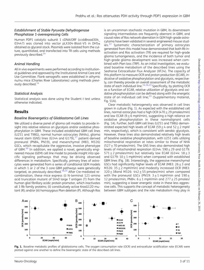
Since baseline cellular metabolism demonstrated a limited ca-pacity in providing insight into tumor-specific metabolism, weextended our investigation to further characterize the respira-tory phenotype of these lines by defining their mitochondrial re-serve capacities. As the above studies suggested, Ras signaling(TR and TRP) attenuated baseline OCR, and these specific lineswere associated with the aggressive phenotype of GBM; initialinvestigations were performed using the isogenic T/R/P model.These studies were performed by metabolically perturbing indi-vidual lines by adding specific compounds in succession andthereby shifting the bioenergetic profile of the cells, which rep-resents a methodology derived from established approachesfor studying mitochondria.15 In these studies, cells were firsttreated with the ATP synthase inhibitor oligomycin, thereby de-creasing OCR associated with ATP synthesis. Next, cells were ex-posed to FCCP, which is an uncoupling agent that dissipates themitochondrial membrane potential and leads to rapid andmaximal OCR without generating ATP. The spare respiratory ca-pacity (SRC) of a cell line is defined as the quantitative differ-ence between the maximal uncontrolled OCR and initial basalOCR, as described in Supplementary material, Fig. S1B. Cellswere subsequently exposed to the mitochondrial inhibitors ro-tenone and antimycin A, thereby, shutting down mitochondrialrespiration and allowing calculation of the mitochondrial andnonmitochondrial fractions contributing to respiration. Boththe Rb mutated T and TP lines, which again typically do notdevelop high-grade gliomas in vivo,19,21 demonstrated similarrespiratory phenotypes with significant increases in OCR follow-ing decoupling that resulted in SRCs of 120%+17% and 86%+11%, respectively. However, the addition of a mutation in Ras,demonstrated in both TR and TRP lines, exhibited attenuatedmaximal OCR following decoupling that only returned back tobaseline levels and resulted in diminished SRC (TR¼ 8%+0.4% and TRP¼ 11%+0.4%; Fig. 2A and B). This pattern wasconsistent when tested in our other model systems. For exam-ple, the respiratory phenotype of normal astrocytes was similarto the T and TP lines and resulted in a significant SRC of 106%+8% (Fig. 2C and D). However, both the established GBM linesand invasive GNS lines demonstrated an attenuated SRC similarto the TRP and TR lines (Fig. 2E and F). This concept was furthersupported when we compared the respiratory phenotype
between GSC subtypes. The proneural GSCs, which demonstrat-ed slow growth in vivo with minimal angiogenesis and necro-sis,20 demonstrated a mitochondrial phenotype consistentwith normal astrocytes (ie, SRC of 112%+3% and 114%+8% in PN19 and PN84 cells, respectively). The rapidly growing,invasive mesenchymal GSC lines, however, demonstrated anattenuated SRC (Supplementary material, Fig. S2A and B). Col-lectively, these findings suggest that an attenuated SRC mayrepresent a metabolic phenotype of GBM and contribute to-wards its aggressive biology. We extended our investigationsto determine if mitochondrial potential could serve as anothertool for determining cell-autonomous metabolic heterogeneity.Despite clear differences in metabolism between T and TR cells,no appreciable differences were observed in how this translatedto steady-state mitochondrial function (as measured by mem-brane potential) (Supplementary material, Fig. S3). This sug-gests that many factors may influence a cell’s energyrequirements and that baseline evaluation of metabolism invitro may be limited for elucidating how they translate into itsmetabolic phenotype.
Ras-mediated Signaling Actively Modulates SpareRespiratory Capacity
Next, we investigated the potential mechanism underlying theobserved changes in respiratory phenotype. As in the initial in-vestigation, we determined if observed changes in reserve ca-pacity were hardwired in the mitochondria or if they could bemodulated through cell signaling. Mitochondria were isolatedfrom both U251, which demonstrated an attenuated SRC,and normal astrocytes by using methods previously de-scribed.14 Interestingly, SRC was identical in isolated mitochon-dria in these lines, suggesting that the observed changes inmitochondrial phenotype may be actively modulated throughcellular signaling (Supplementary material, Fig. S2C). Sinceour GEM lines demonstrated that Ras-pathway activation con-tributed to the observed attenuated SRC, we next sought todetermine if this respiratory phenotype could be reversedthrough signaling inhibition. To test this, we exposed TR cellsto the MEK inhibitor AZD6244 and the Akt inhibitor MK2206,which represent downstream mediators of 2 of the most ex-tensively studied effectors of Ras signaling (ie, Raf and PI3K, re-spectively24) with initial studies confirming target engagement(Supplementary material, Fig. S2D). In the TR line, baseline OCRand OCR remained unchanged following exposure to the ATPsynthase inhibitor oligomycin when compared with vehicle con-trol following 24 hour pretreatment with each agent (Fig. 3A).However, both Akt and MEK inhibition independently demon-strated the capacity to reverse the previously described atten-uated respiratory capacity following decoupling to determinemaximal OCR and SRC, resulting in 13%+0.1% and 28%+0.4% increases in reserve capacities, respectively (Fig. 3A andB). Interestingly, combining Akt and MEK inhibition led to a fur-ther increase in SRC (38%+0.5%), suggesting redundancy inthe ability of signaling pathways to influence the mitochondrialphenotype. Similarly, MEK and/or Akt inhibition reverted the at-tenuated oxidative reserve capacity in U251 and G179 cells, al-though their relative reliance on each pathway appearedcell-line dependent (Supplementary material, Fig. S4A–D). Col-lectively, these findings suggest that Ras-mediated signaling
Prabhu et al.: Ras attenuates PDH activity through PDP1 expression in GBM
4 of 11 Neuro-Oncology
at Ohio State U
niversity Prior Health Sciences L
ibrary on February 26, 2015http://neuro-oncology.oxfordjournals.org/
Dow
nloaded from
actively modulates SRC. We went on to determine if baselinelevels of Akt or MEK activation could be used to determine acell’s SRC and/or differential response to pathway inhibition.Unfortunately, this line of investigation failed to provide furtherinsight into our findings (Supplementray material, Fig. S5). Forexample, in the isogenic TRP model, Ras mutation increasedAkt activation, while ERK remained relatively unchanged,which supported this line’s increased metabolic response to
Akt inhibition. U251 cells demonstrated relatively low Akt acti-vation, supporting its minimal response to Akt inhibition whencompared with MEK; however, these MEK responsive cells hadless ERK activity when compared with the other models. Fur-ther, both PN lines, which are less aggressive and haveincreased SRC when compared with mesenchymal lines, dem-onstrated relatively high levels of both Akt and MEK activation.Collectively, these findings suggest that the downstream
Fig. 2. Glioblastoma cells demonstrate an attenuated spare respiratory capacity. (A, C and E) The respiratory phenotype was defined in thedescribed cell lines using established methods to study mitochondrial function that involved sequentially exposing cells to oligomycin (O),carbonyl cyanide-4-(trifluoromethoxy)phenylhydrazone (FCCP) (F), and rotenone/antimycin (R/A) in real-time on the Seahorse platform. (B, Dand F) The relatively spare respiratory capacity (%) was calculated as (oxygen consumption rate [OCR] following FCCP – baseline OCR)/baselineOCR. * P , .01.
Prabhu et al.: Ras attenuates PDH activity through PDP1 expression in GBM
Neuro-Oncology 5 of 11
at Ohio State U
niversity Prior Health Sciences L
ibrary on February 26, 2015http://neuro-oncology.oxfordjournals.org/
Dow
nloaded from

sequelae of pathway activation likely involves a complex signal-ing network that eventually translates to their metabolic con-sequences and reiterates the difficulty in identifying functional”signaling pathways in a given tumor.
Ras-mediated Signaling Modulates Oxygen ConsumptionRate Through Inhibition of Pyruvate DehydrogenaseActivity
Next, we investigated potential mechanistic underpinnings ofthe observed Ras signaling-mediated regulation of OCR. SincePDH represents a key gatekeeping enzyme that regulates
glycolytic flux into the mitochondria, we hypothesized thatoncogenic signaling associated with Ras pathway activation in-hibited enzyme activity, thereby actively attenuating mitochon-drial flux and maximal OCR. Although the role of PDH inmodulating glycolytic flux and baseline oxygen consumptionhas been established, its capacity to regulate maximal OCR,and thereby a cell’s mitochondrial SRC, has yet to be explored.As an initial investigation to determine if changes in PDH activ-ity could modulate SRC, we evaluated OCR in cells treated withthe metabolic modulator dichloroacetate (DCA). DCA has beenpreviously shown to inhibit PDH kinase (PDK), which is anendogenous inhibitor of PDH. By inhibiting PDK, DCA in turnactivates PDH, allowing for increased glycolytic flux into the
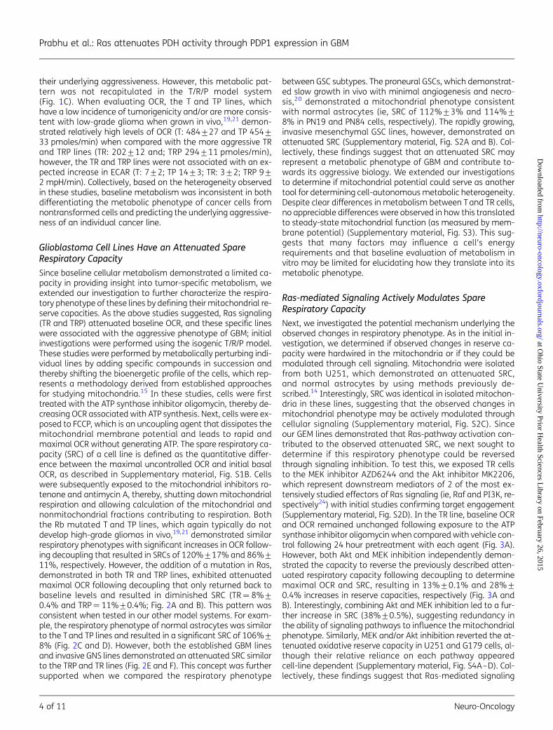
Fig. 3. Ras-mediated signaling modulates oxygen consumption rate (OCR) and spare respiratory capacity through inhibition of pyruvatedehydrogenase (PDH) activity. (A and B) The respiratory phenotype was defined in TR cells exposed to either vehicle control or the describedagents 24 hours prior to analysis. To study mitochondrial function, cells were sequentially exposed to oligomycin (O), carbonyl cyanide-4-(trifluoromethoxy)phenylhydrazone (FCCP) (F), and rotenone/antimycin (R/A) in real time. The relative spare respiratory capacity (%) wascalculated as (OCR following FCCP – baseline OCR)/baseline OCR. (C) PDH enzyme activity in the described cell lines and treatment conditionsusing a microplate assay kit. (D) Western blot was performed to determine levels of phosphorylated PDH (pPDH) in the described cell lines.Levels were quantified by determining the pPDH/PDH ratio for each line and then normalized to the ratio of T cells. (E) Western blot wasperformed to determine levels of pPDH in TR cells exposed to vehicle control or the stated agents. Levels were quantified by determining thepPDH/PDH ratio for each treatment condition and then normalizing to the ratio of vehicle control cells. *P , .05.
Prabhu et al.: Ras attenuates PDH activity through PDP1 expression in GBM
6 of 11 Neuro-Oncology
at Ohio State U
niversity Prior Health Sciences L
ibrary on February 26, 2015http://neuro-oncology.oxfordjournals.org/
Dow
nloaded from
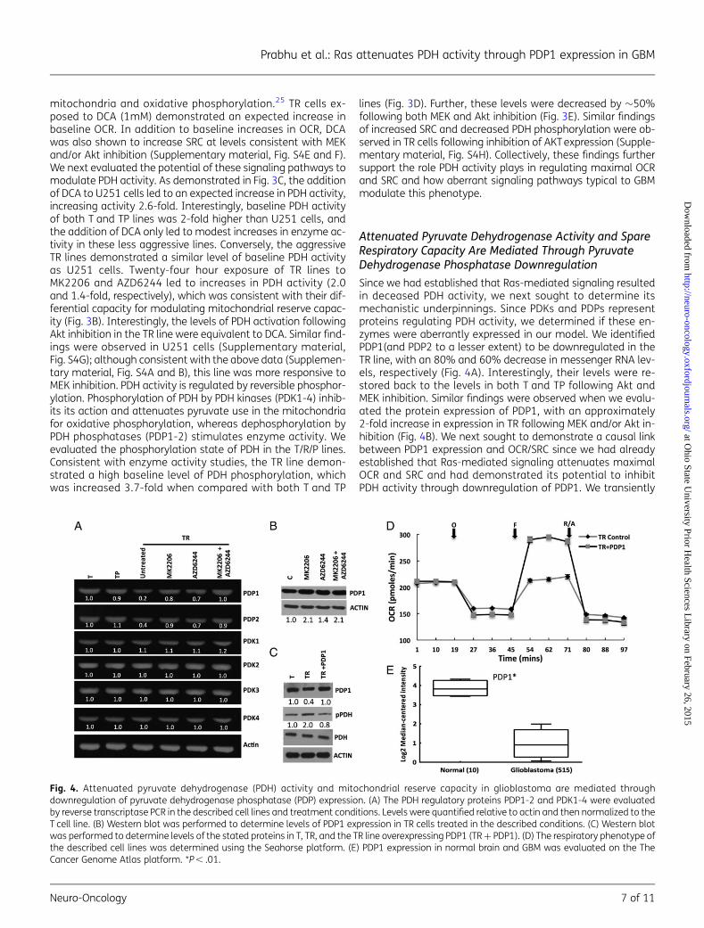
mitochondria and oxidative phosphorylation.25 TR cells ex-posed to DCA (1mM) demonstrated an expected increase inbaseline OCR. In addition to baseline increases in OCR, DCAwas also shown to increase SRC at levels consistent with MEKand/or Akt inhibition (Supplementary material, Fig. S4E and F).We next evaluated the potential of these signaling pathways tomodulate PDH activity. As demonstrated in Fig. 3C, the additionof DCA to U251 cells led to an expected increase in PDH activity,increasing activity 2.6-fold. Interestingly, baseline PDH activityof both T and TP lines was 2-fold higher than U251 cells, andthe addition of DCA only led to modest increases in enzyme ac-tivity in these less aggressive lines. Conversely, the aggressiveTR lines demonstrated a similar level of baseline PDH activityas U251 cells. Twenty-four hour exposure of TR lines toMK2206 and AZD6244 led to increases in PDH activity (2.0and 1.4-fold, respectively), which was consistent with their dif-ferential capacity for modulating mitochondrial reserve capac-ity (Fig. 3B). Interestingly, the levels of PDH activation followingAkt inhibition in the TR line were equivalent to DCA. Similar find-ings were observed in U251 cells (Supplementary material,Fig. S4G); although consistent with the above data (Supplemen-tary material, Fig. S4A and B), this line was more responsive toMEK inhibition. PDH activity is regulated by reversible phosphor-ylation. Phosphorylation of PDH by PDH kinases (PDK1-4) inhib-its its action and attenuates pyruvate use in the mitochondriafor oxidative phosphorylation, whereas dephosphorylation byPDH phosphatases (PDP1-2) stimulates enzyme activity. Weevaluated the phosphorylation state of PDH in the T/R/P lines.Consistent with enzyme activity studies, the TR line demon-strated a high baseline level of PDH phosphorylation, whichwas increased 3.7-fold when compared with both T and TP
lines (Fig. 3D). Further, these levels were decreased by �50%following both MEK and Akt inhibition (Fig. 3E). Similar findingsof increased SRC and decreased PDH phosphorylation were ob-served in TR cells following inhibition of AKT expression (Supple-mentary material, Fig. S4H). Collectively, these findings furthersupport the role PDH activity plays in regulating maximal OCRand SRC and how aberrant signaling pathways typical to GBMmodulate this phenotype.
Attenuated Pyruvate Dehydrogenase Activity and SpareRespiratory Capacity Are Mediated Through PyruvateDehydrogenase Phosphatase Downregulation
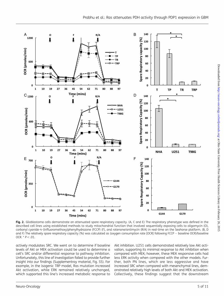
Since we had established that Ras-mediated signaling resultedin deceased PDH activity, we next sought to determine itsmechanistic underpinnings. Since PDKs and PDPs representproteins regulating PDH activity, we determined if these en-zymes were aberrantly expressed in our model. We identifiedPDP1(and PDP2 to a lesser extent) to be downregulated in theTR line, with an 80% and 60% decrease in messenger RNA lev-els, respectively (Fig. 4A). Interestingly, their levels were re-stored back to the levels in both T and TP following Akt andMEK inhibition. Similar findings were observed when we evalu-ated the protein expression of PDP1, with an approximately2-fold increase in expression in TR following MEK and/or Akt in-hibition (Fig. 4B). We next sought to demonstrate a causal linkbetween PDP1 expression and OCR/SRC since we had alreadyestablished that Ras-mediated signaling attenuates maximalOCR and SRC and had demonstrated its potential to inhibitPDH activity through downregulation of PDP1. We transiently
Fig. 4. Attenuated pyruvate dehydrogenase (PDH) activity and mitochondrial reserve capacity in glioblastoma are mediated throughdownregulation of pyruvate dehydrogenase phosphatase (PDP) expression. (A) The PDH regulatory proteins PDP1-2 and PDK1-4 were evaluatedby reverse transcriptase PCR in the described cell lines and treatment conditions. Levels were quantified relative to actin and then normalized to theT cell line. (B) Western blot was performed to determine levels of PDP1 expression in TR cells treated in the described conditions. (C) Western blotwas performed to determine levels of the stated proteins in T, TR, and the TR line overexpressing PDP1 (TR + PDP1). (D) The respiratory phenotype ofthe described cell lines was determined using the Seahorse platform. (E) PDP1 expression in normal brain and GBM was evaluated on the TheCancer Genome Atlas platform. *P , .01.
Prabhu et al.: Ras attenuates PDH activity through PDP1 expression in GBM
Neuro-Oncology 7 of 11
at Ohio State U
niversity Prior Health Sciences L
ibrary on February 26, 2015http://neuro-oncology.oxfordjournals.org/
Dow
nloaded from

overexpressed PDP1 in TR, resulting in an expected decrease inPDH phosphorylation (Fig. 4C). Next, we directly evaluated therole of PDP1 expression on the cells’ mitochondrial phenotype.Similar to studies in TR cells following MEK and Akt inhibition,transient overexpression of PDP1 did not influence baselinelevels of OCR; it did, however, result in restoration of SRC by in-creasing it 36%+0.6% (Fig. 4D). Since we had already estab-lished the capacity of aberrant signaling pathways typical inGBM to downregulate PDP1 expression, we went on to evaluatefor PDP1 expression in human samples using the TCGA data-base,10 which demonstrated significantly decreased levels ofPDP1 in GBM when compared with normal brain (Fig. 4E) andfurther supported the interplay between Ras-mediated onco-genic signaling in GBM and PDH regulation.
PDP1-mediated Regulation of PyruvateDehydrogenase Activity Contributes ToGlioblastoma Tumorigenesis
To determine if observed changes in PDH activity and SRC in-fluenced tumorigenesis, we generated stable TR lines that over-expressed PDP1. We confirmed PDP1 overexpression in theselines, along with their expected decrease in PDH phosphoryla-tion and increase in SRC (Supplementary material, Fig. S6A–C).We then evaluated the growth kinetics of these lines in vitro. Al-though PDP1-overexpressing clones maintained viability, theygrew at a decreased rate when compared with vector controls(Fig. 5A). Next, we evaluated in vivo growth rate using an s.c.mouse xenograft model. Although a tumor was palpable in TRclones overexpressing PDP1, they failed to grow larger than30 mm3 when compared with vector controls (Fig. 5B and C).
DiscussionIn this report, we have presented several novel findings that in-volve unique metabolic states associated with gliomagenesisand how these processes are modulated through oncogenicsignaling pathways. Initial studies focused on cellular metabo-lism at baseline to define relative metabolism through oxidativephosphorylation and aerobic glycolysis, which collectively sug-gested that baseline metabolism has a limited capacity todetermine the tumorigenicity and/or aggressiveness of agiven cell line when grown in vitro. These results are notcompletely unexpected because the metabolic programsevolved during gliomagenesis are likely a direct consequenceof their need to adapt to the tumor microenvironment, whichis likely not recapitulated in in-vitro conditions devoid of oxygenand nutrient restrictions. Furthermore, it is likely that severalfactors play a role in maintaining an individual cell’s baselineoxidative phosphorylation state including reliance on glycolysisor alternate forms of energy metabolism (eg, glutamine me-tabolism and/or fatty acid oxidation, and baseline levels of ox-idative stress). These factors likely play a role in the observedmetabolic heterogeneity in these lines.
Based on these potential limitations associated with study-ing the metabolic state of cell lines in baseline conditions, weextended our investigations to determine if differential re-sponse to bioenergetic stress were associated with and/or con-tributed towards gliomagenesis. We uncovered an activelyregulated oxidative state involving attenuated mitochondrialSRC associated with gliomagenesis. Our understanding aboutthe potential relevance of SRC, which is defined as the differ-ence between the maximal unregulated respiration and the ini-tial basal respiration of an individual cell line15 in both normal
Fig. 5. Modulation of pyruvate dehydrogenase (PDH) activity through PDP1 expression influences glioblastoma growth. (A) TR vector control cells(TR_VC) and clones overexpressing PDP1 (TR_PDP1_c1/2) were evaluated growth kinetics were evaluated in vitro. (B) Tumor growth curves wereobtained in the described cell lines at stated times in an s.c. mouse xenograft model (n¼ 10/cell line) using perpendicular diameter measurementsof each tumor with digital calipers; volumes were calculated using the formula (LxWxW)/2. (C) A representative image of described cell lines grownin an s.c. mouse xenograft model. *P , .01.
Prabhu et al.: Ras attenuates PDH activity through PDP1 expression in GBM
8 of 11 Neuro-Oncology
at Ohio State U
niversity Prior Health Sciences L
ibrary on February 26, 2015http://neuro-oncology.oxfordjournals.org/
Dow
nloaded from
and cancer cells, is continuing to evolve. It has been suggestedthat reserve capacity is utilized to provide for increased energydemands during stressed conditions. This was demonstrated tobe particularly relevant in cardiac tissue, which is rich in mito-chondria and capable of dynamically responding to increasedbioenergetic demands during stressed conditions.23 In ourstudies, this metabolic adaptation appeared to be maintainedin the nontumorigenic normal lines tested but was consistentlylost in GBM lines. Interestingly, recent studies support this as anemerging tumor-specific metabolic phenotype. Sandulacheet al identified that loss of mitochondrial reserve capacitywas attributed to p53 mutation in squamous cell carcinoma,conferring therapeutic resistance.26 Anso et al identified aloss of SRC following myc-induced malignant transformationof osteocytes to osteogenic sarcoma.27 Collectively, these find-ings, coupled with our results performed on a diverse panel ofGBM lines, suggest that an attenuated SRC represents a meta-bolic shift associated with tumorigenesis. However, the specificbiological advantage(s) offered to tumor cells by this metabolicshift remain unclear. Again, our current understanding of theSRC is that it allows normal cells to adapt to increased bioener-getic demands during periods of short-term stress. However,the GBM microenvironment poses a constant array of stressesto an individual cell. Although vascularized tumors, newlyformed vessels in GBM are tortuous with compromised vascularintegrity. This limits tissue perfusion, resulting in a microenvi-ronment with variable levels of hypoxia and nutrient depriva-tion.28,29 Our findings, which demonstrate the capacity ofsignaling pathways to attenuate maximal OCR and SRC, maybe particularly relevant in the context of intermittent hypoxia,30
as we hypothesize that it may be disadvantageous for cancercells to rely on compensatory increases in OCR long-term; thiscould potentially lead to increased generation of reactive oxy-gen species and cellular damage associated with increased ox-idative phosphorylation.31,32
Next, we demonstrated that the observed changes in SRCwere a direct consequence of PDH activity. Although the centralregulatory role PDH plays in baseline oxidative phosphorylationthrough modulation of glycolytic flux into the mitochondria iswell established, the secondary role of this gatekeeping en-zyme has yet to be described. Further, its tight regulation by on-cogenic signaling reinforces its potential implications intumorigenesis, contributing to tumorigenesis. We proceededto mechanistically link changes in PDH activity and SRC withRas pathway activation and demonstrated the ability to restoreSRC through targeted inhibition of its downstream effectors.These findings add to the growing body of data demonstratingthe capacity of Ras signaling for modulating metabolic pro-grams at multiple levels in cancer. Chiaradonna et al demon-strated the role of oncogenic Ras in enhancing utilization ofthe glycolytic pathway,33 and Ying et al identified the potentialof Ras to control glycolysis at several rate-limiting steps, whichled to shunting of glucose metabolism towards anabolic me-tabolism.8 Our data provide further insight into the dynamicrole this oncogene plays in tumor metabolism. Interestingly,we demonstrated that multiple signaling pathways have thecapacity to modulate PDP1 expression. Redundant signalingpathways are commonly observed in cancer and allow cellsto maintain activity of key downstream circuits in the presenceof altered upstream ligand-dependent signaling and/or to
dynamically acquire resistance to targeted inhibition of a singlepathway. Further, the redundancy of AKT and MEK/ERK path-ways has been described previously.34,35 Therefore, the ob-served redundancy of these pathways further underscoresthe potential biological importance of PDH regulation in con-tributing to the aggressive phenotype of GBM and the potentialtherapeutic limitations of targeting these upstream pathwaysindividually.
Lastly, we demonstrated that Ras-mediated regulation ofPDH activity and mitochondrial function was mediated throughthe capacity of this pathway to attenuate expression of PDHregulatory proteins PDPs. The potential for oncogenic signalingtypical of GBM to downmodulate expression of PDPs was vali-dated in patient samples, with PDP1 expression being signifi-cantly repressed in GBM compared with normal brain tissuewhen evaluated from the TCGA dataset. More importantly, re-storing expression of PDP1 in our model diminished tumorige-nicity. Targeting the PDH/PDP1 axis may also have application inGSCs, which studies have shown to have a unique metabolicstate when compared with differentiated cells.16 Althoughthe multiple roles Ras signaling plays in tumorigenesis arewell established, our findings demonstrated that targetingthis single downstream consequence of pathway activationcan completely revert tumorigenic potential of this oncopro-tein. These findings are consistent with emerging data reinforc-ing the central role played by alterations of PDH activity in theoncogenic transformation following genetic mutation. Recentdiscoveries involving the mechanistic underpinnings ofoncogene-induced senescence represent one of the most nota-ble examples describing this important interplay between adriver mutation and its functional, metabolic consequence.36
In these studies, it was shown that PDH was a crucial mediatorof senescence induced by BRAFV600E in normal melanocytes,leading to activation of PDH through suppression of PDK1 andinduction of PDP2. This resulted in increased cellular respirationand subsequent senescence secondary to redox stress. Abro-gating this mutation-induced metabolic program led to onco-genic transformation and, similar to our findings, restoringPDH activity diminished tumorigenic potential. Our model dem-onstrated that Ras signaling can modulate PDH activity solelythrough regulation of PDPs without requiring alterations inPDK1. These finding are consistent with TCGA data, which dem-onstrated only aberrant expression of PDP1 in clinical samples.This suggests a complex interplay between a specific mutationand an individual cell’s capacity to regulate PDH activitythrough a balance between PDH activators and inhibitors thatrequire further investigation. In addition, further work is still re-quired to better understand the mechanisms contributing tothe differential metabolic response of normal cells and prema-lignant and/or tumor cells to a specific genetic mutation. Thesestudies support the functional consequence of alterations inPDH activity playing a central event driving tumorigenesis.The therapeutic potential of these findings shows strong prom-ise and is being actively investigated, including recent workdemonstrating antitumor activity of the PDH activator DCA inGBM.25
In summary, using a diverse panel of GBM and normal lines,we demonstrated that an attenuated SRC was a common met-abolic phenotype of GBM. We established that this phenotypewas driven by signaling pathways typical to GBM and was a
Prabhu et al.: Ras attenuates PDH activity through PDP1 expression in GBM
Neuro-Oncology 9 of 11
at Ohio State U
niversity Prior Health Sciences L
ibrary on February 26, 2015http://neuro-oncology.oxfordjournals.org/
Dow
nloaded from
consequence of their capacity to inhibit PDH activity by attenu-ating expression PDP1 and normalizing this metabolic pro-gram’s diminished tumorigenic potential. These findingsprovide further support for the central role that PDH regulationplays in tumorigenesis and the scientific rationale to continueto develop and test clinical strategies designed to activatePDH as a form of anticancer therapy in GBM.
Supplementary MaterialSupplementary material is available online at Neuro-Oncology(http://neuro-oncology.oxfordjournals.org/).
FundingUS Army Medical Research and Materiel Command, National FunctionalGenomics Center project, under award number W81XWH-08-2-0101(P.C.), The American Cancer Society (RSG-11-029-01-CSM; P.C.), FloridaDepartment of Health, Bankhead-Coley Cancer Research Program(4BB03; P.C.), and the Southeastern Brain Tumor Foundation (P.C.).
Conflict of interest statement. None declared.
References1. Warburg O. On respiratory impairment in cancer cells. Science.
1956;124(3215):269–270.
2. Warburg O, Posener K, Negelein E. Uber den Stoffwechsel derCarcinomzelle. Biochem Zeitschr. 1924;152:309–344.
3. Marin-Valencia I, Yang C, Mashimo T, et al. Analysis of tumormetabolism reveals mitochondrial glucose oxidation ingenetically diverse human glioblastomas in the mouse brain invivo. Cell Metab. 2012;15(6):827–837.
4. Moreno-Sanchez R, Rodriguez-Enriquez S, Marin-Hernandez A,Saavedra E Energy metabolism in tumor cells. FEBS J. 2007;274(6):1393–1418.
5. Chinnaiyan P, Kensicki E, Bloom G, et al. The metabolomicsignature of malignant glioma reflects accelerated anabolicmetabolism. Cancer Res. 2012;72(22):5878–5888.
6. Locasale JW, Grassian AR, Melman T, et al. Phosphoglyceratedehydrogenase diverts glycolytic flux and contributes tooncogenesis. Nat Genet. 2011;43(9):869–874.
7. Possemato R, Marks KM, Shaul YD, et al. Functional genomicsreveal that the serine synthesis pathway is essential in breastcancer. Nature. 2011;476(7360):346–350.
8. Ying H, Kimmelman AC, Lyssiotis CA, et al. Oncogenic Krasmaintains pancreatic tumors through regulation of anabolicglucose metabolism. Cell. 2012;149(3):656–670.
9. Stupp R, Mason WP, van den Bent MJ, et al. Radiotherapy plusconcomitant and adjuvant temozolomide for glioblastoma. NEngl J Med. 2005;352(10):987–996.
10. TCGA. Comprehensive genomic characterization defines humanglioblastoma genes and core pathways. Nature. 2008;455(7216):1061–1068.
11. Chakravarti A, Zhai G, Suzuki Y, et al. The prognostic significance ofphosphatidylinositol 3-kinase pathway activation in humangliomas. J Clin Oncol. 2004;22(10):1926–1933.
12. Lopez-Gines C, Gil-Benso R, Benito R, et al. The activation of ERK1/2MAP kinases in glioblastoma pathobiology and its relationshipwith EGFR amplification. Neuropathology. 2008;28(5):507–515.
13. Nicoletti NF, Erig TC, Zanin RF, et al. Mechanisms involved inkinin-induced glioma cells proliferation: the role of ERK1/2 andPI3K/Akt pathways. J Neurooncol. 2014;120(2):235–244.
14. Prabhu A, Sarcar B, Kahali S, et al. Cysteine catabolism: a novelmetabolic pathway contributing to glioblastoma growth. CancerRes. 2014;74(3):787–796.
15. Choi SW, Gerencser AA, Nicholls DG. Bioenergetic analysis ofisolated cerebrocortical nerve terminals on a microgram scale:spare respiratory capacity and stochastic mitochondrial failure. JNeurochem. 2009;109(4):1179–1191.
16. Vlashi E, Lagadec C, Vergnes L, et al. Metabolic state of gliomastem cells and nontumorigenic cells. Proc Natl Acad Sci USA.2011;108(38):16062–16067.
17. Pollard SM, Yoshikawa K, Clarke ID, et al. Glioma stem cell linesexpanded in adherent culture have tumor-specific phenotypesand are suitable for chemical and genetic screens. Cell StemCell. 2009;4(6):568–580.
18. Prabhu A, Sarcar B, Kahali S, et al. Targeting the unfolded proteinresponse in glioblastoma cells with the fusion protein EGF-SubA.PLoS One. 2012;7(12):e52265.
19. Vitucci M, Karpinich NO, Bash RE, et al. Cooperativity betweenMAPK and PI3K signaling activation is required for glioblastomapathogenesis. Neuro Oncol. 2013;15(10):1317–1329.
20. Mao P, Joshi K, Li J, et al. Mesenchymal glioma stem cells aremaintained by activated glycolytic metabolism involvingaldehyde dehydrogenase 1A3. Proc Natl Acad Sci USA. 2013;110(21):8644–8649.
21. Song Y, Zhang Q, Kutlu B, et al. Evolutionary etiology of high-gradeastrocytomas. Proc Natl Acad Sci USA. 2013;110(44):17933–17938.
22. Guha A, Feldkamp MM, Lau N, et al. Proliferation of humanmalignant astrocytomas is dependent on Ras activation.Oncogene. 1997;15(23):2755–2765.
23. Hill BG, Dranka BP, Zou L, et al. Importance of the bioenergeticreserve capacity in response to cardiomyocyte stress induced by4-hydroxynonenal. Biochem J. 2009;424(1):99–107.
24. Downward J. Targeting RAS signalling pathways in cancer therapy.Nat Rev Cancer. 2003;3(1):11–22.
25. Michelakis ED, Sutendra G, Dromparis P, et al. Metabolicmodulation of glioblastoma with dichloroacetate. Sci Transl Med.2010;2(31):31ra34.
26. Sandulache VC, Skinner HD, Ow TJ, et al. Individualizingantimetabolic treatment strategies for head and necksquamous cell carcinoma based on TP53 mutational status.Cancer. 2012;118(3):711–721.
27. Anso E, Mullen A, Felsher D, et al. Metabolic changes in cancer cellsupon suppression of MYC. Cancer Metab. 2013;1(1):7.
28. Jain RK. Molecular regulation of vessel maturation. Nat Med. 2003;9(6):685–693.
29. Vartanian A, Singh SK, Agnihotri S, et al. GBM’s multifacetedlandscape: highlighting regional and microenvironmentalheterogeneity. Neuro Oncol. 2014;16(9):1167–1175.
30. Dewhirst MW. Intermittent hypoxia furthers the rationale forhypoxia-inducible factor-1 targeting. Cancer Res. 2007;67(3):854–855.
Prabhu et al.: Ras attenuates PDH activity through PDP1 expression in GBM
10 of 11 Neuro-Oncology
at Ohio State U
niversity Prior Health Sciences L
ibrary on February 26, 2015http://neuro-oncology.oxfordjournals.org/
Dow
nloaded from
31. Brand KA, Hermfisse U. Aerobic glycolysis by proliferating cells: aprotective strategy against reactive oxygen species. FASEB J.1997;11(5):388–395.
32. Sohal RS, Weindruch R. Oxidative stress, caloric restriction, andaging. Science. 1996;273(5271):59–63.
33. Chiaradonna F, Sacco E, Manzoni R, et al. Ras-dependent carbonmetabolism and transformation in mouse fibroblasts. Oncogene.2006;25(39):5391–5404.
34. Cairns RA, Harris IS, Mak TW. Regulation of cancer cell metabolism.Nat Rev Cancer. 2011;11(2):85–95.
35. Logue JS, Morrison DK. Complexity in the signaling network:insights from the use of targeted inhibitors in cancer therapy.Genes Dev. 2012;26(7):641–650.
36. Kaplon J, Zheng L, Meissl K, et al. A key role for mitochondrialgatekeeper pyruvate dehydrogenase in oncogene-inducedsenescence. Nature. 2013;498(7452):109–112.
Prabhu et al.: Ras attenuates PDH activity through PDP1 expression in GBM
Neuro-Oncology 11 of 11
at Ohio State U
niversity Prior Health Sciences L
ibrary on February 26, 2015http://neuro-oncology.oxfordjournals.org/
Dow
nloaded from
Related Documents