RAPID REVIEW BOOK

Welcome message from author
This document is posted to help you gain knowledge. Please leave a comment to let me know what you think about it! Share it to your friends and learn new things together.
Transcript
RAPID REVIEW BOOK

The USMLE Step 1 BIBLE™
Copyright © 2012 USMLE Success Academy. All right reserved. No part of this book may be used or reproduced in any manner whatsoever without written permission except in the case of reprint in the context of review and personal education.

CONTENTS
CHAPTER 1… GROSS ANATOMY
CHAPTER 2… EMBRYOLOGY
CHAPTER 3… HISTOLOGY
CHAPTER 4… NEUROANATOMY
CHAPTER 5… PHYSIOLOGY
CHAPTER 6… BIOCHEMISTRY
CHAPTER 7… ETHICS
CHAPTER 8… BIOSTATISTICS
CHAPTER 9… PSYCHIATRY
CHAPTER 10… IMMUNOLOGY
CHAPTER 11… MICROBIOLOGY
CHAPTER 12… PHARMACOLOGY
CHAPTER 13… PATHOLOGY
CHAPTER 1
GROSS ANATOMY
Gross Anatomy is a very high-‐yield topic on the USMLE exam. The questions you will encounter will require
recognition and understanding of structures, and the ability to understand and identify their clinical significance.
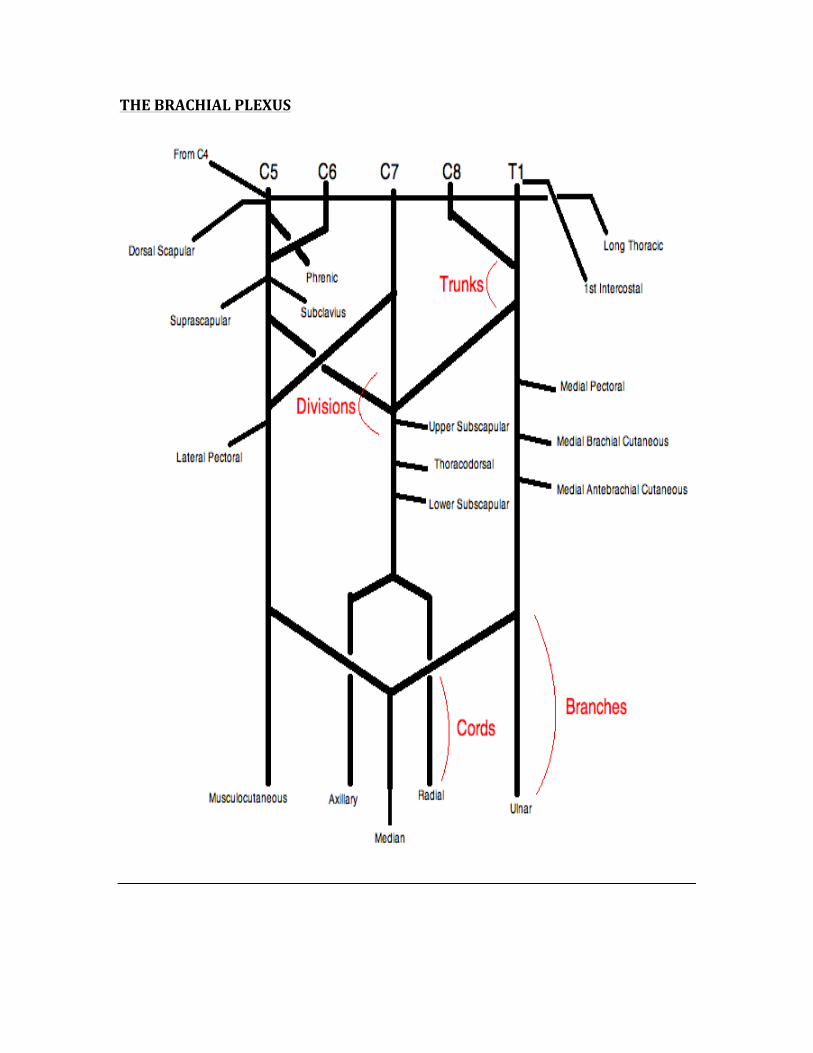
THE BRACHIAL PLEXUS
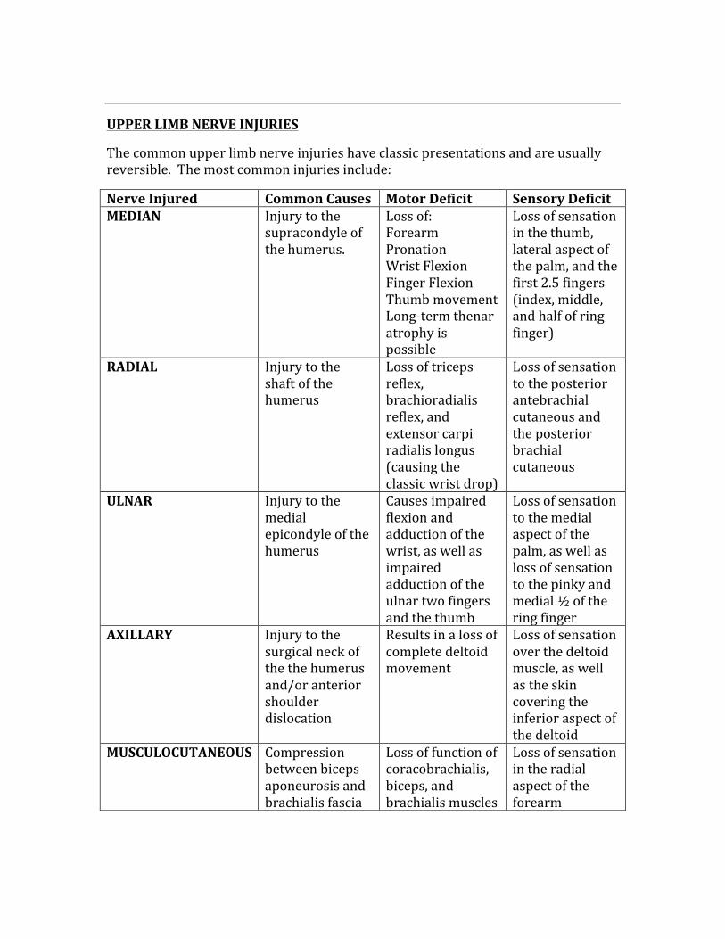
UPPER LIMB NERVE INJURIES
The common upper limb nerve injuries have classic presentations and are usually reversible. The most common injuries include:
Nerve Injured Common Causes Motor Deficit Sensory Deficit MEDIAN Injury to the
supracondyle of the humerus.
Loss of: Forearm Pronation Wrist Flexion Finger Flexion Thumb movement Long-‐term thenar atrophy is possible
Loss of sensation in the thumb, lateral aspect of the palm, and the first 2.5 fingers (index, middle, and half of ring finger)
RADIAL Injury to the shaft of the humerus
Loss of triceps reflex, brachioradialis reflex, and extensor carpi radialis longus (causing the classic wrist drop)
Loss of sensation to the posterior antebrachial cutaneous and the posterior brachial cutaneous
ULNAR Injury to the medial epicondyle of the humerus
Causes impaired flexion and adduction of the wrist, as well as impaired adduction of the ulnar two fingers and the thumb
Loss of sensation to the medial aspect of the palm, as well as loss of sensation to the pinky and medial ½ of the ring finger
AXILLARY Injury to the surgical neck of the the humerus and/or anterior shoulder dislocation
Results in a loss of complete deltoid movement
Loss of sensation over the deltoid muscle, as well as the skin covering the inferior aspect of the deltoid
MUSCULOCUTANEOUS Compression between biceps aponeurosis and brachialis fascia
Loss of function of coracobrachialis, biceps, and brachialis muscles
Loss of sensation in the radial aspect of the forearm
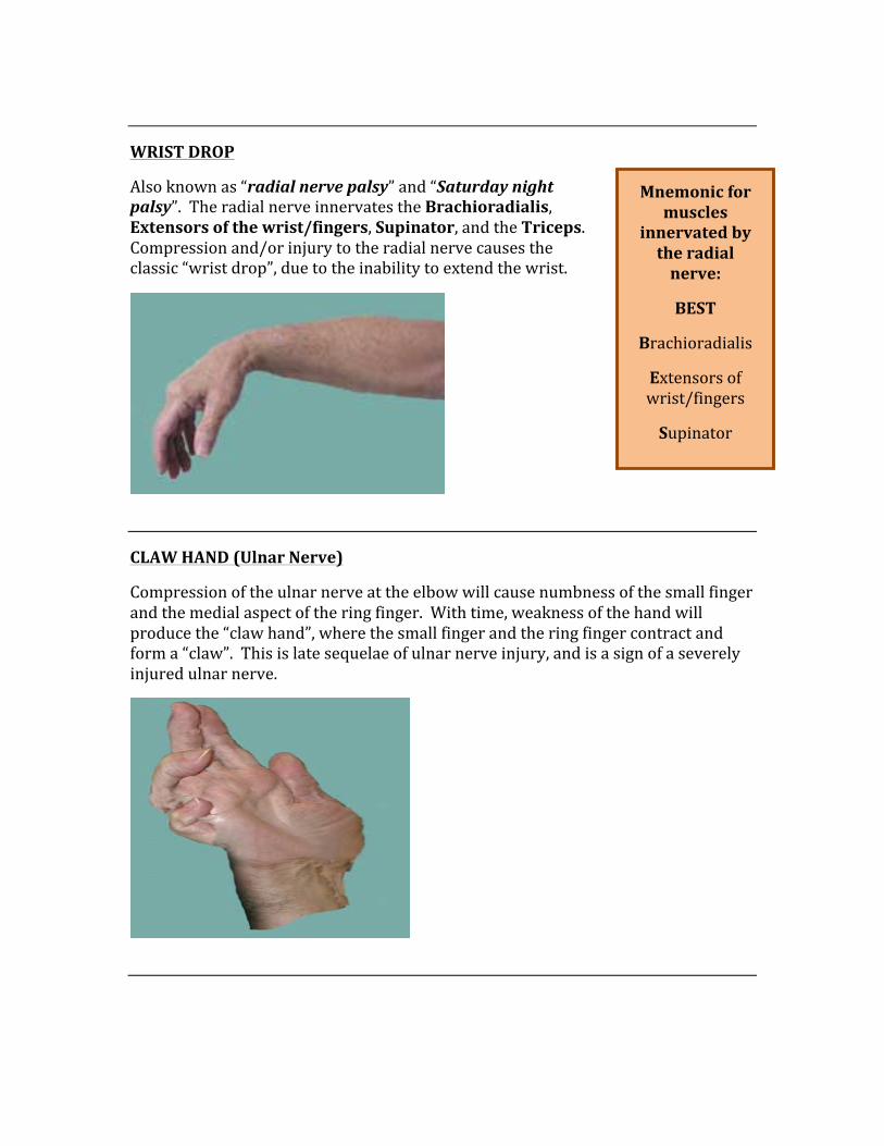
WRIST DROP
Also known as “radial nerve palsy” and “Saturday night palsy”. The radial nerve innervates the Brachioradialis, Extensors of the wrist/fingers, Supinator, and the Triceps. Compression and/or injury to the radial nerve causes the classic “wrist drop”, due to the inability to extend the wrist.
CLAW HAND (Ulnar Nerve)
Compression of the ulnar nerve at the elbow will cause numbness of the small finger and the medial aspect of the ring finger. With time, weakness of the hand will produce the “claw hand”, where the small finger and the ring finger contract and form a “claw”. This is late sequelae of ulnar nerve injury, and is a sign of a severely injured ulnar nerve.
Mnemonic for muscles
innervated by the radial nerve:
BEST
Brachioradialis
Extensors of wrist/fingers
Supinator
Triceps
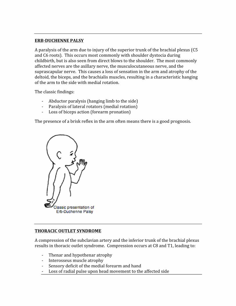
ERB-‐DUCHENNE PALSY
A paralysis of the arm due to injury of the superior trunk of the brachial plexus (C5 and C6 roots). This occurs most commonly with shoulder dystocia during childbirth, but is also seen from direct blows to the shoulder. The most commonly affected nerves are the axillary nerve, the musculocutaneous nerve, and the suprascapular nerve. This causes a loss of sensation in the arm and atrophy of the deltoid, the biceps, and the brachialis muscles, resulting in a characteristic hanging of the arm to the side with medial rotation.
The classic findings:
-‐ Abductor paralysis (hanging limb to the side) -‐ Paralysis of lateral rotators (medial rotation) -‐ Loss of biceps action (forearm pronation)
The presence of a brisk reflex in the arm often means there is a good prognosis.
THORACIC OUTLET SYNDROME
A compression of the subclavian artery and the inferior trunk of the brachial plexus results in thoracic outlet syndrome. Compression occurs at C8 and T1, leading to:
-‐ Thenar and hypothenar atrophy -‐ Interosseus muscle atrophy -‐ Sensory deficit of the medial forearm and hand -‐ Loss of radial pulse upon head movement to the affected side
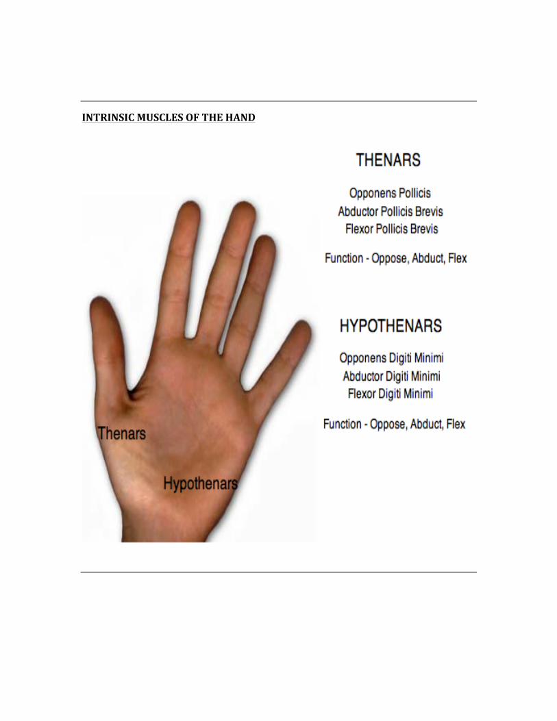
INTRINSIC MUSCLES OF THE HAND
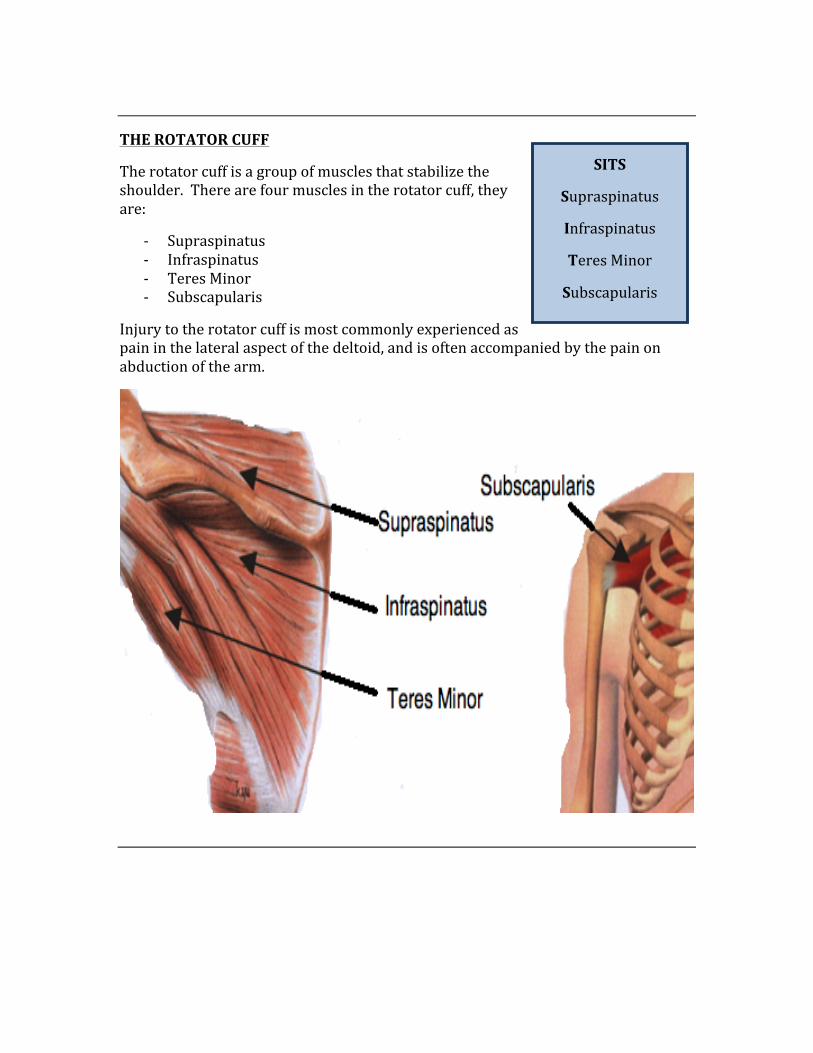
THE ROTATOR CUFF
The rotator cuff is a group of muscles that stabilize the shoulder. There are four muscles in the rotator cuff, they are:
-‐ Supraspinatus -‐ Infraspinatus -‐ Teres Minor -‐ Subscapularis
Injury to the rotator cuff is most commonly experienced as pain in the lateral aspect of the deltoid, and is often accompanied by the pain on abduction of the arm.
SITS
Supraspinatus
Infraspinatus
Teres Minor
Subscapularis
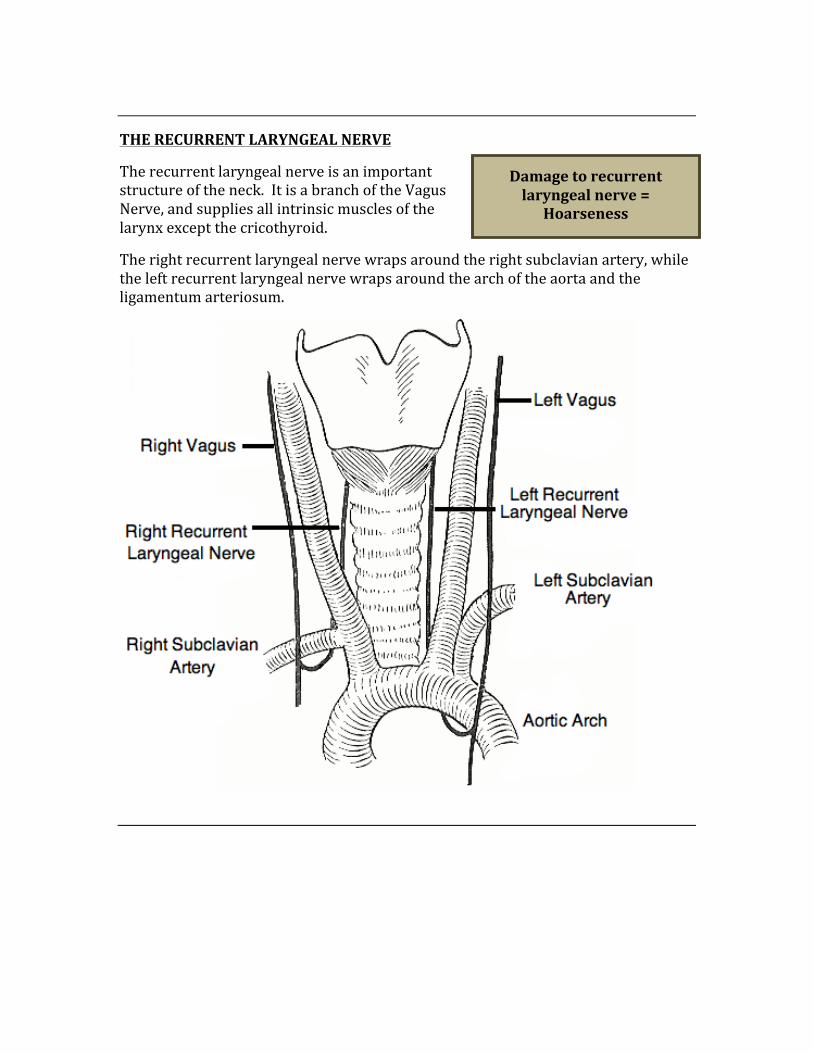
THE RECURRENT LARYNGEAL NERVE
The recurrent laryngeal nerve is an important structure of the neck. It is a branch of the Vagus Nerve, and supplies all intrinsic muscles of the larynx except the cricothyroid.
The right recurrent laryngeal nerve wraps around the right subclavian artery, while the left recurrent laryngeal nerve wraps around the arch of the aorta and the ligamentum arteriosum.
Damage to recurrent laryngeal nerve =
Hoarseness
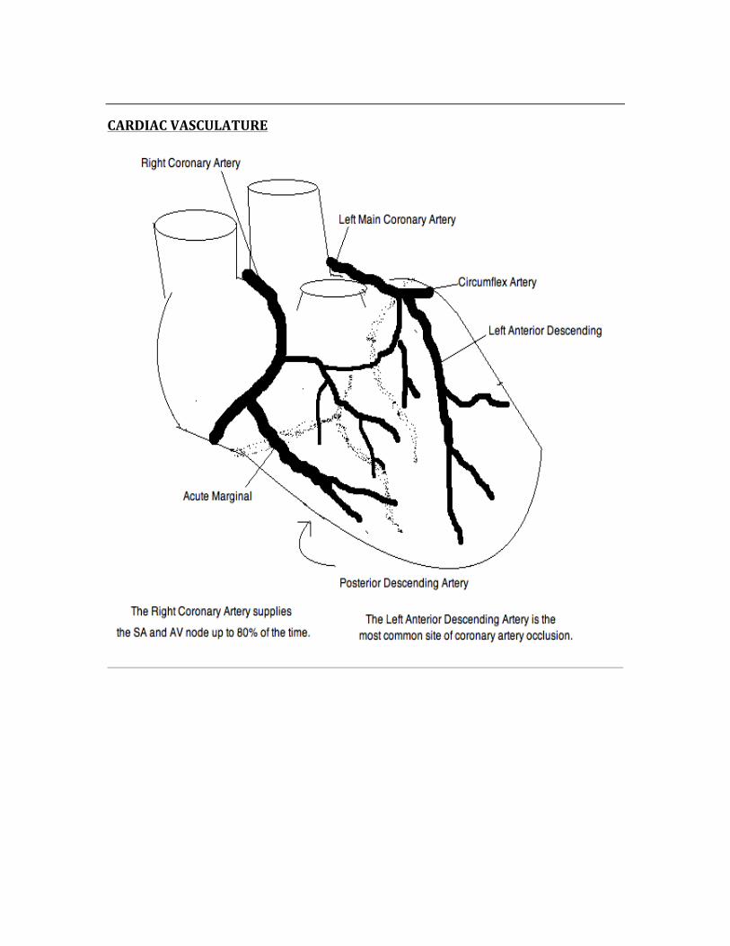
CARDIAC VASCULATURE
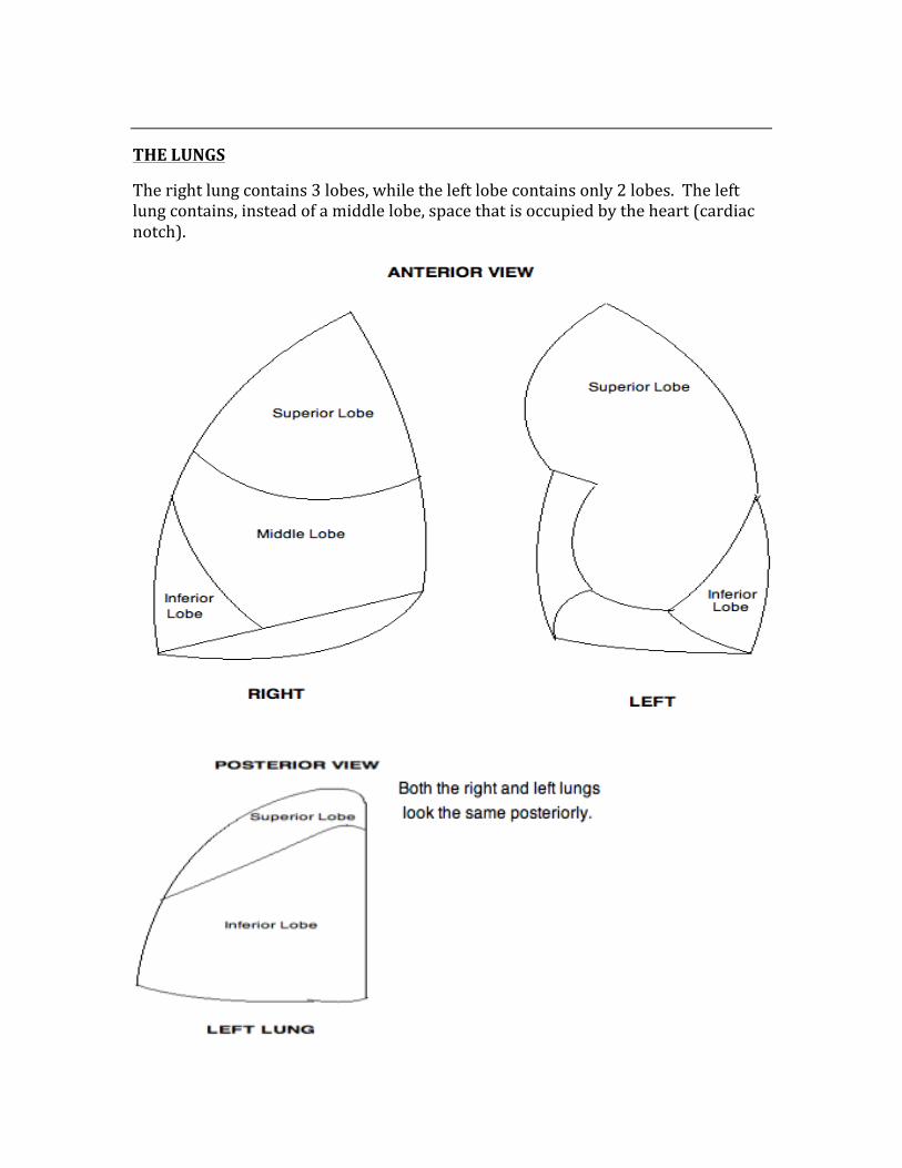
THE LUNGS
The right lung contains 3 lobes, while the left lobe contains only 2 lobes. The left lung contains, instead of a middle lobe, space that is occupied by the heart (cardiac notch).
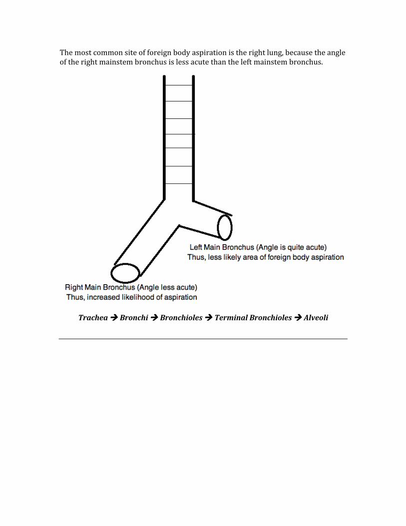
The most common site of foreign body aspiration is the right lung, because the angle of the right mainstem bronchus is less acute than the left mainstem bronchus.
Trachea à Bronchi à Bronchioles à Terminal Bronchioles à Alveoli
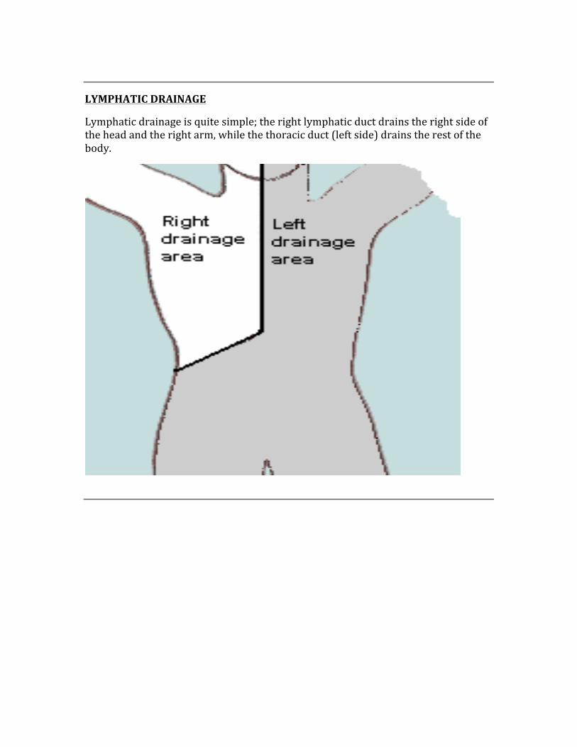
LYMPHATIC DRAINAGE
Lymphatic drainage is quite simple; the right lymphatic duct drains the right side of the head and the right arm, while the thoracic duct (left side) drains the rest of the body.
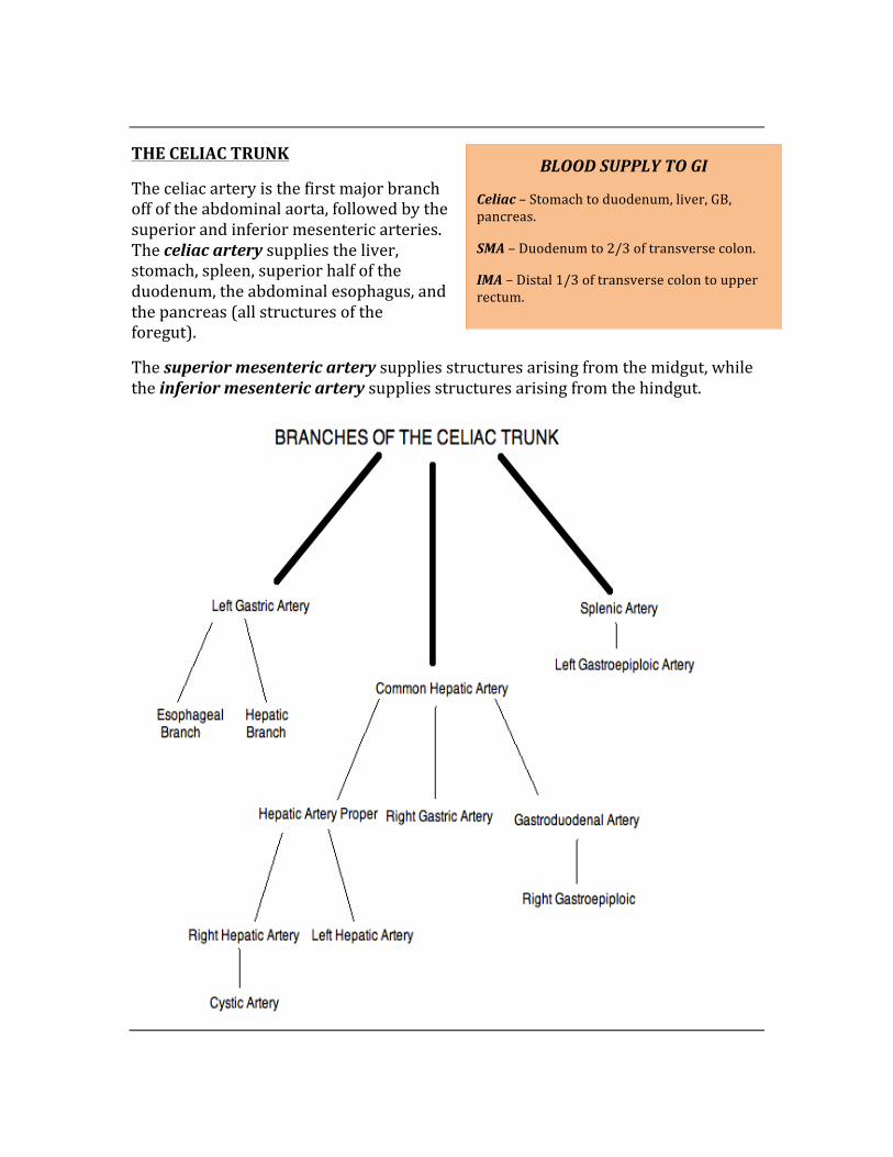
THE CELIAC TRUNK
The celiac artery is the first major branch off of the abdominal aorta, followed by the superior and inferior mesenteric arteries. The celiac artery supplies the liver, stomach, spleen, superior half of the duodenum, the abdominal esophagus, and the pancreas (all structures of the foregut).
The superior mesenteric artery supplies structures arising from the midgut, while the inferior mesenteric artery supplies structures arising from the hindgut.
BLOOD SUPPLY TO GI
Celiac – Stomach to duodenum, liver, GB, pancreas.
SMA – Duodenum to 2/3 of transverse colon.
IMA – Distal 1/3 of transverse colon to upper rectum.
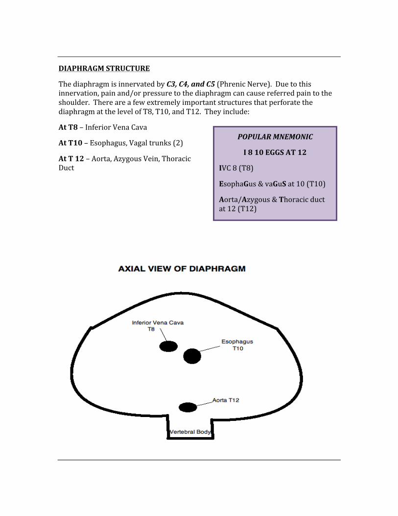
DIAPHRAGM STRUCTURE
The diaphragm is innervated by C3, C4, and C5 (Phrenic Nerve). Due to this innervation, pain and/or pressure to the diaphragm can cause referred pain to the shoulder. There are a few extremely important structures that perforate the diaphragm at the level of T8, T10, and T12. They include:
At T8 – Inferior Vena Cava
At T10 – Esophagus, Vagal trunks (2)
At T 12 – Aorta, Azygous Vein, Thoracic Duct
POPULAR MNEMONIC
I 8 10 EGGS AT 12
IVC 8 (T8)
EsophaGus & vaGuS at 10 (T10)
Aorta/Azygous & Thoracic duct at 12 (T12)
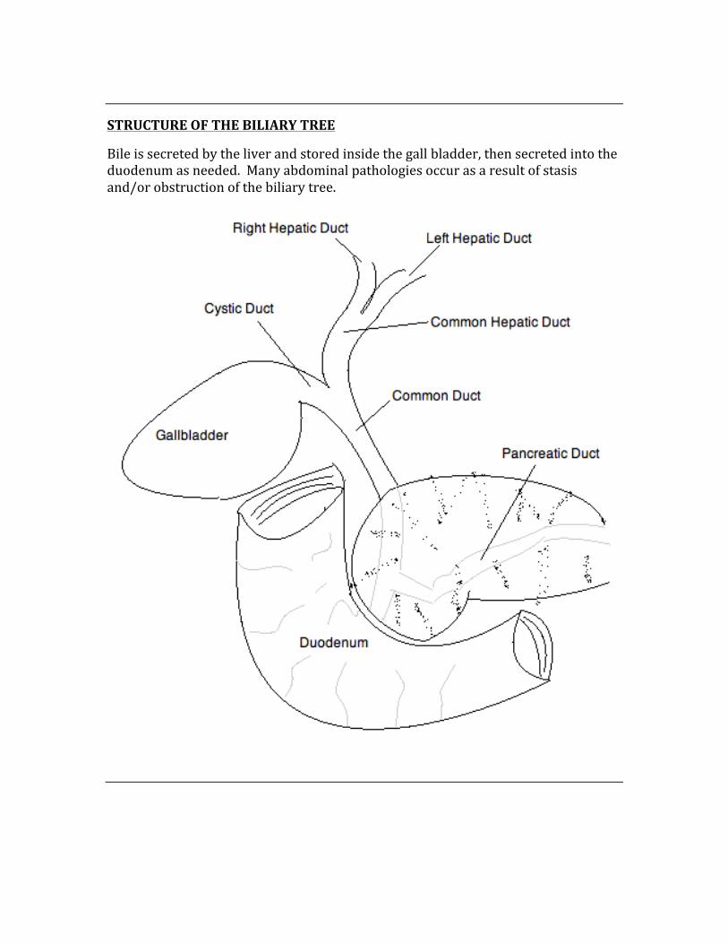
STRUCTURE OF THE BILIARY TREE
Bile is secreted by the liver and stored inside the gall bladder, then secreted into the duodenum as needed. Many abdominal pathologies occur as a result of stasis and/or obstruction of the biliary tree.
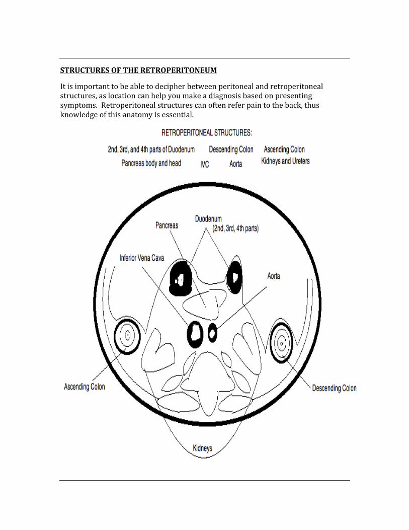
STRUCTURES OF THE RETROPERITONEUM
It is important to be able to decipher between peritoneal and retroperitoneal structures, as location can help you make a diagnosis based on presenting symptoms. Retroperitoneal structures can often refer pain to the back, thus knowledge of this anatomy is essential.
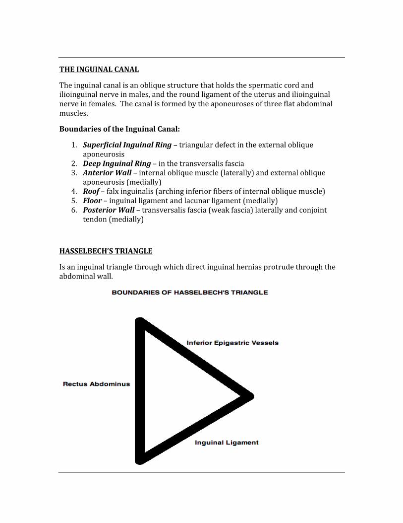
THE INGUINAL CANAL
The inguinal canal is an oblique structure that holds the spermatic cord and ilioinguinal nerve in males, and the round ligament of the uterus and ilioinguinal nerve in females. The canal is formed by the aponeuroses of three flat abdominal muscles.
Boundaries of the Inguinal Canal:
1. Superficial Inguinal Ring – triangular defect in the external oblique aponeurosis
2. Deep Inguinal Ring – in the transversalis fascia 3. Anterior Wall – internal oblique muscle (laterally) and external oblique
aponeurosis (medially) 4. Roof – falx inguinalis (arching inferior fibers of internal oblique muscle) 5. Floor – inguinal ligament and lacunar ligament (medially) 6. Posterior Wall – transversalis fascia (weak fascia) laterally and conjoint
tendon (medially)
HASSELBECH’S TRIANGLE
Is an inguinal triangle through which direct inguinal hernias protrude through the abdominal wall.
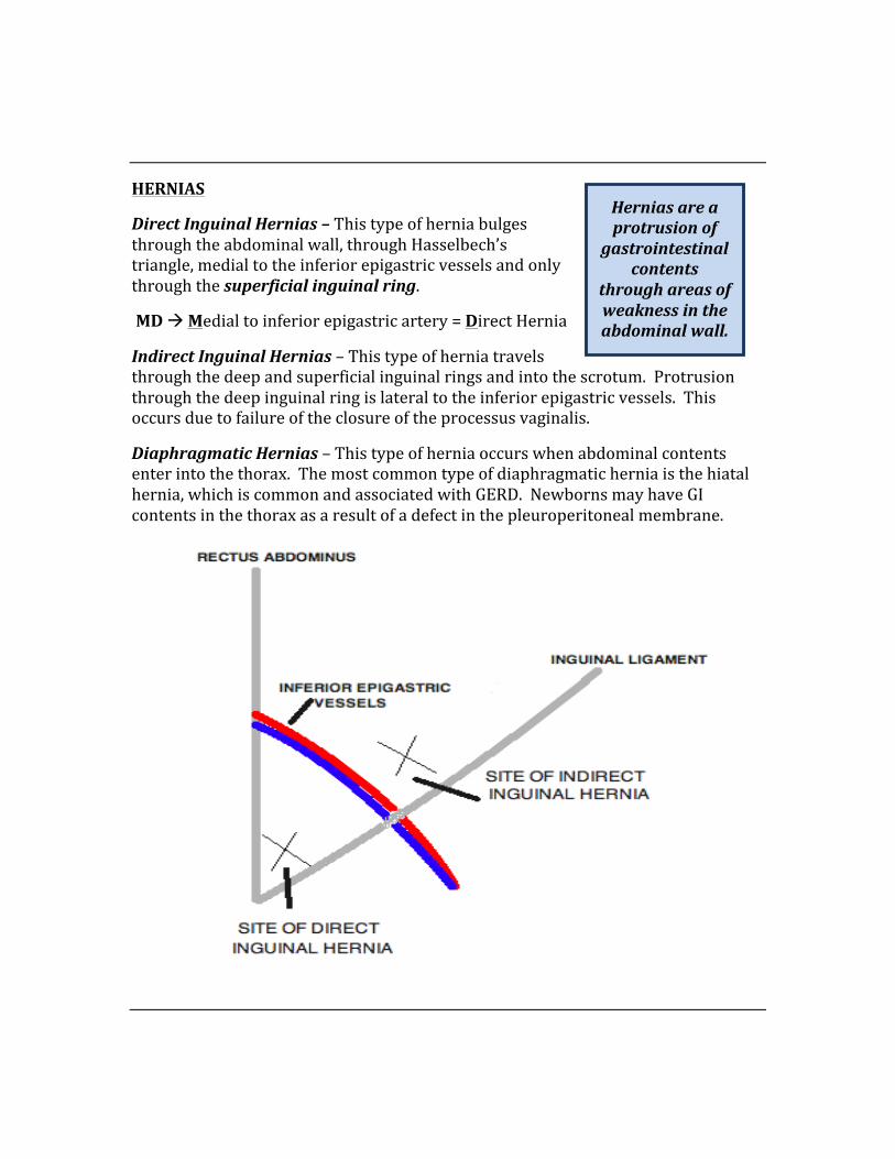
HERNIAS
Direct Inguinal Hernias – This type of hernia bulges through the abdominal wall, through Hasselbech’s triangle, medial to the inferior epigastric vessels and only through the superficial inguinal ring.
MD à Medial to inferior epigastric artery = Direct Hernia
Indirect Inguinal Hernias – This type of hernia travels through the deep and superficial inguinal rings and into the scrotum. Protrusion through the deep inguinal ring is lateral to the inferior epigastric vessels. This occurs due to failure of the closure of the processus vaginalis.
Diaphragmatic Hernias – This type of hernia occurs when abdominal contents enter into the thorax. The most common type of diaphragmatic hernia is the hiatal hernia, which is common and associated with GERD. Newborns may have GI contents in the thorax as a result of a defect in the pleuroperitoneal membrane.
Hernias are a protrusion of
gastrointestinal contents
through areas of weakness in the abdominal wall.
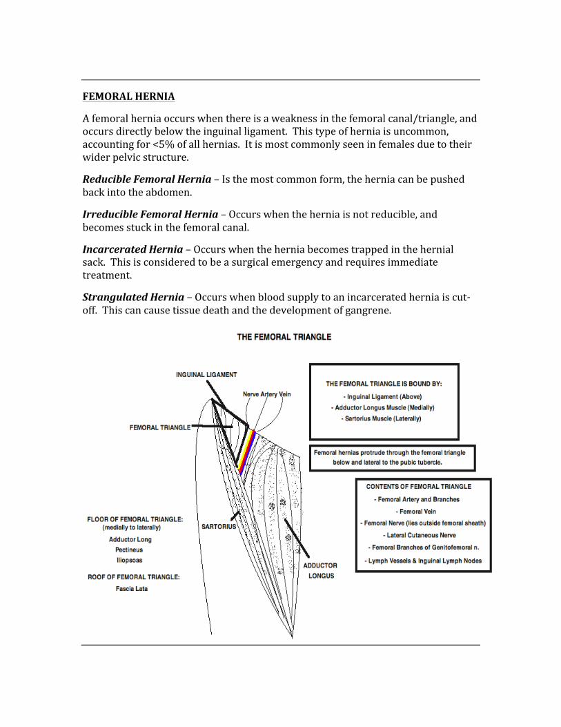
FEMORAL HERNIA
A femoral hernia occurs when there is a weakness in the femoral canal/triangle, and occurs directly below the inguinal ligament. This type of hernia is uncommon, accounting for <5% of all hernias. It is most commonly seen in females due to their wider pelvic structure.
Reducible Femoral Hernia – Is the most common form, the hernia can be pushed back into the abdomen.
Irreducible Femoral Hernia – Occurs when the hernia is not reducible, and becomes stuck in the femoral canal.
Incarcerated Hernia – Occurs when the hernia becomes trapped in the hernial sack. This is considered to be a surgical emergency and requires immediate treatment.
Strangulated Hernia – Occurs when blood supply to an incarcerated hernia is cut-‐off. This can cause tissue death and the development of gangrene.
PORTAL-‐SYSTEMIC ANASTOMOSES
These are anastomoses that occur between veins of the portal and systemic circulation. These sites are important because several conditions may occur as a result of changes in pressure within each system.
The most common conditions include: Hemorrhoids, Esophageal Varices, and Caput Medusae.
CONDITION SYSTEMIC CIRCULATION PORTAL CIRCULATION Hemorrhoids Middle Rectal and Inferior
Rectal Veins Superior Rectal Veins
Esophageal Varices Azygous Veins Left Gastric Vein Caput Medusae Superficial Epigastric Vein Paraumbilical Veins
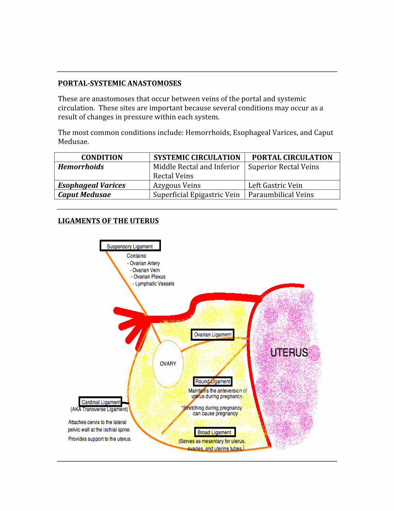
LIGAMENTS OF THE UTERUS
DRAINAGE OF THE TESTCILES/OVARIES
The left ovary/teste drains from the left gonadal vein, to the left renal vein, and into the inferior vena cava.
Left Gonadal Vein à Left Renal Vein à IVC
• In 25% of men, this system is not completely competent, and results in a varicocele of the left teste
The right ovary/test drains directly from the gonadal vein into the IVC. This rarely results in a varicocele in men due to the angle of drainage.
THE PECTINATE LINE
The pectinate line is most commonly useful when a patient has hemorrhoids. Hemorrhoids that are above the pectinate line do not cause pain due to visceral innervation. Hemorrhoids that are below the pectinate line have somatic innervation, and are therefore painful.
The pectinate line is formed where the hindgut and the ectoderm meet.
Arterial supply above the pectinate line is from the superior rectal artery. Venous drainage is from the superior rectal vein to the inferior mesenteric vein, and into the portal system.
Arterial supply below the pectinate line is from the inferior rectal artery. Venous drainage is from the inferior rectal vein to the internal pudendal vein, then into the internal iliac vein and into the IVC.
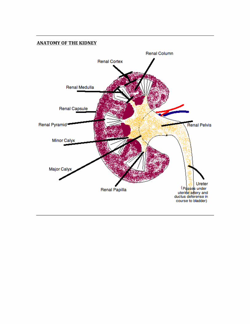
ANATOMY OF THE KIDNEY
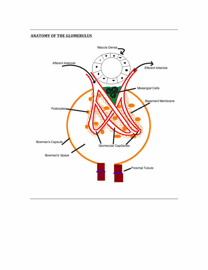
ANATOMY OF THE GLOMERULUS
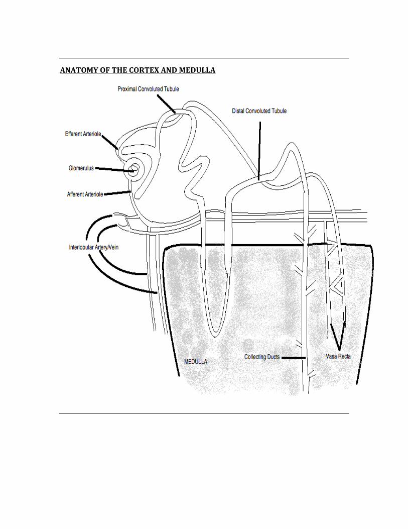
ANATOMY OF THE CORTEX AND MEDULLA
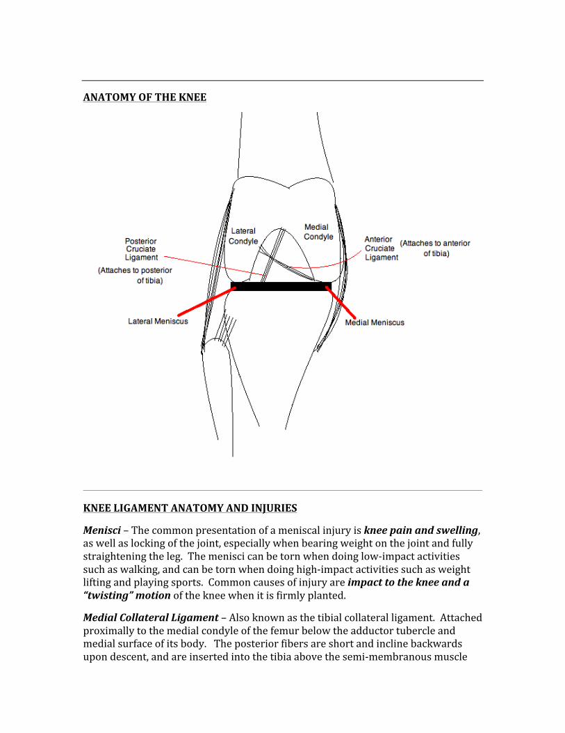
ANATOMY OF THE KNEE
KNEE LIGAMENT ANATOMY AND INJURIES
Menisci – The common presentation of a meniscal injury is knee pain and swelling, as well as locking of the joint, especially when bearing weight on the joint and fully straightening the leg. The menisci can be torn when doing low-‐impact activities such as walking, and can be torn when doing high-‐impact activities such as weight lifting and playing sports. Common causes of injury are impact to the knee and a “twisting” motion of the knee when it is firmly planted.
Medial Collateral Ligament – Also known as the tibial collateral ligament. Attached proximally to the medial condyle of the femur below the adductor tubercle and medial surface of its body. The posterior fibers are short and incline backwards upon descent, and are inserted into the tibia above the semi-‐membranous muscle
groove. It inserts into the medial surface of the tibial body about 2.5cm below the level of the condyle. Injuries are common in skiing and in football when valgus stress is applied (this is “abduction stress” aka stress to the lateral aspect of the knee).
Lateral Collateral Ligament – Also known as the “fibular collateral ligament”, is narrow and less broad that the medial collateral ligament. The LCL travels obliquely from the lateral epicondyle of the femur to the head of the fibula. The anatomy of the LCL gives it more flexibility than the MCL, and thus it is less commonly injured.
Anterior Cruciate Ligament – The ACL gets the term “anterior” because it attaches to the anterior aspect of the tibia, with an origin from deep within the notch of the distal femur. Injury to the anterior cruciate ligament is the most common knee injury, and is especially seen in athletes. Lateral rotational moves such as those in soccer, basketball, and skiing are common causes of ACL strains and/or tears. Testing for ACL injury is done with the anterior drawer test, where the flexed knee is drawn forward in an attempt to identify an increased amount of anterior tibial translation as compared to the opposite joint. The Lachman test is another diagnostic test that is similar in nature, but with the patient’s knee in 20-‐30 degree flexion instead of the 90-‐degree flexion used in the anterior drawer test.
Posterior Cruciate Ligament – The posterior cruciate ligament gets its name because it attaches to the posterior aspect of the tibia. It connects the posterior intercondylar area of the tibia to the medial condyle of the femur. Injury to the PCL causes less pain and disability than those to the ACL, and often goes undiagnosed. The common cause of PCL injury is the “dashboard injury”, where something forcefully strikes the tibia and drives it posteriorly. A common mechanism of injury during sporting events is a fall on a hyperflexed knee with the foot pointed downwards. Patients often complain of a “pop” during the injury. The PCL injury is diagnosed initially with the posterior drawer test, where the tibia is pushed backwards and a decreased resistance is experienced.
The Unhappy Triad – This is a common knee injury seen in football, where a player is hit on the lateral aspect of the knee, causing damage to the ACL, MCL, and Medial Meniscus.
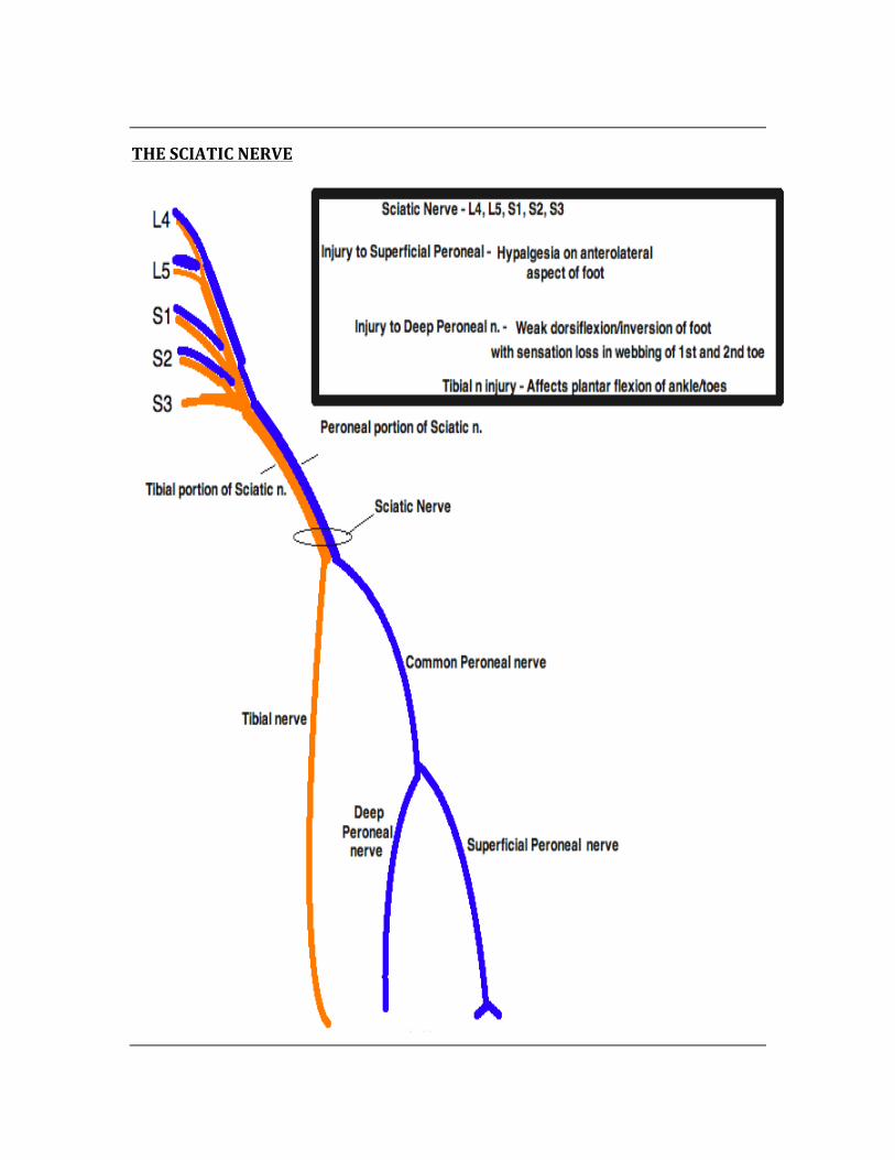
THE SCIATIC NERVE
CHAPTER 2
EMBRYOLOGY
Most of the information from the embryology section is high-‐yield. Memorization of the embryologic derivatives is important, as it is likely to show up on the exam, as is the
majority of the information from this chapter.
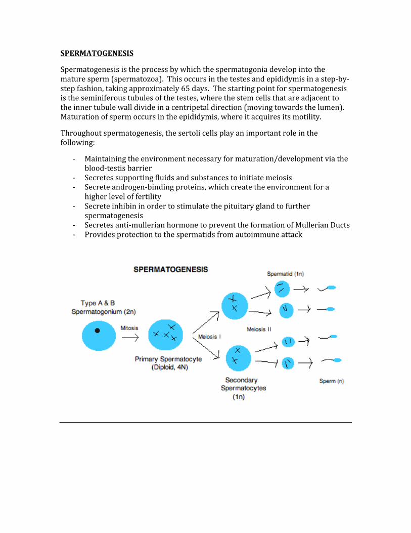
SPERMATOGENESIS
Spermatogenesis is the process by which the spermatogonia develop into the mature sperm (spermatozoa). This occurs in the testes and epididymis in a step-‐by-‐step fashion, taking approximately 65 days. The starting point for spermatogenesis is the seminiferous tubules of the testes, where the stem cells that are adjacent to the inner tubule wall divide in a centripetal direction (moving towards the lumen). Maturation of sperm occurs in the epididymis, where it acquires its motility.
Throughout spermatogenesis, the sertoli cells play an important role in the following:
-‐ Maintaining the environment necessary for maturation/development via the blood-‐testis barrier
-‐ Secretes supporting fluids and substances to initiate meiosis -‐ Secrete androgen-‐binding proteins, which create the environment for a
higher level of fertility -‐ Secrete inhibin in order to stimulate the pituitary gland to further
spermatogenesis -‐ Secretes anti-‐mullerian hormone to prevent the formation of Mullerian Ducts -‐ Provides protection to the spermatids from autoimmune attack
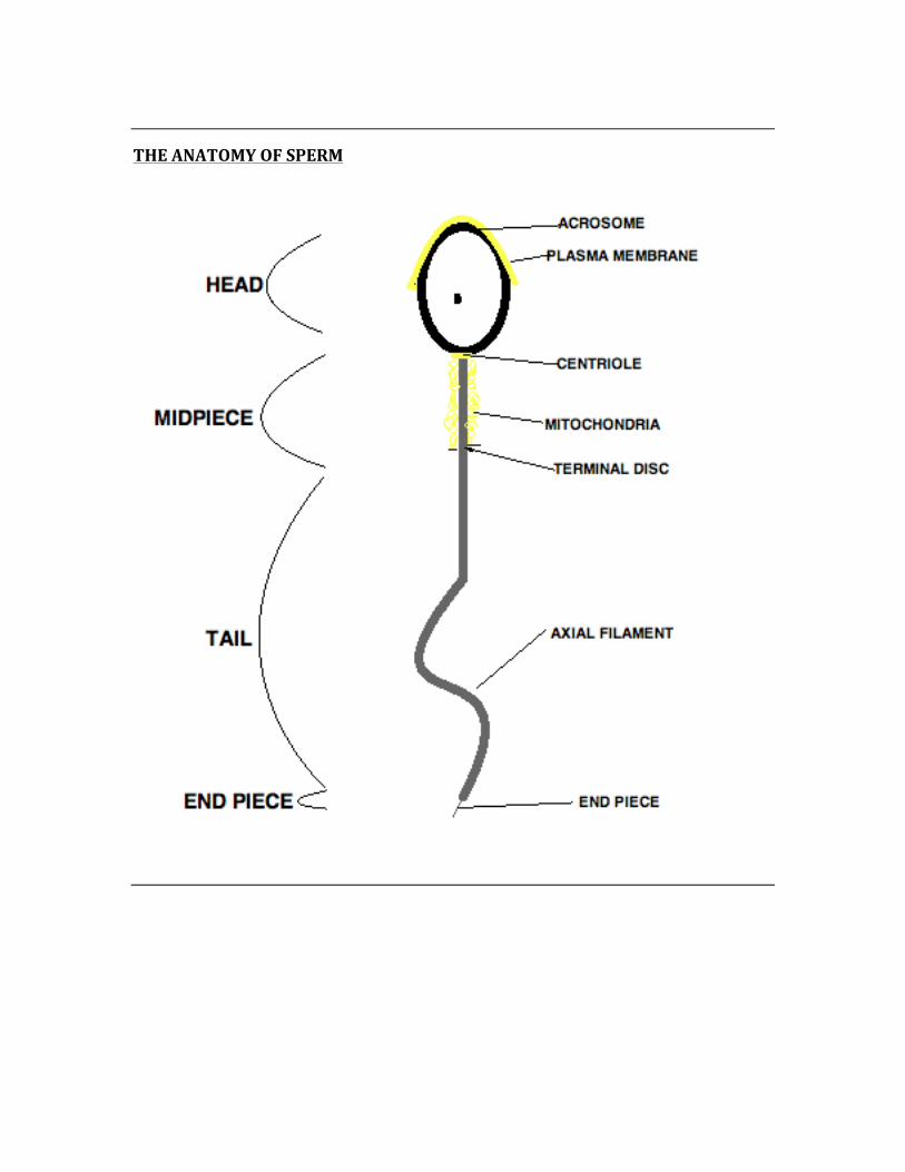
THE ANATOMY OF SPERM
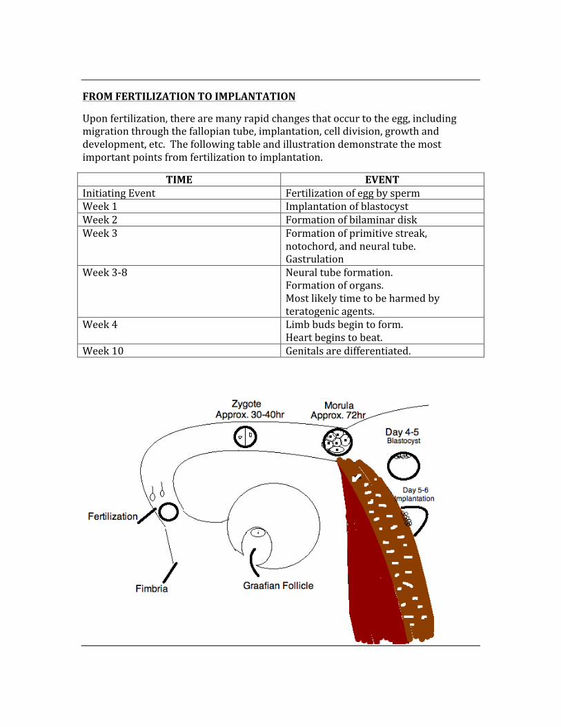
FROM FERTILIZATION TO IMPLANTATION
Upon fertilization, there are many rapid changes that occur to the egg, including migration through the fallopian tube, implantation, cell division, growth and development, etc. The following table and illustration demonstrate the most important points from fertilization to implantation.
TIME EVENT Initiating Event Fertilization of egg by sperm Week 1 Implantation of blastocyst Week 2 Formation of bilaminar disk Week 3 Formation of primitive streak,
notochord, and neural tube. Gastrulation
Week 3-‐8 Neural tube formation. Formation of organs. Most likely time to be harmed by teratogenic agents.
Week 4 Limb buds begin to form. Heart begins to beat.
Week 10 Genitals are differentiated.
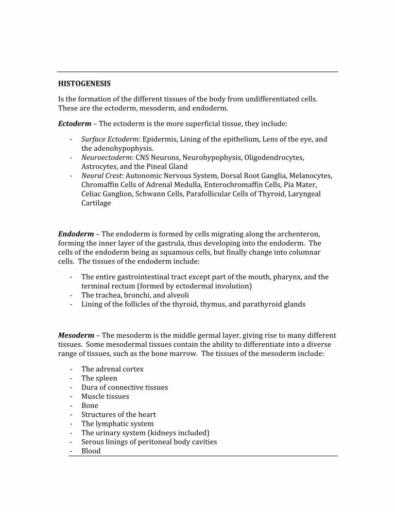
HISTOGENESIS
Is the formation of the different tissues of the body from undifferentiated cells. These are the ectoderm, mesoderm, and endoderm.
Ectoderm – The ectoderm is the more superficial tissue, they include:
-‐ Surface Ectoderm: Epidermis, Lining of the epithelium, Lens of the eye, and the adenohypophysis.
-‐ Neuroectoderm: CNS Neurons, Neurohypophysis, Oligodendrocytes, Astrocytes, and the Pineal Gland
-‐ Neural Crest: Autonomic Nervous System, Dorsal Root Ganglia, Melanocytes, Chromaffin Cells of Adrenal Medulla, Enterochromaffin Cells, Pia Mater, Celiac Ganglion, Schwann Cells, Parafollicular Cells of Thyroid, Laryngeal Cartilage
Endoderm – The endoderm is formed by cells migrating along the archenteron, forming the inner layer of the gastrula, thus developing into the endoderm. The cells of the endoderm being as squamous cells, but finally change into columnar cells. The tissues of the endoderm include:
-‐ The entire gastrointestinal tract except part of the mouth, pharynx, and the terminal rectum (formed by ectodermal involution)
-‐ The trachea, bronchi, and alveoli -‐ Lining of the follicles of the thyroid, thymus, and parathyroid glands
Mesoderm – The mesoderm is the middle germal layer, giving rise to many different tissues. Some mesodermal tissues contain the ability to differentiate into a diverse range of tissues, such as the bone marrow. The tissues of the mesoderm include:
-‐ The adrenal cortex -‐ The spleen -‐ Dura of connective tissues -‐ Muscle tissues -‐ Bone -‐ Structures of the heart -‐ The lymphatic system -‐ The urinary system (kidneys included) -‐ Serous linings of peritoneal body cavities -‐ Blood
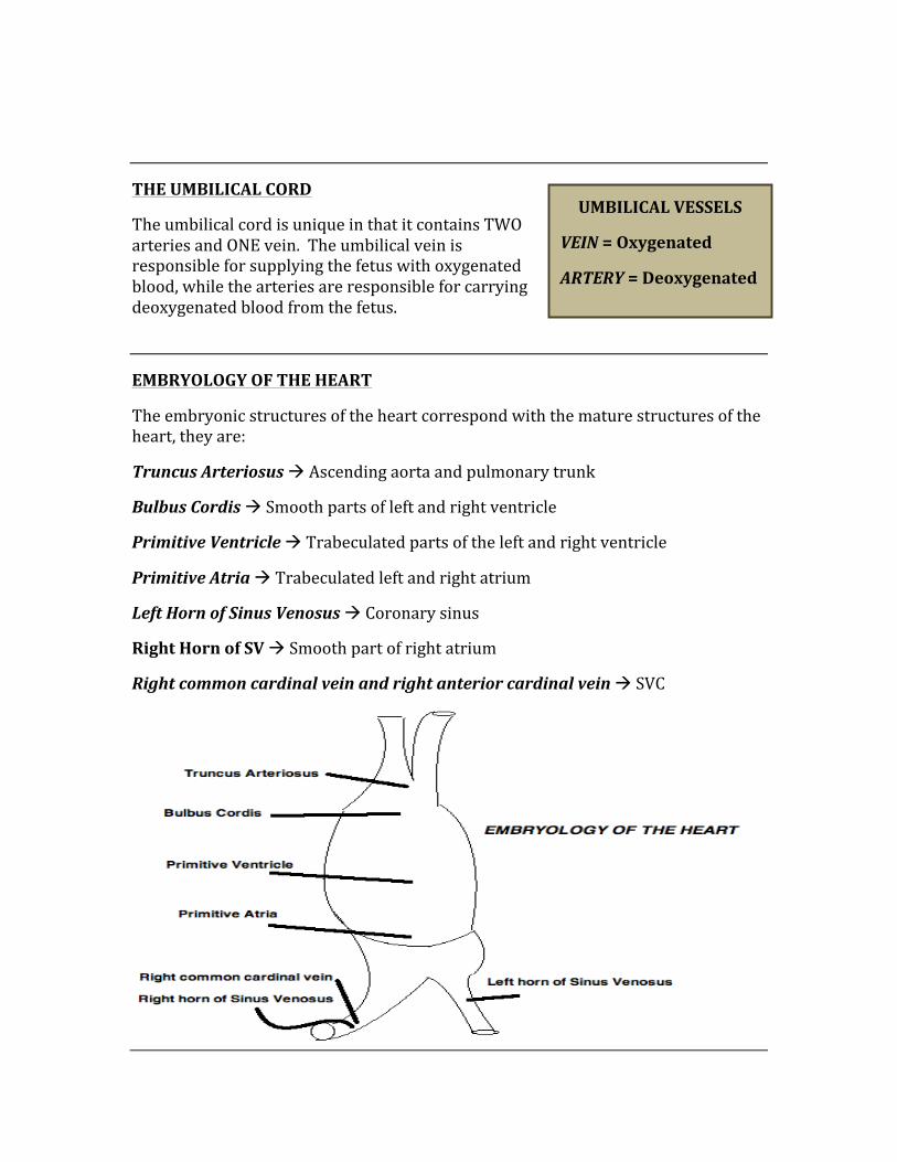
THE UMBILICAL CORD
The umbilical cord is unique in that it contains TWO arteries and ONE vein. The umbilical vein is responsible for supplying the fetus with oxygenated blood, while the arteries are responsible for carrying deoxygenated blood from the fetus.
EMBRYOLOGY OF THE HEART
The embryonic structures of the heart correspond with the mature structures of the heart, they are:
Truncus Arteriosus à Ascending aorta and pulmonary trunk
Bulbus Cordis à Smooth parts of left and right ventricle
Primitive Ventricle à Trabeculated parts of the left and right ventricle
Primitive Atria à Trabeculated left and right atrium
Left Horn of Sinus Venosus à Coronary sinus
Right Horn of SV à Smooth part of right atrium
Right common cardinal vein and right anterior cardinal vein à SVC
UMBILICAL VESSELS
VEIN = Oxygenated
ARTERY = Deoxygenated
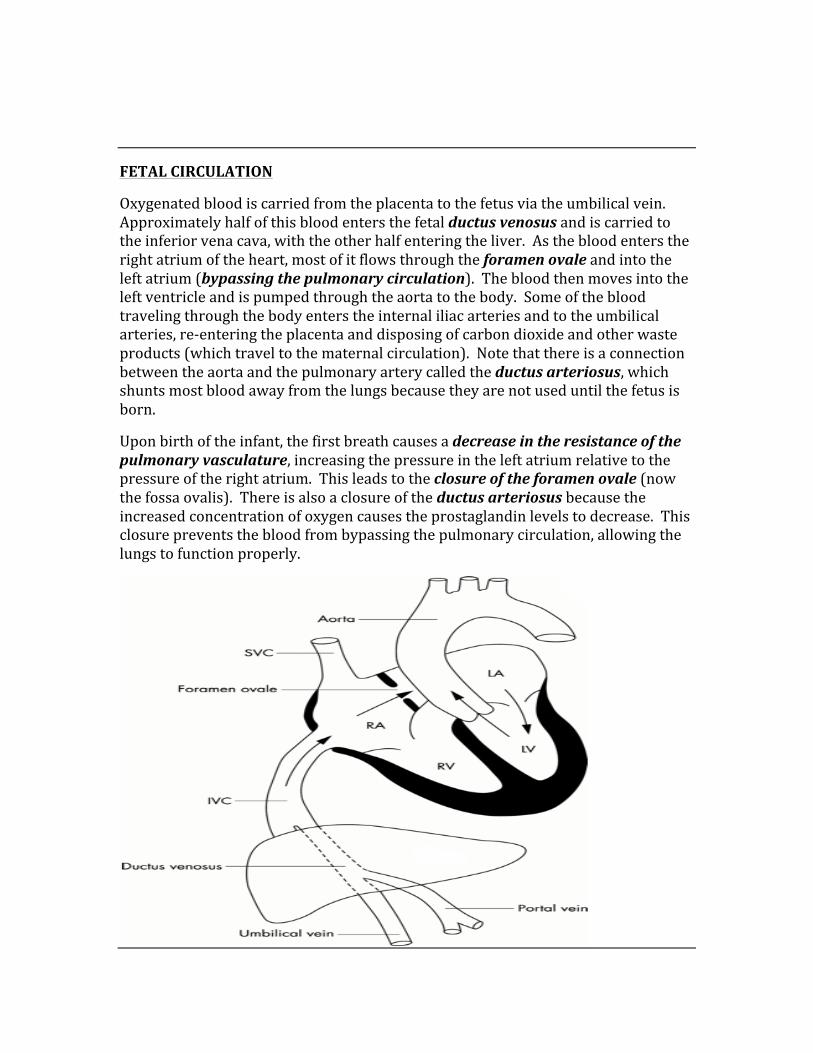
FETAL CIRCULATION
Oxygenated blood is carried from the placenta to the fetus via the umbilical vein. Approximately half of this blood enters the fetal ductus venosus and is carried to the inferior vena cava, with the other half entering the liver. As the blood enters the right atrium of the heart, most of it flows through the foramen ovale and into the left atrium (bypassing the pulmonary circulation). The blood then moves into the left ventricle and is pumped through the aorta to the body. Some of the blood traveling through the body enters the internal iliac arteries and to the umbilical arteries, re-‐entering the placenta and disposing of carbon dioxide and other waste products (which travel to the maternal circulation). Note that there is a connection between the aorta and the pulmonary artery called the ductus arteriosus, which shunts most blood away from the lungs because they are not used until the fetus is born.
Upon birth of the infant, the first breath causes a decrease in the resistance of the pulmonary vasculature, increasing the pressure in the left atrium relative to the pressure of the right atrium. This leads to the closure of the foramen ovale (now the fossa ovalis). There is also a closure of the ductus arteriosus because the increased concentration of oxygen causes the prostaglandin levels to decrease. This closure prevents the blood from bypassing the pulmonary circulation, allowing the lungs to function properly.
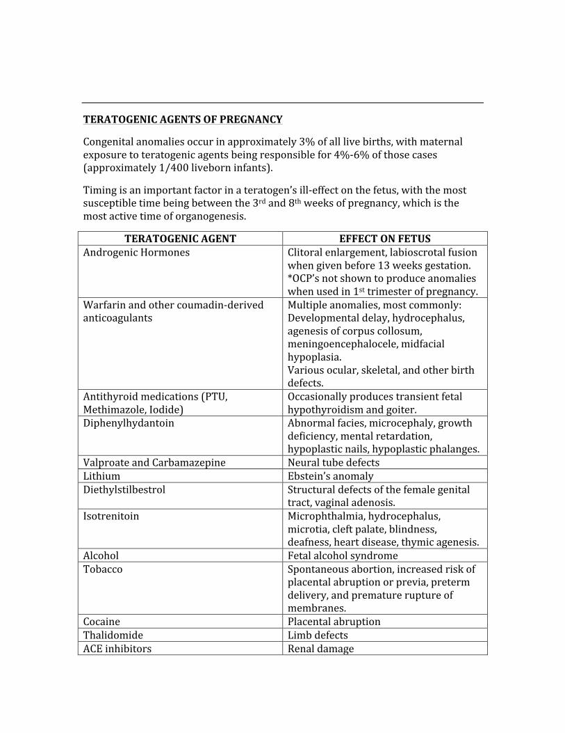
TERATOGENIC AGENTS OF PREGNANCY
Congenital anomalies occur in approximately 3% of all live births, with maternal exposure to teratogenic agents being responsible for 4%-‐6% of those cases (approximately 1/400 liveborn infants).
Timing is an important factor in a teratogen’s ill-‐effect on the fetus, with the most susceptible time being between the 3rd and 8th weeks of pregnancy, which is the most active time of organogenesis.
TERATOGENIC AGENT EFFECT ON FETUS Androgenic Hormones Clitoral enlargement, labioscrotal fusion
when given before 13 weeks gestation. *OCP’s not shown to produce anomalies when used in 1st trimester of pregnancy.
Warfarin and other coumadin-‐derived anticoagulants
Multiple anomalies, most commonly: Developmental delay, hydrocephalus, agenesis of corpus collosum, meningoencephalocele, midfacial hypoplasia. Various ocular, skeletal, and other birth defects.
Antithyroid medications (PTU, Methimazole, Iodide)
Occasionally produces transient fetal hypothyroidism and goiter.
Diphenylhydantoin Abnormal facies, microcephaly, growth deficiency, mental retardation, hypoplastic nails, hypoplastic phalanges.
Valproate and Carbamazepine Neural tube defects Lithium Ebstein’s anomaly Diethylstilbestrol Structural defects of the female genital
tract, vaginal adenosis. Isotrenitoin Microphthalmia, hydrocephalus,
microtia, cleft palate, blindness, deafness, heart disease, thymic agenesis.
Alcohol Fetal alcohol syndrome Tobacco Spontaneous abortion, increased risk of
placental abruption or previa, preterm delivery, and premature rupture of membranes.
Cocaine Placental abruption Thalidomide Limb defects ACE inhibitors Renal damage
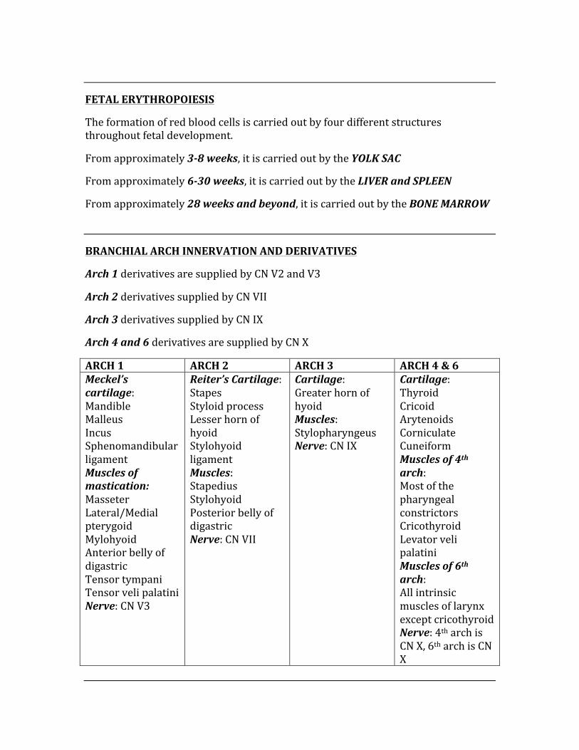
FETAL ERYTHROPOIESIS
The formation of red blood cells is carried out by four different structures throughout fetal development.
From approximately 3-‐8 weeks, it is carried out by the YOLK SAC
From approximately 6-‐30 weeks, it is carried out by the LIVER and SPLEEN
From approximately 28 weeks and beyond, it is carried out by the BONE MARROW
BRANCHIAL ARCH INNERVATION AND DERIVATIVES
Arch 1 derivatives are supplied by CN V2 and V3
Arch 2 derivatives supplied by CN VII
Arch 3 derivatives supplied by CN IX
Arch 4 and 6 derivatives are supplied by CN X
ARCH 1 ARCH 2 ARCH 3 ARCH 4 & 6 Meckel’s cartilage: Mandible Malleus Incus Sphenomandibular ligament Muscles of mastication: Masseter Lateral/Medial pterygoid Mylohyoid Anterior belly of digastric Tensor tympani Tensor veli palatini Nerve: CN V3
Reiter’s Cartilage: Stapes Styloid process Lesser horn of hyoid Stylohyoid ligament Muscles: Stapedius Stylohyoid Posterior belly of digastric Nerve: CN VII
Cartilage: Greater horn of hyoid Muscles: Stylopharyngeus Nerve: CN IX
Cartilage: Thyroid Cricoid Arytenoids Corniculate Cuneiform Muscles of 4th arch: Most of the pharyngeal constrictors Cricothyroid Levator veli palatini Muscles of 6th arch: All intrinsic muscles of larynx except cricothyroid Nerve: 4th arch is CN X, 6th arch is CN X
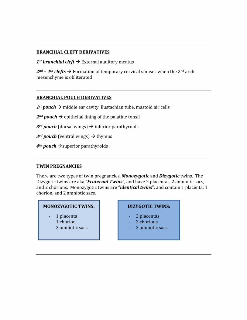
BRANCHIAL CLEFT DERIVATIVES
1st branchial cleft à External auditory meatus
2nd – 4th clefts à Formation of temporary cervical sinuses when the 2nd arch mesenchyme is obliterated
BRANCHIAL POUCH DERIVATIVES
1st pouch à middle ear cavity, Eustachian tube, mastoid air cells
2nd pouch à epithelial lining of the palatine tonsil
3rd pouch (dorsal wings) à inferior parathyroids
3rd pouch (ventral wings) à thymus
4th pouch àsuperior parathyroids
TWIN PREGNANCIES
There are two types of twin pregnancies, Monozygotic and Dizygotic twins. The Dizygotic twins are aka “Fraternal Twins”, and have 2 placentas, 2 amniotic sacs, and 2 chorions. Monozygotic twins are “identical twins”, and contain 1 placenta, 1 chorion, and 2 amniotic sacs.
MONOZYGOTIC TWINS:
-‐ 1 placenta -‐ 1 chorion -‐ 2 amniotic sacs
DIZYGOTIC TWINS:
-‐ 2 placentas -‐ 2 chorions -‐ 2 amniotic sacs
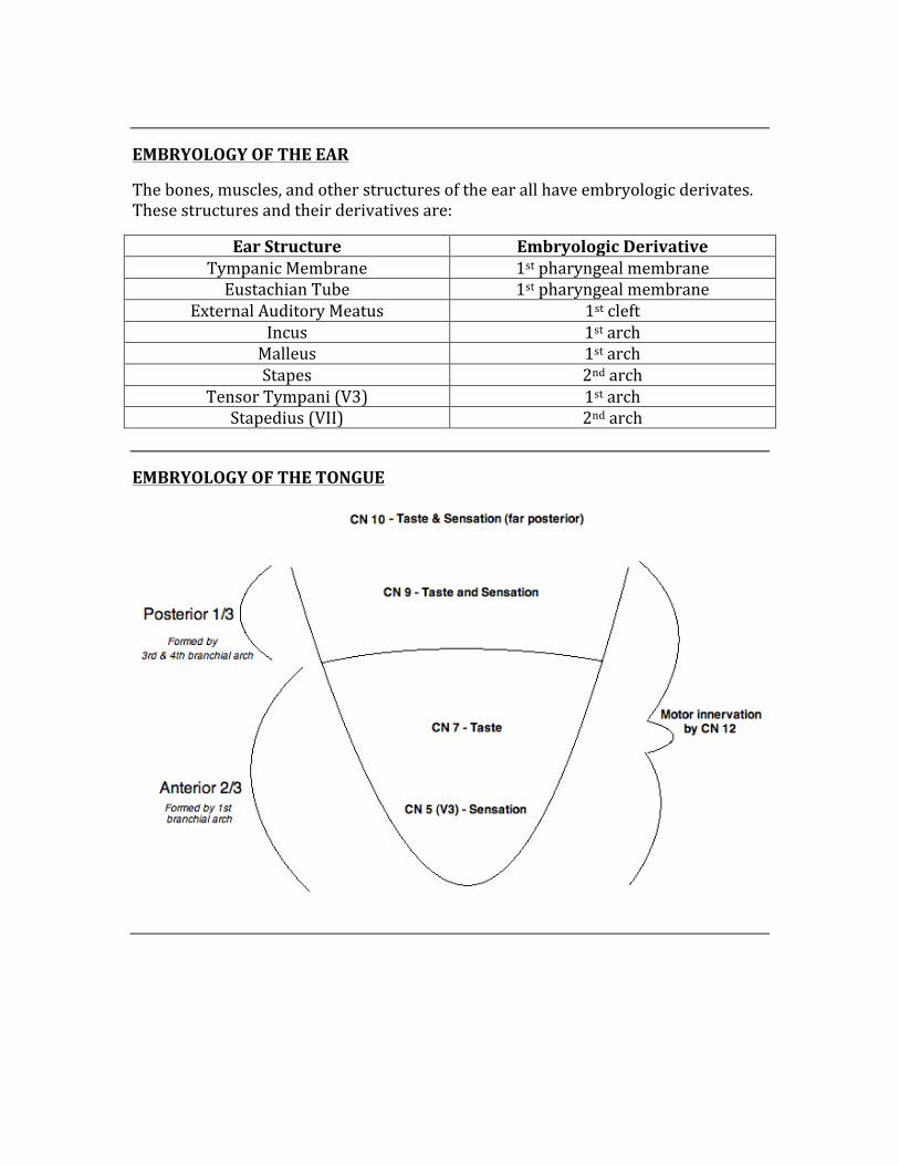
EMBRYOLOGY OF THE EAR
The bones, muscles, and other structures of the ear all have embryologic derivates. These structures and their derivatives are:
Ear Structure Embryologic Derivative Tympanic Membrane 1st pharyngeal membrane Eustachian Tube 1st pharyngeal membrane
External Auditory Meatus 1st cleft Incus 1st arch Malleus 1st arch Stapes 2nd arch
Tensor Tympani (V3) 1st arch Stapedius (VII) 2nd arch
EMBRYOLOGY OF THE TONGUE
EMBRYOLOGY OF THE THYMUS
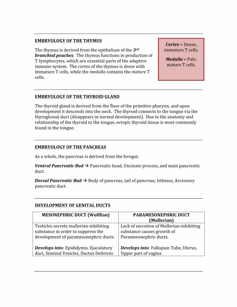
The thymus is derived from the epithelium of the 3rd branchial pouches. The thymus functions in production of T-‐lymphocytes, which are essential parts of the adaptive immune system. The cortex of the thymus is dense with immature T cells, while the medulla contains the mature T cells.
EMBRYOLOGY OF THE THYROID GLAND
The thyroid gland is derived from the floor of the primitive pharynx, and upon development it descends into the neck. The thyroid connects to the tongue via the thyroglossal duct (disappears in normal development). Due to the anatomy and relationship of the thyroid to the tongue, ectopic thyroid tissue is most commonly found in the tongue.
EMBRYOLOGY OF THE PANCREAS
As a whole, the pancreas is derived from the foregut.
Ventral Pancreatic Bud à Pancreatic head, Uncinate process, and main pancreatic duct.
Dorsal Pancreatic Bud à Body of pancreas, tail of pancreas, Isthmus, Accessory pancreatic duct.
DEVELOPMENT OF GENITAL DUCTS
MESONEPHRIC DUCT (Wolffian) PARAMESONEPHRIC DUCT (Mullerian)
Testicles secrete mullerian-‐inhibiting substance in order to suppress the development of paramesonephric ducts. Develops into: Epididymis, Ejaculatory duct, Seminal Vesicles, Ductus Deferens
Lack of secretion of Mullerian-‐inhibiting substance causes growth of Paramesonephric ducts. Develops into: Fallopian Tube, Uterus, Upper part of vagina
Cortex = Dense, immature T cells.
Medulla = Pale, mature T cells.
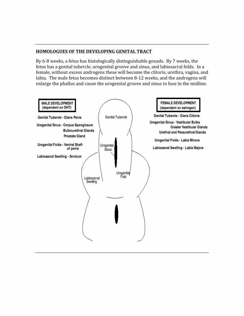
HOMOLOGUES OF THE DEVELOPING GENITAL TRACT
By 6-‐8 weeks, a fetus has histologically distinguishable gonads. By 7 weeks, the fetus has a genital tubercle, urogenital groove and sinus, and labiosacral folds. In a female, without excess androgens these will become the clitoris, urethra, vagina, and labia. The male fetus becomes distinct between 8-‐12 weeks, and the androgens will enlarge the phallus and cause the urogenital groove and sinus to fuse in the midline.
EMBRYOLOGY OF THE DIAPHRAGM
During initial development, the diaphragm is innervated by nerves C3, C4, and C5. As the diaphragm descends, it maintains this innervation.
The diaphragm is derived from the following embryologic structures:
-‐ Septum Transversum -‐ Pleuroperitoneal Folds -‐ Body Wall -‐ Dorsal Mesentary of the Esophagus
FORMATION OF BONE
There are two main types of bone development, those being “intramembranous” bone and “endochondral” bone. Intramembranous bone is formed spontaneously without the presence or need of any pre-‐existing cartilage. On the other hand, endochondral bone (long bones) requires the presence of cartilaginous molds in order to form its bony structure. The cartilaginous mold ossifies and produces the endochondral bone.
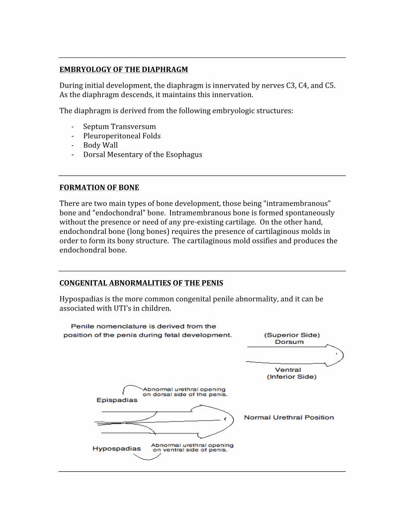
CONGENITAL ABNORMALITIES OF THE PENIS
Hypospadias is the more common congenital penile abnormality, and it can be associated with UTI’s in children.
CHAPTER 3
HISTOLOGY
Histology is not traditionally a very high-‐yield component of the Step 1 exam, but it is important to know which type of epithelium makes up all of the structures of the body, as
well as all pathologies that are linked to histological changes.
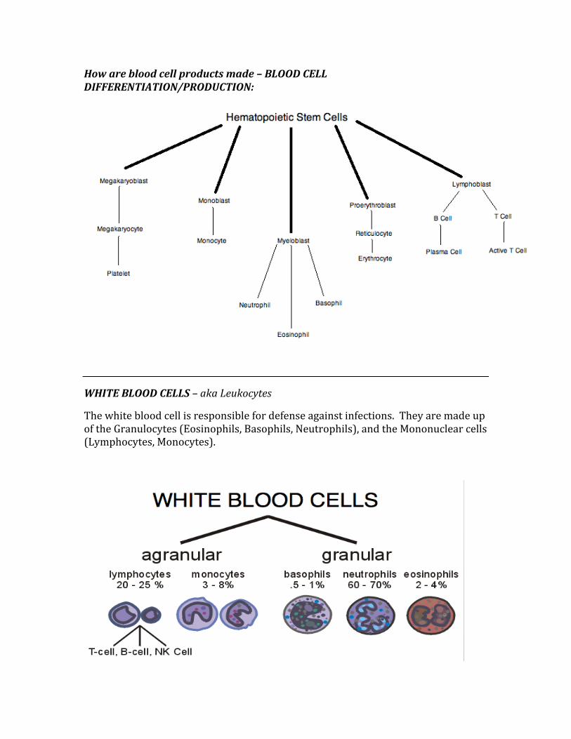
How are blood cell products made – BLOOD CELL DIFFERENTIATION/PRODUCTION:
WHITE BLOOD CELLS – aka Leukocytes
The white blood cell is responsible for defense against infections. They are made up of the Granulocytes (Eosinophils, Basophils, Neutrophils), and the Mononuclear cells (Lymphocytes, Monocytes).
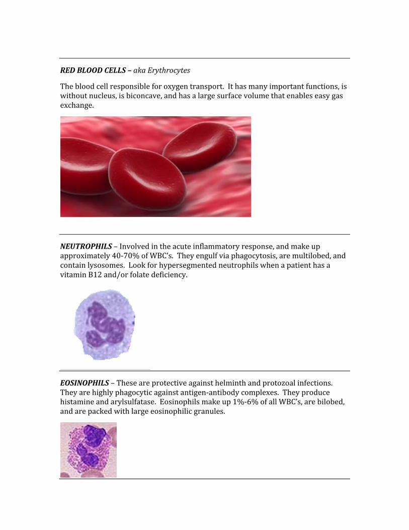
RED BLOOD CELLS – aka Erythrocytes
The blood cell responsible for oxygen transport. It has many important functions, is without nucleus, is biconcave, and has a large surface volume that enables easy gas exchange.
NEUTROPHILS – Involved in the acute inflammatory response, and make up approximately 40-‐70% of WBC’s. They engulf via phagocytosis, are multilobed, and contain lysosomes. Look for hypersegmented neutrophils when a patient has a vitamin B12 and/or folate deficiency.
EOSINOPHILS – These are protective against helminth and protozoal infections. They are highly phagocytic against antigen-‐antibody complexes. They produce histamine and arylsulfatase. Eosinophils make up 1%-‐6% of all WBC’s, are bilobed, and are packed with large eosinophilic granules.
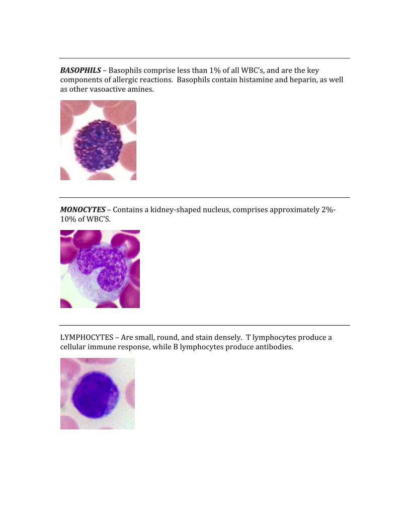
BASOPHILS – Basophils comprise less than 1% of all WBC’s, and are the key components of allergic reactions. Basophils contain histamine and heparin, as well as other vasoactive amines.
MONOCYTES – Contains a kidney-‐shaped nucleus, comprises approximately 2%-‐10% of WBC’S.
LYMPHOCYTES – Are small, round, and stain densely. T lymphocytes produce a cellular immune response, while B lymphocytes produce antibodies.
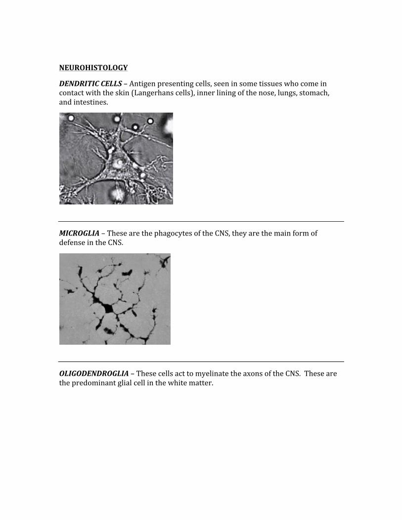
NEUROHISTOLOGY
DENDRITIC CELLS – Antigen presenting cells, seen in some tissues who come in contact with the skin (Langerhans cells), inner lining of the nose, lungs, stomach, and intestines.
MICROGLIA – These are the phagocytes of the CNS, they are the main form of defense in the CNS.
OLIGODENDROGLIA – These cells act to myelinate the axons of the CNS. These are the predominant glial cell in the white matter.
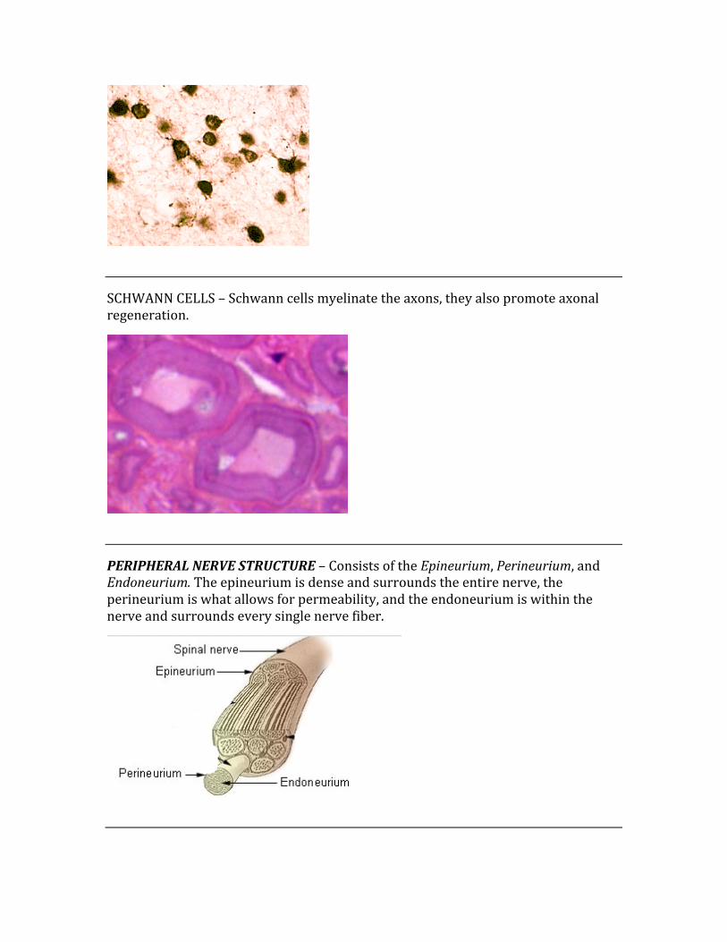
SCHWANN CELLS – Schwann cells myelinate the axons, they also promote axonal regeneration.
PERIPHERAL NERVE STRUCTURE – Consists of the Epineurium, Perineurium, and Endoneurium. The epineurium is dense and surrounds the entire nerve, the perineurium is what allows for permeability, and the endoneurium is within the nerve and surrounds every single nerve fiber.
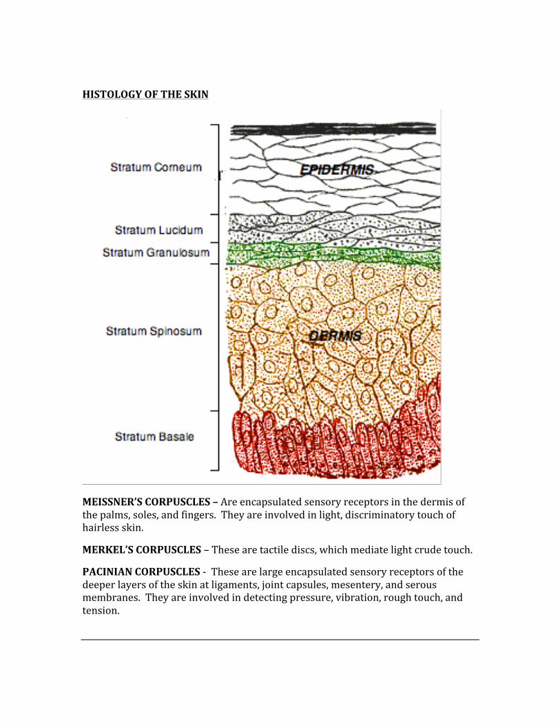
HISTOLOGY OF THE SKIN
MEISSNER’S CORPUSCLES – Are encapsulated sensory receptors in the dermis of the palms, soles, and fingers. They are involved in light, discriminatory touch of hairless skin.
MERKEL’S CORPUSCLES – These are tactile discs, which mediate light crude touch.
PACINIAN CORPUSCLES -‐ These are large encapsulated sensory receptors of the deeper layers of the skin at ligaments, joint capsules, mesentery, and serous membranes. They are involved in detecting pressure, vibration, rough touch, and tension.
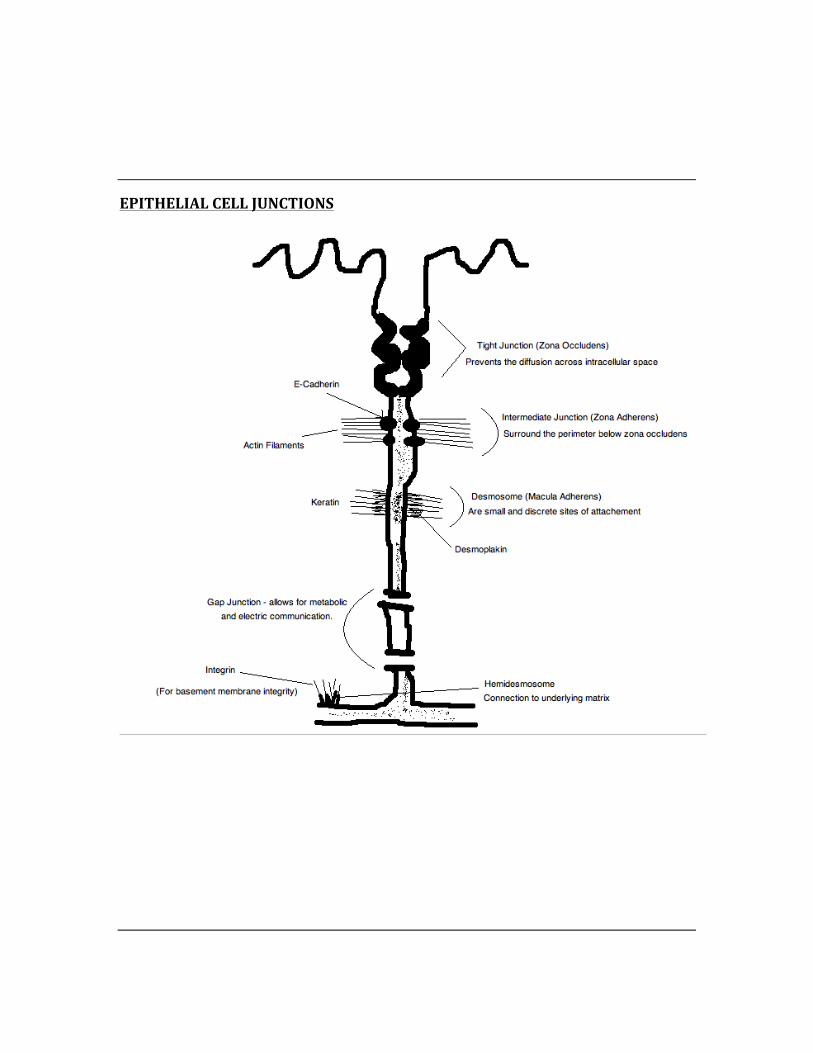
EPITHELIAL CELL JUNCTIONS
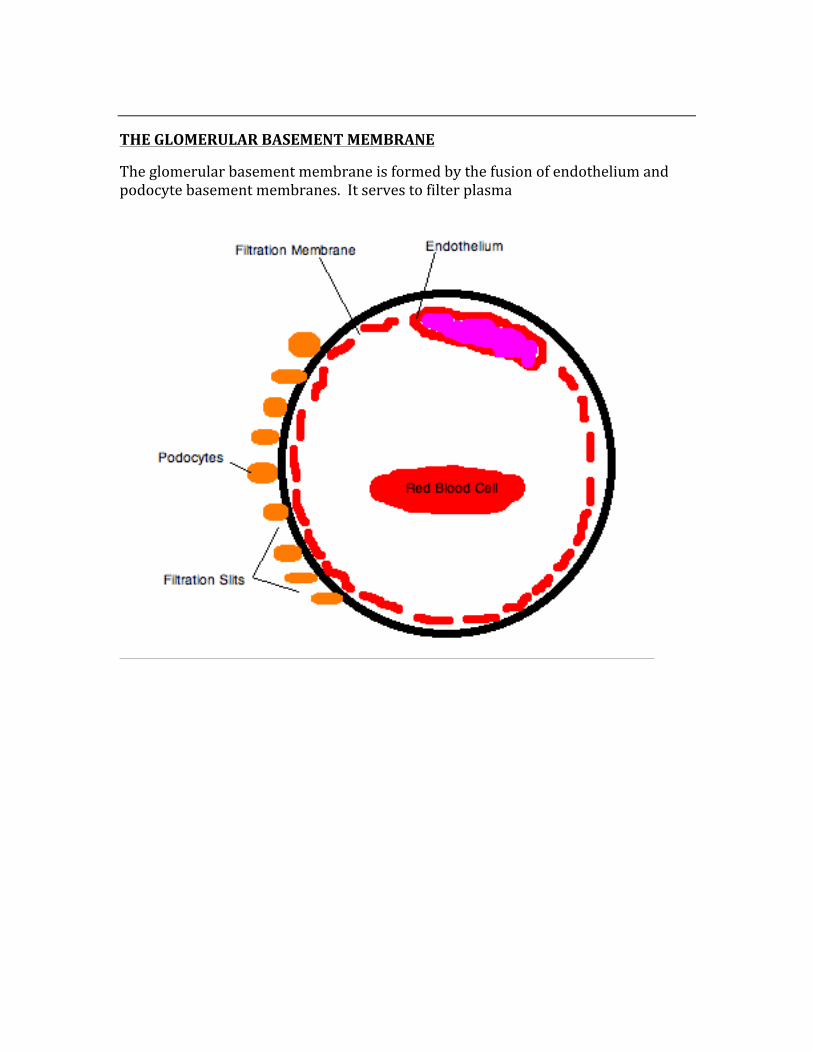
THE GLOMERULAR BASEMENT MEMBRANE
The glomerular basement membrane is formed by the fusion of endothelium and podocyte basement membranes. It serves to filter plasma

THE GOLGI APPARATUS
The golgi apparatus serves the purpose of processing and packaging proteins and lipids before they are secreted to the rest of the body.
The golgi apparatus is made of stack of membrane-‐bound structures of cisternae, which carry golgi enzymes to help or modify the proteins that travel through them.
The main functions of the golgi apparatus include:
-‐ Distribution of proteins and lipids from the endoplasmic reticulum to the plasma membrane, lysosomes, and through secretory vesicles
-‐ Addition of an O-‐oligosaccharide to Serine and Threonine -‐ Addition of N-‐oligosaccharide to Asparagine -‐ Proteoglycan assembly -‐ Sulfation of sugars in proteoglycans
ROUGH ENDOPLASMIC RETICULUM (RER)
The Rough Endoplasmic Reticulum is responsible for many functions, including:
-‐ N-‐linked glycosylation -‐ Addition of lysosomal enzymes with mannose-‐6-‐phosphate marker -‐ Integration of membrane proteins
Inside of neurons, there is the “Nissl body”, which is the RER of the neuron.
SMOOTH ENDOPLASMIC RETICULUM (SER)
The Smooth Endoplasmic Reticulum is where steroids are synthesized and where drug detoxification takes place.
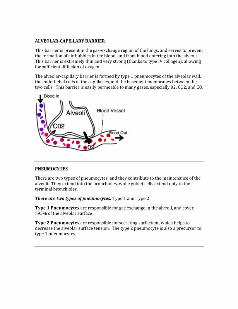
ALVEOLAR-‐CAPILLARY BARRIER
This barrier is present in the gas-‐exchange region of the lungs, and serves to prevent the formation of air bubbles in the blood, and from blood entering into the alveoli. This barrier is extremely thin and very strong (thanks to type IV collagen), allowing for sufficient diffusion of oxygen.
The alveolar-‐capillary barrier is formed by type 1 pneumocytes of the alveolar wall, the endothelial cells of the capillaries, and the basement membranes between the two cells. This barrier is easily permeable to many gases, especially 02, CO2, and CO.
PNEUMOCYTES
There are two types of pneumocytes, and they contribute to the maintenance of the alveoli. They extend into the bronchioles, while goblet cells extend only to the terminal bronchioles.
There are two types of pneumocytes: Type 1 and Type 2
Type 1 Pneumocytes are responsible for gas exchange in the alveoli, and cover >95% of the alveolar surface
Type 2 Pneumocytes are responsible for secreting surfactant, which helps to decrease the alveolar surface tension. The type 2 pneumocyte is also a precursor to type 1 pneumocytes.
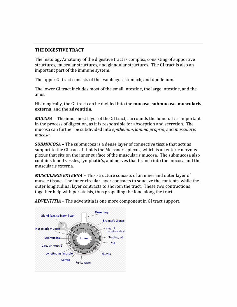
THE DIGESTIVE TRACT
The histology/anatomy of the digestive tract is complex, consisting of supportive structures, muscular structures, and glandular structures. The GI tract is also an important part of the immune system.
The upper GI tract consists of the esophagus, stomach, and duodenum.
The lower GI tract includes most of the small intestine, the large intestine, and the anus.
Histologically, the GI tract can be divided into the mucosa, submucosa, muscularis externa, and the adventitia.
MUCOSA – The innermost layer of the GI tract, surrounds the lumen. It is important in the process of digestion, as it is responsible for absorption and secretion. The mucosa can further be subdivided into epithelium, lamina propria, and muscularis mucosa.
SUBMUCOSA – The submucosa is a dense layer of connective tissue that acts as support to the GI tract. It holds the Meissner’s plexus, which is an enteric nervous plexus that sits on the inner surface of the muscularis mucosa. The submucosa also contains blood vessles, lymphatic’s, and nerves that branch into the mucosa and the muscularis externa.
MUSCULARIS EXTERNA – This structure consists of an inner and outer layer of muscle tissue. The inner circular layer contracts to squeeze the contents, while the outer longitudinal layer contracts to shorten the tract. These two contractions together help with peristalsis, thus propelling the food along the tract.
ADVENTITIA – The adventitia is one more component in GI tract support.
There are two enteric plexuses that help co-‐ordinate all of the functions of the GI tract.
The Myenteric Plexus – Co-‐ordination of motility along the entire gut wall. This plexus is located between the longitudinal and circular smooth muscle of the GI tract, and is also known as Auerbach’s plexus.
The Submucosal Plexus – This plexus regulates secretions, blood flow, and absorption. Located between the mucosa and the inner layer of smooth muscle, it is also known as Meissner’s plexus.
*Both plexuses contain parasympathetic terminal effector neurons.
BRUNNER’S GLANDS
Brunner’s glands are the only glands in the GI submucosa. They are responsible for secreting alkaline mucus, and may hypertrophy in the case of a duodenal ulcer.
PEYER’S PATCHES
Peyer’s patches are aggregations of lymphoid tissue that are found in the ileum. They are ovally-‐shaped lymphoid follicles in the lamina propria layer of the mucosa, extending into the submucosa of the ileum.
These patches are unencapsulated, covered by a single layer of cuboidal enterocytes with specialized M cells interspersed. These M cells are responsible for taking up antigens.
Stimulated B cells travel from the Peyer’s patches, going through the lymph and blood to the lamina propria of the intestine, where they differentiate into IgA-‐secreting plasma cells. The IgA is protective, traveling across the epithelium to the gut to deal with the intraluminal antigen.
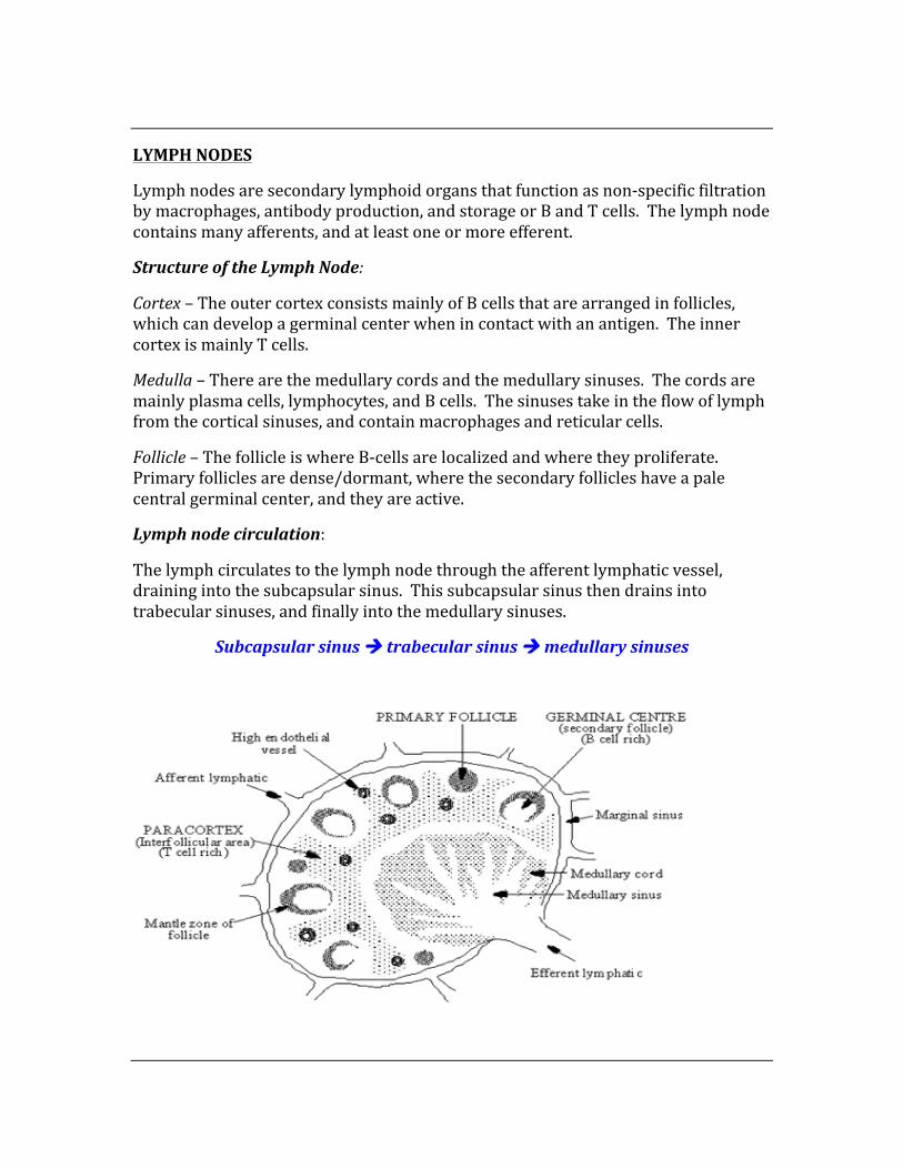
LYMPH NODES
Lymph nodes are secondary lymphoid organs that function as non-‐specific filtration by macrophages, antibody production, and storage or B and T cells. The lymph node contains many afferents, and at least one or more efferent.
Structure of the Lymph Node:
Cortex – The outer cortex consists mainly of B cells that are arranged in follicles, which can develop a germinal center when in contact with an antigen. The inner cortex is mainly T cells.
Medulla – There are the medullary cords and the medullary sinuses. The cords are mainly plasma cells, lymphocytes, and B cells. The sinuses take in the flow of lymph from the cortical sinuses, and contain macrophages and reticular cells.
Follicle – The follicle is where B-‐cells are localized and where they proliferate. Primary follicles are dense/dormant, where the secondary follicles have a pale central germinal center, and they are active.
Lymph node circulation:
The lymph circulates to the lymph node through the afferent lymphatic vessel, draining into the subcapsular sinus. This subcapsular sinus then drains into trabecular sinuses, and finally into the medullary sinuses.
Subcapsular sinus à trabecular sinus à medullary sinuses
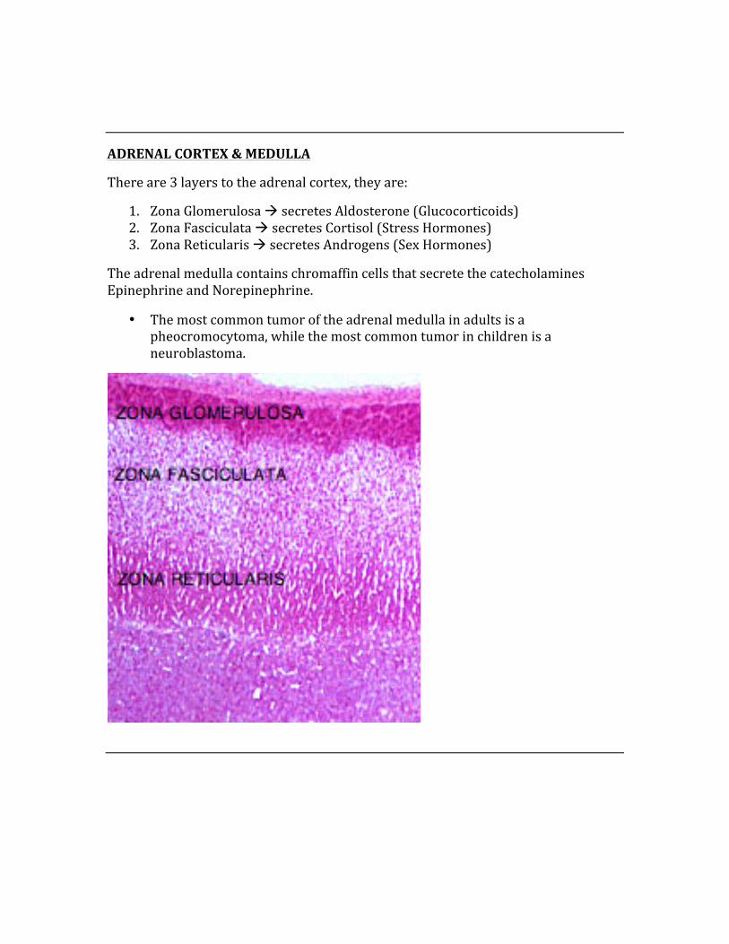
ADRENAL CORTEX & MEDULLA
There are 3 layers to the adrenal cortex, they are:
1. Zona Glomerulosa à secretes Aldosterone (Glucocorticoids) 2. Zona Fasciculata à secretes Cortisol (Stress Hormones) 3. Zona Reticularis à secretes Androgens (Sex Hormones)
The adrenal medulla contains chromaffin cells that secrete the catecholamines Epinephrine and Norepinephrine.
• The most common tumor of the adrenal medulla in adults is a pheocromocytoma, while the most common tumor in children is a neuroblastoma.
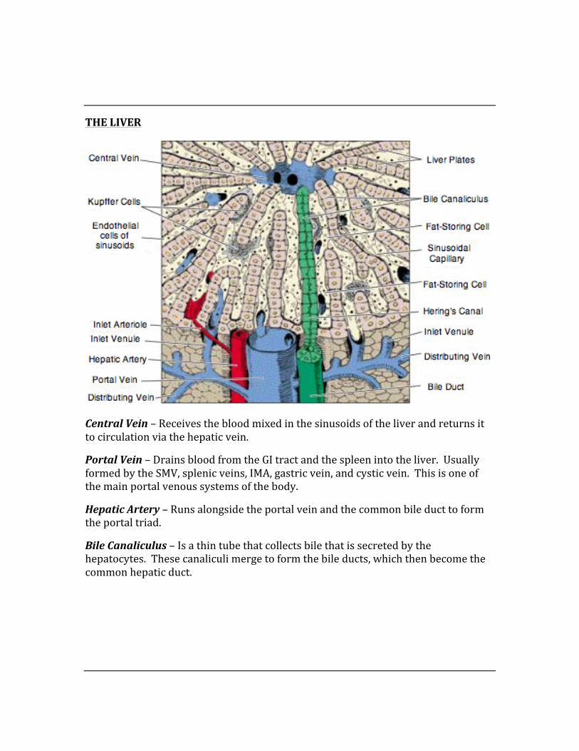
THE LIVER
Central Vein – Receives the blood mixed in the sinusoids of the liver and returns it to circulation via the hepatic vein.
Portal Vein – Drains blood from the GI tract and the spleen into the liver. Usually formed by the SMV, splenic veins, IMA, gastric vein, and cystic vein. This is one of the main portal venous systems of the body.
Hepatic Artery – Runs alongside the portal vein and the common bile duct to form the portal triad.
Bile Canaliculus – Is a thin tube that collects bile that is secreted by the hepatocytes. These canaliculi merge to form the bile ducts, which then become the common hepatic duct.
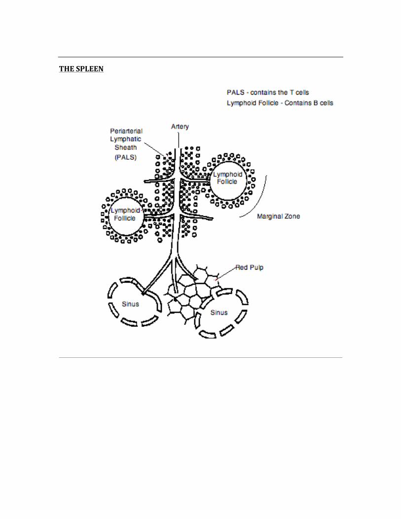
THE SPLEEN
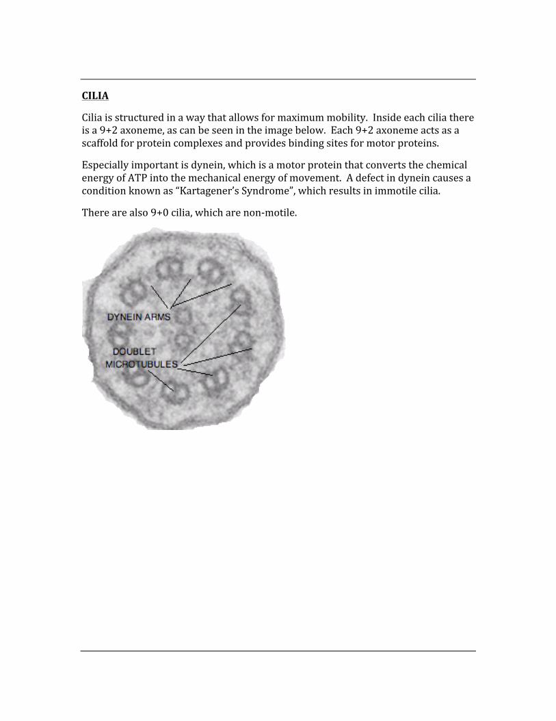
CILIA
Cilia is structured in a way that allows for maximum mobility. Inside each cilia there is a 9+2 axoneme, as can be seen in the image below. Each 9+2 axoneme acts as a scaffold for protein complexes and provides binding sites for motor proteins.
Especially important is dynein, which is a motor protein that converts the chemical energy of ATP into the mechanical energy of movement. A defect in dynein causes a condition known as “Kartagener’s Syndrome”, which results in immotile cilia.
There are also 9+0 cilia, which are non-‐motile.
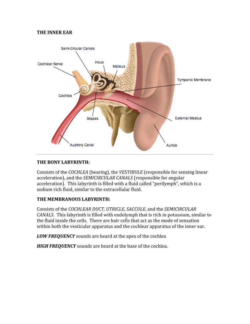
THE INNER EAR
THE BONY LABYRINTH:
Consists of the COCHLEA (hearing), the VESTIBULE (responsible for sensing linear acceleration), and the SEMICIRCULAR CANALS (responsible for angular acceleration). This labyrinth is filled with a fluid called “perilymph”, which is a sodium rich fluid, similar to the extracellular fluid.
THE MEMBRANOUS LABYRINTH:
Consists of the COCHLEAR DUCT, UTRICLE, SACCULE, and the SEMICIRCULAR CANALS. This labyrinth is filled with endolymph that is rich in potassium, similar to the fluid inside the cells. There are hair cells that act as the mode of sensation within both the vesticular apparatus and the cochlear apparatus of the inner ear.
LOW FREQUENCY sounds are heard at the apex of the cochlea
HIGH FREQUENCY sounds are heard at the base of the cochlea.
CHAPTER 4
NEUROANATOMY
Neuroanatomy is a high-‐yield section of the USMLE exam. Focus on basic neuroanatomy (blood supply, nerve supply),
as well the associated neuropathology and neurophysiology.
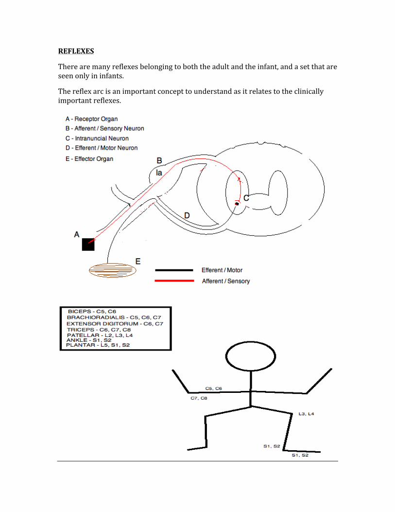
REFLEXES
There are many reflexes belonging to both the adult and the infant, and a set that are seen only in infants.
The reflex arc is an important concept to understand as it relates to the clinically important reflexes.
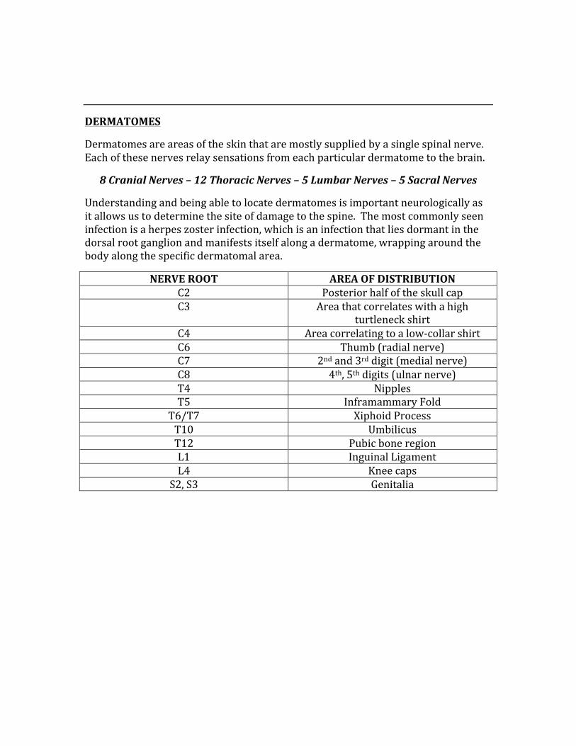
DERMATOMES
Dermatomes are areas of the skin that are mostly supplied by a single spinal nerve. Each of these nerves relay sensations from each particular dermatome to the brain.
8 Cranial Nerves – 12 Thoracic Nerves – 5 Lumbar Nerves – 5 Sacral Nerves
Understanding and being able to locate dermatomes is important neurologically as it allows us to determine the site of damage to the spine. The most commonly seen infection is a herpes zoster infection, which is an infection that lies dormant in the dorsal root ganglion and manifests itself along a dermatome, wrapping around the body along the specific dermatomal area.
NERVE ROOT AREA OF DISTRIBUTION C2 Posterior half of the skull cap C3 Area that correlates with a high
turtleneck shirt C4 Area correlating to a low-‐collar shirt C6 Thumb (radial nerve) C7 2nd and 3rd digit (medial nerve) C8 4th, 5th digits (ulnar nerve) T4 Nipples T5 Inframammary Fold
T6/T7 Xiphoid Process T10 Umbilicus T12 Pubic bone region L1 Inguinal Ligament L4 Knee caps
S2, S3 Genitalia
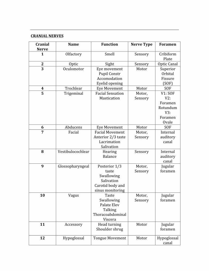
CRANIAL NERVES
Cranial Nerve
Name Function Nerve Type Foramen
1 Olfactory Smell Sensory Cribiform Plate
2 Optic Sight Sensory Optic Canal 3 Oculomotor Eye movement
Pupil Constr Accomodation Eyelid opening
Motor Superior Orbital Fissure (SOF)
4 Trochlear Eye Movement Motor SOF 5 Trigeminal Facial Sensation
Mastication Motor, Sensory
V1: SOF V2:
Foramen Rotundum
V3: Foramen Ovale
6 Abducens Eye Movement Motor SOF 7 Facial Facial Movement
Anterior 2/3 taste Lacrimation Salivation
Motor, Sensory
Internal auditory canal
8 Vestibulocochlear Hearing Balance
Sensory Internal auditory canal
9 Glossopharyngeal Posterior 1/3 taste
Swallowing Salivation
Carotid body and sinus monitoring
Motor, Sensory
Jugular foramen
10 Vagus Taste Swallowing Palate Elev Talking
Thoracoabdominal Viscera
Motor, Sensory
Jugular foramen
11 Accessory Head turning Shoulder shrug
Motor Jugular foramen
12 Hypoglossal Tongue Movement Motor Hypoglossal canal
Mnemonic for the Cranial nerves:
On Old Olympus’ Towering Tops A Friendly Viking Grew Vines And Hops
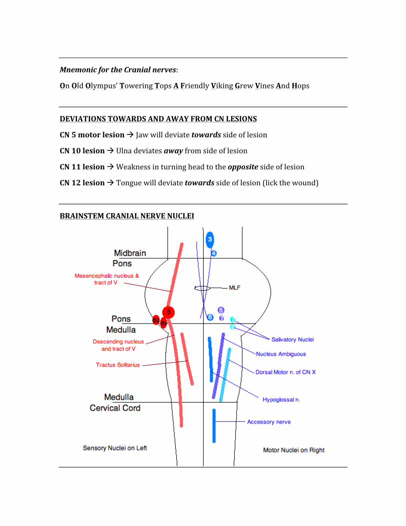
DEVIATIONS TOWARDS AND AWAY FROM CN LESIONS
CN 5 motor lesion à Jaw will deviate towards side of lesion
CN 10 lesion à Ulna deviates away from side of lesion
CN 11 lesion à Weakness in turning head to the opposite side of lesion
CN 12 lesion à Tongue will deviate towards side of lesion (lick the wound)
BRAINSTEM CRANIAL NERVE NUCLEI
THE TRACTS OF THE SPINAL CORD
Memorization of the functions of different areas of the spinal cord is essential to being able to identify where a particular spinal cord lesion may be located.
Pyrimidal:
Lateral Corticospinal – Controls movement of ipsilateral limbs
Anterior Corticospinal – Conduction of voluntary motor impulses from precentral gyrus to the motor center of the cord
Extrapyrimidal:
Rubrospinal – Main route for mediation of voluntary movement (large muscles and fine motor control)
Reticulospinal – Co-‐ordinates automatic movement of locomotion and posture, influences muscle tone, mediates autonomic functions, and modulates pain impulses
Vestibulospinal – Lateral: Ipsilateral descent to paravertebral and proximal limb extension. Medial: Bilateral descent, controls eye movement, neck position, gaze
Dorsal Column Medial Lemniscus System:
Gracile Faciculus – Fine touch, vibration, proprioception to lower body
Cuneate Fasciculus – Fine touch, vibration, and proprioception to upper body
Spinocerebellar Tract:
Posterior Spinocerebellar Tract – Limb and joint position
Anterior Spinocerebellar Tract – Limb and joint position
Anterolateral System:
Lateral Spinothalamic Tract – Pain and temperature
Anterior Spinothalamic Tract – Soft nocioception
Spino-‐Olivary Tract: Proprioception from muscles and tendons as well as cutaneous impulses to the olivary nucleus
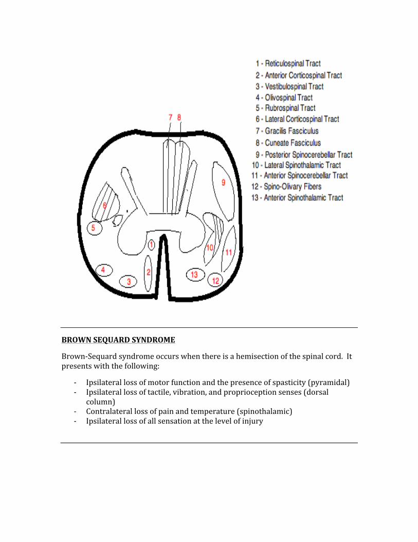
BROWN SEQUARD SYNDROME
Brown-‐Sequard syndrome occurs when there is a hemisection of the spinal cord. It presents with the following:
-‐ Ipsilateral loss of motor function and the presence of spasticity (pyramidal) -‐ Ipsilateral loss of tactile, vibration, and proprioception senses (dorsal
column) -‐ Contralateral loss of pain and temperature (spinothalamic) -‐ Ipsilateral loss of all sensation at the level of injury
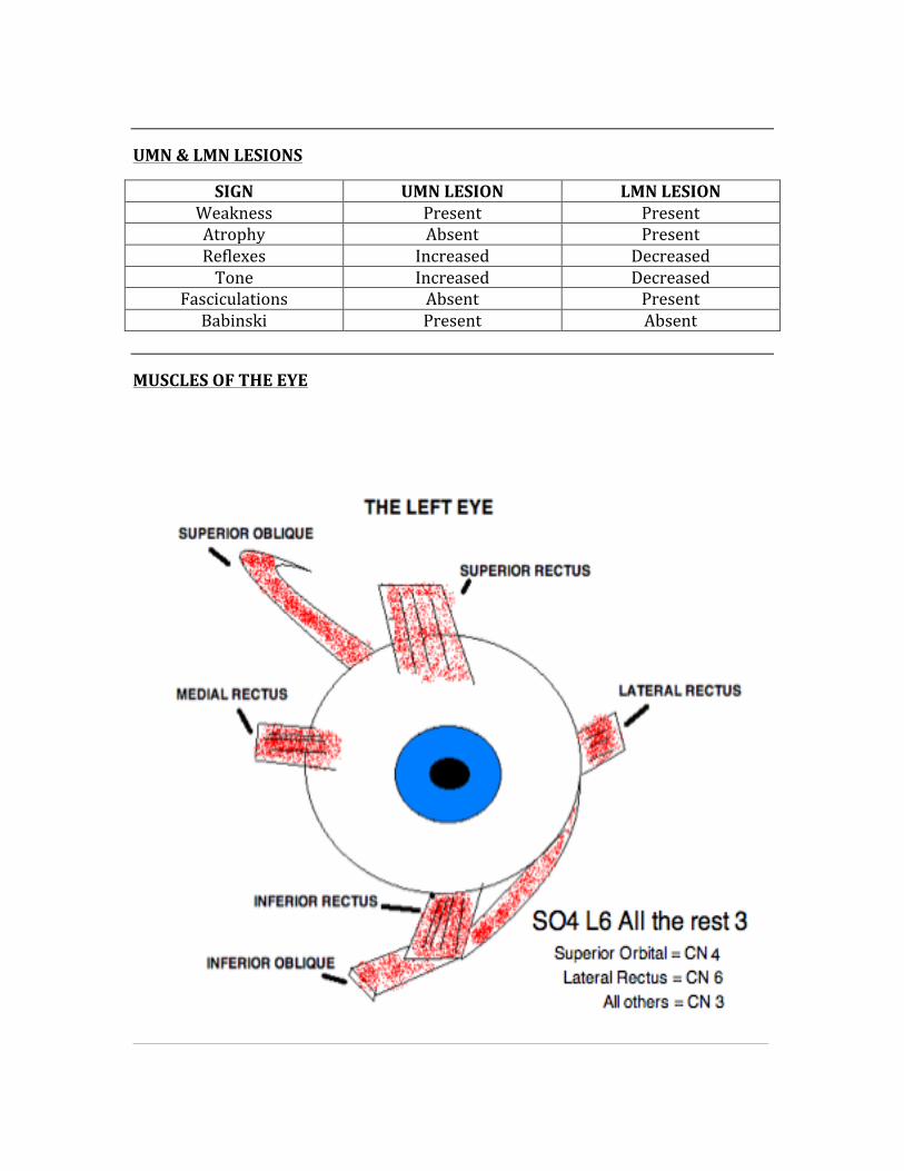
UMN & LMN LESIONS
SIGN UMN LESION LMN LESION Weakness Present Present Atrophy Absent Present Reflexes Increased Decreased Tone Increased Decreased
Fasciculations Absent Present Babinski Present Absent
MUSCLES OF THE EYE
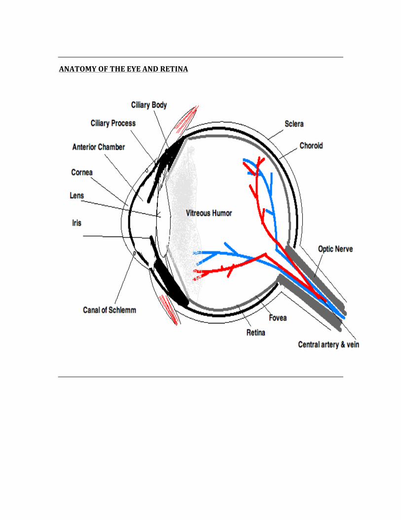
ANATOMY OF THE EYE AND RETINA
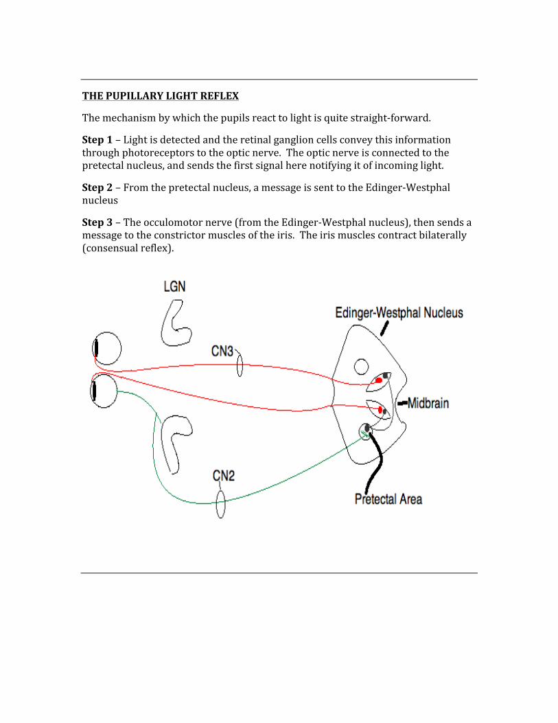
THE PUPILLARY LIGHT REFLEX
The mechanism by which the pupils react to light is quite straight-‐forward.
Step 1 – Light is detected and the retinal ganglion cells convey this information through photoreceptors to the optic nerve. The optic nerve is connected to the pretectal nucleus, and sends the first signal here notifying it of incoming light.
Step 2 – From the pretectal nucleus, a message is sent to the Edinger-‐Westphal nucleus
Step 3 – The occulomotor nerve (from the Edinger-‐Westphal nucleus), then sends a message to the constrictor muscles of the iris. The iris muscles contract bilaterally (consensual reflex).
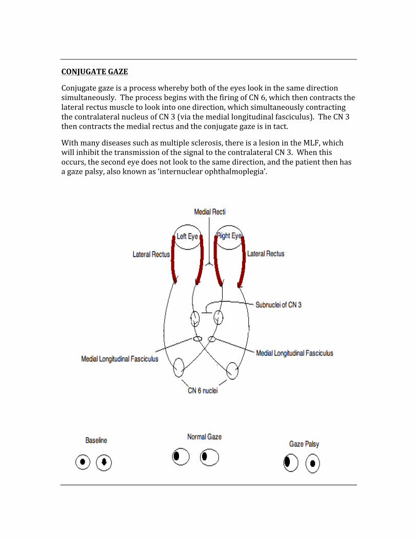
CONJUGATE GAZE
Conjugate gaze is a process whereby both of the eyes look in the same direction simultaneously. The process begins with the firing of CN 6, which then contracts the lateral rectus muscle to look into one direction, which simultaneously contracting the contralateral nucleus of CN 3 (via the medial longitudinal fasciculus). The CN 3 then contracts the medial rectus and the conjugate gaze is in tact.
With many diseases such as multiple sclerosis, there is a lesion in the MLF, which will inhibit the transmission of the signal to the contralateral CN 3. When this occurs, the second eye does not look to the same direction, and the patient then has a gaze palsy, also known as ‘internuclear ophthalmoplegia’.
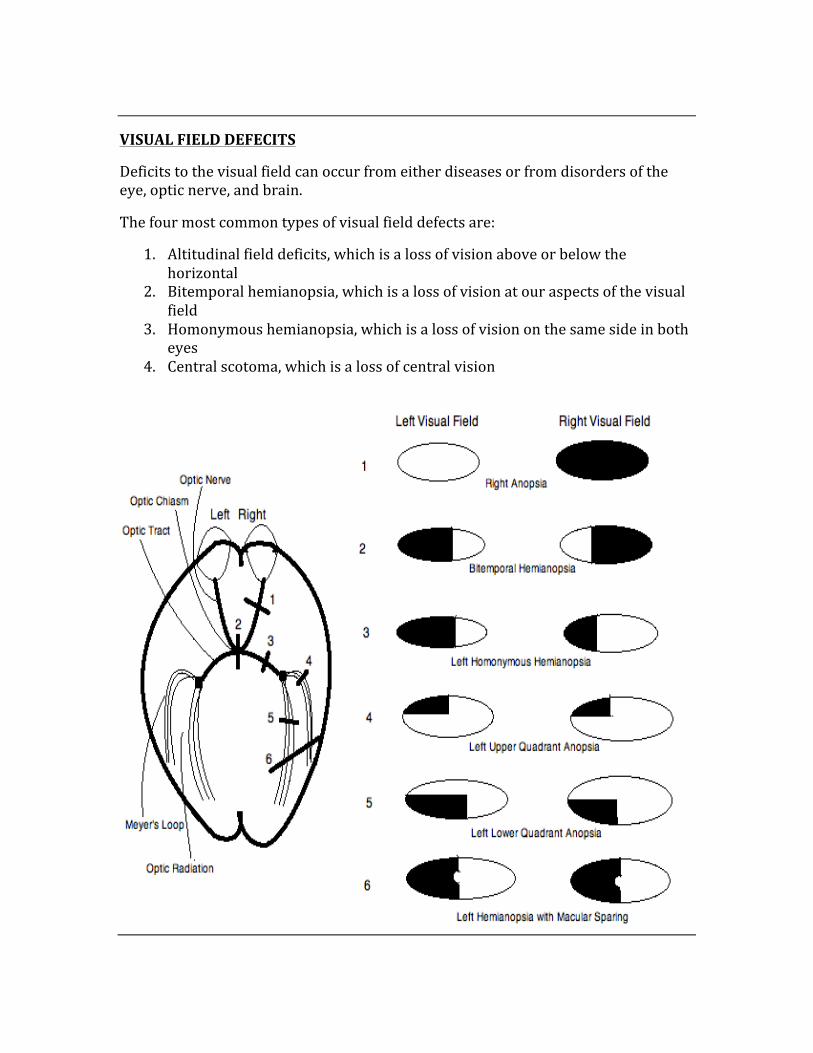
VISUAL FIELD DEFECITS
Deficits to the visual field can occur from either diseases or from disorders of the eye, optic nerve, and brain.
The four most common types of visual field defects are:
1. Altitudinal field deficits, which is a loss of vision above or below the horizontal
2. Bitemporal hemianopsia, which is a loss of vision at our aspects of the visual field
3. Homonymous hemianopsia, which is a loss of vision on the same side in both eyes
4. Central scotoma, which is a loss of central vision
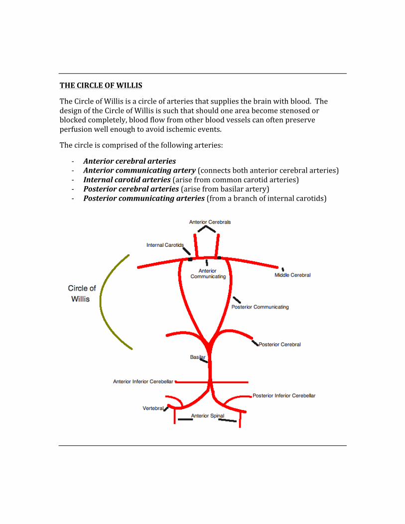
THE CIRCLE OF WILLIS
The Circle of Willis is a circle of arteries that supplies the brain with blood. The design of the Circle of Willis is such that should one area become stenosed or blocked completely, blood flow from other blood vessels can often preserve perfusion well enough to avoid ischemic events.
The circle is comprised of the following arteries:
-‐ Anterior cerebral arteries -‐ Anterior communicating artery (connects both anterior cerebral arteries) -‐ Internal carotid arteries (arise from common carotid arteries) -‐ Posterior cerebral arteries (arise from basilar artery) -‐ Posterior communicating arteries (from a branch of internal carotids)
BLOOD SUPPLY TO THE BRAIN
Anterior Cerebral Artery – supplies the medial surface of the brain, and the leg-‐foot area of the motor and sensory cortices.
Middle Cerebral Artery – supplies the lateral aspect of the brain, the trunk-‐arm-‐face area of the motor and sensory cortices, as well as Broca’s and Wernicke’s speech areas.
Anterior Communicating Artery – connects the anterior cerebral arteries, and is the most common site of Circle of Willis aneurysm.
Posterior Communicating Artery – connects three cerebral arteries on each side, is another common site of aneurysm, and can cause cranial nerve 3 palsies.
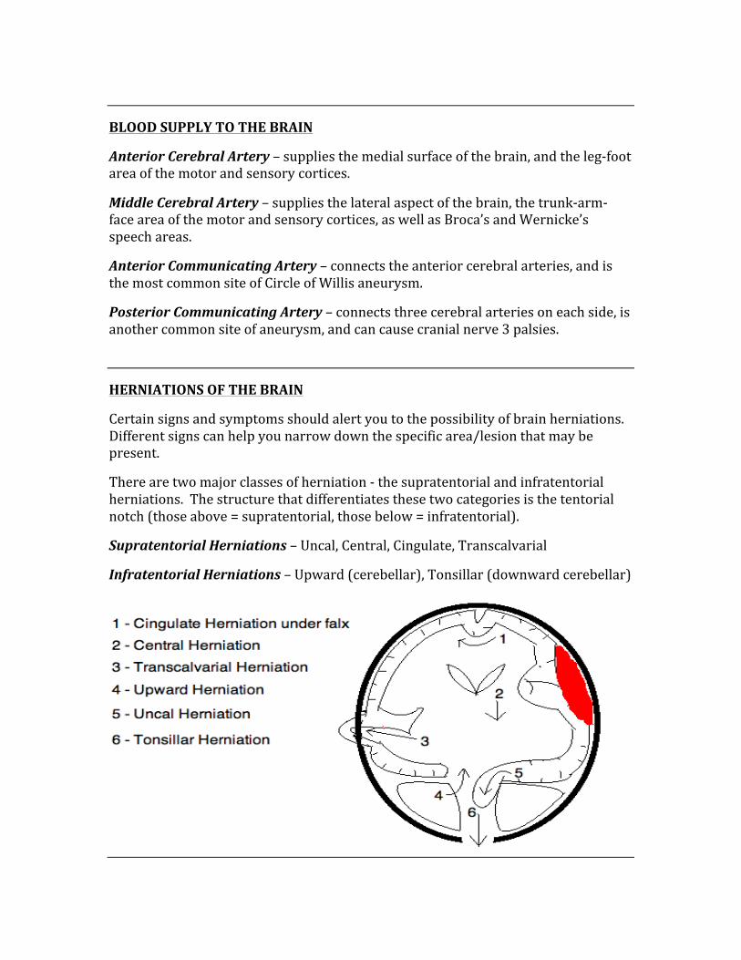
HERNIATIONS OF THE BRAIN
Certain signs and symptoms should alert you to the possibility of brain herniations. Different signs can help you narrow down the specific area/lesion that may be present.
There are two major classes of herniation -‐ the supratentorial and infratentorial herniations. The structure that differentiates these two categories is the tentorial notch (those above = supratentorial, those below = infratentorial).
Supratentorial Herniations – Uncal, Central, Cingulate, Transcalvarial
Infratentorial Herniations – Upward (cerebellar), Tonsillar (downward cerebellar)
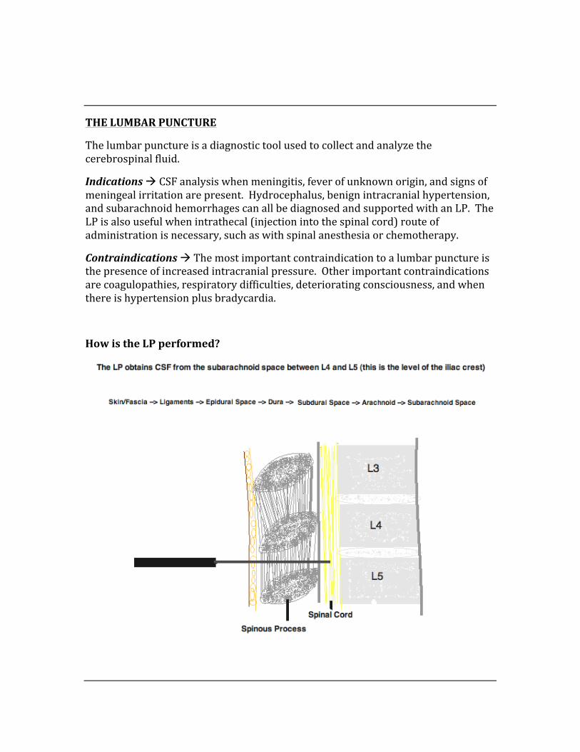
THE LUMBAR PUNCTURE
The lumbar puncture is a diagnostic tool used to collect and analyze the cerebrospinal fluid.
Indications à CSF analysis when meningitis, fever of unknown origin, and signs of meningeal irritation are present. Hydrocephalus, benign intracranial hypertension, and subarachnoid hemorrhages can all be diagnosed and supported with an LP. The LP is also useful when intrathecal (injection into the spinal cord) route of administration is necessary, such as with spinal anesthesia or chemotherapy.
Contraindications à The most important contraindication to a lumbar puncture is the presence of increased intracranial pressure. Other important contraindications are coagulopathies, respiratory difficulties, deteriorating consciousness, and when there is hypertension plus bradycardia.
How is the LP performed?
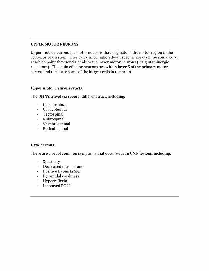
UPPER MOTOR NEURONS
Upper motor neurons are motor neurons that originate in the motor region of the cortex or brain stem. They carry information down specific areas on the spinal cord, at which point they send signals to the lower motor neurons (via glutaminergic receptors). The main effector neurons are within layer 5 of the primary motor cortex, and these are some of the largest cells in the brain.
Upper motor neurons tracts:
The UMN’s travel via several different tract, including:
-‐ Corticospinal -‐ Corticobulbar -‐ Tectospinal -‐ Rubrospinal -‐ Vestibulospinal -‐ Reticulospinal
UMN Lesions:
There are a set of common symptoms that occur with an UMN lesions, including:
-‐ Spasticity -‐ Decreased muscle tone -‐ Positive Babinski Sign -‐ Pyramidal weakness -‐ Hyperreflexia -‐ Increased DTR’s
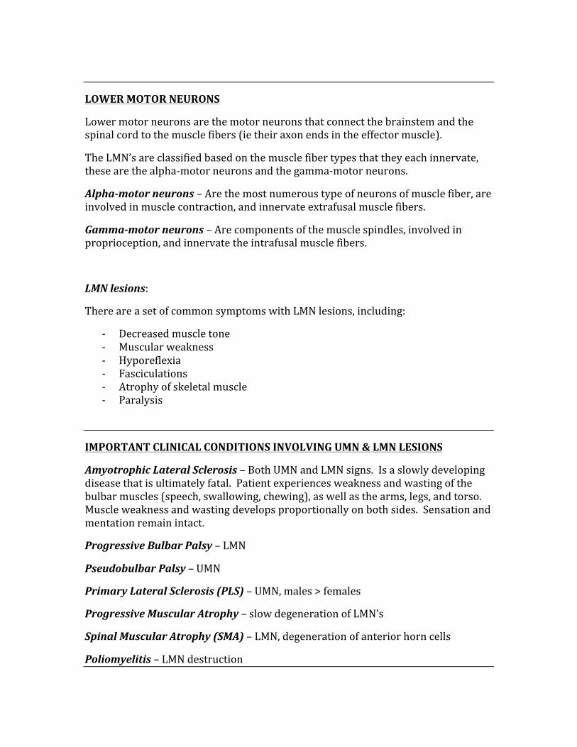
LOWER MOTOR NEURONS
Lower motor neurons are the motor neurons that connect the brainstem and the spinal cord to the muscle fibers (ie their axon ends in the effector muscle).
The LMN’s are classified based on the muscle fiber types that they each innervate, these are the alpha-‐motor neurons and the gamma-‐motor neurons.
Alpha-‐motor neurons – Are the most numerous type of neurons of muscle fiber, are involved in muscle contraction, and innervate extrafusal muscle fibers.
Gamma-‐motor neurons – Are components of the muscle spindles, involved in proprioception, and innervate the intrafusal muscle fibers.
LMN lesions:
There are a set of common symptoms with LMN lesions, including:
-‐ Decreased muscle tone -‐ Muscular weakness -‐ Hyporeflexia -‐ Fasciculations -‐ Atrophy of skeletal muscle -‐ Paralysis
IMPORTANT CLINICAL CONDITIONS INVOLVING UMN & LMN LESIONS
Amyotrophic Lateral Sclerosis – Both UMN and LMN signs. Is a slowly developing disease that is ultimately fatal. Patient experiences weakness and wasting of the bulbar muscles (speech, swallowing, chewing), as well as the arms, legs, and torso. Muscle weakness and wasting develops proportionally on both sides. Sensation and mentation remain intact.
Progressive Bulbar Palsy – LMN
Pseudobulbar Palsy – UMN
Primary Lateral Sclerosis (PLS) – UMN, males > females
Progressive Muscular Atrophy – slow degeneration of LMN’s
Spinal Muscular Atrophy (SMA) – LMN, degeneration of anterior horn cells
Poliomyelitis – LMN destruction
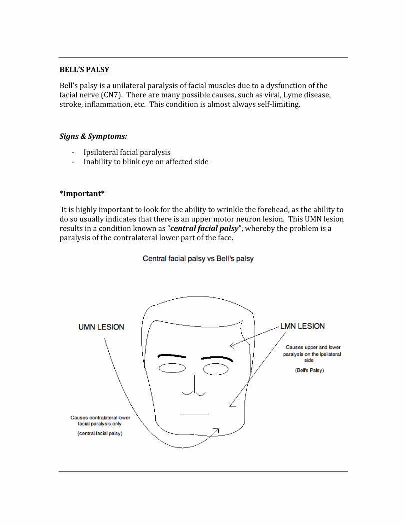
BELL’S PALSY
Bell’s palsy is a unilateral paralysis of facial muscles due to a dysfunction of the facial nerve (CN7). There are many possible causes, such as viral, Lyme disease, stroke, inflammation, etc. This condition is almost always self-‐limiting.
Signs & Symptoms:
-‐ Ipsilateral facial paralysis -‐ Inability to blink eye on affected side
*Important*
It is highly important to look for the ability to wrinkle the forehead, as the ability to do so usually indicates that there is an upper motor neuron lesion. This UMN lesion results in a condition known as “central facial palsy”, whereby the problem is a paralysis of the contralateral lower part of the face.
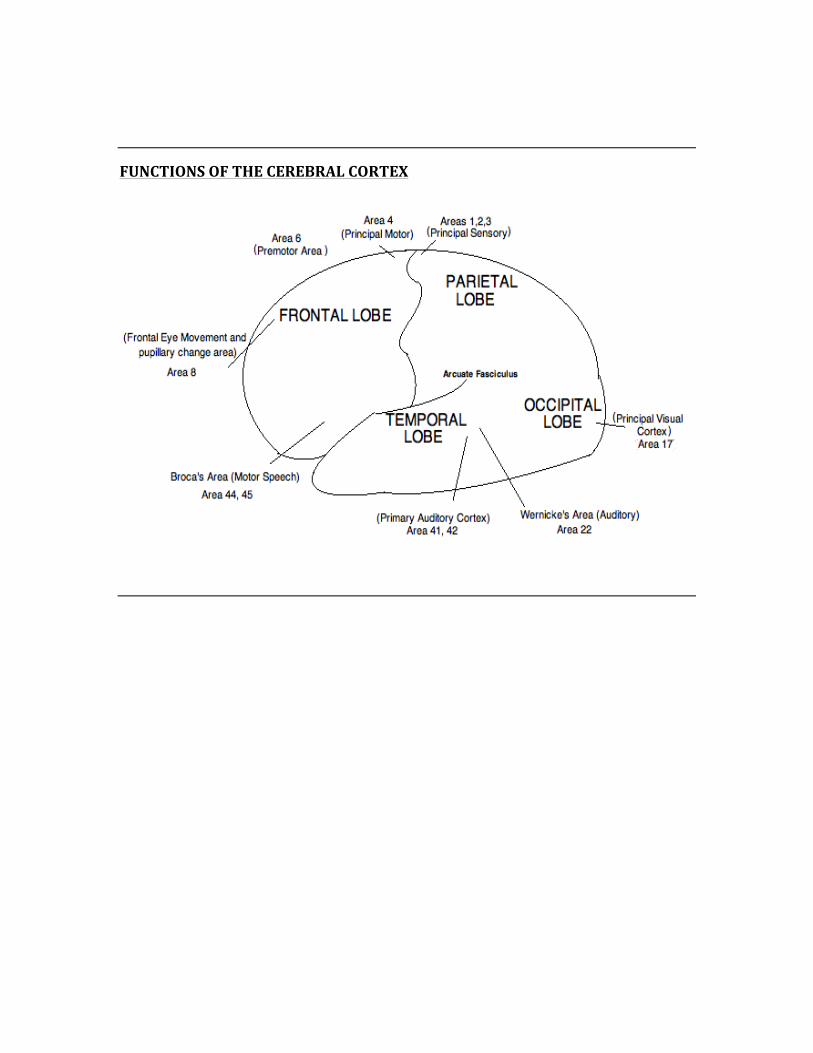
FUNCTIONS OF THE CEREBRAL CORTEX
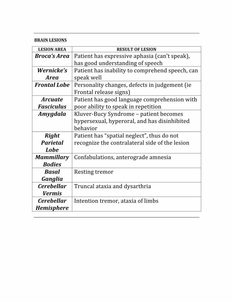
BRAIN LESIONS
LESION AREA RESULT OF LESION Broca’s Area Patient has expressive aphasia (can’t speak),
has good understanding of speech Wernicke’s
Area Patient has inability to comprehend speech, can speak well
Frontal Lobe Personality changes, defects in judgement (ie Frontal release signs)
Arcuate Fasciculus
Patient has good language comprehension with poor ability to speak in repetition
Amygdala Kluver-‐Bucy Syndrome – patient becomes hypersexual, hyperoral, and has disinhibited behavior
Right Parietal Lobe
Patient has “spatial neglect”, thus do not recognize the contralateral side of the lesion
Mammillary Bodies
Confabulations, anterograde amnesia
Basal Ganglia
Resting tremor
Cerebellar Vermis
Truncal ataxia and dysarthria
Cerebellar Hemisphere
Intention tremor, ataxia of limbs
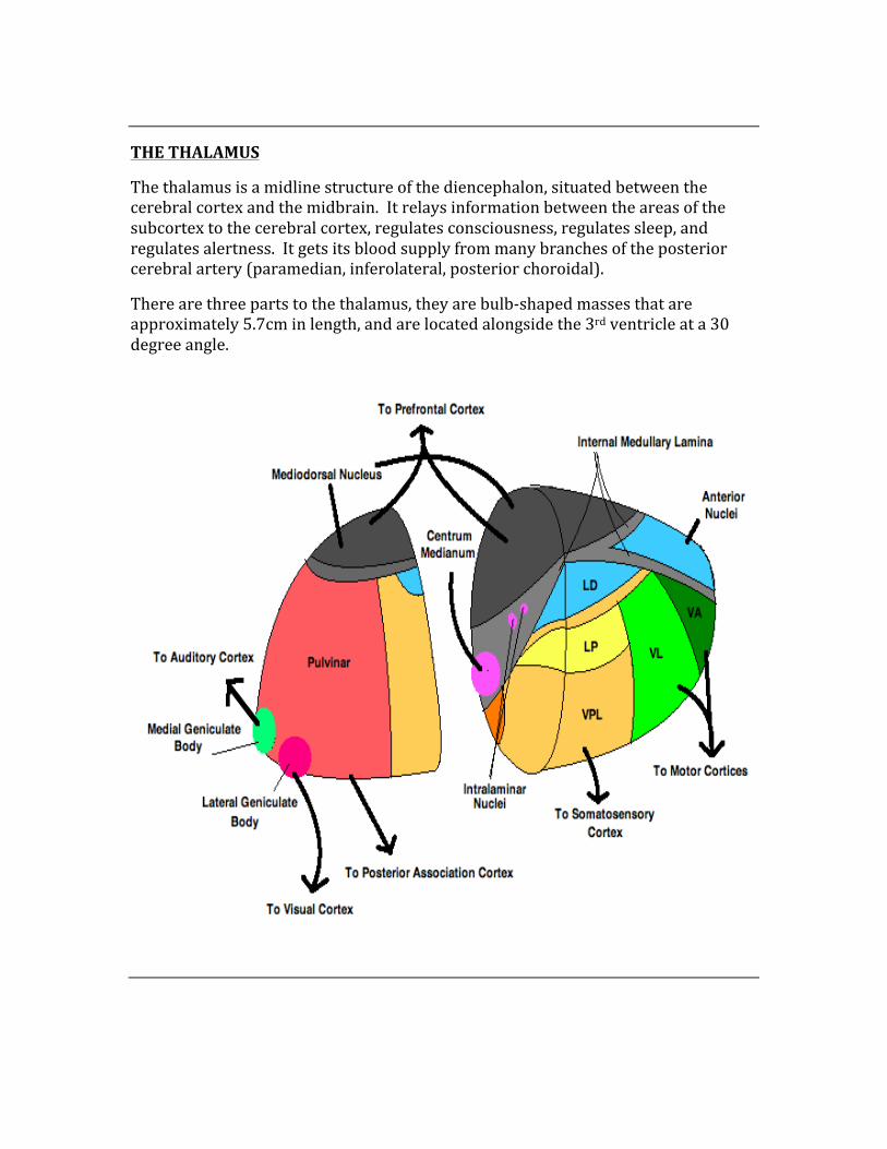
THE THALAMUS
The thalamus is a midline structure of the diencephalon, situated between the cerebral cortex and the midbrain. It relays information between the areas of the subcortex to the cerebral cortex, regulates consciousness, regulates sleep, and regulates alertness. It gets its blood supply from many branches of the posterior cerebral artery (paramedian, inferolateral, posterior choroidal).
There are three parts to the thalamus, they are bulb-‐shaped masses that are approximately 5.7cm in length, and are located alongside the 3rd ventricle at a 30 degree angle.
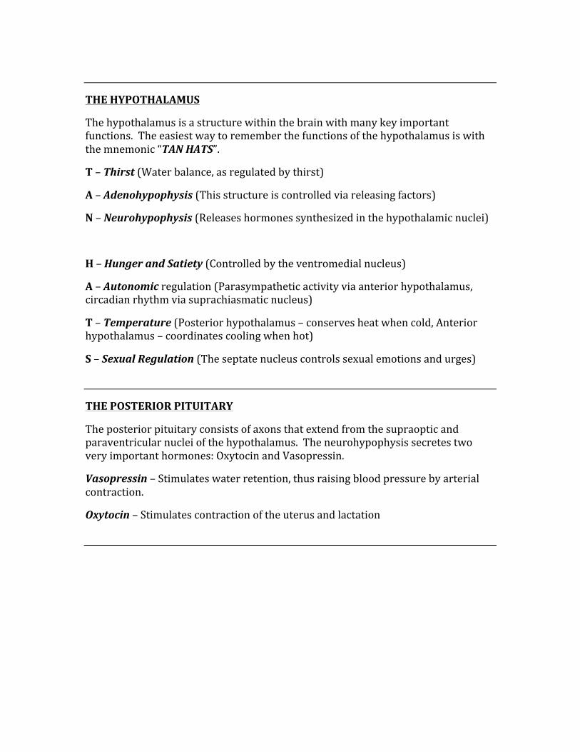
THE HYPOTHALAMUS
The hypothalamus is a structure within the brain with many key important functions. The easiest way to remember the functions of the hypothalamus is with the mnemonic “TAN HATS”.
T – Thirst (Water balance, as regulated by thirst)
A – Adenohypophysis (This structure is controlled via releasing factors)
N – Neurohypophysis (Releases hormones synthesized in the hypothalamic nuclei)
H – Hunger and Satiety (Controlled by the ventromedial nucleus)
A – Autonomic regulation (Parasympathetic activity via anterior hypothalamus, circadian rhythm via suprachiasmatic nucleus)
T – Temperature (Posterior hypothalamus – conserves heat when cold, Anterior hypothalamus – coordinates cooling when hot)
S – Sexual Regulation (The septate nucleus controls sexual emotions and urges)
THE POSTERIOR PITUITARY
The posterior pituitary consists of axons that extend from the supraoptic and paraventricular nuclei of the hypothalamus. The neurohypophysis secretes two very important hormones: Oxytocin and Vasopressin.
Vasopressin – Stimulates water retention, thus raising blood pressure by arterial contraction.
Oxytocin – Stimulates contraction of the uterus and lactation
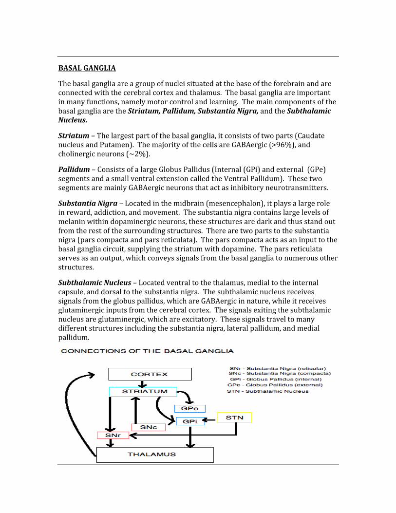
BASAL GANGLIA
The basal ganglia are a group of nuclei situated at the base of the forebrain and are connected with the cerebral cortex and thalamus. The basal ganglia are important in many functions, namely motor control and learning. The main components of the basal ganglia are the Striatum, Pallidum, Substantia Nigra, and the Subthalamic Nucleus.
Striatum – The largest part of the basal ganglia, it consists of two parts (Caudate nucleus and Putamen). The majority of the cells are GABAergic (>96%), and cholinergic neurons (~2%).
Pallidum – Consists of a large Globus Pallidus (Internal (GPi) and external (GPe) segments and a small ventral extension called the Ventral Pallidum). These two segments are mainly GABAergic neurons that act as inhibitory neurotransmitters.
Substantia Nigra – Located in the midbrain (mesencephalon), it plays a large role in reward, addiction, and movement. The substantia nigra contains large levels of melanin within dopaminergic neurons, these structures are dark and thus stand out from the rest of the surrounding structures. There are two parts to the substantia nigra (pars compacta and pars reticulata). The pars compacta acts as an input to the basal ganglia circuit, supplying the striatum with dopamine. The pars reticulata serves as an output, which conveys signals from the basal ganglia to numerous other structures.
Subthalamic Nucleus – Located ventral to the thalamus, medial to the internal capsule, and dorsal to the substantia nigra. The subthalamic nucleus receives signals from the globus pallidus, which are GABAergic in nature, while it receives glutaminergic inputs from the cerebral cortex. The signals exiting the subthalamic nucleus are glutaminergic, which are excitatory. These signals travel to many different structures including the substantia nigra, lateral pallidum, and medial pallidum.
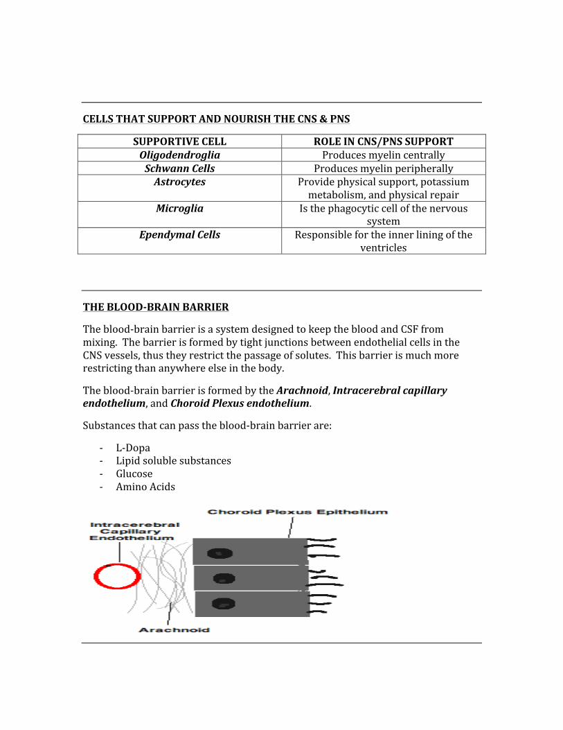
CELLS THAT SUPPORT AND NOURISH THE CNS & PNS
SUPPORTIVE CELL ROLE IN CNS/PNS SUPPORT Oligodendroglia Produces myelin centrally Schwann Cells Produces myelin peripherally Astrocytes Provide physical support, potassium
metabolism, and physical repair Microglia Is the phagocytic cell of the nervous
system Ependymal Cells Responsible for the inner lining of the
ventricles
THE BLOOD-‐BRAIN BARRIER
The blood-‐brain barrier is a system designed to keep the blood and CSF from mixing. The barrier is formed by tight junctions between endothelial cells in the CNS vessels, thus they restrict the passage of solutes. This barrier is much more restricting than anywhere else in the body.
The blood-‐brain barrier is formed by the Arachnoid, Intracerebral capillary endothelium, and Choroid Plexus endothelium.
Substances that can pass the blood-‐brain barrier are:
-‐ L-‐Dopa -‐ Lipid soluble substances -‐ Glucose -‐ Amino Acids
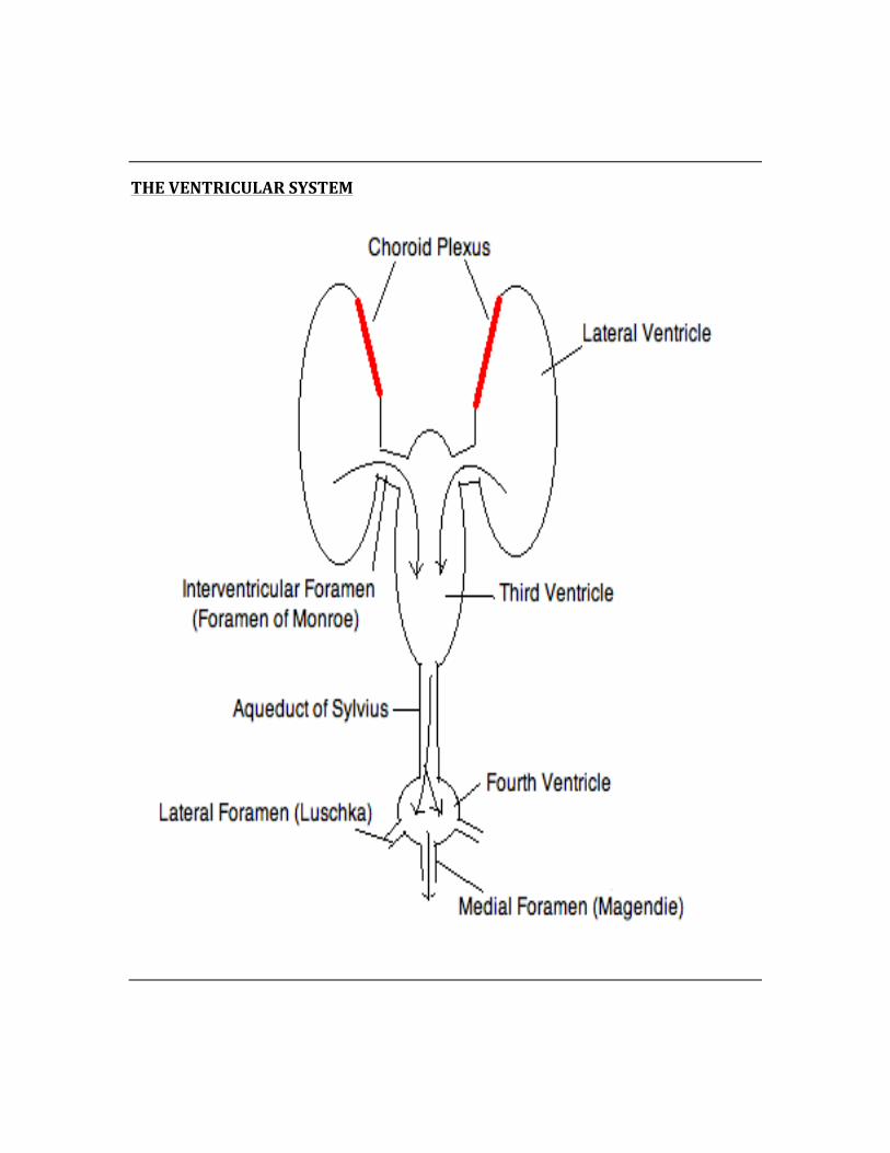
THE VENTRICULAR SYSTEM
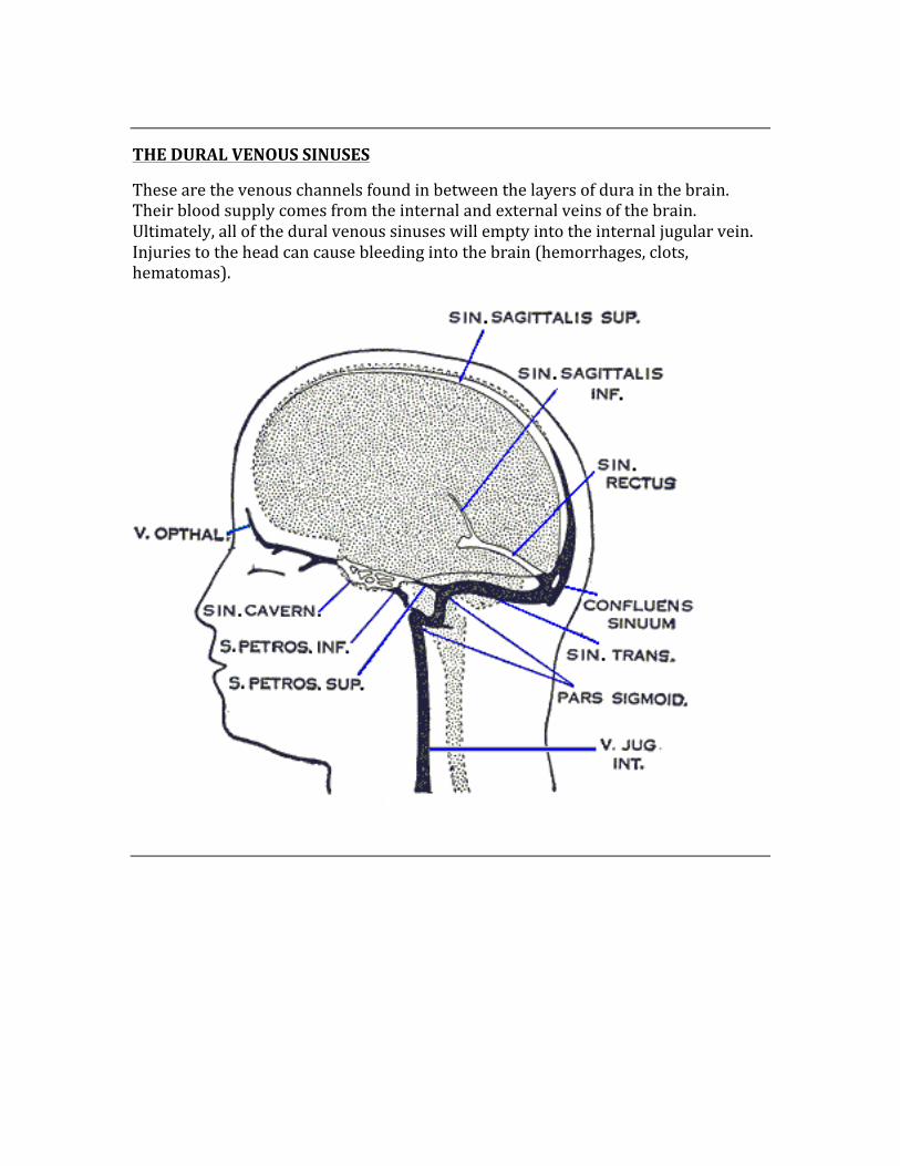
THE DURAL VENOUS SINUSES
These are the venous channels found in between the layers of dura in the brain. Their blood supply comes from the internal and external veins of the brain. Ultimately, all of the dural venous sinuses will empty into the internal jugular vein. Injuries to the head can cause bleeding into the brain (hemorrhages, clots, hematomas).
CHAPTER 5
PHYSIOLOGY
Physiology fits into many aspects of the USMLE exam. It is a highly conceptual topic, and full understanding of these
concepts is essential to success on the Step 1 exam.
RENAL PHYSIOLOGY
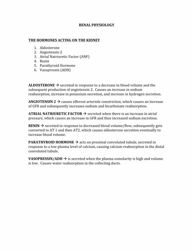
THE HORMONES ACTING ON THE KIDNEY
1. Aldosterone 2. Angiotensin 2 3. Atrial Natriuretic Factor (ANF) 4. Renin 5. Parathyroid Hormone 6. Vasopressin (ADH)
ALDOSTERONE à secreted in response to a decrease in blood volume and the subsequent production of angiotensin 2. Causes an increase in sodium reabsorption, increase in potassium secretion, and increase in hydrogen secretion.
ANGIOTENSIN 2 à causes efferent arteriole constriction, which causes an increase of GFR and subsequently increases sodium and bicarbonate reabsorption.
ATRIAL NATRIURETIC FACTOR à secreted when there is an increase in atrial pressure, which causes an increase in GFR and thus increased sodium excretion.
RENIN à secreted in response to decreased blood volume/flow, subsequently gets converted to AT 1 and then AT2, which causes aldosterone secretion eventually to increase blood volume.
PARATHYROID HORMONE à acts on proximal convoluted tubule, secreted in response to a low plasma level of calcium, causing calcium reabsorption in the distal convoluted tubule.
VASOPRESSIN/ADH à is secreted when the plasma osmolarity is high and volume is low. Causes water reabsorption in the collecting ducts.
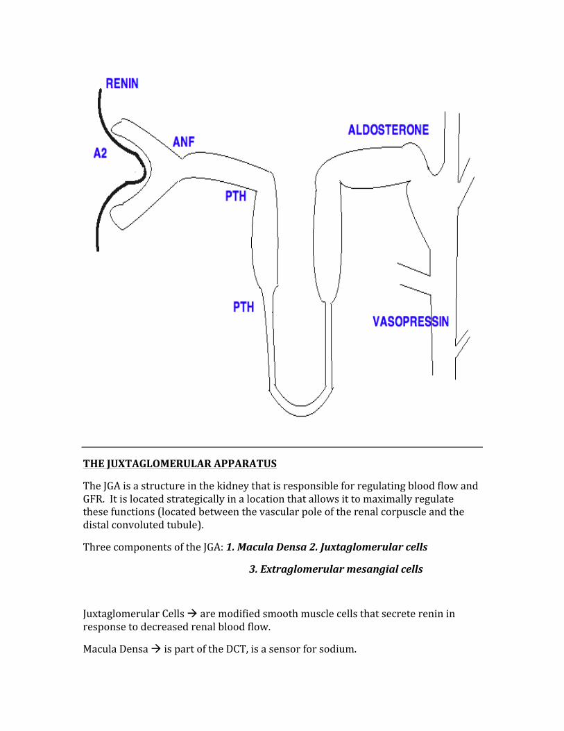
THE JUXTAGLOMERULAR APPARATUS
The JGA is a structure in the kidney that is responsible for regulating blood flow and GFR. It is located strategically in a location that allows it to maximally regulate these functions (located between the vascular pole of the renal corpuscle and the distal convoluted tubule).
Three components of the JGA: 1. Macula Densa 2. Juxtaglomerular cells
3. Extraglomerular mesangial cells
Juxtaglomerular Cells à are modified smooth muscle cells that secrete renin in response to decreased renal blood flow.
Macula Densa à is part of the DCT, is a sensor for sodium.
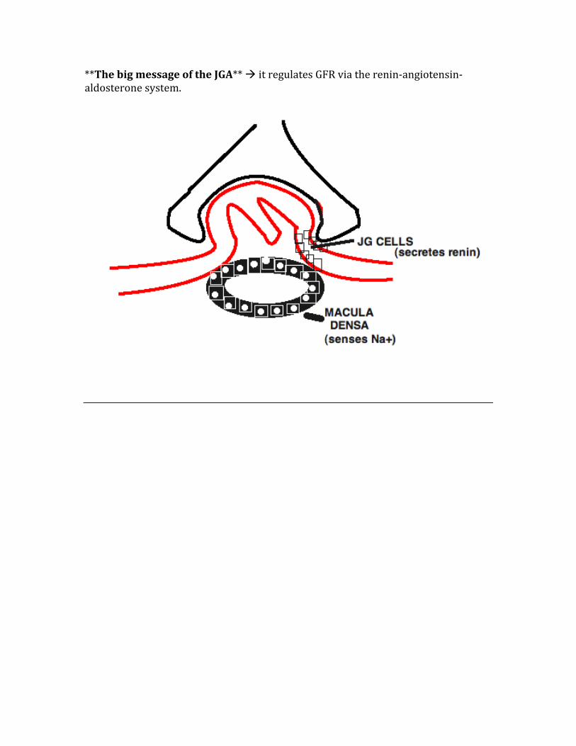
**The big message of the JGA** à it regulates GFR via the renin-‐angiotensin-‐aldosterone system.
IMPORTANT EQUATIONS IN RENAL PHYSIOLOGY
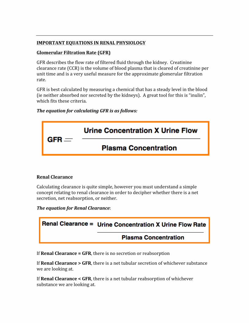
Glomerular Filtration Rate (GFR)
GFR describes the flow rate of filtered fluid through the kidney. Creatinine clearance rate (CCR) is the volume of blood plasma that is cleared of creatinine per unit time and is a very useful measure for the approximate glomerular filtration rate.
GFR is best calculated by measuring a chemical that has a steady level in the blood (ie neither absorbed nor secreted by the kidneys). A great tool for this is “inulin”, which fits these criteria.
The equation for calculating GFR is as follows:
Renal Clearance
Calculating clearance is quite simple, however you must understand a simple concept relating to renal clearance in order to decipher whether there is a net secretion, net reabsorption, or neither.
The equation for Renal Clearance:
If Renal Clearance = GFR, there is no secretion or reabsorption
If Renal Clearance > GFR, there is a net tubular secretion of whichever substance we are looking at.
If Renal Clearance < GFR, there is a net tubular reabsorption of whichever substance we are looking at.
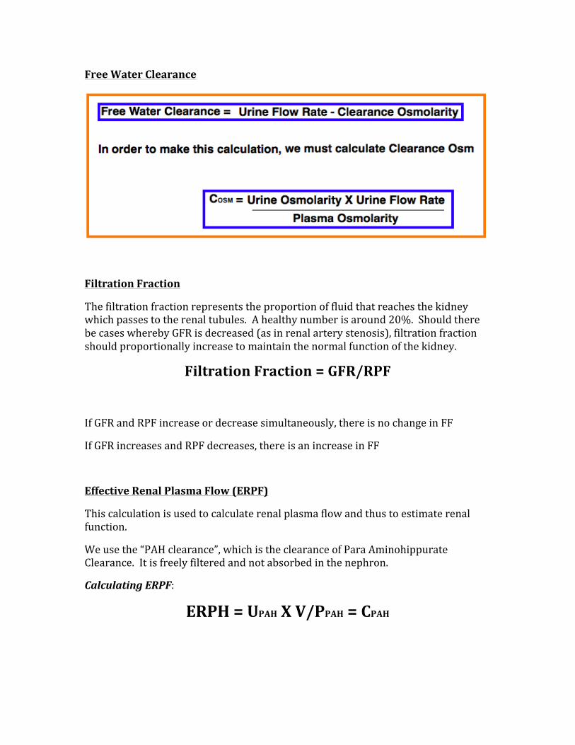
Free Water Clearance
Filtration Fraction
The filtration fraction represents the proportion of fluid that reaches the kidney which passes to the renal tubules. A healthy number is around 20%. Should there be cases whereby GFR is decreased (as in renal artery stenosis), filtration fraction should proportionally increase to maintain the normal function of the kidney.
Filtration Fraction = GFR/RPF
If GFR and RPF increase or decrease simultaneously, there is no change in FF
If GFR increases and RPF decreases, there is an increase in FF
Effective Renal Plasma Flow (ERPF)
This calculation is used to calculate renal plasma flow and thus to estimate renal function.
We use the “PAH clearance”, which is the clearance of Para Aminohippurate Clearance. It is freely filtered and not absorbed in the nephron.
Calculating ERPF:
ERPH = UPAH X V/PPAH = CPAH
Glucose Clearance
Important in diabetes, it should simply be known that glucosuria occurs when plasma glucose reaches 200mg/dL, because the PCT cannot reabsorb once these levels are reached.
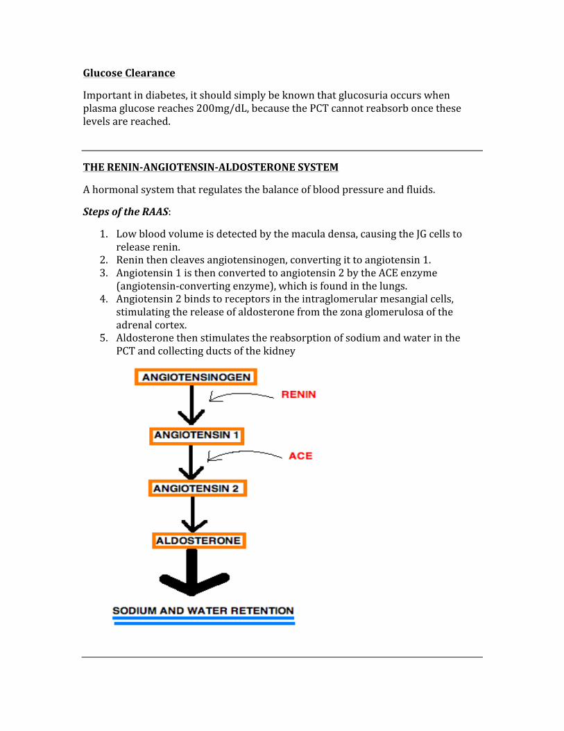
THE RENIN-‐ANGIOTENSIN-‐ALDOSTERONE SYSTEM
A hormonal system that regulates the balance of blood pressure and fluids.
Steps of the RAAS:
1. Low blood volume is detected by the macula densa, causing the JG cells to release renin.
2. Renin then cleaves angiotensinogen, converting it to angiotensin 1. 3. Angiotensin 1 is then converted to angiotensin 2 by the ACE enzyme
(angiotensin-‐converting enzyme), which is found in the lungs. 4. Angiotensin 2 binds to receptors in the intraglomerular mesangial cells,
stimulating the release of aldosterone from the zona glomerulosa of the adrenal cortex.
5. Aldosterone then stimulates the reabsorption of sodium and water in the PCT and collecting ducts of the kidney
PHYSIOLOGY AT EACH PORTION OF THE NEPHRON
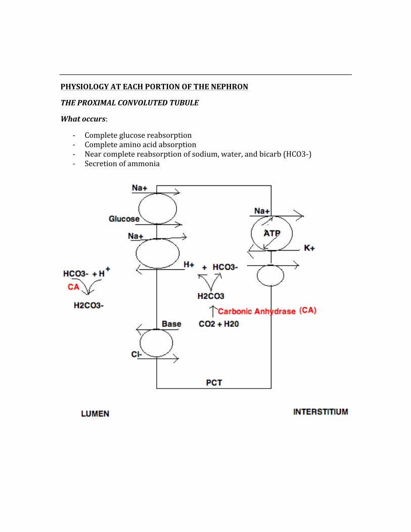
THE PROXIMAL CONVOLUTED TUBULE
What occurs:
-‐ Complete glucose reabsorption -‐ Complete amino acid absorption -‐ Near complete reabsorption of sodium, water, and bicarb (HCO3-‐) -‐ Secretion of ammonia
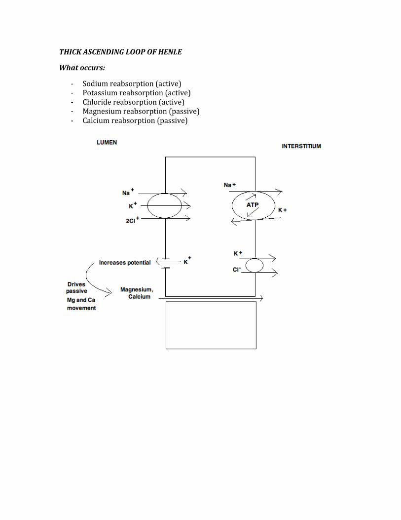
THICK ASCENDING LOOP OF HENLE
What occurs:
-‐ Sodium reabsorption (active) -‐ Potassium reabsorption (active) -‐ Chloride reabsorption (active) -‐ Magnesium reabsorption (passive) -‐ Calcium reabsorption (passive)
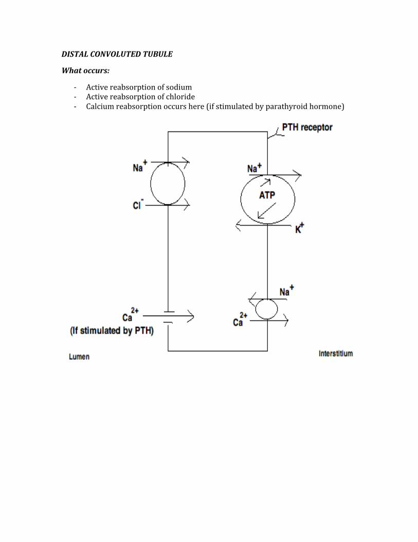
DISTAL CONVOLUTED TUBULE
What occurs:
-‐ Active reabsorption of sodium -‐ Active reabsorption of chloride -‐ Calcium reabsorption occurs here (if stimulated by parathyroid hormone)
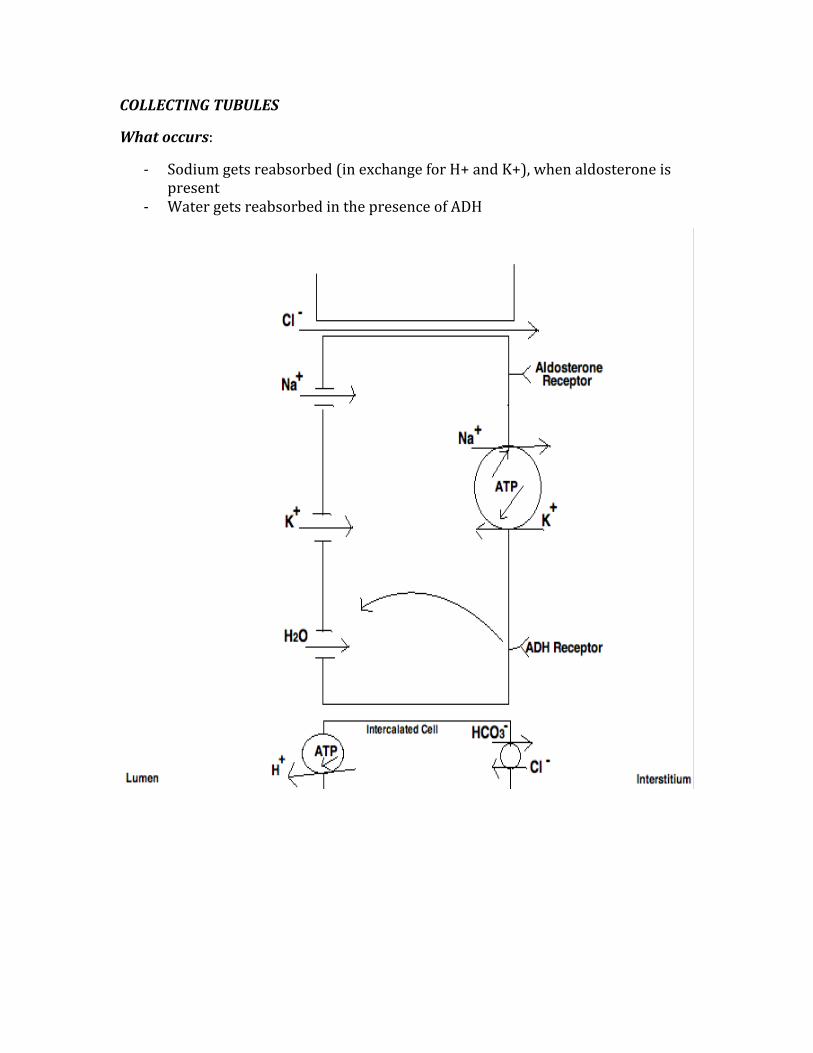
COLLECTING TUBULES
What occurs:
-‐ Sodium gets reabsorbed (in exchange for H+ and K+), when aldosterone is present
-‐ Water gets reabsorbed in the presence of ADH
GASTROINTESTINAL PHYSIOLOGY
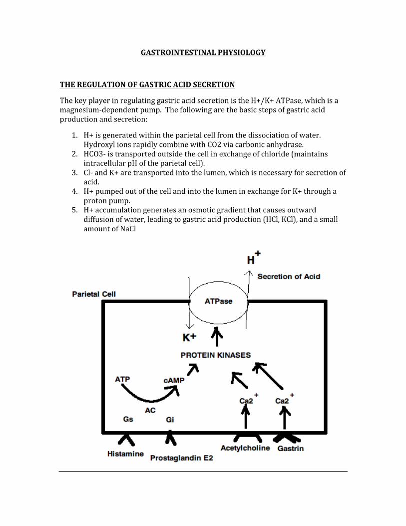
THE REGULATION OF GASTRIC ACID SECRETION
The key player in regulating gastric acid secretion is the H+/K+ ATPase, which is a magnesium-‐dependent pump. The following are the basic steps of gastric acid production and secretion:
1. H+ is generated within the parietal cell from the dissociation of water. Hydroxyl ions rapidly combine with CO2 via carbonic anhydrase.
2. HCO3-‐ is transported outside the cell in exchange of chloride (maintains intracellular pH of the parietal cell).
3. Cl-‐ and K+ are transported into the lumen, which is necessary for secretion of acid.
4. H+ pumped out of the cell and into the lumen in exchange for K+ through a proton pump.
5. H+ accumulation generates an osmotic gradient that causes outward diffusion of water, leading to gastric acid production (HCl, KCl), and a small amount of NaCl
HORMONES OF THE GI
GASTRIN:
-‐ Secreted from the G cells in the antrum of the stomach -‐ Cause stimulation of H+ secretion -‐ Increased when there is stomach distention, vagal stimulation, and the
presence of amino acids in the stomach -‐ Decreased when there is a stomach acid <1.5 -‐ Overstimulation can lead to PUD, gastritis, Zollinger-‐Ellison syndrome
CHOLECYSTOKININ:
-‐ Secreted from the I cells of the duodenum and jejunum -‐ Causes an increase in pancreatic secretion -‐ Stimulates gallbladder contraction -‐ Inhibits the emptying of gastric contents -‐ Inhibited by secretin and a stomach pH <1.5 -‐ Stimulated by the presence of fats and proteins in the stomach
SECRETIN:
-‐ Secreted from the S cells of the duodenum -‐ Causes an increase in pancreatic bicarbonate secretion -‐ Inhibits the secretion of gastric acids -‐ Stimulated by the presence of acids and fatty acids in the lumen of the
duodenum
SOMATOSTATIN:
-‐ Secreted from the D cells in the pancreatic islet cells -‐ Causes inhibition of gastric acid and pepsinogen secretion -‐ Causes inhibition of fluid secreted from the pancreas and small intestine -‐ Inhibits gallbladder contraction -‐ Inhibits the release of insulin and glucagon -‐ Secretion is stimulated by increased acid and inhibited by vagal stimulation
GASTRIC INHIBITORY PEPTIDE:
-‐ Secreted by the K cells in the duodenum and jejunum -‐ Decreases the amount of gastric acid that is secreted -‐ Increases insulin release
SECRETORY PRODUCTS OF THE GI
INTRINSIC FACTOR:
-‐ Secreted by the parietal cells -‐ Binds vitamin B12 -‐ Autoimmune destruction leads to pernicious anemia
PEPSIN:
-‐ Secreted by the chief cells -‐ Aids in protein digestion -‐ Increased through vagal stimulation
GASTRIC ACID:
-‐ Secreted by the parietal cells -‐ Decreases stomach acid (ie Low pH) -‐ Stimulated by histamine and acetylcholine -‐ Inhibited by prostaglandins, somatostains, and GIP
BICARBONATE:
-‐ Secreted by the mucosal cells of the duodenum and stomach -‐ Prevents autodigestion by acid neutralization -‐ Stimulated by secretin
ENZYMES SECRETED BY THE PANCREAS
Lipase à Aids in fat digestion, elevated in pancreatitis
Amylase à Helps in starch digestion, also elevated in pancreatitis
Proteases à Are secreted as proenzymes, help with protein digestion
ENDOCRINE PHYSIOLOGY
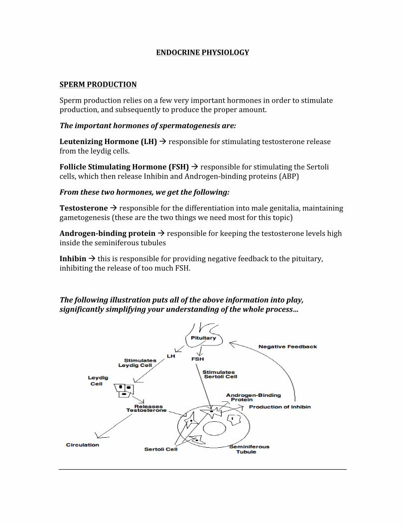
SPERM PRODUCTION
Sperm production relies on a few very important hormones in order to stimulate production, and subsequently to produce the proper amount.
The important hormones of spermatogenesis are:
Leutenizing Hormone (LH) à responsible for stimulating testosterone release from the leydig cells.
Follicle Stimulating Hormone (FSH) à responsible for stimulating the Sertoli cells, which then release Inhibin and Androgen-‐binding proteins (ABP)
From these two hormones, we get the following:
Testosterone à responsible for the differentiation into male genitalia, maintaining gametogenesis (these are the two things we need most for this topic)
Androgen-‐binding protein à responsible for keeping the testosterone levels high inside the seminiferous tubules
Inhibin à this is responsible for providing negative feedback to the pituitary, inhibiting the release of too much FSH.
The following illustration puts all of the above information into play, significantly simplifying your understanding of the whole process…
ANDROGENIC HORMONES
The androgenic hormones include:
-‐ Testosterone -‐ Dihydrotestosterone (DHT) -‐ Androstenedione
Potencies: DHT > Testosterone > Androstenedione
Functions of each hormones:
DHT à Synthesized by the enzyme “5α-‐reductase”
-‐ Formation of secondary sexual characteristic in men
Testosterone à Promotes protein synthesis and growth of all tissues with androgen receptors.
-‐ Muscle growth/mass -‐ Bone density -‐ Bone maturation -‐ Maturation of sex organs (penis and scrotum in fetus) -‐ Hair growth (facial hair, axillary hair) -‐ Development of secondary sex characteristics -‐ Development of prostate and seminal vesicles -‐ Libido
Androstenedione à a precursor of both male and female sex hormones
PROGESTERONE
The hormone involved in the female menstrual cycle, pregnancy, and embryogenesis.
It comes from the testes, corpus luteum, placenta, and the adrenal cortex.
The main functions of Progesterone are:
-‐ Relaxation of the smooth muscle of the uterus -‐ Pregnancy maintenance -‐ Spiral artery development
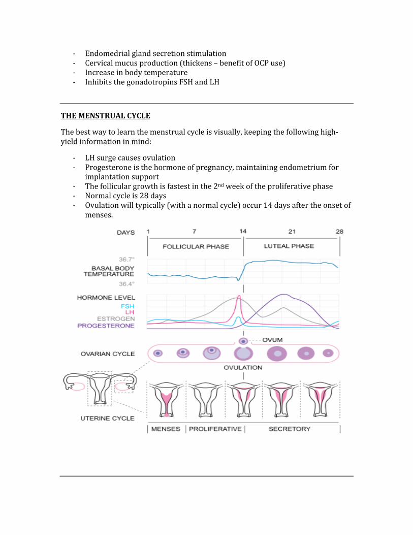
-‐ Endomedrial gland secretion stimulation -‐ Cervical mucus production (thickens – benefit of OCP use) -‐ Increase in body temperature -‐ Inhibits the gonadotropins FSH and LH
THE MENSTRUAL CYCLE
The best way to learn the menstrual cycle is visually, keeping the following high-‐yield information in mind:
-‐ LH surge causes ovulation -‐ Progesterone is the hormone of pregnancy, maintaining endometrium for
implantation support -‐ The follicular growth is fastest in the 2nd week of the proliferative phase -‐ Normal cycle is 28 days -‐ Ovulation will typically (with a normal cycle) occur 14 days after the onset of
menses.
MENOPAUSE
Menopause is indicative of the cessation of ovarian function, resulting in the cessation of ovulation and menstruation.
The following are the hormonal changes that occur with menopause:
-‐ Estrogen decreases -‐ Gonodotropin-‐releasing hormone increases -‐ LH increases -‐ FSH increases significantly
The following are the most common symptoms associated with menopause:
-‐ Hot flashes -‐ Vaginal atrophy -‐ Osteoporosis -‐ Coronary artery disease (estrogen is said to be a protective factor against
this)
HUMAN CHORIONIC GONODOTROPIN (hCG)
hCG is secreted from the placental syncytiotrophoblast, and is responsible for the following functions:
-‐ Is the #1 indicator of pregnancy -‐ Helps to maintain the corpus luteum during the 1st trimester of pregnancy -‐ Helps in diagnosing reproductive pathologies such as choriocarcinoma and
hydatiform moles (discussed in pathology section)
REGULATION OF PROLACTIN
Prolactin is a hormone secreted from the anterior pituitary and is responsible for some important functions, as well it is responsible for certain pathologies (prolactinoma, infertility).
Important functions à Lactation, orgasm, oligodendrocyte precursor cell proliferation.
Inhibited by à Dopamine
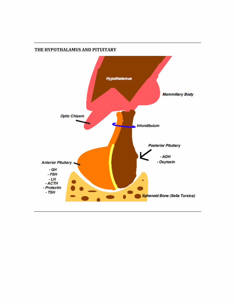
THE HYPOTHALAMUS AND PITUITARY
THE THYROID HORMONE
Thyroid hormone is an extremely important and versatile hormone, controlling a wide-‐range of functions and important for proper growth.
Functions of thyroid hormone:
-‐ CNS maturation -‐ Bone growth -‐ β-‐adrenergic effects -‐ Increases BMR (via increasing the Na+/K+ pump) -‐ Lipolysis (increases) -‐ Gluconeogenesis (increases) -‐ Glycogenolysis (increases)
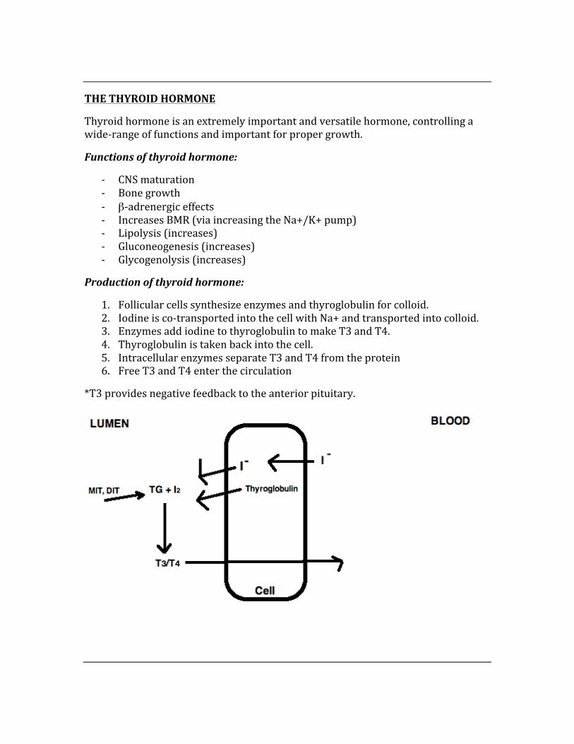
Production of thyroid hormone:
1. Follicular cells synthesize enzymes and thyroglobulin for colloid. 2. Iodine is co-‐transported into the cell with Na+ and transported into colloid. 3. Enzymes add iodine to thyroglobulin to make T3 and T4. 4. Thyroglobulin is taken back into the cell. 5. Intracellular enzymes separate T3 and T4 from the protein 6. Free T3 and T4 enter the circulation
*T3 provides negative feedback to the anterior pituitary.

PARATHYROID HORMONE (PTH)
PTH comes from the chief cells of the parathyroid glands. In response to low serum calcium, PTH is released and performs the following:
-‐ Increases bone resorption which increases Ca2+ and PO4-‐ -‐ Increases the reabsorption of calcium from the kidneys (distal convoluted
tubules) -‐ Decreases the reabsorption of phosphate from the kidneys -‐ Stimulates the enzyme 1α-‐hydroxylase in the kidney, which increases 1,25-‐
(OH)2 vitamin D (ie cholecalciferol)
CALCITONIN
Calcitonin works opposite of PTH by recognizing an increase in serum Ca2+ and thus decreasing the bone resorption of calcium. Calcitonin is secreted from the parafollicular (c cells) of the thyroid gland.
LINKING PATHOLOGY TO Ca2+, PO4-‐, and ALKALINE PHOSPHATASE
The following list of conditions alter these levels in the following ways…
Disesase Calcium Level Phosphate Level Alk Phosph Level
Vitamin D Intox Increases Increases Increases Osteoporosis No change No change No change
Hyperparathyroidism Increases Decreases Increases Paget’s bone disease Normal-‐increased Normal Large increase Renal Insufficiency Decreased Increased No change
CARDIAC PHYSIOLOGY
NOTE: Cardiac physiology is unique in that almost everything is conceptual in nature, which means that there are many graphs/charts, etc. By completely understanding the concept behind all of this information, you will not have to memorize anything, rather you will be able to apply it to any question the USMLE exam throws your way.
THE CARDIAC CYCLE
The cardiac cycle refers simply to the steps that are undertaken by the heart as it goes from filling, to pumping the blood systematically, to filling once again.
The phases of the cardiac cycle:
1. Isovolumetric Contraction
This is the point between the closure of the mitral valve and the opening of the aortic valve. The heart is contracted but valves are closed.
2. Systolic Ejection
The heart squeezes and blood is ejected through the aortic valve. This phase can be considered the phase between the time the aortic valve opens and closes.
3. Isovolumetric Relaxation
This is the period of time between the closure of the aortic valve and the opening of the mitral valve.
4. Rapid filling phase
After the opening of the mitral valve, blood pools rapidly into the left ventricle.
5. Slow filling phase
At this point, blood flows into the LV slowly as the mitral valve is about to close.
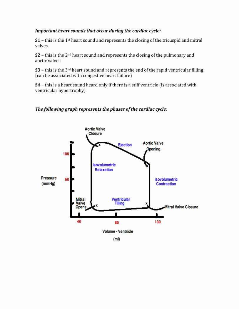
Important heart sounds that occur during the cardiac cycle:
S1 – this is the 1st heart sound and represents the closing of the tricuspid and mitral valves
S2 – this is the 2nd heart sound and represents the closing of the pulmonary and aortic valves
S3 – this is the 3rd heart sound and represents the end of the rapid ventricular filling (can be associated with congestive heart failure)
S4 – this is a heart sound heard only if there is a stiff ventricle (is associated with ventricular hypertrophy)
The following graph represents the phases of the cardiac cycle:
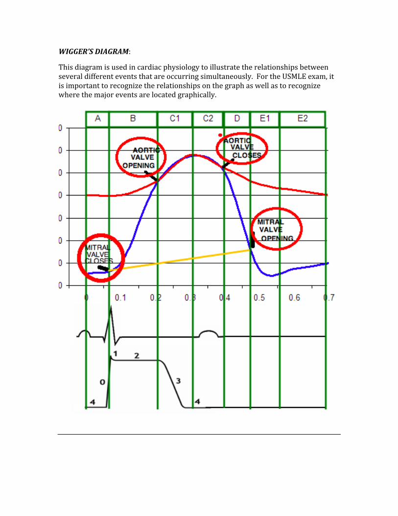
WIGGER’S DIAGRAM:
This diagram is used in cardiac physiology to illustrate the relationships between several different events that are occurring simultaneously. For the USMLE exam, it is important to recognize the relationships on the graph as well as to recognize where the major events are located graphically.
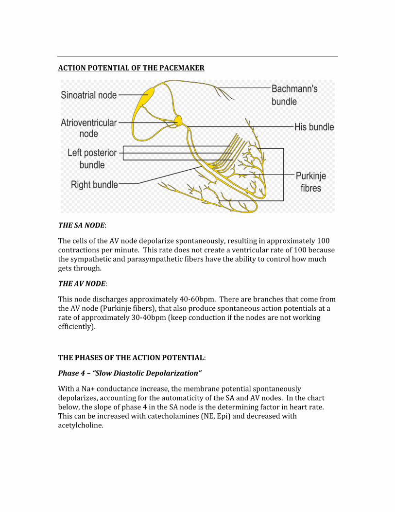
ACTION POTENTIAL OF THE PACEMAKER
THE SA NODE:
The cells of the AV node depolarize spontaneously, resulting in approximately 100 contractions per minute. This rate does not create a ventricular rate of 100 because the sympathetic and parasympathetic fibers have the ability to control how much gets through.
THE AV NODE:
This node discharges approximately 40-‐60bpm. There are branches that come from the AV node (Purkinje fibers), that also produce spontaneous action potentials at a rate of approximately 30-‐40bpm (keep conduction if the nodes are not working efficiently).
THE PHASES OF THE ACTION POTENTIAL:
Phase 4 – “Slow Diastolic Depolarization”
With a Na+ conductance increase, the membrane potential spontaneously depolarizes, accounting for the automaticity of the SA and AV nodes. In the chart below, the slope of phase 4 in the SA node is the determining factor in heart rate. This can be increased with catecholamines (NE, Epi) and decreased with acetylcholine.
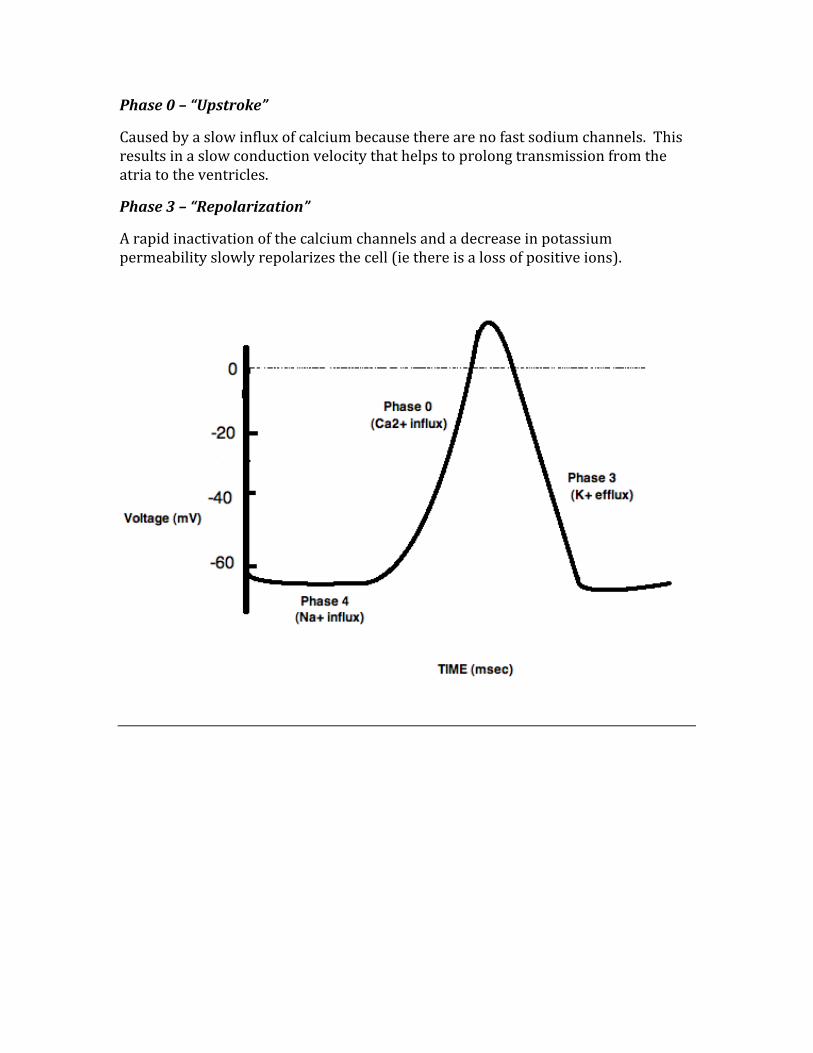
Phase 0 – “Upstroke”
Caused by a slow influx of calcium because there are no fast sodium channels. This results in a slow conduction velocity that helps to prolong transmission from the atria to the ventricles.
Phase 3 – “Repolarization”
A rapid inactivation of the calcium channels and a decrease in potassium permeability slowly repolarizes the cell (ie there is a loss of positive ions).
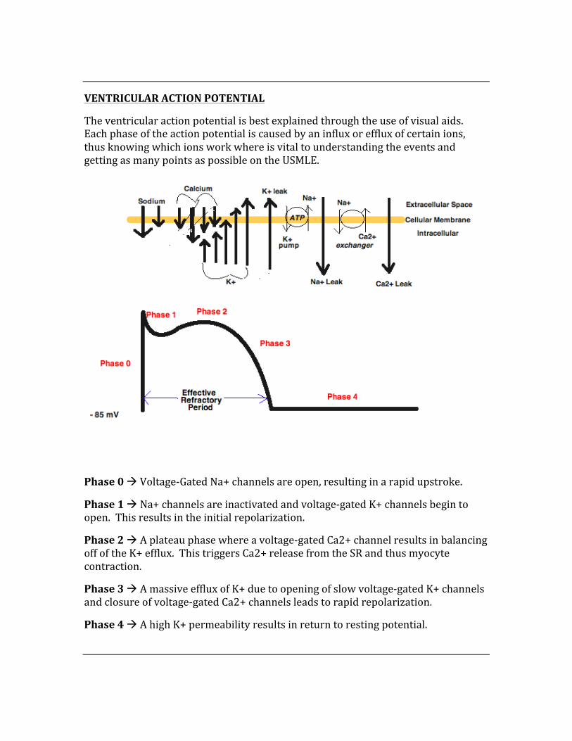
VENTRICULAR ACTION POTENTIAL
The ventricular action potential is best explained through the use of visual aids. Each phase of the action potential is caused by an influx or efflux of certain ions, thus knowing which ions work where is vital to understanding the events and getting as many points as possible on the USMLE.
Phase 0 à Voltage-‐Gated Na+ channels are open, resulting in a rapid upstroke.
Phase 1 à Na+ channels are inactivated and voltage-‐gated K+ channels begin to open. This results in the initial repolarization.
Phase 2 à A plateau phase where a voltage-‐gated Ca2+ channel results in balancing off of the K+ efflux. This triggers Ca2+ release from the SR and thus myocyte contraction.
Phase 3 à A massive efflux of K+ due to opening of slow voltage-‐gated K+ channels and closure of voltage-‐gated Ca2+ channels leads to rapid repolarization.
Phase 4 à A high K+ permeability results in return to resting potential.
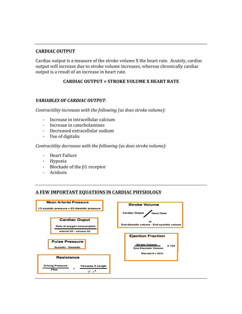
CARDIAC OUTPUT
Cardiac output is a measure of the stroke volume X the heart rate. Acutely, cardiac output will increase due to stroke volume increases, whereas chronically cardiac output is a result of an increase in heart rate.
CARDIAC OUTPUT = STROKE VOLUME X HEART RATE
VARIABLES OF CARDIAC OUTPUT:
Contractility increases with the following (as does stroke volume):
-‐ Increase in intracellular calcium -‐ Increase in catecholamines -‐ Decreased extracellular sodium -‐ Use of digitalis
Contractility decreases with the following (as does stroke volume):
-‐ Heart Failure -‐ Hypoxia -‐ Blockade of the β1 receptor -‐ Acidosis
A FEW IMPORTANT EQUATIONS IN CARDIAC PHYSIOLOGY
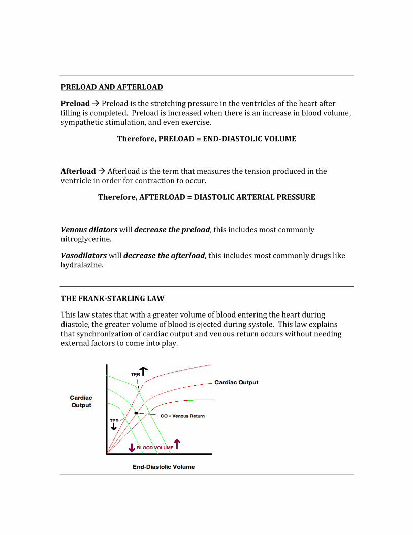
PRELOAD AND AFTERLOAD
Preload à Preload is the stretching pressure in the ventricles of the heart after filling is completed. Preload is increased when there is an increase in blood volume, sympathetic stimulation, and even exercise.
Therefore, PRELOAD = END-‐DIASTOLIC VOLUME
Afterload à Afterload is the term that measures the tension produced in the ventricle in order for contraction to occur.
Therefore, AFTERLOAD = DIASTOLIC ARTERIAL PRESSURE
Venous dilators will decrease the preload, this includes most commonly nitroglycerine.
Vasodilators will decrease the afterload, this includes most commonly drugs like hydralazine.
THE FRANK-‐STARLING LAW
This law states that with a greater volume of blood entering the heart during diastole, the greater volume of blood is ejected during systole. This law explains that synchronization of cardiac output and venous return occurs without needing external factors to come into play.
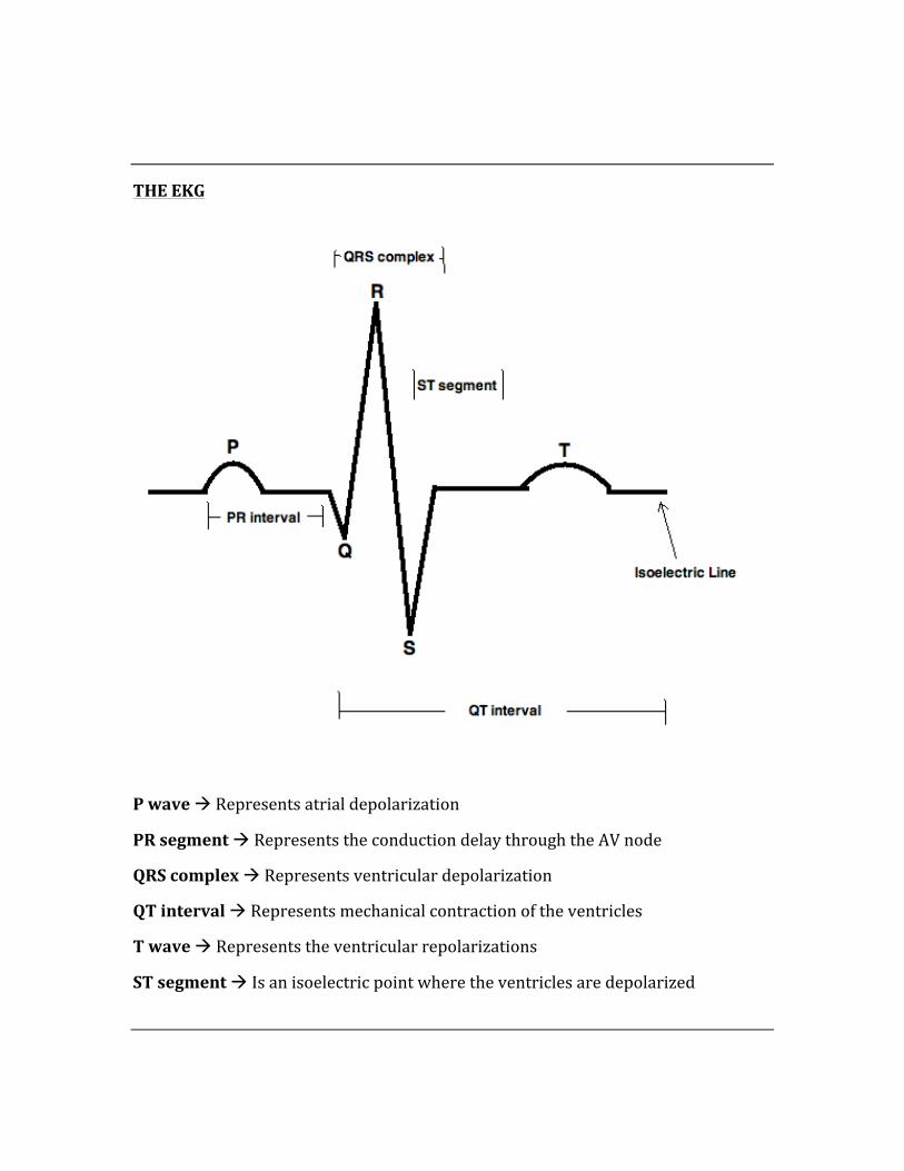
THE EKG
P wave à Represents atrial depolarization
PR segment à Represents the conduction delay through the AV node
QRS complex à Represents ventricular depolarization
QT interval à Represents mechanical contraction of the ventricles
T wave à Represents the ventricular repolarizations
ST segment à Is an isoelectric point where the ventricles are depolarized
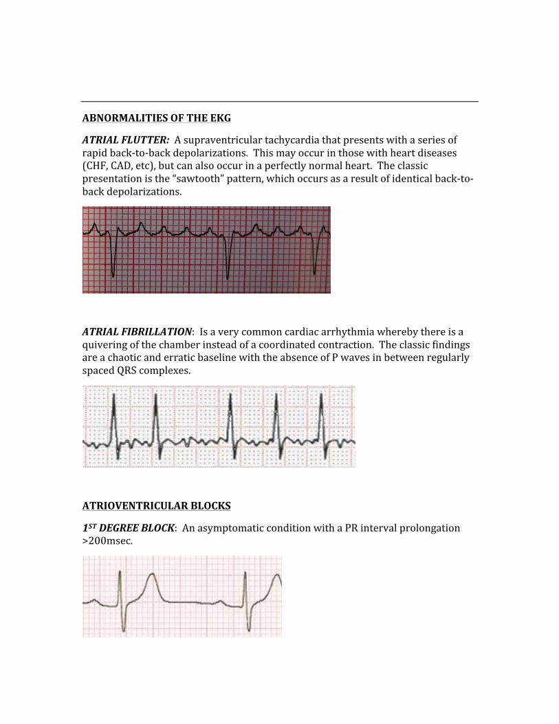
ABNORMALITIES OF THE EKG
ATRIAL FLUTTER: A supraventricular tachycardia that presents with a series of rapid back-‐to-‐back depolarizations. This may occur in those with heart diseases (CHF, CAD, etc), but can also occur in a perfectly normal heart. The classic presentation is the “sawtooth” pattern, which occurs as a result of identical back-‐to-‐back depolarizations.
ATRIAL FIBRILLATION: Is a very common cardiac arrhythmia whereby there is a quivering of the chamber instead of a coordinated contraction. The classic findings are a chaotic and erratic baseline with the absence of P waves in between regularly spaced QRS complexes.
ATRIOVENTRICULAR BLOCKS
1ST DEGREE BLOCK: An asymptomatic condition with a PR interval prolongation >200msec.
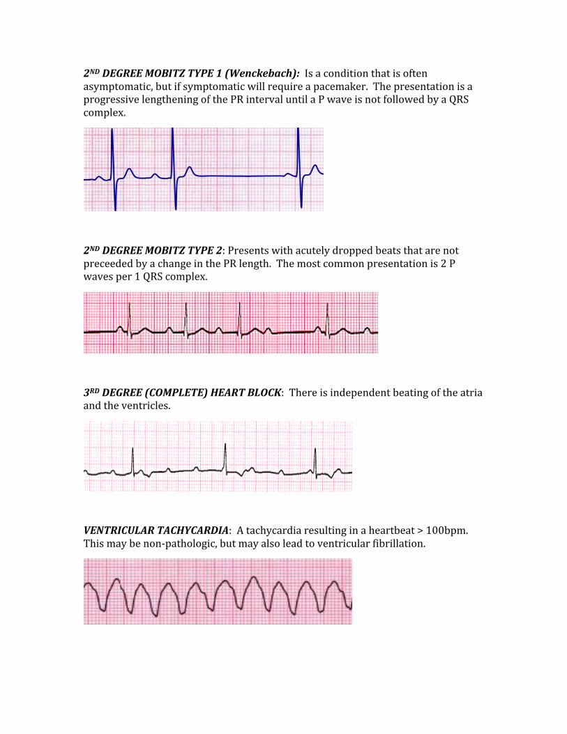
2ND DEGREE MOBITZ TYPE 1 (Wenckebach): Is a condition that is often asymptomatic, but if symptomatic will require a pacemaker. The presentation is a progressive lengthening of the PR interval until a P wave is not followed by a QRS complex.
2ND DEGREE MOBITZ TYPE 2: Presents with acutely dropped beats that are not preceeded by a change in the PR length. The most common presentation is 2 P waves per 1 QRS complex.
3RD DEGREE (COMPLETE) HEART BLOCK: There is independent beating of the atria and the ventricles.
VENTRICULAR TACHYCARDIA: A tachycardia resulting in a heartbeat > 100bpm. This may be non-‐pathologic, but may also lead to ventricular fibrillation.
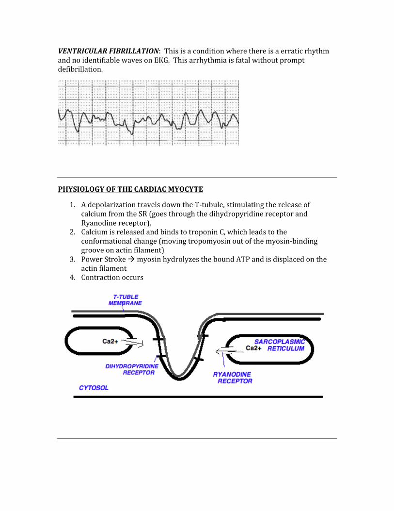
VENTRICULAR FIBRILLATION: This is a condition where there is a erratic rhythm and no identifiable waves on EKG. This arrhythmia is fatal without prompt defibrillation.
PHYSIOLOGY OF THE CARDIAC MYOCYTE
1. A depolarization travels down the T-‐tubule, stimulating the release of calcium from the SR (goes through the dihydropyridine receptor and Ryanodine receptor).
2. Calcium is released and binds to troponin C, which leads to the conformational change (moving tropomyosin out of the myosin-‐binding groove on actin filament)
3. Power Stroke à myosin hydrolyzes the bound ATP and is displaced on the actin filament
4. Contraction occurs
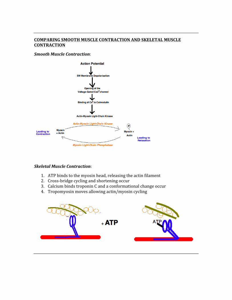
COMPARING SMOOTH MUSCLE CONTRACTION AND SKELETAL MUSCLE CONTRACTION
Smooth Muscle Contraction:
Skeletal Muscle Contraction:
1. ATP binds to the myosin head, releasing the actin filament 2. Cross-‐bridge cycling and shortening occur 3. Calcium binds troponin C and a conformational change occur 4. Tropomyosin moves allowing actin/myosin cycling
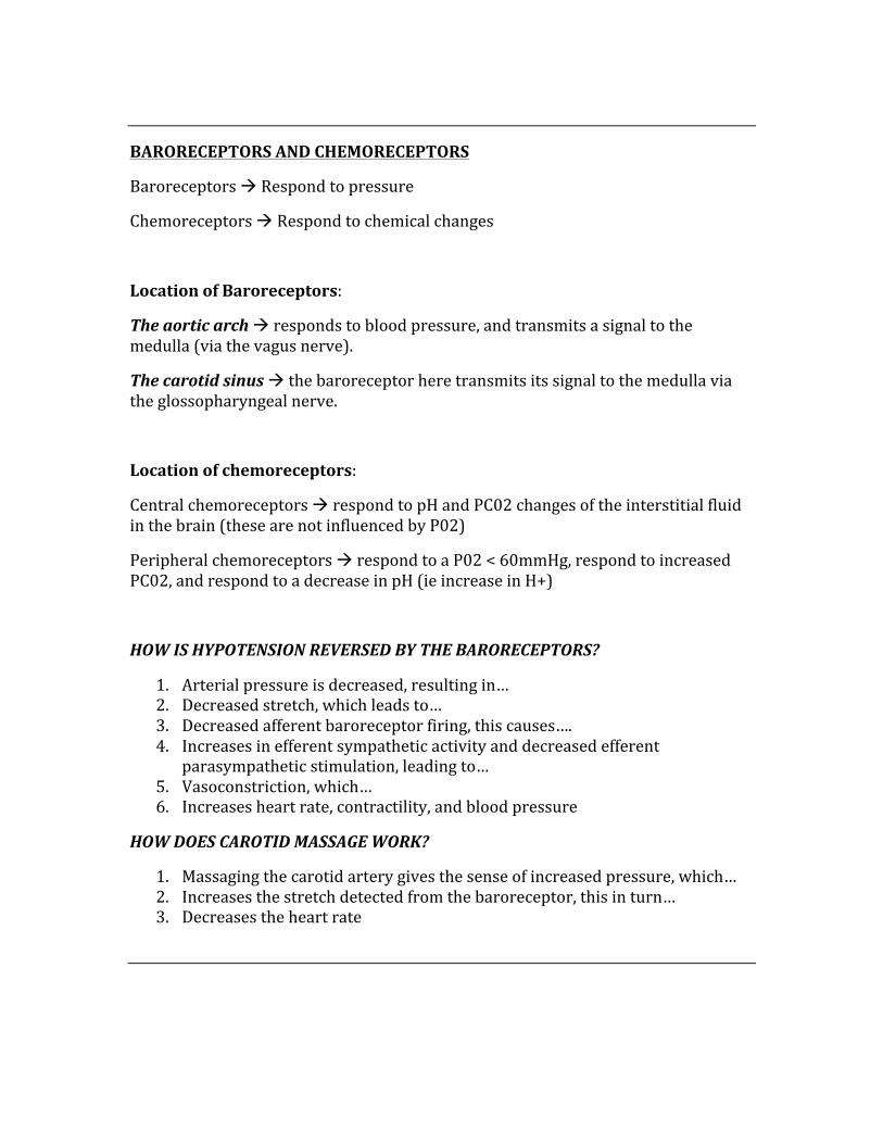
BARORECEPTORS AND CHEMORECEPTORS
Baroreceptors à Respond to pressure
Chemoreceptors à Respond to chemical changes
Location of Baroreceptors:
The aortic arch à responds to blood pressure, and transmits a signal to the medulla (via the vagus nerve).
The carotid sinus à the baroreceptor here transmits its signal to the medulla via the glossopharyngeal nerve.
Location of chemoreceptors:
Central chemoreceptors à respond to pH and PC02 changes of the interstitial fluid in the brain (these are not influenced by P02)
Peripheral chemoreceptors à respond to a P02 < 60mmHg, respond to increased PC02, and respond to a decrease in pH (ie increase in H+)
HOW IS HYPOTENSION REVERSED BY THE BARORECEPTORS?
1. Arterial pressure is decreased, resulting in… 2. Decreased stretch, which leads to… 3. Decreased afferent baroreceptor firing, this causes…. 4. Increases in efferent sympathetic activity and decreased efferent
parasympathetic stimulation, leading to… 5. Vasoconstriction, which… 6. Increases heart rate, contractility, and blood pressure
HOW DOES CAROTID MASSAGE WORK?
1. Massaging the carotid artery gives the sense of increased pressure, which… 2. Increases the stretch detected from the baroreceptor, this in turn… 3. Decreases the heart rate
EXCHANGE OF CAPILLARY FLUIDS
Forces inside and outside of the capillaries are what can move fluids back and forth. There are different methods by which fluid is moved, it can either be pushed out of the capillary (capillary pressure), or it can be moved via osmotic pressure, where it is pulled.
The forces are called “Starling” forces, and they are the following:
1. Capillary Pressure (Cp) – this pressure usually causes a movement of fluid out of the capillary
2. Interstitial Pressure (Pi) – this is pressure the pushes on the capillaries and moves fluid into the capillary.
3. Plasma colloid osmotic pressure (πc) – usually moves fluid into the capillary 4. Interstitial fluid colloid osmotic pressure (πi) – usually moves fluid out of the
capillary.
Determining whether fluid will move into or out of the capillary is based on the net filtration pressure. By having all of the values of the pressures above, we can determine this figure:
Net Filtration Pressure = (Pc-‐Pi) – (πc -‐ πi)
LINKING THE ABOVE TO EDEMATOUS STATES:
Edema is caused by excess fluids outside of the capillaries, thus states in which this is favorable will likely lead to edema. The following will likely lead to edematous states:
1. Increased interstitial fluid colloid osmotic pressure 2. Increased capillary pressure 3. Increased capillary permeability 4. Decreased quantities of plasma proteins
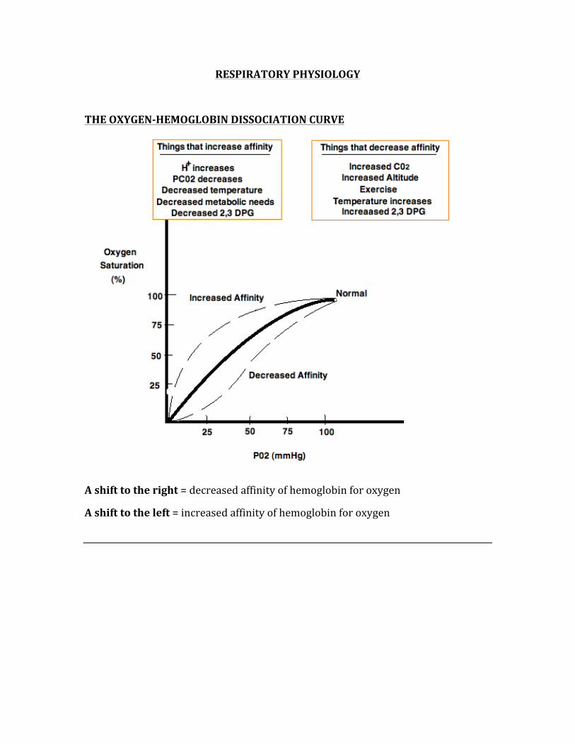
RESPIRATORY PHYSIOLOGY
THE OXYGEN-‐HEMOGLOBIN DISSOCIATION CURVE
A shift to the right = decreased affinity of hemoglobin for oxygen
A shift to the left = increased affinity of hemoglobin for oxygen
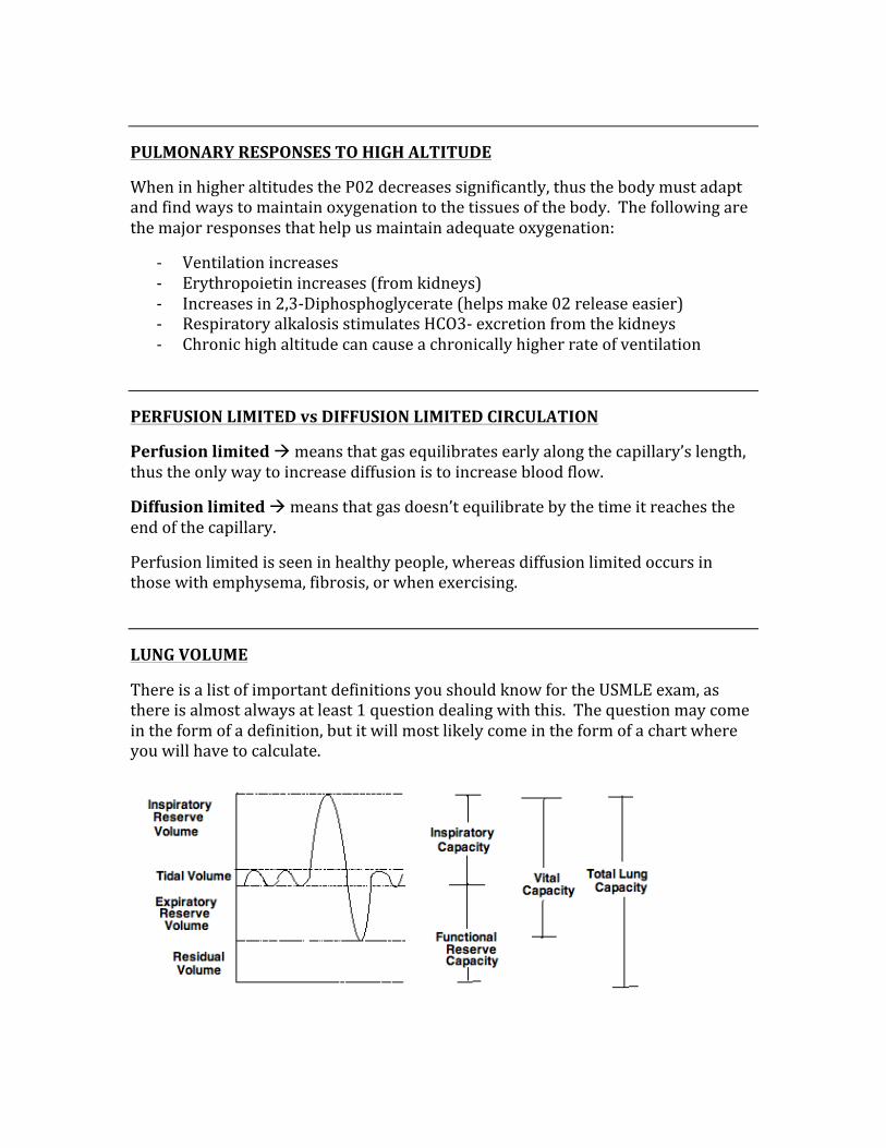
PULMONARY RESPONSES TO HIGH ALTITUDE
When in higher altitudes the P02 decreases significantly, thus the body must adapt and find ways to maintain oxygenation to the tissues of the body. The following are the major responses that help us maintain adequate oxygenation:
-‐ Ventilation increases -‐ Erythropoietin increases (from kidneys) -‐ Increases in 2,3-‐Diphosphoglycerate (helps make 02 release easier) -‐ Respiratory alkalosis stimulates HCO3-‐ excretion from the kidneys -‐ Chronic high altitude can cause a chronically higher rate of ventilation
PERFUSION LIMITED vs DIFFUSION LIMITED CIRCULATION
Perfusion limited à means that gas equilibrates early along the capillary’s length, thus the only way to increase diffusion is to increase blood flow.
Diffusion limited à means that gas doesn’t equilibrate by the time it reaches the end of the capillary.
Perfusion limited is seen in healthy people, whereas diffusion limited occurs in those with emphysema, fibrosis, or when exercising.
LUNG VOLUME
There is a list of important definitions you should know for the USMLE exam, as there is almost always at least 1 question dealing with this. The question may come in the form of a definition, but it will most likely come in the form of a chart where you will have to calculate.
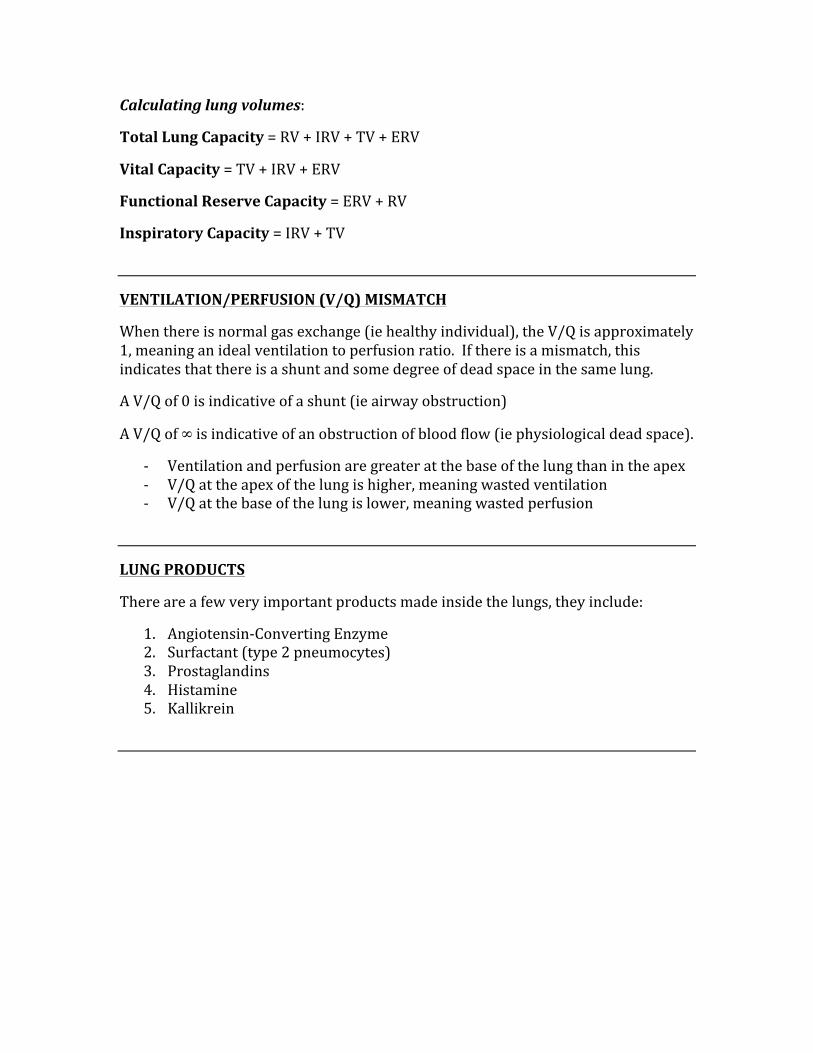
Calculating lung volumes:
Total Lung Capacity = RV + IRV + TV + ERV
Vital Capacity = TV + IRV + ERV
Functional Reserve Capacity = ERV + RV
Inspiratory Capacity = IRV + TV
VENTILATION/PERFUSION (V/Q) MISMATCH
When there is normal gas exchange (ie healthy individual), the V/Q is approximately 1, meaning an ideal ventilation to perfusion ratio. If there is a mismatch, this indicates that there is a shunt and some degree of dead space in the same lung.
A V/Q of 0 is indicative of a shunt (ie airway obstruction)
A V/Q of ∞ is indicative of an obstruction of blood flow (ie physiological dead space).
-‐ Ventilation and perfusion are greater at the base of the lung than in the apex -‐ V/Q at the apex of the lung is higher, meaning wasted ventilation -‐ V/Q at the base of the lung is lower, meaning wasted perfusion
LUNG PRODUCTS
There are a few very important products made inside the lungs, they include:
1. Angiotensin-‐Converting Enzyme 2. Surfactant (type 2 pneumocytes) 3. Prostaglandins 4. Histamine 5. Kallikrein
CARBON DIOXIDE TRANSPORT
There are three methods by which carbon dioxide are carried from the tissues of the body back to the lungs, they include:
1. In the form of bicarbonate (this is the majority) ~90% 2. As dissolved carbon dioxide ~5% 3. Bound to hemoglobin as carbaminohemoglobin ~5%
CHAPTER 6
BIOCHEMISTRY
This section is very high-‐yield, and includes many sub-‐categories. The metabolic pathways are very important; paying special attention to regulatory steps is crucial.
While the details of biochemistry are not high-‐yield, the big picture as a whole is very high-‐yield material.
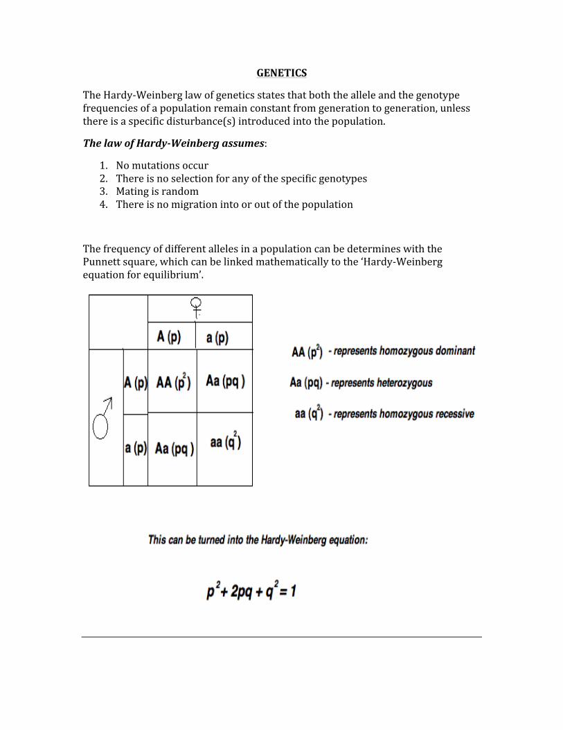
GENETICS
The Hardy-‐Weinberg law of genetics states that both the allele and the genotype frequencies of a population remain constant from generation to generation, unless there is a specific disturbance(s) introduced into the population.
The law of Hardy-‐Weinberg assumes:
1. No mutations occur 2. There is no selection for any of the specific genotypes 3. Mating is random 4. There is no migration into or out of the population
The frequency of different alleles in a population can be determines with the Punnett square, which can be linked mathematically to the ‘Hardy-‐Weinberg equation for equilibrium’.
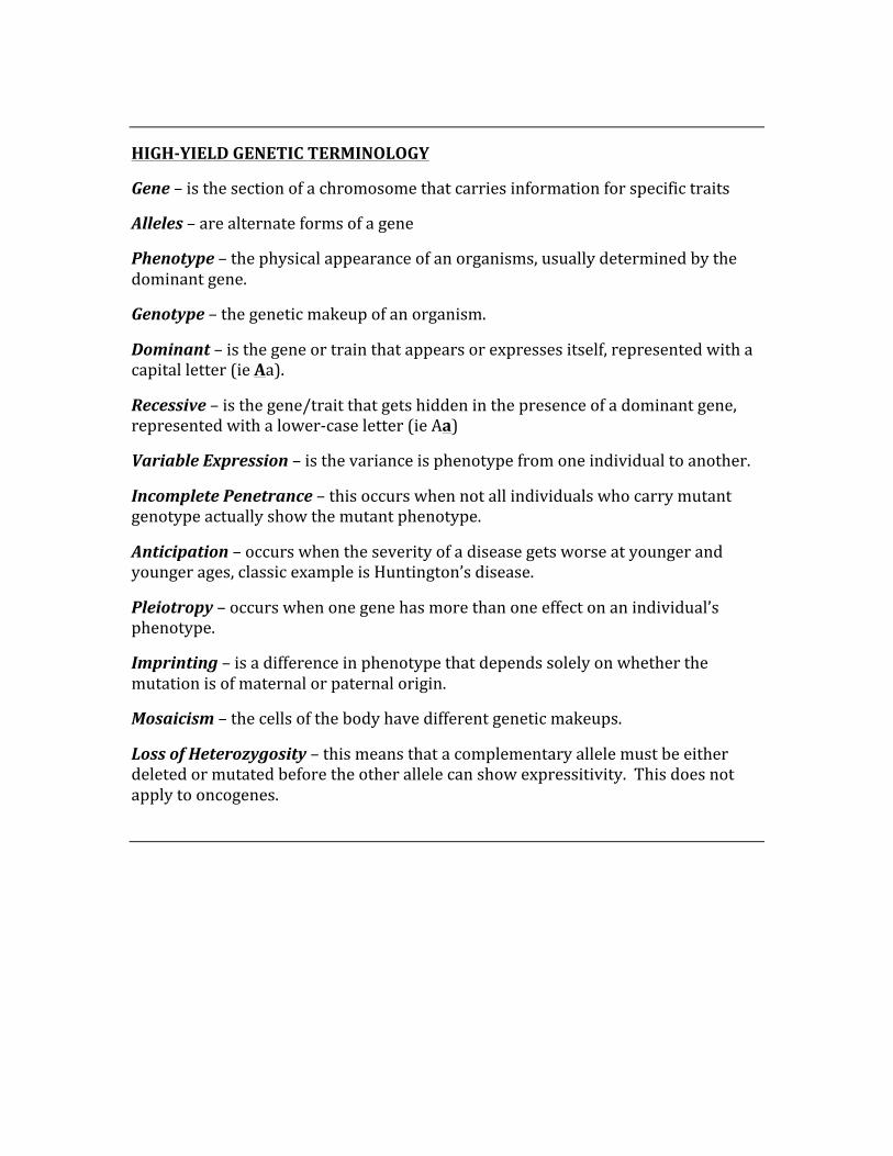
HIGH-‐YIELD GENETIC TERMINOLOGY
Gene – is the section of a chromosome that carries information for specific traits
Alleles – are alternate forms of a gene
Phenotype – the physical appearance of an organisms, usually determined by the dominant gene.
Genotype – the genetic makeup of an organism.
Dominant – is the gene or train that appears or expresses itself, represented with a capital letter (ie Aa).
Recessive – is the gene/trait that gets hidden in the presence of a dominant gene, represented with a lower-‐case letter (ie Aa)
Variable Expression – is the variance is phenotype from one individual to another.
Incomplete Penetrance – this occurs when not all individuals who carry mutant genotype actually show the mutant phenotype.
Anticipation – occurs when the severity of a disease gets worse at younger and younger ages, classic example is Huntington’s disease.
Pleiotropy – occurs when one gene has more than one effect on an individual’s phenotype.
Imprinting – is a difference in phenotype that depends solely on whether the mutation is of maternal or paternal origin.
Mosaicism – the cells of the body have different genetic makeups.
Loss of Heterozygosity – this means that a complementary allele must be either deleted or mutated before the other allele can show expressitivity. This does not apply to oncogenes.
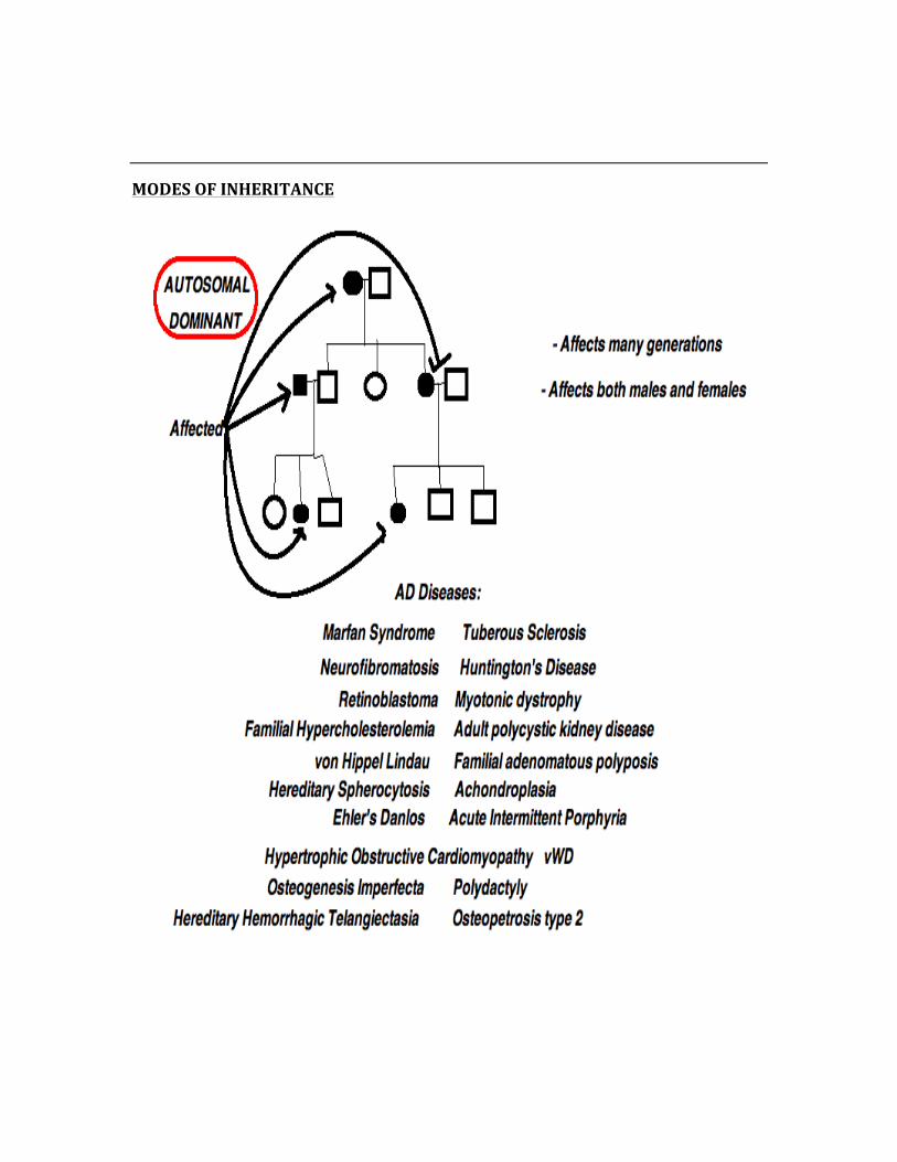
MODES OF INHERITANCE
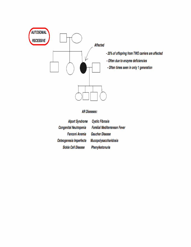
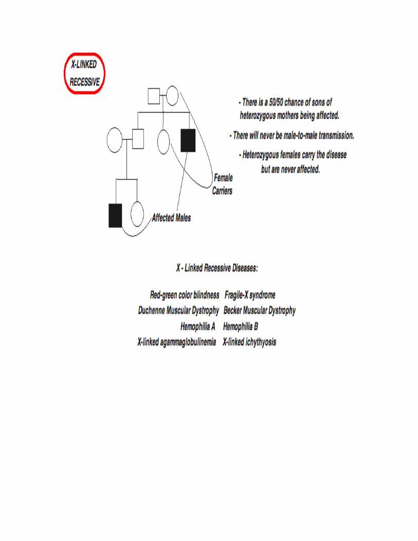
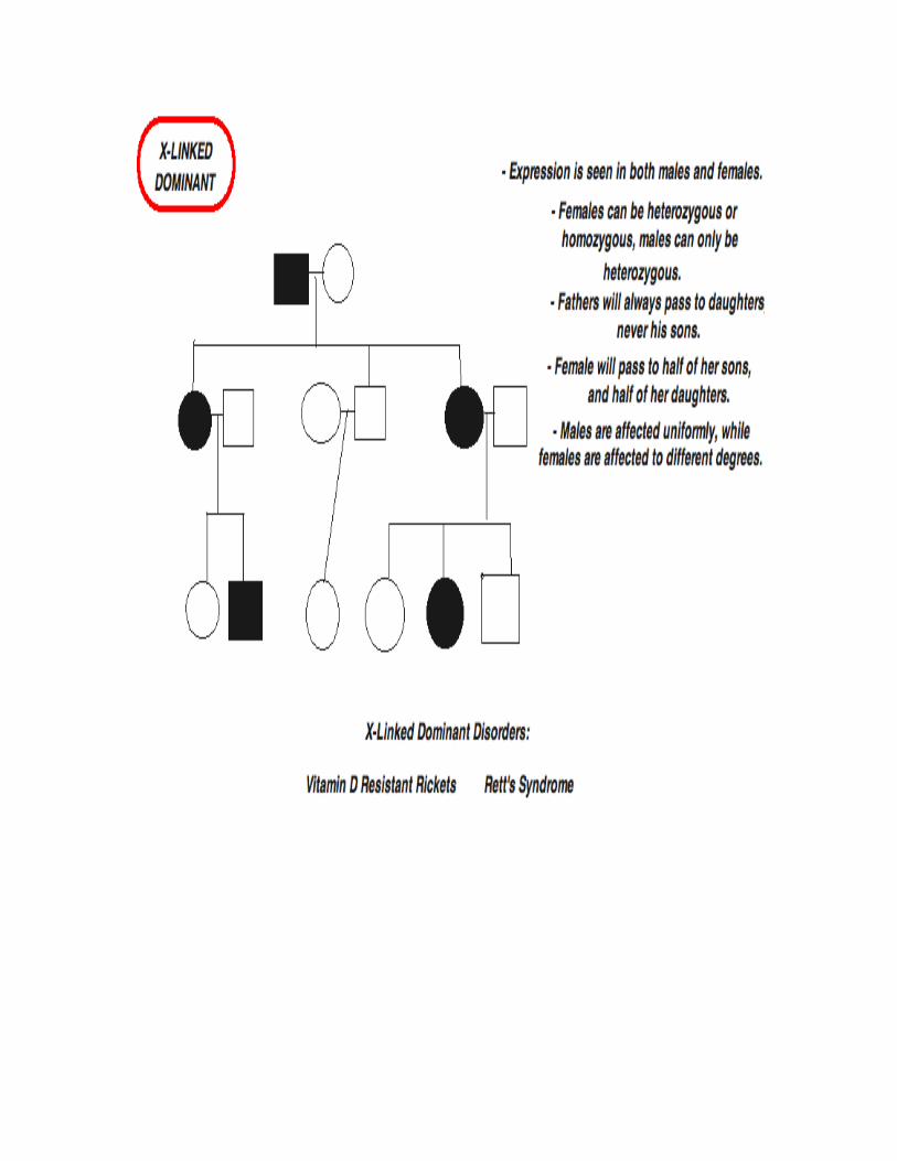
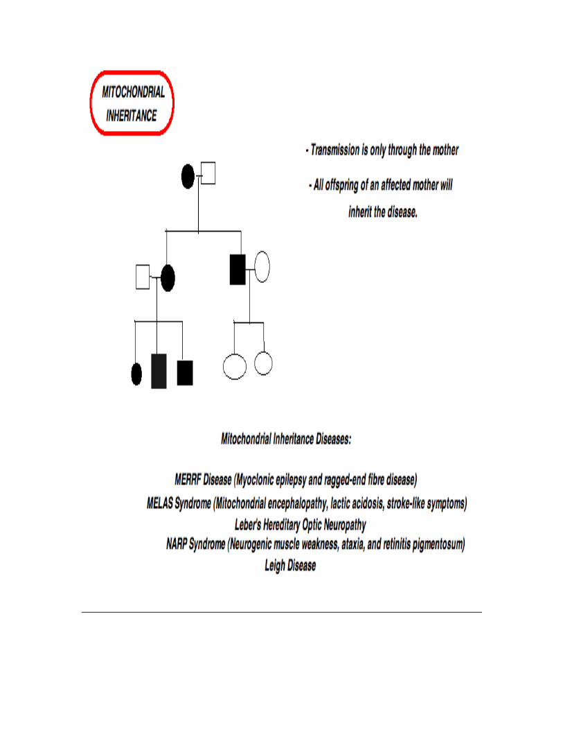
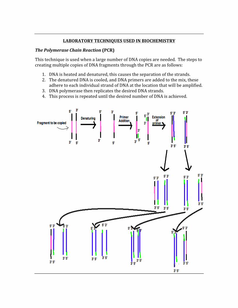
LABORATORY TECHNIQUES USED IN BIOCHEMISTRY
The Polymerase Chain Reaction (PCR)
This technique is used when a large number of DNA copies are needed. The steps to creating multiple copies of DNA fragments through the PCR are as follows:
1. DNA is heated and denatured, this causes the separation of the strands. 2. The denatured DNA is cooled, and DNA primers are added to the mix, these
adhere to each individual strand of DNA at the location that will be amplified. 3. DNA polymerase then replicates the desired DNA strands. 4. This process is repeated until the desired number of DNA is achieved.
ELISA (Enzyme-‐Linked Immunoabsorbent Assay)
This technique is used as a means of detecting the presence of an antibody or an antigen in a sample. The antibody or antigen that is added is linked to an enzyme, then a test solution is added to see if an intense color illuminates, indicating that there is a positive result.
-‐ This test is most commonly used when looking for HIV. -‐ Sensitivity and specificity for the ELISA are extremely high, both approaching
100%, however they are not perfect, and false results do occur.
SOUTHERN BLOT TECHNIQUE
This technique is used to detect specific sequences of DNA. The technique combines the transfer of electrophoresis-‐separated DNA fragments and membrane filtration, and then fragments are detected by probe hybridization.
WESTERN BLOT TECHNIQUE
This is a technique used to detect specific proteins, separating native or denatured proteins by the length of the polypeptide. These proteins are then transferred to a membrane where they are probed using antibodies specific to the target protein.
NORTHERN BLOT TECHNIQUE
Is a technique used to study gene expression by RNA detection in a sample. This technique allows for the detection of cellular control by determination of gene expression levels during differentiation and morphogenesis.
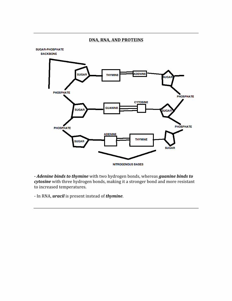
DNA, RNA, AND PROTEINS
-‐ Adenine binds to thymine with two hydrogen bonds, whereas guanine binds to cytosine with three hydrogen bonds, making it a stronger bond and more resistant to increased temperatures.
-‐ In RNA, uracil is present instead of thymine.
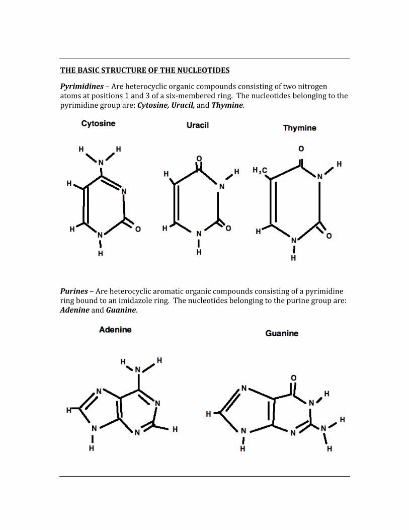
THE BASIC STRUCTURE OF THE NUCLEOTIDES
Pyrimidines – Are heterocyclic organic compounds consisting of two nitrogen atoms at positions 1 and 3 of a six-‐membered ring. The nucleotides belonging to the pyrimidine group are: Cytosine, Uracil, and Thymine.
Purines – Are heterocyclic aromatic organic compounds consisting of a pyrimidine ring bound to an imidazole ring. The nucleotides belonging to the purine group are: Adenine and Guanine.
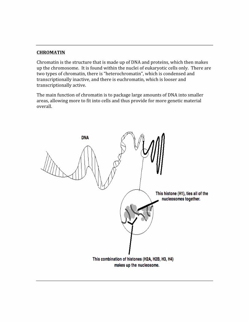
CHROMATIN
Chromatin is the structure that is made up of DNA and proteins, which then makes up the chromosome. It is found within the nuclei of eukaryotic cells only. There are two types of chromatin, there is “heterochromatin”, which is condensed and transcriptionally inactive, and there is euchromatin, which is looser and transcriptionally active.
The main function of chromatin is to package large amounts of DNA into smaller areas, allowing more to fit into cells and thus provide for more genetic material overall.
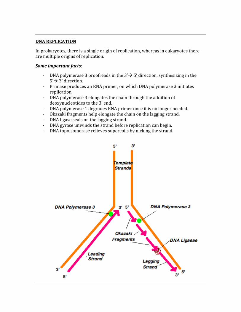
DNA REPLICATION
In prokaryotes, there is a single origin of replication, whereas in eukaryotes there are multiple origins of replication.
Some important facts:
-‐ DNA polymerase 3 proofreads in the 3’à 5’ direction, synthesizing in the 5’à 3’ direction.
-‐ Primase produces an RNA primer, on which DNA polymerase 3 initiates replication.
-‐ DNA polymerase 3 elongates the chain through the addition of deoxynucleotides to the 3’ end.
-‐ DNA polymerase 1 degrades RNA primer once it is no longer needed. -‐ Okazaki fragments help elongate the chain on the lagging strand. -‐ DNA ligase seals on the lagging strand. -‐ DNA gyrase unwinds the strand before replication can begin. -‐ DNA topoisomerase relieves supercoils by nicking the strand.
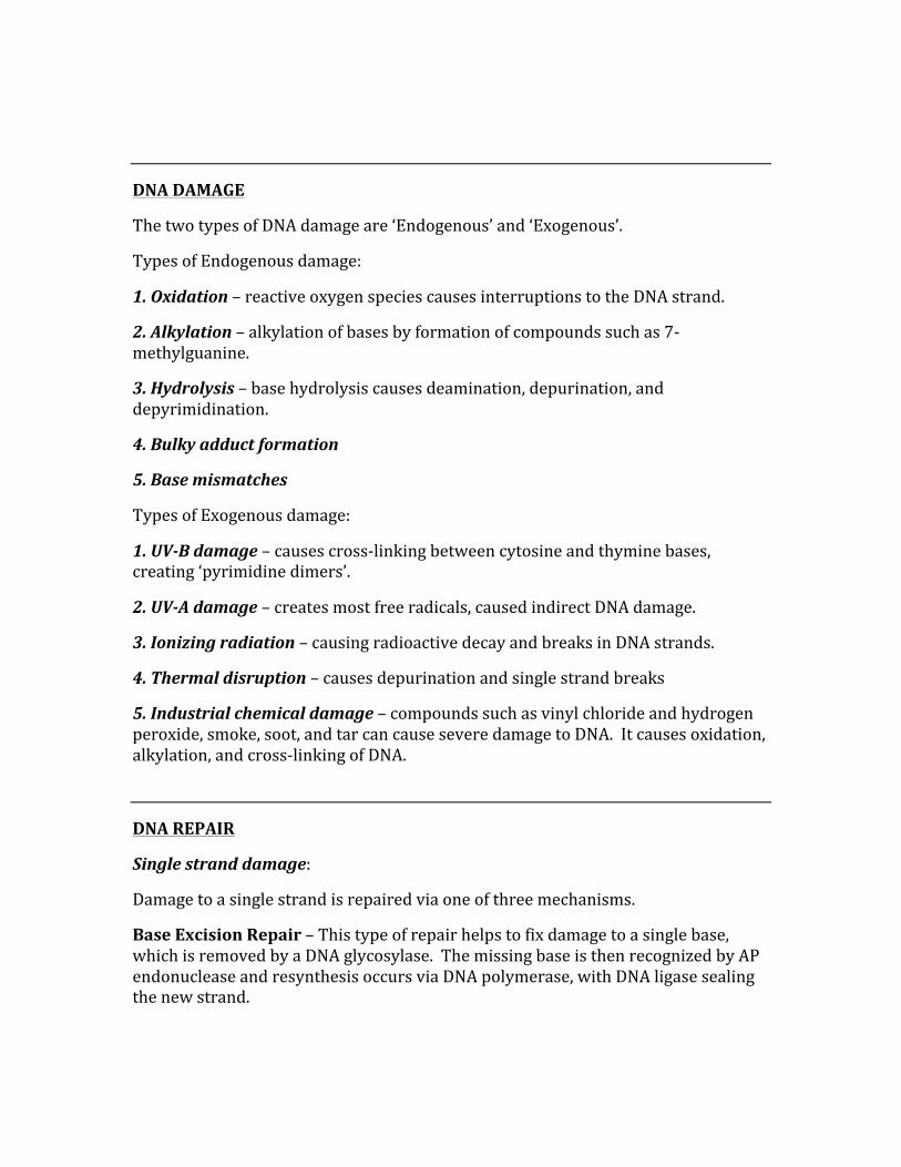
DNA DAMAGE
The two types of DNA damage are ‘Endogenous’ and ‘Exogenous’.
Types of Endogenous damage:
1. Oxidation – reactive oxygen species causes interruptions to the DNA strand.
2. Alkylation – alkylation of bases by formation of compounds such as 7-‐methylguanine.
3. Hydrolysis – base hydrolysis causes deamination, depurination, and depyrimidination.
4. Bulky adduct formation
5. Base mismatches
Types of Exogenous damage:
1. UV-‐B damage – causes cross-‐linking between cytosine and thymine bases, creating ‘pyrimidine dimers’.
2. UV-‐A damage – creates most free radicals, caused indirect DNA damage.
3. Ionizing radiation – causing radioactive decay and breaks in DNA strands.
4. Thermal disruption – causes depurination and single strand breaks
5. Industrial chemical damage – compounds such as vinyl chloride and hydrogen peroxide, smoke, soot, and tar can cause severe damage to DNA. It causes oxidation, alkylation, and cross-‐linking of DNA.
DNA REPAIR
Single strand damage:
Damage to a single strand is repaired via one of three mechanisms.
Base Excision Repair – This type of repair helps to fix damage to a single base, which is removed by a DNA glycosylase. The missing base is then recognized by AP endonuclease and resynthesis occurs via DNA polymerase, with DNA ligase sealing the new strand.
Nucleotide Excision Repair – This mechanism recognizes bulky helix-‐distorting damage and is fixed with transcription-‐coupled repair which emits NER enzymes that are actively being transcribed.
Mismatch Repair – This repair mechanism corrects errors of DNA replication and recombination that resulted from mispaired nucleotides.
Double-‐Strand Damage:
Three mechanisms exist to repair double-‐stranded damage, they are:
1. Non-‐homologous end joining (NHEJ) 2. Microhomology-‐mediated end joining (MMEJ) 3. Homologous recombination
Non-‐Homologous End Joining – This form of repair is mediated by a specialized form of DNA ligase (DNA ligase IV), which works by forming a complex with a cofactor (XRCC4) and then directly joining the two non-‐damaged ends.
Microhomology-‐Mediated End Joining – This type of repair mechanism works by using a 5-‐25 base pair homologous sequence to align broken strands before joining them. It uses a “Ku protein” and DNA-‐PK independent repair mechanism and then repair occurs during the S phase of the cell cycle.
Homologous Recombination – This type of repair requires the presence of an identical sequence that is used as a template for repair of the break.
DEFECTS TO THE DNA REPAIR MECHANISM
When the repair mechanisms fail, there is an expression of improper DNA, and this can result in conditions that are severe and/or lethal. Three conditions that result from failed DNA repair are:
Xeroderma Pigmentosum – This condition occurs when there is a defect in one of the seven genes required for DNA repair. Those afflicted with this disease are extremely sensitive to sunlight and have a significantly high risk for skin cancer. This patient will only live to be middle-‐aged at best.
Trichothiodystrophy – This condition is caused by defects that result in reduced RNA transcription of proteins. Symptoms include: photosensitivity, brittle hair and nails, scaly skin, protruding ears, physical and mental retardation, and a receding chin. * The problem ultimately lies in the fact that the hair lacks sulfur-‐containing proteins.

Cockayne Syndrome – This condition results from an inability to repair DNA damage that is detected during transcription. The patient suffers from sensitivity to sunlight, have short-‐stature, and age prematurely.
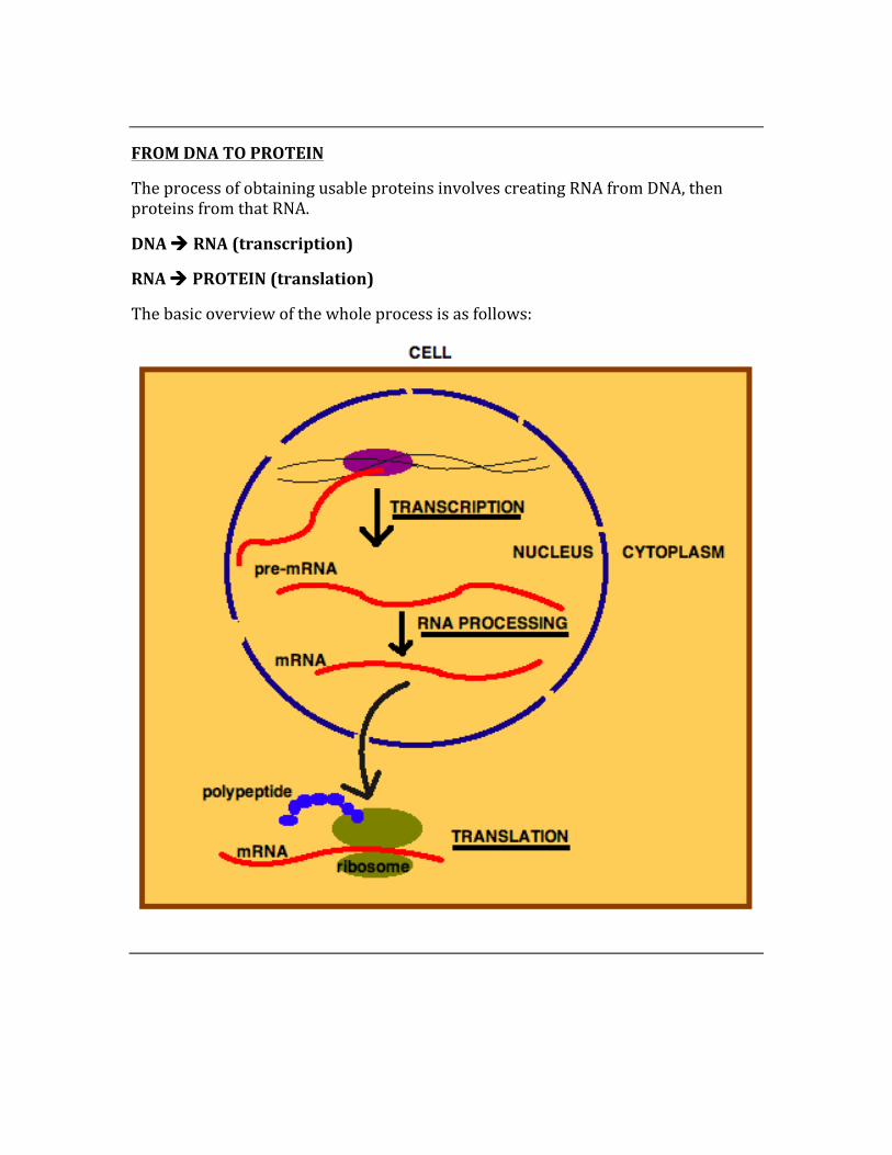
FROM DNA TO PROTEIN
The process of obtaining usable proteins involves creating RNA from DNA, then proteins from that RNA.
DNA à RNA (transcription)
RNA à PROTEIN (translation)
The basic overview of the whole process is as follows:
TYPES OF RNA
Transcription is the process by which RNA is made from each strand of DNA.
There are 3 types of RNA: rRNA, mRNA, and tRNA
rRNA – Is made via RNA polymerase I, and is the most abundant type of RNA. Ribosomal RNA combines with protein in the cytoplasm to form a ribosome, which serves as the site and carries all of the necessary enzymes required for protein synthesis.
mRNA – Is made via RNA polymerase II, and is the largest type of RNA. Messenger RNA is the RNA that is created from a gene segment of DNA . The mRNA then carries the code it receives from DNA into the cytoplasm where protein synthesis will occur
tRNA – Is made via RNA polyerase III, and is the smallest type of RNA. Transfer RNA is the type of RNA that reads the code from the mRNA and carries the amino acid to be incorporated into the developing proteins. There are more than 20 different tRNA’s, meaning there is one for each amino acid. tRNA contains approximately 75 nucleotides, three of these are “anticodons”.
* Prokaryotes have only one RNA polymerase that makes all of the RNA sub-‐types.
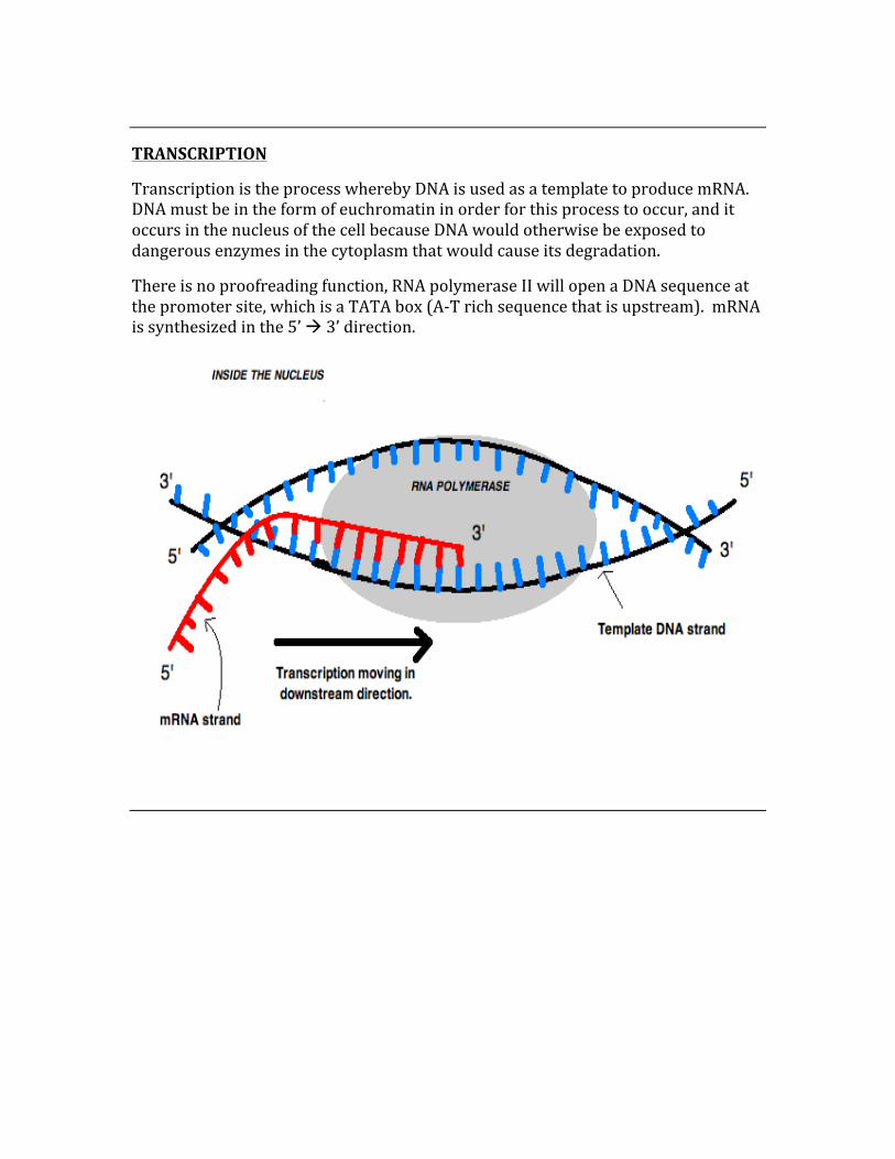
TRANSCRIPTION
Transcription is the process whereby DNA is used as a template to produce mRNA. DNA must be in the form of euchromatin in order for this process to occur, and it occurs in the nucleus of the cell because DNA would otherwise be exposed to dangerous enzymes in the cytoplasm that would cause its degradation.
There is no proofreading function, RNA polymerase II will open a DNA sequence at the promoter site, which is a TATA box (A-‐T rich sequence that is upstream). mRNA is synthesized in the 5’ à 3’ direction.
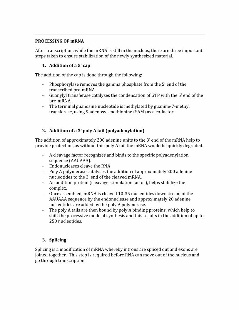
PROCESSING OF mRNA
After transcription, while the mRNA is still in the nucleus, there are three important steps taken to ensure stabilization of the newly synthesized material.
1. Addition of a 5’ cap
The addition of the cap is done through the following:
-‐ Phosphorylase removes the gamma phosphate from the 5’ end of the transcribed pre-‐mRNA.
-‐ Guanylyl transferase catalyzes the condensation of GTP with the 5’ end of the pre-‐mRNA.
-‐ The terminal guanosine nucleotide is methylated by guanine-‐7-‐methyl transferase, using S-‐adenosyl-‐methionine (SAM) as a co-‐factor.
2. Addition of a 3’ poly A tail (polyadenylation)
The addition of approximately 200 adenine units to the 3’ end of the mRNA help to provide protection, as without this poly A tail the mRNA would be quickly degraded.
-‐ A cleavage factor recognizes and binds to the specific polyadenylation sequence (AAUAAA).
-‐ Endonucleases cleave the RNA -‐ Poly A polymerase catalyses the addition of approximately 200 adenine
nucleotides to the 3’ end of the cleaved mRNA. -‐ An addition protein (cleavage stimulation factor), helps stabilize the
complex. -‐ Once assembled, mRNA is cleaved 10-‐35 nucleotides downstream of the
AAUAAA sequence by the endonuclease and approximately 20 adenine nucleotides are added by the poly A polymerase.
-‐ The poly A tails are then bound by poly A binding proteins, which help to shift the processive mode of synthesis and this results in the addition of up to 250 nucleotides.
3. Splicing
Splicing is a modification of mRNA whereby introns are spliced out and exons are joined together. This step is required before RNA can move out of the nucleus and go through transcription.
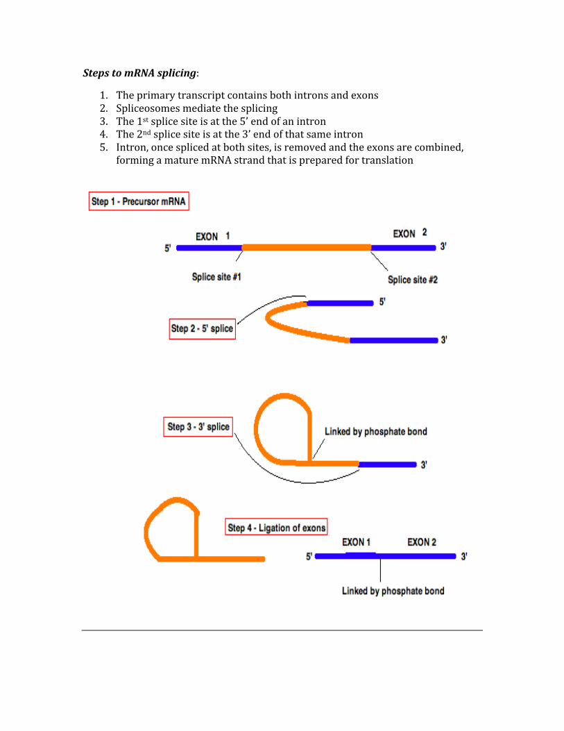
Steps to mRNA splicing:
1. The primary transcript contains both introns and exons 2. Spliceosomes mediate the splicing 3. The 1st splice site is at the 5’ end of an intron 4. The 2nd splice site is at the 3’ end of that same intron 5. Intron, once spliced at both sites, is removed and the exons are combined,
forming a mature mRNA strand that is prepared for translation
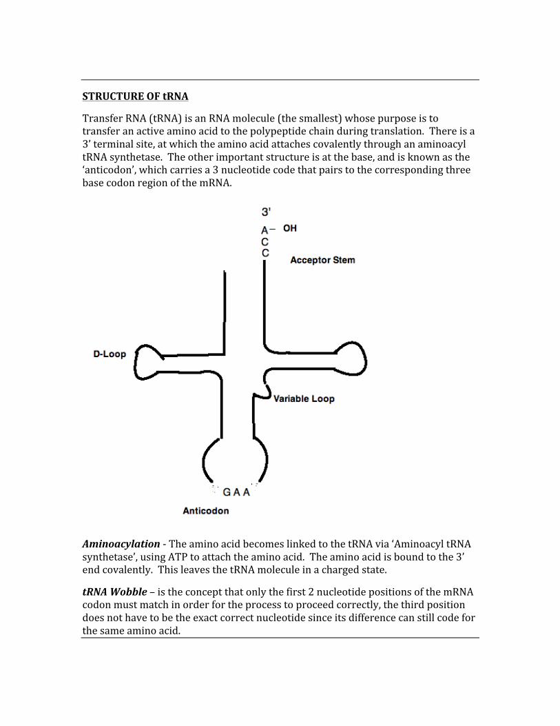
STRUCTURE OF tRNA
Transfer RNA (tRNA) is an RNA molecule (the smallest) whose purpose is to transfer an active amino acid to the polypeptide chain during translation. There is a 3’ terminal site, at which the amino acid attaches covalently through an aminoacyl tRNA synthetase. The other important structure is at the base, and is known as the ‘anticodon’, which carries a 3 nucleotide code that pairs to the corresponding three base codon region of the mRNA.
Aminoacylation -‐ The amino acid becomes linked to the tRNA via ‘Aminoacyl tRNA synthetase’, using ATP to attach the amino acid. The amino acid is bound to the 3’ end covalently. This leaves the tRNA molecule in a charged state.
tRNA Wobble – is the concept that only the first 2 nucleotide positions of the mRNA codon must match in order for the process to proceed correctly, the third position does not have to be the exact correct nucleotide since its difference can still code for the same amino acid.
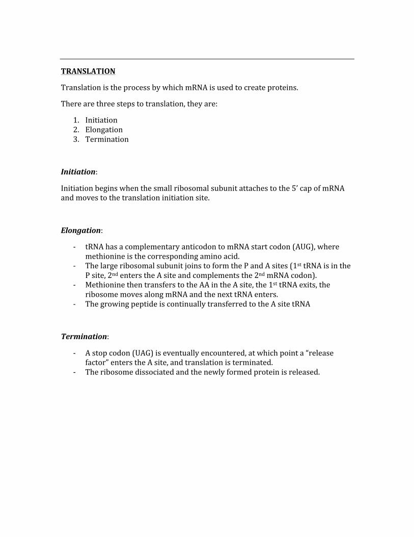
TRANSLATION
Translation is the process by which mRNA is used to create proteins.
There are three steps to translation, they are:
1. Initiation 2. Elongation 3. Termination
Initiation:
Initiation begins when the small ribosomal subunit attaches to the 5’ cap of mRNA and moves to the translation initiation site.
Elongation:
-‐ tRNA has a complementary anticodon to mRNA start codon (AUG), where methionine is the corresponding amino acid.
-‐ The large ribosomal subunit joins to form the P and A sites (1st tRNA is in the P site, 2nd enters the A site and complements the 2nd mRNA codon).
-‐ Methionine then transfers to the AA in the A site, the 1st tRNA exits, the ribosome moves along mRNA and the next tRNA enters.
-‐ The growing peptide is continually transferred to the A site tRNA
Termination:
-‐ A stop codon (UAG) is eventually encountered, at which point a “release factor” enters the A site, and translation is terminated.
-‐ The ribosome dissociated and the newly formed protein is released.
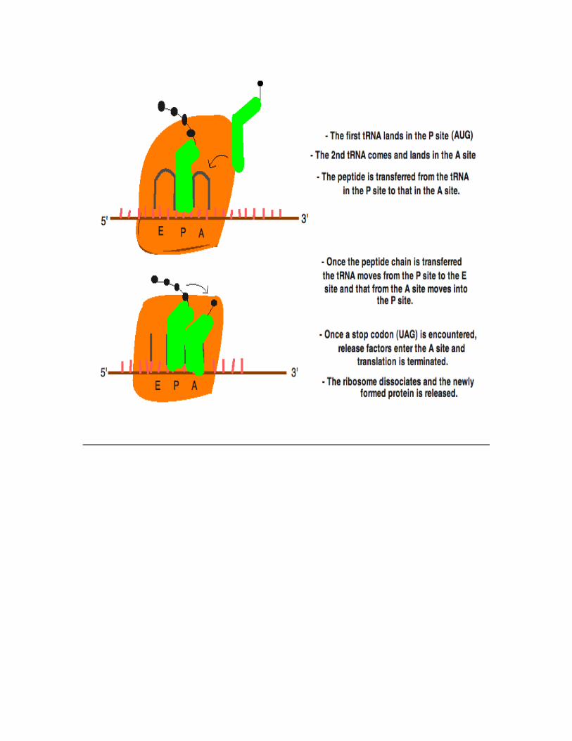
METABOLISM
High-‐Yield disorders of metabolism:
Glycogen storage diseases:
Type 1 – von Gierke disease (Hepatorenal Glycogenosis)
-‐ Caused by glucose-‐6-‐phosphatase deficiency.
Patient may experience:
-‐ Hypoglycemia -‐ Chronic hunger -‐ Delayed puberty and/or underdevelopment -‐ Hepatomegaly -‐ Fatigue
Type 2 – Pompe’s disease
-‐ Caused by a lysosomal α-‐1,4-‐glucosidase deficiency.
Patient may experience:
-‐ Muscle weakness, especially in the heart -‐ Most commonly seen in newborn children OR those in their 30’s and 40’s -‐ Fatigue as a result of weakening of the heart and liver -‐ Curvature of the spine is a progressive symptoms -‐ Difficulty breathing, such as labored breathing, and infections of the
respiratory tract -‐ Dizziness and syncope
Type 3 – Cori’s disease
-‐ Caused by a deficiency of the debranching enzyme α-‐1,6-‐glucosidase
Patient may experience:
-‐ Symptoms similar to von Gierke’s disease, but milder -‐ Young children typically have massive hepatomegaly that diminishes with
increasing age
Type 4 – Andersen disease
-‐ The main clinical features of Anderson disease are insufficiency of the liver and abnormalities of the heart and nervous system
-‐ This disease is rare and will lead to early death
Type 5 – McArdle’s disease
-‐ Caused by a deficiency of skeletal muscle glycogen phosphorylase
Patient experiences:
-‐ Increased glycogen found within the muscle -‐ Painful cramps and myoglobinuria when activity is increased
Type 6 – Hers disease
-‐ Caused by a hepatic phosphorylase deficiency -‐ Patient may have the inability to maintain blood-‐glucose levels during
periods of fasting. -‐ Urine and serum ketones are elevated proportionally to the level of fasting -‐ Mild to moderate hyperlipidemia may be present -‐ Prominent hepatomegaly and growth retardation are common findings of
Hers disease
Type 7 – Tarui disease
-‐ Caused by a deficiency of phosphofructokinase (PFK) in glycolysis -‐ Patient will experience increased muscle glycogen that cannot be broken
down -‐ Cramping -‐ Higher levels of myoglobin in the urine when there is increased physical
activity
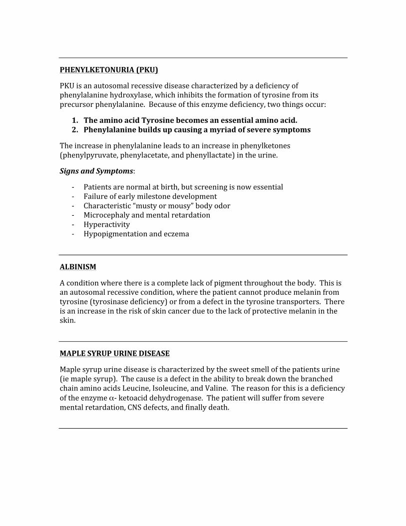
PHENYLKETONURIA (PKU)
PKU is an autosomal recessive disease characterized by a deficiency of phenylalanine hydroxylase, which inhibits the formation of tyrosine from its precursor phenylalanine. Because of this enzyme deficiency, two things occur:
1. The amino acid Tyrosine becomes an essential amino acid. 2. Phenylalanine builds up causing a myriad of severe symptoms
The increase in phenylalanine leads to an increase in phenylketones (phenylpyruvate, phenylacetate, and phenyllactate) in the urine.
Signs and Symptoms:
-‐ Patients are normal at birth, but screening is now essential -‐ Failure of early milestone development -‐ Characteristic “musty or mousy” body odor -‐ Microcephaly and mental retardation -‐ Hyperactivity -‐ Hypopigmentation and eczema
ALBINISM
A condition where there is a complete lack of pigment throughout the body. This is an autosomal recessive condition, where the patient cannot produce melanin from tyrosine (tyrosinase deficiency) or from a defect in the tyrosine transporters. There is an increase in the risk of skin cancer due to the lack of protective melanin in the skin.
MAPLE SYRUP URINE DISEASE
Maple syrup urine disease is characterized by the sweet smell of the patients urine (ie maple syrup). The cause is a defect in the ability to break down the branched chain amino acids Leucine, Isoleucine, and Valine. The reason for this is a deficiency of the enzyme α-‐ ketoacid dehydrogenase. The patient will suffer from severe mental retardation, CNS defects, and finally death.
ALKAPTONURIA
Another condition involving tyrosine, alkaptonuria results from a deficiency of homogentisate 1,2-‐dioxygenase in the pathway of tyrosine degradation. Homogentisic acid (alkapton) thus accumulates in the blood and is excreted in the urine in large amounts, leading to blackening of the urine upon standing. Excessive amounts of homogentisic acid cause damage the cartilage, leading to severe arthralgias.
HOMOCYSTEINURIA
Also referred to as Cystathionine beta synthase deficiency (CBS deficiency), it is an autosomal recessive disorder.
In the case of deficiency, patient will have excessive homocysteine in the urine. In this case, cysteine will be essential and should be increased in the diet, while simultaneously decreasing the amount of methionine in the diet.
Signs and symptoms include:
-‐ Mental retardation -‐ Seizure -‐ Musculoskeletal abnormalities (tall build, long limbs, pectus excavatum, pes
cavus, and genu valgum) -‐ Abnormalities of the eyes (glaucoma, subluxation of lens) -‐ Vascular conditions (early thromboses)
CYSTINURIA
Cystinuria is a condition whereby there is a defect in the transport of tubular amino acids for the following amino acids: Cystine, Ornithine, Lysine, and Arginine. This results in an excess of cystine in the urine, which can predispose the patient to kidney stones. Management is to alkalinize the urine with acetazolamide.
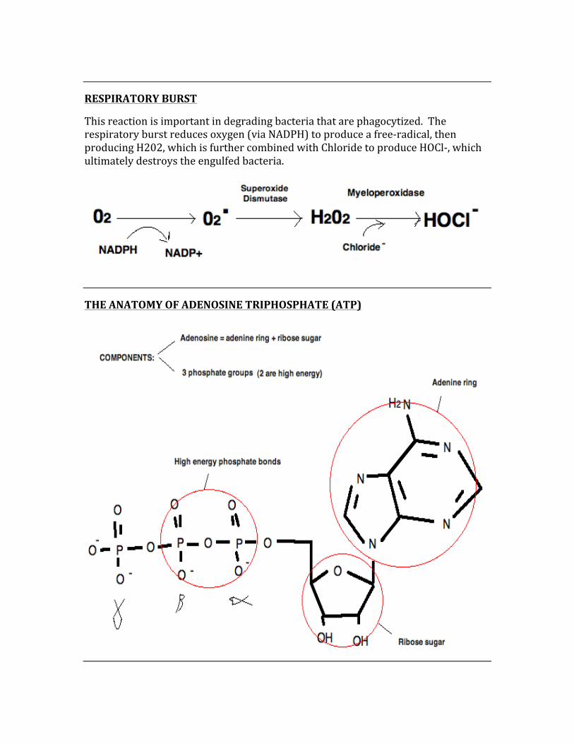
RESPIRATORY BURST
This reaction is important in degrading bacteria that are phagocytized. The respiratory burst reduces oxygen (via NADPH) to produce a free-‐radical, then producing H202, which is further combined with Chloride to produce HOCl-‐, which ultimately destroys the engulfed bacteria.
THE ANATOMY OF ADENOSINE TRIPHOSPHATE (ATP)
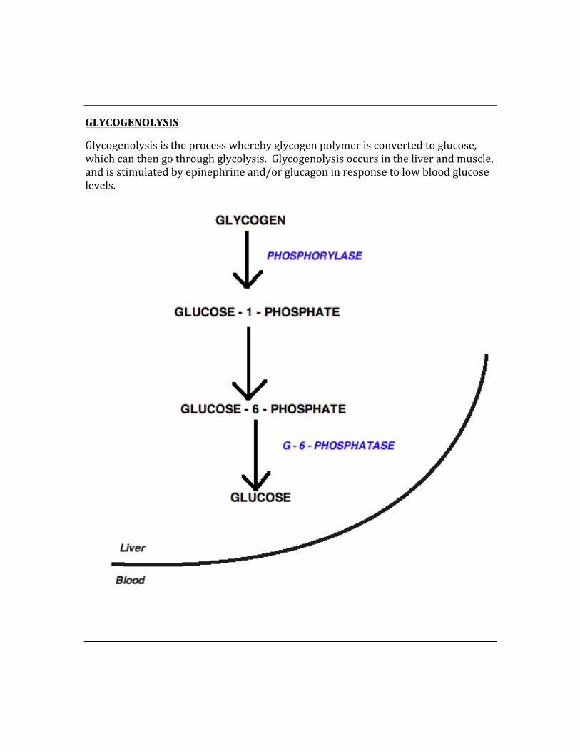
GLYCOGENOLYSIS
Glycogenolysis is the process whereby glycogen polymer is converted to glucose, which can then go through glycolysis. Glycogenolysis occurs in the liver and muscle, and is stimulated by epinephrine and/or glucagon in response to low blood glucose levels.
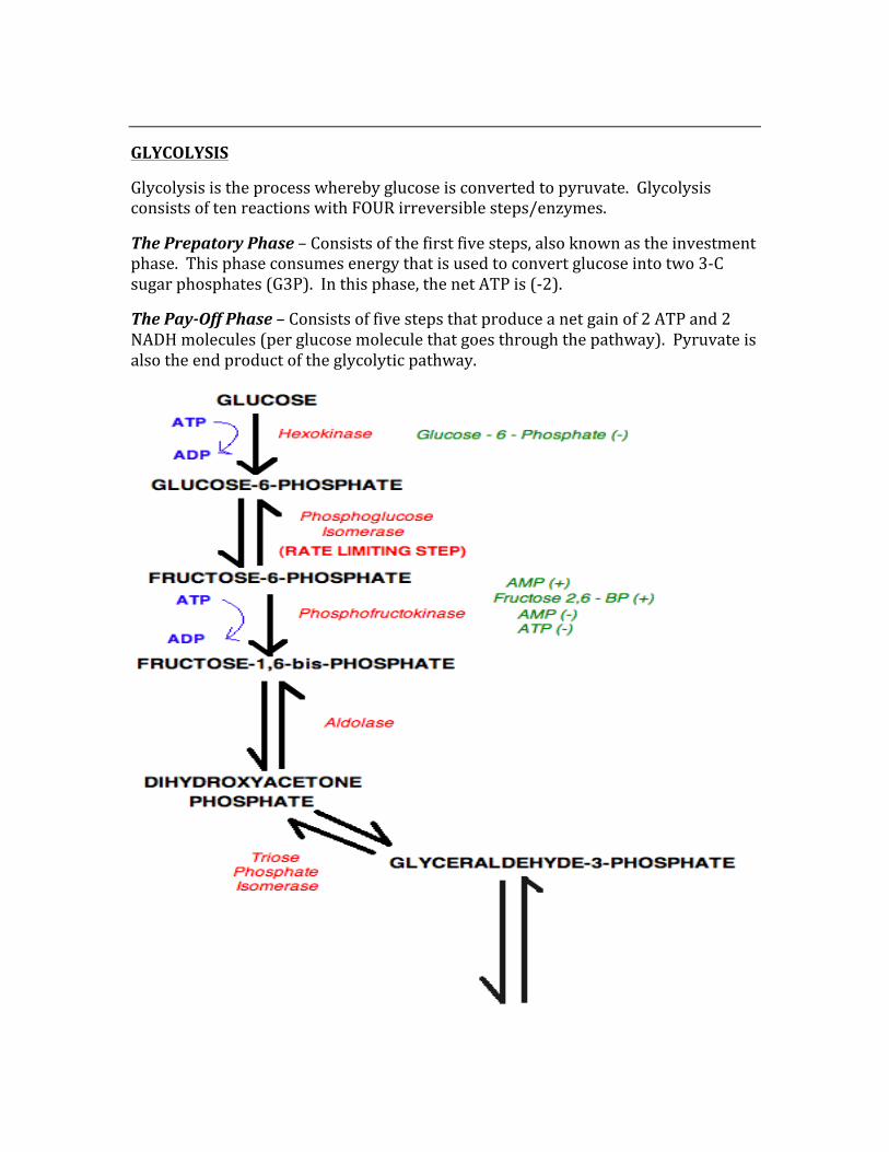
GLYCOLYSIS
Glycolysis is the process whereby glucose is converted to pyruvate. Glycolysis consists of ten reactions with FOUR irreversible steps/enzymes.
The Prepatory Phase – Consists of the first five steps, also known as the investment phase. This phase consumes energy that is used to convert glucose into two 3-‐C sugar phosphates (G3P). In this phase, the net ATP is (-‐2).
The Pay-‐Off Phase – Consists of five steps that produce a net gain of 2 ATP and 2 NADH molecules (per glucose molecule that goes through the pathway). Pyruvate is also the end product of the glycolytic pathway.
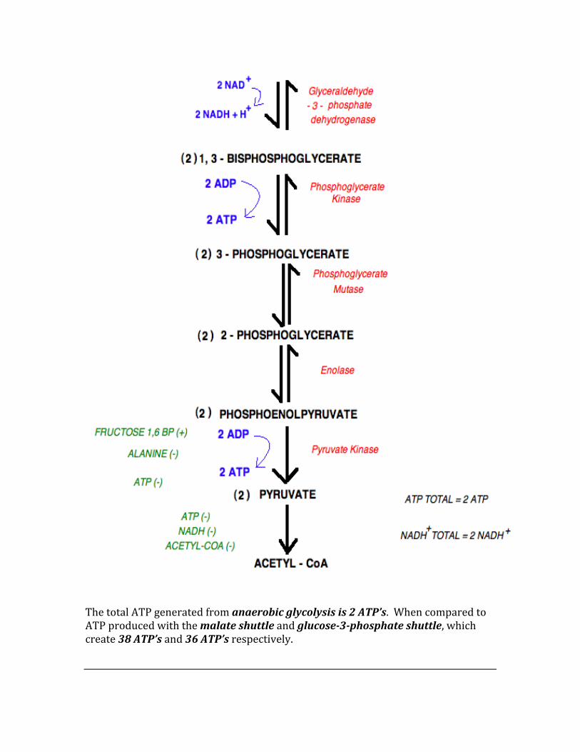
The total ATP generated from anaerobic glycolysis is 2 ATP’s. When compared to ATP produced with the malate shuttle and glucose-‐3-‐phosphate shuttle, which create 38 ATP’s and 36 ATP’s respectively.
REGULATION OF GLYCOLYSIS
There are FOUR steps in glycolyisis that are considered to be irreversible, thus once they have occurred, glycolysis must progress in the forward direction. The four regulatory enzymes are:
1. Hexokinase 2. Phosphofructokinase 3. Pyruvate kinase 4. Pyruvate dehydrogenase
Hexokinase:
-‐ Hexokinase is responsible for the first step of glycolysis in the muscles and brain.
-‐ It is inhibited by the presence of glucose-‐6-‐phosphate, which is the product of its activity.
-‐ This step is important because it prevents the consumption of too much cellular ATP in the formation of glucose–6–phosphate when glucose is not limiting.
-‐ Hexokinase has a low affinity to glucose, thus it permits glycolysis initiation even if blood glucose levels are low.
Phosphofructokinase:
-‐ PFK is the rate-‐limiting step of glycolysis, thus it is the most important control point throughout the whole process.
-‐ Regulation is by both alloesteric effectors and by covalent modifications (ie phosphorylation).
-‐ It is stimulated by the presence of AMP and fructose-‐2,6-‐bisphosphate. -‐ Even if ATP is high, the presence of AMP can overcome its inhibitory effects
due to the ability to allosterically activate PFK. -‐ It is inhibited by the presence of ATP and citrate
Pyruvate Kinase:
-‐ Similar to PFK, is regulated by allosteric effectors and by phosphorylation. -‐ PK is activated by fructose-‐1,6-‐bisphosphate and inhibited by ATP and
alanine.
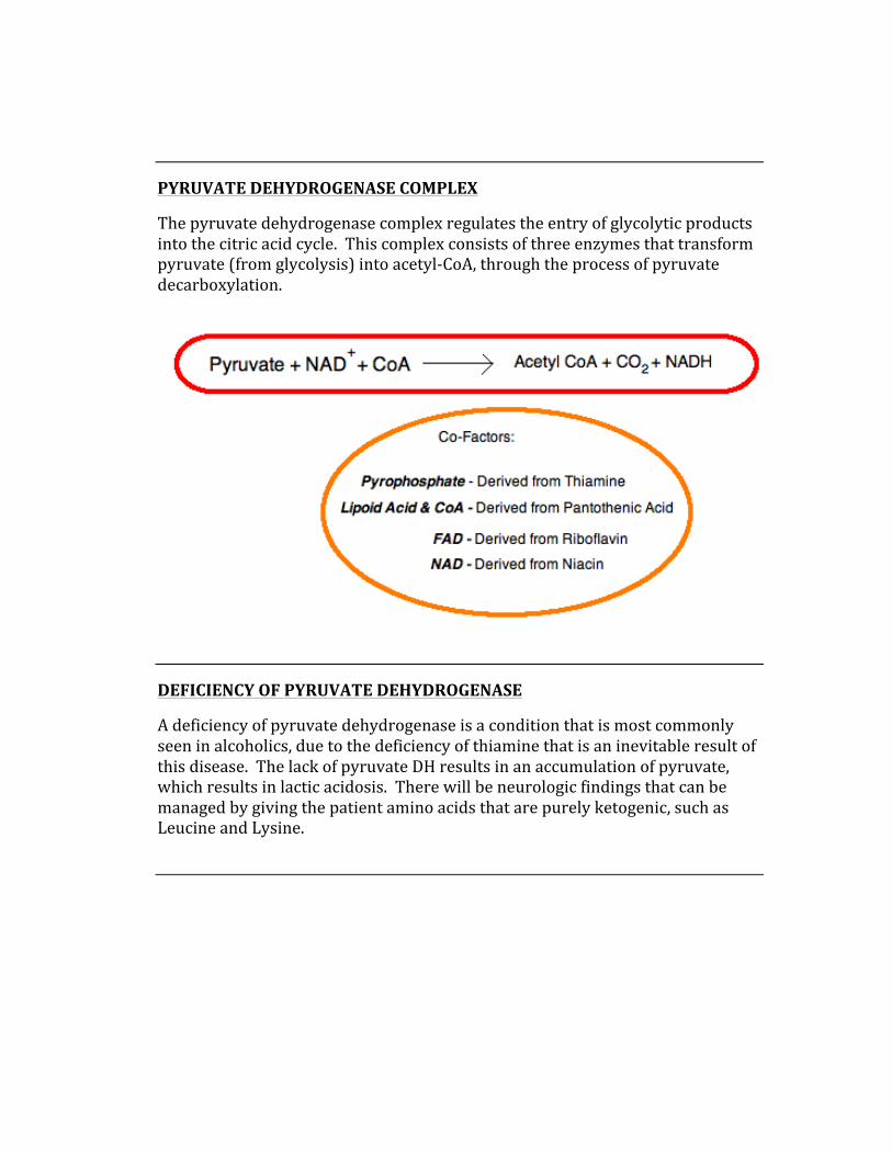
PYRUVATE DEHYDROGENASE COMPLEX
The pyruvate dehydrogenase complex regulates the entry of glycolytic products into the citric acid cycle. This complex consists of three enzymes that transform pyruvate (from glycolysis) into acetyl-‐CoA, through the process of pyruvate decarboxylation.
DEFICIENCY OF PYRUVATE DEHYDROGENASE
A deficiency of pyruvate dehydrogenase is a condition that is most commonly seen in alcoholics, due to the deficiency of thiamine that is an inevitable result of this disease. The lack of pyruvate DH results in an accumulation of pyruvate, which results in lactic acidosis. There will be neurologic findings that can be managed by giving the patient amino acids that are purely ketogenic, such as Leucine and Lysine.
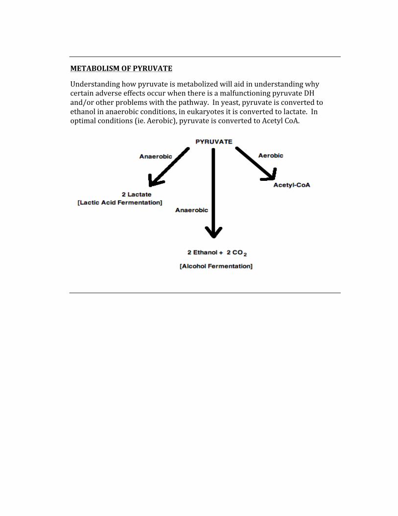
METABOLISM OF PYRUVATE
Understanding how pyruvate is metabolized will aid in understanding why certain adverse effects occur when there is a malfunctioning pyruvate DH and/or other problems with the pathway. In yeast, pyruvate is converted to ethanol in anaerobic conditions, in eukaryotes it is converted to lactate. In optimal conditions (ie. Aerobic), pyruvate is converted to Acetyl CoA.
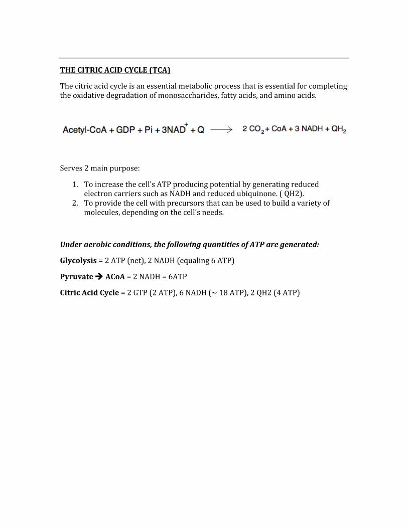
THE CITRIC ACID CYCLE (TCA)
The citric acid cycle is an essential metabolic process that is essential for completing the oxidative degradation of monosaccharides, fatty acids, and amino acids.
Serves 2 main purpose:
1. To increase the cell’s ATP producing potential by generating reduced electron carriers such as NADH and reduced ubiquinone. ( QH2).
2. To provide the cell with precursors that can be used to build a variety of molecules, depending on the cell’s needs.
Under aerobic conditions, the following quantities of ATP are generated:
Glycolysis = 2 ATP (net), 2 NADH (equaling 6 ATP)
Pyruvate à ACoA = 2 NADH = 6ATP
Citric Acid Cycle = 2 GTP (2 ATP), 6 NADH (~ 18 ATP), 2 QH2 (4 ATP)
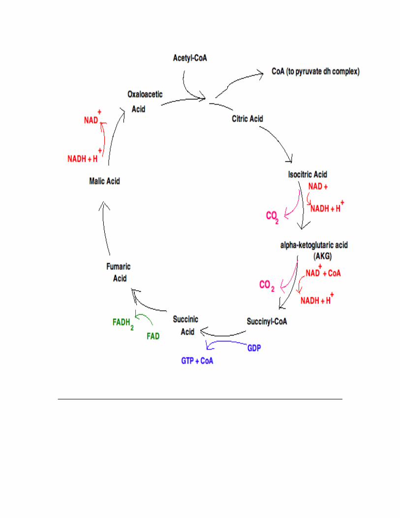
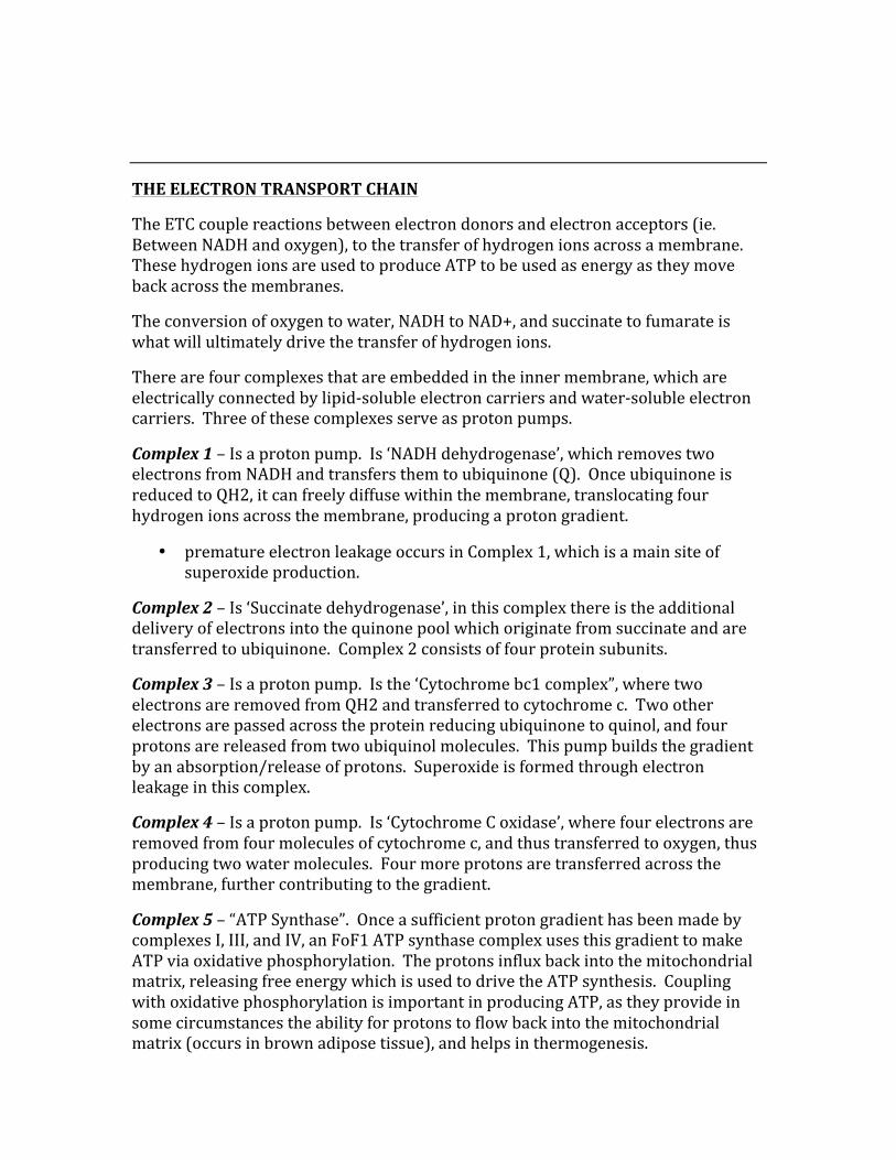
THE ELECTRON TRANSPORT CHAIN
The ETC couple reactions between electron donors and electron acceptors (ie. Between NADH and oxygen), to the transfer of hydrogen ions across a membrane. These hydrogen ions are used to produce ATP to be used as energy as they move back across the membranes.
The conversion of oxygen to water, NADH to NAD+, and succinate to fumarate is what will ultimately drive the transfer of hydrogen ions.
There are four complexes that are embedded in the inner membrane, which are electrically connected by lipid-‐soluble electron carriers and water-‐soluble electron carriers. Three of these complexes serve as proton pumps.
Complex 1 – Is a proton pump. Is ‘NADH dehydrogenase’, which removes two electrons from NADH and transfers them to ubiquinone (Q). Once ubiquinone is reduced to QH2, it can freely diffuse within the membrane, translocating four hydrogen ions across the membrane, producing a proton gradient.
• premature electron leakage occurs in Complex 1, which is a main site of superoxide production.
Complex 2 – Is ‘Succinate dehydrogenase’, in this complex there is the additional delivery of electrons into the quinone pool which originate from succinate and are transferred to ubiquinone. Complex 2 consists of four protein subunits.
Complex 3 – Is a proton pump. Is the ‘Cytochrome bc1 complex”, where two electrons are removed from QH2 and transferred to cytochrome c. Two other electrons are passed across the protein reducing ubiquinone to quinol, and four protons are released from two ubiquinol molecules. This pump builds the gradient by an absorption/release of protons. Superoxide is formed through electron leakage in this complex.
Complex 4 – Is a proton pump. Is ‘Cytochrome C oxidase’, where four electrons are removed from four molecules of cytochrome c, and thus transferred to oxygen, thus producing two water molecules. Four more protons are transferred across the membrane, further contributing to the gradient.
Complex 5 – “ATP Synthase”. Once a sufficient proton gradient has been made by complexes I, III, and IV, an FoF1 ATP synthase complex uses this gradient to make ATP via oxidative phosphorylation. The protons influx back into the mitochondrial matrix, releasing free energy which is used to drive the ATP synthesis. Coupling with oxidative phosphorylation is important in producing ATP, as they provide in some circumstances the ability for protons to flow back into the mitochondrial matrix (occurs in brown adipose tissue), and helps in thermogenesis.
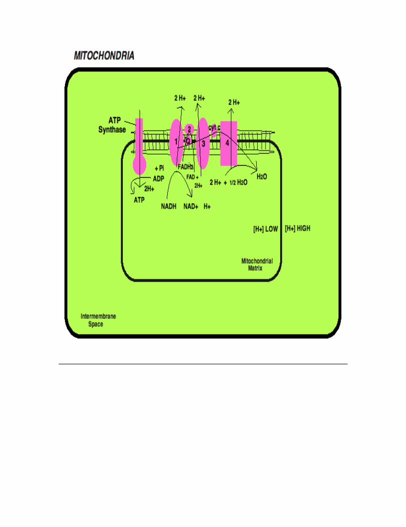
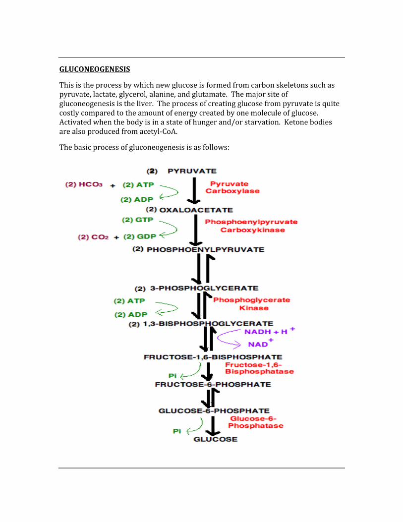
GLUCONEOGENESIS
This is the process by which new glucose is formed from carbon skeletons such as pyruvate, lactate, glycerol, alanine, and glutamate. The major site of gluconeogenesis is the liver. The process of creating glucose from pyruvate is quite costly compared to the amount of energy created by one molecule of glucose. Activated when the body is in a state of hunger and/or starvation. Ketone bodies are also produced from acetyl-‐CoA.
The basic process of gluconeogenesis is as follows:
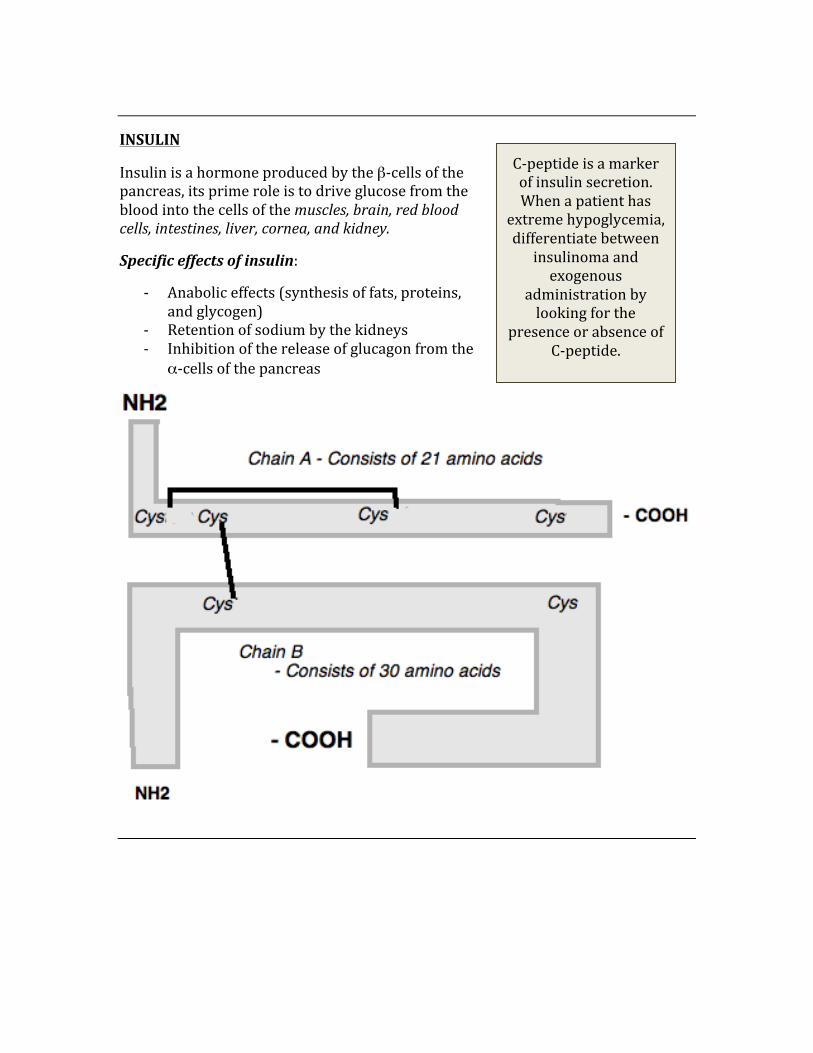
INSULIN
Insulin is a hormone produced by the β-‐cells of the pancreas, its prime role is to drive glucose from the blood into the cells of the muscles, brain, red blood cells, intestines, liver, cornea, and kidney.
Specific effects of insulin:
-‐ Anabolic effects (synthesis of fats, proteins, and glycogen)
-‐ Retention of sodium by the kidneys -‐ Inhibition of the release of glucagon from the
α-‐cells of the pancreas
C-‐peptide is a marker of insulin secretion. When a patient has
extreme hypoglycemia, differentiate between insulinoma and exogenous
administration by looking for the
presence or absence of C-‐peptide.
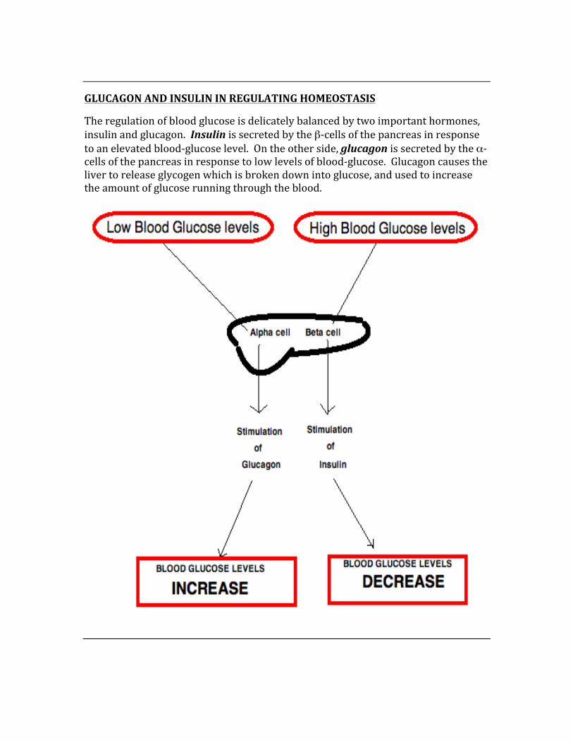
GLUCAGON AND INSULIN IN REGULATING HOMEOSTASIS
The regulation of blood glucose is delicately balanced by two important hormones, insulin and glucagon. Insulin is secreted by the β-‐cells of the pancreas in response to an elevated blood-‐glucose level. On the other side, glucagon is secreted by the α-‐cells of the pancreas in response to low levels of blood-‐glucose. Glucagon causes the liver to release glycogen which is broken down into glucose, and used to increase the amount of glucose running through the blood.
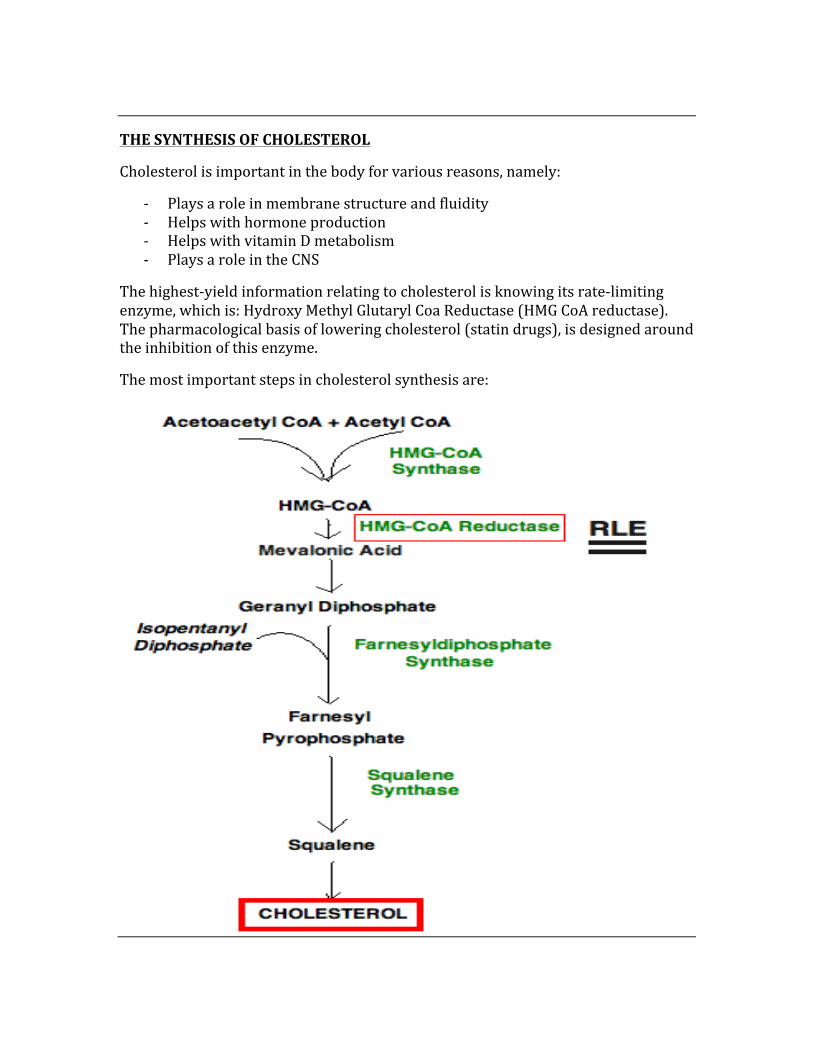
THE SYNTHESIS OF CHOLESTEROL
Cholesterol is important in the body for various reasons, namely:
-‐ Plays a role in membrane structure and fluidity -‐ Helps with hormone production -‐ Helps with vitamin D metabolism -‐ Plays a role in the CNS
The highest-‐yield information relating to cholesterol is knowing its rate-‐limiting enzyme, which is: Hydroxy Methyl Glutaryl Coa Reductase (HMG CoA reductase). The pharmacological basis of lowering cholesterol (statin drugs), is designed around the inhibition of this enzyme.
The most important steps in cholesterol synthesis are:
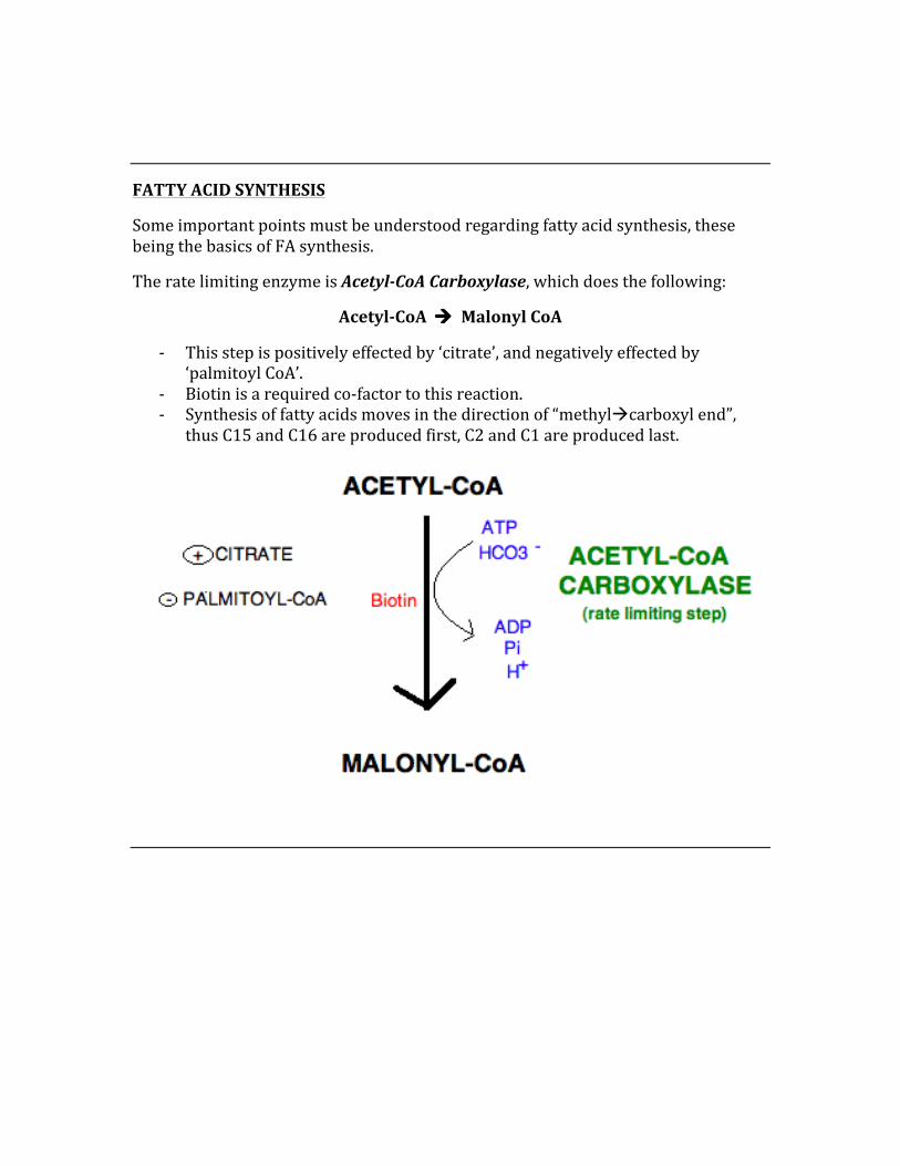
FATTY ACID SYNTHESIS
Some important points must be understood regarding fatty acid synthesis, these being the basics of FA synthesis.
The rate limiting enzyme is Acetyl-‐CoA Carboxylase, which does the following:
Acetyl-‐CoA à Malonyl CoA
-‐ This step is positively effected by ‘citrate’, and negatively effected by ‘palmitoyl CoA’.
-‐ Biotin is a required co-‐factor to this reaction. -‐ Synthesis of fatty acids moves in the direction of “methylàcarboxyl end”,
thus C15 and C16 are produced first, C2 and C1 are produced last.
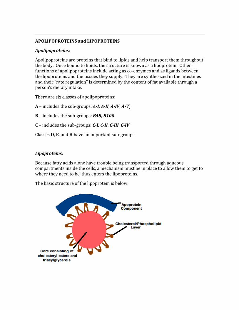
APOLIPOPROTEINS and LIPOPROTEINS
Apolipoproteins:
Apolipoproteins are proteins that bind to lipids and help transport them throughout the body. Once bound to lipids, the structure is known as a lipoprotein. Other functions of apolipoproteins include acting as co-‐enzymes and as ligands between the lipoproteins and the tissues they supply. They are synthesized in the intestines and their “rate regulation” is determined by the content of fat available through a person’s dietary intake.
There are six classes of apolipoproteins:
A – includes the sub-‐groups: A-‐I, A-‐II, A-‐IV, A-‐V)
B – includes the sub-‐groups: B48, B100
C – includes the sub-‐groups: C-‐I, C-‐II, C-‐III, C-‐IV
Classes D, E, and H have no important sub-‐groups.
Lipoproteins:
Because fatty acids alone have trouble being transported through aqueous compartments inside the cells, a mechanism must be in place to allow them to get to where they need to be, thus enters the lipoproteins.
The basic structure of the lipoprotein is below:
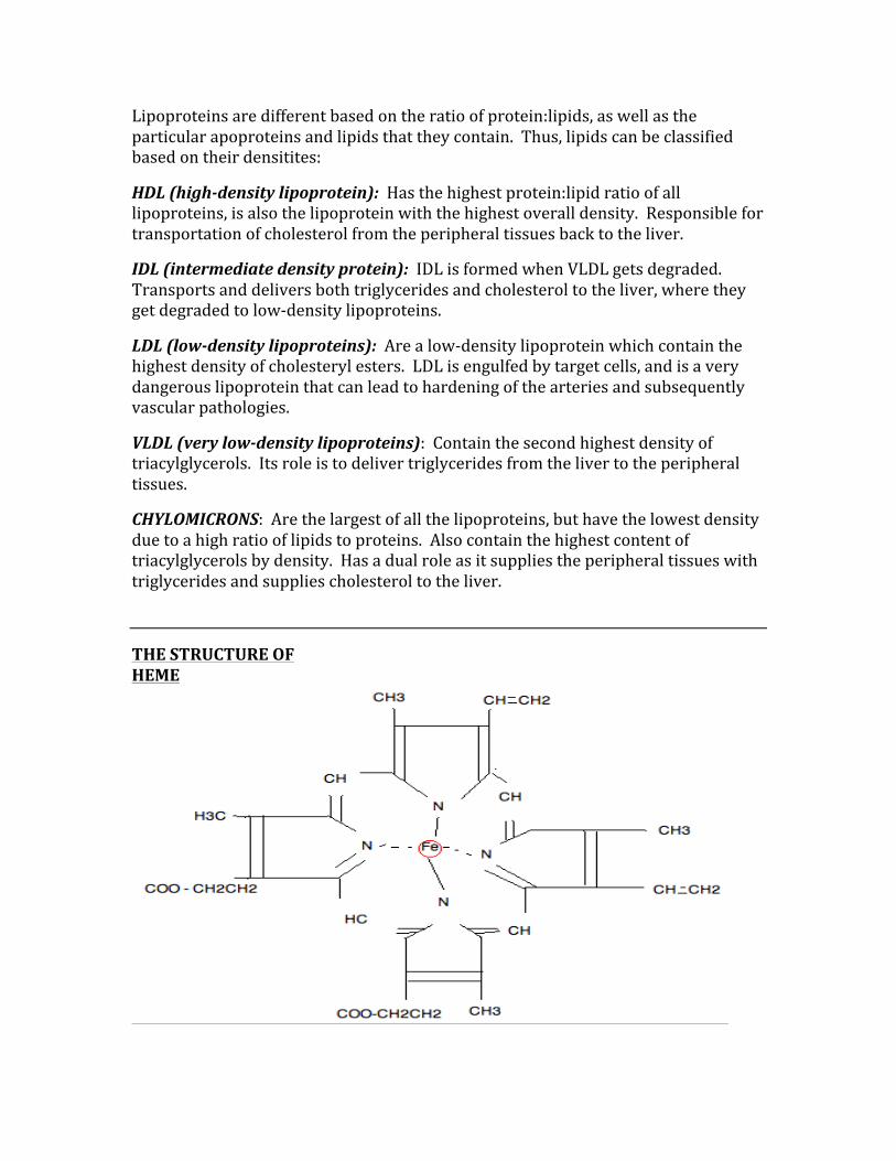
Lipoproteins are different based on the ratio of protein:lipids, as well as the particular apoproteins and lipids that they contain. Thus, lipids can be classified based on their densitites:
HDL (high-‐density lipoprotein): Has the highest protein:lipid ratio of all lipoproteins, is also the lipoprotein with the highest overall density. Responsible for transportation of cholesterol from the peripheral tissues back to the liver.
IDL (intermediate density protein): IDL is formed when VLDL gets degraded. Transports and delivers both triglycerides and cholesterol to the liver, where they get degraded to low-‐density lipoproteins.
LDL (low-‐density lipoproteins): Are a low-‐density lipoprotein which contain the highest density of cholesteryl esters. LDL is engulfed by target cells, and is a very dangerous lipoprotein that can lead to hardening of the arteries and subsequently vascular pathologies.
VLDL (very low-‐density lipoproteins): Contain the second highest density of triacylglycerols. Its role is to deliver triglycerides from the liver to the peripheral tissues.
CHYLOMICRONS: Are the largest of all the lipoproteins, but have the lowest density due to a high ratio of lipids to proteins. Also contain the highest content of triacylglycerols by density. Has a dual role as it supplies the peripheral tissues with triglycerides and supplies cholesterol to the liver.
THE STRUCTURE OF HEME
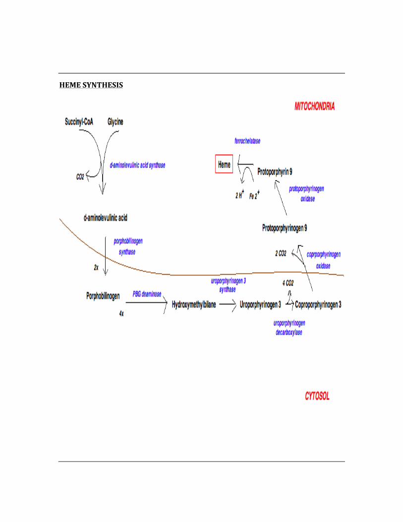
HEME SYNTHESIS
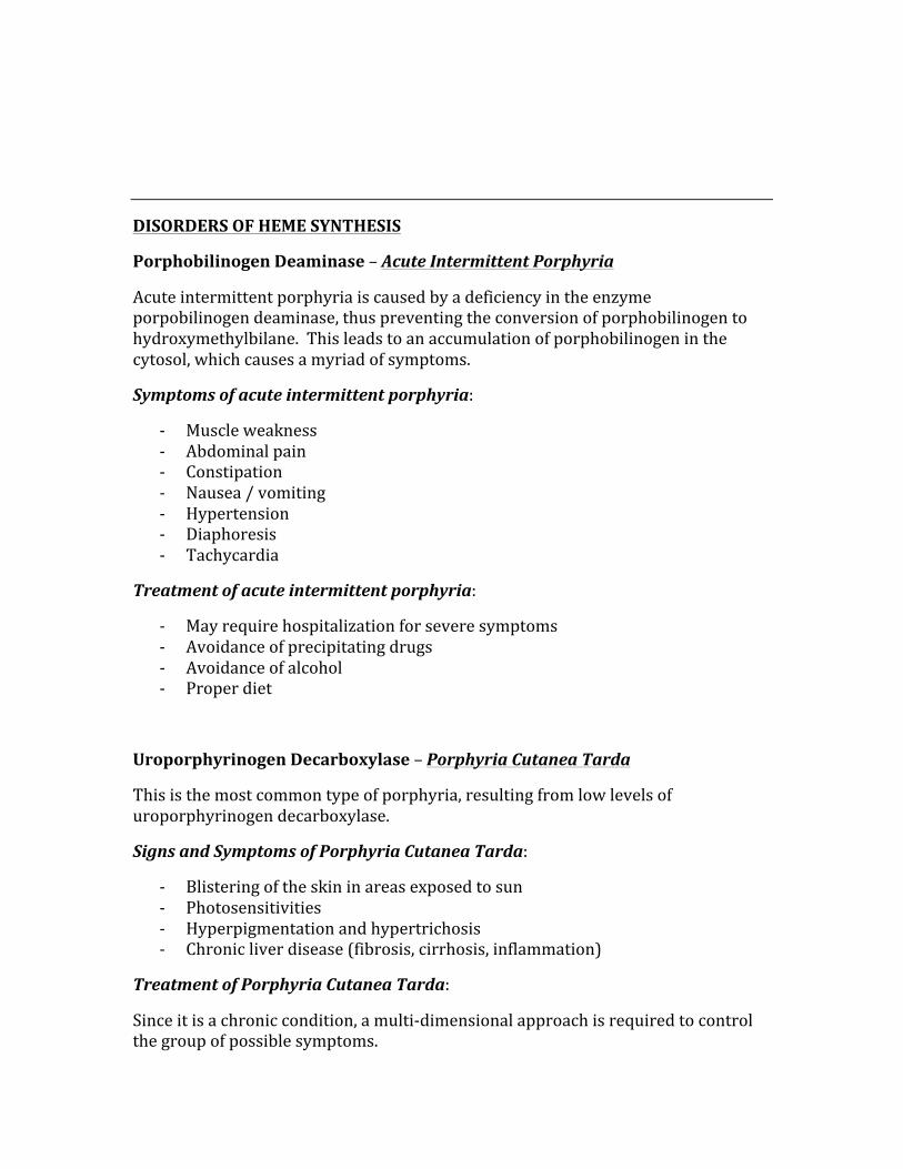
DISORDERS OF HEME SYNTHESIS
Porphobilinogen Deaminase – Acute Intermittent Porphyria
Acute intermittent porphyria is caused by a deficiency in the enzyme porpobilinogen deaminase, thus preventing the conversion of porphobilinogen to hydroxymethylbilane. This leads to an accumulation of porphobilinogen in the cytosol, which causes a myriad of symptoms.
Symptoms of acute intermittent porphyria:
-‐ Muscle weakness -‐ Abdominal pain -‐ Constipation -‐ Nausea / vomiting -‐ Hypertension -‐ Diaphoresis -‐ Tachycardia
Treatment of acute intermittent porphyria:
-‐ May require hospitalization for severe symptoms -‐ Avoidance of precipitating drugs -‐ Avoidance of alcohol -‐ Proper diet
Uroporphyrinogen Decarboxylase – Porphyria Cutanea Tarda
This is the most common type of porphyria, resulting from low levels of uroporphyrinogen decarboxylase.
Signs and Symptoms of Porphyria Cutanea Tarda:
-‐ Blistering of the skin in areas exposed to sun -‐ Photosensitivities -‐ Hyperpigmentation and hypertrichosis -‐ Chronic liver disease (fibrosis, cirrhosis, inflammation)
Treatment of Porphyria Cutanea Tarda:
Since it is a chronic condition, a multi-‐dimensional approach is required to control the group of possible symptoms.
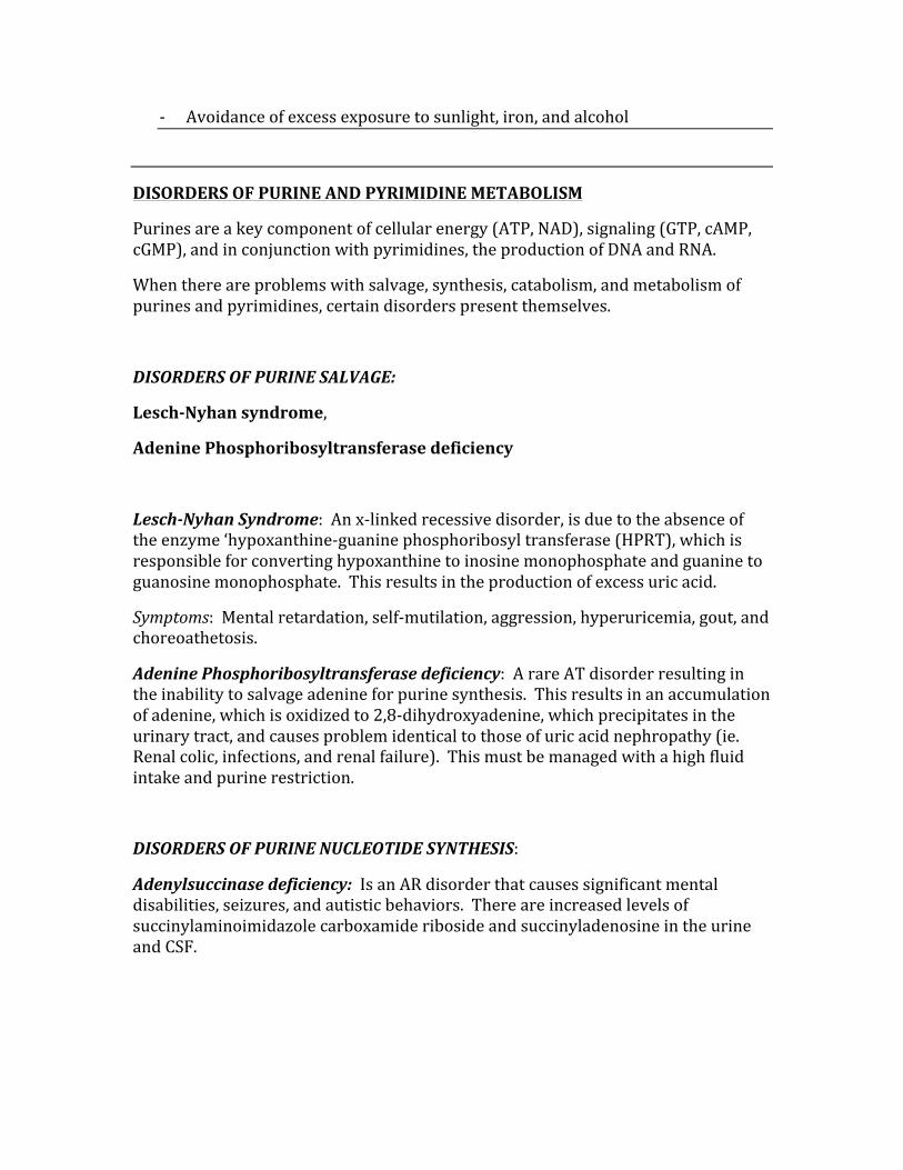
-‐ Avoidance of excess exposure to sunlight, iron, and alcohol
DISORDERS OF PURINE AND PYRIMIDINE METABOLISM
Purines are a key component of cellular energy (ATP, NAD), signaling (GTP, cAMP, cGMP), and in conjunction with pyrimidines, the production of DNA and RNA.
When there are problems with salvage, synthesis, catabolism, and metabolism of purines and pyrimidines, certain disorders present themselves.
DISORDERS OF PURINE SALVAGE:
Lesch-‐Nyhan syndrome,
Adenine Phosphoribosyltransferase deficiency
Lesch-‐Nyhan Syndrome: An x-‐linked recessive disorder, is due to the absence of the enzyme ‘hypoxanthine-‐guanine phosphoribosyl transferase (HPRT), which is responsible for converting hypoxanthine to inosine monophosphate and guanine to guanosine monophosphate. This results in the production of excess uric acid.
Symptoms: Mental retardation, self-‐mutilation, aggression, hyperuricemia, gout, and choreoathetosis.
Adenine Phosphoribosyltransferase deficiency: A rare AT disorder resulting in the inability to salvage adenine for purine synthesis. This results in an accumulation of adenine, which is oxidized to 2,8-‐dihydroxyadenine, which precipitates in the urinary tract, and causes problem identical to those of uric acid nephropathy (ie. Renal colic, infections, and renal failure). This must be managed with a high fluid intake and purine restriction.
DISORDERS OF PURINE NUCLEOTIDE SYNTHESIS:
Adenylsuccinase deficiency: Is an AR disorder that causes significant mental disabilities, seizures, and autistic behaviors. There are increased levels of succinylaminoimidazole carboxamide riboside and succinyladenosine in the urine and CSF.
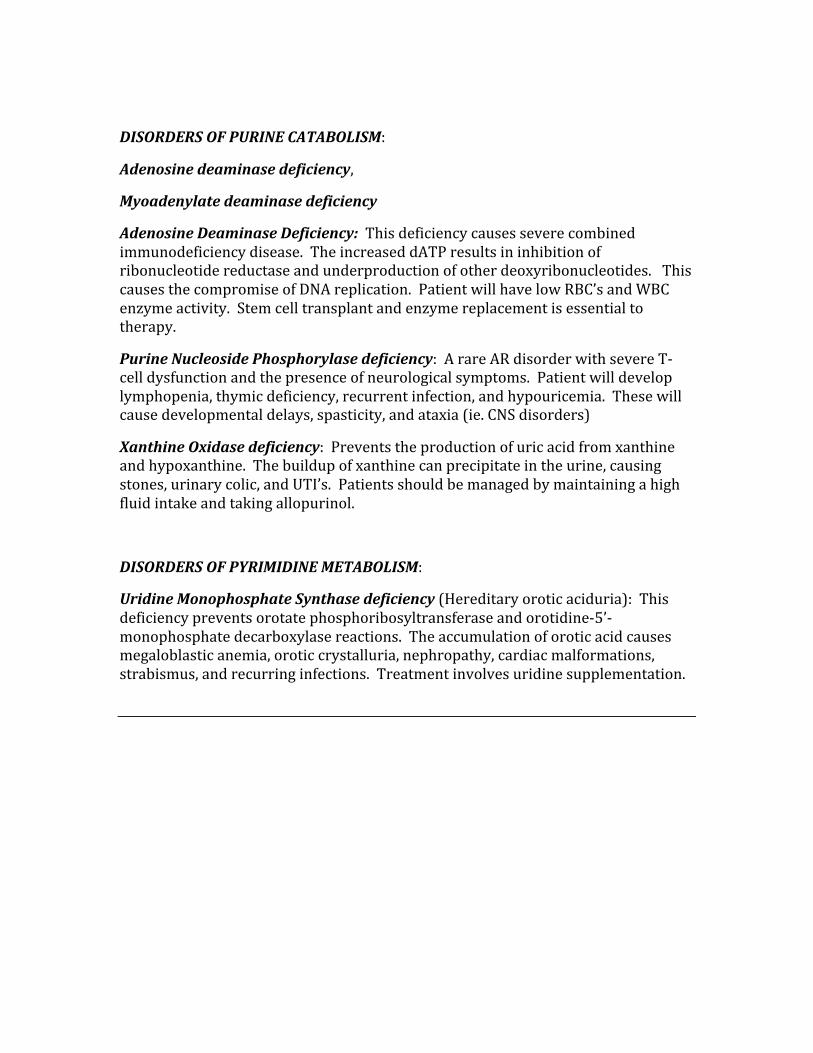
DISORDERS OF PURINE CATABOLISM:
Adenosine deaminase deficiency,
Myoadenylate deaminase deficiency
Adenosine Deaminase Deficiency: This deficiency causes severe combined immunodeficiency disease. The increased dATP results in inhibition of ribonucleotide reductase and underproduction of other deoxyribonucleotides. This causes the compromise of DNA replication. Patient will have low RBC’s and WBC enzyme activity. Stem cell transplant and enzyme replacement is essential to therapy.
Purine Nucleoside Phosphorylase deficiency: A rare AR disorder with severe T-‐cell dysfunction and the presence of neurological symptoms. Patient will develop lymphopenia, thymic deficiency, recurrent infection, and hypouricemia. These will cause developmental delays, spasticity, and ataxia (ie. CNS disorders)
Xanthine Oxidase deficiency: Prevents the production of uric acid from xanthine and hypoxanthine. The buildup of xanthine can precipitate in the urine, causing stones, urinary colic, and UTI’s. Patients should be managed by maintaining a high fluid intake and taking allopurinol.
DISORDERS OF PYRIMIDINE METABOLISM:
Uridine Monophosphate Synthase deficiency (Hereditary orotic aciduria): This deficiency prevents orotate phosphoribosyltransferase and orotidine-‐5’-‐monophosphate decarboxylase reactions. The accumulation of orotic acid causes megaloblastic anemia, orotic crystalluria, nephropathy, cardiac malformations, strabismus, and recurring infections. Treatment involves uridine supplementation.
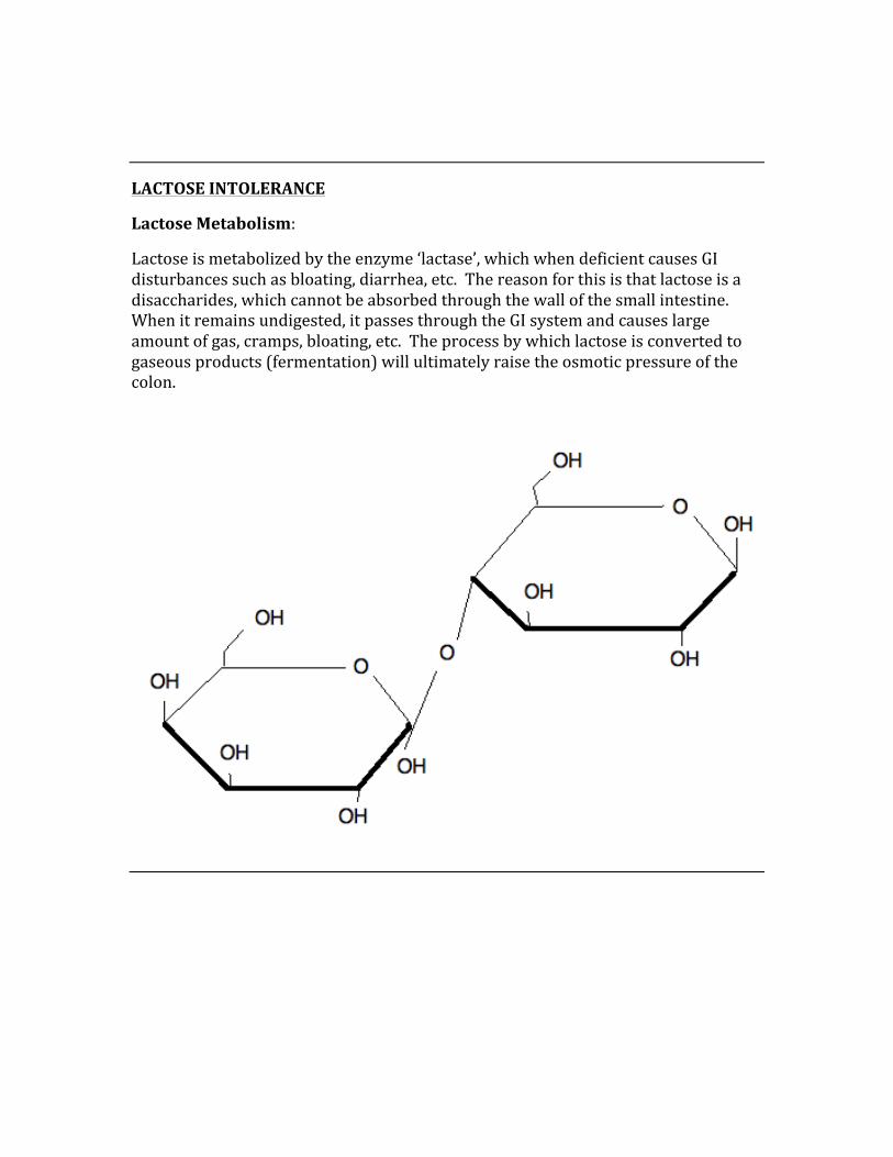
LACTOSE INTOLERANCE
Lactose Metabolism:
Lactose is metabolized by the enzyme ‘lactase’, which when deficient causes GI disturbances such as bloating, diarrhea, etc. The reason for this is that lactose is a disaccharides, which cannot be absorbed through the wall of the small intestine. When it remains undigested, it passes through the GI system and causes large amount of gas, cramps, bloating, etc. The process by which lactose is converted to gaseous products (fermentation) will ultimately raise the osmotic pressure of the colon.
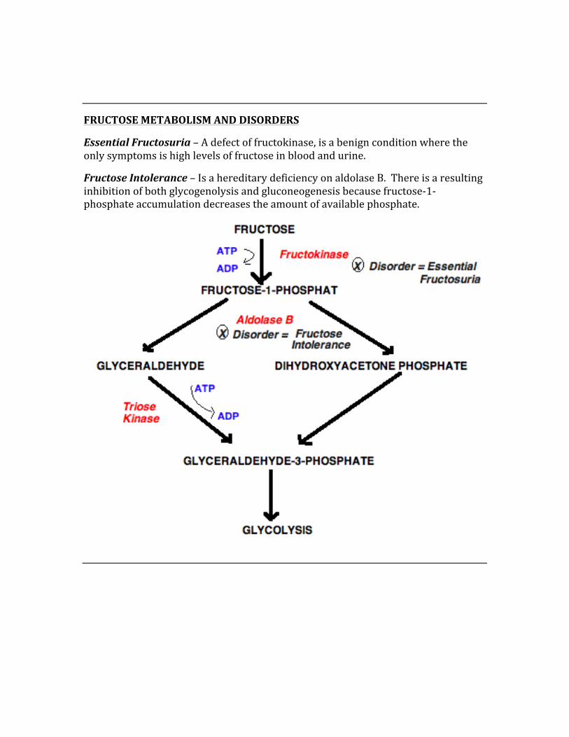
FRUCTOSE METABOLISM AND DISORDERS
Essential Fructosuria – A defect of fructokinase, is a benign condition where the only symptoms is high levels of fructose in blood and urine.
Fructose Intolerance – Is a hereditary deficiency on aldolase B. There is a resulting inhibition of both glycogenolysis and gluconeogenesis because fructose-‐1-‐phosphate accumulation decreases the amount of available phosphate.
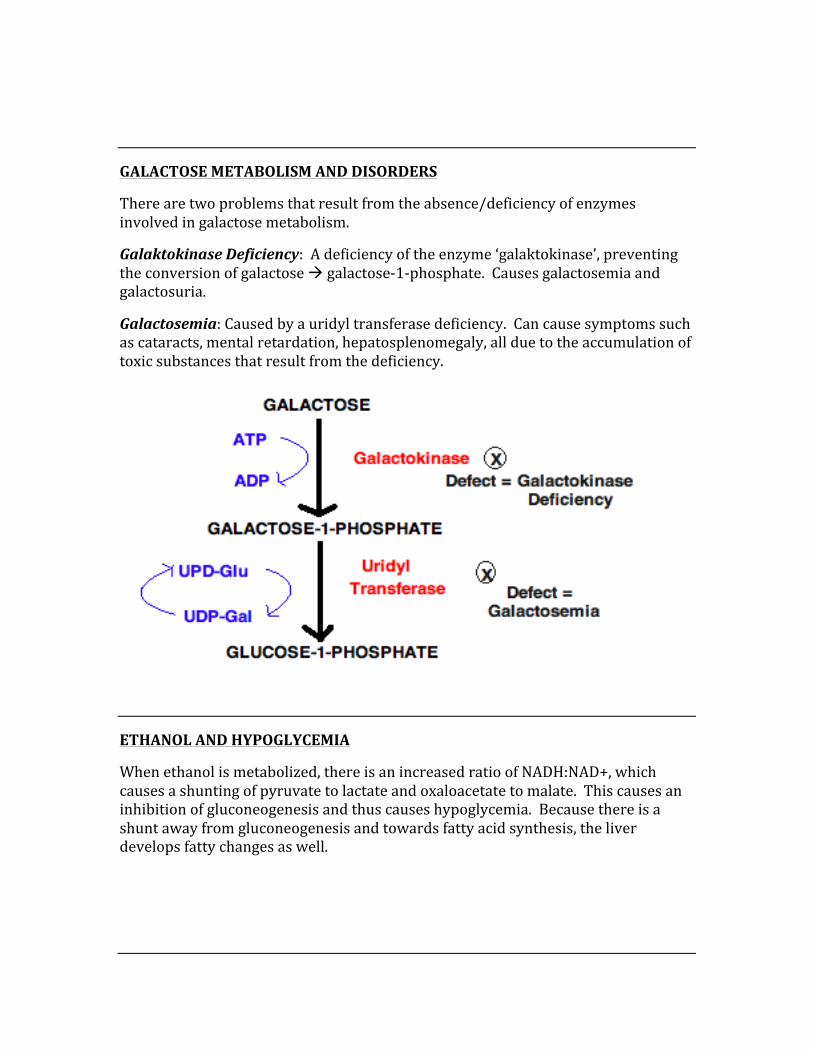
GALACTOSE METABOLISM AND DISORDERS
There are two problems that result from the absence/deficiency of enzymes involved in galactose metabolism.
Galaktokinase Deficiency: A deficiency of the enzyme ‘galaktokinase’, preventing the conversion of galactose à galactose-‐1-‐phosphate. Causes galactosemia and galactosuria.
Galactosemia: Caused by a uridyl transferase deficiency. Can cause symptoms such as cataracts, mental retardation, hepatosplenomegaly, all due to the accumulation of toxic substances that result from the deficiency.
ETHANOL AND HYPOGLYCEMIA
When ethanol is metabolized, there is an increased ratio of NADH:NAD+, which causes a shunting of pyruvate to lactate and oxaloacetate to malate. This causes an inhibition of gluconeogenesis and thus causes hypoglycemia. Because there is a shunt away from gluconeogenesis and towards fatty acid synthesis, the liver develops fatty changes as well.
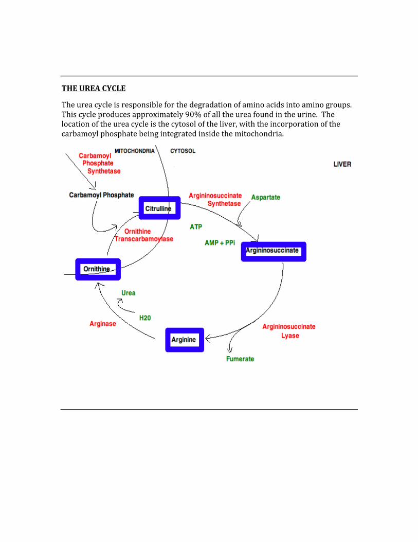
THE UREA CYCLE
The urea cycle is responsible for the degradation of amino acids into amino groups. This cycle produces approximately 90% of all the urea found in the urine. The location of the urea cycle is the cytosol of the liver, with the incorporation of the carbamoyl phosphate being integrated inside the mitochondria.
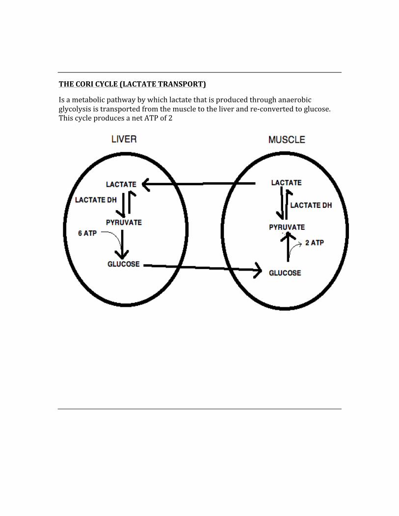
THE CORI CYCLE (LACTATE TRANSPORT)
Is a metabolic pathway by which lactate that is produced through anaerobic glycolysis is transported from the muscle to the liver and re-‐converted to glucose. This cycle produces a net ATP of 2
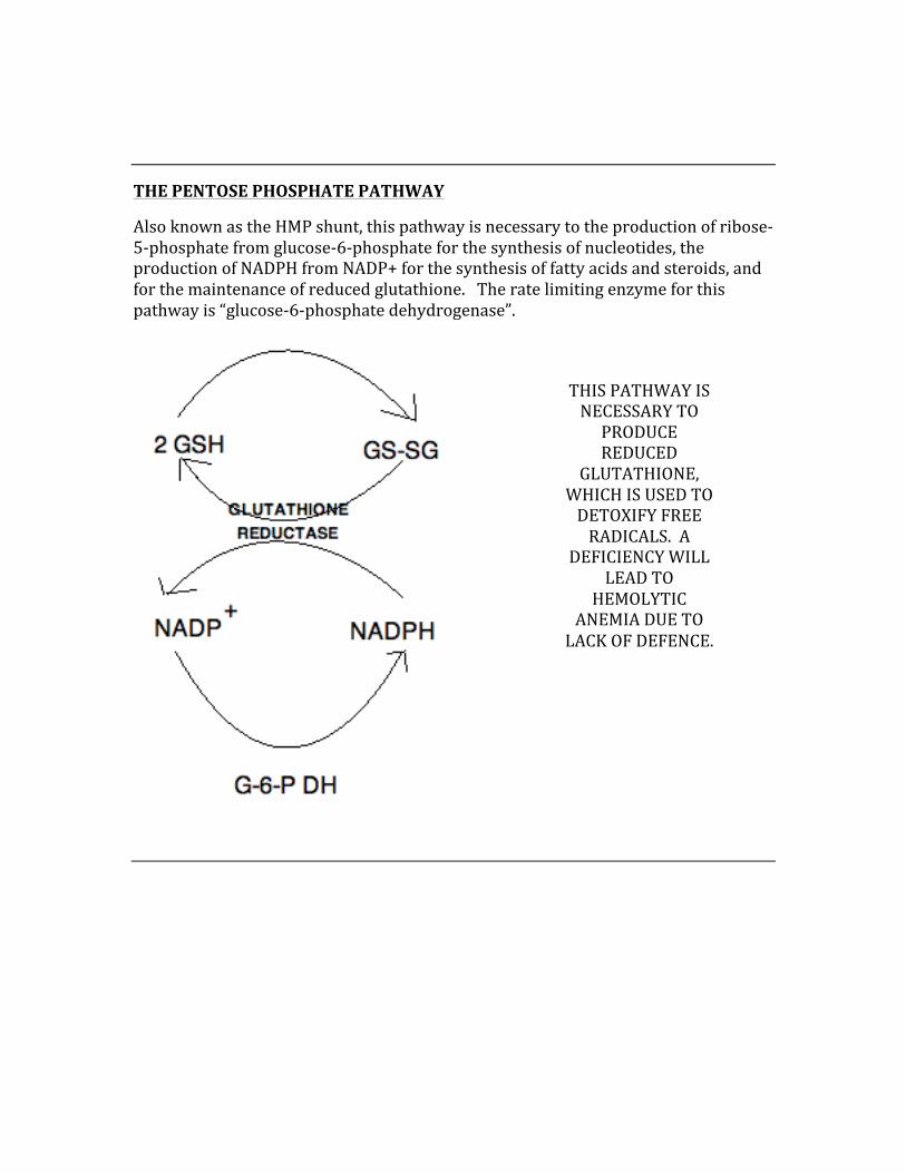
THE PENTOSE PHOSPHATE PATHWAY
Also known as the HMP shunt, this pathway is necessary to the production of ribose-‐5-‐phosphate from glucose-‐6-‐phosphate for the synthesis of nucleotides, the production of NADPH from NADP+ for the synthesis of fatty acids and steroids, and for the maintenance of reduced glutathione. The rate limiting enzyme for this pathway is “glucose-‐6-‐phosphate dehydrogenase”.
THIS PATHWAY IS NECESSARY TO PRODUCE REDUCED
GLUTATHIONE, WHICH IS USED TO DETOXIFY FREE RADICALS. A
DEFICIENCY WILL LEAD TO
HEMOLYTIC ANEMIA DUE TO LACK OF DEFENCE.
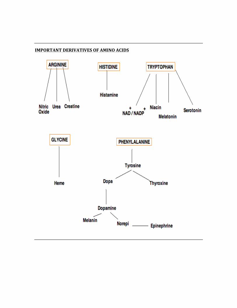
IMPORTANT DERIVATIVES OF AMINO ACIDS

AMINO ACIDS: ESSENTIAL vs. NON-‐ESSENTIAL
Amino acids are biochemical molecules containing an amine group, a carboxylic acid group, and a side chain that varies between each amino acid. They contain nitrogen, carbon, oxygen, and hydrogen.
Basic Structure:
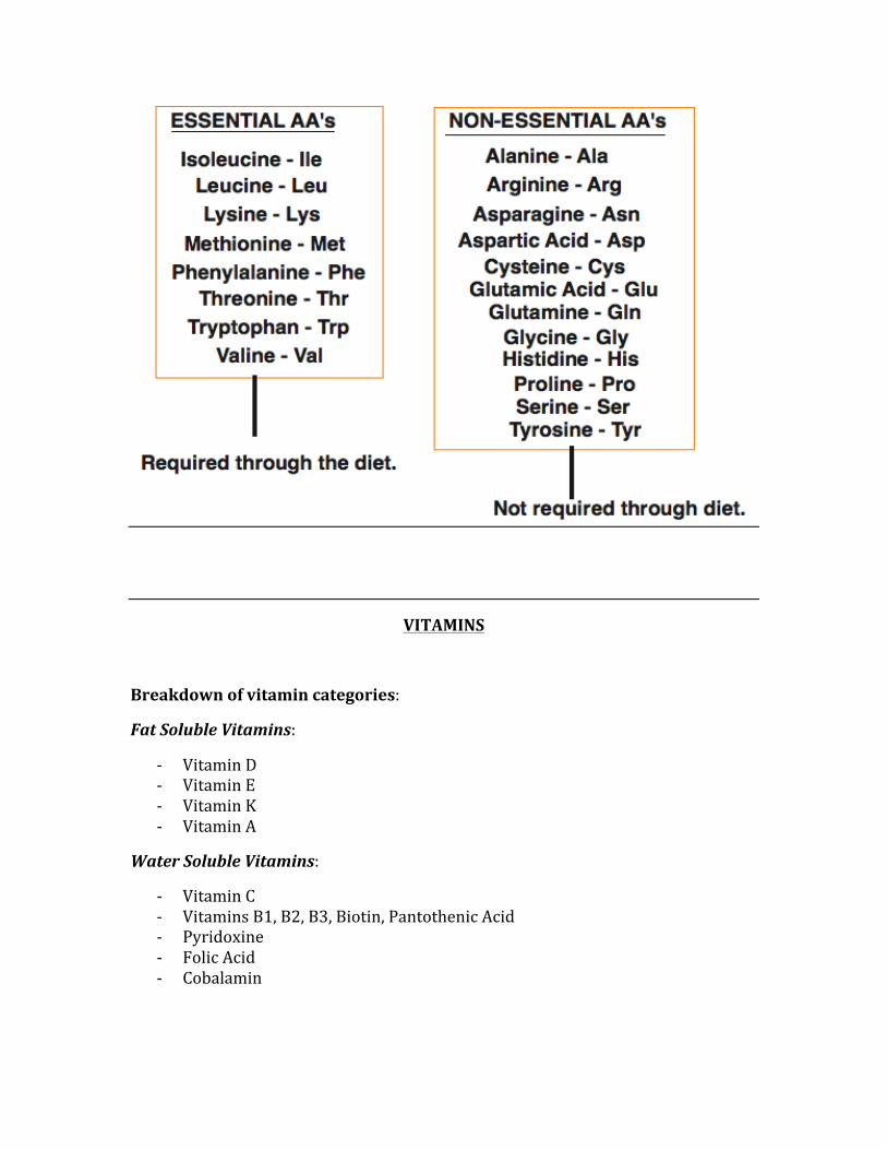
VITAMINS
Breakdown of vitamin categories:
Fat Soluble Vitamins:
-‐ Vitamin D -‐ Vitamin E -‐ Vitamin K -‐ Vitamin A
Water Soluble Vitamins:
-‐ Vitamin C -‐ Vitamins B1, B2, B3, Biotin, Pantothenic Acid -‐ Pyridoxine -‐ Folic Acid -‐ Cobalamin
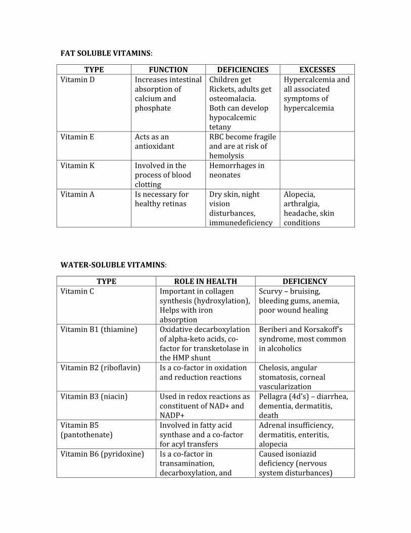
FAT SOLUBLE VITAMINS:
TYPE FUNCTION DEFICIENCIES EXCESSES Vitamin D Increases intestinal
absorption of calcium and phosphate
Children get Rickets, adults get osteomalacia. Both can develop hypocalcemic tetany
Hypercalcemia and all associated symptoms of hypercalcemia
Vitamin E Acts as an antioxidant
RBC become fragile and are at risk of hemolysis
Vitamin K Involved in the process of blood clotting
Hemorrhages in neonates
Vitamin A Is necessary for healthy retinas
Dry skin, night vision disturbances, immunedeficiency
Alopecia, arthralgia, headache, skin conditions
WATER-‐SOLUBLE VITAMINS:
TYPE ROLE IN HEALTH DEFICIENCY Vitamin C Important in collagen
synthesis (hydroxylation), Helps with iron absorption
Scurvy – bruising, bleeding gums, anemia, poor wound healing
Vitamin B1 (thiamine) Oxidative decarboxylation of alpha-‐keto acids, co-‐factor for transketolase in the HMP shunt
Beriberi and Korsakoff’s syndrome, most common in alcoholics
Vitamin B2 (riboflavin) Is a co-‐factor in oxidation and reduction reactions
Chelosis, angular stomatosis, corneal vascularization
Vitamin B3 (niacin) Used in redox reactions as constituent of NAD+ and NADP+
Pellagra (4d’s) – diarrhea, dementia, dermatitis, death
Vitamin B5 (pantothenate)
Involved in fatty acid synthase and a co-‐factor for acyl transfers
Adrenal insufficiency, dermatitis, enteritis, alopecia
Vitamin B6 (pyridoxine) Is a co-‐factor in transamination, decarboxylation, and
Caused isoniazid deficiency (nervous system disturbances)
trans-‐sulfuration Biotin A co-‐factor in
carboxylation reactions Dermatitis and enteritis, can be caused by eating raw eggs
Folic Acid Co-‐enzyme in carbon transfer in methylation reactions, involved in synthesis of nitro bases in DNA and RNA
Macrocytic anemia, neural tube defects in developing fetus
Cobalamin Is a co-‐factor in homocysteine methylation and methylmalonyl-‐CoA activity
Megaloblastic anemia with neurological symptoms, glossitis
CHAPTER 7
ETHICS
This topic is important as there are usually a handful of ethics questions on the exam. Understanding definitions
and being able to apply them to tricky situations is essential for picking up a handful of easy points on the exam.
BENEFICENCE
This term describes the responsibility of the physician to always act in the best interest of the patient. Beneficence may not always be in place, as a patient’s right to make their own decisions may not always be in their very best interest. In this situation, the physician has a duty to honor the desires of the patient with respect to his or her own care.
NON-‐MALEFICENCE
This term means the physician shall “Do No Harm”, and is always priority #1 when it comes to medical ethics and practice principles.
AUTONOMY
Autonomy refers to the patient’s right to make their own decisions after being properly educated and informed. Whether a physician believes these decisions to be right or wrong, they have a duty to respect and honor the patient’s autonomy.
A PATIENT’S ABILITY TO MAKE DECISIONS
A patient’s ability to make their own decisions is based on a few principles that must be in place:
-‐ Patient must be psychologically stable (ie not skewed by mental illness) -‐ Patient must be the one who tells you their desires, not the family -‐ Patient does not switch back and forth between their wishes (shows
instability of the patient’s mentation) -‐ Patient receives complete information of advantages and disadvantages of
treatment options -‐ Patient makes their choice, which is not influenced by family, friends, etc
INFORMED CONSENT
Informed consent is when a patient gives the physician the consent to proceed with medical management. It must be based on properly informing the patient, whereby they understand the risks, benefits, and alternative options. Decisions must be based on complete autonomy, not of persuasion.
WHEN IS INFORMED CONSENT NOT REQUIRED?
It is fully legal to proceed with medical intervention without a patient’s consent when any of the following are present:
-‐ Intervention will be life-‐saving, such as in the ER -‐ Patient is not in a mental state to make a decision (psychosis, intoxication) -‐ The patient waives their right to informed consent -‐ There is a therapeutic advantage to not getting informed consent
PATIENT’S RIGHT TO CONFIDENTIALITY
A patient has the right to complete confidentiality, whereby disclosing a patient’s information is illegal unless they give you direct permission to do so.
WHEN IS CONFIDENTIALITY BREACHABLE?
There are certain situations in which it is the physician’s responsibility to breach confidentiality for the safety of society and/or for the greater benefit of the patient.
These instances include:
-‐ There is the potential for harm to others (Tarasoff decision) -‐ The patient has a high risk of self-‐harming -‐ There is the presence of a reportable infectious disease -‐ Patient is either suicidal or homicidal -‐ There is abuse to a child or an elder
THE ADVANCED DIRECTIVE
There are numerous ways by which a patient can give their advanced directive.
Living Wills – the patient informs the physician whether they want to be treated or not should the need arise where they cannot communicate this to the physician
Oral Advanced – while less likely to stand up in court, this is an oral request given by the patient to the physician in the past requesting their desires for medical intervention
DURABLE POWER OF ATTORNEY
The durable Power of Attorney is a person designated by the patient to make their medical decisions in the event that they are unable to do so for themselves.
NEGLIGENCE/MALPRACTICE
There are four criteria that must be met in order for a malpractice suit to be warranted, they are:
Duty à This implies that there is a physician-‐patient relationship that is established.
Dereliction à When the physician fails to comply with the standards of care for the patient
Direct Cause à Where a patient incurs injury/damage that resulted from the physician’s breach of duty, where there are no other circumstances that may have caused the injury
Damages à The injuries suffered by the client

** It should be noted that the absolute most common reason for a medical lawsuit is a lack of communication or poor communication between the physician and patient. Establishing a strong physician-‐patient relationship is the best way to prevent a lawsuit.
CHAPTER 8
BIOSTATISTICS
Application of the biostatistical equations as well as fully understanding the types of studies and types of bias is
essential “must-‐know” information. While it is tempting to forego studying biostatistics, it is easy to grab plenty of easy
points by simply spending a few hours on this topic.
TYPES OF STUDIES
Case-‐Control/Experimental -‐ This test is the gold standard of epidemiological testing, where two equal groups are compared where one group has a changed variable
Prospective -‐ This is also known as a Cohort, Observational, and Incidence study. A sample is taken and divided into two groups based on the presence or absence of a risk factor. The groups are then followed over time to see what should develop. These tests are very time consuming and expensive, thus less used.
Retrospective -‐ This study chooses a population of samples based on either the presence or absence of a certain risk factor. The sample is chosen after a disease has occurred, not before it has occurred. This test is cheaper and much faster to perform than the others.
Case Series -‐ This simply describes what the clinical presentation looks like in people who have a certain disease.
Cross-‐Sectional/Prevalence -‐ This study takes a sample of population at one point in time, and looks at the prevalence of disease and the prevalence of risk factors. This test is optimal for comparing two different cultures, looking for associations between lifestyle choices and prevalence of diseases.
EPIDEMIC vs. PANDEMIC
An epidemic is seen when there is an observed incidence of a particular disease that greatly exceeds the expected incidence, whereas a pandemic is an epidemic that is seen over a wide geographical area.
TEST METHODS
Two-‐Sample T-‐test -‐ This test is used to compare the means of two groups of subjects.
ANOVA – This test is the “analysis of variance”, and is used to compare three or more variables.
Chi-‐Squared – This test compares the proportions of a categorized outcome (2x2 table). With a large difference between the observed and expected values, there is assumed to be an association between the exposure and the outcome.
Meta-‐Analysis – This test is done by pooling data from several studies, which gives the test a big statistical power.
TYPES OF BIAS
Selection Bias – This type of bias results from the manner by which people are selected and/or from selective losses from follow-‐up studies.
Observer and Measurement Bias – This bias results from the distortion of measurement of association by misclassification of the exposed/unexposed and/or diseased/non-‐diseased study subjects.
Recall Bias – Bias due to inaccuracies in recall of past exposure by people in the study.
Hawthorne Effect – This bias occurs when a patient deliberately changes their behavior because they know they are being studied.
Confounding – This bias occurs as a result of the addition of extraneous factors. For example if a study is looking for cirrhosis, they find an association between smoking and cirrhosis, and find there to be a strong association. Then subsequently, the study shows that some smokers are also heavy drinkers while some are not. In this
instance, alcohol is the confounding factor. An effective way of controlling confounding is Matching.
Lead Time Bias – This bias has to do with the time frame by which diagnoses and treatments are examined.
Admission Rate Bias – This bias type is due to the differences in hospital admission rates, which distorts the risk ratio.
Unacceptability Bias – This type of bias occurs when the participants purposely give desirable responses, which then lead to the underestimation of risk factors.
SENSITIVY, SPECIFICITY, PPV, NPV, OR, RR, and ATTRIBUTABLE RISK
The following table will be the basis for all calculations relating to the above topics:
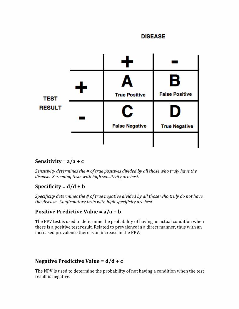
Sensitivity = a/a + c
Sensitivity determines the # of true positives divided by all those who truly have the disease. Screening tests with high sensitivity are best.
Specificity = d/d + b
Specificity determines the # of true negative divided by all those who truly do not have the disease. Confirmatory tests with high specificity are best.
Positive Predictive Value = a/a + b
The PPV test is used to determine the probability of having an actual condition when there is a positive test result. Related to prevalence in a direct manner, thus with an increased prevalence there is an increase in the PPV.
Negative Predictive Value = d/d + c
The NPV is used to determine the probability of not having a condition when the test result is negative.

Odds Ratio = (a/b)/ (c/d)
The OR determines the incidence of disease in people in the exposed groups divided by those in an unexposed group.
OR > 1 = States that the factor being studied is a risk factor for the outcome
OR<1 = States that the factor being studied is a protective factor in respect to the outcome
OR = 1, States that no significant difference in outcome in either exposed or unexposed group
Relative Risk = [a/(a+b) / d/(c+d)]
Relative risk compares the disease risk in people exposed to a certain factor with disease risk in people who have not been exposed
Attributable Risk = [a/(a+b) – d/(c+d)]
The attributable risk is the number of cases that can be attributed to one risk factor
INCIDENCE vs. PREVALENCE
Incidence is the number of new cases of a disease over a unit time, whereas prevalence is the total number of cases of a disease (both new and old) at a certain point in time. Any disease treated with the sole purpose of prolonging life (ie terminal cancers), the incidence stays the same but prevalence will increase.
Short-‐term diseases: Incidence > Prevalence
Long-‐term diseases: Prevalence > Incidence
VALIDITY vs. RELIABILITY
Validity is simply a test’s ability to measure what it claims to measure, whereas the reliability of a test determines its ability to consistent results on repeated attempts.
STANDARD DEVIATION
Standard deviation is a term that measures the variability of results.
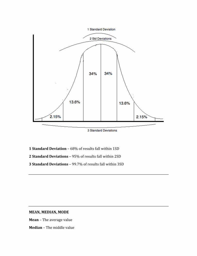
1 Standard Deviation – 68% of results fall within 1SD
2 Standard Deviations – 95% of results fall within 2SD
3 Standard Deviations – 99.7% of results fall within 3SD
MEAN, MEDIAN, MODE
Mean – The average value
Median – The middle value
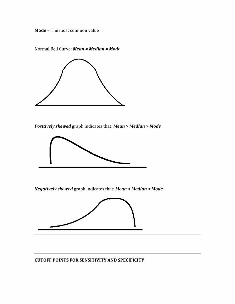
Mode – The most common value
Normal Bell Curve: Mean = Median = Mode
Positively skewed graph indicates that: Mean > Median > Mode
Negatively skewed graph indicates that: Mean < Median < Mode
CUTOFF POINTS FOR SENSITIVITY AND SPECIFICITY
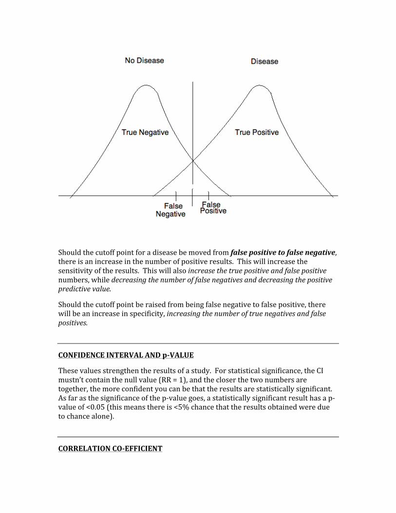
Should the cutoff point for a disease be moved from false positive to false negative, there is an increase in the number of positive results. This will increase the sensitivity of the results. This will also increase the true positive and false positive numbers, while decreasing the number of false negatives and decreasing the positive predictive value.
Should the cutoff point be raised from being false negative to false positive, there will be an increase in specificity, increasing the number of true negatives and false positives.
CONFIDENCE INTERVAL AND p-‐VALUE
These values strengthen the results of a study. For statistical significance, the CI mustn’t contain the null value (RR = 1), and the closer the two numbers are together, the more confident you can be that the results are statistically significant. As far as the significance of the p-‐value goes, a statistically significant result has a p-‐value of <0.05 (this means there is <5% chance that the results obtained were due to chance alone).
CORRELATION CO-‐EFFICIENT
Two numbers that are between -‐1 and +1, it measures to what degree the variables are related.
-‐ A number of zero (0) means there is no correlation between variables. -‐ A number of +1 means there is a perfect correlation (both variables increase
or decrease proportionally) -‐ A number of -‐1 means there is a perfect negative correlation (variables move
in opposite directions proportionally)
ATTRIBUTABLE RISK PERCENT (ARP)
The ARP measures the impact of the particular risk factor being studied on a particular population. It represents excess risk that can be explained by exposure to a particular risk factor.
Calculate the ARP: ARP = [(RR -‐1)/RR]
STATISTICAL HYPOTHESES
The statistical hypotheses are used to determine whether or not there is an association between risk factors and disease in a population. They are the ‘null hypothesis’ and the ‘alternative’ hypothesis.
Null Hypothesis (Ho) – This hypothesis is the ‘hypothesis of no difference’, meaning there is not an association between the disease and the risk factor.
Alternative Hypothesis (H1) – This hypothesis is the ‘hypothesis of some difference’, meaning there is an association between the disease and the risk factor.
POWER
The power of a statistical test is the probability that a test will reject a false null hypothesis, meaning it will not make a Type 2 error. With increased sample size there is increased power and there is a decreased chances of seeing a type 2 error.
ERRORS
Type 1 Error (α) – A type 1 error mistakenly accepts the experimental hypothesis and rejects the null hypothesis. This error means that something statistically is seen that in fact is not there.

Type 2 Error (β) – This type of error occurs when you fail to reject the null hypothesis when it is in fact false. This error means you essentially something that is statistically present was in fact missed.
CHAPTER 9
PSYCHIATRY/PSYCHOLOGY
The questions asked on this topic can range from a straightforward simple question to a question that expects
you to combine many aspects of medicine in making the best choice. The highest-‐yield information includes psychiatric pharmacology as well as all of the personality disorders.
SUBSTANCE DEPENDENCE AND ABUSE
There is a big difference between substance dependence and substance abuse.
Substance dependence is a pattern of substance use that involves at least 3 out of 7 criteria, they are:
-‐ There are decreased social, occupational, or recreational activities because of the substance use
-‐ Patient has developed tolerance to substance -‐ Patient experiences withdrawal symptoms when refraining from use -‐ There is a chronic desire to cut back or stop use -‐ Patient will spend excess time and energy in trying to attain their substance

-‐ The substance is taken in amounts much larger than intended -‐ Continuation of use despite the knowledge of its harmful effects
When any three of the previous points are present, the patient is diagnosed with substance dependence.
Substance abuse is a pattern of substance use that causes significant social impairment and/or distress. The diagnosis of substance abuse is made when there are any of the following:
-‐ Excess and recurrent use that causes failure to fulfill major obligations such as going to work, taking care of kids, etc
-‐ Chronic and recurrent use of substance in situations that are hazardous -‐ Recurrent problems with the law due to the drug abuse -‐ Persistent use despite the knowledge of its dangerous effects
THE MOST COMMON TYPES OF SUBSTANCE ABUSE
There are many different types of drugs, and they all have different kinds of signs/symptoms and different degrees of withdrawal severity. The drugs and substances most commonly used and abused include:
-‐ Alcohol -‐ Amphetamines -‐ Barbiturates and Benzodiazepines -‐ Caffeine -‐ Cocaine -‐ LSD -‐ Marijuana -‐ Nicotine -‐ Opioids (heroin, oxycodone, morphine, etc)
The following is a list of the most common drugs, their primary signs/symptoms, and the common findings of withdrawal
SUBSTANCE SIGNS OF INTOXICATION WITHDRAWAL SYMPTOMS
ALCOHOL Disinhibition, slurred speech
Tremor, tachycardia, seizure, DT’s
AMPHETAMINES Psychomotor agitation, mydriasis
Depression, lethargy, excessive sleep
BARBITURATES Sedation, respiratory depression
Anxiety, cardiovascular collapse
BENZODIAZEPINES Sedation, respiratory Anxiety, seizure, tremor
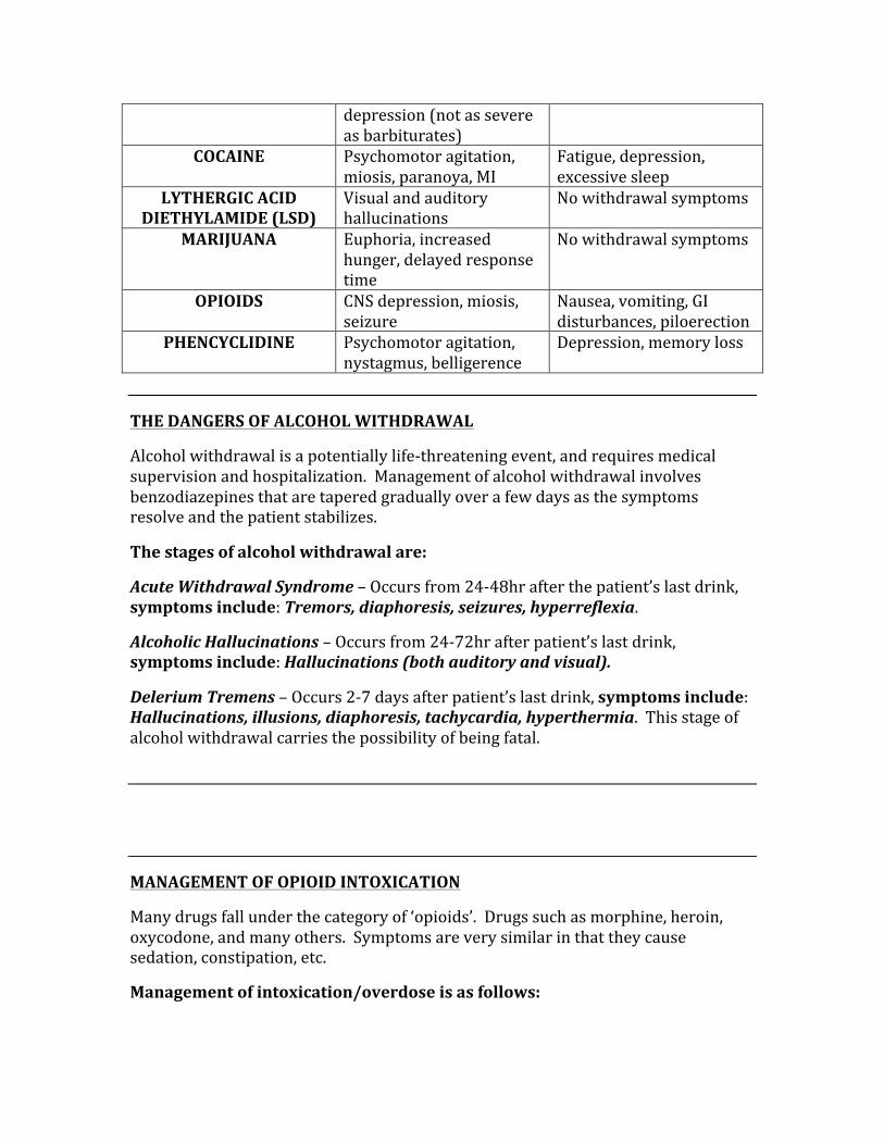
depression (not as severe as barbiturates)
COCAINE Psychomotor agitation, miosis, paranoya, MI
Fatigue, depression, excessive sleep
LYTHERGIC ACID DIETHYLAMIDE (LSD)
Visual and auditory hallucinations
No withdrawal symptoms
MARIJUANA Euphoria, increased hunger, delayed response time
No withdrawal symptoms
OPIOIDS CNS depression, miosis, seizure
Nausea, vomiting, GI disturbances, piloerection
PHENCYCLIDINE Psychomotor agitation, nystagmus, belligerence
Depression, memory loss
THE DANGERS OF ALCOHOL WITHDRAWAL
Alcohol withdrawal is a potentially life-‐threatening event, and requires medical supervision and hospitalization. Management of alcohol withdrawal involves benzodiazepines that are tapered gradually over a few days as the symptoms resolve and the patient stabilizes.
The stages of alcohol withdrawal are:
Acute Withdrawal Syndrome – Occurs from 24-‐48hr after the patient’s last drink, symptoms include: Tremors, diaphoresis, seizures, hyperreflexia.
Alcoholic Hallucinations – Occurs from 24-‐72hr after patient’s last drink, symptoms include: Hallucinations (both auditory and visual).
Delerium Tremens – Occurs 2-‐7 days after patient’s last drink, symptoms include: Hallucinations, illusions, diaphoresis, tachycardia, hyperthermia. This stage of alcohol withdrawal carries the possibility of being fatal.
MANAGEMENT OF OPIOID INTOXICATION
Many drugs fall under the category of ‘opioids’. Drugs such as morphine, heroin, oxycodone, and many others. Symptoms are very similar in that they cause sedation, constipation, etc.
Management of intoxication/overdose is as follows:
Naloxone/Naltrexone – This is a medication given in the ER when patient is suspected of opioid intoxication, it acts by competitively inhibiting the opioid receptors.
Methadone – A controversial drug, methadone is used to manage patients who are undergoing heroin detoxification because it is long lasting and is good for long-‐term management.
MAJOR DEPRESSIVE DISORDER
Major depressive disorder is a serious condition characterized by several specific signs and symptoms. In general, the best treatment for an episode of major depression is an SSRI anti-‐depressant medication. If a patient is experiencing their first bout of depression, it is advised to keep them on their medication for at least 6 months. If it is their second or more bout of depression, they should be kept on their anti-‐depressant on an ongoing basis. Diagnosis requires that there are symptom-‐free periods of at least two months in between each episode.
The diagnosis of depression is made when any of the following FIVE are present for at least TWO weeks:
-‐ Sleep Disturbances (patient may sleep too little or too much) -‐ Loss of Interest (interest lost in things that they previously enjoyed) -‐ Feeling of Guilt (these guilty feelings are usually unwarranted) -‐ Energy (loss of energy, which results in a loss of drive to do other things they
previously enjoyed) -‐ Loss of Concentration -‐ Appetite Changes (most of the time the patient has a LOSS of appetite, but
they may also get an increase in appetite) -‐ Psychomotor Retardation (results in slowed mentation, related to loss of
concentration) -‐ Suicidal Ideation (always inquire about suicide, it is not going to increase
the patient’s risk of committing suicide)
The popular mnemonic for factors of depression is: SIG E CAPS. Females > Males
* For depression that isn’t helped with SSRI or other anti-‐depressants, ECT should be tried. ECT is also the management of choice for major depression in a pregnant patient.
SUICIDE
Suicide is a big concern in depressed patients, and any patient who seems to have a depressed mood should be asked about suicide. It is important to ask if they have thought about it, if they have thought of how they would do it, and if they have a
plan. It is highly important to inquire about this, as asking WILL NOT increase the risk of them killing themselves.
Women attempt suicide more often than men, however men are more likely to succeed. The reason for this is because women often use less violent measures such as pills, while men take more violent measures such as guns and hanging.
Risk factors for suicide:
-‐ Prior attempts -‐ Presence of plan/lethality of plan (gun vs. pills, etc) -‐ Current medical illness -‐ Alcohol or drug dependence -‐ Currently taking three or more prescription drugs -‐ Lack of a social circle (few friends, no family, no spouse)
DYSTHYMIA
Dysthymia is a chronic mood disorder that is similar to depression, however it is an ongoing, less severe type of depression. It has fewer symptoms than major depression, but is much longer lasting. At least 75% of those with dysthymia have a co-‐morbid psychiatric disorder such as anxiety, alcoholism, etc.
DELERIUM vs. DEMENTIA
These are two terms often confused in medicine, and especially on the USMLE exams.
Delerium is a state of decreased attention span, hallucinations and illusions, and cognitive dysfunctions. The key to making the diagnosis of delirium is that levels of functionality go in and out (waxing and waning), and has an acute onset. On the other hand, dementia is a disease with multiple cognitive defects that develop slowly over time. The key to diagnosing dementia is that the patient is fully conscious but has cognitive defects.
BIPOLAR DISORDER
Bipolar disorder is a mood disorder where a patient experiences both mania and depression. There are two types of bipolar disorder (types 1 and 2), where type 1 is mania and type 2 is hypomania. Regardless of type, in order to make a diagnosis of
a manic episode the patient must have certain symptoms present for at least 1 WEEK.
The criteria for a manic episode are:
-‐ Easy distractibility -‐ Insomnia (the patient can go for days without sleeping) -‐ Grandiosity (the patient has an unusually exaggerated sense of self-‐esteem) -‐ Flight of Ideas -‐ Increased activity (patient will do numerous activities in one day) -‐ Pressured speech (fast, non-‐sensical, etc) -‐ Thoughtlessness (patient will do things without fully thinking about what
they are doing first)
At least 3 of these must be present to make a diagnosis of a manic episode.
Mania causes severe social impairment and occupational dysfunction. Hypomania is similar to a manic episode except there is no impairment to the patient in any of their day-‐to-‐day functioning.
The drug of choice for Bipolar disorder management is LITHIUM.
PERSONALITY DISORDERS
These are pervasive, fixed, and inappropriate patterns of relating to others, causing social and occupational impairment. Patients with personality disorders do not seek help for their disorder because they are not aware that they are the cause of their own problems.
There are three categories of personality disorders: Cluster A, Cluster B, Cluster C
Cluster A disorders: Paranoid, Schizoid, Schizotypal
Cluster B disorders: Histrionic, Narcissistic, Antisocial, Borderline
Cluster C disorders: Avoidant, Obsessive-‐Compulsive, Dependent, Passive-‐Aggressive
CLUSTER A PERSONALITY DISORDERS
Paranoid – This patient does not trust others, is suspicious of everything, and believes all of their problems are caused by others.
Schizoid – This patient will have a long history of voluntarily avoiding social situations. There is no psychosis associated with the schizoid personality disorder.
Schizotypal – This patient has an odd/peculiar appearance, has magical thoughts and an odd pattern of behavior. There is no pscyhosis with the schizotypal personality disorder.
CLUSTER B PERSONALITY DISORDERS
Histrionic – This patient is an extrovert, is sexually provocative, and very emotional.
Narcissistic – This patient has a sense of entitlement and lacks empathy for others.
Antisocial – This patient refuses to accept the rules of society, shows no concern for their own actions, and shows no remorse for bad behavior. This disorder is associated with conduct disorder in childhood.
Borderline – This patient has erratic behavior, impulsiveness, and mini psychotic episodes. Look for self-‐mutilation and other mood disorders.
CLUSTER C PERSONALITY DISORDERS
Avoidant – This patient is sensitive to rejection and timid, thus socially withdrawn. They also feel inferior to others for no apparent reason.
Obsessive-‐Compulsive – This patient is a perfectionist, orderly, stubborn, and indecisive. This is not the same as obsessive-‐compulsive disorder (OCD).
Dependent – This patient allows and wants others to make decisions for them, related to a lack of self-‐confidence.
Passive-‐Aggressive – This patient has “outward compliance with inner defiance”.
ANXIETY DISORDERS
Anxiety disorders are characterized by subjective and physical manifestations of fear. The symptoms are similar: Tremor, Palpitations, Diaphoresis, Dizziness, GI disturbances, Urinary symptoms.
The common anxiety disorders are:
-‐ Panic Disorder -‐ Phobias -‐ Obsessive-‐Compulsive Disorder (OCD) -‐ Generalized Anxiety Disorder -‐ Post-‐traumatic Stress Disorder (PTSD)
Panic Disorder – Panic attacks that occur approximately twice per week, last approximately 30 minutes, and present with symptoms similar to an MI. Patients often have a fear of another attack in-‐between each episode. Management of panic disorder is SSRI’s, with possible benzodiazepines for acute treatment.
Phobias – Specific phobias an irrational fear of specific objects, such as spiders, snakes, etc. A social phobia is an exaggerated fear of social or environmental situations (the most common social phobia is public speaking). Management is desensitization, can use beta-‐blockers for short-‐term control of autonomic symptoms.
Obsessive-‐Compulsive Disorder (OCD) – Patient experiences recurring intrusive feelings, thoughts, and images which cause anxiety that is relieved in part by performing repetitive actions (compulsions). Patients realize that their actions are irrational and desire to be helped. SSRI’s are mainstay of management.
Generalized Anxiety Disorder – Patient experiences persistent symptoms of anxiety for at least 6 months. The symptoms are unrelated to any specific person or situation. Seen in women > men.
Post-‐Traumatic Stress Disorder – Is a condition that affects someone who has been through a catastrophic event (classically a war veteran). The patient experiences hyperarousal (anxiety, sleeplessness, intrusive memories) and withdrawal (flattened affect, numbing, survivor’s guilt). These symptoms must be present for at least 1 month for a diagnosis of PTSD, if symptoms are less than 1 month, the diagnosis is Acute Stress Disorder (ASD).
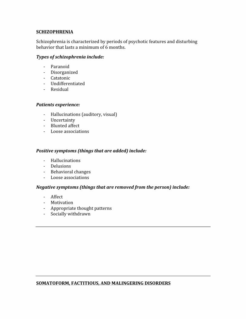
SCHIZOPHRENIA
Schizophrenia is characterized by periods of psychotic features and disturbing behavior that lasts a minimum of 6 months.
Types of schizophrenia include:
-‐ Paranoid -‐ Disorganized -‐ Catatonic -‐ Undifferentiated -‐ Residual
Patients experience:
-‐ Hallucinations (auditory, visual) -‐ Uncertainty -‐ Blunted affect -‐ Loose associations
Positive symptoms (things that are added) include:
-‐ Hallucinations -‐ Delusions -‐ Behavioral changes -‐ Loose associations
Negative symptoms (things that are removed from the person) include:
-‐ Affect -‐ Motivation -‐ Appropriate thought patterns -‐ Socially withdrawn
SOMATOFORM, FACTITIOUS, AND MALINGERING DISORDERS
Somatoform Disorders
Somatoform disorders are characterized by physical symptoms without any organic cause. Those with this condition are not malingering, are not delusional, and truly believe they have a physical problem.
Somatoform Disorders include:
-‐ Somatization disorder -‐ Conversion disorder -‐ Hypochondriasis -‐ Body Dysmorphic disorder -‐ Pain disorder -‐ Undifferentiated Somatoform disorder
Somatization Disorder – Patient with history of multiple somatic complaints over several years, they must include: 4 pain symptoms, 2 GI symptoms, 1 sexual symptom, and 1 pseudoneurologic symptom.
Conversion Disorder – Patient experiences an abrupt, dramatic loss of motor or sensory function. Most commonly the patient experiences paralysis, seizures, paresthesias, anesthesias, and visual problems.
Hypochondriasis – These patients have an exaggerated concern that they have illnesses despite being told repeatedly that there is no cause for concern. Must occur for at least a 6-‐month period for this diagnosis.
Body Dysmorphic Disorder – The patient has an excessive and possibly obsessive focus on a minor or possibly imagined physical defect.
Pain Disorder – Pain that is not explained by any physical problem, often co-‐exists with another medical problem. If lasting < 6 months, it is an acute pain disorder, if lasting > 6 months it is a chronic pain disorder.
Undifferentiated Somatoform Disorder – This is the diagnosis when the persistent physical symptoms don’t meet criteria for any of the other disorders. The most common problems falling under this umbrella are: Fatigue, GI/GU symptoms, appetite changes.
Factitious Disorders
A patient with factitious disorder consciously creates their symptoms in order to assume the role of the sick patient so they can get medical attention (the motivation however is unconscious). There are two categories of factitious disorder, they are: Munchausen’s Syndrome and Munchausen’s Syndrome by Proxy.
Munchausen’s Syndrome – Patient will have a chronic history of multiple hospital admissions, as well as a history of willingness to receive invasive procedures, even when not warranted.
Munchausen’s by Proxy – This is when a child is ill because of the parent.
Malingering Disorders
Patients with malingering disorder consciously simulate or exaggerate a physical or mental illness for an obvious gain (Money, Care, Avoidance of work). This patient will avoid treatment, as opposed to those with factitious disorder. Once the patient receives the gain they are seeking, the symptoms will resolve.
SLEEP
Understanding wave patterns and activities during each stage of sleep are important aspects of understanding the sleep cycle.
Awake with eyes open– Beta waves, serotonin stimulates the rape nuclei to induce sleep
Awake with eyes closed – Alpha waves
Stage 1 – This is the stage of “light sleep”. This stage is short, lasting approximately 5% of all sleep time, Theta waves are predominant.
Stage 2 – This is deeper sleep, lasting approximately 45% of all sleep time. Predominantly see sleep spindles and K complexes.
Stage 3 – This is the deepest sleep, but is non-‐REM. Lasts approximately 25% of all sleep time, and is characterized by Delta waves. *This is the stage at which children will experience enuresis and bed-‐wetting.
Stage 4 – This is REM sleep. The patient will be dreaming and lose all muscle tone. There are predominantly Beta waves during the REM sleep cycle.
AMNESIA
Amnesia can occur for many reasons, including anesthesia, drug use, old age, head injuries, alcoholism, etc. The two forms of amnesia are: Anterograde and Retrograde.
Anterograde – This person will be unable to remember anything after occurring after the incident that caused their amnesia, therefore they cannot form any new memories.
Retrograde – This person will be unable to remember things that occurred before the causative factor of their amnesia.
SLEEP APNEA
Sleep apnea is a serious condition where a person has periods of cessation of breathing while asleep. This may last from a few seconds to minutes, and may occur up to 30 times per hour. The two types of sleep apnea are “Central” and “Obstructive”.
Central sleep apnea – this type of apnea occurs due to a lack of respiratory drive from the CNS.
Obstructive sleep apnea – this type of apnea is usually caused by excessive weight, which causes obstruction and physical blockage of breathing. Management is weight loss and CPAP machine. If these fail surgery may be required.
In obstructive sleep apnea, a patient will often complain of excessive daytime sleepiness, and the spouse will complain of very loud snoring.
NARCOLEPSY
A condition where someone falls asleep suddenly without warning, and may experience cataplexy (sudden collapse while they are still awake). They fall into REM sleep upon falling asleep. Manage these patients first with scheduled daytime naps, and if that doesn’t work use a stimulant such as methylphenidate.
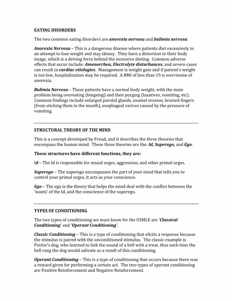
EATING DISORDERS
The two common eating disorders are anorexia nervosa and bulimia nervosa.
Anorexia Nervosa – This is a dangerous disease where patients diet excessively in an attempt to lose weight and stay skinny. They have a distortion to their body image, which is a driving force behind the excessive dieting. Common adverse effects that occur include: Amenorrhea, Electrolyte disturbances, and severe cases can result in cardiac etiologies. Management is weight gain and if patient’s weight is too low, hospitalization may be required. A BMI of less than 19 is worrisome of anorexia.
Bulimia Nervosa – These patients have a normal body weight, with the main problem being overeating (bingeing) and then purging (laxatives, vomiting, etc). Common findings include enlarged parotid glands, enamel erosion, bruised fingers (from sticking them in the mouth), esophageal varices caused by the pressure of vomiting.
STRUCTURAL THEORY OF THE MIND
This is a concept developed by Freud, and it describes the three theories that encompass the human mind. These three theories are the: Id, Superego, and Ego.
These structures have different functions, they are:
Id – The Id is responsible for sexual urges, aggression, and other primal urges.
Superego – The superego encompasses the part of your mind that tells you to control your primal urges, it acts as your conscience.
Ego – The ego is the theory that helps the mind deal with the conflict between the ‘wants’ of the Id, and the conscience of the superego.
TYPES OF CONDITIONING
The two types of conditioning we must know for the USMLE are ‘Classical Conditioning’ and ‘Operant Conditioning’.
Classic Conditioning – This is a type of conditioning that elicits a response because the stimulus is paired with the unconditioned stimulus. The classic example is Pavlov’s dog, who learned to link the sound of a bell with a treat, thus each time the bell rang the dog would salivate as a result of this conditioning.
Operant Conditioning – This is a type of conditioning that occurs because there was a reward given for performing a certain act. The two types of operant conditioning are Positive Reinforcement and Negative Reinforcement.
Positive Reinforcement – A reward produces an action
Negative Reinforcement – Removing a negative stimulus will elicit action
DEVELOPMENTAL MILESTONES IN INFANTS
Age Gross Motor Fine Motor Language Social/Cognition Newborn Moro reflex,
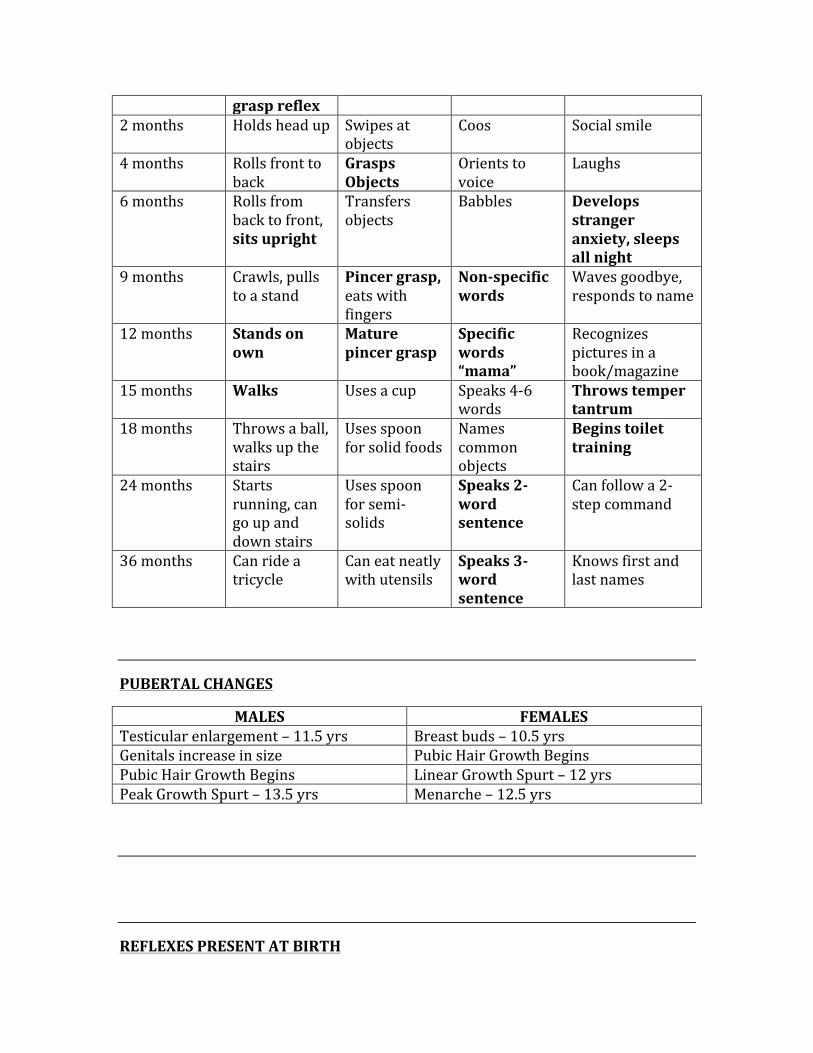
grasp reflex 2 months Holds head up Swipes at
objects Coos Social smile
4 months Rolls front to back
Grasps Objects
Orients to voice
Laughs
6 months Rolls from back to front, sits upright
Transfers objects
Babbles Develops stranger anxiety, sleeps all night
9 months Crawls, pulls to a stand
Pincer grasp, eats with fingers
Non-‐specific words
Waves goodbye, responds to name
12 months Stands on own
Mature pincer grasp
Specific words “mama”
Recognizes pictures in a book/magazine
15 months Walks Uses a cup Speaks 4-‐6 words
Throws temper tantrum
18 months Throws a ball, walks up the stairs
Uses spoon for solid foods
Names common objects
Begins toilet training
24 months Starts running, can go up and down stairs
Uses spoon for semi-‐solids
Speaks 2-‐word sentence
Can follow a 2-‐step command
36 months Can ride a tricycle
Can eat neatly with utensils
Speaks 3-‐word sentence
Knows first and last names
PUBERTAL CHANGES
MALES FEMALES Testicular enlargement – 11.5 yrs Breast buds – 10.5 yrs Genitals increase in size Pubic Hair Growth Begins Pubic Hair Growth Begins Linear Growth Spurt – 12 yrs Peak Growth Spurt – 13.5 yrs Menarche – 12.5 yrs
REFLEXES PRESENT AT BIRTH
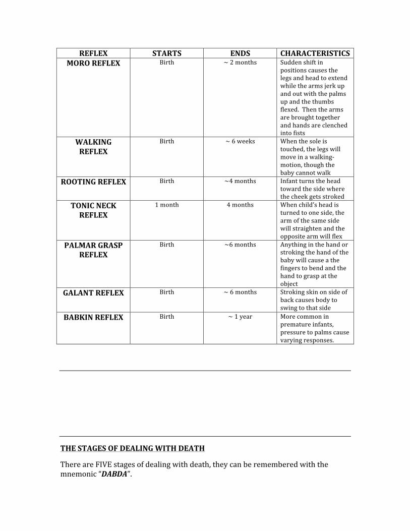
REFLEX STARTS ENDS CHARACTERISTICS MORO REFLEX Birth ~ 2 months Sudden shift in
positions causes the legs and head to extend while the arms jerk up and out with the palms up and the thumbs flexed. Then the arms are brought together and hands are clenched into fists
WALKING REFLEX
Birth ~ 6 weeks When the sole is touched, the legs will move in a walking-‐motion, though the baby cannot walk
ROOTING REFLEX Birth ~4 months Infant turns the head toward the side where the cheek gets stroked
TONIC NECK REFLEX
1 month 4 months When child’s head is turned to one side, the arm of the same side will straighten and the opposite arm will flex
PALMAR GRASP REFLEX
Birth ~6 months Anything in the hand or stroking the hand of the baby will cause a the fingers to bend and the hand to grasp at the object
GALANT REFLEX Birth ~ 6 months Stroking skin on side of back causes body to swing to that side
BABKIN REFLEX Birth ~ 1 year More common in premature infants, pressure to palms cause varying responses.
THE STAGES OF DEALING WITH DEATH
There are FIVE stages of dealing with death, they can be remembered with the mnemonic “DABDA”.
Denial, Anger, Bargaining, Depression, Acceptance
CHANGES SEEN IN THE ELDERLY
There are a few changes that occur with advanced age that are seen almost uniformly across the geriatric population, they include:
-‐ Change in sleep patterns, namely less need for sleep, decreased REM sleep, more frequent awakening throughout the night.
-‐ Sexual changes such as longer refractory period, delayed ejaculation, slower onset of erection (in men). Females frequently experience thinning and dryness of the vagina.
-‐ Medical conditions such as degenerative diseases are quite common
CHAPTER 10
IMMUNOLOGY
A sub-‐category of microbiology, immunology is very high-‐yield. There are important concepts you must know, and all of the immunologic disorders are fair game on the Step 1
exam.
IMMUNOLOGY
PASSIVE vs. ACTIVE IMMUNITY
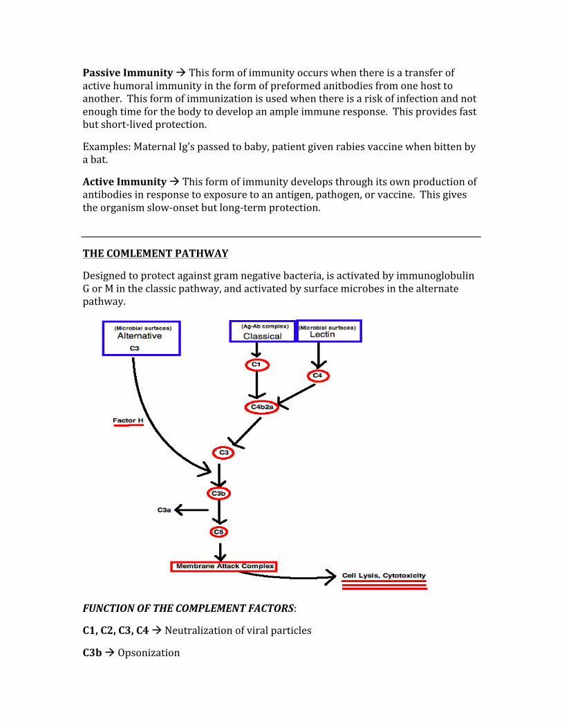
Passive Immunity à This form of immunity occurs when there is a transfer of active humoral immunity in the form of preformed anitbodies from one host to another. This form of immunization is used when there is a risk of infection and not enough time for the body to develop an ample immune response. This provides fast but short-‐lived protection.
Examples: Maternal Ig’s passed to baby, patient given rabies vaccine when bitten by a bat.
Active Immunity à This form of immunity develops through its own production of antibodies in response to exposure to an antigen, pathogen, or vaccine. This gives the organism slow-‐onset but long-‐term protection.
THE COMLEMENT PATHWAY
Designed to protect against gram negative bacteria, is activated by immunoglobulin G or M in the classic pathway, and activated by surface microbes in the alternate pathway.
FUNCTION OF THE COMPLEMENT FACTORS:
C1, C2, C3, C4 à Neutralization of viral particles
C3b à Opsonization
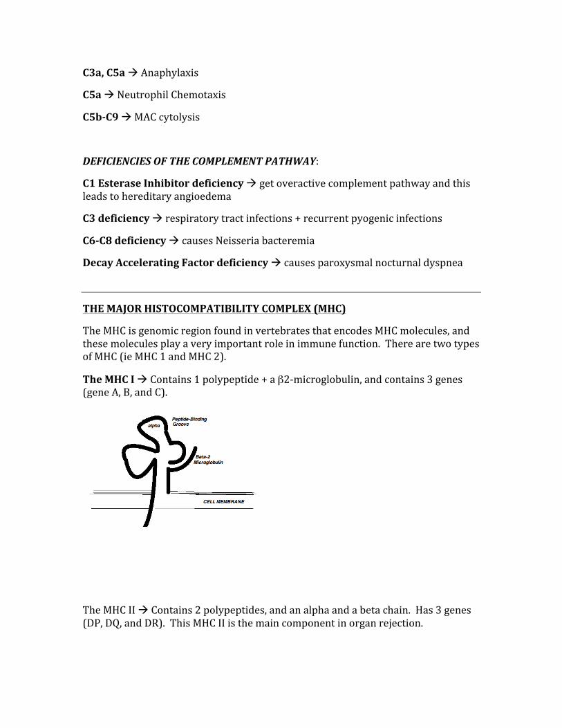
C3a, C5a à Anaphylaxis
C5a à Neutrophil Chemotaxis
C5b-‐C9 à MAC cytolysis
DEFICIENCIES OF THE COMPLEMENT PATHWAY:
C1 Esterase Inhibitor deficiency à get overactive complement pathway and this leads to hereditary angioedema
C3 deficiency à respiratory tract infections + recurrent pyogenic infections
C6-‐C8 deficiency à causes Neisseria bacteremia
Decay Accelerating Factor deficiency à causes paroxysmal nocturnal dyspnea
THE MAJOR HISTOCOMPATIBILITY COMPLEX (MHC)
The MHC is genomic region found in vertebrates that encodes MHC molecules, and these molecules play a very important role in immune function. There are two types of MHC (ie MHC 1 and MHC 2).
The MHC I à Contains 1 polypeptide + a β2-‐microglobulin, and contains 3 genes (gene A, B, and C).
The MHC II à Contains 2 polypeptides, and an alpha and a beta chain. Has 3 genes (DP, DQ, and DR). This MHC II is the main component in organ rejection.
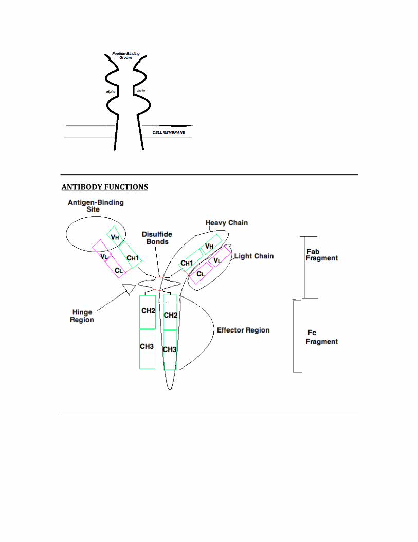
ANTIBODY FUNCTIONS
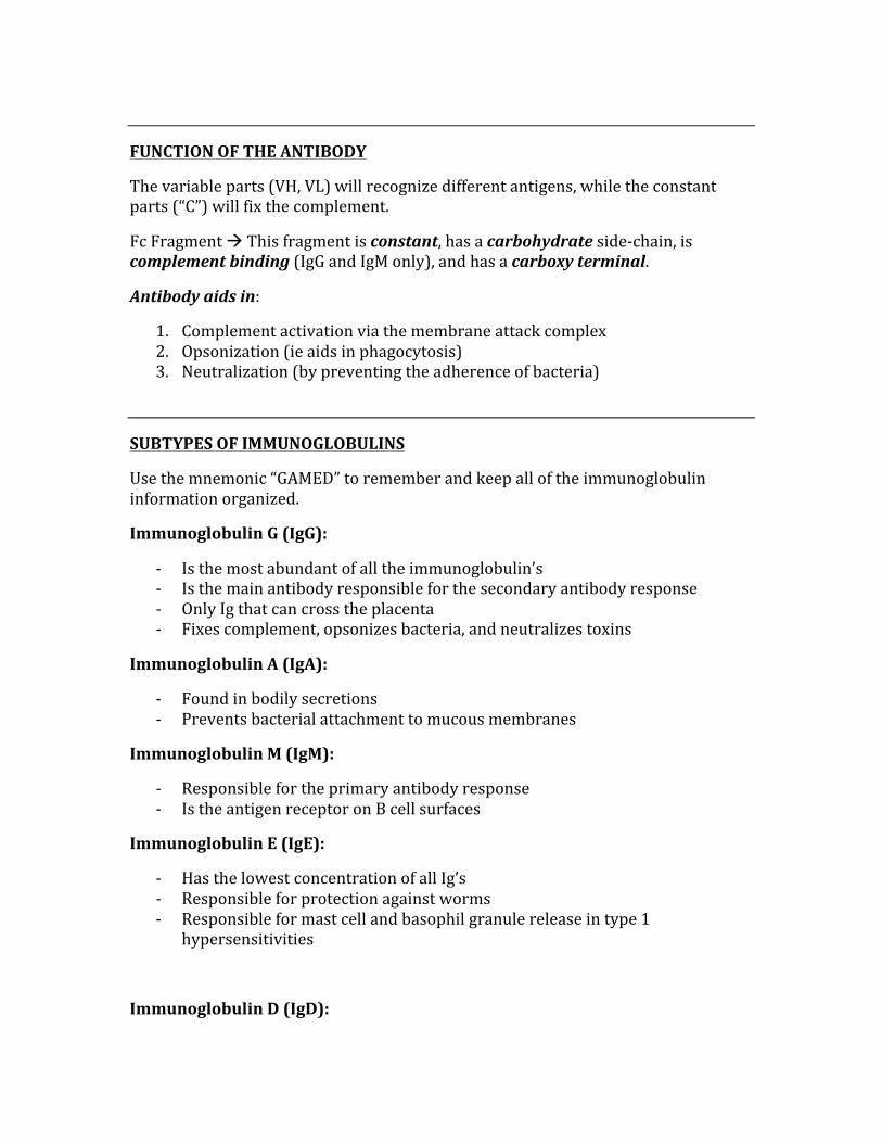
FUNCTION OF THE ANTIBODY
The variable parts (VH, VL) will recognize different antigens, while the constant parts (“C”) will fix the complement.
Fc Fragment à This fragment is constant, has a carbohydrate side-‐chain, is complement binding (IgG and IgM only), and has a carboxy terminal.
Antibody aids in:
1. Complement activation via the membrane attack complex 2. Opsonization (ie aids in phagocytosis) 3. Neutralization (by preventing the adherence of bacteria)
SUBTYPES OF IMMUNOGLOBULINS
Use the mnemonic “GAMED” to remember and keep all of the immunoglobulin information organized.
Immunoglobulin G (IgG):
-‐ Is the most abundant of all the immunoglobulin’s -‐ Is the main antibody responsible for the secondary antibody response -‐ Only Ig that can cross the placenta -‐ Fixes complement, opsonizes bacteria, and neutralizes toxins
Immunoglobulin A (IgA):
-‐ Found in bodily secretions -‐ Prevents bacterial attachment to mucous membranes
Immunoglobulin M (IgM):
-‐ Responsible for the primary antibody response -‐ Is the antigen receptor on B cell surfaces
Immunoglobulin E (IgE):
-‐ Has the lowest concentration of all Ig’s -‐ Responsible for protection against worms -‐ Responsible for mast cell and basophil granule release in type 1
hypersensitivities
Immunoglobulin D (IgD):
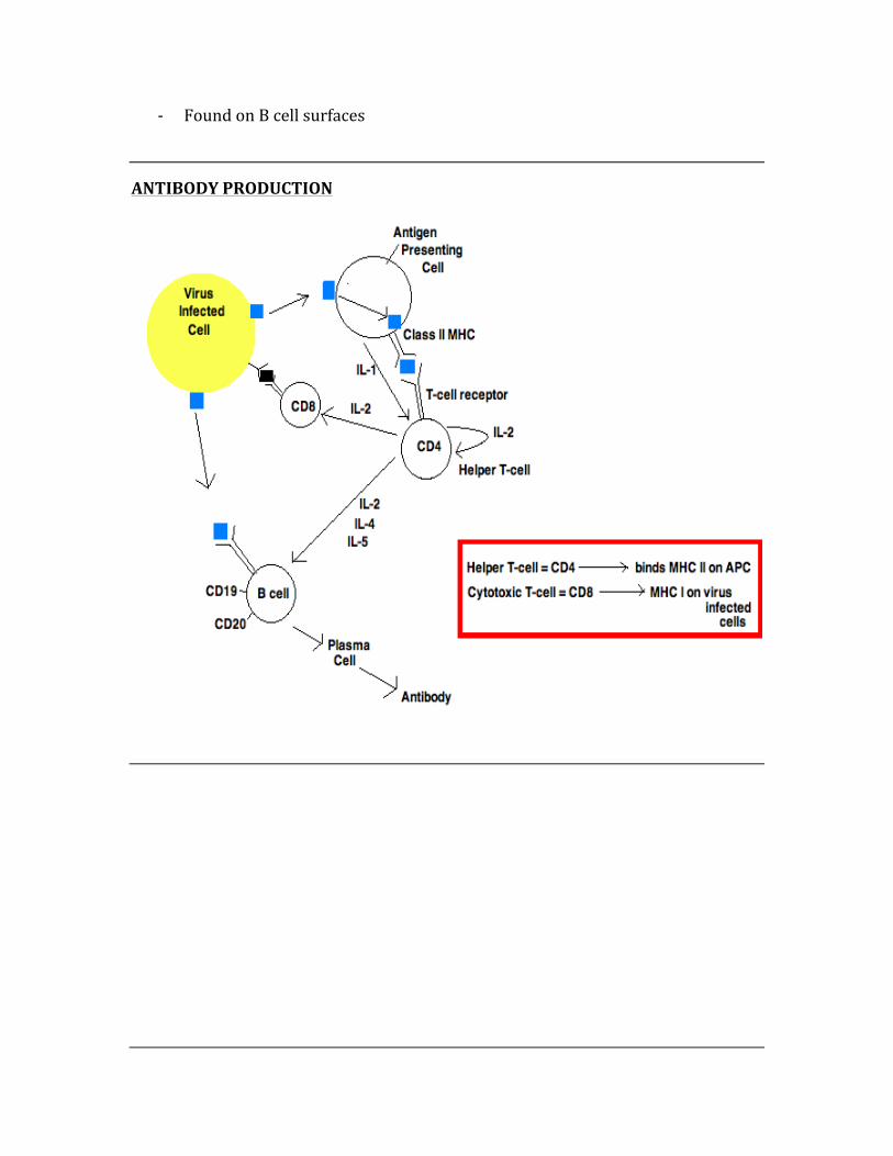
-‐ Found on B cell surfaces
ANTIBODY PRODUCTION
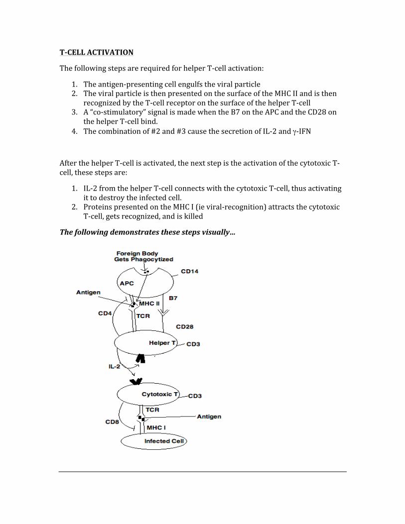
T-‐CELL ACTIVATION
The following steps are required for helper T-‐cell activation:
1. The antigen-‐presenting cell engulfs the viral particle 2. The viral particle is then presented on the surface of the MHC II and is then
recognized by the T-‐cell receptor on the surface of the helper T-‐cell 3. A “co-‐stimulatory” signal is made when the B7 on the APC and the CD28 on
the helper T-‐cell bind. 4. The combination of #2 and #3 cause the secretion of IL-‐2 and γ-‐IFN
After the helper T-‐cell is activated, the next step is the activation of the cytotoxic T-‐cell, these steps are:
1. IL-‐2 from the helper T-‐cell connects with the cytotoxic T-‐cell, thus activating it to destroy the infected cell.
2. Proteins presented on the MHC I (ie viral-‐recognition) attracts the cytotoxic T-‐cell, gets recognized, and is killed
The following demonstrates these steps visually…
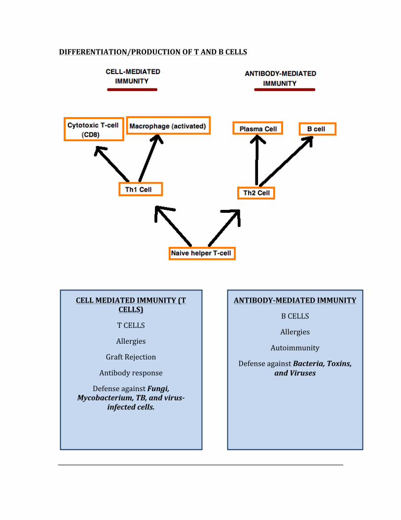
DIFFERENTIATION/PRODUCTION OF T AND B CELLS
CELL MEDIATED IMMUNITY (T CELLS)
T CELLS
Allergies
Graft Rejection
Antibody response
Defense against Fungi, Mycobacterium, TB, and virus-‐
infected cells.
ANTIBODY-‐MEDIATED IMMUNITY
B CELLS
Allergies
Autoimmunity
Defense against Bacteria, Toxins, and Viruses
CYTOKINE FUNCTION
There is a great mnemonic that can help you remember the first five cytokines, it is: “Hot T-‐bone stEAk”
IL – 1 à Hot = FEVER
IL – 2 à stimulation of T-‐cells
IL – 3 à stimulation of BONE marrow
IL – 4 à IgE production stimulation
IL – 5 à IgA production stimulation
Now the rest of the cytokines are responsible for the following:
IL – 8 à PMN chemotactic factor
TNF α à Increases IL-‐2 receptor synthesis by helper T cells, increases B cell proliferation, and attracts and stimulates PMNs. This is secreted by macrophages.
TNF β à This is secreted by activated T lymphocytes, and performs the same functions as the TNF α
γ -‐ Interferon à Stimulates macrophages, secreted by helper T cells
INTERFERONS
Interferons play an essential role in preventing the proliferation and production of a virus by acting in certain ways to prevent it from infecting other cells.
The three major functions of the interferons are:
1. Activation of natural killer (NK) cells which act by directly killing virus-‐infected cells.
2. α and β interferon act by inhibition of viral protein synthesis 3. γ -‐ Interferon works by increasing the expression of the MHC I and MHC II as
well as antigen presentation in all cells
Big picture MOA of interferons:
à Interferons stimulate the production of a protein that degrades viral mRNA. When this occurs, the virus cannot infect a cell since the proper genetic materials for this function are absent.
TRANSPLANT REJECTION
Graft-‐vs-‐Host Rejection:
Occurs when the grafted T cells proliferate and reject the host cells that contain foreign proteins. As this occurs, severe organ dysfunction ensues, causing damage to the liver, skin, mucosa, and gastrointestinal tract. This condition also affects the bone marrow and the lungs.
Hyperacute Rejection:
Occurs almost immediately after transplant, whereby preformed anti-‐donor antibodies cause a response.
Acute Rejection:
This is a cell-‐mediated reaction that occurs via cytotoxic T lymphocytes that react against foreign MHCs, occurring weeks post-‐transplant.
Chronic Rejection:
Occurring months-‐years post-‐transplant, is caused by antibody-‐mediated vascular damage.
TYPES OF HYPERSENSITIVITY REACTIONS
TYPE 1 HYPERSENSITIVITY: Anaphylactic/Atopic
A very rapid reaction that occurs in response to antigen exposure. Antigens trigger the release of histamine by binding to basophils and mast cells.
Examples à Hives, asthma, anaphylaxis
TYPE 2 HYPERSENSITIVITY: Cytotoxic
Is a cytotoxic reaction whereby IgM and/or IgG bind to cells leading to lysis by complement activation or phagocytosis.
Examples à Autoimmune hemolytic anemia, Goodpasture’s disease, rheumatic fever, Rh disease, Grave’s disease.
TYPE 3 HYPERSENSITIVITY: Antigen-‐Antibody Mediated
The formation of antigen-‐antibody complexes leads to complement activation, attracting PMN’s and ultimately leading to the release of lysosomal enzymes.
Examples à Glomerulonephritis, Lupus, Rheumatoid Arthritis
TYPE 4 HYPERSENSITIVITY: Cell-‐Mediated
Is a delayed response, whereby T cells become sensitized and encounter antigens, thus releasing cytokines.
Examples à Contact Dermatitis, Transplant Rejections, TB Skin Test
REMEMBERING THE HYPERSENSITIVITIES:
Mnemonic: ACID
A – Anaphylaxis
C – Cytotoxic
I – Immune Complex
D -‐ Delayed
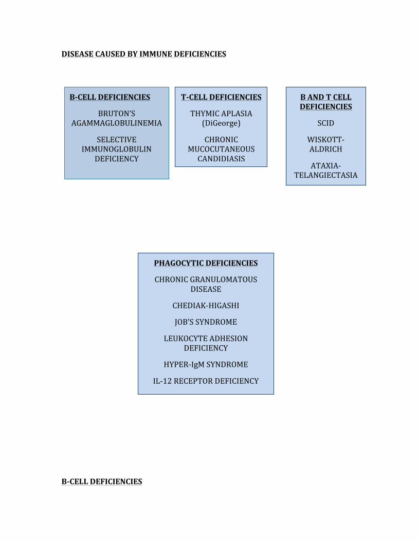
DISEASE CAUSED BY IMMUNE DEFICIENCIES
B-‐CELL DEFICIENCIES
B-‐CELL DEFICIENCIES
BRUTON’S AGAMMAGLOBULINEMIA
SELECTIVE IMMUNOGLOBULIN
DEFICIENCY
T-‐CELL DEFICIENCIES
THYMIC APLASIA (DiGeorge)
CHRONIC MUCOCUTANEOUS CANDIDIASIS
B AND T CELL DEFICIENCIES
SCID
WISKOTT-‐ALDRICH
ATAXIA-‐TELANGIECTASIA
PHAGOCYTIC DEFICIENCIES
CHRONIC GRANULOMATOUS DISEASE
CHEDIAK-‐HIGASHI
JOB’S SYNDROME
LEUKOCYTE ADHESION DEFICIENCY
HYPER-‐IgM SYNDROME
IL-‐12 RECEPTOR DEFICIENCY
BRUTON’S AGAMMAGLOBULINEMIA:
An x-‐linked recessive defect in tyrosine kinase gene. results in:
-‐ All Ig classes are decreased -‐ Get recurrent bacterial infections (after 6 months of age) -‐ Only in boys
SELECTIVE IMMUNOGLOBULIN DEFICIENCY:
A deficiency of a specific Ig class resulting in defect I isotype switching, results in:
-‐ Sinus infection -‐ Lung infections -‐ Most commonly the deficient Ig is IgA
T-‐CELL DEFICIENCIES:
THYMIC APLASIA:
There is a failure of the development in the thymus and parathyroid’s, due to failure of development of the 3rd and 4th pharyngeal pouch, results in:
-‐ Tetany -‐ Viral and fungal infections (recurrent) -‐ Heart defects
CHRONIC MUCOCUTANEOUS CANDIDIASIS:
A T-‐cell dysfunction against Candida, results in:
-‐ Candidal infections of the skin and mucous membranes
B AND T CELL DEFICIENCIES
SEVERE COMBINED IMMUNODEFICIENCY (SCID):
This condition leads to a defect in differentiation of the early stem-‐cells, results in:
-‐ Recurrent infections (viral, bacterial, fungal, and protozoal)
WISKOTT-‐ALDRICH SYNDROME:
Is an x-‐linked defect in the ability to mount an IgM response to the capsular polysaccharides of bacteria, results in:
-‐ Low IgM levels -‐ High IgA levels -‐ Normal IgE levels -‐ Classic triad of symptoms: Infections, Eczema, and Thrombocytopenic
Purpura.
PHAGOCYTIC DEFICIENCIES
CHRONIC GRANULOMATOUS DISEASE:
A lack of NADPH leads to a defect in neutrophil phagocytosis, results in:
-‐ Succeptibility to opportunitic bacterial infections -‐ Diagnosis based on negative nitroblue tetrazolium dye reduction test
(CLASSIC USMLE QUESTION)
CHEDIAK-‐HIGASHI DISEASE:
A defect in microtubular function and lysosomal emptying of the phagocytic cells, results in:
-‐ Recurrent pyogenic infections due to staph and strep
JOB’S SYNDROME:
T-‐cells fail to produce γ-‐interferon, thus PMN’s fail to respond, results in:
-‐ Eczema, staph abscesses, and elevated IgE
LEUKOCYTE ADHESION DEFICIENCY SYNDROME:
There is a defect in the LFA-‐1 adhesion proteins on the phagocytes, results in severe early life pyogenic infections.
HYPER-‐IgM SYNDROME:
There is a defect in the CD40 ligand on the CD4 Th cells, leading to severe pyogenic infections early in life, results in:
-‐ High IgM levels -‐ Extremely low levels of IgG, IgA, and IgE
IL-‐12 RECEPTOR DEFICIENCY:
A defect in the IL-‐12 receptor leads to disseminated mycobacterial infections.
CHAPTER 11
MICROBIOLOGY
Historically, bacteriology occupies the majority of the microbiology questions on the Step 1 exam. After bacteria, virology is very important, both conceptually and in detail. Pay very special attention to HIV and all that it entails, as
this is one of the highest-‐yielding topics on the exam. When it comes to parasites and helminths, your strategy should be
memorization of modes of transmission, signs and symptoms, and treatments.
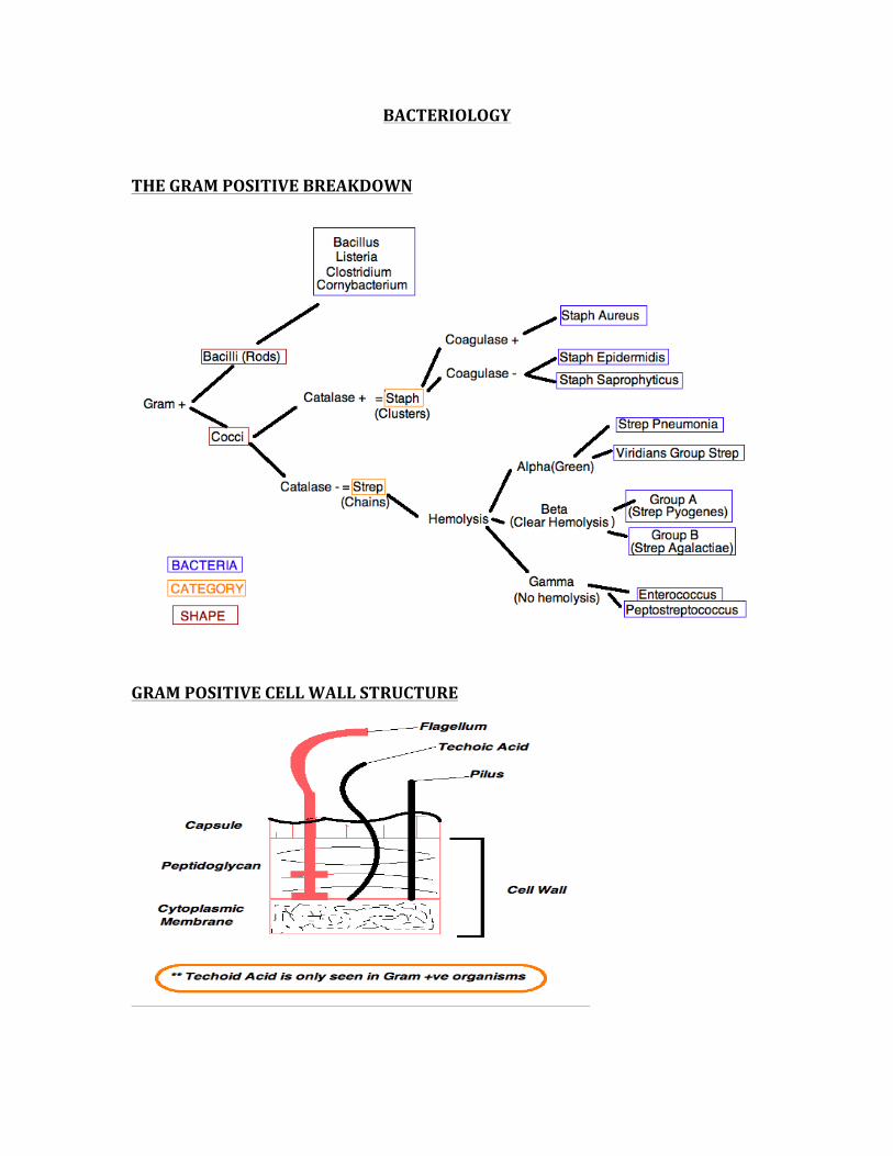
BACTERIOLOGY
THE GRAM POSITIVE BREAKDOWN
GRAM POSITIVE CELL WALL STRUCTURE
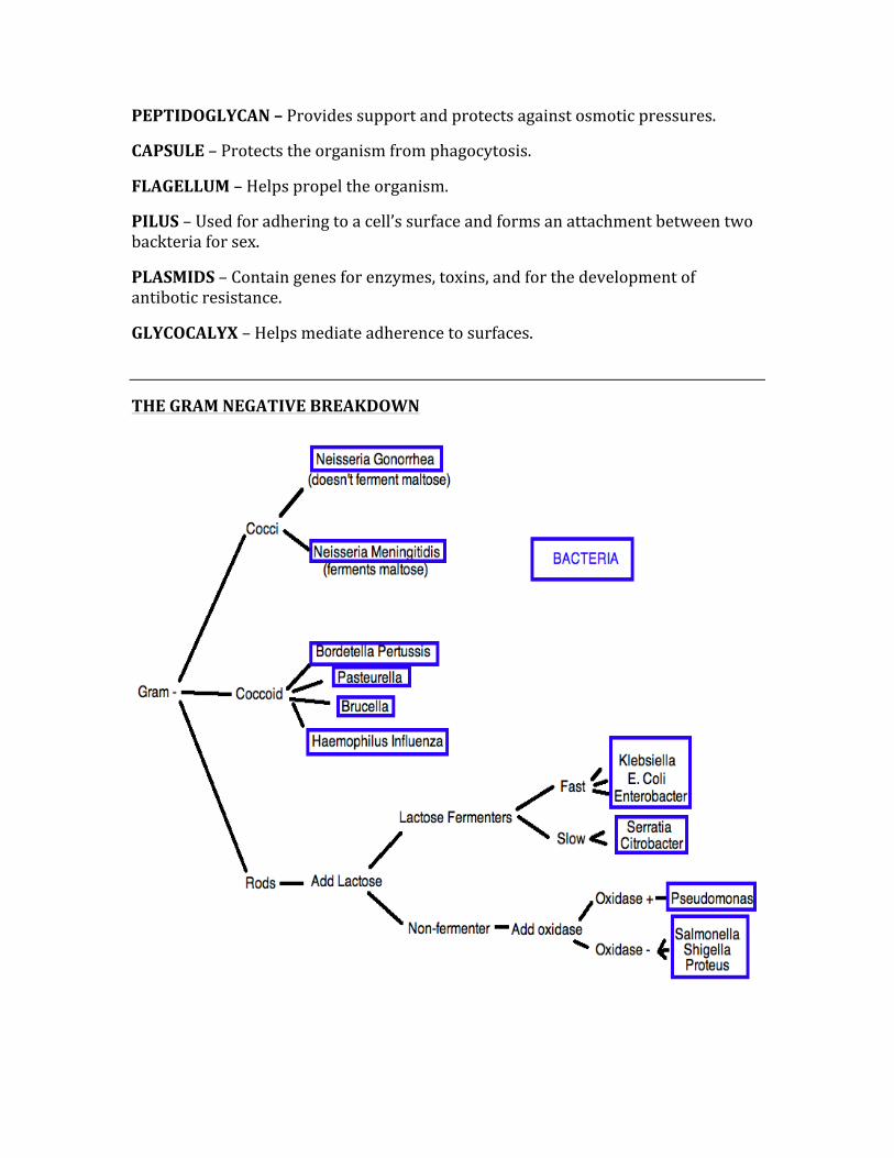
PEPTIDOGLYCAN – Provides support and protects against osmotic pressures.
CAPSULE – Protects the organism from phagocytosis.
FLAGELLUM – Helps propel the organism.
PILUS – Used for adhering to a cell’s surface and forms an attachment between two backteria for sex.
PLASMIDS – Contain genes for enzymes, toxins, and for the development of antibotic resistance.
GLYCOCALYX – Helps mediate adherence to surfaces.
THE GRAM NEGATIVE BREAKDOWN
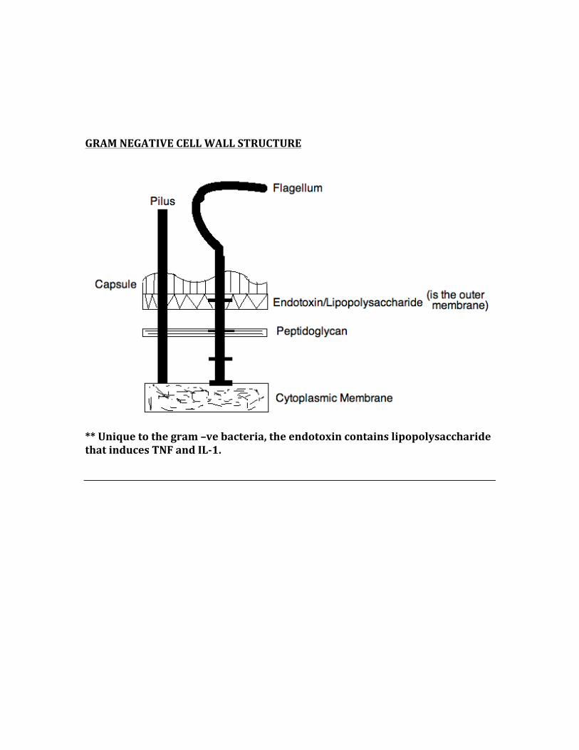
GRAM NEGATIVE CELL WALL STRUCTURE
** Unique to the gram –ve bacteria, the endotoxin contains lipopolysaccharide that induces TNF and IL-‐1.
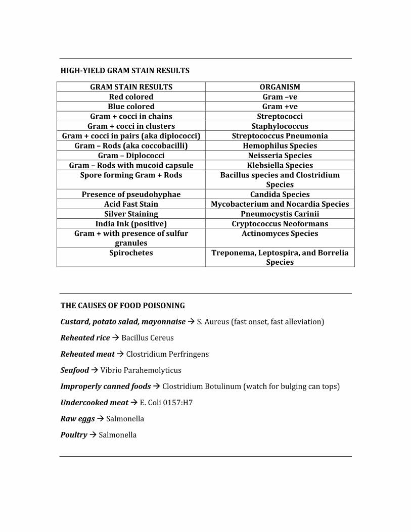
HIGH-‐YIELD GRAM STAIN RESULTS
GRAM STAIN RESULTS ORGANISM Red colored Gram –ve Blue colored Gram +ve
Gram + cocci in chains Streptococci Gram + cocci in clusters Staphylococcus
Gram + cocci in pairs (aka diplococci) Streptococcus Pneumonia Gram – Rods (aka coccobacilli) Hemophilus Species
Gram – Diplococci Neisseria Species Gram – Rods with mucoid capsule Klebsiella Species
Spore forming Gram + Rods Bacillus species and Clostridium Species
Presence of pseudohyphae Candida Species Acid Fast Stain Mycobacterium and Nocardia Species Silver Staining Pneumocystis Carinii
India Ink (positive) Cryptococcus Neoformans Gram + with presence of sulfur
granules Actinomyces Species
Spirochetes Treponema, Leptospira, and Borrelia Species
THE CAUSES OF FOOD POISONING
Custard, potato salad, mayonnaise à S. Aureus (fast onset, fast alleviation)
Reheated rice à Bacillus Cereus
Reheated meat à Clostridium Perfringens
Seafood à Vibrio Parahemolyticus
Improperly canned foods à Clostridium Botulinum (watch for bulging can tops)
Undercooked meat à E. Coli 0157:H7
Raw eggs à Salmonella
Poultry à Salmonella
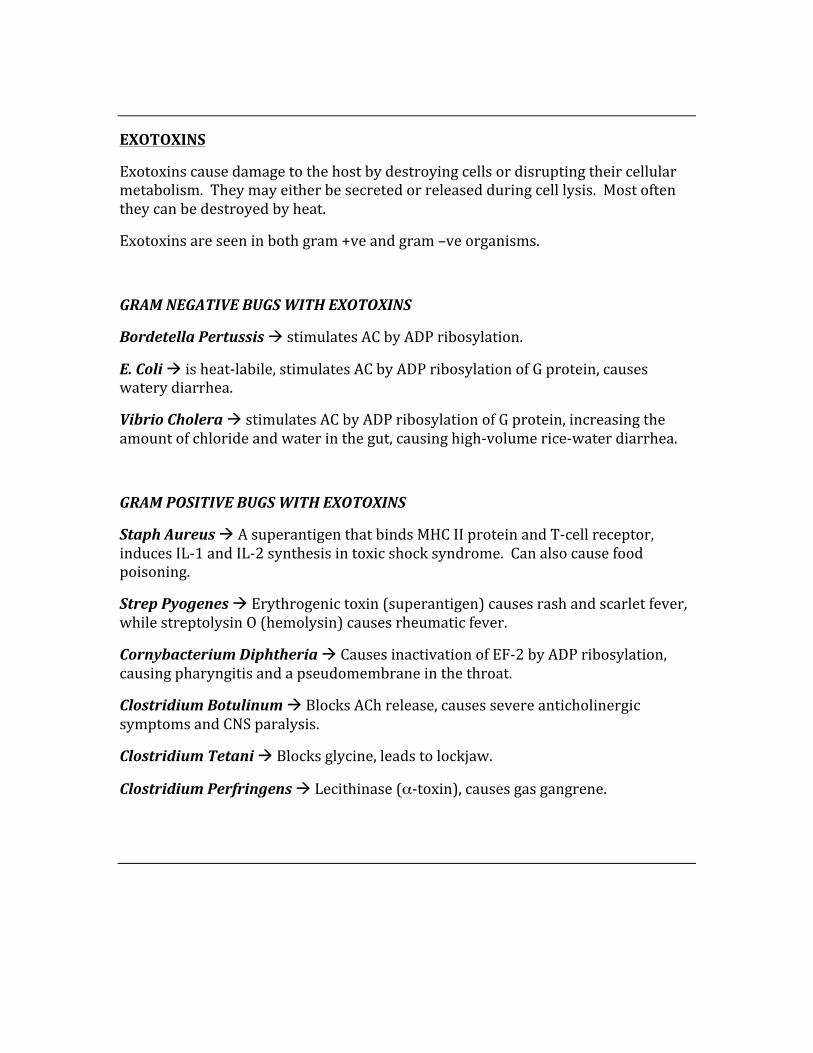
EXOTOXINS
Exotoxins cause damage to the host by destroying cells or disrupting their cellular metabolism. They may either be secreted or released during cell lysis. Most often they can be destroyed by heat.
Exotoxins are seen in both gram +ve and gram –ve organisms.
GRAM NEGATIVE BUGS WITH EXOTOXINS
Bordetella Pertussis à stimulates AC by ADP ribosylation.
E. Coli à is heat-‐labile, stimulates AC by ADP ribosylation of G protein, causes watery diarrhea.
Vibrio Cholera à stimulates AC by ADP ribosylation of G protein, increasing the amount of chloride and water in the gut, causing high-‐volume rice-‐water diarrhea.
GRAM POSITIVE BUGS WITH EXOTOXINS
Staph Aureus à A superantigen that binds MHC II protein and T-‐cell receptor, induces IL-‐1 and IL-‐2 synthesis in toxic shock syndrome. Can also cause food poisoning.
Strep Pyogenes à Erythrogenic toxin (superantigen) causes rash and scarlet fever, while streptolysin O (hemolysin) causes rheumatic fever.
Cornybacterium Diphtheria à Causes inactivation of EF-‐2 by ADP ribosylation, causing pharyngitis and a pseudomembrane in the throat.
Clostridium Botulinum à Blocks ACh release, causes severe anticholinergic symptoms and CNS paralysis.
Clostridium Tetani à Blocks glycine, leads to lockjaw.
Clostridium Perfringens à Lecithinase (α-‐toxin), causes gas gangrene.
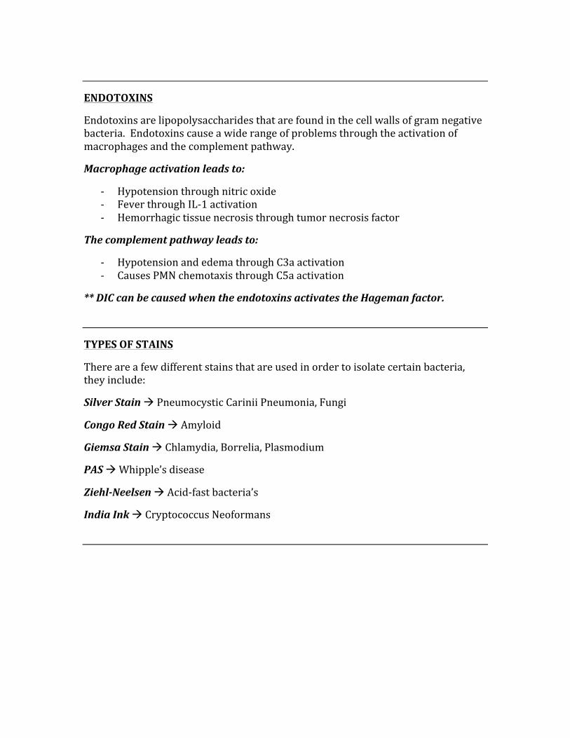
ENDOTOXINS
Endotoxins are lipopolysaccharides that are found in the cell walls of gram negative bacteria. Endotoxins cause a wide range of problems through the activation of macrophages and the complement pathway.
Macrophage activation leads to:
-‐ Hypotension through nitric oxide -‐ Fever through IL-‐1 activation -‐ Hemorrhagic tissue necrosis through tumor necrosis factor
The complement pathway leads to:
-‐ Hypotension and edema through C3a activation -‐ Causes PMN chemotaxis through C5a activation
** DIC can be caused when the endotoxins activates the Hageman factor.
TYPES OF STAINS
There are a few different stains that are used in order to isolate certain bacteria, they include:
Silver Stain à Pneumocystic Carinii Pneumonia, Fungi
Congo Red Stain à Amyloid
Giemsa Stain à Chlamydia, Borrelia, Plasmodium
PAS à Whipple’s disease
Ziehl-‐Neelsen à Acid-‐fast bacteria’s
India Ink à Cryptococcus Neoformans
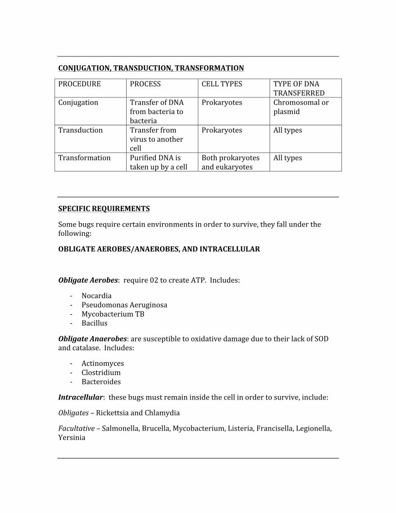
CONJUGATION, TRANSDUCTION, TRANSFORMATION
PROCEDURE PROCESS CELL TYPES TYPE OF DNA TRANSFERRED
Conjugation Transfer of DNA from bacteria to bacteria
Prokaryotes Chromosomal or plasmid
Transduction Transfer from virus to another cell
Prokaryotes All types
Transformation Purified DNA is taken up by a cell
Both prokaryotes and eukaryotes
All types
SPECIFIC REQUIREMENTS
Some bugs require certain environments in order to survive, they fall under the following:
OBLIGATE AEROBES/ANAEROBES, AND INTRACELLULAR
Obligate Aerobes: require 02 to create ATP. Includes:
-‐ Nocardia -‐ Pseudomonas Aeruginosa -‐ Mycobacterium TB -‐ Bacillus
Obligate Anaerobes: are susceptible to oxidative damage due to their lack of SOD and catalase. Includes:
-‐ Actinomyces -‐ Clostridium -‐ Bacteroides
Intracellular: these bugs must remain inside the cell in order to survive, include:
Obligates – Rickettsia and Chlamydia
Facultative – Salmonella, Brucella, Mycobacterium, Listeria, Francisella, Legionella, Yersinia
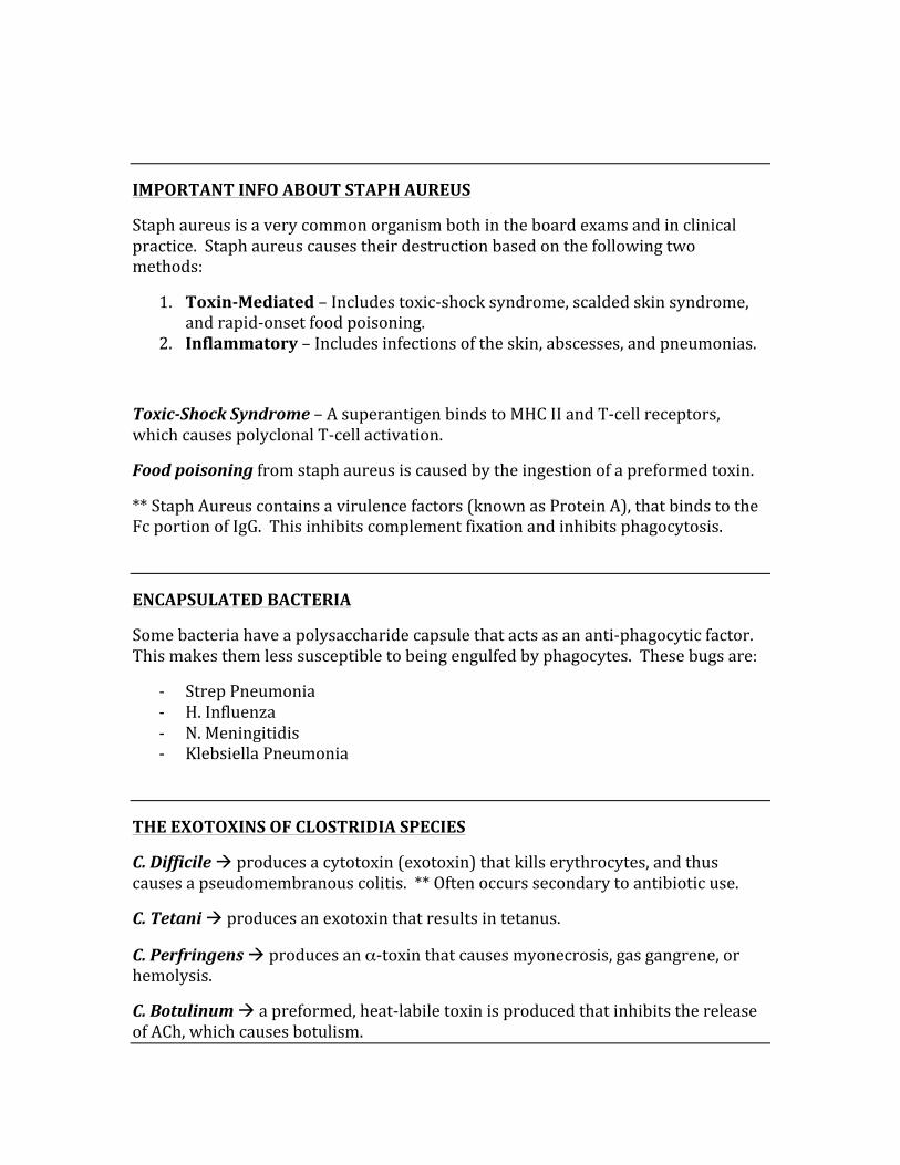
IMPORTANT INFO ABOUT STAPH AUREUS
Staph aureus is a very common organism both in the board exams and in clinical practice. Staph aureus causes their destruction based on the following two methods:
1. Toxin-‐Mediated – Includes toxic-‐shock syndrome, scalded skin syndrome, and rapid-‐onset food poisoning.
2. Inflammatory – Includes infections of the skin, abscesses, and pneumonias.
Toxic-‐Shock Syndrome – A superantigen binds to MHC II and T-‐cell receptors, which causes polyclonal T-‐cell activation.
Food poisoning from staph aureus is caused by the ingestion of a preformed toxin.
** Staph Aureus contains a virulence factors (known as Protein A), that binds to the Fc portion of IgG. This inhibits complement fixation and inhibits phagocytosis.
ENCAPSULATED BACTERIA
Some bacteria have a polysaccharide capsule that acts as an anti-‐phagocytic factor. This makes them less susceptible to being engulfed by phagocytes. These bugs are:
-‐ Strep Pneumonia -‐ H. Influenza -‐ N. Meningitidis -‐ Klebsiella Pneumonia
THE EXOTOXINS OF CLOSTRIDIA SPECIES
C. Difficile à produces a cytotoxin (exotoxin) that kills erythrocytes, and thus causes a pseudomembranous colitis. ** Often occurs secondary to antibiotic use.
C. Tetani à produces an exotoxin that results in tetanus.
C. Perfringens à produces an α-‐toxin that causes myonecrosis, gas gangrene, or hemolysis.
C. Botulinum à a preformed, heat-‐labile toxin is produced that inhibits the release of ACh, which causes botulism.
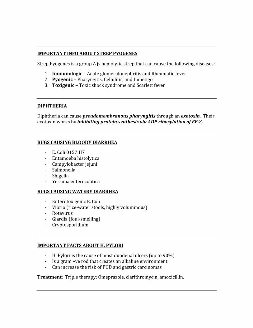
IMPORTANT INFO ABOUT STREP PYOGENES
Strep Pyogenes is a group A β-‐hemolytic strep that can cause the following diseases:
1. Immunologic – Acute glomerulonephritis and Rheumatic fever 2. Pyogenic – Pharyngitis, Cellulitis, and Impetigo 3. Toxigenic – Toxic shock syndrome and Scarlett fever
DIPHTHERIA
Diphtheria can cause pseudomembranous pharyngitis through an exotoxin. Their exotoxin works by inhibiting protein synthesis via ADP ribosylation of EF-‐2.
BUGS CAUSING BLOODY DIARRHEA
-‐ E. Coli 0157:H7 -‐ Entamoeba histolytica -‐ Campylobacter jejuni -‐ Salmonella -‐ Shigella -‐ Yersinia enterocolitica
BUGS CAUSING WATERY DIARRHEA
-‐ Enterotoxigenic E. Coli -‐ Vibrio (rice-‐water stools, highly voluminous) -‐ Rotavirus -‐ Giardia (foul-‐smelling) -‐ Cryptosporidium
IMPORTANT FACTS ABOUT H. PYLORI
-‐ H. Pylori is the cause of most duodenal ulcers (up to 90%) -‐ Is a gram –ve rod that creates an alkaline environment -‐ Can increase the risk of PUD and gastric carcinomas
Treatment: Triple therapy: Omeprazole, clarithromycin, amoxicillin.
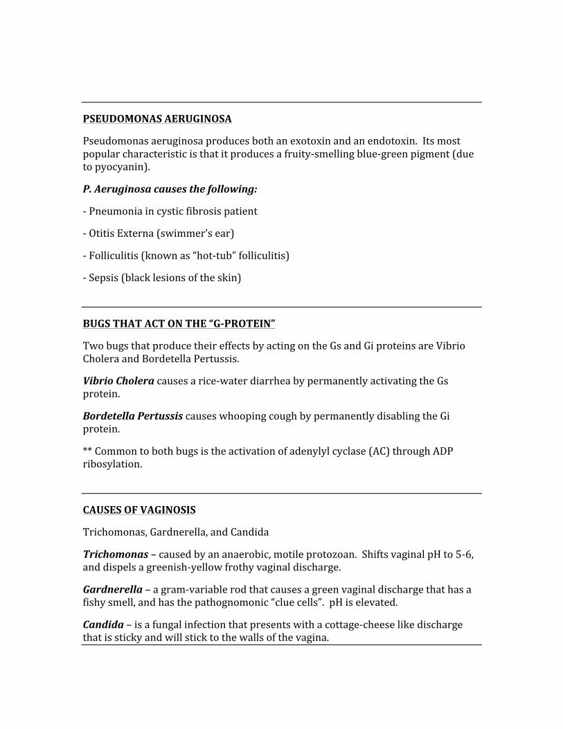
PSEUDOMONAS AERUGINOSA
Pseudomonas aeruginosa produces both an exotoxin and an endotoxin. Its most popular characteristic is that it produces a fruity-‐smelling blue-‐green pigment (due to pyocyanin).
P. Aeruginosa causes the following:
-‐ Pneumonia in cystic fibrosis patient
-‐ Otitis Externa (swimmer’s ear)
-‐ Folliculitis (known as “hot-‐tub” folliculitis)
-‐ Sepsis (black lesions of the skin)
BUGS THAT ACT ON THE “G-‐PROTEIN”
Two bugs that produce their effects by acting on the Gs and Gi proteins are Vibrio Cholera and Bordetella Pertussis.
Vibrio Cholera causes a rice-‐water diarrhea by permanently activating the Gs protein.
Bordetella Pertussis causes whooping cough by permanently disabling the Gi protein.
** Common to both bugs is the activation of adenylyl cyclase (AC) through ADP ribosylation.
CAUSES OF VAGINOSIS
Trichomonas, Gardnerella, and Candida
Trichomonas – caused by an anaerobic, motile protozoan. Shifts vaginal pH to 5-‐6, and dispels a greenish-‐yellow frothy vaginal discharge.
Gardnerella – a gram-‐variable rod that causes a green vaginal discharge that has a fishy smell, and has the pathognomonic “clue cells”. pH is elevated.
Candida – is a fungal infection that presents with a cottage-‐cheese like discharge that is sticky and will stick to the walls of the vagina.
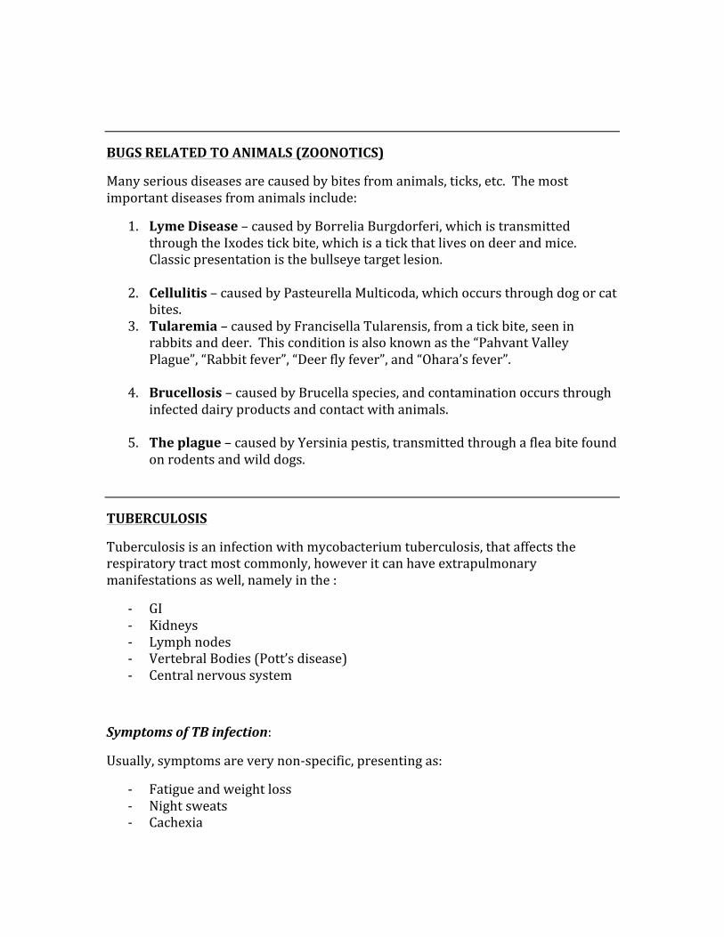
BUGS RELATED TO ANIMALS (ZOONOTICS)
Many serious diseases are caused by bites from animals, ticks, etc. The most important diseases from animals include:
1. Lyme Disease – caused by Borrelia Burgdorferi, which is transmitted through the Ixodes tick bite, which is a tick that lives on deer and mice. Classic presentation is the bullseye target lesion.
2. Cellulitis – caused by Pasteurella Multicoda, which occurs through dog or cat bites.
3. Tularemia – caused by Francisella Tularensis, from a tick bite, seen in rabbits and deer. This condition is also known as the “Pahvant Valley Plague”, “Rabbit fever”, “Deer fly fever”, and “Ohara’s fever”.
4. Brucellosis – caused by Brucella species, and contamination occurs through infected dairy products and contact with animals.
5. The plague – caused by Yersinia pestis, transmitted through a flea bite found on rodents and wild dogs.
TUBERCULOSIS
Tuberculosis is an infection with mycobacterium tuberculosis, that affects the respiratory tract most commonly, however it can have extrapulmonary manifestations as well, namely in the :
-‐ GI -‐ Kidneys -‐ Lymph nodes -‐ Vertebral Bodies (Pott’s disease) -‐ Central nervous system
Symptoms of TB infection:
Usually, symptoms are very non-‐specific, presenting as:
-‐ Fatigue and weight loss -‐ Night sweats -‐ Cachexia
If the symptoms become more pulmonary, expect to find:
-‐ Dyspnea -‐ Hemoptysis -‐ Chest pain (pleuritic in nature) -‐ Productive cough
1° infection à A primary TB infection refers to the infectious process by which the body is able to contain the infection and prevent its dissemination. This results in the Ghon complex, which is a calcified focus of infection usually in the lower segments of the lung.
2° infection à Also known as “reactivated TB”, this type of infection can occur to those who are in a state of weakened or suppressed immunity.
When tuberculosis is suspected?
-‐ A +ve PPD test warrants a chest xray looking for the TB cavitary lesion -‐ Acid fast stain looking for the mycobacterium
Management/Treatment?
Active TB à A 4-‐drug regimen (RIPE – Rifampin, Isoniazid, Pyrazinamide, and Ethambutol). Beware of adverse neurological effects of Isoniazid, supplement with vitamin B6 – pyridoxine.
Latent TB à With latent TB, you will begin the patient on a 4-‐drug regimen lasting up to 9 months. If they present at a future date with latent TB, they will not need to be treated again. ** Latent TB = 1 time 4-‐drug course lasting several months.
RICKETTSIAL INFECTIONS AND VECTORS
Rickettsial infections are those infections that are responsible for causing Rocky Mountain Spotted Fever, Typhus, and Q fever.
Rickettsial infections usually lead to a similar presentation, which is:
-‐ Fever -‐ Headache -‐ Rash
ROCKY MOUNTAIN SPOTTED FEVER
Caused by Rickettsia Rickettsii, which causes a rash that starts on the palms and soles and moves inward. Treat with tetracycline.
Two forms of Typhus’ are:
ENDEMIC TYPHUS – caused by Rickettsia Typhi, which is a flea. Treat with tetracycline.
EPIDEMIC TYPHUS – caused by Rickettsia Prowazekii, a body louse. Treat with tetracycline.
And finally,
Q FEVER – caused by Coxiella Burnetti, which caused infection via aerosolized particles. Treat with tetracycline.
SPIROCHETES
Spirochetes are “spiral-‐shaped” bacterial elements containing axial filaments. The most commonly tested spirochete-‐related infection is syphilis (treponema). The other two types of spirochetes are Borrelia and Leptospira, which are stainable with light microscopy, whereas treponema is visualized only with dark-‐field microscopy.
With that said, there are three forms of Syphilis:
1° syphilis à Patient will get a painless chancre, treat with IM pen G
2° syphilis à Patient has disseminated disease + constitutional symptoms, includes a maculopapular rash on the palms and soles. Treat with IM pen G
3° syphilis à Patient has neurological problems (neurosyphilis, Argyll Robertson pupil), aortic disorders (aortitis), and gummas. Treat with IV penicillin.
TESTING FOR SYPHILIS
VDRL and FTA-‐ABS:
VDRL – this test is used for nonspecific antibody detection, however due to its high rate of false-‐positives is not the primary test used for syphilis detection. This test will be positive when there are a few other diseases present, including: SLE, RA, RF, and a few other viral infections like mononucleosis.
FTA-‐ABS – is a test specific for the treponema bacteria, and has the best combination of benefits in finding syphilis, including high specificity, very early positivity, and remains positive the longest.
VIROLOGY
DNA AND RNA VIRAL GENOMES
All DNA viruses are double stranded and linear, with the exception of parvovirus (is single-‐stranded), and hepadnavirus/papovavirus (circular). All RNA viruses are single-‐stranded, except the reovirus (double stranded).
DNA is double, RNA is single.
HOW DO VIRUSES REPRODUCE
Recombination à is the exchange of genetic material between 2 chromosomes by cross-‐over of certain regions that have important base sequence homology.
Reassortment à is the exchange of viral segments (from segmented viral genomes). This occurs at high frequency and is responsible for worldwide illnesses (pandemics).
Complementation à occurs when one functional virus helps another non-‐functional virus become functional.
Phenotypic Mixing à occurs when a certain virus has the surface coating from another virus’ protein, which will then determine the infectivity of this virus particle.
VIRAL PLOIDY
Retroviruses contain 2 identical single-‐stranded RNA molecules, making them “diploid”, while all other viruses contain a single copy of both DNA and RNA, making them “haploid”.
DNA Viruses à all replicate in the nucleus, except for the poxvirus.
RNA Viruses à all replicate in the cytoplasm, except for retroviruses and the influenza virus.
THE IMPORTANT CHARACTERISTICS OF VIRAL DNA
The following are important characteristics that fit for almost all of the DNA viruses:
Nuclear replication à all DNA viruses replicate in the nucleus except for the Poxvirus, which has its own DNA-‐dependent RNA polymerase.
Are linear à all DNA viruses are linear, except for the Papovavirus and the Hepadna virus. Papovavirus is circular/supercoiled, and the Hepadna virus is circular/incomplete.
Are double stranded (dsDNA) à all DNA viruses are double-‐stranded except for the Parvovirus, which is single-‐stranded (ssDNA).
Are icosahedral in shape à all DNA viruses are icosahedral except for the Poxvirus, which is complete in capsid shape.
GENETIC SHIFT vs. GENETIC DRIFT
Genetic Shift à Is the process by which there is a re-‐assortment of different viral strains, which combine to form new forms/subtypes that contain a mixture of the surface antigens of the strains involved. Genetic shift is very important because it creates new viral pathogens, and is responsible for the emergence of new viruses.
Genetic Drift à Is a process of natural mutations over a period of time. This leads to loss of immunity and the inability of vaccines to cure illnesses.
THE DNA VIRUSES
The list of DNA viruses:
1. Hepadnavirus 2. Herpesvirus 3. Adenovirus 4. Parvovirus 5. Papovavirus 6. Poxvirus
Remember this list with the mnemonic: HHAPPPy
THE CHARACTERISTICS OF EACH DNA VIRUS
1. Hepadnavirus – dsDNA partial circular, enveloped.
Diseases à Hepatitis B virus
2. Herpesvirus – dsDNA linear, enveloped.
Diseases à Human Herpes Virus 6 & 8, Herpes Simplex 1 & 2, Varicella Zoster, Ebstein-‐Barr Virus, and Cytomegalovirus
HHV 6 – Reseola
HHV 8 – Kaposi’s sarcoma
HSV 1 – Thought to be oral ulcers, but now can be from both oral and genital ulcers
HSV 2 – Thought to be genital ulcers, but now can be from both oral and genital ulcers
VZV – Responsible for chickenpox (not seen anymore due to vaccination), shingles
EBV – Causes mononucleosis and Burkitt’s lymphoma
CMV – Congenital infections
3. Adenovirus – dsDNA linear, has no envelope.
Diseases à Conjunctivitis, Pneumonias, Pharyngitis
4. Parvovirus – ssDNA linear (is the smallest DNA), has no envelope
Diseases à Parvo B19 (slapped cheek syndrome)
5. Papovavirus – dsDNA circular, has no envelope
Diseases à Human Papilloma virus, Progressive Multifocal Leukoencephalopathy
6. Poxvirus – dsDNA linear (is the largest of all DNA viruses), has an envelope
Diseases à Cowpox, Smallpox, Molluscum Contagiosum
THE CHARACTERISTICS OF EACH RNA VIRUS
1. Picornavirus – (+)ssRNA, linear, icosahedral, no envelope.
Responsible for:
-‐ Hepatitis A -‐ Coxsackievirus -‐ Poliovirus -‐ Echovirus -‐ Rhinovirus
2. Calicivirus – (+)ssRNA, linear, icosahedral, no envelope.
Responsible for:
-‐ Norwalk virus (gastroenteritis) -‐ Hepatitis E
3. Reovirus – dsRNA, linear segmented, double icosahedral, no envelope.
Responsible for:
-‐ Reovirus (Colorado tick fever) -‐ Rotavirus (MCC of diarrhea in children)
4. Flavivirus – (+)ssRNA, linear, icosahedral, enveloped.
Responsible for:
-‐ Dengue fever -‐ Yellow fever -‐ Hepatitis C -‐ St. Louis encephalitis -‐ West Nile virus
5. Togavirus – (+)ssRNA, linear, icosahedral, enveloped.
Responsible for:
-‐ Rubella -‐ Eastern/Western equine encephalitis
6. Retrovirus – (+)ssRNA, linear, icosahedral, enveloped.
Responsible for:
-‐ HIV/AIDS -‐ Hairy T-‐cell leukemia
7. Orthomyxovirus – (-‐)ssRNA, linear/segmented, helical, enveloped.
Responsible for:
-‐ Influenza
8. Paramyxovirus – (-‐)ssRNA, linear/non-‐segmented, helical, enveloped.
Responsible for:
-‐ Measles -‐ Mumps -‐ Respiratory Syncitial Virus (RSV) -‐ Parainfluenza
9. Rhabdovirus – (-‐)ssRNA, linear, helical, enveloped.
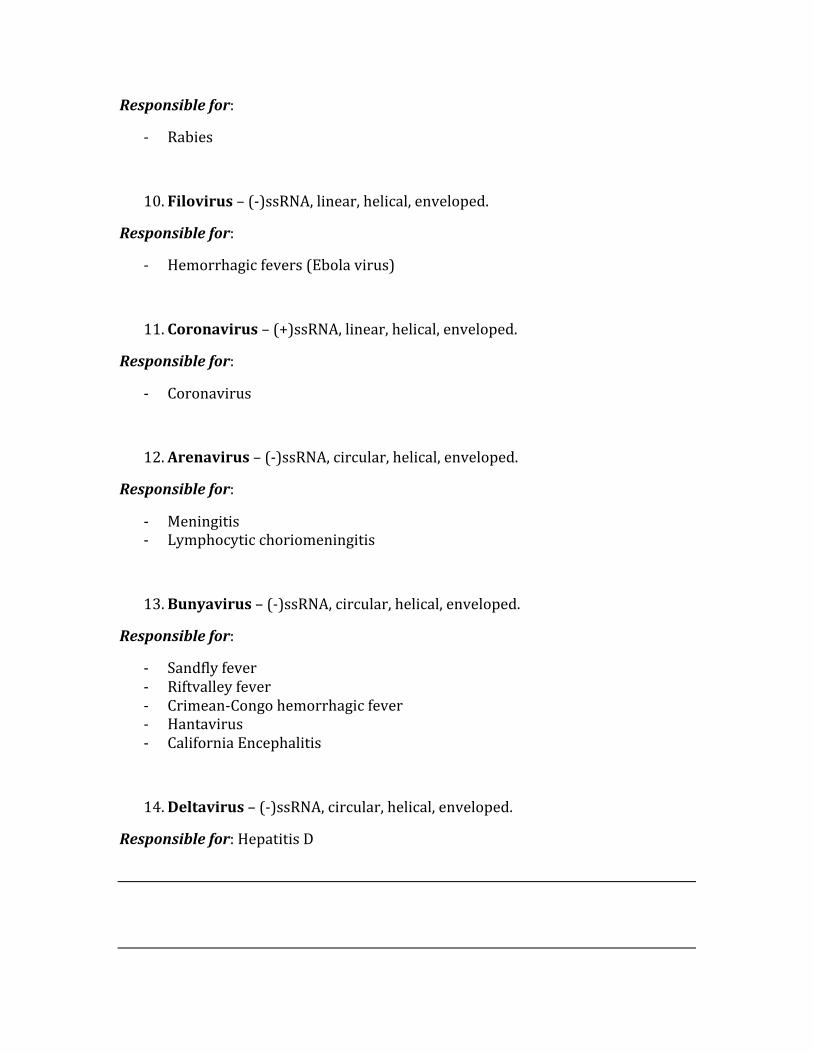
Responsible for:
-‐ Rabies
10. Filovirus – (-‐)ssRNA, linear, helical, enveloped.
Responsible for:
-‐ Hemorrhagic fevers (Ebola virus)
11. Coronavirus – (+)ssRNA, linear, helical, enveloped.
Responsible for:
-‐ Coronavirus
12. Arenavirus – (-‐)ssRNA, circular, helical, enveloped.
Responsible for:
-‐ Meningitis -‐ Lymphocytic choriomeningitis
13. Bunyavirus – (-‐)ssRNA, circular, helical, enveloped.
Responsible for:
-‐ Sandfly fever -‐ Riftvalley fever -‐ Crimean-‐Congo hemorrhagic fever -‐ Hantavirus -‐ California Encephalitis
14. Deltavirus – (-‐)ssRNA, circular, helical, enveloped.
Responsible for: Hepatitis D
LIVE-‐ATTENUATED vs. KILLED VACCINES
Live-‐Attenuated Vaccines à these types of vaccines induce both humoral and cell-‐mediated immunity.
Killed Vaccines à these types of vaccines induce humoral immunity only.
Examples of Live-‐Attenuated Vaccines:
-‐ Measles -‐ Mumps -‐ Rubella -‐ Smallpox -‐ Polio (Sabin) -‐ VZV -‐ Yellow Fever
** Remember there is danger in giving a live-‐attenuated vaccine to someone who is immunocompromised.
Examples of Killed Vaccines:
-‐ Rabies -‐ Influenza -‐ Polio (Salk) -‐ Hepatitis A
SUPER HIGH-‐YIELD MICROBIOLOGY INFORMATION
HERPRES VIRUSES
There are quite a few different forms of “herpes viruses”, they are all high-‐yield USMLE information. The most common herpes viruses and their common findings include:
HSV-‐1 – this is known to be responsible for the oral lesions of herpes, however due to different sexual practices, it may be seen in the genital region as well. HSV-‐1 is also a cause of keratoconjunctivitis and temporal lobe encephalitis (HSV is the most common cause of adult onset altered mental status).
HSV-‐1 is transmitted via respiratory secretions and/or saliva. For HSV diagnosis, use the Tzanck test (smear the open vesicle, looking for multinucleated giant cells).
HSV-‐2 – HSV-‐2 is most commonly seen as herpes of the genitals (herpes labialis), as well as neonatal herpes.
HSV-‐2 is transmitted via perinatal transmission as well as through sexual contact.
EBV – causes Burkitt’s lymphoma and infectious mononucleosis (causes severe fatigue, sore throat, usually in teenagers). Transmitted through saliva and respiratory secretions.
CMV – Causes congenital infections as well as some types of pneumonia. Transmitted congenitally, through sexual contact, through saliva, and through transfusion.
HHV – Human Herpes Virus, is the cause of Kaposi’s sarcoma in AIDS patients. Transmission is sexually.
MONONUCLEOSIS
Mononucleosis is always encountered on the USMLE exams, thus be sure to know as much as possible about this condition.
Presentation:
-‐ Young adult (16-‐20yr) -‐ Fever -‐ Hepatosplenomegaly -‐ Pharyngitis -‐ Lymphadenopathy
Cause:
-‐ Ebstein-‐Barr Virus (EBV) -‐ “kissing disease”, because it is easily transmitted through saliva -‐ There are abnormal circulating cytotoxic T cells
Diagnosis:
The best diagnostic test is the “Monospot test”, which detects heterophil antibodies through sheep RBC agglutination.
What to tell the patient:
-‐ The most important thing for patient safety is to AVOID CONTACT SPORTS, because the spleen is enlarged and if it experiences blunt trauma, it may rupture and cause severe adverse effects such as hypotension, shock, and sepsis.

HIV/AIDS
A retrovirus, the HIV disease uses the reverse transcriptase enzyme to replicate. HIV infection can be transmitted through sexual contact/fluids, blood, and breast milk. The helper T-‐cells (CD4+ T cells), macrophages, and dendritic cells are affected.
DIAGNOSING HIV:
Diagnosis of HIV is made with the ELISA test because of its high sensitivity. When a test is positive, a Western Blot is performed, which is very specific.
After confirmation, a viral load can be done to measure the quantity of the virus in the blood, which allows you to measure the effect of medical treatment.
** False negatives with the ELISA test and the Western Blot are common in the first few months of HIV infection.
IMMUNITY TO HIV:
Certain mutations in our genes can give us immunity to HIV, can give us partial immunity to HIV, and can even cause us to deteriorate rapidly from an HIV infection. The mutations responsible for this include:
Mutation causing immunity à CCR5 mutation (homozygous)
Mutation causing partial immunity à CCR5 mutation (heterozygous)
Mutation causing rapid progression/deterioration à CXCR1 mutation
AIDS – OPPORTUNISTIC INFECTIONS:
The most commonly encountered opportunistic infection are:
FUNGAL – Candida (thrush), Cryptococcal Meningitis (Cryptococcus), Pneumocystis Pneumonia (PCP, when CD4<200), Histoplasmosis.
VIRAL – PML (caused by JC virus), HSV, VZV, CMV
BACTERIAL – TB, Mycobacterium Avium-‐Intracellular complex (MAC)
PROTOZOA – Cryptosporidium, Toxoplasmosis
TREATMENT:
Treatment involves the use of antiretroviral medications, given to symptomatic patients regardless of their CD4 counts and/or to asymptomatic patients with CD4 counts <500.
Triple-‐drug regimens (2 nucleoside reverse transcriptase inhibitors and either: 1 non-‐nucleoside reverse transcriptase inhibitor OR 1 protease inhibitor) are used, these are known as HAART, which target and prevent HIV replication at three different points along the virus’ replication process.
OTHER MANIFESTATIONS OF HIV INFECTION:
Pulmonary: Community-‐acquired pneumonia, PCP, TB, CMV, MAC, Histoplasmosis.
CNS: Toxoplasmosis, AIDS-‐dementia, Cryptococcal Meningitis, Lymphoma, HSV, Neurosyphilis, PML
GI: Diarrhea, Oral lesions (thrush, ulcerations, oral hairy leukoplakia), Candida esophagitis, Proctitis
Dermatological: HSV, Shingles, Molluscum Contagiosum, Kaposi’s sarcoma
HEPATITIS
Is inflammation of the liver, characterized by the presence of inflammation of the tissue of the liver. There is a wide range of routes of infection, there is a wide range of symptoms and prognosis, which can range from self-‐limited diseases to cirrhosis and death.
There are FIVE common forms of hepatitis, they are:
Hepatitis A, B, C, D, and E
Hepatitis A (HAV) – Is usually asymptomatic, is transmitted via fecal-‐oral route. Usually a very short incubation period (~ 3 weeks). Is a RNA picornavirus.
Hepatitis B (HBV) – Often presents with mild symptoms such as: loss of appetite N/V, myalgias, fever, and jaundice. Hep B is usually self-‐limited, but can progress and cause cirrhosis, hepatic failure, and death. Transmission is usually sexual, through sharing dirty needles (parenteral), and from the mother to fetus (vertical transmission). Is a DNA hepadnavirus.
Hepatitis C (HCV) – Hepatitis C usually carries the same symptoms as does hepatitis B, and is transmitted through blood products (sex, dirty needles). Hepatitis C is often a chronic illness that leads to carcinoma and/or cirrhosis. People infected will usually become carriers of hep C. Is an RNA flavivirus.
Hepatitis D (HDV) – Alone, hepatitis D cannot infect, and requires a co-‐infection with hepatitis B in order to infect (requires HBsAg as its envelope). Is a deltavirus.
Hepatitis E (HEV) – Has a fecal-‐oral transmission route, and has the tendency to cause water-‐borne epidemics. Very similar symptomatically to hepatitis A. HEV can cause significant illness and even death in pregnant women. Is an RNA calicivirus.
** Hep B and C à chronic, cause cirrhosis, and hepatocellular carcinoma.
** Hep A and E à affect the GI tract.
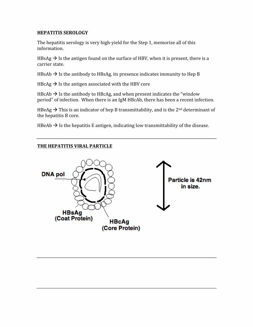
HEPATITIS SEROLOGY
The hepatitis serology is very high-‐yield for the Step 1, memorize all of this information.
HBsAg à Is the antigen found on the surface of HBV, when it is present, there is a carrier state.
HBsAb à Is the antibody to HBsAg, its presence indicates immunity to Hep B
HBcAg à Is the antigen associated with the HBV core
HBcAb à Is the antibody to HBcAg, and when present indicates the “window period” of infection. When there is an IgM HBcAb, there has been a recent infection.
HBeAg à This is an indicator of hep B transmittability, and is the 2nd determinant of the hepatitis B core.
HBeAb à Is the hepatitis E antigen, indicating low transmittability of the disease.
THE HEPATITIS VIRAL PARTICLE
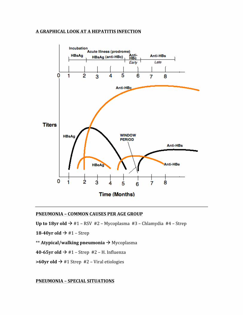
A GRAPHICAL LOOK AT A HEPATITIS INFECTION
PNEUMONIA – COMMON CAUSES PER AGE GROUP
Up to 18yr old à #1 – RSV #2 – Mycoplasma #3 – Chlamydia #4 – Strep
18-‐40yr old à #1 – Strep
** Atypical/walking pneumonia à Mycoplasma
40-‐65yr old à #1 – Strep #2 – H. Influenza
>60yr old à #1 Strep #2 – Viral etiologies
PNEUMONIA – SPECIAL SITUATIONS
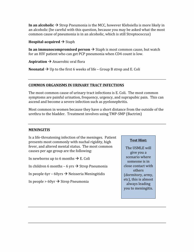
In an alcoholic à Strep Pneumonia is the MCC, however Klebsiella is more likely in an alcoholic (be careful with this question, because you may be asked what the most common cause of pneumonia is in an alcoholic, which is still Streptococcus)
Hospital-‐acquired à Staph
In an immunocompromised person à Staph is most common cause, but watch for an HIV patient who can get PCP pneumonia when CD4 count is low.
Aspiration à Anaerobic oral flora
Neonatal à Up to the first 6 weeks of life – Group B strep and E. Coli
COMMON ORGANISMS IN URINARY TRACT INFECTIONS
The most common cause of urinary tract infections is E. Coli. The most common symptoms are painful urination, frequency, urgency, and suprapubic pain. This can ascend and become a severe infection such as pyelonephritis.
Most common in women because they have a short distance from the outside of the urethra to the bladder. Treatment involves using TMP-‐SMP (Bactrim)
MENINGITIS
Is a life-‐threatening infection of the meninges. Patient presents most commonly with nuchal rigidity, high fever, and altered mental status. The most common causes per age group are the following:
In newborns up to 6 months à E. Coli
In children 6 months – 6 yrs à Strep Pneumonia
In people 6yr – 60yrs à Neisseria Meningitidis
In people > 60yr à Strep Pneumonia
Test Hint:
The USMLE will give you a
scenario where someone is in
close contact with others
(dormitory, army, etc), this is almost always leading
you to meningitis.
A LIST OF THE MOST COMMONLY TESTED STD’S
Gonorrhea à presents with urethritis, cervicitis, epididymitis (men). Can cause pelvic inflammatory disease (common cause of infertility). Caused by N. Gonorrhea, treat with ceftriaxone.
Chlamydia à presents similarly to gonorrhea. When this is found, there is usually a co-‐infection with gonorrhea. Caused by Chlamydia Trachomatis.
Herpes à PAINFUL ulcer of the penis, vulva, and cervix
Syphilis à PAINLESS chancre (seen in 1° form), systemic findings in 2° form, and neurological findings in the 3° form.
Lymphogranuloma Venereum à PAINLESS ulcer at the beginning, with infection spreading along the inguinal pathway. Caused by Chlamydia Trachomatis types L1-‐L3.
Vaginitis à Vaginal itching, pain, and discharge. May be due to gardnerella, trichomonas, or candida.
Chancroid à PAINFUL genital ulcers, caused by Haemophilus Ducreyi
Condyloma Acuminata à Causes genital warts that can be on the labia, in the vagina, and on the cervix. Caused by HPV types 6 and 11.
PELVIC INFLAMMATORY DISEASE
Is a scarring of the fallopian tubes, and is a very common cause of infertility in women. There is often symptoms such as cervical motion tenderness, vaginal discharge, and even tubo-‐ovarian abscess.
Most common cause is Chlamydia and Gonorrhea.
THE TORCH INFECTIONS
“TORCH” is the mnemonic used to recall the most commonly encountered congenital infections, they are:
T – Toxoplasmosis
O – stands for “other”, which is Syphilis
R – Rubella
C – CMV
H – HSV and HIV
OSTEOMYELITIS
Osteomyelitis is an infection of the bone, which can be caused by a number of different organisms, they include:
Staphylococcus Aureus à Is the most common cause overall
Salmonella à Causes osteomyelitis in sickle cell patients
Gonorrhea à Can cause osteomyelitis in sexually active people (is a less common cause)
Pseudomonas Aeruginosa à Common cause in intravenous drug users
Staph Epidermidis à Common cause of osteomyelitis in those with prosthetic devices.
PARASITOLOGY
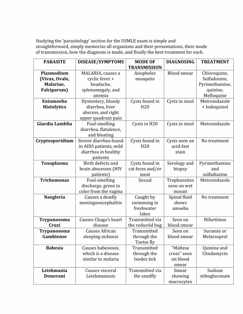
Studying the ‘parasitology’ section for the USMLE exam is simple and straightforward, simply memorize all organisms and their presentations, their mode of transmission, how the diagnosis is made, and finally the best treatment for each.
PARASITE DISEASE/SYMPTOMS MODE OF TRANSMISSION
DIAGNOSING TREATMENT
Plasmodium (Vivax, Ovale, Malariae,
Falciparum)
MALARIA, causes a cyclic fever + headache,
splenomegaly, and anemia
Anopheles mosquito
Blood smear Chloroquine, Sulfadoxine,
Pyrimethamine, quinine,
Mefloquine Entamoeba Histolytics
Dystentery, bloody diarrhea, liver
abscess, and right upper quadrant pain
Cysts found in H20
Cysts in stool Metronidazole + Iodoquinol
Giardia Lamblia Foul-‐smelling diarrhea, flatulence,
and bloating
Cysts in H20 Cysts in stool Metronidazole
Cryptosporidium Severe diarrhea found in AIDS patients, mild diarrhea in healthy
patients
Cysts found in H20
Cysts seen on acid-‐fast stain
No treatment
Toxoplasma Birth defects and brain abscesses (HIV
patients)
Cysts found in cat feces and/or
meat
Serology and biopsy
Pyrimethamine and
sulfadiazine Trichomonas Foul-‐smelling
discharge, green in color from the vagina
Sexual Trophozoites seen on wet mount
Metronidazole
Naegleria Causes a deadly meningoencephalitis
Caught by swimming in freshwater lakes
Spinal fluid shows amoeba
No treatment
Trypanosoma Cruzi
Causes Chaga’s heart disease
Transmitted via the reduviid bug
Seen on blood smear
Nifurtimox
Trypanosoma Gambiense
Causes African sleeping sickness
Transmitted through the Tsetse fly
Seen on blood smear
Suramin or Melarsoprol
Babesia Causes babesiosis, which is a disease similar to malaria
Transmitted through the Ixodes tick
“Maltese cross” seen on blood smear
Quinine and Clindamycin
Leishmania Donovani
Causes visceral Leishmaniasis
Transmitted via the sandfly
Smear showing macrocytes
Sodium stibogluconate

that contain amastigotes
HELMINTHS (Worms)
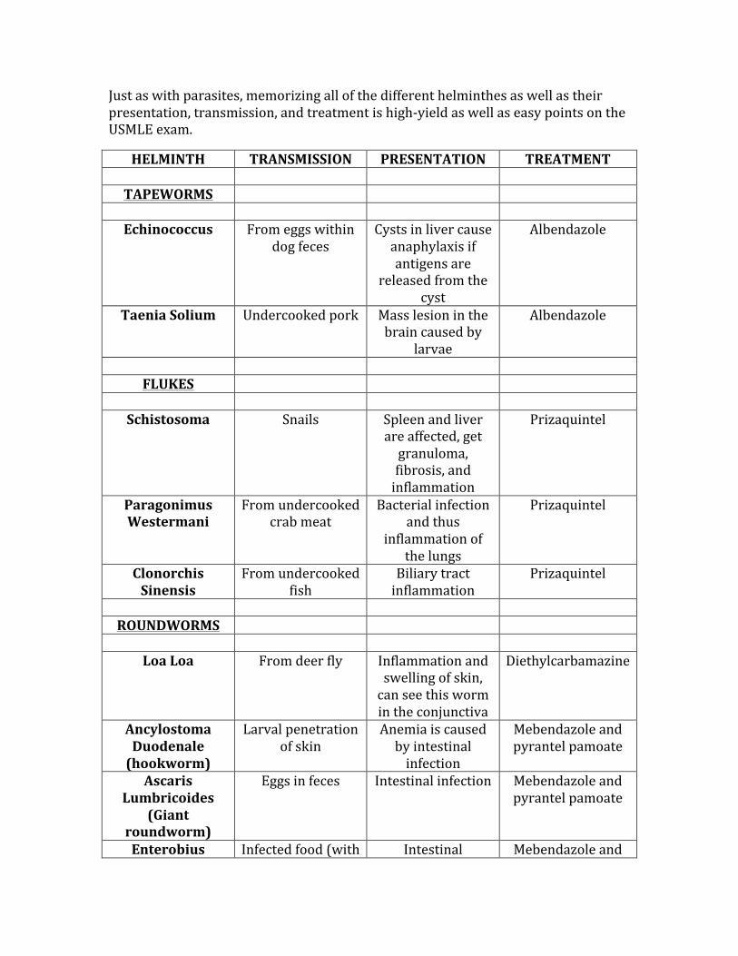
Just as with parasites, memorizing all of the different helminthes as well as their presentation, transmission, and treatment is high-‐yield as well as easy points on the USMLE exam.
HELMINTH TRANSMISSION PRESENTATION TREATMENT TAPEWORMS
Echinococcus From eggs within
dog feces Cysts in liver cause anaphylaxis if antigens are
released from the cyst
Albendazole
Taenia Solium Undercooked pork Mass lesion in the brain caused by
larvae
Albendazole
FLUKES
Schistosoma Snails Spleen and liver
are affected, get granuloma, fibrosis, and inflammation
Prizaquintel
Paragonimus Westermani
From undercooked crab meat
Bacterial infection and thus
inflammation of the lungs
Prizaquintel
Clonorchis Sinensis
From undercooked fish
Biliary tract inflammation
Prizaquintel
ROUNDWORMS
Loa Loa From deer fly Inflammation and
swelling of skin, can see this worm in the conjunctiva
Diethylcarbamazine
Ancylostoma Duodenale (hookworm)
Larval penetration of skin
Anemia is caused by intestinal infection
Mebendazole and pyrantel pamoate
Ascaris Lumbricoides
(Giant roundworm)
Eggs in feces Intestinal infection Mebendazole and pyrantel pamoate
Enterobius Infected food (with Intestinal Mebendazole and
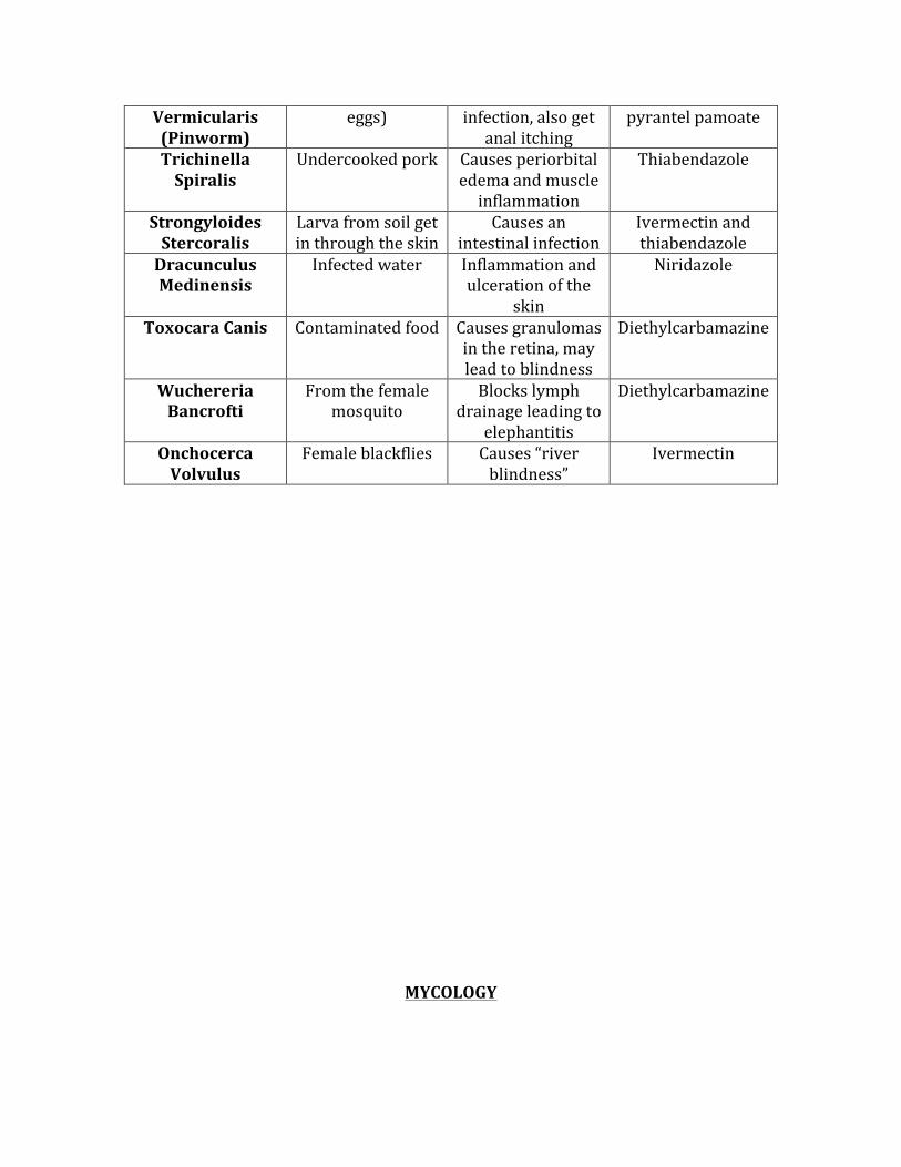
Vermicularis (Pinworm)
eggs) infection, also get anal itching
pyrantel pamoate
Trichinella Spiralis
Undercooked pork Causes periorbital edema and muscle inflammation
Thiabendazole
Strongyloides Stercoralis
Larva from soil get in through the skin
Causes an intestinal infection
Ivermectin and thiabendazole
Dracunculus Medinensis
Infected water Inflammation and ulceration of the
skin
Niridazole
Toxocara Canis Contaminated food Causes granulomas in the retina, may lead to blindness
Diethylcarbamazine
Wuchereria Bancrofti
From the female mosquito
Blocks lymph drainage leading to
elephantitis
Diethylcarbamazine
Onchocerca Volvulus
Female blackflies Causes “river blindness”
Ivermectin
MYCOLOGY
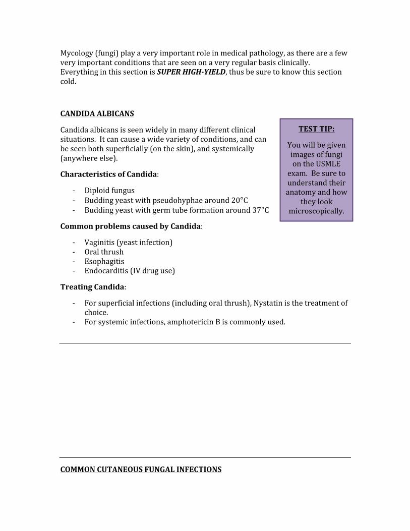
Mycology (fungi) play a very important role in medical pathology, as there are a few very important conditions that are seen on a very regular basis clinically. Everything in this section is SUPER HIGH-‐YIELD, thus be sure to know this section cold.
CANDIDA ALBICANS
Candida albicans is seen widely in many different clinical situations. It can cause a wide variety of conditions, and can be seen both superficially (on the skin), and systemically (anywhere else).
Characteristics of Candida:
-‐ Diploid fungus -‐ Budding yeast with pseudohyphae around 20°C -‐ Budding yeast with germ tube formation around 37°C
Common problems caused by Candida:
-‐ Vaginitis (yeast infection) -‐ Oral thrush -‐ Esophagitis -‐ Endocarditis (IV drug use)
Treating Candida:
-‐ For superficial infections (including oral thrush), Nystatin is the treatment of choice.
-‐ For systemic infections, amphotericin B is commonly used.
COMMON CUTANEOUS FUNGAL INFECTIONS
TEST TIP:
You will be given images of fungi on the USMLE exam. Be sure to understand their anatomy and how
they look microscopically.
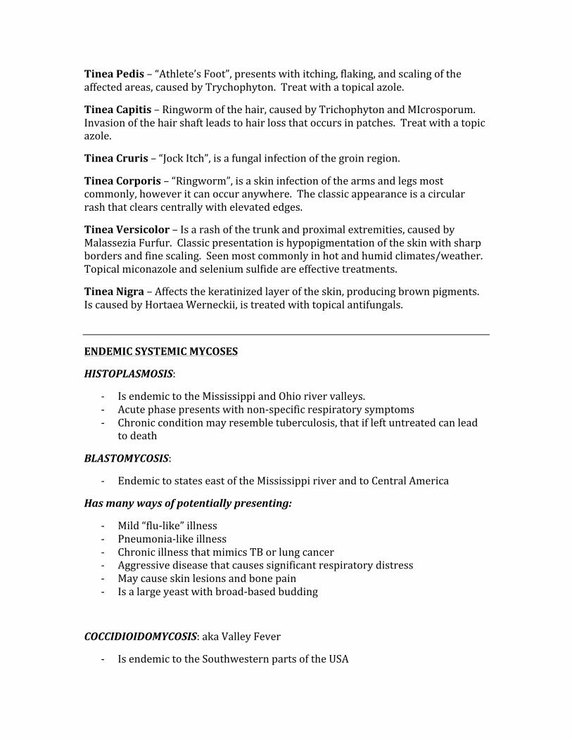
Tinea Pedis – “Athlete’s Foot”, presents with itching, flaking, and scaling of the affected areas, caused by Trychophyton. Treat with a topical azole.
Tinea Capitis – Ringworm of the hair, caused by Trichophyton and MIcrosporum. Invasion of the hair shaft leads to hair loss that occurs in patches. Treat with a topic azole.
Tinea Cruris – “Jock Itch”, is a fungal infection of the groin region.
Tinea Corporis – “Ringworm”, is a skin infection of the arms and legs most commonly, however it can occur anywhere. The classic appearance is a circular rash that clears centrally with elevated edges.
Tinea Versicolor – Is a rash of the trunk and proximal extremities, caused by Malassezia Furfur. Classic presentation is hypopigmentation of the skin with sharp borders and fine scaling. Seen most commonly in hot and humid climates/weather. Topical miconazole and selenium sulfide are effective treatments.
Tinea Nigra – Affects the keratinized layer of the skin, producing brown pigments. Is caused by Hortaea Werneckii, is treated with topical antifungals.
ENDEMIC SYSTEMIC MYCOSES
HISTOPLASMOSIS:
-‐ Is endemic to the Mississippi and Ohio river valleys. -‐ Acute phase presents with non-‐specific respiratory symptoms -‐ Chronic condition may resemble tuberculosis, that if left untreated can lead
to death
BLASTOMYCOSIS:
-‐ Endemic to states east of the Mississippi river and to Central America
Has many ways of potentially presenting:
-‐ Mild “flu-‐like” illness -‐ Pneumonia-‐like illness -‐ Chronic illness that mimics TB or lung cancer -‐ Aggressive disease that causes significant respiratory distress -‐ May cause skin lesions and bone pain -‐ Is a large yeast with broad-‐based budding
COCCIDIOIDOMYCOSIS: aka Valley Fever
-‐ Is endemic to the Southwestern parts of the USA
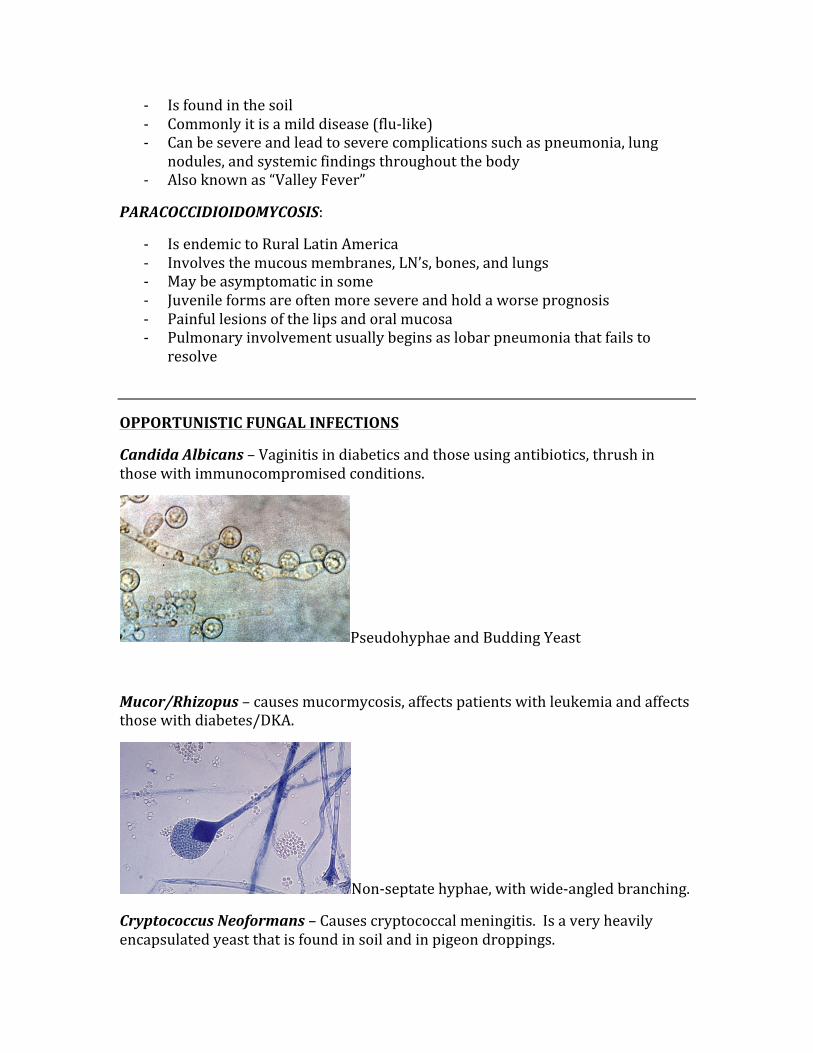
-‐ Is found in the soil -‐ Commonly it is a mild disease (flu-‐like) -‐ Can be severe and lead to severe complications such as pneumonia, lung
nodules, and systemic findings throughout the body -‐ Also known as “Valley Fever”
PARACOCCIDIOIDOMYCOSIS:
-‐ Is endemic to Rural Latin America -‐ Involves the mucous membranes, LN’s, bones, and lungs -‐ May be asymptomatic in some -‐ Juvenile forms are often more severe and hold a worse prognosis -‐ Painful lesions of the lips and oral mucosa -‐ Pulmonary involvement usually begins as lobar pneumonia that fails to
resolve
OPPORTUNISTIC FUNGAL INFECTIONS
Candida Albicans – Vaginitis in diabetics and those using antibiotics, thrush in those with immunocompromised conditions.
Pseudohyphae and Budding Yeast
Mucor/Rhizopus – causes mucormycosis, affects patients with leukemia and affects those with diabetes/DKA.
Non-‐septate hyphae, with wide-‐angled branching.
Cryptococcus Neoformans – Causes cryptococcal meningitis. Is a very heavily encapsulated yeast that is found in soil and in pigeon droppings.
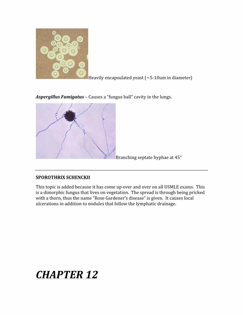
Heavily encapsulated yeast (~5-‐10um in diameter)
Aspergillus Fumigatus – Causes a “fungus ball” cavity in the lungs.
Branching septate hyphae at 45°
SPOROTHRIX SCHENCKII
This topic is added because it has come up over and over on all USMLE exams. This is a dimorphic fungus that lives on vegetation. The spread is through being pricked with a thorn, thus the name “Rose Gardener’s disease” is given. It causes local ulcerations in addition to nodules that follow the lymphatic drainage.
CHAPTER 12
PHARMACOLOGY
Pharmacology is a very high-‐yield topic on the USMLE Step 1 exam. Focus should be made on the following: Mechanisms
of Action, clinical use/applications, side effects. Pharmacokinetics, pharmacodynamics, and the autonomic
nervous system are also very high-‐yield information.
THE EC 50
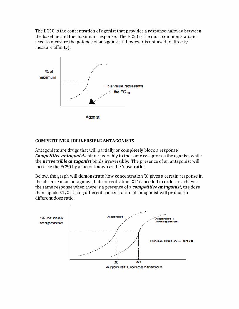
The EC50 is the concentration of agonist that provides a response halfway between the baseline and the maximum response. The EC50 is the most common statistic used to measure the potency of an agonist (it however is not used to directly measure affinity).
COMPETITIVE & IRRIVERSIBLE ANTAGONISTS
Antagonists are drugs that will partially or completely block a response. Competitive antagonists bind reversibly to the same receptor as the agonist, while the irreversible antagonist binds irreversibly. The presence of an antagonist will increase the EC50 by a factor known as the ‘dose-‐ratio’.
Below, the graph will demonstrate how concentration ‘X’ gives a certain response in the absence of an antagonist, but concentration ‘X1’ is needed in order to achieve the same response when there is a presence of a competitive antagonist, the dose then equals X1/X. Using different concentration of antagonist will produce a different dose ratio.
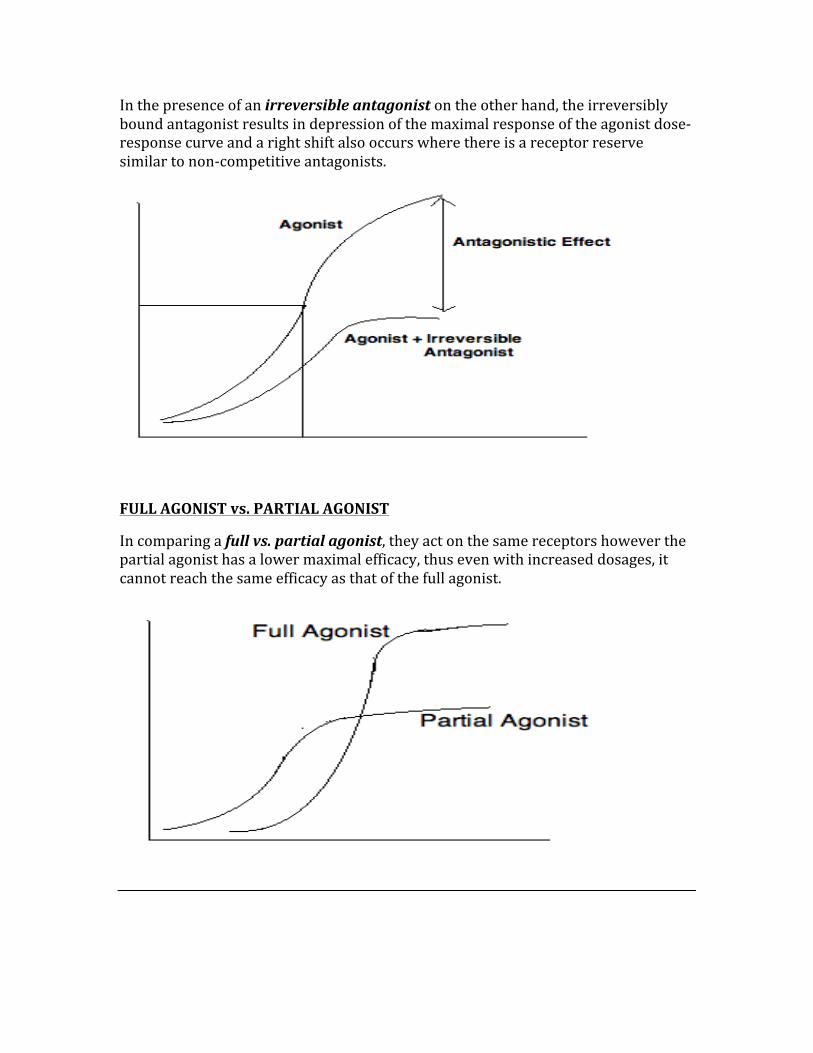
In the presence of an irreversible antagonist on the other hand, the irreversibly bound antagonist results in depression of the maximal response of the agonist dose-‐response curve and a right shift also occurs where there is a receptor reserve similar to non-‐competitive antagonists.
FULL AGONIST vs. PARTIAL AGONIST
In comparing a full vs. partial agonist, they act on the same receptors however the partial agonist has a lower maximal efficacy, thus even with increased dosages, it cannot reach the same efficacy as that of the full agonist.
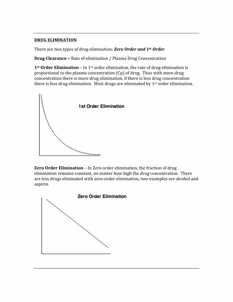
DRUG ELIMINATION
There are two types of drug elimination: Zero Order and 1st Order
Drug Clearance = Rate of elimination / Plasma Drug Concentration
1st Order Elimination – In 1st order elimination, the rate of drug elimination is proportional to the plasma concentration (Cp) of drug. Thus with more drug concentration there is more drug elimination, if there is less drug concentration there is less drug elimination. Most drugs are eliminated by 1st order elimination.
Zero Order Elimination – In Zero order elimination, the fraction of drug elimination remains constant, no matter how high the drug concentration. There are less drugs eliminated with zero-‐order elimination, two examples are alcohol and aspirin.
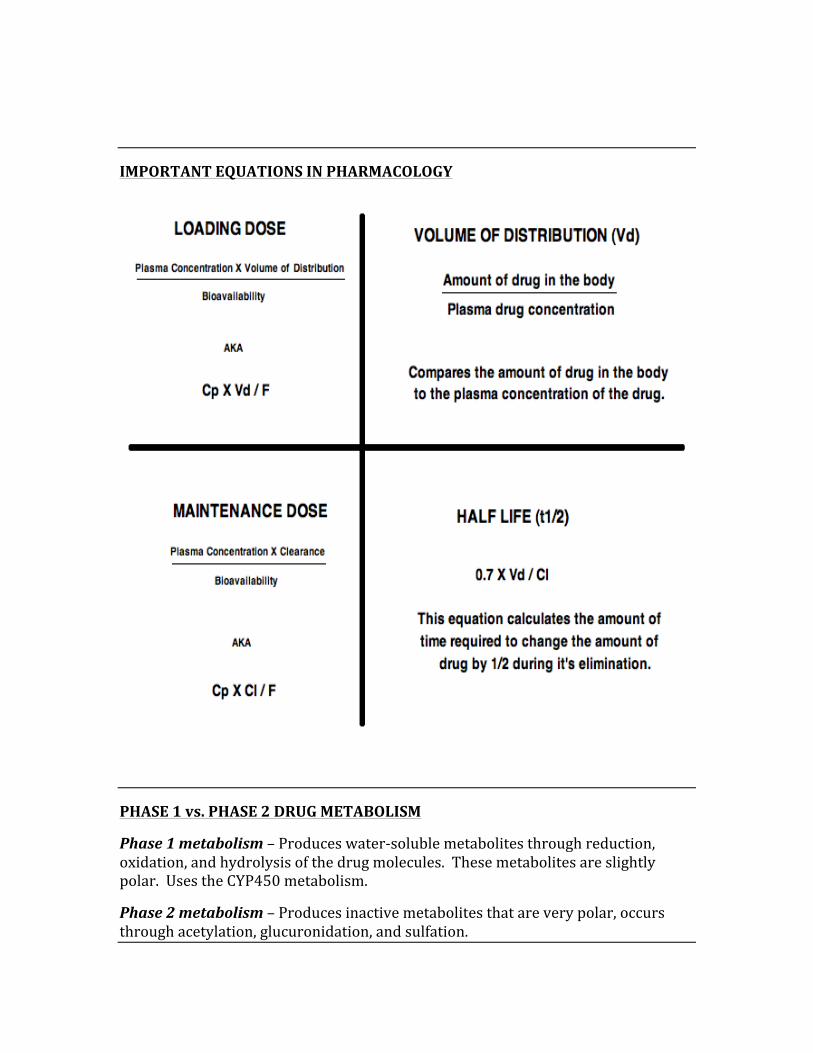
IMPORTANT EQUATIONS IN PHARMACOLOGY
PHASE 1 vs. PHASE 2 DRUG METABOLISM
Phase 1 metabolism – Produces water-‐soluble metabolites through reduction, oxidation, and hydrolysis of the drug molecules. These metabolites are slightly polar. Uses the CYP450 metabolism.
Phase 2 metabolism – Produces inactive metabolites that are very polar, occurs through acetylation, glucuronidation, and sulfation.
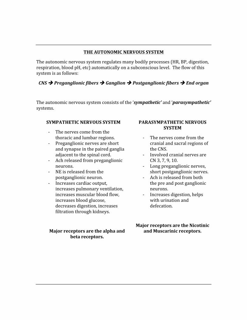
THE AUTONOMIC NERVOUS SYSTEM
The autonomic nervous system regulates many bodily processes (HR, BP, digestion, respiration, blood pH, etc) automatically on a subconscious level. The flow of this system is as follows:
CNS à Preganglionic fibers à Ganglion à Postganglionic fibers à End organ
The autonomic nervous system consists of the ‘sympathetic’ and ‘parasympathetic’ systems.
SYMPATHETIC NERVOUS SYSTEM
-‐ The nerves come from the thoracic and lumbar regions.
-‐ Preganglionic nerves are short and synapse in the paired ganglia adjacent to the spinal cord.
-‐ Ach released from preganglionic neurons.
-‐ NE is released from the postganglionic neuron.
-‐ Increases cardiac output, increases pulmonary ventilation, increases muscular blood flow, increases blood glucose, decreases digestion, increases filtration through kidneys.
Major receptors are the alpha and beta receptors.
PARASYMPATHETIC NERVOUS SYSTEM
-‐ The nerves come from the cranial and sacral regions of the CNS.
-‐ Involved cranial nerves are CN 3, 7, 9, 10.
-‐ Long preganglionic nerves, short postganglionic nerves.
-‐ Ach is released from both the pre and post ganglionic neurons.
-‐ Increases digestion, helps with urination and defecation.
Major receptors are the Nicotinic and Muscarinic receptors.
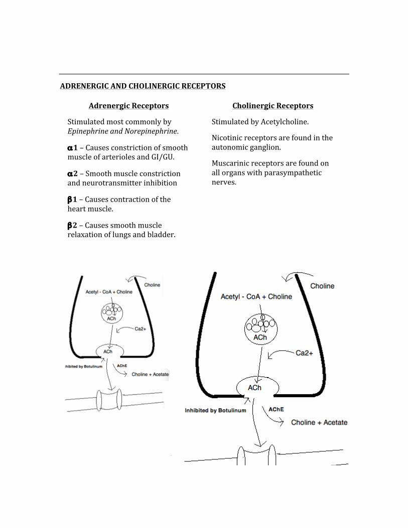
ADRENERGIC AND CHOLINERGIC RECEPTORS
Adrenergic Receptors
Stimulated most commonly by Epinephrine and Norepinephrine.
α1 – Causes constriction of smooth muscle of arterioles and GI/GU.
α2 – Smooth muscle constriction and neurotransmitter inhibition
β1 – Causes contraction of the heart muscle.
β2 – Causes smooth muscle relaxation of lungs and bladder.
Cholinergic Receptors
Stimulated by Acetylcholine.
Nicotinic receptors are found in the autonomic ganglion.
Muscarinic receptors are found on all organs with parasympathetic nerves.
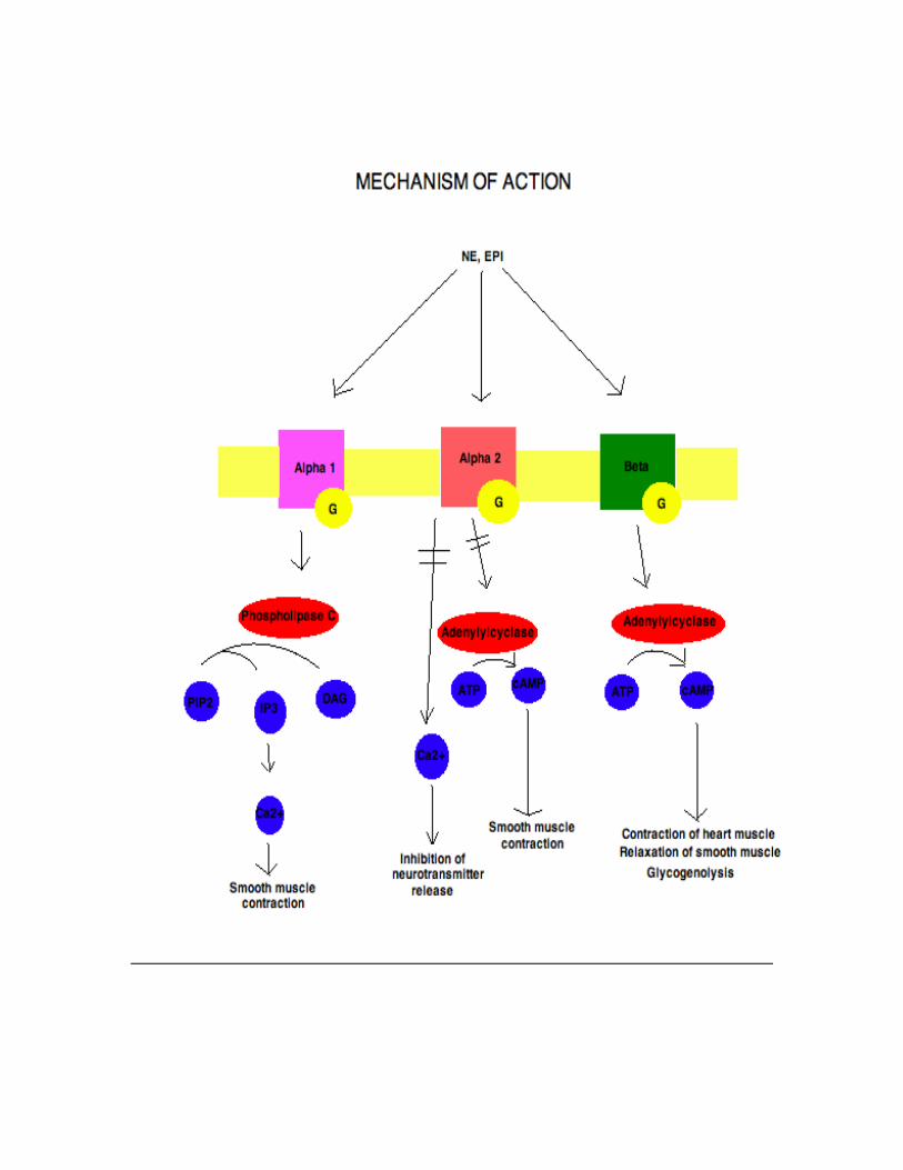
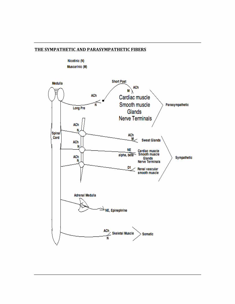
THE SYMPATHETIC AND PARASYMPATHETIC FIBERS
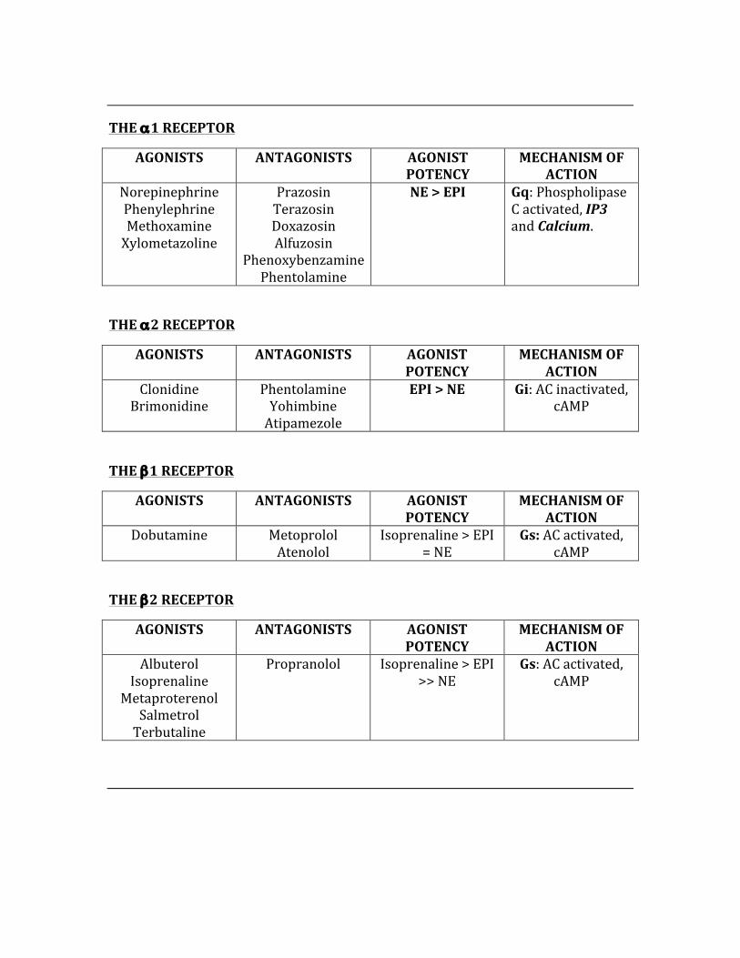
THE α1 RECEPTOR
AGONISTS ANTAGONISTS AGONIST POTENCY
MECHANISM OF ACTION
Norepinephrine Phenylephrine Methoxamine Xylometazoline
Prazosin Terazosin Doxazosin Alfuzosin
Phenoxybenzamine Phentolamine
NE > EPI Gq: Phospholipase C activated, IP3 and Calcium.
THE α2 RECEPTOR
AGONISTS ANTAGONISTS AGONIST POTENCY
MECHANISM OF ACTION
Clonidine Brimonidine
Phentolamine Yohimbine Atipamezole
EPI > NE Gi: AC inactivated, cAMP
THE β1 RECEPTOR
AGONISTS ANTAGONISTS AGONIST POTENCY
MECHANISM OF ACTION
Dobutamine Metoprolol Atenolol
Isoprenaline > EPI = NE
Gs: AC activated, cAMP
THE β2 RECEPTOR
AGONISTS ANTAGONISTS AGONIST POTENCY
MECHANISM OF ACTION
Albuterol Isoprenaline
Metaproterenol Salmetrol Terbutaline
Propranolol Isoprenaline > EPI >> NE
Gs: AC activated, cAMP
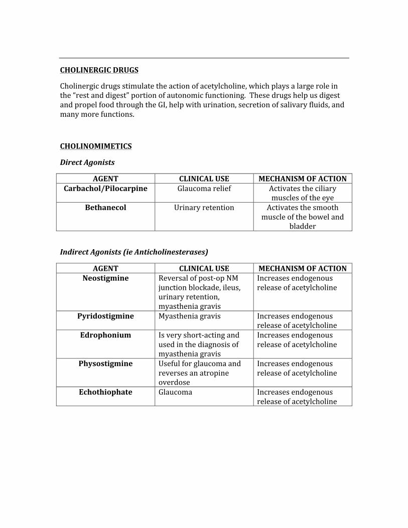
CHOLINERGIC DRUGS
Cholinergic drugs stimulate the action of acetylcholine, which plays a large role in the “rest and digest” portion of autonomic functioning. These drugs help us digest and propel food through the GI, help with urination, secretion of salivary fluids, and many more functions.
CHOLINOMIMETICS
Direct Agonists
AGENT CLINICAL USE MECHANISM OF ACTION Carbachol/Pilocarpine Glaucoma relief Activates the ciliary
muscles of the eye Bethanecol Urinary retention Activates the smooth
muscle of the bowel and bladder
Indirect Agonists (ie Anticholinesterases)
AGENT CLINICAL USE MECHANISM OF ACTION Neostigmine Reversal of post-‐op NM
junction blockade, ileus, urinary retention, myasthenia gravis
Increases endogenous release of acetylcholine
Pyridostigmine Myasthenia gravis Increases endogenous release of acetylcholine
Edrophonium Is very short-‐acting and used in the diagnosis of myasthenia gravis
Increases endogenous release of acetylcholine
Physostigmine Useful for glaucoma and reverses an atropine overdose
Increases endogenous release of acetylcholine
Echothiophate Glaucoma Increases endogenous release of acetylcholine
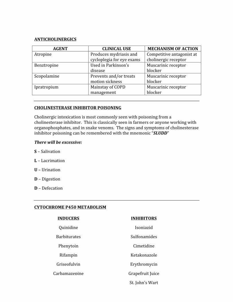
ANTICHOLINERGICS
AGENT CLINICAL USE MECHANISM OF ACTION Atropine Produces mydriasis and
cycloplegia for eye exams Competitive antagonist at cholinergic receptor
Benztropine Used in Parkinson’s disease
Muscarinic receptor blocker
Scopolamine Prevents and/or treats motion sickness
Muscarinic receptor blocker
Ipratropium Mainstay of COPD management
Muscarinic receptor blocker
CHOLINESTERASE INHIBITOR POISONING
Cholinergic intoxication is most commonly seen with poisoning from a cholinesterase inhibitor. This is classically seen in farmers or anyone working with organophosphates, and in snake venoms. The signs and symptoms of cholinesterase inhibitor poisoning can be remembered with the mnemonic “SLUDD”
There will be excessive:
S – Salivation
L – Lacrimation
U – Urination
D – Digestion
D – Defecation
CYTOCHROME P450 METABOLISM
INDUCERS
Quinidine
Barbiturates
Phenytoin
Rifampin
Griseofulvin
Carbamazepine
INHIBITORS
Isoniazid
Sulfonamides
Cimetidine
Ketakonazole
Erythromycin
Grapefruit Juice
St. John’s Wart
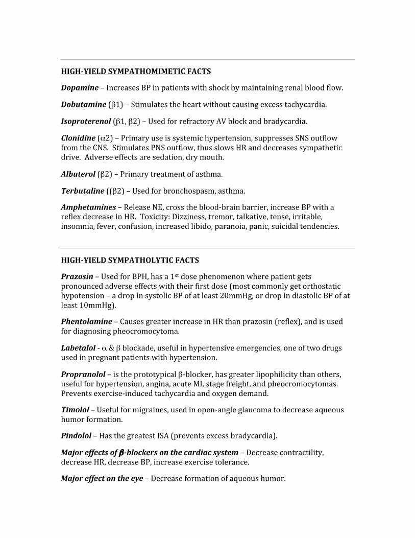
HIGH-‐YIELD SYMPATHOMIMETIC FACTS
Dopamine – Increases BP in patients with shock by maintaining renal blood flow.
Dobutamine (β1) – Stimulates the heart without causing excess tachycardia.
Isoproterenol (β1, β2) – Used for refractory AV block and bradycardia.
Clonidine (α2) – Primary use is systemic hypertension, suppresses SNS outflow from the CNS. Stimulates PNS outflow, thus slows HR and decreases sympathetic drive. Adverse effects are sedation, dry mouth.
Albuterol (β2) – Primary treatment of asthma.
Terbutaline ((β2) – Used for bronchospasm, asthma.
Amphetamines – Release NE, cross the blood-‐brain barrier, increase BP with a reflex decrease in HR. Toxicity: Dizziness, tremor, talkative, tense, irritable, insomnia, fever, confusion, increased libido, paranoia, panic, suicidal tendencies.
HIGH-‐YIELD SYMPATHOLYTIC FACTS
Prazosin – Used for BPH, has a 1st dose phenomenon where patient gets pronounced adverse effects with their first dose (most commonly get orthostatic hypotension – a drop in systolic BP of at least 20mmHg, or drop in diastolic BP of at least 10mmHg).
Phentolamine – Causes greater increase in HR than prazosin (reflex), and is used for diagnosing pheocromocytoma.
Labetalol -‐ α & β blockade, useful in hypertensive emergencies, one of two drugs used in pregnant patients with hypertension.
Propranolol – is the prototypical β-‐blocker, has greater lipophilicity than others, useful for hypertension, angina, acute MI, stage freight, and pheocromocytomas. Prevents exercise-‐induced tachycardia and oxygen demand.
Timolol – Useful for migraines, used in open-‐angle glaucoma to decrease aqueous humor formation.
Pindolol – Has the greatest ISA (prevents excess bradycardia).
Major effects of β-‐blockers on the cardiac system – Decrease contractility, decrease HR, decrease BP, increase exercise tolerance.
Major effect on the eye – Decrease formation of aqueous humor.
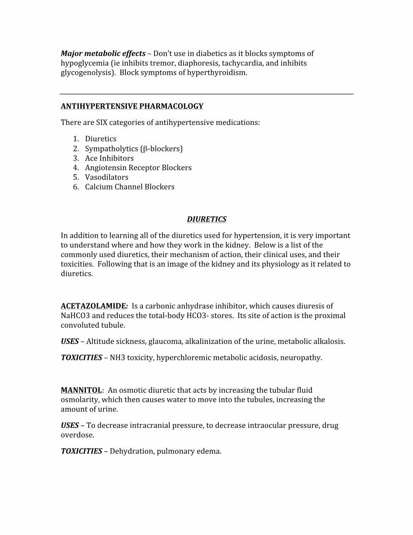
Major metabolic effects – Don’t use in diabetics as it blocks symptoms of hypoglycemia (ie inhibits tremor, diaphoresis, tachycardia, and inhibits glycogenolysis). Block symptoms of hyperthyroidism.
ANTIHYPERTENSIVE PHARMACOLOGY
There are SIX categories of antihypertensive medications:
1. Diuretics 2. Sympatholytics (β-‐blockers) 3. Ace Inhibitors 4. Angiotensin Receptor Blockers 5. Vasodilators 6. Calcium Channel Blockers
DIURETICS
In addition to learning all of the diuretics used for hypertension, it is very important to understand where and how they work in the kidney. Below is a list of the commonly used diuretics, their mechanism of action, their clinical uses, and their toxicities. Following that is an image of the kidney and its physiology as it related to diuretics.
ACETAZOLAMIDE: Is a carbonic anhydrase inhibitor, which causes diuresis of NaHCO3 and reduces the total-‐body HCO3-‐ stores. Its site of action is the proximal convoluted tubule.
USES – Altitude sickness, glaucoma, alkalinization of the urine, metabolic alkalosis.
TOXICITIES – NH3 toxicity, hyperchloremic metabolic acidosis, neuropathy.
MANNITOL: An osmotic diuretic that acts by increasing the tubular fluid osmolarity, which then causes water to move into the tubules, increasing the amount of urine.
USES – To decrease intracranial pressure, to decrease intraocular pressure, drug overdose.
TOXICITIES – Dehydration, pulmonary edema.
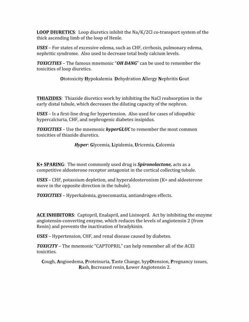
LOOP DIURETICS: Loop diuretics inhibit the Na/K/2Cl co-‐transport system of the thick ascending limb of the loop of Henle.
USES – For states of excessive edema, such as CHF, cirrhosis, pulmonary edema, nephritic syndrome. Also used to decrease total body calcium levels.
TOXICITIES – The famous mnemonic “OH DANG” can be used to remember the toxicities of loop diuretics.
Ototoxicity Hypokalemia Dehydration Allergy Nephritis Gout
THIAZIDES: Thiazide diuretics work by inhibiting the NaCl reabsorption in the early distal tubule, which decreases the diluting capacity of the nephron.
USES – Is a first-‐line drug for hypertension. Also used for cases of idiopathic hypercalciuria, CHF, and nephrogenic diabetes insipidus.
TOXICITIES – Use the mnemonic hyperGLUC to remember the most common toxicities of thiazide diuretics.
Hyper: Glycemia, Lipidemia, Uricemia, Calcemia
K+ SPARING: The most commonly used drug is Spironolactone, acts as a competitive aldosterone receptor antagonist in the cortical collecting tubule.
USES – CHF, potassium depletion, and hyperaldosteronism (K+ and aldosterone move in the opposite direction in the tubule).
TOXICITIES – Hyperkalemia, gynecomastia, antiandrogen effects.
ACE INHIBITORS: Captopril, Enalapril, and Lisinopril. Act by inhibiting the enzyme angiotensin-‐converting enzyme, which reduces the levels of angiotensin 2 (from Renin) and prevents the inactivation of bradykinin.
USES – Hypertension, CHF, and renal disease caused by diabetes.
TOXICITY – The mnemonic “CAPTOPRIL” can help remember all of the ACEI toxicities.
Cough, Angioedema, Proteinuria, Taste Change, hypOtension, Pregnancy issues, Rash, Increased renin, Lower Angiotensin 2.
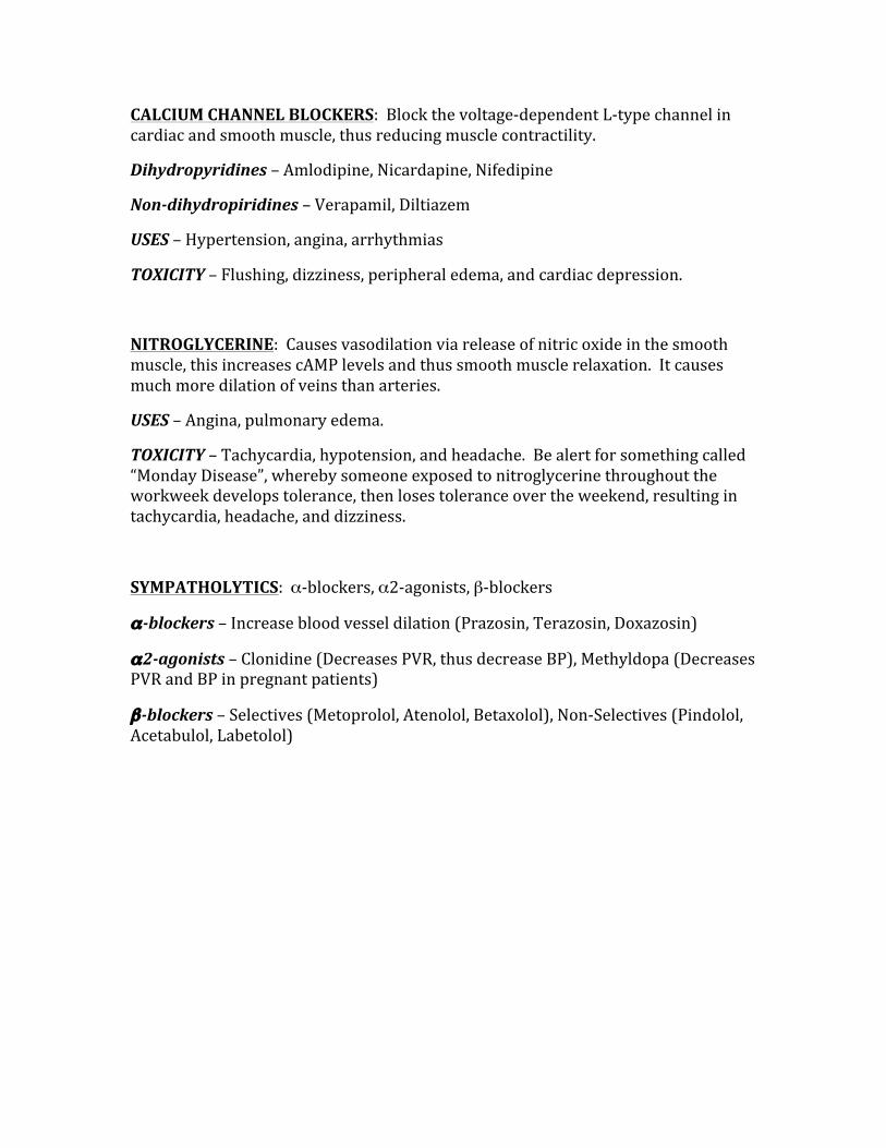
CALCIUM CHANNEL BLOCKERS: Block the voltage-‐dependent L-‐type channel in cardiac and smooth muscle, thus reducing muscle contractility.
Dihydropyridines – Amlodipine, Nicardapine, Nifedipine
Non-‐dihydropiridines – Verapamil, Diltiazem
USES – Hypertension, angina, arrhythmias
TOXICITY – Flushing, dizziness, peripheral edema, and cardiac depression.
NITROGLYCERINE: Causes vasodilation via release of nitric oxide in the smooth muscle, this increases cAMP levels and thus smooth muscle relaxation. It causes much more dilation of veins than arteries.
USES – Angina, pulmonary edema.
TOXICITY – Tachycardia, hypotension, and headache. Be alert for something called “Monday Disease”, whereby someone exposed to nitroglycerine throughout the workweek develops tolerance, then loses tolerance over the weekend, resulting in tachycardia, headache, and dizziness.
SYMPATHOLYTICS: α-‐blockers, α2-‐agonists, β-‐blockers
α-‐blockers – Increase blood vessel dilation (Prazosin, Terazosin, Doxazosin)
α2-‐agonists – Clonidine (Decreases PVR, thus decrease BP), Methyldopa (Decreases PVR and BP in pregnant patients)
β-‐blockers – Selectives (Metoprolol, Atenolol, Betaxolol), Non-‐Selectives (Pindolol, Acetabulol, Labetolol)
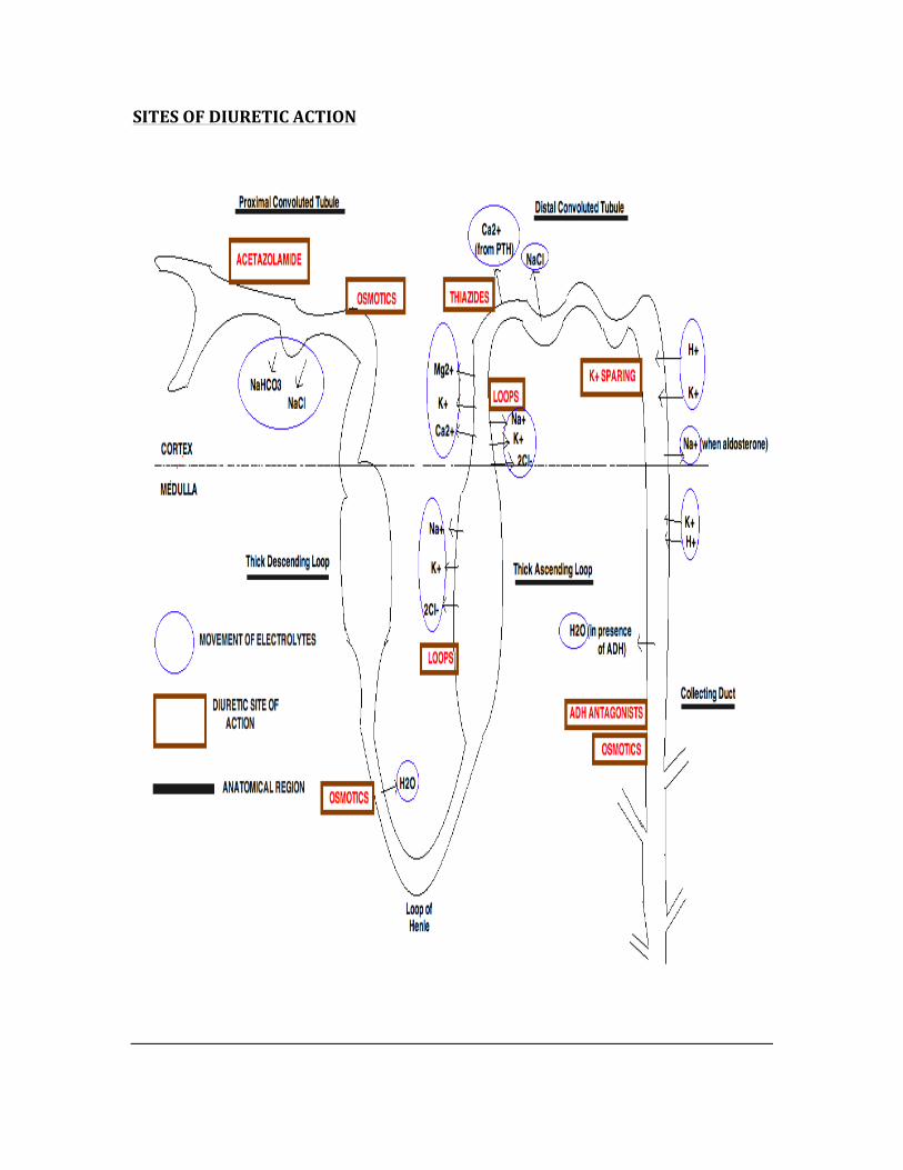
SITES OF DIURETIC ACTION
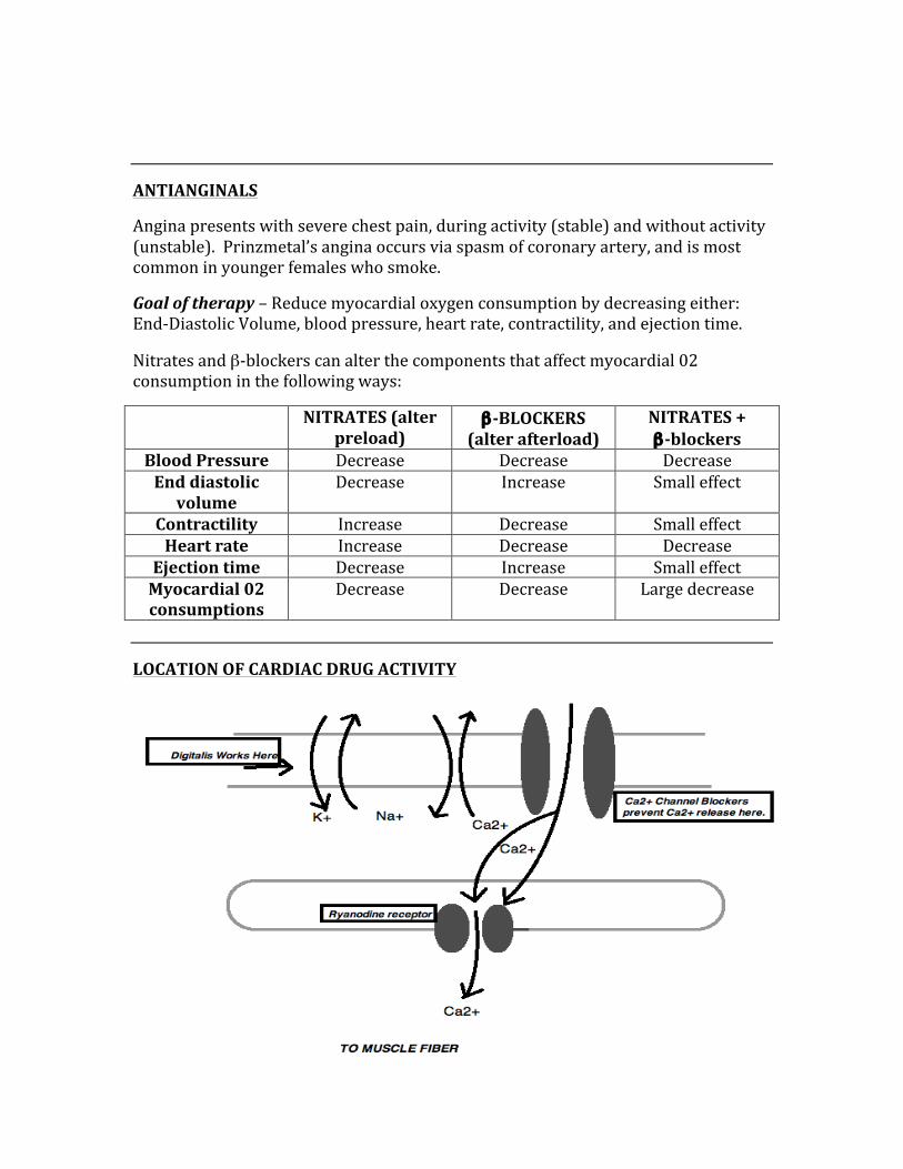
ANTIANGINALS
Angina presents with severe chest pain, during activity (stable) and without activity (unstable). Prinzmetal’s angina occurs via spasm of coronary artery, and is most common in younger females who smoke.
Goal of therapy – Reduce myocardial oxygen consumption by decreasing either: End-‐Diastolic Volume, blood pressure, heart rate, contractility, and ejection time.
Nitrates and β-‐blockers can alter the components that affect myocardial 02 consumption in the following ways:
NITRATES (alter preload)
β-‐BLOCKERS (alter afterload)
NITRATES + β-‐blockers
Blood Pressure Decrease Decrease Decrease End diastolic volume
Decrease Increase Small effect
Contractility Increase Decrease Small effect Heart rate Increase Decrease Decrease
Ejection time Decrease Increase Small effect Myocardial 02 consumptions
Decrease Decrease Large decrease
LOCATION OF CARDIAC DRUG ACTIVITY
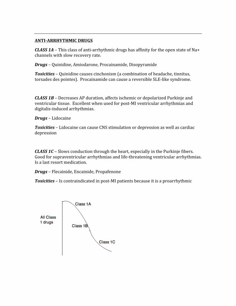
ANTI-‐ARRHYTHMIC DRUGS
CLASS 1A – This class of anti-‐arrhythmic drugs has affinity for the open state of Na+ channels with slow recovery rate.
Drugs – Quinidine, Amiodarone, Procainamide, Disopyramide
Toxicities – Quinidine causes cinchonism (a combination of headache, tinnitus, torsades des pointes). Procainamide can cause a reversible SLE-‐like syndrome.
CLASS 1B – Decreases AP duration, affects ischemic or depolarized Purkinje and ventricular tissue. Excellent when used for post-‐MI ventricular arrhythmias and digitalis-‐induced arrhythmias.
Drugs – Lidocaine
Toxicities – Lidocaine can cause CNS stimulation or depression as well as cardiac depression
CLASS 1C – Slows conduction through the heart, especially in the Purkinje fibers. Good for supraventricular arrhythmias and life-‐threatening ventricular arrhythmias. Is a last resort medication.
Drugs – Flecainide, Encainide, Propafenone
Toxicities – Is contraindicated in post-‐MI patients because it is a proarrhythmic
CLASS 2 (β-‐blockers) – Decrease cAMP, decrease Ca2+, decrease phase 4 slope, increase PR-‐interval.
Drugs – Metoprolol, Esmolol, Atenolol, Propranolol
Toxicities – Impotence, Asthma exacerbations, CNS effects, cardiovascular effects (bradycardia, AV block, CHF), hypoglycemia.
CLASS 3 (K+ channel blockers) – Increase AP duration, increase ERP, increase QT-‐interval. Amiodarone blocks the K+ rectifier channel that repolarizes the heart during phase 3.
Drugs – Sotolol, Amiodarone
Toxicities – Sotolol (Torsades des Pointes), Amiodarone (prolongs AP duration, elongates QRS, pulmonary/liver/thyroid toxicities, thus check PFT, LFT, TFT’s)
CLASS 4 (Ca2+ channel blockers) – Decrease conduction velocity in AV nodes, increases ERP, increases PR interval, prevents nodal arrhythmias.
Drugs – Verapamil, Diltiazem
Toxicities – Constipation, Flushing, Edema
OTHER – Adenosine
Causes hyperpolarization by blocking the Ca2+ influx and preventing K+ outward flow.
Is the drug of choice for diagnosing and abolishing AV nodal arrhythmias (is a substitute used between cardioversions, has a half-‐life of 10s.
CHF DRUGS
Stages of CHF:
Class 1 (Asymptomatic) – No limits on activity, only affects patient with normal exercise.
Class 2 (Symptoms with moderate exercise) – Slightly limits ordinary activity (fatigue, palpitations)
Class 3 (Symptoms with mild exercise) – No symptoms at rest, but occur with less than ordinary activities.
Class 4 (Symptomatic at rest) – Severe physical limitations, symptoms at rest (when sitting)
Symptoms of CHF:
-‐ Tachycardia -‐ Weakness -‐ Fatigue -‐ Orthopnea -‐ Peripheral edema -‐ Pulmonary congestion -‐ Hyperreninemia and hyperaldosterone -‐ Ventricular hypertrophy and remodeling -‐ Increased filling pressure/increased end-‐diastolic volume
Drugs used for CHF:
IONOTROPES – These drugs increase strength of contraction, thus increase stroke volume.
DIURETICS – Move fluids out of the body thus decreasing congestion.
VASODILATORS – Decrease venous pressure, congestion, and edema.
CHRONOTROPES – Increase the speed of heart contractions.
ACE INHIBITORS – Decrease mortality in heart failure patients, decreases ventricular remodeling.
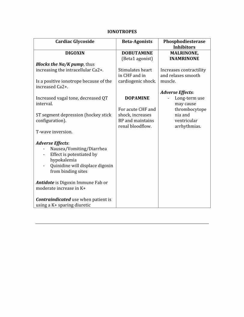
IONOTROPES
Cardiac Glycoside Beta-‐Agonists Phosphodiesterase Inhibitors
DIGOXIN Blocks the Na/K pump, thus increasing the intracellular Ca2+. Is a positive ionotrope because of the increased Ca2+. Increased vagal tone, decreased QT interval. ST segment depression (hockey stick configuration). T-‐wave inversion. Adverse Effects:
-‐ Nausea/Vomiting/Diarrhea -‐ Effect is potentiated by
hypokalemia -‐ Quinidine will displace digoxin
from binding sites Antidote is Digoxin Immune Fab or moderate increase in K+ Contraindicated use when patient is using a K+ sparing diuretic
DOBUTAMINE (Beta1 agonist) Stimulates heart in CHF and in cardiogenic shock.
DOPAMINE For acute CHF and shock, increases BP and maintains renal bloodflow.
MALRINONE, INAMRINONE
Increases contractility and relaxes smooth muscle. Adverse Effects:
-‐ Long-‐term use may cause thrombocytopenia and ventricular arrhythmias.
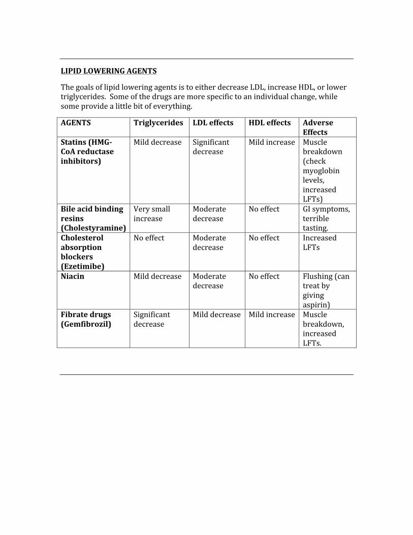
LIPID LOWERING AGENTS
The goals of lipid lowering agents is to either decrease LDL, increase HDL, or lower triglycerides. Some of the drugs are more specific to an individual change, while some provide a little bit of everything.
AGENTS Triglycerides LDL effects HDL effects Adverse Effects
Statins (HMG-‐CoA reductase inhibitors)
Mild decrease Significant decrease
Mild increase Muscle breakdown (check myoglobin levels, increased LFTs)
Bile acid binding resins (Cholestyramine)
Very small increase
Moderate decrease
No effect GI symptoms, terrible tasting.
Cholesterol absorption blockers (Ezetimibe)
No effect Moderate decrease
No effect Increased LFTs
Niacin Mild decrease Moderate decrease
No effect Flushing (can treat by giving aspirin)
Fibrate drugs (Gemfibrozil)
Significant decrease
Mild decrease Mild increase Muscle breakdown, increased LFTs.
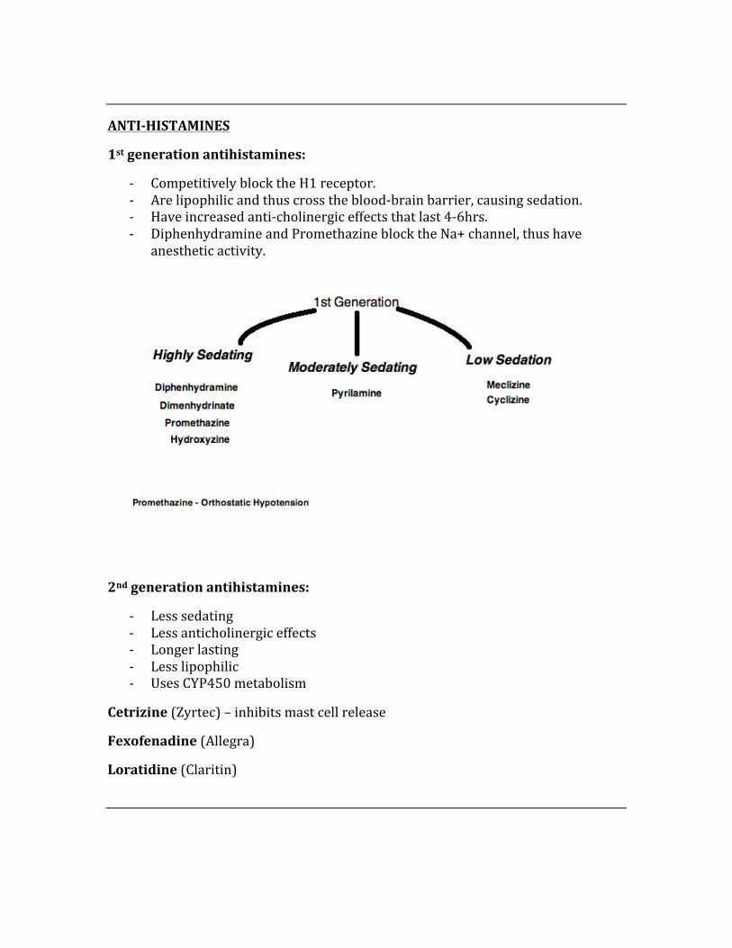
ANTI-‐HISTAMINES
1st generation antihistamines:
-‐ Competitively block the H1 receptor. -‐ Are lipophilic and thus cross the blood-‐brain barrier, causing sedation. -‐ Have increased anti-‐cholinergic effects that last 4-‐6hrs. -‐ Diphenhydramine and Promethazine block the Na+ channel, thus have
anesthetic activity.
2nd generation antihistamines:
-‐ Less sedating -‐ Less anticholinergic effects -‐ Longer lasting -‐ Less lipophilic -‐ Uses CYP450 metabolism
Cetrizine (Zyrtec) – inhibits mast cell release
Fexofenadine (Allegra)
Loratidine (Claritin)
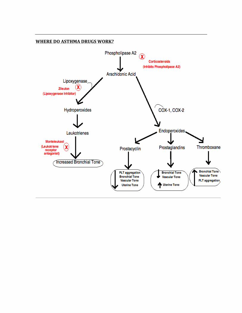
WHERE DO ASTHMA DRUGS WORK?
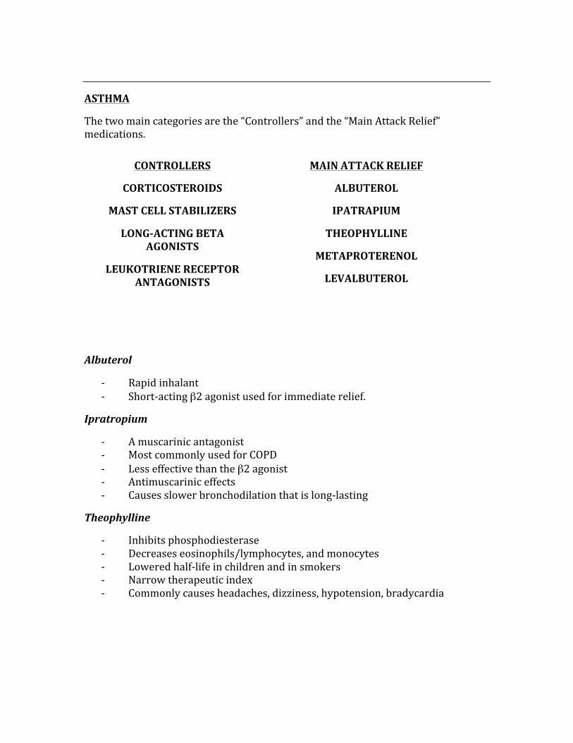
ASTHMA
The two main categories are the “Controllers” and the “Main Attack Relief” medications.
Albuterol
-‐ Rapid inhalant -‐ Short-‐acting β2 agonist used for immediate relief.
Ipratropium
-‐ A muscarinic antagonist -‐ Most commonly used for COPD -‐ Less effective than the β2 agonist -‐ Antimuscarinic effects -‐ Causes slower bronchodilation that is long-‐lasting
Theophylline
-‐ Inhibits phosphodiesterase -‐ Decreases eosinophils/lymphocytes, and monocytes -‐ Lowered half-‐life in children and in smokers -‐ Narrow therapeutic index -‐ Commonly causes headaches, dizziness, hypotension, bradycardia
CONTROLLERS
CORTICOSTEROIDS
MAST CELL STABILIZERS
LONG-‐ACTING BETA AGONISTS
LEUKOTRIENE RECEPTOR ANTAGONISTS
MAIN ATTACK RELIEF
ALBUTEROL
IPATRAPIUM
THEOPHYLLINE
METAPROTERENOL
LEVALBUTEROL
Corticosteroids
Inhaled – Beclomethasone, Triamclinolone, Budesonide, Fluticasone
Oral – Prednisone, Prednisolone
-‐ Inhibit phopholipase A2 -‐ Are the cornerstone of asthma management -‐ Decreases arachidonic acid through phospholipase A2, inhibiting the COX2
pathway. -‐ Oral corticosteroids can cause oral thrush -‐ Long-‐term use can cause osteoporosis, hypertension, diabetes, suppression
of the pituitary-‐adrenal axis, obesity, thinning of the skin, and muscle weakness.
Mast Cell Stabilizers
-‐ Cromolyn and Nedocromil -‐ Prevent mast cell degranulation -‐ Used as prophylaxis -‐ Are safer to use in kids
Long-‐Acting Beta Agonists
-‐ Salmetrol is the prototype -‐ Prevents nocturnal asthmatic effects
Leukotriene Receptor Antagonists
-‐ Monteleukast and Zafirlukast -‐ Antagonizes leukotrienes thus preventing an increase in bronchial tone
Treating Status Asthmaticus
The cornerstone of management is epinephrine or prednisone.
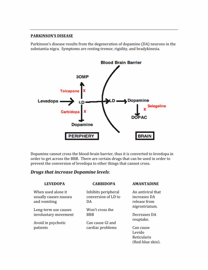
PARKINSON’S DISEASE
Parkinson’s disease results from the degeneration of dopamine (DA) neurons in the substantia nigra. Symptoms are resting tremor, rigidity, and bradykinesia.
Dopamine cannot cross the blood-‐brain barrier, thus it is converted to levedopa in order to get across the BBB. There are certain drugs that can be used in order to prevent the conversion of levedopa to other things that cannot cross.
Drugs that increase Dopamine levels:
LEVEDOPA
When used alone it usually causes nausea and vomiting
Long-‐term use causes involuntary movement
Avoid in psychotic patients
CARBIDOPA
Inhibits peripheral conversion of LD to DA
Won’t cross the BBB
Can cause GI and cardiac problems
AMANTADINE
An antiviral that increases DA release from nigrostriatum.
Decreases DA reuptake.
Can cause Levido Reticularis (Red-‐blue skin).
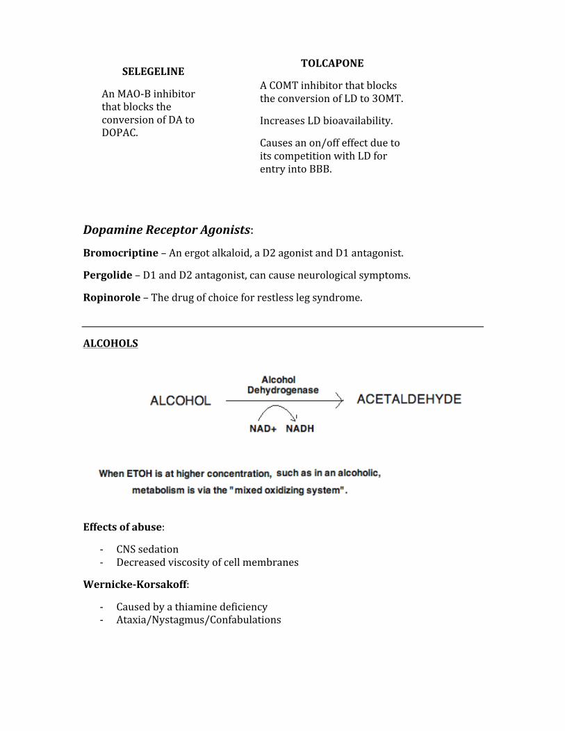
Dopamine Receptor Agonists:
Bromocriptine – An ergot alkaloid, a D2 agonist and D1 antagonist.
Pergolide – D1 and D2 antagonist, can cause neurological symptoms.
Ropinorole – The drug of choice for restless leg syndrome.
ALCOHOLS
Effects of abuse:
-‐ CNS sedation -‐ Decreased viscosity of cell membranes
Wernicke-‐Korsakoff:
-‐ Caused by a thiamine deficiency -‐ Ataxia/Nystagmus/Confabulations
SELEGELINE
An MAO-‐B inhibitor that blocks the conversion of DA to DOPAC.
TOLCAPONE
A COMT inhibitor that blocks the conversion of LD to 3OMT.
Increases LD bioavailability.
Causes an on/off effect due to its competition with LD for entry into BBB.
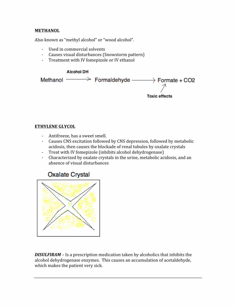
METHANOL
Also known as “methyl alcohol” or “wood alcohol”.
-‐ Used in commercial solvents -‐ Causes visual disturbances (Snowstorm pattern) -‐ Treatment with IV fomepizole or IV ethanol
ETHYLENE GLYCOL
-‐ Antifreeze, has a sweet smell. -‐ Causes CNS excitation followed by CNS depression, followed by metabolic
acidosis, then causes the blockade of renal tubules by oxalate crystals -‐ Treat with IV fomepizole (inhibits alcohol dehydrogenase) -‐ Characterized by oxalate crystals in the urine, metabolic acidosis, and an
absence of visual disturbances
DISULFIRAM – Is a prescription medication taken by alcoholics that inhibits the alcohol dehydrogenase enzymes. This causes an accumulation of acetaldehyde, which makes the patient very sick.
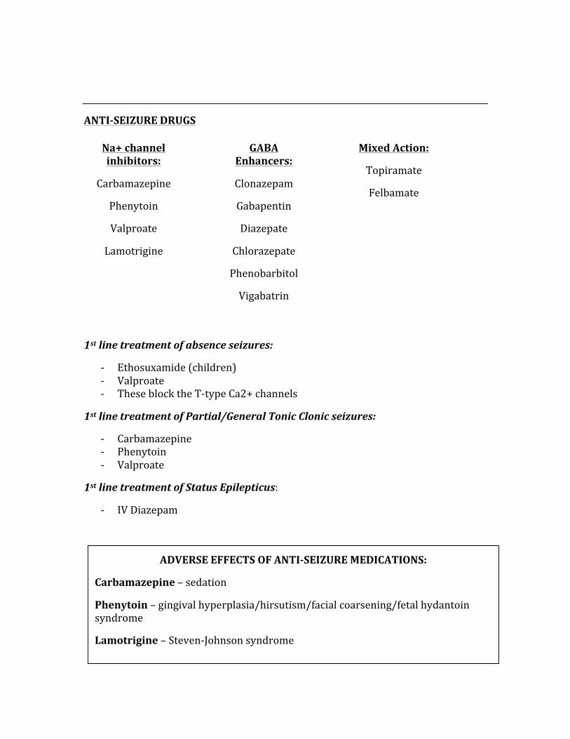
ANTI-‐SEIZURE DRUGS
1st line treatment of absence seizures:
-‐ Ethosuxamide (children) -‐ Valproate -‐ These block the T-‐type Ca2+ channels
1st line treatment of Partial/General Tonic Clonic seizures:
-‐ Carbamazepine -‐ Phenytoin -‐ Valproate
1st line treatment of Status Epilepticus:
-‐ IV Diazepam
Na+ channel inhibitors:
Carbamazepine
Phenytoin
Valproate
Lamotrigine
GABA Enhancers:
Clonazepam
Gabapentin
Diazepate
Chlorazepate
Phenobarbitol
Vigabatrin
Mixed Action:
Topiramate
Felbamate
ADVERSE EFFECTS OF ANTI-‐SEIZURE MEDICATIONS:
Carbamazepine – sedation
Phenytoin – gingival hyperplasia/hirsutism/facial coarsening/fetal hydantoin syndrome
Lamotrigine – Steven-‐Johnson syndrome
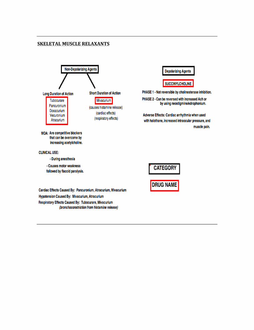
SKELETAL MUSCLE RELAXANTS
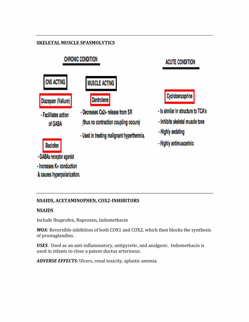
SKELETAL MUSCLE SPASMOLYTICS
NSAIDS, ACETAMINOPHEN, COX2-‐INHIBITORS
NSAIDS
Include Ibuprofen, Naproxen, Indomethacin
MOA: Reversible inhibition of both COX1 and COX2, which then blocks the synthesis of prostaglandins.
USES: Used as an anti-‐inflammatory, antipyretic, and analgesic. Indomethacin is used in infants to close a patent ductus arteriosus.
ADVERSE EFFECTS: Ulcers, renal toxicity, aplastic anemia.
ACETAMINOPHEN
Also known as Tylenol
MOA: Causes reversible inhibition of the COX pathway.
USES: Is used for its analgesic and antipyretic properties, but lacks the anti-‐inflammatory properties seen in NSAIDS.
ADVERSE EFFECTS: An overdose is deadly, because of hepatic necrosis due to toxic metabolites which deplete glutathione in the liver, causing the formation of toxic NAPQI in the liver.
COX-‐2 INHIBITORS
Include Celecoxib, Rofecoxib
MOA: Selective inhibition of COX-‐2
USES: Excellent for inflammation and pain, but helps to maintain gastric mucosa because it doesn’t use the COX-‐1. Main clinical uses are arthritis (osteo and RA).
ADVERSE EFFECTS: Renal toxicity, same as other NSAIDS with less risk of gastric ulcerations
ANESTHETICS
MAC (Minimum Alveolar Concentration) is a concept used in anesthesiology to compare the potency of anesthetic agents. Anesthetics with a higher MAC are cause faster induction but have much lower potency, while those with lower MAC value are slower to induce anesthesia but have a higher potency.
Example:
MAC of nitrous oxide is ‘104’, thus it acts fast and has weaker potency (has increased blood and lipid solubility).
MAC of halothane is ‘0.75’, thus is acts slower but has a stronger potency (has decreased blood and lipid solubility).
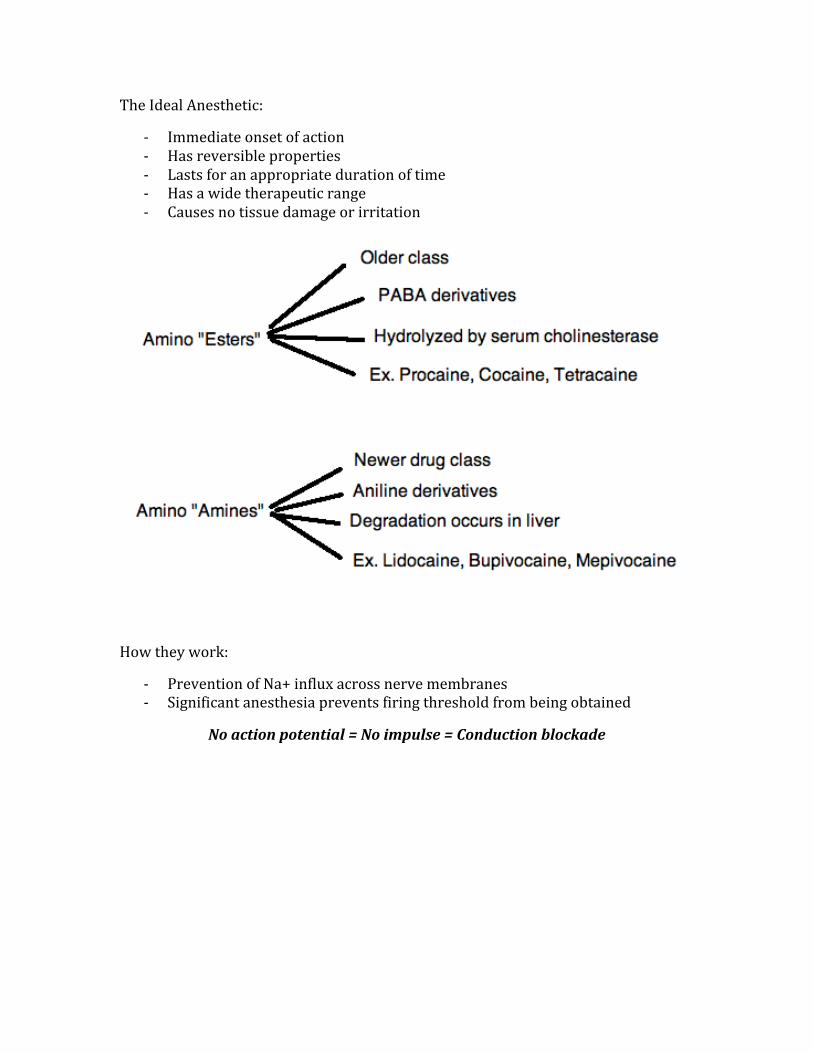
The Ideal Anesthetic:
-‐ Immediate onset of action -‐ Has reversible properties -‐ Lasts for an appropriate duration of time -‐ Has a wide therapeutic range -‐ Causes no tissue damage or irritation
How they work:
-‐ Prevention of Na+ influx across nerve membranes -‐ Significant anesthesia prevents firing threshold from being obtained
No action potential = No impulse = Conduction blockade

LOCAL ANESTHETICS
Blockage of Na+ channels, inactivation is by hydrolysis.
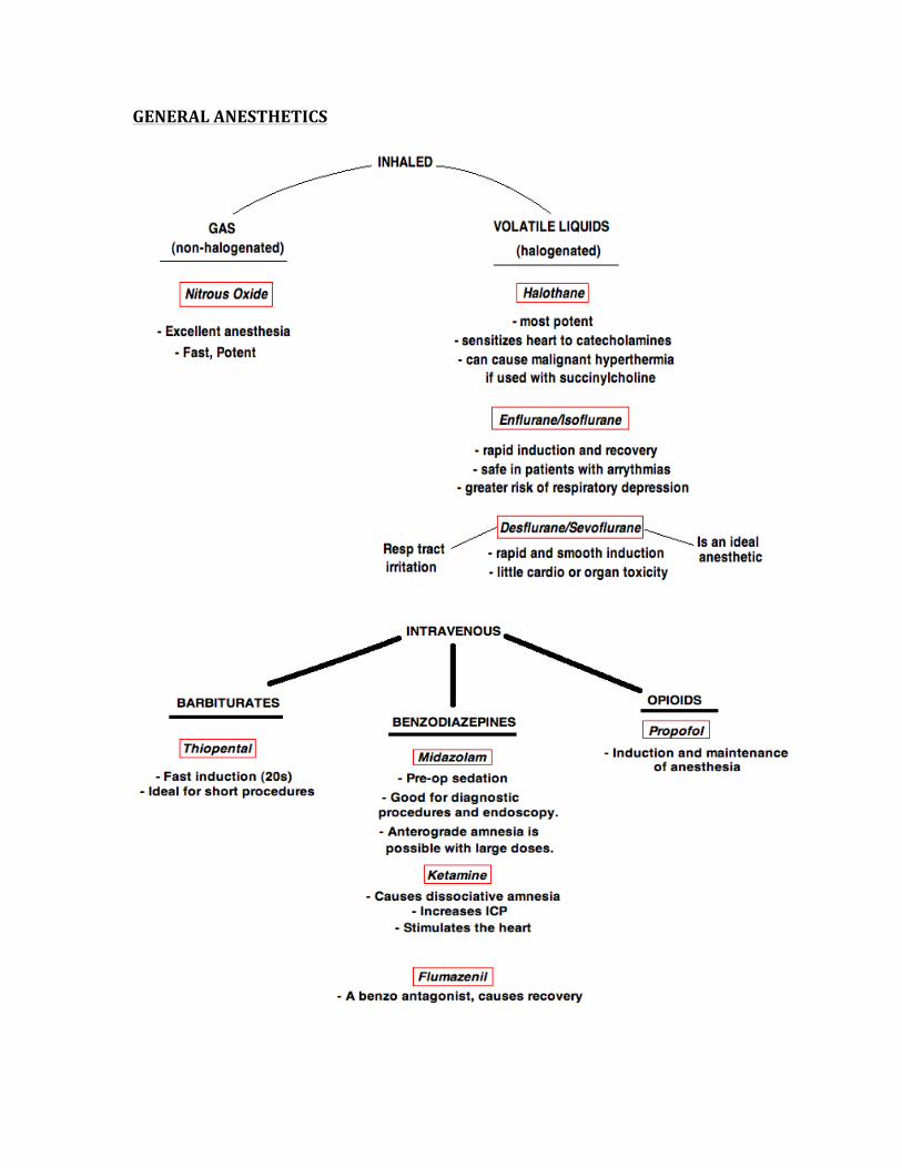
GENERAL ANESTHETICS
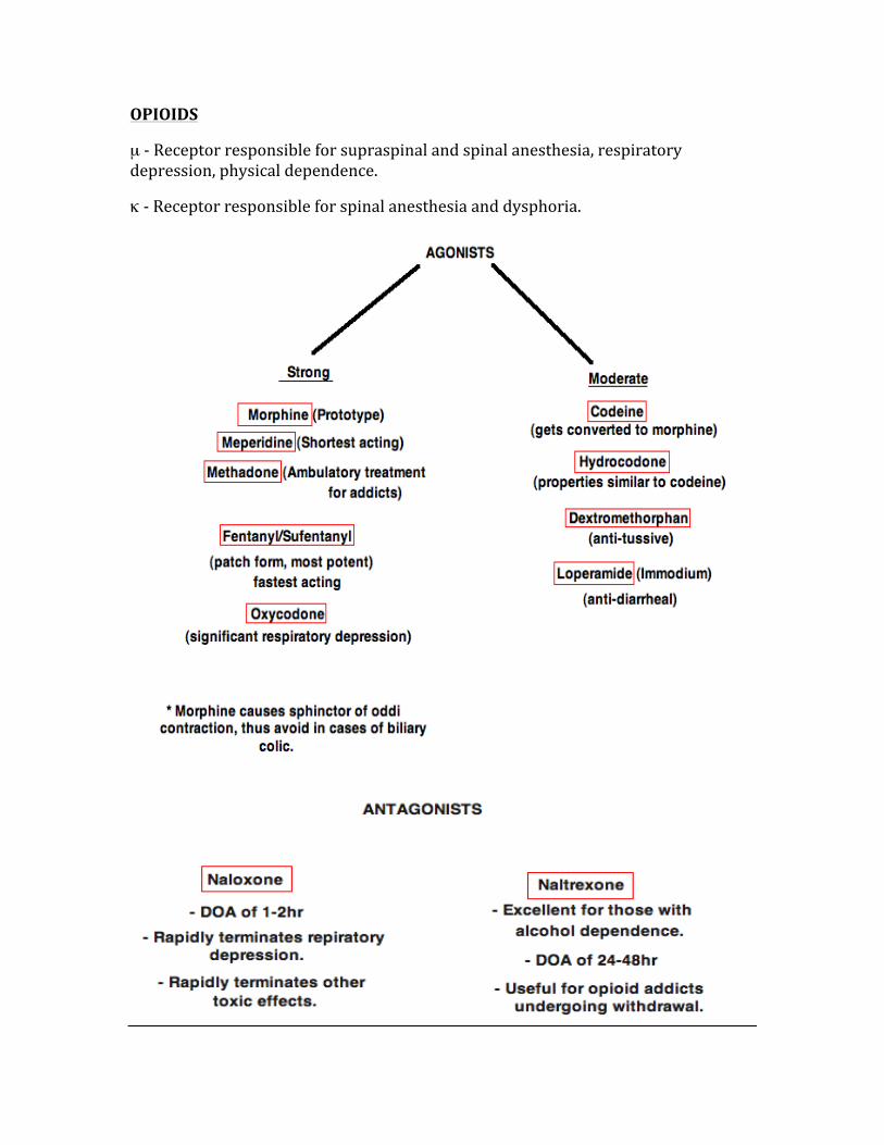
OPIOIDS
µ -‐ Receptor responsible for supraspinal and spinal anesthesia, respiratory depression, physical dependence.
κ -‐ Receptor responsible for spinal anesthesia and dysphoria.
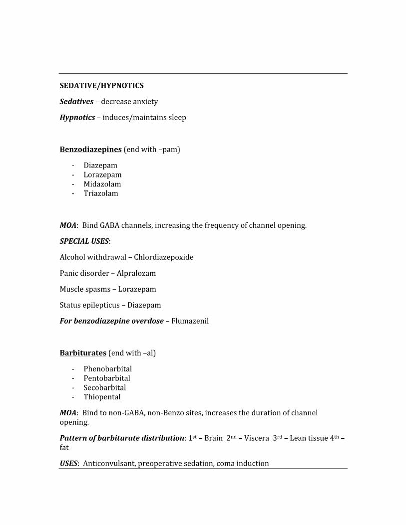
SEDATIVE/HYPNOTICS
Sedatives – decrease anxiety
Hypnotics – induces/maintains sleep
Benzodiazepines (end with –pam)
-‐ Diazepam -‐ Lorazepam -‐ Midazolam -‐ Triazolam
MOA: Bind GABA channels, increasing the frequency of channel opening.
SPECIAL USES:
Alcohol withdrawal – Chlordiazepoxide
Panic disorder – Alpralozam
Muscle spasms – Lorazepam
Status epilepticus – Diazepam
For benzodiazepine overdose – Flumazenil
Barbiturates (end with –al)
-‐ Phenobarbital -‐ Pentobarbital -‐ Secobarbital -‐ Thiopental
MOA: Bind to non-‐GABA, non-‐Benzo sites, increases the duration of channel opening.
Pattern of barbiturate distribution: 1st – Brain 2nd – Viscera 3rd – Lean tissue 4th – fat
USES: Anticonvulsant, preoperative sedation, coma induction
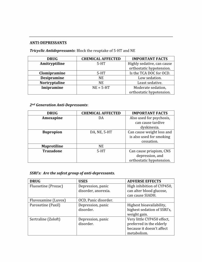
ANTI-‐DEPRESSANTS
Tricyclic Antidepressants: Block the reuptake of 5-‐HT and NE
DRUG CHEMICAL AFFECTED IMPORTANT FACTS Amitryptiline 5-‐HT Highly sedative, can cause
orthostatic hypotension. Clomipramine 5-‐HT Is the TCA DOC for OCD. Desipramine NE Low sedation. Nortryptaline NE Least sedative. Imipramine NE = 5-‐HT Moderate sedation,
orthostatic hypotension.
2nd Generation Anti-‐Depressants:
DRUG CHEMICAL AFFECTED IMPORTANT FACTS Amoxapine DA Also used for psychosis,
can cause tardive dyskinesia.
Bupropion DA, NE, 5-‐HT Can cause weight loss and is also used for smoking
cessation. Maprotiline NE Trazadone 5-‐HT Can cause priapism, CNS
depression, and orthostatic hypotension.
SSRI’s: Are the safest group of anti-‐depressants.
DRUG USES ADVERSE EFFECTS Fluoxetine (Prozac) Depression, panic
disorder, anorexia. High inhibition of CYP450, can alter blood glucose, can cause SIADH.
Fluvoxamine (Luvox) OCD, Panic disorder. Paroxetine (Paxil) Depression, panic
disorder. Highest bioavailability, highest sedation of SSRI’s, weight gain.
Sertraline (Zoloft) Depression, panic disorder.
Very little CYP450 effect, preferred in the elderly because it doesn’t affect metabolism.
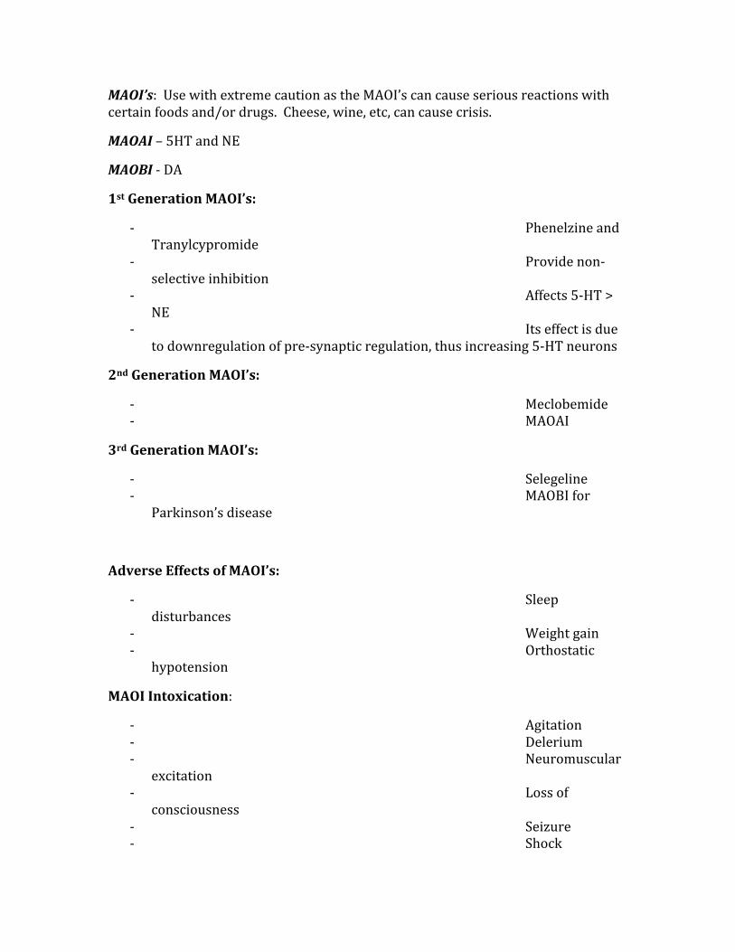
MAOI’s: Use with extreme caution as the MAOI’s can cause serious reactions with certain foods and/or drugs. Cheese, wine, etc, can cause crisis.
MAOAI – 5HT and NE
MAOBI -‐ DA
1st Generation MAOI’s:
-‐ Phenelzine and Tranylcypromide
-‐ Provide non-‐selective inhibition
-‐ Affects 5-‐HT > NE
-‐ Its effect is due to downregulation of pre-‐synaptic regulation, thus increasing 5-‐HT neurons
2nd Generation MAOI’s:
-‐ Meclobemide -‐ MAOAI
3rd Generation MAOI’s:
-‐ Selegeline -‐ MAOBI for
Parkinson’s disease
Adverse Effects of MAOI’s:
-‐ Sleep disturbances
-‐ Weight gain -‐ Orthostatic
hypotension
MAOI Intoxication:
-‐ Agitation -‐ Delerium -‐ Neuromuscular
excitation -‐ Loss of
consciousness -‐ Seizure -‐ Shock
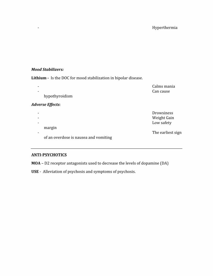
-‐ Hyperthermia
Mood Stabilizers:
Lithium -‐ Is the DOC for mood stabilization in bipolar disease.
-‐ Calms mania -‐ Can cause
hypothyroidism
Adverse Effects:
-‐ Drowsiness -‐ Weight Gain -‐ Low safety
margin -‐ The earliest sign
of an overdose is nausea and vomiting
ANTI-‐PSYCHOTICS
MOA – D2 receptor antagonists used to decrease the levels of dopamine (DA)
USE -‐ Alleviation of psychosis and symptoms of psychosis.
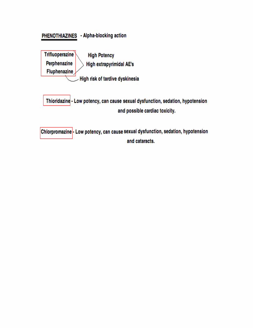
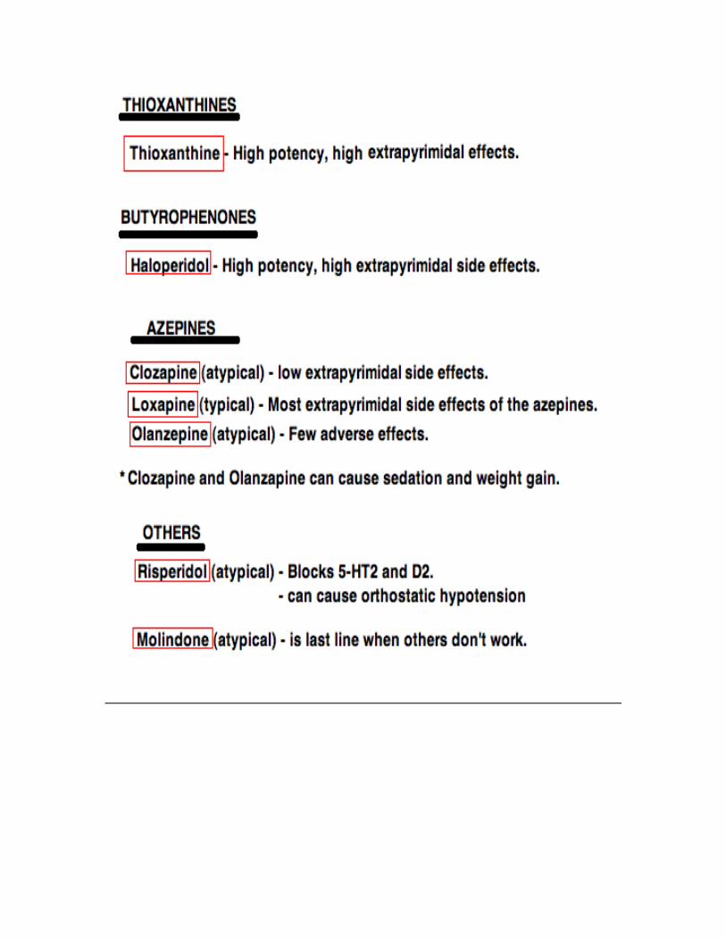
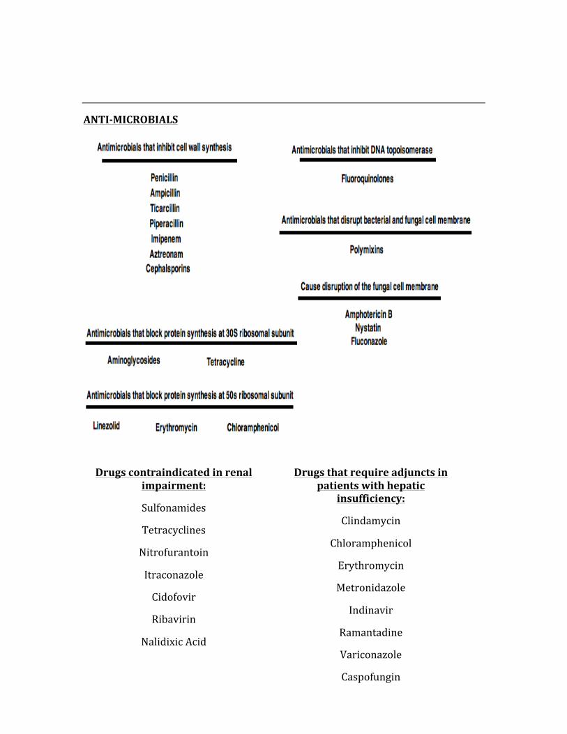
ANTI-‐MICROBIALS
Drugs contraindicated in renal impairment:
Sulfonamides
Tetracyclines
Nitrofurantoin
Itraconazole
Cidofovir
Ribavirin
Nalidixic Acid
Drugs that require adjuncts in patients with hepatic
insufficiency:
Clindamycin
Chloramphenicol
Erythromycin
Metronidazole
Indinavir
Ramantadine
Variconazole
Caspofungin
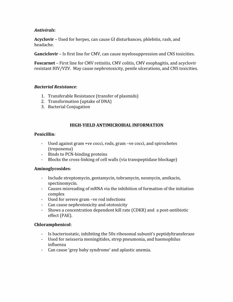
Antivirals:
Acyclovir – Used for herpes, can cause GI disturbances, phlebitis, rash, and headache.
Ganciclovir – Is first line for CMV, can cause myelosuppression and CNS toxicities.
Foscarnet – First line for CMV retinitis, CMV colitis, CMV esophagitis, and acyclovir resistant HIV/VZV. May cause nephrotoxicity, penile ulcerations, and CNS toxicities.
Bacterial Resistance:
1. Transferable Resistance (transfer of plasmids) 2. Transformation (uptake of DNA) 3. Bacterial Conjugation
HIGH-‐YIELD ANTIMICROBIAL INFORMATION
Penicillin:
-‐ Used against gram +ve cocci, rods, gram –ve cocci, and spirochetes (treponema)
-‐ Binds to PCN-‐binding proteins -‐ Blocks the cross-‐linking of cell walls (via transpeptidase blockage)
Aminoglycosides:
-‐ Include streptomycin, gentamycin, tobramycin, neomycin, amikacin, spectinomycin.
-‐ Causes misreading of mRNA via the inhibition of formation of the initiation complex
-‐ Used for severe gram –ve rod infections -‐ Can cause nephrotoxicity and ototoxicity -‐ Shows a concentration dependent kill rate (CDKR) and a post-‐antibiotic
effect (PAE).
Chloramphenicol:
-‐ Is bacteriostatic, inhibiting the 50s ribosomal subunit’s peptidyltransferase -‐ Used for neisseria meningitides, strep pneumonia, and haemophilus
influenza -‐ Can cause ‘grey baby syndrome’ and aplastic anemia.
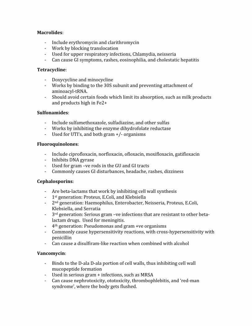
Macrolides:
-‐ Include erythromycin and clarithromycin -‐ Work by blocking translocation -‐ Used for upper respiratory infections, Chlamydia, neisseria -‐ Can cause GI symptoms, rashes, eosinophilia, and cholestatic hepatitis
Tetracycline:
-‐ Doxycycline and minocycline -‐ Works by binding to the 30S subunit and preventing attachment of
aminoacyl-‐tRNA. -‐ Should avoid certain foods which limit its absorption, such as milk products
and products high in Fe2+
Sulfonamides:
-‐ Include sulfamethoxazole, sulfadiazine, and other sulfas -‐ Works by inhibiting the enzyme dihydrofolate reductase -‐ Used for UTI’s, and both gram +/-‐ organisms
Fluoroquinolones:
-‐ Include ciprofloxacin, norfloxacin, ofloxacin, moxifloxacin, gatifloxacin -‐ Inhibits DNA gyrase -‐ Used for gram –ve rods in the GU and GI tracts -‐ Commonly causes GI disturbances, headache, rashes, dizziness
Cephalosporins:
-‐ Are beta-‐lactams that work by inhibiting cell wall synthesis -‐ 1st generation: Proteus, E.Coli, and Klebsiella -‐ 2nd generation: Haemophilus, Enterobacter, Neisseria, Proteus, E.Coli,
Klebsiella, and Serratia -‐ 3rd generation: Serious gram –ve infections that are resistant to other beta-‐
lactam drugs. Used for meningitis. -‐ 4th generation: Pseudomonas and gram +ve organisms -‐ Commonly cause hypersensitivity reactions, with cross-‐hypersensitivity with
penicillin -‐ Can cause a disulfiram-‐like reaction when combined with alcohol
Vancomycin:
-‐ Binds to the D-‐ala D-‐ala portion of cell walls, thus inhibiting cell wall mucopeptide formation
-‐ Used in serious gram + infections, such as MRSA -‐ Can cause nephrotoxicity, ototoxicity, thrombophlebitis, and ‘red-‐man
syndrome’, where the body gets flushed.
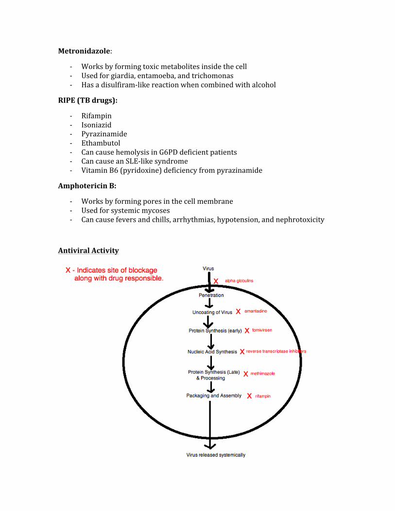
Metronidazole:
-‐ Works by forming toxic metabolites inside the cell -‐ Used for giardia, entamoeba, and trichomonas -‐ Has a disulfiram-‐like reaction when combined with alcohol
RIPE (TB drugs):
-‐ Rifampin -‐ Isoniazid -‐ Pyrazinamide -‐ Ethambutol -‐ Can cause hemolysis in G6PD deficient patients -‐ Can cause an SLE-‐like syndrome -‐ Vitamin B6 (pyridoxine) deficiency from pyrazinamide
Amphotericin B:
-‐ Works by forming pores in the cell membrane -‐ Used for systemic mycoses -‐ Can cause fevers and chills, arrhythmias, hypotension, and nephrotoxicity
Antiviral Activity
CHEMOTHERAPEUTICS
Drugs classifications are based on their MOA’s:
Folate antagonists (Methotrexate)
Purine antagonists (6-‐mercaptopurine)
Pyrimidine antagonists (5-‐fluorouracil)
Ribonucleic reductase inhibitors (Hydroxyurea)
Drug Resistance:
Innate – Primary resistance develops due to exposure.
Acquired – Caused by genomic mutations that may be to a single drug or to multiple drugs.
MOA of tumor cell resistance:
-‐ Decreased drug accumulation -‐ Altered affinity of target enzymes -‐ Loss of drug-‐activating enzymes -‐ Increased function of tumor cell repair mechanisms
High-‐yield chemotherapeutic toxicities:
Common chemotherapy drug toxicities – myelosuppression, nausea/vomiting, leucopenia.
Neurotoxicity – Vincristine, Paclitaxel
Pulmonary Toxicity – Bleomycin, Busulfan
Renal Toxicity – Cisplatin
Hemorrhagic Cystitis – Cyclophosphamide
Cardiac Toxicity – Doxorubicin

Drug-‐specific toxicities:
Carmustine – produces leukocyte suppression
Cisplatin, Carmustine – most emetic anti-‐neoplastics
5HT3 antagonists – prevent emesis
Metochlopromide – useful in preventing chemotherapy-‐related nausea and vomiting.
DIABETES PHARMACOLOGY
INSULIN TYPE ONSET OF ACTION
PEAK OF DRUG
DURATION OF ACTION
TYPICAL USE
Ultrashort Acting(Lispro)
5 minutes 1hr 3hr Before meals
Short-‐Acting (Regular)
30 minutes 3-‐4hr 6-‐8hr Morning and night
Intermediate Acting (Lente)
30 minutes 12hr 24hr qid
Long-‐Acting (Ultralente)
2hr 12hr 24hr qid
• There is a risk of hypoglycemic crisis if proper regulation of insulin not used
Sulfonylureas:
Glyburide, Tolbutamide
MOA: Causes depolarization of beta cells of the pancreas, thus increasing the release of insulin.
GASTROINTESTINAL PHARMACOLOGY
H2 antagonists: Cimetidine, Ranitidine.
-‐ Lowers acid secretion -‐ Not used as 1st line drug for GERD, PUD, etc.
Proton Pump Inhibitors: Omeprazole
-‐ Is diagnostic and therapeutic drug of choice for GERD and PUD -‐ Also used in cases of Zollinger-‐Ellison syndrome
Mucosal Protectants: Sucralfate
-‐ Works by adhering to proteinaceous lesions on the surface -‐ Is as effective as an H2 receptor antagonist would be in 4-‐8 weeks
Prostaglandins: Misoprostal
-‐ Can be given when patient is using high-‐dose NSAID therapy -‐ Blocks cAMP -‐ Also caused abortion in pregnant women -‐ Can cause watery diarrhea
Anti-‐emetics: 5HT3 receptor antagonists
-‐ Ondasetron
Prokinetics: Alosetron
-‐ Can be used for treatment of IBS, but is last line after conservative therapies fail
Irritable Bowel Disease:
-‐ 1st line treatment of ulcerative colitis is Sulfasalazine -‐ 1st line treatment of Crohn’s disease is Budesonide
CALCIUM AND BONE HOMEOSTASIS
PTH – for bone resorption, and is stimulated when serum calcium decreases.
Calcitonin – performs all opposite action of PTH (parathyroid hormone).
1,25 – dihydroxy vitamin D3 – produced in the kidney
Osteoporosis – 1st line pharmacological treatment is alendronate (bisphosphonate)
Paget’s Disease – Treatment involves bisphosphonate medication such as alendronate.
THYROID PHARMACOLOGY
Levothyroxine (T4) – is the drug of choice for all types of hypothyroidism.
Liothyronine (T3) – more potent than levothyroxine, has a shorter half-‐life, is not used as a therapeutic agent in thyroid problems.
Methimazole and Propylthiouracil – two drugs used for hyperthyroidism.
Propylthiouracil (PTU) – Inhibits the peripheral conversion of T4àT3
Grave’s Disease – Methimazole or PTU is used to induce remission or to control symptoms prior to surgery or radioiodine ablation.
Iodide Salts – inhibit the release of thyroid hormone from the thyroid gland.
Potassium Iodide solution – is used to control the symptoms of acute thyrotoxicosis, decreases the vascularity and size of the thyroid, and inhibits thyroid hormone release following RAI treatment.
RAI (I-‐131) – is used in the treatment of Grave’s disease, works by emitting chemicals that kill tissue of the thyroid.
PHARMACOLOGY OF PITUITARY AND HYPOTHALAMUS
Cosyntropin – is a synthetic corticosyntropin analog, is used to diagnose adrenal insufficiency.
Octreotide – is a synthetic somatostatin that inhibits GH secretion, is used to treat acromegaly.
Menotropins – induces ovulation in infertile women.
Gonadorelin – is a GnRH that is administered in a pulsatile form, this induces ovulation in women with amenorrhea due to hypothalamic dysfunction.
Leuprolide – used in kids with precocious puberty, it acts by suppressing gonadotropin secretion from the pituitary.
Bromocriptine – is given to those with prolactinomas, used to shrink then mass so the symptoms will cease. Is a DA antagonist.
Oxytocin – used to induce and/or augment labor in women who have trouble with delivery, also stimulates milk letdown in nursing women.
Desmopressin – is a synthetic analog of vasopressin, treatment for diabetes insipidus.
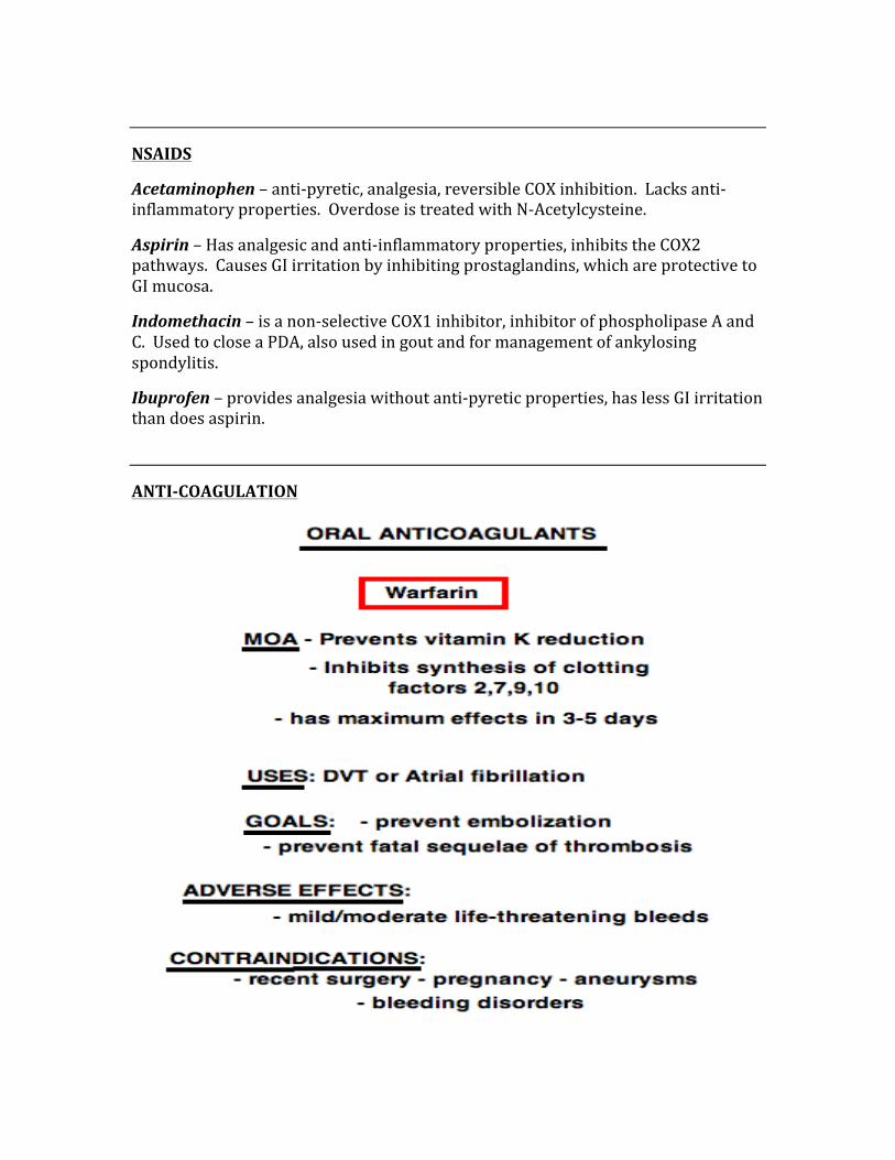
NSAIDS
Acetaminophen – anti-‐pyretic, analgesia, reversible COX inhibition. Lacks anti-‐inflammatory properties. Overdose is treated with N-‐Acetylcysteine.
Aspirin – Has analgesic and anti-‐inflammatory properties, inhibits the COX2 pathways. Causes GI irritation by inhibiting prostaglandins, which are protective to GI mucosa.
Indomethacin – is a non-‐selective COX1 inhibitor, inhibitor of phospholipase A and C. Used to close a PDA, also used in gout and for management of ankylosing spondylitis.
Ibuprofen – provides analgesia without anti-‐pyretic properties, has less GI irritation than does aspirin.
ANTI-‐COAGULATION
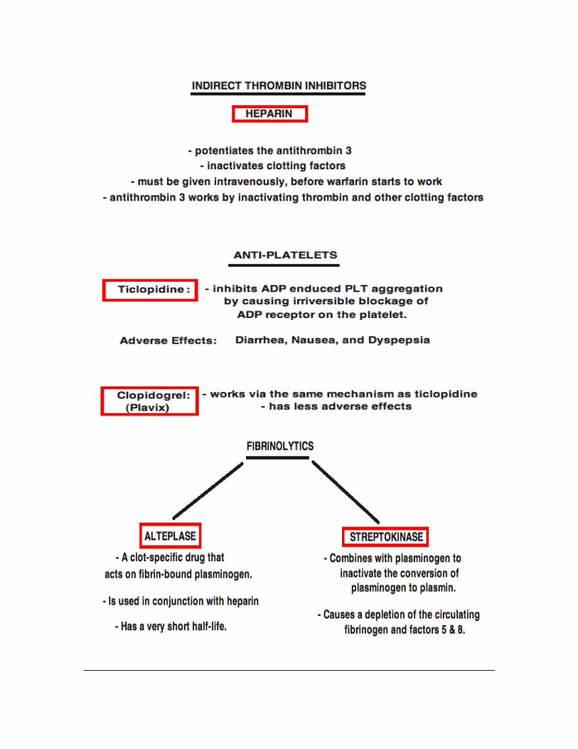
REPRODUCTIVE PHARMACOLOGY
There are a few very common medications used to induce ovulation and thus increase the chances of pregnancy.
CLOMIPHENE: The agent of choice for ovulation induction, it acts by increasing GnRH secretion from the hypothalamus, which then increases the levels of FSH, increasing the fertility. This all happens via the blocking of the estrogen receptors, thus tricking the body into believing that the levels of estrogen are much lower than they actually are.
HUMAN MENOPAUSAL GONADOTROPIN (Menotropin): Is extracted from the urine of menopausal women. It is high in LH and FSH, which increases fertility.
CHAPTER 13
PATHOLOGY
Due to sheer volume, the pathology section of the Step 1 exam is by far the most challenging. The USMLE exam is
moving more and more towards being a clinical exam, thus it is extremely important to recognize the signs, symptoms,
common presentations, uncommon presentations, and pathophysiology of pathological conditions.
Special attention should be paid to disease entities that are more common in the United States, as opposed to those that
are more prevalent in other areas of the world.
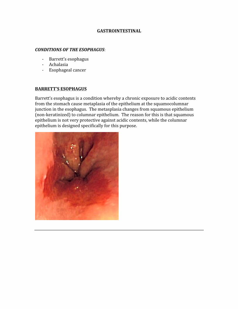
GASTROINTESTINAL
CONDITIONS OF THE ESOPHAGUS:
-‐ Barrett’s esophagus -‐ Achalasia -‐ Esophageal cancer
BARRETT’S ESOPHAGUS
Barrett’s esophagus is a condition whereby a chronic exposure to acidic contents from the stomach cause metaplasia of the epithelium at the squamocolumnar junction in the esophagus. The metasplasia changes from squamous epithelium (non-‐keratinized) to columnar epithelium. The reason for this is that squamous epithelium is not very protective against acidic contents, while the columnar epithelium is designed specifically for this purpose.
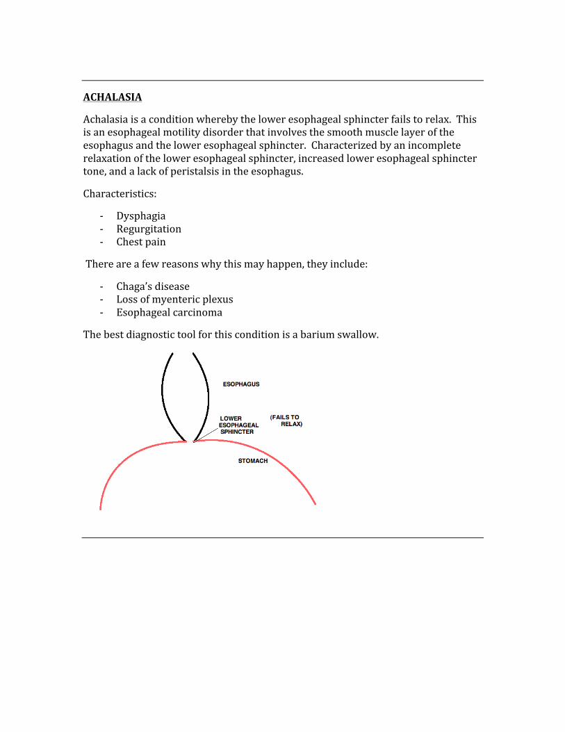
ACHALASIA
Achalasia is a condition whereby the lower esophageal sphincter fails to relax. This is an esophageal motility disorder that involves the smooth muscle layer of the esophagus and the lower esophageal sphincter. Characterized by an incomplete relaxation of the lower esophageal sphincter, increased lower esophageal sphincter tone, and a lack of peristalsis in the esophagus.
Characteristics:
-‐ Dysphagia -‐ Regurgitation -‐ Chest pain
There are a few reasons why this may happen, they include:
-‐ Chaga’s disease -‐ Loss of myenteric plexus -‐ Esophageal carcinoma
The best diagnostic tool for this condition is a barium swallow.
ESOPHAGEAL CANCER
The two types of esophageal cancer are: Adenocarcinoma and Squamous Cell carcinoma.
Adenocarcinoma – this type of cancer is often secondary to Barrett’s esophagus.
Squamous Cell Carcinoma – this type of cancer is often caused by exposure to alcohol and cigarette smoke (on a chronic basis).
The popular mnemonic “ABCDEF” is excellent for remembering the common causes of esophageal cancer.
A – Alcohol
B – Barrett’s esophagus
C – Cigarette smoke
D – Diverticula (especially Zenker’s)
E – Esophageal Webs
F – Family history of esophageal cancer
GASTRITIS
Gastritis is caused by an inflammation of the stomach lining. The most common cause of gastritis is prolonged use of NSAIDs (ie Aspirin), which blocks the synthesis of prostaglandins, thus decreasing the protection of the stomach lining. Other common causes are alcohol consumption and H. Pylori.
Symptoms include:
-‐ Pain in the epigastric region (most common presentation) -‐ Weight loss -‐ Loss of appetite
Acute Gastritis – also known as erosive gastritis, this form of gastritis is caused most commonly by damages to the stomach’s mucosal defense system. NSAIDs and alcohol are most common causes of acute gastritis.
Chronic Gastritis – this is the result of an H. Pylori infection.
Types of chronic gastritis:
Type A -‐ Occurs in the fundus of the stomach, is of autoimmune nature.
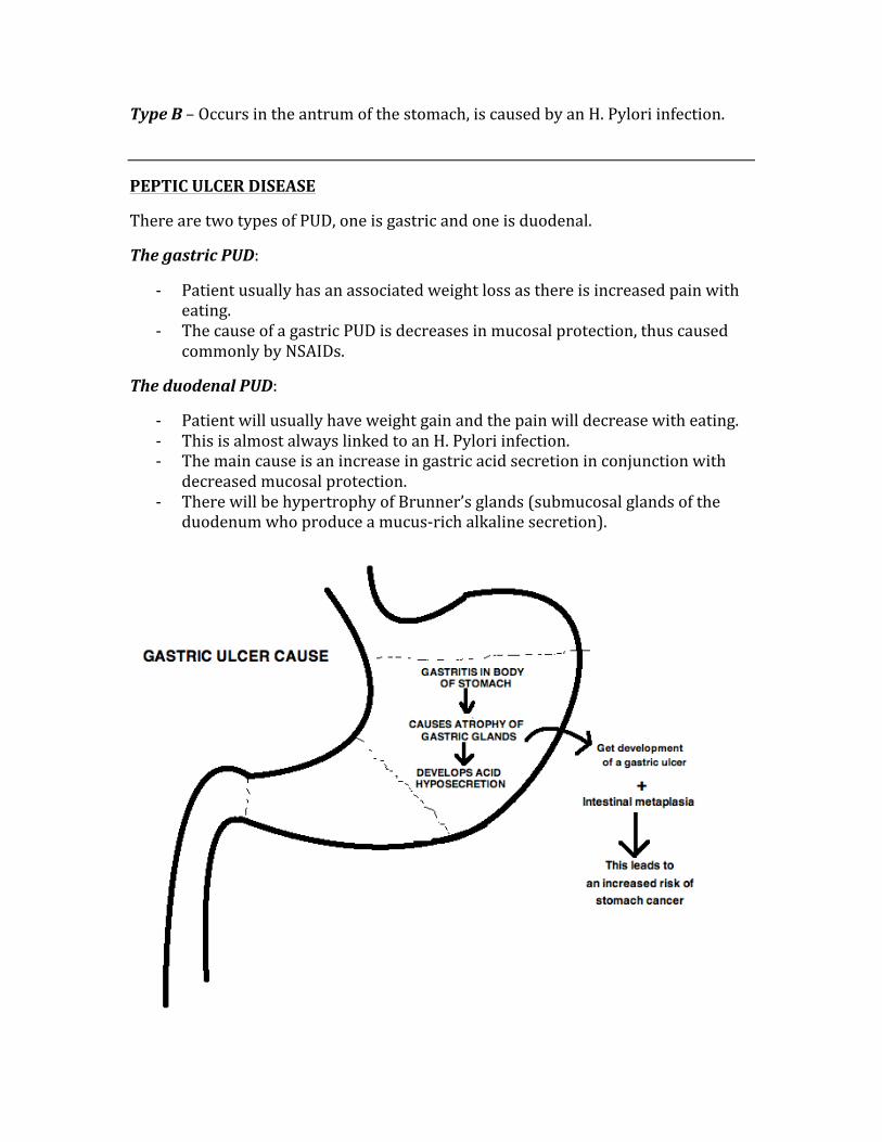
Type B – Occurs in the antrum of the stomach, is caused by an H. Pylori infection.
PEPTIC ULCER DISEASE
There are two types of PUD, one is gastric and one is duodenal.
The gastric PUD:
-‐ Patient usually has an associated weight loss as there is increased pain with eating.
-‐ The cause of a gastric PUD is decreases in mucosal protection, thus caused commonly by NSAIDs.
The duodenal PUD:
-‐ Patient will usually have weight gain and the pain will decrease with eating. -‐ This is almost always linked to an H. Pylori infection. -‐ The main cause is an increase in gastric acid secretion in conjunction with
decreased mucosal protection. -‐ There will be hypertrophy of Brunner’s glands (submucosal glands of the
duodenum who produce a mucus-‐rich alkaline secretion).
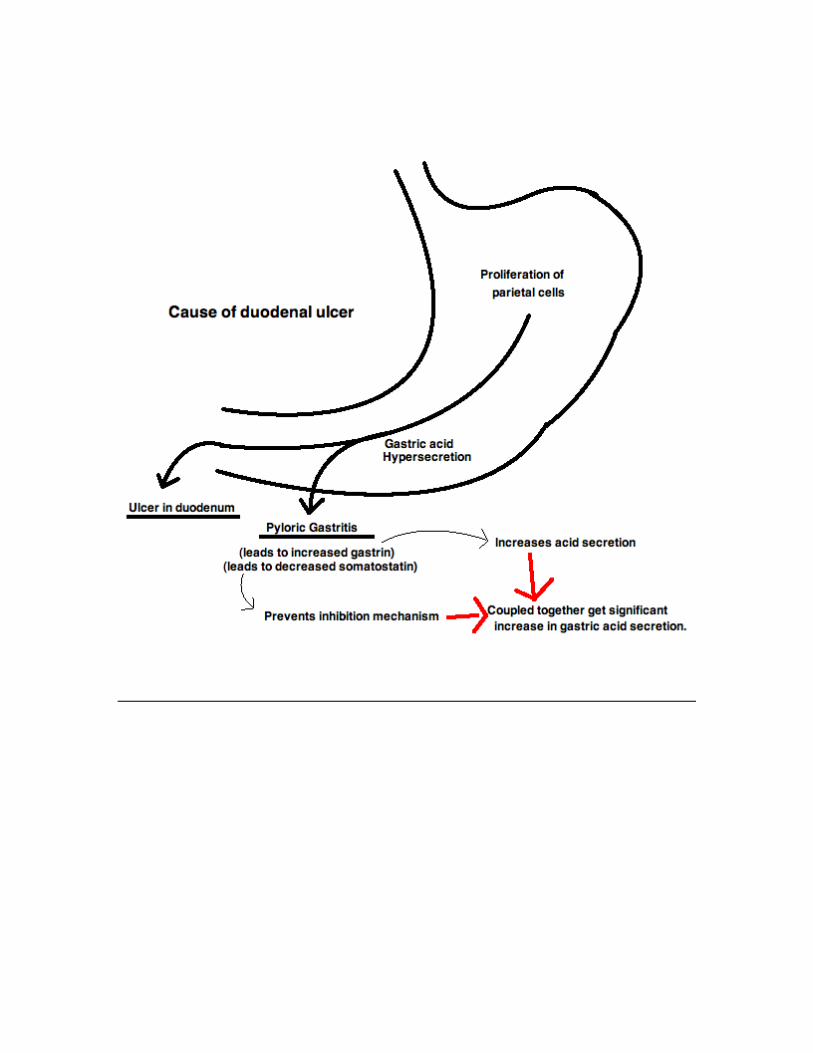
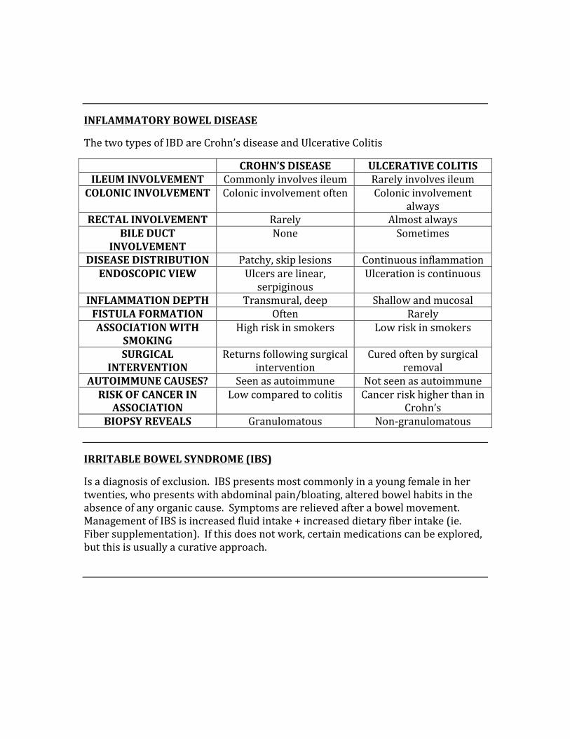
INFLAMMATORY BOWEL DISEASE
The two types of IBD are Crohn’s disease and Ulcerative Colitis
CROHN’S DISEASE ULCERATIVE COLITIS ILEUM INVOLVEMENT Commonly involves ileum Rarely involves ileum COLONIC INVOLVEMENT Colonic involvement often Colonic involvement
always RECTAL INVOLVEMENT Rarely Almost always
BILE DUCT INVOLVEMENT
None Sometimes
DISEASE DISTRIBUTION Patchy, skip lesions Continuous inflammation ENDOSCOPIC VIEW Ulcers are linear,
serpiginous Ulceration is continuous
INFLAMMATION DEPTH Transmural, deep Shallow and mucosal FISTULA FORMATION Often Rarely ASSOCIATION WITH
SMOKING High risk in smokers Low risk in smokers
SURGICAL INTERVENTION
Returns following surgical intervention
Cured often by surgical removal
AUTOIMMUNE CAUSES? Seen as autoimmune Not seen as autoimmune RISK OF CANCER IN ASSOCIATION
Low compared to colitis Cancer risk higher than in Crohn’s
BIOPSY REVEALS Granulomatous Non-‐granulomatous
IRRITABLE BOWEL SYNDROME (IBS)
Is a diagnosis of exclusion. IBS presents most commonly in a young female in her twenties, who presents with abdominal pain/bloating, altered bowel habits in the absence of any organic cause. Symptoms are relieved after a bowel movement. Management of IBS is increased fluid intake + increased dietary fiber intake (ie. Fiber supplementation). If this does not work, certain medications can be explored, but this is usually a curative approach.
SYNDROMES OF MALABSORPTION
The commonly tested syndromes of malabsorption are Celiac Sprue, Tropical Sprue, and Whipple’s disease. Symptoms of these conditions are: Weight loss, cramping, diarrhea, steatorrhea, indigestion, and fatigue.
Celiac Sprue – Caused by autoantibodies (antigliadin, antiendomysial, anti-‐tissue transglutaminase) against gluten, which causes villous blunting and infiltration of lymphocytes. Treatment is to consume a gluten-‐free diet.
Tropical Sprue – This is an infectious condition commonly found in tropical regions, and is market by abnormal flattening of the villi in the small intestine.
Whipple’s Disease – This is rarer than the other two malabsorption syndrome. It is caused by an infection with the bacterium Tropheryma Whipplei, and can cause malabsorption in conjunction with many systemic problems (cardiac, respiratory, neurological, rheumatologic, and visual).
HEPATITIS
Hepatitis is a general term to describe the inflammation of the liver. There are many possible causes of hepatitis, such as alcohol, drugs, viral, and disease-‐related.
Causes of Viral Hepatitis:
Five main types of viral hepatitis are: Hep A, B, C, D, and E. Hepatitis can also be caused by Epstein-‐Barr virus, cytomegalovirus, and herpes simplex virus.
Transmission of viral hepatitis:
Hepatitis A & E – fecal-‐oral transmission, higher in developing countries
Hepatitis B – transmission is parenteral or sexual
Hepatitis D – requires co-‐infection with hepatitis B (requires the outer envelope of the HbsAg)
Hepatitic C – transmitted parenterally and is thus more common in IV drug users
* Only hepatitis B, C, and D can progress to chronic hepatitis.
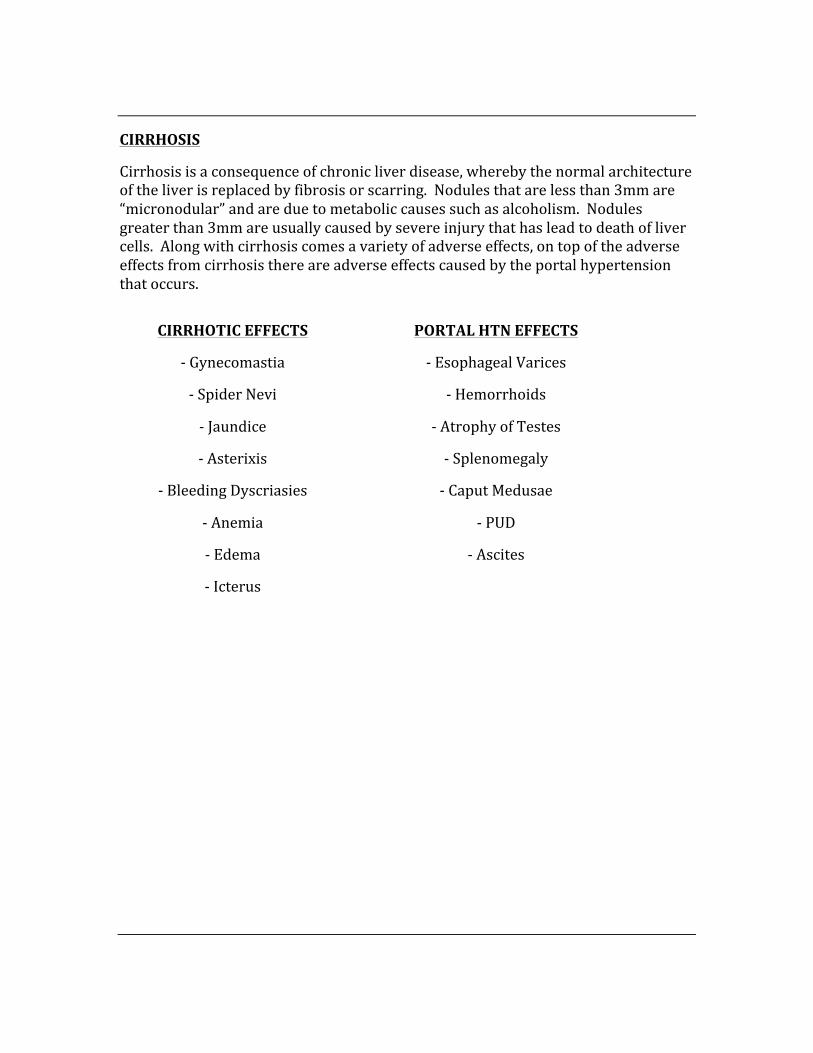
CIRRHOSIS
Cirrhosis is a consequence of chronic liver disease, whereby the normal architecture of the liver is replaced by fibrosis or scarring. Nodules that are less than 3mm are “micronodular” and are due to metabolic causes such as alcoholism. Nodules greater than 3mm are usually caused by severe injury that has lead to death of liver cells. Along with cirrhosis comes a variety of adverse effects, on top of the adverse effects from cirrhosis there are adverse effects caused by the portal hypertension that occurs.
CIRRHOTIC EFFECTS
-‐ Gynecomastia
-‐ Spider Nevi
-‐ Jaundice
-‐ Asterixis
-‐ Bleeding Dyscriasies
-‐ Anemia
-‐ Edema
-‐ Icterus
PORTAL HTN EFFECTS
-‐ Esophageal Varices
-‐ Hemorrhoids
-‐ Atrophy of Testes
-‐ Splenomegaly
-‐ Caput Medusae
-‐ PUD
-‐ Ascites
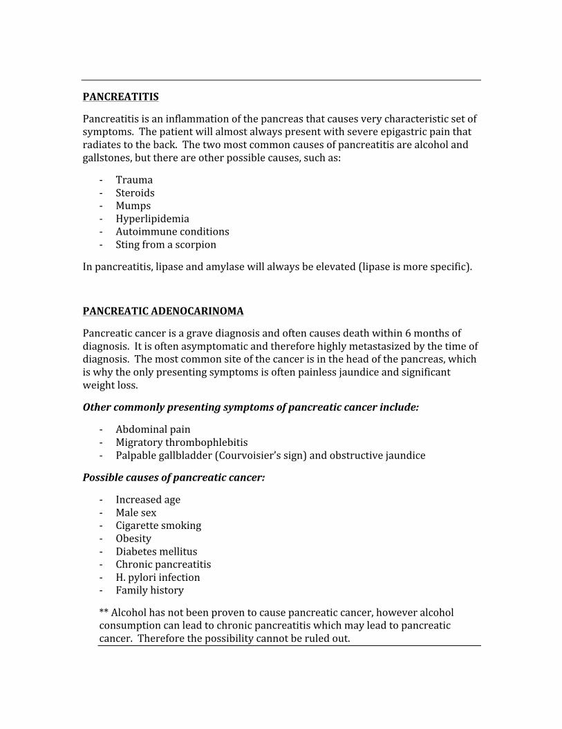
PANCREATITIS
Pancreatitis is an inflammation of the pancreas that causes very characteristic set of symptoms. The patient will almost always present with severe epigastric pain that radiates to the back. The two most common causes of pancreatitis are alcohol and gallstones, but there are other possible causes, such as:
-‐ Trauma -‐ Steroids -‐ Mumps -‐ Hyperlipidemia -‐ Autoimmune conditions -‐ Sting from a scorpion
In pancreatitis, lipase and amylase will always be elevated (lipase is more specific).
PANCREATIC ADENOCARINOMA
Pancreatic cancer is a grave diagnosis and often causes death within 6 months of diagnosis. It is often asymptomatic and therefore highly metastasized by the time of diagnosis. The most common site of the cancer is in the head of the pancreas, which is why the only presenting symptoms is often painless jaundice and significant weight loss.
Other commonly presenting symptoms of pancreatic cancer include:
-‐ Abdominal pain -‐ Migratory thrombophlebitis -‐ Palpable gallbladder (Courvoisier’s sign) and obstructive jaundice
Possible causes of pancreatic cancer:
-‐ Increased age -‐ Male sex -‐ Cigarette smoking -‐ Obesity -‐ Diabetes mellitus -‐ Chronic pancreatitis -‐ H. pylori infection -‐ Family history
** Alcohol has not been proven to cause pancreatic cancer, however alcohol consumption can lead to chronic pancreatitis which may lead to pancreatic cancer. Therefore the possibility cannot be ruled out.
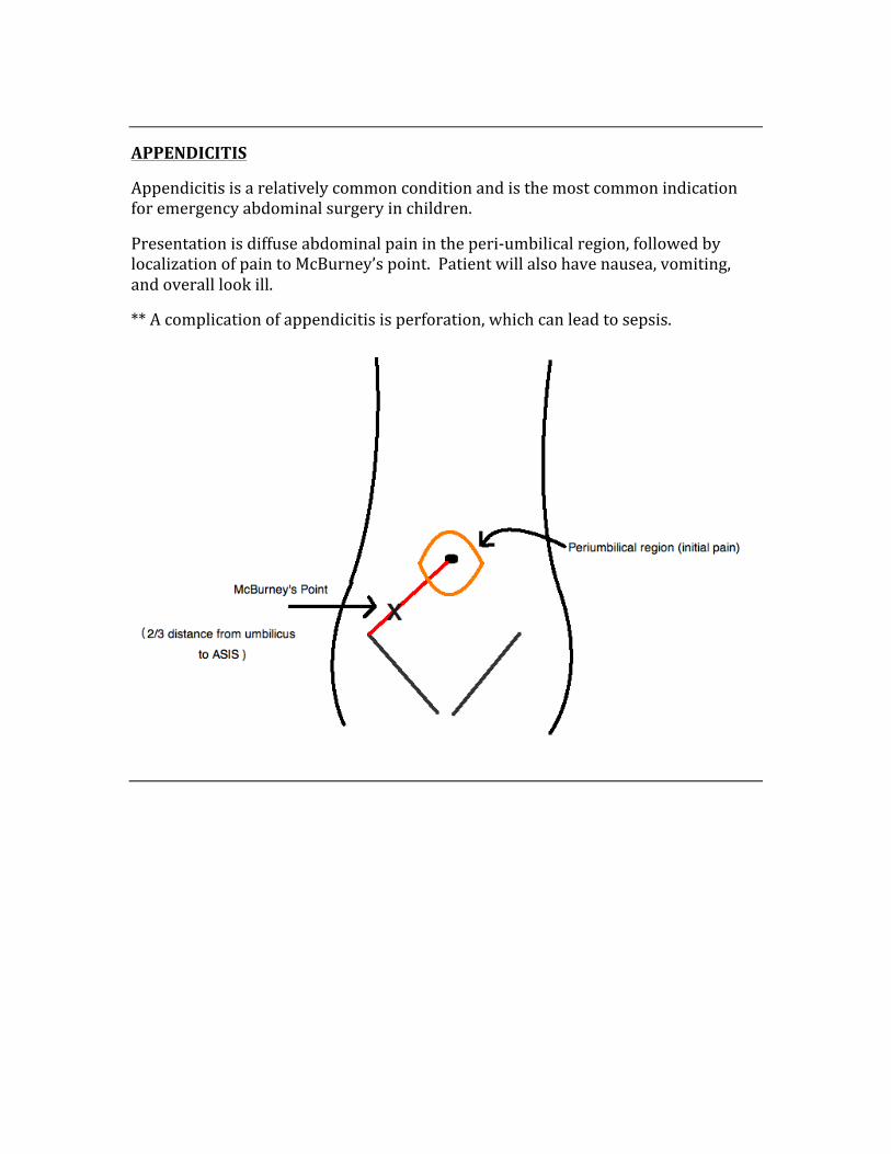
APPENDICITIS
Appendicitis is a relatively common condition and is the most common indication for emergency abdominal surgery in children.
Presentation is diffuse abdominal pain in the peri-‐umbilical region, followed by localization of pain to McBurney’s point. Patient will also have nausea, vomiting, and overall look ill.
** A complication of appendicitis is perforation, which can lead to sepsis.
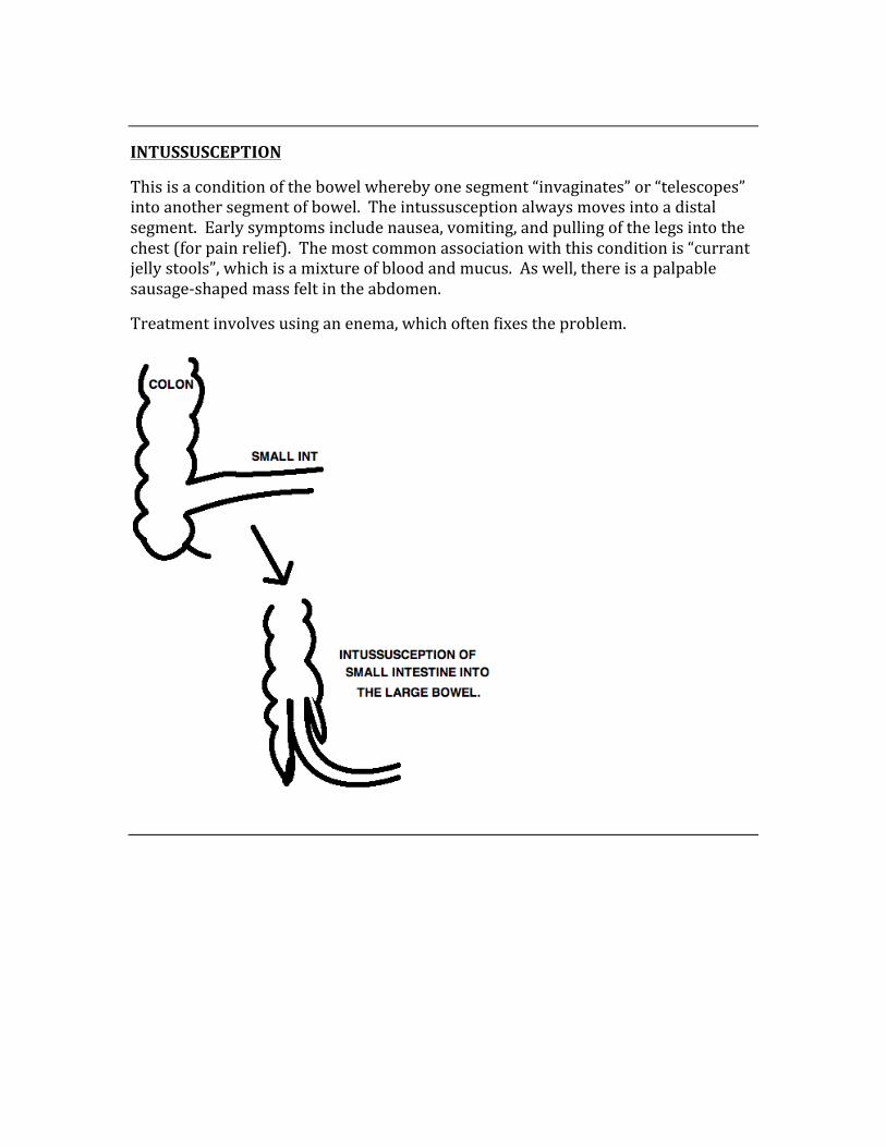
INTUSSUSCEPTION
This is a condition of the bowel whereby one segment “invaginates” or “telescopes” into another segment of bowel. The intussusception always moves into a distal segment. Early symptoms include nausea, vomiting, and pulling of the legs into the chest (for pain relief). The most common association with this condition is “currant jelly stools”, which is a mixture of blood and mucus. As well, there is a palpable sausage-‐shaped mass felt in the abdomen.
Treatment involves using an enema, which often fixes the problem.
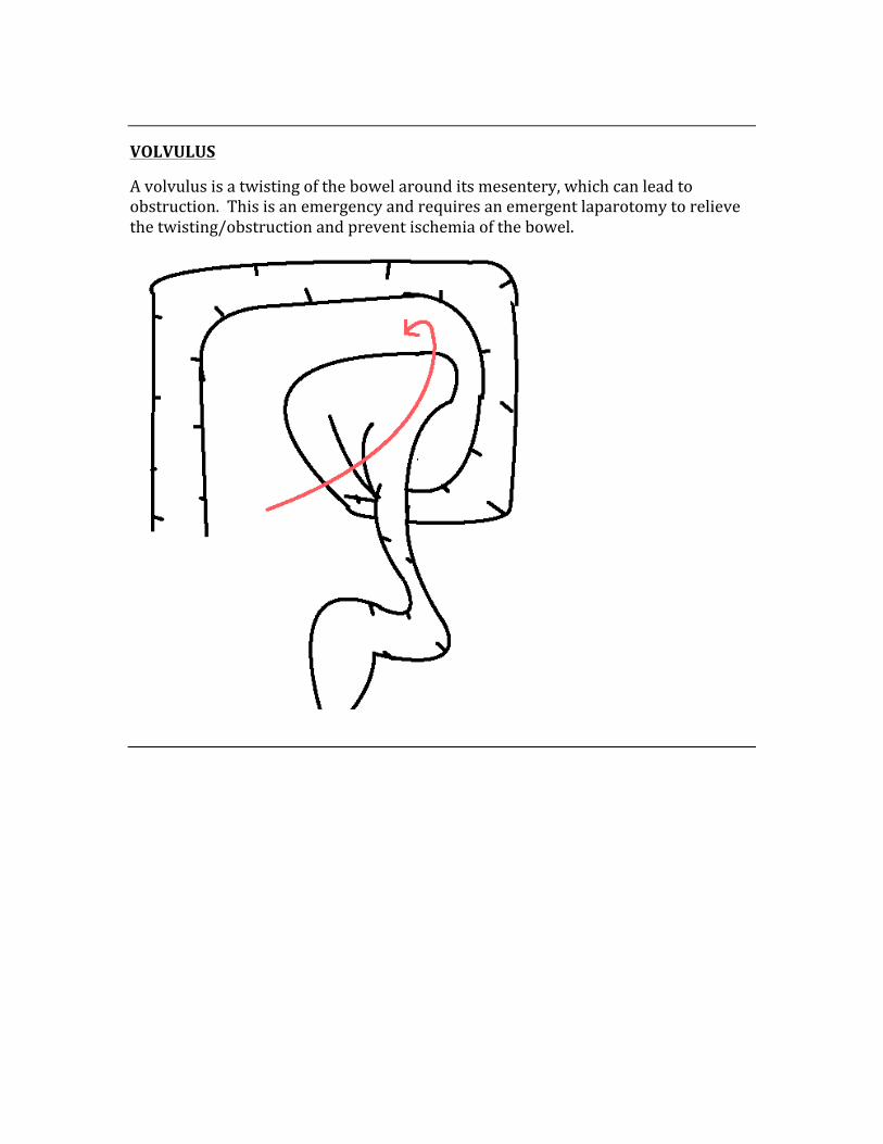
VOLVULUS
A volvulus is a twisting of the bowel around its mesentery, which can lead to obstruction. This is an emergency and requires an emergent laparotomy to relieve the twisting/obstruction and prevent ischemia of the bowel.
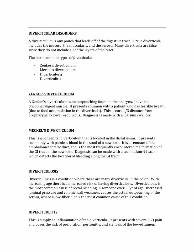
DIVERTICULAR DISORDERS
A diverticulum is any pouch that leads off of the digestive tract. A true diverticula includes the mucosa, the muscularis, and the serosa. Many diverticula are false since they do not include all of the layers of the tract.
The most common types of diverticula:
-‐ Zenker’s diverticulum -‐ Meckel’s diverticulum -‐ Diverticulosis -‐ Diverticulitis
ZENKER’S DIVERTICULUM
A Zenker’s diverticulum is an outpouching found in the pharynx, above the cricopharyngeal muscle. It presents common with a patient who has terrible breath (due to food accumulation in the diverticula). This occurs 1/3 distance from oropharynx to lower esophagus. Diagnosis is made with a barium swallow.
MECKEL’S DIVERTICULUM
This is a congenital diverticulum that is located in the distal ileum. It presents commonly with painless blood in the stool of a newborn. It is a remnant of the omphalomesenteric duct, and is the most frequently encountered malformation of the GI tract of the newborn. Diagnosis can be made with a technetium-‐99 scan, which detects the location of bleeding along the GI tract.
DIVERTICULOSIS
Diverticulosis is a condition where there are many diverticula in the colon. With increasing age there is an increased risk of having diverticulosis. Diverticulosis is the most common cause of rectal bleeding in someone over 50yr of age. Increased luminal pressure and colonic wall weakness causes the actual outpouching of the serosa, where a low-‐fiber diet is the most common cause of this condition.
DIVERTICULITIS
This is simply an inflammation of the diverticula. It presents with severe LLQ pain and poses the risk of perforation, peritonitis, and stenosis of the bowel lumen.
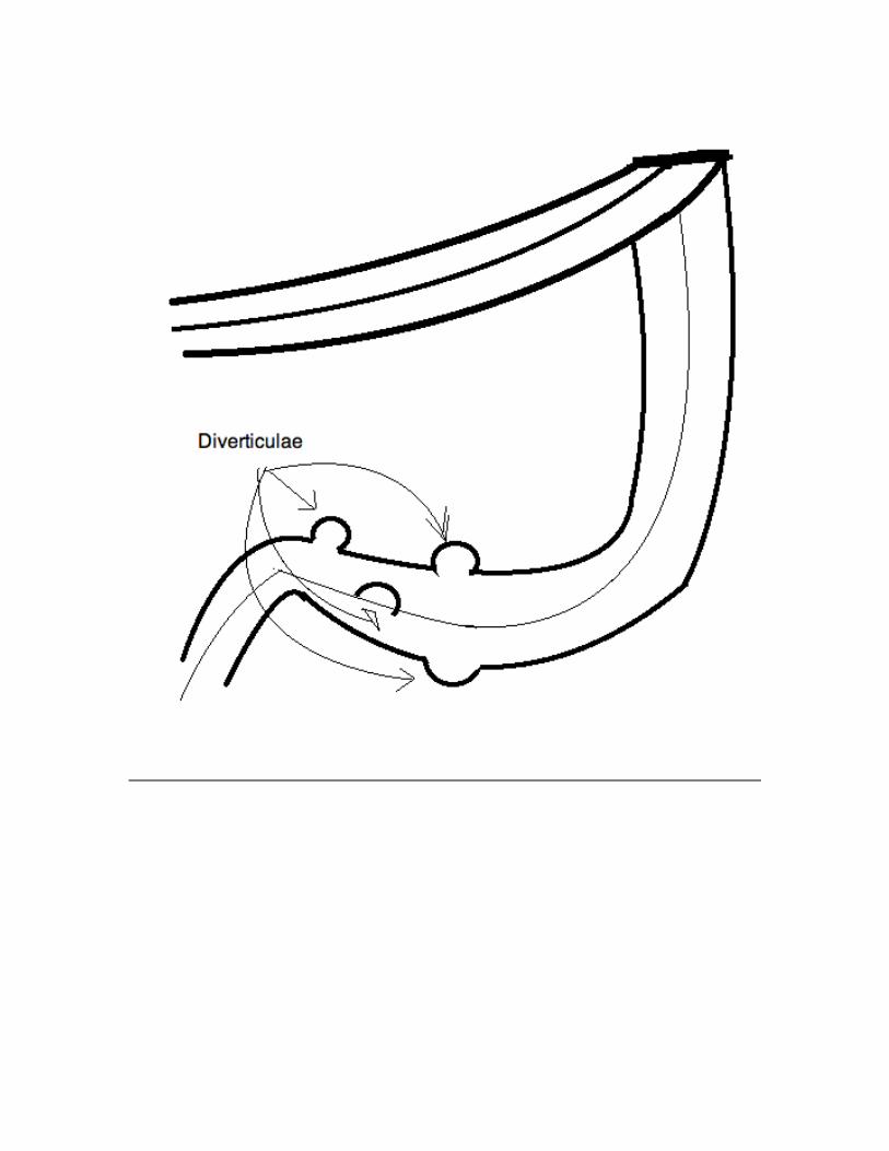
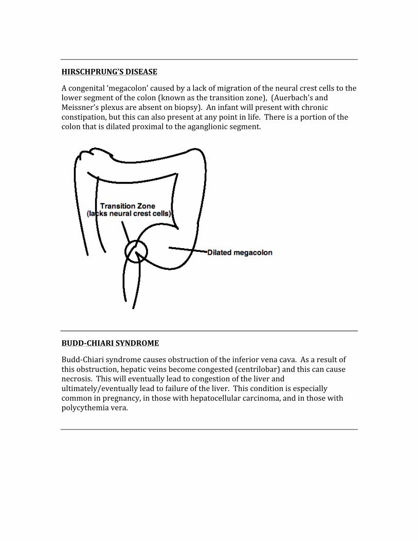
HIRSCHPRUNG’S DISEASE
A congenital ‘megacolon’ caused by a lack of migration of the neural crest cells to the lower segment of the colon (known as the transition zone), (Auerbach’s and Meissner’s plexus are absent on biopsy). An infant will present with chronic constipation, but this can also present at any point in life. There is a portion of the colon that is dilated proximal to the aganglionic segment.
BUDD-‐CHIARI SYNDROME
Budd-‐Chiari syndrome causes obstruction of the inferior vena cava. As a result of this obstruction, hepatic veins become congested (centrilobar) and this can cause necrosis. This will eventually lead to congestion of the liver and ultimately/eventually lead to failure of the liver. This condition is especially common in pregnancy, in those with hepatocellular carcinoma, and in those with polycythemia vera.
HEMOCHROMATOSIS
This is a very common condition that is caused by a defect in iron metabolism, which leads to an iron overload in vital organs, joints, and tissues. Early diagnosis can help prevent adverse effects of the iron overload.
Hemochromatosis presents with a class triad of:
1. Micronodular cirrhosis 2. Pancreatic fibrosis 3. Skin pigmentation
This condition is classically known as bronze diabetes due to the fact that it tints the skin “bronze” and also affects the pancreas. Total body iron levels may reach upwards of 50g, and this must be managed with repeated phlebotomy. This condition can lead to congestive heart failure and can increase the risk of hepatocellular carcinoma.
LABS: In those with hemochromatosis, labs will show ↑Iron and Ferritin, with a ↓ total iron binding capacity.
WILSON’S DISEASE
Is an autosomal recessive disorder where there is a failure of copper’s ability to enter circulation in the form of ceruloplasmin. This leads to copper accumulation in certain tissues (liver, brain, cornea), and is treated with penicillamine (chelation of copper).
The most common signs and symptoms of Wilson’s disease are:
-‐ Cornea deposits (Kayser-‐Fleischer rings), very common in Wilson’s disease -‐ Asterixis -‐ Parkinson-‐like symptoms due to accumulation in basal ganglia -‐ Carcinoma -‐ Dementia
HEPATOCELLULAR CARCINOMA HCC)
Hepatocellular carcinoma is a very common cause of metastasis, and spreads by hematogenous route. Most cases of hepatocellular carcinoma are due to hepatitis B and/or C, as well as cirrhosis. Other causes of HCC include Wilson’s disease, hemochromatosis, alcoholic cirrhosis, and α-‐1 antitrypsin deficiency.
The outcome is usually poor, however 1-‐2 out of 10 cases are treatable with surgical removal of cancers.
HYPERBILIRUBINEMIAS (HEREDITARY)
There are three commonly testes and encountered hereditary hyperbilirubinemias, they are:
1. Gilbert’s syndrome 2. Crigler-‐Najjar syndrome 3. Dubin-‐Johnson syndrome
Gilbert’s Syndrome:
Gilbert’s syndrome is a benign condition where there is a mild decrease in the UDP-‐glucuronyl transferase enzymes. This leads to an elevation of unconjugated bilirubin.
Crigler-‐Najjar Syndrome:
This is a severe condition that leads to death early in life. There is a complete absence of UDP-‐glucuronyl transferase, which leads to significant increases in unconjugated bilirubin and causes it to deposit in the brain (kernicterus), as well as jaundice. There is a less severe version of Crigler-‐Najjar called “type 2”, and it can be managed with Phenobarbital.
Dubin-‐Johnson Syndrome:
This syndrome occurs as a result of a defect in the liver’s ability to excrete conjugated bilirubin. It is benign but there is a change in color of the liver to black. A different form of this syndrome is “Rotor’s syndrome”, which is even milder and causes no change in the color of the liver.
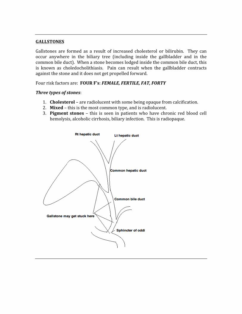
GALLSTONES
Gallstones are formed as a result of increased cholesterol or bilirubin. They can occur anywhere in the biliary tree (including inside the gallbladder and in the common bile duct). When a stone becomes lodged inside the common bile duct, this is known as choledocholithiasis. Pain can result when the gallbladder contracts against the stone and it does not get propelled forward.
Four risk factors are: FOUR F’s: FEMALE, FERTILE, FAT, FORTY
Three types of stones:
1. Cholesterol – are radiolucent with some being opaque from calcification. 2. Mixed – this is the most common type, and is radiolucent. 3. Pigment stones – this is seen in patients who have chronic red blood cell
hemolysis, alcoholic cirrhosis, biliary infection. This is radiopaque.
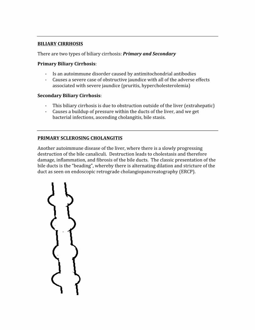
BILIARY CIRRHOSIS
There are two types of biliary cirrhosis: Primary and Secondary
Primary Biliary Cirrhosis:
-‐ Is an autoimmune disorder caused by antimitochondrial antibodies -‐ Causes a severe case of obstructive jaundice with all of the adverse effects
associated with severe jaundice (pruritis, hypercholesterolemia)
Secondary Biliary Cirrhosis:
-‐ This biliary cirrhosis is due to obstruction outside of the liver (extrahepatic) -‐ Causes a buildup of pressure within the ducts of the liver, and we get
bacterial infections, ascending cholangitis, bile stasis.
PRIMARY SCLEROSING CHOLANGITIS
Another autoimmune disease of the liver, where there is a slowly progressing destruction of the bile canaliculi. Destruction leads to cholestasis and therefore damage, inflammation, and fibrosis of the bile ducts. The classic presentation of the bile ducts is the “beading”, whereby there is alternating dilation and stricture of the duct as seen on endoscopic retrograde cholangiopancreatography (ERCP).
CONGENITAL PATHOLOGY
There are a group of common congenital pathologies that are high-‐yield for the USMLE Step 1 exam, they include:
-‐ Defects of the heart -‐ Spina bifida -‐ Hypospadias -‐ Cleft lip -‐ Pyloric stenosis -‐ Anencephaly
CONGENITAL HEART DEFECTS
The most common congenital heart defects include:
-‐ Ventricular septal defects -‐ Atrial septal defects -‐ Patent ductus arteriosus -‐ Tetralogy of fallot -‐ Truncus arteriosus -‐ Transposition of the great vessels -‐ Coarctation of the aorta
DEFECTS CAUSING A RIGHT TO LEFT SHUNT
These defects cause defects that force blood from the right side of the heart to the left side of the heart due to pressure, resulting in early cyanosis because systemic blood is lacking oxygen. The babies are often blue in color because they do not receive adequate oxygen.
The three common congenital malformations causing a RàL shunt are:
1. Tetralogy of fallot 2. Transposition of the great vessels 3. Truncus arteriosus
TETRALOGY OF FALLOT:
This condition results in a group of problems, that ultimately lead to early cyanosis due to shunting of blood from the right to the left through the ventricular septal defect. This is caused by an anteriosuperior displacement of the infundibular septum.
The 4 pathologies of tetralogy of Fallot are:
1. Pulmonary stenosis 2. Right ventricular hypertrophy 3. Overriding aorta 4. Ventricular septal defect (provides area for shunting)
TRANSPOSITION OF THE GREAT VESSELS:
This condition results in the aorta connected from the right ventricle while the pulmonary trunk leaves from the left ventricle. This results in a separation of the systemic and pulmonary circulations. Since there is no oxygenated blood being pumped systemically, this condition is incompatible with life (unless there is the presence of a shunt). Transposition of the great vessels warrants immediate surgical correction for survival. The condition is caused by failure of the aorticopulmonary septum to spiral.
TRUNCUS ARTERIOSUS:
Truncus arteriosus occurs when there is an incomplete or failed septation of the embryonic truncus arteriosus. This results in a single arterial trunk that arises from two normally formed ventricles. The pulmonary arteries can arise from the common trunk in a myriad of patterns, thus giving this condition several different subtypes.
DEFECTS CAUSING A LEFT TO RIGHT SHUNT
There are three conditions that cause a LàR shunt, they include:
1. Ventricular septal defects 2. Atrial septal defects 3. Patent ductus arteriosus
VENTRICULAR SEPTAL DEFECT
This is the most common of all cardiac congenital anomalies. This defect does not result is cyanosis because the LàR shunt doesn’t put non-‐oxygenated blood back into the systemic circulation. This can be detected by hearing a pansystolic murmur on auscultation. There is usually no detection at birth, but within a few weeks it will be detectable.
ATRIAL SEPTAL DEFECT
This is a condition where there is a communication between both the right and left atria of the heart. The ASD is the most common congenital heart defect seen in adults.
PATENT DUCTUS ARTERIOSUS
The ductus arteriosus is a vascular connection between the pulmonary artery and the aortic arch in the developing fetus. Upon a newborn’s first breath, the process of PD closure should occur, however it sometimes does not. If closure fails to occur, the neonate will experience persistent respiratory problems. The PDA can be closed by giving indomethacin, and can be kept opened with prostaglandin E.
COARCTATION OF THE AORTA
This is a narrowing of the aorta that can occur in two different places. The ‘preductal’ form occurs proximal to the ductus arteriosus, the ‘postductal’ form occurs distal to the ductus arteriosus. The post-‐ductal form is associated with rib notching, upper extremity hypertension, and weak pulses in the lower extremities. Coarctation of the aorta is seen in males much more than in females.
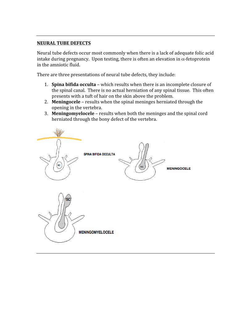
NEURAL TUBE DEFECTS
Neural tube defects occur most commonly when there is a lack of adequate folic acid intake during pregnancy. Upon testing, there is often an elevation in α-‐fetoprotein in the amniotic fluid.
There are three presentations of neural tube defects, they include:
1. Spina bifida occulta – which results when there is an incomplete closure of the spinal canal. There is no actual herniation of any spinal tissue. This often presents with a tuft of hair on the skin above the problem.
2. Meningocele – results when the spinal meninges herniated through the opening in the vertebra.
3. Meningomyelocele – results when both the meninges and the spinal cord herniated through the bony defect of the vertebra.
TRISOMY DISORDERS
The three most commonly encountered autosomal trisomy disorders are:
1. Patau’s syndrome 2. Edward’s syndrome 3. Down’s syndrome
Patau’s Syndrome:
-‐ Caused by trisomy 13 -‐ Cleft lip and palate -‐ Severe mental retardation -‐ Microphthalmia -‐ Microcephaly -‐ Death usually within 1st year of birth
Edward’s Syndrome:
-‐ Caused by trisomy 18 -‐ Rocker bottom feet -‐ Low-‐set ears -‐ Clenched hands -‐ Prominent occiput -‐ Death usually within 1st year of birth
Down’s Syndrome:
-‐ The most common chromosomal disorder -‐ The most common cause of congenital mental retardation -‐ Caused by trisomy 21 -‐ Prominent epicanthal folds -‐ Simian crease -‐ Increased risk of ALL -‐ Congenital heart disease (ASD most commonly) -‐ Caused most commonly by meiotic non-‐disjunction of homologous
chromosomes
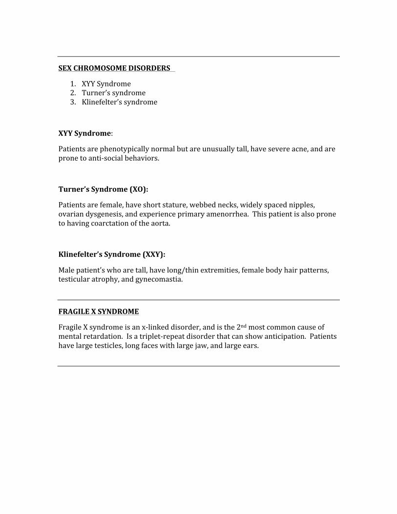
SEX CHROMOSOME DISORDERS
1. XYY Syndrome 2. Turner’s syndrome 3. Klinefelter’s syndrome
XYY Syndrome:
Patients are phenotypically normal but are unusually tall, have severe acne, and are prone to anti-‐social behaviors.
Turner’s Syndrome (XO):
Patients are female, have short stature, webbed necks, widely spaced nipples, ovarian dysgenesis, and experience primary amenorrhea. This patient is also prone to having coarctation of the aorta.
Klinefelter’s Syndrome (XXY):
Male patient’s who are tall, have long/thin extremities, female body hair patterns, testicular atrophy, and gynecomastia.
FRAGILE X SYNDROME
Fragile X syndrome is an x-‐linked disorder, and is the 2nd most common cause of mental retardation. Is a triplet-‐repeat disorder that can show anticipation. Patients have large testicles, long faces with large jaw, and large ears.
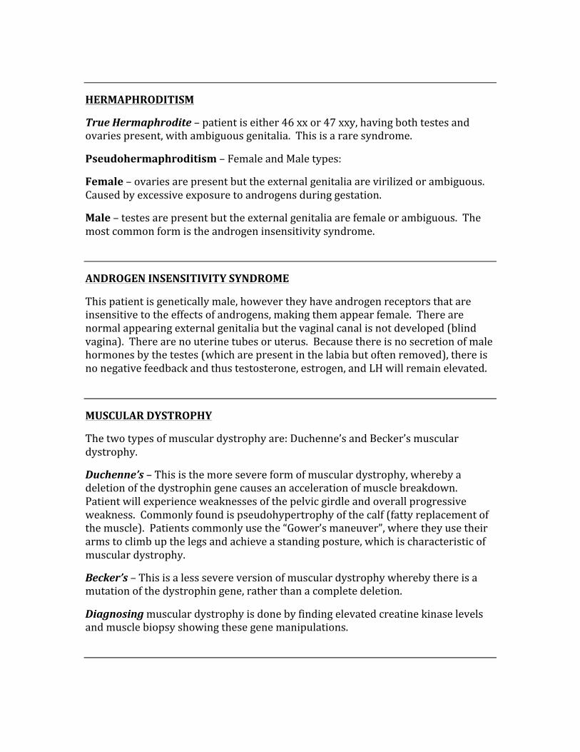
HERMAPHRODITISM
True Hermaphrodite – patient is either 46 xx or 47 xxy, having both testes and ovaries present, with ambiguous genitalia. This is a rare syndrome.
Pseudohermaphroditism – Female and Male types:
Female – ovaries are present but the external genitalia are virilized or ambiguous. Caused by excessive exposure to androgens during gestation.
Male – testes are present but the external genitalia are female or ambiguous. The most common form is the androgen insensitivity syndrome.
ANDROGEN INSENSITIVITY SYNDROME
This patient is genetically male, however they have androgen receptors that are insensitive to the effects of androgens, making them appear female. There are normal appearing external genitalia but the vaginal canal is not developed (blind vagina). There are no uterine tubes or uterus. Because there is no secretion of male hormones by the testes (which are present in the labia but often removed), there is no negative feedback and thus testosterone, estrogen, and LH will remain elevated.
MUSCULAR DYSTROPHY
The two types of muscular dystrophy are: Duchenne’s and Becker’s muscular dystrophy.
Duchenne’s – This is the more severe form of muscular dystrophy, whereby a deletion of the dystrophin gene causes an acceleration of muscle breakdown. Patient will experience weaknesses of the pelvic girdle and overall progressive weakness. Commonly found is pseudohypertrophy of the calf (fatty replacement of the muscle). Patients commonly use the “Gower’s maneuver”, where they use their arms to climb up the legs and achieve a standing posture, which is characteristic of muscular dystrophy.
Becker’s – This is a less severe version of muscular dystrophy whereby there is a mutation of the dystrophin gene, rather than a complete deletion.
Diagnosing muscular dystrophy is done by finding elevated creatine kinase levels and muscle biopsy showing these gene manipulations.
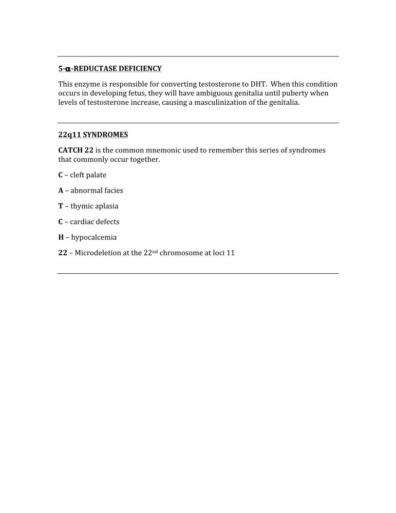
5-‐α-‐REDUCTASE DEFICIENCY
This enzyme is responsible for converting testosterone to DHT. When this condition occurs in developing fetus, they will have ambiguous genitalia until puberty when levels of testosterone increase, causing a masculinization of the genitalia.
22q11 SYNDROMES
CATCH 22 is the common mnemonic used to remember this series of syndromes that commonly occur together.
C – cleft palate
A – abnormal facies
T – thymic aplasia
C – cardiac defects
H – hypocalcemia
22 – Microdeletion at the 22nd chromosome at loci 11
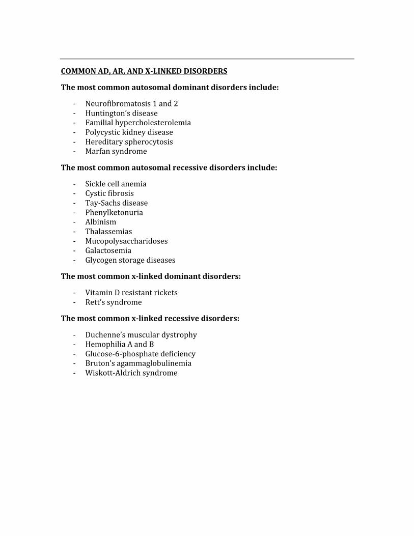
COMMON AD, AR, AND X-‐LINKED DISORDERS
The most common autosomal dominant disorders include:
-‐ Neurofibromatosis 1 and 2 -‐ Huntington’s disease -‐ Familial hypercholesterolemia -‐ Polycystic kidney disease -‐ Hereditary spherocytosis -‐ Marfan syndrome
The most common autosomal recessive disorders include:
-‐ Sickle cell anemia -‐ Cystic fibrosis -‐ Tay-‐Sachs disease -‐ Phenylketonuria -‐ Albinism -‐ Thalassemias -‐ Mucopolysaccharidoses -‐ Galactosemia -‐ Glycogen storage diseases
The most common x-‐linked dominant disorders:
-‐ Vitamin D resistant rickets -‐ Rett’s syndrome
The most common x-‐linked recessive disorders:
-‐ Duchenne’s muscular dystrophy -‐ Hemophilia A and B -‐ Glucose-‐6-‐phosphate deficiency -‐ Bruton’s agammaglobulinemia -‐ Wiskott-‐Aldrich syndrome
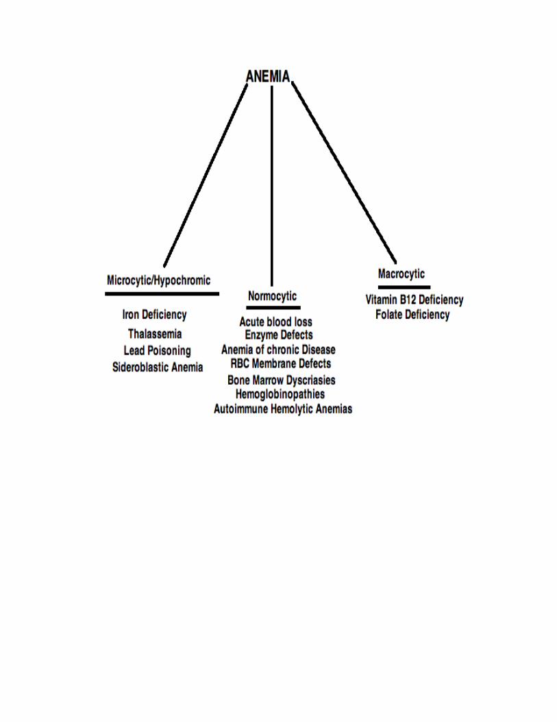
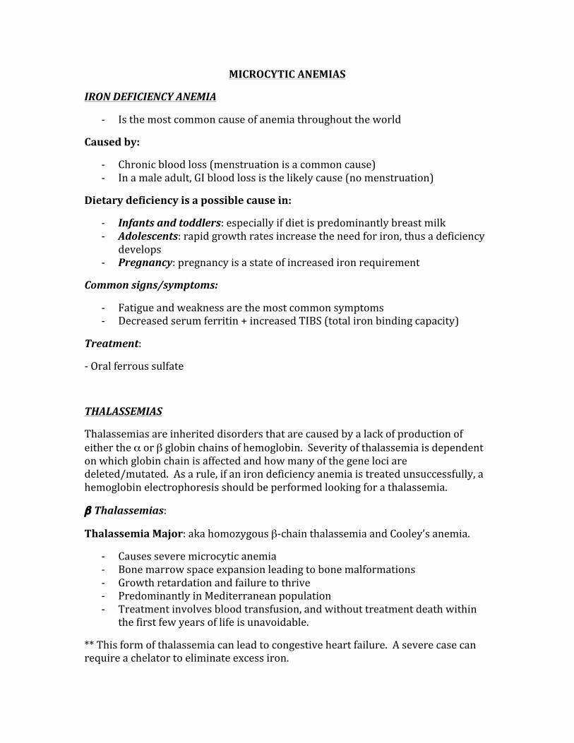
MICROCYTIC ANEMIAS
IRON DEFICIENCY ANEMIA
-‐ Is the most common cause of anemia throughout the world
Caused by:
-‐ Chronic blood loss (menstruation is a common cause) -‐ In a male adult, GI blood loss is the likely cause (no menstruation)
Dietary deficiency is a possible cause in:
-‐ Infants and toddlers: especially if diet is predominantly breast milk -‐ Adolescents: rapid growth rates increase the need for iron, thus a deficiency
develops -‐ Pregnancy: pregnancy is a state of increased iron requirement
Common signs/symptoms:
-‐ Fatigue and weakness are the most common symptoms -‐ Decreased serum ferritin + increased TIBS (total iron binding capacity)
Treatment:
-‐ Oral ferrous sulfate
THALASSEMIAS
Thalassemias are inherited disorders that are caused by a lack of production of either the α or β globin chains of hemoglobin. Severity of thalassemia is dependent on which globin chain is affected and how many of the gene loci are deleted/mutated. As a rule, if an iron deficiency anemia is treated unsuccessfully, a hemoglobin electrophoresis should be performed looking for a thalassemia.
β Thalassemias:
Thalassemia Major: aka homozygous β-‐chain thalassemia and Cooley’s anemia.
-‐ Causes severe microcytic anemia -‐ Bone marrow space expansion leading to bone malformations -‐ Growth retardation and failure to thrive -‐ Predominantly in Mediterranean population -‐ Treatment involves blood transfusion, and without treatment death within
the first few years of life is unavoidable.
** This form of thalassemia can lead to congestive heart failure. A severe case can require a chelator to eliminate excess iron.
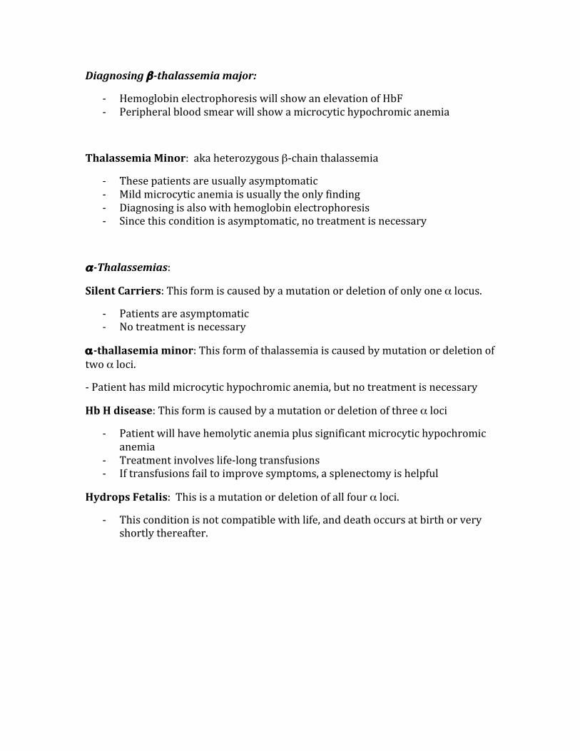
Diagnosing β-‐thalassemia major:
-‐ Hemoglobin electrophoresis will show an elevation of HbF -‐ Peripheral blood smear will show a microcytic hypochromic anemia
Thalassemia Minor: aka heterozygous β-‐chain thalassemia
-‐ These patients are usually asymptomatic -‐ Mild microcytic anemia is usually the only finding -‐ Diagnosing is also with hemoglobin electrophoresis -‐ Since this condition is asymptomatic, no treatment is necessary
α-‐Thalassemias:
Silent Carriers: This form is caused by a mutation or deletion of only one α locus.
-‐ Patients are asymptomatic -‐ No treatment is necessary
α-‐thallasemia minor: This form of thalassemia is caused by mutation or deletion of two α loci.
-‐ Patient has mild microcytic hypochromic anemia, but no treatment is necessary
Hb H disease: This form is caused by a mutation or deletion of three α loci
-‐ Patient will have hemolytic anemia plus significant microcytic hypochromic anemia
-‐ Treatment involves life-‐long transfusions -‐ If transfusions fail to improve symptoms, a splenectomy is helpful
Hydrops Fetalis: This is a mutation or deletion of all four α loci.
-‐ This condition is not compatible with life, and death occurs at birth or very shortly thereafter.
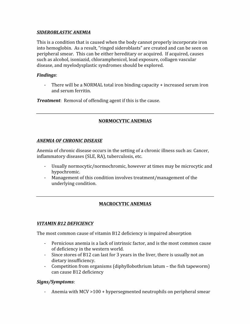
SIDEROBLASTIC ANEMIA
This is a condition that is caused when the body cannot properly incorporate iron into hemoglobin. As a result, “ringed sideroblasts” are created and can be seen on peripheral smear. This can be either hereditary or acquired. If acquired, causes such as alcohol, isoniazid, chloramphenicol, lead exposure, collagen vascular disease, and myelodysplastic syndromes should be explored.
Findings:
-‐ There will be a NORMAL total iron binding capacity + increased serum iron and serum ferritin.
Treatment: Removal of offending agent if this is the cause.
NORMOCYTIC ANEMIAS
ANEMIA OF CHRONIC DISEASE
Anemia of chronic disease occurs in the setting of a chronic illness such as: Cancer, inflammatory diseases (SLE, RA), tuberculosis, etc.
-‐ Usually normocytic/normochromic, however at times may be microcytic and hypochromic.
-‐ Management of this condition involves treatment/management of the underlying condition.
MACROCYTIC ANEMIAS
VITAMIN B12 DEFICIENCY
The most common cause of vitamin B12 deficiency is impaired absorption
-‐ Pernicious anemia is a lack of intrinsic factor, and is the most common cause of deficiency in the western world.
-‐ Since stores of B12 can last for 3 years in the liver, there is usually not an dietary insufficiency.
-‐ Competition from organisms (diphyllobothrium latum – the fish tapeworm) can cause B12 deficiency
Signs/Symptoms:
-‐ Anemia with MCV >100 + hypersegmented neutrophils on peripheral smear
-‐ Neurological manifestations such as loss of vibration/position sense, ataxia, and UMN signs (+ve Babinski, spasticity, increased DTR’s)
-‐ Glossitis -‐ Increased serum levels of methylmalonic acid and homocysteine (B12 is a co-‐
factor in conversion of these two molecules into succinyl CoA and methionine, respectively)
Treatment: Intramuscular administration of vitamin B12 one time per month.
FOLIC ACID DEFICIENCY
Symptoms similar to vitamin B12 deficiency without any neurological signs or symptoms.
-‐ Dietary deficiency is the most common cause, as stores run out in 3 months. Commonly the patient eats the “tea and toast” diet
-‐ The best sources for folate are green leafy vegetables -‐ Other common causes aside from dietary insufficiency are: alcoholism,
pregnancy, folate antagonists, hemolysis, hemodialysis.
Treatment: Daily folic acid supplementation
HEMOLYTIC ANEMIA
Is a destruction of red blood cells before their programmed time of death.
-‐ There will be an increased reticulocyte count as the bone marrow responds to the increased need for RBC’s
-‐ An anemia will result when the bone marrow cannot keep up with the new demand for RBC’s.
There are four kinds of hemolytic anemia, they are:
1. Intrinsic – these are factors that are hereditary in nature, including: Sickle cell disease, thalassemias, hemoglobin C disease
2. Extrinsic – there are acquired factors causing hemolysis, including: Immune regulated hemolysis, mechanical hemolysis (prosthetic heart valves), toxic insults (drugs, poisons, etc).
3. Membrane defects – defects of the membrane can result in RBC hemolysis, they include: Hereditary spherocytosis, PNH
4. Defects of the enzymes – G6PD deficiency, pyruvate kinase deficiency
If the hemolysis occurs within the circulation, “intravascular hemolysis” occurs.
If the hemolysis occurs within the reticuloendothelial system, “extravascular hemolysis” occurs.
Main features of hemolytic anemia:
-‐ Jaundice -‐ Fatigue/pallor -‐ Dark urine (caused by hemoglobin) -‐ Hepatosplenomegaly
Diagnosing:
-‐ Hemoglobin and hematocrit levels -‐ Peripheral smear to differentiate between different types of hemolysis • Heinz bodies à G6PD deficiency • Schistocytes à intravascular hemolysis • Sickled RBC à sickle cell anemia • Spherocytes/helmet cells à extravascular hemolysis
SICKLE CELL DISEASE
Is caused by a hemoglobin S (HbS) mutation. There is a single amino acid replacement in the β-‐chain.
-‐ Valine replaces glutamic acid -‐ A low oxygen state caused “sickling” of the red blood cells -‐ Sickle cell “trait” is a heterozygote -‐ Sickle cell trait patients are usually malaria-‐resistant
Adverse Effects:
-‐ Aplastic crisis caused by Parvovirus B19 infection -‐ Increased risk of infection by encapsulated bacteria (H. Infl, S. Pneumo,
Neisseria) à Give patient pneumococcal vaccine, Hib vaccine, meningococcal vaccine.
-‐ Vaso-‐occlusive crisis causing severe pain (due to microcirculation obstruction by sickled red blood cells)
-‐ Splenic sequestration crisis
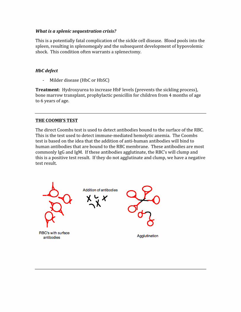
What is a splenic sequestration crisis?
This is a potentially fatal complication of the sickle cell disease. Blood pools into the spleen, resulting in splenomegaly and the subsequent development of hypovolemic shock. This condition often warrants a splenectomy.
HbC defect
-‐ Milder disease (HbC or HbSC)
Treatment: Hydroxyurea to increase HbF levels (prevents the sickling process), bone marrow transplant, prophylactic penicillin for children from 4 months of age to 6 years of age.
THE COOMB’S TEST
The direct Coombs test is used to detect antibodies bound to the surface of the RBC. This is the test used to detect immune-‐mediated hemolytic anemia. The Coombs test is based on the idea that the addition of anti-‐human antibodies will bind to human antibodies that are bound to the RBC membrane. These antibodies are most commonly IgG and IgM. If these antibodies agglutinate, the RBC’s will clump and this is a positive test result. If they do not agglutinate and clump, we have a negative test result.
HEREDITARY SPHEROCYTOSIS
This is an AD disorder where there is a defect in the gene that codes for spectrin, resulting in a decreased content of spectrin. This causes a loss of the membrane surface area with no decrease in volume. These two cause the shape to shift from circular to spherical.
-‐ The osmotic fragility test is a way to test the RBC’s ability to withstand hypotonic saline. The spherical shape will tolerate the solution less than the regular RBC shape, thus causing it to rupture faster.
-‐ Peripheral smear would show spherocytes -‐ Coombs test is negative
GLUCOSE-‐6-‐PHOSPHATE DEHYDROGENASE DEFICIENCY
This is an x-‐linked recessive disorder that is usually precipitated by infections, fava beans, primaquine, dimercaptol, sulfonamides, and nitrofurantoin.
-‐ Patient gets episodes of hemolytic anemia that is often precipitated by an aggravating factor
-‐ Patient will get jaundice and have dark urine -‐ Peripheral smear will show bite cells (caused by macrophages) and Heinz
bodies (hemoglobin precipitation inside RBCs) -‐ Diagnose by measuring G6PD levels.
Treatment: Avoid precipitating factors, transfuse as necessary
AUTOIMMUNE HEMOLYTIC ANEMIA
Autoantibodies against the RBC membrane cause destruction of RBC’s. There are two possible causes for this, IgG antibodies or IgM antibodies
IgG causes à “warm” autoimmune hemolytic anemia. This means that binding of IgG to the RBC membrane occurs optimally at 37°C. This causes extravascular hemolysis where the primary site of sequestration is the spleen.
IgM causes à “cold” autoimmune hemolytic anemia. Binding of IgM to the RBC membrane occurs optimally at 0°C to 5°C. This causes intravascular hemolysis and complement activation, where the primary site of sequestration is the liver.
Diagnosing:
-‐ Direct Coombs test: If +ve = warm, if –ve = cold
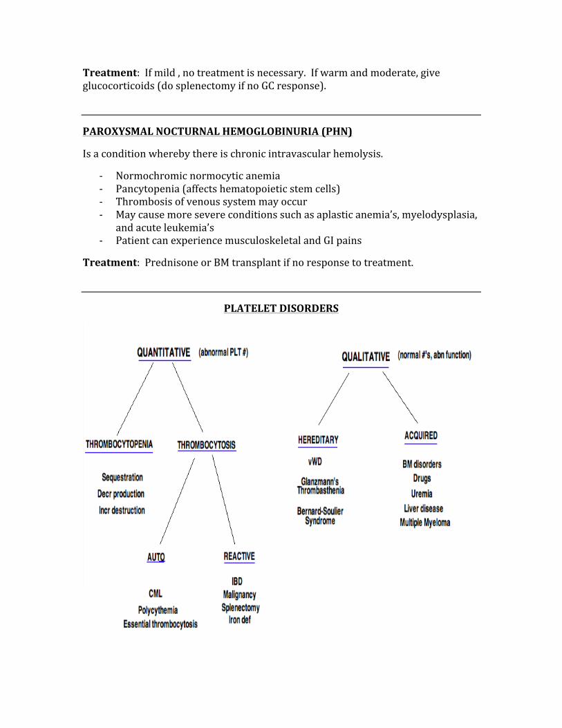
Treatment: If mild , no treatment is necessary. If warm and moderate, give glucocorticoids (do splenectomy if no GC response).
PAROXYSMAL NOCTURNAL HEMOGLOBINURIA (PHN)
Is a condition whereby there is chronic intravascular hemolysis.
-‐ Normochromic normocytic anemia -‐ Pancytopenia (affects hematopoietic stem cells) -‐ Thrombosis of venous system may occur -‐ May cause more severe conditions such as aplastic anemia’s, myelodysplasia,
and acute leukemia’s -‐ Patient can experience musculoskeletal and GI pains
Treatment: Prednisone or BM transplant if no response to treatment.
PLATELET DISORDERS

THROMBOCYTOPENIA
This is when the platelet count falls below 150,000
Causes:
-‐ Decreased production due to: BM injury, suppression, invasions -‐ Increased destruction due to: DIC, TTP, infection, drugs, ITP, HIV -‐ Sequestration -‐ Pregnancy: usually not a major concern -‐ Dilutional effects from transfusion -‐ Determination of cause can be made with CBC, peripheral smear, and bone
marrow biopsy.
Signs/Symptoms:
-‐ Petechial bleeding (pinpoint bleeding) -‐ Mucosal bleeding (ie epistaxis, menorrhagia, hemoptysis) -‐ Excessive bleeds after injury and/or surgical procedures
THROMBOTIC THROMBOCYTOPENIC PURPURA (TTP)
A condition whereby there is excessive platelet consumption, leading to an emergency situation that can lead to death rather quickly (few months).
Signs/Symptoms:
-‐ Altered mental status -‐ Hemolytic anemia -‐ Thrombocytopenia
Treatment: Plasmapharesis is required to maintain life, corticosteroids and splenectomy may also be required.
IDIOPATHIC THROMBOCYTOPENIC PURPUA (ITP)
Is an autoimmune formation of antibodies against platelets. IgG antibodies adhere to and destroy the platelets which are then removed by splenic macrophages.
Acutely à Is a self-‐limited condition seen in children, where the condition is almost always preceeded by a viral infection.
Chronically à Is a spontaneous form of ITP seen most commonly in middle-‐aged females. Is self-‐limited.
GLANZMANN’S THROMBASTHENIA
This is an AR disorder where there is a deficiency in platelet aggregation due to a deficiency of glycoprotein GPIIb-‐IIIa. The only altered test is increased bleeding time.
BERNARD-‐SOULIER SYNDROME
Is an AR disorder of platelet adhesion due to a deficiency of glycoprotein GPIb-‐IX. The platelet count will be low with abnormally large platelets on peripheral smear.
DISORDERS OF COAGULATION
1. von Willebrand’s Disease (vWD) 2. Hemophilia A 3. Hemophilia B 4. Disseminated Intravascular Coagulation (DIC) 5. Vitamin K deficiency 6. Coagulopathy of liver disease 7. Inherited hypercoagulable states
VON WILLEBRAND’S DISEASE
Is an AD disorder that is caused by a deficiency or defect of the vWF (factor 8-‐related antigen). vWF is required for the first step of platelet aggregation in clot formation.
* vWF is the most common inherited bleeding disorder, affecting up to 3% of the population.
Signs/Symptoms:
-‐ Cutaneous bleeding -‐ Mucosal bleeding -‐ Menorrhagia seen in more than half of females with vWD -‐ Many patients won’t show anything sign or symptoms until they undergo a
surgical procedure and have excessive bleeding -‐ Bleeding time will be prolonged, platelet count is normal
-‐ PTT may be prolonged, vWF is decreased, factor 8 activity is decreased -‐ Ristocetin levels should be checked in diagnosing vWD
Treatment: Desmopressin to induce endothelial cells to secrete von Willebrand factor (not effective in type 3 vWD). Factor 8 concentrates is recommended for type 3 vWD. Patient should avoid NSAIDs.
HEMOPHILIA A
Is an x-‐linked recessive disorder that affects male patients, and is caused by a deficiency or defect of factor 8.
Signs/Symptoms:
-‐ Bleeding into joints (hemarthroses) -‐ Intramuscular bleeds -‐ Intracranial bleeds (therefore head trauma must be taken very seriously in
these patients)
Diagnosing:
-‐ Low factor 8 levels + normal vWF -‐ PTT is prolonged
Treatment: Replace clotting factors, desmopressin may be helpful in some patients.
HEMOPHILIA B
Is an x-‐linked recessive disorder caused by a deficiency of factor 9, and is much less common than hemophilia A. Management involves replacing missing factors.
DISSEMINATED INTRAVASCULAR COAGULATION (DIC)
DIC is a disorder characterized by an abnormal activation of the coagulation sequence, which leads to widespread formation of microthrombi throughout the microcirculation. This leads to the consumption of clotting factors, platelets, and fibrin. There is also an activation of fibrinolytic mechanisms, thus leading to hemorrhages. BLEEDS + THROMBOSIS
Common causes:
-‐ The most common cause is infection (Gram –ve sepsis is MCC) -‐ Pregnancy complications -‐ Trauma
-‐ Malignancy -‐ Shock
Signs/Symptoms:
-‐ Oozing from procedure sites -‐ Ecchymoses -‐ Petechia -‐ Purpura -‐ Thromboses seen more often in chronic cases of DIC
Treatment: Correct underlying conditions and apply supportive measures.
VITAMIN K DEFICIENCY
There are many clotting factors that require vitamin K as a cofactor in synthesis, including: Protein C & S, and factors 2, 7, 9, 10. Vitamin K deficiency is seen in very ill patients who are being fed through a tube, as well as those who are using oral warfarin as an anti-‐coagulant.
Signs/Symptoms:
-‐ Significant hemorrhages -‐ PT prolongation (is the first finding), then PTT prolongation.
Treatment: Vitamin K replacement.
• If patient has a severe bleed, fresh frozen plasma should be given as it contains all of the clotting factors.
COAGULOPATHY OF LIVER DISEASE
Since the liver synthesizes all clotting factors, any disease of the liver can cause coagulopathies (abnormal bleeding, prolongation of PT/PTT).
Treatment: Fresh frozen plasma if PT/PTT are altered or there is significant bleeding.
INHERITED HYPERCOAGULOPATHIES
-‐ Antithrombin 3 deficiency (increased thrombosis) -‐ Antiphospholipid antibody syndrome (arterial or venous thrombosis) -‐ Protein C deficiency (unregulated fibrin synthesis) -‐ Protein S deficiency (leads to a deficiency of protein C activity) -‐ Factor V leiden (unregulated prothrombin activation – increased
thromboembolic events) -‐ Prothrombin gene mutation
PLASMA CELL DISORDERS
1. Multiple Myeloma 2. Waldenstrom’s Macroglobulinemia 3. Monoclonal Gammopathy of Undetermined Significant (MGUS)
MULTIPLE MYELOMA
Is a cancer of the bone marrow that produces large amounts of IgG or IgA. This is the most common tumor arising in adults
It causes:
-‐ Bone lesions -‐ Hypercalcemia -‐ Anemia -‐ Infections
Signs/Symptoms:
-‐ Skeletal manifestations (bone pain, fractures, vertebral collapse) -‐ Normocytic normochromic anemia (due to BM infiltration and renal failure) -‐ Renal failure -‐ Infections (secondary to deprivation of normal Ig’s affects humoral
immunity, and is the MCC of death) -‐ Amyloidosis
Characteristic Findings:
-‐ Bence Jones proteins in urine (are Ig light chains) -‐ M-‐spike (is a monoclonal Ig spike seen on serum electrophoresis)
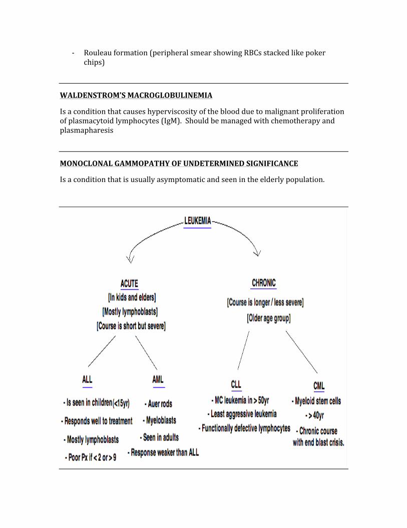
-‐ Rouleau formation (peripheral smear showing RBCs stacked like poker chips)
WALDENSTROM’S MACROGLOBULINEMIA
Is a condition that causes hyperviscosity of the blood due to malignant proliferation of plasmacytoid lymphocytes (IgM). Should be managed with chemotherapy and plasmapharesis
MONOCLONAL GAMMOPATHY OF UNDETERMINED SIGNIFICANCE
Is a condition that is usually asymptomatic and seen in the elderly population.
LYMPHOMAS
HODGKIN’S vs. NON-‐HODGKIN’S
HODGKIN’S NON-‐HODGKIN’S Reed-‐Sternberg cells present.
Only single group of LN’s affected.
Low-‐grade fever, weight loss, night
sweats.
Associated with Epstein Barr Virus.
Prognosis dependent on amount of lymphocytes and Reed-‐Sternberg cells (best when increased lymphocytes and decreased Reed-‐Sternberg cells).
Is associated with states of immunosuppression.
Many LN’s involved with extranodal
involvement.
B cells are of the majority.
Very few constitutional symptoms Occurs from 20-‐40yr of age.
CARDIAC PATHOLOGY
ISCHEMIC HEART DISEASE
1. Stable Angina 2. Unstable Angina 3. Variant Angina (Prinzmetal’s)
STABLE ANGINA
Fixed atherosclerotic lesions narrow the coronary arteries, leading to an imbalance between blood supply and 02 demand. This leads to inadequate perfusion, and oxygen demand exceeds blood supply. Atherosclerosis leads to narrowing > 75%.
Signs/Symptoms:
-‐ Substernal chest pain lasting less than 15 minutes -‐ Pain is described as squeezing, heaviness, pressure -‐ Always brought on by physical exertion -‐ Pain goes away with rest and/or nitroglycerine
UNSTABLE ANGINA
In unstable angina, the cause of chest pain is due to a reduced resting coronary blood flow. The main difference between unstable vs. stable angina is that the pain of unstable angina occurs at rest.
PRINZMETAL’S ANGINA
Prinzmetal’s angina is caused by a transient coronary vasospasm that is accompanied by a fixed atherosclerotic lesion. The symptoms occur at rest.
Signs/Symptoms:
-‐ Chest pain at rest -‐ Most common in younger females who smoke cigarettes -‐ There will be a transient ST-‐segment elevation on EKG during the episodes of
chest pain
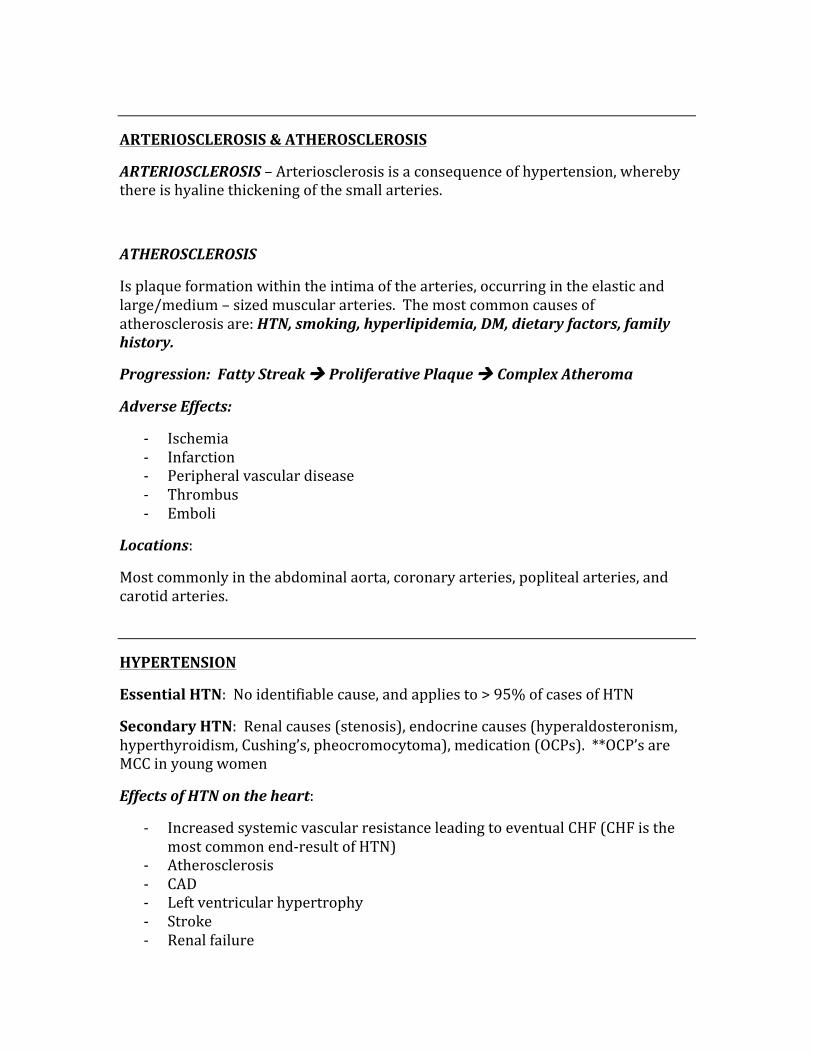
ARTERIOSCLEROSIS & ATHEROSCLEROSIS
ARTERIOSCLEROSIS – Arteriosclerosis is a consequence of hypertension, whereby there is hyaline thickening of the small arteries.
ATHEROSCLEROSIS
Is plaque formation within the intima of the arteries, occurring in the elastic and large/medium – sized muscular arteries. The most common causes of atherosclerosis are: HTN, smoking, hyperlipidemia, DM, dietary factors, family history.
Progression: Fatty Streak à Proliferative Plaque à Complex Atheroma
Adverse Effects:
-‐ Ischemia -‐ Infarction -‐ Peripheral vascular disease -‐ Thrombus -‐ Emboli
Locations:
Most commonly in the abdominal aorta, coronary arteries, popliteal arteries, and carotid arteries.
HYPERTENSION
Essential HTN: No identifiable cause, and applies to > 95% of cases of HTN
Secondary HTN: Renal causes (stenosis), endocrine causes (hyperaldosteronism, hyperthyroidism, Cushing’s, pheocromocytoma), medication (OCPs). **OCP’s are MCC in young women
Effects of HTN on the heart:
-‐ Increased systemic vascular resistance leading to eventual CHF (CHF is the most common end-‐result of HTN)
-‐ Atherosclerosis -‐ CAD -‐ Left ventricular hypertrophy -‐ Stroke -‐ Renal failure
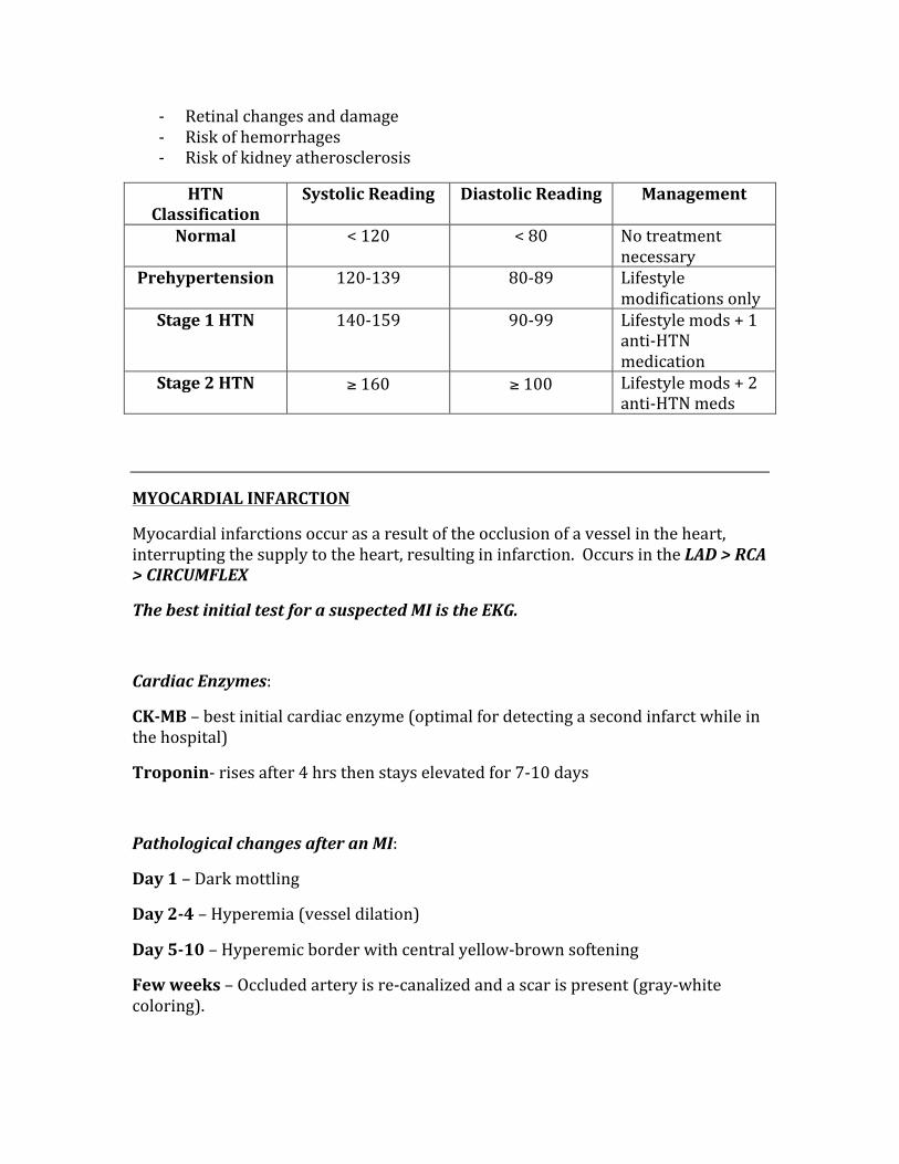
-‐ Retinal changes and damage -‐ Risk of hemorrhages -‐ Risk of kidney atherosclerosis
HTN Classification
Systolic Reading Diastolic Reading Management
Normal < 120 < 80 No treatment necessary
Prehypertension 120-‐139 80-‐89 Lifestyle modifications only
Stage 1 HTN 140-‐159 90-‐99 Lifestyle mods + 1 anti-‐HTN medication
Stage 2 HTN ≥ 160 ≥ 100 Lifestyle mods + 2 anti-‐HTN meds
MYOCARDIAL INFARCTION
Myocardial infarctions occur as a result of the occlusion of a vessel in the heart, interrupting the supply to the heart, resulting in infarction. Occurs in the LAD > RCA > CIRCUMFLEX
The best initial test for a suspected MI is the EKG.
Cardiac Enzymes:
CK-‐MB – best initial cardiac enzyme (optimal for detecting a second infarct while in the hospital)
Troponin-‐ rises after 4 hrs then stays elevated for 7-‐10 days
Pathological changes after an MI:
Day 1 – Dark mottling
Day 2-‐4 – Hyperemia (vessel dilation)
Day 5-‐10 – Hyperemic border with central yellow-‐brown softening
Few weeks – Occluded artery is re-‐canalized and a scar is present (gray-‐white coloring).
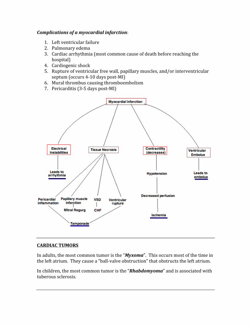
Complications of a myocardial infarction:
1. Left ventricular failure 2. Pulmonary edema 3. Cardiac arrhythmia (most common cause of death before reaching the
hospital) 4. Cardiogenic shock 5. Rupture of ventricular free wall, papillary muscles, and/or interventricular
septum (occurs 4-‐10 days post-‐MI) 6. Mural thrombus causing thromboembolism 7. Pericarditis (3-‐5 days post-‐MI)
CARDIAC TUMORS
In adults, the most common tumor is the “Myxoma”. This occurs most of the time in the left atrium. They cause a “ball-‐valve obstruction” that obstructs the left atrium.
In children, the most common tumor is the “Rhabdomyoma” and is associated with tuberous sclerosis.
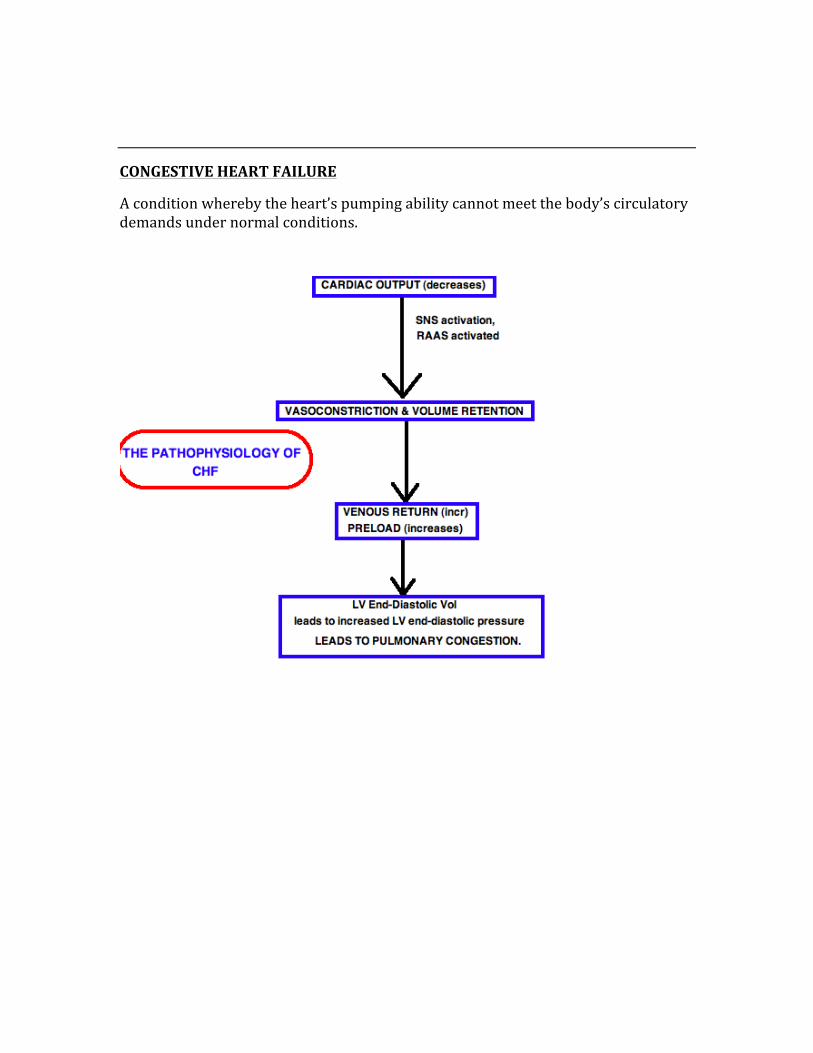
CONGESTIVE HEART FAILURE
A condition whereby the heart’s pumping ability cannot meet the body’s circulatory demands under normal conditions.
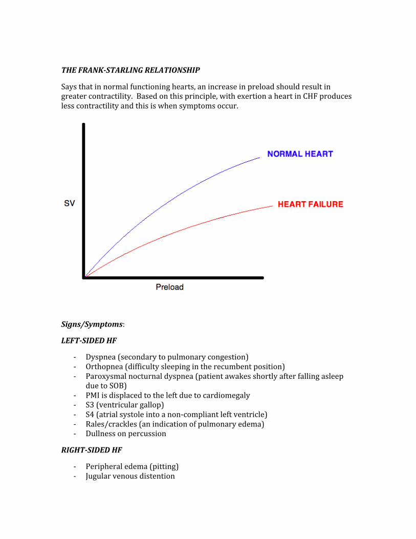
THE FRANK-‐STARLING RELATIONSHIP
Says that in normal functioning hearts, an increase in preload should result in greater contractility. Based on this principle, with exertion a heart in CHF produces less contractility and this is when symptoms occur.
Signs/Symptoms:
LEFT-‐SIDED HF
-‐ Dyspnea (secondary to pulmonary congestion) -‐ Orthopnea (difficulty sleeping in the recumbent position) -‐ Paroxysmal nocturnal dyspnea (patient awakes shortly after falling asleep
due to SOB) -‐ PMI is displaced to the left due to cardiomegaly -‐ S3 (ventricular gallop) -‐ S4 (atrial systole into a non-‐compliant left ventricle) -‐ Rales/crackles (an indication of pulmonary edema) -‐ Dullness on percussion
RIGHT-‐SIDED HF
-‐ Peripheral edema (pitting) -‐ Jugular venous distention
-‐ Ascites -‐ Hepatomegaly -‐ Right ventricular heave
VALVULAR HEART DISEASE (MURMURS)
1. Mitral Regurgitation 2. Aortic Stenosis 3. Ventricular Septal Defect 4. Mitral Prolapse 5. Aortic Regurgitation 6. Mitral Stenosis 7. Patent Ductus Arteriosus 8. Hypertrophic obstructive cardiomyopathy (HOCM)
All valvular heart diseases present with shortness of breath initially. In young people, the most commonly encountered valve disorders are: Mitral prolapse, mitral stenosis, or bicuspid aortic valves.
Murmur Intensity:
I/VI – Only heard with specific maneuvers (ie Valsalva)
II/VI and III/VI – This is where the majority of murmurs lie.
IV/VI – Palpable thrill is present
V/VI – Murmur can be heard with the stethoscope partially off the chest
VI/VI – Murmur can be heard without a stethoscope
MITRAL REGURGITATION – A holosystolic “blowing murmur” that is best heard at the apex of the heart.
AORTIC STENOSIS – The “crescendo-‐decrescendo” systolic ejection murmur following an ejection click.
VSD – A holosystolic murmur
MITRAL PROLAPSE – A late systolic murmur with a midsystolic click
AORTIC REGURGITATION – High-‐pitched blowing diastolic murmur
MITRAL STENOSIS – A late rumbling diastolic murmur following an opening snap
PDA – A machine-‐like murmur
HYPERTROPHIC OBSTRUCTIVE CARDIOMYOPATHY – An autosomal dominant trait that can result in sudden death in young athletes. The walls of the LV and interventricular septum hypertrophy, creating a “banana shape” in the ventricle.
CARDIAC ARRHYTHMIAS
Tachyarrhythmias:
-‐ Atrial Fibrillation -‐ Atrial Flutter -‐ Multifocal Atrial Tachycardia -‐ Paroxysmal Supraventricular Tachycardia -‐ Wolff-‐Parkinson-‐White Syndrome -‐ Ventricular Tachycardia -‐ Ventricular Fibrillation
Bradyarrhythmias:
-‐ Sinus Bradycardia -‐ Sick Sinus Syndrome -‐ AV Blocks (1st, 2nd, and 3rd degree blocks)
ATRIAL FIBRILLATION
An irregular, rapid ventricular rate is caused by multiple foci in the atria that fire erratically. The atrial rate is as high as 400bpm.
Patient will experience:
-‐ Palpitations -‐ Dizziness -‐ Exertional dyspnea -‐ Irregularly irregular pulse
Goals of treatment are:
1. Ventricular rate control 2. Restoration of normal sinus rhythm 3. Give anticoagulation (if needed)
ATRIAL FLUTTER
One foci in the atrium fires automatically, causing an atrial rate of 250-‐350bpm, with only 1 out of 3 of these contractions making it to the ventricle. The EKG will show with the classic “sawtooth” pattern.
MULTIFOCAL ATRIAL TACHYCARDIA
This is a condition that occurs most commonly in those with COPD. There will be at least three different P-‐wave morphologies with variable PR and RR intervals.
Can diagnose with vagal maneuvers and/or adenosine administration to show the an AV block without disrupting the atrial tachycardia.
PAROXYSMAL SUPRAVENTRICULAR TACHYCARDIA
Is due to a reentrant circuit within the AV node. There will be narrow QRS complexes with no discernable P waves on EKG. This condition may be caused by the following:
-‐ Digoxin toxicity -‐ Ischemic heart disease -‐ Atrial flutter -‐ Presence of accessory pathways -‐ Excessive consumption of alcohol or caffeine
WOLFF-‐PARKINSON-‐WHITE SYNDROME
There is an accessory pathway from the atria to the ventricles that cause premature ventricular excitations due to a lack of delay in the AV node.
There are two mechanisms by which this condition can cause a paroxysmal tachycardia:
1. Supraventricular Tachycardias – All impulses get through to the ventricle in this condition, whereas in normal circumstances only one Atrial impulse gets through.
2. Orthodromic Reciprocation Tachycardia – creates a re-‐entry loop that causes multiple depolarizations of the atria.
VENTRICULAR TACHYCARDIA
This condition is defined as a rapid and repetitive firing of three or more premature ventricular contractions in a row, at a rate of 100-‐250bpm. Is responsible fro up to 75% of cardiac arrest.
The most common causes of ventricular tachycardia are:
-‐ Cardiomyopathies -‐ Hypotension -‐ CAD -‐ Prolonged QT syndrome -‐ Drug toxicities
Signs/Symptoms:
-‐ Palpitations -‐ Dyspnea -‐ Angina -‐ Syncope -‐ Widened and erratic QRS complex on EKG
** A rapid, polymorphic form of ventricular tachycardia can lead to a condition known as “Torsades des Pointes”.
VENTRICULAR FIBRILLATION
Occurs when there are multiple foci within the ventricles that fire rapidly, which leads to a chaotic quivering of the ventricles. Most cases of VFib occur as a result of ventricular tachycardia. This condition is fatal when untreated.
** Association of VFib with an MI creates a favorable long-‐term prognosis, whereas no association between the two gives a high rate of recurrence.
Signs/Symptoms:
-‐ Lack of pulse, heart sounds, and BP -‐ Patient loses consciousness and will die without intervention
** Immediate defibrillation and CPR should be performed to prevent sudden death. There are no medications that can convert this condition to normal rhythm.
** Note that all narrow complex tachycardias will originate from above the ventricle, whereas wide complex tachycardias originate within the ventricles.
Bradyarrhythmias:
SINUS BRADYCARDIA
Is a sinus rate of < 60 bpm, and becomes dangerous when the rate drops below 45 bpm. Common causes of sinus bradycardia are: Ischemia, Increased Vagal Tone, Anti-‐arrhythmics
** Patients are usually symptomatic and may present with only fatigue
** Atropine will block vagal stimulation and thus elevate the sinus rate.
SICK SINUS SYNDROME
This is a dysfunction of the sinus node that causes persistent spontaneous bouts of sinus bradycardia. Patient will experience dizziness, confusion, fatigue, and CHF.
AV BLOCKS
1st degree – A benign condition where the PR interval is prolonged to >0.20 seconds
2nd degree – There are Mobitz type 1 and Mobitz type 2
Mobitz Type 1 – progressive prolongation of PR interval until there is an absent P wave. Does not require treatment.
Mobitz Type 2 – there is a missing P wave without any changes in the preceding PR interval, causing the dropping of a QRS complex all of a sudden. May progress and become a complete heart block. Requires pacemaker implant.
3rd degree (Complete heart block) – there is an absence of atrial impulses to the ventricle, thus there is no relationship between P waves and QRS complexes. Requires pacemaker.
CARDIOMYOPATHIES
HYPERTROPHIC CARDIOMYOPATHY
Asymmetric cardiomyopathy that involves the interventricular septum results in diastolic dysfunction. The walls of the left ventricle become thickened. A banana shape occurs in the LV, can result in sudden death in young athletes.
DILATED CARDIOMYOPATHY
Dilation of the heart that is most commonly caused by: Alcohol, Adriamycin, Radiation, Chaga’s disease, Coxsackie B virus. This condition result in systolic dysfunction.
RESTRICTIVE CARDIOMYOPATHY
Myocardial infiltration results in impaired diastolic filling of the ventricular due to a decrease in ventricular compliance. Common causes are: Amyloidosis, Sarcoidosis, Hemochromatosis, Scleroderma.
PERICARDIAL DISORDERS
1. Acute Pericarditis 2. Constrictive Pericarditis 3. Pericardial Effusion 4. Cardiac Tamponade
ACUTE PERICARDITIS
Is an acute condition that results in chest pain, diffuse ST-‐elevations and PR depression on EKG, a pericardial friction rub, and possible pericardial effusion.
May be caused by any of the following:
-‐ Most cases occur after a viral illness (most commonly an URI) -‐ Infectious (Coxsackie virus, hepatitis, HIV, TB, toxoplasmosis, or fungal
infections) -‐ Collagen vascular disease -‐ Post-‐surgery -‐ Amyloidosis -‐ Lupus -‐ Post-‐MI (known as Dressler’s syndrome)
** Recovery occurs within 1-‐3 weeks and requires only NSAIDs for management.
Complications may include:
-‐ Effusion -‐ Tamponade occurs in 10%-‐20% of patients
CONSTRICTIVE PERICARDITIS
Occurs secondary to fibrous scarring of the pericardium, leading to rigidity and thickening of the pericardium. There are a few possible causes, including:
-‐ Connective tissue disorders -‐ Chronic pericardial effusion -‐ Radiation therapy -‐ Uremia -‐ Prior heart surgeries
Signs/Symptoms:
-‐ Symptoms of hepatic congestion (edema, ascites) -‐ Pulmonary congestion -‐ JVD -‐ Pericardial knock
PERICARDIAL EFFUSION
Occurs when pericardial space becomes occupied with fluid. Occurs when there is ascites and pleural effusion present. Often occurs when the patient has CHF, nephrotic conditions, and/or cirrhosis.
Signs/Symptoms:
-‐ Pericardial friction rub -‐ Muffled heart sounds -‐ Point of maximal impulse (PMI) is softened
CARDIAC TAMPONADE
Is a pericardial effusion that impairs diastolic filling of the heart. Pressure of all chambers, the pulmonary artery, and pericardium are equal in pressure, thus ventricular filling is impaired.
Common causes:
-‐ Free-‐wall rupture after an MI -‐ Medical errors causing puncture to heart -‐ Penetrating injuries
Signs/Symptoms:
-‐ Hypotension -‐ JVD -‐ Muffled heart sounds
** Known as “Beck’s Triad”
-‐ Narrowed pulse pressure -‐ Pulsus paradoxus (exaggerated decrease in arterial pressure during
inspiration > 10mmHg) -‐ Muffled heart sounds -‐ Cardiogenic shock
RHEUMATIC HEART DISEASE
Rheumatic fever/heart disease is a condition that occurs as a consequence of pharyngitis from a group A strep infection. The mitral valve is the most common valve affected. The progression to a heart disease is an immune-‐mediated process, not a result of the bacterial infection.
Diagnosing is made with the JONES criteria, and require the presence of two major criteria OR one major and one minor criteria:
MAJOR CRITERIA
Migratory Polyarthritis
Erythema Marginatum
Chorea
Cardiac Involvement
Subcutaneous Nodules
MINOR CRITERIA
Fever
ESR elevation
Polyarthralgias
PR interval prolongation
Prior history of RF infection
Evidence of preceeding strep infection
ENDOCARDITIS
Is an infection of the endocardium that can be classified as either acute or subacute. Most often, the mitral valve is involved, whereas an infection of the tricuspid valve is usually due to IV drug abuse.
Acute Endocarditis – Caused by Staph Aureus and creates large vegetations on a previously healthy/normal valve.
Subacute Endocarditis – Caused by Viridians group Strep and creates small vegetations on an abnormal/diseased valve. ** Occurs with dental procedures
The symptoms of bacterial endocarditic can be remembered with the mnemonic “FROM JANE”.
F – Fever
R – Roth’s spots (retinal hemorrhages)
O – Osler’s nodes (painful, raised red lesions on hands and feet)
M – Murmur
J – Jane way lesions (non-‐tender lesions on palms and soles)
A – Anemia
N – Nail-‐bed hemorrhage
E – Emboli
** Libman-‐Sacks Endocarditis – is a form of endocarditis seen in patients with Lupus, where there are vegetations found on both sides of the valve, leading to stenosis.
EMBOLI
Emboli are things that travel through the circulation an get lodged in the pulmonary vasculature, leading to a pulmonary embolus (PE).
The most commonly encountered types of emboli are: Fat (due to long bone breaks and liposuction), Air (can occur as a result of injection of air into the circulatory system), Thrombus (breaks of from a deep vein thrombosus), Amniotic Fluid (from pregnancy complications).
** NOTE: Thrombi causing a pulmonary embolism will present with chest pain and shortness of breath in the patient.
DVT
A deep vein thrombosis (DVT) is the precursor to an emboli, and is caused by patients who have the following:
1. Stasis (they don’t move enough – classically they took a long flight) 2. Hypercoagulability (due to disease states or familial) 3. Endothelial Damage
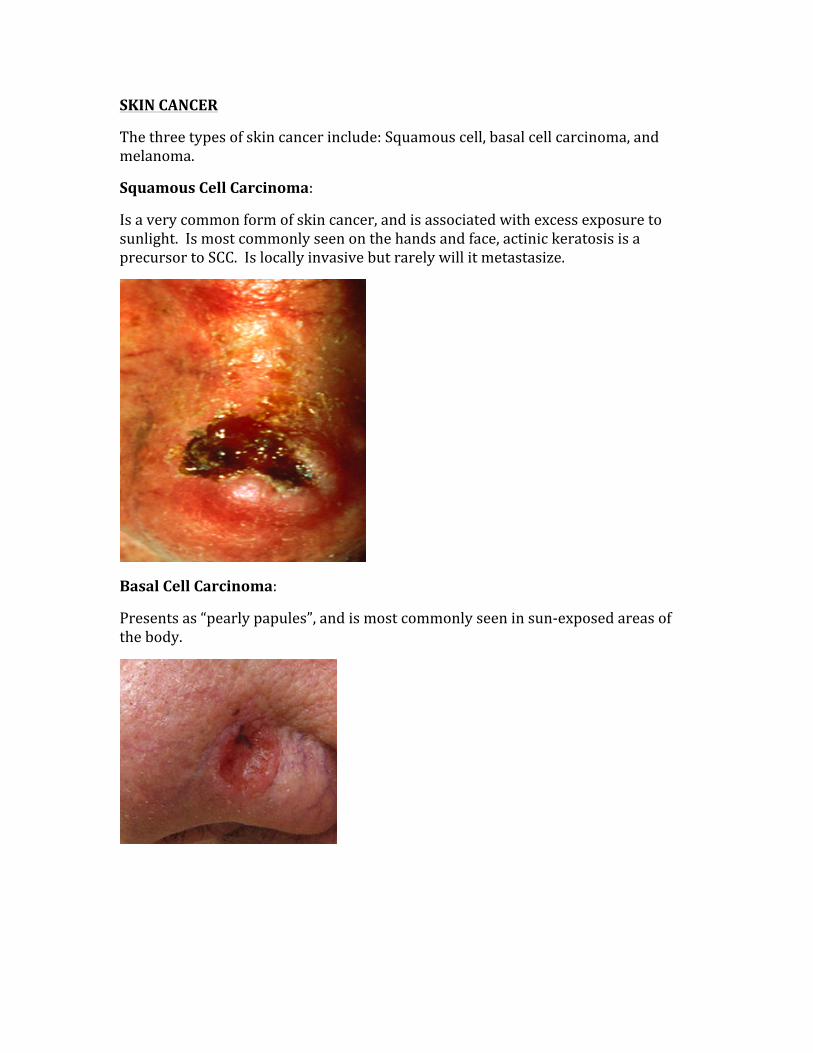
SKIN CANCER
The three types of skin cancer include: Squamous cell, basal cell carcinoma, and melanoma.
Squamous Cell Carcinoma:
Is a very common form of skin cancer, and is associated with excess exposure to sunlight. Is most commonly seen on the hands and face, actinic keratosis is a precursor to SCC. Is locally invasive but rarely will it metastasize.
Basal Cell Carcinoma:
Presents as “pearly papules”, and is most commonly seen in sun-‐exposed areas of the body.
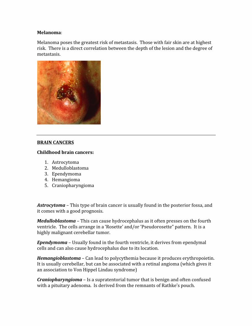
Melanoma:
Melanoma poses the greatest risk of metastasis. Those with fair skin are at highest risk. There is a direct correlation between the depth of the lesion and the degree of metastasis.
BRAIN CANCERS
Childhood brain cancers:
1. Astrocytoma 2. Medulloblastoma 3. Ependymoma 4. Hemangioma 5. Craniopharyngioma
Astrocytoma – This type of brain cancer is usually found in the posterior fossa, and it comes with a good prognosis.
Medulloblastoma – This can cause hydrocephalus as it often presses on the fourth ventricle. The cells arrange in a ‘Rosette’ and/or ‘Pseudorosette” pattern. It is a highly malignant cerebellar tumor.
Ependymoma – Usually found in the fourth ventricle, it derives from ependymal cells and can also cause hydrocephalus due to its location.
Hemangioblastoma – Can lead to polycythemia because it produces erythropoietin. It is usually cerebellar, but can be associated with a retinal angioma (which gives it an association to Von Hippel Lindau syndrome)
Craniopharyngioma – Is a supratentorial tumor that is benign and often confused with a pituitary adenoma. Is derived from the remnants of Rathke’s pouch.
Adulthood brain cancers:
Pituitary adenoma – Most commonly as prolactin-‐secreting form, the most common presentation is bitemporal hemianopsia.
Glioblastoma Multiforme – Is the most common primary brain tumor, has a terrible prognosis. This is found in the hemispheres and often crosses the corpus callosum (giving it the term ‘butterfly glioma’).
Meningioma – Comes from arachnoid cells, making it external to the brain. Is the 2nd most common brain tumor in adults.
Oligodendroglioma – Is a slow growing and rare tumor, seen usually in the frontal lobe of the brain.
Schwannoma – Is the 3rd most common adult brain tumor, originating from Schwann cells. When located near the 8th cranial nerve can cause an acoustic schwannoma.
TUMORS OF THE BONE
BENING TUMORS OF BONE
Enchondroma
Is a cartilaginous tumor found in the intramedullary bone, most often in distal extremities.
Osteochondroma
In men <25yr of age, is the most common benign tumor of bone. Origination is from the long
metaphyseal bone.
Giant Cell Tumor
Occurs from 20-‐40yr of age. Usually found in distal
femur/proximal tibia. Has the classic “double bubble” sign on
xray.
MALIGNANT TUMORS OF BONE
Osteosarcoma
The most common malignant bone tumor, occurring in 10-‐20yr olds. Is most commonly seen in the metaphysis of long bones.
Ewing’s Sarcoma
In males <15yr of age, has the 11;22 translocation. Is likely to metastasize early, and has the characteristic ‘onion skinning on xray. Seen in diaphysis of long bones, ribs, pelvis, and scapula.
Chondrosarcoma
A malignant tumor of cartilaginous bone, seen in males >30yr of age. Not seen in the long bones.
WHICH TUMORS METASTASIZE TO THE BRAIN, BONE, AND LIVER
TUMOR MARKERS
The following list are the common markers that are used to either make a diagnosis of a certain cancer, or to monitor the effectiveness of therapy.
TUMOR MARKER WHAT IS WATCHES Carcinoembryonic Antigen (CEA) Colorectal and pancreatic cancers Prostatic Specific Antigen (PSA) Used for screening of prostatic cancer CA-‐125 Detects ovarian cancer and malignant
epithelial tumors. Alkaline Phosphatase Bone metastasis, bile duct obstruction,
Paget’s bone disease β-‐hCG Hydatiform moles, GTD’s, and
choriocarcinomas α-‐feto protein Non-‐seminomatous germ cell tumors of
the testicles Tartrate-‐resitant acid phosphatase Hairy cell leukemia
TO BRAIN
Lung
Skin
Kidney
GI tract
TO BONE
Breast
Lung
Thyroid
Tested
Kidney
Prostate
TO LIVER
Colon
Stomach
Pancreas
Breast
Lung
TUMOR SUPPRESSORS
Tumor suppressors work by suppressing the growth of certain tumors, when there is a loss of function, both alleles of the gene have been changed (ie mutation, deletion, etc)
TUMOR SUPPRESSOR TUMOR IT SUPPRESSES Rb Retinoblastoma BRCA 1 and 2 Breast cancer, ovarian cancer p53 Helps screen/follow almost all cancers APC Colorectal cancer NF1 and NF2 Neurofibromatosis 1 and 2 WT1 Wilm’s tumor
ONCOGENES
ONCOGENE ASSOCIATED TUMOR Ret MEN syndromes type 2 and 3 c-‐myc Burkitt’s lymphoma L-‐myc Lung tumors N-‐myc Neuroblastoma Bcl-‐2 Follicular lymphomas Erb-‐B2 Breast, ovary, gastric Ras Colon
RESPIRATORY
CANCER OF THE LUNG
CENTRALLY ARISING PERIPHERALLY ARISING Small Cell Carcinoma – linked to smoking, can produce ACTH and ADH, may be linked to Lambert-‐Eaton syndrome Squamous Cell Carcinoma – linked to smoking and the production of ectopic PTrP.
Adenocarcinoma – is the most common peripherally arising cancer of the lung. Large Cell Carcinoma – is an undifferentiated carcinoma of the lung. Bronchioalveolar Carcinoma – this is the lung cancer that is not thought to be related to smoking
Lung cancer can cause a wide array of symptoms (aside from cough, hemoptysis, wheezing, bronchial obstruction). The most common symptoms that can arise from lung cancer are:
-‐ Pancoast’s tumor (a carcinoma that originates in the apex of the lung and can compress the cervical sympathetic plexus, resulting in Horner’s syndrome)
-‐ Superior vena cava syndrome -‐ Horner’s syndrome (Ptosis, Anhydrosis, Miosis) -‐ Paraneoplastic disorders (PTrP, ADH, ACTH) -‐ Recurrent laryngeal nerve symptoms (hoarseness)
OBSTRUCTIVE AND RESTRICTIVE LUNG DISEASE
OBSTRUCTIVE – this condition causes an obstruction of air -‐low that results in air-‐trapping in the lungs. There will always be an increased in total lung volume with a FEV1/FVC of <80%.
The categories of obstructive lung disorders are:
1. Chronic Bronchitis: This condition is characterized by the presence of a productive cough for at least 3 consecutive months for 2 or more years. There is a hypertrophy of the mucus-‐secreting glands of the bronchioles, giving a Reid index of > 50%. Patient will have wheezing, crackles, and cyanosis on physical exam. *This patient is a “blue bloater” because they become cyanotic.
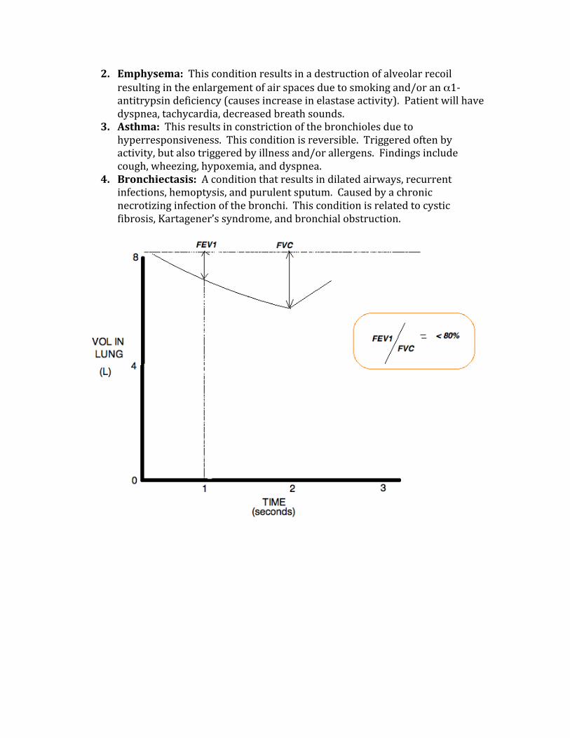
2. Emphysema: This condition results in a destruction of alveolar recoil resulting in the enlargement of air spaces due to smoking and/or an α1-‐antitrypsin deficiency (causes increase in elastase activity). Patient will have dyspnea, tachycardia, decreased breath sounds.
3. Asthma: This results in constriction of the bronchioles due to hyperresponsiveness. This condition is reversible. Triggered often by activity, but also triggered by illness and/or allergens. Findings include cough, wheezing, hypoxemia, and dyspnea.
4. Bronchiectasis: A condition that results in dilated airways, recurrent infections, hemoptysis, and purulent sputum. Caused by a chronic necrotizing infection of the bronchi. This condition is related to cystic fibrosis, Kartagener’s syndrome, and bronchial obstruction.

RESTRICTIVE – Restrictive lung disease prevent the expansion of the lungs, resulting in lowered lung volumes and a FEV1/FVC > 80%.
The categories of restrictive lung diseases are:
1. Extrapulmonary: This means conditions that affect the breathing mechanics and the support of the lungs. Conditions that weaken the muscles such as myasthenia gravis, and conditions that alter the supportive structure of the lungs such as scoliosis and ankylosing spondylitis.
2. Interstitial: Anything that alters the interstitium can cause a restrictive lung disease. Examples: ARDS, pneumoconioses, sarcoidosis, Goodpasture’s syndrome, Wegener’s granulomatosis.
PNEUMONIA
Pneumonia presents in many different ways, such as: lobar, interstitial, and as bronchopneumonia.
Lobar Pneumonia: Consolidation of infection to one area of the lobe, often the lower lobe. The most common cause is Strep Pneumonia. Patient develops symptoms acutely (shaking chills, fever, chest pain).
Bronchopneumonia: There is patchy infiltration in more than one lung. The MCC is Staph Aureus.
Atypical Pneumonia: Also known as interstitial pneumonia, presents with diffuse patchy infiltration localized to interstitial areas at the alveolar walls. Presentation is atypical in that the patient is less sick and slower to develop symptoms than with lobar pneumonia.
THE CHEST EXAM
A few terms must be understood in order to be able to differentiate between the different pathologies of the respiratory system.
Fremitus – Is a vibration that is felt when the patient speaks. It is elevated when a patient has pneumonia, decreased in effusion and obstruction, and completely absent in pneumothorax. Vibration is felt as a result of fluid accumulation.
Resonance – The same sound as made by a drum. When there is nothing in the lung cavity, such as with a pneumothorax, there is “hyperresonance”. When there is fluid accumulation, such as when there is a pneumonia or effusion, there is decreased resonance (ie Dullness). Think of a drum with and without water inside to visualize what is inside the lung.
Breath Sounds – This can be figured out with common sense. When there is an obstruction, there will be no breath sound. When there is a pneumothorax, there will be decreased breath sounds.
Tracheal Deviation – Pay close attention to the trachea during the pulmonary examination. A deviation can indicate a pneumothorax. A pneumothorax will push the trachea away from the affected side, while a bronchial obstruction will pull the trachea towards the side of the lesion.
GOODPASTURE’S SYNDROME
A condition that affects both the lungs and the kidneys. Leads to hemoptysis as the primary lung finding, with hematuria/anemia/ and crescentic glomerulonephritis as the kidney findings. Seen most commonly in males from 20-‐40yr of age.
Caused by anti-‐glomerular basement membrane antibodies, which produce linear staining on immunofluorescence.
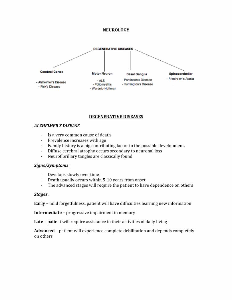
NEUROLOGY
DEGENERATIVE DISEASES
ALZHEIMER’S DISEASE
-‐ Is a very common cause of death -‐ Prevalence increases with age -‐ Family history is a big contributing factor to the possible development. -‐ Diffuse cerebral atrophy occurs secondary to neuronal loss -‐ Neurofibrillary tangles are classically found
Signs/Symptoms:
-‐ Develops slowly over time -‐ Death usually occurs within 5-‐10 years from onset -‐ The advanced stages will require the patient to have dependence on others
Stages:
Early – mild forgetfulness, patient will have difficulties learning new information
Intermediate – progressive impairment in memory
Late – patient will require assistance in their activities of daily living
Advanced – patient will experience complete debilitation and depends completely on others
PICKS DISEASE
-‐ Frontal and temporal lobe dysfunction caused by degeneration -‐ Accumulation of tau proteins -‐ Pick bodies are characteristic -‐ Memory impairment + impulsive behavior + behavioral changes
MOTOR NEURON DEGENERATIVE DISORDERS
AMYOTROPHIC LATERAL SCLEROSIS
ALS is also known as Lou Gehrig’s disease, which is a fatal neurodegenerative disease of both the upper and lower motor neurons.
POLIOMYELITIS
Polio is a virus that affects the anterior horn cells and motor neurons of the spinal cord and brainstem
-‐ Causes LMN symptoms
Features of Polio:
-‐ Asymmetric muscle weakness (most commonly the legs) -‐ Muscle atrophy -‐ Absent of DTR’s -‐ Flaccidity
*Sensation is intact *
WERDNIG-‐HOFFMAN DISEASE
A genetic condition that presents in infancy and/or early childhood.
-‐ Progressive skeletal muscle atrophy due to anterior horn cell degeneration -‐ Often presents with floppy baby at birth (congenital hypotonia) -‐ Lack of sucking ability -‐ Tongue fasciculation’s -‐ Death occurs at a young age due to respiratory muscle failure
BASAL GANGLIA DEGENERATION
PARKINSON’S DISEASE
-‐ Loss of dopaminergic neurons from the substantia nigra -‐ Usually presents in mid-‐late life (ie >50yr) -‐ Diagnosis is completely clinical and based on symptoms
Signs/Symptoms:
-‐ Resting tremor (pill rolling) -‐ Cogwheel rigidity -‐ Bradykinesia -‐ Difficulty in initiating movement -‐ Mask-‐like facies -‐ Brain biopsy will show Lewy bodies • certain medications can cause Parkinsonism: Reserpine, Metoclopramide,
Haloperidol, Perphenazine, MPTP
HUNTINGTON’S CHOREA
-‐ Is a genetic condition (AD) where there is atrophy of the caudate nucleus -‐ Disease onset is between 30-‐50yr with a steady worsening of symptoms and
death within 15 years of onset.
Signs/Symptoms:
-‐ Progressive dementia -‐ Chorea of the limbs, face, head/neck, and trunk -‐ Behavior disturbances such as: Depression, aggression, psychosis, changes in
personality. -‐ Depression occurs and suicide is somewhat common because patients are
aware of their deterioration • There is no treatment, only symptomatic management.
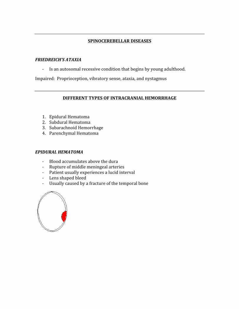
SPINOCEREBELLAR DISEASES
FRIEDREICH’S ATAXIA
-‐ Is an autosomal recessive condition that begins by young adulthood.
Impaired: Proprioception, vibratory sense, ataxia, and nystagmus
DIFFERENT TYPES OF INTRACRANIAL HEMORRHAGE
1. Epidural Hematoma 2. Subdural Hematoma 3. Subarachnoid Hemorrhage 4. Parenchymal Hematoma
EPIDURAL HEMATOMA
-‐ Blood accumulates above the dura -‐ Rupture of middle meningeal arteries -‐ Patient usually experiences a lucid interval -‐ Lens shaped bleed -‐ Usually caused by a fracture of the temporal bone

SUBDURAL HEMATOMA
-‐ Symptoms occur gradually -‐ Tearing of the bridging veins -‐ Seen in elders and alcoholics experiencing blunt trauma -‐ Crescent shaped
SUBARACHNOID HEMORRHAGE
-‐ Aneurysm rupture and/or AVM rupture -‐ Classic presentation is “worst headache of patients life” -‐ LP will show xanthochromia
BERRY ANEURYSM
Aneurysms are focal weaknesses in the vasculature that result in outpouchings. The Berry aneurysm is seen at the bifurcation of the anterior communicating artery.
-‐ Rupture leads to hemorrhagic stroke -‐ Creates the “worst headache of my life” -‐ Associated with Ehler’s danlos, Marfan’s, and APKD -‐ Is a surgical emergency
DISEASES OF DEMYELINATION
The most common demyelinating diseases are Multiple Sclerosis and Progressive Multifocal Leukoencephalopathy, Guillain-‐Barre syndrome, Metachromatic Leukodystrophy, and Post-‐Infectious Encephalomyelitis.
MULTIPLE SCLEROSIS
-‐ Multifocal zones of demyelination scattered throughout the white matter -‐ Commonly involves the pyramidal and cerebellar pathways, medial
longitudinal fasciculus, optic nerve, and the posterior columns -‐ Presents with transient sensory deficits, fatigue, weakness, and spasticity -‐ Visual disturbances (monocular vision loss, ipsilateral medial rectus palsy on
lateral gaze) -‐ Usually presents in the 20s and 30s in a relapsing fashion -‐ Diagnosing is made mostly with MRI -‐ Management/treatment is with corticosteroids
Classic Triad: Scanning Speech, Intention Tremor, Nystagmus
PROGRESSIVE MULTIFOCAL LEUKOENCEPHALOPATHY
-‐ Is the reactivation of a latent viral infection -‐ Seen in approximately 4% of AIDS patients -‐ Associated with JC Virus
GUILLAIN-‐BARRE SYNDROME
-‐ Is an ascending inflammation and demyelination of the peripheral nerves and motor fibers of ventral roots
-‐ Presents in an ascending fashion -‐ Causes symmetrical muscle weakness that starts in the lower extremities -‐ Presents most commonly following an upper respiratory infection -‐ The most common cause of death is due to paralysis of respiratory muscles
(thus monitoring respiratory function is essential to safety of the patient)

METACHROMATIC LEUKODYSTROPHY
-‐ A lysosomal storage disease that affects the growth and development of myelin
-‐ Due to deficiency of the enzyme arylsulfatase A, which causes accumulation of sulfatides in the tissues and thus destroys myelin sheath
-‐ Affects both CNS and PNS -‐ Children with the “late infantile form” may have difficulty in walking after the
first year of life -‐ Muscle wasting, weakness, muscle rigidity, developmental delays, and
progressive loss of vision, odynophagia, paralysis, and dementia are all possible complications
-‐ Juvenile form (3-‐10yrs) get mental deterioration and can develop dementia -‐ Adult form (>16yr) presents as a psychiatric disorder or progressive
dementia -‐ No cure for this condition
POST-‐INFECTIOUS ENCEPHALOMYELITIS
-‐ An immune-‐mediated disease of the brain -‐ Occurs following a viral infection most commonly -‐ Is similar to multiple sclerosis in that it involves autoimmune demyelination -‐ Symptoms begin 1-‐3 weeks post-‐infection -‐ Fever, headache, drowsiness, seizure, and coma
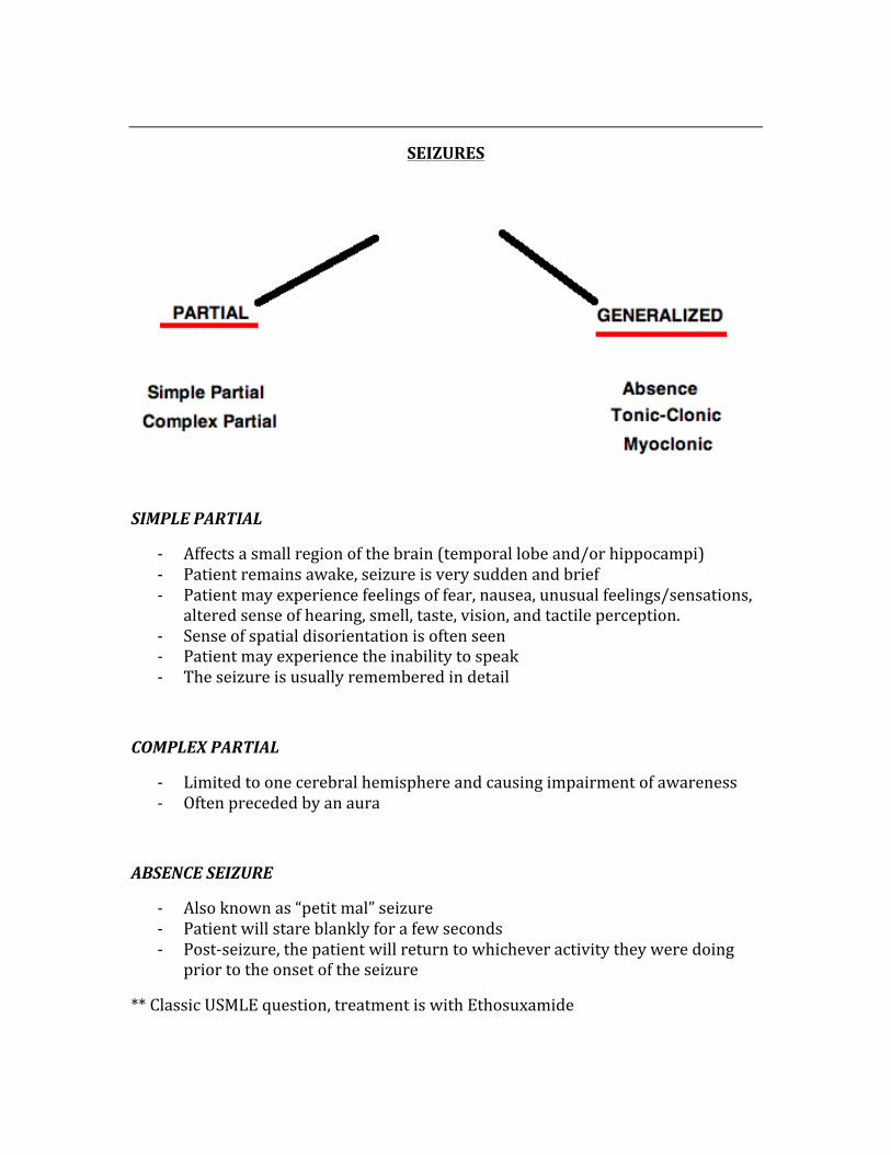
SEIZURES
SIMPLE PARTIAL
-‐ Affects a small region of the brain (temporal lobe and/or hippocampi) -‐ Patient remains awake, seizure is very sudden and brief -‐ Patient may experience feelings of fear, nausea, unusual feelings/sensations,
altered sense of hearing, smell, taste, vision, and tactile perception. -‐ Sense of spatial disorientation is often seen -‐ Patient may experience the inability to speak -‐ The seizure is usually remembered in detail
COMPLEX PARTIAL
-‐ Limited to one cerebral hemisphere and causing impairment of awareness -‐ Often preceded by an aura
ABSENCE SEIZURE
-‐ Also known as “petit mal” seizure -‐ Patient will stare blankly for a few seconds -‐ Post-‐seizure, the patient will return to whichever activity they were doing
prior to the onset of the seizure
** Classic USMLE question, treatment is with Ethosuxamide
TONIC-‐CLONIC SEIZURE
-‐ “Grand-‐mal” seizure, it affects the entire brain -‐ Is the most well-‐known type of seizure that occurs with epilepsy -‐ Associated with an aura
Tonic Phase – Patient loses consciousness, tension of skeletal muscles occurs. Lasts only a few seconds.
Clonic Phase – Rapid contraction/relaxation of muscles, eyes roll to the back of the head, tongue is often bitten due to jaw contractions. Incontinence may occur at this phase.
-‐ Patient will be confused and will have no memory of the seizure -‐ Initial management is with a benzodiazepine given IV
MYOCLONIC
-‐ Is a brief and involuntary twitching of the muscles -‐ Presents with abnormal movements on both sides of the body at the same
time -‐ Occurs commonly while patient is falling asleep -‐ Is not an actual disorder, rather is a sign of other potential nervous system
disorders
APHASIA
Aphasias are acquired language disorders whereby there is an impairment of either difficulty producing or comprehending spoken or written language. The two types clinically encountered are Expressive Aphasia (Broca’s), and Receptive Aphasia (Wernicke’s).
Expressive Aphasia – Patient has complete intact comprehension with the inability to speak in an understanding fashion. The site of pathology is the inferior frontal gyrus. Expressive = Inferior frontal gyrus. E=I (two vowels)
Receptive Aphasia – Patient has complete intact ability to speak understandable with the inability to comprehend language. The site of pathology is the superior temporal gyrus. Receptive = Superior Temporal gyrus. RST
CENTRAL SLEEP APNEA
During central sleep apnea, the brain’s respiratory control centers are imbalanced during sleep. The neurological feedback mechanism does not respond to levels of CO2 and thus cannot react to drive the respiratory rate. Following this pathology, breathing rate will increase and excess CO2 will be expelled from the body.
BELL’S PALSY
Is a condition of hemifacial weakness/paralysis of the muscle that are innervated by CN7, due to inflammation of the nerve.
-‐ Patient will experience an acute onset of unilateral facial weakness and paralysis, affecting both upper and lower face.
-‐ Idiopathic, Lyme disease, AIDS, sarcoidosis, and diabetes are all possible causes
Comparing to UMN and LMN lesions:
UMN lesion – will affect the lower half of the contralateral face
LMN lesion – will affect the upper and lower face on the ipsilateral sides
HORNER’S SYNDROME
Horner’s syndrome is a clinical syndrome caused by damage to the sympathetic nervous system. On most occasions, Horner’s syndrome is a benign condition, but it can occur as a result of more serious conditions such as Pancoast tumor or thyrocervical venous dilatation. These conditions cause compression of the thoracic sympathetic chain, which leads to the symptoms of Horner’s syndrome:
Ptosis – drooping of the eyelid
Anhydrosis – the absence of sweating
Miosis – constriction of the pupil
NEUROTRANSMITTERS AFFECTED IN DISEASE
Many neurotransmitters are affected during different diseases, they include:
DOPAMINE – Increased in Schizophrenia, decreased in Parkinson’s disease
NOREPINEPHRINE – Increased in anxiety, decreased in depression
ACETYLCHOLINE – Increased in organophosphate poisoning, decreased in Alzheimer’s and Huntington’s disease
GABA – Decreased in Huntington’s, decreased in anxiety
SERETONIN (5-‐HT) – Decreased in both anxiety and depression
TABES DORSALIS
Tabes dorsalis is a condition whereby there is degeneration of the dorsal columns and dorsal roots of the spinal cord. This occurs as a result of tertiary syphilis. Patient will experience all symptoms associated with dorsal column malfunction (lack of proprioception, ataxia during locomotion). Patient will develop malformed joints (Charcot’s joints) due to lack of pain receptors, shooting pains, absence of deep tendon reflexes, and Argyll Robertson pupil that results from reaction to accommodation but not to light.
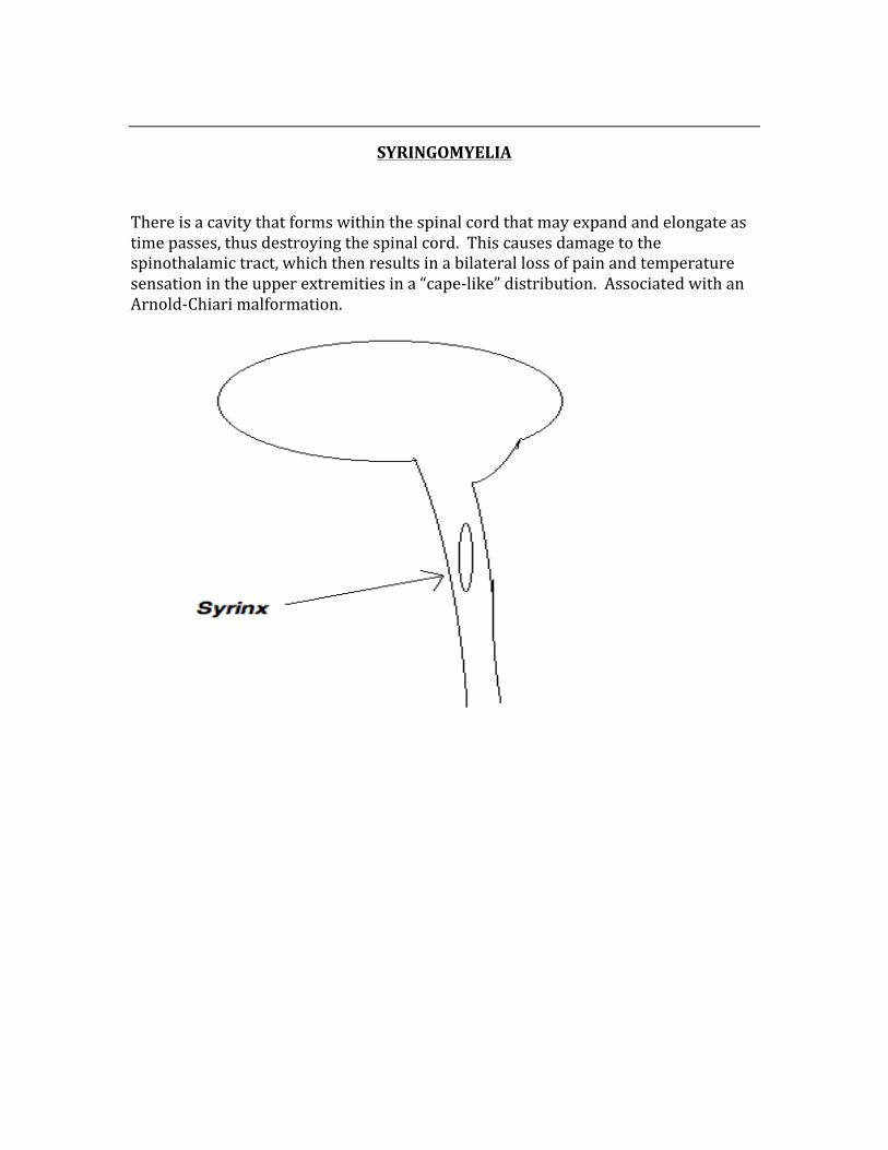
SYRINGOMYELIA
There is a cavity that forms within the spinal cord that may expand and elongate as time passes, thus destroying the spinal cord. This causes damage to the spinothalamic tract, which then results in a bilateral loss of pain and temperature sensation in the upper extremities in a “cape-‐like” distribution. Associated with an Arnold-‐Chiari malformation.
RENAL PATHOLOGY
PATHOLOGY OF THE GLOMERULUS
NEPHRITIC SYNDROMES
ACUTE POST-‐STREP GLOMERULONEPHRITIS
-‐ The most common cause of nephritic syndrome -‐ Occurs after a group A β-‐hemolytic strep infection (develops 10-‐14 days after
infection) -‐ Affects children between 2-‐6 years of age -‐ Is self-‐limited condition -‐ Patient gets hematuria, edema, hypertension, and low complement levels -‐ Some cases may progress to rapidly progressive glomerulonephritis
NEPHRITIC SYNDROMES
Present with: Hematuria, Hypertension, Oliguria, Azotemia
ACUTE POST-‐STREP GLOMERULONEPHRITIS
RAPIDLY PROGRESSIVE GLOMERULONEPHRITIS
GOODPASTURE’S SYNDROME
MEMBRANOPROLIFERATIVE GLOMERULONEPHRITIS
BERGER’S DISEASE (IgA NEPHROPATHY)
ALPORT’S SYNDROME
NEPHROTIC SYNDROMES
Present with: Massive Proteinuria, Hyperlipidemia, Hypoalbuminemia, and Peripheral/Periorbital Edema.
MEMBRANOUS GLOMERULONEPHRITIS
MINIMAL CHANGE DISEASE
FOCAL SEGMENTAL GLOMERULAR SCLEROSIS
DIABETIC NEPHROPATHY
SYSTEMIC LUPUS ERYTHEMATOUS
RAPIDLY PROGRESSIVE GLOMERULONEPHROPATHY
-‐ Is “crescent-‐moon” shaped on light microscopy
GOODPASTURE’S SYNDROME
-‐ There is a triad of IgG anti-‐glomerular basement membrane antibodies, pulmonary hemorrhage, and crescentic glomerulonephritis
-‐ The lung findings will occur before the renal findings -‐ Patient will have hemoptysis, rapidly progressive renal failure, fever, and
myalgias
MEMBRANOPROLIFERATIVE GLOMERULONEPHRITIS
-‐ On electron microscopy, there are the classic “tram-‐track” subendothelial humps
-‐ Has a slow progression to renal failure
BERGER’S DISEASE (IgA nephropathy)
-‐ Mesangial deposits of IgA -‐ Is a very mild disease -‐ Occurs after an infection
ALPORT’S SYNDROME
-‐ A mutation of type 4 collagen -‐ Patient can have nerve deafness and ocular disorders -‐ There are split basement membranes
NEPHROTIC SYNDROMES
MEMBRANOUS GLOMERULONEPHRITIS
-‐ Very common cause of adult nephrotic syndrome -‐ Light microscope shows basement membrane thickening
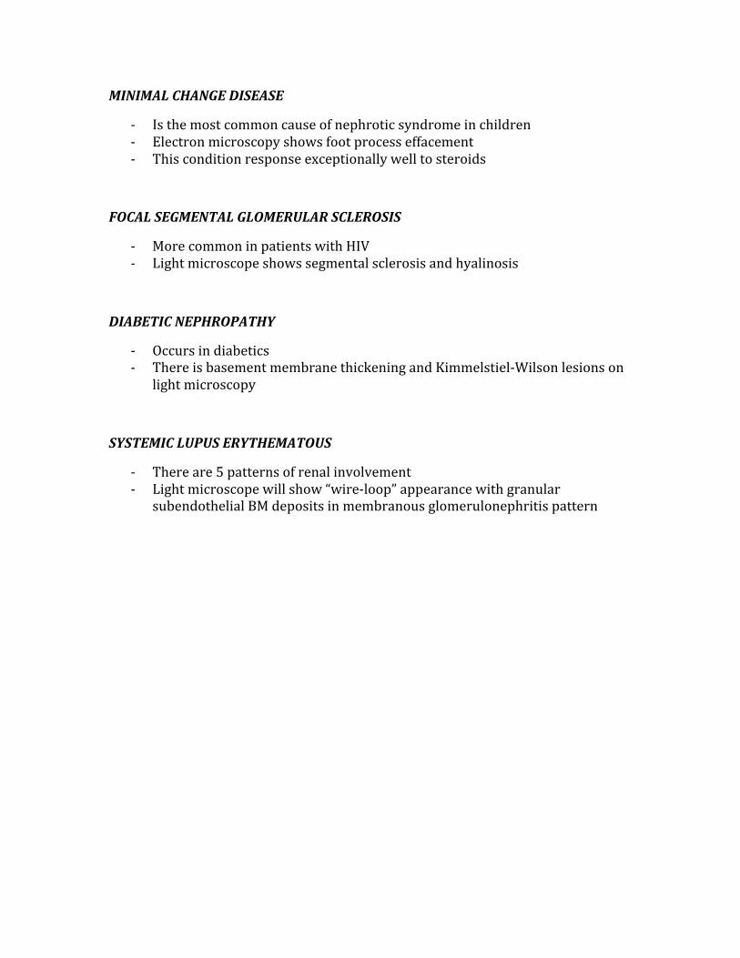
MINIMAL CHANGE DISEASE
-‐ Is the most common cause of nephrotic syndrome in children -‐ Electron microscopy shows foot process effacement -‐ This condition response exceptionally well to steroids
FOCAL SEGMENTAL GLOMERULAR SCLEROSIS
-‐ More common in patients with HIV -‐ Light microscope shows segmental sclerosis and hyalinosis
DIABETIC NEPHROPATHY
-‐ Occurs in diabetics -‐ There is basement membrane thickening and Kimmelstiel-‐Wilson lesions on
light microscopy
SYSTEMIC LUPUS ERYTHEMATOUS
-‐ There are 5 patterns of renal involvement -‐ Light microscope will show “wire-‐loop” appearance with granular
subendothelial BM deposits in membranous glomerulonephritis pattern
RENAL CELL CARCINOMA
Renal cell carcinoma is the most common of all renal malignancies, occurring most commonly in men between 50-‐70yr of age, and is more common in smokers.
-‐ Associated with von Hippel-‐Lindau -‐ Originates in renal tubule cells
Manifests as:
-‐ Hematuria -‐ Palpable flank mass -‐ Secondary polycythemia (due to production of erythropoietin from kidney) -‐ Many paraneoplasias are common due to production of Prolactin, PTHrP, and
ACTH)
WILM’S TUMOR
This is the most common malignancy seen in young children between 2-‐4yr of age.
The following are important regarding a Wilm’s tumor:
-‐ There is a deletion of the WT1 gene that is found on chromosome 11 -‐ Presents with a huge, palpable flank mass
Wilm’s tumor is often seen in conjunction with the WAGR complex:
W – Wilm’s tumor
A – Aniridia (absence of the iris)
G – Genitorurinary malformations
R – Retardation (both mental and motor)
TRANSITIONAL CELL CARCINOMA
Is a common cancer seen in the urinary tract (bladder, ureters, renal pelvis, and renal calyces). The most common causes include: Smoking, Alanine Dyes, Cyclophosphamide.
PYELONEPHRITIS
An ascending infection that spreads from the bladder to the kidneys. The most common cause of pyelonephritis is an E. Coli infection.
Signs/Symptoms:
-‐ Flank pains (CVA tenderness) -‐ Fever -‐ Nausea/vomiting -‐ Patient will appear quite ill
** Patients with vesicoureteral reflux have an increased tendency to getting pyelonephritis.
ACUTE TUBULAR NECROSIS (ATN)
Acute tubular necrosis occurs when there is an insult to the renal tubules. Anything that can harm the kidneys can cause ATN, things such as: Ischemia due to shock, increased myoglobins due to muscle breakdown, drugs, toxins.
** Is the most common cause of acute renal failure.
ACUTE RENAL FAILURE
Acute renal failure occurs most commonly from ATN (mentioned above), and occurs when a decline in renal function causes an increase in the levels of BUN and Creatinine.
The causes of renal failure:
Pre-‐renal Azotemia à is when there is a decrease in renal blood flow, which leads to a decrease in the glomerular filtration, and thus retention of water and sodium in the kidneys. BUN>Cr is > 20
Post-‐renal Azotemia à this condition only occurs if there is bilateral obstruction, things such as prostatic hyperplasia, neoplasia, and stone (less likely) will cause a post-‐renal azotemia. BUN/Cr is > 15
Intrinsic à this is due to ATN (usually), and the urine will give this cause away by having granular casts or epithelial casts. BUN/Cr is < 15
WHAT HAPPENS WHEN RENAL FAILURE IS PRESENT?
Renal failure leads to a build-‐up of toxins and leads to the inability to excrete nitrogenous bases.
Acute renal failure is usually due to hypoxemia, while chronic renal failure is usually caused by either hypertension or diabetes.
When there is a build-‐up of BUN and Creatinine, uremia develops. When uremia develops, the following symptoms may be present:
-‐ Anemia due to failure of EPO production by the kidneys -‐ CHF/Pulmonary edema due to sodium and water excess -‐ Metabolic acidosis -‐ Hyperkalemia -‐ Osteodystrophy due to failure of vitamin D production by the kidneys
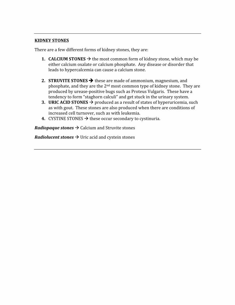
KIDNEY STONES
There are a few different forms of kidney stones, they are:
1. CALCIUM STONES à the most common form of kidney stone, which may be either calcium oxalate or calcium phosphate. Any disease or disorder that leads to hypercalcemia can cause a calcium stone.
2. STRUVITE STONES à these are made of ammonium, magnesium, and phosphate, and they are the 2nd most common type of kidney stone. They are produced by urease-‐positive bugs such as Proteus Vulgaris. These have a tendency to form “staghorn calculi” and get stuck in the urinary system.
3. URIC ACID STONES à produced as a result of states of hyperuricemia, such as with gout. These stones are also produced when there are conditions of increased cell turnover, such as with leukemia.
4. CYSTINE STONES à these occur secondary to cystinuria.
Radiopaque stones à Calcium and Struvite stones
Radiolucent stones à Uric acid and cystein stones
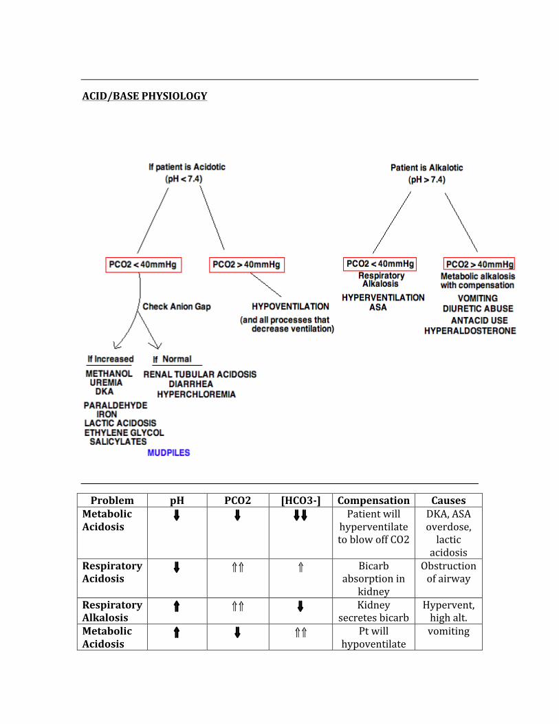
ACID/BASE PHYSIOLOGY
Problem pH PCO2 [HCO3-‐] Compensation Causes Metabolic Acidosis
⇓ ⇓ ⇓⇓ Patient will hyperventilate to blow off CO2
DKA, ASA overdose, lactic acidosis
Respiratory Acidosis
⇓ ⇑⇑ ⇑ Bicarb absorption in
kidney
Obstruction of airway
Respiratory Alkalosis
⇑ ⇑⇑ ⇓ Kidney secretes bicarb
Hypervent, high alt.
Metabolic Acidosis
⇑ ⇓ ⇑⇑ Pt will hypoventilate
vomiting
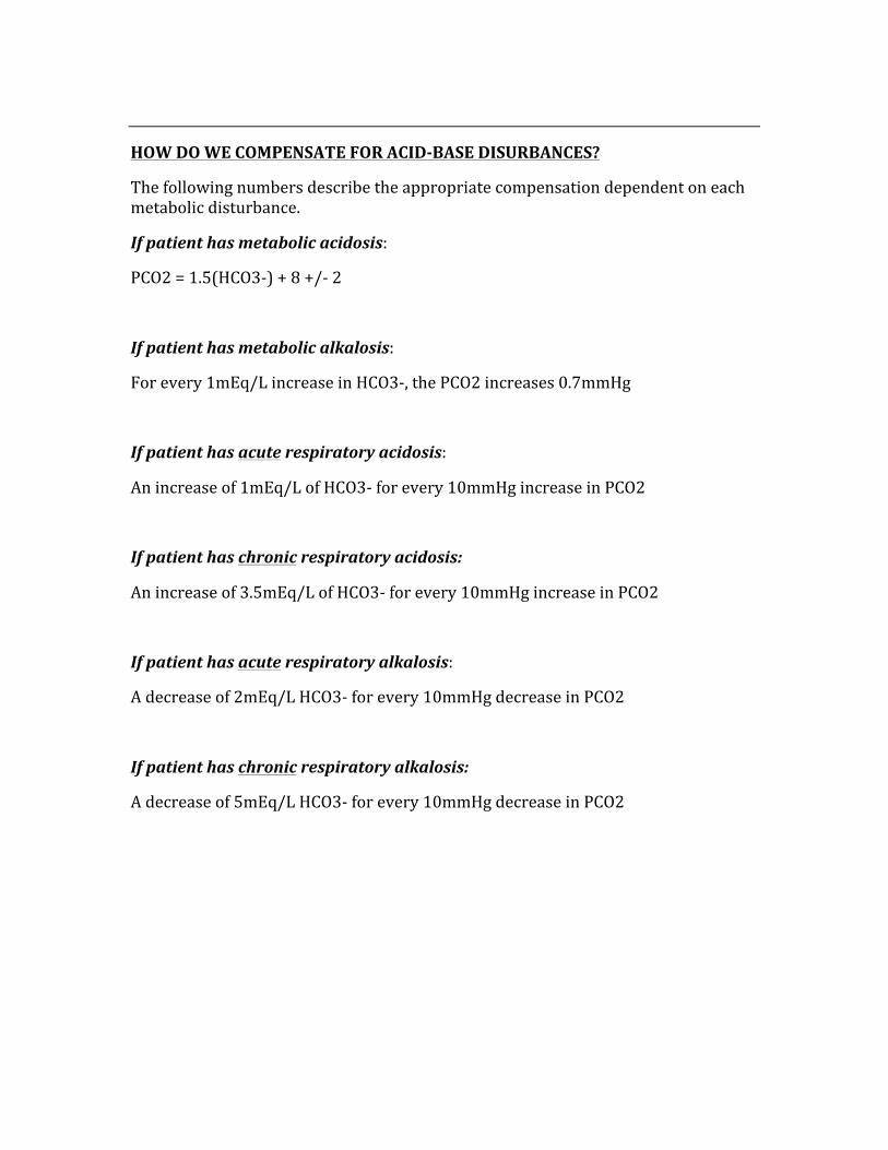
HOW DO WE COMPENSATE FOR ACID-‐BASE DISURBANCES?
The following numbers describe the appropriate compensation dependent on each metabolic disturbance.
If patient has metabolic acidosis:
PCO2 = 1.5(HCO3-‐) + 8 +/-‐ 2
If patient has metabolic alkalosis:
For every 1mEq/L increase in HCO3-‐, the PCO2 increases 0.7mmHg
If patient has acute respiratory acidosis:
An increase of 1mEq/L of HCO3-‐ for every 10mmHg increase in PCO2
If patient has chronic respiratory acidosis:
An increase of 3.5mEq/L of HCO3-‐ for every 10mmHg increase in PCO2
If patient has acute respiratory alkalosis:
A decrease of 2mEq/L HCO3-‐ for every 10mmHg decrease in PCO2
If patient has chronic respiratory alkalosis:
A decrease of 5mEq/L HCO3-‐ for every 10mmHg decrease in PCO2
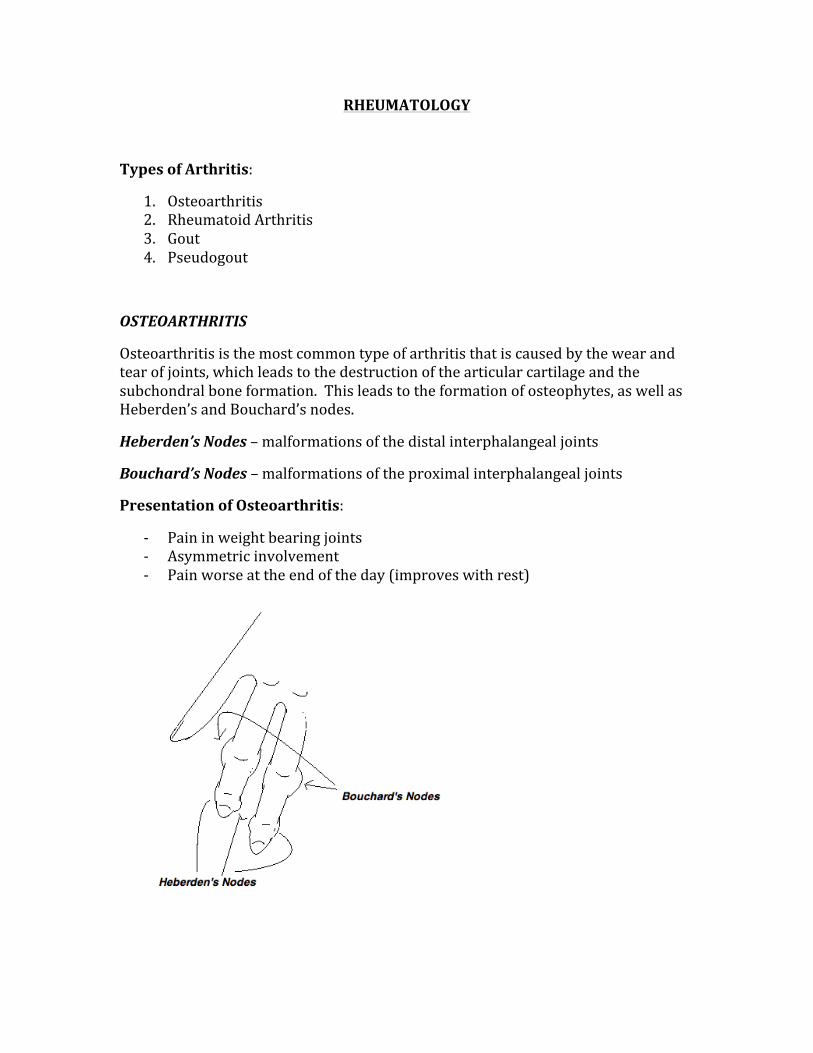
RHEUMATOLOGY
Types of Arthritis:
1. Osteoarthritis 2. Rheumatoid Arthritis 3. Gout 4. Pseudogout
OSTEOARTHRITIS
Osteoarthritis is the most common type of arthritis that is caused by the wear and tear of joints, which leads to the destruction of the articular cartilage and the subchondral bone formation. This leads to the formation of osteophytes, as well as Heberden’s and Bouchard’s nodes.
Heberden’s Nodes – malformations of the distal interphalangeal joints
Bouchard’s Nodes – malformations of the proximal interphalangeal joints
Presentation of Osteoarthritis:
-‐ Pain in weight bearing joints -‐ Asymmetric involvement -‐ Pain worse at the end of the day (improves with rest)

RHEUMATOID ARTHRITIS
Is an autoimmune condition that attacks the synovial joints of the body.
-‐ Pannus formation in joints leads to deformities of the metacarpophalangeal joints and proximal interphalangeal joints.
-‐ Formation of subcutaneous nodules -‐ Ulnar deviation at the wrist joint -‐ Seen in females >>> males
Presentation of Rheumatoid Arthritis:
-‐ Stiffness in the morning that improves with use -‐ Symmetric involvement -‐ Systemic symptoms are present (fever, fatigue, cardiac conditions,
pulmonary conditions)
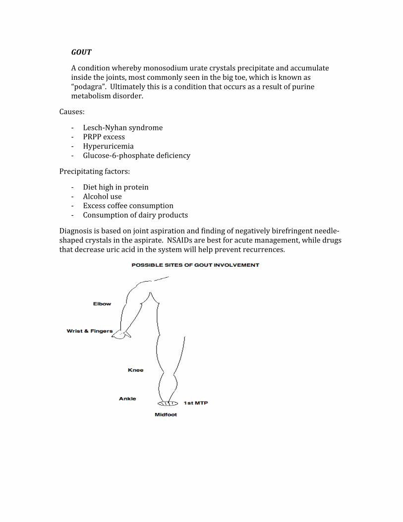
GOUT
A condition whereby monosodium urate crystals precipitate and accumulate inside the joints, most commonly seen in the big toe, which is known as “podagra”. Ultimately this is a condition that occurs as a result of purine metabolism disorder.
Causes:
-‐ Lesch-‐Nyhan syndrome -‐ PRPP excess -‐ Hyperuricemia -‐ Glucose-‐6-‐phosphate deficiency
Precipitating factors:
-‐ Diet high in protein -‐ Alcohol use -‐ Excess coffee consumption -‐ Consumption of dairy products
Diagnosis is based on joint aspiration and finding of negatively birefringent needle-‐shaped crystals in the aspirate. NSAIDs are best for acute management, while drugs that decrease uric acid in the system will help prevent recurrences.

PSEUDOGOUT
A condition similar to gout, pseudogout is caused by the deposition of calcium pyrophosphate crystals inside the joint space. The crystals formed are rhomboid-‐shaped and basophilic. The large joints are most commonly affected.
SERONEGATIVE SPONDYLOARTHROPATHIES
The group of seronegative spondyloarthropathies include:
1. Ankylosing Spondylitis 2. Reiter’s Syndrome (Reactive Arthritis) 3. Psoriatic Arthritis 4. IBD Arthropathy 5. Undifferentiated Spondyloarthropathy
This groups of arthritis types has the following in common:
-‐ They are rheumatic factor negative -‐ They have extra-‐articular symptoms (visual, pulmonary, cardiac, etc) -‐ Association with HLA-‐B27 antigen -‐ Inflammatory processes -‐ Asymmetrical presentation -‐ Familial
ANKYLOSING SPONDYLITIS
-‐ Low back pain and stiffness (patient is bent over at the hips) -‐ C-‐spine motion is limited due to neck pains -‐ Diminished chest expansion -‐ Most common extra-‐articular involvement is in the eyes (anterior uveitis) -‐ Best diagnostic modality is xray of lumbar spine and pelvis -‐ Management is with NSAIDs and physical therapy
REITER’S SYNDROME
-‐ Is a clinical diagnosis based on the presentation of arthritic symptoms that is preceded by an infection (salmonella, shigella, campylobacter, Chlamydia, and yersinia)
-‐ Classic triad is Arthritis, Urethritis, and Uveitis (can’t see, can’t pee, can’t climb a tree)
-‐ There is a sequential involvement of new joints -‐ Patient often has an accompanying set of symptoms such as fatigue, weight
loss, and overall sense of malaise
PSORIATIC ARTHRITIS
-‐ Develops in patients who have arthritis -‐ Onset is gradual and seen long after patient develops arthritis -‐ Is similar in presentation to rheumatoid arthritis -‐ Management is with NSAIDs
POLYARTICULAR JOINT INVOLVEMENT: RA, REITER’S
MONOARTICULAR JOINT INVOLVEMENT: OSTEOARTHRITIS, GOUT, PSEUDOGOUT
SYSTEMIC LUPUS ERYTHEMATOUS
An autoimmune disorder whereby there is multi-‐system and multi-‐organ inflammation and tissue damage.
Types:
-‐ SLE -‐ Discoid Lupus (skin manifestation without systemic disease) -‐ Drug-‐Induced -‐ ANA-‐negative Lupus
Who gets it?
-‐ 90% of cases are females of childbearing age -‐ African-‐American’s are affected more than Caucasians -‐ Severity tends to be less in older patients, and more in younger patients
Findings:
-‐ Butterfly rash -‐ Photosensitivity -‐ Discoid rash -‐ Alopecia -‐ Raynaud’s phenomenon -‐ Joints pain/arthralgias/myalgias -‐ Pericarditis/endocarditis/myocarditis -‐ Pleuritis/pleural effusion/pneumonitis -‐ Hemolytic anemia/leucopenia/thrombocytopenia/lymphopenia -‐ Proteinuria >0.5g/day, glomerulonephritis/pyuria/azotemia/uremia/HTN

-‐ Abnormal T cell function, lymphocyte autoantibodies -‐ Nausea/vomiting/PUD/dyspepsia -‐ Seizures/psychosis/depression/TIA/headaches/CVA -‐ Fever, malaise, conjunctivitis
Diagnostic Criteria:
-‐ Positive ANA is seen in almost all SLE patients -‐ Anti-‐ds DNA is seen in approximately 40% of SLE patients -‐ Anti-‐Sm antibodies is seen in approximately 30% of SLE patients
SARCOIDOSIS
Is a condition characterized by immune-‐mediated, widespread noncaseating granulomas. The classic finding is bilateral hilar adenopathy.
There is also the incidence of:
-‐ Increased levels of ACE -‐ Rheumatoid arthritis -‐ Interstitial fibrosis -‐ Gammaglobulinemia -‐ Restrictive lung diseases -‐ Erythema nodosum -‐ Hypercalcemia
AUTOIMMUNE CONDITIONS OF THE SKIN
PSORIASIS
Autoimmune condition where the skin develops red, scaly patches on the skin. The plaques that develop are known as “psoriatic plaques”, and are caused by excessive production of skin and a faster skin cycle than normal skin.
-‐ Skin appears silvery-‐white in color -‐ Most commonly present on extensor surfaces of the body (knees, elbows),
but may be seen on the hands, feet, genitals, and the scalp.
This condition is not contagious*
Psoriasis is associated with the B27 haplotype (HLA-‐B27), HLA-‐13, and HLA-‐17.
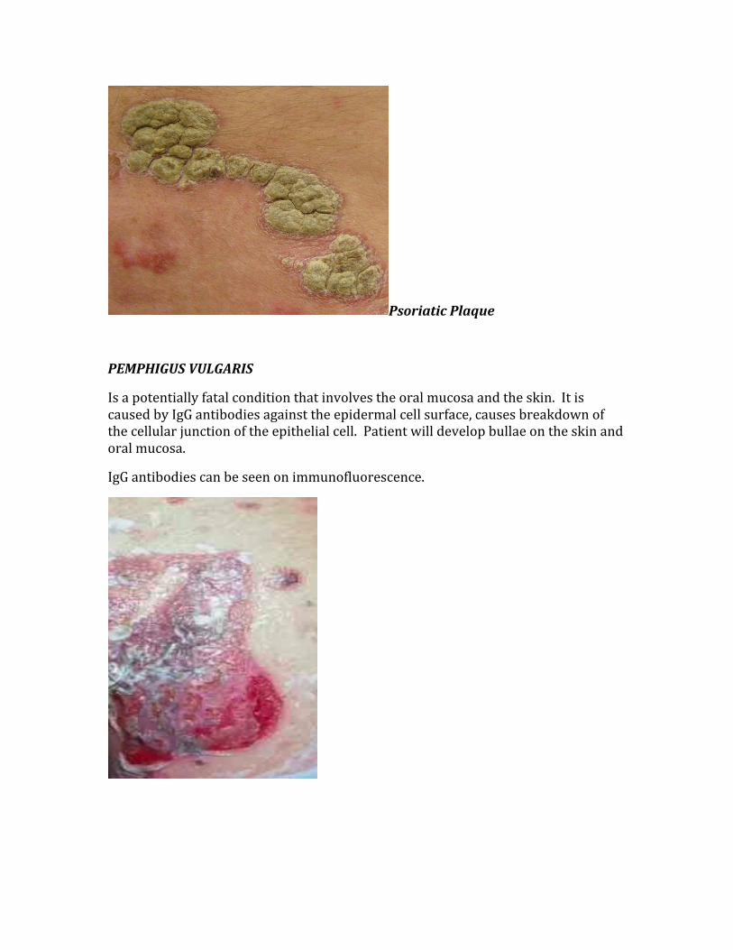
Psoriatic Plaque
PEMPHIGUS VULGARIS
Is a potentially fatal condition that involves the oral mucosa and the skin. It is caused by IgG antibodies against the epidermal cell surface, causes breakdown of the cellular junction of the epithelial cell. Patient will develop bullae on the skin and oral mucosa.
IgG antibodies can be seen on immunofluorescence.
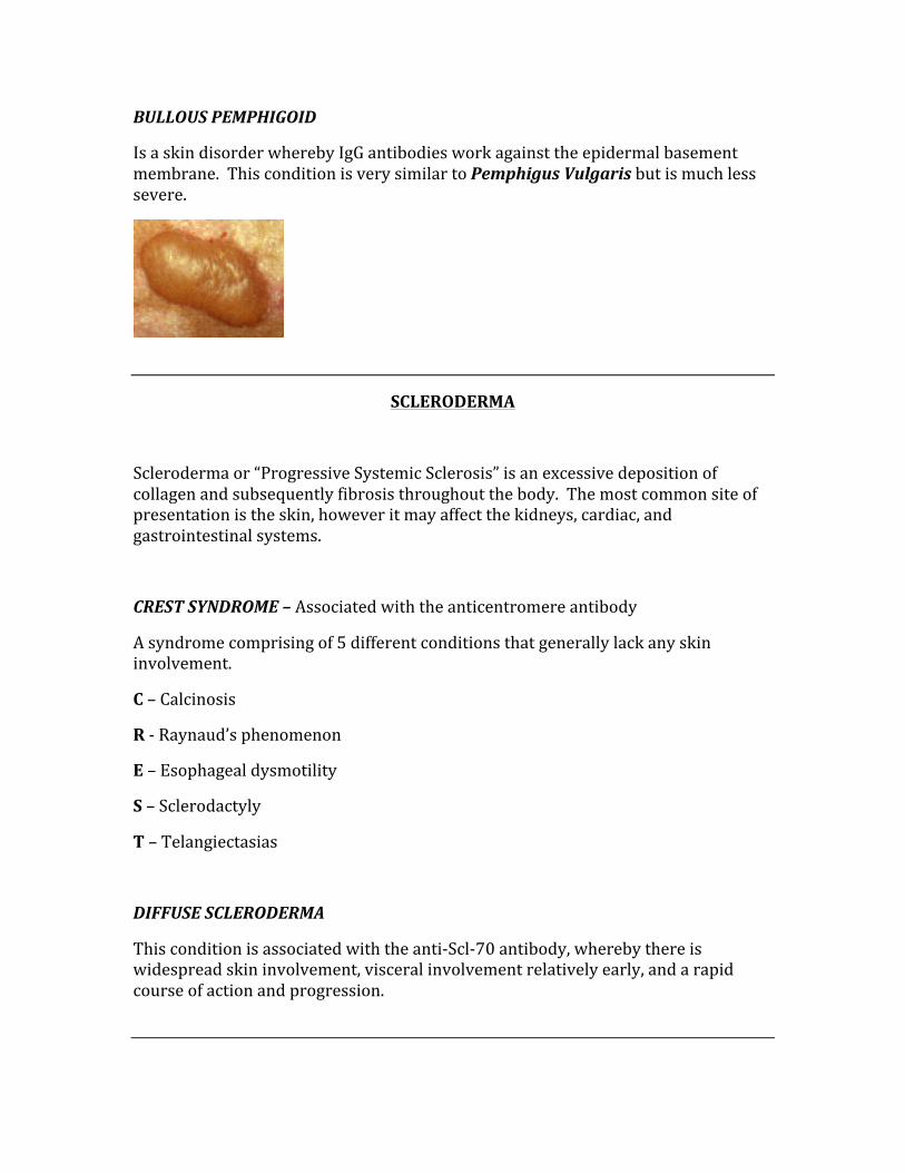
BULLOUS PEMPHIGOID
Is a skin disorder whereby IgG antibodies work against the epidermal basement membrane. This condition is very similar to Pemphigus Vulgaris but is much less severe.
SCLERODERMA
Scleroderma or “Progressive Systemic Sclerosis” is an excessive deposition of collagen and subsequently fibrosis throughout the body. The most common site of presentation is the skin, however it may affect the kidneys, cardiac, and gastrointestinal systems.
CREST SYNDROME – Associated with the anticentromere antibody
A syndrome comprising of 5 different conditions that generally lack any skin involvement.
C – Calcinosis
R -‐ Raynaud’s phenomenon
E – Esophageal dysmotility
S – Sclerodactyly
T – Telangiectasias
DIFFUSE SCLERODERMA
This condition is associated with the anti-‐Scl-‐70 antibody, whereby there is widespread skin involvement, visceral involvement relatively early, and a rapid course of action and progression.
VASCULITIS CONDITIONS
Large vessel conditions – Temporal arteritis,
CONDITION FEATURES DIAGNOSIS TREATMENT Temporal Arteritis
Severe headache Visual impairment in 25%-‐50% Jaw pains Temporal tenderness
Age >50yr New onset headache Elevated ESR Temporal artery biopsy
High-‐dose steroids ESR follow-‐ups during management
Takayasu’s Arteritis
Usually seen in young Asian females Decreased and/or absent peripheral pulses Blood pressure differences in arm vs legs Many complications like limb ischemia, aneurysms, stroke, HTN from renal artery stenosis
Based on clinical symptoms
Steroids HTN management
Churg-‐Strauss A vasculitis affecting many different organ systems Fatigue, weight loss, fever Respiratory
Biopsy of lung tissue Biopsy of skin tissue (shows high eosinophils) p-‐ANCA will be found
Poor prognosis 5-‐yr survival is only 25% Steroids can prolong treatment
symptoms and skin findings are primary findings
Wegener’s Granulomatosis
A vasculitis of the kidneys and respiratory tract Upper respiratory symptoms Painful oral ulcers Eye disease (scleritis, conjunctivitis) Musculoskeletal findings (myalgia, arthralgia) Tracheal stenosis
CXR (nodules or infiltrates are present) Elevated ESR Anemia p-‐ANCA seen in most patients Confirmatory test is an open lung biopsy
Steroids + cyclophosphamide can help patients into remission With renal failure, a transplant is curative
Polyarteritis Nodosa
Is a medium-‐vessel vasculitis that involves the nervous system and GI tract Causes intimal proliferation which causes a decrease in the luminal area, resulting in ischemia, infarction, and then aneurysm Livedo reticularis Hypertension Fever/wt loss/arthralgia/abd pain
Biopsy ESR p-‐ANCA Fecal occult blood
Steroids can improve prognosis, which is poor without treatment
Behcet’s Syndrome
A multi-‐system vasculitis of unknown etiology Recurrent oral and genital ulcerations Arthritis Visual impairment CNS involvement Fever/weight loss
Biopsy of all involved tissues
Steroids
Buerger’s Disease Seen in young males who smoke Small and medium sized veins and arteries Affects arms and legs Claudication occurs Smoking is imperative in fixing the problem
Clinically based Smoking cessation
Hypersensitivity Vasculitis
Is a hypersensitivity reaction that occurs in response to certain medications (sulfas, PCN) Causes skin problems (purpura, macules, vesicles)
Tissue biopsy of affected tissues
Stop offending agent(s) Remission is often spontaneous
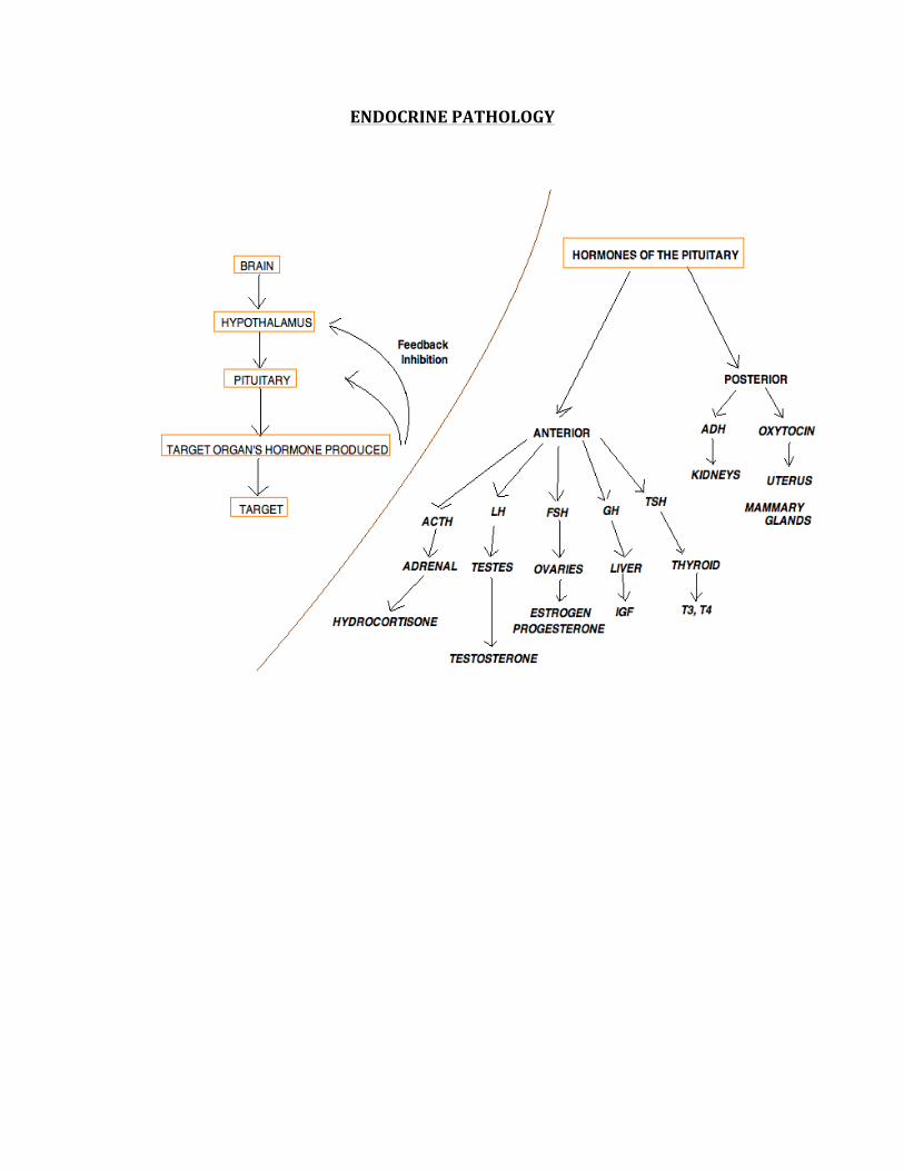
ENDOCRINE PATHOLOGY
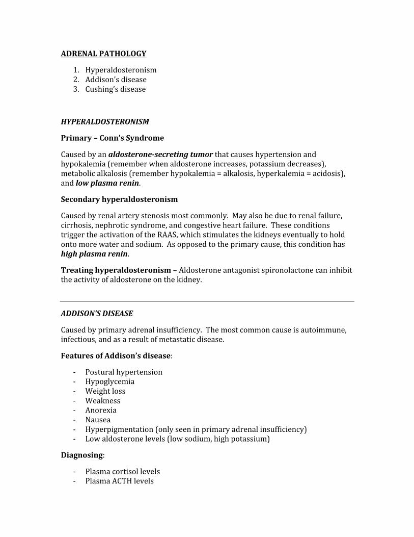
ADRENAL PATHOLOGY
1. Hyperaldosteronism 2. Addison’s disease 3. Cushing’s disease
HYPERALDOSTERONISM
Primary – Conn’s Syndrome
Caused by an aldosterone-‐secreting tumor that causes hypertension and hypokalemia (remember when aldosterone increases, potassium decreases), metabolic alkalosis (remember hypokalemia = alkalosis, hyperkalemia = acidosis), and low plasma renin.
Secondary hyperaldosteronism
Caused by renal artery stenosis most commonly. May also be due to renal failure, cirrhosis, nephrotic syndrome, and congestive heart failure. These conditions trigger the activation of the RAAS, which stimulates the kidneys eventually to hold onto more water and sodium. As opposed to the primary cause, this condition has high plasma renin.
Treating hyperaldosteronism – Aldosterone antagonist spironolactone can inhibit the activity of aldosterone on the kidney.
ADDISON’S DISEASE
Caused by primary adrenal insufficiency. The most common cause is autoimmune, infectious, and as a result of metastatic disease.
Features of Addison’s disease:
-‐ Postural hypertension -‐ Hypoglycemia -‐ Weight loss -‐ Weakness -‐ Anorexia -‐ Nausea -‐ Hyperpigmentation (only seen in primary adrenal insufficiency) -‐ Low aldosterone levels (low sodium, high potassium)
Diagnosing:
-‐ Plasma cortisol levels -‐ Plasma ACTH levels
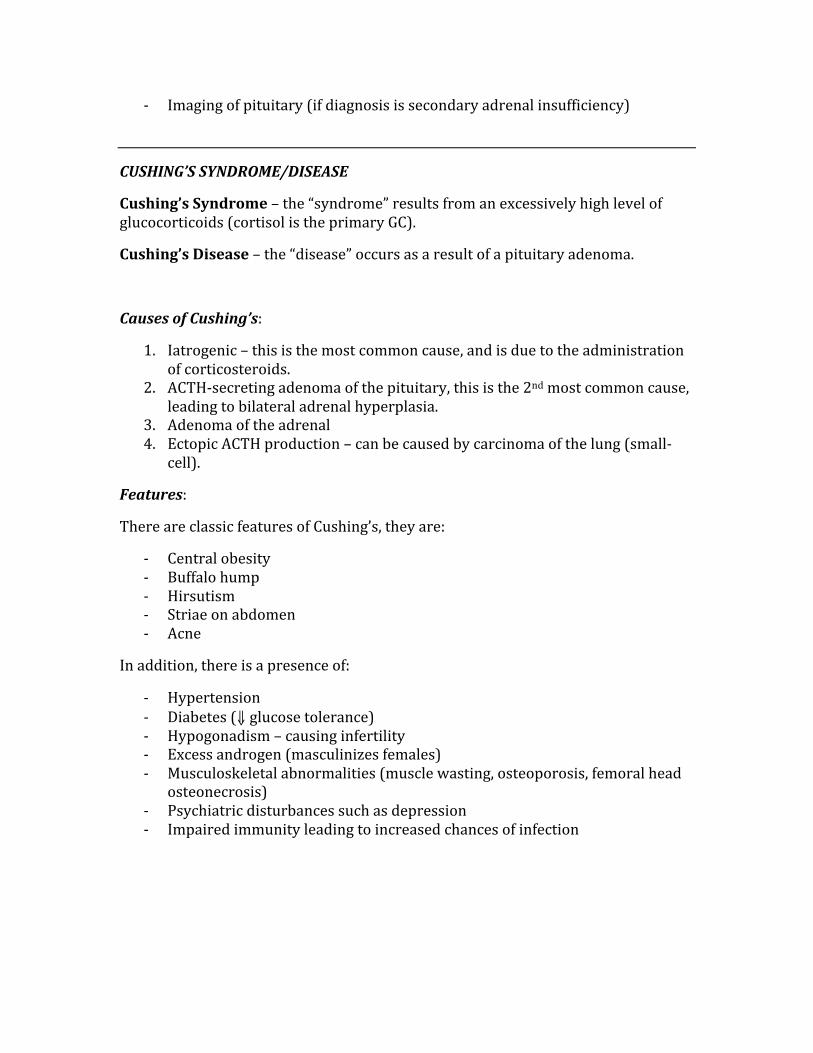
-‐ Imaging of pituitary (if diagnosis is secondary adrenal insufficiency)
CUSHING’S SYNDROME/DISEASE
Cushing’s Syndrome – the “syndrome” results from an excessively high level of glucocorticoids (cortisol is the primary GC).
Cushing’s Disease – the “disease” occurs as a result of a pituitary adenoma.
Causes of Cushing’s:
1. Iatrogenic – this is the most common cause, and is due to the administration of corticosteroids.
2. ACTH-‐secreting adenoma of the pituitary, this is the 2nd most common cause, leading to bilateral adrenal hyperplasia.
3. Adenoma of the adrenal 4. Ectopic ACTH production – can be caused by carcinoma of the lung (small-‐
cell).
Features:
There are classic features of Cushing’s, they are:
-‐ Central obesity -‐ Buffalo hump -‐ Hirsutism -‐ Striae on abdomen -‐ Acne
In addition, there is a presence of:
-‐ Hypertension -‐ Diabetes (⇓ glucose tolerance) -‐ Hypogonadism – causing infertility -‐ Excess androgen (masculinizes females) -‐ Musculoskeletal abnormalities (muscle wasting, osteoporosis, femoral head
osteonecrosis) -‐ Psychiatric disturbances such as depression -‐ Impaired immunity leading to increased chances of infection
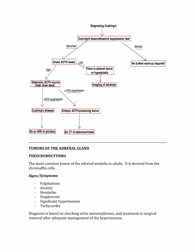
TUMORS OF THE ADRENAL GLAND
PHEOCROMOCYTOMA
The most common tumor of the adrenal medulla in adults. It is derived from the chromaffin cells.
Signs/Symptoms:
-‐ Palpitations -‐ Anxiety -‐ Headache -‐ Diaphoresis -‐ Significant hypertension -‐ Tachycardia
Diagnosis is based on checking urine metanephrines, and treatment is surgical removal after adequate management of the hypertension.
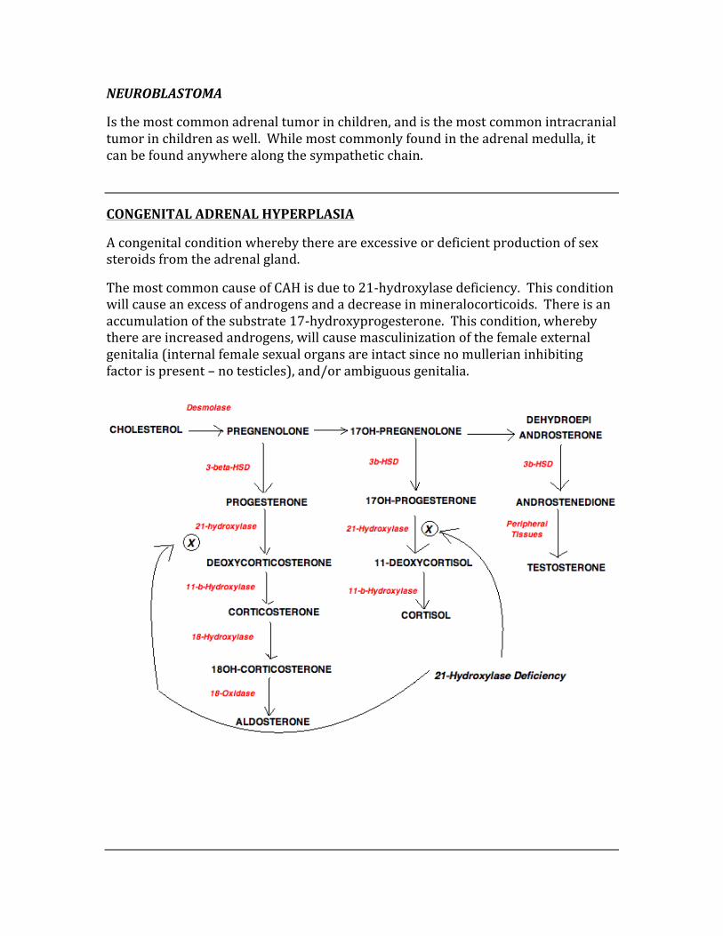
NEUROBLASTOMA
Is the most common adrenal tumor in children, and is the most common intracranial tumor in children as well. While most commonly found in the adrenal medulla, it can be found anywhere along the sympathetic chain.
CONGENITAL ADRENAL HYPERPLASIA
A congenital condition whereby there are excessive or deficient production of sex steroids from the adrenal gland.
The most common cause of CAH is due to 21-‐hydroxylase deficiency. This condition will cause an excess of androgens and a decrease in mineralocorticoids. There is an accumulation of the substrate 17-‐hydroxyprogesterone. This condition, whereby there are increased androgens, will cause masculinization of the female external genitalia (internal female sexual organs are intact since no mullerian inhibiting factor is present – no testicles), and/or ambiguous genitalia.
THYROID GLAND PATHOLOGY
CANCERS
PAPILLARY CARCINOMA
-‐ Presence of ground-‐glass nuclei and psammoma bodies -‐ Is the most common type of thyroid cancer and holds the best prognosis
FOLLICULAR CARCINOMA
-‐ There is a presence of uniform follicles -‐ Holds a good prognosis (better than medullary but worse than papillary)
MEDULLARY CARCINOMA
-‐ Derived from the parafollicular “C cells”, thus produces calcitonin. -‐ Is a member of the MENII and MEN III syndromes -‐ Has a bad prognosis
ANAPLASTIC CARCINOMA
-‐ This thyroid carcinoma occurs in older patients -‐ Holds a terrible prognosis and the worst of all thyroid carcinomas
HYPERTHYROIDISM
There will be a low TSH (due to feedback inhibition), with a high T3/T4
Patient will have the following:
-‐ Heat intolerance -‐ Weight loss -‐ Palpitations -‐ Warm/moist skin -‐ Arrhythmias
HYPOTHYROIDISM
There will be a high TSH and low T3/T4
Patient will have the following:
-‐ Cold intolerance -‐ Weight gain -‐ Fatigue -‐ Lethargy -‐ Weakness -‐ Decreased reflexes -‐ Dry/cool skin -‐ Coarse/brittle hair -‐ Myxedema
GRAVES DISEASE
Grave’s disease is the most common cause of hyperthyroidism, accounting for up to 80% of all cases.
-‐ Is an autoimmune disorder whereby a thyroid-‐stimulating immunoglobulin G antibody binds to the TSH receptors on the thyroid cells, triggering the synthesis of excess thyroid hormone
-‐ Diffuse radioiodide uptake on scan
PLUMMER’S DISEASE
Also known as “toxic multinodular goiter), accounting for approximately 15% of all cases.
-‐ There are areas of hyperfunctioning thyroid tissue that produce excess T3 and T4
-‐ More common in older patients -‐ Elderly patients with hyperthyroidism may present simply with unexplained
weight loss, weakness, and/or atrial fibrillation
HASHIMOTO’S THYROIDITIS
An autoimmune condition that causes hypothyroidism.
-‐ Presents with a moderately enlarged, but non-‐tender thyroid gland -‐ There is lymphocytic infiltration with germinal centers -‐ Anti-‐microsomal antibodies are responsible for the condition
SUBACUTE THYROIDITIS (de Quervain’s)
This is a transient thyrotoxic phase lasting 2-‐5 months that is usually self-‐limited.
-‐ Absence of pain/tenderness of thyroid gland -‐ Often follows a flu-‐like syndrome -‐ Low radioactive iodine uptake
HYPERCALCEMIA
There are many causes of hypercalcemia, they can be remembered with the mnemonic “CHIMPANZEES”
C – Calcium Ingestion
H – Hyperparathyroidism/hyperthyroidism
I – Iatrogenic causes (such as thiazide diuretics)
M – Multiple myeloma
P – Paget’s disease
A – Addison’s disease
N – Neoplasms
Z – Zollinger-‐Ellison syndrome
E – Excessive vitamin A intake
E – Excessive vitamin D intake
S – Sarcoidosis
Signs/Symptoms – Stones, Bones, Moans, Groans, and Psychiatric overtones
Stones – kidney stones
Bones – bone pain, especially with an increased PTH
Moans – psychiatric noise
Groans – constipation
Psychiatric Overtones – confusion, depression, etc
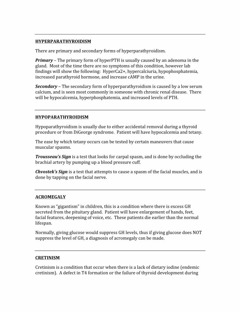
HYPERPARATHYROIDISM
There are primary and secondary forms of hyperparathyroidism.
Primary – The primary form of hyperPTH is usually caused by an adenoma in the gland. Most of the time there are no symptoms of this condition, however lab findings will show the following: HyperCa2+, hypercalciuria, hypophosphatemia, increased parathyroid hormone, and increase cAMP in the urine.
Secondary – The secondary form of hyperparathyroidism is caused by a low serum calcium, and is seen most commonly in someone with chronic renal disease. There will be hypocalcemia, hyperphosphatemia, and increased levels of PTH.
HYPOPARATHYROIDISM
Hypoparathyroidism is usually due to either accidental removal during a thyroid procedure or from DiGeorge syndrome. Patient will have hypocalcemia and tetany.
The ease by which tetany occurs can be tested by certain maneuvers that cause muscular spasms.
Trousseau’s Sign is a test that looks for carpal spasm, and is done by occluding the brachial artery by pumping up a blood pressure cuff.
Chvostek’s Sign is a test that attempts to cause a spasm of the facial muscles, and is done by tapping on the facial nerve.
ACROMEGALY
Known as “gigantism” in children, this is a condition where there is excess GH secreted from the pituitary gland. Patient will have enlargement of hands, feet, facial features, deepening of voice, etc. These patients die earlier than the normal lifespan.
Normally, giving glucose would suppress GH levels, thus if giving glucose does NOT suppress the level of GH, a diagnosis of acromegaly can be made.
CRETINISM
Cretinism is a condition that occur when there is a lack of dietary iodine (endemic cretinism). A defect in T4 formation or the failure of thyroid development during
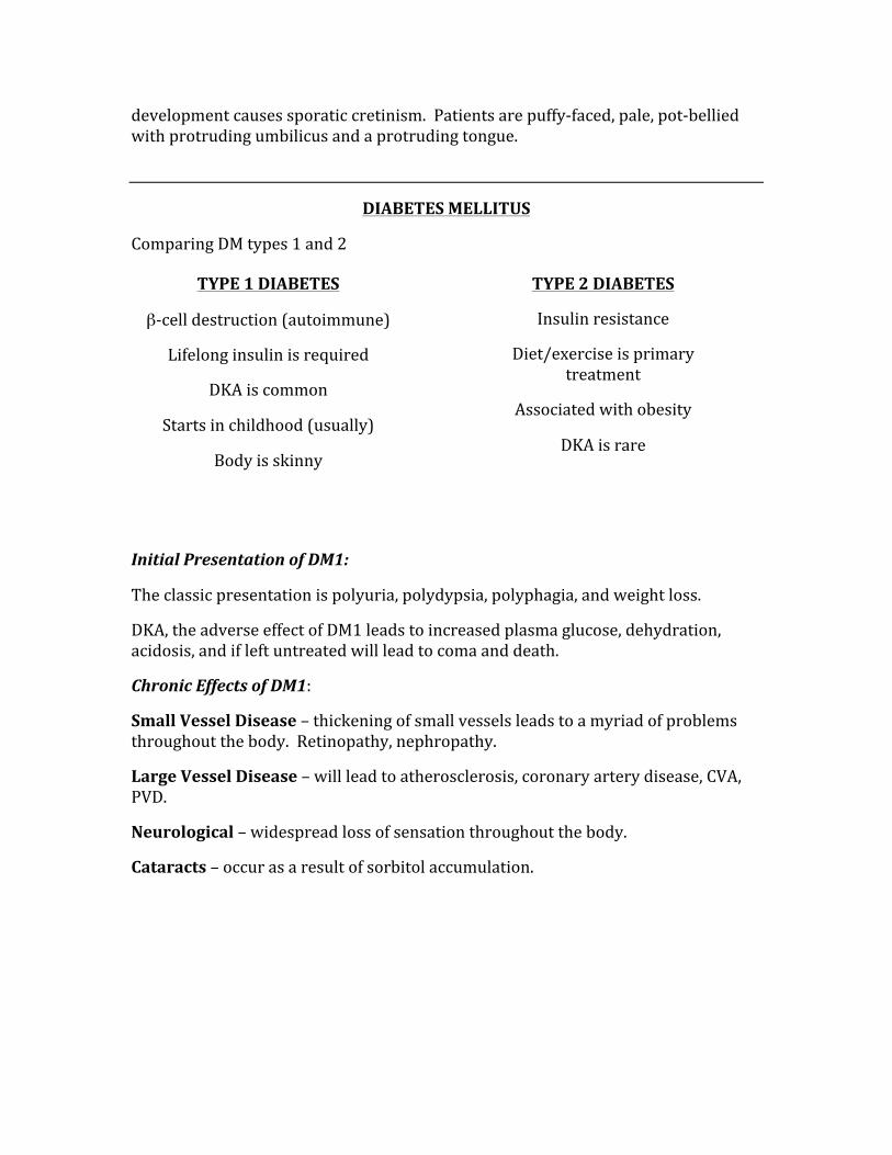
development causes sporatic cretinism. Patients are puffy-‐faced, pale, pot-‐bellied with protruding umbilicus and a protruding tongue.
DIABETES MELLITUS
Comparing DM types 1 and 2
Initial Presentation of DM1:
The classic presentation is polyuria, polydypsia, polyphagia, and weight loss.
DKA, the adverse effect of DM1 leads to increased plasma glucose, dehydration, acidosis, and if left untreated will lead to coma and death.
Chronic Effects of DM1:
Small Vessel Disease – thickening of small vessels leads to a myriad of problems throughout the body. Retinopathy, nephropathy.
Large Vessel Disease – will lead to atherosclerosis, coronary artery disease, CVA, PVD.
Neurological – widespread loss of sensation throughout the body.
Cataracts – occur as a result of sorbitol accumulation.
TYPE 1 DIABETES
β-‐cell destruction (autoimmune)
Lifelong insulin is required
DKA is common
Starts in childhood (usually)
Body is skinny
TYPE 2 DIABETES
Insulin resistance
Diet/exercise is primary treatment
Associated with obesity
DKA is rare
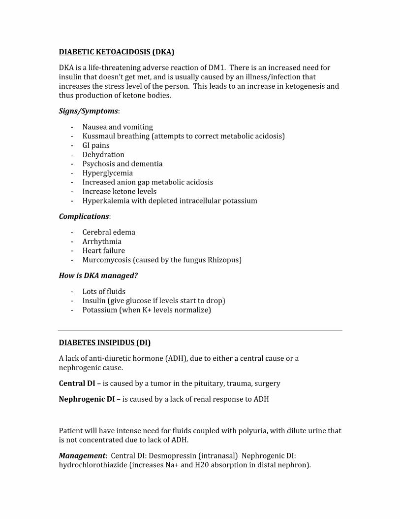
DIABETIC KETOACIDOSIS (DKA)
DKA is a life-‐threatening adverse reaction of DM1. There is an increased need for insulin that doesn’t get met, and is usually caused by an illness/infection that increases the stress level of the person. This leads to an increase in ketogenesis and thus production of ketone bodies.
Signs/Symptoms:
-‐ Nausea and vomiting -‐ Kussmaul breathing (attempts to correct metabolic acidosis) -‐ GI pains -‐ Dehydration -‐ Psychosis and dementia -‐ Hyperglycemia -‐ Increased anion gap metabolic acidosis -‐ Increase ketone levels -‐ Hyperkalemia with depleted intracellular potassium
Complications:
-‐ Cerebral edema -‐ Arrhythmia -‐ Heart failure -‐ Murcomycosis (caused by the fungus Rhizopus)
How is DKA managed?
-‐ Lots of fluids -‐ Insulin (give glucose if levels start to drop) -‐ Potassium (when K+ levels normalize)
DIABETES INSIPIDUS (DI)
A lack of anti-‐diuretic hormone (ADH), due to either a central cause or a nephrogenic cause.
Central DI – is caused by a tumor in the pituitary, trauma, surgery
Nephrogenic DI – is caused by a lack of renal response to ADH
Patient will have intense need for fluids coupled with polyuria, with dilute urine that is not concentrated due to lack of ADH.
Management: Central DI: Desmopressin (intranasal) Nephrogenic DI: hydrochlorothiazide (increases Na+ and H20 absorption in distal nephron).
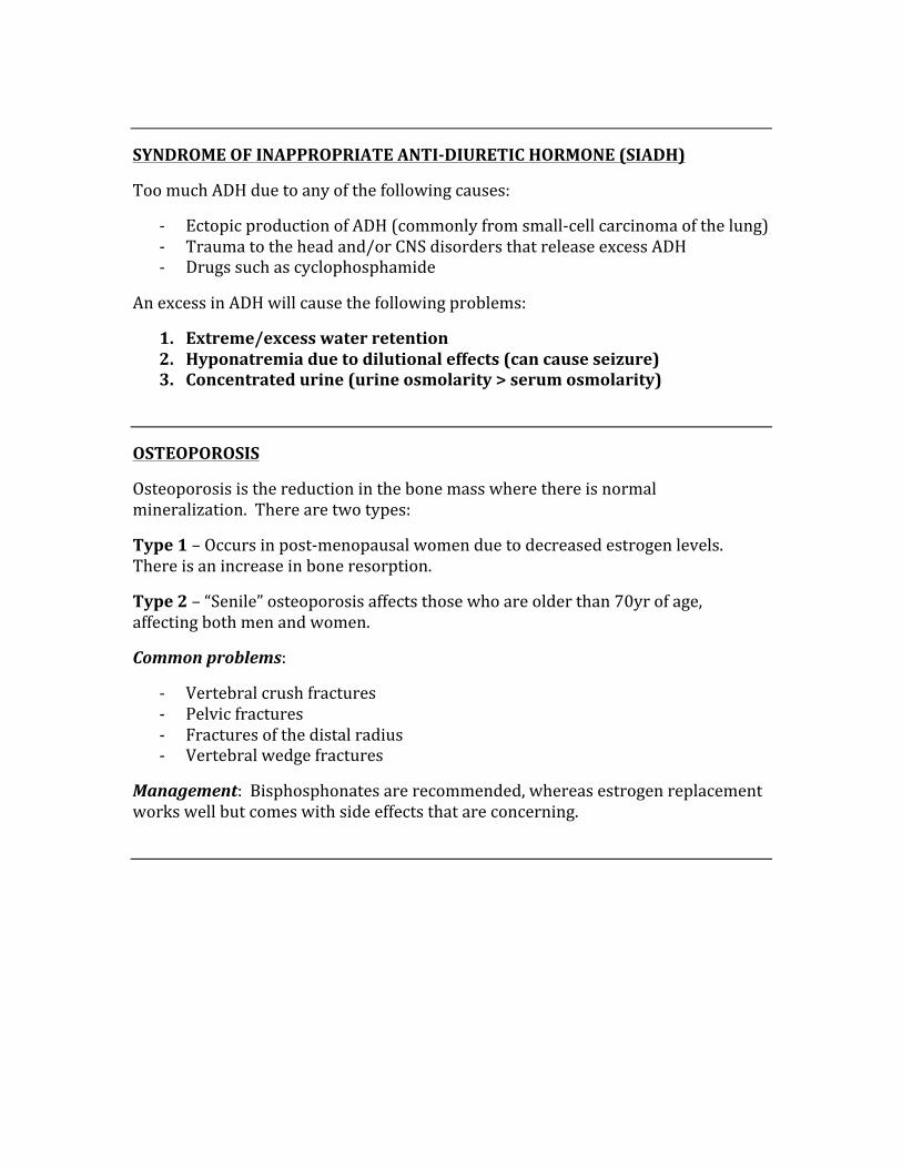
SYNDROME OF INAPPROPRIATE ANTI-‐DIURETIC HORMONE (SIADH)
Too much ADH due to any of the following causes:
-‐ Ectopic production of ADH (commonly from small-‐cell carcinoma of the lung) -‐ Trauma to the head and/or CNS disorders that release excess ADH -‐ Drugs such as cyclophosphamide
An excess in ADH will cause the following problems:
1. Extreme/excess water retention 2. Hyponatremia due to dilutional effects (can cause seizure) 3. Concentrated urine (urine osmolarity > serum osmolarity)
OSTEOPOROSIS
Osteoporosis is the reduction in the bone mass where there is normal mineralization. There are two types:
Type 1 – Occurs in post-‐menopausal women due to decreased estrogen levels. There is an increase in bone resorption.
Type 2 – “Senile” osteoporosis affects those who are older than 70yr of age, affecting both men and women.
Common problems:
-‐ Vertebral crush fractures -‐ Pelvic fractures -‐ Fractures of the distal radius -‐ Vertebral wedge fractures
Management: Bisphosphonates are recommended, whereas estrogen replacement works well but comes with side effects that are concerning.
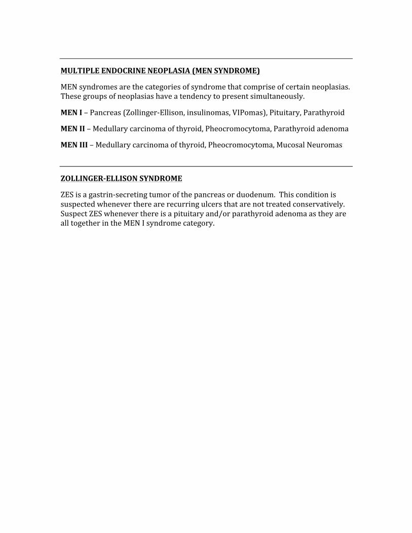
MULTIPLE ENDOCRINE NEOPLASIA (MEN SYNDROME)
MEN syndromes are the categories of syndrome that comprise of certain neoplasias. These groups of neoplasias have a tendency to present simultaneously.
MEN I – Pancreas (Zollinger-‐Ellison, insulinomas, VIPomas), Pituitary, Parathyroid
MEN II – Medullary carcinoma of thyroid, Pheocromocytoma, Parathyroid adenoma
MEN III – Medullary carcinoma of thyroid, Pheocromocytoma, Mucosal Neuromas
ZOLLINGER-‐ELLISON SYNDROME
ZES is a gastrin-‐secreting tumor of the pancreas or duodenum. This condition is suspected whenever there are recurring ulcers that are not treated conservatively. Suspect ZES whenever there is a pituitary and/or parathyroid adenoma as they are all together in the MEN I syndrome category.
BREAST DISEASES
FIBROCYSTIC DISEASE
Fibrocystic breast disease/changes affects between 30%-‐60% of women. Characterized by benign lesions and diffuse breast pain that is often related to hormonal changes associated with her menstrual cycle.
There is a fibrous, lumpy texture to the lesions of the breast. Mammogram is not required to make this diagnosis, but fine-‐needle aspiration is commonly done to check the characteristics of the fluid.
Treatment is not necessary, however pain relief should be done
**There is no increased risk of breast cancer in fibrocystic disease.
BREAST CANCER
Risk Factors:
-‐ Family history of a 1st degree relative with breast cancer at a young age -‐ Age and gender -‐ Menarche (<12yr) is shown to increase risk -‐ Pregnancy (>30) can increase risk -‐ Late menopause (>50)
INVASIVE BREAST CARCINOMAS
Are divided into two major categories based on their cytoarchitectural features:
Intraductal Papillomas are tumors of the lactiferous ducts, they present with nipple discharge. Arising from mammary duct epithelium or lobular glands, and overexpression of estrogen/progesterone receptors.
Ductal Carcinoma in Situ (DCIS) – is early malignancy without basement membrane penetration
Invasive Ductal Carcinoma – presents as a firm/fibrous mass
Comedocarcinoma – is ductal with cheesy consistency due to central necrosis
Inflammatory – has lymphatic involvement and carries a poor prognosis
Invasive Lobular Carcinoma – presents bilaterally with multiple lesions
Medullary Carcinoma – carries a good prognosis, is fleshy with lymphocytic infiltration
INVASIVE DUCTAL CARCINOMA
Tubular Carcinoma
Sebaceous Carcinoma
Secretory Breast Carcinoma
Neuroendocrine Carcinoma
Mucinous Carcinoma
Micropapillary Carcinoma
Acinic Cell Carcinoma
Adenoid Cystic Carcinoma
Apocrine Carcinoma
Cribriform Carcinoma
Glycogen-‐Rich/Clear Cell
Inflammatory Carcinoma
Lipid-‐Rich Carcinoma
LOBULAR CARCINOMA
Pleomorphic
Signet Ring Cell
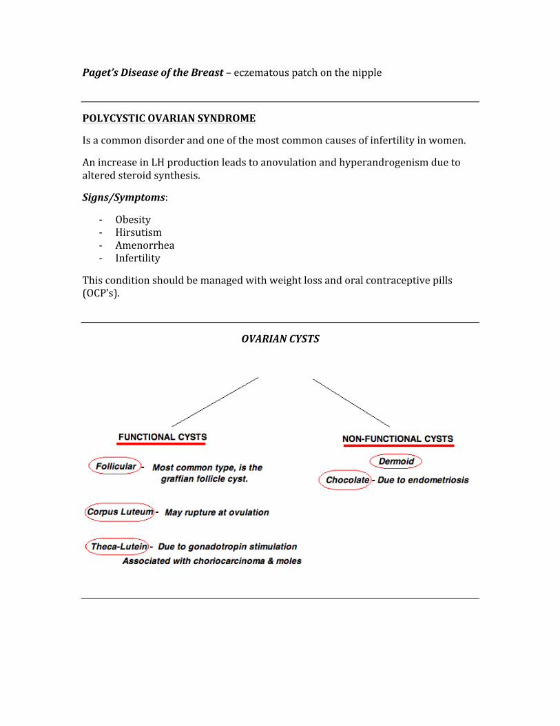
Paget’s Disease of the Breast – eczematous patch on the nipple
POLYCYSTIC OVARIAN SYNDROME
Is a common disorder and one of the most common causes of infertility in women.
An increase in LH production leads to anovulation and hyperandrogenism due to altered steroid synthesis.
Signs/Symptoms:
-‐ Obesity -‐ Hirsutism -‐ Amenorrhea -‐ Infertility
This condition should be managed with weight loss and oral contraceptive pills (OCP’s).
OVARIAN CYSTS
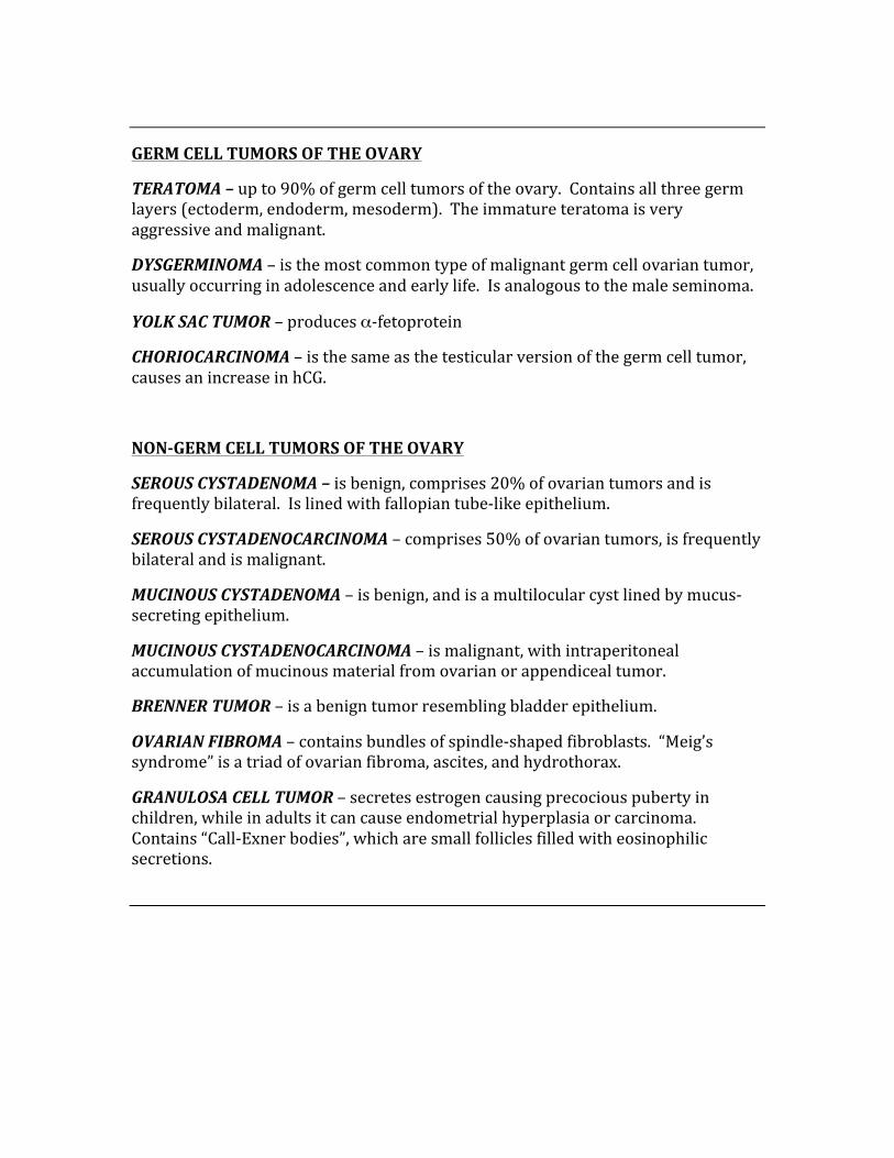
GERM CELL TUMORS OF THE OVARY
TERATOMA – up to 90% of germ cell tumors of the ovary. Contains all three germ layers (ectoderm, endoderm, mesoderm). The immature teratoma is very aggressive and malignant.
DYSGERMINOMA – is the most common type of malignant germ cell ovarian tumor, usually occurring in adolescence and early life. Is analogous to the male seminoma.
YOLK SAC TUMOR – produces α-‐fetoprotein
CHORIOCARCINOMA – is the same as the testicular version of the germ cell tumor, causes an increase in hCG.
NON-‐GERM CELL TUMORS OF THE OVARY
SEROUS CYSTADENOMA – is benign, comprises 20% of ovarian tumors and is frequently bilateral. Is lined with fallopian tube-‐like epithelium.
SEROUS CYSTADENOCARCINOMA – comprises 50% of ovarian tumors, is frequently bilateral and is malignant.
MUCINOUS CYSTADENOMA – is benign, and is a multilocular cyst lined by mucus-‐secreting epithelium.
MUCINOUS CYSTADENOCARCINOMA – is malignant, with intraperitoneal accumulation of mucinous material from ovarian or appendiceal tumor.
BRENNER TUMOR – is a benign tumor resembling bladder epithelium.
OVARIAN FIBROMA – contains bundles of spindle-‐shaped fibroblasts. “Meig’s syndrome” is a triad of ovarian fibroma, ascites, and hydrothorax.
GRANULOSA CELL TUMOR – secretes estrogen causing precocious puberty in children, while in adults it can cause endometrial hyperplasia or carcinoma. Contains “Call-‐Exner bodies”, which are small follicles filled with eosinophilic secretions.
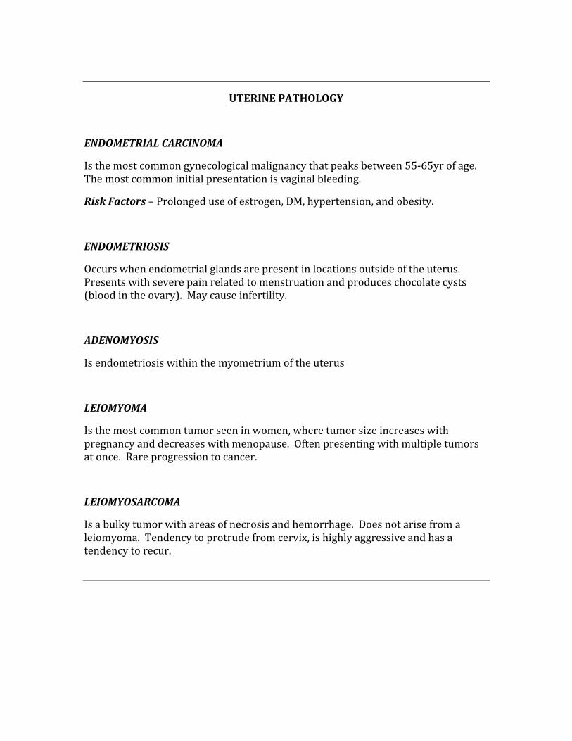
UTERINE PATHOLOGY
ENDOMETRIAL CARCINOMA
Is the most common gynecological malignancy that peaks between 55-‐65yr of age. The most common initial presentation is vaginal bleeding.
Risk Factors – Prolonged use of estrogen, DM, hypertension, and obesity.
ENDOMETRIOSIS
Occurs when endometrial glands are present in locations outside of the uterus. Presents with severe pain related to menstruation and produces chocolate cysts (blood in the ovary). May cause infertility.
ADENOMYOSIS
Is endometriosis within the myometrium of the uterus
LEIOMYOMA
Is the most common tumor seen in women, where tumor size increases with pregnancy and decreases with menopause. Often presenting with multiple tumors at once. Rare progression to cancer.
LEIOMYOSARCOMA
Is a bulky tumor with areas of necrosis and hemorrhage. Does not arise from a leiomyoma. Tendency to protrude from cervix, is highly aggressive and has a tendency to recur.
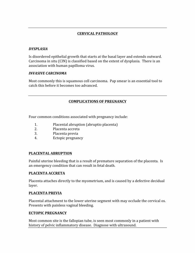
CERVICAL PATHOLOGY
DYSPLASIA
Is disordered epithelial growth that starts at the basal layer and extends outward. Carcinoma in situ (CIN) is classified based on the extent of dysplasia. There is an association with human papilloma virus.
INVASIVE CARCINOMA
Most commonly this is squamous cell carcinoma. Pap smear is an essential tool to catch this before it becomes too advanced.
COMPLICATIONS OF PREGNANCY
Four common conditions associated with pregnancy include:
1. Placental abruption (abruptio placenta) 2. Placenta accreta 3. Placenta previa 4. Ectopic pregnancy
PLACENTAL ABRUPTION
Painful uterine bleeding that is a result of premature separation of the placenta. Is an emergency condition that can result in fetal death.
PLACENTA ACCRETA
Placenta attaches directly to the myometrium, and is caused by a defective decidual layer.
PLACENTA PREVIA
Placental attachment to the lower uterine segment with may occlude the cervical os. Presents with painless vaginal bleeding.
ECTOPIC PREGNANCY
Most common site is the fallopian tube, is seen most commonly in a patient with history of pelvic inflammatory disease. Diagnose with ultrasound.
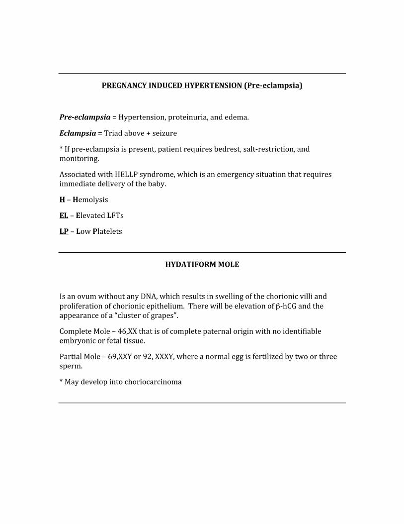
PREGNANCY INDUCED HYPERTENSION (Pre-‐eclampsia)
Pre-‐eclampsia = Hypertension, proteinuria, and edema.
Eclampsia = Triad above + seizure
* If pre-‐eclampsia is present, patient requires bedrest, salt-‐restriction, and monitoring.
Associated with HELLP syndrome, which is an emergency situation that requires immediate delivery of the baby.
H – Hemolysis
EL – Elevated LFTs
LP – Low Platelets
HYDATIFORM MOLE
Is an ovum without any DNA, which results in swelling of the chorionic villi and proliferation of chorionic epithelium. There will be elevation of β-‐hCG and the appearance of a “cluster of grapes”.
Complete Mole – 46,XX that is of complete paternal origin with no identifiable embryonic or fetal tissue.
Partial Mole – 69,XXY or 92, XXXY, where a normal egg is fertilized by two or three sperm.
* May develop into choriocarcinoma
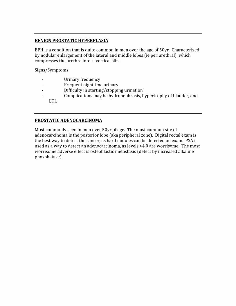
BENIGN PROSTATIC HYPERPLASIA
BPH is a condition that is quite common in men over the age of 50yr. Characterized by nodular enlargement of the lateral and middle lobes (ie periurethral), which compresses the urethra into a vertical slit.
Signs/Symptoms:
-‐ Urinary frequency -‐ Frequent nighttime urinary -‐ Difficulty in starting/stopping urination -‐ Complications may be hydronephrosis, hypertrophy of bladder, and
UTI.
PROSTATIC ADENOCARCINOMA
Most commonly seen in men over 50yr of age. The most common site of adenocarcinoma is the posterior lobe (aka peripheral zone). Digital rectal exam is the best way to detect the cancer, as hard nodules can be detected on exam. PSA is used as a way to detect an adenocarcinoma, as levels >4.0 are worrisome. The most worrisome adverse effect is osteoblastic metastasis (detect by increased alkaline phosphatase).
Related Documents


