Beyond Anthrax

Welcome message from author
This document is posted to help you gain knowledge. Please leave a comment to let me know what you think about it! Share it to your friends and learn new things together.
Transcript

Beyond Anthrax

Larry I. Lutwick l Suzanne M. LutwickEditors
Beyond Anthrax
The Weaponization of Infectious Diseases
1 3

Editors
Larry I. LutwickVANewYorkHarborHealth Care SystemDivision of Infectious DiseasesSUNY Downstate Medical SchoolBrooklyn, NY, [email protected]
Suzanne M. LutwickDirector of Grant DevelopmentHackensack University MedicalCenter Foundation
Hackensack, NJ, [email protected]
ISBN: 978-1-58829-438-8 e-ISBN: 978-1-59745-326-4DOI: 10.1007/978-1-59745-326-4
Library of Congress Control Number: 2008934792
# 2009 Humana Press, a part of Springer ScienceþBusiness Media, LLCAll rights reserved. This workmay not be translated or copied in whole or in part without the writtenpermission of the publisher (Springer Science+BusinessMedia, LLC, 233 Spring Street, NewYork,NY 10013, USA), except for brief excerpts in connection with reviews or scholarly analysis. Use inconnection with any form of information storage and retrieval, electronic adaptation, computersoftware, or by similar or dissimilar methodology now known or hereafter developed is forbidden.The use in this publication of trade names, trademarks, service marks, and similar terms, even if theyare not identified as such, is not to be taken as an expression of opinion as to whether or not they aresubject to proprietary rights.
Printed on acid-free paper
springer.com

Piglet sidled up to Pooh from behind.‘‘Pooh,’’ he whispered. ‘‘Yes, Piglet?’’‘‘Nothing,’’ said Piglet, taking Pooh’s paw.‘‘I just wanted to be sure of you.’’
This book is dedicated to those whom wealways want to be sure of:Our parents and siblings andTalora and Zev,Rachel and Robert,Zach and Mollie,Arielle and Talal,Nina and. . .
May all of you be able to spend playfullyexploring the world of Pooh without anyinterference by the contents of this text.And to a best friend who is looking downupon us, still not a sports fan, Steve Straus.
Larry LutwickSuzanne Lutwick

Preface
‘‘If there is one thing the history of evolution has taught us it’s that life will not becontained. Life breaks free, expands to new territory, and crashes through barriers,painfully, maybe even dangerously.’’
– Dr. Ian Malcolm, ‘‘Jurassic Park’’ (1993)
‘‘The most merciful thing in the world, I think, is the inability of the human mind tocorrelate all its contents. We live on a placid island of ignorance in the midst of blackseas of infinity, and it was not meant that we should voyage far. The sciences, eachstraining in its own direction, have hitherto harmed us little; but some day the piecingtogether of dissociated knowledge will open such terrifying vistas of reality, and of ourown frightful position therein, that we shall either go mad from the revelation or fleefrom the deadly light into the peace and safety of a new dark age.’’
– H.P. Lovecraft, ‘‘The Call of Cthulhu’’ (published 1928)
Howard Phillips Lovecraft was an American author of horror, fantasy, andscience fiction. His major inspiration and invention was overt cosmic horror,and he is often regarding as one of the most influential horror writers of thetwentieth century, exerting widespread and indirect influence, and frequentlycompared to Edgar Allan Poe. The above quotation begins the story and couldbe applied tomore modern times, nearly 80 years since the story’s publication in‘‘Weird Tales.’’ Of course, it is easy to argue that the sciences have no longer‘‘harmed us little’’ with such developments of nuclear bombs and other weaponsof mass destruction (WMDs). Among the WMDs (as the media enjoys callingthem) are those of the biological variety. It is these agents that serve as the basisof this textbook.
‘‘Beyond Anthrax, The Weaponization of Infectious Diseases’’ has been indevelopment for a number of years and is meant as a primer for clinicians andepidemiologists on a variety of agents, organisms, or toxins, which are generallyconsidered at the forefront of potential use in a biological attack from a rougenation or radical group. In the aftermath of the September 11 World TradeCenter attacks, a number of cases of inhalational anthrax were diagnosed in theeastern United States, specifically the New York City metropolitan area andWashington, DC, although the first case was diagnosed in Florida. The finding
vii

of a disease such as anthrax outside of its general geographic area with anuncommon presentation (inhalation rather than cutaneous exposure) is thefactor that raises the red flag of a possible bioterrorist attack. In this case, thespores of Bacillus anthracis were found to have been weaponized to increaseinfectivity and placed (by a person still unknown) in the mail. The letters, byprocessing in post office facilities or by opening at the final destination,delivered a deadly message producing inhalation anthrax in 11 individualswith a 45% case fatality rate [1]. Much information has been publishedregarding anthrax as a biological agent, and for reasons of space andminimizing repetition regarding this disease, the text will start beyondanthrax and discuss the remaining Category A agents as well as delve into anumber of the diseases placed in Category B. It will suffice to say, however, thatthe anthrax incident has demonstrated what terrorism really does in getting ahuge bang for its buck; that is, for a small number of cases, the outbreak causedmajor disruption to much of the fiber of this country changing some of itforever.
Before going forward, we must take stock of reality and not just jump, willy-nilly, on to the Lovecraftian slippery slope of the inevitability of something eviloccurring fashioned by the hand of some bad person. No doubt, the possibilityof the use of biological warfare has always existed millennia before theacceptance of the Germ Theory, a short century or so ago. Many of theseevents are discussed in Stuart Handysides’ introductory chapter on thehistory of the topic. It is, to this point, useful to refer to the John Snow [2]Memorial Outbreak Scoreboard during the last decade or so. In doing so, ourevil task doers are clearly trounced in overall numbers of cases and outbreaks byMother Nature (MN), the world’s most devious bioterrorist.
Although aided by humankind,MNhas fashioned newly recognized diseasessuch as SARS (severe acute respiratory syndrome) [3] like a Golem out of thevirtual molecular mud and has facilitated diseases such as monkey pox [4] andWest Nile virus [5] unknown on a continent to appear there.
Furthermore, she has assisted in the production of multidrug resistantorganisms [6,7] in a healthcare arena where fully sensitive ones had beenpresent. Additionally, and certainly last but not least, MN continues topercolate new strains of influenza A including the current H5N1 avian strain[8] that threaten to win the primary race for next pandemic candidate. Althoughthe diseases forthcoming in this text may be formidable opponents in the future,it remains a solid wager that infectious disease clinicians, epidemiologists, andpublic health personnel will have their hands soiled with many more threatsthan that are contained here.
Following the Stuart Handysides (former Medical Editor of CommunicableDisease and Public Health) chapter on the history of biological warfare, the texthas five chapters regarding the Category A diseases that are (true to the title,beyond anthrax), namely, smallpox, plague, tularemia, botulism, and the viralhemorrhagic fevers. The chapters are written by some of the foremost experts ofeach field including representatives of the National Institutes of Health, the
viii Preface

Centers for Disease Control and Prevention, and UK’s Defense Science andTechnology Laboratory at Porton Down. The chapters for the most partcontain similar sections including outbreak scenarios, a historical perspective,microbiologic considerations, natural infection with its epidemiology anddiagnostic considerations, as well as specific biowarfare issues. Additionally,the chapters discuss both therapeutic and preventative measures and mayinclude infection control, prophylactic drugs, reservoir controls, andvaccinations.
The next part of the text contains chapters dealing with many, but not all, ofthe Category B agents, selected for overall interest. This includes a chapter onthe intentional contamination of food and water as well as ones dealing withmelioidosis, epidemic typhus, and some of the biotoxins such as ricin andstaphylococcal enterotoxin B. Overall, the organization of these chaptersparallel that in those of the Category A diseases. With much more emphasison the ‘‘A’’ diseases, the inclusion of these entities provides a good source ofinformation for the clinician and epidemiologist.
Following the ‘‘B’’ list are a number of chapters that concentrate on a varietyof issues that are important in any contribution in the biowarfare arena. All ofthese have direct applications to natural outbreaks and epidemics, and theyinclude Public Health Infrastructure, Public Health Law, Public HealthSurveillance, Mental Health Management, as well as a chapter regarding therole of the media in outbreaks written by David Brown, a physician who haswritten regularly for the Washington Post. The text ends with an overview ofrapid detection of pathogens and a final chapter discussing agroterrorism, thatis, biological attacks on the potentially very vulnerable food-producing systemsof the world. Biowarfare aimed at flora and fauna rather than on humankind,although not as often written about, are ripe areas for further discussions andprotective measures.
In several of the original versions of the ‘‘Table of Contents,’’ several otherchapters were envisioned, but as the text matured, they were not included. Theeditors thank those who contributed additional but unused material.
We hope that the topics contained here, as biowarfare events, remain purelydidactic exercises and not issues that interject themselves into clinical medicine.
References
1. Holty, J.-E., Bravata, D. M., Liu, H., et al: A century of inhalational anthrax cases from1900 to 2005. Ann. Intern. Med. 144, 270–280, 2006.
2. Newsom, S. W.: Pioneers in infection control: John Snow, Henry Whitehead, the BroadStreet pump, and the beginnings of geographical epidemiology. J. Hosp. Infect. 64,210–216, 2006.
3. Conly, J. M., Johnston B. L.: SARS: A tale of two epidemics. Can. J. Infect. Dis. 14,147–149, 2003.
4. Reed, K. D., Melski, J. W., Graham, M. B., et al: The detection of monkeypox in humansin the Western Hemisphere. N. Engl. J. Med. 350, 342–350, 2004.
ix
Preface ix

5. Gubler, D. J.: The continuing spread of West Nile virus in the western hemisphere. Clin.Infect. Dis. 45, 1039–1046, 2007.
6. Gorwitz, R. J.: A review of community-associated methicillin-resistant Staphylococcusaureus skin and soft tissue infections. Pediatr. Infect. Dis. J. 27, 1–7, 2008.
7. Tumbarello, M., Spanu, T., Sanguinetti, M., et al: Bloodstream infections caused byextended-spectrum-beta-lactamase-producing Klebsiella pneumoniae. Antimicrob. AgentsChemother. 50, 498–504, 2006.
8. Peiris, J. S., de Jong, M. D., Guan, Y.: Avian influenza virus(H5N1): a threat to humanhealth. Clin. Microbiol. Rev. 20, 243–267, 2007.
x Preface

Contents
1 The History of Bioterrorism: Old Idea, New Word,
Continuing Taboo. . . . . . . . . . . . . . . . . . . . . . . . . . . . . . . . . . . . . . . . 1Stuart Handysides
2 Smallpox and Bioterrorism. . . . . . . . . . . . . . . . . . . . . . . . . . . . . . . . . 17Daniel R. Lucey, Joel G. Breman and Donald A. Henderson
3 Plague . . . . . . . . . . . . . . . . . . . . . . . . . . . . . . . . . . . . . . . . . . . . . . . . 55Petra C. F. Oyston and Richard W. Titball
4 Tularemia. . . . . . . . . . . . . . . . . . . . . . . . . . . . . . . . . . . . . . . . . . . . . . 77Daniel S. Shapiro
5 Botulism . . . . . . . . . . . . . . . . . . . . . . . . . . . . . . . . . . . . . . . . . . . . . . . 85Jeremy Sobel
6 The Viral Hemorrhagic Fevers . . . . . . . . . . . . . . . . . . . . . . . . . . . . . . 107Daniel G. Bausch and C. J. Peters
7 Melioidosis . . . . . . . . . . . . . . . . . . . . . . . . . . . . . . . . . . . . . . . . . . . . . 145Pooja Tolaney and Larry I. Lutwick
8 Epidemic Typhus Fever . . . . . . . . . . . . . . . . . . . . . . . . . . . . . . . . . . . 159Mohammad Mooty and Larry I. Lutwick
9 Category B Biotoxins . . . . . . . . . . . . . . . . . . . . . . . . . . . . . . . . . . . . . 181Larry I. Lutwick, Jeremy Gradon and Jonathan Zellen
10 Intentional Terrorist Contamination of Food and Water . . . . . . . . . . 207Jeremy Sobel and John C. Watson
11 Public Health Infrastructure . . . . . . . . . . . . . . . . . . . . . . . . . . . . . . . 219Isaac B. Weisfuse
xi

12 Public Health Law and Biological Terrorism . . . . . . . . . . . . . . . . . . . 239Lance Gable and James G. Hodge
13 Public Health Surveillance for Bioterrorism . . . . . . . . . . . . . . . . . . . . 253Peter N. Wenger, William Halperin and Edward Ziga
14 Psychosocial Management of Bioterrorism Events . . . . . . . . . . . . . . . 279David M. Benedek and Thomas A. Grieger
15 The Role of the Media in Bioterrorism . . . . . . . . . . . . . . . . . . . . . . . . 295David Brown
16 Rapid Detection of Bioterrorism Pathogens . . . . . . . . . . . . . . . . . . . . 317David Perlin
17 Plant Pathogens as Biological Weapons Against Agriculture . . . . . . . 335Forrest W. Nutter and Lawrence V. Madden
Index . . . . . . . . . . . . . . . . . . . . . . . . . . . . . . . . . . . . . . . . . . . . . . . . . . . . . 365
xii Contents
, Jr.

Contributors
Daniel G. Bausch, MD, MPH&TMDepartment of Tropical Medicine, Tulane School of Public Health and
Tropical Medicine, New Orleans, Louisiana, [email protected]
David M. Benedek, MD, LTC, MC, USACenter for the Study of Traumatic Stress, Uniformed Services University,
Bethesda, Maryland, [email protected]
Joel G. Breman, MD, DTPHFogarty International Center, National Institutes of Health, Bethesda,
Maryland
David Brown, MDThe Washington Post, Washington, District of Columbia, browndavidm
@comcast.net
Lance Gable, JD, MPHWayne State University Law School; Center for Law and the Public’s Health,
Detroit, Michigan
Jeremy Gradon, MDSinai Hospital of Baltimore; Johns Hopkins Hospital School of Medicine,
Baltimore, Maryland
Thomas A. Grieger, MD, Capt., MC, USNCenter for the Study of Traumatic Stress, Uniformed Services University,
Bethesda, Maryland
William Halperin, MD, DrPHDepartment of Preventive Medicine and Community Health, University of
Medicine and Dentistry of New Jersey, New Jersey Medical School, Newark,
New Jersey, [email protected]
Stuart Handysides, BSc, MBBSProgram for Monitoring Emerging Diseases, Hertford, England
xiii

Donald A. Henderson, MD, MPHCenter for Biosecurity, University of Pittsburgh Medical Center, JohnsHopkins Hospital School of Medicine Distinguished Service Professor,Baltimore, Maryland
James G. Hodge, Jr., JD, LLMCenter for Law and the Public’s Health; Johns Hopkins Bloomberg School ofPublic Health, Baltimore, Maryland, [email protected]
Daniel R. Lucey, MD, MPHBiohazardous Threat Agents and Emerging Infectious Diseases, Departmentof Microbiology and Immunology, Georgetown University School ofMedicine, Washington, District of Columbia, [email protected]
Larry I. Lutwick, MDVeterans Affairs New York Harbor Health Care System, Brooklyn Campus;State University of New York, Downstate Medical Center, Brooklyn,New York, [email protected]
Suzanne M. Lutwick, BS, MPHDirector of Grant Development, Hackensack University Medical CenterFoundation, Hackensack, New Jersey, [email protected]
Laurence V. Madden, PhDDepartment of Plant Pathology, Ohio State University, Wooster, Ohio
Mohammad Mooty, MDInfectious Diseases, Eastern Maine Medical Center, Bangor, Maine
Forrest W. Nutter, Jr., PhDDepartment of Plant Pathology, Iowa State University, Ames, Iowa,[email protected]
Petra C. F. Oyston, BSc, PhDDefense Science and Technology Laboratory, Porton Down, Wiltshire, England
David Perlin, PhDPublic Health Research Institute, Newark, New Jersey, [email protected]
C. J. Peters, MDDepartments of Pathology, and Microbiology and Immunology, Center forTropical Diseases, University of Texas, Galveston, Texas
Daniel S. Shapiro, MDBoston University School of Medicine; Director of Clinical MicrobiologyLaboratory, Lahey Clinic, Burlington, Massachusetts, [email protected]
Jeremy Sobel, MD, MPHFoodborne and Diarrheal Diseases Branch, Centers for Disease Control andPrevention, Atlanta, Georgia, [email protected]
xiv Contributors

Richard W. Titball, BSc, PhD, DScSchool of Biosciences, University of Exeter, Exeter, England, [email protected]
Pooja Tolaney, MDInfectious Diseases Clinician, Hartford, Connecticut
John C. Watson, MD, MPHCaptain, US Public Health Service, Division of Parasitic Diseases, NationalCenter for Zoonotic, Vectorborne, and Enteric Diseases, Coordinating Centerfor Infectious Diseases, Centers for Disease Control and Prevention, Atlanta,Georgia
Isaac B. Weisfuse, MD, MPHNew York City Department of Health and Mental Hygiene, New York, NewYork, [email protected]
Jonathan Zellen, MDDepartment of Medicine, North General Hospital, New York, New York
Edward Ziga, MD, MPHOffice of Surveillance and Prevention, Communicable Diseases Division,Newark Department of Health and Human Services, Newark, New Jersey
Contributors xv

Chapter 1
The History of Bioterrorism: Old Idea, NewWord,
Continuing Taboo
Stuart Handysides
1.1 Definitions
The word ‘‘bioterrorism’’ first appeared in a book called Killing Winds, byJeanneMcDermott, in 1987 [1], and took another 9 years to reach a biomedicalpublication [2]. It appears in only the post-2003 editions of large dictionaries inmy local bookshop and the Oxford English Dictionary online [3]. A Medlinesearch yielded 2,018 entries on the topic––none before 1996, only 50 before2000, and then an exponential increase (70 in 2000, 445 in 2001, 862 in 2002, and3,998 by November 13, 2007) [4]. The history of bioterrorism would be rathershort if written to consider events only since the word was coined. From ahistorical perspective it will be better, I think, to consider the phenomenon itselfand take a longer view. Nevertheless, the word itself needs defining.
The word ‘‘terrorism’’ goes back only as far as the French Revolution. ‘‘Thereign of terror’’ was a 2-year period of mob rule and bloodshed, led by theJacobin government of Robespierre and the so-called Committee of PublicSafety that followed the September massacres of 1792. Edmund Burke wasone of the first persons to use the word ‘‘terrorist’’ to describe agents of theJacobin government, in 1795, and the word ‘‘terrorism’’ was first used, in thesame context, the same year.
Robespierre, translated from the original French by J. M. Thompson,explained the need for harsh measures: ‘‘If the basis of popular governmentin time of peace is virtue, its basis in time of revolution is both virtueand intimidation––virtue, without which intimidation is disastrous, andintimidation, without which virtue has no power . . . . Intimidation is merelyjustice––prompt, severe and inflexible. It is therefore an emanation of virtue,and results from the application of democracy to the most pressing needs ofthe country’’ [5]. Thompson, commenting on these words, noted that intimi-dation was generally the weapon of tyrants. The new despotism of postrevolu-tionary France took this weapon out of the tyrant’s hand and turned it against
S. Handysides25 Fordwich Rise, Hertford SG14 2BW, Englande-mail: [email protected]
L.I. Lutwick, S.M. Lutwick (eds.), Beyond Anthrax,DOI: 10.1007/978-1-59745-326-4_1, � Springer ScienceþBusiness Media, LLC 2008
1

him in the name of the people, but ended up turning it against anyone who
disagreed with the current ‘‘party line’’.The word terrorist today is usually applied to a nongovernmental agent, an
agent without state sponsorship, but this usage goes back only to the second
half of the nineteenth century. The word was used in 1866 to describe the
dissident Irish, and in 1883 Russian revolutionaries who were prepared to use
violence received the same appellation.Webster’s New International Dictionary
(1913) defined terrorism as ‘‘a mode of governing, or opposing government, by
intimidation’’ and a terrorist as ‘‘one who administers or coerces a government
or community by intimidation’’ [6], thus covering the actions of established
governments and those with grievances against them.The word terrorist is often given a negative connotation, but history some-
times changes our perspective on people and events. As an example, the South
African government imprisoned Nelson Mandela as a terrorist, but many saw
his fight against oppression and demand for human rights and the end of
apartheid as the just desires of a freedom fighter. The latter appear to have
been right.Central to the meaning of terrorism is the fear experienced by those being
terrorized. The acts of violence or warfare themselves kill or injure, but the
terror is experienced by those who wonder where it will end and whether they or
their loved ones will be the next victims of pain, injury, or death. Perhaps, the
greatest act of terrorism the world has seen was the unleashing of atomic bombs
on Japan. Terror belonged not only to the survivors in Japan, but to the world
in general in the following 40 years of the ‘‘ColdWar’’ and the uncertainty of the
deterrent power of mutually assured destruction.The prefix ‘‘bio’’ denotes the use or threat of biological agents to terrorize.
Human beings themselves are biological agents, whose fists, feet, and teeth may
be used as weapons. Various large and/or fierce animals have been used to
intimidate enemies––Hannibal’s elephants must have been an awesome sight,
and the pots of snakes hurled by his men onto the ships of King Eumenes of
Pergamon spread fear and distracted his enemies [7, 8]; Nero’s lions in the
Coliseum were an attempt to reduce the attraction of Christianity; the Romans
also catapulted bees and hornets at their enemies; and the horses of the Spanish
conquistadors menaced the Incas and Aztecs [7]. The word bioterrorism is
usually used, however, to describe the threat or deployment of microorganisms
or the use of contagion or infection, as weapons. TheOxford English Dictionary
defines bioterrorism as ‘‘the use of infectious agents or biologically active
substances as weapons of terrorism’’ [3].Does the term bioterrorism deserve to exist? Do we speak of ballistic, explo-
sive, or chemical terrorism? Is the use of biological weaponry different from other
methods of attack? Roman jurists protested when their attempt to conquer
Germanic tribes was thwarted by the poisoning of wells: armis bella non venenis
geri––war is fought with weapons, not with poisons [8]. Commanders might see
their armies decimated by outbreaks of plague, typhoid, cholera, smallpox, or
2 S. Handysides

influenza, but these were misfortunes rather than potential tools. Why is the useof a biological weapon seen as ‘‘fighting dirty’’ or ‘‘not cricket’’?
This chapter, on the history of bioterrorism, will take an inclusive approach.It will draw little distinction between those who practice ‘‘warfare’’ and thosewho practice ‘‘terrorism.’’ It will also use the term bioterrorism to describeactions taken before the existence of microorganisms was discovered.
1.2 Acts of God
Several of the Biblical plagues meted out to the Egyptians when Pharaoh refusedto allow the children of Israel their freedom could be regarded as bioterrorist acts[9]. In the first plague, the rivers turned to blood and the fish died and stank. Theplague of frogs was unwelcome, as was their smell when they died and rotted inheaps. Plagues of lice and flies and a die-off of cattle, which spared the livestock ofthe Israelites, followed the plague of frogs. Pharaoh continued to take a hard line,refusing to bargain with the terrorists, and the next visitation was a plague ofboils and blains on the skin of men and beasts. At this point, the Lord diversified,using hail and fire to destroy the ripening crops, but then again returned to abiological plague, employing locusts to finish off whatever the hail had left. Thepenultimate plaguewas a period of darkness, lasting 3 days. In the last plague, thefirstborn of all families, human and animal, were slain in households where aprescribed sacrifice had not been made. At this point, Pharaoh hollered ‘‘’nough’’and sent the Israelites on their way.
The exact nature and factual existence of the plagues have been debated. Theboils have been attributed to both plague and anthrax, but it all happened along time ago. At the very least, however, the biblical report indicates that thepotential for bioterrorism had been realized when the book of Exodus waswritten (about 1500 BC). If the book is taken as a literal historical report, it couldbe argued that God, as well as creator, was also the first bioterrorist.
The Bible also describes an outbreak––possibly plague––that befell thePhilistines, when they captured the Ark of the Covenant from the Israelites[8, 10].
Another early reference to possible bioterrorism is found in Homer’s Iliad,which tells how the city of Troy came under siege of the Greeks, after thebeautiful Helen (married to the Greek Menelaus) ran off with Paris, one ofthe sons of the Trojan king, Priam [11]. The tale, written perhaps 7–10 centuriesbefore the birth of Christ, starts with a description of a plague on mules, dogs,andmenmeted out by Apollo, after his priest, Chryses, was sent away rudely byAgamemnon when he asked for the return of his daughter.
Thus, the earliest descriptions of what might be called bioterrorism wereplagues attributed to the God of the Hebrews and the Greek god Apollo. Gods,unlike men, could not be fought. They had to be appeased. Pharaoh andAgamemnon capitulated.
1 History of Bioterrorism 3

1.3 Poisoning Water Supplies
A little later, in the fifth century BC, Hippocrates said that impurities in the air(Greek: miasmata) were the cause of plagues [12]. At much the same time,Thucydides noted that the people of Athens, suffering from plague (or was itmeasles? [13]), believed that the city’s water supply had been poisoned by theSpartans. Although the Romans had declared that poisoning was not anappropriate way to wage war, they poisoned the wells of the remnants of thearmy of Aristonicus, who kept fighting despite being, to a large extent, defeated[8]. Many years later the Black Death spread from Constantinople to the wholeof western Europe, following trade routes both on land and on sea. Jewishcommunities in some of the plague-affected cities were accused of well poison-ing and were exterminated [12]. The practice of poisoning water supplies hascontinued: It occurred in the American Civil War, the BoerWar, and, allegedly,in Turkish Kurdistan as recently as 1997 [8].
1.4 Ballistic Biological Weapons
The archers of Scythia, in about 400 BC, are said to have dipped their arrow tipsin blood mixed with manure [8]. Other early instances of the ballistic distribu-tion of biological weapons are arrow tips and darts from blowpipes poisonedwith curare by native Amazonians and with batrachotoxin from frogs byHawaiian islanders [8, 14]. A book published in 1777 suggested to dip ‘‘arrowsin matter of smallpox and twang them at the American rebels’’ [8]. Morerecently, grenade shrapnel was contaminated with botulinum toxin in theSecond World War [8].
Sometimes the missiles were rather larger. The original catapult and ballista,as used by the Romans, could hurl rocks of approximately 30 kg at and over thewalls of castles [15]. Later developments, such as the trebuchet, could projectmissiles of 150 kg and even as much as 1,500 kg. When the rocks ran out, otherobjects could be used as ammunition. Gabriel deMussis provided an eyewitnessaccount of the siege of Caffa (now Feodosiya), on the Black Sea, from 1344 to1346 [15]. The Genoese-occupied city held out for 3 years. Suddenly, thebesieging Tartars and Saracens fell victim to plague: ‘‘the humors coagulatedin the groins, they developed a subsequent putrid fever and died.’’ Mountains ofcadavers were ‘‘placed on their hurling machines and thrown into the city ofCaffa . . . soon all the air was infected and the water poisoned, corrupt andputrefied, and such a great odor increased.’’ The Genoese took to their shipsand fled, taking plague with them to Italy, Sicily, and Sardinia. It has beenargued that the fleas responsible for transmission of plague would have left thedead to occupy living hosts and the cadaver missiles, therefore, may not havecarried the vectors into the city [14]. But how dead were the dead, and howquickly would the fleas have fled?
4 S. Handysides

Other similar incidents have been documented. A few years earlier, in 1340,
dead horses and other animals were hurled mechanically at the castle of Thun
L’Eveque in northern France. The reporter of this incident, Jean Froissart,
wrote, ‘‘the stink and the air were so abominable . . . they could not long endure’’[16]. Less successful was the siege of Carolstein, or Karlstein, in Bohemia in
1422. Despite a barrage of soldiers’ bodies and 2,000 cartloads of excrement,
and an outbreak of fever attributed to the stench, the siege was abandoned after
5 months [8, 15–17]. The Hippocratic idea of miasma as the means by which
illness was transmitted was still commonly held at the time. A rain of mutilated,
rotting corpses within the city walls might well have induced terror of contagion
as well as spreading disease.
1.5 Fomites
It appears that the development of gunpowder and the ways of using it occupied
the minds of militarists for the next few hundred years [8]. In the early
eighteenth century, almost a 100 years before Edward Jenner showed that
inoculation with cowpox offered protection against smallpox, the practice of
variolation––inoculation with fluid from smallpox pustules––was recognized as
a means of protecting against severe infection with smallpox [8]. Variolation
had a mortality rate of 2–3%, one-tenth that of natural infection, and became
popular. It offered the protection needed by an aggressor to contemplate the
potential of using an infectious disease as a weapon.British forces in North America, whether through natural infection or
through variolation, probably had more immunity to smallpox than the Native
Americans. In a well-documented incident from the Pontiac Rebellion in 1763,
Captain Ecuyer (under the command of General Jeffrey Amherst) gave blan-
kets and a handkerchief from the smallpox hospital at Fort Pitt to Delaware
Indians, with the hope that they would ‘‘have the desired effect’’ [18]. An
epidemic of smallpox among the Native American tribes in the area followed,
although it is not clear whether it was the blankets or other contacts with
colonists that introduced the virus into the Native American population [14].
The intention to harm was clear, and letters expressing genocidal intent have
been preserved [18]. The attempt wasmade, however, in a way intended to cause
harm without, necessarily, intimidation. Both British and American troops
were variolated during the American War of Independence [8], and the British
are said to have used smallpox against the Americans in both Quebec and
Boston [16]. Fomites are said to have been used by ‘‘land speculators and
corrupt agents of the Brazilian Indian Protective Service’’ to spread smallpox
among native American tribes of the Amazonian basin between 1957 and
1965 [16].
1 History of Bioterrorism 5

1.6 Living Human Carriers
The traditional image of people with leprosy is of their being herded intocolonies, being cut off from their healthy fellows, and having to carry a belland ring it to warn people of their arrival and enable them to get out of the way[13, 19]. The potential for transmission was clearly known: Spaniards are said tohave spiked French wine with blood from people with leprosy in 1495. Thepotential for using infectious people to spread disease was at least discussed inthe American War of Independence. A letter from General Alexander Leslie toGeneral Cornwallis in 1781 talks of distributing a cohort of 700 Negroessuffering from smallpox in the rebel plantations [8].
1.7 Economic Sabotage Through Biological Weapons
The work of Robert Koch and Louis Pasteur in the 1870s identified specificinfectious agents as the causes of contagion rather than the ‘‘bad air’’ hypothesisthat had held since the days of Hippocrates [13]. Bacteria could be grown in alaboratory and, presumably, released among or dispatched to your enemies.Germany tried it out in the First World War. The targets were not humans,however, but animals, which perhaps reflects persistence of the Roman sensi-tivity about the rules of engagement. The attacks were made, in general, onnonaligned countries that were supplying Germany’s enemies with animals fortransport and food [14]. Romanian sheep to be exported to Russia, mules fromMesopotamia, horses from the United States destined for the allied forces,livestock from Argentina, and reindeer and horses from Norway were allattacked. The organisms used were Bacillus anthracis and Pseudomonas mallei,the pathogens associated with anthrax and glanders, respectively. Severalmethods were used: injections using needles that had been dipped in cultures,infected solutions poured onto feed, and capillary tubes embedded in sugarlumps [16]. The effectiveness of the campaign is not clear [16], but the intentionwas plain. InMesopotamia, 4,500mules were inoculated, and in Argentina over200 mules died after infection with B. anthracis and P. mallei [14].
1.8 Biological Warfare and Terrorism by Established Powers
Since the Geneva Protocol
Although biological weapons had not been used against humans in the FirstWorld War, their potential use and the need to control them were recognized inthe 1925 Geneva Protocol for the Prohibition of Asphyxiating, Poisonous orOther Gases, and of Bacteriological Methods of Warfare [14]. The treatybanned the use of biological weapons, but did not outlaw research, production,or possession, and made no provision for inspection [14]. Several countries that
6 S. Handysides

had ratified the protocol set up basic research programs; Britain, France, andthe Soviet Union declared that they were bound only so long as their enemiesdid not use chemical or biological weapons [20]; the United States refused toratify the protocol until 1975 [14].
1.8.1 Japan
Japan, like the United States, did not ratify the protocol in 1925. Japanconducted large-scale research from 1932 to 1945 using human subjects, mainlyin the occupied Chinese province of Manchuria [14, 16]. Led by Shiro Ishii, themain focus of activity was the ‘‘Epidemic Prevention and Water Supply Unit’’designated Unit 731, near the town of Ping Fan [14, 16, 21]. Over 10,000 people(political prisoners, local people, and prisoners of war) died as a result ofexperimental infection and subsequent vivisection. Several infectious agentswere tried: B. anthracis, Neisseria meningitidis, Shigella sp., Vibrio cholerae,and Yersinia pestis [14]. Experiments also employed typhoid, paratyphoid, andglanders [21].
Field attacks onmilitary and civilian targets in China followed the early Unit731 experiments. Starting in 1939, saboteurs contaminated wells with intestinalpathogens; distributed infected food; dropped biodegradable bombs that con-tained live plague-infected rats and fleas, which exploded to let the creatures fallsafely to the ground; and dropped anthrax-infected birds and feathers [16, 21].Hundreds of thousands of Chinese (also Russians and Koreans) in at least11 cities are believed to have been killed in these attacks [16, 21]. Japanesetroops were themselves not immune: 1,700 are estimated to have died fromenteric diseases (mainly cholera) among about 10,000 casualties of biologicalwarfare [14, 16].
Some of the officers from Unit 731 were captured by Soviet troops andserved sentences of hard labor for war crimes [14, 16]. The leaders of thebioweapons program, having been captured by Western forces in Tokyo,were granted immunity fromwar crimes prosecution in return for full disclosureabout the program [16, 21]. Whether this was done in order to use the ‘‘treasuretrove’’ of the results of these barbaric experiments for Western bioweaponsprograms [21] or whether it represented an altruistic intention to prevent the(distrusted) Soviet Union from acquiring the information [16] will probablynever be known.
1.8.2 Germany
Germany made little use of biological weaponry in the Second World War;apparently Hitler prohibited their development [14]. Some research was carriedout in concentration camps, where prisoners were infected forcibly with rick-ettsiae, hepatitis A virus, and malaria in experiments on pathogenesis and
1 History of Bioterrorism 7

vaccine and drug testing [14]. One of the Germans’ last acts of the war was thepollution with sewage of a large reservoir in Bohemia [14]. Evidence recentlycame to light that the Nazis sabotaged hydraulic pumps in southern Italy thatwere used to drain the Pontine Marshes [22]. Mosquitoes, and malaria, hadflourished in the marshes between the decline of the Roman Empire, whenirrigation and drainage became neglected, and the start of restoration of drai-nage in the eighteenth century, which was completed by Mussolini in the 1930s[13]. With the pumps out of action, seawater again flooded the area, themosquitoes returned, and a large outbreak of malaria followed. A nationalmalaria eradication program was set up in 1947, using insecticide (DDT)spraying and environmental sanitation measures, as a result of which transmis-sion ceased [23].
1.8.3 Allied Forces
Both Britain and the United States researched bioweaponry during the SecondWorld War. The Scottish island of Gruinard, which was bombed experimen-tally with weaponized anthrax spores, remained contaminated with viablespores until 1986, when it was decontaminated with formaldehyde and seawater[14]. The US offensive biological weapons program started in 1942, and5,000 bombs filled with B. anthracis spores were produced at a pilot plant atCampDetrick,Maryland [14]. A production plant at Terre Haute, Indiana, andits surroundings were shown to be contaminated, which precluded large-scaleproduction [14].
It was alleged that an outbreak of tularemia that arose among Germantroops in 1942, and then spread to Russian troops in the Volga basin, was dueto Soviet biowarfare [16]. The outbreak is said to have affected 10,000 people,and 70% of the early cases were pneumonic, an unusual frequency for a diseasemost often transmitted by insect bite or exposure to infected animals or theirremains [24]. Further analyses of the outbreak have concluded that there isinsufficient evidence to say whether the Soviet Union was prepared to usebioweaponry in 1942 and that the outbreak may have been natural [16].
The fear of biological warfare was a motivation for the development ofcommunicable disease surveillance institutions. Examples include the Emer-gency Public Health Laboratory Service, later without the ‘‘Emergency’’ andnow part of the Health Protection Agency in England and Wales [25], and theCenters for Disease Control and Prevention in the United States [26].
1.8.4 Postwar Stockpiling, Psychology, and Propaganda
The Cold War is generally thought of as a nuclear standoff, but its periodincluded various wars and other military conflicts––Korea, Vietnam, Suez,
8 S. Handysides

Cuba, and theMiddle East. There were continuing battles worldwide for heartsandminds; both the east and the west attempted to claim the moral high groundand discredit the opposing side. Allegations about the development and use ofbiological weapons were a way of bringing shame upon the enemy and ofspreading distrust. They contributed to the pervading fear, or terror, aboutwhat would happen if a major war were to break out again.
The US program of research, development, and production of biologicalweapons was extensive and is said to have built on information obtained fromsecret debriefings of Shiro Ishii and other Japanese scientists [14, 16, 21]. TheAmerican experiments utilized animals, military and civilian volunteers, andboth active biological agents and supposedly harmless organisms. Severallethal and incapacitating agents and anticrop agents were weaponized andstockpiled [14]. The ‘‘harmless’’ simulants were aerosolized and droppedsurreptitiously over cities such as New York and San Francisco to assesstheir dispersibility [14].
BothNorthKorea and China alleged that the United States dropped variousfomites and live insects contaminated with infective agents over their countriesin 1952 [14, 16]. The concerned countries argued about which body would beimpartial and competent to investigate. An International Scientific Commis-sion investigated the issue, but relied on evidence supplied by the Chinese,rather than collecting its own [16]. A subsequent investigation concluded thatthe evidence was fabricated [16], but publicity at the time reduced internationalgoodwill toward the United States [14].
Further allegations were made over the next 20–30 years. America was saidto have infected turkeys in Cuba with Newcastle disease, to have introducedAfrican swine fever to Cuba, and to have released the insect Thrips palmi froman aircraft flying over the country’s air space [16]. The United States was alsoaccused of testing plague weapons on Canadian Eskimos, conducting an attackon Colombian and Bolivian peasants, releasing dengue in Cuba, and planningto initiate a cholera epidemic in China [14].
For their part, the Americans alleged that the Soviet-backed Vietnamesetroops had used mycotoxins (‘‘yellow rain’’––plant-based toxins that inhibitprotein synthesis) in Laos, Cambodia, and Afghanistan [14, 16]. No evidencefor the claim was found. Further allegations of the use of glanders inAfghanistan by Soviet troops were also not substantiated [16].
Over 10,000 human cases of anthrax, including 182 deaths, occurred in theblack-held Tribal Trust Lands in Rhodesia (now Zimbabwe), during its War ofIndependence in 1979–1980. Allegations that this outbreak was begun deliber-ately have not been substantiated, and the outbreak is believed to have arisenbecause the preventive vaccination program in the area had broken down [16].
All these stories seem to have provided useful propaganda in their time, butseem to have been backed by little evidence when investigated thoroughly.Interested parties made the allegations and refutations. Were investigators,however independent and disinterested, privy to full and unbiased data?Could they, necessarily, have known if information was being withheld or
1 History of Bioterrorism 9

‘‘spun’’? Outbreaks of infection can arise without military help, which makes ithard to investigate suspected biological warfare or terrorism.
Biological ‘‘friendly fire’’ is no easier to identify. Covert drops of Serratiamarcescens over San Francisco in 1951 were followed by an outbreak of urinarytract infections caused by S. marcescens at Stanford University Hospital [27].The army convened an investigation of the possible link, but the organismcontinued to be used as a simulant until 1968 [14]. The public became awareof these experiments in 1976 when an article in the Washington Post broughtthem to light [14]. A subsequent investigation of S. marcescens outbreaks by theCenters for Disease Control and Prevention showed that they were caused bystrains different from those used by the military [14]. The US Army publisheddata on occupational infections amongworkers at FortDetrick, to demonstratethe attention paid to the safety of workers there [14].
Just as the Americans said little about their biological weapons experimentsin the 1950s, similarly little is known in the public domain about what washappening elsewhere. The British chemical and biological defense establish-ment at Porton Down in Wiltshire was famous for common cold experiments,but National Service Volunteers (sworn to secrecy) were exposed to far moresinister substances. A verdict of ‘‘misadventure’’ was returned when servicemanRonald Maddison died there in 1953 after the nerve gas sarin was dripped ontohis arm [28]. After a long campaign the inquest was reopened and in 2004 theverdict was changed to ‘‘unlawful killing’’ [29].
These incidents occurred long ago and were hidden from the public eye formany years. The two world wars were still close enough for people to remem-ber fighting against external enemies. The Cold War had continued to focusattention on ‘‘them’’ rather than on ‘‘us.’’ Nevertheless, concern grew in thelate 1950s and the 1960s that, perhaps, good and bad were not always clearlydemarcated on national lines and that those in authority did not always knowbest. Further, if we were all going to die in a nuclear holocaust, there wasnothing to be lost by protesting about the way things were and by trying tochange them.
1.9 Biological and Toxins Weapons Convention, 1972
The next step toward control was the Biological and Toxins WeaponsConvention (formally the Convention on the Prohibition of the Development,Production and Stockpiling of Bacteriological [Biological] and ToxinWeapons and on Their Destruction [BWC]), which was opened for signaturein 1972 and came into effect in 1975 [14, 20]. It has been signed by 140 nations.The 1925 Geneva Protocol had failed to prevent the proliferation of biologicalweapons and concern had grown that biological weapons were indiscriminateand unpredictable, and their effects uncontrollable. Several nations––theUnited States, Canada, Sweden, the Warsaw Pact countries, and the United
10 S. Handysides

Kingdom––announced proposals in 1969 along the lines of the subsequenttreaty’s title [14, 20]. Ratification of the treaty required a signatory nation todestroy stocks of biological agents, delivery systems, and equipment within9 months.
Despite the scope of the BWC, it does not include provisions for implemen-tation or verification [20]. It is known that the Soviet Union violated the treatyand that a large outbreak of inhalational anthrax in Sverdlovsk resulted froman accident at a military biological weapons facility [14, 20]. At least onesuccessful assassination was performed using ricin, a toxin derived from castorbeans, sealed into a tiny pellet and shot from a modified umbrella [14]. Anothersignatory, Iraq, was said to have an offensive biological weapons program atthe time of the first Persian Gulf War in 1991 [20]. A defector reported thatmissiles, bombs, and aircraft spray tanks were prepared with botulinum toxin,aflatoxin, and anthrax spores, and research had been carried out onClostridiumperfringens, rotavirus, echovirus 71, and camelpox [20]. Weapons inspectors ofthe United Nations Special Commission have never found them [20, 30].
1.10 Non-state-Sponsored Bioterrorism
The essence of terrorism is fear, and the fear that extremist factions––whetherpolitical, religious, or psychopathic––will perpetrate bioterrorist acts has beenaround for over 30 years [31]. The threat has been takenmore seriously since theAum Shrinrikyo cult released sarin gas into the Tokyo subway in 1995, killing11 people and injuring over 5,000 [32]. The cult was alleged to be researching theuse of botulinum toxin and Coxiella burnetii (the bacterium responsible forQ fever) [14], trying to obtain Ebola virus [14], and is known to have sprayed asuspension of a vaccine strain of B. anthracis from its headquarters building inTokyo in 1993 [32].
The sarin attack in 1995 was big enough to prompt the realization thatchemical and biological weapons were not only in the hands of nations (witha lot to lose if they used them) but also available to other groups with variousmotives, who might be prepared to use them whatever the consequences.A distinction was drawn between traditional terrorists, whose aims might wellbe shared by a proportion of the public even if they did not endorse theirmethods, and a new breed of wanton destroyers whose target was society ingeneral [2].
Biological weapons were said to be cheap to produce and the raw materialseasy to acquire, they were not detected by metal detectors and x-rays, and thosewho used them could be far away by the time the target population felt theireffects [2, 33]. It was said, however, that the United States itself had only twolaboratories designated Biosafety Level 4, which were equipped to handle themost dangerous pathogens [2]. This suggests that such resources would not bereadily available for the would-be bioterrorist. The technical expertise was said
1 History of Bioterrorism 11

to be available from scientists and technicians previously employed in such
countries as the Soviet Union and South Africa and now (so the thesis implied)
in the marketplace [31]. The disadvantages of biological weapons––that they
were indiscriminate and unpredictable, and their effects uncontrollable
[14]––seemed to be forgotten.Despite their attractions, and the fear they have generated, biological
weapons seem to have been little used. The database of theMonterey Institute’s
Center for Nonproliferation Studies identified 66 criminal and 55 terrorist
incidents in which biological agents were used between 1960 and 1999 [31].
Among these, eight criminal attacks caused casualties (29 deaths and 31 inju-
ries), and only one terrorist attack––salmonella used by members of a religious
commune to contaminate restaurant salad bars––was associated with casualties
(751; no deaths) [31, 34]. Case studies collected by the Monterey Institute
between 1970 and 1998, other than those already mentioned, included plots
hatched by anti-imperialist, ecoterrorist, Marxist, right-wing anti-government,
and white supremacist groups and individuals [31]. Two of the cases appear to
have been hoaxes; several expressed persecutory or apocalyptic visions and
were led by charismatic figures [31].A series of hoax letters were sent and telephone calls were made in theUnited
States between October and December 1998 [35, 36]. Letters sent to health
clinics and a private business were said to contain anthrax, and telephone
threats said that ventilation systems of public and private businesses had been
contaminated with anthrax [35, 36]. The incidents prompted emergency
responses, with decontamination and chemoprophylaxis of sites and of those
who might have been exposed and examination of samples from the allegedly
contaminated letters and the ventilation systems. Security and public health
strategists drew up plans for how best to respond to such events [35].The real thing followed in 2001, early in October, just as the United States
was reeling from the attacks of September 11 [37]. The country’s first case of
inhalational anthrax in 25 years was confirmed in Florida and––over the next
6 weeks––a total of 22 cases of anthrax (11 inhalational, 5 of which were fatal)
were identified in seven states plus the District of Columbia [37]. B. anthracis
was isolated from powder in four envelopes; 20 of the patients were either mail
handlers or had links to the workplaces where contaminatedmail was processed
or sent [37].Who mailed the contaminated envelopes from New Jersey, and why, is not
known. Speculation was rife: Was it the work of Al-Qaida, the organization
responsible for the September 11 attacks, or could the bioterrorist attacks have
been made by a party who wished to prompt and justify what became known as
the ‘‘war on terror’’? Terror was certainly achieved, and a massive industry for
the surveillance of diseases that might represent future attacks, civil defense,
and the protection of public health swiftly evolved [38–43]. Ciprofloxacin (an
antibiotic active against anthrax) was stockpiled, and mass vaccination against
smallpox, a virus infection that the World Health Organization had declared
12 S. Handysides

eradicated over 20 years earlier, began (and ended ‘‘because few people volun-
teered for it’’) [44].
1.11 Continuing Bioterror?
The loss of life from bioterrorism in the past 50 years has been infinitesimal in
comparison with that caused by conventional terrorism, warfare, homicide, and
road accidents. It is miniscule compared with the loss of life associated with the
Japanese experiments on bioweaponry in the 1930s and 1940s. The threat of
biological weapons has not required enormous loss of life to spread fear,
however, or to divert time, effort, and money into responding to the perceived
threat. Why, in an era when suicide attacks occur almost daily, do terrorists
rarely take advantage of biological weapons?Perhaps, the idea that they themselves may die from disease is more fearsome
or less honorable than the prospect of dying swiftly from a bomb. Perhaps, they
fear falling into the hands of their enemy and giving way to interrogation while
weakened by disease. Perhaps, the adverse publicity for the terrorist’s cause,
brought about by scenes of innocent victims disfigured by open sores, choking,
vomiting, convulsing, and expiring, acts as a deterrent.We can only hope that the taboo retains its power. Whether biological
weapons of mass destruction will be unleashed and whether the infrastructure
will cope with them remains to be seen.
Acknowledgments My thanks are due to the following people for their advice on sources andhelpful discussions about the drafting of this chapter: Julia Heptonstall, Harry Leonard,Larry Madoff, and David Woolliscroft.
References
1. McDermott, J. Killing Winds. New York: Arbor House Pub Co, 1987.2. Stephenson, J. Confronting a biological Armageddon: experts tackle prospect of bioter-
rorism. JAMA 1996; 276: 349–351.3. Oxford English Dictionary online. <http://www.oed.com/> accessed 14 November 2003.4. Medline. <http://www.ncbi.nlm.nih.gov/sites/entrez/> accessed 13 November 2007.5. Thompson, JM. Robespierre. Oxford: Blackwell, 1988 (reprint from 1935 edition).6. Harris, WT (editor in chief).Webster’s New International Dictionary. London: Bell, 1913.7. Loefler, I. Bioterrorism. BMJ 2003; 327: 817.8. A Brief History of Chemical and Biological Weapons: Ancient Times to The 19th Century.<http://www.cbwinfo.com/History/History.html/> accessed 30 September 2003.
9. The Bible (King James version): Exodus chapters 8–12.10. The Bible (King James version): I Samuel chapters 4–6.11. Homer. The Iliad (translated by E. V. Rieu). Harmondsworth: Penguin, 1950.12. Leven, K-H. Poisoners and ‘‘plague-smearers’’. Lancet 354(Suppl.) SIV53, 1999.13. Thomas, H. An Unfinished History of the World. London: Pan, 1981, 137–147, 562–573.
1 History of Bioterrorism 13

14. Christopher, GW, Cieslak, J, Pavlin, JA, Eitzen EM Jr. Biological warfare: a historicalperspective. JAMA 1997; 278: 412–417.
15. Derbes, VJ. De Mussis and the Great Plague of 1348. JAMA 1966; 196: 179–182.16. Wheelis, M. A short history of biological warfare and weapons. In: Chevrier MI, Cho-
miczewski K, DandoMR, Garrigue H, Granasztoi G, Pearson GS (eds.). The Implemen-tation of Legally Binding Measures to Strengthen the Biological and Toxin WeaponsConvention.Dordrecht: Springer, 2004.
17. History of Biowarfare. <http://www.pbs.org/wgbh/nova/bioterror/hist_nf.html/>accessed 18 July 2003.
18. d’Errico, P. Jeffrey Amherst and Smallpox Blankets. <http://www.nativeweb.org/pages/legal/amherst/lord_jeff.html/> accessed 18 July 2003.
19. Handysides, S. All the history that you can remember.CommunDis Public Health 1999; 2:230–232. <http://www.hpa.org.uk/cdph/issues/CDPHvol2/no4/editorials.pdf/>accessed 25 November 2003.
20. Kadlec, RP, Zelicoff, AP, Vrtis, AM. Biological weapons control: prospects and implica-tions for the future. JAMA 1997; 278: 351–356.
21. Hill, A. The day the earth died. Observer 2003; 20 August <http://observer.guardian.co.uk/print/0,4616329–110648,00.html/> accessed 13 November 2007.
22. BBC. Document: mosquito wars. 8 September 2003. <http://www.bbc.co.uk/radio4/history/document.shtml/> accessed 26 November 2003.
23. Sabatinella, G, Majori, G. Malaria surveillance in Italy: 1986–1996 analysis and 1997provisional data. Eurosurveillance 1998; 3: 38–40.<http://www.eurosurveillance.org/em/v03n04/0304-223.asp/> accessed 26 November 2003.
24. Benenson, AS. (editor). Control of Communicable Diseases Manual (16th edition).Washington DC: American Public Health Association, 1995.
25. Williams, REO. Microbiology for the Public Health. London, UK: PHLS, 1985.26. Centers for Disease Control and Prevention. History of CDC. MMWR Morb Mortal
Wkly Rep1996; 45: 526–530. <http://www.cdc.gov/mmwr/PDF/wk/mm4525.pdf/>accessed 1 November 2003.
27. Wheat, RP, Zuckerman, A, Rantz LA. Infection due to chromobacteria.Arch InternMed1951; 88: 461–466.
28. Wellcome Trust. Death of a serviceman.<http://www.wellcome.ac.uk/doc_WTX023670.html/> accessed 13 November 2007.
29. ShukmanD.MoDpays out over nerve gas death.BBCNews online. 2006; 25May.<http://news.bbc.co.uk/2/hi/uk_news/england/5018084.stm/> accessed 13 November 2007.
30. Anonymous. Weapons: a question of trust. Observer 2003; 1 June. <http://observer.guardian.co.uk/iraq/story/0,12239,968036,00.html/> accessed 25 November 2003.
31. Tucker, JB. Historical trends related to bioterrorism: an empirical analysis. Emerg InfectDis 1999; 5: 498–504.
32. Keim, P, Smith, KL, Keys, C, et al. Molecular Investigation of the Aum ShinrikyoAnthrax Release in Kameido, Japan. J Clin Microbiol 2001; 39: 4566–4567. <http://jcm.asm.org/cgi/content/full/39/12/4566/> accessed 15 November 2003.
33. Henderson, DA. Weapons for the future. Lancet 2000; 354(Suppl.): SIV64.34. Torok, TJ, Tauxe, RV, Wise, RP, et al. A large community outbreak of salmonellosis
caused by intentional contamination of restaurant salad bars. JAMA 1997; 278: 389–395.35. Centers for Disease Control and Prevention. Bioterrorism alleging use of anthrax and
interim guidelines for management––US. MMWR Morb Mortal Wkly Rep 1999; 48:69–74.
36. Bales, ME, Dannenberg, AL, Brachman, PS, et al. Epidemiologic response to anthraxoutbreaks: field investigations, 1950–2001. Emerg Infect Dis 2002; 8: 1163–1174.
37. Jernigan, DB, Raghunathan, PL, Bell, BP, et al. Investigation of bioterrorism-relatedanthrax, United States, 2001: epidemiologic findings. Emerg Infect Dis 2002; 8:1019–1028.
14 S. Handysides

38. Hoffman, RE. Preparing for a bioterrorist attack: legal and administrative strategies.Emerg Infect Dis 2003; 9: 241–245.
39. Dworkin,MS, Xinfang,M,Golash, RG. Fear of bioterrorism and implications for publichealth preparedness. Emerg Infect Dis 2003; 9: 503–505.
40. Ashford, DA. Planning against biological terrorism: lessons from outbreak investiga-tions. Emerg Infect Dis 2003; 9: 515–519.
41. Kaplan, EH, Patton, CA, Fitzgerald, WP, Wein, LM. Detecting bioterror attacks byscreening blood donors: a best-case analysis. Emerg Infect Dis 2003; 9: 909–914.
42. M’ikanatha, NM, Southwell, B, Lautenbach, E. Automated laboratory reporting ofinfectious diseases in a climate of bioterrorism. Emerg Infect Dis 2003; 9: 1053–1057.
43. Buehler JW, Berkelman, RL, Hartley, DM, Peters CJ. Syndromic surveillance andbioterrorism-related epidemics. Emerg Infect Dis 2003; 9: 1197–1204.
44. ProMED-mail. Smallpox vaccination adverse events––USA (11): few. ProMED-mail2003; 20 June: 20030620.1519<[email protected]>accessed 26 November 2003.
1 History of Bioterrorism 15

Chapter 2
Smallpox and Bioterrorism
Daniel R. Lucey, Joel G. Breman, and Donald A. Henderson
2.1 Introduction
This chapter will focus on information regarding smallpox and smallpox
vaccination since 2001, notably, the persisting threat of smallpox as a bioter-
rorist agent, international preparedness for a smallpox outbreak, vaccine
adverse event issues including myopericarditis, second- and third-generation
smallpox vaccines, HIV/AIDS issues, similarities with other microbial threats
such as monkeypox and SARS, and an example of hospital and city smallpox
preparedness efforts beginning in late 2001. Recent reviews by us and others,
including the Centers for Disease Control and Prevention (CDC) and theWorld
Health Organization (WHO), have addressed the history, clinical features,
pathogenesis, prevention, diagnosis, and management of smallpox [1–7]. In
addition, reference is also made to classic comprehensive texts on smallpox
from 1962, 1972, and 1988 [8–10].After the terrorist airplane hijacking attacks of September 11, 2001, and
the subsequent anthrax bioterrorism attacks, additional international efforts
were undertaken to reassess the threat of smallpox being reintroduced into the
human population a quarter century after its eradication. These efforts, includ-
ing those of the WHO, were focused on recognition of the clinical aspects of
smallpox, the public health response, smallpox vaccination, and the need for
expanded smallpox vaccine stockpiles [11–20].In the United States, the CDC and the Department of Defense (DoD)
initiated extensive educational training regarding smallpox and smallpox vac-
cination [21–29]. According to CDC [30], between January 2003, the beginning
of the civilian vaccination program, and October 31, 2004, at least 39,597
civilians were vaccinated against smallpox. The civilian program declined by
the summer of 2003, temporally linked with three events that began in March
2003: These were the unexpected finding of myopericarditis in a small
D.R. LuceyGeorgetown University School of Medicine, NE 317, Medical-Dental Building, 3900Reservoir Road, Washington, DC 20057-1411, USAe-mail: [email protected]
L.I. Lutwick, S.M. Lutwick (eds.), Beyond Anthrax,DOI: 10.1007/978-1-59745-326-4_2, � Springer ScienceþBusiness Media, LLC 2008
17

percentage of vaccinees, a growing appreciation of the risks of vaccination, andthe apparent absence of biological weapons in Iraq, as confirmed subsequent toinvasion. In the DoD, between December 13, 2002, and October 14, 2004, over656,000 smallpox vaccinations were administered [24] (www.smallpox.army.mil/event/SPSafetySum.asp). Unlike the civilian program, the DoD smallpoxvaccination program has continued without pause, and in fact, expanded in thelatter half of 2004. On June 28, 2004, a memorandum from the Pentagon by theDeputy Secretary of Defense directed the expansion of the vaccination pro-grams in the military for both smallpox and anthrax. Expansion of vaccinationincluded ‘‘all uniformed DoD personnel serving in the Central Command Areaof Responsibility,’’ which includes central Asia, parts of east Africa, and theKorean peninsula areas considered at special risk for military personnel [31](www.smallpox.army.mil/resources/policies.asp).
2.2 Smallpox: A Persisting Bioterrorist Threat
The primary source threat of smallpox being reintroduced into the world wasthe former Soviet Union because of its alleged former massive program toweaponize smallpox. As reported by Alibek [32], the former deputy directorof this Soviet effort, after the WHO announced in 1980 that smallpox had beeneradicated [33], the Kremlin provided the resources and planning to produceand store up to 20 tons of smallpox per year. This alleged illegal and secretsmallpox production effort, involving tens of thousands of persons over multi-ple years, was cited again in October 2003 at an international smallpox vaccinemeeting in Geneva [34] by Henderson. According to Alibek, this viral produc-tion facility was located at Zagorsk, now known as Sergiyev Posad, located lessthan an hour northwest of Moscow [32]. This is still a top-secret facility underthe Russian Ministry of Defense, according to Henderson, and it is unknownwhether smallpox is still present in this facility [35].
There are only two facilities that are approved by the WHO for storageof variola virus and for limited research: the CDC and the State ResearchCenter of Virology and Biotechnology (VECTOR) in Koltsovo, Novosibirsk,Siberia [35]. Increased laboratory research on smallpox at these two locationssince 2001, including monkey and other animal model studies and efforts todevelop antiviral drugs and attenuated vaccines against smallpox, inevitablycarry an intrinsic risk of an accidental laboratory-associated infection.A laboratory-associated variola virus infection would trigger internationalpublic concern such as the one that occurred with SARS coronavirus infectionin lab workers in Singapore, Taipei, and Beijing in late 2003 and 2004 [36].
In addition, there is concern that someworkers in the former Soviet smallpoxweapons program have left Russia, and may have taken variola virus with themand shared their expertise on smallpox with other nations or organizations[34, 35]. Such linkages could serve as a means whereby smallpox could be
18 D.R. Lucey et al.

reintroduced into a now largely unvaccinated and susceptible human popula-tion. In an attempt to decrease the risk of smallpox and other biologicalweapons, the United States and European nations are reported to be devoting$90 million each year to assist Russia to employ approximately 6,000 formerbioweapons scientists and to secure better its large bioweapons complex. Oneexample of these funding initiatives is the planned construction in 2005 of newand more secure laboratories to study high-risk pathogens, although not small-pox, in Kazakhstan [37].
Prior to the 2003 war with Iraq, theWashington Post reported in a front-pagearticle on November 5, 2002, that unnamed sources in the US governmentsuggested that Iraq and North Korea, as well as the United States and Russia,possessed the variola virus [38]. No specific information was provided. OnFebruary 5, 2003, the US Secretary of State Colin Powell, in his detailedpresentation at the United National Security Council regarding specific con-cerns about Iraqi weapons of mass destruction, mentioned smallpox only once,in referring to Saddam Hussein: ‘‘And he also has the wherewithal to developsmallpox’’ [39].
Even though the war with Iraq did not reveal any smallpox stockpiles orweapons of mass destruction, concern persists regarding the possible use ofsmallpox as a bioterrorist weapon. In the summer of 2003, Richard Danzig,former secretary of the US Navy and a biodefense expert, argued that aero-solized smallpox and aerosolized anthrax are two of the fourmajor catastrophicbioterrorism threats for which theUnited States needs to prepare better [40]. Hediscussed specific measures and made recommendations for dealing with anemergency of 200,000 smallpox-infected persons. These difficult issues includerapid detection of the aerosol smallpox attack and rapid vaccination of largenumbers of persons within a 4-day (96 h) window after infection, the periodwhen vaccination can prevent or decrease the severity of clinical illness. Simi-larly, Alibek and Charles Bailey, bioweapons experts from the former SovietUnion and the United States, respectively, have recently emphasized the threatof an aerosolized attack with a bioterrorism weapon [41].
In the summer of 2004, the CDC and Federal partners began the planningand implementation with Departments of Health in multiple US cities, includ-ing theWashington, DC, National Capital Region of the new ‘‘Cities ReadinessInitiative (CRI).’’ The specific funding and rationale for the CRI, listed on theCDC website in June 2004, is to enhance readiness in at least 20 US cities andtheir surrounding regions for a catastrophic event, such as an aerosol release ofa bioterrorist agent over or within one or more cities [42].
Multiple organizations continue to create and critique computer models ofsmallpox outbreaks [43–47]. A recent review article on smallpox modeling byFerguson and colleagues discussed the benefits and drawbacks of differenttypes of smallpox vaccination policy options in controlling a smallpox attack[43]. These options included quarantine/isolation, movement restrictions, con-tainment by ‘‘ring’’ vaccination, targeted vaccination, mass vaccination, andprophylactic vaccination.
2 Smallpox and Bioterrorism 19

In 2004, Dr. Alibek published a paper [48] on smallpox as a disease and as aweapon, in which he reviewed in detail specific aspects of the former SovietUnion’s program to weaponize smallpox such as field testing at Vozrozhdenieisland until the late 1970 s and production and testing using large reactors (up to630L) during the 1980 s. He also presented information on methods that mightbe used to release smallpox virus as a bioterrorist weapon, such as the use ofmechanical devices to generate an aerosol, explosive devices, contamination offood or various articles, or release within a subway to generate an aerosol byevaporation of a liquid smallpox formulation or a dry powder.
2.2.1 Genetic and Immunologic Scenarios
Alibek concludes his paper with a discussion of genetically modified variolavirus, designed to enhance its effectiveness as a weapon of mass destruction.This scenario builds on the work published from Australia in 2001 involvingmousepox (ectromelia) with a gene inserted for interleukin (IL)-4 [49]. The IL-4cytokine weakens the cell-mediated immune response against viruses such asorthopoxviruses, by inhibiting cytotoxic T-cells and interferon (IFN)-g produc-tion. Clinically, the mice infected with this IL-4-modified mousepox hadincreased mortality and significantly decreased protection against mousepoxby prior vaccination [49].
Similar laboratory work with variola virus, such as inserting the gene forIL-4 or related cytokines such as IL-13, has not been performed or approved byWHO for future experiments, given the risk that the findings with mousepox-IL-4 might be similar to that with variola-IL-4. However, IL-4-modified vacci-nia virus has been studied recently in a mouse model. In these experiments,reported in 2004 by NIH researchers, an otherwise fatal challenge with vacciniavirus that had beenmodified to express murine IL-4 could be prevented by priorimmunization with the non-replicating, attenuated vaccinia virus, ModifiedVaccinia Ankara (MVA) [50]. This was an important finding because vacciniavirus, with or without the expression of IL-4, can infect humans (as well asmice), whereas mousepox virus does not infect humans.
Additional research has applied the immunologic model [51, 52] of type 1cytokines (Th1) such as IFN-g and type 2 cytokines (Th2) such as IL-4 tomousepox and vaccinia; such studies in mice and inferences from human con-ditions such as atopic dermatitis, immunocompromising diseases, and preg-nancy could lead to a rationale for novel immunologic therapies fororthopoxviruses including vaccinia and variola.
In a paper published in 2004 from Australia [53], mousepox (ectromelia)infection of virus-resistant mice (C57BL/6) resulted in IFN-g production and astrong cytotoxic T-cell cellular immune response. In contrast, mousepox infec-tion of susceptible mice (BALB/c and A/J) resulted in little or no IFN-g, butinstead resulted in production of IL-4. Deletion of the IL-4 gene did not change
20 D.R. Lucey et al.

the disease in the susceptible mice, but loss of IFN-g function in the resistantmice lead to 100% mortality. Similar earlier studies [54] from Australia andJapan found that mousepox-susceptible mice (BALB/c) were made less suscep-tible when ‘‘STAT-6’’ (signal transducer and activator of transcription), theintracellular signaling molecule for IL-4, was deleted.
A common theme in these studies is that a strong Th1 response that isexemplified by IFN-g is needed to prevent or decrease mousepox disease.Since the Th1 IFN-g and the type 2 cytokine IL-4 are cross-inhibitory[51, 52], the impairment of IFN-g may be at least as important as the enhance-ment of IL-4. A strong cellular immune response (controlled by type 1 or Th1cytokines) may be more critical than a predominant antibody immune response(controlled by type 2 or Th2 cytokines); this is particularly true when response isdirectly associated with a weak cell-mediated immune response as evidenced byimpaired IFN-g and cytotoxic T-cell production. In the early 1990 s, IFN-gitself had been reported to have antiviral activity against vaccinia [55–57].
In extending the type 1/type 2 cytokine model to humans, the situation isoften less clear cut than in mice [51, 52]. There are data to support the view thatatopic dermatitis, immunocompromising diseases, such as HIV/AIDS andsome malignancies, and even normal human pregnancy are characterized by arelative decrease in the normal ratio of IFN-g (type 1 cytokine) to IL-4 (type 2cytokine) [52, 58–62]. These conditions have been associated with a relativedecrease in cell-mediated immune responses. In addition, they are all associatedwith an increased risk of adverse events due to smallpox vaccination withvaccinia.
These observations on the importance of IFN-g for the control of ortho-poxviruses could generate the hypothesis that subcutaneous IFN-g, alreadyFDA licensed since 1990 for chronic granulomatous disease and specific med-ical conditions [63], could be beneficial to control some life-threatening adverseeffects of smallpox vaccination including progressive vaccinia. Specifically, forthe rare cases of progressive vaccinia that do not respond to Vaccinia ImmuneGlobulin (VIG), and for which surgical therapy (resection) is being considered,along with VIG, subcutaneous IFN-g could be administered under an investi-gational new drug (IND) protocol, if one were available. If successful in such aclinical setting, IFN-g use would avoid surgical resection.
2.3 The Two Viruses: Vaccinia virus (Smallpox Vaccine)
and Variola virus (Smallpox)
Vaccinia virus is the virus found in smallpox vaccine, while variola virus is thecausative agent of smallpox; they are two distinct viruses. Table 2.1 comparesthese two related orthopoxviruses, their routes of transmission, virus–immunesystem interactions, and potential therapy. Vaccinia virus never causes small-pox. Vaccinia virus is not spread via respiratory droplets, and therefore no
2 Smallpox and Bioterrorism 21

respiratory precautions are needed for persons vaccinated with vaccinia.
Some serious vaccination reactions can be treated with VIG, whereas small-
pox disease due to variola virus is not responsive to VIG. An intravenous
formulation of VIG has replaced the older intramuscular formulation (IM)
[64–66] after approval by the FDA on February 18, 2005 (www.fda.gov/cber/
products/ vigivdyn021805.htm) [64–66]. There are no FDA-licensed antiviral
drugs to prevent or treat illness due to either vaccinia virus or variola virus.
Table 2.2 lists 10 ways that smallpox vaccine differs from other FDA-licensed
vaccines, including routine use of a bifurcated needle (Fig. 2.1), and that a
successful vaccination (a ‘‘take’’) is documentable on the skin by day 6–8
(Fig. 2.2).Variola virus can be transmitted in multiple ways: By far, the most common
is via respiratory droplets, but transmission by fomites such as clothing or bed
linens, has occurred [1]. Transmission as an aerosol, involving droplet nuclei, is
rare but occasionally has been documented such as in a hospital in Meschede,
Germany [67].The incubation period of variola virus is 7–17 (mean¼ 12) days, after which
a febrile prodrome begins with headache, backache, nausea, and prostration
Table 2.1 Comparison of vaccinia virus and variola virus
Vaccinia virus: smallpox vaccine Variola virus: smallpox disease
VACCINIA : the vaccine virus VARIOLA: the disease virus
Definitions
Vaccinia: The virus in the smallpox vaccine Variola: The virus that causes smallpox disease
Vaccinia does not cause smallpox Variola is not used in smallpox vaccines
Distinct from cowpox (L. ‘‘vacca,’’ cow) Two forms: variola major and variola minor
Transmission
Vaccinia is spread only by direct contact Variola is usually spread by direct contact,occasionally in bedding or clothes
Vaccinia is not spread through the air Variola is spread through the air by dropletaerosols
Virus–immunity interactions
Vaccinia induces immunity against variola Variola major is often (30%) fatal ifunvaccinated
Immunocompromised persons candevelop more severe vaccine reactions,e.g., ‘‘progressive’’ or ’’necrotic’’ vaccinia
Immunocompromised and pregnant personsmay have more severe smallpox and presentwith atypical skin lesions: ‘‘Hemorrhagic’’,or ‘‘flat’’ smallpox (both with >90%mortality).
Therapy
Vaccinia Immune Globulin (VIG) iseffective for some, not all, seriousvaccine reactions.
Vaccination within 3–4 days can protectagainst disease [166]. VIG is not used againstvariola.
No FDA-licensed antiviral drugs. No FDA-licensed antiviral drugs
Cidofovir may be tested as an antiviral onan investigational new drug (IND) basis.
Cidofovir may be tested as an antiviral on aninvestigational new drug (IND) basis.
22 D.R. Lucey et al.

(Table 2.3) [1, 9, 68]. Some have speculated that the incubation period may be
shorter if a highly virulent and high-dose exposure to smallpox is accomplished
by an aerosol release as a bioweapon [48]. After 1–4 days of the febrile pro-
drome, a rash begins in the oral mucosa and then in the skin; typically the rash is
concentrated centrifugally, including the face, palms, and soles. Infectivity prior
to the clear-cut onset of rash is rare, and the highest degree of infectivity occurs
once the rash is present [1].
Table 2.2 Ten (10) ways that smallpox vaccine differs from other vaccines
1. Rationale for use: to protect against a disease eradicated over 25 years ago.
2. One virus (vaccinia) protects against disease due to a second virus (variola).
3. Contraindications (absent smallpox exposure) include any history of eczema.
4. A ‘‘bifurcated’’ needle is routinely used for vaccination.
5. Either 3 (naıve) or 15 (revaccinee) intradermal jabs of the needle are recommended.
6. A trace of blood must be seen after last intradermal jab, or vaccination is repeated.
7. A successful vaccination (a ‘‘take’’) is documentable on the skin by day 6–8.
8. The vaccine site is infectious to self and ‘‘contacts’’ until the scabs are fully formed: �3weeks.
9. Least safe FDA-licensed vaccine: 15 life-threatening reactions, and one or two deaths, permillion primary vaccinations.
10. Some, but not all, serious vaccine reactions can be treated with Vaccinia ImmuneGlobulin(VIG).
Fig. 2.1 Bifurcated needle with smallpox vaccine liquid (CDC)
2 Smallpox and Bioterrorism 23

Fig. 2.2 Time course of typical skin reactions to smallpox vaccination in a vaccinia-naıveperson (CDC)
Table 2.3 Major and minor criteria for the diagnosis of smallpox
Major criteria (3)
1. Febrile prodrome: occurs 1–4 days before rash. Fever >101F and at least one of thefollowing: prostration, headache, backache, chills, vomiting, or severe abdominal pain.
2. Classic smallpox lesions: deep-seated, firm-hard, round well-circumscribed vesicles orpustules as they evolve lesions may become umbilicated or confluent.
3. Lesions in the same stage of development: on any one part of the body lesions are all in thesame stage, e.g., all vesicles or all pustules at the same time.
Minor criteria (5)
1. Centrifugal distribution with greatest concentration of lesions on face and distalextremities.
2. First lesions on the oral mucosa/palate, face, or forearms.
3. Patient appears toxic or moribund.
4. Slow evolution of lesions evolving from macules to papules to pustules over several days.
5. Lesions on the palms and soles.
‘‘High,’’ ‘‘Moderate,’’ and Low’’ risk of smallpox defined using these major and minor criteria
‘‘High’’ Risk: all three major criteria
‘‘Moderate’’ Risk:Febrile prodrome and either one other major criteria or 4–5minor criteria.
‘‘Low’’ Risk: either no febrile prodrome or febrile prodrome and <4 minor criteria.
Reference [21] http://www.bt.cdc.gov/agent/smallpox/diagnosis/pdf/spox-poster-full.pdf.
24 D.R. Lucey et al.

2.4 Myopericarditis and other adverse events after vaccination:
2002–2004
Prior to 2003, cases of myocarditis and/or pericarditis after smallpox vaccina-
tion were seldom reported in the United States, but were reported from Europe,
especially Scandinavia, and from Australia [69–76]. One possible explanation
was the use of a different vaccinia strain in the United States (New York City
Board of Health strain) from those used in Europe and Australia. In a study of
military conscripts from Finland [76], all of whom were routinely vaccinated
against smallpox, an incidence of symptomatic myocarditis after the smallpox
vaccination was approximately 1:10,000. This figure was based on 12 cases of
myocarditis occurring 8–14 days after vaccination, without any other etiology
for the myocarditis being found on investigation.In the United States, the initial reports of myopericarditis after smallpox
vaccination appeared in March 2003 and triggered immediate investigation by
the CDC and the DoD [27, 77–80]. While investigations were ongoing to assess
causality, new safeguards were implemented to avoid vaccination in persons
with a history of either cardiac disease or stroke, or in those in whom three or
more risk factors were present (Table 2.4). These traditional risk factors for
heart disease included high blood pressure, elevated cholesterol, diabetes,
smoking, and a positive family history of heart disease before the age of
50 years [27].As of October 14, 2004, the DoD had diagnosed 82 cases of myopericarditis
in over 656,000 vaccinees (about 1:8,000), most of whom were primary vacci-
nees. Out of 39,213 vaccinees, the CDC identified 5 probable and 16 suspected
cases of myopericarditis after smallpox vaccination [81]. The DoD published
Table 2.4 CDC updated (November 15, 2003) smallpox guidelines for ‘‘Smallpox Pre-Vaccination Information Packet: Contents and Instructions.’’ ‘‘Smallpox Vaccination PatientMedical History and Consent Form.’’ Heart Problems
1. Have you ever been diagnosed by a doctor as having a heart condition with or withoutsymptoms such as a previous myocardial infarction (heart attack), angina (chest paincaused by a lack of blood flow to the heart), congestive heart failure, or cardiomyopathy?
2. Have you ever had a stroke or transient ischemic attack (a ‘‘mini-stroke’’ that producesstroke-like symptoms but no lasting damage)?
3. Do you have chest pain or shortness of breath when you exert yourself (such as when youwalk up stairs)?
4. Do you have any other heart condition for which you are under the care of a doctor?
5. Do you have three or more of the following risk factors?
a. You have been told by a doctor that you have high blood pressure.
b. You have been told by a doctor that you have high blood cholesterol.
c. You have been told by a doctor that you have diabetes or high blood sugar.
d. You have a first-degree relative (for example, mother, father, brother, or sister) who hada heart condition before the age of 50).
e. You smoke cigarettes now.
2 Smallpox and Bioterrorism 25

their initial findings in June 2003 [82] and in two updated articles [83, 84] in 2004
as well as on the DoD website dedicated to their smallpox vaccination program
(www.smallpox.army.mil/event/SPSafetySum.asp). A causal relationship has
been accepted for the myopericarditis, in part because it is more common in the
DoD after primary vaccination than revaccination, because of its occurrence
within 7–14 days after vaccination, and because of the absence of other etiologies.
The DoD reported in 2004 a statistically significant association between devel-
opingmyopericarditis and beingmale and white. Among primary vaccinees there
was a significantly increased risk of myopericardits within 30 days after smallpox
vaccination, with an observed incidence of 16.11/100,000. In contrast, the DoD
found no increased risk of myopericarditis in revaccinees [83]. No deaths have
occurred, and the prognosis has been good for a full recovery after vaccine-
associated myopericarditis. The DoD reported that 64 of the initial 67 patients
(96%) had normalization of their functional status, echocardiography, EKG,
and graded exercise testing at a mean of 32 weeks’ follow-up. Atypical, but non-
limiting, persistent chest discomfort was reported by 8 of the 67 patients (13%)
[84].No causal relationship has been found for myocardial infarction or other
ischemic events after smallpox vaccination [24, 85]. Likewise, a retrospective
analysis of cardiac deaths after the 1947 mass vaccination program of approxi-
mately 6.4million persons inNewYork City revealed no evidence of an increase
in cardiac deaths [86].Among other adverse events following vaccination, the DoD reported as of
October 14, 2004, that one death may have been attributable to smallpox
vaccination, although the results are inconclusive, according to two indepen-
dent civilian physician panels. This patient was a 22-year-old reservist who
received five vaccines, including smallpox vaccine, at the same time and
developed a lupus-like illness prior to her death 33 days after the five vaccina-
tions [84] (www.smallpox.mil/event/panelreport.asp). Six other deaths after
vaccination were judged to be clearly unrelated to vaccination. Sixteen other
cases of ‘‘ischemic heart disease’’ such as angina or myocardial infarction
occurred within 6 weeks after smallpox vaccination in the 656,000 vaccinees,
but these cases were judged to be ‘‘similar to what normally occurs among
unvaccinated military personnel of similar age’’ [86, October 14, 2003, sum-
mary at www.smallpox.mil/event/panelreport.asp]. Forty cases of generalized
vaccinia were reported, and most were treated as outpatients; 50 cases of
contact vaccinia were found, nearly all between spouses and adult intimate
contacts outside the workplace [24]. More importantly, no cases of vaccinia
transmission occurred between the 27,700 vaccinated health-care workers
(HCWs) and patients or co-workers. Neither progressive vaccinia nor eczema
vaccinatum cases were observed in military or civilian vaccinees (www.
smallpox.army.mil/event/SPSafetySum.asp). Similarly, there were no epi-
sodes of vaccinia transmission from a civilian health-care worker to a patient
or a co-worker.
26 D.R. Lucey et al.

2.5 HIV/AIDS and Smallpox Vaccination
In 2004, the DoD reported that 10 of the initial 438,000 patients who received
smallpox vaccination since December 2002 also had undiagnosed infection with
the human immunodeficiency virus (HIV) [88]. All 10 persons had a normal
major reaction to the vaccination and normal healing. More importantly,
however, none of these persons had AIDS-defining CD4-cell counts (<250cells/ul), opportunistic infections, or malignancies. Their CD4 counts ranged
from 286 to 751 cells/ul. In addition, 7 of 10 patients had previously been
vaccinated against smallpox.Only one patient with HIV infection has been reported to have had a serious,
but nonfatal, adverse event after receiving the standard smallpox vaccination
[89]. This occurred in 1984 in the US Army, prior to the availability of HIV
antibody testing. At that time, smallpox vaccination was still routinely admi-
nistered in the military due to concern about the potential use of smallpox virus
as a bioweapon. Soon after smallpox vaccination, the first for this 19-year-old
army recruit, the patient presented with AIDS-defining cryptococcal meningi-
tis, and oral candidiasis. His CD4 T-cell count was <25 cells/ul. Four weeksafter vaccination, while hospitalized for meningitis, a 3 cm � 4 cm ulcer devel-
oped at the vaccination site, and then over the next 3 days, 80–100 pustular
lesions appeared on the posterior legs and buttocks. These lesions also ulcer-
ated, and vaccinia virus was cultured from the lesions. After 12 weekly intra-
muscular treatments with VIG, the skin lesions completely resolved. When this
case report was published in 1987, the accompanying editorial by Halsey and
Henderson [90] commented that several hundred HIV-infected military recruits
must have received multiple immunizations, including vaccinia, without com-
plications prior to the mandatory HIV-antibody testing and exclusion of HIV-
positive recruits.Although no other instances of complications of smallpox vaccination in
patients withHIV infection have been reported, the use of recombinant vaccinia
to express HIV proteins as an investigational form of cell immunotherapy did
raise concerns about vaccinia virus potentially contributing to the deaths of
three patients with AIDS and CD4 T-cell counts<50 cells/ul in a Phase I trial inParis in 1989–1990 [91, 92]. According to Zagury, earlier clinical trials at the
Cliniques Universitaires, Kinshasa, Democratic Republic of the Congo, and
Paris had not shown similar toxicities [93]. This particular HIV cell immu-
notherapy, using paraformaldehyde inactivation and recombinant vaccinia to
express HIV proteins in autologous EBV-transformed B-cells, was reviewed in
1991 and the decision was made to discontinue its use [92, 93].Since 2001, concerns regarding complications of smallpox vaccination and
smallpox infection in persons with immunocompromised conditions, such as
HIV/AIDS or transplantations [94], have been discussed by Bartlett and others
[95–97]. Issues regarding HIV/AIDS and smallpox and smallpox vaccination
were presented at the 2002 International AIDS conference in Barcelona, Spain
2 Smallpox and Bioterrorism 27

[98]. In the event of a smallpox attack and possible exposure, the possibility ofrapid testing for HIV infection has been considered as a possible screening tool.However, the critical point is that if a person has been exposed to smallpox,there are no contraindications to vaccination with vaccinia, including HIVinfection.
One such rapid test for HIV antibody using fingerstick specimens, calledOraQuick RapidHIV-1 antibody test, was approved by the FDA onNovember7, 2002 [99, 100]. The FDA later approved the OraQuick test for use withroutine whole blood venipuncture samples in September 2003. The FDArevised the time during which the results of the test would be interpreted tobetween 20 and 40min.
How would the ongoing global HIV/AIDS pandemic impact the globalpublic health response if smallpox was to reenter the human population as abioterrorist weapon? The potential public health, societal, and economic impli-cations in terms of trying to contain and control smallpox in this setting,especially in parts of the world with the highest prevalence of HIV infection,such as sub-Saharan Africa and India have been debated. Some [101] haveviewed with alarm the potential for dual infections with HIV and variola, orvaccinia, but there are others who doubt that this would pose an insuperableproblem.
Safer smallpox vaccines are needed. Third-generation smallpox vaccines,using attenuated vaccinia viruses such as MVA or the LC16m8 strain used ina smallpox vaccine that was licensed in Japan in 1975, are being reevaluated asoptions for immunocompromised patients in the event of a smallpox attack. Animmunocompromised animal model has been studied in which rhesus maca-ques are infected with simian immunodeficiency virus (SIV) or a SIV/HIVhybrid virus. They are vaccinated with an attenuated, replication-deficientvaccinia and/or with the standard non-attenuated first-generation Dryvaxvaccine. Giving the attenuated vaccinia vaccine first, followed by Dryvax,decreases the adverse events seen with Dryvax alone [102].
Given the global susceptibility to smallpox since its eradication 25 years agoand subsequent cessation of routine vaccination, the threat of smallpox beingreintroduced as an act of bioterrorism makes the insufficient amount of small-pox vaccine in most nations of the world today particularly concerning. Forthese and other reasons, it is evident why destruction of all stocks of smallpoxvirus has been called for and its use as a bioterror weapon has been character-ized as a ‘‘crime against humanity’’ as one of several recommended internationalmeasures to prevent the return of smallpox [68, 103].
2.6 Hemorrhagic Smallpox
The rare and highly fatal (92–100%) form of smallpox known as ‘‘hemorrhagicsmallpox’’ deserves specific consideration. Due to the striking and rapidlyprogressive clinical illness, some have speculated that terrorists would attempt
28 D.R. Lucey et al.

a smallpox attack that will cause a high incidence of hemorrhagic smallpox[104, 105], if such a strain of variola were able to be identified. At this time nostrain of variola has been reported that reproducibly causes hemorrhagicsmallpox.
The ‘‘early’’ form of hemorrhagic smallpox was found by Rao in his 1964report of 100 such patients to have a much shortened time (mean of 5.95 days)from the onset of the smallpox febrile prodrome until death [106]. If an aerosolattack with smallpox occurs in which tens or thousands of persons are infected,some of the index cases may be patients with early hemorrhagic smallpox.Recognition of this rare (�1%) manifestation of smallpox would be critical totrigger an immediate public health response to such a smallpox outbreak.
A monkey model of hemorrhagic smallpox has been developed at the CDCby the US Army researchers [107–109]. High-dose intravenous challengewith variola virus caused a hemorrhagic form of smallpox with a monocyte-associated viremia. Analysis of the immune response in these animals suggestedmarked impairment of both the tumor necrosis factor alpha (TNF-a) responseand the expression of the transcription factor NF-kB, in these animals [109].Cidofovir did not confer any prophylactic protection in this hemorrhagicsmallpox model—a finding that the authors attributed to the overwhelmingnature of the hemorrhagic smallpox with 100% lethality and a mean time todeath of 4 days [108]. The high-level viremia with variola associated withhemorrhagic smallpox in the above-mentioned monkey models is reminiscentof the findings published in 1969 by a team of researchers from India, England,and the United States working in Madras, India, that patients with hemorrha-gic smallpox also had high-level viremia with variola [110].
As an example of concern regarding hemorrhagic smallpox as a clue toweaponization of the virus, in late 2001, information was made public in aRussian newspaper and to the West regarding a previously unreported small-pox outbreak involving 10 patients in Aralsk, Kazakhstan, in the former SovietUnion in 1971 [111]. The fact that 3 of the 10 patients were diagnosed withhemorrhagic smallpox was raised as a possible clue by one investigator that amore virulent smallpox virus was being developed and tested at that time nearAralsk, on Vozrozhdeniye island in the Aral Sea, by the Soviet military [111].Henderson doubted this interpretation, pointing out that the one person whowas ostensibly infected by the aerosol actually had a mild case. All other casesresulted from secondary transmission, and the three who subsequently mani-fested hemorrhagic smallpox did not transmit infection to others. This would beconsistent with Rao’s thesis that the cause of hemorrhagic smallpox relates tohost response rather than to the intrinsic character of the virus strain [8, 112].
Rao has reported the largest series of hemorrhagic smallpox cases [8, 106].Although earlier clinicians, such as Osler in his 1892 textbook of Medicine, hadrecognized two types of hemorrhagic smallpox, a rapidly fatal ‘‘black smallpox’’and a later pustular form [113], the most detailed description of the clinical andepidemiological aspects of the disease has been given by Rao. In 1964, hepublished a paper describing in detail 240 hemorrhagic cases seen between
2 Smallpox and Bioterrorism 29

1959 and 1963, representing 2.3% of 10,857 total smallpox cases [106]. In his1972 smallpox monograph [8], Rao reviewed 200 patients with hemorrhagicsmallpox beginning in 1961 (and thus partially overlapping with his earlierseries from 1959 to 1963), for a total of 385 cases of hemorrhagic smallpoxout of approximately 30,000 total smallpox cases he had seen over 30 years ofhis work in India. Rao reported that, of these 385 cases of hemorrhagicsmallpox, not even one transmitted hemorrhagic smallpox to another person,suggesting that it was the host response rather than the virus strain that was thecritical variable.[8].
This observation by Rao is also used to counter the argument advanced bySarkar and Mitra in 1967 that hemorrhagic smallpox patients have a morevirulent virus than patients with confluent or discrete smallpox. Working inCalcutta, they isolated variola virus from 75 patients, comparing 25 withhemorrhagic smallpox, 25 with confluent smallpox, and 25 with discrete small-pox [114]. They used four methods to assess virulence: at least 50%mortality inthe chick embryo, at least 50% mortality in infant mice, the histopathology ofpocks on the chorioallantoic membrane (CAM), and at least 1,000 pock-forming units (PFU) per gram of liver in an infected chick embryo. Thesefour methods were used, in part, because there were no standard methods toassay virulence of the variola virus. A positive result leading to being classifiedas more virulent was found in all four assays in 48% of hemorrhagic smallpoxcases, compared with 36% of confluent cases, and 0%of discrete-type smallpoxcases. A virological mechanism for these findings has never been reported, butone methodological difference, albeit of uncertain significance, is that thevariola virus from all 25 hemorrhagic smallpox cases was isolated from venousblood, whereas 49/50 confluent and discrete cases were isolated from vesicularor pustular fluid. These studies from 1967 have not been replicated or restudied.
Salient clinical points from Rao’s 240 hemorrhagic smallpox patients [106]distinguish the ‘‘early’’ and ‘‘late’’ form of hemorrhagic smallpox. The ‘‘early’’form (100 of 240 patients) presented with fever and severe prodromal symptomsincluding excruciating backache and severe headache with hemorrhages intothe mucous membranes and skin, which was described as having a ‘‘velvetytouch and colour.’’ Death occurred in 100% of these patients on average 5.95days later. Classic smallpox skin lesions never developed, an important pointbecause the current CDC algorithm for evaluating a rash would likely miss apatient with early hemorrhagic smallpox because of the lack of an acute,generalized vesicular or pustular rash, which is one of the major criteria in theCDC algorithm [21] (Table 2.3). Potentially diagnostic clues to even atypicalforms of smallpox would still be recognized at autopsy [115, 116].
Rao’s description of ‘‘late’’ hemorrhagic smallpox, based on the remaining140 of the total 240 hemorrhagic smallpox patients, starts with the febrileprodrome that may or may not be severe, but with a rash actually developingto a papulovesicular stage [106]. The average time to death, which occurred in92% of the 140 cases, was 10.2 days, considerably longer than the 5.95 daysfound in the early hemorrhagic smallpox form. In both early and late forms,
30 D.R. Lucey et al.

pregnant women were especially vulnerable to hemorrhagic smallpox. Thiswas most striking with the early form in which 44 of the 100 patients (44%)were pregnant women, compared with 14 of the 140 patients (10%) with thelate form.
In both early and late forms, death occurred despite past successful vaccina-tion, at least a few of which were recent. However, the vaccines used in Indiaduring Rao’s studies were of variable quality, and an apparent vaccination scarcould be caused by trauma to the skin when a rotary lancet was used even whenthe smallpox vaccine was impotent. An illustrated historical review of devicesand tools used to administer smallpox vaccines was provided by Baxby in 2002[117]. This visually striking review emphasizes that the bifurcated needle(Fig. 2.1), successfully used during the smallpox global eradication program,became available only in the late 1960 s.
Increased hormone levels associated with pregnancy were considered by Raoand other researchers to contribute to the predisposition and high case fatalityrate of pregnant women for hemorrhagic smallpox [118]. In 1963, Rao andcolleagues in India reported on their extensive clinical experience with smallpoxand pregnancy, totaling 244 pregnant women. They also compared in detail 94consecutive pregnant women admitted over a 12-month period from 1961 to1962 with a comparison group of non-pregnant women andmen. Their multiplefindings included that the highest risk of premature termination of pregnancyoccurred if the woman was infected with variola in the very early or very latemonths of pregnancy. The incidence of hemorrhagic smallpox was much higherin pregnant women than in non-pregnant women or in men. The specificmanifestation of hemorrhagic smallpox was reported to be lowest in the firsttrimester, then increasing to a peak in the sixth month, declining in the seventhand eighth month, but rising again at the end of pregnancy.
In his 1972 monograph, Rao summarized results of his experiments, pub-lished in 1968 in the Indian Journal of Medical Research [119], using a monkeymodel to define the pathogenesis of smallpox in pregnancy and in immuno-compromised hosts, for example, by administering corticosteroids (cortisone)prior to infection with variola [8]. He concluded: ‘‘Thus cortisone has beenshown to enhance the disease of variola in monkeys. Adequate doses of corti-sone before and after variolation produced a fatal form of smallpox, associatedwith internal as well as external hemorrhages. Pregnant monkey and cortiso-nised monkey reacted to smallpox infection in the same way as a pregnantwoman to smallpox. The mechanism by which cortisone enhances the disease isstill vague.’’
The monkeys used in these experimental variola infection studies wereIndian bonnet monkeys (Macacus radiata), 2–4 kg in weight, and caught inand around Madras. Only one of these monkeys was pregnant. A fourth eggpassage variola virus suspension derived from vesicular fluid from a patientwith smallpox was used to infect these monkeys by variolation on the abdomen,using a tuberculin syringe and injecting the variola suspension intradermally.A total of 30 monkeys were variolated, 16 of whom also received varying doses
2 Smallpox and Bioterrorism 31

of cortisone before and after infection, while 14 others received a placebo ratherthan cortisone. Twelve of sixteen (75%) of the cortisoned animals died ofsmallpox, whereas 0/14 of the control monkeys died. All 16 of the cortisonizedmonkeys developed varying degrees of generalized smallpox rash, as did 13/14control animals albeit less extensive. Comparing the time course of viremia inboth groups showed that higher percentage of cortisonized animals were vire-mic on days 4, 6, and 8 after infection. Autopsies of the 12 cortisonized monkeysthat died revealed macroscopic petechial hemorrhages in the lungs and gastro-intestinal mucosal membranes. Variola was found in the viscera of multiplecortisonized animals at autopsy, but no virus was found in the single controlanimal sacrificed. The pregnant monkey did not receive cortisone, aborted onthe sixth day after variolation and died on the twelfth day with extensivehemorrhages in the lung and intestinal mucous membranes at autopsy.
For unknown reasons, few other viral infections, with the exception of LassaFever virus and hepatitis E [120], have such an increased case fatality rate inpregnant women as does smallpox. Whether the immunologic paradigmdescribed during pregnancy of increased type 2 cytokines such as IL-4 anddecreased type 1 cytokines such as IFN-g plays an etiologic role in hemorrhagicsmallpox is uncertain [59–62]. Interestingly, progesterone, a hormone elevatedduring pregnancy, has been reported to increase IL-4 production from T-cells,including those not normally producing this cytokine [62]. A speculative ana-logy to pregnancy and increased risk of severe smallpox exists in the recentexperiments with mousepox engineered to express IL-4 and inhibit cytotoxicT-cells that produce IFN-g, causing more virulent disease and overcoming theprotective effect of prior mousepox vaccination.
2.7 International Preparedness for Smallpox
In October 2001, the director of the WHO, Dr. Gro Harlem Brundtland, stated[121]: ‘‘I want to emphasize that should an outbreak of smallpox be detected inany country, this should be considered an international emergency. WHO willhelp countries to pool available resources so as to contain the disease as rapidlyand effectively as possible.’’
The WHO has continued to provide international support to efforts relatedto smallpox detection and vaccination, including the provision of educationalresources on its website and the sharing of results of annually sanctionedresearch on variola virus in Russia and the United States. Worldwide, theWHO provides support to surveillance networks for smallpox and other out-breaks via their Global Outbreak Alert and Response Network (GOARN).Preparations for a possible smallpox virus release and other potential bioter-rorist events were initiated by a number of nations. These planning effortsinvolved the WHO, US scientists, public health officials, politicians, regulatoryofficials, and others.
32 D.R. Lucey et al.

The WHO has provided regular updates on smallpox and smallpox vaccina-tion since 2001. These updates include addressing smallpox as a global publichealth emergency if even one case occurs [11–13], posting on their websitephotographs of smallpox and smallpox vaccination responses, including com-parisons with chickenpox (Fig. 2.3), updating in 2004 the WHO 1970 review ofthe risks posed by biological weapons [5], and posting annually the laboratoryresearch on variola virus in Russia and the United States. Such work hasincluded the pathogenesis of variola infections, serological assays, PCR-baseddiagnostic assays, animal model development, studies of new vaccines, andantiviral drugs including the tyrosine kinase inhibitors such as the anti-leukemiadrug ‘‘Gleevac’’ (5, 13 and www.who.int/csr/disease/smallpox/research/en/).
Israel was the first nation to begin smallpox vaccination following theanthrax attacks of September–October 2001 in the United States. An initialphase of vaccinationwas carried out between September 2002 and January 2003when 17,000 first responders, includingHCWs, were vaccinated using the Listerstrain of vaccinia [122]. A study of a subset of these vaccinees, all of whom had
Fig. 2.3 Smallpox skinlesions (WHO): day 5 ofsmallpox vs. chickenpox
2 Smallpox and Bioterrorism 33

been previously vaccinated, showed that only 96/158 (61%) of these vaccineeshad a successful clinical take, but this was understandable because vaccine titerswere much lower than international standards..
On September 5–6, 2002, a multination (G7þ) Global Health SecurityInitiative (GHSI) workshop was held at the Paul Ehrlich Institute in Langen,Germany, on ‘‘Best practices in vaccine production for smallpox and otherpotential pathogens’’ [10]. National and regional information on current andprojected smallpox vaccines and antiviral drugs was presented by scientistsfrom Japan, the Pan American Health Organization, the European Union,Germany, France, Belgium, the United States, the WHO, and others. The USFood andDrug Administration (FDA) regulatory requirements for licensure ofsmallpox vaccines were presented [123].
On September 8–10, 2003, an international command post exercise named‘‘Global Mercury,’’ involving a scenario with multiple ‘‘terrorists’’ who wereinoculated with smallpox and traveled to different parts of the world wasconducted. Canada, Mexico, Japan, Italy, Germany, France, the UK, theUnited States, the European Commission, and the WHO were involved withthis real-time exercise, details of which were posted on the Health Canadawebsite (www.hc-sc.gc.ca/english/media/issues/global_mercury_summary.html).Six recommendations resulted from this exercise. One of the most important wasthe recognition of the need to strengthen already existing national smallpoxresponse plans by ‘‘greater elaboration of their international components.’’ Itwas also found that all forms of communicationswere tomaintain in an adequatereal-time manner during the exercise across multiple continents and nations.Better communications infrastructure, improved information management pro-cesses, and trained public health personnel were needed [14, 15].
In October 2003, an international conference was held in Geneva on pastsmallpox weapons development, current threats, and smallpox vaccinationissues. Copies of slide presentations were posted online at www.smallpoxbiose-curity.org and partly in a special supplement of the International Journal ofInfectious Diseases [124].
In November 2003, at a symposium on smallpox and smallpox vaccinationheld in Hong Kong, sponsored by the Health Department of Hong Kong, oneof us (DL) presented both pre-event and post-event smallpox vaccinationscenario discussions. Lessons learned from the US smallpox vaccination pro-gram were also presented. The smallpox preparedness program in Hong Konghad already been initiated prior to the onset of the SARS epidemic in February2003. Issues regarding sharing of smallpox vaccine if needed to help control aninternational outbreak were also discussed.
As of December 2003, the UK had posted on its Department of Healthwebsite (www.dh.gov.uk) its updated smallpox plan [125]. This is a valuablereference document, with 17 appendices and a large amount of detail regardingthe common and critical public health issues that would occur anywhere in theworld once a smallpox outbreak had occurred. Particularly helpful are highlyspecific algorithms for how to manage initial suspected smallpox cases
34 D.R. Lucey et al.

depending on where they are first located. Separate algorithms are given if thesuspected case is at home, in the emergency department (‘‘Accident and Emer-gency’’), on a general hospital ward, in a surgery clinic, in an intensive care unit,in an infectious disease unit, or in a port health control unit.
Additionally, detailed recommendations are provided for vaccination stra-tegies at each level of alert, for example, if smallpox is reported outside the UK(alert level 2) or in the UK (alert level 3). Notably, the UK plan includes aspecific smallpox outbreak alert level for when a large-scale outbreak occursthat is not contained by ‘‘ring’’ vaccination (alert level 4). Whereas ‘‘surveillanceand containment’’ (sometimes referred to as ‘‘ring’’ vaccination) is recom-mended by the UK for alert levels 2 and 3, as it is done by the WHO, and theCDC mass vaccination is to be considered for alert level 4, depending on thecircumstances and the risk/benefit analyses at that time.
In January 14, 2005, a smallpox tabletop exercise named ‘‘Atlantic Storm’’was carried out, as described online at www.upmc-biosecurity.org. In thishypothetical scenario, terrorists released smallpox virus in six target areas viaa commercially available dry powder dispenser hidden in a backpack. Thetargets included crowded public sites in Frankfurt, Istanbul, Rotterdam,Warsaw, Los Angeles, and New York City [17–21].
Two observations from this Atlantic Storm exercise deserve particularnotice: (1) With respect to the availability of vaccine, only 40 of more than200 nations now have any stocks of smallpox vaccine, and no country has morevaccine than what it believes it would need for its own citizens. The total globalstockpiles of vaccine amount to about 750million doses (about 10–12% relativeto the global population); theWHO stockpile consists of only 2.5 million doses;there are only five vaccine production laboratories and, under emergencyconditions, output would not be much more than 40 million doses per month.At present, there is, in place, no mechanism for deciding on priorities for globalallocation of vaccine in case of an emergency. (2) There is, at present, no forumwherein different countries, in case of an emergency, could work to effectcommon policies with respect to restrictions on travel, harmonization ofnational policies, and mobilization of non-health resources.
In sum, the need to work collaboratively on an international basis, includingdiscussion of sharing smallpox vaccine supplies, prior to a smallpox outbreak, iscritical to prepare best for what could rapidly become a global public healthemergency.
2.8 The Centers for Disease Control and Prevention: Smallpox
algorithm for generalized vesicular or pustular rash
After the events of 2001, the CDCdeveloped an algorithm to evaluate suspectedpatients with smallpox, focusing on patients with fever followed by a general-ized vesicular or pustular rash [21]. During 30 months (from January 2002 until
2 Smallpox and Bioterrorism 35

June 2004), the CDC was consulted on 43 patients with suspected smallpoxas part of this algorithm [126]. Major and minor criteria were developed(Table 2.3), and ‘‘high,’’ ‘‘medium,’’ and ‘‘low’’ risk patients identified. Todecrease the number of false-positive laboratory tests for variola virus, onlypersons classified by this algorithm as ‘‘high-risk’’ for smallpox underwenttesting for variola. Any ‘‘high-risk’’ case was not to undergo laboratory testingfor another disease until laboratory testing was completed for variola virus.According to this algorithm, all patients with a generalized vesicular or pustularrash and fever were immediately placed on airborne and contact precautions,and the infection control team was notified.
The CDC investigators reported that, during the 30-month period, none of the43 cases of suspected smallpox met the criteria for ‘‘high-risk’’; eight were classifiedas ‘‘moderate-risk’’; and 35 as ‘‘low-risk’’. Despite not being classified as ‘‘high-risk,’’ one patient did have variola testing performed and the result was negative;the final diagnosis was HSV-2. The most common diagnosis was varicella. Of theeight ‘‘moderate-risk’’ cases, five were due to varicella, one due to a drug reaction,one due to erythema multiforme, and one due to eczema. On seven occasions,hospital or emergency department closures or diversions occurred. Use of thealgorithm facilitated the prompt reversal of these closures and diversions.
The CDC authors added an important caveat to this algorithm, that it ‘‘is notdesigned to detect the most severe and atypical forms of smallpox – that is, flat-type or hemorrhagic type.’’ A color poster and online version of this CDCsmallpox algorithm, with rapid interactive individual patient classificationoptions, is available via the CDC website at: www.bt.cdc.gov/agent/smallpox/diagnosis/pdf/spox-poster-full.pdf.
The CDC website on smallpox, www.bt.cdc.gov/agent/smallpox/index.asp,contains extensive information on smallpox, smallpox vaccination, contacttracing, quarantine and isolation, criteria for a ‘‘contagious’’ facility wheresmallpox patients could be hospitalized or otherwise cohorted for care, VIG,cidofovir, and multiple other related issues. Several hundred photographsillustrating key points about smallpox and smallpox vaccination are available(e.g., Figs. 2.1 and 2.2) at www.bt.cdc.gov/agent/smallpox/smallpox-images/.
The CDC has sought outside guidance from the Institute ofMedicine (IOM)on smallpox vaccine–related issues. Reports of at least six meetings have beensubmitted to the CDC director by The Institute of Medicine Committee onSmallpox Vaccination Program Implementation. Each of the six IOM reportscan be found online at www.iom.edu/report.asp?id=21243.
The committee recommendations and assessments have traced a course fromfocusing on smallpox and smallpox vaccination, including active surveillancefor adverse events, to recommending that smallpox preparedness be incorpo-rated into a more general ‘‘all-hazards’’ emergency preparedness program. Thecommittee has emphasized that smallpox vaccination is only one aspect ofpreparedness for smallpox and that detailed preparedness plans for smallpoxand other public health emergencies should be written, critiqued, and assessedvia training exercises [127].
36 D.R. Lucey et al.

2.9 Smallpox Vaccines 2004: First, Second, and Third Generations
Smallpox vaccine development can be divided into three generations of vac-
cines. The first generation vaccines are those that use vaccinia virus grown on
the skin of a calf and subsequently purified after harvest. In the United States,
through 2007, this vaccine was a lyophilized (freeze-dried) preparation pro-
duced by Wyeth–Ayerst that was reconstituted before use [128]. This has been
replaced by a second-generation vaccine, ACAM 2000 (see below) [129].Another first-generation smallpox vaccine, available in the United States,
was stored frozen, rather than lyophilized, and approximately 80 million doses
were provided to the US government by Aventis Pasteur. Dilution studies in
340 vaccinia-naıve adults using 1:5 and 1:10 dilutions of this vaccine showed
equivalent vaccination take rates with this vaccine (undiluted: 100%; 1:5 dilu-
tion: 98.2%; and 1:10 dilution: 100%) [130]. Under field conditions, lower take
rates would be expected; thus, the vaccine is recommended for use at a 1:5
dilution at most and only under emergency circumstances.So-called second-generation smallpox vaccines are grown in tissue cell
cultures. The vaccinia virus strains are plaque purified strains derived from
those used in preparation of the animal lymph vaccines; the frequency of
adverse events is expected to be comparable. At least two different tissue cell
culture vaccines of this type entered clinical trials. These are ACAM 2000
grown in African green monkey kidney (‘‘Vero’’ cells) made by Acambis in
partnership with Baxter and a chick embryo cell-culture smallpox vaccine
made by Bavarian Nordic, a Danish company. The ACAM 2000 vaccine uses
a vaccinia virus derived from the New York City Board of Health seed strain
[131, 132]; Bavarian Nordic uses a Lister-derived vaccinia strain. On Septem-
ber 1, 2007, the FDA announced licensure of this second-generation smallpox
vaccine, ACAM 2000, including a medication guide (www.fda.gov/cber/
products/acam2000qa.htm). This six-page medication guide includes infor-
mation regarding possible side effects of the vaccine, what are the medical
conditions that predispose to some of these side effects, how to care for the
vaccination site, and what to avoid after getting vaccinated. As of 2007,
Acambis has produced and supplied to the US stockpile some 200 million
doses of their new vaccine.Initial Phase I clinical trials of ACAM2000 demonstrated comparable safety
and immunogenicity with the Dryvax first-generation vaccine. In 2004, during
the larger Phase III trials required for licensure, patients with myopericarditis
were diagnosed, both in the Dryvax comparator-control vaccine recipients and
in the tissue culture vaccine recipients. The symptoms were mild and transient,
and no sequelae were detected.Third-generation smallpox vaccines include those made from attenuated
vaccinia virus strains, such as MVA and LC16m8, and more recently, DNA-
based vaccines [133–135], using only selected DNA segments of the vaccinia
virus, rather than the entire virus [133]. The central concept behind the
2 Smallpox and Bioterrorism 37

third-generation vaccines is to increase the safety profile by using attenuatedvaccinia viruses or DNA-based vaccines rather than non-attenuated live, repli-cating vaccinia virus as are used in the first- and second-generation vaccines.The DNA subunit vaccine approach reported in 2004 by Hooper et al. demon-strated that rhesus macaques monkeys were protected from severe disease whena normally lethal challenge with monkeypox virus was administered [135].
MVA is a non-replicating attenuated vaccinia strain. It was developed inGermany in the 1960 s and 1970 s to be used prior to vaccinating with thetraditional Lister lymph vaccine strain in expectation that MVA might protectagainst vaccine complications caused by the Lister strain. It has not been testedfor efficacy during a smallpox outbreak. Immunocompetent monkeys giveneither two doses of MVA or one dose of MVA followed by one dose of Dryvax[136] have been successfully protected against intravenous infection with mon-keypox, an orthopoxvirus closely related to smallpox. The precise vaccine-associated correlates of protection are unknown [137–140], but these authorsfound that antibody binding and neutralization titers, as well as vaccinia-virus-specific IFN-g producing T-cells, were equivalent or higher in the immunizedmonkeys compared with those who received a single dose of the standard lymphvaccine (Dryvax) [136].
Extending the lethal monkeypox virus challenge to immunocompromisedmonkeys, specifically macaques infected with the AIDS-causing SIV, failed toshow vaccine protection if the animals had become severely immunodeficient(CD4+ T-cell counts <300 cells/ul) [141].
On June 4, 2007, the US government announced purchase of approximately20 million doses of MVA to stockpile in the event of a smallpox bioterroristattack.Using a two-dose regimen, this stockpile would be sufficient to vaccinatethe estimated 10 million immunocompromised people in the USA (www.hhs.gov/news/press/2007pres/06/pr20070604a.html).
Another attenuated vaccinia virus third-generation vaccine, LC16m8, is asmallpox vaccine developed in Japan by repeated low-temperature tissue cellculture passage of Lister strain vaccinia; it was licensed for use in Japan in 1975.The vaccine is a replicating strain, cultured in primary rabbit kidney cellcultures. It has been administered to more than 50,000 Japanese children andproduces a smaller primary vaccination lesion and fewer secondary symptomsand signs than Dryvax (www.who.int/entity/csr/disease/smallpox/lance_gordon.pdf).
2.10 Vaccinia Immune Globulin and Antiviral Drug Development
VIG is being produced and tested as an intravenous formulation rather than theintramuscular form that has been traditionally used. In 2004, a study using IV-VIG showed that, compared with the IM product, IV-VIG is very safe and well
38 D.R. Lucey et al.

tolerated, yielding higher peak levels sooner than the 3–7 days seen after
administration of lyophilized IM-VIG. A liquid form of IV-VIG was found to
have a comparable adverse reaction rate to the lyophilized formulation [64].
The FDA licensed an intravenous formulation of VIG (‘‘VIGIV’’ by DynPort
Vaccine Company LLC) in February 2005.In an historical review of VIG use and efficacy, Hopkins and Lane found
that there have been no randomized controlled trials of VIG prior to FDA
licensure [65]. Thus, recommendations for use of VIG are based on obser-
vational data. VIG is believed to prevent or decrease vaccinia complications
in persons at increased risk, such as those with eczema or atopic dermatitis.
In studies performed in Dutch military recruits, �50,000 of whom received
VIG before smallpox vaccination using a European strain of vaccinia,
compared with the same number receiving a placebo before vaccination,
only three recruits developed encephalitis in the group receiving VIG com-
pared to 14 controls. The significance of this observation is puzzling as it
has been believed that the pathogenesis of post-vaccinal encephalitis is an
auto-immune response rather than the result of vaccinia infection of the
brain.No antiviral drug has yet been licensed by the FDA for the prevention or
treatment of smallpox, monkeypox, or vaccinia virus disease. In June 2003,
the IOM convened a committee to review and discuss the possibilities for the
development of smallpox of an antiviral smallpox drug. Several new candidate
antiviral drugs and strategies were reviewed, and seven recommendations with
regard to future initiatives were made. These included the expansion of broad
international collaborations, centralized resources, pharmaceutical company
engagement, the training of a new cohort of investigators, and the formation of
a high-level oversight panel, much like the AIDS Vaccine Research Working
Group that would report to the directors of the NIH, CDC, and other federal
agencies [142].The antiviral drug, cidofovir, has been extensively studied for its possible
uses in treatment or prevention of orthopoxvirus infection as well as other
DNA viruses. The drug is licensed by the FDA only for the treatment of
cytomegalovirus (CMV) in immunocompromised patients with HIV/AIDS.
Recent literature on cidofovir for poxvirus infection, including investigational
oral formulations, has been published by Bray and colleagues [143–145]. The
possible role of cidofovir in prophylaxis is limited as it prevents infection in
experimental animals (and presumably man) only if given at the time of actual
infection or before. Since vaccination itself serves to protect even when given
several days after infection, cidofovir offers no advantage. It has not been
shown to have any effect in animal studies after infection is established. The
current IV form of cidofovir is highly toxic, including renal toxicity. Renal shut
down has occurred after the administration of only a single dose, and an oral
formulation is under development.
2 Smallpox and Bioterrorism 39

2.11 Smallpox Preparedness in Hospitals and Public Health
Partners
To illustrate one hospital’s effort to implement a smallpox response plan, an
example is given from Washington Hospital Center, the largest (909 bed)
hospital inWashington, DC, where one of us (DRL) has worked, in partnership
with the DC Department of Health. By noon of September 11, 2001, a 30-min
education program about the major bioterrorism agents, including anthrax,
plague, and smallpox, had begun in the Department of Medicine, including
trainees and senior physicians. This effort was directed by Infectious Diseases
and Infection Control Services and by late September 2001, included many
other disciplines. Photographic and written information regarding the clinical
recognition and diagnosis of smallpox, issues related to smallpox vaccine and
bifurcated needle access limitations, and infection control issues to prevent
transmission of variola were discussed. By September 21, thousands of N-95
respirators had been stockpiled with thousands of bottled doses of doxycycline
for management of potential bacterial bioterrorism threats. On October 1, the
interim biodefense plans were discussed in ameeting with the director of the DC
Department of Health and President of the DC Hospital Association. A multi-
dimensional educational program about bioterrorism, including smallpox, was
initiated throughout the hospital in September and accelerated in October and
November after the anthrax attacks.By December 2001, an initial protocol for the management of patients with
suspected or confirmed smallpox was completed by the Infection Control and
Infectious Diseases services in coordination with the multidisciplinary bioter-
rorism preparedness task force working closely with the Department of Emer-
gencyMedicine. Information on the potential off-label use of the antiviral drug
cidofovir for therapy of variola or vaccinia viruses was obtained and a protocol
submitted to the hospital Institutional Review Board (IRB) for its use against
smallpox or severe vaccinia reactions not resolved with VIG [146]. In addition,
Medicine Grand Rounds was given on smallpox and vaccinia vaccination.Beginning in January 2002, a series of 17 monthly bioterrorism 2-h continu-
ing medical education (CME) public forums began for regional hospitals,
clinics, and public health officials. These presentations were offered across the
DC and surrounding areas of Virginia and Maryland within the National
Capital Region. The subsequent monthly meetings included discussions on
smallpox and smallpox vaccination and included smallpox experts from the
NIH and Johns Hopkins University. These monthly meetings included hands-
on opportunities to use the bifurcated needle on an artificial skin-covered
deltoid teaching device by June 2002.These hands-on training sessions were combined with a standardized power-
point slide presentation and expanded to health-care settings across the region,
including private clinics, hospitals, and the Medical Society of DC. Smallpox
vaccination information and plans were shared between DC hospitals via the
40 D.R. Lucey et al.

DCHospital Association Infectious Disease and Infection Control Committee.These hospitals included three DC-regional military medical facilities as wellthe civilian hospitals in DC. The smallpox vaccination training slides wereposted on a biodefense website, www.bepast.org, along with related informa-tion and frequently asked questions (FAQs).
As partners in this effort, the DCDepartment of Health began to co-sponsorthe training exercises and to issue ‘‘smallpox immunization technician’’ certifi-cation cards to over 300 persons completing these hands-on educational ses-sions. The Department of Health initiated a written record of contactinformation for recipients of these training certificates, anticipating that inthe event of a smallpox emergency these persons could volunteer to assist theDepartment of Health with vaccination efforts.
The nursing director and senior nurses within the hospital were trained inthese same hands-on sessions, and copies of the training slides were provided for‘‘train-the-trainer’’ exercises with other nurses. Similarly, senior members of thelargest DC-regional Visiting Nurse Association (VNA) joined in the training onvaccination issues and how to use the bifurcated needle if they were needed forlarge-scale hospital or community-based smallpox vaccinations.
Bifurcated needle hands-on training exercises were also coordinated withcolleagues in nearby areas of northern Virginia and Maryland starting inSeptember 2002. These sessions included both the respective health depart-ments and clinicians and emergency response volunteers. From this experience,we learned of one superb example of a large community-based ‘‘BioterrorismMedical Action Team (B-MATS)’’ [147]. Initiated by a senior pediatrician,Dr. Daniel Keim, and his colleagues in Fairfax County, Virginia, and thenintegrated into, and expanded by, the Fairfax Department of Health, thisB-MATS organization began with a focus on being able to administer smallpoxvaccinations to everyone in the county on a round-the-clock basis. Thousandsof volunteers were organized into teams of 75–80 people, only few of whomwere physicians. Specific community facilities that were well known to eachneighborhood in Fairfax County were identified as mass vaccination sites.Volunteers were recruited who were not full-time hospital employees to avoidany potential conflict of duties in the event of a major emergency. While thefocus of these teams was initially on smallpox mass vaccination, the B-MATSconcept was enlarged to include any type of bioterrorism. Details of the FairfaxCounty B-MATS are posted on their website at www.fairfaxcounty.gov/service/hd/actsurv_clinic.htm.
An illustrated teaching guide for the prevention, diagnosis, and managementof the six CDC Category A bioterrorism agents, including smallpox, was createdusing the acronym ‘‘BE Past’’ for these six agents (botulism, ebola-viral hemor-rhagic fevers, plague, anthrax, smallpox, and tularemia) in June 2002. Thisposter guide was disseminated throughout the hospital, and thousands wereprovided to regional and national hospitals, clinics, public health facilities, andfire/EMS stations, were posted on the website (www.bepast.org), and sharedwith colleagues in Italy, Hong Kong, China, Thailand, and the Czech Republic.
2 Smallpox and Bioterrorism 41

By December 2002, a separate nine-page illustrated community guide tosmallpox and smallpox vaccine was co-authored with the DC Department ofHealth. This guide was subsequently distributed in the District of Columbia bythe Department of Health. The guide was posted on their health departmentwebsite [148] and translated into Spanish, Mandarin Chinese, and severaladditional languages.
In earlyMarch 2003, four persons volunteered to be the first to be vaccinatedagainst smallpox by the Washington, DC, Department of Health. Two of thevaccinees, a DC health department pediatrican and a hospital infectious diseasephysician (DL), then worked with colleagues at Washington Hospital Centerand the health department to immunize 40 HCWs on March 20. The followingmonth, a 270-person multidisciplinary smallpox tabletop exercise was orga-nized by the Washington Hospital Center for the DC National Capital Region.Issues ranging from clinical recognition of a smallpox outbreak to communica-tion mechanisms across the region to vaccination plans and implementationwere discussed. By 2004, however, only 105 non-military persons had beenvaccinated by the DC Department of Health.
2.11.1 Vaccination Coverage
Reasons for the near-complete cessation of smallpox vaccination included thefollowing: a lack of adequate liability and compensation protection in place atthe time the vaccination program began [149] (Table 2.5); potentially mandatedtime away from clinical work (‘‘furlough’’) until the vaccination scab fell off;perceived health risk of transmitting vaccinia to colleagues or patients; health
Table 2.5 Compensation coverage for smallpox (Vaccinia) vaccine injuries*
Injury or condition in vaccine recipient or in a contact of the recipient.
1. Significant local skin reaction
2. Stevens–Johnson syndrome
3. Inadvertent inoculation
4. Generalized vaccinia
5. Eczema vaccinatum
6. Progressive vaccinia
7. Postvaccinial encephalopathy, encephalitis, or encephalomyelitis
8. Fetal vaccinia
9. Secondary infection
10. Anaphylaxis or anaphylactic shock
11. Vaccinial myocarditis, pericarditis, or myopericarditis
12. Death resulting from an injury referred to above in which the injury arose within thedefined time frame.
*Above table and detailed definitions of each vaccine injury listed online at the HealthResources and Services Administration (HRSA) at www.hrsa.gov/smallpoxinjury/table.htmand in the Federal Register [149].
42 D.R. Lucey et al.

concerns of the candidate vaccinees; insufficient definition of the threat ofsmallpox being used as a bioterrorism weapon [167]; hospital legal concerns;competing obligations; priorities and resources by departments of health;unanticipated myopericarditis cases (nonfatal); the small number of cardiacdeaths that were temporally, but not causally, linked to smallpox vaccinationand the media coverage given to them; lack of weapons of mass destructionbeing found in Iraq after the March 2003 invasion; and the lack of additionalterrorism attacks during 2002–2004.
To prepare for a potential bioterrorism agent such as smallpox is also toprepare for an emerging disease such as SARS (for similarities see Table 2.6)[36, 150, 151], or pandemic influenza [152], or another new respiratory infec-tious disease in terms of similarities in transmission, needed personal protectiveequipment (PPE), hospital preparedness, and public health responses. Accord-ingly, over the course of 5 months starting at the end of 2003, the WashingtonHospital Center undertook a formal fit-testing program for N-95 respiratorsthat successfully trained over 6,000 clinical and non-clinical workers in theappropriate use of these respirators. This better prepared the hospital to carefor patients with a spectrum of droplet or aerosol-transmitted respiratorydiseases, including smallpox, viral hemorrhagic fevers, pneumonic plague,and recently emerging diseases such as SARS, avian or pandemic influenza,Nipah virus, and traditional threats such as tuberculosis andmeasles. Similar tothe smallpox hospital outbreak in Meschede, Germany [67], the SARS corona-virus was reported to be transmitted on at least some occasions via droplet
Table 2.6 Similarities between smallpox and SARS
1. Viral etiology: orthopoxvirus (smallpox) vs coronavirus (SARS)
2. No antiviral therapy or prophylactic drug proven effective.
3. Transmission by close contact face-to-face contact, including by respiratory droplets.
4. Transmission sometimes by airborne aerosol: (e.g., smallpox outbreak in a hospital inMeschede, Germany 1970 and SARS in Amoy Gardens apartments, Hong Kong, 2003).
5. Health care workers, family, and other close contacts at high risk or infection.
6. Infection control recommendations include: standard, contact, droplet, and airborne.
7. Personal protective equipment recommended byCDC includes: fit-testedN-95 (or higher)respirator, eye protection, gowns, and gloves.
8. Patients should wear a surgical mask to decrease transmission risk.
9. Patient isolation, contact tracing, and quarantine-monitoring of contacts.
10. Hospitals would actively screen persons entering hospitals for the disease and restrictentrance of visitors to non-essential personnel.
11. Require a ‘‘surge’’ in medical and public health response personnel and facilities.
12. Safeguards would be implemented by blood banks to prevent transfusion-related viraltransmission.
13. Significant, and perhaps catastrophic, economic burden to affected nations.
14. On the list of mandated reportable diseases in the USA.
15. Would involve potential limitations on travel and gathering of large numbers of people.
16. Would require a coordinated global response, with the WHO, CDC, and National andHealth Departments.
2 Smallpox and Bioterrorism 43

nuclei as an aerosol such as in the Amoy Gardens residential complex outbreakin 2003 [153].
At the city-wide level, while working at the DC Department of Health inthe spring of 2004, one of us (DL) initiated the purchase and stockpiling of alarge number of N-95 respirators (435,000) and surgical masks (2.5 million).Because of the experience of other nations hard hit by the SARS epidemic, acuteshortages of N-95 respirators and surgical masks must be anticipated. Thesewould need to be available immediately following the release of smallpox,certain hemorrhagic fevers, pandemic influenza, SARS, or other bioterroristor emerging pathogens.
To prepare for smallpox is to prepare for many of the other public healththreats that we face today and will face in the future. The integration ofsmallpox preparedness measures into broader public health preparednesslocally, regionally, and nationally is one of the primary recommendationsfrom the Committee on Smallpox Vaccination Program Implementation ofthe IOM of the National Academy of Science [127]. Explicit comparisonsbetween smallpox, pandemic influenza, and SARS are made in the August 28,2004, US draft pandemic influenza plan [152].
Preparing optimally for smallpox in the United States requires preparing ona global basis to prevent the return of smallpox. Whereas an intensified globalprogram was required to eradicate smallpox from the human population in the1960 s and 1970 s, today and in our future an intensified long-term globalprogram of watchfulness and preparation for response is required to preventsmallpox from being reestablished as an endemic disease.
2.12 Monkeypox: An Emerging Disease in the United States
in 2003 and in Sudan in 2005
In 2003, an outbreak of monkeypox occurred for the first time in the UnitedStates [154, 155]. The outbreak component involving humans began inMay andwas confirmed by laboratory testing at the CDC by early June 2003. Monkey-pox, known in animals since 1958 and in humans since 1970, is an orthopox-virus related to variola and vaccinia viruses. Monkeypox had never previouslybeen reported outside of Africa. There, secondary attack rates were low and didnot extend beyond four generations, making it a disease of low epidemicpotential. Two clades had been found, one West African and one Congolese.Unlike smallpox, monkeypox is a zoonosis. As in Africa, the US outbreak in2003 was linked to infected animals.
In general, clinical manifestations of monkeypox, including fever and thesequential appearance and resolution of the skin lesions, as well as the incuba-tion period, are similar to smallpox. One specific finding that distinguishesmonkeypox from smallpox is lymphadenopathy, especially in the cervical andinguinal regions, in patients with monkeypox [156]. Chickenpox (VZV) is
44 D.R. Lucey et al.

another disease that should routinely be considered in the differential diagnosisof monkeypox. Indeed, in 2007 Rimoin and colleagues from the United States,the Democratic Republic of the Congo (DRC), and the WHO reported in alaboratory study of 136 patients from the DRC with suspected monkeypox, 51(37.5%) were confirmed to have monkeypox, whereas 61 (45%) had Varicella-Zoster Virus (VZV) and one had infection with both viruses [157].
The potential for monkeypox to be an emerging disease problem was dis-cussed by Breman in 2000 and is related to the increasing contact of humanswith animals in endemic areas and the waning of vaccinia-induced immunity[158]. In addition to the US outbreak in 2003, Inger Damon at the CDC andcolleagues from theWHO andMedecins sans Frontiers (MSF) reported in 2006laboratory confirmation of monkeypox virus for the first time in Sudan. Theyidentified monkeypox virus isolated in November 2005 from the mother of ayoung child in southern Sudan, both of whom had clinically suspected mon-keypox. Genetic sequencing of this isolate was most consistent with the cladefrom the Congo basin. Epidemiologic studies in Sudan by the MSF revealed‘‘small clusters of self-limited disease compatible with monkeypox hadoccurred that were not widely spread within the community. No deaths werereported among patients with suspected cases’’ [159].
The endemic natural host for monkeypox is not known with certainty, butserologic evidence of orthopoxvirus infections, presumably due to monkeypox,has been found in some rodents in Africa, including the Gambian giant pouchrat and the rope squirrel. The first outbreak in the United States was traced toimported wild rodents from Accra, Ghana, West Africa, that arrived in Texason April 9, 2003. Subsequent testing of these animals by the CDC revealedPCR, and virus isolation demonstrated that at least one Gambian giant rat,three dormice, and two rope squirrels were positive formonkeypox. Some of theGambian giant rats were housed with prairie dogs at an Illinois pet distributor[160]. These prairie dogs were found to be susceptible to monkeypox, and closecontact between humans and infected pets, including prairie dogs, resulted inmonkeypox infection of at least 37 persons. Initial studies of the pathogenesis ofmonkeypox in the North American prairie dog were published in 2004 [161].
Most of these 37 laboratory-confirmed infections in the United States weremild, manifest as fever and a rash with a limited number of lesions. Patientswere confirmed with monkeypox in Illinois, Indiana, Kansas, Missouri, andWisconsin. Occupational exposure to prairie dog–associated monkeypox infec-tion in veterinary facilities inWisconsin was reported in detail [162]. On June 11,2003, a combined order from the CDCand the FDAprohibited the importationof African rodents and the sale or transport in the United States of six genera ofAfrican rodents and of US prairie dogs [160]. All patients had had directexposure to infected animals; no evidence of person-to-person transmissionoccurred [163]. One-third of the patients had a known history of at least onesmallpox vaccination in the distant past.
None of the patients died. Two children became seriously ill, buteventually recovered. One child had respiratory difficulty with marked cervical
2 Smallpox and Bioterrorism 45

adenopathy and pharyngeal lesions, but did not require mechanical ventilation.Severe encephalitis occurred in one previously healthy 6-year-old whose familypurchased one of the monkeypox-infected prairie dogs [164]. Two adults in thissame family had mild monkeypox disease. One of these adults who had beenvaccinated as a child hadminimal symptoms and very few skin lesions. All threemembers of the family had one ormore skin lesions on the palms. The child withencephalitis also had cervical lymphadenopathy, an uncommon finding withsmallpox. She developed seizures and was placed on a ventilator. Her cere-brospinal fluid was IgM positive for orthopoxvirus, while PCR and culture ofthe CSF were negative. Eventually, she made a full recovery. As with smallpoxand vaccinia-associated encephalitis, neither VIG nor cidofovir are recom-mended in the treatment of monkeypox encephalitis. Neither agent was givento this patient.
On June 12, 2003, the CDC issued interim guidance regarding use of small-pox vaccine, VIG, and the antiviral drug cidofovir for prevention and therapyof monkeypox during this outbreak [165]. Regarding smallpox vaccination(vaccinia virus), it was known from studies in Africa to confer protection(�85%) againstmonkeypoxwhen given before exposure tomonkeypox.Unlikesmallpox disease [166], no data exist on the efficacy of giving smallpox vaccineafter exposure to monkeypox, in terms of preventing or decreasing the severityof illness although it is believed that it would be effective. Accordingly, the CDCoffered guidance for use of smallpox vaccination in different groups at risk forexposure to monkeypox.
HCWs caring for proven or suspected cases of monkeypox, and veterinar-ians exposed to animals (such as prairie dogs) with monkeypox, were advised toreceive the smallpox vaccine within 4 days of initial exposure and to considervaccination up to 2 weeks after themost recent exposure. CDC also advised thateven previously vaccinated HCWs workers should continue to use PPE includ-ing a fit-tested N-95 respirator and should follow airborne, contact, and stan-dard infection control precautions. HCWs who could be assigned to care forpatients with monkeypox in the future were advised to receive smallpox vaccineand to have a confirmed take before caring for such patients. If this had notbeen done, then vaccination just before caring for these patients was indicated.
Similar to smallpox, vaccination of close contacts of monkeypox patientswas addressed by CDC. The same working definition for close contact wasapplied to monkeypox as has been used for smallpox.
Some, but not all, medical contraindications to smallpox vaccination weremaintained even for persons with close or intimate contact with a symptomatic,laboratory-confirmed case of monkeypox within the prior 2 weeks. Personswith T-cell immunodeficiencies were advised not to be vaccinated, includingAIDS-defining CD4 T-cell counts, solid or bone marrow transplant recipients,or other persons receiving high-dose immunosuppressive medications,hematologic malignancies, or congenital T-cell defects. Otherwise, neitherpregnancy, nor age or a history of active eczema were to be considered contra-indications to smallpox vaccination.
46 D.R. Lucey et al.

VIG was not recommended for either prophylaxis or treatment of monkey-pox patients because no data existed on its role in either setting. Considerationof cidofovir was only to be given as a last resort in the clinical setting of a life-threatening monkeypox infection, and not for prophylaxis.
The pathologic findings and clinical presentation of monkeypox werereported in two of the prairie dogs infected during this outbreak in the UnitedStates [161]. Evidence of viral replication was found in both the lungs, where anecrotizing bronchopneumonia was found, and in ulcerative lesions of thetongue. The potential for transmission of monkeypox from both mucocuta-neous exposures and from the respiratory route is evident. Given the suscept-ibility of the prairie dog to severe monkeypox disease, this animal could serve asa model for further research into antiviral drug and new vaccine developmentagainst monkeypox [161]. There is no evidence to date that monkeypox hasbecome endemic in US animals such as the prairie dog, as occurred withYersinia pestis (plague) after emerging in California in the early 1900 s followingits spread from China (Guangdong Province to Hong Kong [167]) to SanFrancisco.
References
1. Breman JG, Henderson DA. Diagnosis and management of smallpox. N Engl J Med2002;346:1300–1308.
2. Albert M, Lucey DR, Breman JG. Preparedness for a bioterrorism attack with smallpox.In: Hospital Epidemiology and Infection Control, 3rd edition. Mayhall, CG, editor.2004:1965–1978. Lippincott, Williams & Wilkins, Philadelphia.
3. Rotz LD, Cono J, DamonI. Smallpox and Bioterrorism. Principles and Practices ofInfectious Diseases, 6th edition 2005. Mandell GL, Benett JE, Dolin R, editors. 2005.Elsevier, Inc., Philadelphia.
4. HendersonDA, Inglesby TV, Bartlett JG, et al. Smallpox as a biological weapon.Medicaland Public Health Management. J Am Med Assoc 1999;281:2127–2137.
5. WHO. Public health response to biological and chemical weapons: WHO guidance(2004). www.who.int/csr/delibepidemics/biochemguide/en/index.html
6. Henderson DA, Moss B. Smallpox and Vaccinia (chapter 6). In: Vaccines, 3rd edition.Stanley Plotkin, Walter Orentstein, editors. 1999:74–97. W.B. Saunders Company,Philadelphia.
7. Cono J, Casey CG, Bell, DM. Centers for disease control and prevention. Smallpoxvaccination and adverse reactions: guidance for clinicians. MMWR Recomm Rep2003;52:1–28.
8. Dixon CW. Smallpox (512 pages). 1962. Little, Brown, and Company, Boston.9. Rao AR. Smallpox (220 pages). 1972. Kothari Book Depot, Bombay.
10. Fenner F, Henderson DA, Arita I, et al. Smallpox and its eradication. (1460 pages). In:History of International Public Health. No. 6. 1988. World Health Organization, Geneva.
11. WHO. G7+ Global Health Security Initiative (GHSI) Workshop (5–6 September 2002).‘‘Best practices in vaccine production for smallpox and other potential pathogens’’. Paul-Ehrlich-Institut, Langen, Germany. www.who.int/csr/disease/smallpox/preparedness/en/print.html
12. WHO. Smallpox. Historical Significance. www.who.int.mediacentre/factsheets/smallpox/en/print.html
2 Smallpox and Bioterrorism 47

13. WHO. Preparedness in the Event of a Smallpox Outbreak. www.who.int/csr/disease/smallpox/preparedness/en/print.html
14. Health Canada website. www.hc-sc.gc.ca/english/media/issues/global_mercury_summary.html
15. Vedantam S. WHO assails wealthy nations on bioterror. Coordination of defenses poorin simulation; U.S. support for agency questioned. Washington Post. 5 November2003:A08.
16. Lucey D, Sum M. Hong Kong Department of Health. Symposium on Smallpox Vaccina-tion. 1 November 2003. Wanchai, Hong Kong.
17. Drogin B. Smallpox exercise poses big question: Is anyone ready? Los Angeles Times,17 January 2005.
18. Mintz J. Bioterrorism war game shows lack of readiness. Washington Post, 15 Jan2005, A12.
19. De Vreij H. Atlantic Storm. On a Tabletop. Radio Netherlands. www.wereldomroep.nl20. Center for Biosecurity-University of Pittsburgh Medical Center. Atlantic Storm Scenario
Assumptions. 14 January 2005. www.upmc-biosecurity.org21. Centers for Disease Control and Prevention. Evaluating Patients for Smallpox. http://www.
bt.cdc.gov/agent/smallpox/diagnosis/pdf/spox-poster-full.pdf.22. Centers for Disease Control and Prevention. Smallpox Response Plan and Guidelines.
Annex 3: Smallpox Vaccination Clinic Guide. http://www.bt.cdc.gov/agent/smallpox/response-plan/index.asp.
23. Centers for Disease Control and Prevention. Smallpox Response Plan and Guidelines.DraftGuide A: Smallpox Surveillance and Case Reporting; Contact Identification, Tracing,Vaccination, and Surveillance; and Epidemiologic Investigation. http://www.bt.cdc.gov/agent/smallpox/response-plan/index.asp.
24. Department of Defense Smallpox Vaccination Website. www.smallpox.army.mil/event/SPSafetySum.asp
25. Damon I, Li Y, Kline R, et al. (2002). Variola virus and smallpox: past, present, or futuretense? From Centers for Disease Control and Prevention Web site: http://ftp.cdc.gov/pub/infectious diseases/iceid/2002/pdf/regnery.pdf
26. Centers for Disease Control and Prevention. Smallpox Fact Sheet: People Who Should NotGet the Smallpox Vaccine. http://www.bt.cdc.gov/agent/smallpox/vaccination/contrain-dications-public.asp.
27. Centers for Disease Control and Prevention. Interim Smallpox Fact Sheet: SmallpoxVaccine and Heart Problems. http://www.bt.cdc.gov/agent/smallpox/vaccination/heartproblems.asp.
28. Grabenstein JD, Winkenwerder W. US military smallpox vaccination program experi-ence. JAMA 2003;289:3278–3282.
29. LeDuc JW, Damon I, Meegan JM, et al. Smallpox research activities: U.S. interagencycollaboration, 2001. Emerging Infect Dis 2002;8:743–745.
30. CDC. Smallpox Vaccination Program Status State by State (as of 31 August 2004). www.cdc.gov/od/ocmedia/spvaccin.htm
31. Department of Defense. www.smallpox.army.mil/resources/policies.asp32. Alibek K. Biohazard. 1999. Delta, New York.33. Breman JG, Arita I. The confirmation and maintenance of smallpox eradication. N Engl
J Med 1980;303:1263–1273.34. http://www.smallpoxbiosecurity.org, Geneva 21–22 October 2003.35. Henderson DA, Borio L. Bioterrorism: An Overview. In: Principles and Practice of
Infectious Diseases 2004, 6th Edition. Mandell GL, Bennett JE, Dolin R, editors. ElsevierCo., Philadelphia.
36. WHO. SARS Lab Outbreaks. WHOGuidelines for the Global Surveillance of Severe AcuteRespiratory Syndrome (SARS). Updated recommendations, October 2004.
37. Malakoff D. Biosecurity goes global. Science 2004;305:1706–1707.
48 D.R. Lucey et al.

38. Gellman B. 4 Nations thought to possess smallpox. Iraq, N. Korea named, two officialssay. Washington Post 2002;Nov 5:A1.
39. Powell C. Publication of presentation to the United Nations Security Council onFebruary 5, 2003. Washington Post 6 February 2003.
40. Danzig R. Catastrophic Bioterrorism-What is to be Done? August 2003. Center for Tech-nology and National Security Policy. National Defense University, Washington DC.
41. Alibek K, Bailey C. Bioshield or Biogap? Biosec and Bioterr Biodef Strat Pract Sci2004;2(2):132–133.
42. CDC. Cities Readiness Initiative. June 14, 2004. http://www.bt.cdc.gov/cri/43. FergusonNM,KeelingMJ, EdmundsWJ, et al. Planning for smallpox outbreaks. Nature
2003;425:681–685.44. Bozzette SA, Boer R, Bhatnagar V, et al. A model for a smallpox vaccination policy.
N Engl J Med 2003;348:416–425.45. Massoudi MS, Barker L, Schwartz B. Effectiveness of postexposure vaccination for the
prevention of smallpox: results of a Delphi analysis. J Infect Dis 2003;188:973–976.46. Kaplan EH, Craft DL, Wein LM. Emergency response to a smallpox attack: the case for
mass vaccination. Proc Natl Acad Sci 2002;99:10935–10940.47. Bauch CT, Galvani AP, Earn DJD. Group interest versus self-interest in smallpox
vaccination policy. Proc Natl Acad Sci 2003;100:10564–10567.48. Alibek K. Smallpox: a disease and a weapon. Intl J Infect Dis 2004;852:S3–S8.49. Jackson RJ, RamsayAJ, Christensen CD, Beaton S, Hall DF, Ramshaw IA. Expression
of mouse interleukin-4 by a recombinant ectromelia virus suppresses cytolyticlymphocyte responses and overcomes genetic resistance to mousepox. J Virol2001;75:1205–1210.
50. McCurdy LH, Rutigliano JA, Johnson TR, Chen M, Graham BS. Modified vacciniavirus Ankara immunization protects against lethal challenge with recombinant vacciniavirus expressing murine interleukin-4. J Virol 2004;78(22):12471–12479.
51. Mosmann T, Cherwinski H, Bond M, Giedlin M, Coffmann R. Two types of murinehelper T cell clones. 1 Definition according to profiles of lymphokine activities andsecreted proteins. J Immunol 1986;136:2348–2357.
52. Lucey DR., Clerici M, Shearer G. Type 1 and type 2 cytokine dysregulation in humaninfectious, neoplastic, and inflammatory diseases. Clin Microbiol Reviews1996;9(4):532–562.
53. Chaudri G, Panchanathan V, Buller RML, et al. Polarized type 1 cytokine response andcell-mediated immunity determine genetic resistance to mousepox. Proc Nal Acad Sci2004;101(24):9057–9062.
54. Mahalingam S, Karupiah G, Takeda k, Akira S, Matthaei KI, Foster PS. Enhancedresistance in STAT-6 deficient mice to infection with ectromelia virus. Proc Natl Acad Sci2001;98(12):6812–6817.
55. Karupiah G, Xie QW, Buller RM, Nathan C, Duarte C, MacMicking JD. Inhibition ofviral replication by interferon-gamma-induced nitric oxide synthase. Science1993;261:1445–1448.
56. Harris N, Nuller RM, Karupiah G. Gamma-interferon-induced, nitric-oxide-mediatedinhibition of vaccinia virus replication. J Virol 1995;69:910–915.
57. Ruby J, Ramshaw I. The antiviral activity of immune CD8+ T-cells is dependent oninterferon-gamma. Lymphokine Cytokine Res 1991;10:353–358.
58. Engler RJM,Kenner J, Leung DYM. Smallpox vaccination: risk consideration forpatients with atopic dermatitis. J Allergy Clin Immunol 2002;110:357–365.
59. Jones CA, Williams KA, Finlay-Jones J, Hart PH. Interleukin 4 production by humanamnion epithelial cells and regulation of its activity by glycosamnoglycan binding. BiolReprod 1995;52:839–847.
60. Lin H, Mosmann T, Guilbert L, Tuntipopipat S, Wegmann T. Synthesis of T helper-2type cytokines at the maternal fetal interface. J Immunol 1993;151:4562–4573.
2 Smallpox and Bioterrorism 49

61. Wegman T, Lin H, Guibert L, Mosmann T. Bidirectional cytokine interactions in thematernal-fetal relationship: is successful pregnancy a Th2 phenomenon? Immunol Today1993;14:353.
62. Piccinni M, Guidizi M, Biagiotti R, et al. Progesterone favors the development of humanT helper cells producing Th2-type cytokines and promotes both IL-4 production andmembrane CD30 expression in established Th1 cell clones. J Immunol 1995;155:128–133.
63. Interferon-gamma. www.biopharma.com/sample_entries/184.html64. Hopkins RJ, Kramer WG, Blackwelder WC, et al. Safety and pharmacokinetic evalua-
tion of intravenous vaccinia immune globulin in healthy volunteers. Clin Infect Dis2004;39:759–766.
65. Hopkins RJ, Lane JM. Clinical efficacy of intramuscular vaccinia immune globulin: aliterature review. Clin Infect Dis 2004;39:819–826.
66. Bray M. Editorial commentary: Henry Kempe and the birth of Vaccinia Immune Glo-bulin. Clin Infect Dis 2004;39:767–769.
67. Wehrle PF, Posch J, Richter KH, et al. An airborne outbreak of smallpox in a Germanhospital and its significance with respect to other recent outbreaks in Europe. Bull WorldHealth Organ 1970;43:669–679.
68. Breman JG, Henderson DA. Poxvirus dilemmas – monkeypox, smallpox, and biologicterrorism. N Engl J Med 1998;339:556–559.
69. Matthews AW, Griffiths ID. Post-vaccinial pericarditis and myocarditis. Brit Heart J1974;36:1043–1045.
70. Helle E-P, Koskenvuo K, Heikkila J, Pikkarainen J, Weckstrom P. Myocardial compli-cations of immunizations. Ann Clin Research 1978;10:280–287.
71. Feery BJ. Adverse reactions after smallpox vaccination. Med J Australia 1977;2:180–183.72. Finlay-Jones LR. Fatal myocarditis after vaccination against smallpox. N Engl J Med
1964;270:41–42.73. Moschos A, Papaioannou AC, Nicolopoulos D, Anagnostakis D. Cardiac complications
after vaccination for smallpox. Helv Paediatr Acta 1976;31:257–260.74. Mead J. Serum transaminase and elcetrocardiographic findings after smallpox vaccina-
tion: Case report. J Am Geriatr Soc 1966;14(7):754–756.75. Bengtsson E, Holmgren A, Nystrom B. Circulatory studies in patients with abnormal
ECG in the course of postvaccinial complications. Acta Med Scand Suppl1966;464:113–126.
76. Karjalainen J, Heikkila J, Nieminen MS, et al. Etiology of mild acute infectious myo-carditis. Acta Med Scand 1983;213:65–73.
77. CDC. Update: cardiac and other adverse events following civilian smallpox vaccination.United States, 2003. MMWR Morb Mortal Wkly Rep 2003;52:639–642.
78. Couzin J. Panel urges caution over heart problems. Science 2003;300:2013–2014.79. Centers for Disease Control and Prevention. Smallpox Fact Sheet: Adverse Reactions
Following Smallpox Vaccination. http://www.bt.cdc.gov/agent/smallpox/vaccination/reactions-vacc-clinic.asp.
80. Chen RT, Lane MJ. Myocarditis: the unexpected return of smallpox vaccine adverseevents. Lancet 2003;362:1345–1346.
81. CDC. Update: Adverse events following civilian smallpox vaccination—United States,2003. MMWR 2004;53(5):106–107.
82. Halsell JS, Riddle JR, Atwood JE, et al. Myopericarditis following smallpox vaccinationamong vaccinia naive US military personnel. JAMA 2003;289:3283–3289.
83. Arness MK, Eckart RE, Love SS, et al. Myopericarditis following smallpox vaccination.Am J Epidemiol 2004;160:642–651.
84. Eckart RE, Love SS, Atwood JE, et al. Incidence and follow-up of inflammatory cardiaccomplications after smallpox vaccination. J Am Coll Cardiol 2004;44:201–205.
85. CDC. Update: Adverse events following smallpox vaccination—United States, 2003.MMWR 2003;52(13):278–282.
50 D.R. Lucey et al.

86. DoD. http://www.smallpox.mil/event/panelreport.asp.87. CDC. Cardiac deaths after a mass smallpox vaccination campaign – New York City,
1947. MMWR 2003;52(9):933–936.88. Tasker SA, Schnepf GA, Lim M, et al. Unintended smallpox vaccination of HIV-1-
infected individuals in the United States military. Clin Infect Dis 2004;38:1320–1322.89. Redfield R, Wright DC, James WD, Jones TS, Brown C, Burke DS. Disseminated
vaccinia in a military recruit with human immunodeficiency virus (HIV) disease.N Engl J Med 1987;316:673–676.
90. Halsey NA, Henderson DA. HIV infection and immunization against other agents.N Engl J Med 1987;316:683–685.
91. Guillaume JC, Saiag P, Wechsler J, Lescs MC, Roujeau JC. Vaccinia from recombinantvirus expressing HIV genes. Lancet 1991;337:1034–1035.
92. Picard O, Lebas J, Imbert JC, Bigel P, Zagury D. Complication of intramuscular/subcutaneous immune therapy in severely immune-compromised individuals. J AIDS1991;4(6):641–643.
93. Zagury D. Anti-HIV cellular immunotherapy in AIDS. Lancet 1991;338:694–695.94. Dropulic LK, Rubin RH, Bartlett JG. Smallpox vaccination and the patient with an
organ transplant. Clin Infect Dis 2003;36:786–788.95. Bartlett J, Borio L, Radonovich L, et al. Smallpox vaccination in 2003: key information
for clinicians. Clin Infect Dis 2003;36:883–902.96. Bartlett JG. Smallpox vaccination and patients with human immunodeficiency virus
infection of acquired immunodeficiency syndrome. Clin Infect Dis 2003;36:468–471.97. Amorosa VK, Isaacs SN. Separate worlds set to collide:smallpox, vaccinia virus vacci-
nation, and human immunodeficiency virus and acquired immunodeficiency syndrome.Clin Infect Dis 2003;37:426–432.
98. Shoham S, Lucey DR. Smallpox and HIV/AIDS: Patient and Public Health Prepared-ness. XIVth International Conference on HIV/AIDS. 7–12 July 2002. Barcelona, Spain.
99. OraQuick Rapid HIV-1 Antibody Test Package Insert. www.orasure.com100. CDC. Approval of a new rapid test for HIV antibody. MMWR 2002;51(46):1051–1052.101. Heymann DL. Smallpox containment updated: considerations for the 21st century. Intl
J Infect Dis 2004;852:S15–S20.102. Edghill-Smith Y, Venzon D, Karpova T, et al. Modeling a safer smallpox vaccine
regimen, for human immunodeficiency virus type 1-infected patients, in immunocom-promised macaques. J Infect Dis 2003;188:1181–1191.
103. Breman JG, Arita I, Fenner F. Preventing the return of smallpox. N Engl J Med2003;348:463–466.
104. Zanders JP. Addressing the concerns about smallpox. Intl J Infect Dis 2004;852:S9–S14.105. Lucey DR. Hemorrhagic Smallpox: Initial Clue to a Smallpox Attack? Washington
Newsletter 15 November 2004. www.bepast.org106. Rao AR. Hemorrhagic smallpox: A study of 240 cases. J Indian Med Assoc 1964
(Sept 1);43(5):225–229.107. Jahrling PB, Hensley LE, Martinez MJ, et al. Exploring the potential of variola virus
infection of cynomolgous macaques as a model for human smallpox. Proc Natl Acad Sci2004;101:15196–15200.
108. Huggins JW, Zwiers SH, Baker RO, Hensley LE, Larsen T, Martinez MJ, Jahrling PB.Cidofovir treatment of variola (smallpox) in the hemorrhagic smallpox primate modeland the IV monkeypox primate model. WHO website.www.who.int/csr/disease/smallpox/smallpox/cidofovirtreatment/en/print.html.
109. Rubins KH, Hensley LE, Jahrling PB, et al. The host response to smallpox: analysis ofthe gene expression program in peripheral blood cells in a nonhuman primate model.Proc Natl Acad Sci 2004;101:15190–15195.
110. Downie AW, Fedson DS, Vincent LS, Rao AR, Kempe CH. Hemorrhagic smallpox.J Hyg (Camb) 1969;67:619–629.
2 Smallpox and Bioterrorism 51

111. Tucker JB, Zilinskas RA. The 1971 Smallpox Epidemic in Aralsk, Kazakhstan, and theSoviet Biological Warfare Program. July 2002. Monterey Institute of InternationalStudies, Monterey.
112. Henderson DA. Commentary on Dr. Alan Zelicoff’s epidemiological analysis of theAralsk outbreak (No. 3). Crit Rev Microbiol 2003;29(2):169–170.
113. Osler W. The Principles and Practice of Medicine. 1892:46–60. D. Appleton and Com-pany, New York. Smallpox.
114. Sarkar JK,Mitra AC. Virulence of variola virus isolated from smallpox cases of varyingseverity. Indian J Med Res 1967;55:13–20.
115. CDC. Medical Examiners, coroners, and biologic terrorism. MMWR 2004;53/No.RR-8:1–36.
116. Martin DB. The cause of death in smallpox: an examination of the pathology record.Mil Med 2002;167:546–551.
117. Baxby D. Smallpox vaccination techniques; from knives and forks to needles and pins.Vaccine 2002;20:2140–2149.
118. Rao AR, Prahlad I, Swaminathan M, Lakshmi A. Pregnancy and smallpox. J IndianMed Assoc 2003;40(8):353–363.
119. Rao AR, Sukumar, MS, Kamalakshi S, et al. Experimental variola in monkeys. Part 1.Studies on disease enhancing property of cortisone in smallpox: a preliminary report.(1968). Ind J Med Res 1968;56:12, 18655–18665.
120. Kumar A, Beniwal M, Kar P, Sharma JB, Murphy NS. Hepatitis E in pregnancy. IntJ Gynaecol Obstet 2004 (June);85(3):240–244.
121. Brundtland, Gro Harlem. October 2001. www.who.int/inf-pr-2001/en/state2001-16.html.
122. Orr N, Forman M, Marcus H, et al. Clinical and immune responses after revaccinationof Israeli adults with the Lister strain of vaccinia virus. J Infect Dis 2004;190:1295–1302.
123. Midthun K. Smallpox – Regulatory Requirements for Historical and New SmallpoxVaccines. G7+ Workshop. Langen, Germany. 5–6 September 2002. www.fda.gov/cber/smplx/smplxreg.htm
124. Gouvras G. Policies in place throughout the world: action by the EuropeanUnion. Intl JInfect Dis 2004;852:S21–S30.
125. United Kingdom Smallpox Plan Update December 2003. www.dh.gov.uk/Publication-sAndStatistics/Publications/PublicationsPolicyAndGuidance/PublicationsPolicyAnd-GuidanceArticle/fs/en?CONTENT_ID=4070830&chk=XRWF7m
126. Seward JF, Galil K, Damon I, et al. Development and experience with an algorithm toevaluate suspected smallpox cases in the United States, 2002–2004. Clin Infect Dis2004;39:1477–1483.
127. Baciu A, Anason AP, Stratton K, Strom B. (editors). The Smallpox Vaccination Pro-gram. Public Health in an Age of Terrorism. Institute of Medicine. 2005:1–370. TheNational Academies Press, Washington, D.C.
128. Dryvax Package Insert. www.fda.gov/cber/label/smalwye102502LB.htm129. Artenstein A, Johnson C, Marbury T, et al. A novel, cell culture-derived smallpox
vaccine in vaccinia-naıve adults. Vaccine 2005 (May);23:3301–3309.130. Talbot TR, Stapleton JT, Brady RC, et al. Vaccination success rate and reaction profile
with diluted and undiluted smallpox vaccine. JAMA 2004;292(10):1205–1212.131. Monath TP, Caldwell JR,MundtW, et al. ACAM 2000 clonal Vero cell culture vaccinia
(New York Board of Health strain) – a second generation smallpox vaccine for biolo-gical defense. Intl J Infect Dis 2004;852:S31–S44.
132. Weltzin R, Liu J, PugachevKV, et al. Clonal vaccinia virus grown in cell culture as a newsmallpox vaccine. Nat Med 2003;9(9):1125–1130.
133. Hooper JW, Custer DM, Thompson E. Four-gene combination DNA vaccine protectsmice against a lethal vaccinia virus challenge and elicits appropriate antibody responsesin nonhuman primates. Virology 2003;306:181–195.
52 D.R. Lucey et al.

134. Enserink M. Smallpox vaccines: looking beyond the next generation. Science2004;304:809.
135. Hooper JW, et al. Smallpox DNA vaccine protects nonhuman primates against lethalmonkeypox. J Virol 2004;78.9:4433–4443.
136. Earl PL, Americo JL, Wyatt LS, et al. Immunogenicity of a highly attenuated MVAsmallpox vaccine and protection against smallpox. Nature 2004;428:182–185.
137. Ennis FA, Cruz J, Demkowicz WE, et al. Primary induction of human CD8+ cytotoxicT lymphocytes and interferon-gamma-producing T cells after smallpox vaccination.J Infect Dis 2002;185:1657–1659.
138. Belyakov IM, Earl P Dzutsev A, et al. Shared modes of protection gainst poxvirusinfection by attenuated and conventional smallpox vaccine viruses. Proc Natl Acad Sci2003;100:9458–9463.
139. Hammarlund E, Lewis MW, Hansen SG, et al. Duration of antiviral immunity aftersmallpox vaccination. Nat Med 2003;9(9):1131–1137.
140. Kennedy JS, Frey S, Yan L, et al. Induction of human T cell-mediated immuneresponses after primary and secondary smallpox vaccination. J Infect Dis2004;190:1286–1294.
141. Edghill-Smith Y, Bray M, Whitehouse CA, et al. Smallpox vaccine does not protectmacaques with AIDS from a lethal monkeypox virus challenge. J Infect Dis2005;191:372–381.
142. Harrison SC, Alberts B, Ehrenfield E, et al. Discovery of antivirals against smallpox.Proc Natl Acad Sci 2004;online July 12.
143. Smee DF, Bailey KW, Sidwell RW. Treatment of lethal vaccinia virus respiratoryinfections in mice with cidofovir. Antivir Chem Chemother 2001;12:71–76.
144. Bray M, Martinez M, Smee DF, et al. Cidofovir protects mice against lethal aerosol orintranasal cowpox virus challenge. J Infect Dis 2000;181:10–19.
145. Bray M. Pathogenesis and potential antiviral therapy of complications of smallpoxvaccination. Antiviral Res 2003;58:101–114.
146. Wilck M, Dass, K, Shoham S, Moore J, Lucey DR. Smallpox Vaccination: Use ofCidofovir for Severe Complications. American College of Physicians (ACP) AssociatesAnnual Meeting, Washington DC, 11 May 2002.
147. Bioterrorism–Medical Action Team (‘‘B-MATS’’). www.fairfaxcounty.gov/service/hd/actsurv_clinic.htm
148. Richardson M, Lucey D, Berry K, Coleman B. Washington DC Department of HealthCommunity Guide on Smallpox and Smallpox Vaccination. http://bioterrorism.doh.dc.gov/biot/frames.asp?doc= /biot/lib/biot/pdf/smallpox_9_panel.pdf
149. Health Resources and Services Administration. Department of Health and HumanServices. Smallpox vaccine injury compensation program: smallpox (Vaccinia) vaccineinjury table. Fed Regist 2003;68(166):(August 27):51492–51499.
150. Saijo M, Ami Y, Suzaki Y, et al. LC16m8, a highly attenuated vaccinia virus vaccinelacking expression of the membrane protein B5R, protects monkeys from monkeypox.J Virol 2006;80:5179–5188.
151. Lucey DR, Perl T, Karchmer T, et al. SARS Lessons Learned for the USA from IDSAPhysicians Who Worked in Toronto. Late-Breaker Abstract. 9–12 October 2003. AnnualMeeting of the Infectious Disease Society of America, San Diego.
152. USA Draft Pandemic Influenza Plan (26 August 2004). Annex 12. Synergies andDifferences in Preparedness and Response for Influenza and Other Infectious DiseaseThreats. http://www.hhs.gov/nvpo/pandemicplan/
153. Ignatius TSY, Li Y, Wong TW, et al. Evidence of airborne transmission of the severeacute respiratory syndrome virus. N Engl J Med 2004;350:1731–1739.
154. Reed KD, Melski JW, Graham MB, et al. The detection of monkeypox in the WesternHemisphere. N Engl J Med 2004;350:342–350.
2 Smallpox and Bioterrorism 53

155. Update: multistate outbreak of monkeypox – Illinois, Indiana, Kansas, Missouri, Ohio,and Wisconsin, 2003. MMWR Morb Mortal Wkly Rep 2003;52:642–646.
156. Nalca A, Rimoin A, Bavari S, Whitehouse C. Reemergence of monkeypox: prevalence,diagnostics, and countermeasures. Clin Infect Dis 2005;41:1765–1771.
157. Rimoin A, Kisalu N, Kebela-Ilunga B, et al. Endemic human monkepox, DemocraticRepublic of Congo, 2001–2004. Emerg Infect Dis 2007 (June);13(7). http://www.cdc.gov/EID/content/13/6/934.htm
158. Breman JG. Monkeypox: an emerging infection for humans? In: EmergingInfections. Scheld WM, Craig WA, Hughes JM (editors). 2000;45–67. ASM Press,Washington D.C.
159. Damon I, Roth C, Chowdhary V. Discovery of monkeypox I Sudan. N Engl J Med2006;355(9):962–963.
160. CDC. Multistate outbreak of monkeypox – Illinois, Indiana, Kansas, Missouri, Ohio,and Wisconsin, 2003. MMWR. Morb Mortal Wkly Rep 2003;52:616–618.
161. Guarner J, Johnson BJ, Paddock CD, et al. Monkeypox transmission and pathogenesisin prairie dogs. Emerg Infect Dis J 2004;10:426–431.
162. Croft D. Sotir M, Kazmierczak J, et al. Occupational risks during a monkeypox out-break, Wisconsin, 2003. Emerg Infect Dis 2007; (August). http://www.cdc.gov/EID/content/13/8/1150.htm
163. CDC. Update: Multistate outbreak of monkeypox – Illinois, Indiana, Missouri, Ohio,and Wisconsin, 2003. MMWR Morb Mortal Wkly Rep 2003;52:642–646 (11 July).
164. Sejvar JL, Chowdary Y, Schomogyi M, et al. Human monkeypox infection: a familycluster in the Midwestern United States. J Infect Dis 2004;190:1833–1840.
165. CDC. Interim guidance for use of smallpox vaccine, cidofovir, and vaccinia immuneglobulin (VIG) for prevention and treatment in the setting of outbreak of monkeypoxinfections. 12 June 2003:1–6. www.cdc.gov/ncidod/monkeypox
166. Sommer A. The 1972 smallpox outbreak in Khulna municipality, Bangladesh. II.Effectiveness of surveillance and containment in urban epidemic control. Am J Epide-miol 1974;99(4):303–313.
167. Solomon T. The Hong Kong plague of 1894 and the discovery of the cause of plague.Hong Kong Museum of Medical Sciences, 2 Caine Lane, Mid-Levels, Hong Kong.Museum visited 2 November 2003 (DL).
168. Lucey DR. Surveillance and Management of SARS in the Emergency Department.www.BePast.org March 19, 2003. (Accessed on July 3, 2008).
54 D.R. Lucey et al.

Chapter 3
Plague
Petra C.F. Oyston and Richard W. Titball
3.1 Outbreak Scenarios
3.1.1 India 2002 – Spread in an Endemic Area
Ahunter spent a night in the jungle areas of the Himalayas, probably dismissing
the fleabite on his leg as innocuous. Falling ill several days later, he was
admitted to a local hospital with severe chest pain, difficulty in breathing, and
a cough with bloody sputum [1]. He died soon after admission to hospital. Five
relatives, who had paid their last respects to the patient, developed pneumonic
plague and died [1].While this is a true report of a recent case of plague in India, this is also the
type of event that might have been the start of the Indian epidemic in 1994.
Although some controversy still remains over the identity of the pathogen that
was responsible for the outbreak of disease in Surat, in west central India, a
general consensus exists that a significant proportion of these cases were indeed
plague [2–4]. The outbreak is believed to have started with an individual who
had contracted bubonic plague in the Beed district and traveled to Surat in
August of 1994 [2]. Coincidentally, there had been major earthquakes in that
area of India, resulting in people abandoning their homes [2]. Grain and other
foodstuffs left in these abandoned houses became a source of food for rodents,
the zoonotic reservoir for plague, whose numbers increased rapidly.From the Surat index case of plague, events unfolded, which had conse-
quences worldwide. By 24 September, more than 300 suspected cases of pneu-
monic plague were reported in the Surat area, with 36 deaths [5]. Hundreds of
thousands fled from Surat to the major cities of Bombay, Calcutta, and New
Delhi [5]. The actual incidence of disease is uncertain because confirmatory tests
were not available. The reports of pneumonic plague, however, sparked a frenzy
of press reporting, which in turn fanned a great degree of public concern.
R.W. TitballSchool of Biosciences, Geoffrey Pope Building, University of Exeter, EX4 4QD, U.Ke-mail: [email protected]
L.I. Lutwick, S.M. Lutwick (eds.), Beyond Anthrax,DOI: 10.1007/978-1-59745-326-4_3, � Springer ScienceþBusiness Media, LLC 2008
55

This outbreak provides a valuable indication of the types of problems thatmight be associated with a biowarfare attack withYersinia pestis [6], due in nosmall part to mass panic induced by reports of plague. During the Suratoutbreak and against a background of fear of the disease spreading, somecountries closed their borders to travelers from India [5]. Others introducedstrict control procedures. In the USA, travelers who arrived from India weregiven a plague alert notice and requested to notify the public health authoritiesif they developed a febrile illness within 7 days of arriving in the USA [5].Those who arrived with a febrile illness were examined and quarantined ifplague was suspected. This outbreak of plague had a marked impact on theeconomy of India, with losses totaling $1.7 billion [7]. Hotel bookings fell byas much as 60%, and one airline alone lost $1 million each week during theoutbreak [7].
3.1.2 New York City 2002 – Importation to a Nonendemic Area
On 7November 2002 [8], a 53-year-old man, while staying at a midtown hotel inNew York City, sought medical attention for fever and fatigue with left-sidedinguinal pain and swelling for 2 days. He and his wife, who was also ill, hadarrived in the city from Santa Fe County, New Mexico, 4 days earlier.
The husband, the sicker of the two, was found to have a temperature of40.28C, a blood pressure of 78/50, painful left groin lymphadenopathy, andlower extremity cyanosis. One blood culture yielded Gram-negative bacilli withbipolar staining and, within a day of admission, was identified as Y. pestis byboth direct fluorescent antibody staining and PCR. He was treated in anintensive care unit with multiple antimicrobials and activated protein C andsurvived with diagnoses of bubonic and septicemic plague associated with adultrespiratory distress syndrome, disseminated intravascular coagulation, acuterenal failure requiring hemodialysis, mechanical ventilation, and bilateral footamputations.
The wife had fever to 398C and right inguinal lymphadenopathy. Both bloodcultures and lymph node aspiration were nondiagnostic, and she was treated forplague. Paired acute and convalescent serum samples revealed a fourfold rise toantibody against Y. pestis F1 antigen. Subsequently, flea pools from areasaround the couple’s home revealed Y. pestis, which was found to be indistin-guishable from the husband’s isolate by pulsed field gel electrophoresis.
It was quickly recognized that the couple had Y. pestis infection and that theinfection was likely to have been acquired inNewMexico, not in NewYork (thelatter a nonendemic area). No evidence for bioterrorism was found. Althoughthere was no clear secondary plague pneumonia proven, the possibility of suchspread was present. This had occurred in the USA in the early part of thetwentieth century as exemplified by a northern California cluster of pneumonicplague in the fall of 1919 [9].
56 P.C.F. Oyston, R.W. Titball

3.2 The Organism
3.2.1 History of Plague
Plague has been suggested to have caused the death of 200 million peoplethrough history [10]. The toll of deaths from this disease had a significantimpact on shaping the European civilization in theMiddle Ages [11, 12]. Cyclesof plague have swept across the world in three documented pandemics. The firstpandemic is known as the Justinian Plague (AD 541–544). The plague arrived inEgypt from Ethiopia, and then spread through North Africa, Europe, Arabia,and Central and Southern Asia. Subsequent epidemics spread in 8–12-yearcycles, often repeatedly infecting the same areas.
The second pandemic started in the fourteenth century, spreading from thesteppes of Central Asia westward along trade routes. The plague arrived in Europefollowing the first documented use of plague as a biological weapon [13]. During asiege of Kaffa, corpses of plague victims were catapulted into the city. As plaguebroke out in the city, the besieged merchants fled on ships back to Genoa, takingthe infection with them. The plague then spread northwards in Europe, killing anestimated 40% of the population and earning it the name of Black Death.
The third pandemic appears to have originated in the Chinese province ofYunnan in 1855, spreading due to war and troop movements to the southerncoast, reaching Hong Kong in 1894. Maritime routes facilitated the globalspread of infection. The Americas were infected for the first time, resulting instable enzootic foci on every major continent except Australia. The vestigialremnants of the third pandemic persist to the present day, although the numberof cases is much reduced, largely due to effective public health measures and theintroduction of antimicrobial agents.
3.2.2 Yersinia pestis
Plague is caused by the bacterial pathogen Y. pestis, a Gram-negative nonmo-tile, non-spore-forming bacillus. It is capable of growth between 4 and 408C,but grows optimally at 28–308C. Growth is somewhat slow, requiring 48 h onenriched media for colony formation. The organism exhibits a range of auxo-trophies [14, 15]. Interestingly, although the mutation of the aromatic aminoacid biosynthetic pathway has been shown to be attenuating for many patho-genic bacteria, an aroA mutant of Y. pestis retained virulence for mice [16].
Y. pestis is very closely related to the enteropathogen Yersinia pseudotuber-culosis. It has been proposed that Y. pestis is a clone evolving from Y. pseudo-tuberculosis serotype O:1b 1,500–20,000 years ago [17]. Comparing thesespecies, extensive genome rearrangements and reductive evolution throughgene loss appeared to be more important than acquisition of new genes in theevolution of the plague bacillus [18].
3 Plague 57

Three biovars are recognized, separated by their ability to ferment glyceroland reduce nitrate. Strains belonging to all of the biovars (Antiqua, Medievalis,and Orientalis) are virulent, and it has been suggested that each bivor isassociated with one of the three pandemics. Nowadays, biovar Antiqua strainscan be isolated from human cases of plague in Africa, and these strains may bedescended from the bacteria that caused the first pandemic. Biovar Medievalisstrains are isolated in central Asia and may be related to the bacteria of thesecond pandemic. Orientalis strains are widespread and appear to be the causeof the third pandemic [19].
Complete genome sequences are available for several strains of Y. pestis,including a biovar Orientalis strain (strain CO92) [20] and a biovar Medievalisstrain (strainKIM) [21]. The genome of strain CO92 consists of a 4.56megabasechromosome and three plasmids. These plasmids are designated pFra/pMT1,pYV/pCD1, and pPst/pPCP [22–24], and carry many of the known virulencefactors ofY. pestis. The genome possesses a large number of insertion sequencesand appears to have undergone frequent intragenomic recombinations. Indeed,recombination appears to be an ongoing process even in the present day [20].While the organism has acquired additional genes during its adaptation fromenteric pathogen to a systemic, insect-vectored pathogen, it also contains manypseudogenes in pathways no longer essential in its new niche. Y. pestis appearsto have passed through an evolutionary bottleneck, and in this relativelyisolated niche, it is unable to restore the array of pseudogenes it possesses. Itis thus probably at an evolutionary dead end.
3.2.3 Virulence Factors
Many of the known virulence factors of Y. pestis were identified as far back asthe 1950s, although for many, their role was not elucidated until recently. Forexample, V antigen was recognized as a protein essential for virulence in themid-1950s [15]. Expression of V antigen coincides with resistance to phagocy-tosis by polymorphonuclear leukocytes and multiplication in monocytes, andV antigen is only expressed at 378C. It was only recently that V antigen wasshown to belong to the paradigm type III secretion system carried on plasmidpCD1 [25], and even now the full role of V antigen has not been elucidated [26].
Upon contact with a macrophage, the type III secretion system injects Yopeffectors into the host cell. The effectors assist the bacteria to resist phagocy-tosis by disrupting the macrophage cytoskeleton, downregulating the inflam-matory response and inducing apoptosis [27, 28]. More recently, a secondtype III secretion system has been shown to be present on the chromosome ofY. pestis [20]. The role of this second system is not yet known.
Many bacterial pathogens express a capsule, which is required for virulence,by conferring serum resistance. Y. pestis produces a protein capsule composedof F1 antigen and encoded by the caf operon. The F1 antigen is expressed only
58 P.C.F. Oyston, R.W. Titball

at 378C and is exported to the cell surface where fibrillar structures are formed[29]. Natural mutants ofY. pestis unable to produce the F1 capsule exist and aremore susceptible to phagocytosis by macrophages [30], but they remain virulent[31]. However, the time to death is delayed in comparison with wild-type strains.Therefore, Y. pestis is unusual in that the capsule is a relatively minor virulencefactor for this pathogen.
A further example of the divergence in the role between the virulence factorsof Y. pestis and other Gram-negative pathogens is the somatic O antigen of thelipopolysaccharide. Many Gram-negative bacilli protect their surface from thelytic activity of complement by the expression of the O antigen. Inactivation ofthe O antigen results in attenuation, including in Y. pseudotuberculosis [32].Y. pestis, however, does not produce an O antigen as the biosynthetic operonhas been inactivated by multiple mutations [33, 34]. The basis for serumresistance in the absence of an O antigen is not known, although it may be inpart due to surface proteases such as Pla cleaving complement components [35].Expression of a heterologous O antigen inY. pestis had no effect on virulence oralteration in other surface-dependent phenotypes such as resistance to cationicantimicrobial peptides, serum, or polymyxin [36].
The plague bacillus encounters many diverse environments in transmissionfrom flea to mammal as well as at various stages during infection of themammal. The organism must therefore regulate expression of its genes differ-entially for survival in these niches. Temperature is an important signal inregulation of gene expression by Y. pestis. The transcription and secretion ofYops (the effectors of the pCD1 type III secretion operon) as well as of the F1antigen only occur at 378C. The pH within the macrophage phagolysosome isacidic. The pH6 antigen is expressed only at acidic pH and at 378C. On releasefrom the macrophage, therefore, the organism expresses pH6 antigen, whichappears to bind to lipoproteins in plasma and on the surface of macrophages[37]. Although thought to be an adhesion, pH6 antigen seems to promoteresistance to phagocytosis [38]. Mutants unable to produce functional pH6antigen correctly are attenuated [39].
The ability of the plague bacillus to survive in macrophages is critical to theearly stages of infection. Y. pestis possesses a PhoPQ two-component responseregulatory system that has been shown to play a role in virulence in other Gram-negative bacteria, including Salmonella enterica serovar Typhimurium. In thislatter organism, PhoPQ has been shown to be essential for survival withinmacrophages and for virulence in mice [40]. Inactivation of the phoP gene ofY. pestis resulted in a change in the expression ofmore than 20 proteins [41]. Theidentity of these regulated proteins is not known at present but appears toinclude those responsible for modification of lipo-oligosaccharide [42]. ThephoP mutant was more susceptible to macrophage killing and less virulent inthe mouse model [41], but the impact of the mutation was not as marked as hadbeen observed for Salmonella. This may reflect the intracellular lifestyle of theSalmonella organism versus the predominantly extracellular location ofY. pestis.
3 Plague 59

3.3 Natural Infection
3.3.1 Epidemiology
Plague is one of three epidemic diseases notifiable to the World Health
Organization. Infection circulates in sylvatic foci of rodent populations, trans-
mitted between animals by the bite of infected fleas. Humans are accidental
hosts, with most infections occurring following the bite of an infected flea which
has been in close contact with the reservoir rodent. In hunters, infection can
result from the handling and skinning of animals that have been suffering from
plague. Domestic animals such as cats are also susceptible to plague and can
transmit the infection to humans by aerosol during close contact [43].The distribution of human plague coincides with the distribution of sylvatic
plague (Fig. 3.1). Figure 3.2 shows the records of the worldwide reported
incidence of disease since 1954. During the years 1954–1997, plague was
reported in 38 countries, with 80,613 cases and 6587 deaths [44]. More than
half of the cases were reported fromAsia, followed by Africa and the Americas.
The peak incidence occurred during the period 1967–1974, which corresponded
to the conflict in Vietnam.It is assumed that the number of cases of plague is significantly under-
reported for many parts of the world. In the USA, where reporting may be
closer to the number of cases, 247 cases were reported from 1980–1997 with a
Fig. 3.1 Geographical distribution of natural plague foci in rodent population, 1997(reproduced with permission from the World Health Organization). All regions reportingplague are shown in gray. Regions where plague is endemic are shown in dark gray
60 P.C.F. Oyston, R.W. Titball

case fatality rate of 15%. Human cases occur most often in two regions: the
southwestern region includes northeastern Arizona, southern Colorado, south-
ernUtah, andNewMexico, and the Pacific region includes California, southern
Oregon, and western Nevada. The US cases occur primarily (93%) during the
months between April and November, with a peak incidence in July [45]. Rapid
urbanization has resulted in increasing number of people living near active
plague foci and increased peridomestic transmission, and infection by domestic
cats have become significant sources of plague.
3.3.2 Life Cycle of Plague
Naturally occurring plague is primarily a zoonotic disease affecting rodents.
A range of other mammals are susceptible to infection, but play no role in the
long-term survival of the organism. The susceptibility of different animal
species varies. Humans are considered to be highly susceptible, while rodents,
which form the enzootic hosts, are more resistant. Transmission between
rodents is by infected fleas, and the cyclic infection of rodent and flea is essential
for the maintenance of plague in nature.Fleas become infected upon feeding on the blood of a rodent itself suffering
from bacteremic infection. The bacteria are restricted to the alimentary tract of
the flea where they multiply in the midgut. The bacteria form large brown
clumps that extend throughout the midgut, esophagus, and proventriculus, a
valve-like chamber situated between the esophagus and the midgut. The clumps
increase in mass over a few days until the proventriculus is blocked. The
‘‘blocked’’ flea then feeds on blood, but the meal is unable to pass into the
stomach. The flea continues to attempt to feed, but as it futilely sucks blood
from the host, the blood meal mixes with bacteria from the foregut and is
regurgitated into the mammal. As many as 24,000 bacilli may be transmitted
in a single attempted feeding [46].
0
1000
2000
3000
4000
5000
6000
7000
1954 1959 1964 1969 1974 1979 1984 1989 1994 1999
Year
inci
denc
e (r
epor
ted
case
s)
Fig. 3.2 Worldwideincidence of plague,1954–1999. (Data from theWorld Health Organization,the Centers for DiseaseControl and Prevention, andcountry sources). Totalreported cases of plague areshown as the solid line.Deaths from plague areshown as the dashed line
3 Plague 61

Blockage of the flea, and thus efficient transmission of bacteria, has been
shown to be dependent on the Y. pestis hemin storage locus (hms) [47].
Although originally identified as being responsible for binding and storage
of hemin (and Congo Red) in the outer membrane, the hms (+) phenotype
appears to be required for the hydrophobic surface properties of the bacteria.
Y. pestis hms mutants are hydrophilic and not autoaggregating in aqueous
environments. The mutants are unable to colonize the proventriculus and
produce blockage, although they do colonize the midgut and produce the
large pigmented masses. Proventriculus blockage is temperature dependent,
with temperature �308C preventing the obstruction. The effect is not due to
differences in hms protein expression but rather due to posttranslational
stability [48]. Since one hms protein possesses enzyme activity similar to
enzymes vital to the production of polysaccharides for biofilms, it is possible
that biofilm, rather than the hms proteins directly, is responsible for the
blockage [48].The introduced phagocytosed plague bacilli are carried to the local lymph
nodes draining the site where they multiply. In humans, the resulting swollen,
tender, inflamed lymph node is termed a bubo. A bubo, by definition, is a
swelling caused by an inflamed, tender lymph node usually in the groin. The
bacteria lyse the phagocytic cells, enter the bloodstream, and are taken up by
cells in the liver and spleen. Multiplication in these organs can result in popula-
tions of 106 bacteria per gram of tissue [14]. Bacteria appear in increasing
numbers in the bloodstream until death, probably related to diffuse endovas-
cular injury and endotoxic shock. The bacilli are seeded through the blood-
stream throughout the body, most importantly on a transmission basis,
possibly causing metastatic (secondary) plague pneumonia and the generation
of infectious aerosols.Flea-vectored transmission depends on a significant septicemia developing
in the infected mammal. The Oriental rat flea Xenopsylla cheopis is considered
the classic vector with the highest transmission efficiency of fleas studied.
Analysis has shown that transmission by fleas is inherently variable, as about
half of fleas ingested a high-grade bacteremia blood meal are able to clear the
organisms and less than half of those colonized develop proventriculus block-
age [49]. Cat fleas (Ctenocephalides felis) and human fleas (Pulex irritans)
appear to be poor vectors [46, 50]. Although transmission to humans is usually
through the bite of an infected animal flea, human-to-human transmission
vectored by fleas is, therefore, considered rare.Human epidemic spread primarily occurs when a plague victim develops
secondary pneumonia. The infectious respiratory aerosols generated from the
pneumonic infection allow the development of primary pneumonic plague
cases, which are of concern in public health. A cluster of fulminating pneumo-
nia with hemoptysis should bring up the possibility of pneumonic plague. This
epidemic has been of natural causes but may be biowarfare related, particularly
relevant when occurring in a non-plague endemic area.
62 P.C.F. Oyston, R.W. Titball

3.4 Diagnosis
3.4.1 Clinical Presentation
Y. pestis infection occurs principally in three clinical forms in humans: bubonic,
septicemic, and pneumonic. Bubonic plague is the classical form of the disease,
representing 85–90% of clinical presentations. Individuals present with fever,
chills, headache, and the painful bubo, arising as a result of a bite from an
infected flea or by contamination of an open skin lesion. Local bacterial pro-
liferation is sometimes evident (4–10% of cases) as an abscess or ulcer at the site
of infection [45].Symptoms of fever and malaise develop 2–6 days after exposure. The bubo
generally occurs in the groin lymph nodes (90%), more commonly in the
femoral area than in the inguinal area. Axillary and cervical lymph nodes may
be involved reflective of the location of the flea bite. Usually heralded by very
severe pain within 6–8 h after the onset of constitutional symptoms, the bubo
becomes evident within 24 h. It has been said [45] that the bubomay be so tender
that semicomatose patients will posture in a way as to attempt to decrease
pressure on the swelling. Sometimes, the bubo may not be evident in the first
few days of symptoms. Furthermore, involvement of deeper lymph nodes may
not be visible. In these cases, abdominal pain suggestive of appendicitis, colitis,
enteritis, or cholecystitis may be described. In such cases, tenderness upon
abdominal palpitation will be misleading and can result in hazardous explora-
tory surgery and a potentially lethal delay in specific antimicrobial therapy.Patients develop a significant clinically apparent bacteremia with secondary
septicemic plague reported in about 23% of those with bubonic plague. Blood
cultures obtained prior to antimicrobial therapy will almost always be positive,
but most do not have clinical evidence of septicemia. Those with blood colony
counts higher than 100/mL have higher fatality rates, although a patient with
107/mL count did survive [51]. Untreated, the case fatality rate is 40–60%, but
where therapy is used, this can be reduced to about 14% [52].When Y. pestis infection with bacteremia occurs without the development of
lymphadenopathy, primary septicemic plague is diagnosed, which is found in
about 10–15%of presentations [53, 54]. Symptoms of septicemic plague resemble
those of most Gram-negative bacteremias: fever, chills, headache, and malaise.
Due to difficulties in diagnosis and thus delays in appropriate antimicrobial
therapy, mortality rates are higher than for bubonic plague. Untreated, septice-
mic plague is almost always fatal. In the course of septicemic plague (primary or
secondary), endovascular injury causes disseminated intravascular coagulopathy
leading to arterial thrombosis, tissue necrosis, and bleeding into the skin. These
manifest to produce the classical ‘‘Black Death’’ of plague.Plague meningitis is characterized by fever, headache, stiff neck, delerium,
and confusion progressing to coma. Gram-negative bacteria and polymorpho-
nuclear leukocytes are visible in cerebrospinal fluid. Meningitis arises as a
3 Plague 63

complication of bubonic and septicemic plague, usually in patients with axillarybuboes with delayed antimicrobial therapy [55, 56].
Another uncommon presentation of plague is pharyngeal/enteric plague,which can arise as a result of exposure to infectious aerosols or by ingestionof infected meat [57]. Enteric infection does not lead to enteric pathology orresult in fecal excretion of plague bacilli [58]. Pharyngeal plague may be asymp-tomatic or may present clinically similarly to viral pharyngitis, although withmore severe cervical lymphadenopathy. Diagnosis usually requires laboratoryidentification, unless historical information indicates plague infection.
Overall, death from plague occurs as a result of shock, probably due toendotoxin, resulting in disseminated intravascular coagulation, multiple organfailure, and respiratory distress syndrome.
3.4.2 Clinical Presentation of Biowarfare Plague
Primary pneumonic plague (1% of natural plague presentations) arises as aresult of inhalation of plague bacilli in infectious aerosols, such as would beproduced when there are secondary pneumonic complications in bubonic pla-gue. It is also a form of disease contracted from infected cats [59, 60]. Primarypneumonic plague is the form of the disease that would bemost likely ifY. pestiswere to be used in an aerosol as a biological weapon. Such an aerosol would belikely to be used in an indoor setting to avoid the outdoor UV radiationinactivation of the organism. Bubonic plague can also be a manifestation ofbiowarfare. During World War II [61], the infamous Japanese Unit 731 facili-tated the dropping of a mixture of materials with infected fleas over China,resulting in cases of plague.
Primary plague pneumonia has a short incubation period of 1–3 days, afterwhich there is sudden onset of flu-like symptoms including fever, chills, head-ache, generalized body pains, weakness, and chest discomfort. A cough devel-ops with sputum production, which may be bloody, and increasing chest painand difficulty in breathing. As the disease progresses, hypoxia and hemoptysisare prominent. The disease is invariably fatal unless antibiotic therapy com-mences within 24 h of exposure.
3.4.3 Radiographic Diagnosis
The presence of chest x-ray infiltrates in a patient with bubonic or septicemicplague does not necessarily confirm the diagnosis of plague pneumonia. Indeed,minimal and transient infiltrates have been found with bubonic plague [62] thatmay represent atelectasis. As many as a quarter of all individuals with bubonicplaguemanifest a cough without overt chest x-ray changes [63]. Diffuse alveolarpulmonary infiltrates are also well described [62], also as were seen in the
64 P.C.F. Oyston, R.W. Titball

New York scenario case above, representing the pulmonary manifestations ofsystemic inflammatory response syndrome often found in severe septicemia.
3.4.4 Laboratory Diagnosis
Definitive laboratory diagnosis ofY. pestis infection is based on isolation of thepathogen from clinical specimens or on the demonstration of a diagnosticchange in antibody titer in paired serum samples. Blood cultures obtainedbefore treatment will usually result in isolation of the organism. Gram, Giemsa,Wright, or Wayson staining can provide supportive, but not presumptive orconfirmatory, evidence of the plague bacillus by the presence of the typicalbipolar, safety-pin uptake of stain. However, this staining characteristic otherGram-negative bacilli may display it. Routine specimens for smear and cultureinclude blood, bubo aspirates, pharyngeal swabs, and sputum samples fromsuspected pneumonic plague patients and cerebrospinal fluid in cases of sus-pected plague meningitis.
Y. pestis can grow on most routine laboratory media, such as brain-heartinfusion, sheep blood, or MacConkey agars. For the field, selective media havebeen developed to recover Y. pestis from the mixed bacterial flora in decayingcarcasses of infected rodents [63A]. Incubation for 2 days at 378C will producevisible opaque colonies with irregular edges. Automated bacteriological testsystems have misidentified Y. pestis as organisms such asY. pseudotuberculosis,Shigella boydii, and Enterobacter agglomerans [64]. Cultures, however, can bedefinitively identified as Y. pestis by specific phage lysis.
Direct detection of F1 antigen itself has also been used for diagnosis. Rapidtests based on a monoclonal antibody against F1 antigen, including an antigencapture ELISA and a dipstick format, have been developed and used success-fully in the field [65, 66]. Both F1 antigen and antibodies against it should beassayed simultaneously, as patients will be positive for one while negative forthe other [67]. Real-time PCR tests may contribute to rapid diagnosis of plaguein the future. With a turnaround time of less than 5 h, as few as 102 colony-forming units can be detected [67A].
Even ifY. pestis is not being isolated, plague can be confirmed by serologicalresponses to F1 antigen. A fourfold or greater increase shown by passivehemagglutination testing of paired serum specimens, bolstered by the F1 anti-gen hemagglutination-inhibition test, is confirmatory. ELISAs for IgG andIgM antibodies are used in laboratory diagnosis in early phases of infection[65, 67, 68]. Paired serum samples, either from acute and convalescent phases orfrom convalescent and post-convalescent phases, are optimal for these tests, buta singe serum sample can also provide presumptive evidence of plague. Mostpatients seroconvert between 7 and 14 days after the onset of disease.Occasionally a patient will seroconvert as early as 5 days, and up to 5% ofpatients fail to seroconvert at all [69].
3 Plague 65

CDC guidelines (www.cdc.gov/ncidod/dvbid/plague/lab-test-criteria.htm)suggest that naturally spread plague should be considered if clinical symptomsare observed that are compatible with plague in a person who resides in or hasrecently traveled to a plague endemic area, and small Gram-negative coccoba-cilli are seen on a smear taken from affected tissues.
Concern about a biological warfare event, however, may void any geogra-phical considerations.
Presumptive diagnosis of plague can be made if immunofluorescence stain-ing of smears or material is positive for the presence of F1 antigen and/or asingle serum specimen is tested and the anti-F1 antigen titer by agglutinationis >1:10. Agglutination testing must be shown to be specific to Y. pestisF1 antigen by hemagglutination inhibition.
Confirmed plague is only diagnosed if
1) the culture isolated is lysed by specific bacteriophage,2) two serum specimens demonstrate a fourfold anti-F1 antigen titer difference
by agglutination testing confirmed by hemagglutination inhibition, or3) a single serum specimen tested by agglutination has a titer of over 1:128
without a known previous plague infection or vaccination history.
3.5 Therapeutic Interventions
If plague is suspected, antimicrobial treatment should be started immediatelywithout waiting for laboratory confirmation. Patients must be placed in isola-tion initially to reduce the risk of spread in the event that pneumonic plaguedevelops. Parenteral antimicrobial therapy is preferred initially for overt humaninfection withY. pestis, and the aminoglycoside streptomycin has been the drugof choice (Table 3.1). Gentamicin, another aminoglycoside, is comparable withstreptomycin in the treatment of plague [69A]. Due to the toxicity of theaminoglycosides, patients may be switched to another agent, often a tetracy-cline, 3 days after their temperature has returned to normal. A further advan-tage of tetracyclines is that they can be effective when given orally. In cases ofplague meningitis, chloramphenicol is the drug of choice due to its tissuepenetration. Fluoroquinolones such as ciprofloxacin have been shown to beeffective in laboratory animals [70], The newer fluoroquinolones, gatifloxacinand moxifloxacin, appear effective for both prophylactic and therapeutic use ina murine model [71]. There is little published data showing its efficacy in humanplague [72].
Other classes of antibiotics, such as penicillins, cephalosporins, and macro-lides, are ineffective in treatment of plague. Antibiotic-resistant strains are rare,but a multiply antibiotic-resistant strain was reported in 1997 fromMadagascar[73]. The plasmid-based resistance included streptomycin, tetracycline, sulfo-namides, and chloramphenicol, but not trimethoprim. A streptomycin-resistantstrain was isolated in 2001 again in Madagascar [74]. Fluoroquinolone
66 P.C.F. Oyston, R.W. Titball

resistance was not reported in either strain, but ciprofloxacin resistance can beselected for in the laboratory [75]. Efficient transfer of resistance genes to Y.pestis in the midgut of the flea has been demonstrated [76]. It is important tonote that ifY. pestis is used as a biowarfare agent, the organism could have beenengineered to be resistant to a variety of usually effective antimicrobial agents.
3.6 Preventive Measures
3.6.1 Infection Control
Persons who have been in contact with pneumonic plague patients or have beenhandling potentially infectious body fluids or tissues without appropriate pro-tection should receive preventive antimicrobial therapy. The preferred
Table 3.1 Antimicrobial treatment of plague (adapted from ref. 6 and 45)
Important points
Begin as soon as diagnosis is suspected especially in primary pneumonic disease where delay of�18 h portends a quite high mortality rate
Treatment is generally to be continued for 10 days
Oral therapy with a tetracycline or other agent may be used initially only in very mild bubonicplague or in a mass casualty setting
If clinically appropriate, oral tetracycline can be substituted for parenteral therapy for the last5 days of the course
Biowarfare strains may be selected for antimicrobial resistance
Preferred choices
Streptomycin
Adults: 1 g intramuscularly (IM) twice per day
Children: 15mg/kg IM twice per day (up to adult dose)
(or) Gentamicin
Adults: 5mg/kg IM or intravenously (IV) once per day or 2mg/kg and then 1.7mg/kgIM or IV three times per day
Children: 2.5mg/kg IM or IV three times per day
Alternative choices
Doxycycline (other tetracyclines may be used at appropriate dosing)
Adults: 100mg IV twice daily or 200mg IV once per day
Children: (>45 kg or 100 lbs) adult dosing (<45 kg) 2.2mg/kg IV twice per day (up toadult dose)
(or) Ciprofloxacin (other fluoroquinolones may be used at appropriate dosing)
Adults: 400mg IV two times per day
Children: 15mg/kg IV two times per day
(or) Chloramphenicol
All ages: 25mg/kg IV four times per day (not for use in<2 years old) (observe for bonemarrow suppression)
Doses may need adjustment in the presence of hepatic or renal disease or in infants. Bloodlevels of gentamicin and streptomycin should be obtained
3 Plague 67

antimicrobial agents for prophylaxis are tetracyclines, quinolones, orchloramphenicol (Table 3.2). If available, vaccination should be offered tothese individuals. The lack of clear protection against pneumonic plague, andthe number of months required to develop an immune response, suggest thatvaccination is of little use in the early stages of a plague outbreak or following adeliberate release where pneumonic plague would predominate. Vaccinationcould, however, be useful in containing an ongoing plague outbreak.
Patients with primary pneumonic plague generate large quantities of infec-tious aerosols that pose a significant risk to close contacts. CDC guidelinesidentify contacts within 2m as being at greatest risk and do not consider theorganism likely to be carried through air ducts or vents [44]. Isolation precau-tions, such as hand washing, wearing latex gloves and gowns, and protection ofmucous membranes, should be undertaken for all bubonic plague patients for48 h. If no pneumonia is found or there are no draining lesions, isolation can bediscontinued.
Pneumonic plague patients should also be managed under respiratorydroplet precautions, including
1) accommodation in an individual room,2) restriction of patient movement outside the room and access to the room,
and3) masking of both patient and health-care deliverers.
It is thought that the risk of transmission is ended after the completion of atleast 4 days of therapy [45]. The Working Group on Civilian Biodefenserecommends isolation during the first 48 h and until clinical improvementoccurs [6].
Table 3.2 Postexposure antimicrobial prophylaxis for prevention of plague pneumonia(adapted from ref. 6)
Important points
For close (�6m or 20 ft) contacts of a case of pneumonic plague or in a circumstance of aclandestine attack of aerosolized Yersinia Pestis
Chemoprophylaxis is to be continued for 7 days
Those who manifest fever �38.58C or a new cough after exposure should receive parenteral,not oral, antimicrobial agents except in a setting of mass causalities
Biowarfare strains may be selected for antimicrobial resistance
Doxycycline (other tetracyclines may be used at appropriate dosing)
Adults: 100mg orally two times per day
Children: >45 kg (100 lbs) use adult dosing (<45 kg) 2.2mg/kg orally two times per day
(or) Ciprofloxacin (other fluoroquinolones may be used at appropriate dosing)
Adults: 500mg orally twice per day
Children: 20mg/kg orally twice per day (up to adult dose)
(or) Chloramphenicol
All ages: 25mg/kg orally four times daily (not for use in <2 years old) (observe for bonemarrow suppression)
Doses may need adjustment in the presence of hepatic or renal disease
68 P.C.F. Oyston, R.W. Titball

Laboratory-acquired plague has been reported and can result in primarypneumonic plague. Probably initially reported in Wu’s 1926 classic monographon plague [77], the laboratory transmission of Y. pestis appears to be rare.A case report with a review of four other cases was published in theUSA in 1962[78]. Y. pestis has been found to maintain some viability for some periods oftime (at least 5 days) on environmental surfaces under controlled conditions[79]. Such an environmental risk for humans is likely to be minimal, andenvironmental decontamination is not recommended [6].
3.6.2 Immunization
Historically, various live attenuated strains of Y. pestis have been used inhumans in an attempt to prevent plague. It was known from early studies thatan effective vaccine strain had to express F1 and V antigen to induce a protec-tive immune response. For example, immunization with strain Tjiwidej, whichdoes not produce V antigen, did not reduce the incidence of pneumonic plague,whereas strain EV, which does produce V antigen, was able to induce a protec-tive immune response [15]. Sera from vaccinated individuals were able toprotect passively immunized mice against plague. The degree of protectionwas dependent on titers of antibody directed against F1 antigen [15].
The EV series of strains has been most widely used for human vaccination,primarily in the former Soviet Union. These strains are descended from anattenuated strain isolated early in the twentieth century. Although laboratorypassage led to heterogeneity in the strains [80], the primary basis for attenuationwas loss of the pigmentation locus and the associated loss of an adjacentpathogenicity island. There were several problems associated with using suchlive vaccines. The immune response varied between individuals, and somevaccinees failed to respond, even to multiple inoculations. In addition, theside effects associated with the use of such strains may be severe, even to thepoint of a significant proportion of recipients requiring hospitalization [80]. Forthese reasons, vaccination with live attenuated strains ofY. pestis declined in the1960s.
Killed whole-cell plague vaccines have been used in humans since 1946. Thepreparations used have been based on Y. pestis cells inactivated by either heator formaldehyde. The history of the vaccine has been reviewed by Meyer [81].The initial Haffkine vaccine caused a significant amount of local and systemicreaction, as it was thought, erroneously, that the degree of reactogenicitycorrelated with protection. An available vaccine, produced by the Common-wealth Serum Laboratories in Australia, consists of a suspension ofagar-grown, heat-killed bacteria (approximately 3,000 cells/mL) in salinecontaining 0.5% phenol as a preservative. The vaccine is given subcuta-neously as an initial course of two doses of 0.5mL followed by six boosterdoses at monthly intervals. Side effects such as malaise, headache, fever, and
3 Plague 69

lymphadenopathy occur in approximately 10% of recipients receiving thekilled whole-cell plague vaccines, and are more frequent with increasingnumbers of booster injections [82]. In this review of 1,219 vaccinees over21 years, 29% had local reactions, 2% had systemic reactions, and only0.1% of the total reactions were severe.
Although uncontrolled, the incidence of bubonic plague was �1 case per106 person-years of exposure in US troops during the Vietnam conflict ascompared with �333 cases per 106 person-years of exposure in Vietnamesecivilians [83]. The inactivated vaccine appears to reduce the incidence ofbubonic plague, but cases of pneumonic plague have been reported in immu-nized individuals [80, 84], a result that has been reproduced in a mousemodel [85].
Because of variable degree of protection afforded by this vaccine and thehigh level of adverse side effects, it had been restricted to individuals such asveterinarians in endemic areas or laboratory workers who are at greatest risk ofexposure to the organism. In the USA, the FDA-licensed inactivated whole-cellvaccine was available until 1999 when distribution was discontinued.
Research is underway to develop a more effective vaccine, capable ofprotecting against pneumonic plague, which would be suitable for more generaluse. Work has focused on formulations containing F1 and V antigens. Theantigens have been produced as recombinant proteins, allowing a safe sourcefor large-scale production. The monomeric unit of the F1 antigen has a mole-cular weight of 15.5 kDa, but aggregation of the monomers occurs, whichproduces large complexes in excess of 3MDa [86]. This aggregation occursspontaneously in solution. The V antigen has a molecular mass of 37 kDa andalso spontaneously aggregates [87]. Folding of V antigen is important to immu-nogenicity, as studies have shown that B-cell epitopes in the protein areconformational. V antigen is more difficult to produce than F1 antigen due toinherent instability of the protein. It is produced as a fusion protein, which ispurified and subsequently cleaved to give pure V antigen.
The F1 and V antigens have been shown to be able to induce a protectiveimmune response individually, and a combination of the proteins had anadditive effect. The optimum ratio for immunization was 2:1 (F1:V) [88]. Atpresent, the antigens are being studied in clinical trials using alhydrogel as anadjuvant and are given intramuscularly. A combination of F1 andV vaccine hasbeen found in a murine model to be fully protective for pneumonic plague ascompared with 16% protection for the killed whole-cell vaccine [89]. A recom-binant fusion protein vaccine with F1 and V antigens was also found to beprotective in a flea transmission model [49].
Although this vaccine induces protection in mice against aerosol challenge,work is now underway to develop a vaccine that is mucosally delivered. Inaddition to inducing local immune responses in the respiratory tract, mucosaldelivery has the advantage of being needle free. Much work has been under-taken to deliver antigens using biodegradable polymeric microencapsulation.A preparation suitable for nasal administration has been produced, which is
70 P.C.F. Oyston, R.W. Titball

fully protective against aerosol challenge in the mouse model after just twodoses [90]. Using a live, recombinant Salmonella typhimurium strain (aroA), anF1 and V fusion protein produced a degree of protection in the mouse model aswell [91].
Passive immunization may play a role in protection against epidemic plague.As an example, monoclonal antibodies directed against V and F1 antigens wereprotective alone or in combination synergistically in murine models of bubonicand pneumonic plague. The effect occurred even when given 48 h postex-posure [92].
3.6.3 Rodent Control
Over 200 species of mammals have been reported to be susceptible to infectionwith Y. pestis. Of these, rodents are the most important hosts for plague, whileother species, such as domestic cats, are important in being a potential source ofinfection for human contacts. Death of large numbers of rodents is one indica-tion that plague is erupting in the local animal population. For example, ratdeaths in large numbers were seen prior to the outbreak of human plague inIndia in 1994. Serological surveillance of animals is another method of mon-itoring plague activity in a given area. Texas, California, Colorado, and NewMexico all have ongoing surveillance programs for plague infection of rodentsand carnivores. Insecticides must be used to kill fleas if rodent hosts are to bekilled, and this must be done before rodenticides are employed. Suchapproaches are labor intensive, generally used only after a epizootic hasbegun and not particularly effective on a large scale in enzootic areas.
More effective are measures to eliminate habitat for rodents and reduce theappeal of residential areas to rodents, combined with treatment of domesticpets for fleas [93]. During an outbreak of plague in humans, however, it isimportant to control populations of both fleas and rodents. Again, fleapopulations must be reduced, therefore, before control of rodent reservoirscan be undertaken. Safe disposal of rodent corpses is a further priority inrodent control. Rural areas pose a specific problem in rodent control asremoval of rodents from surrounding habitation can result in subsequentinvasion by field rodents. Therefore, rodent proofing to prevent re-entry isimportant.
Vaccines may be utilized in animals such as rodents and cats to facilitateimmunity to plague similar to the live, attenuated vaccine used to immunizeferal animals against rabies. One such potential immunogen is a recombinantraccoon pox vaccine expressing F1 antigen that when given parenterally wasprotective in mice [94]. Voluntary oral administration of this vaccine in apalatable bait to black-tailed prairie dogs is immunogenic [95]. Inclusion ofV antigen in this biologic may be an even more effective immunogen for feralreservoirs of the plague.
3 Plague 71

References
1. Thomas, K. S. Curbing a killer. The Week, Mar 3, 2002.2. Anonymous. The plague epidemic of 1994. Curr. Sci., 71, 781–806, 1994.3. Ranga, S., Gulati, I., and Pandey, J. Plague – a review. Indian J. Pathol. Microbiol., 38,
213–222, 1995.4. World Health Organization. Plague. Weekly Epidemiol. Rec., 70, 35, 1995.5. Fritz, C. L., Dennis, D. T., Tipple, M. A., et al. Surveillance for pneumonic plague in the
United States during an international emergency: a model for control of importedemerging disease. Emerg. Infect. Dis., 2, 30–36, 1996.
6. Inglesby, T. V., Dennis, D. T., Henderson, D. A., et al. Plague as a biological weapon –medical and public health management. J. Am. Med. Assoc., 283, 2281–2290, 2000.
7. Davey, S. Removing Obstacles to Healthy Development, ed. Leotsakos, A.: WorldHealth Organization, Geneva, 1999.
8. Centers for Disease Control and Prevention. Imported plague – New York City, 2002.MMWR – Morbid. Mortal. Week. Rep., 52, 725–728, 2003.
9. Kellogg, W. H. An epidemic of pneumonic plague. Am. J. Public. Health, 10, 599–605,1920.
10. Duplaix, N. Fleas – the lethal leapers. Natl. Geogr., 173, 672–694, 1988.11. Haddock, D. D., and Kiesling, L. The Black death and property rights. J. Legal Stud., 31,
S545–S587, 2002.12. Moore, J. W. The crisis of feudalism – an environmental history. Organ. Environ. 15,
301–322, 2002.13. Wheelis, M. Biological warfare at the 1346 Siege of Caffa. Emerg. Infect. Dis. 8, 971–975,
2002.14. Brubaker, R. R. Factors promoting acute and chronic diseases caused by yersiniae. Clin.
Microbiol. Rev., 4, 309–324, 1991.15. Burrows, T. W. Virulence of Pasteurella pestis and immunity to plague. In Ergebnisse der
Mikrobiologie Immunitatsforschung und Experimentellen Therapie (Tomcsik, J. ed.),Springer-Verlag: Berlin, 1963, pp. 59–113.
16. Oyston, P. C. F., Russell, P., Williamson, E. D., and Titball, R. W. An aroA mutant ofYersinia pestis is attenuated in guinea-pigs, but virulent in mice. Infect. Immun., 142,1847–1853, 1966.
17. Achtman, M., Zurth, K., Morelli, C., et al. Yersinia pestis, the cause of plague, is arecently emerged clone of Yersinia pseudotuberculosis. Proc. Natl. Acad. Sci. U.S.A., 96,14043–14048, 1999.
18. Chain, P. S. G., Carniel, E., Larimer, F. W., et al. Insights into the evolution of Yersiniapestis through whole-genome comparison with Yersinia pseudotuberculosis. Proc. Natl.Acad. Sci. U.S.A.,101,13826–13831, 2004.
19. Devignat, R. Varietes de l’espece Pasteurella pestis. Nouvelle hyphothese. Bull. WorldHealth Org., 4, 247–263, 1951.
20. Parkhill, J., Wren, B. W., Thomson, N. R., et al. Genome sequence of Yersinia pestis, thecausative agent of plague. Nature, 413, 523–527, 2001.
21. Deng, W., Burland, V., Plunkett, G., et al. Genome sequence of Yersinia pestis KIM.J. Bacteriol., 184, 4601–4611, 2002.
22. Lindler, L. E., Plano, G. V., Burland, V., et al. Complete DNA sequence and detailedanalysis of theYersinia pestisKIM5 plasmid encodingmurine toxin and capsular antigen.Infect. Immun., 66, 5731–5742, 1998.
23. Hu, P., Elliott, J., McCready, P., et al. Structural organization of virulence-associatedplasmids of Yersinia pestis. J. Bacteriol., 180, 5192–5202, 1998.
24. Perry, R. D., Straley, S. C., Fetherston, J. D., et al. DNA sequencing and analysis of thelow-Ca2+-response plasmid pCD1 of Yersinia pestis KIM5. Infect. Immun., 66,4611–4623, 1998.
72 P.C.F. Oyston, R.W. Titball

25. Cornelis, G. R. The Yersinia Yop virulon, a bacterial system to subvert cells of theprimary host defense. Folia Microbiol., 43, 253–261, 1998.
26. Titball, R. W., Hill, J., Lawton, D. G., and Brown, K. A. Yersinia pestis and plague.Biochem. Soc. Trans., 31, 104–107, 2003.
27. Cornelis, G. R., Boland, A., Boyd, A. P., et al. The virulence plasmid of Yersinia, anantihost genome. Microbiol. Mol. Biol. Rev., 62, 1315–1352, 1998.
28. Hueck, C. J. Type III protein secretion systems in bacterial pathogens of animals andplants. Microbiol. Mol. Biol. Rev., 62, 379–433, 1998.
29. Tito, M. A., Miller, J., Griffin, K. F., et al. Macromolecular organization of the Yersiniapestis capsular F1 antigen: insights from time-of-flight mass spectrometry. Protein Sci.,10, 2408–2413, 2001.
30. Du, Y. D., Rosqvist, R., and Forsberg, A. Role of fraction 1 antigen of Yersinia pestis ininhibition of phagocytosis. Infect. Immun., 70, 1453–1460, 2002.
31. Davis, K. J., Fritz, D. L., Pitt, M. L., et al. Pathology of experimental pneumonic plagueproduced by fraction 1-positive and fraction 1-negative Yersinia pestis in African greenmonkeys (Cercopithecus aethiops). Arch. Pathol. Lab. Med., 120, 156–163, 1996.
32. Karlyshev, A. V., Oyston, P. C. F., Williams, K., et al. Application of high-density array-based signature-tagged mutagenesis to discover novel Yersinia virulence-associatedgenes. Infect. Immun., 69, 7810–7819, 2001.
33. Prior, J. L., Parkhill, J., Hitchen, P. G., et al. The failure of different strains of Yersiniapestis to produce lipopolysaccharide O-antigen under different growth conditions is dueto mutations in the O-antigen gene cluster. FEMS Microbiol. Lett., 197, 229–233, 2001.
34. Skurnik,M., Peippo, A., and Ervela, E. Characterization of the O-antigen gene clusters ofYersinia pseudotuberculosis and the cryptic O-antigen gene cluster ofYersinia pestis showsthat the plague bacillus is most closely related to and has evolved from Y. pseudotubercu-losis serotype O:1b. Mol. Microbiol., 37, 316–330, 2000.
35. Sodeinde, O. A., Subrahmanyam, Y., Stark, K., et al. A surface protease and the invasivecharacter of plague. Science, 258, 1004–1007, 1992.
36. Oyston, P. C. F., Prior, J. L., Kiljunen, S., et al. Expression of heterologous O-antigen inYersinia pestis KIM does not affect virulence by the intravenous route. J. Med.Microbiol., 52, 289–294, 2003.
37. Makoveichuk, E., Cherepanov, P., Lundberg, S., et al. pH6 antigen of Yersinia pestisinteracts with plasma lipoproteins and cell membranes. J. Lipid Res., 44, 320–330, 2003.
38. Huang, X. Z., and Lindler, L. E. The pH 6 antigen is an antiphagocytic factor producedby Yersinia pestis independent of Yersinia outer proteins and capsule antigen. Infect.Immun., 72, 7212–7219, 2004.
39. Lindler, L. E., Klempner, M. S., and Straley, S. C. Yersinia pestis pH6 antigen – genetic,biochemical, and virulence characterization of a protein involved in the pathogenesis ofbubonic plague. Infect. Immun., 58, 2569–2577, 1990.
40. Groisman, E. A. The pleiotropic two-component regulatory system PhoP-PhoQ.J. Bacteriol., 183, 1835–1842, 2001.
41. Oyston, P. C. F., Dorrell, N., Williams, K., et al. The response regulator PhoP isimportant for survival under conditions of macrophage-induced stress and virulence inYersinia pestis. Infect. Immun., 68, 3419–3425, 2000.
42. Hitchen, P. G., Prior, J. L., Oyston, P. C. F., et al. Structural characterization of lipo-oligosaccharide (LOS) from Yersinia pestis: regulation of LOS structure by the PhoPQsystem. Mol. Microbiol., 44, 1637–1650, 2002.
43. Gage, K. L., Dennis, D. T., Orloski, K. A., et al. Cases of cat-associated human plague inthe western US, 1977–1998. Clin. Infect. Dis., 30, 893–900, 2000.
44. Dennis, D. T., Gage, K. L., Grantz, N., et al. Plague Manual: Epidemiology, Distribu-tion, Surveillance and Control. World Health Organization, Geneva, 1999.
45. McGovern, T. W., and Friedlander, A. M. Plague. In Textbook of Military Medicine:Medical Aspects of Chemical and Biological Warfare (Zajtchuk, R., and Bellamy, R. F.
3 Plague 73

eds.), Office of the Surgeon General, Borden Institute, Washington, D. C., 1997,pp. 479–502.
46. Burroughs, A. L. Sylvatic plague studies. The vector efficiency of nine species of plaguecompared with Xenopsylla cheopis. J. Hyg., 45, 371–396, 1947.
47. Cavanaugh, D. C. Specific effect of temperature upon transmission of the plaguebacillus by the oriental rat flea Xenopsylla cheopis. Am. J. Trop. Med. Hyg., 20,264–272, 1971.
48. Perry, R. D., Bobrov, A. G., Kirillina, O., et al. Temperature regulation of the heminstorage (hms+) phenotype of Yersinia pestis is postranscriptional. J. Bacteriol., 186,1638–1647, 2004.
49. Jarrett, C. O., Sebbane, F., Adamovicz, J. J., et al. Flea-borne transmission model toevaluate vaccine efficacy against naturally acquired bubonic plague. Infect. Immun., 72,2052–2056, 2004.
50. Wheeler, C. M., and Douglas, J. R. Sylvatic plague studies. V. The determination ofvector efficiency. J. Infect. Dis., 77, 4–12, 1945.
51. Perry, R. D., and Fetherston, J. D. Yersinia pestis – etiologic agent of plague. Clin.Microbiol. Rev., 10, 35–66, 1997.
52. Craven, R. B., Maupin, G. O., Beard, M. L., et al. Reported cases of human plagueinfections in the United-States, 1970–1991. J. Med. Entomol., 30, 758–761, 1993.
53. Hull, H. F., Montes, J.M., andMann, J.M. Septicemic plague in NewMexico. J. Infect.Dis., 155, 113–118, 1987.
54. Poland, J. D., and Barnes, A. M. Plague. In CRC Handbook Series in Zoonoses. SectionA. Bacterial, Rickettsial, and Mycotic Diseases (Steele, J. H. ed.), CRC Press: BocaRaton, Florida, 1979, pp. 515–559.
55. Becker, T.M., Poland, J. D., Quan, T. J., et al. Plaguemeningitis – a retrospective analysisof cases reported in the United States, 1970–1979.West. J. Med., 147, 554–557, 1987.
56. Christie, A. B., Chen, T. H., and Elberg, S. S. Plague in camels and goats: their role inhuman epidemics. J. Infect. Dis., 141, 724–726, 1980.
57. Butler, T., Levin, J., Nguyen, N. L., et al. Yersinia pestis infection in Vietnam II.Quantitative blood cultures and detection of endotoxin in the cerebrospinal fluid ofpatients with meningitis. J. Infect. Dis., 133, 493–499, 1976.
58. Butler, T., Fu, Y.-S., Furman, L., et al. Experimental Yersinia pestis infection in rodentsafter intragastric inoculation and ingestion of bacteria. Infect. Immun., 36, 1160–1167,1982.
59. Doll, J. M., Zeitz, P. S., Ettestad, P., et al. Cat-transmitted fatal pneumonic plague in aperson who traveled from Colorado to Arizona. Am. J. Trop. Med. Hyg., 51, 109–114,1994.
60. Gasper, P. W., Barnes, A. M., Quan, T. J., et al. Plague (Yersinia pestis) in cats –description of experimentally induced disease. J. Med. Entomol., 30, 20–26, 1993.
61. Williams, P., and Wallace, D. Unit 731: Japan’s Secret Biological Warfare in WorldWar II., The Free Press, New York, New York, 1989.
62. Alsofrom, D. J., Mettler, F. A., andMann, J.M. Radiographic manifestations of plaguein New Mexico, 1975–1980. Radiology, 139, 561–565, 1981.
63. Butler, T. A clinical study of bubonic plague. Observations of the 1970 Vietnamepidemic with emphasis on coagulation studies, skin histology and electrocardiograms.Am. J. Med., 53, 268–276, 1972.
63A. Ber, R., Mamroud, E., Aftalion, M., et al. Development of an improved selective agarmedium for isolation of Yersinia pestis. Appl. Environ. Microbiol., 69, 5787–5792, 2003.
64. Wilmoth, B.A., Chu,M.C., andQuan, T. J. Identification ofYersinia pestis byBBLCrystalenteric/nonfementor identification system. J. Clin. Microbiol., 34, 2829–2830, 1996.
65. Chanteau, S., Rahalison, L., Ratsitorahina, M., et al. Early diagnosis of bubonic plagueusing F1 antigen capture ELISA assay and rapid immunogold dipstick. Int. J. Med.Microbiol., 290, 279–283, 2000.
74 P.C.F. Oyston, R.W. Titball

66. Chanteau, S., Rahalison, L., Ralafiarisoa, L., et al. Development and testing of a rapiddiagnostic test for bubonic and pneumonic plague. Lancet, 361, 211–216, 2003.
67. Williams, J. E., Gentry, M. K., Braden, C. A., et al. Use of an enzyme-linked immuno-sorbent-assay to measure antigenaemia during acute plague. Bull. World Health Organ.,62, 463–466, 1984.
67A Loıez, C., Herwegh, S., Wallet, F., et al. Detection of Yersinia pestis in sputum by real-time PCR. J. Clin. Microbiol., 41, 4873–4875, 2003.
68. Rasoamanana, B., Leroy, F., Boisier, P., et al. Field evaluation of an immunoglobulin Ganti-F1 enzyme-linked immunosorbent assay for serodiagnosis of human plague inMadagascar. Clin. Diagn. Lab. Immunol., 4, 587–591, 1997.
69. Butler, T., and Hudson, B. W. (1977) The serological response to Yersinia pestis. Bull.World Health Organ., 55, 39–42, 1977.
69A Boulanger, L. L., Ettestad, P., Fogarty, J. D., Gentamicin and tetracyclines for thetreatment of human plague: review of 75 cases in New Mexico, 1985–1999, Clin. Infect.Dis., 38, 663–669, 2004.
70. Byrne, W. R., Welkos, S. L., Pitt, M. L., et al. Antibiotic treatment of experimentalpneumonic plague in mice. Antimicrob. Agents Chemother., 42, 675–681, 1998.
71. Steward, J., Lever, M. S., Russell, P., et al. Efficacy of the latest fluoroquinolonesagainst experimental Yersinia pestis. Int. J. Antimicrob. Agents., 24, 609–612, 2004.
72. Kuberski, T., Robinson, L., and Schurgin, A. A case of plague successfully treated withciprofloxacin and sympathetic blockade for treatment of gangrene.Clin. Infect. Dis., 36,521–523, 2003.
73. Galimand, M., Guiyoule, A., Gerbund, G., et al. Multidrug resistance in Yersinia pestismediated by a transferable plasmid. N. Engl. J. Med., 337, 677–680, 1997.
74. Guiyoule, A., Gerbaud, G., Buchrieser, C., et al. Transferable plasmid-mediated resistanceto streptomycin in a clinical isolate of Yersinia pestis. Emerg. Infect. Dis., 7, 43–48, 2001.
75. Hartle, W., Lindler, L., Fan, W., et al. Detection and identification of ciprofloxacin-resistant Yersinia pestis by denaturing high-performance liquid chromatography. J.Clin. Microbiol., 41, 3273–3283, 2003.
76. Hinnebusch, B. J., Rosso, M. L., Schwan, T. G., and Carniel, E. High-frequencyconjugative transfer of antibiotic resistance genes to Yersinia pestis in the flea midgut.Mol. Microbiol., 46, 349–354, 2002.
77. Wu, L-T. A Treatise on Pneumonic Plague. League of Nations, Geneva, 1926.78. Burmeister, R. W., Tigertt, W. D., and Overholt, E. L. Laboratory-acquired pneumonic
plague. Report of a case and review of previous cases. Ann. Intern. Med., 56, 789–800,1962.
79. Rose, L. J., Donlan, R., Banerjee, S. N., and Arduino, M. J. Survival of Yersinia pestison environmental surfaces. Appl. Environ. Microbiol., 69, 2166–2171, 2003.
80. Meyer, K. F. Effectiveness of live or killed plague vaccines in man. Bull. World HealthOrgan., 42, 653–666, 1970.
81. Meyer, K. F., Cavanaugh, D. C., Bartelloni, P. J., and Marshall, J. D. Plague immuni-zation. I. Past and present vaccines. J. Infect. Dis., 129 (Suppl.), S13–S18, 1974.
82. Marshall, J. D., Bartelloni, P. J., Cavanaugh, D. C., et al. Plague immunization II.Relation of adverse clinical reactions to multiple immunizations with killed vaccine. J.Infect. Dis., 129 (Suppl.), S19–S25, 1974.
83. Cavanaugh, D. C., Elisberg, B. L., Llewellyn, C. H., et al. Plague immunization. V.Indirect evidence of the efficacy of plague vaccine. J Infect. Dis. 129(Suppl.), S37–S40,1974.
84. Cohen, R. J., and Stockard, J. L. Pneumonic plague in an untreated plague vaccinatedindividual. J. Amer. Med. Assoc., 2, 365–366, 1967.
85. Russell, P., Eley, S. M., Hibbs, S. E., Manchee, R. J., Stagg, A. J., and Titball, R. W.(1995) A comparison of plague vaccine, USP and EV76 vaccine induced protectionagainst Yersinia pestis in a murine model. Vaccine, 13, 1551–1556, 1995.
3 Plague 75

86. Miller, J., Williamson, E. D., Lakey, J. H., et al. Macromolecular organisation ofrecombinant Yersinia pestis F1 antigen and the effect of structure on immunogenicity.FEMS Immunol. Med. Microbiol., 21, 213–221, 1998.
87. Titball, R. W., and Williamson, E. D. Vaccination against bubonic and pneumonicplague. Vaccine, 19, 4175–4184, 2001.
88. Williamson, E. D., Vesey, P. M., Gillhespy, K. J., et al. An IgG1 titre to the F1 and Vantigens correlates with protection against plague in the mouse model. Clin. Exp. Immu-nol., 116, 107–114, 1999.
89. Williamson, E. D., Eley, S. M., Stagg, A. J., et al. A single dose sub-unit vaccine protectsagainst pneumonic plague. Vaccine, 19, 566–571, 2000.
90. Eyles, J. E., Sharp, G. J. E., Williamson, E. D., et al. Intranasal administration of poly-lactic acid microsphere co-encapsulated Yersinia pestis subunits confers protection frompneumonic plague in the mouse. Vaccine, 16, 698–707, 1998.
91. Leary, S. E., Griffin, K. F., Garmory, H. S., et al. Expression of an F1/V fusion protein inattenuated Salmonella typhimurium and protection of mice against plague. Microb.Pathog., 23, 167–179, 1997.
92. Hill, J., Copse, C., Leary, S., et al. Synergistic protection of mice against plague withmonoclonal antibodies specific for the F1 and V antigens of Y. pestis. Infect. Immun., 71,2234–2238, 2003.
93. Mann, J. M., Martone, W. J., Boyce, J. M., et al. Endemic human plague in NewMexico:risk factors associated with infection. J. Infect. Dis., 140, 397–401, 1979.
94. Osorio, J. E., Powell, T. D., Frank, R. S., et al. Recombinant raccoon pox vaccineprotects mice against lethal plague. Vaccine, 21, 1232–1238.
95. Mencher, J. S., Smith, S. R., Powell, T., D., et al. Protection of black-tailed prairie dogs(Cynomys ludovicianus) against plague after voluntary consumption of baits containingrecombinant reccoon poxvirus vaccine. Infect. Immun., 72, 5502–5505, 2004.
76 P.C.F. Oyston, R.W. Titball

Chapter 4
Tularemia
Daniel S. Shapiro
4.1 Outbreak Scenario
A 50-year-old African-American male was admitted to an inner city hospital in
Brooklyn, New York, with fever, chills, and cough of 2 days’ duration. He was
unemployed and spent most of his time in the neighborhood, which included a
number of vacant lots. The man had a relatively rapid downhill course despite
broad-spectrum antimicrobials and respiratory support, dying on the fourth
hospital day. Postmortem, a blood culture revealed a Gram-negative coccoba-
cillus that was subsequently identified as Francisella tularensis, the causative
agent of tularemia. Despite aggressive investigations by several tiers of law
enforcement personnel, no evidence was found to suggest that the case was
anything other than an isolated case of pneumonic tularemia acquired from the
local fauna of the vacant lots.The most likely type of tularemia outbreak due to bioterrorism is caused as a
result of the purposeful use of an aerosol of F. tularensis [1]. One large outbreak
not due to bioterrorism, affecting 676 people in Sweden, was caused by either
inhalation or skin contamination of hay dust related to farm work that had
been contaminated by vole feces [2]. This was caused by the less virulent
F. tularensis biovar palearctica (type B). Of note, the majority of the serologi-
cally proven cases thought to have been acquired via inhalation did not have
documented pneumonia.Although there have been waterborne outbreaks of tularemia [3–5], some
of which have been relatively large in scope, water as a vehicle is not thought to
be as likely a scenario for a bioterrorist attack with this agent as is aerosol
delivery.
D.S. ShapiroClinical Microbiology Laboratory and Department of Infectious Diseases, LaheyClinic, 41 Mall Rd., Burlington, MA, 01805 USAe-mail: [email protected]
L.I. Lutwick, S.M. Lutwick (eds.), Beyond Anthrax,DOI: 10.1007/978-1-59745-326-4_4, � Springer ScienceþBusiness Media, LLC 2008
77

4.2 The Organism
Francisella tularensis, a Category A bioterrorism agent, is a fastidious, small,
non-motile, Gram-negative, facultative coccobacillus. It may require
cysteine supplementation for good growth on general laboratory media; of
the commonly available laboratory media, it is worth noting that it can be
isolated on buffered charcoal yeast extract agar (BCYE), which is generally
used for the isolation of the bacterium causing legionellosis. In attempting to
isolate the organism from sites that may have a normal flora, the use of a
selective medium typically used for the isolation of the gonococcus, such as
modified Thayer–Martin agar, may be helpful. Minimum, maximum, and
optimal temperatures for the growth of F. tularensis are 248C, 378C, and398C, respectively [6].
As previously noted, F. tularensis has different subspecies, also called bio-
vars. F. tularensis subsp. holarctica (biovar holarctica; also known as type B and
as biovar palearctica and as subspecies palearctica) tends to produce milder
disease and is representative of Eurasian strains and is also found in North
America. F. tularensis subspecies tularensis (biovar nearctica; also known as
type A and as subspecies nearctica) is a more virulent organism and is found
primarily in North America. This organism has recently been found in Europe.
An additional subspecies, F. tularensis subsp. mediasiatica, has been found in
the central Asian republics of the former Soviet Union.Misidentification of F. tularensis has been reported in a number of published
papers, and there is no commercial ‘‘kit’’ system that is available for its identi-
fication in clinical laboratories. The organism has been misidentified as a
Haemophilus species [7–9], Actinobacillus actinomycetemcomitans [10], and
Neisseria meningitidis [10]. F. tularensis is characteristically isolated as small,
poorly staining Gram-negative rods that on microscopy are seen most often as
single cells. On growth, it often grows as pinpoint colonies on chocolate agar
(and sometimes on sheep agar) at 48 h, does not grow on either MacConkey or
eosin methylene blue agars, is oxidase-negative, and has a weakly positive or a
negative catalase test. Perhaps most notably, the satellite test is negative with
F. tularensis, a test that is now included in a flow chart in the current protocol
for this organism for Sentinel (Level A) Laboratories in the United States [11].
Potential F. tularensis isolates should only be manipulated within biological
safety cabinets by individuals wearing appropriate personal protective equip-
ment, such as gloves and a gown. This includes those isolates that grow only on
BCYE and chocolate agar. If the identification of F. tularensis cannot be
excluded in a clinical laboratory that performs the protocol for this organism,
the isolate should be sent without delay to a Level B laboratory, such as a State
Public Health Laboratory, that is able to identify the organism.There have been rare reports of F. tularensis strains that do not require
cysteine [7], and strains that grow well on sheep blood agar and trypticase soy
agar [10], though these are uncommon.
78 D.S. Shapiro

Concerns exist about engineered antibiotic resistance in F. tularensis. In theSoviet Union, efforts were reportedly made to engineer fully virulent strainswith resistance to multiple antibiotics [12].
Given the very real concern about laboratory infection with F. tularensis andthe difficulty in growing, isolating, and identifying this organism, there are anumber of non-culture methods that are available for diagnostic purposes.Serologic diagnosis is commonly used, but may be suboptimal in the acutesetting of an epidemic due to a bioterrorism event, as the time required forpatients to produce diagnostic antibody levels is likely to result in delay. DFA,which is available throughmany Level B laboratories, can be performed rapidlyon direct specimens such as sputum, conjunctival scrapings, lymph node aspi-rates, throat swabs, and bronchial washings. The polymerase chain reaction toestablish the diagnosis of tularemia from clinical specimens and autopsy mate-rial is also available from specific laboratories within the Laboratory ResponseNetwork. It appears to be, if anything, more sensitive than is culture in thediagnosis of ulceroglandular tularemia in the published literature [13], thoughthe sensitivity and specificity of the method used within the LaboratoryResponse Network has not been submitted to the peer review process andpublished.
In the event that there is a bioterrorism event, strain typing of F. tularensiswill be of importance during the investigation of the incident and, potentially, inany subsequent law enforcement efforts. Molecular methods for strain typinghave included multiple-locus, variable-number tandem repeat analysis [14],repetitive extragenic palindromic element PCR, enterobacterial repetitive inter-genic consensus sequence PCR, and random amplified polymorphic DNAassays [15].
4.3 Epidemiology and Modes of Transmission
Tularemia is found in numerous mammals and is especially common inrabbits and hares. Individuals who hunt or skin infected animal carcassesmay acquire tularemia. Ticks, mosquitoes, and biting flies have beenimplicated as vectors of tularemia. Contaminated hay, infected carcasses,chronically infected animals and, as noted above, both contaminated waterand infectious aerosols, including aerosols resulting from lawn mowing [16,17] and brush cutting [17], have been implicated as sources of infection.Person-to-person transmission has only been documented in a singleinstance. This was in the setting of an accidental inoculation during anautopsy [18].
Tularemia is a relatively uncommon disease in the United States. A total of1,368 cases from 44 states were reported to the CDC from 1990 to 2000 [19]. Thedisease was more common in the earlier portion of the twentieth century.
4 Tularemia 79

Despite the relative infrequency of tularemia, in Pike’s study of 3,921 cases of
laboratory-associated infections it ranked second in theUnited States as a cause
of laboratory-associated infections [20]. Infection does not confer immunity to
reinfection [21]. It is important to point out again, however, that although
generally looked at as a zoonotic infection occurring in more suburban and
rural parts of the US, well documented urban tularemia infections (including
pneumonic disease) have occurred in inner city environments such asNewYork
City and Washington DC.
4.4 Clinical Presentation
The symptoms of tularemia are not unique. The incubation period is typically
3–5 days but may range from 1 to 14 days [1]. There are several different
clinical presentations, the most common of which is ulceroglandular disease
(45%–80%). In this clinical setting, following the introduction of bacteria
percutaneously, such as via entry following the skinning of an infected animal
or by the bite of an infected tick, bacterial replication causes the formation of a
local ulcer. The bacteria are subsequently transported to regional nodes from
which they may disseminate via the hematogenous route to distant sites. The
onset of clinical symptoms is sudden. An infected individual will commonly
have a fever of 388C–408C with nonspecific symptoms of chills, headache,
body aches, pharyngitis, cough, and chest pain or tightness. Without treat-
ment, these nonspecific symptoms will persist for weeks or even months, and
result in progressive weakness and weight loss. Similar symptomatology may
occur in cases of glandular tularemia, in which there is no cutaneous ulcer at
the site of inoculation. Another form of tularemia, historically the first to be
bacteriologically identified [22], is oculoglandular tularemia, in which direct
inoculation of the organism via contaminated hands or via aerosol results in
conjunctival involvement and localized lymphadenopathy of the draining
lymph nodes [23]. Tularemic pneumonia is the form that is of most importance
in the setting of a possible bioterrorism event and is associated with a sig-
nificant mortality rate. It may have a variety of different radiologic appear-
ances and pleural fluid is often present. Typhoidal tularemia, in which the
source of the infection is not clinically apparent, may be complicated by
bacteremic spread to lung or by septic shock. Pharyngeal involvement is
common when the source of the organism is contaminated water [3–5].
Meningitis, though rare, has been reported [24–26].A poor outcome in tularemia has been associated with serious underlying
disease, the presence of bacteremia, an elevated CK level, and undiagnosed
pneumonia [27]. Rhabdomyolysis is seen in some cases of tularemia [28] as is
sterile pyuria [27]. The adult respiratory distress syndrome has been described in
patients with tularemic pneumonia [29].
80 D.S. Shapiro

4.5 Bioterrorism Presentations and How They may Differ
from Natural Disease
The anticipated presentation of tularemia in the setting of a bioterrorismincident is one in which there has been simultaneous exposure of a group ofpeople to an infectious aerosol of F. tularensis. As a result, one would anticipatethat epidemiologically there is likely to be a common geographical point ofexposure, a defined window of time during which the exposure occurred, andthat there is the potential for large numbers of people to be infected within oneincubation period of the time of exposure (assuming that terrorists use theorganism in aerosol form only a single time and not on multiple occasions).On the basis of the experience in the large aerosol outbreak in Sweden [2], it islikely that in addition to pulmonary involvement due to inhalation of theaerosol, there could be significant numbers of cases of oculoglandular andulceroglandular tularemia. The number of people who will have serologicevidence of infection without clinical disease is uncertain, as one would antici-pate that a virulent isolate would be used if it is available to terrorists.
Given the relative ease with which laboratory infections due to F. tularensisoccur, an individual who presents with tularemia, especially in the absence of aknown geographic focus of the disease, should raise a red flag not only as apossible index case for a bioterrorism incident, but also as a possible bioterroristwho was infected during the preparation of F. tularensis as a weapon.
4.6 Therapy
Although streptomycin is the drug of choice and has the highest reported curerate for tularemia of any of the antibiotics with which there is significantexperience [30], the possibility of engineered antibiotic-resistance must be con-sidered in the setting of a bioterrorist event. Other antibiotics that have beentried with varying degrees of success have included gentamicin, tetracycline,chloramphenicol, tobramycin, tetracycline, imipenem, and ciprofloxacin [30].In vitro studies have shown that fluoroquinolones other than ciprofloxacinappear to be active against F. tularensis, but clinical experience is even morelimited than is the case with ciprofloxacin in the treatment of tularemia.
4.6.1 Preventative Measures (Infection Control, Antimicrobials,Vaccines)
There are limited data on the efficacy of prophylaxis against tularemia withtetracyclines. In a study involving human challenge to F. tularensis, 2 g oftetracycline given for 15 days was shown to be effective prophylaxis [31]. Oral
4 Tularemia 81

doxycycline, which is dosed less frequently than is tetracycline, has been used as
prophylaxis in the setting of laboratory exposure [32].There is no currently available vaccine for the prevention of tularemia in the
United States. The live vaccine strain (LVS) is no longer available in the United
States. It was descended from strain 15, which was developed by the Soviet
Union’s Institute of Epidemiology and Microbiology Gamalcia Institute and
sold to the US military in 1956 [33]. In the early 1960 s, the LVS strain had been
further purified and studied for tularemia prevention in at-risk US military
personnel [34, 35]. Efficacy studies in civilian laboratory employees at the
military’s Fort Detrick facility revealed that the vaccine was safe and signifi-
cantly reduced the incidence of typhoidal tularemia from 5.70 to 0.27 cases per
1,000 at-risk employee-years [36]. Although the incidence of ulceroglandular
tularemia was unchanged by the vaccines, the disease was found to be milder in
the vaccine cohort. Worldwide, LVS has since been used as seed stock for
tularemia vaccines [35].Several limitations of the LVS tularemia vaccine have indicated a need to
move forward on developing an improved vaccine. One limitation is the current
mode of administration, which requires scarification that is both cumbersome
and difficult to standardize. Further limitations deal with gaps in understanding
the factors responsible for the virulence and genetic stability of F. tularensis, as
well as which antigens are needed to produce an effective cell-mediated immu-
nity. Apparently, only one of the two phenotypes of LVS, the blue colony type,
appears to be immunogenic [34, 35]. Another issue relates to which arm of the
immune system should be targeted. Although it has been thought that the
humoral immune response is not important in protection against tularemia, a
relatively recent report from the Fort Detrick group has suggested that this may
not be the case [37]. Pooled sera from humans immunized with LVS were found
to fully protect mice against a large lethal challenge of LVS organisms. LVS
produced in the United States clearly show immunogenicity in human volun-
teers, producing both brisk cell-mediated and humoral immune responses [38].
In the future, techniques to optimize the appropriate humoral response may be
used.Strategies for development of a new generation of vaccines include identifi-
cation of individual components of F. tularensis, such as the lipopolysaccharide
(LPS) or various outer surface proteins as potential vaccine components (either
native or recombinant). After identification, these components would then be
used as immunogens, instead of using the entire organism. In laboratory
studies, some of these cell components have been tested and found to have
variable protection in mice. For instance, one of the membrane proteins of the
F. tularensis that has been studied as a potential immunogen is a 17-kDa
lipoprotein, TUL4. The gene for this antigen has been cloned into a Salmonella
typhimurium mutant [39]. In mice immunized with this recombinant vector,
both humoral and cell-mediated immune response to the antigen developed,
and mice were protected from an LVS challenge.
82 D.S. Shapiro

References
1. Dennis DT, Inglesby TV, Henderson DA, et al. Tularemia as a biological weapon:medical and public health management. JAMA 2001;285:2763–73.
2. Dahlstrand S, Ringertz O, Zetterberg B. Airborne tularemia in Sweden. Scand J Infect Dis1971;3:7–16.
3. Karpoff SA, Antonoff NI. The spread of tularemia through water, as a new factor in itsepidemiology. J Bacteriol 1936;32:243–58.
4. Jellison WL, Epler DC, Kuhns E, Kohls GM. Tularemia in man from a domestic ruralwater supply. Public Health Rep 1950;65:1219–26.
5. Mignani E, Palmieri F, FontanaM,Marigo S. Italian epidemic of waterborne tulaeremia.Lancet 1988;2:1423.
6. McDowell JW, Scott HG, Stojanovich CJ, Weinburgh HB. Tularemia. Atlanta: U.S.Department of Health, Education, and Welfare, Public Health Service, CommunicableDisease Center, Training Branch; 1964.
7. Bernard K, Tessier S, Winstanley J, Chang D, Borczyk A. Early recognition of atypicalFrancisella tularensis strains lacking a cysteine requirement. J Clin Microbiol1994;32:551–3.
8. Fredricks DN, Remington JS. Tularemia presenting as community-acquired pneumonia.Implications in the era of managed care. Arch Intern Med 1996;156:2137–40.
9. Shapiro DS, Mark EJ. Case records of the Massachusetts General Hospital. Weeklyclinicopathological exercises. Case 14-2000. A 60-year-old farm worker with bilateralpneumonia. N Engl J Med 2000;342:1430–8.
10. Clarridge JE, 3rd, Raich TJ, Sjosted A, et al. Characterization of two unusual clinicallysignificant Francisella strains. J Clin Microbiol 1996;34:1995–2000.
11. Centers for Disease Control and Prevention. Basic protocols for level A laboratories for thepresumptive identification of Francisella tularensis. Centers for Disease Control and Preven-tion, American Society forMicrobiology, Association of Public Health Laboratories; 2001.Location is: http://www.asm.org/ASM/files/LEFTMARGINHEADERLIST/DOWN-LOADFILENAME/0000000525/tularemiaprotocol[1].pdf.
12. Alibek K, Handelman S. Biohazard: the chilling true story of the largest covert biologicalweapons program in the world, told from the inside by the man who ran it. 1st edn. NewYork: Random House; 1999.
13. Johansson A, Berglund L, Eriksson U, et al. Comparative analysis of PCR versus culturefor diagnosis of ulceroglandular tularemia. J Clin Microbiol 2000;38:22–6.
14. Farlow J, Smith KL, Wong J, Abrams M, Lytle M, Keim P. Francisella tularensis straintyping using multiple-locus, variable-number tandem repeat analysis. J Clin Microbiol2001;39:3186–92.
15. de la Puente-Redondo VA, del Blanco NG, Gutierrez-Martin CB, Garcia-Pena FJ,Rodriguez Ferri EF. Comparison of different PCR approaches for typing of Francisellatularensis strains. J Clin Microbiol 2000;38:1016–22.
16. McCarthyVP,MurphyMD. Lawnmower tularemia.Pediatr Infect Dis J 1990;9:298–300.17. Feldman KA, Enscore RE, Lathrop SL, et al. An outbreak of primary pneumonic
tularemia on Martha’s Vineyard. N Engl J Med 2001;345:1601–6.18. Weilbacher JO, Moss ES. Tularemia following injury while performing post-mortem
examination of a human case. J Lab Clin Med 1938;24:34–8.19. Centers for Disease Control and Prevention. Tularemia –United States, 1990-2000.Morb
Mortal Wkly Rep 2002;51:181–4.20. Pike RM. Laboratory-associated infections: summary and analysis of 3921 cases.Health
Lab Sci 1976;13:105–14.21. Green TW, Eigelsbach HT. Immunity in tularemia: report of 2 cases of proved reinfec-
tion. Arch Intern Med 1950;85:777.
4 Tularemia 83

22. Wherry WB, Lamb BH. Infection of man with Bacterium tularense. J Infect Dis.1914;15:331–40.
23. Hughes WT. Oculoglandular tularemia: transmission from rabbit, through dog and tickto man. Pediatrics 1965;36:270–2.
24. Bryant AR, Hirsch EF. Tularemic leptomeningitis. Report of a case. Arch Pathol.1931;12:917–23.
25. Stuart BM, Pullen RL. Tularemic meningitis. Review of the literature and report of a casewith postmortem analysis. Arch Intern Med. 1945;76:163–6.
26. Rodgers BL, Duffield RP, Taylor T, Jacobs RF, Schutze GE. Tularemic meningitis.Pediatr Infect Dis J 1998;17:439–41.
27. Penn RL, Kinasewitz GT. Factors associated with a poor outcome in tularemia. ArchIntern Med 1987;147:265–8.
28. Kaiser AB, Rieves D, Price AH, et al. Tularemia and rhabdomyolysis. JAMA1985;253:241–3.
29. Sunderrajan EV, Hutton J, Marienfeld RD. Adult respiratory distress syndrome second-ary to tularemia pneumonia. Arch Intern Med. 1985;145:1435–7.
30. Enderlin G,Morales L, Jacobs RF, Cross JT. Streptomycin and alternative agents for thetreatment of tularemia: review of the literature. Clin Infect Dis 1994;19:42–7.
31. SawyerWD, Dangerfield HG, Hogge AL, Crozier D. Antibiotic prophylaxis and therapyof airborne tularemia. Bacteriol Rev 1966;30:542–50.
32. Shapiro DS, Schwartz DR. Exposure of laboratory workers to Francisella tularensisdespite a bioterrorism procedure. J Clin Microbiol 2002;40:2278–81.
33. Tigertt WD. Soviet viable Pasteurella tularensis vaccines. Bacteriol Rev 1962;26:354–73.34. Sandstrom G. The tularemia vaccine. J Chem Tech Biotechnol 1994;59:315–20.35. Cieslak T, Christopher GW, Kortepeter MG, et al. Immunization against potential
biological warfare agents. Clin Infect Dis 2000;30:843–50.36. Burke DS. Immunization against tularemia: analysis of the effectiveness of live Franci-
sella tularensis vaccine in prevention of laboratory-acquired tularemia. J Infect Dis1977;135:55–60.
37. Drabick JJ, Narayanan RB, Williams JC, Leduc JW, Nacy CA. Passive protection ofmice against lethal Francisella tularensis (live tularemia vaccine strain) infection by thesera of human recipients of the live tularemia vaccine. Am J Med Sci 1994;308:83–7.
38. Waag DM, Galloway A, Sandstrom G, et al. Cell-mediated and humoral immuneresponses induced by scarification vaccination of human volunteers with a new lot ofthe live vaccine strain of Francisella tularensis. J Clin Microbiol 1992;30:2256–64.
39. Sjostedt A, Sandstrom G, Tarnvik A. Humoral and cell-mediated immunity in mice to a17-kilodalton lipoprotein of Francisella tularensis expressed by Salmonella typhimurium.Infect Immun 1992;60:2855–62.
84 D.S. Shapiro

Chapter 5
Botulism
Jeremy Sobel
5.1 Documented Aerosol Exposure Through Laboratory Accident
In the early 1960s, as part of the German biowarfare program [1], laboratory
workers exposed rabbits and guinea pigs to aerosolized botulinum toxin type A.
The animals were enclosed in hermetically sealed containers during the expo-
sures, and the workers wore ‘‘completely protective clothing. [2]’’ Following the
exposures, the animals were transferred to other enclosures, and later, exam-
inations were made by the workers with only protective gloves.By the third day after the exposures, the workers developed pooling of
secretions in the mouth, an influenza-like feeling, and some dysphagia to solids.
The next day, increased weakness, difficulties with gait and speech, and oculo-
motor pareses were noted. All three were treated with botulinum antitoxin.
Subsequently, they had slow recoveries and their serum was shown to contain
type A toxin in a mouse assay.
5.2 Clostridium botulinum
5.2.1 Vegetative Forms
Clostridium botulinum in spore form is ubiquitously found in soil. The species
comprises several clostridial organisms that produce seven immunologically
distinct toxins, designated by the letters A through G but are not otherwise
related [3]. Strains of C. botulinum vary according to proteolytic tendency,
fermentative abilities, lipase production, and other characteristics. Culturally
identical but nontoxigenic organisms exist and are designated as different
species, for example, C. sporogenes and C. novyi [4].
J. SobelFoodborne and Diarrheal Diseases Branch, CDC, MS-A38, 1600 Clifton Rd., NE,Atlanta, GA 30333, USAe-mail: [email protected]
L.I. Lutwick, S.M. Lutwick (eds.), Beyond Anthrax,DOI: 10.1007/978-1-59745-326-4_5, � Springer ScienceþBusiness Media, LLC 2008
85

The species C. botulinum is divided into four groups based on culture andserological characteristics. The proteolytic or nonproteolytic character of astrain correlates with the severity of the paralytic syndrome produced by thestrain’s toxin in humans. Group I consists of all strains of type A (all areproteolytic) and proteolytic strains of types B and F. Group II consists of thenonproteolytic strains of types B and F and all type E strains. Group IIIincludes all strains of types C alpha, C beta, and D. Group IV consists of alltype G strains [5].
Rare non-botulinum clostridia produce botulinum toxins and are considereda fifth group. This group consists of C. baratii and C. butyricum that producetoxins of type F and E, respectively [6–8]. The genes encoding toxin types A, B,E, and F are located on the bacterial chromosome; genes of toxin types C andDare encoded by bacteriophages; and genes of typeG are located on a plasmid [5].All strains of C. botulinum are mesophilic, although considerable variation ingrowth temperatures is observed across groups. The organisms are stronglystainingGram-positive rods, with straight to slightly curved appearance, from 2to 10 mm in length and 0.5–2 mm in width, with an oval, subterminal spore [4].
5.2.2 The Spore
Under stress, C. botulinum forms a spore that is the hardiest and most efficientbiological entity in nature, capable of withstanding extreme environmentalconditions. The spores can survive for 30 years in liquid medium and are likelycapable of withstanding many months in conditions of outer space [4]. Conse-quently, C. botulinum spores can survive standard cooking and food processingmeasures and remain viable in preserved foods, where, under appropriateconditions, the spores could germinate and toxin production can occur. Thetechnique of modern industrial canning (retort canning) was developedexpressly for killing C. botulinum spores and is defined as the ability to achievea 12 log kill of C. botulinum spores. This is accomplished by heating to 1218C at15–20 lb/in.2 for at least 20 min [9, 10].
The types of C. botulinum are differentially distributed in the environment.Type A is found more commonly in the western United States and type B in theeast [11]; in Europe and the Caucus, type B is the most common. Thesedistributions are reflected in the toxin types that cause human disease [12].Type E is found in aquatic environments (salt, fresh, and estuarine) as reflectedin the strong association between human cases caused by toxin type E and foodsof marine origin [13]. Toxin types C, D, F, and G are less commonly found insoil, and this may account in part for the rarity of human cases of type F andnearly utter absence of cases caused by types C, D, and G [14].
Conditions in the normal human intestine are not conducive to germinationand vegetation of C. botulinum. Ubiquitous in soil, C. botulinum spores areroutinely ingested and excreted by humans without germination, toxin
86 J. Sobel

production, or any harm to the person through whom they pass. The exceptionsare the small number of infants who develop infant botulism and the handful ofadults who develop adult toxemic infectious botulism.
5.2.3 The Toxins
Botulinum toxins are the most potent toxins known to man. The exact lethaldose (LD) is not known, but extrapolations can be made from primate studies.Commonly cited estimated LDs for purified crystalline botulinum toxin type Afor a 70 kg man are as follows: 0.09–0.15 mg intravenously, 0.80–0.90 mg byinhalation, and 70 mg orally [15]. Considerably lower figures, however, havebeen generated by other studies [16, 17] and estimates from human cases [18].Because a mouse bioassay is the standard for detection and quantification, thetoxin is usually expressed in terms of biological activity in terms of mouseintraperitoneal LD50 (MIPLD50).
The seven toxin types produced byC. botulinum are immunologically distinctbut produce a clinically similar and highly recognizable syndrome by similarpharmacological mechanisms. The molecular site of action and some clinicalfeatures, however, differ. All the toxins are translated as single polypeptidechains of similar structure that are nicked by proteases. The protease nick yieldsa light chain of approximately 50 kD containing a catalytic subunit (a zinc-dependent metallo-endopeptidase) responsible for the toxic effects and a heavychain of about 100 kD that binds to the target. Both clostridial proteolyticenzymes and human digestive enzymes such as trypsin can activate the toxin.
The toxins are released from C. botulinum in association with two otherproteins, a hemagglutinin and a nontoxin, nonhemagglutinin [19]. The complexof the toxin and its covalently bound associated proteins is referred to as theprogenitor toxin. These associated proteins, however, neither play a role in thesymptoms of botulism nor are needed for binding or penetration.
All toxin types exert their action on the cholinergic system at the presynapticmotor-neuron terminal by blocking acetylcholine transmission across the neu-romuscular junction [5, 14, 20, 21]. Toxins types A and E enzymatically cleave aspecific presynaptic membrane-associated protein; toxin types B, D, F, and Gcleave a synaptic vesicle-associated protein. Type C toxin cleaves both themembrane-associated protein targeted by types A and E and an additionalprotein associated with exocytosis. The effect in all cases is neuromuscularblockade resulting in flaccid paralysis. The toxins also affect the adrenergicsystem but apparently with insignificant consequences.
Toxin type A causes most cases of foodborne and wound botulism in theUnited States. The syndrome produced by toxin type A is most severe, withmore rapid progression of paralysis and a higher proportion of patients requir-ing mechanical ventilation [22]. This may be the toxin type weaponized by Iraq,and before the 1972 treaty, the United States [23]. Type B is the predominant
5 Botulism 87

cause of illness in Europe. Both proteolytic and nonproteolytic forms of C.botulinum type B appear to cause milder disease than type A. Only two cases ofhuman illness from toxin type C [24, 25] and one outbreak caused by toxin typeD [26] have been reported. The reasons for the rarity of cases of these types arenot understood. Small-scale animal experiments have established that primatesare susceptible to ingested toxin type C andD [27]. Massive outbreaks of type Cbotulism in birds occur periodically [28], particularly in the Salton Sea area ofsouthwest America, which is rich in C. botulinum type C [29].
Type E toxin, associated exclusively with foodborne botulism from aquatic-origin foods in man, produces a syndrome of variable severity [13]. This mayreflect the differing effects of proteolytic and nonproteolytic strain toxins andhow well trypsin activation of the toxin from some strains in the humangastrointestinal tract occurs [14]. Massive outbreaks of fish botulism fromtype E toxin in the Great Lakes region of the United States are well described,and in recent years they have been associated with large avian outbreaks as well[30, 31]. To date, however, no human cases have been associated with theseanimal die-offs. Although no human type G case has been reported, there is nopharmacological reason that would prevent it from causing botulism in man.
Botulinum toxins are variably sensitive to temperature, depending on toxintype and the matrix in which the toxin is located (e.g., the food), but all will beinactivated by heating to 858C for 5 min [10]. Irradiation can destroy toxin, butthis method is impractical for food treatment, as the required level would so alterthe organoleptic characteristics of most foods to the point of unpalatability [4].Little published information exists on the decay of botulinum toxin in theenvironment; thus it is difficult to predict exactly what risk might be posed byenvironmental exposure following aerosolized toxin release. It has been esti-mated that, under most atmospheric conditions, weaponized botulinum toxinwould be inactivated at the rate of about 1–4% per hour, and would be inacti-vated completely within 48 h [32], although these estimates must be regarded asspeculative. Similarly, it has been postulated that once settled onto surfaces,weaponized botulinum toxin is unlikely to re-aerosolize, but given the absence ofpublished data, this too must be regarded as a guess. It is worth rememberingthat in the early stages of the investigation of the terroristic anthrax mailingsin 2001, the possibility of re-aerosolization of the finely milled anthrax spores incontaminated buildings was dismissed, a supposition subsequently disproved. Inlaboratory settings, denaturation of toxin by treatment of surfaces with alkalisolutions such as 0.1 M potassium hydroxide for 20 min is effective [10].
5.3 Modes of Transmission
Based on the setting for the introduction of C. botulinum or its toxin, fournaturally occurring syndromes of botulism exist: foodborne, wound, infant,and adult intestinal toxemia. A fifth syndrome, inhalational botulism, does not
88 J. Sobel

occur naturally but could be produced by aerosolization of botulinum toxin byterrorists or in a battlefield setting. The viability of aerosolized toxin transmis-sion has been documented in one laboratory accident involving three humans[1, 2] and in primate studies [33]. All clinical syndromes result from toxin uptakeinto circulation and subsequent binding at the neuromuscular junction, andtherefore the clinical illness in each syndrome is essentially identical.
5.4 Diagnosis
5.4.1 Clinical Presentation
The clinical syndrome of botulism is highly distinctive, consisting of symme-trical cranial nerve palsies followed by symmetrical descending flaccid paralysisthat may progress to respiratory arrest [11, 22]. For a sporadic (isolated) case,the differential diagnosis is short and the combination of neurological findingsand common laboratory tests provide highly sensitive clinical diagnosispending laboratory confirmation [15]. A cluster of two or more cases withcompatible symptoms is essentially pathognomonic since the illnesses thatresemble botulism do not produce outbreaks. The diagnosis in sporadic casesand even in small outbreaks is frequentlymissed, however, because botulism is arare disease with which most clinicians are unfamiliar [34].
Every case of botulism is a public health emergency, and immediately uponsuspecting the diagnosis, the clinician should report the suspected case to the24-h emergency telephone number of the state health department [35]. The statehealth department will initiate an epidemiological investigation, and at thesame time, will put the physician in contact with the 24-h botulism consultancyservice of the Center for Disease Control and Prevention (CDC). The on-callCDC consultant will review the case with the clinician telephonically, and ifindicated, will help arrange for laboratory confirmation by testing appropriatespecimens either at the state public health laboratory or at CDC and forshipment of antitoxin, which in the United States is available exclusively fromthe CDC [10]. The state health departments of California and Alaska maintaintheir own botulism clinical consultation services.
The incubation period from exposure to onset of symptoms is best estab-lished for foodborne botulism. The median time from ingestion of toxin to firstsymptoms is 18–36 h, but has occurred as early as 6 h and as late as 8 daysfollowing ingestion. Primate experiments suggest that the incubation periodfollowing inhalation of aerosolized toxin would be similar [33].
Cranial nerve palsies are invariably the presenting symptoms of botulism.Their absence, or onset after other true neurological symptoms have made theirappearance, almost entirely rules out the disease. Extraocular muscle paralysisis due to paralysis of cranial nerves III, IV, and VI and manifests as blurryvision or frank diplopia and the inability to accommodate near vision. Paralysis
5 Botulism 89

of cranial nerve VII produces expressionless facies, and dysphagia is causedby cranial nerve IX paralysis that may present as regurgitation, at times nasal,of masticated food or beverages. Dysarthria is prominent. In some cases,pharyngeal collapse secondary to cranial nerve paralysis may compromise theairway and require intubation in the absence of the failure of respiratorymuscles.
Prominent autonomic symptoms include ptosis, dilated and fixed pupils,anhydrosis, which often manifests with pronounced mucosal erythema andpain and has been mistaken for pharyngitis, and postural hypotension. Rarecases of autonomic dysfunction as the most prominent symptom of botulismhave been described [36].
In foodborne botulism, particularly of types B and E, gastrointestinal symp-toms of nausea and vomiting may precede neurological symptoms. It isunknown if these are caused by direct action of botulinum toxin, other productsof C. botulinum, or some other contaminant of spoiled food. These symptomshave never been reported in wound botulism [37] nor have corresponding signsbeen observed in primate experiments in which pure toxin was administeredintragastrically or intravenously [16, 27, 33, 38–40]. Therefore, these symptomsmay be absent in illness resulting from consumption of food deliberately con-taminated with pure toxin.
Following manifestation of cranial nerve palsies, flaccid, descending,completely symmetric paralysis of voluntary muscles may occur, affecting inorder the muscles of the neck, shoulders, and proximal and then distal upperextremities, followed by proximal-to-distal paresis of the lower extremities.Paralysis of the diaphragm and accessory breathing muscles results in respira-tory compromise or arrest. Constipation is nearly universal.
Vital signs are usually normal, with preservation of normal-range bloodpressure possibly manifesting in consequence of an equilibrium between vagalblockade and extensive peripheral vasodilatation, both caused by the toxin, butin some cases hypotension occurs. Deep tendon reflexes progressively disappear.
The ultimate extent of paralysis in untreated patients, and the rapidity ofprogression, are variable. Symptoms may be limited to a few cranial nerves ormay progress to complete paralysis of all voluntarymuscle paralysis. Symptomsmay progress over hours to days, with the rate apparently proportional to dose.Toxin binding is noncompetitive and irreversible. Nerve terminals do regener-ate slowly, allowing for eventual full recovery in 95% of cases in the UnitedStates. Paralysis resolves in weeks to months and often requires extendedoutpatient rehabilitation therapy.
The sensory system is unaffected. In some cases, sensory symptoms, prin-cipally parasthesias, have been reported (CDC, unpublished data); these mayrepresent skin irritation from secondary immobility to paralysis. Intellectualfunction is preserved throughout. Patients are able to respond appropriatelyto all questions. Once intubated, they can continue communicating by signalusing fingers or toes, so long as paralysis has not affected the digits. Tragi-cally, in some instances the patient’s ptosis, expressionless facies, and altered
90 J. Sobel

voice have been interpreted as signs of mental status changes from alcohol
intoxication, recreational drug overdose, encephalitis, or meningitis; critical
components of the history including potential sources of toxin were not
sought by questioning. Because of skeletal muscle paralysis, patients experi-
encing respiratory distress do not present signs of agitation such as restless-
ness, tossing, gasping, thrashing, or flailing, and may appear placid and
detached even as they near respiratory arrest. Death in untreated botulism
patients is due to airway obstruction from pharyngeal muscle paralysis and
inadequate tidal volume resulting from paralysis of diaphragmatic and acces-
sory respiratory muscles.Standard blood work and radiological studies are not useful in diagnosing
botulism. On lumbar puncture, cerebrospinal fluid (CSF) values are normal, in
particular the protein level, in contrast to Guillain-Barre Syndrome (see below).
Brain imaging may help rule out rare stroke syndromes that produce non-
lateralizing symptoms. The Tensilon test helps diagnose myasthenia gravis. In
experienced hands, electromyography can be an exceedingly helpful adjunct to
diagnosis. In affected muscles, findings consistent with neuromuscular junction
blockage, normal axonal conduction, and potentiation with rapid repetitive
stimulation are indicative of botulism [41].In the setting of an outbreak, where several or many persons present with
the signs and symptoms of botulism, the diagnosis readily suggests itself. The
situation for the lone, or sporadic, botulism patient (who may, in fact, be but
the first case in a larger outbreak) is more precarious because of general
unfamiliarity with the syndrome. However, if the diagnosis is considered, the
clinician should immediately call the state health department emergency num-
ber, and free expert consultation will be provided within minutes over the phone
[35]. The differential diagnosis includes the diseases listed in Table 5.1.
Table 5.1 Partial differential diagnosis of botulism
Disease Comment
Guillain-Barresyndrome (GBS)
Often occurs after acute infection; 95% of cases ascending, notdescending [42], only occurs as sporadic (lone) cases, not clusters
Miller Fischer variantof GBS
5% of GBS is descending [43, 44]; only occurs as sporadic (lone)cases, not clusters
Myaesthenia gravis Positive tensilon test, only occurs as sporadic (lone) cases, notclusters
Cerebrovascularaccident
Asymmetry of paralysis; upper motor neuron signs; positive brainimaging studies; only occurs as sporadic (lone) cases, not clusters
Eaton Lambertsyndrome
Proximal limb weakness; patient usually known to have cancer,only occurs as sporadic (lone) cases, not clusters
Tick paralysis Feeding gravid female tick found on body; removal usually resultsin rapid resolution [45]; only occurs as sporadic (lone) cases, notclusters
Clusters of cases are overwhelmingly likely to be botulism
5 Botulism 91

5.4.2 Epidemiology
5.4.2.1 Foodborne Botulism
Foodborne disease is caused by consumption of foods contaminated withbotulinum toxin. Although spores of C. botulinum are ubiquitous in the envir-onment [14], growth and elaboration of toxin occur rarely, only when the foodpresents conditions that include an anaerobic milieu, pH < 4.5, low salt andsugar content, and temperatures above 48C and below 1218C [46]. Heatingcontaminated food to 858C for at least 5 min destroys the toxin, and sporesare inactivated by heating to 1218C at 15–20 lb/in.2 for at least 20 min [10].
Canning and fermentation of foods are particularly conducive to the crea-tion of anaerobic conditions that may allow the germination of C. botulinumspores. Commercially canned foods caused outbreaks in the nineteenth and theearly twentieth centuries before standard methods for inactivatingC. botulinumspores in cans were perfected [9]. Early in the twentieth century, the proportionof botulism outbreaks caused by commercially produced contaminated foodsdeclined, and improperly made home-canned foods have long constituted amajor source of intoxication in the continental United States [11, 47]. Since the1970s, restaurant-associated botulism outbreaks may account for a large pro-portion of U.S. cases [48]. Traditional Alaskan native dishes, especially fermen-ted foods like fish and fish eggs, seal, beaver, and whale, also pose a significantrisk and account for the high incidence of botulism in Alaska [49]. These foodsare prepared by allowing the products to putrefy at ambient temperatures andare often consumed without cooking.
In the United States during 1990–2000, the median number of cases per yearwas 23 (range 17–43) yielding an annual incidence of 0.1 per million. Mostbotulism cases are sporadic (not part of outbreaks); outbreaks are typicallysmall, involving two or three persons. The largest outbreak in U.S. historyincluded 55 cases. The highest incidence rates were in Alaska, Idaho, andWashington. During 1990–2000, 50% of cases were caused by toxin type A,10%by toxin type B, 37%by toxin type E, and<1%by type F. The average ageof patients was 44–50, and gender distribution was approximately even.
Outside Alaska, a food was implicated by laboratory detection of toxin orepidemiological investigation without laboratory confirmation in 77 (76%)events, of which 68 (67%) were caused by homemade foods, and of these, 47(69%) were home canned. Of the nine events caused by non-homemade foods,five, affecting 10 people, were caused by commercial foods, and two, affecting25 people, were caused by restaurant-prepared foods. In Alaska, all casesduring this period were caused by Alaskan native foods such as fermentedwhale, beaver, or seal [50].
Obtaining a 3- to 5-day food history from the patient, with focused questionsabout home-canned foods or exotic or unusual foods prepared by fermentation,smoking, or other atypical methods, can specifically further the suspicion offoodborne botulism. A history of home-canned food consumption within the
92 J. Sobel

incubation period in a patient with compatible history and findings substan-tially enhances its probability. Close contacts who may have shared foodsshould also be noted. It is important that the clinician solicit this informationearly because if, despite supportive and specific therapy, the patient progressesto respiratory failure and mechanical ventilation, eliciting further informationwill be compromised.
Control of foodborne botulism outbreaks rests on early diagnosis of cases byclinicians, immediate reporting to public health authorities, rapid epidemiolo-gical investigation to identify the contaminated food(s) and their removal fromcirculation, and warning the public.
5.4.2.2 Wound Botulism
This form of the disease is caused by contamination of a wound withC. botulinum spores from the environment and their subsequent germinationwith production of toxin in the anaerobic milieu of a focal infection. Thecondition was first reported in 1943 and for several decades was usually asso-ciated with traumatic wounds. The condition was exceedingly rare until theearly 1990s; since that time, the western United States has experienced a dra-matic and continuing increase in incidence, virtually exclusively in injectiondrug users [36]. Almost all injection-drug-associated cases are users of so-calledblack tar heroin, a specific preparation of heroin, and they ‘‘skin-pop,’’ that is,inject the black tar heroin into tissues, as opposed to veins [51]. The typicalpatient is an adult in the fourth or fifth decade of life with a long history of blacktar heroin injection and residing in the western United States.
The incubation period is hard to establish as most patients inject severaltimes daily. The clinical syndrome is indistinguishable from that of foodbornebotulism. Quite often, the site of toxin production is a minor lesion, at times nomore than a small furuncle or resembling mild cellulitis. Whenever possible,necrotic areas or abscesses should be cleaned and debrided, tissue materialcollected in anaerobic culture tubes for testing at public health laboratories,and appropriate antimicrobial therapy provided.
For the clinician, ascertaining a history of injection drug use, particularly ofblack tar heroin, is crucial; in combination with compatible presentation, it ishighly predictive. As always, the case should be immediately reported to thestate public health department.
5.4.2.3 Infant Botulism
Infant botulism results from absorption of toxin produced in situ byC. botulinum colonization of the intestine of certain infants less than 1 year ofage [52]. This is the most common form of botulism, with about 80–100 casesreported annually in the United States [11]. Colonization is believed to occurbecause normal bowel flora that could compete with C. botulinum has not beenfully established, a theory supported by animal studies. Studies have implicated
5 Botulism 93

honey consumption as a risk factor for illness, but honey consumption onlyaccounts for up to 20% of cases [53]. For reasons unknown, the highestincidence is found in the vicinity of Philadelphia [54]. The clinical presentationresembles that of adult forms of the disease, with common symptoms includinginability to suck and swallow, weakened voice, ptosis, and floppy neck, whichmay progress to generalized flaccidity and respiratory compromise [55]. Specifictherapy for infant botulism is a newly licensed human-source antitoxin thathalves the median hospitalization period from 6 to 3 weeks; with appropriateintensive care, survival is nearly 100% with or without antitoxin therapy [52].
5.4.2.4 Adult Intestinal Toxemia Botulism
Adult intestinal toxemia botulism is the consequence of absorption of toxinproduced in situ by the rarely occurring intestinal colonization in a few adultsby toxigenic clostridia. Typically, patients have some anatomical or functionalbowel abnormality, which is postulated to permit normally fastidious clostridiaprotection from competition with normal bowel flora [7, 56–59]. Protractedsymptoms and relapse in the face of antitoxin treatment due to ongoing intra-lumenal production of toxin may be observed. Diagnosis in a patient withsporadic botulism and no known food or wound source rests on demonstratingprotracted excretion of organisms and toxin in the stool.
5.4.3 Laboratory Diagnosis and Confirmation
Confirmation of botulism rests on demonstration of the toxin in patientserum, gastric secretions, or stool or in a food sample [10]. Demonstration ofC. botulinum in a patient’s stool or in cultures of wound material is generallysatisfactory for diagnosis of adult botulism syndromes and is considered defi-nitive in infant botulism. Demonstration of toxin is by means of a bioassayinvolving intraperitoneal injection of toxin into mice and observing the devel-opment of botulism-specific symptoms. This remains the most sensitive test forthe toxin and can detect concentrations on the order of 1 MIPLD50/mL.
Toxin type is determined by injecting a panel of mice with mixtures of testsample and a monoclonal type-specific antitoxin (e.g., anti-A, anti-B, etc.) andobserving which antitoxin confers protection on the mice. The mouse bioassayis carried out in a limited number of public health laboratories. From the timemice are injected, final results may not be available for 24 h or even 48 h.Accordingly, all clinical management decisions and initial public health inter-ventions are determined solely on the basis of clinical diagnosis. Variousenzyme-linked immunoabsorbent assays (ELISAs) for botulinum toxin arecurrently in advanced stages of validation. Amicromechanosensor for botulismtoxin type B has been described [60]. Although not as sensitive as the standardmouse assay, this assay allows toxin identification within minutes.
94 J. Sobel

Clinical samples for suspected cases of foodborne botulism include serum;vomitus, or gastric secretions; stool; and suspect foods in original containers.For suspect wound botulism cases, samples include 10 mL of serum andanaerobic wound material; for infant botulism, stool is the preferred material[10]. The overall sensitivity of laboratory testing of clinical specimens has beenreported as low as 33–44% [61, 62] but varies inversely with the time elapsedbetween symptom onset and sample collection. Accordingly, when syndromesother than infant botulism are suspected, serum (at least one 10 mL red topserum tube, spun and separated) should be drawn immediately and alwaysbefore antitoxin administration. The antitoxin will neutralize all circulatingtoxin and render the test meaningless. If possible, the earliest available serumsuch as that from admission blood work should be salvaged and preserved fortesting. Vomitus should be collected, and if a nasogastric tube is placed, gastricsecretions should be collected immediately. Since constipation is the rule, stoolshould be collected by means of a sterile water enema. These samples should bekept refrigerated, but not frozen, pending shipping directions from publichealth officials. In general, ingested toxin is not demonstrable in serum morethan a week after exposure, although exceptions have been reported. Toxin canbe isolated from stool further in the course of illness, and the toxin is stable inmany food matrices for considerably longer time. In the setting of a largeoutbreak, laboratory confirmation of every case would be unnecessary.
No standard protocols exist for diagnosis of botulism post mortem, buthepatic sections and stool should be collected and refrigerated. No standardprotocols exist for environmental testing in case of suspected aerosolizedrelease; nasal and surface swabs may be collected for experimental purposes ifrecommended by public health officials.
5.4.4 Botulism as a Biowarfare Event
5.4.4.1 The Threat
Governmental military programs, including those of the former Soviet Union,Nazi Germany, imperial Japan, and the United States [32], have extensivelyweaponized botulinum toxin. In violation of the 1972 Biological and ToxinWeapons Convention, the Soviet Union and Iraq continued large-scale produc-tion of botulinum toxin for offensive warfare purposes [15]. Reportedly, Sovietscientists attempted splicing the C. botulinum toxin gene into other bacterialspecies [63]. Before the Persian Gulf War of 1991, thousands of liters of con-centrated botulinum toxin, according to the United Nations inspection teamreports, were loaded by Iraq into munitions, including long-range missiles andbombs. Several thousand liters of concentrated botulinum toxin from the Iraqiprogram have never been accounted for [64]. Iran, Syria, and North Korea arebelieved to be developing or to have developed botulinum toxin as a weapon[65, 66].
5 Botulism 95

What information that has been published regarding inhalation of anthrax inhumans is summarized in the first scenario. In a small report in primates, within12–18 h after exposure, mild motor weakness and extraocular muscle dysfunc-tion were observed followed by more weakness and difficulty in breathing.Death occurred in some of the nine exposed primates between 2 and 4 daysafter exposure [33]. In an in vitro model for toxin inhalation [67], the nontoxicheavy chain of the toxin was shown to be specifically bound and transported, inan energy-dependent way, across rat primary alveolar epithelial cells.
Aum Shinrikyo, the Japanese cult whose members released sarin nerve gas inTokyo subways in 1995, unsuccessfully attempted aerosolized release of botu-linum toxin in downtown Tokyo and against U.S. military installations inJapan in 1990 and 1995 [68, 69]. The episode underscores the relative ease ofproducing toxin and potential technical difficulties to aerosolized dissemina-tion. These difficulties have led some military analysts to downgrade the riskfrom botulinum toxin. These represent military concerns related to battlefielddeployment of aerosolized toxin over many square kilometers by spraying froman airplane or dispersion by bombs [23]. However, what would constitute amilitarily ‘‘ineffective’’ casualty rate, either by aerosolization or by contamina-tion of food or water, could be attractive to terrorists. For example, while amilitary analyst may consider a 10% casualty rate at a range of 0.5 km fromweaponized botulinum toxin militarily ineffective, this figure in a crowdedurban population would be catastrophic [15].
While contamination of foods or beverages with botulinum toxin is notan important concern on the military battlefield, this mode of disseminationmay serve a terrorist’s aims of terrorizing a civilian population, decreasingconfidence in the government’s ability to protect the food supply, and perpe-trating economic losses. Depending on a perpetrator’s resources and intentions,deliberate food contamination could take any form, from small-scale tampering(which could still create considerable public anxiety and challenge thepublic health system) to a sophisticated contamination of a mass-produced,widely distributed food item resulting in mass casualties. Furthermore, as theanthrax mailings of 2001 demonstrated, a technically sophisticated terroristmay deploy a biological or chemical agent in novel ways, and modes of disper-sion other than aerosolization and food or beverage contamination could beattempted [70].
There have been no known cases of botulism occurring related to a watersupply contamination with botulinum toxin, naturally or criminally. This isunlikely to occur because the toxin is inactivated by standard water treatmentmethodology and, since the turnover of water in a reservoir is quite slow, verylarge quantities of toxin would be needed.
5.4.4.2 The Potential Role of Commercial Botulinum Toxin
Commercially available purified botulinum toxin A has been used at low dosagefor the treatment of strabismus, dystonia, and other disorders of muscle
96 J. Sobel

spasticity, focal hyperhidrosis, and as an antiwrinkle therapy. Using single-fiberelectromyography, local injections of recommended dosages of toxin have beenfound to have measurable effects in muscles distant from the treatment [71].Although these measurable effects do not generally have clinical manifesta-tions, generalized muscular weakness and even respiratory arrest have beenreported with usual dosing [72–74]. A cluster of botulism with respiratoryfailure was reported in late 2004 involving four individuals [75]. The source ofthe toxin administered was thought to be an illicit one of much higher potency.
5.4.4.3 Recognizing the Intentional Nature of an Outbreak of Botulism
An outbreak of botulism due to deliberate dissemination of toxin can initiallyresemble an unintentional outbreak of foodborne botulism andmay be detectedby the public, astute clinicians, or local public health officials. Current surveil-lance practices typically result in rapid reporting of diagnosed cases to thepublic health system [70]. While epidemiological features alone cannot provea terrorist event, deviation from established patterns of disease manifest inunusual relationships between patient, exposure vehicle, and toxin can provideclues of an unnatural event [76].
Given the rarity of botulism and the small size of unintentional outbreaks(median in the previous decade, one case; largest recorded, 55), even a smalloutbreak would be considered unusual. Any outbreak in which no commonfood exposure is apparent would raise the possibility of dissemination byaerosol or some other means. Epidemiological features consistent with aerosoldissemination may include victims’ being in a common location such as abuilding or public area, attending an event, and especially exposure to acommon ventilation system [15]. Unusual epidemiological features of a food-borne botulism outbreak might include implication of a commercial food,which is rare compared with home-canned or Alaskan native foods. Illnesswith toxin type E has been associated exclusively with aquatic foods [9]; illnessfrom a non-aquatic source would be highly unusual. Individual cases or out-breaks caused by more than one toxin type are extraordinarily rare [77]; humancases caused by toxin types D, or G are unknown, and those of type F or C arevery rare [10]. This said, new food vehicles and even toxin types are reportedover time, and an unintentional foodborne botulism outbreak may have veryunusual features, while a terroristic one may have natural ones; only a detailedinvestigation by epidemiologists, food safety regulators, and law enforcementcan prove intentional contamination.
5.4.4.4 Responding to an Outbreak of Botulism of Intentional Origin
The public health response to a botulism event consists of two components. Oneis the epidemiological investigation to identify the agent and contaminated foodand implement control measures. Public health agencies and their counterpartsin food safety regulatory agencies address these tasks routinely in response to
5 Botulism 97

naturally arising foodborne disease outbreaks of assorted pathogens [70, 78,79]. The second component is the medical response to casualties. Depending onthe number of casualties, medical supplies and personnel might need to betransported rapidly to the outbreak site(s); alternatively, large numbers ofpatients might need to be evacuated. The complexity of the logistics involvedrequires integrated action by local, state, and federal agencies [70]. The objec-tive of the epidemiological investigation of an outbreak would be confirmationof the toxin type(s), mode and vehicle of transmission, and manner of contam-ination, followed by timely implementation of control measures including, inthe case of contaminated food, removal from circulation, and proper treatmentof exposed people.
Resources and protocols for the medical response component, includingrapid transport of medical supplies and personnel or patient evacuation, havebeen described [80]. Stocks of antitoxin, ventilators, and other supplies aremaintained in stockpiles, and reserves of medical personnel must be availablefor immediate deployment to casualty locations. A contamination event thattargets a food or foods distributed over a wide geographical area could challengethe assurance of adequate medical supplies and personnel in far-flung locations.
The central challenges to the medical response in a botulism event involvingmany casualties will be the requirement for immediate provision of intensivecare–level treatment and mechanical ventilation [15]. Excess capacity beyondthat available in hospitals might be required, including physical facilities,ventilators, and ancillary equipment. Appropriately trained nursing andmedical staff would need to be available immediately and would need to remaindedicated to the care of victims for many weeks.
Respiratory compromise in botulism is due to muscular paralysis; alveolargas exchange is unaffected. Therefore, the ventilatory requirement of thebotulism patient in respiratory compromise can be met with small, portable,inexpensive ventilators using compressed air (not oxygen). Emergency responseplanners at all levels, and hospital emergency directors, should consider inte-grating such devices into their plans.
The government’s ability to transport large amounts of antitoxin to thelocation(s) of victims and the capacity of the emergency medical infrastructureon site to evaluate and triage patients and administer the antitoxin will criticallyaffect the need for ventilators, because early treatment may avert respiratorycompromise in many cases.
5.5 Therapeutic Interventions
5.5.1 Supportive Intensive Care
During the first decades of the twentieth century in the United States, mortalityamong botulism patients was 60–70% even when equine antitoxin was admi-nistered in heroic doses. During the late 1940s and 1950s, mortality dropped
98 J. Sobel

precipitously, until it reached its current rate of 3–5% [47]. The difference is due
largely to the development of modern intensive care techniques, principally
mechanical ventilation. Persons with suspected botulism should be placed
immediately in an intensive care setting, with frequent monitoring of vital
capacity and institution of mechanical ventilation if required. Paralysis from
botulism is protracted, lasting weeks tomonths, andmeticulous intensive care is
required during this period of debilitation.
5.5.2 Antitoxin Therapy
The only specific treatment for botulism is administration of botulinum anti-
toxin. Antitoxin can arrest the progression of paralysis or decrease the duration
of paralysis and dependence on mechanical ventilation. Antitoxin should be
given early in the course of illness, ideally within less than 24 h of symptom
onset [81], because antitoxin neutralizes only toxin molecules yet unbound to
nerve endings. Animal experiments confirm this relationship [16, 17, 33, 40].
Use of this equine-derived antitoxin is associated with side effects, including
anaphylaxis, other hypersensitivity reactions, and serum sickness. Approxi-
mately 9% of persons treated in previous decades experienced hypersensitivity
reactions [82], at a time that the recommended antitoxin dose was two- to
fourfold higher than at present.Of patients who were treated with one vial of antitoxin in the past few
years, <1% experienced serious reactions. Before administration of antitoxin,
skin testing should be performed to test for sensitivity to serum or antitoxin, a
procedure that may be difficult in a massive outbreak with many casualties.
With a positive skin test, desensitization protocols can be used for a nonmass
casualty setting. Administration of one vial of botulism antitoxin produces
serum levels of toxin-type-specific antibodies capable of neutralizing serum
toxin concentrations many fold in excess of those reported for botulism
patients [83].Botulinum antitoxin is toxin-type specific, that is, antitoxin to toxin type A
will only be effective for patients intoxicated by toxin type A, and so on. It is
therefore critical to establish the toxin type(s) (A–G) involved in a bioterrorism
event and to treat with the appropriate antitoxin(s). Both licensed (trivalent –A,
B, and E) and IND-status preparations of equine-source and human-source
botulinum antitoxin are in the possession of the U.S. Government. Antitoxin
preparations currently not licensed can be used for patient treatment in a
bioterrorist event under an emergency-use IND protocol [15]. An equine hep-
tavalent antitoxin has been evaluated in which the Fc fragments have been
removed (‘‘despeciated’’), leaving only F(ab)2 fragments [84].Given the high predictive value of objectively noted symmetric cranial nerve
palsies in previously healthy patients in the setting of a mass outbreak, all such
patients should be diagnosed with probable botulism. Exposed persons should
5 Botulism 99

be observed closely, and if they develop symptoms compatible with botulism,they should be treated with antitoxin immediately.
Monoclonal antibodies have been produced against botulism toxin. A com-bination of three recombinant monoclonals (oligocolonal antibodies) had a 90times greater potency than a hyperimmune globulin [85]. The effect was due to alarge increase in functional antibody binding affinity.
5.6 Preventative Measures
5.6.1 Isolation and Infection Control
Standard precautions should be exercised when evaluating and treatingpatients. Botulinum toxin cannot be absorbed through intact skin but can beabsorbed through mucosal surfaces, the eye, or non-intact skin. No case ofperson-to-person transmission of botulinum has ever been described, includingin patient-care settings. Nevertheless, persons exposed to bodily fluids or stoolof botulinum patients should be advised of the early signs of botulism andshould report for evaluation if these are noted. Objects contaminated with toxinshould be treated with 0.1 M sodium hydroxide [10].
5.6.2 Prophylactic Treatment
Persons who were exposed to botulinum toxin should be evaluated by a physi-cian and carefully observed for the development of symptoms of botulism. Ifsymptoms appear, the patient should be treated immediately with botulinumantitoxin.
5.6.3 Immunization
No licensed vaccine is currently available for botulism. An experimental toxoid(deactivated toxin vaccine) providing some immunity against toxin types A, B,C, D, and E is available from CDC for laboratorians working with botulinumtoxin. The toxoid is administered by primary series of deep subcutaneousinjections at 0, 2, and 12 weeks, a booster is given at 6 months, and repeatedtiter testing and frequent booster shots are required to maintain immunity. Thevaccine has mild reactogenicity with some local reactions in 2–4% of primaryinjections and as much as 20% of booster injections. Because of the duration ofthe primary series and the need for titer determinations and boosters, thisvaccine is not deemed a useful public health tool either in an acute event orfor preparedness purposes.
100 J. Sobel

Current potential, next-generation botulism vaccines utilize a number of
modern technological advances. These methods include cloning the toxin’s
nontoxic heavy chain into an attenuated Salmonella enterica var. Typhimurium
strain [86], a Venezuelan equine encephalitis replicon particle [87], or the
methyltropic yeast Pichia pastoris [88]. Either the supernatant of the yeast
culture or the vector in the first two methods produced protection against
type-specific botulism toxin in the murine model. Botulinum toxin heavy
chain administered intranasally also demonstrated protection against botulism
in the mouse model [66].
5.7 Preparedness and Research Agenda
The rarity of botulism, thanks to food safety standards, has provided little
impetus to advance standard diagnosis and treatment modalities beyond those
of the early twentieth century – equine antitoxin and the mouse bioassay.
Accordingly, preparedness entails optimizing the effectiveness of available
technology for emergency response while vigorously developing improved
therapeutics and diagnostics.Preparedness and research priorities are shown in Table 5.2
Table 5.2 Preparedness and research botulism priorities
Preparedness priorities
Educate all emergency care and primary care healthcare providers about the signs andsymptoms of botulism, and the need to contact public health authorities immediately if acase is suspected
Assure that emergency contacts for public health authorities are available at all emergencyand primary care medical facilities
Enhance the capacity of local and state health departments to conduct outbreakinvestigations, most effectively by hiring and training appropriate staff
Address at the local level the options for substantially increased ventilator capacity. Identifyfacilities, trained personnel, ventilators and other equipment, and outside resources
Formalize and thoroughly drill the integration of federal, state, and local bioterrorism andemergency medical plans. Include hospitals and local care givers in planning and exercisesto identify and close gaps when responsibility and material are transferred from oneorganization to another
Research priorities
Investigate the effectiveness of currently available adjunct therapies, including guanidine,activated charcoal, enemas, emetics, cathartics, and intralumenal intestinal toxin bindingagents
Establish the effectiveness of alternatives to equine antitoxin therapy, including monoclonaland transgenic antitoxins (ongoing)
Develop and test the efficacy of new vaccines
Develop, validate, and license highly sensitive and specific rapid assays for detection of toxinin clinical specimens and foods
Develop, validate, and license highly sensitive and specific environmental monitors forbotulinum toxins
5 Botulism 101

References
1. Holzer, V. E. Botulismus durch inhalation. Med. Klin. 41, 1735–1738, 1962.2. Middlebrook, J.L. and Franz, D.R. Botulinum toxins, in Medical Aspects of Chemical
and Biological Warfare, Sidell, F.R., Takafuji, E.T., Franz, D.R., (eds.), 1997, BordenInstitute, Walter Reed Army Medical Center: Washington, DC. p. 643–654.
3. Hatheway, C.L. Toxigenic clostridia. Clin. Microbiol. Rev. 3, 66–98, 1990.4. Smith, L.D.Botulism: TheOrganism, its Toxins, the Disease.The BannerstoneDivision of
American Lectures in Clinical Microbiology, ed. Balows. A. 1977, Charles C Thomas:Springfiled.
5. Hatheway, C.L. Clostridium botulinum and other clostridia that produce botulinumneurotoxin, in Clostridium botulinum. Ecology and Control in Foods, Hauschild, A.H.S.,Dodds, K.L., (eds.), 1992. Marcel Dekker, Inc.: New York, Basel, Hong Kong. p. 3–20.
6. Aureli, P., Di Cunto, M., Maffei, A., et al. An outbreak in Italy of botulism associatedwith a dessert made with mascarpone cream cheese. Eur. J. Epidemiol. 16, 913–918, 2000.
7. McCroskey, L., Hatheway, C.L., Woodruff, B.A., et al. Type F botulism due to neuro-toxigenic Clostridium baratii from an unknown source in an adult. J. Clin. Microbiol. 29,2618–2620, 1991.
8. Gupta, A., Sumner, C., Castor, M., et al. Adult botulism type F in the United States,1981–2002. Interagency Botulism Research Coordinating Committee Conference(IBRCC): Atlanta, GA 2003.
9. Meyer, K. The protective measures of the state of California against botulism. J. Prev.Med. 5, 261–293, 1931.
10. Centers for Disease Control and Prevention. Botulism in the United States, 1899–1996,handbook for epidemiologists, clinicians and laboratory workers. Centers for DiseaseControl and Prevention: Atlanta, GA, 1998.
11. Shapiro, R., Hatheway, C., and Swerdlow, D.L. Botulism in the United States: a clinicaland epidemiologic review. Ann. Intern. Med. 129, 221–228, 1998.
12. Meyer, K. The status of botulism as a world health problem. Bull. World Health Org. 15,281–298, 1956.
13. Dolman, C.E. and Iida, H. Type E botulism: its epidemiology, prevention and specifictreatment. Can. J. Public Health 54, 293–308, 1963.
14. Hauschild, A.H. Clostridium botulinum, in Foodborne bacterial pathogens, Doyle, M.,Editor. 1989, Marcel Dekker: New York. p. 112–189.
15. Arnon, S.S., Schechter, R., Inglesby, T.V., et al. Botulism toxin as a biological weapon:medical and public health management. 285, 1059–1070, 2001.
16. Dack, G.M. and Wood, W.L. Serum therapy of botulism in monkeys. J. Infect. Dis. 42,209–212, 1928.
17. Ono, T., Karashimada, T., and Iida, H. Studies on the serum therapy of type E botulism(Part III). Japan J. Med. Sci. Biol. 28, 177–191, 1970.
18. Morton H.E. The toxicity of Clostridium botulinum Type A toxin for various species ofanimals, including man. The Institute for Cooperative Research, University of Pennsyl-vania: Philadelphia, 1961.
19. Oguma, K., Fujinaga, Y., and Inoue, K. Structure and function of Clostridium botulinumtoxins. Microbiol. Immunol. 39, 161–168, 1995.
20. Hatheway, C.L.Clostridium botulinum, in InfectiousDiseases, Bartlett, J.G., Blacklow,N.R.,(eds.), 1991, W. B. Saunders Company: Orlando, FL. p. 1583–1586.
21. Sugiyama, H. Clostridium botulinum neurotoxin. Microbiol. Rev. 44, 419–448, 1980.22. Hughes, J.M., Blumenthal, J.R., Merson, M.H., et al. Clinical features of types A and B
food-borne botulism. Ann. Intern. Med. 95, 442–445, 1981.23. Patrick, W.C. Analysis of botulinum toxin, type A, as a biological warfare threat (unpub-
lished report), 1998.
102 J. Sobel

24. Prevot, A.R., Terrasse, J., Daumail, J., et al. Existence en France du botulisme humainede type C. Bull. Acad. Natl. Med. 139, 355–358, 1955.
25. Matveev, J.I., Nefejeva, N., Bulatova, T.I., and Skolov, I.S. Epidemiology of botulism inthe USSR, in Botulism 1966; Proceedings of the Fifth International Symposium on FoodMicrobiology. Ingrom,M., Roberts, T.A., (eds.), 1966, Chapman andHall Ltd.:Moscow
26. Demarchi, J., Mourgues, E., Orio, J., and Prevot, A.R. Existence du botulisme humain detype D. Bull. Acad. Natl. Med. 142, 580–582, 1958.
27. Gunnison, J.B. and Mayer, K.F. Susceptibility of monkeys, goats and small animals tooral administration of botulinum toxin types B, C, and D. J. Infect. Dis. 46, 335–340,1930.
28. Wobeser, G. Avian botulism – another perspective. J. Wildlife Dis. 33, 181–186, 1997.29. Nol, P., Williamson, J.L., and Rocke, T.E. The epizootiology of type C botulism at the
Salton sea. Interagency Botulism Research Coordinating Committee Meeting (IBRCC):Madison, WI 2002.
30. Rocke, T.E. andMcLaughlin, G.S. Recent outbreaks of type E botulism in waterbirds inthe Great Lakes. Interagency BotulismResearch Coordinating Committee: Madison, WI2002.
31. Brand, C.J., Schmitt, S.M., Duncan, R.M., and Cooley, T.M. An outbreak of type Ebotulism among common loons (Gavia immer) in Michigan’s upper peninsula. J. WildlifeDis. 24, 471–476, 1988.
32. Dorsey, E.L., Beebe, J.M., and Johns, E.E. Responses of airborne Clostridium botulinumtoxin to certain atmospheric stresses. US Army Biological Laboratories: Frederick, MD,1964.
33. Franz, D.R., Pitt, L.M., Clayton, M.A., et al. Efficacy of prophylactic and therapeuticadministration of antitoxin for inhalation botulism, in Botulinum and Tetanus Neurotox-ins: Neurotransmission and Biomedicine Aspects, Das Gupta, B.R., (ed.), 1993, PlenumText: New York, p. 473–476.
34. St. Louis, M.E., Peck, S.H., Bowering, D., et al. Botulism from chopped garlic: delayedrecognition of a major outbreak. Ann. Intern. Med. 108, 363–368, 1988.
35. Shapiro, L., Hatheway, C., Becher, J., and Swerdlow, D.L. Botulism surveillance andemergency response: a public health strategy for a global challenge. J. Am. Med. Assoc.278, 433–435, 1997.
36. Merz, B., Bigalke, H., Stoll, G., and Naumann, M. Botulism type B presenting as pureautonomic dysfunction. Clin. Auton. Res. 13, 337–338, 2003.
37. Werner, S.B., Passaro, D.J., McGee, J., et al. Wound botulism in California, 1951–1998:recent epidemic in heroin injectors. Clin. Infect. Dis. 31, 1018–1024, 2000.
38. Herrero, B.A., Ecklund, A.E., Streett, C.S., et al. Experimental botulism in monkeys – aclinical pathological study. Exp. Mol. Pathol. 6, 84–95, 1967.
39. Stookey, J.L., Streett, C., and Ford, D.F. Preliminary studies on the disappearance ofbotulinum toxin from the circulating blood of rhesus monkeys. US Army EdgewoodArsenal Chemical Research and Development Laboratories: Edgewood Arsenal, 1965.
40. Oberst, F.W., Crook, J.W., Cresthull, P., and House, M.J. Evaluation of botulismantitoxin, supportive therapy, and artificial respiration in monkeys with experimentalbotulism. Clin. Pharmacol. Ther. 9, 209–214, 1968.
41. Cherington, M. Electrophysiologic methods as an aid in diagnosis of botulism: a review.Muscle Nerve 5, S28–S29, 1982.
42. Pascuzzi, R.M., and Fleck, J.D. Acute peripheral neuropathy in adults. Guillian-Barresyndrome and related disorders. Neurol. Clin. North Am. 15, 529–547, 1997.
43. Asbury, A. New concepts of Guillian-Barre syndrome. J. Child. Neurol. 15, 183–191,2000.
44. Willison, H.J. and O’Hanlon, G.M. The immunopathogenesis of Miller Fisher Syn-drome. J. Neuroimmunol. 100, 3–12, 1999.
5 Botulism 103

45. Felz, M.W., Smith, C.D., and Swift, T.R. A six-year-old girl with tick paralysis. N. Engl.J. Med. 342, 90–94, 2000.
46. ICMSF. Clostridium botulinum, inMicro-Organisms in Foods 5: Characteristics of Micro-bial Pathogens. 1996, Blackie Academic & Professional: New York. p. 68–111.
47. Gangarosa, E.A. Botulism in the U.S., 1899–1969. Am. J. Epidemiol. 93, 93–101, 1971.48. MacDonald, K.L., Cohen, M.L., and Blake, P.A. The changing epidemiology of adult
botulism in the United States. Am. J. Epidemiol. 124, 794–799, 1986.49. Wainright, R.B., Heyward, W.L., Middaugh, J.P., et al. Food-borne botulism in Alaska,
1947–1985: epidemiology and clinical findings. J. Infect. Dis. 157, 1158–1162, 1987.50. Sobel, J., Tucker, N., McLaughlin, J., and Maslanka, S. Foodborne botulism in the
United States, 1990–2000. Emerg. Infect. Dis. 10, 1606–1611, 2004.51. Passaro, D.J., Werner, S.B., McGee, J., et al. Wound botulism associated with black tar
heroin among injecting drug users. J. Am. Med. Assoc. 279, 859–863, 1998.52. Arnon, S. Infant botulism, in Textbook of Pediatric Infectious Diseases, Feigen R.D.,
Cherry, J.D., (eds.), 1998, WB Saunders: Philadelphia. p. 1570–1577.53. Spika J.S., Shafer N., Hargrett-Bean N. Risk factors for infant botulism in the United
States. Am. J. Dis. Child. 143, 828–832, 1989.54. Long, S. Epidemiologic study of infant botulism in Pennsylvania: report of the infant
botulism study group. Pediatrics 75, 928–934, 1985.55. Arnon, S.S., Midura, T.F., Clay, S.A., et al. Infant botulism: epidemiological, clinical,
and laboratory aspects. J. Am. Med. Assoc. 237, 1946–1951, 1977.56. Chia, J.K.,Clark, J.B.,Ryan,C.A., andPollack,M., Botulism in an adult associatedwith food-
borne intestinal infection with Clostridium botulinum. N. Engl. J. Med. 315, 239–254, 1986.57. Arnon, S. Botulism as an intestinal toxemia, in Infections of the Gastrointestinal Tract,
Blaser M. J., Ravdin, J. I., Greenberg, H.B., Gusrrant, R.L., (eds.), 1995, Raven Press:New York. p. 257–271.
58. Fenicia, L., Franciosa, G., Pourshaban, M., and Aureli, P. Intestinal toxemia botulism intwo young people, caused by Clostridium butyricum type E. Clin. Infect. Dis. 29,1381–1387, 1999.
59. Griffin, P.M., Hatheway, C., Rosenbaum, R.B., and Sokolow, R. Endogenous antibodyproduction to botulinum toxin in an adult with intestinal colonization botulism andunderlying Crohn’s disease. J. Infect. Dis. 175, 633–637, 1997.
60. Liu, W., Montana, V., Chapman, E.R., et al. Botulinum toxin type B micromechano-sensor. Proc. Natl. Acad. Sci. USA 100, 13621–13625, 2003.
61. Woodruff, B.A., Griffin, P.M., McCroskey, L.M., et al. Clinical and laboratory compar-ison of botulism from toxin types A, B, and E in the United States, 1975–1988. J. Infect.Dis. 166, 1281–1286, 1992.
62. Dowell, V.R., Jr., McCroskey, L.M., Hatheway, C.L., et al. Coproexamination forbotulinal toxin and Clostridium botulinum. A new procedure for laboratory diagnosis ofbotulism. J. Am. Med. Assoc. 238, 1829–1832, 1977.
63. Alibek K., Handleman S. Biohazard. Random House: New York, 1999.64. Ekeus, R. Report of the Secretary General on the status of the Implementation of the
Special Commision’s plan for the ongoing monitoring and verification of Iraq’s compli-ance with relevant parts of Sector C of Security Council Resolution 687. 1991, UnitedNations Special Commision: New York.
65. Cordesman, A. Weapons of Mass Destruction in the Gulf and Greater Middle East:Force Trends, Strategy, Tactics and Damage Effects. Center for Strategic and Interna-tional Studies: Washington DC. 1998, p. 18–52.
66. Bermudez, J. The Armed Forces of North Korea. IB Tauris: London, 2001.67. Park, J.B. and Simpson, L.L. Inhalational poisoning by botulinum toxin and inhalation
vaccination with its heavy chain component. Infect. Immun. 71, 1147–1154, 2003.68. Tucker, J. Toxic Terror: Assessing the Terrorist Use of Chemical and Biological Weap-
ons. MIT Press: Cambridge, 2000.
104 J. Sobel

69. WuDunn, S., Miller, J., and Broad, W.J. How Japan germ terror alerted world, in NewYork Times. 1998: New York. p. A1, A10.
70. Sobel, J., Khan, A.S., and Swerdlow, D.L. Threat of a biological terrorist attack on theUS food supply: the CDC perspective. Lancet 359, 874–880, 2002.
71. Sanders, D.B., Massey, E.W., and Buckley, E.G. Botulinum toxin for blepharospasm:single-fiber EMG studies. Neurology. 36, 545–547, 1986.
72. Bhatia, K.P., Munchau, A., Thompson, P.D., et al. Generalised muscular weakness afterbotulinum toxin injections for dystonia: a report of three cases. J. Neurol. Neurosurg.Psych. 67, 90–93, 1999.
73. Cobb, D.B., Watson, W.A., and Fernandez, M.C. Botulism-like syndrome after injec-tions of botulinum toxin. Vet. Hum. Toxicol. 42, 163, 2000.
74. Tugnoli, V., Eleopra, R., Quatrale, R., et al. Botulism-like syndrome after botulinumtoxin type A injections for focal hyerhidrosis. Br. J. Dermatol. 147, 808–809, 2002.
75. ProMED-mail. Botulism, human, botox related – USA (Florida): suspected. ProMED-mail 2004; 29 Nov: 20041129.3185. <http://www.promedmail.org.> Accessed 6December 2004.
76. Treadwell, T.A., Koo, D., Kuker, K., and Khan, A.S. Epidemiologic clues to bioterror-ism. Public Health Rep. 118, 92–98, 2003.
77. Barash, J.R. and Arnon, S.S. Dual toxin-producing strain of Clostridium botulinum typeBf isolated from a California patient with infantile botulism. J. Clin. Microbiol. 42,1713–1715, 2004.
78. Khan, A.S., Swerdlow, D., and Juranek, D.D. Precautions against biological and chemi-cal terrorism directed at food and water supplies. Public Health Rep. 116, 3–14, 2001.
79. Sobel, J., Griffin, P.M., Slutsker, L., et al. Investigation of mulstistate foodborne diseaseoutbreaks. Public Health Rep. 117, 8–19, 2002.
80. Khan, A.S., Morse, S., and Lillibridge, S. Public-health preparedness for biologicalterrorism in the USA. Lancet 356, 1179–1182, 2000.
81. Tacket, C.O., Shandera, W.X., Mann, J.M., et al. Equine antitoxin use and other factorsthat predict outcome in type A foodborne botulism. Am. J. Med. 76, 794–798, 1984.
82. Black, R.E. and Gunn, R.A. Hypersensitivity reactions associated with botulinal anti-toxin. Am. J. Med. 69, 567–570, 1980.
83. Hatheway, C.H., Snyder, J.D., Seals, J.E., et al. Antitoxin levels in botulism patientstreated with trivalent equine botulism antitoxin to toxin types A, B, and E. J. Infect. Dis.150, 407–412, 1984.
84. Hibbs, R.G., Weber, J.T., Corwin, A., et al. Experience with the use of an investigationalF(ab’)2 heptavalent botulism immune globulin of equine origin during an outbreak oftype E botulism in Egypt. Clin. Infect. Dis. 23, 337–340, 1996.
85. Nowakowski, A., Wang, C., Powers, D.B., et al. Potent neutralization of botulinumneurotoxin by recombinant oligoclonal antibody. Proc. Natl. Acad. Sci. USA 99,11346–11350, 2002.
86. Foynes, S., Holley, J.L., Garmory, H.S., et al. Vaccination against type F botulinumtoxin using attenuated Salmonella enterica var Typhimurium strains expressing theBoNT/F H(C) fragment. Vaccine 7, 1052–1059, 2003.
87. Lee, J.S., Pushko, P., Parker, M.D., et al. Candidate vaccine against botulinum neuro-toxin serotype A derived from a Venezuelan equine encephalitis virus vaccine system.Infect. Immun. 69, 5709, 2001.
88. Smith, L.A. Development of recombinant vaccines for botulinum neurotoxin. Toxicon36, 1539–1548, 1998.
5 Botulism 105

Chapter 6
The Viral Hemorrhagic Fevers
Daniel G. Bausch and C. J. Peters
6.1 Disease Outbreak Scenario
A 34-year-old male presents to the emergency room with a 4-day history of
fever, headache, myalgia, nausea, and general malaise. Physical exam reveals
hyperthermia, tachycardia, and diaphoresis. Laboratory tests show mild leu-
copenia and thrombocytopenia and elevated BUN and creatinine. A rapid test
for influenza is negative. The patient is a clerk in the municipal county building.
He has no significant past medical history, has not recently traveled outside of
the United States, and reports no history of exposure to exotic pets or foods.
Acetaminophen and oral rehydration solution are prescribed and the patient is
discharged with a diagnosis of ‘‘viral syndrome.’’The next morning, a 43-year-old woman presents with a similar clinical
picture, but with a fine morbilliform rash over her face and chest. As the doctor
working the shift is not the same from the day before, the coincidence that the
woman also works at the municipal county building goes unnoticed. She is
similarly discharged with a diagnosis of ‘‘viral syndrome.’’ Later that afternoon,
an ambulance arrives returning the 34-year-old man seen the previous day,
whose condition has deteriorated to include severe abdominal pain and hema-
temesis. Physical exam now shows hypothermia, hypotension, tachycardia, and
oozing of blood from the nose and gums. He is admitted to the critical care unit.Two days later at a joint morning report held with physicians from all the
hospitals in the area, one of the residents presents a case of a 28-year-old woman
with fever and bleeding. When it is reported that the patient is a secretary at the
municipal county building, the doctors begin to suspect a common source. They
contact the city public health officials who, through further inquiry with emer-
gency rooms throughout the city, identify five more workers from themunicipal
county building with recent febrile syndromes over the past week, one with a
D.G. BauschDepartment of Tropical Medicine, Tulane School of Public Health and TropicalMedicine, SL-17, 1430 Tulane Avenue, New Orleans, LA 70112-2699, USAe-mail: [email protected]
L.I. Lutwick, S.M. Lutwick (eds.), Beyond Anthrax,DOI: 10.1007/978-1-59745-326-4_6, � Springer ScienceþBusiness Media, LLC 2008
107

rash and another with bleeding. City public health officials are concerned abouta bioterrorist attack with a hemorrhagic fever (HF) virus.
6.2 Introduction
The term ‘‘viral HF’’ refers to an acute systemic illness classically involvingfever, a constellation of initially nonspecific signs and symptoms, and a pro-pensity for bleeding and shock. Viral HFs may be caused by more than25 different viruses from four families: Filoviridae, Arenaviridae, Bunyaviridae,and Flaviviridae (Table 6.1). Characteristics of the viral HFs with regard to theirpotential for use as biological weapons are presented in Table 6.2. Many of theHF viruses have been placed on the CDC’s Select Agents list of pathogens thatpose a potential bioterrorism threat [1]. The HF viruses of highest concern areEbola and Marburg (filoviruses), Lassa, Junin, Machupo (arenaviruses), RiftValley Fever (RVF), and Crimean-Congo HF (CCHF) (bunyaviruses). Thereasons that some HF viruses are of lesser concern with regard to the potentialfor bioterrorism include availability of an effective vaccine, low potential forperson-to-person transmission, low associated mortality, or difficulty in pro-duction in the laboratory. This chapter will focus specifically on the potential ofHF viruses as potential biological weapons. The reader is referred to variouspublished reviews for a more comprehensive background of all viral HFs [2, 3].
6.3 History of the Weaponization of HF Viruses
Although there is no record of deployment of a HF virus as a weapon, researchand development to weaponize various HF viruses, including Ebola, Marburg,Lassa, Machupo, and Junin viruses is reported to have taken place in the ex-Soviet Union during the cold war and, to a lesser degree, in the United Statesprior to the abolishment of its biological warfare program in 1969 [4]. Nocountry is presently recognized as actively developing HF viruses as weapons,but concerns persist over clandestine activity, such as the reported attempt bythe Japanese cult group Aum Shinrikyo to acquire Ebola virus, and the where-abouts of virus stocks produced during the Soviet era.
6.3.1 The Viruses
All viral HFs are caused by small, lipid-enveloped, single-stranded RNAviruses. Most of the HF viruses can be cultured in a variety of vertebrate ormosquito cell lines, depending on the specific virus (Table 6.2) [5, 6]. Laboratoryanimal systems for isolating and propagating HF viruses in mice, hamsters, orguinea pigs also exist, again depending upon the specific virus. Pathogenicity
108 D.G. Bausch, C.J. Peters

Table6.1
Principalvirusescausinghem
orrhagicfevers
Virus
Disease
Biosafety
level
Principal
reservoir/vector
Geographic
distributionof
reservoir/vector
Geographic
distributionof
humandisease
Annualcases
Disease-
to-infection
ratio
Filoviridae
Ebola
Ebola
HF
BSL-4
Unknown,bat
suspected
Sub-Saharan
Africa,
Philippines?
Sub-Saharan
Africa
�a
1:1
Marburg
Marburg
HF
BSL-4
Unknown,bat
suspected
Sub-Saharan
Africa
Sub-Saharan
Africa
�a
1:1
Arenaviridaeb
Lassa
Lassafever
BSL-4
Rodent(‘‘m
ultim
ammate
rat’’orMastomys
natalensis)
Sub-Saharan
Africa
WestAfrica
100,000–300,000
1:5–10
Junin
Argentine
HF
BSL-4
Rodent(‘‘corn
mouse’’
orCalomysmusculinus)
South
America
Argentine
pampas
�100
1:1.5
Machupo
BolivianHF
BSL-4
Rodent(‘‘large
vesper
mouse’’
orCalomys
callosus)
South
America
Beni
department,
Bolivia
<50
1:1.5
Guanarito
Venezuelan
HF
BSL-4
Rodent(‘‘cane
mouse’’orZygodontomys
brevicauda)
South
America
Portuguesa
state,
Venezuela
<50
1:1.5
Sabia
cProposed
name:
Brazilian
HF
BSL-4
Unknown,
rodent
suspected
Unknown
Ruralareanear
Sao
Paulo,Brazil?
�c
1:1.5
Flexald
None
BSL-3
Rodent(O
ryzomys
species)
South
America
Brazilian
Amazon
�d
Unknown
Bunyaviridae
Hantaan,
Seoul,
HFwithrenal
syndrome
BSL-3
Rodent(H
antaan:
‘‘striped
field
Striped
field
mouse:
Hantaan:
northeast
50,000–150,000
Hantaan:1:1.5;
Others:1:20
6 Viral Hemorrhagic Fevers 109

Table6.1
(continued)
Virus
Disease
Biosafety
level
Principal
reservoir/vector
Geographic
distributionof
reservoir/vector
Geographic
distributionof
humandisease
Annualcases
Disease-
to-infection
ratio
Puumala,
Dobrava,
others
mouse’’orApodem
us
agrarius;Seoul:‘‘N
orw
ay
rat’’orRattusnorvegicus;
Puumala:‘‘b
ankvole’’or
Clethrionomysglareolus;
Dobrava:‘‘yellow-necked
fieldmouse’’orApodem
us
flavicollis)
northeastAsia;
Norw
ayrat:
worldwide;
bankvole:
northern
Europe;
yellow-necked
fieldmouse:
southern
Europe
Asia;Seoul:
urban
areas
worldwide;
Puumala
and
Dobrava:
Europe
RiftValley
fever
RiftValley
fever
BSL-3
Domesticlivestock/
mosquitoes
(Aedes
and
others)
Worldwide
Africa,Middle
East
100–100,000a
1:100
Crimean-
CongoHF
Crimean-
CongoHF
BSL-4
Wildanddomestic
vertebrates/tick
(Hyalommaspecies)
Worldwide?
Africa,Southern
Europeand
Russia,eastern
Asia
�500
1:1–2
Flaviviridae
Yellowfever
Yellowfever
BSL-3
Monkey/m
osquito(A
edes
aegypti,other
Aedes
and
Haem
agogusspecies)
Tropicsand
subtropics
worldwide
Sub-Saharan
Africa,South
America
5,000–200,000e
1:2–20
Dengue
Denguefever
anddengue
HF
BSL-2
Human/m
osquito(A
edes
aegypti)
Tropicsand
subtropics
worldwide
Tropicsand
subtropics
worldwide
Denguefever:100
million;Dengue
HF:
100,000–200,000e
1:10–100
dependingon
age,previous
infection,
genetic
background,
andinfecting
serotype
110 D.G. Bausch, C.J. Peters

Table6.1
(continued)
Virus
Disease
Biosafety
level
Principal
reservoir/vector
Geographic
distributionof
reservoir/vector
Geographic
distributionof
humandisease
Annualcases
Disease-
to-infection
ratio
Omsk
HF
Omsk
HF
BSL-4
Rodent/tick
(Ixodes),
maintenance
cycle
incompletely
understood
Worldwide
Western
Siberia
100–200
Unknown
Kyasanur
Forest
disease
Kyasanur
Forest
disease
BSL-4
Vertebrate
(rodents,bats,
birds,monkeys,others)/
tick
(Ixodes)
Karnatakastate,
India
Karnatakastate,
India
400–500
Unknown
Alkhumra
HFf
Proposed
name:
Alkhumra
HF
BSL-4
Unknown,tickssuspected
Unknown
SaudiArabia
<50
Unknown
BSLbiosafety
level,HFhem
orrhagicfever
aAlthoughsomeendem
ictransm
issionofthefiloviruses(Ebola>Marburg)andRiftValley
fever
virusoccurs,theseviruseshavemostoften
beenassociatedwith
epidem
ics.
Epidem
icsofEbola
hem
orrhagic
typicallyinvolve<500people
andMarburg<200.Filovirusepidem
icshavebeenrecognized
withincreasing
frequency
duringtheperiod1994–2008
bAlthoughevidence
ofinfectionwiththeNorthAmericanarenavirusWhitew
aterArroyohasbeennotedin
sick
persons,itsroleasapathogen
hasnotbeen
clearlyestablished
cOnly
threecases(onefatal)ofSabia
virusinfectionhavebeennotedsince
discoveryofthevirusin
1990.Twoofthem
wererelatedto
laboratory
infection.
Disease
from
thisvirusispresumed
tobesimilarto
theother
South
AmericanarenavirusHFs
dA
singlecase
ofhumaninfection(nonfatal)withFlexalvirushasbeenreported
from
apresumed
laboratory
exposure
inBelem
,Brazil,in
1978
eBasedonestimatesfrom
theWorldHealthOrganization.Significantunderreportingoccurs.Incidence
mayfluctuate
widelydependingonepidem
icactivity
fAlkhumra
virusisconsidered
bysometo
beavariantofKyasanurForest
disease
virus.Controversy
exists
over
theproper
spellingofthevirus,written
as
‘‘Alkhurm
a’’in
somepublications
6 Viral Hemorrhagic Fevers 111

Table6.2
Characteristics
ofthehem
orrhagicfever
viruseswithregard
topotentialforuse
asbioweapons
Virus
Case
fatality
Potentialfor
person-to-person
transm
ission
Infectious
dose
Aerosol
infectivity
between
humans
innature
Aerosol
infectivityin
laboratory
experim
ents?
Vaccine
available?
Potential
tocause
panic
Ease
of
acquiring
virus
Feasibilityof
production
Reported
or
suspected
history
of
development
asa
bioweapon?
Filoviridae
Ebola
55%–85%,
depending
upon
species
High
Low
Lowor
none
Yes
No
High
Difficult
Growseasily
inprimate
cell
lines.
Dangerousto
manipulate
outsideofa
high
containment
facility.
Yes,ex-Soviet
Union
Marburg
25%–85%
High
Low
Lowor
none
Yes
No
High
Difficult
Growseasily
inprimate
cell
lines.
Dangerousto
manipulate
outsideofa
high
containment
facility.
Yes,ex-Soviet
Union
Arenaviridae
Lassa
2%–50%
Moderate
Moderate
Lowor
none
Yes
No
High
Relatively
easy
Growseasily
inprimate
cell
lines.
Yes,ex-Soviet
Union
112 D.G. Bausch, C.J. Peters

Table6.2
(continued)
Virus
Case
fatality
Potentialfor
person-to-person
transm
ission
Infectious
dose
Aerosol
infectivity
between
humans
innature
Aerosol
infectivityin
laboratory
experim
ents?
Vaccine
available?
Potential
tocause
panic
Ease
of
acquiring
virus
Feasibilityof
production
Reported
or
suspected
history
of
development
asa
bioweapon?
Dangerousto
manipulate
outsideofa
high
containment
facility.
South
AmericaHF
viruses
15%–30%
Low
Moderate
Lowor
none
Yes
Yes
aModerate
Relatively
easy
Groweasily
inprimate
cell
lines.
Dangerousto
manipulate
outsideofa
high
containment
facility.
Yes
(Machupo
andJunin),
ex-Soviet
Union
Bunyaviridae
HFwith
renal
syndrome
<1%–50%,
depending
onspecific
virus
None
Low
None
No
Yes
bModerate
Relatively
easy
Difficultto
culture
No
RiftValley
fever
(HF
syndrome)
50%
None
Moderate
Lowor
none
Yes
Noc
Moderate
Moderate
Growseasily
inprimate
cell
lines.
No
6 Viral Hemorrhagic Fevers 113

Table6.2
(continued)
Virus
Case
fatality
Potentialfor
person-to-person
transm
ission
Infectious
dose
Aerosol
infectivity
between
humans
innature
Aerosol
infectivityin
laboratory
experim
ents?
Vaccine
available?
Potential
tocause
panic
Ease
of
acquiring
virus
Feasibilityof
production
Reported
or
suspected
history
of
development
asa
bioweapon?
Dangerousto
manipulate
outsideofa
high
containment
facility.
Crimean-
CongoHF
15%–30%
High
Low
Lowor
none
Yes
No
Moderate
Moderate
Growseasily
invariouscell
lines.
Dangerousto
manipulate
outsideofa
high
containment
facility.
Yes,ex-Soviet
Union
Flaviviridae
Yellowfever
20%–50%
None
High
None
No
Yes
Moderate
Moderate
Growseasily
inmosquitocell
lines
andin
eggs.
No
DengueHF
Untreated:
10%–15%
Treated:
<1%:
None
High
None
No
No
Low
Relatively
easy
Growseasily
inmosquitocell
lines.
No
114 D.G. Bausch, C.J. Peters

Table6.2
(continued)
Virus
Case
fatality
Potentialfor
person-to-person
transm
ission
Infectious
dose
Aerosol
infectivity
between
humans
innature
Aerosol
infectivityin
laboratory
experim
ents?
Vaccine
available?
Potential
tocause
panic
Ease
of
acquiring
virus
Feasibilityof
production
Reported
or
suspected
history
of
development
asa
bioweapon?
Omsk
HF
1%–3%
Notreported
Unknown
Unknown
Nodata
No
Low
Moderate
Growseasily
invariouscell
lines.
No
Kyasanur
Forest
disease
3%–5%
Notreported,b
ut
laboratory
infections
have
occurred
Unknown
Unknown
Nodata
Yes
bLow
Moderate
Growseasily
invariouscell
lines.
No
Alkhumra
HF
Notreported
Unknown
Unknown
Nodata
No
Low
Moderate
Unknown(virus
recently
discovered)
No(virus
recently
discovered)
Thedata
inthistableare
basedonpublished
researchorconsensusopinionofexpertsregardingtherespectivewild-typeviruses.Geneticmodificationofthevirusand/or
theuse
ofadvancedphysicaldisseminationmechanismscould
alter
thestatedprinciples
HFhem
orrhagicfever
aA
liveattenuatedvaccine,Candid
#1,licensedonly
inArgentina,hasbeenshownto
decrease
morbidityandmortality
associatedwithArgentineHF[163].Laboratory
studiessuggestthatCandid
#1mayalsobeeffectivein
BolivianHF,althoughitdoes
notappearto
cross-protect
against
Guanarito
orSabia
viruses[Jahrling,P.B.,
PersonalCommunication]
bVaccines
forHFwithrenalsyndrome(dueto
Hantaanvirus)andKyasanurforestdisease
are
inuse
inother
countries,buthavenotbeenlicensedin
theUnited
States
cVaccines
forRiftValley
fever
virusinfectionare
licensedforuse
inanim
als,butnotyet
humans
6 Viral Hemorrhagic Fevers 115

varies among viruses and can also differ between species and strains of the samevirus.
6.3.1.1 Natural Maintenance and Transmission
HF viruses are primarily maintained in nature in mammalian reservoirs (Table6.1). The geographic distribution of any given HF virus is restricted by thedistribution of its natural reservoir and/or arthropod vector. Human infectionis generally rare. Humans are dead-end hosts, playing no role in the naturalmaintenance of any HF virus, with the exception of dengue.
Arenaviruses are maintained in nature via chronic asymptomatic infection inrodents, with a strict pairing between the specific arenavirus and the rodentspecies [7–11]. Virus is transmitted to humans primarily via exposure to rodentexcreta [12]. The highest virus titers are in urine [12, 13]. Although the precisemechanism of human infection is unknown, the arenaviruses are relativelystable and infectious to nonhuman primates in aerosol form, suggesting thathumans may be infected from primary aerosols produced when rodents urinate[14, 15]. Secondary aerosol generation is notoriously inefficient so disturbingshed urine is a less likely mechanism of infection. Direct inoculation into theconjunctivae or mucous membranes or, rarely, through a rodent bite, may alsooccur. InWest Africa, Lassa virus is sometimes contracted through exposure tocontaminated excreta and blood when rodents are trapped and prepared forconsumption [16]. Oral infection may also occur, perhaps through a gastricportal, although there are few data on the subject [17].
Bats are suspected to be the reservoir for the filoviruses, although thisremains to be definitively proven [18–20]. Humans can also contract theseviruses from exposure to infected nonhuman primates which are, like humans,dead-end hosts [181, 182].
The bunya- and flaviviruses are arboviruses, spread to humans primarily bymosquitoes and ticks, depending on the specific virus.
Secondary human-to-human transmission occurs most commonly with thefiloviruses, CCHF, and Lassa virus (Table 6.2). Contrary to popular belief,secondary attack rates are generally low, probably because transmissionbetween humans usually requires direct contact with contaminated body fluids[21, 22]. Tertiary transmission is unusual and, for unclear reasons, often asso-ciated with milder disease [23–25].
Human infection probably usually occurs through oral or mucous mem-brane exposure, most often in the context of providing care to a sick familymember (community) or patient (nosocomial transmission) [21, 26]. Funeralrituals which entail the touching of the corpse prior to burial have also played asignificant role in transmission [27]. Infection through fomites cannot be ruledout, although virus culture and PCR were usually negative from environmentalsamples taken in a isolation ward for Ebola HF unless the samples wereobviously contaminated with blood or body fluids [28, 29]. With the exceptionof the flavivirus and hantavirus infections, the viremia and infectivity of persons
116 D.G. Bausch, C.J. Peters

with viral HFs generally parallels their clinical state. Persons are especiallyinfectious late in the course of severe disease, especially when bleeding. Therisk of transmission during the incubation period or from asymptomatic per-sons is negligible.
6.3.1.2 Infectious Dose and Route of Infection
Based on limited data, the infectious dose for most HF viruses appears to be lowon the order of a few virions or less [30]. Few data exist on the effect of the dose orroute of infection on the clinical presentation of viral HF in humans. Further-more, it is difficult to extrapolate results of studies performed using laboratory-adapted virus strains in mice and guinea pig models, in which the pathogenesisoften differs significantly, to nonhuman primates and humans [30–32].
Epidemiologic and some limited laboratory-based data do not suggest aero-sol transmission of the HF viruses between humans in natural settings [21,33–37]. However, infectious and moderately stable aerosols have been artifi-cially produced in the laboratory [14, 15, 38–44]. Aerosol transmission ofEbola–Reston virus appears to have occurred between caged monkeys,although infection through large-droplet deposition on mucous membranescannot be excluded [41]. Aerosol spread of Lassa virus was speculated tooccur in the first recognized outbreaks of Lassa fever, but extensive experiencesince then has not supported this hypothesis [45].
6.4 Possible Strategies for the Dissemination of HF Viruses
as Bioweapons
Three dissemination strategies that bioterrorists might attempt, either singly orin combination, can be envisioned:
6.4.1 ‘‘Implantation’’ of Infected Humans in the Communityor Hospitals to Initiate Person-to-Person Transmission
Although seemingly the easiest approach for those with access to HF viruses,experience from naturally occurring transmission suggests that this strategywould be unlikely to initiate a large outbreak in industrialized countries, at leastif the initial number of implanted persons was small, perhaps less than the 10–20people that could be accommodated by most hospital emergency rooms. Ende-mic transmission of CCHF, Lassa, and some of the South American HF virusesoccurs in various resource-poor settings where community sanitation is sub-standard and laboratory confirmation is unavailable or delayed [12, 23, 24,46–48]. Nevertheless, large community-based outbreaks are unusual. Despitebeing probably themost transmissible of all HF viruses, the secondary attack rate
6 Viral Hemorrhagic Fevers 117

associatedwith anoutbreak of Ebola virus in a community in sub-SaharanAfricawas only 16%, despite the low level of sanitation (i.e., no running water) and littleaccess to protective materials [21]. Similar attack rates have been noted duringoutbreaks of Marburg virus [183]. This number would likely be still lower inindustrialized countries with heightened levels of sanitation and hygiene.
Large nosocomial outbreaks under the scenario of implanted cases are similarlyunlikely. Hospital outbreaks of viral HFs have virtually always been associatedwith substandard infection control practices and have been rapidly curtailed withtheir improvement [25, 37, 49, 50]. When importation of a case of viral HF hasoccurred in modern hospitals with well-maintained barrier nursing precautions,little secondary transmission has resulted, even when the diagnosis of viral HFappears not to have been considered [21, 34, 37, 51]. Between 1975 and 2007 over 30cases of laboratory-confirmed viral HF have been exported fromAfrica to variouscountries around the globe, resulting in just three secondary transmission events(one case each for Marburg, Ebola, and Lassa viruses – all in healthcare workers),despite thousands of contacts collectively occurring before the diagnosis was madeand protective measures implemented [52] (A. Sanzone, manuscript in prepara-tion). Similarly, although unwitting importations of persons infected with Ebola orMarburg viruses has occasionally resulted in fatalities, extensive secondary spreadhas not occurred [51, 53, 54].
The simultaneous implantation of a larger number of infected persons mightexceed the surge capacity of a given facility, resulting in a breakdown ofinfection control measures and increased likelihood of significant secondarytransmission. However, such an event would be unlikely to go undetected forvery long. Contemporaneous measures to impair the response capacity of thepublic health system, such as simultaneous use of other biological or conven-tional weapons, attacks on hospitals and healthcare workers, or measures todisrupt communications or incite public panic, could significantly enhancesecondary transmission regardless of the size of the initially implanted group.Attention required to the ‘‘worried well,’’ those with ‘‘sociogenic illness,’’ and tothe safe burial of corpses would further stress the surge capacity [55].
Lastly, although terrorists often show little reluctance to die for their cause, theclinical course of most viral HFs can be incredibly painful, perhaps requiring alevel of sacrifice even above that of those who are willing to die instantaneouslythrough the detonation of explosives. Furthermore, even with themost lethal HFviruses, some of the terrorist implantees would survive and likely become prison-ers who could be interrogated as a source of information for intelligence agencies.
6.4.2 Release of an Infected Reservoir or Vector
This strategy could be aimed directly at causing human infection or at infectingthe domestic or wild animal population to disrupt the food supply and econ-omy. Strategies aimed at direct human infection would have to overcome a host
118 D.G. Bausch, C.J. Peters

of natural impediments. The strict pairing of many HF viruses with specificreservoirs limits the potential for propagation in a new environment. HF virusinfection of a non-host species usually results in transient asymptomatic infec-tion and subsequent immunity. With the exception of dengue virus (for whichthe host is effectively humans) and some of the potential hosts for RVF virus, noreservoir for a HF virus is endemic in North America (Table 6.1). Widespreaddissemination would thus require either the adaptation of the natural hostspecies of a given HF virus to a new environment or a ‘‘species jump’’ inwhich the foreign virus would be introduced and chronically shed by a newreservoir. Even if these events were to occur, subsequent transmission of virus tohumans would be far from certain, as transmission of the rodent-borne HFviruses to humans appears to be relatively inefficient, occurring infrequentlyeven in areas where infected rodents are common [56, 57].
An alternative and perhaps more dangerous strategy might be to attempt totransiently infect a large number of animals with which humans would subse-quently have close contact, such as domestic livestock destined for slaughter,pets, or research animals. Although not terrorist acts, various past and recentevents illustrate the danger. The unwitting importation of monkeys infectedwith Marburg virus to Germany and the former Yugoslavia for harvesting oforgans for use in vaccine production resulted in 26 primary and six secondaryhuman infections with seven deaths [58, 59]. Although not a viral HF, cases ofhuman monkeypox in the United States in 2004 have been traced to theimportation of infected Gambian rats from Ghana with subsequent spread toNorth American prairie dogs sold as pets [60].
Introduction of a zoonotic virus could be especially devastating if the appro-priate animal reservoir or arthropod vector is already endemic in the region,permitting autochthonous transmission and the potential establishment of apermanent endemic focus. The introduction of yellow fever or dengue virusesinto endogenous populations of Aedes aegypti or Ae. albopictus in the southernUnited States would be of particular concern. Mosquito control measures andvaccination for yellow fever would eventually eliminate transmission, but per-haps not before significant human morbidity and mortality would occur. Thesometimes limited availability of yellow fever vaccine could further compoundthe situation [61].
The introduction of a HF virus into the domestic livestock in industrializedcountries could have grave consequences. Industrial scale farming practicespredominant in North America and Western Europe render these regionsparticularly vulnerable. Even if no significant human morbidity or mortalityresults, the economic consequences of a declaration of a serious infectious agentin a country’s livestock can be devastating, as illustrated by recent zoonoticoutbreaks of avian influenza, Nipah virus, and foot and mouth disease.Although the HF viruses generally score low on the selection criteria forbiological agents likely to be used against animals, a few of these viruses dopose a threat [62]. RVF virus is the agent of most concern because of its abilityto infect a wide variety of wild and domestic animals, including ruminant
6 Viral Hemorrhagic Fevers 119

livestock, and to be spread by many species of mosquitoes present in NorthAmerica and Europe [63]. Mechanical transmission from several arthropodsmay also occur [64]. Although not an act of bioterrorism, the epidemic spread ofRVF virus after its introduction into Egypt in 1977, resulting in over 100,000human infections, 18,000 clinical cases, and 2,000 deaths, as well as 100,000s ofthousands of deaths in livestock, is an example of the danger [65].
6.4.3 Direct Dissemination Through Artificially Produced Aerosolsor Fomites
The most dangerous scenario is that of artificial aerosol dissemination of a HFvirus resulting in the simultaneous infection of hundreds or even thousands ofpeople. Decay, meteorological conditions, the effects of buildings, and othervariables would impact virus dissemination [66]. Dissemination could theoreti-cally be enhanced through genetic manipulation (‘‘bioengineering’’) of the virusto alter transmission properties and/or the use of artificial devices, such as ‘‘cropduster’’ airplanes, backpack sprayers, or even small perfume atomizers. Theaerosol stability of Marburg virus has been shown to be enhanced by the simpleaddition of glycerol [33]. The most dangerous, but also most technically diffi-cult, approach would perhaps aim at engineering a chimeric agent possessing,for example, the aerosol transmissibility of smallpox and the virulence of Ebolavirus, still further enhanced by a physical mass-dissemination device. TheSoviet Union’s bioweapon program may have encompassed such aims, butthe data were either lost in the subsequent upheaval of the country or, forobvious reasons, are not available for public scrutiny [4].
6.5 Clinical Presentation
6.5.1 The Illness
The rare and sporadic nature of most viral HFs, along with their predominancein resource-poor settings, has impeded systematic clinical observations. Mostdescriptions are thus of an anecdotal nature. Although the pathophysiology ofthe viral HFs varies with the specific virus, certain common hallmarks can beidentified, namely microvascular instability and impaired hemostasis. Contraryto popular thought, mortality usually results not directly from exsanguination,but rather from a process akin to septic shock, with insufficient effectivecirculating intravascular volume leading to cellular dysfunction and multi-organ system failure. In fact, external bleeding is seen in a minority of cases.Viral HF is seen in both genders and all age groups, with a spectrum fromrelatively mild or even asymptomatic infection to severe vascular permeabilityresulting in shock, multi-organ system failure, and death.
120 D.G. Bausch, C.J. Peters

Although the clinical presentation may differ for each of viral HF as itprogresses, in most cases the limited data do not permit clear distinctions,especially in the early phases of disease. At presentation most patients shownonspecific signs and symptoms difficult to distinguish from a host of otherfebrile illnesses (Table 6.3). Illness typically begins with fever and constitutionalsymptoms, including general malaise, anorexia, headache, chest or retrosternalpain, sore throat, myalgia, arthralgia, lumbosacral pain, and dizziness (Table 6.4).Relative bradycardia and orthostatic hypotension are often noted [56, 67–72]. Thepharynx may be erythemic or, less frequently, exudative, especially in Lassa fever,incorrectly leading to a diagnosis of streptococcal pharyngitis. Gastrointestinalsigns and symptoms readily ensue, including nausea and vomiting, epigastric andabdominal pain and tenderness (especially over the liver in Ebola HF, presumedto be due to stretching of the liver capsule), and diarrhea. Viral HF has sometimesbeen mistaken for acute appendicitis or other abdominal emergencies. Hepatos-plenomegaly is frequently seen, but it is unknown whether this is specific to theviral HF or simply represents the high underlying prevalence of hepatosplenome-galy in populations in sub-Saharan Africa where most clinical observations havebeen made. A dry cough, sometimes accompanied by a few scattered rales onauscultation, is frequently noted, but prominent pulmonary symptoms or thepresence of productive sputum early in the course of disease are uncommon.Conjunctival injection or hemorrhage is seen in about a third of the patients butis not typically accompanied by itching, discharge, or rhinitis. Various forms ofskin rash, includingmorbilliform,maculopapular, petechial, and ecchymotic,maybe seen, depending upon the specific viral HF. The presence of a maculopapularrash on the torso or face may be one early relatively specific, although insensitive,indicator of Ebola or Marburg HF [29]. For unclear reasons, a rash is almostalways seen in whites with Lassa fever, but almost never in blacks.
In severe cases, patients progress to vascular instability and hemorrhage.Evidence of vascular instability may include conjunctival injection/hemor-rhage, facial flushing, edema, bleeding, hypotension, shock, and proteinuria.The likelihood of clinically discernible hemorrhage varies with the infectingvirus and may be manifested as hematemesis, melena, hematochezia, metror-rhagia, petechiae, purpura, epistaxis, and bleeding from the gums and veni-puncture sites. Hemoptysis and hematuria are infrequently seen. Hemorrhage isalmost never present in the first 48 h of illness. Central nervous system mani-festations, including disorientation, tremor, gait anomalies, convulsions, andhiccups may be noted in end-stage disease, and are especially common in theSouth American HFs [73, 74]. Renal insufficiency or failure may occur, espe-cially in HF with renal syndrome, as the name implies. Pregnant women oftenpresent with spontaneous abortion and vaginal bleeding [77–79]. With theexception of yellow fever, jaundice is not typical of the viral HFs.
The clinical course of viral HF usually unfolds quite rapidly, with death infatal cases 7–10 days after symptom onset. Distinct phases of disease andrecovery are classically described for HF with renal syndrome, yellow fever,and dengue HF, although not seen in all cases. Encephalitis and retinitis may
6 Viral Hemorrhagic Fevers 121

Table 6.3 Differential diagnosis of the viral hemorrhagic fevers
DiseaseDistinguishing Characteristicsand Comments
Parasites
Malaria Classically shows paroxysms of fever andchills. Hemorrhagic manifestations lesscommon. Malaria smears or rapid testusually positive. Responds to anti-malarials.
Amebiasis Hemorrhagic manifestations other thanbloody diarrhea generally not seen.Amoebic trophozoites identified in thestool. Responds to antiparasitics.
Giardiasis
African trypanosomiasis (acute phase) Acute stages may mimic viral HF ifneurologic syndrome not prominent.
Bacteria
Typhoid fever Hemorrhagic manifestations other thanbloody diarrhea generally not seen.Responds to antibiotics.
Bacillary dysentery (includingshigellosis, campylobacteriosis,salmonellosis, and enterohemorrhagicEscherichia coli and others)
Hemorrhagic manifestations other thanbloody diarrhea generally not seen.Respond to antibiotics.
Meningococcemia Bacterial-induced DIC may mimic thebleeding diathesis of viral HF. Largeecchymoses typical of meningococcemiaare unusual in the viral HFs except forCCHF. May respond to antibiotics.
Staphylococcemia Bacterial-induced DIC may mimic thebleeding diathesis of viral HF. Mayrespond to antibiotics.
Septicemic plague Bacterial-induced DIC may mimic thebleeding diathesis of viral HF. Largeecchymoses typical of plague are unusualin the viral HFs except for CCHF. Mayrespond to antibiotics.
Streptococcal pharyngitis (may mimicthe exudative pharyngitis sometimesseen in Lassa fever)
Tularemia
Acute abdominal emergencies(appendicitis and peritonitis)
Pyelonephritis and post-streptococcalglomerulonephritis (may mimic HFwith renal syndrome)
Anthrax (inhalation or gastrointestinal)
Psittacosis Pulmonary symptoms often not presentuntil late in the illness. Responds toantibiotics.
122 D.G. Bausch, C.J. Peters

Table 6.3 (continued)
DiseaseDistinguishing Characteristicsand Comments
Viruses
Influenza Prominent respiratory component toclinical presentation. No hemorrhagicmanifestations. Influenza rapid test maybe positive. May respond to anti-influenza drugs.
Arbovirus infection (including dengue,yellow fever, and West Nile fever)
Encephalitis unusual, but when presentmay mimic the viral HFs with significantneurologic involvement (Kyasanur forestdisease, Omsk HF). Usually less severethan viral HF. Hemorrhage not reported.
Viral hepatitis (including hepatitisA, B, and E, Epstein-Barr, andcytomegalovirus)
Jaundice atypical in HF except YF.Serologic tests for hepatitis positive.
Measles Rash may mimic that seen in early stages ofsome viral HFs. Prominence of coryzaand upper respiratory symptoms inmeasles should help differentiate.Vaccine preventable.
Rubella Rash may mimic that seen in early stages ofsome viral HFs. Usually a mild disease.Vaccine preventable.
Hemorrhagic or flat smallpox Diffuse hemorrhagic or macular lesions. Incontrast to the viral HFs, the rash mayinvolve the oral mucosa, palms, andsoles. Smallpox in the wild has beeneradicated.
Alphavirus infection (includingchikungunya and o’nyong-nyong)
Other viral HFs Recent travel to tropics (YF, dengue HF).Jaundice common in YF but atypical inthe other viral HFs. See Tables 6.1–6.3.
Spirochetes, Rickettsia, Ehrlichia, andCoxiella
Relapsing fever Recurrent fevers and flu-like symptoms,with direct neurologic involvement andsplenomegaly. Spirochetes visible inblood while febrile. Responds toantibiotics.
Leptospirosis Jaundice, renal failure, and myocarditis insevere cases. Responds to antibiotics.
Spotted fever group rickettsia (includingRocky Mountain spotted fever,Boutonneuse fever, African tick bitefever)
Necrotic lesion (eschar) at site of tick bite insome forms. Respond to antibiotics.
Typhus group rickettsia (includingmurine- and louse-borne typhus)
6 Viral Hemorrhagic Fevers 123

develop in RVF [76]. Deafness during convalescence is frequently noted afterLassa virus infection and has also been reported in Venezuelan HF [72, 75].Common indicators of a poor prognosis include shock, bleeding, neurologicalmanifestations, high viremia (or surrogate measurements of antigen or genomecopies), and elevated levels of aspartate aminotransferase (>150 IU/L). Mater-nal and fetal mortality are elevated in pregnancy, especially during the thirdtrimester [77–79]. However, mild and even asymptomatic cases have beenreported even for what are considered the most virulent viral HFs. Reasonsfor the heterogeneity in severity are largely unknown, although differences inroute and dose of infection, underlying comorbid illness, and genetic predis-position have been postulated. An association between certain major histocom-patibility markers and severity of disease has been reported for hantavirusinfections [184].
Common clinical laboratory findings are summarized in Table 6.5. Althoughdata are limited, radiographic and EKG findings appear to be nonspecific andusually correlate with the physical exam [80, 81].
6.5.2 Bioterrorism Presentation
Bioterrorism strategies that rely on propagation through the usual contact withbody fluids would be expected to result in a clinical presentation identical tothat noted during naturally occurring outbreaks. If supra-normal infectiousdoses could be generated, the course of the disease would likely be accelerated.
Table 6.3 (continued)
DiseaseDistinguishing Characteristicsand Comments
Q fever
Ehrlichiosis
Non-infectious etiologies
Idiopathic and thromboticthrombocytopenic purpura (ITP/TTP)
Presentation usually less acute than viralHF. May have prominent neurologicsymptoms in TTP. Coagulation factorsnormal and DIC absent. Often respondto corticosteroids (ITP) or plasmaexchange (TTP).
Acute glaucoma (may mimic the acuteocular manifestations of Rift Valleyfever)
Leukemia (may resemble the leukemoidreaction occasionally seen in HF withrenal syndrome)
CCHF Crimean-Congo hemorrhagic fever, DIC disseminated intravascular coagulopa-thy, HF hemorrhagic fever, YF yellow fever
124 D.G. Bausch, C.J. Peters

Table6.4
Clinicalaspectsofviralhem
orrhagicfevers
Disease
Incubation
period(days)
Onset
Bleeding
Rash
Jaundice
Heart
Lung
Kidney
CNS
Eye
Clinical
Managem
ent
Filoviridae
Ebola
HF
3–21
Abrupt
++
++
++
++
?+
++
+Supportive
Marburg
HF
3–21
Abrupt
++
++
++
++
?+
++
+Supportive
Arenaviridae
Lassafever
5–16
Gradual
++
+0
++
+0
+0
Ribavirin
South
American
HFsa
7–14
Gradual
++
+0
0+
++
0+
++
0Convalescent
plasm
a,
ribavirin
Bunyaviridae
HFwith
renal
syndrome
9–35
Abrupt
++
+0
0+
++
++
++
0Ribavirin
RiftValley
fever
b2–5
Abrupt
++
++
++
+?
0+
++
++
Ribavirin?
Crimean-
CongoHF
3–12
Abrupt
++
+0
++
+?
+0
+0
Ribavirin
Flaviviridae
Yellowfever
3–6
Abrupt
++
+0
++
++
++
++
++
0Supportive
DengueHF
3–15
Abrupt
++
++
++
++
+0
+0
Supportive
Omsk
HF
3–8
Abrupt
++
00
++
+0
++
++
Supportive
Kyasanur
forest
disease
3–8
Abrupt
++
00
++
+0
++
++
Supportive
Alkhumra
HFc
3–8
Abrupt
++
++
++
0+
++
Supportive
HFhem
orrhagic
fever,0signnottypicallynoted/organnottypicallyaffected,+
signoccasionallynoted/organoccasionallyaffected,+
+sign
commonly
noted/organcommonly
affected,+
++
signcharacteristic/organinvolvem
entsevere.
aData
are
insufficientto
distinguishbetweenArgentine,
Bolivian,Venezuelan,andBrazilianHF.They
are
thusfrequentlygrouped
asthe‘‘S
outh
Americanhem
orrhagicfevers.’’
bHF,encephalitis,andretinitismaybeseen
inRiftValley
fever
independentlyofeach
other
cBasedonpreliminary
observations.Lessthan100caseshavebeenreported
6 Viral Hemorrhagic Fevers 125

Table6.5
Clinicallaboratory
findingsin
theviralhem
orrhagicfevers.Laboratory
derangem
entsare
estimatedbyarrowsona1–3scale,with1being
occasionalormildandthreeindicatingcharacteristicandoften
severe
Disease
Platelets
Leukocyte
count
Clottingtimes
(PT/
PTT)
DIC
Transaminases
Azotemia
Filoviridae
Ebola
andMarburg
HF
###
#/"
""""
""""
Arenaviridae
Lassafever
#(w
ithim
paired
aggregation)
#"
""
"
South
AmericanHFs
###(w
ithim
paired
aggregation)
###
"""
""
Bunyaviridae
HFwithrenalsyndrome
###(w
ithim
paired
aggregation)
""""
"""
"""
RiftValley
fever
###
##""
""""
""Crimean-C
ongoHF
###
##"""
"""
"""
""Flaviviridae
Yellowfever
##(w
ithim
paired
aggregation)
##""
"""""(also"""
bilirubin)
""
DengueHF
###(w
ithim
paired
aggregation)
##(neutropenia,
atypical
lymphocytes)
""""
"""
Omsk
HF,Kyasanur
Forestdisease,
Alkhumra
HF
####
""?
"""
PTprothrombin
time,PTTpartialthromboplastin
time,DIC
disseminatedintravascularcoagulopathy,C
SFcerebrospinalfluid,C
NScentraln
ervous
system
126 D.G. Bausch, C.J. Peters

A bioengineered virus causing aerosol infection might be expected to result inmore predominant pulmonary symptoms and infiltrates than typically seen,especially early in the course. Interestingly, in recent experiments of aerosolchallenge of filoviruses in non-human primates, the disease course appeared tobe slightly delayed realtive to that seen with intramuscular challenge (ThomasGeisbert, personal communication).
6.5.3 Differential Diagnosis
The nonspecific clinical presentation of most viral HFs makes them extremelydifficult to diagnose clinically, especially early in the course of the disease whenhemorrhage is usually absent. The differential diagnosis includes a broad arrayof febrile illnesses (Table 6.3). The situation becomes even more complex in thebioterrorism context when the most valuable clues – a history of recent travel toan endemic area, exposure to exotic animals, or possible laboratory exposure tothe HF viruses – are eliminated, although this would also readily allow theexclusion from the differential diagnosis of many other tropical pathogens aslong as they are not also considered potential agents of bioterrorism.
6.5.4 Laboratory Diagnosis
Consultation with an infectious disease specialist and local and federal publichealth officials should occur as soon as a diagnosis of viral HF is entertained.The difficulty in making a clinical diagnosis makes prompt laboratory testingimperative [82]. Enzyme-linked immunosorbent assays (ELISA) for viral anti-gen and specific IgM and the reverse transcriptase polymerase chain reaction(RT-PCR) form the mainstay of diagnosis [83–90]. Although generally asso-ciated with sensitivities and specificities of over 90%, the rarity of most viralHFs has precluded extensive standardization and validation [91]. Immuno-fluorescent antibody tests may also be employed, but are not as routinelysensitive or specific [83, 92]. Due to their sensitivity, careful attention shouldbe paid to the potential for laboratory contamination resulting in false-positiveresults on RT-PCR, especially considering the significant ramifications of suchfalse alarms in the bioterrorism context. Sequencing of PCR products todistinguish them from reference strains, targeting different portions of thegenome, and/or the use of multiple supporting diagnostic methods can mini-mize the risk. Post mortem diagnosis for many viral HFs can be established bypathology examination with immunohistochemical staining of formalin fixedtissue [93, 94]. Research aimed at increasing the rapidity, sensitivity, andspecificity of diagnostic assays for the viral HFs is ongoing. Assays underinvestigation include various forms of real-time and multiplex PCR, immuno-blot tests, flow cytometry, and microarrays [87, 88, 95–98].
6 Viral Hemorrhagic Fevers 127

6.6 Therapeutic Interventions
Treatment of the viral HFs can be divided into four categories: general suppor-tive measures, specific antiviral drugs, convalescent immune plasma, andimmunomodulating drugs (Table 6.4). Detailed guidelines for clinical manage-ment can be found in recent publication [185, 186].
6.6.1 Supportive Measures
The treatment of most viral HFs is supportive. Management should take place inan intensive care unit if possible, since a patient’s clinical status may abruptly andrapidly deteriorate. Electrolytes should be monitored closely, as third spacing,vomiting, diarrhea, and decreased fluid intake may result in significant imbalance.Judicious use of electrolyte and colloid containing solutions, supplemental oxygen,blood products, vasopressor agents, and hemodialysis may all be indicated. Theanorexia, vomiting, and diarrhea of viral HF frequently result in hypokalemia, soregular potassium supplementation may be needed [Rollin, P.E., unpublised data].Intramuscular and subcutaneous injections and the use of salicylates and non-steroidal anti-inflammatory drugs should be avoided if possible because of the riskof bleeding. Although invasive hemodynamic monitoring through the placementof arterial or a Swan-Ganz catheter would seem to be in order, with the exceptionof peripheral intravenous lines, indwelling vascular devices should probably beconsidered contraindicated due to the risk of bleeding at the site, although theyhave been occasionally placed without reported complications. The decisionwhether to place an indwelling catheter or reply on blood pressure cuff valuesshould probably be made on a case-by-case basis.
Opiates or other analgesics for pain, and psychoactive or sedativemedications suchas diphenhydramine or benzodiazepines for agitation may be indicated. Carefulattention must be present to these medications’ potential to aggravate hypotensionthrough their vasodilatory side effects. Anti-epileptic medications can be indicated inpatients with advanced diseased who seize or show signs of hypertonicity, but shouldnot be given empirically. It is reasonable to cover a patient suspected of having a viralHFwith appropriate antibacterial and/or antiparasitic therapy until the diagnosis canbe confirmed or when secondary infection is suspected. Uterine evacuation may beeffective in decreasing maternal mortality in pregnant women with viral HFs [77–79].
The fluid management of patients with viral HFs poses a particular challenge.Vascular instability and permeability dictate a need for aggressive fluid replace-ment, which may also prevent disseminated intravascular coagulopathy. Over-aggressive and unmonitored rehydration, however, may lead to significant thirdspacing and pulmonary edema, especially given the decreased cardiac function insome viral HFs. Early use of vasopressors when blood pressure support is requiredmay diminish the risk of fluid overload. Internal bleeding may be difficult torecognize. A 10% drop in hematocrit once fluid balance has been achieved shouldbe taken as a sign of significant internal bleeding meriting transfusion. Carefuldiuresis may be needed to avoid pulmonary edema during the transfusion.
128 D.G. Bausch, C.J. Peters

6.6.2 Antiviral Drugs
The arenavirus infections and CCHF are the only viral HFs for which aneffective antiviral drug, the guanosine analogue ribavirin, is known. The mostdata are available for the arenaviruses, especially Lassa fever, for which intra-venous ribavirin given within the first 6 days of illness reduced mortality from55% to around 5% [99–103]. Ribavirin has in vitro activity against CCHF virusand appears to be efficacious in the treatment of clinical disease, althoughprospective evaluations of ribavirin in CCHF have yet to be undertaken[105–110]. Immune plasma and interferon have sometimes been used inCCHF but no data are available on their efficacy [108]. Ribavirin has in vitroactivity against RVF virus and has been shown to be efficacious in some animalmodels, but use in human infections has met with mixed results [Khan, A.S.personal communication][110]. The drug has in vitro activity against Omsk HFvirus as well [116].
The precise mechanism of action of ribavirin is uncertain, but may includeboth direct antiviral as well as immunomodulatory effects [115, 116]. Extensiveinvestigation of the most efficacious dose has not been undertaken, but mostsources recommend the 10-day intravenous regimen employed for Lassa fever,which consists of a single 30 mg/kg loading dose (maximum 2 g) followed by16 mg/kg (maximum 1 g) every 6 h for 4 days and then 8 mg/kg (maximum500 mg) every 8 h for 6 days [100]. The main side effect of ribavirin is areversible, dose-dependent, mild-to-moderate hemolytic anemia that infre-quently necessitates transfusion. Although conflicting results have beenreported, findings of teratogenicity and fetal loss in rodent experiments haverendered ribavirin technically contraindicated in pregnancy [117–120]. Its use inpregnant women, however, must still be considered given the extremely highmaternal and fetal mortality associated with most viral HFs.
Despite its potential benefits, treatment with intravenous ribavirin is fraughtwith significant logistical problems. The intravenous form of the drug is availablefor the viral HFs only on a compassionate use, investigational new drug format.The sole producer in the United States is ICN pharmaceuticals in Costa Mesa,California. Supplies of intravenous ribavirin are limited, making the drug unli-kely to be available in sufficient speed or quantity in response to a bioterrorismevent of any significant magnitude unless ribavirin is included in the strategicnational stockpile. Commercially available oral ribavirin has been advocated as asubstitute, but there are few data on the drug’s efficacy in this format.
No specific antiviral therapy is available for the filoviruses or flaviviruses,although various chemical compounds are under study [121–127].
6.6.3 Convalescent Immune Plasma
Transfusion of appropriately titered immune plasma appears to be effective inthe arenavirus infections, reducing the case-fatality rate of Argentine HF from
6 Viral Hemorrhagic Fevers 129

15% to 30% to less than 1% when administered within the first 8 days aftersymptom onset [130, 131]. However, the use of immune plasma in the treatmentof Argentine HF has been associated with a convalescent phase neurologicsyndrome characterized by fever, cerebellar signs, and cranial nerve palsies in10% of those treated [130, 132]. The syndrome occurs after a period of 7–80days (mean 20 days) after recovery and differs from the neurologic manifesta-tions seen in the acute disease. Immune plasma may be of benefit in Lassa feverif the plasma has a high titer of neutralizing antibody and is appropriatelymatched to the infecting strain [133–137]. Although successful in some mouseand guinea pig models, the most well controlled investigations of immuneplasma for treatment of the filoviruses in nonhuman primates have not shownthe therapy to be efficacious [138–145]. Transfusion of whole blood fromconvalescent patients with Ebola HF appeared to reduce mortality, but con-clusions on the treatment’s efficacy are clouded by the fact that most transfu-sions occurred after the typical mean time to death for fatal Ebola HF and aftermany patients had already begun making antibody [146]. This strategy has notbeen shown protective in studies with nonhuman primates [147]. Few data areavailable on the use of immune plasma for CCHF or RVF, although it appearsto be efficacious [109, 148].
The use of immune plasma for the viral HFs is complicated by significantlogistical challenges. Sera must contain a high titer of neutralizing antibody tothe specific viral HF in question, which is not always the case even in survivors,and sometimes must even be matched to the geographic source of the infectingstrain [137]. No such bank of immune plasma is presently available nor could becollected from populations in North America or Europe where, with the excep-tion of Seoul hantavirus and CCHF, none of the HF viruses are endemic. Therisk of transmission of blood-borne pathogens such as HIV and the hepatitisviruses with immune therapy is also a concern. Given these challenges, it isunlikely that a strategy of large-scale treatment with immune plasma could beundertaken. Animal sources of targeted hyperimmune serum might be sought,but their efficacy has not yet been shown and the logistics of production andcold storage would be formidable.
6.6.4 Immunomodulating Drugs
Recent advances in the understanding of the pathogenesis of shock and hemor-rhage have engendered new therapeutic strategies aimed at modulating theunderlying immune processes inherent in most severe shock syndromes, includ-ing the viral HFs [149–151]. A key realization is that inflammation, derange-ments in the coagulation cascade, and shock are often linked [152]. One of theadvantages of this syndromic approach is that it could allow effective treatmentto be initiated prior to the identification of the specific offending pathogen.Administration of a recombinant inhibitor of factor VIIa/tissue factor to Ebola
130 D.G. Bausch, C.J. Peters

virus-infected rhesus monkeys diminished mortality by 33% and should betested in the viral HFs in which DIC appears to play a major role (Table 6.5)[153]. Activated protein C has been shown to be effective in humans with septicshock and plans are underway to evaluate its efficacy in nonhuman primatesexperimentally infected with filoviruses [154]. Various other immunomodula-tory compounds, including interferon and anti-tumor necrosis factor, have alsoundergone limited investigation in various viral HFs [144, 155–160]. Clinicaltrials have not been undertaken for the use of heparin or corticosteroids in mostviral HFs. Until proven otherwise, steroids should best be considered contra-indicated, with the exception of their administration along with mannitolshould signs of cerebral edema develop.
The difficulty in rapid clinical diagnosis and distinguishing one viral HFfrom another and the lack of availability of intravenous ribavirin and immuneplasma make empiric therapy difficult. Until a regular supply of these productsand algorithms for their use are established, treatment should be reserved forlaboratory confirmed cases.
6.7 Preventative Measures and Infection Control
6.7.1 Prophylaxis
Ribavirin is the only drug potentially indicated for post-exposure prophylaxis for theHF viruses although there are no data on efficacy, dose, or duration of administra-tion of the drug for this purpose. Given the necessity for direct contact with bloodand body fluids for transmission of HF viruses, the generally low secondary attackrates, the risk of adverse effects (most often nausea and vomiting), and the lack ofefficacy data, the administration of post-exposure ribavirin should be reserved forthose with clear direct unprotected contact with blood or bodily fluids from a personwith a confirmed symptomatic arenavirus, CCHF, or RVF virus infection. Thisapplies most often to family members and healthcare workers in contact with aninfected person prior to diagnosis. Doses of oral ribavirin for post-exposure prophy-laxis have not been standardized and range from 1 to 4 g total per day divided intotwo or four doses, usually for a duration of 10 days. Various experimental vaccinesshow promise for use as prophylaxis for the filoviruses, but probably would need tobe administered within hours after exposure to have effect [187].
6.7.2 Patient Isolation
Infection control of the HF viruses relies on classic public health principles ofisolation of infected persons. Given the difficulty of clinical diagnosis, all patientswith a syndrome clinically compatible with a viral HF should be presumedinfectious and isolated until a specific diagnosis is made. Although specific viral
6 Viral Hemorrhagic Fevers 131

HF isolation precautions to prevent parenteral and droplet exposure to bloodand body fluids are advised for added security, experience has shown that theobservance of routine strict universal precautions is probably protective in mostcases [22, 161, 162]. The effectiveness of such simple precautions tends to dete-riorate, however, when the limits of the surge capacity are reached. Despite thelack of evidence for natural aerosol transmission between humans, it is prudent toplace the patient in a negative airflow roomwhen available. Small particle aerosolprecautions, such as the use of HEPA filter masks, should be employed whenperforming procedures that may generate aerosols, such as endotracheal intuba-tion. Disinfection of items coming into direct contact with the patient, includingchemical or heat inactivation of human waste, is advised.
6.7.3 Contact Tracing
Persons with unprotected direct contact with someone during the symptomaticphase of a viral HF should be followed daily for evidence of disease for theduration of the longest possible incubation period. Those who develop fever orother signs and symptoms suggestive of viral HF should be immediately iso-lated until the diagnosis of viral HF can be ruled out.
6.7.4 Vaccines
Vaccines for the viral HFs are considered to be in the experimental stages. TheCandid #1 live attenuated vaccine is effective in decreasing the morbidity andmortality of Argentine HF and may also protect against Bolivian and the otherSouth American HFs [163]. The vaccine is not approved in the United States,however, and supplies are insufficient even in Argentina, the sole country inwhich the vaccine is produced. A trial of an adenovirus-vectored vaccineexpressing the glyco- and nucleoproteins of Ebola virus has been shown to beprotective in nonhuman primates [164]. Phase I human trials are planned.A vaccinia virus-vectored vaccine expressing Lassa virus glycoproteins has alsoproved protective in studies in nonhuman primates [165]. Although approved forhuman use, the formalin inactivated vaccine for RVF is not generally available[166, 167]. A live attenuated vaccine for RVF has been tested in both ruminantsand nonhuman primates, with promising results [168–170]. A formalin inacti-vated Kyasanur forest disease appears to be effective [171].
6.7.5 Environmental Clean-up
There is virtually no published literature on the airflow dynamics or viability ofthe HF viruses once released into the environment, especially data that could beapplied outside of the laboratory, as would be required for the sanitation of a
132 D.G. Bausch, C.J. Peters

contaminated environment after a bioterrorism attack. The lipid envelope ofthe HF viruses is generally easily disrupted. The survival of arenaviruses inrodent urine is probably affected by factors such as the animal’s diet, andurinary pH and protein. The environmental stability of most HF viruses shednaturally in body fluids, which would then dry, appears to be on the order ofhours to days [33, 188]. Viruses can be inactivated by exposure to temperaturesabove 608C, disinfectants containing phenolic compounds, hypochlorite, qua-ternary amines, acidic or basic pH, ultraviolet light, gamma irradiation andsurfactant nanoemulsions [28, 172–176]. However, HF viruses have been iso-lated from samples kept for weeks at ambient temperatures if stored hydrated ina biological buffer, such as blood or serum [115]. Freeze drying of pure viruscultures could potentially produce a stable concentrate which could be surrep-titiously transported and reconstituted [39].
6.8 Surveillance
6.8.1 Human Disease
The early nonspecific presentation of the viral HFs poses a serious challenge toeffective epidemiologic surveillance. Circulation of a HF virus as a bioterroristact may be detected by rigorous syndromic surveillance for clusters of patientswith compatible syndromes or by recognition of a single case by a clinician[177]. Critical components of such a surveillance system should include:
1. astute clinicians armed with an appropriate index of suspicion, and soundknowledge of the epidemiology and clinical presentation of the viral HFs;
2. ready access to consultants and other up-to-date information for guidance;and
3. a mechanism for real-time integration of data from diverse sources to readilydetect trends.
It should be considered that many patients may initially seek care in ambu-latory facilities and thus may not be registered by hospital based surveillancesystems. The difficult task will be striking a balance between the high number offalse alarms that would result from clusters of the early nonspecific HF syn-drome, which may be easily confused with many other diseases, and delayeddetection if one waits for the more specific manifestations. The size and disper-sion of the population in question along with the season (such as influenzaseason or not) will be significant factors [177].
Surveillance aimed at the detection of disease in the early stages should focuson patients presenting with worsening febrile systemic syndromes accompaniedby leucopenia, thrombocytopenia, and elevated hepatic transaminases,although it must be recognized that even these characteristics are not uniformto all viral HFs (Table 6.5). Inclusion of a rash in the case definition would add
6 Viral Hemorrhagic Fevers 133

to the specificity of the diagnosis, but sacrifice sensitivity. Leucocytosis, bleed-ing, shock, and multi-organ failure would be more obvious clues as diseaseprogresses. Bioterrorism should be suspected if the above clinical criteria arefulfilled in the absence a history of travel or work-related exposure to HFviruses.
6.8.2 Environmental Sampling
The absence of endemic transmission of most HF viruses in North America andEurope makes environmental sampling a theoretically useful tool for the detec-tion of the release of such agents formalicious aims in these areas, perhaps usingmicroarrays or real-time PCR. The specificity of these tests would have to behigh to avoid the considerable alarm and expenditure of resources which a false-positive result would initiate.
6.9 How Likely is a Bioterrorist Attack with a HF Virus?
Perhaps even more than for the other potential agents of bioterrorism, thelikelihood of use of a HF virus as a biological weapon is virtually impossibleto predict. Setting public health priorities as well as providing guidance for theindividual healthcare worker classically relies on a thorough analysis of the riska given disease poses to the population. Precise quantification of risks from thedeliberately clandestine activities of bioterrorists is obviously difficult, how-ever, and opinions vary from the alarmist to the dismissive, even among experts[178–181].
On one hand, the relative rarity and instability of most HF viruses in nature(especially the more virulent ones), low secondary attack rates, lack of naturalaerosol transmissibility between humans, high public profile, and relative viru-lence for those performing unprotected manipulation would seem to make theHF viruses a poor choice for would-be terrorists, at least in the absence of themost sophisticated and well funded network. The manipulation of HF virusesand delivery mechanisms to invoke large-scale dissemination and fatality wouldlikely require a degree of knowledge and technology presently known to exist inonly a handful of sites worldwide. Furthermore, most HF viruses have neverbeen widely disseminated in research laboratories, making them relativelydifficult to obtain and clandestinely develop, although the development ofreverse genetic systems, through which a HF virus could potentially be created,is a concern.
On the other hand, the dangers of pathogens generally associated with highcase-fatality ratios, a rapid progression of disease, and the absence of a readilyavailable therapy or vaccine should not be underestimated. Furthermore, theabsence of an endemic presence of most HF viruses in industrialized countries
134 D.G. Bausch, C.J. Peters

of North America and Europe presumably renders virtually 100% of theirpopulations immunologically naıve and thus susceptible. Regardless of theactual biological risk presently attributable to use of the HF viruses for bioter-rorism, the potential for widespread panic and diversion of resources due to thefear that the viral HFs invoke cannot be overlooked [182]. Fortunately, inten-sive research over the last decade shows promise for improved detection meth-ods, treatment, and vaccine prevention for the viral HFs that may soon be ofbenefit to those concerned with their potential use in bioterrorism as well as inareas of endemic transmission.
References
1. Centers for Disease Control and Prevention. Emergency Preparedness and Response,BioterrorismAgents/Diseases. Centers for Disease Control and Prevention, Atlanta, GA;2004.
2. Bausch, D. G. and Ksiazek, T. G. Viral hemorrhagic fevers including hantavirus pul-monary syndrome in the Americas. Clin. Lab. Med. 22,981–1020, 2002.
3. Peters, C. J. and Zaki, S. R. Viral hemorrhagic fevers: an overview. In: Guerrant, R. L.,Walker, D. H. and Weller, P. F., editors. Tropical Infectious Diseases: Principles, Patho-gens, and Practice. W.B. Saunders Co., New York, NY; 1999, p. 1180–1188.
4. Alibek, K. and Handelman, S. Biohazard, 1st edn. Random House, New York; 1999.5. Buchmeier, M. and Bowen, M. Arenaviridae: The viruses and their replication. In:
Fields’ Virology, 4th edn. Lippincott, Williams, and Wilkins, Philadelphia, PA; 2001,p. 1635–1668.
6. Ksiazek, T. G., Rollin, P. E.,Williams, A. J., et al. Clinical virology of Ebola hemorrhagicfever (EHF): virus, virus antigen, and IgG and IgM antibody findings among EHFpatients in Kikwit, Democratic Republic of the Congo, 1995. J. Infect. Dis. 179 Suppl1,S177–S187, 1999.
7. Bowen, M. D., Peters, C. J. . Nichol, S. T. Phylogenetic analysis of the Arenaviridae:patterns of virus evolution and evidence for cospeciation between arenaviruses and theirrodent hosts. Mol. Phylogenet. Evol. 8,301–16, 1997.
8. Johnson, K. M., Kuns, M. L., Mackenzie, R. B., et al. Isolation of Machupo virus fromwild rodent Calomys callosus. Am. J. Trop. Med. Hyg. 15,103–106, 1966.
9. Fulhorst, C. F., Bowen, M. D., Salas, R. A., et al. Natural rodent host associations ofGuanarito and Pirital viruses (family Arenaviridae) in central Venezuela. Am. J. Trop.Med. Hyg. 61,325–330, 1999.
10. Childs, J. E. and Peters, C. J. Ecology and epidemiology of arenaviruses and their hosts.In: Salvato, M. S., editor. The Arenaviridae. Plenum Press, New York; 1993, p. 331–384.
11. Mills, J. N., Bowen, M. D. and Nichol, S. T. African arenaviruses – coevolution betweenvirus and murid host? Belg. J. Zool. 127,19–28, 1997.
12. McCormick, J. B. and Fisher-Hoch, S. P. Lassa fever. Curr. Top. Microbiol. Immunol.262:75–109, 2002.
13. Fulhorst, C. F., Ksiazek, T. G., Peters, C. J. and Tesh, R. B. Experimental infection ofthe cane mouse Zygodontomys brevicauda (family Muridae) with Guanarito virus(Arenaviridae), the etiologic agent of Venezuelan hemorrhagic fever. J. Infect. Dis.180,966–969, 1999.
14. Stephenson, E. H., Larson, E. W. and Dominik, J. W. Effect of environmental factors onaerosol-induced Lassa virus infection. J. Med. Virol. 14,295–303, 1984.
15. Kenyon, R. H., McKee, K. T., Jr., Zack, P. M., et al. Aerosol infection of rhesusmacaques with Junin virus. Intervirology 33,23–31, 1992.
6 Viral Hemorrhagic Fevers 135

16. ter Meulen, J., Lukashevich, I., Sidibe, K., et al. Hunting of peridomestic rodents andconsumption of their meat as possible risk factors for rodent-to-human transmission ofLassa virus in the Republic of Guinea. Am. J. Trop. Med. Hyg. 55,661–666, 1996.
17. Rai, S. K., Micales, B. K., Wu, M. S., et al. Timed appearance of LymphocyticChoriomeningitis virus after gastric inoculation of mice. Am. J. Pathol. 151,633–639, 1997.
18. Towner, J. S., Pourrut, X., Albarino, C. G. et al. Marburg virus infection detected in acommon African bat. PLoS ONE 2:e764, 2007.
19. Swanepoel, R., Smit, S. B., Rollin, P. E., et al. Studies of reservoir hosts for Marburgvirus. Emerg. Infect. Dis. 12,1847–1851, 2007.
20. Leroy, E. M., Kumulungui, B., Pourrut, X., et al. Fruit bats as reservoirs of Ebola virus.Nature 438,575–576, 2005.
21. Dowell, S. F., Mukunu, R., Ksiazek, T. G., et al. Transmission of Ebola hemorrhagicfever: a study of risk factors in family members, Kikwit, Democratic Republic of theCongo, 1995. Commission de Lutte contre les Epidemies a Kikwit. J. Infect. Dis. 179(Suppl 1),S87–S91, 1999.
22. Fisher-Hoch, S. Stringent precautions are not advisable when caring for patients withviral haemorrhagic fevers. Med. Virol. 3,7–13, 1993.
23. Centers for Disease Control. Crimean-Congo hemorrhagic fever – Republic of SouthAfrica. MMWR Morb. Mortal. Wkly. Rep. 34,94,99–101, 1985.
24. Burney, M. I., Ghafoor, A., Saleen, M., et al. Nosocomial outbreak of viral hemorrhagicfever caused by Crimean hemorrhagic fever-Congo virus in Pakistan, January 1976. Am.J. Trop. Med. Hyg. 29,941–947, 1980.
25. Khan, A. S., Tshioko, F. K., Heymann, D. L., et al. The reemergence of Ebola hemor-rhagic fever, Democratic Republic of the Congo, 1995. Commission de Lutte contre lesEpidemies a Kikwit. J. Infect. Dis. 179 (Suppl 1),S76–S86, 1999.
26. Jaax, N. K., Davis, K. J., Geisbert, T. J., et al. Lethal experimental infection of rhesusmonkeys with Ebola-Zaire (Mayinga) virus by the oral and conjunctival route ofexposure. Arch. Pathol. Lab. Med. 120,140–155, 1996.
27. Centers for Disease Control and Prevention. Outbreak of Ebola hemorrhagicfever Uganda, August 2000-January 2001. MMWR Morb. Mortal. Wkly. Rep.50,73–77, 2001.
28. Chepurnov, A. A., Chuev Iu, P., P’Iankov, O. V. and Efimova, I. V. The effect of somephysical and chemical factors on inactivation of the Ebola virus (Russian). Vopr. Virusol.40,74–76, 1995.
29. Bausch, D. G., Towner, J. S., Dowell, S. F., Kaducu, F., et al. Assessment of the riskof Ebola virus transmission from bodily fluids and fomites. J Infect Dis 196(Suppl 2),S142–S147, 2007.
30. Bray,M., Davis, K., Geisbert, T., et al. A mouse model for evaluation of prophylaxis andtherapy of Ebola hemorrhagic fever. J. Infect. Dis. 179 (Suppl 1),S248–S258, 1999.
31. Mahanty, S., Gupta, M., Paragas, J., et al. Protection from lethal infection is determinedby innate immune responses in a mouse model of Ebola virus infection. Virology312,415–424, 2003.
32. Lukashevich, I. S., Djavani, M., Rodas, J. D., et al. Hemorrhagic fever occurs afterintravenous, but not after intragastric, inoculation of rhesus macaques with lymphocyticchoriomeningitis virus. J. Med. Virol. 67,171–186, 2002.
33. Belanov, E. F., Muntyanov, V. P., Kryuk, D., et al. Survival of Marburg virus oncontaminated surfaces and in aerosol (Russian). Prog. Virol. 1:47–50, 1997.
34. World Health Organization. Ebola haemorrhagic fever in Sudan, 1976. Report of aWHO/International study team. Bull. World Health Organ. 56,247–270, 1978.
35. World Health Organization. Ebola haemorrhagic fever in Zaire, 1976. Bull. WorldHealth Organ. 56,271–293, 1978.
136 D.G. Bausch, C.J. Peters

36. Baron, R. C., McCormick, J. B. and Zubeir, O. A. Ebola virus disease in southern Sudan:hospital dissemination and intrafamilial spread. Bull. World Health Organ.61,997–1003, 1983.
37. Ndambi, R., Akamituna, P., Bonnet,M. J., et al. Epidemiologic and clinical aspects of theEbola virus epidemic in Mosango, Democratic Republic of the Congo, 1995. J. Infect.Dis. 179 (Suppl 1),S8–S10, 1999.
38. Pokhodiaev, V. A., Gonchar, N. I. and Pshenichnov, V. A. An experimental study of thecontact transmission of the Marburg virus (Russian). Vopr. Virusol. 36,506–508, 1991.
39. Bazhutin, N. B., Belanov, E. F., Spiridonov, V. A., et al. The influence of the methods ofexperimental infection with Marburg virus on the course of illness in green monkeys(Russian). Vopr. Virusol. 37,153–156, 1992.
40. Johnson, E., Jaax, N., White, J. and Jahrling, P. Lethal experimental infections of rhesusmonkeys by aerosolized Ebola virus. Int. J. Exp. Pathol. 76,227–236, 1995.
41. Jaax, N., Jahrling, P., Geisbert, T., et al. Transmission of Ebola virus (Zaire strain)to uninfected control monkeys in a biocontainment laboratory. Lancet 346,1669–1671, 1995.
42. Miller, W. S., Demchak, P., Rosenberger, C. R., et al. Stability and infectivity of airborneyellow fever and Rift Valley fever viruses. Am. J. Hyg. 77,114–121, 1963.
43. Jahrling, P. B., Geisbert, T. W., Jaax, N. K., et al. Experimental infection of cynomolgusmacaques with Ebola-Reston filoviruses from the 1989–1990 U.S. epizootic. Arch. Virol.11 (Suppl),115–134, 1996.
44. McKinney, R. W., Barkley, W. E. andWedum, A. G. The hazards of infectious agents inmicrobiological laboratories. In: Block, S. S., editor. Disinfections, Sterilization, andPreservation, 4th edn. Lea and Febiger, Philadelphia, PA; 1991, p. 748–756.
45. Carey, D. E., Kemp, G. E.,White, H. A., et al. Lassa fever. Epidemiological aspects of the1970 epidemic, Jos, Nigeria. Trans. R. Soc. Trop. Med. Hyg. 66,402–408, 1972.
46. Suleiman, M. N., Muscat-Baron, J. M., Harries, J. R., et al. Congo/Crimean haemor-rhagic fever in Dubai. An outbreak at the Rashid Hospital. Lancet 2,939–941, 1980.
47. Mackenzie, R. B. Epidemiology of Machupo virus infection. I. Pattern of human infec-tion, San Joaquin, Bolivia, 1962–1964. Am. J. Trop. Med. Hyg. 14,808–813, 1965.
48. Weissenbacher, M. C., Sabattini, M. S., Avila, M. M., et al. Junin virus activity in tworural populations of the Argentine hemorrhagic fever (AHF) endemic area. J.Med. Virol.12,273–280, 1983.
49. Muyembe-Tamfum, J. J., Kipasa, M., Kiyungu, C. and Colebunders, R. Ebola outbreakin Kikwit, Democratic Republic of the Congo: discovery and control measures. J. Infect.Dis. 179 (Suppl 1),S259–262, 1999.
50. Fisher-Hoch, S. P., Tomori, O., Nasidi, A., et al. Review of cases of nosocomial Lassafever in Nigeria: the high price of poor medical practice. Br. Med. J. 311,857–859, 1995.
51. Gear, J. S., Cassel, G. A., Gear, A. J., et al. Outbreak of Marburg virus disease inJohannesburg. Br. Med. J. 4,489–493, 1975.
52. Gunther, S., Emmerich, P., Laue, T., et al. Imported Lassa fever in Germany: molecularcharacterization of a new Lassa virus strain. Emerg. Infect. Dis. 6,466–476, 2000.
53. Formenty, P., Hatz, C., Le Guenno, B., et al. Human infection due to Ebola virus,subtype Cote d’Ivoire: clinical and biologic presentation. J. Infect. Dis. 179 (Suppl 1),S48–S53, 1999.
54. Richards, G. A., Murphy, S., Jobson, R., et al. Unexpected Ebola virus in a tertiarysetting: clinical and epidemiologic aspects. Crit. Care Med. 28,240–244, 2000.
55. Alexander, D. A. Bioterrorism: preparing for the unthinkable. J. R. Army Med. Corps149,125–130, 2003.
56. Bausch, D. G., Demby, A. H., Coulibaly, M., et al. Lassa fever in Guinea: I. Epidemiol-ogy of human disease and clinical observations. Vector Borne Zoonotic Dis. 1, 269–281,2001.
6 Viral Hemorrhagic Fevers 137

57. Demby, A. H., Inapogui, A., Kargbo, K., et al. Lassa fever in Guinea: II. Distributionand prevalence of Lassa virus infection in small mammals. Vector Borne Zoonotic Dis. 1,283–297, 2001.
58. Siegert, R., Shu, H. L. and Slenczka, W. Detection of the ‘‘Marburg Virus’’ in patients.Ger. Med. Mon. 13,521–524, 1968.
59. Martini, G. A., Knauff, H.G., Schmidt, H. A., et al. A hitherto unknown infectious diseasecontracted from monkeys. ‘‘Marburg-virus’’ disease. Ger. Med. Mon. 13, 457–470, 1968.
60. Reed, K. D., Melski, J. W., Graham,M. B., et al. The detection of monkeypox in humansin the Western Hemisphere. N. Engl. J. Med. 350, 342–350, 2004.
61. Nathan, N., Barry, M., Van Herp,M. and Zeller, H. Shortage of vaccines during a yellowfever outbreak in Guinea. Lancet 358,2129–2130, 2001.
62. Blancou, J. and Pearson, J. E. Bioterrorism and infectious animal diseases. Comp.Immunol. Microbiol. Infect. Dis. 26,431–443, 2003.
63. Meegan, J. M. and Bailey, C. L. Rift Valley fever. In: Monath, T. P., editor. TheArboviruses: Epidemiology and Ecology. CRC, Boca Raton, FL; 1988, p. 51–76.
64. Hoch, A. L., Gargan, T. P., 2nd and Bailey, C. L. Mechanical transmission of Rift Valleyfever virus by hematophagous Diptera. Am. J. Trop. Med. Hyg. 34,188–193, 1985.
65. World Health Organisation. Rift Valley fever: an emerging human and animal problem.World Health Organisation, Geneva; 63, 1982, p. 1–69.
66. Cox, C. S. Inactivation kinetics of some microorganisms subjected to a variety of stresses.Appl. Environ. Microbiol. 31,836–846, 1976.
67. Bwaka, M. A., Bonnet, M. J., Calain, P., et al. Ebola hemorrhagic fever in Kikwit,Democratic Republic of the Congo: clinical observations in 103 patients. J. Infect. Dis.179 (Suppl 1),S1–S7, 1999.
68. McCormick, J. B., King, I. J., Webb, P. A., et al. A case-control study of the clinicaldiagnosis and course of Lassa fever. J. Infect. Dis. 155,445–455, 1987.
69. Egbring, R., Slenczka, W. and Baltzer, G. Clinical manifestations and mechanism of thehaemorrhagic diathesis in Marburg virus disease. In: Martini, G. A. and Siegert, R.,editors. Marburg Virus Disease. Springer-Verlag, Berlin; 1971, p. 42–49.
70. Lisieux, T., Coimbra, M., Nassar, E. S., et al. New arenavirus isolated in Brazil. Lancet343,391–392, 1994.
71. Stinebaugh, B. J., Schloeder, F. X., Johnson, K. M., et al. Bolivian hemorrhagic fever.A report of four cases. Am. J. Med. 40,217–230, 1966.
72. de Manzione, N., Salas, R. A., Paredes, H., et al. Venezuelan hemorrhagic fever: clinicaland epidemiological studies of 165 cases. Clin. Infect. Dis. 26,308–313, 1998.
73. Biquard, C., Figini, H. A., Monteverde, D. A., et al. Neurological manifestations ofArgentine hemorrhagic fever. Medicina (Buenos Aires) 37 (Suppl. 3),193–199, 1977.
74. Cummins, D., Bennett, D., Fisher-Hoch, S. P., et al. Lassa fever encephalopathy: clinicaland laboratory findings. J. Trop. Med. Hyg. 95,197–201, 1992.
75. Cummins, D.,McCormick, J. B., Bennett, D., et al. Acute sensorineural deafness in Lassafever. JAMA 264,2093–2096, 1990.
76. Madani, T. A., Al-Mazrou, Y. Y., Al-Jeffri, M. H., et al. Rift Valley fever epidemic inSaudi Arabia: epidemiological, clinical, and laboratory characteristics. Clin. Infect. Dis.37,1084–1092, 2003.
77. Briggiler, A. M., Levis, S., Enria, D. A., et al. Fiebre hemorragica Argentina (FHA) en lamujer embarazada (Spanish). Medicina (Buenos Aires) 50,443, 1990.
78. Mupapa, K., Mukundu, W., Bwaka, M. A., et al. Ebola hemorrhagic fever and preg-nancy. J. Infect. Dis. 179 (Suppl 1),S11–S12, 1999.
79. Price,M. E., Fisher-Hoch, S. P., Craven, R. B. andMcCormick, J. B. A prospective studyof maternal and fetal outcome in acute Lassa fever infection during pregnancy. Br. Med.J. 297,584–587,1988.
80. Ketai, L., Alrahji, A. A., Hart, B., et al. Radiologic manifestations of potential bioter-rorist agents of infection. Am. J. Roentgenol. 180,565–575, 2003.
138 D.G. Bausch, C.J. Peters

81. Smyth, A. G. and Powell, G. M. The electrocardiogram in hemorrhagic fever. Am. HeartJ. 47,218–240, 1954.
82. Rotz, L. D., Khan, A. S., Lillibridge, S. R., et al. Public health assessment of potentialbiological terrorism agents. Emerg. Infect. Dis. 8, 225–230, 2002.
83. Bausch, D. G., Rollin, P. E., Demby, A. H., et al. Diagnosis and clinical virology of Lassafever as evaluated by enzyme-linked immunosorbent assay, indirect fluorescent-antibodytest, and virus isolation. J. Clin. Microbiol. 38,2670–2677, 2000.
84. Ksiazek, T. G., West, C. P., Rollin, P. E., et al. ELISA for the detection of antibodies toEbola viruses. J. Infect. Dis. 179 (Suppl 1),S192–S198, 1999.
85. Leroy, E. M., Baize, S., Lu, C. Y., et al. Diagnosis of Ebola haemorrhagic fever byRT-PCR in an epidemic setting. J. Med. Virol. 60,463–467, 2000.
86. Niklasson, B., Peters, C. J., Grandien, M. and Wood, O. Detection of human immuno-globulins G andMantibodies toRift Valley fever virus by enzyme-linked immunosorbentassay. J. Clin. Microbiol. 19,225–229, 1984.
87. Drosten, C., Gottig, S., Schilling, S., et al. Rapid detection and quantification of RNA ofEbola and Marburg viruses, Lassa virus, Crimean-Congo hemorrhagic fever virus, RiftValley fever virus, dengue virus, and yellow fever virus by real-time reverse transcription-PCR. J. Clin. Microbiol. 40,2323–2330, 2002.
88. Towner, J. S., Rollin, P. E., Bausch, D. G., et al. Rapid diagnosis of Ebola hemorrhagicfever by reverse transcription-PCR in an outbreak setting and assessment of patient viralload as a predictor of outcome. J. Virol. 78,4330–4341, 2004.
89. Palacios, G., Briese, T., Kapoor, V., et al. MassTag polyermase chain reaction fordifferential diagnosis of viral hemorrhagic fevers. Emerg. Infect. Dis. 12,692–695, 2006.
90. Riera, L. M., Feuillade, M. R., Saavedra, M. C. and Ambrosio, A. M. Evaluation of anenzyme immunosorbent assay for the diagnosis of Argentine haemorrhagic fever. ActaVirol. 41,305–310, 1997.
91. Niedrig,M., Schmitz, H., Becker, S., et al. First international quality assurance study onthe rapid detection of viral agents of bioterrorism. J. Clin. Microbiol. 42,1753–1755,2004.
92. Van der Waals, F. W., Pomeroy, K. L., Goudsmit, J., et al. Hemorrhagic fever virusinfections in an isolated rainforest area of central Liberia. Limitations of the indirectimmunofluorescence slide test for antibody screening in Africa. Trop. Geogr. Med.38,209–214, 1986.
93. Zaki, S. R., Shieh, W. J., Greer, P. W., et al. A novel immunohistochemical assay for thedetection of Ebola virus in skin: implications for diagnosis, spread, and surveillance ofEbola hemorrhagic fever. Commission de Lutte contre les Epidemies a Kikwit. J. Infect.Dis. 179 (Suppl 1),S36–S47, 1999.
94. Burt, F. J., Swanepoel, R., Shieh, W. J., et al. Immunohistochemical and in situ localiza-tion of Crimean-Congo hemorrhagic fever (CCHF) virus in human tissues and implica-tions for CCHF pathogenesis. Arch. Pathol. Lab. Med. 121,839–846, 1997.
95. ter Meulen, J., Koulemou, K., Wittekindt, T., et al. Detection of Lassa virus antinucleo-protein immunoglobulin G (IgG) and IgM antibodies by a simple recombinant immuno-blot assay for field use. J. Clin. Microbiol. 36,3143–3148, 1998.
96. Garcia, S., Crance, J.M., Billecocq, A., et al. Quantitative real-time PCRdetection ofRiftValley fever virus and its application to evaluation of antiviral compounds. J. Clin.Microbiol. 39,4456–4461, 2001.
97. Gibb, T. R., Norwood, D. A., Jr., Woollen, N. and Henchal, E. A. Development andevaluation of a fluorogenic 50 nuclease assay to detect and differentiate between Ebolavirus subtypes Zaire and Sudan. J. Clin. Microbiol. 39,4125–4130, 2001.
98. Weidmann, M., Muhlberger, E. and Hufert, F. T. Rapid detection protocol for filo-viruses. J. Clin. Virol. 30,94–99, 2004.
99. McCormick, J. B., King, I. J., Webb, P. A., et al. Lassa fever. Effective therapy withribavirin. N. Engl. J. Med. 314,20–26, 1986.
6 Viral Hemorrhagic Fevers 139

100. Barry, M., Russi, M., Armstrong, L., et al. Brief report: treatment of a laboratory-acquired Sabia virus infection. N. Engl. J. Med. 333,294–296,1995.
101. Kilgore, P. E., Ksiazek, T. G., Rollin, P. E., et al. Treatment of Bolivian hemorrhagicfever with intravenous ribavirin. Clin. Infect. Dis. 24,718–722, 1997.
102. Enria, D. A. and Maiztegui, J. I. Antiviral treatment of Argentine hemorrhagic fever.Antiviral Res. 23,23–31, 1994.
103. McKee, K. T., Jr., Huggins, J. W., Trahan, C. J. and Mahlandt, B. G. Ribavirinprophylaxis and therapy for experimental Argentine hemorrhagic fever. Antimicrob.Agents Chemother. 32,1304–1309, 1988.
104. Watts, D. M., Ussery, M. A., Nash, D. and Peters, C. J. Inhibition of Crimean-Congohemorrhagic fever viral infectivity yields in vitro by ribavirin. Am. J. Trop. Med. Hyg.41,581–585, 1989.
105. Mardani, M., Jahromi, M. K., Naieni, K. H. and Zeinali, M. The efficacy of oralribavirin in the treatment of Crimean-Congo hemorrhagic fever in Iran. Clin. Infect.Dis. 36,1613–1618, 2003.
106. Ergonul, O., Celikbas, A., Dokuzoguz, B., et al. Characteristics of patients withCrimean-Congo hemorrhagic fever in a recent outbreak in Turkey and impact of oralribavirin therapy. Clin. Infect. Dis. 39,284–287, 2004.
107. Fisher-Hoch, S. P., Khan, J. A., Rehman, S., et al. Crimean Congo-haemorrhagic fevertreated with oral ribavirin. Lancet 346,472–475, 1995.
108. van Eeden, P. J., van Eeden, S. F., Joubert, J. R., et al. A nosocomial outbreak ofCrimean-Congo haemorrhagic fever at Tygerberg Hospital. Part II. Management ofpatients. S. Afr. Med. J. 68,718–721, 1985.
109. Athar, M. N., Baqai, H. Z., Ahmad, M., et al. Short report: Crimean-Congo hemor-rhagic fever outbreak in Rawalpindi, Pakistan, February 2002. Am. J. Trop. Med. Hyg.69,284–287, 2003.
110. Huggins, J. W. Prospects for treatment of viral hemorrhagic fevers with ribavirin, abroad-spectrum antiviral drug. Rev. Infect. Dis. 11 (Suppl 4),S750–S761, 1989.
111. Peters, C. J., Reynolds, J. A., Slone, T. W., et al. Prophylaxis of Rift Valley fever withantiviral drugs, immune serum, an interferon inducer, and a macrophage activator.Antiviral Res. 6,285–297, 1986.
112. Huggins, J. W., Jahrling, P. B., Kende, M. and Canonico, P. G. Efficacy of ribavirinagainst virulent RNA virus infections. In: Smith, J. A. D., editor. Clinical Applicationsof Ribavirin. Academic Press, New York; 1984, p. 49–63.
113. Stephen, E. L., Jones, D. E., Peters, C. J., et al. Ribavirin treatment of toga-, arena-, andbunyavirus infections in subhuman primates and other animal species. In:Kirkpatrick, W., editor. Ribavirin: A Broad Spectrum Antiviral Agent. AcademicPress, New York; 1980, p. 169–183.
114. Loginova, S., Efanova, T. N., Koval’chuk, A. V., et al. [Effectiveness of virazol,realdiron and interferon inductors in experimental Omsk hemorrhagic fever]. Vopr.Virusol. 47,27–30, 2002.
115. Tam, R. C., Lau, J. Y. and Hong, Z. Mechanisms of action of ribavirin in antiviraltherapies. Antivir. Chem. Chemother. 12,261–272, 2001.
116. Hong, Z. and Cameron, C. E. Pleiotropic mechanisms of ribavirin antiviral activities.Prog. Drug. Res. 59,41–69, 2002.
117. Kilham, L. and Ferm, V. H. Congenital anomalies induced in hamster embryos withribavirin. Science 195,413–414, 1977.
118. Hoffmann, S. H., Wade, M. J., Staffa, J. A., et al. Dominant lethal study of ribavirin inmale rats. Mutat. Res. 188,29–34, 1987.
119. Prows, C. A. Ribavirin’s risks in reproduction – how great are they? MCN Am. J.Matern. Child. Nurs. 14,400–404, 1989.
120. Hegenbarth, K., Maurer, U., Kroisel, P. M., et al. No evidence for mutagenic effects ofribavirin: report of two normal pregnancies. Am. J. Gastroenterol. 96,2286–2287, 2001.
140 D.G. Bausch, C.J. Peters

121. Huggins, J., Zhang, Z. X. and Bray, M. Antiviral drug therapy of filovirus infections: S-adenosylhomocysteine hydrolase inhibitors inhibit Ebola virus in vitro and in a lethalmouse model. J. Infect. Dis. 179 (Suppl 1),S240–S247, 1999.
122. Bray, M., Driscoll, J. and Huggins, J. W. Treatment of lethal Ebola virus infection inmice with a single dose of an S-adenosyl-L-homocysteine hydrolase inhibitor. AntiviralRes. 45,135–147, 2000.
123. Barrientos, L. G., O’Keefe, B. R., Bray, M., et al. Cyanovirin-N binds to the viralsurface glycoprotein, GP1,2 and inhibits infectivity of Ebola virus. Antiviral Res.58,47–56, 2003.
124. Tikunova, N. V., Kolokol’tsov, A. A., Chepurnov, A. A. Recombinant monoclonalhuman antibodies against Ebola virus. Dokl. Biochem. Biophys. 378,195–197, 2001.
125. Maruyama, T., Rodriguez, L. L., Jahrling, P. B., et al. Ebola virus can be effectivelyneutralized by antibody produced in natural human infection. J. Virol. 73,6024–6030, 1999.
126. Parren, P. W., Geisbert, T. W., Maruyama, T., et al. Pre- and postexposure prophylaxisof Ebola virus infection in an animal model by passive transfer of a neutralizing humanantibody. J. Virol. 276,6408–6412, 2002.
127. Borisevich, G. V., Lebedev, V. N., Pashchenko Iu, I., et al. [The use of monoclonalantibodies in studying the causative agents of viral hemorrhagic fevers]. Vopr. Virusol.48,4–8, 2003.
128. Maiztegui, J. I., Fernandez, N. J. and de Damilano, A. J. Efficacy of immune plasma intreatment of Argentine haemorrhagic fever and association between treatment and a lateneurological syndrome. Lancet 2,1216–1217, 1979.
129. Enria, D. A., Briggiler, A.M., Fernandez, N. J., et al. Importance of dose of neutralisingantibodies in treatment of Argentine haemorrhagic fever with immune plasma. Lancet2,255–256, 1984.
130. Enria, D. A., de Damilano, A. J., Briggiler, A. M., et al. [Late neurologic syndromein patients with Argentinian hemorrhagic fever treated with immune plasma]. Medicina(B Aires) 45,615–620, 1985.
131. Leifer, E., Gocke, D. J. and Bourne, H. Lassa fever, a new virus disease of man fromWest Africa. II. Report of a laboratory-acquired infection treated with plasma from aperson recently recovered from the disease. Am. J. Trop. Med. Hyg. 19,677–679,1970.
132. Frame, J. D., Verbrugge, G. P., Gill, R. G., et al. The use of Lassa fever convalescentplasma in Nigeria. Trans. R. Soc. Trop. Med. Hyg. 78(3),319–324, 1984.
133. Jahrling, P. B., Peters, C. J. and Stephen, E. L. Enhanced treatment of Lassa fever byimmune plasma combined with ribavirin in cynomolgus monkeys. J. Infect. Dis.149,420–427, 1984.
134. Jahrling, P. B. and Peters, C. J. Passive antibody therapy of Lassa fever in cynomolgusmonkeys: importance of neutralizing antibody and Lassa virus strain. Infect. Immun.44,528–533, 1984.
135. Jahrling, P. B., Frame, J. D., Rhoderick, J. B. and Monson, M. H. Endemic Lassa feverin Liberia. IV. Selection of optimally effective plasma for treatment by passive immuni-zation. Trans. R. Soc. Trop. Med. Hyg. 79,380–384, 1985.
136. Donchenko, V. V., Lebedev, V. N., Markin, V. A. and Firsova, I. V. [Effectiveness ofvirus-specific proteins in immunogenesis during experimental Marburg fever]. Vopr.Virusol. 41,216–218, 1996.
137. Gupta, M., Mahanty, S., Bray, M., et al. Passive transfer of antibodies protects immu-nocompetent and immunodeficient mice against lethal Ebola virus infection withoutcomplete inhibition of viral replication. J. Virol. 75,4649–4654, 2001.
138. Kudoyarova-Zubavichene, N.M., Sergeyev, N. N., Chepurnov, A. A. and Netesov, S. V.Preparation and use of hyperimmune serum for prophylaxis and therapy of Ebola virusinfections. J. Infect. Dis. 179 (Suppl 1),S218–S223, 1999.
139. Knobloch, J., Dietrich, M., Peters, D., et al. [Maridi haemorrhgic fever: a new viraldisease (author’s transl)]. Dtsch. Med. Wochenschr. 102,1575–1581, 1977.
6 Viral Hemorrhagic Fevers 141

140. Borisevich, I. V., Mikhailov, V. V., Krasnianskii, V. P., et al. [Development and study ofthe properties of immunoglobulin against Ebola fever]. Vopr. Virusol. 40,270–273, 1995.
141. Mikhailov, V. V., Borisevich, I. V., Chernikova, N. K., et al. [The evaluation inhamadryas baboons of the possibility for the specific prevention of Ebola fever]. Vopr.Virusol. 39,82–84, 1994.
142. Jahrling, P. B., Geisbert, T.W., Geisbert, J. B., et al. Evaluation of immune globulin andrecombinant interferon-alpha2b for treatment of experimental Ebola virus infections. J.Infect. Dis. 179 (Suppl 1),S224–S234, 1999.
143. Jahrling, P. B., Geisbert, J., Swearengen, J. R., et al. Passive immunization of Ebolavirus-infected cynomolgus monkeys with immunoglobulin from hyperimmune horses.Arch. Virol. 11 (Suppl),135–140, 1996.
144. Mupapa, K., Massamba, M., Kibadi, K., et al. Treatment of Ebola hemorrhagic feverwith blood transfusions from convalescent patients. International Scientific andTechnical Committee. J. Infect. Dis. 179 (Suppl 1),S18–S23, 1999.
145. Jahrling et al., Ebola Hemorrhagic Fever: Evaluation of Passive Immunotherapy inNonhuman Primates. JID 196 (Suppl 2), S400–S403, 2007.
146. Peters, C. J., Jones, D., Trotter, R., et al. Experimental Rift Valley fever in rhesusmacaques. Arch. Virol. 99,31–44, 1988.
147. Bray, M. and Mahanty, S. Ebola hemorrhagic fever and septic shock. J. Infect. Dis.188,1613–1617, 2003.
148. Mahanty, S., Bausch, D. G., Thomas, R. L., et al. Low levels of interleukin-8 andinterferon-inducible protein-10 in serum are associated with fatal infections in acuteLassa fever. J. Infect. Dis. 183,1713–1721, 2001.
149. Sanchez, A., Lukwiya, M., Bausch, D., et al. Analysis of human peripheral bloodsamples from fatal and nonfatal cases of Ebola (Sudan) hemorrhagic fever: cellularresponses, virus load, and nitric oxide levels. J. Virol. 78,10370–10377, 2004.
150. Esmon, C. T. Role of coagulation inhibitors in inflammation. Thromb. Haemost.86,51–56, 2001.
151. Geisbert, T. W., Hensley, L. E., Jahrling, P. B., et al. Treatment of Ebola virus infectionwith a recombinant inhibitor of factor VIIa/tissue factor: a study in rhesus monkeys.Lancet 362,1953–1958, 2003.
152. Bernard, G. R., Vincent, J. L., Laterre, P. F., et al. Efficacy and safety of recombinanthuman activated protein C for severe sepsis. N. Engl. J. Med. 344,699–709, 2001.
153. Kolokol’tsov, A. A., Davidovich, I. A., Strel’tsova,M. A., et al. The use of interferon foremergency prophylaxis of Marburg hemorrhagic fever in monkeys. Bull. Exp. Biol.Med. 132,686–688, 2001.
154. Kaliberov, S. A., Ignat’ev, G. M., Pereboeva, L. A. and Kashentseva, E. A. [Experi-mental study of the possibility of emergency prophylaxis of Bolivian hemorrhagic fever].Vopr. Virusol. 40,211–215,1995.
155. Sergeev, A. N., Lub, M., P’Iankova, O. G. and Kotliarov, L. A. [The efficacy of theemergency prophylactic and therapeutic actions of immunomodulators in experimentalfilovirus infections]. Antibiot. Khimioter. 40,24–27, 1995.
156. Ignatyev, G., Steinkasserer, A., Streltsova, M., et al. Experimental study on the possi-bility of treatment of some hemorrhagic fevers. J. Biotechnol. 83,67–76, 2000.
157. Morrill, J. C., Czarniecki, C. W. and Peters, C. J. Recombinant human interferon-gamma modulates Rift Valley fever virus infection in the rhesus monkey. J. Interferon.Res. 11,297–304, 1991.
158. Morrill, J. C., Jennings, G. B., Cosgriff, T. M., et al. Prevention of Rift Valley fever inrhesus monkeys with interferon-alpha. Rev. Infect. Dis. 11 (Suppl 4),S815–S825, 1989.
159. Centers for Disease Control and Prevention. Update: management of patients withsuspected viral hemorrhagic fever – United States. MMWR Morb. Mortal. Wkly.Rep. 44,475–479, 1995.
142 D.G. Bausch, C.J. Peters

160. Peters, C. J., Jahrling, P. B. and Khan, A. S. Patients infected with high-hazard viruses:scientific basis for infection control. Arch. Virol. 11 (Suppl),141–168, 1996.
161. Maiztegui, J. I., McKee, K. T., Jr., Barrera Oro, J. G., et al. Protective efficacy of a liveattenuated vaccine against Argentine hemorrhagic fever. AHF Study Group. J. Infect.Dis. 177,277–283, 1998.
162. Sullivan, N. J., Geisbert, T. W., Geisbert, J. B., et al. Accelerated vaccination for Ebolavirus haemorrhagic fever in non-human primates. Nature 424,681–684, 2003.
163. Fisher-Hoch, S. P., Hutwagner, L., Brown, B. and McCormick, J. B. Effective vaccinefor Lassa fever. J. Virol. 74,6777–6783, 2000.
164. Frank-Peterside, N. Response of laboratory staff to vaccination with an inactivated RiftValley fever vaccine – TSI-GSD 200. Afr. J. Med. Med. Sci. 29,89–92, 2000.
165. Pittman, P. R., Liu, C. T., Cannon, T. L., et al. Immunogenicity of an inactivated RiftValley fever vaccine in humans: a 12-year experience. Vaccine 18,181–189, 1999.
166. Morrill, J. C. and Peters, C. J. Pathogenicity and neurovirulence of a mutagen-attenu-ated Rift Valley fever vaccine in rhesus monkeys. Vaccine 21,2994–3002, 2003.
167. Morrill, J. C., Mebus, C. A. and Peters, C. J. Safety and efficacy of a mutagen-attenuated Rift Valley fever virus vaccine in cattle. Am. J. Vet. Res. 58,1104–1109, 1997.
168. Baskerville, A., Hubbard, K. A. and Stephenson, J. R. Comparison of the pathogenicityfor pregnant sheep of Rift Valley fever virus and a live attenuated vaccine. Res. Vet. Sci.52,307–311, 1992.
169. Dandawate, C. N., Desai, G. B., Achar, T. R. and Banerjee, K. Field evaluation offormalin inactivatedKyasanur forest disease virus tissue culture vaccine in three districtsof Karnataka state. Indian J. Med. Res. 99,152–158, 1994.
170. Mitchell, S.W. andMcCormick, J. B. Physicochemical inactivation of Lassa, Ebola, andMarburg viruses and effect on clinical laboratory analyses. J. Clin. Microbiol.20,486–489, 1984.
171. Elliott, L. H., McCormick, J. B. and Johnson, K. M. Inactivation of Lassa, Marburg,and Ebola viruses by gamma irradiation. J. Clin. Microbiol. 16,704–708, 1982.
172. Chepurnov, A. A., Bakulina, L. F., Dadaeva, A. A., et al. Inactivation of Ebola viruswith a surfactant nanoemulsion. Acta Trop. 87,315–320, 2003.
173. Lupton, H. W. Inactivation of Ebola virus with 60Co irradiation. J. Infect. Dis.143,291, 1981.
174. Logan, J. C., Fox, M. P., Morgan, J. H., et al. Arenavirus inactivation on contact withN-substituted isatin beta-thiosemicarbazones and certain cations. J. Gen. Virol.28,271–283, 1975.
175. Buehler, J. W., Berkelman, R. L., Hartley, D. M. and Peters, C. J. Syndromic surveil-lance and bioterrorism-related epidemics. Emerg. Infect. Dis. 9.1197–1204, 2003.
176. Haas, C. N. The role of risk analysis in understanding bioterrorism. Risk Anal.22,671–677, 2002.
177. Cohen, H., Sidel, V. and Gould, R. Preparedness for bioterrorism? N. Engl. J. Med.345,1423, 2001.
178. Danzig, R. and Berkowsky, P. B.Why should we be concerned about biological warfare?JAMA 278,431–432, 1997.
179. Marklund, L. A. Patient care in a biological safety level-4 (BSL-4) environment. Crit.Care Nurs. Clin. North Am. 15,245–255, 2003.
180. Slovic, P. Perception of risk. Science 236,280–285, 1987.181. Georges, A. J., Leroy, E. M., Renaut, A. A., et al. Ebola hemorrhagic fever outbreaks
in Gabon, 1994–1997: Epidemiologic and health control issues. J. Infect. Dis. 179(Suppl 1),S65–S75, 1999.
182. Formenty, P., Hatz, C., Le Guenno, B., et al. Human infection due to Ebola virus,subtype Cote d’Ivoire: clinical and biologic presentation. J. Infect. Dis. 179(Suppl 1),S48–S53, 1999.
6 Viral Hemorrhagic Fevers 143

183. Borchert M, Mulangu S, Swanepoel R, et al. Serosurvey on household contacts ofMarburg hemorrhagic fever patients. Emerg. Infect. Dis. 12,433–439, 2006.
184. Mustonen, J., Partanen, J., Kanerva, M., et al. Genetic susceptibility to severe course ofnephropathia epidemica caused by Puumala hantavirus. Kidney Int. 49(1),217–221,1996.
185. Bausch D.G. Viral Hemorrhagic Fevers. In Schlossberg D (ed): Clinical InfectiousDisease. New York, NY, Cambridge University Press, 2008, pp 1319–1332.
186. Bausch D.G. (2007). Marburg and Ebola viruses. PIER: The Physicians’ Informationand Education Resource. American College of Physicians, electronic publication: pier.acponline.org/physicians/diseases/d891/d891.htmlhttp://pier.acponline.org/physicians/diseases/d891/d891.html.
187. Paragas J. and Geisbert T.W. Development of treatment strategies to combat Ebola andMarburg viruses. Expert. Rev. Anti. Infect. Ther. 4,67–76, 2006.
188. Pfau C.J. Biochemical and biophysical properties of the arenaviruses. Prog. Med. Virol.18,64–80, 1974.
144 D.G. Bausch, C.J. Peters

Chapter 7
Melioidosis
Pooja Tolaney and Larry I. Lutwick
7.1 Clinical Scenarios
These cases illustrate the potentially fulminant nature of this infection that may
lie dormant for years and the propensity of the organism to cause metastatic
abscesses.
7.1.1 Acute Fulminant Disease
A 21-year-old soldier deployed to Vietnam during the Vietnam conflict was
admitted to a military hospital with the chief complaints of fever, chills, malaise
and chest pain [1]. Historically, he had been well enough to play in a softball
game the previous evening.On initial examination, he was found to have a fever of 408C. and clear
lungs upon auscultation. By the end of the first day, however, the patient had
developed a cough and a chest radiograph was reported to reveal bilateral
pulmonary infiltrates. Scattered cutaneous pustules were observed on the
trunk. Despite penicillin, chloramphenicol and streptomycin, progressive
respiratory insufficiency and cyanosis ensued and he died 72 h after admission.One day prior to death, a sputum culture revealed a Gram-negative bacillus
that was subsequently identified as Pseudomonas (now Burkholderia) pseudo-
mallei. On the day of death, the same organism was isolated from a pustule.
Necrotizing bronchitis and pneumonia with Gram-negative bacilli were found
at postmortem examination.
L.I. LutwickInfectious Diseases (IIIE), VANew York Harbor Health Care System, 800 Poly Place,Brooklyn, NY 11209, USAe-mail: [email protected]
L.I. Lutwick, S.M. Lutwick (eds.), Beyond Anthrax,DOI: 10.1007/978-1-59745-326-4_7, � Springer ScienceþBusiness Media, LLC 2008
145

7.1.2 Reactivation Fatal Disease
A 64-year-old man was admitted to a hospital 16 years after a tour of duty inVietnam [2]. With an underlying history of chronic lung disease and cigaretteuse, he had developed fever to 39.88C. and chills 2 days prior to admission andwas found to have a left upper lobe infiltrate/mass on chest x-ray. The x-ray hadbeen normal 5 months before. His sputum culture revealed normal mouthflora and his temperature became normal during therapy with intravenouserythromycin. He was discharged on oral erythromycin to be readmitted in2 weeks for further evaluation.
Several days prior to the scheduled readmission, a fever to 408C. and a coughproductive of rusty sputum precipitated hospital evaluation. His peripheralwhite blood cell count was elevated and there was pyuria, neither abnormalityhad been present during the recent stay and his infiltrate had expanded. Thepatient was begun on intravenous penicillin. Sputum culture again revealednormal flora and no evidence for acid-fast bacilli or Legionnaires’ disease wasfound. Progressive bilateral infiltrates were noted with respiratory failurenecessitating mechanical ventilation. Clindamycin and gentamicin were addedto the therapy but he expired on the 11th hospital day.
On the day of death, respiratory secretions from one of the several bronchos-copies done during the admission produced aGram-negative bacillus subsequentlyidentified as B. pseudomallei. Postmortem exam revealed multiple abscesses inlung, liver, spleen, prostate and other organs. The well developed nature of theprostatic abscess suggested it as the primary focus for dissemination.
7.2 The Organism
7.2.1 The History of Melioidosis
The disease was first described inRangoon, Burma (nowMyanmar) byWhitmoreand Krishnaswami in 1912 among homeless, debilitated morphine addicts.Autopsies performed on the remains of these individuals revealed a processreminiscent of glanders, an abscess-forming infection of horses and, quite rarelynow, man. Microbiologically, the physicians from Her Majesty’s Indian HealthService could distinguish the isolated organism from the glanders bacteriumand others. The term melioidosis as related by White [3] was coined by Fletcherand Stanton from Kuala Lumpur, Malaysia, from the Greek words melis(a distemper of asses) and eidos (resemblance). Cases were subsequently describedwith isolation of the organism from clinical specimens and soil from manycountries primarily in eastern Asia.
The infection was recognized in both Allied and Japanese soldiers during theSecond World War and subsequently was recognized in northern Australia.Later, during the Vietnamese war of independence with France and, more so,
146 P. Tolaney, L.I. Lutwick

the United States involvement there were many more cases described. Because
of the infection’s potential to produce potentially life-threatening reactivations
several decades after exposure, the term ‘‘Vietnam time bomb’’ was used. It is
likely that many of the acute and fatal cases in troops remained undiagnosed.Also a disease of animals, melioidosis is not truly a zoonosis since it is
not transmitted from animals to man but rather both acquire the infection
from its soil reservoir. It may cause infection in many species and has
become a significant veterinary pathogen in zoological gardens. As pointed
out by White [3], the infamous L’affaire du Jardin des Plantes was said to
have occurred after a panda donated in 1973 by Mao Tse-Tung to the
French president Pompidou was the index case of melioidosis that signifi-
cantly impacted on several French zoos as well as race and equestrian
horses.
7.2.2 Burkholderia pseudomallei
The organism has gone through many name changes from Loefflerella or
Pfeifferella whitmori and Bacillus or P. pseudomallei to, in 1992, its current
designation. The genus is named for Walter Burkholder who first characterized
B. cepacia as a phytopathogen responsible for a root rot of onions.Burkholderia
pseudomallei is a motile, aerobic and nonspore-forming Gram-negative bacillus
with a genome divided between two segments of DNA of about 4.1 and
3.2 megabases in length.Although primarily an intracellular organism, it readily grows on most solid
media resulting in prominently wrinkled (rugose) colonies that may manifest
an earthy-like aroma. Selective media are available for isolation as well [4, 5].
Gram-stain of the bacillus can reveal the safety pin bipolar appearance
often seen with Y. pestis. There are some B. pseudomallei–like organisms that
are much less virulent. Formerly considered to be a separate biotype, these
L-arabinoside assimilators are now classified as B. thailandensis and account
for about a quarter of soil isolates in Thailand [6]. B. thailandensis, causing
pneumonia and bacteremia, has been acquired in the United States [7].
Additionally, another similar organism, B. oklahomensis [8], has been isolated
from soil and human sources in the US.This is a hard-core survivalist organism is so nutritionally versatile that it can
persist in triple-distilled water for long periods of time [3]. Among the virulence
factors associated with B. pseudomallei is its polysaccharide capsule that is
important in the formation of slime aroundmicrocolonies. This biofilm appears
to protect the organism from antimicrobials by decreasing accessibility and
helps resist phagocytosis. Other virulence factors include the cell wall lipopo-
lysaccharide (LPS) and the ability of the organism to stimulate the host’s
inflammatory cytokine cascade.
7 Melioidosis 147

7.3 Natural Infection
7.3.1 Epidemiology
The organism exists as an environmental saprophyte living in soil and surface
water in endemic areas (Southeast Asia and northern, tropical Australia),
particularly in rice paddies [9–11]. In endemic countries, the organism exists
primarily in focal areas and not equally distributed throughout the landscape.
Sporadic cases have been reported to have been acquired in parts of Africa and
the Americas. The organism may exist in a viable, non-cultivable state in the
environment, interacting with other organisms, particularly protozoa, which
might explain its adaptation to an intracellular niche [12, 13]. Two recent
outbreaks in Australia have also implicated potable water supplies rather
than surface water as a potential source of the infection [14, 15].Melioidosis is a disease of rainy season in the endemic areas [9, 16], mainly
affecting people who have direct contact with soil and water. Many have an
underlying predisposing condition such as diabetes (most common risk factor),
renal disease, cirrhosis, thalassemia, alcohol dependence, immunosuppressive
therapy, chronic obstructive lung disease, cystic fibrosis, and excess kava
consumption [17–20]. Kava is an herbal member of the pepper family that can
be associated with chronic liver disease. HIV infection, however, may not a
clear risk factor for more severe disease [21]. Melioidosis may present at any
age, but peaks in the fourth and fifth decades of life [14], affecting males more
than women. In addition, although severe fulminating infection can and does
occur in healthy individuals, severe disease and fatalities are much less common
in those without risk factors.In northeastern Thailand, melioidosis accounts for 18% of community-
acquired septicemia [9] and in the Northern Territory of Australia it is the
commonest cause of community-acquired septicemia [13, 14]. Most of the
population in endemic areas of East Asia has antibodies to B. pseudomallei,
but these antibodies have not been shown to be protective against future overt
infection.
7.3.2 Modes of Transmission
Infection in humans is usually acquired by inoculation in an open wound or
inhalation of aerosolized soil or water and not generally by ingestion. In
hamsters, however, infection following ingestion has been reported to be
possible but much less efficient than by other routes [22]. Although inhalation
of aerosolized organisms causing pneumonia clearly occurs, pneumonia has
also occurred following well documented skin injuries [23] suggesting that the
lung involvement can be related to bacteremic spread as well.
148 P. Tolaney, L.I. Lutwick

Rarely, nosocomial transmission has been observed in patients and laboratorypersonnel [24, 25], hence this bacterium is considered a level 3 pathogen. Bothneonatal and sexual person-to-person spreads have also been reported but(as well as animal-to-person spread) are quite uncommon [24–27]. The risk oftransmission may be higher if the recipient has diabetes, cystic fibrosis or otherdiseases for which B. pseudomallei is more opportunistic [28, 29].
The incubation period after exposure can be as short as 1 day but averagesabout 9 days; however, because of ‘‘latency’’ (the mechanism of which isunclear) has been up to 63 years [30]. Recrudescent infections in veterans ofVietnam War have given rise to the nickname ‘‘Vietnamese Time Bomb’’ [31].Despite of this risk of reactivation, documented American cases were fairlyuncommon as compared to the individuals affected in Vietnam. An Australianstudy, in fact, suggested that only 3% of melioidosis infections were related toreactivation and 97% were acute disease [32].
7.4 Diagnosis
7.4.1 Clinical Presentation
Melioidosis presents mostly as a febrile illness, ranging from an acute fulmin-ant septicemia to a chronic debilitating localized infection to an unknownsubclinical infection. As virtually every organ can be affected, melioidosis hasbeen termed a ‘‘great imitator’’ of many other infectious diseases [33].
The majority of infected patients are asymptomatic. In northeasternThailand, 80% of children have antibodies to B. pseudomallei by 4 years of agewithout having developed recognized clinical disease [34]. Influenza-like illnesscan be associated with seroconversion and has reported from Australia [35].
The most commonly recognized presentation of melioidosis is pneumoniaassociated with high fever, significant muscle aches, chest pain and although thecough can be nonproductive, respiratory secretions can be purulent, significantin quantity and associated with on and off bright red blood. The lung infectioncan be rapidly fatal with bacteremia and shock or somewhat more indolent.
In addition to an acute pneumonitis, chronic pulmonary infection may alsobe caused by B. pseudomallei, either as a continuum for acute disease orreactivation years later. The presentation is quite similar to reactivation tuber-culosis with upper lobe involvement associated with productive cough, weightloss and hemoptysis. Fever and pleuritic chest pain are also prominentcomplaints [36]. Histopathologically, the lung shows granulomatous changeswith few bacilli seen in tissue sections [37].
Acutemelioidosis septicemia is themost severe complication of the infection.It presents as a typical sepsis syndrome with hypotension, high cardiac outputand low systemic vascular resistance. In many cases, a primary focus in the softtissues or lung can be found. The syndrome, usually in patients with risk factor
7 Melioidosis 149

comorbidities, is characteristically associated with multiple abscesses involvingthe cutaneous tissues, the lung, the liver and spleen and a very high mortalityrate of 80%–95%. With prompt optimal therapy, the case fatality rate can bedecreased to 40%–50%.
In acute severe melioidosis, there is characteristically the rapid progressionof respiratory failure that is due to acute respiratory distress syndrome and/orpneumonia. It has been suggested that the ARDS to melioidosis sepsis is morerapid in progression than with other bacteria and may be related to the intra-cellular interactions of the bacillus and the leukocyte [38]. Bacteremia withoutshock/hypotension has a substantially better prognosis.
Abscesses can be found in many organs. Two organs that are particularlyrelevant in disease are the prostate and the parotid gland. Acute prostaticabscess may cause urinary retention. Residual prostatic abscess appears to bea potential focus for reactivation infection or relapse and unlike other visceralcollections unless the abscesses are large and accessible, ought to be definitivelydrained as needed. The purulent material obtained is yellow to tan in color andodorless. Parotitis is a common manifestation of melioidosis among pediatriccases in Thailand but not in Australia and may be associated with a peripheralseventh cranial nerve palsy. In focal melioidosis without bacteremia, themortality rate is 4%–5%.
Neurologic involvement occurs in 4%–5% of cases in northern Australia(much less frequently in Thailand) and is noteworthy because, although brainabscesses can occur, the process is more likely to be a brainstem encephalitiswith cerebellar signs, cranial nerve palsy (especially sixth and seventh) or flaccidparaparesis [39]. This complication of melioidosis has a 25%mortality rate anda substantial degree of neurological residua. The patients generally have anormal or almost normal degree of alertness. The CSF pleocytosis is usuallymononuclear.
7.4.2 Clinical Presentation of Biowarfare Melioidosis
The pathogen, if used in biological warfare, would likely to be spread via anaerosol. Presumably, therefore, victims suffering from a biological attack thatemployed B. pseudomallei would present clinically with influenza-like illnessesassociated in some cases with pneumonic disease. Such high concentration ofthe organism aerosolized as a dry powder would contaminate the environmentand also give rise to the whole spectrum of disease ranging from skin/subcuta-neous abscesses to fulminant septicemic pneumonia. Large enough respiratoryinoculamay be able to overcome the severe infection rarity in normal individuals.Indeed, in a mouse model for aerosol transmission of B. pseudomallei, highermortality is associated higher inocula [40].
Since a whole variety of animals are susceptible to infection with melioidosis,parallel illnesses may be seen in rodents, primates, sheep, swine, horses, dolphins
150 P. Tolaney, L.I. Lutwick

and birds. Melioidosis, therefore, would need to be considered with a scenario ofa cluster of influenza-like illnesses in humans who may be exposed to anunknown biological weapon. This may be particularly relevant with a coexistingoutbreak in animals.
Illness may begin abruptly, or with a vague prodrome of headache, anorexia,and myalgia. Fever (often over 398C.), pleuritic chest pain, and cough will usuallybe present. It can progress to acute septicemic disease that may follow a terminalcourse with death within days as demonstrated by the first scenario.
Finding cases of melioidosis without a travel history in the Americas can bringup to the forefront the possibility of a bioterrorism event. Naturally acquiredcases of B. pseudomallei have clearly been described from the Western Hemi-sphere [41] especially fromBrazil and the Caribbean.A humanoutbreak has beendescribed in Brazil [42] and an animal outbreak reported fromAruba [43]. Travelto South America has produced imported melioidosis in Europe [44]. Locallyacquired cases have also been described from the Middle East and Africa [45].
7.4.3 Radiographic Diagnosis
Quite widely variable chest radiograph findings are described in melioidosis.Acute pneumonia can present with patchy, diffuse or discrete lobar or multi-lobar abnormalities with or without pleural fluid. Infiltrates may coalesce andcavitate as well. It has been pointed out [23], at first, the x-ray can show muchmore limited involvement than what might be expected based on constitutionalcomplaints. This supports that contention that the pneumonia is a manifesta-tion of a bacteremia. In chronic melioidosis, upper lobe involvement is commonthat mimics tuberculosis as streaky, fibrotic-looking lesions with nodularityand, often cavitation.
Because of the propensity for B. pseudomallei infection to cause abscesses,abdominal imaging should be done on all suspected cases to see if hepatic and/or splenic abscesses could be visualized. These abscesses may have a ‘‘swisscheese’’ like appearance.
7.4.4 Laboratory Diagnosis
7.4.4.1 Microbiology
Laboratory diagnosis generally relies on the isolation and identification ofB. pseudomallei in clinical samples, although serology may be useful in nonen-demic areas. The organism is easily from cutaneous sites or the blood and throatcultures seem to be useful in those individuals who are not able to produce sputa[46]. Direct immunofluorescence of sputum or pus has been used to facilitaterapid diagnosis but is not as sensitive as culture [47]. A positive sputum culturemay be an independent risk factor formortality in patients with this disease [48].
7 Melioidosis 151

Microscopy reveals bipolar, irregularly staining Gram-negative bacilli.Motile, obligately aerobic, nonspore-forming bacteria with dry wrinkledappearance on culture media appear after a few days of inoculation. Selectivemedia such as Ashdown’s media may be required to be able to identify theorganism in respiratory culture as it may need 48–72 h to grow and is easilyovergrown in the mixed culture of upper respiratory flora using nonselectivemedia.
7.4.4.2 Serology
Serodiagnosis is generally not helpful in those individuals who are native toendemic areas since a substantial background of positivity exists in thepopulation as a whole. Serologic assays that are available are the indirecthemagglutination test, which is easy to perform and low cost item with IgMtiters of >1:40 [34] in nonendemic areas and 1:80 with increasing titers inendemic areas is suggestive of disease. The ELISA with the IgG antibodybeing 97% sensitive and specific, and the IgM is 74% sensitive and 99%specific [13]. Borderline or false negative serologic assays have been reportedespecially in acute septic episodes of B. pseudomallei [49]. Overall [3], how-ever, the serological testing may help to rule out the infection in endemicareas but may be useful in a biowarfare scenario occurring in a nonendemicarea.
A urinary melioidosis antigen test has been developed with a reported 81%sensitivity and 96% specificity [50]. A latex agglutination assay using a mono-clonal antibody has been reported to be quite useful in patients with community-acquired melioidosis bacteremia in Thailand [51]. Polymerase chain reactionassays for rapid diagnosis have been developed and have been used for rapiddiagnosis of the melioidosis bacterium in blood [52] and soil [53].
7.5 Therapeutic Interventions
The melioidosis bacillus is intrinsically insensitive to many antimicrobials. Itshould be noted that bioterrorism strains may be engineered to be even moreresistant. Burkholderia pseudomallei is usually inhibited by tetracyclines, chlor-amphenicol, trimethoprim-sulfamethoxazole (SXT), antipseudomonal penicil-lins, carbapenems, ceftazidime and amoxicillin/clavulanate or ampicillin/sulbactam. Ceftriaxone and cefotaxime have good in vitro activity but poorefficacy [3] and cefepime did not appear, as well, to be equivalent to ceftazidimein amousemodel [54]. The unusual antimicrobial profile of resistance to colistinand polymyxin B and the aminoglycosides but sensitivity to amoxicillin/clavulanate is a useful tool to consider the organism.
Samuel and Ti [55] have reviewed the randomized and quasi-randomizedtrials comparing melioidosis treatment and found that the formerly standard
152 P. Tolaney, L.I. Lutwick

therapy of chloramphenicol, doxycycline and SXT combination had a highermortality rate than therapy with ceftazidime, imipenem/cilastatin or amoxicillin/clavulanate (or ampicillin/sulbactam). The betalactam-betalactamase inhibitortherapy, however, seemed to have a higher failure rate [56].
Amore prolonged oral phase of treatment is used to decrease the risk of laterelapse with total therapy of 20 weeks. During the oral therapy phase, theconventional standard regimen appears to be equivalent to any newertherapies. Table 7.1 lists current treatment recommendations [3, 58, 59].
Table 7.1 Treatment of Burkholderia pseudomallei Infectiona
Initial Parenteral Therapy for Severe Infection (usual 14 days minimum)
Ceftazidimeb40mg/kg intravenous (iv) every 8 h (typical adult dose 2 g)
or
Imipenem/Cilastatinc 20mg/kg iv every 6–8 h (typical adult dose 1 g)
(Note: IV amoxicillin/clavulanate or ampicillin/sulbactam can be used in a every 4-h dosingbut is associated with a higher failure rate)
Followup Oral Therapy (to complete 20 weeks of treatment)
(Note: In mild, localized disease, oral therapy can be used for the entire 20 weeks)
Doxycycline 2mg/kg orally (po) every 12 h (typical adult dose 100–200mg)
and
Trimethoprim-Sulfamethoxazole (Fixed 1:5 Combination) (typical adult dose 2 doublestrength (trimethoprim 320/sulfamethoxazole 1600) po every 12 h
andd
Chloramphenicol 10mg/kg po every 6 h for the first 8 weeks (typical adult dose500–1000mg)
or (especially in children or pregnant women)
Amoxicillin/Clavulanate (Fixed Combination 2:1) 10mg/kg amoxicillin/5mg/kgclavulanate po every 8 h (typical adult dose 1000mg/500mg)
and
Amoxicillin 10mg/kg po every 8 h (typical adult dose 1000mg)
a Dosing may require adjustments in renal or hepatic dysfunctionb Ceftriaxone and cefotaxime has good in vitro activity but a higher mortality rate and should
not been used. No human data is found for cefepimec Meropenem [57], 1 g or 25mg/kg iv every 8 h) may be used in lieu of imipenem/cilastatind Reference [3] recommends chloramphenicol and reference [59] does not
7 Melioidosis 153

Based on historical control data [13] and in the absence of any randomized,controlled studies, recombinant granulocyte colony stimulating factor (G-CSF)has been used empirically in cases of melioidosis presenting with shock. In a2003 murine study, however, G-CSF and ceftazidime did not offer any survivaladvantage over ceftazidime alone [60] but retrospective clinical data suggeststhat the compound may contribute to decreased mortality [61].
7.6 Preventative Measures
7.6.1 Infection Control
The organism should be handled in Containment Level 3 facilities in the lab andpatients should ideally be nursed in standard isolation in case of an epidemic,with strict isolation and quarantine until smallpox and plague is ruled out. Therisk of person-to-person spread is quite low but described.
Despite the ability of this organism to survive in the environment, except fora highly localized contamination as might occur following a lab accident,environmental antibacterial disinfection is not generally suggested. Thoseexposed to a laboratory accident or clandestine release of a B. pseudomalleiaerosol should remove their outer clothes and shower. Although there is littlerecent data, the organism is susceptible to a variety of disinfectants such as a5-min exposure to 0.7% tincture of iodine or sodium hypochlorite (500 ppmchlorine) or 10-min exposure to 5% phenol [62].
Prevention of the infection in endemic-disease areas can be difficult since contactwith contaminated soil is so common. Persons with diabetes and skin lesionsshould avoid contact with soil and standing water in these areas. Wearing bootsduring agricultural work can prevent infection through the feet and lower legs.
There is no data on the use and efficacy of prophylactic antibiotics in case ofa biologic attack. In a rodent model, either doxycycline or ciprofloxacin, whenadministered before or at the same time as a intraperitoneal challenge, were ableto increase the mean lethal dose of B. pseudomallei by as many as five logs butdid not completely protect [63]. Because of this, either doxycycline 100mg orciprofloxacin 500mg orally twice daily could be suggested for individuals whohave been exposed to significant contamination. There is no reported efficacyin humans and those significantly exposed should be made aware of potentiallife-long risk. In human trials, fluoroquinolones, however, have been disappointingwhen used therapeutically [3].
7.6.2 Immunization
There is no commercially available vaccine for melioidosis prevention in man,although experimental vaccines are under development and have been used inanimals. Using a conjugate of the flagellin and the LPS, it has been found that
154 P. Tolaney, L.I. Lutwick

this vaccine produced IgG antibodies that protected diabetic rats from achallenge with heterologous B. pseudomallei [64]. Antibodies against the LPSII of the organisms seemed to correlate with human survival from melioidosiswhen examined retrospectively [65].
Since B. thailandensis is much less virulent that B. pseudomallei, Reckseidleret al. [66] used subtraction hybridization to analyze virulence factors. Thecapsular polysaccharide seemed to represent a major virulence determinantand in a mouse model capsular mutants in a mouse model did not seem to beprotective for subsequent wild type challenge [67]. B. pseudomallei auxotrophicmutants are also attenuated and have been found to be protective in a mousemodel [68]. Vaccines for melioidosis have recently been reviewed by Warawaand Woods [69].
References
1. Brundage, W. G., Thuss, C. J., andWalden, D. C. Four fatal cases of melioidosis in U. S.soldiers in Vietnam. Am. J. Trop. Med. Hyg. 17, 183–191, 1968.
2. Morrison, R. E., Lamb, A. S., Craig, D. B., and Johnson, W.M.Melioidosis: a reminder.Am. J. Med. 84, 965–967, 1988.
3. White, N. J. Melioidosis. Lancet 361, 1715–1722, 2003.4. Ashdown, L. R. An improved screening technique for isolation of Pseudomonas
pseudomallei from clinical specimens. Pathology 11, 293–297, 1979.5. Howard, K. and Inglis, T. J. Novel selective medium for isolation of Burkholderia
pseudomallei. J. Clin. Microbiol. 41, 3312–3316, 2003.6. Smith, M. D., Angus, B. J., Wuthiekanun, V., and White, N. J. Arabinose assimilation
defines a nonvirulent biotype ofBurkholderia pseudomallei. Infect. Immun. 65, 4319–4321,1997.
7. Glass, M. B., Gee, J. E., Steigerwalt, A. G., et al. Pneumonia and septicemia caused byBurkholderia thailandensis in the United States. J. Clin. Microbiol. 44, 4601–4604, 2006.
8. Glass, M. B., Steigerwalt, A. G., Jordan, J. G., et al. Burkholderia oklahomensis sp. nov.,a Burkholderia pseudomallei-like species formerly known as the Oklahoma strain ofPseudomonas pseudomallei. Int. J. Syst. Evol. Microbiol. 56, 2171–2176, 2006.
9. Chaowagul, W., White, N. J., Dance, D. A., et al. Melioidosis: a major cause of commu-nity-acquired septicemia in northeastern Thailand. J. Infect. Dis. 159, 890–899, 1989.
10. Wuthiekanun, V., Smith, M. D., Dance, D. A. B., and White, N. J. The isolation ofPseudomonas pseudomallei from soil in Northeastern Thailand.Trans. R. Soc. Trop.Med.Hyg. 89, 41–43, 1995.
11. Strauss, J. M., Groves, M. G., Mariappan, M., et al. Melioidosis in Malaysia. II.Distribution of Pseudomonas pseudomallei in soil and surface water. Am. J. Trop. Med.Hyg. 18, 698–702, 1969.
12. Phetsouvanh, R., Phongmany S., Newton, P., et al.Melioidosis and Pandora’s box in LaoPeople’s Democratic Republic. Clin. Infect. Dis. 32, 653–654, 2001.
13. Dance, D. A. Melioidosis. Curr. Opin. Infect. Dis. 15, 127–132, 2002.14. Currie, B. J., Fisher, D. A., Howard, D. M., et al. Endemic melioidosis in tropical
northern Australia: a 10-year prospective study and review of the literature. Clin. Infect.Dis. 31, 981–986, 2000.
15. Currie, B. J., Mayo, M., Anstey, N. M., et al. A cluster of melioidosis cases from anendemic region is clonal and is linked to the water supply using molecular typing ofBurkholderia pseudomallei isolates. Am. J. Trop. Med. Hyg. 65, 177–179, 2001.
7 Melioidosis 155

16. Leelarasamee, A. and Bovornkitti, S.Melioidosis: review and update.Rev. Infect. Dis. 11,413–425, 1989.
17. Suputtamongkol, Y., Chaowagul, W., Chetchotisakd, P., et al. Risk factors formelioidosis and bacteremic melioidosis. Clin. Infect. Dis. 29, 408–413, 1999.
18. Suputtanongkol, Y., Hall, A. J., Dance, D. A., et al. The epidemiology of meliodoisis inUbon Ratchutani, northeast Thailand. Int. J. Epidemiol. 23, 1082–1090, 1994.
19. Heng, B. H., Goh, K. T., Yap, E. H., et al. Epidemiological surveillance of melioidosis inSingapore. Ann. Acad. Med. Singap. 27, 478–484, 1998.
20. Holland,D. J.,Wesley, A., Drinkovic,D., andCurrie, B. J. Cystic fibrosis andBurkholderiapseudomallei infection: an emerging problem? Clin. Infect. Dis. 35, 138–140, 2002.
21. Chierakul, W., Wuthiekunun, V., Chaowagul, W., et al. Disease severity and outcome ofmelioidosis in HIV-coinfected individuals. Am. J. Trop. Med. Hyg. 73, 1165–1166, 2005.
22. Miller, W. R., Pannel, L., Cravitz, L., et al. Studies on certain biological characteristicsof Malleomyces mallei and Malleomyces pseudomallei. II. Virulence and infectivity ofisolates. J. Bacteriol. 55, 127–135, 1948.
23. Currie, B. J., Fisher, D. A., Howard, D. M., et al. The epidemiology of meliodosis inAustralia and Papua New Guinea. Acta Trop. 74, 121–127, 2000.
24. Ashdown, L. R. Nosocomial infection due to Pseudomonas pseudomallei: two cases andan epidemiologic study. Rev. Infect. Dis. 1, 891–894, 1979.
25. Schlech, W. F., Turchik, J. B., Westlake, R. E., et al. Laboratory-acquired infection withPseudomonas pseudomallei (melioidosis). N. Engl. J. Med. 305, 1133–1135, 1981.
26. Abbink, F. C., Orendi, J. M., and de Beaufort, A. J. Mother-to-child transmission ofBurkholderia pseudomallei. N. Engl. J. Med. 344, 1171–1172, 2001.
27. McCormick, J. B., Sexton, D. J., McMurray, J. G., et al. Human-to-human transmissionof Pseudomonas pseudomallei. Ann. Intern. Med. 83, 512–513, 1975.
28. Kunakorn, M., Jayanetra, P., and Tanphaichitra, D. Man-to-man transmission ofmelioidosis. Lancet 337, 1290–1291, 1991.
29. Currie, B. J. Advances and remaining uncertainties in the epidemiology of Burkholderiapseudomallei and Melioidosis. Trans. Roy. Soc. Trop. Med. Hyg. 102, 225–227, 2008.
30. Ngauy, V., Lemeshev, Y., Sadkowski, L., and Crawford, G. Cutaneous melioidosis in aman who was taken prisoner of war by the Japanese during World War II. J. Clin.Microbiol., 43, 97–972, 2005.
31. Currie, B. J., Fisher, D. A., Anstey, N. M., and Jacups, S. P. Melioidosis: acute andchronic disease, relapse and reactivation. Trans. R. Soc. Trop. Med. Hyg. 94, 301–304,2000.
32. Goshorn, R. K. Recrudescent pulmonary melioidosis. A case report involving theso-called ‘‘Vietnamese time bomb.’’ Indiana Med. 80, 247–249, 1987.
33. Poe, R. H., Vassalo, C. L., and Domm, B. M. Melioidosis: the remarkable imitator.Am. Rev. Respir. Dis. 104, 427–431, 1971.
34. Kanaphun, P., Thirawattasuk, N., Suputtamongkol, Y., et al., Serology and carriageof Pseudomonas pseudomallei: a prospective study in 1000 hospitalized children innortheastern Thailand. J. Infect. Dis. 167, 230–233, 1993.
35. Ashdown, L. R., Johnson, R. W., Koehler, J. M., and Cooney, C. A. Enzyme linkedimmunosorbent assay for the diagnosis of clinical and subclinical melioidosis. J. Infect.Dis. 160, 253–260, 1989.
36. Everett, E. D. and Nelson, R. A. Pulmonary melioidosis. Observations in thirty-ninecases. Am. Rev. Resp. Dis. 112, 331–340, 1975.
37. Piggott, J. A. and Hochholzer, L. Human melioidosis. A histopathologic study of acuteand chronic melioidosis. Arch. Pathol. 90, 101–111, 1970.
38. Puthucheary, S. D., Vadivelu, J., Wong, K. T., and Ong, G. S. Y. Acute respiratoryfailure in melioidosis. Singapore Med. J. 42, 117–121, 2001.
39. Currie, B. J., Fisher, D. A., Howard, D. M., and Burrow, J. N. Neurological melioidosis.Acta Trop. 74, 145–151, 2000.
156 P. Tolaney, L.I. Lutwick

40. Jeddeloih, J. A., Fritz, D. L., Waag, D. M., et al. Biodefense-driven murine model ofpneumonic melioidosis. Infect. Immun. 71, 584–587, 2003.
41. Inglis, T. J. J., Rolim, D. B., and Sousa, A. Q. Melioidosis in the Americas. Am. J. Trop.Med. Hyg. 75, 947–954, 2006.
42. Rolim, D. B., Vilar, D. C., Sousa, A. Q., et al.Melioidosis, northern Brazil.Emerg. Infect.Dis. 11, 1458–1460, 2005.
43. Sutmoller, P., Kraneveld, F. C., and van der Schaaf, D. Melioidosis (Pseudomalleus) insheep, goats, and pigs on Aruba (Netherlands Antilles). J. Am. Vet. Med. Assoc. 130,415–417, 1957.
44. Aardema, H., Luijnenburg, E. M., Salera, E. F., et al. A case of pulmonary melioidosiswith fatal outcome imported from Brazil. Epidemiol. Infect. 133, 871–875, 2005.
45. Cheng, A. C. and Currie, B. J. Melioidosis: epidemiology, pathophysiology, andmanagement. Clin. Microbiol. Rev. 18, 383–416, 2005.
46. Wuthiekanun, V., Suputtamongkol, Y., Simpson, A. J. H., et al. Value of throat culture indiagnosis of melioidosis. J. Clin. Microbiol. 39, 3801–3802, 2001.
47. Walsh, A. L., Smith, M. D., Wuthiekanun, V., et al. Immunofluorescence microscopy forthe rapid diagnosis of melioidosis. J. Clin. Pathol. 47, 377–379, 1994.
48. Huis int’ Veld, D., Wuthiekanun, V., Cheng, A. C., et al. The role and significance ofsputum cultures in the diagnosis of melioidosis. Am. J. Trop. Med. Hyg. 73, 657–661,2005.
49. Appassakij, H., Silpapojakul, K. R., Wansit, R., and Pornpatkul, M. Diagnostic value ofthe indirect hemaglutination test for melioidosis in an endemic area. Am. J. Trop. Med.Hyg. 42, 248–253, 1990.
50. Aucken, H., Suntharasamai, P., Rajchanuwong, A., and White, N. J. Detection ofP. pseudomallei antigen in urine for the diagnosis of melioidosis. Am. J. Trop. Med.Hyg. 51, 627–633, 1994.
51. Ekpo, P., Rungpanich, J., Pongsunk, V., et al. Use of a protein-specific monoclonalantibody bound latex agglutination for rapid diagnosis of Burkholderia pseudomalleiinfections in patients with community-acquired septicemia. Clin. Vaccine Immunol. 14,811–812, 2007.
52. Supraprom, C., Wang, D., Leeaquwat, C., et al. Development of real-time PCR assaysand evaluation of their potential use for rapid detection of Burkholderia pseudomallei inclinical blood specimens. J. Clin. Microbiol. 45, 2894–2901, 2007.
53. Kaestri, M., Mayo, M., Harrington, G., et al. Sensitive and specific detection ofBurkholderia pseudomallei, the causative agent of melioidosis, in the soil of tropicalnorthern Australia. Appl. Environ. Microbiol. 73, 6891–6897, 2007.
54. Ulett, G. C., Hirst, R., Bowden, B., et al. A comparison of antibiotic regimens in thetreatment of acute melioidosis in a mouse model. J. Antimicrob. Chemother. 51, 77–81,2003.
55. Samuel, M. and Ti, T. Y. Interventions for treating melioidosis (Cochrane Review).In: The Cochane Library, Issue 4, 2003. Chichester, UK: John Wiley & Sons, Ltd.
56. Suputtamongkol, Y., Dance, D., Chaowagul, W., et al. Amoxycillin-clavulanic acidtreatment of melioidosis, Trans. R. Soc. Trop. Med. Hyg. 85, 672–675, 1991.
57. Cheng, A. C., Fisher, D. A., Anstey, N. M., et al. Outcome of patients with melioidosistreated with meropenem. Antimicrob. Agents Chemother. 48, 1763–1765, 2004.
58. Short, B. H. Melioidosis: an important emerging infectious disease – a military problem?A. D. F. Health 3, 13–21, 2002 http://www.defence.gov.au/dpe/dhs/infocentre/publica-tions/journals/NoIDs/Topics/topicindex.html Accessed 30 November 2003.
59. Health Protection Agency – Colindale. Glanders and melioidosis. Interim Guidelines forActionb in the Event of a Deliberate Release. Version 2.2, issued 14 Aug 2003.<http://www.hpa.org.uk/infections/topics_az/deliberate_release/menu.htm> Accessed 1 December 2003.
60. Powell, K., Ulett, G., Hirst, R., and Norton, R. G-CSF immunotherapy for treatment ofacute disseminated murtine melioidosis. FEMS Microbiol. Lett. 224, 315–318, 2003.
7 Melioidosis 157

61. Cheng, A. C., Stephens, D. P., Anstey, N. M. and Currie, B. J. Adjunctive granulocytecolony-stimulating factor for treatment of septic shock due to melioidosis. Clin. Infect.Dis. 38, 32–37, 2004.
62. Russell, P., Eley, S. M., Ellis, J., et al. Comparison of efficacy of ciprofloxacin anddoxycycline against experimental melioidosis and glanders. J. Antimicrob. Chemother.45, 813–818, 2000.
63. Miller, W. R., Pannel, L., Cravitz, L., et al. Studies on certain biological characteristics ofMalleomyces mallei and Malleomyces pseudomallei. I. Morphology, cultivation, viabilityand isolation of contaminated specimens. J. Bacteriol. 55, 115–126, 1948.
64. Brett, P. J. andWoods, D. E. Structural and immunological characterization ofBurkholderiapseudomallei O-polysaccharide-flagellin protein conjugates. Infect. Immun. 64, 2824–2828,1996.
65. Charuchaimontri, C., Suputtamongkol, Y., Nilakul, C., et al. Antilipopolysaccharide II:an antibody protective against fatal melioidosis. Clin. Infect. Dis. 29, 813–818, 1999.
66. Reckseidler, S. L., DeShazer, D., Sokol, P. A., and Woods, D. E. Detection of bacterialvirulence genes by subtractive hybridization identification of capsular polysaccharide ofBurkholderia pseudomallei as a major virulence determinant. Infect. Immun. 69, 34–44, 2001.
67. Atkins, T., Prior, R., Mack, K., et al. Characterisation of an acapsular mutant ofBurkholderia pseudomallei identified by signature tagged mutagenesis. J. Med. Microbiol.51, 539–547, 2002.
68. Atkins, T., Prior, R. G., Mack, K., et al. A mutant of Burkholderia pseudomallei,auxotrophic in the branched chain amino acid biosynthetic pathway, is attenuated andprotective in a murine model of melioidosis. Infect. Immun. 70, 5290–5294, 2002.
69. Warawa, J. andWoods, D. E.Melioidosis vaccines.Expert Rev. Vaccines 1, 477–482, 2002.
158 P. Tolaney, L.I. Lutwick

Chapter 8
Epidemic Typhus Fever
Mohammad Mooty and Larry I. Lutwick
8.1 Clinical Scenarios
8.1.1 Imported Acute Epidemic Typhus [1]
A 38-year-old Red Cross nurse was hospitalized in Switzerland after 5 days offever, chills and myalgias. Other than a fever to 398C, the physical exam wasunremarkable and no rash was observed. Laboratory evaluation was remark-able for thrombocytopenia only. Because of a history of having returned fromworking in a prison in Burundi, malaria was considered but blood smears forparasites were negative and blood and urine cultures unrevealing. Ciproflox-acin was begun but her condition rapidly deteriorated with stupor, dyspnea,hypotension and multiorgan failure developing. Death ensued on the fourthhospital day.
Histopathology from the autopsy revealed glial nodules in the brain sugges-tive of a rickettsial process and immunostaining confirmed a typhus groupinfection. An indirect fluorescent antibody (IFA) titer for Rickettsia prowazekiiwas found to be 1:2,048 and a PCR for a R. prowazekii DNA fragment codingfor surface protein in the blood was positive.
8.1.2 Imported Latent Epidemic Typhus [2]
An immigrant from Poland who lived resided in the United States for 16 yearswas admitted to a medical facility with fever to 418C and severe headache. Aninfectious disease consult, called on the fifth hospital day for persistent fever,found the patient to be seemingly indifferent to his surroundings withoutmeningeal signs and a maculopapular rash on the back and abdomen wasnoted. Initial blood and urine cultures and cerebrospinal exam were negative
L.I. LutwickInfectious Diseases (IIIE), VANew York Harbor Health Care System, 800 Poly Place,Brooklyn, NY 11209, USAe-mail: [email protected]
L.I. Lutwick, S.M. Lutwick (eds.), Beyond Anthrax,DOI: 10.1007/978-1-59745-326-4_8, � Springer ScienceþBusiness Media, LLC 2008
159

or unremarkable. A history of incarceration in the World War II Germanconcentration camp at Belsen was obtained from family members.
On day 7 of his illness, because of a suspicion of recrudescent typhus, oraltetracycline was begun. The headache ceased within 24 h and the fever remittedwithin 48 h. An IFA titer for R. prowazekii was 1:8,192.
8.2 The Organism
8.2.1 The History of Epidemic Typhus
Many classical typhus sources including Osler [3] quote August Hirsch’sseminal text, Handbook of Geographical and Historical Pathology [4], to bestdescribe the significance of typhus in history:
The history of typhus is written in those dark pages of the world’s story thattells of the grievous visitations of mankind by war, famine and misery of everykind. In every age, as far back as the historical inquirer can follow the disease asall, typhus is met with an association with the saddest misfortunes of thepopulace; and it is, therefore, a well-grounded surmise that the numerouspestilences of war and famine in ancient times and in the Middle Ages, whichare known to us . . . had included typhus fever as a prominent figure amongthem.
There has always been some disagreement as to when epidemic typhus firstentered Europe. It is believed to be responsible for the Athenian Epidemic(430–426 BC) during the Peloponnesian War, playing a major role in the fallof Greece [5]. The first reliable description is from the Spanish siege of MoorishGranada in 1489 [6]. Of the 20,000 Spanish soldiers who died by early 1490,17,000 died from disease, mostly probably from typhus. The infection alsoprobably played a substantial role in the defeat of Napoleon’s invasion ofRussia.
Typhus disseminates quickly among distressed, disorganized populationsproduced by wars, disasters or famines. During World War I, it ravaged thearmies of the Eastern front causing Lenin to say, ‘‘either socialism will defeatthe louse, or the louse will defeat socialism.’’ During the 8-year period from1917 to 1925, over 25 million cases of epidemic typhus occurred in Russia,causing an estimated 3 million deaths [7]. Stephenson notes, indeed, that amongthe great wars for which records are available only the American Civil War andthe Franco-Prussian War of 1870 are thought to not have had typhus in astarring role [8].
DuringWorldWar II, typhus erupted throughout Europe, North Africa andthe Middle East. It became so important to the war effort that, in 1942, theUnited States established a special Typhus Commission by an executive orderof President Franklin D. Roosevelt [8]. Its reputation as a military medicalproblem was cleverly utilized to protect residents of occupied areas from
160 M. Mooty, L.I. Lutwick

departure to concentration camps [9]. Using the blood Weil–Felix reaction todetect pockets of infection, the German army avoided the areas for exportationto the camps. Ingeniously, Dr. Eugene Lazowski and others saved more than8,000 individuals by using formalin killed Proteus OX-19 organisms as animmunogen to create an artificial ‘‘epidemic area.’’ Control efforts during anda war are exemplified by Foster [10] and Davis [11].
Not known to be native to the Western Hemisphere, R. prowazekii wasintroduced by the Spanish during the conquests of the sixteenth century. TheUnited States and Canada, in North America, experienced epidemic typhus inthe late 1840s during a great wave of Irish immigration caused by one ofMother Nature’s biological warfare events, the potato famine caused by thefungus (Phytophthora infestans). Many of the cases were acquired and causeddeath on route [12], causing many of the ships to be called ‘‘coffin ships.’’ InCanada, among the 75,000 immigrants from Ireland in 1847, 30,000 con-tracted typhus and 20,000 died [8]. In the late twentieth century, outbreaksof epidemic typhus continued to occur given opportunistic human conditionsincluding more than 45,000 cases in the African country of Burundi associatedwith civil war [13].
8.2.2 The Pathogen: Rickettsia prowazekii
Epidemic typhus and the zoonosis Q fever have been placed among the categoryB bioterrorism diseases with illness from other rickettsiae in category C.Walker[14] argues, however, that because of high infectivity by a stable, small particleaerosol, a low level of immunity in most populations and the potential ofsubstantial morbidity and mortality, epidemic typhus should be a category Aagent. He believes that the other rickettsiae require consideration for anupgrade as well. Despite this, most of this chapter will focus on the epidemictyphus organism.
R. prowazekii is named to honor two early workers in rickettsiology, HowardRicketts and Stanislaus Prowazek, who both died from typhus in the early partof the twentieth century. R. prowazekii is an obligate non-motile intracellularbacterium. The organism reproduces by binary fission resulting in pair mor-phology. Difficulty of staining with common methodologies is a striking fea-ture, including failure to retain the stain by Gram’s method. Giemsa’s solution,a modification of Romanowsky’s method, is the most satisfactory tool forvisualization of the organisms in the cytoplasm of cells, which can also bereadily visualized by immunohistological staining.
Rickettsiae appear to attach to host cell receptors via outer membraneproteins (omp) as adhesions, inducing focal cytoskeletal rearrangements toenter the cell. The entry, requiring rickettsial metabolic activity, is followedby rapid lysis of the cell’s phagosomal membrane before phagolysosomalfusion, avoiding exposure to the lysosomal enzymatic process [15]. R. prowazekii
8 Epidemic Typhus Fever 161

produces an invasion protein, InvA, which after entering the cytoplasm, functionsas a dinucleoside oligophosphatase hydrolyzing stress-induced compounds [16].Target cells are primarily vascular endothelial cells of all organs. Spread is byhematogenous means. The typhus group rickettsiae spread from cell to cellby rupture of an infected cell but the spotted fever group appears, in part, to useactin-based motility to spread [17].
Host entry of rickettsia is via the skin and mucous membrane but potentiallyall the rickettsiae can enter through the respiratory tract mucosa. The latter site,via aerosol, can occur naturally for R. prowazekii via contaminated clothing.Aerosolization in epidemic typhus can also occur from a laboratory accident orfrom an intentional bioterrorism event.
The complete nucleic acid sequence of R. prowazekii has been reported byAndersson et al [18], consisting of about 1.11 megabase pairs and 834 protein-coding genes. Characterization of the functional profiles of the genes by thisgroup suggested that the organism is more closely related to mitochondria thanany other microbe studied. This includes similar methods of ATP productionand the reliance on host cell proteins for the biosynthesis and regulation ofamino acids and nucleosides. The epidemic typhus rickettsia contains the highestproportion of noncoding DNA found in microbial genomes, 24%. It has beensuggested that these noncoding sequences of DNA that these genomic areas areremnants of degenerated DNA [19].
8.2.3 The Vector: Pediculus humanus humanus
Of the 3,000 or so species of lice that have been characterized, only threeare strictly human parasites, the clothing or body louse, P. humanus humanus;the head louse, P. humanus capitus; and the crab or pubic louse, Pthrius pubis.The taxonomy of the clothing and head louse has been debated over the yearsand it is generally regarded that they are variants of the same species [20,21].Human lice have been recognized as parasites of man for thousands of years [6],having been identified on Egyptian mummies and on Pompeii’s conservedbodies [21]. The principal vector of R. prowazekii is the clothing louse. Thisobservation, resulting in a Nobel prize, was made by Dr. Charles Nicolle [22].Nicolle, while working as the director of the Pasteur Institute in Tunis, observedthat it was the clothing of typhus-infected individuals that contained the infec-tivity and subsequently demonstrated in 1909 that body lice were the source oftyphus using chimpanzees. This louse is also the vector for Borrelia recurrentis,the agent of louse-borne relapsing fever and Bartonella quintana, the agent oftrench fever [23].
For interest, it should be pointed out that free living lice-like insects(Psocoptera) may infest humans. Sometimes referred book lice because they canfeed on mildewed books as well as other decaying matter, they are not known totransmit diseases to man [24] but can be a major household allergen [25].
162 M. Mooty, L.I. Lutwick

Although it has generally been taught that the human body louse is the solevector of R. prowazekii and that man was the sole reservoir of infection, neitheris actually true. Indeed, it was shown byNicolle that P. h. capitus is a competentvector of epidemic typhus as well. This observation has been confirmed by otherinvestigators [26] and Murray and Torrey [27] have demonstrated that virulentR. prowazekii were excreted by head lice beginning 6 days after exposure. Manhas been found to have company as a reservoir for R. prowazekii as well.In 1975, it was demonstrated that infection occurred in flying squirrels insoutheastern United States [28]. Subsequent studies revealed that the squirrelflea, Orchopeas howardii, may feed on humans and transmit R. prowazekii ifits principal host, the flying squirrel (Glaucomys volans), is unavailable [29].Alternatively, since the flea is quite host-specific, infectious feces may beaerosolized. A case of R. prowazekii without travel history was diagnosed inNewMexico [30] suggesting that other small mammals may be reservoirs as well.
The P. humanus lice are members of Anoplura, sucking lice, feeding onmammals to use blood as a source of nutrients and water. It is important torealize that these lice are under constant water stress because their cuticularlipid components are not able to prevent dehydration [21]. Consequently, thelice must consistently replace its water supply by feeding on its host’s blood.In the native situation, for survival the louse must feed 5 or 6 times daily. Thefeeding is usually performed leisurely due to the small diameter of the louseproboscis that does not allow rapid ingestion against the high pressure gradientneeded to suck in the viscous blood [21]. Lice may survive without a blood mealfor about 3 days [20].
Using lice raised in the laboratory, the natural interaction of R. prowazekiiand the louse has been studied. Louse populations are now raised using rabbitsbut initially human volunteers were utilized [31]. As summarized and confirmedby Raoult’s laboratory [32], the louse acquires R. prowazekii after feeding on abacteremic host. The rickettsiae enter and replicate in the gut endothelium withsubsequent release of large numbers of organisms into the gut lumen andpassage in louse feces. Unlike the interactions between most vectors and theirtransmitted human pathogens, the life expectancy of the louse is clearlyshortened when infected by R. prowazekii. The louse’s demise is related tothe transmural, infection-related disruption of the gut releasing blood into thehemolymph and turning the louse red, with death occurring within hours. Thelife span of infected lice in the laboratory was found to be 14.5 – 3 days ascompared to 39 – 1.7 days for uninfected lice [32]. Rickettsiae-harboring lice arefound in the feces beginning on day 5 following exposure. Viable organismscould be cultured into the feces for at least 10 days after emission but noevidence of rickettsiae could be found in the eggs or subsequently hatchedlarvae of infected lice [32].
The feeding itself is facilitated by the injection into the host skin of a varietyof biologically active substances including an anticoagulant and an anesthetic.These proteins elicit a host immune response after several weeks. The reactionproduces itching, the onset of which is likely to be the first sign of infestation.
8 Epidemic Typhus Fever 163

Heavily fed areas of the body such as the groin and flanks can become increasingpigmented, referred to as Vagabond’s disease [33]. The female body louseattaches her eggs to clothing, not to the body or hair, often on the inner beltsof underwear, pants or skirts [20]. When removed from the body, even in theabsence of insecticides, the unwashed clothes will be absent of viable lice and eggswithin 7 days. Raoult and Roux [33] cite Maunder [34] who hypothesized thatreligious Sabbath and Sunday ritual days of rest with a change of clothes could beattributed to a delousing cycle.
8.3 Natural Disease
8.3.1 Epidemiology
Epidemic typhus is a vector-borne disease with a complex epidemiology.Because lice live in clothing, weather, humidity, and lack of hygiene determinetheir prevalence. Consequently, P. h. humanus is more prevalent during thecolder months and epidemic typhus is more frequently reported during thewinter and early spring [35]. The permanent foci of the body louse occur inregions subject to cold weather, where inhabitants need to wear multiplelayers of clothes, and in poverty-stricken communities whose inhabitants lackmultiple sets of clothes. Such populations are most common in mountainousregions of countries in intertropical zones, including Ethiopia [36], Burundi, andRwanda in Africa [13], Peru in South America, and Nepal and Tibet in centralAsia [37]. The prevalence of body lice increases with altitude [38]. Infestation withlice is more frequent during wars, in trenches and in jail, where conditions arecramped, when cold is present, and where hygiene is limited. Large outbreaks oflice have been associated with the recent civil wars in Burundi [39]. Two yearslater, an outbreak of typhus occurred in jail in Burundi [40], and subsequently, ahuge outbreak of typhus occurred in several refugee camps where nearly allinhabitants were louse-infested [13]. More recently, the disease has reemergedin the highlands of Algeria [41] and a case was more recently diagnosed inMarseilles, France [42].
8.3.2 Modes of Transmission
During a blood meal, the louse defecates highly infective feces at the site of itsfeeding. Rickettsiae present in louse feces may then be introduced into abradedor injured skin or mucus membranes by either scratching or hand contamina-tion since skin irritation commonly occurs at the site of a louse bite. Lice fecesmay remain infectious for as long as 100 days and, as a result, human to humantransmission can occur via the sharing of clothes or via transfer of the dust-like,
164 M. Mooty, L.I. Lutwick
rickettsia-laden feces from one human to another [35] with an incubation period
of 1–2 weeks. In the absence of lice, person-to-person transmission ought not
to occur.The feces may be aerosolized and typical epidemic typhus has been clearly
associated with previously infested clothing long after any viable lice or eggs
would have remained. Themechanism of prolonged survival ofR. prowazekii in
louse feces is unclear. In examining the ultrastructure of R. prowazekii from
various parts of the louse gut as well as the feces, no morphological differences
were noted [43]. Louse feces do not appear to contain any unique protective
substances and has been postulated [43] that an adaptive physiological change
may account for the survival, which may be related to the mechanism of
‘‘latency’’ in Brill–Zinsser disease (see Sect. 8.4.1.2).The sylvatic cycle of infection involving flying squirrels and their ectopar-
asites with secondary transmission to humans has been recognized in theUnited
States [29]. In these conditions, transmission occurs only when human have
direct contact with infected flying squirrels especially when the animals are
nesting in the attics of homes during colder months. Using restriction endonu-
clease digestion, human and flying squirrel-associated epidemic typhus strains
can be distinguished [44].
8.3.3 Pathogenesis
The precise mechanism by which R. prowazekii produces cellular injury is still
uncertain. Oxidative stress injury to the host cell membrane occurs during
rickettsial infection of endothelial cells, (at least with R. rickettsii) and antiox-
idant moieties such as �-lipoic acid in vitro can lessen injury in a cell culture
model [45]. A rickettsia-produced phospholipase is also likely to be a virulence
factor during infection [46]. A gene encoding for a phospholipase D has been
identified in R. prowazekii [47] and antibody against this enzyme was found to
decrease rickettsial cytotoxicity in cell culture.R. prowazekii causes cellular injury in the absence of inflammatory responses
with a widespread vasculitis contributing to increased vascular permeability,
edema, and activation of humoral inflammatory and coagulation mechanisms.
As illness advances, progressive endothelial damage leads to widespread vas-
cular dysfunction. Mural and intimal thrombi in small vessels surrounded by
inflammatory infiltrates may occur throughout the central nervous system,
named typhus nodules.In severe infection, plasma and protein permeability is enhanced from
the intravascular compartment to the interstitium. In addition, microscopic
and macroscopic foci of hemorrhage occur as a result of disrupted vessel
injury [48]. Vasculitis can be generalized and virtually any organ can be
affected [49].
8 Epidemic Typhus Fever 165
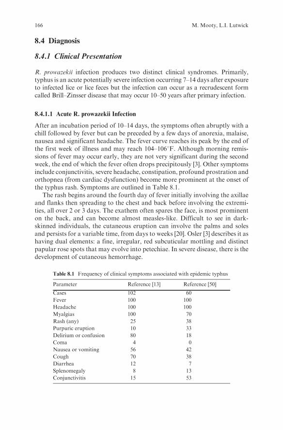
8.4 Diagnosis
8.4.1 Clinical Presentation
R. prowazekii infection produces two distinct clinical syndromes. Primarily,typhus is an acute potentially severe infection occurring 7–14 days after exposureto infected lice or lice feces but the infection can occur as a recrudescent formcalled Brill–Zinsser disease that may occur 10–50 years after primary infection.
8.4.1.1 Acute R. prowazekii Infection
After an incubation period of 10–14 days, the symptoms often abruptly with achill followed by fever but can be preceded by a few days of anorexia, malaise,nausea and significant headache. The fever curve reaches its peak by the end ofthe first week of illness and may reach 104–1068F. Although morning remis-sions of fever may occur early, they are not very significant during the secondweek, the end of which the fever often drops precipitously [3]. Other symptomsinclude conjunctivitis, severe headache, constipation, profound prostration andorthopnea (from cardiac dysfunction) become more prominent at the onset ofthe typhus rash. Symptoms are outlined in Table 8.1.
The rash begins around the fourth day of fever initially involving the axillaeand flanks then spreading to the chest and back before involving the extremi-ties, all over 2 or 3 days. The exathem often spares the face, is most prominenton the back, and can become almost measles-like. Difficult to see in dark-skinned individuals, the cutaneous eruption can involve the palms and solesand persists for a variable time, from days to weeks [20]. Osler [3] describes it ashaving dual elements: a fine, irregular, red subcuticular mottling and distinctpapular rose spots that may evolve into petechiae. In severe disease, there is thedevelopment of cutaneous hemorrhage.
Table 8.1 Frequency of clinical symptoms associated with epidemic typhus
Parameter Reference [13] Reference [50]
Cases 102 60
Fever 100 100
Headache 100 100
Myalgias 100 70
Rash (any) 25 38
Purpuric eruption 10 33
Delirium or confusion 80 18
Coma 4 0
Nausea or vomiting 56 42
Cough 70 38
Diarrhea 12 7
Splenomegaly 8 13
Conjunctivitis 15 53
166 M. Mooty, L.I. Lutwick

Potentially impacting on the aerosolization of organisms in a bioterrorism
event is the presence of respiratory symptoms in natural occurring typhus.Stephenson [8] remarks that cough is extremely frequent and, although it mayoccur at the onset of symptoms, is most prominent at the time of the appear-ance of the rash. He refers to physicians who believe that some amount of
pneumonia is always present in epidemic typhus but does not differentiatefrom rickettsial or secondary bacterial causes. Pulmonary involvement inscrub typhus (caused by Orientia tsutsugamushi) spread by infected mitebites) has been found to occur frequently and in part is due to an ARDS-
like picture rather than a focal process [51]. Histopathologic studies of thelung in scrub typhus have shown neither vasculitis nor evidence of rickettisaein the lung tissue suggesting an immunologic mechanism [52]. Q fever, therickettsial disease that is spread by aerosol without a vector, does cause
pneumonia as its primary manifestation.The symptom complex for which the illness typhus is named relates to the
mental status changes accompanying the illness, developing towards the end ofthe first week. The term typhus is derived from a Greek word for cloud thatrelates to the cloudymental status of the affected person. Typhoid (also referred
to as abdominal typhus) has a similar neuropsychiatric picture that also can beseen in other infectious diseases. Often suggesting a poor prognosis, the state isreferred to as coma vigil or the typhoid state [53]. Coma vigil, also referred to asmuttering delirium, is a condition in the patient lies on his or her back with
muscular twitching and tremulous hands picking at the bedclothes and atimaginary objects. Despite deceptively bright eyes and continual whispering,the individual is unconscious of the surroundings [3]. Alternative neuropsychia-tric manifestations include restlessness and hallucinations. Some degree ofconfusion and disorientation may remain for weeks or months following the
acute illness. Hearing loss may also be a residual symptom [54].Patients with severe disease may develop gangrene of the distal extremities.
Mortality (Table 8.2) generally occurs during the second week and, as reportedby Osler [3], was 12%–20% in the preantimicrobial era. Case fatality rates weremuch lower in children but reached as high as 50% in older adults. Deaths are
related in cardiac failure and vascular collapse and later deaths from secondarybacterial infections (Table 8.2). Adequate and prompt therapy can reduce themortality substantially.
Table 8.2 Mortality rate: epidemic typhus Egypt 1943–1944 (adapted from [55])
Age (years) Male mortality rate (%) Female mortality rate (%)
16–20 9.6 8.7
21–25 15.2 10.4
26–30 25.5 13.7
31–35 30.8 18.8
36–40 33.6 25.4
41–48 47.0 32.6
8 Epidemic Typhus Fever 167

8.4.1.2 Brill–Zinsser Disease
Recrudescent typhus or Brill–Zinsser disease can appear in patients whohad totally recovered from epidemic typhus, years after the onset of the firstinfection [2,56]. For unclear reasons, immunological or otherwise, viableR. prowazekii retained in the body become activated. The disease is sporadic,occurs in the absence of infected lice and, for unknown reasons, clusters morecommonly in the months of June and July [57]. In contrast to acute primaryinfection, Brill–Zinsser disease is generally milder with minimal mortality.
The disease is best described by Nathan Brill’s 1910 report of 221 cases fromNew York City [57] and it is consistent with a typhus-like illness. Brill’s originalpostulate ruled out typhoid by virtue of negative serological and blood cultureconfirmation and he felt that, in the absence of transmissibility, no mortality andthe occurrence during warmer months, it was not typhus. Subsequently, HansZinsser utilized epidemiological and cultural means to suggest that Brill’s diseasewas an imported form of classical epidemic typhus, recrudescent from an initialinfection acquired in Europe [58]. Additionally, R. prowazekii was not onlyconfirmed as obtainable for the blood of Brill–Zinsser patients but clothing licefed on these patients became infected with the rickettsia [59].
8.4.1.3 Latency in Rickettsial Infections
Although the pathophysiology of persistent rickettsial presence in individualsinfected months or decades previously is not well established, documentation ofsuch isolations exists. Indeed, Price [60, 61] demonstrated that R. prowazekiicould be isolated from abdominal lymph nodes of two Russians who migratedto the United States decades before. Isolation of the organism required initialincubation of minced lymph node in tissue culture, intraperitoneal inoculationinto cotton rats and passage of a rat brain suspension into chicken embryo yolksacs. Primary isolation was not accomplished, suggesting the organism was in adormant state initially.
Similar observations have been made with several other rickettsiae. Smadeland colleagues isolated the scrub typhus rickettsia from lymph nodes obtainedfrom individuals 1–2 years following acute infection [62]. Likewise, similarobservations have been made in Rocky Mountain spotted fever (RMSF) [63].That such strains may be of lower virulence is suggested by R. prowazekiiisolated after long-term persistence in cotton rats was of lower antigenicityand virulence [64] and an attenuated strain of Coxiella burnetii used forvaccinating against Q fever was able to persist in mice [65].
8.4.1.4 Bioterrorism-Associated R. prowazekii Infection
The relatively stable, easily aerosolizableR. prowazekii-laden dust-like lice fecesclearly represent a readily easily producible source of the agent of epidemictyphus. Both the Japanese during World War II [66] and the Soviet Union
168 M. Mooty, L.I. Lutwick

during the ColdWar era of the 1970s [67] studied the agent in an airborne form. Adisease of substantial morbidity and mortality if not recognized and treatedquickly, in and of itself typhus is not felt to be transmissible from person-to-person.Substantial spread, however, can occur if the organism is introduced into aP. h. humanus infested population. A biologically weaponized R. prowaze-kii could be engineered to be manifest antimicrobial resistance and couldproduce mortality rates as reported with natural disease during World War IIin Egypt [68].
Demonstrating potential infectivity of a rickettsial aerosol are the numerouslaboratory-associated cases of infection. Although direct transcutaneous inocu-lation of the pathogen in the lab milieu occurs, many cases appear to be relatedto infectious aerosols of the rickettsia causing RMSF [69], murine typhus [70],scrub typhus [71] as well as R. prowazekii [72]. In the latter, a cumulative reportof 3,921 cases of laboratory-associated infection collected over several decadesand published in 1976, Pike cited a total of 573 cases of rickettsial infection (381in the United States) with 56 cases of R. prowazekii infection (22 in the UnitedStates). In the analysis of the proven or probable cases, 217 of the rickettsialtotal (36%) were aerosol in nature and 230 (40%) were of unknown or unclearsource.
Rickettsiae can be found in the lung after transcutaneous or aerosolexposure. In a histopathological study of 10 fatal cases of R. rickettsii [73],the distribution of the spotted fever organism by immunofluorescencecoincided with vasculitis in the lung. Here, the location of the organismssuggested that person-to-person aerosol spread was unlikely, the sameobservation made epidemiologically. In epidemic typhus, however, apathologic study from Egypt during World War II [74] found rickettsiae-like organisms primarily in the cytoplasm of leukocytes in alveolar andbronchial exudates. Besides demonstrating that pneumonia in typhus wasat least partly due to the primary infection, the possibility of respiratoryspread was brought up by the findings. The authors, however, cited earlierwork that neither sputum, nasal washings, nor tracheal aspirates demon-strated the organism.
8.4.2 Laboratory Diagnosis
8.4.2.1 Laboratory Abnormalities
Thrombocytopenia, elevated serum aminotransferases, and increased biliru-bin levels may occur in severe cases. The frequency with which theselaboratory abnormalities occur is illustrated in Table 8.3. Electrocardio-graphic evidence of myocarditis and diffuse or focal pulmonary infiltrateson chest x-ray occur in minority of patients may occur in a small percentageof patients [55].
8 Epidemic Typhus Fever 169

8.4.2.2 Serology
Weil and Felix described the classical assay for rickettsial antibody detection in
1916 [75]. The methodology took advantage of heterophilic (cross-reacting) anti-
gens of members of the genus Rickettsia and several species of the Gram negative
bacillus Proteus. In the Weil–Felix test, bacteria of Proteus vulgaris OX-19 agglu-
tinate with sera of the infected with the typhus group organisms as well as from
RMSF. Cells of P. vulgaris OX-2, on the other hand, agglutinate with sera from
individuals with spotted fever infection except RMSF. The OX-19 agglutination isseen in epidemic typhus but usually not in Brill–Zinsser recrudescent disease.
P. mirabilis OX-K can be similarly used in the serological diagnosis of scrub
typhus. Because of the both relatively poor sensitivity and specificity of this test
including for epidemic typhus [76], more specific assays are needed for diagnosis.Such specific serologic tests are the mainstay of diagnosis, since isolation of
R. prowazekii is generally impractical and can be dangerous to laboratorians.
Serologic tests include indirect fluorescence antibody (IFA) [77, 78], latexagglutination [79], complement fixation and enzyme-linked immunosorbent
assay [78, 80]. A diagnosis of recent epidemic or murine typhus rickettsial
infection can be established by demonstrating a fourfold or greater rise in
titer of antibody in properly collected acute and convalescent serum samples.
Titers are usually detectable during or after the second week.Twoof the serological tests arewidely available for epidemic typhus: an IFA test
and an immunoblot technique. These tests are available in most state health
departments, the CDC, and a few specialized research laboratories. Neither ofthese can reliably differentiate between acute primary infections fromBrill–Zinsser
disease [81]. Additionally, epidemic and endemic typhus (due toR. typhi) cannot be
differentiated by serology, unless Western immunoblot (WB) and/or cross-
adsorption of sera are done [82].The IFA is generally considered to be the gold standard for rickettsial
serological diagnosis [78]. The WB assay has been found in Mediterranean
spotted fever to be somewhat more sensitive that IFA for early antibody withthe first antibody detected being against the lipopolysaccharide [83]. Latex
agglutinins that are group specific are available for R. prowazekii and require
minimal equipment [79]. Antibodies against outer membrane proteins (omp)
Table 8.3 Frequency of laboratory abnormalities associated with epidemic typhus
Parameter Reference [50]
Cases 60
Thrombocytopenia 43
Increased aspartate aminotransferase level 63
Increased alanine aminotransferase level 35
Increased bilirubin level 20
Increased serum creatinine level 2
Hematuria 44
Proteinuria 28
170 M. Mooty, L.I. Lutwick

may be useful for even earlier serological diagnosis. Using a recombinant ompof scrub typhus, an immunochromatographic assay was significantly betterthan IFA [84]. In the study, this assay was positive before IFA (by about aweek) in 50 and the reverse in only seven.
8.4.2.3 Molecular
Molecular diagnostic assays are becoming increasingly common for the diag-nosis of infectious diseases. Techniques using polymerase chain reaction (PCR)technology have been used to detect typhus in blood and to detect theseorganisms in their vector [13, 39, 85, 86]. DNA sequencing of PCR productsprovides a definitive method for the differentiation of closely related etiologicagents that are cross-reactive serologically and that cause infections with similarclinical presentations, as is the case with murine and epidemic typhus.
In a 2001 case report [30], typhus-specific 17 kDa gene assay that used a nestedPCR protocol was found to increase sensitivity. This assay amplified the DNA ofboth R. typhi and R. prowazekii. Using nine nucleotide differences within theregion of the 17 kDa gene amplified by the nested assay, it allowed identificationof the infection as either endemic or epidemic. Subsequently, a real-time quanti-tative PCR assay, specific for R. prowazekii, showed it to be sensitive to 1–5copies per sample and was useful in experimentally infected mice [87].
Biopsy of a skin rash can lead to a definitive diagnosis by demonstrating thecharacteristic changes of rickettsial vasculitis and the presence of rickettsiaein tissue by use of fluorescent antibody conjugates. The use of monoclonalantibodies has been utilized to improve the specificity of the assay and candifferentiate epidemic from endemic typhus [88].
8.4.2.4 Culture
Due to the nature of the organism, culture is usually difficult to perform requiring5–8-day-old embryonated eggs from flocks on antimicrobial-free diets or suscep-tible animals such as guinea pigs or mice in an isolated environment to provideprotection to the laboratory worker. More recently, a centrifugation-shell vialsystem has been used to cultivate R. prowazekii, reported to be the first clinicalisolation of the epidemic typhus fever rickettsia in 30 years. The small area of thecell containing coverslip enhanced the ratio of organisms to cells as well as detec-tion [89]. Identification was then made by PCR amplification of specific genes.
8.5 Therapeutic Intervention
Rickettsial in vitro susceptibility has been evaluated in lice [90] and cells [91].Tetracycline and chloramphenicol have been generally thought to be the agentsto use in the treatment of epidemic typhus [92]. Doxycycline, 200 mg orally
8 Epidemic Typhus Fever 171

once, is the regimen of choice. Medical facilities are often diagnostically inade-quate in areas where epidemic typhus is seen. An alternative for empiric treat-ment in the absence of confirmation of the diagnosis serologically would be theadministration of chloramphenicol 500 mg orally or intravenously four timesfor 5 days. This regimen is highly effective in epidemic typhus and simulta-neously covers the possibility of meningococcemia and typhoid fever. Doxycyclineis, however, preferable for therapy of human R. prowazekii infection.
Most typhus-infected individuals treated with doxycycline or chloramphe-nicol improve markedly within 48 h following initiation of therapy. Failure toshow a response within 48–72 h after starting empirical treatment is oftenconsidered to be clinical evidence that a rickettsial disease is not present. In asmall study, a single 200 mg oral dose of doxycycline cured 35 of 37 patients,and 29 of 37 patients were afebrile 48 h after therapy was started [93]. Althoughantimicrobial resistance to the tetracyclines and/or chloramphenicol in thegenus Rickettsia is rare, poor responsiveness of some northern Thailand strainsofR. tsutsugamushi in human disease, murine models and cell culture have beendescribed [94]. A biowarfare laboratory should be able to transform R. prowazekiiinto a tetracycline and/or chloramphenicol resistant form.
Other antimicrobial agents with evidence for activity [91] include fluoroqui-nolones such as ciprofloxacin, ofloxacin and levofloxacin, macrolides such aserythromycin and clarithromycin, and rifampin. Typhus group rickettsiae aremore sensitive to the macrolides than the spotted fever group [91] althoughpatients with recrudescent typhus have been reported not to respond to azi-thromycin [95]. Additionally, heterogeneity in rifampin resistance has beenfound in some subgroups of rickettsiae but not the typhus group [91].
Beta-lactams, aminoglycosides and sulfonamides are not active. Importantly,not only are sulfa drugs not active but the use of this class of antimicrobials forhuman rickettsial infection is harmful. The medications inhibit production ofpara-aminobenzoic acid, a compound that has a therapeutic effect in rickettsialinfections including louse-borne typhus [96]. Indeed, more severe rickettsialinfections and higher mortality rates have been reported after the use of thisclass of antimicrobials [97, 98].
Anecdotal case reports have appeared of a patient misdiagnosed as havingtyphoid who died from typhus after treatment with ciprofloxacin, despite invitro efficacy [1] and of patients with the successful use of the fluoroquinolonein R. typhi infection [99, 100].
Supportive care, including fluids, vasopressors, oxygen, and even dialysismay be required in patients with severe illness. The prognosis is dependent uponseveral factors including age, underlying nutritional status, previous health ofthe patient, and the delay in initiation of therapy. In the preantibiotic era, themortality rate was higher in older and male patients. In the modern era,mortality is uncommon if treatment is given. In a series of 60 treated patients,none of those treated died [101]. In another small study, two of nine patientsdied despite chloramphenicol therapy [39]. The death rate of untreated epidemictyphus is approximately 15%. This rate is reduced to 0.5% with a single 200 mg
172 M. Mooty, L.I. Lutwick

dose of doxycycline. Infections are rarely fatal in children; however, themortalityrate can be as high as 60%without treatment [102]. The prognosis of Brill–Zinsserdisease is generally good, although rare fatalities have been reported [103].
8.6 Preventive Measures
8.6.1 Infection Control
Eradication of human infestation with lice will prevent natural transmissionof epidemic typhus. People who live and work in close proximity to a louse-infested individual may secondarily acquire lice, even if they regularly washtheir clothes and have good hygiene. Thus, all louse-infested persons andworkers in close contact with infested persons may require long acting insec-ticides. Application of effective residual insecticide powder at appropriateintervals by hand or power blower to clothes and populations at risk is recom-mended. Spread of infection does not need contact with live, typhus-infected lice,as previous mentioned, since organism-laden feces can be aerosolized fromclothing or bed sheets.
Insecticides that can be used include DDT, malathion, and lindane. Reportsof resistance to one or more of these agents have appeared. The syntheticpyrethroid permethrin has been demonstrated to be effective and long-lastingwhen applied as a dust or spray on clothing and bedding [104]. Fabric treatedwith permethrin has been shown to retain toxicity to body lice even after 20washings, offering long-term passive protection [105].
The use of chloramphenicol or tetracycline for prophylaxis may be highlyeffective in interrupting typhus outbreaks [91]. Some experts recommend theuse of one 200 mg dose of doxycycline once weekly by travelers in whichepidemic typhus is present. Prophylaxis is generally continued for 1 weekafter leaving such areas.
Afflicted patients should be reported to local health authority. Louse-infestedsusceptibles exposed to typhus fever should be quarantined for 15 days afterapplication of insecticide with residual effect. Isolation is not required afterproper delousing of patient, clothing, living quarters and household contacts.Lice tend to leave hot or cold bodies in search for a normothermic clothed body.If death from louse-borne typhus occurred, delousing the body and clothing byapplication of an insecticide is recommended [106].
All immediate contacts should be kept under surveillance for 2 weeks. Everyeffort should be made to trace the infection to the index case. Application of aninsecticide with residual effect to all contacts will result in rapid control oftyphus. Systematic application of residual insecticide to all people in the com-munity is indicated in widespread infestations. Notification by governments toWorld Health Organization (WHO), and to adjacent disease free countries isindicated.
8 Epidemic Typhus Fever 173

8.6.2 Immunization
The history of typhus vaccination is one of the most colorful in vaccinology.
Both inactivated and live attenuated vaccines against R. prowazekii have been
developed and tested. Woodward [107] stated that the earliest protective meth-
odology for epidemic typhus was the eating of lice but it was and is of totally
unclear efficacy.Weigl in Poland developed the first vaccine forR. prowazekii in
lice [107], using a technique developed for the RMSF/Dermacentor tick model
by Spencer and Parker. The vaccine required the intrarectal inoculation of the
lice with viable rickettisae using the equivalent of a miniature enema and
subsequent feeding of the lice on convalescent typhus individuals. The louse
intestinal tract was then harvested and formalinized to produce an inactivated
vaccine, which appeared to be protective, requiring 30–100 intestines for each
human dose. The procedure was not only tedious but also resulted in infections
and death of laboratory workers.Woodward also described another early epidemic typhus vaccine and pro-
duced in a unique manner. Originally reported by Blanc and Baltazard from
Morocco, the technique used murine typhus-infected guinea pigs as rickettsial
donors placed in pits containing tens of thousands of hungry fleas. As a result,
the donors died from exsanguination and the fleas became infected with
R. typhi. The vaccine recipe then required the addition of white cotton rats to
the pit. These rodents also died of blood loss and became brown colored from the
flea fecal matter deposited in their coat. The rats were removed and rubbed over a
mesh to collect the rickettsial-laden flea feces. This material was ‘‘reconstituted’’
with saline and ox bile prior to injection. Ox bile was used as ‘‘attenuation,’’ which
may have attenuated R. typhi as the organism caused a milder disease in most
cases and could provide subsequent protection against R. prowazekii [107].An attenuated R. prowazekii, referred to as the E (for Espana) or Madrid E
strain, was developed in Spain in the early 1940s. The organism, isolated from a
severe case of typhus, was passed 11 times in eggs prior to the decrease of
virulence in guinea pigs [108]. This vaccine seemed to be able to prevent classical
typhus but further development was limited [107] by moderate to severe reac-
tions and concern about possible reversion to virulence. Interestingly, in terms
of rickettsial replication and louse mortality, there is no different in virulence
between the E and the Breinl (virulent) strain [109]. Comparing the genome
of the E and Breinl strains, there are genomic variations in about 3% of the
834 protein-coding sequences of the attenuated strain [109] and 24 genes in
E had decreased expression and one had increased expression as compared to
the virulent strain.A number of inactivated typhus vaccines have been evaluated including
chicken egg (Cox) and rat lung (Durand) types. In a small study, Woodward
reported [107] that both were reasonably well tolerated with little local or
systemic toxicity and most recipients developed antibody. The inactivated
vaccines provided some level of protection [107, 110] but incidence reduction
174 M. Mooty, L.I. Lutwick

was not conclusively demonstrated [111]. Vaccinees are more likely to havemilder disease [68]. In the United States, Cox-type vaccine had been available inthe past with the recommendation of two subcutaneous doses given at least amonth apart with a booster given every 6–12 months as long as continued riskexisted [112].
Genomic technology has begun being utilized in the production of DNAtyphus vaccines. As an example, Coker et al [113] utilized the genome sequenceof the E strain of R. prowazekii to identify genes that might be potential targetsin immunoprotection to provide, in particular, cellular immune responses to theintracellular rickettsiae. These genes have been amplified and introduced into acloning vector for further study. Using a spotted fever model, a DNA vaccinewith outer membrane protein (omp) A and B genes was studied in mice [114].Protection was achieved in this model with the use of a DNA vaccine primefollowed by boosting with a recombinant omp. A cellular immune response wasdetected and protection occurred without any detectable antibody response. Amurine typhus ompB was completely protective in mice and partially protectivein guinea pigs against lethal doses of R. typhi [115, 116].
References
1. Zanetti, G., Francioli, P., Tugan, D., et al. Imported epidemic typhus. Lancet 352, 1709,1998.
2. Lutwick, L. I. Brill-Zinsser disease. Lancet 357, 1198–1200, 2001.3. Osler, W. The Principles and Practice of Medicine, 3rd edn. D. Appleton and Co.,
New York, 1899.4. Hirsch, A. Handbook of Geographical and Historical Pathology. Translated by
Creighton, C. New Syndenham Society, London, 1885.5. Retief, F. P. and Cilliers, L. The epidemics of Athens, 430-426 BC. S. Afr. Med. J. 88,
50–3, 1998.6. Zinsser, H. Rats, Lice and History. Little Brown, Boston, 1934.7. Weiss, E. The role of rickettsioses in history. In: Biology of Rickettsial Diseases, vol. 1,
Walker, D. H (ed). CRC Press, Boca Raton, pp.1, 1988.8. Stephenson, C. S. Epidemic typhus fever and other rickettsial diseases of military impor-
tance. N. Engl. J. Med. 231, 407–413, 1944.9. Lasowski, E. S. and Matulewicz, S. Serendipitous discovery of artificial positive
Weil–Felix reaction used in ‘‘primitive immunological war’’. ASM News 43, 300–302, 1977.10. Foster, G. M. Typhus disaster in the wake of war: the American-Polish relief expedition,
1919–1920. Bull. Hist. Med. 55, 221–232, 1981.11. Davis, W. A. Typhus at Belsen. I. Control of the typhus epidemic. Am. J. Hyg. 46, 66–83,
1947.12. Gelston, A. L. and Jones, T. C. Typhus fever: report of an epidemic in New York City in
1847. J. Infect. Dis. 136, 813–821, 1977.13. Raoult, D., Ndihokubwayo, J. B., Tissot-Dupont, H., et al. Outbreak of epidemic typhus
associated with trench fever in Burundi. Lancet 352, 353–358, 1998.14. Walker, D. H. Principles of the malicious use of infectious agents to create terror.
Reasons for concern for organisms of the genus Rickettsia. Ann. N. Y. Acad. Sci. 990,739–742, 2003.
15. Walker, D. H., Valbuena, G. A., and Olano, J. P. Pathogenic mechanisms of diseasescaused by Rickettsia. Ann. N. Y. Acad. Sci. 990, 1–11, 2003.
8 Epidemic Typhus Fever 175

16. Gaywee, J., Xu,W., Radulovic, S., et al. TheRickettsia prowazekii invasion gene homolog(invA) encodes a nudix hydrolase active on adenosine (50)-pentaphospho-(50)-adenosine.Mol. Cell. Proteomics 1, 179–185, 2002.
17. Heinzen, R. A., Grieshaben, S. S., Van Kirk, L. S., et al. Dynamics of actin-basedmovement of Rickettsia rickettsii in Vero cells. Infect. Immun. 67, 4201–4207, 1999.
18. Andersson, S.G., Zomorodopour, A., Andersson, J. O. The genome sequence ofRickettsiaprowazekii and the origin of mitochondria. Nature 396, 133–140, 1998.
19. Andersson, J. O. and Andersson, S. G. Pseudogenes, junk DNA, and the dynamics ofrickettsial genomes. Mol. Biol. Evol. 18, 829–839, 2001.
20. Ko, C. J. and Elston, D. M. Pediculosis. J. Am. Acad. Dermatol. 50, 1–12, 200421. Burgess, I. F. Human lice and their management. Adv. Parasitol. 36, 271–342, 1995.22. Gross L. How Charles Nicolle of the Pasteur Institute discovered that epidemic typhus is
transmitted by lice: reminiscences from my years at the Pasteur Institute in Paris. Proc.Natl. Acad. Sci. U.S.A. 93, 10539–10540, 1996.
23. Fournier, P. E., Ndihokubwayo, J. B., Guidran, J., et al. Human pathogens in body andhead louse. Emerg. Infect. Dis. 8, 1515–1518, 2002.
24. Elston, D. M. What’s eating you? Psocoptera (Book lice, Psocids) Cutis 64, 307–308,1999.
25. Patil, M. P., Niphadkar, P. V., and Bapat, M. M. Psocoptera spp. (book louse): a newmajor household allergen in Mumbai. Ann. Allergy Asthma Immunol. 87, 151–155,2001.
26. Robinson, D., Leo, N., Prociv, P., and Barker, S. C. Potential role of head lice, Pediculushumanus capitus, a vector of Rickettsia prowazekii. Parasitol. Res. 90, 209–211, 2003.
27. Murray, E. S. and Torrey, S. B. Virulence ofRickettsia prowazekii for head lice.Ann. N. Y.Acad. Sci. 266, 25–34, 1975.
28. Bozeman, F. M., Masicllo, S. A., Williams, M. S., and Elisberg, B. L. Epidemic typhusisolated from flying squirrels. Nature 255, 545, 1975.
29. Sonenshine, D. E., Bozeman, F. M., Williams, M. S., et al. Epizootiology of epidemictyphus (Rickettsia prowazekii) in flying squirrels. Am. J. Trop. Med. Hyg. 27, 339–349,1978.
30. Massung, R. F., Davis, L. E., Slater, K., et al. Epidemic typhus meningitis in thesouthwestern United States. Clin. Infect. Dis. 32, 979–982, 2001.
31. Culpepper, G. H. The rearing and maintenance of a laboratory colony of the body louse.Am. J. Trop. Med. Hyg. 24, 327–329, 1944.
32. Houhamdi, L., Fournier, P.-E., Fang, R., et al. An experimental model of human bodylouse infection with Rickettsia prowazekii. J. Infect. Dis. 186, 1639–1646, 2002.
33. Raoult, D. and Roux, V. The body louse as a vector of reemerging human diseases. Clin.Infect. Dis. 29, 888–911, 1999.
34. Maunder, J. W. The appreciation of lice. Proc. R. Inst. Great Britain 55, 1–31, 1983.35. Patterson, K.D. Typhus and its control in Russia, 1870–1940.MedHist. 37, 361–381, 1993.36. Mumcuoglu, K. Y., Miller, J., Manor, O., et al. The prevalence of ectoparasites in
Ethiopian immigrants. Isr. J. Med. Sci. 29, 371–373, 1993.37. Fan, M. Y., Walker, D. H., Yu, S. R., and Liu, Q. H. Epidemiology and ecology
of rickettsial diseases in the People’s Republic of China. Rev. Infect. Dis. 9, 823–840,1987.
38. Tesfayohannes, T. Prevalence of body lice in elementary school students in threeEthiopian towns at different attitudes. Ethiop. Med. J. 27, 201–207, 1989.
39. World Health Organization. A large outbreak of epidemic louse-borne typhus inBurundi. Wkly. Epidemiol Rec. 72, 152–153, 1997.
40. Raoult, D., Roux, V., Ndihokubwaho, J. B., et al. Jail fever (epidemic typhus) outbreak inBurundi. Emerg. Infect. Dis. 3, 357–360, 1997.
41. Mokrani, K., Fournier, P. E., Dalichaouche, M., et al. Reemerging threat of epidemictyphus in Algeria. J. Clin. Microbiol. 42, 3898–3900, 2004.
176 M. Mooty, L.I. Lutwick

42. Badiaga, S., Brouqui, P., andRaoult, D. Autochthonous epidemic typhus associated withBartonella quintana bacteremia in a homeless person. Am. J. Trop. Med. Hyg. 72,638–639, 2005.
43. Silverman, D. J., Boese, J. L., andWissman Jr., C. L. Ultrastructural studies ofRickettsiaprowazekii from louse midgut cells to feces: search for ‘‘dormant’’ forms. Infect. Immun. 10,257–263, 1974.
44. Regner, R. L., Yuan Fu, Z., and Spruill, C. L. Flying squirrel-associated Rickettsiaprowazekii (epidemic typhus rickettsiae) characterized by a specific DNA fragmentproduced by restriction endonuclease digestion. J. Clin. Microbiol. 23, 189–191, 1986.
45. Eremeeva, M. E. and Silverman, D. J. Effects of the antioxidant �-lipoic acid on humanumbilical vein endothelial cells infected with Rickettsia rickettsii. Infect. Immun. 66,2290–2299, 1998.
46. Winkler, H. H. andMiller, E. T. Immediate cytotoxicity and phospholipase A: the role ofphospholipase A in the interaction of R. prowazekii and L cells. In: Rickettsiae andRickettsial Diseases, Burgdorfer, W. and Anacker, R. L. (eds). Academic Press, NewYork. pp.327, 1981.
47. Renesto, P., Dehoux, P., Gouin, E., et al. Identification and characterization of aphospholipase D-superfamily gene in rickettsiae. J. Infect. Dis. 188, 1276–1283, 2003.
48. Walker, D. H. Pathology and pathogenesis of the vasculotropic rickettsioses. In: Biologyof Rickettsial Disease, Walker, D. H. (ed.), CRC Press, Boca Raton. pp.115–1138, 1988.
49. Wolbach, S., Todd, J., and Palfrey, F. The Etiology and Pathology of Typhus. HarvardUniversity Press, Cambridge, MA, 1922.
50. Perine, P. L., Chandler, B. P., Krause, D. K., et al. A clinico-epidemiological study ofepidemic typhus in Africa. Clin. Infect. Dis. 14, 1149–1158, 1992.
51. Tsay, R.-W. and Chang, F.-Y. Acute respiratory distress syndrome in scrub typhus.Q. J.Med. 95, 126–128, 2002.
52. Park, J. S., Jee, Y. K., Lee, K. Y., et al. Acute respiratory distress syndrome associatedwith scrub typhus: diffuse alveolar damage without pulmonary vasculitis. J. KoreanMed.Sci. 15, 343–345, 2000.
53. Verghese, A. The ‘‘typhoid state’’ revisited. Am. J. Med. 79, 370–372, 1985.54. Friedmann, I., Frohlich, A., and Wright A. Epidemic typhus fever and hearing loss: a
histological study (Hall pike collection of temporal bone sections). J. Laryngol. Otol. 107,275–283, 1993.
55. Diab, S.M., Araj, G. F., and Fenech, F. F. Cardiovascular and pulmonary complicationsof epidemic typhus. Trop. Geogr. Med. 41, 76–79, 1989.
56. Green, C., Fishbein, D., and Gleiberman, I. Brill-Zinsser: still with us. JAMA 264,1811–1812, 1990.
57. Brill, N. E. An acute infectious disease of unknown origin. A clinical study based on 221cases. Am. J. Med. Sci. 139, 484–502, 1910.
58. Zinsser, H. Varieties of typhus fever and the epidemiology of the American form ofEuropean typhus fever (Brill’s disease). Am. J. Hyg. 20, 513–532, 1934.
59. Murray, E. S. and Snyder, J. C. Brill’s disease. II. Etiology. Am. J. Hyg. 53, 22–32, 1951.60. Price, W. H. Studies on the interepidemic survival of louse-borne epidemic typhus fever.
J. Bacteriol. 69, 106–107, 1954.61. Price, W. H., Emerson, H., Nagle, E., et al. Ecologic studies on the interepidemic survival
of louse-borne epidemic typhus fever. Am. J. Hyg. 67, 155–178, 1958.62. Smadel, J. E., Ley, H. L., Diercks, F. H., and Cameron, J. A. P. Persistence of Rickettsia
tsutsugamushi in tissue of patients recovered from scrub typhus. Am. J. Hyg. 56, 294–302,1952.
63. Parker, R. T.,Menon, P. G.,Merideth, A.M., et al. Persistence of Rickettsia rickettsii in apatient recovered from Rocky Mountain spotted fever. J. Immunol. 73, 383–386, 1954.
64. Ignatovich, V. F. Biological properties of Rickettsia prowazekii on long-term persistencein infected cotton rats. Acta Virol. 24, 144–148, 1980.
8 Epidemic Typhus Fever 177

65. Freylikhman, O., Tokarerevich, N., Surorov, A., et al. Coxiella burnetii persistence inthree generations of mice after application of live attenuated humanM-44 vaccine againstQ fever. Ann. N. Y. Acad. Sci. 990, 496–499, 2003.
66. Harris, S. Japanese biological warfare research in humans: a case study of microbiologyand ethics. Ann. N. Y. Acad. Sci. 666, 21–49, 1992.
67. Alibek, K and Handelsman, S. Biohazard. Random House, New York, 1999.68. Ecke, R. S., Gillaim, A. G., Snyder, J. C., et al. The effect of Cox-type vaccine on
louse-borne typhus fever. Am. J. Trop. Med. 25, 447–462, 1945.69. Johnson, J. E. and Kadull, P. J. Rocky Mountain spotted fever acquired in a laboratory.
N. Engl. J. Med. 277, 842–847, 1967.70. Centers of Disease Control. Laboratory-acquired endemic typhus – Maryland. MMWR
Morb. Mortal Wkly. Rep. 27, 215–216, 1978.71. Oh, M., Kim, N., Huh, M., et al. Scrub typhus pneumonitis acquired through the
respiratory tract in a laboratory worker. Infection 29, 54–56, 2001.72. Pike, R. M. Laboratory-associated infections: Summary and analysis of 3921 cases.
Health. Lab. Sci. 13, 105–114, 1976.73. Walker, D. H., Crawford, C. G., and Cain, B. G. Rickettsial infection of the pulmonary
microcirculation: the basis for interstitial pneumonitis in Rocky Mountain spotted fever.Hum. Pathol. 11, 263–272, 1980.
74. Committee on Pathology, Division of Medical Sciences, National Research Council.Pathology of epidemic typhus. Report of fatal cases studied by United States of AmericaTyphus Commission in Cairo, Egypt during 1943–1945. Arch. Pathol. 56, 397–435, 1953.
75. Weil, E. and Felix, A. Zur serologischen diagnose des fleckfiebers. Wien. Klin.Wichenschr. 29, 33–35, 1916.
76. Ormsbee, R., Peacock, M., Philip, E., et al. Serologic diagnosis of epidemic typhus fever.Am J. Epidemiol. 105, 261–271, 1977.
77. Newhouse, V. F., Shepard, C. C., Redus, M. D., et al. A comparison of the complementfixation, indirect fluorescent antibody and microagglutination tests for the serologicaldiagnosis of rickettsial diseases. Am. J. Trop. Med. Hyg. 28, 387–395, 1979.
78. La Scola, B. and Raoult, D. Laboratory diagnosis of rickettsioses: current approaches tothe diagnosis of old and new rickettsial diseases. J. Clin. Microbiol. 35, 2715–2727, 1997.
79. Hechemy, K. E., Osterman, J. V., Eisemann, C. S., et al. Detection of typhus antibody bylatex agglutination. J. Clin. Microbiol. 13, 214–216, 1981.
80. Halle, S. and Dasch, G. A. Use of sensitive microplate enzyme-linked immunosorbentassay in a retrospective serological analysis of a laboratory population at risk to infectionwith typhus group rickettsiae. J. Clin. Microbiol. 12, 343–350, 1980.
81. Eremeeva, M. E., Balayeva, N. M., and Raoult, D. Serological response of patientssuffering from primary and recrudescent typhus: Comparison of complement fixationreaction, Weil–Felix test, microimmunofluorescence, and immunoblotting. Clin. Diagn.Lab. Immunol. 1, 318–324, 1994.
82. La Scola, B., Rydkina, L., Ndihokobwayo, J. B., et al. Serological differentiation ofmurine typhus and epidemic typhus using cross-adsorption and Western blotting. Clin.Diagn. Lab. Immunol. 7, 612–616, 2000.
83. Teysselre, N. and Raoult, D. Comparison ofWestern immunoblotting and microimmuno-fluorescence for diagnosis of Mediterranean spotted fever. J. Clin. Microbiol. 30, 455–460,1992.
84. Ching,W.-M., Rowland, D., Zhang, Z., et al. Early diagnosis of scrub typhus with a rapidflow assay using recombinant major outer membrane protein antigen (r56) of Orientiatsutsugamushi. Clin. Diagn. Lab. Immunol. 8, 409–414, 2001.
85. Eremeeva,M. E., Ignatovich, V. F., Dasch, G.A., et al. Genetic, biological, and serologicaldifferentiation of Rickettsia prowazekii and Rickettsia typhi. In: Rickettsia and RickettsialDiseases, Kazar, J. and Toman, R. (eds). Publishing House of the Slovak Academy ofSciences, Veda, Bratislava. pp.43–50, 1996.
178 M. Mooty, L.I. Lutwick

86. Carl, M., Tibbs, C. W., Dobson, M. E., et al. Diagnosis of acute typhus infection usingthe polymerase chain reaction. J. Infect. Dis. 161, 791–793, 1990.
87. Svraka, S., Rolain, J. M., Bechach, Y., et al. Rickettsia prowazekii and real-time poly-merase chain reaction. Emerg. Infect Dis. 12, 428–432, 2006.
88. Fang, R., Houhamdi, L., and Raoult, D. Detection of Rickettsia prowazekii in body liceand their feces by using monoclonal antibodies. J. Clin. Microbiol. 40, 3358–3363, 2002.
89. Birg, M.-L., La Scola, B., Roux, V., et al. Isolation of Rickettsia prowazekii from bloodby shell vial culture. J. Clin. Microbiol. 37, 3722–3724, 1999.
90. Boese, J. L., Wisseman, C. L. J., Walsh, W. T., and Fiset, P. Antibody and antibioticaction onRickettsia prowazekii in body lice across the host-vector interface, with observa-tion on strain virulence and retrieval mechanisms. Am. J. Epidemiol. 98, 262–282, 1973.
91. Rolain, J. M., Maurin, M., Vestris, G., and Raoult, D. In vitro susceptibilities of 27rickettsiae to 13 antimicrobials. Antimicrob. Agents Chemother. 42, 1537–1541, 1998.
92. Krause, D. W., Perine, P. L., McDade, J. E., and Awoke, S. Treatment of louse-bornetyphus fever with chloramphenicol, tetracycline, or doxycycline. East Afr. Med. J. 52,421–427, 1975.
93. Huys, J., Kayihigi, J., Freyens, P., et al. Single-dose treatment of epidemic typhus withdoxycycline. Chemotherapy18, 314–317, 1973.
94. Watt, G., Chouriyagune, C., Ruangweerayud, R., et al. Scrub typhus infections poorlyresponsive to antibiotics in northern Thailand. Lancet 348, 86–89, 1996.
95. Turcinov, D., Kuzman, I., andHerendic B. Failure of azithromycin in treatment of Brill-Zinsser disease. Antimicrob. Agents Chemother. 44, 1737–1738, 2000.
96. Yeomans, A., Snyder, J. C., Murray, E. S., et al. The therapeutic effect of para-aminobenzoic acid in louse borne typhus fever. JAMA 126, 349–356, 1944.
97. Steigman, A. J. Rocky Mountain spotted fever and the avoidance of sulfonamides.J. Pediatr. 91, 163–164, 1977.
98. Ruiz Beltran, R. and Herrero Herrero, J. I. Deleterious effect of trimethoprim-sulfamethoxazole in Mediterranean spotted fever. Antimicrob. Agents Chemother. 36,1342–1343, 1992.
99. Eaton,M., Cohen,M. T., Shlim, D. R., and Innes B. Ciprofloxacin treatment of typhus.JAMA 262, 772–773, 1989.
100. Strand, O. and Stromberg, A. Ciprofloxacin treatment of murine typhus. Scand. J. Infect.Dis. 22, 503–504, 1990.
101. Matossian, R.M., Thaddeus, J., andGarabedian, G. A. Outbreak of epidemic typhus inthe northern region of Saudi Arabia. Am. J. Trop. Med. Hyg. 12, 82–90, 1963.
102. TYPHUS Clinical Reference, http://allhazards.state.wy.us. Accessed Feb 2004.103. Murray, E. S., Baehr,G., Shwartzman,G., et al. Brill’s disease. JAMA 142, 1059–1066, 1950.104. Campbell, W. C. Insect infestations of man. In: Chemotherapy of Parasitic Diseases,
Campbell, W. C. and Rew, R. S. (eds). Plenum Press, New York. pp.531–540, 1986.105. Sholdt, L. L.,Rogers Jr., E. J.,Gerberg, E. J., and Schreck, C. E. Effectiveness of permethrin-
treated military uniform fabric against human body lice.Mil. Med. 154, 90–93, 1989.106. Notifiable Conditions, Washington State Department of Health, http://www.doh.wa.
gov/Notify/guidelines/typhus.htm. Accessed Feb 2004.107. Woodward, T. E. Rickettsial vaccines with emphasis on epidemic typhus: initial report
of an old vaccine trial. S. Afr. Med. J. 11, 73–76, 1986.108. PerezGallardo, F. andFox, J. P. Infection and immunization of laboratory animals with
Rickettsia prowazekii of reduced pathogenicity, strain E. Am. J. Hyg. 48, 6–21, 1948.109. Ge, H., Chuang, Y. Y., Zhao, S., et al. Comparative genomics of Rickettsia prowazekii
Madrid E and Breinl strains. J. Bacteriol. 186, 556–565, 2004.110. Weiss, K. and Walker, D. H. New and improved vaccines against rickettsia infections:
Rocky mounted spotted fever, epidemic typhus, and scrub typhus. In: New GenerationVaccines, Woodrow, G. C. and Levine, M. M. (eds). Marcel Decker, New York.pp.357–374, 1990.
8 Epidemic Typhus Fever 179

111. Wisseman, C. L. The present and future of immunization against the typhus fevers. In:Pan American Health Organization: First International Conference on VaccinesAgainst Viral and Rickettsial Diseases of Man. Pan American Health Organization,Washington, DC. pp.523–527, 1967.
112. Centers for Disease Control. Typhus vaccine. Recommendation on immunizationpractices. Ann. Intern. Med. 68, 785–786, 1968.
113. Coker, C., Majid, M., and Radulovic, S. Development of Rickettsia prowazekii DNAvaccine. Cloning strategies. Ann. N. Y. Acad. Sci. 990, 757–764, 2003.
114. Dıaz-Montero, C. M., Feng, H.-M., Crocquet-Valdes, P. A., and Walker, D. H. Identifi-cation of protective components of two major outer membrane proteins of spotted fevergroup rickettsiae. Am. J. Trop. Med. Hyg. 65, 371–378, 2001.
115. Dasch, G. A., Bourgeois, A. L., and Rollwagen, F. M. The surface protein antigen ofRickettsia typhi: in vitro and in vivo immunogenicity and protective efficacy in mice. In:Raoult, D., and Brouqui, P., eds. Rickettsiae and Rickettsial Diseases at the Turn of theThird Millenium. Paris: Elsevier. pp.116–122, 1999.
116. Bourgeois, A. L. and Dasch, G. A. The species-specific surface protein antigen ofRickettsia typhi: immunogenicity and protective efficacy in guinea pigs. In: Rickettsiaeand Rickettsial Diseases, Burgdorfer, W. and Anacker, R. L. (eds). Academic Press,New York. pp.71–80, 1981.
180 M. Mooty, L.I. Lutwick

Chapter 9
Category B Biotoxins
Ricin, Staphylococcus aureus Enterotoxin B
and Clostridium perfringens Epsilon Toxin
Larry I. Lutwick, Jeremy Gradon, and Jonathan Zellen
9.1 Ricin
9.1.1 Scenarios
9.1.1.1 The Assassination of Georgi Markov [1]
Mr. Markov was a novelist and playwright and political dissident in Bulgaria
who left his country in 1969. By 1971, he was in the U.K. working for the
Bulgarian service of the B.B.C., the German Deutsche Welle radio station and
the U.S. associated Radio Free Europe expressing strongly anticommunist
views.On 7 September 1978, while waiting at a bus stop near Waterloo Bridge in
London, he felt something hit his right thigh and turned around to see a man
picking up an umbrella. Apologizing to Markov in a deep foreign accent, the
man hailed a taxi and left. Shortly afterwards, at his London office, complain-
ing of pain in the thigh, Markov observed a red, indurated area at the site of the
pain. The following day, a weak and febrile Markov was admitted to a health
care facility in a toxic looking state.Upon hospital arrival, Mr. Markov was ill appearing, febrile, tachycardic
and normotensive with a 6-cm. indurated, erythematous circular area with a
2-mm. puncture mark in the center in his thigh and tender lymphadenopathy in
the right inguinal area. His white blood count wasmildly elevated at 10.6 k/mm3
and a radiograph did not reveal a foreign body in the thigh. He was placed on
broad-spectrum antimicrobials. About 19 h after admission, Markov became
hypotensive and hypothermic and his WBC had risen to 26.3 k. Within 2 days,
anuria with renal failure, prominent vomiting with some hematemesis and a
complete heart block developed. His WBC rose to 33.2 k and all cultures were
unrevealing. He expired about 60 h after hospital admission.
L.I. LutwickInfectious Diseases (IIIE), VANew York Harbor Health Care System, 800 Poly Place,Brooklyn, NY 11209, USAe-mail: larry.lutwick@gov
L.I. Lutwick, S.M. Lutwick (eds.), Beyond Anthrax,
DOI: 10.1007/978-1-59745-326-4_9, � Springer ScienceþBusiness Media, LLC 2008
181

The postmortem examination revealed congestive heart failure, hemorrhagicnecrosis of the small bowel and right inguinal lymph nodes and scatteredhemorrhages in the heart, particularly around the conduction system. A pieceof the thigh lesion was excised. While preparing a piece of the thigh subcuta-neous tissue for examination, a small 1.5mm. pellet was exuded from thespecimen that had two 0.3mm holes at right angles in it. In evaluating thecase, ricin poisoning was considered in the differential diagnosis but no direc-tion evidence of ricin was found inMarkov’s body or the pellet so that the exactcause of death could not be determined. Of note, another Bulgarian defector,Vladimir Kostov, sustained a blow to the neck while in the Paris Metro 2 weeksbefore the Markov attack. He developed a milder version of Markov’s illness,was hospitalized for 12 days before recovery and a pellet identical to Markov’swas removed from him.
9.1.1.2 In the Mail and Elsewhere
A more recent demonstration of the continuing threat from ricin occurred onOctober 15, 2003, when threatening note and a sealed container that wassubsequently found to contain ricin was discovered in an envelope at a postalfacility in Greenville, South Carolina [2]. Accompanying the container was anote that said the author could make much more ricin and would ‘‘start dump-ing’’ large quantities of the poison if new federal trucking rules went in effect.The letter, signed ‘‘Fallen Angel,’’ said the author was ‘‘a fleet owner of a tankercompany [3].’’
Investigations by federal and state authorities revealed no illness consistentwith ricin exposure in any of the postal workers or in individuals residing in thesurrounding areas. After closure during the investigation, the postal facility wasreopened following all environmental samples being found to be negative forricin and no illnesses were discovered.
The ease in producing ricin has made it relatively popular as a biothreat. Inearly 2004, a white powder found to contain ricin was found in a U.S. Senatemailroom [4]. Another circumstance in 2004 involved two women who plannedto kill the husband of one of them having made a small amount of ricin from arecipe accessed on the Internet [5]. An episode that is particularly worrisome ona biowarfare level involved the arrest in January 2003 of sixMiddle Easternmenfor producing ricin in their apartment in London, U.K. The individualsappeared to have previously attended terrorist training camps including an al-Qa’ida camp in Afghanistan and in Chechnya [6].
9.1.2 The Toxin
Ricin is found in castor beans from the plant Ricinus communis and is a residualproduct of the production of castor oil. The oil has applications as a purgative,
182 L.I. Lutwick et al.

an engine lubricant, and as a component of brake and hydraulic fluid. Theindustry is no longer active in the United States but the oil is produced in largequantities in other areas of the world. Importantly, the oil, if properly prepared,does not contain the toxin.
A fast-drying, non-yellowing oil, castor oil is also used in coating fabrics andother protective coverings, transparent typewriter and printing inks, and intextile dyeing (when converted into sulfonated castor oil or Turkey-Red Oil).The dehydrated oil is an excellent drying agent comparing favorably with tungoil and is used in paints and varnishes. Hydrogenated castor oil is utilized in themanufacture of waxes, polishes, carbon paper, candles and crayons.
Although native to Africa, the plant (belonging to the family of Euphorbia-cea) is now grown all over the world. Any of the plants in the Euphorbiaceafamily is formally called a spurge, derived from the Old French word espurgier,to purge. Indeed, the Euphorbiacea name is derived from the name of a physi-cian to King Juba II of Mauritania in 18 BC whose practice, as many healers ofthe day, was to purge individuals to treat their ills. Both derivations directlyrelated to the castor bean’s effect in diarrhea and emesis.
Onemillion tons of castor beans are used each year for producing the oil. Thewaste mash from the oil production process can have as much as 5% ricin byweight and is readily and inexpensively isolated via a simple process in a lowtechnology setting using materials easily obtainable. Ricin can be prepared inliquid or crystalline forms or as an aerosolizable, lyophilized powder. The toxinis stable and can also be poisonous in its native form in the beans. The toxin hasalso been obtained through the production of transgenic bacteria or plants[7, 8].
The plants are also exported worldwide for horticultural purposes [9]. Thestalked leaves consist of usually eight radiating, pointed leaflets with slightlyserrated edges. Many varieties are green, but some are reddish brown. Theflowers are green and inconspicuous, but pink or red in the pigmented varieties.The soft-spined fruits containing attractively mottled seeds are distinctivefeatures of the plant. It is grown as an ornamental in gardens and sometimesas a houseplant.
The 8–15-mm long brown, mottled seeds are available in the U.S. and else-where as they are used to make jewelry particularly outside the U.S. The genusname, Ricinus, is the Latin term relating to ticks, used since the seeds may bemistaken for an engorged tick. Necklaces, made from the attractive beans, cancause skin irritation at the contact point, an allergic manifestation, and rarelyanaphylaxis. In areas around the globe, raw or roasted castor beans ingestion isnot uncommon and the beans are used for a number of medicinal aims. Suchutilization includes use as a cathartic, an emetic, and as a treatment for leprosyor syphilis [10].
It should be noted that other plant-derived protein synthesis inhibitors exist.One such toxin, abrin [11], is found in jequirity beans (also called precatorybeans or rosary peas) produced by Abrus precatorius of the pea family. Thebeans are not uncommonly used for ornamental purposes such as prayer or
9 Category B Biotoxins 183

rosary beads or in musical shakers such as maracas. The beans are bright red
with a black spot.Stillmark coined the term ricin in the 1880 s because the castor bean material
agglutinated red blood cells [12]. The agglutinin properties, however, are not
due to the toxin molecule itself but rather another coexisting protein. Ricin is a
66-kD globular protein with a toxic mode of action of inhibiting protein
synthesis in eukaryotic cells. The mechanism is due to the enzymatic removal
of a single adenine residue from (amino acid 4324) close to the 30 end of 28S
ribosomal RNA. The removal prevents elongation factor-2 from binding [13].
Structurally it is made up of two approximately equal molecular weight sub-
units, the A and B chains, linked by a disulfide bond. The B chain facilitates
binding to cell surfaces and entry into the cell through binding to terminal
galactose moieties of cellular membrane glycolipids and glycoproteins [10]. The
time from exposure to toxic effects is due to the necessity of internalization and
subsequent transportation via the Golgi apparatus into the cytoplasm.Ricin can be used therapeutically in the production of agents targeting a
particular cell or malignancy. As chimeric toxins, ricin can be bound to immu-
noglobulin or other molecule selecting preferentially the specific target cell and
assist in the treatment of cancer or immunologically generated illnesses. As
examples, the ricin toxin B chain has been produced as part of a fusion protein
with granulocyte-macrophage colony stimulating factor receptor using trans-
genic models. After association with the active toxin A chain, the fusion protein
is selectively toxic to acute myeloid leukemia cells [14]. Similarly, a ricin fusion
toxin has been produced to attack cells with the cytokine interleukin-2 receptor
[15]. Using a mutant ricin toxin, described as a potential vaccine below, may
minimize endovascular damage associated with these potentially useful pro-
ducts. Additionally, the B chain has been used to facilitate antigen delivery in
plant-based mucosal vaccines [16].
9.1.3 Ricin Disease
The clinical presentation of ricin poisoning depends on whether the exposure is
injected, inhaled, or ingested. Inmice, the dose found to be lethal to 50% (LD50)
of animals was found to be 3–5 mg/kg in inhalation or intravenous exposures
and 20–25 mg/kg in intraperitoneal, subcutaneous or intragastric administra-
tion [10]. The time to death in the mice was 60 h for inhalation, 90 h for
intravenous and 80–100 h for the other exposures. It is important to note that
there is a variation of as much as two logarithms in the mg/kg dose of ricin
between animals with the horse seeming to be the most sensitive and the frog
and chicken the least [10]. The first symptoms of toxicity generally occur 6–12 h
exposure but can occur as early as 3 h. This is longer than many of the chemical
agents affecting the lung and more rapid than infectious agents.
184 L.I. Lutwick et al.

9.1.3.1 Natural Disease
Naturally, ricin toxicity is usually related to a castor seed ingestion. Beans
that are swallowed without chewing or otherwise damage to the husk do
not cause toxicity. Symptoms, when they occur, may involve nausea,
vomiting, diarrhea and cramping with an incubation time of one to four
hours. The diarrhea may be profuse and bloody, leading to hypovolemic
shock. The degree of gastrointestinal irritation can be severe, clinically
similar to alkaline burns. In later stages of severe illness, cytotoxic effects
may lead to significant, irreversible damage in the liver, kidneys, pancreas,
adrenals and central nervous system [17].Challoner and McCarron [17] reviewed the medical literature regarding
castor seed ingestion. From case reports where enough clinical data was
elaborated and symptoms did develop they found (Table 9.1) that gastro-
intestinal symptoms were indeed most common. In this review, the mor-
tality associated with symptomatic castor bean ingestion was 3.4% with
most deaths occurring in the literature prior to World War II. The number
of castor seeds ingested that may cause death is somewhat variable. Vari-
ables that may impact include how much the husk of the seed is damaged
during chewing, if the seeds are roasted prior to ingestion, and the ricin
content of the seed.Workers in or near castor bean processing plants exposed to castor bean dust
can develop a hypersensitivity-like illness. The symptoms include nasal and
throat congestion, eye irritation, hives, chest tightness and wheezing. Various
sources describe incubation periods ranging from four to 24 h. The allergen
does not appear to be ricin itself but other components of the bean. The
development of respiratory symptoms after exposure in a setting with a link
to castor beans, therefore, may not necessarily imply a deliberate biowarfare
attack.The plant pollen and components of the bean are efficient sensitizers and
cause a significant degree of occupational allergies. This can occur not only
those in or around bean processing factories but also in a variety of other
Table 9.1 Symptoms of castor bean intoxication
Symptom Percent of those affected
Vomiting 84
Diarrhea 83
Dehydration 35
Shock 27
Abdominal pain 13
Others* –
*(including laboratory tests) – leg cramps 6%, acrocyanosis5%, miosis 3%, gastrointestinal bleeding 3%, hematuria5%, hemolysis 3%, abnormal renal function tests 9%,abnormal liver function tests 5%
9 Category B Biotoxins 185

groups. These include merchant seamen and dock workers (who may been
working only with green coffee bean sacks but sacks that may have previously
stored castor beans or have been keep near bags that stored them) [18]; coffee
industry workers, laboratory workers [19]; and those in the felt or upholstery
industries (castor oil products can be used in the manufacture of felt) [20]. Since
the castor bean plant is in the same Euphorbiaceae family of plants as the rubber
tree Hevea brasiliensis, allergies to latex can be cross-reactive with castor
beans [21].
9.1.3.2 Biowarfare Ricin Use
As a weapon of bioterrorism, ricin would most likely be dispersed as an
aerosol although contamination of food or water supplies is also feasible.
Although easily obtained, massive quantities of toxin are necessary to create a
large-scale effect. Eight metric tons of ricin would have to be aerosolized
compared to kilogram quantities of anthrax in order to cause 50% lethality
over a 100-km2 area [22]. Because signs and symptoms are non-specific,
detection of an attack would require a high index of suspicion based on
clinical and epidemiologic factors. The finding of a geographic cluster of
patients with acute lung injury should arouse suspicion of an attack via an
aerosolized agent, although the list of potential culprits is extensive and
includes chemical as well as biologic agents. Ricin cases do not exhibit med-
iastinitis, as with anthrax, and they do not demonstrate any response to
antibiotic therapy as would be expected with an infectious etiology. Pulmon-
ary edema may develop 1–3 days after ricin exposure, in contrast to staphy-
lococcal enterotoxin B or phosgene where time to development of pulmonary
edema is twelve and six hours respectively.There is limited data describing the outcome from an inhalation exposure. In
the 1940 s, sublethal and accidental exposures were said to have occurred and
were manifest 4–8 h after exposure with fever, cough, shortness of breath and
nausea [23]. Studies in rodents suggest an inhaled ricin aerosol could lead to
necrosis of the upper and lower airway, respiratory distress syndrome and
respiratory failure. A lethal dose of intrapulmonary ricin also results in renal
dysfunction in rodents. However, sublethal dosing did not result in extrapul-
monary disease but can cause subsequent pulmonary fibrosis [24]. Chest x-ray
would be expected to show bilateral infiltrates. In animal studies death occurred
in 36–72 h and was dose dependent [10]. In primates [25], symptoms and time to
death were also dose related and associated with alveolar flooding, fibropuru-
lent pneumonia and necrotizing tracheitis. Death occurred 36–48 h after chal-
lenge following a 8–24-h preclinical period.Exposures due to ingestion of purified ricin have not been reported. Due to
poor absorption, lethality is less than with inhalation. The clinical presentation
is most likely similar to that observed with castor bean ingestion.
186 L.I. Lutwick et al.

9.1.3.3 Diagnosis of Ricin Exposure
In a biological attack, the diagnosis of ricin-induced disease is based on symp-toms related to the route of delivery. Since aerosol transmission is presumed tobe the route of choice in warfare, a cluster of severe respiratory disease linked toa finite area could suggest the diagnosis. Deliberate contamination of food orwater with ricin would cause gastroenteritis, possibly very severe. Inoculationpercutaneously, as seems to have occurred in the Markov incident, causes localinflammatory disease and a systemic illness with prominent leukocytosis indis-tinguishable from septic shock (but with negative cultures).
Specific confirmation of the diagnosis requires the detection of ricin in or onthe victim or, in survivors since the toxin is quite immunogenic, rising anti-ricinantibodies developing by 2 weeks after the illness. Ricin is quickly metabolized.In rats, only 11% of an intravenously administered dose remains after 24 h withmost of the toxin excreted in the urine as lowmolecular weight metabolites [26].Additionally, regardless of the route of exposure, the toxin is rapidly tissuebound so that direct tissue immunohistological analysis may be preferable tobody fluid testing. Franz and Jaax state that, after a ricin inhalation, the castorbean poison could be detectable from a nasal mucosal swab for at least 24 hafter exposure [10]. ELISA using either polyclonal [27] or monoclonal [28]antibody can detect ricin. If assays could be developed to detect the lowmolecular weight metabolites of ricin, urine ricin testing could be valuableeven after several days. Among the methods to detect ricin antibody is abiological system assaying the ability of a sample to protect a tissue cultureline against ricin toxicity [29].
9.1.4 Therapeutic Measures
Therapy is primarily symptomatic and supportive and depends on the type ofexposure. Patients with inhalation exposures may require mechanical ventila-tion andmanagement of pulmonary edema. Ricin ingestion should be managedby gastric lavage and the use of cathartics. The large size of the ricin moleculeseems to preclude the use of charcoal for gut decontamination [30, 31] althoughthe procedure is recommended in most publications. Because exposures thatinvolve ingestion may lead to significant gastrointestinal fluid loss from diar-rhea and hemorrhage careful monitoring of volume status is crucial.
A variety of potential chemotherapeutic modalities have been studied using acell culture model of protein synthesis inhibition [32]. In this model, the sugarsgalactose and lactose, a Golgi transport inhibitor brefeldin A and nucleosidederivatives including zidovudine (azidothymidine, AZT) had activity with pro-tection up to 80% but not in a cell free translation assay [31] or in laboratoryanimals [10]. One report suggested that cholera toxin subunit B was able topartially protect tissue culture cells in vitro. The mechanism of inhibition wasfelt to be alterations in cellular membrane structure [33].
9 Category B Biotoxins 187

9.1.5 Preventive Measures
9.1.5.1 Environmental
Appropriate masking may be useful at the time of an aerosol attack. No cleardata is available regarding the potential of secondary aerosols of ricin in theenvironment. Themolecule, however, is inactivated on surfaces by hypochloritesolutions such as 0.5% sodium hypochlorite that can be used on skin afterextensive washing with soap and water. The toxin is, however, not inactivatedby low chlorine levels or by iodine. Although stable at ambient temperatures, itis detoxified by heat either 808C for 10min or 508C at pH 7.8 for an hour [31].There should be little concern regarding a toxic dermal exposure per se since,unless enhanced by solvents such as DMSO, absorption is insignificant.
9.1.5.2 Passive and Active Immunization
There are no currently available vaccines for ricin exposure. In 1995, it wasnoted that subcutaneously administered, formalin-inactivated ricin toxoid wasimmunogenic in rats [34], which pioneered the search for a vaccine. Challenge ofthe immunized animals with aerosolized ricin was found to protect againstdeath but histopathologically significant pulmonary damage was found in theimmunized animals. In order to deliver the immunogen to the target tissue for aricin biowarfare attack, liposomally encapsulated toxoid administered intra-tracheally was found to be more antigenic than aqueous toxoid or toxoid mixedwith alhydrogel and protection was found [35]. The whole toxin was moreimmunogenic in the model than the A chain alone [36]. The liposome/toxoidimmunogen also protected against nonlethal lung damage [37].
Based on the pursuit of work done over the past decade, an oral formalin-inactivated ricin toxoid microencapsulated into biodegradable polymer (poly-DL-lactide-co-glycolide) microspheres has been found to be protective againstan aerosolized ricin challenge in mice [38]. The vaccine was effective with two 3-consecutive-day series 4weeks apart but not with shorter sequences or as anonencapsulated aqueous toxoid. The microsphere vaccine was also protectivewhen given subcutaneously. No data on the pulmonary pathology of theimmunized survivors was reported. In a structure-based approach using thericin A chain fold epitopes [39], the resultant immunogens manifested greaterstability to thermal denaturation, much less toxicity as compared to native Achain and protected 100% of vaccinated animals challenged with supralethalaerosolized ricin doses. Most recently, a recombinant ricin A chain containingmutations at known toxic sites has been shown to be nontoxic and immuno-genic in humans [40]. In rodents, immunization was protective against eithergastric or aerosol ricin challenge [41].
Passive immunization against an aerosolized ricin exposure has also beenstudied. Aerosol administered polyclonal goat anti-ricin IgGwas protective in amouse model when ricin aerosol was administered 1 h later but some lung
188 L.I. Lutwick et al.

pathology occurred in survivors [42]. Polyclonal antibody was also protectivewhen given intravenously [43]. Monoclonal antibody was also protectiveagainst ricin toxicity in vitro and in vivo when given before and after challenge.Pretreatment alone delayed the onset of toxicity and death [44].
9.2 Staphylococcal Enterotoxin B (SEB)
9.2.1 Scenario [45]
At least nine laboratory workers were exposed to aerosolized staphylococcalenterotoxin B following what was described as an accident in a lab. The ensuingillness was heralded by rigors and fever with reading as high as 1068F. The onsetof the pyrexia averaged 12 h after the exposure (range 8–20 h) and the febrileperiod lasted from 12 to 76 h (mean duration 50 h). The fever was associatedwith myalgias and headache.
Respiratory symptoms began about the same time as the fever andmyalgias as anonproductive cough. Of the nine, five had inspiratory rales associated withdyspnea and three had dyspnea with but inspiratory and expiratory rales as wellas orthopnea. One of these had ‘‘profound’’ shortness of breath for the first 12h ofsymptoms and exertional dyspnea for 10days. Chest x-rays obtained during thissublethal exposure revealed patchy pulmonary edema and interstitial edema. Chestpain, described as moderately intense, substernal and pleuritic also occurred withthe respiratory symptoms lasting an average of 1day (range 4h to 4days).
Vomiting and anorexia developed in most with a mean onset of 17 h (8–24-hrange) with the anorexia lasting several days and the vomiting limited to a meanof 9 h (4–20-h range).
9.2.2 The Toxin
Staphylococcus aureus is one of the most commonly encountered bacteria inclinical medicine, familiar to all practicing clinicians. It appears as a Gram-positive coccus in grape-like clusters on staining andmay be found as part of thenormal flora of the nose and skin in humans as well as widely distributed innature. Common reservoirs and disseminators of the organism are colonized orinfected individuals, particularly if they have draining staphylococcal boils orlesions on their skin.
S. aureus naturally produces a number of enterotoxins [46]. These quitesimilar gastrointestinal toxins are responsible for the second most commonlyreported type of food poisoning in the US. Of them, SEB is thought to have thebest potential for use as a weapon. The ability to act as a superantigen isresponsible for much of the symptoms of SEB intoxication. Superantigenmediated activation of a vast array of lymphocytes results in the release of
9 Category B Biotoxins 189

large numbers of cytokines and the illness that ensues is largely cytokine-mediated. The arachidonic acid cascade is also activated with prostaglandinsand leukotrienes becoming elevated [47]. Primates given an aerosolized non-lethal dose of SEB were found to have elevated cytokines, especially IL-2 andIL-6 [48]. Sequential production of cytokines has been described in an in vitromodel where the same cell subpopulation produced IL-2, interferon-gammaand IL-10 with rapid, intermediate and slow kinetics, respectively [49]. Of note,IL-2 upregulates the interferon production and IL-10 downregulates it. Thebinding is to major histocompatibility complex class II molecules with eachtoxin having additional unique, stabilizing interactions to the cell membrane[50]. By amino acid sequence homology studies, SEB seems most close to thestaphylococcal toxic shock syndrome toxin and SEB has been associated withsome cases of toxic shock syndrome [51].
All the staphylococcal enterotoxins have similarities, in addition to super-antigenity, including causing emesis and gastroenteritis in primates, intermediateresistance to heat and pepsin digestion and tertiary structure similarities includingan intramolecular disulfide bond [51]. Although most of the staphylococcalenterotoxins are resistant to proteolysis by pepsin, trypsin, papain and otherenzymes, SEB is readily nicked by trypsin and the biological activity seemsassociated with the 17kD C-terminal fragment [52]. SEB has been shown to bea single 239 amino acid polypeptide [53] with amolecular weight of about 28kDa.Recombinant toxin can be produced in transgenic bacteria [54] and an attenuatedSEB as a vaccine candidate has been produced in E. coli as well [55].
The effect that superantigens such as SEB have on cytokine production mayinteract with aspects of infectious agent pathogenicity. Not only is inhaled SEB-induced toxicity in mice enhanced by lipopolysaccharide (LPS) [56] but alsoSEB potentiates LPS-induced toxicity in rats [57]. LPS is recognized by Toll-likereceptors primarily targeting macrophages and dendritic cells and the super-antigen/LPS interaction is synergistic through interferon-gamma dependentpathways [58] although some antagonistic interactions are described as well.In a murine model, SEB pretreatment protected mice against lethal Listeriamonocytogenes infection due to the enhancement of cytotoxic T cells [59]. Inanother mouse model [60], there was delayed viral clearance during primaryviral infection but overall little effect on concurrent antiviral immunityalthough others report lethal synergism between influenza and SEB, whichseemed to be mediated by tumor necrosis factor and interferon-gamma [61].
9.2.3 SEB Disease
9.2.3.1 Natural Disease
Staphylococcal food poisoning caused by the ingestion of preformed entero-toxin B (or one or more of the other enterotoxins of the organism) in foodcontaminated with toxin-producing strains of S. aureus. Enterotoxins A and D
190 L.I. Lutwick et al.

appear to be most frequently associated with the illness [52]. It has been the
secondmost common cause of foodborne illness [62] and can be associated withretail-produced as well as homemade foods. Those with a propensity to supporttoxin production are previously cooked or not adequately cooked, unrefriger-ated or not rapidly cooled after preparation. Common vehicles include custard,dairy products, canned foods, potato salad and ice cream although many foodincluding meats have also been implicated. Proteinaceous foods that are semi-preserved with salt or sugar such as cooked meats are not protected from S.aureus growth and are therefore at risk [52]. The food characteristically has anormal taste and appearance despite the presence of S. aureus and/or SEB.
Staphylococcal food poisoning presents with the acute onset of nausea,vomiting abdominal cramping and diarrhea usually within 4–6 h after inges-tion. Following ingestion of the contaminated food there is an increased intest-inal peristalsis due to sympathetic activation. The vomiting that occurs appearsto be due to the effect of SEB on the local neural receptors in the gut, rather thana central neural effect, based on cross-circulation experiments performed innonhuman primates [63]. Using SEB mutant toxins, it has been found that theemetic and T cell stimulatory activities of staphylococcal enterotoxins are notcorrelated [64]. Further studies of the emetic action of SEB may be accom-plished using small animal models with ferrets or house shrews [65, 66].
The diarrhea, caused by more local effects on the intestines of the toxin, maycontain blood. Diarrhea does not occur in the absence of vomiting [62]. Thepatient is usually afebrile in contrast to human aerosol exposure [45]. Hypoten-
sion may develop if fluid losses are of sufficient magnitude. Fatalities areuncommon but may occur in infants, the elderly and with large dose ingestions.
Although most individuals with staphylococcal food poisoning are afebrile,
the presence or absence of fever is not felt to be a reliable diagnostic criterion[62]. In this large series of patients, however, subjective fever was reported in16%, chills in 12% and sweats in 2%. In hospitalized patients, temperaturereadings were no higher than 388C. Other features not found to be helpful indiagnosis were the presence of skin lesions in food handlers and large numbersof S. aureus in the food.
9.2.3.2 Biowarfare Disease
If the toxin is purified and released as an aerosol, it can also act as a pulmonaryand systemic toxin in addition to deliberate contamination of food or water.The syndrome of inhalation of preformed staphylococcal enterotoxin B is notencountered in normal civilian clinical practice.
With effective aerosolization, it is likely that large numbers of persons wouldbe affected. In addition, mixed pulmonary and gastrointestinal presentationsare to be expected as some people will inhale and others ingest the releasedtoxin. Severity of illness is related to initial host functional status and dose oftoxin encountered. Overall, the use of SEB is not designed to be lethal but rather
9 Category B Biotoxins 191

as an incapacitating agent that could be used against military and/or civilianpopulations.
Studies done in nonhuman primates were extensively used as a model forlethal inhaled SEB intoxication [44, 67] and are now used in SEB vaccinationstudies. Generally, self-limited gastrointestinal symptoms occurred within 24 hof exposure and remitted. About 48 h after the exposure, the primates have anacute onset of lethargy and dyspnea rapidly leading to death within hours.Postmortem exams of the monkeys revealed petechial hemorrhages of theintestines and lung and marked interstitial pulmonary edema. The alveolarspaces were found to be full of eosinophilic staining material and also mani-fested an acute purulent alveolitis. A mouse model has also been described forinhalation SEB disease that has similar pathological findings to primate intox-ication [68]. A piglet model for systemic SEB intoxication is also reported [69].
In the 1960s, the United States studied the toxin as such a biological incapa-citant. Low quantities of inhaled SEB (0.0004mg/kg) could incapacitate and asmany as 80% of personnel in the area of attack could be affected [70]. Asillustrated in the first scenario, after an incubation period of 1–6h followingaerosol exposure, exposees would manifest high fever and chills, headache,myalgias, dry cough and periocular inflammation [70]. Most symptoms resolvewithin 7 days, fever within 4–5days, but persistent cough and general unwellnessmay continue for three more weeks. Gastrointestinal symptoms, if the toxin isingested after aerosolization, can also occur. It is felt that shock and death inhumans after most aerosolization exposures would be rare unless that exposurewas large [70]. A 2004 report from the US Army reported three laboratoryworkers who sustained inadvertent conjunctival exposure to SEB [71].
In addition, SEB was included in Project SHAD (Shipboard Hazard andDefense), which was part of the joint service chemical and biological warfaretest program conducted in the 1960s. The testing, also known as Project 112[72], was under the auspices of the Deseret Testing Center at Fort Douglas,Utah, and was to identify US warships’ vulnerability to attacks with biologicalor chemical agents and to develop procedures to respond to such attacks whileremaining in a war-fighting capability. Although many of the tests involvedonly simulants, some utilized actual agents. It is not clear that any results of thetesting has been released but fact sheets describing some of the tests have beendeclassified by the Department of Defense. One such test in late 1968, DTC68–50, involved the release of an aerosol containing SEB over a 40–50-kmdownwind grid encompassing a segment of Eniwetok Atoll (Marshall Islands)and an array of five Army tugboats [73].
9.2.3.3 Diagnosis
In natural foodborne outbreaks, the diagnosis of staphylococcal enterotoxindisease of any subtype is generally made clinically based on a short incubation,24-h self-limited illness with little or no fever associated with prominent nauseaand vomiting and some diarrhea.
192 L.I. Lutwick et al.

Finding supportive evidence of the toxin requires testing directly for therelatively heat stable toxin. There are a variety of assays for the enterotoxins infood including the ELISA that can differentiate between the types of toxin andbe performed rapidly [74]. In a biowarfare setting, rapid, portable settings forenvironmental as well as body fluid specimens such as blood, urine and respira-tory secretions have been described. A variety of assay systems have beendescribed [74–81].
9.2.4 Therapeutic Measures
Supportive care has been the major treatment for SEB toxin disease. Forexposure in the gastrointestinal tract, adequate fluid and electrolyte should bemaintained. Stools of affected patients are not thought to be toxic to others.Following respiratory exposure, cough and fever suppression can be utilized. Insevere cases, mechanical ventilation may be required. Although cyclosporin A,that inhibits T cell cytokine production, does protect mice against lethal SEBintoxication, the compound was not protective against on 6 LD50 aerosolizedSEB dose in rhesus monkeys [82]. Similarly, staphylococcal superantigen pep-tide antagonists were not effective in blocking the effects of SEB in a HLA classII transgenic mouse model [83].
Corticosteroids may have a role in the therapy of SEB-induced cytokinedisease. Schramm andThorlacius have observed [84] that, inmice, pretreatmentwith dexamethasone reduced the production of macrophage inflammatoryprotein 2 and neutrophil chemoattractant and reduced neutrophil recruitmentby 82%. Catechin, a bioflavonoid in green tea, may also have a role in inhibitingthe superantigen function of SEB [85]. Likewise, pirfenidone, a down-regulatorof cytokine expression, decreased cytokine levels and increased survival in amouse model [86].
The emetic response to SEB has been found in nonhuman primates to becompletely blocked by H2 receptor antagonists and calcium channel blockers.The inhibition did not occur with H1 antihistamines or serotonin antagonists[87]. Additionally, cysteinyl leukotriene antagonists block the emetic responsein monkeys [88] but indomethacin and aspirin do not.
9.2.5 Preventive Measures
9.2.5.1 Environmental
The water soluble toxin is quite resistant to temperature fluctuations, canwithstand denaturation by boiling for several minutes, is stable in the environ-ment and remains active for more than a year after freeze drying [89]. It shouldbe noted that the amount of inactivation with heat is variable based on themedium containing the toxin, pH and toxin concentration [52]. In fact, it has
9 Category B Biotoxins 193

been reported that toxin denatured by heat could be restored to activity byadjusting the pH to 11 (the toxins are stable up to pH 11) and then readjustedback to 7 [90]. Out of doors aerosolization can be effective since the toxin isrelatively stable to gradation from ultraviolet light [70]. Appropriate maskingmay be useful at the time of the aerosol attack.
Aerosolization can contaminate water and food. The toxin is not thought tospread from person to person and the risk of secondary aerosols is thought to below butmay depend on how and in what the SEB is aerosolized. Dermal contactdoes not result in systemic disease and is not generally felt to be active by thisroute although intradermally administered SEB causes an immediate-typehypersensitivity reaction with degranulation of cutaneous mast cells [88].Non-chlorinated water supplies are at risk. Contaminated articles can bedecontaminated using 0.05% sodium hypochlorite (one tablespoon of house-hold bleach in a gallon of water) [70]. Contact time should be 10–15min.
9.2.5.2 Passive and Active Protection
Anti-SEB antibody raised in chickens immunized with holotoxin has beenreported to be protective when passively infused into mice and rhesus monkeys[91]. The IgY antibody (purified from egg yolks) did prevent a cytokineresponse and prevented death when given just prior to or 4 hours after aerosolchallenge with 5 LD50 of SEB. The authors suggest that such antibody mightoffer protection as both prophylaxis and treatment against aerosolized SEB.
There are no commercially available vaccines for SEB exposure. LeClaireand colleagues [91] reported fragments of SEB, despite being tolerated andimmunogenic in mice even when given with LPS, did not prevent the T cellresponse to SEB challenge and were not protective. Protection did not occurwith the fragments individually or injected together (encompassing the entiremolecule) with adjuvant. Inactivated, formalin-treated SEB toxoid has beenused as an immunogen. In one study [92], the toxoid was delivered in biode-gradable microspheres made of poly(DL-lactide-co-glycolide) and protectionof rhesus monkeys was demonstrated after a prime with intramuscular vaccinefollowed vaccine given intratracheally. The protection against SEB aerosolchallenge correlated with antibody levels in the respiratory tract and serum.In another [93], the immunogen was inactivated SEB combined in outer mem-brane protein proteosome from Neisseria meningitidis and produced similarresults in the nonhuman primate model.
Using recombinant mutant toxoid instead of natural formalin-treated pro-duct, Boles et al. [94] showed that the mutant was immunogenic and protectiveagainst aerosol challenge. The toxoid did not bind to the HLA class II bindingsites or stimulate the release of cytokines. A recombinant SEB toxoid combinedwith cholera toxin, aerosolized or administered intraperitoneally, also pro-tected against a lethal SEB aerosol but toxoid without adjuvant did not [95].Such mutant toxins may serve as the backbone of possible vaccines for humansagainst life-threatening SEB disease.
194 L.I. Lutwick et al.

9.3 Clostridium Perfringens Epsilon Toxin
9.3.1 Human C. Perfringens Toxin (non-e) Scenario [96]
A large number of persons attended a roast beef luncheon given by the staff of afirehouse in rural Maryland. The food was prepared at a local school and thefirehouse and was served family style. An outbreak of diarrhea and abdominalpain occurred with a mean incubation period of 13.4 h and mean duration ofillness of 21.2 h. The roast beef was implicated as the vehicle as C. perfringensspores count in the stool of affected individuals was >106/g and the sameserotype spore was found in the beef. A crude attack rate of 53.1% wasreported.
9.3.2 The Toxin
The various serotypes of C. perfringens produce at least 12 different toxins [97].Only types B and D produce the e toxin which, probably based on its potentialto produce a lethal toxemia from its initial intestinal location, has been desig-nated as a category B biowarfare agent. The type D–derived toxin has been thetarget of most descriptive work.
Epsilon toxin is synthesized as a 32.5-kDa inactive polypeptide prototoxinthat can be activated by intestinal proteases such as trypsin and chymotrypsinby cleavage of short peptide chains from both the N- and C-terminal ends. TheC. perfringens lambda toxin, a metalloprotease cleaving almost the same sizeresidues producing a 30.5-kDa e toxin, also accomplishes the activation [98].The prototoxin gene (etx) has been cloned and expressed in E. coli [99].
The toxin appears to function as a permease, enabling itself to enter thebloodstream by increased vascular permeability in the gut. From there, bindingto vascular endothelial cells, e toxin causes widespread osmotic alterations andvascular injury in the animal lung, brain, heart and kidney particularly. Severalmosquitocidal toxins,Mtx2 andMtx3, produced by anotherGram positive rod,Bacillus sphaericus, appear to function similarly and are 20%–27% homolo-gous to e toxin [100].
Studies of toxin function in cell culture utilize Madine-Darby canine kidney(MDCK) cells, one of the few continuous cell lines susceptible to the toxin.Guinea pig and rabbit peritoneal macrophages are also susceptible [101] but notother cells from guinea pigs, rabbits, mice or sheep. The toxin causes MDCKcells to increase permeability through the production of a heptameric pore [102]after binding. The prototoxin binds to the cell but does not heptamerize. Similareffects were seen in rat synaptosomal cell membranes and the effect in MDCKcells could be diminished by cholesterol depletion [102]. The permeabilitychange causes a rapid decrease in intracellular K+ and increase in Cl– andNa+ leading to loss of cell viability [103].
9 Category B Biotoxins 195

Absorption of the toxin from the intestinal tract leads to toxemia and
binding of the toxin to target cells. Binding, however, of active toxin does not
necessarily result in severe injury. As an example, substantial accumulation of etoxin in the kidney occurs after intravenous injection in themouse. Unlike in the
natural ovine model, severe kidney injury does not occur and preexisting
bilateral nephrectomy increases murine fatality after toxin exposure [104].Rodent models for e toxin neurotoxicity has shown that, in addition to
widespread permeability changes including effects at the blood-brain barrier
[105], the hippocampus is directly affected by the toxin. This effect, independent
of ischemic or edema effects, resulted in substantial damage of the pyramidal
cell neurons. Excessive neural release of glutamate occurred [106, 107] which
was thought to lead to the neural damage.In a rat model, cardiovascular effects of the toxin were measured. In vivo, a
rise in blood pressure was found without change in heart rate or electrocardio-
gram [108]. The effect was eliminated by pretreatment with specific antibody. In
a rat isolated aorta system, arterial contraction was felt to be mediated through
the nervous system [109].
9.3.3 Epsilon Toxin Disease
9.3.3.1 Natural Disease
Natural disease in humans due toC. perfringens infection and toxin elaboration
includes foodborne enteritis and invasive clostridial infection causing gas gang-
rene. How much the e toxin plays in these circumstances is not clear since a
variety of toxins are elaborated. The foodborne enteritis is a not uncommon
disease and is generally felt to be caused by in situ elaboration in the intestinal
tract after ingestion of the vegetative cells [110]. In Europe and the US, meat
andmeat products are the principal foods associated with outbreaks. Type AC.
perfringens, not a e toxin producer, has been responsible for almost all cases of
foodborne illness. This short-lived toxin illness (usually 24 h or less) presents
with more diarrhea than SEB disease, minimal if any vomiting, usually no fever
and with a longer incubation period, 8–20 h. Clostridium perfringens type C
(also not a e toxin producer) causes the potentially fatal necrotic enteritis in
malnourished children living in New Guinea who ingest undercooked pig meat
(‘‘pig-bel’’).Epsilon toxin causes a rapidly lethal, acute toxemia in sheep primarily but
also in other herbivores such as goats and cattle. Eating habits appear to be
particularly relevant as an introduction of a diet containing protein and energy-
rich foods, as what occurs with fast growing young animals, is associated with
increased susceptibility. The ‘‘overeating’’ disease may be caused by undigested,
starch ladened food reaching the intestines whereC. perfringens spores are more
able to germinate and produce the prototoxin.
196 L.I. Lutwick et al.

The toxin-produced increase in intestinal permeability with enteritis pro-duces toxemia [111] causing pulmonary edema, pericardial effusions and swol-len hyperemic kidneys (‘‘pulpy kidney disease’’). Central nervous system diseaseis reflected by nervousness in sheep and less commonly as seizures or loss ofconsciousness. Cattle and older sheep are more likely to demonstrate neurolo-gical signs and goats manifest more diarrhea. Cerebral edema in toxin-exposedanimals appears to be a stimulus for the release of catecholamines and secon-darily adenyl cyclase [112].
Although large doses of toxin in sheep result in a severe diffuse vasogeniccerebral edema with a rapid progression to death, with lower toxin doses or in apartially immune host more focal lesions with necrosis occur with a slowerprogression to death [113]. In calves and adult goats, subacute nonfatal casesoccur. Intravenous injection of e toxin in calves produces similar histopatholo-gical changes seen in sheep and goats after enteric exposure including acutepulmonary edema and cerebral edema [114].
9.3.3.2 Epsilon Biowarfare Disease
Published studies on human disease related to exposure to e toxin as it may beused in biological warfare are not readily available. In vitro, only one humancell line is susceptible to e toxin [115]. The dose for reducing cell culture viabilityby 50% is 280 mg/mL for the human renal leiomyoblastoma (G-402) cell line,140 times higher and at a slower rate than for the standard nonhuman cell lineMDCK. MDCK is the only other cell line susceptible to the toxin [116]. Itshould be noted that sheep, goat and cattle endothelial cell lines appear not tobe responsive to e toxin as well [117] yet the species are sensitive in vivo.
Delivered as an aerosol in adequate doses, it is thought that the e toxin couldinitially cause pulmonary edema with the possibility of secondary kidney,cardiac and nervous system damage [111]. As with SEB, the e toxin doses ableto be delivered would result in debilitation of military or civilian populationsrather than death. Although not specifically listed as an agent of biologicalwarfare, C. perfringens a toxin, a potent phospholipase, when aerosolizedcauses severe, lethal pulmonary disease in animals due to a pulmonary capillaryleak resulting in ARDS [118]. Absorbed a toxin could lead to intravascularhemolysis, thrombocytopenia and liver damage.
9.3.3.3 Diagnosis of Epsilon Disease
Radioimmunoassays have been developed for the detection of e toxin in serumand intestinal contents [119]. Counterimmunoelectrophoresis and mouse neu-tralization tests have been used as well and have been compared with mono-clonal antibody and polyclonal ELISAs in sheep and goats with markedinconsistency among the tests found [120]. This inconsistency was not notedbetween the mouse test and an ELISA in mice [119]. In animal intestinalinfection/e intoxication and likely in the human non-e toxin-induced diarrhea,
9 Category B Biotoxins 197

the genes for toxin production can be detected in the intestinal contents by PCRanalysis [121]. Toxin can also be directly assayed in the stools of humandiarrhea cases [122].
9.3.4 Therapeutic Interventions
In a mouse model, the lethal activity of the toxin [123] could be substantiallydiminished or eradicated by the prior administration of barbiturates and reser-pine, respectively. Diazepam, apomorphine and gamma-butyrolactone alsocause significant prolongation of the time to death when given prior to toxinexposure whereas atropine, diphenhydramine, chlorpheniramine and verapa-mil did not. The authors of this study suggested that drugs that directly orindirectly inhibit the release or block the receptors of dopamine may lessen thelethal effect of epsilon toxin. In a rat model for neurotoxicity, drugs that eitherinhibit glutamate release or function as a receptor antagonist also appear todiminish e toxin neuroinjury [105, 106].
9.3.5 Preventive Measures
9.3.5.1 Environmental
Appropriate maskingmay be useful in an aerosol attack of e toxin. It is not clearwhether secondary aerosolization could occur from fomites. Little publishedinformation is found regarding the denaturation of the toxin but it is inactivatedby autoclaving at 1218C for 1 hour [124]. Denaturation effectiveness by sodiumhypochlorite with or without sodium hydroxide is not published [124]. Chemi-cals that have been shown to inactivate the toxin or prototoxin in vitro includeiodination [125], tryptophan cleaving agents such as N-bromosuccinimide inurea [126] and modifiers of the amino groups of lysine residues such as 2,3-dimethylmaleic anhydride or 2,4,6-trinitrobenzenesulfonic acid [127].
9.3.5.2 Passive and Active Immunization
All published immunization studies available appear to be in nonprimates only.Epsilon toxin toxoid vaccines have been used in sheep to prevent lamb dysen-tery and pulpy kidney disease. These commercial vaccines are based on for-malin-treated bacterial culture filtrates or whole cell cultures and haveproduced a high degree of protection. Before these vaccines were available, etoxinemia produced more economic losses among both feedlot- and pasture-raised lambs than all other diseases combined [128]. Passive immunization canbe used in conjunction with vaccine in a high-risk situation [129]. Enhancementof immunity using incomplete Freund’s adjuvant instead of alum in goat kids
198 L.I. Lutwick et al.

produced higher antibody levels and protection against mild diarrhea thatoccurred after lethal challenge of commercially vaccinated animals [130].
Modern vaccine technologies have produced potential newer generationvaccines. An internal image anti-idiotype vaccine that raises a high titer ofantibody directed against the active site of the toxin was protective both intissue culture and in vivo studies [131]. Additionally, administration of amutante toxin in which a proline was substituted for histidine at amino acid residue 106resulted in induction of specific antibody and protection against a 1000 LD50
dose of wild type toxin in mice [132]. A liposomally-adjuvanted e toxoid,however, was not immunogenic in goats [133].
References
1. Crompton, C. and Gall, D. Georgi Markov – death in a pellet. Med. Leg. J. 48, 51–62,1980.
2. Schier, J. G., Patel, M. M., Belson, M. G., et al. Public health investigation after thediscovery of ricin in a South Carolina post office. Am. J. Public Health 97, S152–S157,2007.
3. CBS News.com. Feds plead for ricin leads. Available at <http://www.cbsnews.com/stories/2003/10/23/national/main579600.shtml> Accessed January 10, 2004.
4. CNN.com. Early tests show deadly ricin in Senate mailroom. Available at <http://www.cnn.com/2004/US/02/02/senate.hazardous/> Accessed February 3, 2004.
5. WHO TV News. Women accused in plot to poison husband with ricin. Available at<http://www.whotv.com/Global/story.asp?S=1594218> Accessed January 10, 2004.
6. Bale, J. M. Ricin found in London: an al-Qa’ida connection? Monterey Institute ofInternational Studies. CNS Reports. Available at <http://cns.miis.edu/pubs/reports/ricin.htm> Accessed October 23, 2003.
7. Robertus, J. D., Piatak,M., Ferris, R., andHouston, L. L. Crystallization of ricinA chainobtained from a cloned gene expressed inEscherichia coli. J. Biol. Chem. 262, 19–20, 1987.
8. Sehnke, P. C., Pedrosa, L., Paul, A. L., et al. Expression of active, processed ricin intransgenic tobacco. J. Biol. Chem. 269, 22473–22476, 1994.
9. Palmer, M. and Betz, J. M. Plants. In: Goldfrank’s Toxicologic Emergencies, 7th edn.Goldfrank, L. R., Howland, M. A., Flomenbaum, N. E., et al. (eds). McGraw-Hill, NewYork, pp. 1150–1182, 2002.
10. Scarpa, A. and Guerci, A. Various uses of the castor oil plant (Ricinus communis) – areview. J. Ethnopharmacol. 5, 117–137, 1982.
11. Dickers, K. J., Bradberry, S. M., Rice, P., et al. Abrin poisoning. Toxicol. Rev. 22,137–142, 2003.
12. Franz, D. R. and Jaax, N. K. Ricin toxin, In: The Medical Aspects of Chemical andBiological Warfare, Sidell, F.R., Takafuji, E.T., Franz, D.R. (eds), Bordon Institute,Washington, D.C., pp. 631–642, 1997.
13. Endo, Y., Mitsui, K., Motizuki, M., and Tsurugi, K. The mechanism of action of ricinand related toxic lectins on eukaryotic ribosomes. The site and the characteristics of themodification in 28 S ribosomal RNA caused by the toxins. J. Biol. Chem. 262, 5908–5912,1987.
14. Burbage, C., Tagge, E. P., Harris, B., et al. Ricin fusion toxin targeted to the humangranulocyte-macrocyte colony stimulating factor receptor is selectively toxic to acutemyeloid leukemia cells. Leuk. Res. 21, 681–690, 1997.
15. Frankel, A. E., Burbage, C., Fu, T., et al. Characterization of a ricin fusion toxin targetedto the interleukin-2 receptor. Protein Eng. 9, 913–919, 1996.
9 Category B Biotoxins 199

16. Medina-Bolivar, F., Wright, R., Funk, V., et al. A non-toxic lectin for antigen delivery ofplant-based mucosal vaccines. Vaccine 21, 997–1005, 2003.
17. Challoner, K. R. and McCarron, M. M. Castor bean intoxication. Ann. Emerg. Med. 19,1177–1183, 1990.
18. Patussi, V., De Zotti, R., Riva, G., et al. Allergic manifestations due to castorbeans: an undue risk for dock workers handling green coffee beans. Med. Lav. 81,301–307, 1990.
19. Davison, A. G., Britton, M. G., Forrester, J. A., et al. Asthma in merchant seamen andlaboratory workers caused by allergy to castor beans: analysis of allergens. Clin. Allergy13, 553–561, 1983.
20. Topping, M. D., Tyrer, F. H., and Lowing, R. K. Castor bean allergy in the upholsterydepartment of a furniture factory. Br. J. Ind. Med. 38, 293–296, 1981.
21. Palosuo, T., Panzani, R. C., Singh, A. B., et al. Allergen cross-reactivity between proteinsof the latex fromHevea brasiliensis, seeds and pollen of Ricinus communis, and the pollenof Mercurialis annua, members of the Euphorbiaceae family. Allergy Asthma Proc. 23,141–147, 2002.
22. Franz, D. R. Defense against toxin weapons: medical aspects of chemical and biologicalwarfare. In: Textbook of Military Medicine: Medical Aspects of Chemical and BiologicalWarfare. Zajtchuk, B. G. R. and Bellamy, R. F. (eds). Office of the Surgeon General,Walter Reed Army Medical Center, Washington, DC, pp. 606, 1997.
23. Daniels, K. and Schier, J. Recognition, management and surveillance of ricin-associatedillness. CDC Webcast. December 30, 2003. Available at <http://www.phppo.cdc.gov/phtn/webcast/ricin/tp_ricin_final_12-17-03.htm> Accessed on January 20, 2004.
24. Wong, J., Korcheva, V., Jacoby, D. B., andMajun, B. Intrapulmonary delivery of ricin athigh dosage triggers a systemic inflammatory response and glomerular damage. Am J.Pathol. 170, 1497–1510, 2007.
25. Wilhelmsen, C. and Pitt, L. Lesions of acute inhalted lethal ricin intoxication in rhesusmonkeys. Vet. Pathol. 33, 296–302, 1996.
26. Ramsden, C., Drayson, M., and Bell, E. The toxicity, distribution, and excretion of ricinholotoxin in rats. Toxicology 55, 161–171, 1989.
27. Poli, M. A., Rivera, V. R., Hewetson, J. F., and Merrill, C. A. Detection of ricin bycolorimetric and chemiluminescence ELISA. Toxicon 32, 1371–1377, 1994.
28. Shyu, H. F., Chiao, D. J., Liu, H. W., and Tang, S. S. Monoclonal antibody-basedenzyme immunoassay for detection of ricin. Hybrid. Hybridomics 21, 69–73, 2002.
29. Furukawa-Stoffer, T. L., Mah, D. C., Cheranogrodzky, J. W., and Weselake, R. J. Anovel biological-based assay for the screening of neutralizing antibodies to ricin. Hybri-doma 18, 505–511, 1999.
30. Burstein, J. L. Ricin as a biological weapon. Available at <http://www.mcph.org/BT/August%2020.03/Ricin%20JLB%20editsF.pdf> Accessed January 15, 2004.
31. U.S. ArmyMedical Research Institute of Infectious Diseases Ricin. Available at<http://www.nnh.org/BIOCASU/18/html> Accessed January 20, 2004.
32. Thompson, W. L., Scovill, J. P., and Pace, J. G. Drugs that show protective effects fromricin toxicity in in vitro protein synthesis assays. Nat Toxins 3, 369–377, 1995.
33. Delfini, C., Sargiacomo, M., Amici, C., et al. Cholera toxin B-subunit protects mamma-lian cells from ricin and abrin toxicity. J. Cell. Biochem. 20, 359–367, 1982.
34. Griffiths, G. D., Lindsay, C. D., Allenby, A. C., et al. Protection against inhalationtoxicity of ricin and abrin by immunisation. Hum. Exp. Toxicol. 14, 155–162, 1995.
35. Griffiths, G. D., Bailey, S. C., Hambrook, J. L., et al. Liposomally-encapsulated ricintoxoid vaccine delivered intratracheally elicits a good immune response and protectsagainst a lethal pulmonary dose of ricin toxin. Vaccine 15, 1933–1939, 1997.
36. Griffiths, G. D., Bailey, S. C., Hambrook, J. L., and Keyte, M. P. Local and systemicresponses against ricin toxin promoted by toxoid or peptide vaccines alone or in liposo-mal formulations. Vaccine 16, 530–535, 1998.
200 L.I. Lutwick et al.

37. Griffiths, G. D., Phillips, G. J., and Bailey, S. C. Comparison of the quality of protectionelicited by toxoid and peptide liposomal vaccine formulations against ricin as assessed bymarkers of inflammation. Vaccine 17, 2562–2568, 1999.
38. Kende, M., Yan, C., Hewetson, J., et al. Oral immunization of mice with rice toxoidvaccine encapsulated in polymeric microspheres against aerosol challenge. Vaccine 20,1681–1691, 2002.
39. Olson, M. A., Carra, J. H., Royas-Duncan, V., et al. Finding a new vaccine in the ricinprotein fold. Protein Eng. Des. Sel. 17, 391–397, 2004.
40. Vitetta, E. S., Smallshaw, J. E., Coleman, E., et al. A pilot clinical trial of a recombinantricin vaccine in normal humans. Proc. Natl. Acad. Sci. U.S.A. 103, 2268–2273, 2006.
41. Smallshaw, J. E., Richardson, J. A., and Vitetta, E. S. RiVax, a recombinant ricin subunitvaccine, protectsmice against ricin delivery by gavage or aerosol.Vaccine 25, 7459–7469, 2007.
42. Poli,M.A.,Rivera,V.R., Pitt,M.L., andVogel, P.Aerosolized specific antibodyprotectsmicefrom lung injury associated with aerosolized ricin exposure. Toxicon 34, 1037–1044, 1996.
43. Hewetson, J. F., Rivera, V. R., Creasia, D. A., et al. Protection of mice from inhaled ricinby vaccination with ricin or by passive treatment with heterologous antibody.Vaccine 11,743–746, 1993.
44. Chanh, T. C., Romanowski, M. J., and Hewetson, J. F. Monoclonal antibody prophy-laxis against the in vivo toxicity of ricin in mice. Immunol. Invest. 22, 63–72, 1993.
45. Ulrich, R. G., Sidell, S., Taylor, T. J., et al. Staphylococcal enterotoxin B and relatedpyrogenic toxins. In: Medical Aspects of Chemical and Biological Warfare, Sidell, F.R.,Takafuji, E.T., Franz, D.R. (eds), Bordon Institute, Washington, D.C., pp. 621–630, 1997.
46. Marrack, P. and Kappler, J. The staphylococcal enterotoxins and their relatives. Science248, 705–711, 1990.
47. Jett, M., Brinkley, W., Neill, R., et al. Staphylococcus aureus enterotoxin B challenge ofmonkeys: correlation of plasma levels of arachidonic acid cascade products with occur-rence of illness. Infect. Immun. 58, 3494–3499, 1990.
48. Krakauer, T., Pitt, L., and Hunt, R. E. Detection of interleukin-6 and interleukin-2 inserum of rhesus monkeys exposed to a nonlethal dose of staphylococcal enterotoxin B.Mil. Med. 162, 612–615, 1997.
49. Assenmacher, M., Lohning, M., Scheffold, A., et al. Sequential production of IL-2, IFN-gamma and IL-10 by individual staphylococcal enterotoxin B-activated T helper lym-phocytes. Eur. J. Immunol. 28, 1534–1543, 1998.
50. Ulrich, R. G., Bavari, S., and Olson, M. Staphylococcal enterotoxins A and B share acommon structural motif for binding class II major histocompatibility complex mole-cules. Nat. Struct. Biol. 2, 554–560, 1995.
51. Dinges, M. M., Orwin, P. R., and Schlievert, P. M. Exotoxins of Staphylococcus aureus.Clin. Microbiol. Rev. 13, 16–34, 2000.
52. Tranter, H. S. Foodborne staphylococcal illness. Lancet 336, 1044–1046, 1990.53. Huang, I. -H. and Bergdoll,M.G. The primary structure of staphylococcal enterotoxin B.
III. The cyanogen bromide peptides of reduced and aminoethylated enterotoxin B, andthe complete amino acid sequence. J. Biol. Chem. 245, 3518–3525, 1970.
54. Ranelli, D. M., Jones, C. L., Johns, M. B., et al. Molecular cloning of staphylococcalenterotoxin B gene inEscherichia coli andStaphylococcus aureus.Proc. Natl. Acad. Sci. U.S.A. 82, 5850–5854, 1985.
55. Coffman, J. D., Zhu, J., Roach, J.M., et al. Production and purification of a recombinantstaphylococcal enterotoxin B vaccine candidate expressed in Escherichia coli. ProteinExpr. Purif. 24, 302–312, 2002.
56. LeClaire, R. D., Hunt, R. E., Bavari, S., et al. Potentiation of inhaled staphylococcal B-induced toxicity by lipopolysaccharide in mice. Toxicol. Pathol. 24, 619–626, 1996.
57. Beno, D. W., Uhing, M. R., Goto, M., et al. Staphylococcal enterotoxin B potentiatesLPS-induced hepatic dysfunction in chronically catheterized rats. Am. J. Physiol. Gastro-intest. Liver Physiol. 280, G866–G872, 2001.
9 Category B Biotoxins 201

58. Dalpke, A. H. and Heeg, K. Synergistic and antagonistic interactions between LPS andsuperantigens. J. Endotoxin Res. 9, 51–54, 2003.
59. Okamoto, S., Kawabata, S., Nakagawa, I., and Hamada, S. Administration of super-antigens protects mice from lethal Listeria monocytogenes infection by enhancing cyto-toxic T cells. Infect. Immun. 69, 6633–6642, 2001.
60. Huang, C. C., Coppola, M. A., Nguyen, P., et al. Effect of Staphylococcus enterotoxin Bon the concurrent CD8(+) T cell response to influenza virus infection. Cell. Immunol.204, 1–10, 2000.
61. Zhang,W. J., Sarawar, S., Nguyen, P., et al. Lethal synergism between influenza infectionand staphylococcal enterotoxin B in mice. J. Immunol. 157, 5049–5060, 1996.
62. Holmberg, S. D. and Blake, P. A. Staphylococcal food poisoning in the United States.New facts and old misconceptions. JAMA 251, 487–489, 1984.
63. Elwell, M. R., Liu, C. T., Spertzel, R. O., and Beisel, W. R. Mechanisms of oralstaphylococcal enterotoxin B-induced emesis in the monkey. Proc. Soc. Exp. Biol. Med.148, 424–427, 1975.
64. Harris, T. O., Grossman, D., Kappler, J. W., et al. Lack of complete correlation betweenemetic and T-cell-stimulatory activities of staphylococcal enterotoxins. Infect. Immun. 61,3175–3183, 1993.
65. Wright, A., Andrews, P. L., and Titball, R. W. Induction of emetic, pyrexic, andbehavioral effects of Staphylococcus aureus enterotoxin B in the ferret. Infect. Immun.68, 2386–2389, 2000.
66. Hu, D. -L., Omoe, K., Shimoda, Y., et al. Induction of emetic response to staphylo-coccal enterotoxins in the house musk shrew (Suncus murinus). Infect. Immun. 71,567–570, 2003.
67. Mattix, M. E., Hunt, R. E., Wilhelmsen, C. L., et al. Aerosolized staphylococcal enter-otoxin B-induced pulmonary lesions in rhesus monkeys (Macaca mulatta). Toxicol.Pathol. 23, 262–268, 1995.
68. Savransky, V., Rostapshov, V., Pinelis, D., et al. Murine lethal toxic shock caused byintranasal administration of staphylococcal enterotoxin B. Toxicol. Pathol. 31, 373–378,2003.
69. vanGessel, Y. A., Mani, S., Bi, S., et al. Functional piglet model for the clinical syndromeand postmortem findings induced by staphylococcal enterotoxin B. Exp. Biol. Med. 229,1061–1071, 2004.
70. Office of the Surgeon General for the Army. Medical NBC aspects of staphylococcalenterotoxin B (SEB). Available at <http://www.cbiac.apgea.army.mil/products/seb_20010226.ppt> Accessed January 21, 2004.
71. Rusnak, J. M., Kortepeter, M., Ulrich, R., et al. Laboratory exposures to staphylococcalenterotoxin B. Emerg. Infect. Dis. 10, 1544–1549, 2004.
72. Subcommittee on Health of the Committee on Veterans’ Affairs. House of Representa-tives. 117th Congress, Hearing: Military Operations Aspects of SHAD and Project 112,October 9, 2002, Serial 107-43, Washington, U.S. Government Printing Office, 2003.
73. Special Assistant to the Under Secretary of Defense (Personnel and Readiness) for GulfWar Illnesses, Medical Readiness and Military Deployments. Fact Sheet: Project Ship-board Hazard and Defense (SHAD), DTC Test 68–50.
74. Morisette, C., Goulet, J., and Lamoureux, G. Rapid and sensitive sandwich enzyme-linked immunosorbent assay for detection of staphylococcal enterotoxin B in cheese.Appl. Environ. Microbiol. 57, 836–842, 1991.
75. Scotte, U., Langfeldt, N., Peruski, A. H., and Mayer, H. Detection of staphylococcalenterotoxin B (SEB) by enzyme-linked immunosorbent assay and by hand-held assay.Clin. Lab. 48, 395–400, 2002.
76. Peruski, A. H., Johnson, L. H., and Peruski, L. F. Rapid and sensitive detection ofbiological warfare agents using time-resolved fluorescence assays. J. Immunol. Methods263, 35–41, 2002.
202 L.I. Lutwick et al.

77. Naimushin, A. N., Soelberg, S. D., Nguyen, D. K., et al. Detection of Staphylococcusaureus enterotoxin B at femtomolar levels with a miniature integrated two-channel sur-face plasmon resonance (SPR) sensor. Biosens. Bioelectron. 17, 573–584, 2002.
78. Tempelman, L. A., King, K. D., Anderson, G. P., and Ligler, F. S. Quantitating staphy-lococcal enterotoxin B in diverse media using a portable fiber-optic biosensor. Anal.Biochem. 233, 50–57, 1996.
79. Ewalt, K. L., Haigis, R. W., Rooney, R., et al. Detection of biological toxins on an activeelectronic microchip. Anal. Biochem. 289, 162–172, 2001.
80. Lin, H. C. and Tsai, W. C. Piezoelectric crystal immunosensor for the detection ofstaphylococcal enterotoxin B. Biosens. Bioelectron. 18, 1479–1483, 2003.
81. Kijek, T. M., Rossi, C. A., Moss, D., et al. Rapid and sensitive immunomagnetic-electrochemiluminescent detection of staphylococcal enterotoxin B. J. Immunol. Methods236, 9–17, 2000.
82. Komisar, J. L., Weng, C. F., Oyejide, A., et al. Cellular and cytokine responses in thecirculation and tissue reactions in the lung of rhesus monkeys (Macaca mulatta) pre-treated with cyclosporin A and challenged with staphylococcal enterotoxin B. Toxicol.Pathol. 29, 369–378, 2001.
83. Rajagopalon, G., Sen, M. M., and David, C. S. In vitro and in vivo evaluation ofstaphylococcal superantigen peptide antagonists. Infect. Immun. 72, 6733–6737, 2004.
84. Schramm, R. and Thorlacius, H. Staphylococcal enterotoxin B-induced acute inflamma-tion is inhibited by dexamenthasone: importance role of CXC chemokines and macro-phage inflammatory protein 2. Infect. Immun. 71, 2542–2547, 2003.
85. Hisano, M., Yamaguchi, K., Inoue, Y., et al. Inhibitory effect of catechin against thesuperantigen staphylococcal enterotoxin B (SEB). Arch. Dermatol. Res. 295, 183–189,2003.
86. Hale, M. L., Margolin, S. B., Krakauer, T., et al. Pirfenidone blocks in vitro and in vivoeffects of staphylococcal enterotoxin B. Infect. Immun. 70, 2989–2984, 2002.
87. Scheuber, P. H., Denzlinger, C., Wilker, D., et al. Cysteinyl leukotrienes as mediators ofstaphylococcal enterotoxin B in the monkey. Eur. J. Clin. Invest. 17, 455–459, 1987.
88. Scheuber, P. H., Golecki, J. R., Kickhofen, B., et al. Skin reactivity of unsensitizedmonkeys upon challenge with staphylococcal enterotoxin B: a new approach for investi-gating the site of toxin action. Infect. Immun. 50, 869–876, 1985.
89. Williams, J. CBRNE – staphylococcal enterotoxin B. Available at <http://emedicine.com/emerg/topic888.htm> Accessed January 21, 2004.
90. Schwabe, M., Notermans, S., Boot, R., et al. Inactivation of staphylococcal enterotoxinsby heat and reactivation by high pH treatment. Int. J. Food Microbiol. 10, 33–42, 1990.
91. LeClaire, R. D., Hunt, R. E., and Bavari, S. Protection against bacterial superantigenenterotoxin B by passive vaccination. Infect. Immun. 70, 2278–2281, 2002.
92. Tseng, J., Komisar, J. L., Trout, R. N., et al. Humoral immunity to aerosolized staphy-lococcal enterotoxin B (SEB), a superantigen, in monkeys vaccinated with SEB toxoid-containing microspheres. Infect. Immun. 63, 2880–2885, 1995.
93. Lowell, G. H., Colleton, C., Frost, D., et al. Immunogenicity and efficacy against lethalaerosol staphylococcal enterotoxin B challenge in monkeys by intramuscular andrespiratory delivery of proteosome-toxoid vaccines. Infect. Immun. 64, 4686–4693,1996.
94. Boles, J. W., Pitt, M. L., LeClaire, R. D., et al. Generation of protective immunity byinactivated recombinant staphylococcal enterotoxin B vaccine in nonhuman primatesand identification of correlates of immunity. Clin. Immunol. 108, 51–59, 2003.
95. Stiles, B. G., Garza, A. R., Ulrich, R. G., and Boles, J. W. Mucosal vaccination withrecombinationally attenuated staphylococcal enterotoxin B and protection in a murinemodel. Infect. Immun. 69, 2031–2036, 2001.
96. Gross, T. P., Kamara, L. B., Hatheway, C. L., et al. Clostridium perfringens foodpoisoning: use of serotyping in a outbreak setting. J. Clin. Microbiol. 27, 660–663, 1989.
9 Category B Biotoxins 203

97. Smedley, J. G., Fisher, D. J., Sayeed, S., et al. The enteric toxins of Clostridium perfrin-gens. Rev. Physiol. Biochem. Pharmacol. 152, 183–204, 2004.
98. Minami, J., Katayama, S., Matsushita, O., et al. Lambda-toxin of Clostridium perfrin-gens activates the precursor of epsilon-toxin by releasing its N- and C-terminal peptides.Microbiol. Immunol. 41, 527–535, 1997.
99. Hunter, S. E., Clarke, I. N., Kelly, D. C., and Titball, R. W. Cloning and nucleotidesequencing of the Clostridium perfringens epsilon-toxin gene and its expression inEscherichia coli. Infect. Immun. 60, 102–110, 1992.
100. Liu, J. W., Poter, A. G., Wee, B. Y. and Thanabulu, T. New gene from nine Bacillussphaericus strains encoded highly conservedmosquitocidal toxins.Appl. Environ.Micro-biol. 62, 2174–2176, 1996.
101. Buxton, D. In-vitro effects of Clostridium welchii type-D epsilon toxin on guinea-pig,mouse, rabbit and sheep cells. J. Med. Microbiol. 11, 299–302, 1978.
102. Miyata, S., Minami, J., Tamai, E., et al. Clostridium perfringens epsilon-toxin forms aheptameric pore with the detergent-insoluble microdomains of Madin-Darby caninekidney cells and rat synaptosomes. J. Biol. Chem. 277, 39463–39468, 2002
103. Petit, L., Maier, E., Gibert, M., et al. Clostridium perfringens epsilon toxin induces arapid change of cell membrane permeability to ions and forms channels in artificial lipidbilayers. J. Biol. Chem. 276, 15736–15740, 2001.
104. Tamai, E., Ishida, T., Miyata, S., et al. Accumulation ofClostridium perfringens epsilon-toxin in the mouse kidney and its possible biological significance. Infect. Immun. 71,5371–5375, 2003.
105. Worthington, R. W. and Mulders, M. S. Effect of Clostridium perfringens epsilon toxinon the blood brain barrier of mice. Onderstepoort J. Vet. Res. 42, 25–27, 1975,
106. Miyamoto, O., Minami, J., Toyoshima, T., et al. Neurotoxicity of Clostridium perfrin-gens epsilon-toxin for the rat hippocampus via the glutamatergic system. Infect. Immun.66, 2501–2508, 1998.
107. Miyamoto, O., Sumitani, K., Nakamura, T., et al. Clostridium perfringens epsilon toxincauses excessive release of glutamate in the mouse hippocampus. FEMSMicrobiol. Lett.189, 109–113, 2000.
108. Sakurai, J., Nagahama, M., and Fujii, Y. Effect of Clostridium perfringens epsilon toxinon the cardiovascular system of rats. Infect. Immun. 42, 1183–1186, 1983.
109. Nagahama, M., Iida, H., and Sakurai, J. Effect of Clostridium perfringens epsilon toxinon rat isolated aorta. Microbiol. Immunol. 37, 447–450, 1993.
110. Centers for Disease Control and Prevention. Clostridium perfringens gastroenteritisassociated with corned beef served at St. Patrick’s Day meals – Ohio and Virginia,1993. MMWR Morb. Mortal. Wkly. Rep. 43, 137–138, 143–144, 1994.
111. Greenfield, R. A., Brown, B. R., Hutchins, J. B., et al. Microbiological, biological andchemical weapons of warfare and terrorism. Am. J. Med. Sci. 323, 326–340, 2002.
112. Worthington, R. W., Bertschinger, H. J., and Mulders, M. S. Catecholamine and cyclicnucleoside response of sheep to the injection of Clostridium welchii (perfringens) type Depsilon toxin. J. Med. Microbiol. 12, 497–501, 1979.
113. Finnie, J. W. Pathogenesis of brain damage produced in sheep by Clostridium perfrin-gens type D epsilon toxin: a review. Aust. Vet. J. 81, 219–221, 2003.
114. Uzal, F. A., Kelly, W. R., Morris, W. E., and Assis, R. A. Effects of intravenousinjection of Clostridium perfringens type D epsilon toxin in calves. J. Comp. Pathol.126, 71–75, 2002.
115. Shortt, S. J., Titball, R. W., and Lindsay, C. D. An assessment of the in vitro toxicologyof Clostridium perfringens type D epsilon-toxin in human and animal cells. Hum. Exp.Toxicol. 19, 108–116, 2000.
116. Beal, D. R., Titball, R. W., and Lindsay, C. D. The development of tolerance toClostridium perfringens type D epsilon-toxin in MDCK and G-402 cells. Hum. Exp.Toxicol. 22, 595–605, 2003.
204 L.I. Lutwick et al.

117. Uzal, F. A., Rolfe, B. E., Smith, N. J., et al. Resistance of ovine, caprine and bovineendothelial cells to Clostridium perfringens type D epsilon toxin in vitro. Vet. Res.Commun. 23, 275–284, 1999.
118. Headquarters, Departments of the Army, the Navy and the Air Force, and Comman-dant, Marine Corps. Clostridium perfringens toxins. Treatment of Biological WarfareAgent Casualities. Available at <http://www.vnh.org/FM8284/Chapter4/4-15.html>Accessed January 28, 2004.
119. Naylor, R. D., Martin, P. K., and Sharpe, R. T. Detection of Clostridium perfringensepsilon toxin by ELISA. Res. Vet. Sci. 42, 255–256, 1987.
120. Uzal, F. A., Kelly, W. R., Thomas, R., et al. Comparison of four techniques for thedetection ofClostridium perfringens type D epsilon toxin in intestinal contents and otherbody fluids of sheep and goats. J. Vet. Diagn. Invest. 15, 94–99, 2003.
121. Uzal, F. A., Plumb, J. J., Blackall, L. L., et al. Detection by polymerase chain reaction ofClostridium perfringens epsilon toxin in faeces and in gastrointestinal contents of goats.Lett. Appl. Microbiol. 23, 13–17, 1996.
122. Arcieri, R., Dionisi, A.M., Caprioli, A., et al. Direct detection ofClostridium perfringensenterotoxin in patients’ stools during an outbreak of food poisoning. FEMS Immunol.Med. Microbiol. 23, 45–48, 1999.
123. Nagahama, M. and Sakurai, J. Effect of drugs acting on the central nervous system onthe lethality of Clostridium perfringens epsilon toxin. Toxicon 31, 427–435, 1993.
124. University of Florida. Toxins of biological origins. Available at <http://www.ehs.ufl.edu/Bio/toxin.htm> Accessed January 29, 2004.
125. Parreira, M. P., Aurelio, R., Campos, P. C., et al. Studies on the immunogenicity andstability of the epsilon prototoxin of Clostridium perfringens type D detoxified bycontrolled iodination. J. Venom. Anim. Toxins 9, 514, 2003.
126. Kumar, A., Kumar, S., Sarma Dagger, P. V., et al. Differential conformational envir-onment of tryptophan in epsilon native prototoxin and active toxin from Clostridiumperfringens type D. J. Biochem. Mol. Biol. Biophys. 6, 147–150, 2002.
127. Sakurai, J. and Nagahama, M. Amino groups in Clostridium perfringens epsilon proto-toxin and epsilon toxin. Microb. Pathog. 1, 417–423, 1986.
128. Jenson, R. Diseases of Sheep, Lea and Febiger, Philadelphia, PA, 1974.129. Odendaal, M., Visser, J., Bergh, N., et al. The effect of passive immunization on active
immunity againstClostridium perfringens type D in lambs.Onderstepoort J. Vet. Res. 56,251–255, 1989.
130. Uzal, F. A. and Kelly, W. R. Protection of goats against experimental enterotoxaemiaby vaccination with Clostridium perfringens type D epsilon toxoid. Vet. Rec. 142,722–725, 1998.
131. Percival, D. A., Shuttleworth, A. D., Williamson, E. D., and Kelly, D. C. Anti-idiotypicantibody induced protection against Clostridium perfringens type D. Infect. Immun. 58,2487–2492, 1990.
132. Oyston, P. C. F., Payne, D. W., Harvard, H. L., et al. Production of a non-toxic site-directed mutant ofClostridium perfringens e-toxin which induces protective immunity inmice. Microbiology 144, 333–341, 1998.
133. Uzal, F. A., Wong, J. P., Kelly, W. R., and Priest, J. Antibody response in goatsvaccinated with liposomal-adjuvanted Clostridium perfringens type D epsilon toxoid.Vet. Res. Commun. 23, 143–150, 1999.
9 Category B Biotoxins 205

Chapter 10
Intentional Terrorist Contamination of Food
and Water
Jeremy Sobel and John C. Watson
Abstract Sabotage of food and water by terrorists and criminals has occurred
in the United States, though rarely. Recently, the threat of intentional contam-
ination with ricin of community drinking water occurred in the United States.
A multiplicity of suitable biological and chemical agents exists and the vast
contemporary food supply is vulnerable as are community drinking water
systems. Prevention requires enhancement of food and water security. An out-
break caused by sabotage of food or water would be detected and handled by
the existing public health system in close collaboration with law enforcement
and other local, state, and federal agencies. Therefore, minimization of casual-
ties requires a robust standing public health infrastructure capable of detecting,
investigating and controlling all foodborne and waterborne disease outbreaks,
intentional and unintentional, and providing appropriate medical resources.
Keywords Bioterrorism � terrorism � food � water � drinkingwater � epidemiology
� public health � detection � response
10.1 Introduction
The sabotage of water, food, or beverage by contamination with the intention
to assassinate individuals, incapacitate armies or demoralize populations has
been practiced since antiquity. The vast and complex food supplies and water
systems of nations are vulnerable to deliberate contamination [1, 2]. Contam-
ination of water, food, or beverage with biological or chemical agents may serve
the objectives of terrorists who seek to create panic, threaten civil order, or
cause economic losses. Sabotage of crops or livestock may result in similar
consequences.
J. SobelFoodborne and Diarrheal Diseases Branch, Centers for Disease Control andPrevention, MS-A38, 1600 Clifton Road, Atlanta, GA 30333, USAe-mail: [email protected]
L.I. Lutwick, S.M. Lutwick (eds.), Beyond Anthrax,
DOI: 10.1007/978-1-59745-326-4_10, � Springer ScienceþBusiness Media, LLC 2008
207

Over 76 million foodborne illnesses are estimated to occur in the UnitedStates yearly [3] and over 1,000 outbreaks of foodborne disease are reported tothe Centers for Disease Control and Prevention (CDC) each year. During1999–2000, almost 100 outbreaks of disease associated with drinking or recrea-tional water and affecting more than 4,000 persons were reported to the CDC[4]. Although these represent but a fraction of the actual events of foodborneand waterborne illness, the public health system and the food safety and waterquality regulatory apparatus that have evolved over the past century generallyare effective at protecting the public from very frequent and large outbreaks ofillness caused by contaminated food and water. Specific epidemiological,laboratory, and legal approaches have been developed to detect, investigateand control these events [5, 6]. The same personnel who handle naturallyoccurring foodborne disease and waterborne disease in the course of theirroutine duties would almost certainly be the first to respond to an act ofbioterrorism involving food or water [1]. However, in the case both of waterand food, the personnel responsible for ensuring that community water is safeto drink and food is safe to eat are often from different parts of a healthdepartment (e.g., Environmental Health vs. Communicable Disease Control),or even from different departments within the government (e.g., Department ofHealth vs. Department of Environmental Quality vs. Department of Agricul-ture), than those responsible for investigating naturally occurring waterborneillness.
This chapter will focus on public health and human illness aspects of sabo-tage of water, foods, and other packaged beverages. The reader is referred toother sources on the topic of food and water security, which entails protectingthe food and water supply from deliberate contamination in the first place [7, 8].
10.2 Vulnerability of the Food Supply
International and governmental authorities have recognized the threat ofterrorism to the food supply [7, 9–11]. Biological contamination of food byterrorists and criminals has occurred in the US and elsewhere in recent decades[12–14]. The modern food supply comprises thousands of classes of foods,domestically produced or imported. Ever-more centralized production andprocessing and wide distribution of products has resulted in unintentionalfoodborne disease outbreaks that increasingly occur over large, dispersed geo-graphic areas, a situation that may delay recognition of an outbreak andcomplicate identification of the contaminated food [15, 16]. Deliberate contam-ination of foods could produce a similar situation. Improved public healthsurveillance using molecular subtyping greatly increases capacity to detectand respond to such outbreaks [17].
The potential consequences of an attack on the food supply can be inferredfrom examples of large unintentional foodborne disease outbreaks. In 1985,
208 J. Sobel, J.C. Watson

over 170,000 persons in the United States were infected with Salmonellaserotype Typhimurium resistant to nine antimicrobial agents by consumingcontaminated pasteurized milk from a dairy plant in Illinois [18]. In 1994 about224,000 persons in the United States were infected with Salmonella serotypeEnteritidis from contaminated ice cream distributed in numerous states [19]. In1996, over 7,000 children in Sakai City, Japan, were infected withEscherichia coliO157:H7 from contaminated radish sprouts served in school lunches. This out-break resulted in broad-reaching psychological trauma, including suicide [20].However, as themailings ofBacillus anthracis-containing envelopes in theUnitedStates have demonstrated, even limited dissemination of biological agents usingsimplemeans and causing relatively few illnesses can produce considerable publicanxiety and challenge the public health system; and contamination with no casesof illness can produce severe economic loss [21, 22].
Absolute protection of the food supply is impossible. Prevention falls underthe rubric of food security and entails physical protection of the food supplyalong the ‘‘farm to table’’ continuum, including all stages of production, proces-sing, transport, storage and retail [7, 8]. This challenge rests principally with foodsafety regulatory agencies, industry, and law enforcement. Approaches includeidentification of high-risk foods and critical control points at which contamina-tion could be carried out in the complex web of production and commerce andexecuting appropriate control measures. Should an attack occur, preparednessentails maximizing capacity to detect and investigate the consequent outbreakwith the objective of identifying the contaminated food and removing it fromcirculation, advising the public, managing associated illness, apprehending theperpetrators, and seizing the biological threat agents [1, 2].
10.3 Vulnerability of the Water Supply
Public community drinking water systems in the United States serve approxi-mately 273 million persons [23]. Community surface water systems, in whichthe water source may include reservoirs, lakes, or flowing streams, serve about187 million persons, including most communities with populations of morethan 100,000 persons. An additional 86 million persons are served by commu-nity ground water systems in which water is drawn from underground aquifersthat may be deep, confined, and highly protected or shallow and subject tocontamination from surface water. Public water systems have three principalcomponents: (1) a water source which may be surface or underground, (2) a‘‘treatment facility’’ that can range from a sophisticated filtration, decontami-nation, and disinfection plant to a simple pumping station, and (3) a distribu-tion system to carry the ‘‘treated’’ water to the public through a complexnetwork of pipes and storage facilities.
Although sewage overflow, chemical runoff, and agricultural and industrialwaste are examples of common unintentional sources of water contamination,
10 Intentional Terrorist Contamination of Food and Water 209

world events have increased awareness of the potential for intentional contami-nation of water with biological, chemical, and radiologic agents [2]. The threatof intentional contamination with ricin of community drinking water occurredrecently in the U.S. [24]. The large outbreak of cryptosporidiosis in Milwaukeein 1993 illustrates the impact on public health that can result from unintentionalcontamination of a community water system. An estimated 403,000 cases ofillness and at least 54 deaths occurred during this outbreak [25–27]. Likewise,during May 2000, more than 2,300 cases of illness and seven deaths occurred inWalkerton, Ontario as a result of contamination of the community drinkingwater system with E. coli O157:H7 [28]. As noted above regarding food con-tamination, intentional contamination of even a small portion of a public watersystem resulting in relatively few illnesses could have major psychologic andeconomic consequences.
Vulnerability of water systems to terrorism can vary considerably and isinfluenced, among other things, by the type of water source and treatmentprocesses and by characteristics of the distribution system [2]. Contaminationof water can occur at any location, including the source, during treatment, orwithin the pipes and storage structures comprising the distribution system [29,30]. A potential misconception is that because of the effect of dilution, a verylarge quantity of biological agent is necessary to contaminate a water system.This may be true if the point of contamination is a large lake or reservoir.However, a quantity of agent small enough for a terrorist to conceal and carryeasily on his person could be sufficient to contaminate distal parts of a waterdistribution system. Another misconception is that filtration will provideadequate protection against contamination. Depending on the filtrationmethod and skill of the filter operator, filters can effectively remove mostmicrobial contaminants, as well as some chemicals. However, conventionalfiltration, as well as more advanced filtration methods may not remove biotox-ins and most chemicals unless the treatment process has been specificallydesigned with these objectives in mind. Although filtration is required fortreatment of surface water, some large cities with high quality source waterhave not installed filters. Moreover, any contaminant injected after the filtrationprocess will not be removed.
10.4 Potential Threat Agents for Food and Water
The list of pathogens, chemicals, and toxins that can cause disease by ingestionis extensive [29, 31]. It is important to keep in mind that laboratory-baseddiagnosis and surveillance systems are geared to well known agents that arecommonly recognized to cause disease in natural settings.
The CDC strategic plan for Bioterrorism Preparedness and Responseincludes a list of critical biological agents for public health preparedness[31, 31a, 32]. The highest priority category of agents includes the naturally
210 J. Sobel, J.C. Watson

occurring foodborne toxin, Clostridium botulinum neurotoxin, which produces
a flaccid paralysis that can result in death from respiratory arrest if untreated,
and Bacillus anthracis, ingestion of which uncommonly causes a high-mortality
gastrointestinal illness in the developing world [31, 33–35].The category of second most critical biological agents for public health
preparedness consists of organisms that are moderately easy to disseminate,
cause moderate morbidity and low mortality, and require specific enhance-
ment of diagnostic and surveillance capacities [32]. This category includes
several foodborne and waterborne pathogens (Table 10.1). With proper ther-
apy, these organisms generally are rarely lethal. Beyond this list are a variety
of foodborne and waterborne pathogens that could potentially be used,
including viral and parasitic agents such as hepatitis A and Cryptosporidium.
Additionally, various biological agents that have been weaponized may rarely
cause unintentional disease following ingestion, and their full potential
for malicious contamination of food and water is not fully known. These
include Bacillus anthracis, Yersinia pestis, Francisella tularensis, and others.
Outbreaks or cases of foodborne transmission of these agents have been
reported [36–39].In general, agents of greatest concern for water terrorism share certain
characteristics. These agents would cause morbidity and/or mortality princi-
pally upon ingestion. However, unlike foodborne agents, waterborne agents
also may pose a risk from inhalation of aerosolized droplets or from contact
with skin. Agents of greatest concern generally have a low infectious or toxic
oral dose. Waterborne agents of concern also would be easy to produce or
obtain in large quantities, store for long periods of time, and be difficult to
detect in water. Suitable waterborne agents are likely to be reasonably stable in
water and resistant to chlorine and/or other disinfectants used in community
drinking water systems. A common misconception is that chlorine disinfection
of community water systems is effective against all waterborne agents and is
universally practiced. Some biological agents of concern for waterborne terror-
ism are known to be resistant to chlorine levels present in drinking water. In
addition, as many as 26 million persons in the United States live in areas,
including several large cities, that are served by community water systems that
do not disinfect their drinking water. Water utilities, public health agencies, and
other local, state, and federal organizations are working together to address
potential vulnerabilities that have been identified.Assorted chemical agents could be used to contaminate water, foods, and
packaged beverages. Many are available in the form of pesticides, cleaning
compounds or industrial solvents. The CDC list includes blood agents such as
cyanide; heavy metals including arsenic, lead and mercury; and corrosive
industrial chemicals and toxins [31]. Naturally occurring biological toxins and
synthetic chemicals have been weaponized for aerosolized battlefield use.
These and similar substances, including aflatoxins, T-2 mycotoxins, saxitoxin,
tetrodotoxin, and ricin, also could produce illness by ingestion [8, 40].
10 Intentional Terrorist Contamination of Food and Water 211

Table 10.1 Some potential foodborne and/or waterborne biological terrorist agents andselect characteristics
Agent Availability/source
Clinical syndrome(s) following
ingestion
Botulinum toxin Organism ubiquitous inenvironment; cultures requireanaerobic conditions
Descending paralysis, respiratorycompromise
Salmonellaserotypes(excludingS. typhi)
Clinical and research labs, culturecollections, poultry,environmental sources
Acute diarrheal illness, 1%–3%chronic sequelae
Salmonella typhi Clinical and research labs Acute febrile illness, protractedrecovery, 10% relapse, 1%intestinal rupture [9]
Shigella spp. Clinical and research labs Acute diarrhea, often bloody
ShigelladysenteriaeType 1
Clinical and research labs Dysentery, seizures
E. coli O157:H7 Clinical and research labs, bovinesources, farms
Acute bloody diarrhea, 5%hemolytic – uremic syndrome,longer-term complications
Vibrio cholerae Clinical and research labs Acute life-threateningdehydrating diarrhea
Cryptosporidiumspp.
Clinical and research labs; watercontaminated with human oranimal feces
Asymptomatic; diarrhea, may beprofuse and watery, with orwithout abdominal cramping;malaise, weakness, fatigue,anorexia, nausea, vomiting mayaccompany diarrhea
Bacillusanthracis
Contaminated soil, water, meat;processed animal skins/hides;research labs
Oropharyngeal syndrome:oropharyngeal ulcer, sore throat,dysphagia, submental swelling/edema, cervical lymphadeno-pathy, fever, sepsis, death
Abdominal syndrome: nausea,vomiting, anorexia, fever,abdominal pain, hematemesis,bloody diarrhea, sepsis, death
Burkholderiapseudomallei
Contaminated soil and water;clinical and research labs
Acute localized suppurativedisease: mucopurulent, blood-streaked nasal dischargeassociated with septal andturbinate nodules/ulcerations,lymphangitis/lymphadenopathy
Systemic disease/fulminantsepticemia: fever, rigors, sweats,myalgia, rash, liver/spleenabscesses, jaundice, diarrhea,pulmonary lesions/pneumonia,arthritis, osteomyelitis,meningitis, shock
212 J. Sobel, J.C. Watson

10.5 Detecting an Attack on Food or Water
Deliberate contamination of food or water may or may not be accompanied bya threat or statement by the perpetrator. Detection of a covert incident may bequite difficult, particularly when the biological, chemical, or radiological agentdoes not cause an obvious change in the appearance or physical properties ofthe contaminated food or water [41–43]. The potential for hoaxes is wellrecognized and outlandish claims might accompany a small-scale contamina-tion. As with any outbreak involving food or water, early recognition andinvestigation is vital if the food or water has wide distribution. Prevention ofcontinued exposure leading to additional cases may depend on identifying andrecalling the yet-unconsumed food product or of preventing ingestion andpotentially other exposure (e.g. respiratory, cutaneous) to the contaminatedwater. Epidemiologic investigation of the vehicle of transmission for commonfoodborne pathogens should not be limited initially only to food; water shouldalso be considered and investigated as a potential vehicle so as to avoid unne-cessary delay in the event that food eventually is ruled out as the cause of theoutbreak. A health department should not delay in contacting the local
Table 10.1 (continued)
Agent Availability/source
Clinical syndrome(s) following
ingestion
Francisellatularensis
Clinical and research labs;contaminated water and food;rabbits
Oropharyngeal: severe throatpain, exudative/ulcerativepharyngitis and/or tonsillitis,pharyngeal membrane, regionallymphadenopathy with orwithout abscess
Typhoidal: fever, chills, headache,myalgias, pharyngitis, anorexia,nausea, vomiting, diarrhea(rarely bloody), abdominalpain, cough, dehydration,lymphadenopathy,meningismus
Yersinia pestis Clinical and research labs;contaminated water, moist soiland grains; rodents/rabbits;tissues of infected animals; fleas;pets
Pharyngeal: (asymptomaticpharyngeal carriage possible),pharyngitis, anterior cervicallymphadenopathy, cervicalbuboes, peritonsillar abscesses;septicemia; fulminantpneumonia; gastrointestinalsymptoms with nausea,vomiting, diarrhea, andabdominal pain
10 Intentional Terrorist Contamination of Food and Water 213

drinking water utility very early in an investigation. The water utility can
provide important information about water quality parameters, recent consu-
mer complaints about taste, odor, or appearance, or other possible recent
unusual occurrences related to the drinking water. Utilities also can provide
water distribution system maps and information about customers that can
prove useful in identifying areas of risk as well as additional cases of illness.Prompt suspicion of the terroristic nature of an event will help direct the
criminal investigation and bring into play the full array of federal, state, and
local resources available to counter bioterroristic attacks [10]. Because an act of
terrorism is a criminal event, patient interviews as well as clinical specimens and
environmental samples (e.g. food, water, etc.) become potential evidence for
future court proceedings. As such, they can be subject to a ‘‘chain of custody’’ in
which their location and disposition can be accounted for at all times. If a
terrorism event is suspected, the Federal Bureau of Investigation assumes a
leadership role in the criminal investigation and response. As a result, public
health officials, health care providers, the FBI, and other key responders must
be able to work closely together to carry out both the public health and criminal
investigation and response [44].A covert attack involving food or water most likely would be recognized by
epidemiologic investigation of an apparent outbreak of illness. As occurred in
2001 with the deliberate distribution of Bacillus anthracis spores via the postal
system, the discovery of a terrorist attack with a biological or chemical agent
may occur in a clinician’s office, hospital emergency room, or outpatient clinic
[40]. Practicing health care providers must understand their key role as front-
line responders who must be alert to the presence and significance of clusters of
patients with similar clinical symptoms and signs that may signal possible
contamination of food or water and report these immediately to public health
authorities [31, 45, 46]. Such reporting remains among the most rapid current
modes of detection, and training clinicians to rapidly report suspicious syn-
dromes and disease clusters is a cornerstone of preparedness for biological
terrorism and epidemics.An outbreak resulting from intentional contamination of food may or may not
exhibit a wide geographic dispersion (e.g. multistate), whereas an outbreak due to
contamination of water, other than bottled water, more likely will be limited to the
area served by a particular water system. Where cases are widely geographically
dispersed, laboratory-based surveillance systems may detect increases in illnesses.
For foodborne diseases, the Public Health Laboratory Information System
(PHLIS) electronically collects data on foodborne enteric pathogens, many of
them on CDC’s biological agents list [47]. Computerized algorithms such as the
Salmonella Outbreak Detection Algorithm (SODA) analyze disease trends for
increases in the incidence of specific serotypes compared to historical baselines
[48, 49]. A national molecular subtyping network, PulseNet, performs pulsed-field
gel electrophoresis ‘‘finger printing’’ on isolates of select foodborne bacterial
pathogens from patients, foods and farm animals and has detected many
214 J. Sobel, J.C. Watson

common-source outbreaks that occurred over widespread geographic areas with-out the focal increase in case counts required by less sensitive systems [50–52].
In recent years, syndromic surveillance systems have been developed inseveral metropolitan areas [53, 54]. These systems attempt to monitor electro-nically in near-real time the rates of specific syndromes such as diarrhea, flu-likeillnesses, pneumonia, or neurological symptoms from emergency medical ser-vices calls, emergency room admissions, discharge diagnoses, and other patientcontact indicators. Some ‘‘enhanced surveillance’’ systems also monitor beha-vioral patterns such as the purchase of over-the-counter medications or schooland work absenteeism. A unique surveillance system exists for botulism.A clinician suspecting a case must contact the state public health departmentin order to obtain the specific therapy, botulinum antitoxin, which is availablein the United States only from CDC [33, 55]. Health departments and waterutilities are being encouraged to develop stronger working relationships and toroutinely share appropriate information from disease reports and daily waterquality parameters (e.g. chlorine residual, pH, turbidity, coliforms, estheticssuch as odor, taste, color, etc.) in order to be able to more rapidly identify illnesspotentially caused by water contamination.
10.6 Recognition of a Foodborne or Waterborne Disease Event
as a Terrorist or a Criminal Act
Unusual relationships between person, time and place of the outbreak,or unusual or implausible combinations of pathogens and infection vehicles(e.g. water or specific foods) can provide epidemiologic clues to a deliberate,covert act of contamination (Table 10.2) [40, 56, 57]. Recognition of such clues byastute, well-informed clinicians and public health personnel who maintain a highindex of suspicion may lead to more rapid identification of a terrorist event byguiding clinical and environmental laboratory testing and epidemiologic investi-gation to look for uncommon causes (e.g.Brucella sp.,Yersinia pestis, ricin toxin,Francisella tularensis, Burkholderia mallei, and B. pseudomallei, Bacillusanthracis) of common clinical syndromes (e.g. flu-like illness, pharyngitis, gastro-enteritis and diarrhea, rash and skin lesions, cough and respiratory difficulty).However, such features also may occur in an unintentional outbreak or may beabsent in a deliberate contamination event. Therefore, epidemiologic featuresalone cannot prove a terroristic act; rather, they inform the investigators andmayprompt consultation with law enforcement agencies that may confirm or refutethe possibility of malicious contamination.
The adequacy of a response to a terrorist event involving contamination offood or water will depend on public health officials’ capacity to respond toany foodborne and waterborne disease outbreak. Hence a cornerstone of pre-paredness is improving the public health infrastructure for detecting andresponding to unintentional outbreaks by: (1) ensuring robust public health
10 Intentional Terrorist Contamination of Food and Water 215

surveillance, (2) improving laboratory diagnostic capacity for patient, foodproduct, and environmental water samples, (3) increasing trained staff forrapid epidemiologic investigations, and (4) enhancing effective communica-tions. Preparedness for such a situation additionally requires the capacity torespond to and meet the extraordinary demands placed upon emergencyservices and medical resources.
10.7 Diagnosis of the Agent in Suspected Foodborne
and Waterborne Terrorism
Akey factor in rapid diagnosis of the etiologic agent during an investigation ofan outbreak of unexplained foodborne and waterborne disease is ordering theappropriate clinical and environmental diagnostic laboratory tests as part ofthe basic public health response. This requires that clinicians, as well asinfection control practitioners and public health officials investigating apotential outbreak, have some familiarity with the more serious agents andtheir clinical presentations, that they be attuned to epidemiologic clues that
Table 10.2 Epidemiologic clues suggesting a possible terrorism related outbreak
� A large epidemic with a similar disease or syndrome, particularly affecting a discretepopulation.
� Many cases of unexplained disease or death.
� Severe and/or frequent disease manifestations in previously healthy individuals.
� More severe disease than expected for a particular pathogen, or failure to respond tostandard therapy.
� An unusual route of exposure or symptom complex for a particular pathogen, such asgastrointestinal illness rather than inhalational or cutaneous.
� Cases of disease occurring in an unusual geographic location and/or at an unexpected timeof year and/or in an unusual age group and/or an unusual population and/or with azoonotic impact.
� Cases of a disease normally transmitted by vector that is not present in the local area.
� A single case of disease caused by a very uncommon agent (e.g. smallpox).
� Similar genetic type for a pathogen/agent isolated from distinct sources at differentlocations or times.
� Unusual strains or variants of a pathogen and/or unusual antibiotic resistance patterns.
� Simultaneous and/or serial epidemics of different diseases in the same population.
� Adapted from: Distinguishing between natural and intentional disease outbreaks. In:USAMRIID’s Medical Management of Biological Casualties Handbook. Kortepeter,M (lead ed). Operational Medicine Division, US Army Medical Research Institute ofInfectious Diseases (USAMRIID), Fort Detrick, MD, 2001, pp. 11–14.
216 J. Sobel, J.C. Watson

could indicate an intentional contamination of food or water, that they not be
hampered in ordering diagnostic tests by cost concerns, and that they know
how to contact public health sector consultants and do so rapidly when
needed.Most foodborne and waterborne pathogens on CDC’s Strategic Plan for
Bioterrorism Preparedness and Response [31, 32] are detectable in clinical
specimens by using the routine staining, culture, serologic, and/or rapid
diagnostic procedures available in state public health laboratories. Public
health laboratories have protocols for evaluating outbreaks of gastroenteritis.
Botulism is diagnosed in some state and municipal laboratories and at CDC.
Identification of biotoxins requires testing of appropriate samples in specia-
lized laboratories. CDC, in collaboration with the state public health labora-
tories, has developed the national Laboratory Response Network (LRN)
specializing in the diagnosis of biological agents of terrorism [58]. Selected
public health, military, veterinary, and commercial laboratories comprise the
LRN, which provides standardized diagnostic protocols and reagents, makes
rapid initial diagnoses, and refers specimens for confirmatory testing to
appropriate specialty laboratories such as those of CDC and the Department
of Defense (DoD). The LRN provides surge capacity to handle the increased
numbers of diagnostic samples that would be anticipated during a bioterror-
ism event. For example, during the anthrax mailings investigations of 2001,
more than 120,000 environmental and clinical samples were collected and
analyzed.Collection and analysis of samples of potentially contaminated food
and water can be quite challenging. Food samples already may have been
discarded and contaminated water already may have been flushed from the
distribution system before contamination is suspected or illness is detected or
investigated. In the case of community drinking water systems, hydraulic flow
patterns throughout the distribution system change continuously based on
water use demands within the system. These constantly changing flow pat-
terns present a challenge when trying to determine where to draw water
samples for analysis. In addition, because of the effect of dilution, a large
volume of potentially contaminated water (e.g. 10 liters or more per sample)
must be collected and concentrated before it is possible to detect a biological
agent or biotoxin by polymerase chain reaction (PCR), immunodiagnosis, or
culture. Much current research is directed towards developing and evaluating
rapid and field deployable methods both for concentrating large volume
samples of drinking water and for simultaneously identifying multiple biolo-
gical and chemical contaminants in the concentrated samples. Such rapid
water sampling and pathogen extraction and identification methods are
necessary in order to determine, in the presence of a threat, whether and
with what agent water has been contaminated, to assess the extent of an actual
contamination and who is or may be at risk, and to determine if and when
water is again safe to drink.
10 Intentional Terrorist Contamination of Food and Water 217

10.8 Response
In the United States, county, municipal, and in some cases state health depart-
ments typically are the first to be informed of disease outbreaks and to investigate
and respond to them. State public health laboratories and some municipal
laboratories play a primary role in diagnosing the etiology of an outbreak.
Diffuse outbreaks with cases distributed over a wide geographic area without
apparent clustering, such as occurs in a multistate outbreak, may be recognized
first as a result of national laboratory-based surveillance systems. In such circum-
stances, CDC may play a coordinating role. The public health objectives of an
investigation are to define the size and extent of the outbreak, its etiology, the
food, water or other vehicle, to halt the outbreak by controlling that source, and
to learn how to prevent future similar outbreaks.A suspected terrorist event involving potential or actual intentional contami-
nation of food or water requires a coordinated response by agencies represent-
ing public health, law enforcement, emergency response, health care, water
utility, and others. The initial response occurs at the local level, but state and
federal resources also will become involved. Any terrorist threat or action
against the food or water supply is considered a criminal incident and must be
reported to law enforcement authorities. During the response to a foodborne
or waterborne terrorist incident, the goals of the public health investigation
combine public health goals with the goals of the law enforcement investigation:
to protect the public and public safety, to stop the spread of disease and prevent
a criminal act, to protect public health personnel and law enforcement personnel,
to identify and prevent the spread of the biological, chemical, or radiologic agent,
and to identify, apprehend, and prosecute the perpetrators [59].The objectives of the public health investigation of an outbreak of foodborne or
waterborne disease would not change greatly if intentional contamination is
suspected. However, the ‘‘mechanics’’ of the epidemiologic investigation would
be affected by the need for coordination with law enforcement personnel, such as
the FBI and state and local police, carrying out the criminal investigation. Because
of the possibility that the perpetratormay strike again, the investigationmustmove
quickly even if the outbreak appears to be over and be an all out’’ effort. Identifica-
tion of the etiologic agent, vehicle of transmission, and manner of contamination
remain the most important aspects of an investigation, followed by timely imple-
mentation of control measures, including removal of the contaminated food from
circulation, recommendations for boiling or other restrictions on the use of water,
and properly treating exposed persons [5]. The familiar components of the
investigation include formulation of case definitions, case finding, pooling and
evaluation of data on potential exposures in different geographic locations, rapid
development of standardized data-collection instruments and execution of case-
control studies to identify specific foods or other potential risk factors, collection of
laboratory and environmental samples, transport and processing, collating infor-
mation from tracebacks, coordinationwith law enforcement, food safety andwater
218 J. Sobel, J.C. Watson

quality regulatory agencies, water utilities, and agencies involved in emergencymedical response, and standardization of recommendations for treatment, pro-phylaxis, and other preventive measures. CDC and federal food regulatoryagencies, FDA and USDA, routinely collaborate on tracebacks of contaminatedfoods implicated in many of the more than 1,000 foodborne disease outbreaksreported annually in the US, and this norm would be followed in a bioterrorismevent. Similar cooperation between CDC and the US Environmental ProtectionAgency, and the drinking water industry is the norm for waterborne outbreaks,and would be as well in a bioterrorism event involving water.
A sophisticated bioterrorist attack on the food or water supply has thepotential to produce many casualties. In the United States, the medical compo-nents of the response to such an event is part of overall bioterrorism responsepreparedness and have been described elsewhere [60]. Adequate stocks ofantimicrobial drugs, antitoxins, other medications, and ventilators and othermedical equipment are maintained in stockpiles and can be delivered rapidly.A biological terror attack targeting a food or beverage distributed over a widegeographic area could pose the challenge of needing to assure adequate medicalsupplies and personnel in many locations simultaneously. The effectiveness ofthe medical response will depend on timely epidemiologic surveillance datacollected by public health investigators to direct the appropriate medicalresources to the casualties and their caretakers.
A bioterrorist attack involving a community drinking water system might notonly cause medical casualties, but also could affect a community’s access to waterfor drinking, bathing and personal hygiene, cooking and preparing foods, fightingfires, and other health care, manufacturing, and industrial uses. An intentionalcontamination of a community’s food and water supply could undermine publicconfidence in the safety of these universal commodities and the government’sability to ensure that they remain safe for public consumption and use. Publichealth and other government authorities must be well prepared to respond rapidlyto address the public’s questions and fears, to provide authoritative recommenda-tions and guidance to the public about how to safeguard their health and deal withother consequences of the event, and to provide adequate safe alternative sourcesof food and water as necessary. Clinicians and other health care providers must beprepared to not only treat ill and exposed victims but to also address the questionsand concerns of their patients by providing them timely and accurate information.
10.9 Communications
Swift communication among health care providers, public health officials atvarious levels, and government agencies is an absolute requirement for a rapid,appropriate and effective response to any outbreak related to the food or watersupply. Communication patterns similar to those used in coordination of multi-state outbreak investigations will likely be effective for incidents of intentional
10 Intentional Terrorist Contamination of Food and Water 219

contamination of food or water [5]. Clinicians, clinical laboratory staff, andcoroners who identify suspected cases or clusters of illness must have lists ofappropriate local contacts in order to notify the public health sector of theirfindings. Local health departments should notify state public health depart-ments even as they begin their investigation locally. There are standing mod-alities used routinely to inform public health officials at the state and federallevel of ongoing outbreaks and to coordinate multistate investigations.
In the case of an intentional contamination of food, the communication systemswould function much as they do in regular outbreaks. Depending on the foodaffected, the FDA or USDA’s regulatory authorities would be engaged rapidlyduring a bioterrorist event linked to food. Communication between public healthofficials and food industries would be coordinated with the appropriate regulatoryagency that can request a recall of contaminated food from the market. Forincidents involving intentional contamination of awater system, close communica-tion between public health officials, local water utilities, and environmental protec-tion agencies is imperative to try to rapidly identify potential geographic areas ofthe drinking water system that may be at risk and to decide the best courses ofaction to prevent and reduce casualties. Authorities must be able to decide whethera community’s water supply is safe to use, under what conditions it may be used,andwhat remedial actionsmay be necessary to assure it’s current and/or continuedsafety. In addition, for any terrorist act against food orwater, close communicationamong public health, water utility, environmental protection, law enforcement,emergency response, and other agencies at the local, state, and federal levels will berequired. These channels of communication should be developed well before theoccurrence of a terrorist threat or attack in order to be able to function optimallyduring an actual crisis.
Intense media coverage of a bioterrorist event is to be expected. Skill andexperience are required to transmit accurate information through the mediaabout the nature and extent of the event, the suspected or implicated foods orparts of the water system, and the measures to take to prevent exposure or theconsequences of exposure. The accuracy, timeliness and consistency of theinformation provided may in part determine the success of control measures.It is imperative that medical, public health, water utility, law enforcement, andother local, state, and federal government representatives provide consistent,non-contradictory information and recommendations. Failure to do so reducespublic confidence, creates confusion, and ultimately places parts of the public atgreater risk of illness and possibly death.
10.10 Conclusions
Sabotage of food and water by terrorists and criminals has occurred in theUnited States, though rarely. In addition, a recent threat to intentionallycontaminate a community drinking water system with ricin has been under
220 J. Sobel, J.C. Watson

investigation. Several possible biological and chemical agents exist and the vastcontemporary food supply and community drinking water systems are vulner-able. Reducing the risk of intentional contamination requires enhancement offood and water security. Because an outbreak caused by sabotage of food orwater would most likely be detected and handled by the existing public healthsystem in close collaboration with law enforcement, water utility, and otherlocal, state, and federal agencies, minimization of casualties requires a robuststanding public health infrastructure capable of detecting, investigation andcontrolling all foodborne and waterborne disease outbreaks, intentional andunintentional, as well as the presence of well-established and maintained colla-boration, cooperation, and communication among all parties who responsibil-ity would be to detect and respond to such a terrorist event.
References
1. Sobel J., Khan A.S, Swerdlow D.S. The threat of a biological terrorist attack on theUnited States food supply: the CDC perspective. Lancet 359, 874–880, 2002.
2. Khan A.S., Swerdlow D.L., Juranek D.D. Precautions against biological and chemicalterrorism directed at food and water supplies. Public Health Rep 116, 3–14, 2001.
3. Mead P., S., Slutsker L., Dietz V., et al. Food-related illness and death in the UnitedStates. Emerg Infect Dis 5, 607–625, 1999.
4. Lee S.H., Levy D.A., Craun G.F., Beach M.J., Calderon R.L. Surveillance forwaterborne-disease outbreaks – United States, 1999–2000. MMWR 51, 1–47, 2002.
5. Sobel J., Griffin P.M., Slutsker L., Swerdlow D.L., Tauxe R.V. Investigation of multi-state foodborne disease outbreaks. Public Health Rep 117, 8–19, 2002.
6. Waterborne Disease Subcommittee of the Committee on Communicable Diseases Affect-ingMan (Bryan F.L., Chairman). Procedures to InvestigateWaterborne Illness (2nd edn).International Association of Milk, Food and Environmental Sanitarians, Inc., DesMoines, IA, 1996.
7. WHO. Terrorist Threats to Food, Guidelines for Establishing and Strengthening Preventionand Response Systems. World Health Organization, Geneva, 2002.
8. Lee R.V., Harbison R.D., Draughon F.A. Food as a weapon. Food Prot Trends 23,664–674, 2003.
9. WHO. Health Aspects of Chemical and Biological Weapons. Report of a WHO Group ofConsultants. Annex 5, Sabotage of Water Supplies. World Health Organization, Geneva,1970.
10. U.S. General Accounting Office. Food safety: agencies should further test plans forresponding to deliberate contamination. Washington, D.C., 1999, GAO/RCED-00-3.
11. US Food and Drug Administration. Risk assessment for food terrorism and other foodsafety concerns, 2003. Accessed at: www.cfsan.fda.gov/�dms/rabtact.html on October13, 2003.
12. Torok T., Tauxe R.V., Wise R.P., et al. A large community outbreak of Salmonellacaused by intentional contamination of restaurant salad bars. JAMA 278, 389–395, 1997.
13. Kolavic S.A., Kimura A., Simons S.L., et al. An outbreak of Shigella dysenteriae type 2among laboratory workers due to intentional food contamination. JAMA 278, 396–398,1997.
14. Phills J.A., Harrold A.J., Whiteman G.V., Perelmutter L. Pulmonary infiltrates, asthma,and eosinophilia due to Ascaris suum infestation in man. N Engl J Med 286, 965–970,1972.
10 Intentional Terrorist Contamination of Food and Water 221

15. Hedberg C.W., MacDonald K.L., Osterholm M.T. Changing epidemiology of food-borne disease: a Minnesota perspective. Clin Infect Dis 18, 671–682, 1994.
16. Sobel J., Swerdlow D.L., Parsonnet J. Is there anything safe to eat? In: Remington J.S.,Schwartz M.N. (eds). Current Clinical Topics in Infectious Diseases, Vol. 21. BlackwellScientific Publications, Boston, 2001, pg. 114–134.
17. Ribot E.M., Fitzgerald C., Kubota K., Swaminathan B., Barrett T.J. Rapid pulsed-fieldgel electrophoresis protocol for subtyping of Campylobacter jejuni. J Clin Microbiol 39,1889–1894, 2001.
18. Ryan C.A., Nickels M.K., Hargrett-Bean N.T., et al. Massive outbreak of antimicro-bial-resistant salmonellosis traced to pasteurized milk. JAMA 258, 3269–3274, 1987.
19. Hennessy T.W., Hedberg C.W., Slutsker L., et al. A national outbreak of Salmonellaenteritidis infections from ice cream. N Engl J Med 334, 1281–1286, 1996.
20. Mermin J.H., Griffin P.M. Invited commentary: public health crisis in crisis: outbreaksof Escherichia coli O157:H7 in Japan. Am J Epidemiol 150, 797–803, 1999.
21. Centers for Disease Control and Prevention. Update: investigation of bioterrorism-related anthrax and interim guidelines for clinical evaluation of persons with possibleanthrax. MMWR 50, 941–948, 2001.
22. Grigg B., Modeland V. The cyanide scare. A tale of two grapes. FDAConsum 7–11, 1989.23. US Environmental Protection Agency. Factoids: Drinking Water and Ground Water
Statistics for 2003. US Environmental Protection Agency, Office of Ground Waterand Drinking Water (4606 M)/EPA 816-K-03-001/www.epa.gov/safewater; 2004.Accessed at: http://www.epa.gov/safewater/data/pdfs/factoids_2003.pdf on May 3,2004.
24. Centers for Disease Control and Prevention. Investigation of a ricin-containing envel-ope at a postal facility – South Carolina, 2003. MMWR 52, 1129–1131, 2003.
25. MacKenzie W.R., Hoxie N.J., Proctor M.E., et al. A massive outbreak in Milwaukee ofCryptosporidium infection transmitted through the public water supply. N Engl J Med331, 161–167, 1994.
26. Kaminski J.C. Cryptosporidium and the public water supply [letter]. N Engl J Med 331,1529–1530, 1994.
27. Goldstein S.T, Juranek D.J., Ravenholt O., et al. Cryptosporidiosis: an outbreakassociated with drinking water despite state-of-the-art treatment. Ann Intern Med 124,459–468, 1996.
28. KrewskiD., Balbus J., Butler-JonesD., et al.Managing health risks fromdrinkingwater –a report to the Walkerton inquiry. J Toxicol Environ Health 65, 1591–1617, 2002.
29. Deininger R.A. The Threat of Chemical and Biological Agents to the Public Drinkingwater supply systems. Water Pipeline Database, Science Applications InternationalCorporation. 2000, MacLean, VA.
30. Meinhardt P.L. Section 2: Understanding the Threat of Water Terrorism. In: PhysicianPreparedness for Acts of Water Terrorism. 2003, Accessed at: http://www.waterhealth-connection.org/bt/chapter2.asp on May 3, 2004.
31. Centers forDisease Control and Prevention. Biological and chemical terrorism: strategicPlan for preparedness and response. Recommendations of the CDC Strategic PlanningWorkgroup. MMWR 49, 1–14, 2000.
31a. Burrows W.D., Renner S.E. Biological warfare agents as threats to potable water.Environ Health Perspect 107, 974–984, 1999.
32. Centers for Disease Control and Prevention. Emergency preparedness and response:bioterrorism agents/diseases by category, 2004. Accessed at: http://www.bt.cdc.gov/agent/agentlist-category.asp on May 3, 2004.
33. Shapiro R., Hatheway C., Swedlow D. Botulism in the United States: a clinical andepidemiologic review. Ann Intern Med 129, 221–228, 1998.
34. Hatheway C.L. Toxigenic clostridia. Clin Microbiol Rev 3, 66–98, 1990.
222 J. Sobel, J.C. Watson

35. Sirisanthana T., Brown A.E. Anthrax of the gastrointestinal tract. Emerg Infect Dis. 8,649–651, 2002.
36. Erickson M.C., Kornacki J.L. Bacillus anthracis: current knowledge in relation to con-tamination in food. J Food Prot 66, 691–699, 2003.
37. Butler T., Fu Y.S., Furman L., Almeida C., Almeida A. Experimental Yersinia pestisinfection in rodents after intragastric inoculation and ingestion of bacteria. Infect Immun36, 1160–1167, 1982.
38. Reintjes R., Dedushaj I., Gjini A., et al. Tularemia outbreak investigation in Kosovo: acase control and environmental studies. Emerg Infect Dis 8, 69–73, 2002.
39. Tarnvik A., Berglund L. (2003) Tularemia. Eur Respir J 21, 361–373.40. Franz D.R., Jaax N.K. Ricin toxin. In: Sidell FR, Takafuji ET, Franz DR (eds).Medical
Aspects of Chemical and Biological Warfare. Borden Institute, Walter Reed Army Med-ical Center, Washington, DC, pg. 631–642.
41. Meinhardt P.L. Section 4: detection and diagnosis of waterborne terrorism. In: PhysicianPreparedness for Acts of Water Terrorism. Accessed at: http://www.waterhealthconnec-tion.org/bt/chapter2.asp on May 3, 2004.
42. Linstren D.C. Nuclear, Biological, and Chemical (NBC) Contamination to Army FieldWater Supplies. Report 2438, ADB109393, U.S. Army Belvoir Research, Developmentand Engineering Center, Fort Belvoir, VA, 1987.
43. NATO.NATOHandbook on theMedical Aspects of NBCDefensive Operations. AMedP-6,1996. Accessed at http://www.fas.org/nuke/guide/usa/doctrine/dod/fm8-9/toc.htmonMay17, 2004.
44. Institute of Medicine Committee on Research and Development Needs for ImprovingCivilianMedical Response to Chemical and Biological Terrorism Incidents.Chemical andBiological Terrorism. Research and Development to Improve Civilian Medical Response.National Academy Press, Washington, DC, 1999.
45. Meinhardt P.L. Section 1: purpose of physician readiness guide for acts of water terror-ism, 2003. In: Physician Preparedness for Acts of Water Terrorism, 2003. Accessed at:http://www.waterhealthconnection.org/bt/chapter2.asp on May 3, 2004.
46. Gerberding J.L., Hughes J.M., Koplan J.P. Bioterrorism preparedness and response:clinicians and public health agencies as essential partners. JAMA 287, 898–899, 2002.
47. Bean M.H., Martin S.M., Bradford H. PHLIS: an electronic system for reporting publichealth data from remote sites. Am J Public Health 82, 1273–1276, 1992.
48. Hutwagner L.C., Maloney E.K., Bean N.H, Slutsker L., Martin S.M. (1997) Usinglaboratory-based surveillance data for prevention: an algorithm for detecting Salmonellaoutbreaks. Emerg Infect Dis 3, 395–400, 1997.
49. Mahon B., Ponka A., Hall W., et al. An international outbreak of Salmonella infectionscaused by alfalfa sprouts grown from contaminated seed. J Infect Dis 175. 876–882, 1997.
50. Stephenson J. New approaches for detecting and curtailing foodborne microbial infec-tions. JAMA 277, 1337–1340, 1997.
51. Swaminathan B., Barrett T.J., Hunter S.B., Tauxe R.V. PulseNet: the molecular subtypingnetwork for foodborne bacterial disease surveillance, United States. Emerg Infect Dis. 7,382–389, 2001.
52. Sivapalasingam S., Kimura A., Ying M., et al. A multistate outbreak of Salmonellanewport infections linked to mango consumption, November-December 1999. LatebreakerAbstract. 49th Annual Epidemic Intelligence Service (EIS) Conference, Centers for Dis-ease Control and Prevention, Atlanta, GA, 2002.
53. Greenko J., Mostashari F., Fine A., Layton M. Clinical evaluation of the emergencymedical services (EMS) ambulance dispatch-based syndromic surveillance system, NewYork City. J Urban Health 80, i50–i56, 2003.
54. Pavlin J.A. Investigation of disease outbreaks detected by ‘‘syndromic’’ surveillancesystems. J Urban Health 80, i107–i114, 2003.
10 Intentional Terrorist Contamination of Food and Water 223

55. Centers for Disease Control and Prevention. Botulism in the United States,1899–1996. Handbook for Epidemiologists, Clinicians, and Laboratory Workers. Cen-ters for Disease Control and Prevention, Atlanta, GA, 1998.
56. Treadwell T.A., Koo D., Kuker K., Khan A.S. Epidemiologic clues to bioterrorism.Public Health Rep 118, 92–98, 2003.
57. Kortepeter M. USAMRIID’s Medical Management of Biological Casualties Handbook,4th edn. Fort Detrick, MD, 2001.
58. Heatherley S.S. The laboratory response network for bioterrorism. Clin Lab Sci 15,177–179, 2002.
59. U.S. Department of Justice. Criminal and Epidemiological Investigation Handbook, 2003edition. U.S. Department of Justice, Federal Bureau of Investigation, U.S. Army SoldierBiological Chemical Command.
60. Khan A.S., Morse S., Lillibridge S. Public-health preparedness for biological terrorism inthe USA. Lancet 356, 1179–1182, 2000.
61. Chin J (ed). Control of Communicable Diseases Manual, 17th edn. American PublicHealth Association, Washington, DC, 2000.
62. Mead P.S., Griffin P.M. Escherichia coli O157:H7. Lancet 352, 1207–1212, 1998.63. Griffin P.M., Bell B.P., Cieslak P.R., et al. Large outbreak of Escherichia coli O157:H7
infections in the western United States: the big picture. In: Karmali M.A., Golglio A.G.(eds). Recent advances in verocytotoxin-producing Escherichia coli infections. ElsevierScience B.V., New York, NY, 1994, pp. 7–12.
64. BennishM.L. Cholera: pathophysiology, clinical features, and treatment. In:WachsmuthI.K., Blake P.A., Olsvik O. (eds). Vibrio cholerae and cholera, molecular to global perspec-tives. ASM Press, Washington, DC, 1994, pp. 229–256.
65. Friedlander A.M. Anthrax. In: Sidell F.R., Takafuji E.T., Franz D.R. (eds). Medicalaspects of chemical and biological warfare. Borden Institute, Walter Reed Army MedicalCenter, Washington, DC, 1997, pg. 467–478.
Resources
Centers for Disease Control and Prevention. Emergency preparedness and response, 2003.Accessed at: http://www.bt.cdc.gov/index.asp on May 3, 2004.
Meinhardt P.L. (2000) Recognizing waterborne disease and the health effects of water pollu-tion: a physician on-line reference. Accessed at: http://www.waterhealthconnection.org/index.asp on May 3, 2004.
Meinhardt P.L. (2003) Physician preparedness for acts of water terrorism. Accessed at: http://www.waterhealthconnection.org/bt/index.asp on May 3, 2004.
WHO. (2002) Terrorist threats to food, guidelines for establishing and strengthening preventionand response systems. World Health Organization, Geneva.
Sidell F.R., Takafuji E.T., Franz D.R. (eds). (1997)Medical aspects of chemical and biologicalwarfare. Borden Institute, Walter Reed Army Medical Center, Washington, DC.
224 J. Sobel, J.C. Watson

Chapter 11
Public Health Infrastructure
Isaac B. Weisfuse
11.1 Lessons Learned
The events of September 11, 2001, along with the subsequent anthrax attacks,
fundamentally altered public health in the United States by giving it vast new
responsibilities. Departments of Health (DOHs) must now organize themselves
and their communities to respond to terrorist attacks of weapons of mass
destruction and in particular to agents of bioterrorism (BT). Issues ranging
from organizational structure, workforce training, relationships to other govern-
mental entities and the public are being re-thought during this transition period
to meet the new needs. All of this must be done while still maintaining the
traditional mission of protecting the public’s health. This chapter provides a
brief overview of the scope of the challenges facing public health.The ‘‘lessons learned’’ from several exercises and events illustrate some of the
current gaps in preparedness. For example during TOPOFF, a federal drill
involving an outbreak of Yersinia pestis, in Denver, Colorado [1], difficulties
included:
� flawed decision making processes,� limited public health resources,� lack of knowledge about incident command systems (ICS),� logistical issues in distributing prophylactic antimicrobial agents,� overwhelmed hospitals,� decisions on personal protective equipment, and� reaching decisions about disease containment.
TheWorld Trade Center disaster, although not a BT event revealed many of
these same problems [2], as well as others:
I.B. WeisfuseDivision of Disease Control, New York City Department of Health and MentalHygiene, 125 Worth Street, Room 326, CN #22, New York, NY 10013, USAe-mail: [email protected]
L.I. Lutwick, S.M. Lutwick (eds.), Beyond Anthrax,DOI: 10.1007/978-1-59745-326-4_11, � Springer ScienceþBusiness Media, LLC 2008
225

� communication difficulties,� the need to provide guidance and oversight over work-site safety,� environmental risk communication controversies,� difficulty in mobilizing local public health resources,� difficulty in coordinating federal resources, and� the need for mental health services for public health staff.
Issues evident during the anthrax attacks included:
� lack of identification of postal workers at risk,� inadequate communication to the public and health-care providers,� lack of standards for environmental assessment,� lack of surge capacity for public health laboratories, and� challenges in coordinating multijurisdictional investigations.
Other needs documented in a recent survey of state health departments [3]include increased need for planning time, new surveillance systems, and hiringof qualified staff.
11.2 Funding for BT Preparedness
Public health in the United States is seriously underfinanced, especially incomparison to the medical care system. Despite this, meeting the challengesof BT must occur at all levels of public health from large state and urban healthdepartments to small county health departments with few resources. The threatmust be addressed at the same time that public health is struggling to meetdemands of a host of emerging public health issues such as the obesity anddiabetes epidemics and communicable disease problems that require additionalattention and resources such as HIV/AIDS or SARS. The United StatesFederal Government has been instrumental in providing resources to publichealth after 2001. During 2002, $918 million was provided for this purposethrough the Centers for Disease Control and Prevention, with the same amountallocated for 2003–04. In addition, through the Health and Human ServicesAdministration (HRSA), additional funds were provided to health departmentsfor hospital preparedness.
11.3 Organizational Issues
DOHs have developed a deliberative consensus driven decisionmaking process,and do not tend to have rigid hierarchies. As such, they must adapt to usingincident command systems. An ICS requires a clear chain of command topromote rapid decision making with distinct roles for responders and needs amodular approach to managing emergencies that allows for expansion andcontraction of activities. It also facilitates the interaction between responders
226 I.B. Weisfuse

and agencies by using standard terminology and roles. Public health agencieshave only recently adopted ICS, with the resultant need to alter decision-making processes during crisis. ICS is needed not only within but betweenpublic health agencies as well. Federal personnel, such as CDC epidemicintelligence services officers, need to know how their activities will integratewith the state or local response plan and be able to adhere to ICS principles andstructures.
Surge capacity, the ability to quickly obtain additional resources (principallypersonnel) to respond to an emergency, is an important aspect of preparedness.During the 2001 anthrax crises, nearly all public health laboratories reportedrequiring additional trained personnel, regardless of the presence or absence ofactual anthrax cases in their jurisdiction. Several strategies may be employedto meet this need. An internal surge capacity plan involves informing andeducating personnel and their unions of the need, providing training and drillexperiences to allow personnel to feel comfortable in unfamiliar roles, andperiodically providing updates and reminders to personnel. If possible, person-nel should only perform their routine tasks, however they may be performed atdifferent hours, work locations, and under the guidance of different supervisorsduring an emergency.
An additional strategy, most suitable for small health departments under theguidance of their state health departments, is the development of ‘‘mutual aid’’agreements with surrounding jurisdictions. These agreements can provide per-sonnel for such services as epidemiologic investigations and clinical services.This strategy is well recognized for other emergency responders such as firedepartments and emergency medical service units. Finally, the Federal Govern-ment may provide critical surge capacity during emergencies through thedeployment of CDC personnel, disaster medical assistance teams (DMAT),or disaster mortuary assistance teams (DMORT), although these resources areusually not immediately available.
Good emergency preparedness also requires developing working relation-ships with partners prior to an event. Many of these partners are dealt withroutinely such as local or state emergency managers, hospital emergency pre-paredness staff, or CDC’s Division of Quarantine. Some other nonhealthcarelinks are also required. These nontraditional partners may include fire andpolice departments, emergency medical service organizations, the local jointterrorism taskforce, local FBI, transportation authorities, veterinarians andmedical distributors. The integration of the emergency response will beenhanced by these relationships.
11.4 Surveillance
Public Health Surveillance: ‘‘The ongoing, systematic collection, analysis, andinterpretation of outcome-specific data for use in the planning, implementation,and evaluation of public health practice [4]’’
11 Public Health Infrastructure 227

Surveillance is a critical component of BT preparedness. The traditional
form practiced by health departments is case surveillance, requiring the report-
ing of suspicious cases of a legally defined reportable event to the local or state
health department by providers and laboratories. All of the anthrax cases
diagnosed in 2001 were reported through traditional case surveillance. Case
surveillance relies on several factors including the ability of the medical provi-
der to recognize the signs and symptoms of the particular BT agent, and the
knowledge of the need as well as how to report the case immediately to public
health authorities.Routine reporting of public health events has been historically poor [5] and
barriers to reporting must be identified and overcome. Greater recognition of
the crucial role of surveillance and public health may now overcome some of
these barriers. Recognition of the signs and symptoms of BT agents may be
difficult. For example, almost all western physicians have not seen a case of
smallpox. Public health authorities need to work closely with medical schools,
residency programs, hospitals, and state and local medical societies to provide
information to clinicians on the signs and symptoms of bioterrorism agents.
A number of excellent products have already been created, including a poster to
evaluate patients with smallpox (Fig. 11.1), as well as pocket cards for physi-
cians on a variety of terrorism events, including BT, nuclear and chemical
attacks.Syndromic surveillance systems are designed to quickly identify the pro-
dromes and early presentations of BT related agents [6], and have become a
Fig. 11.1 Evaluating patients for smallpox – an example of physician education materials
228 I.B. Weisfuse

standard of public health’s response to bioterrorism. Many BT agents initially
present clinically with nonspecific signs and symptoms, often as an influenza-
like illness. Syndromic surveillance offers the possibility of identifying the
dissemination of a BT agent in a population prior to the diagnosis and reporting
of a patient with clinically manifest signs and symptoms of a particular agent,
decreasing perhaps by several days (depending on the agent) the lag time for
outbreak detection. An early warning may allow for more time to organize a
public health and criminal investigatory response.Syndromic surveillance relies on computerized information systems that can
be shared with and analyzed by public health agencies on routine basis. Exam-
ples of systems include requests for ambulance service, visits to hospital emer-
gency departments, purchases of pharmaceuticals from large distributors,
outpatient clinical visits and school or large employee absenteeism or sick visits.
Not all of these information systems are available or even needed in every
jurisdiction. In addition to automated systems that can identify and locate
increases in these syndromes, is the development of public health system inter-
ventions to clarify the nature of these ‘‘signals.’’ These interventions may range
from requesting to enhance diagnostic workups from physicians (such as use of
rapid influenza testing) to obtaining clinical follow up information from
patients themselves.A new kind of surveillance system being introduced in the United States is
biodetection. Originally developed for military use, these biodetection sys-
tems attempt to identify the earliest possible release of BT agents by collecting
air samples at strategic locations in a municipality. For example, in the
recently deployed Biowatch project, 20 cities are collecting air samples,
using existing EPA monitors. Filter paper samples from these monitors are
then tested routinely by public health laboratories for a variety of BT patho-
gens. The evolving technology of this strategy will include on site testing for
BT agents.The advent of such systems provides a new set of challenges for public health
authorities. For each one, a robust public health and public safety consequence
management plan must be created. These plans must answer basic questions:
� what is the testing algorithm for samples?� at what stage is the public informed of a positive sample?� who is informed of a positive sample?� if there was a BT agent release, what was the dispersion area?� who is at risk?� what recommendations are made in terms of evacuation, clean-up, re-
occupancy decontamination, and post-exposure prophylaxis?
Ideally, these and other concerns should be addressed at the local level
prior to the deployment of any such system. Again, public health authorities
must work withmany other disciplines to create the consequence management
plans.
11 Public Health Infrastructure 229

11.5 Communication
Crisis communication is an integral part of any public health response. Several
kinds of communication must be planned. These include internal communica-
tion to inform and mobilize agency staff, communication to the general public
and communication to the provider community. Communication to staff is
important to notify them of the crisis, provide updated information on their
roles and the agency’s in the response, and to provide a conduit for more
mundane information and activities that may be influenced by the response
(i.e., paycheck distribution and work site locations). In small agencies this may
not be a problem, but in large state or city department’s of health, communica-
tion with staff may be more difficult.In New York City, there are several redundant strategies for communicat-
ing with staff. Each staff is given an employee wallet card that outlines the
incident management system of the agency, providing a space for the contact
information for their supervisor to learn more about deployment issues.
Additionally, a pre-arranged call in number is listed that is only activated
in an emergency so that staff can obtain the most up to date information
about the agency’s emergency response. Alternatively, telecommunication
systems that automatically alert them may be employed. Finally, staff is
instructed to refer to the agency’s website if computer access is available.
Copies of contact information for all staff are kept securely in the agency’s
main as well as backup emergency operations centers. During blackouts and
other telephone disruptions when these methods are compromised, key per-
sonnel are given 800MHz radios to communicate with each other. Simple
radio announcements or newspaper announcements may be necessary to
communicate with staff.Communication with the public is critical to inform them of the latest
information regarding the outbreak and to provide information to help them
protect and reduce anxiety for themselves, their families and their communities.
This issue was one of the most widely criticized aspects of the anthrax crisis
during 2001. Polling data has shown that local medical or public health officials
are credible voices regarding crises. Ideally a cadre of spokespersons, including
those not affiliated with government, should be identified in advance of an
event. Mechanisms need to be established to communicate with them during an
event. Public health spokespersons will need to work alongside political and law
enforcement leadership during times of emergencies. To the extent that is
possible, fact sheets, press releases and frequently asked questions should be
developed anticipating possible scenarios so they can be quickly available for
use. The content of the announcements should follow the tenets of risk com-
munication [7], which advocates for acknowledging the uncertainty of the
situation, telling people what to expect and to acknowledge deficiencies in the
response. It is important to recognize that the public wants to know what they
can do to protect themselves and others and this information should routinely
230 I.B. Weisfuse

be a part of any public outreach during a crisis. Finally, public hotlines may beorganized to provide further answers to the public.
Provider communication is still another important aspect of meeting thedemands of a BT attack. Providers need to know the signs and symptoms of theBT agent in question are, how to report cases, laboratory diagnostic optionsand available treatments. Additionally, they need to understand what personalprotective equipment or infection control precautions need to be taken and howto counsel patients on a myriad of issues. The most effective method to com-municate with providers in the setting of an emergency is through a secureInternet website. Some of the Health Alert Network (HAN) systems notifyregistered providers when a new alert is available, and post these alerts on theirwebsite. Alternatives or supplements to website alerts are broadcast fax systems(with preloaded fax numbers of providers), provider hotlines, letters and grandround speakers. Creating and using these systems prior to emergency will allowproviders to quickly get relevant information during a crisis.
11.6 Workforce Development and Needs
The public health workforce must meet the new challenges that BT prepared-ness requires. Foremost among the needs are the availability of qualifiedpersonnel (especially laboratorians and epidemiologists) to hire for BT readi-ness under a pay scale maintained by governmental agencies. Furthermore,education of the current workforce is important since this group has had noformal training in emergency preparedness. Competency standards for allpublic health workers on bioterrorism and emergency preparedness have beencreated [8]. These provide standards for preparedness and planning, responseand mitigation, and recovery and evaluation for a variety of different publichealth staff. Innovative methods for delivering training for busy staff in accor-dance with principles of adult learning need to be prioritized. The Centers forDisease Control have funded 30 schools of public health to serve as Centers forPublic Health Preparedness whose goal is to assist health departments withthese training needs. Emergency preparedness and bioterrorism training shouldbe integrated into schools of public health, schools of medicine, and otherhealth professional training as well as in residency programs so that the futurepublic health workforce is prepared to further this work.
Other needs of the public health workforce also require addressing. Workersafety must to be a component of every health department’s plans includinginformation, training and monitoring of the use of appropriate personal pro-tective equipment. During 2001, public health departments worked 24 h a dayfor weeks, leading to exhaustion of responding personnel. In the case of theWorld Trade Center attack, public health workers were at the rescue site andsubjected to the same kind of emotional turmoil that faced many New Yorkers.It is very important to address the mental health needs of the workforce before,
11 Public Health Infrastructure 231

during and after a crisis is necessary. Finally, all public health workers shouldcreate in advance a personal emergency plan that describes how issues such aschildcare can be taken care of while they need to work. Planning ahead canallow the workforce to report to work when required.
11.7 Laboratory Services
Laboratory diagnosis of a BT pathogen is a key step in the identification andcontrol of an outbreak caused by bioterrorism agents. Lab diagnosis willestablish or confirm the presence of an outbreak, provide information ondrug susceptibility, and may help in determining the origin of the agent inquestion. During 2001, public health laboratories were overwhelmed by theanthrax attacks, which resulted in the submission of 70,000 samples for sus-pected anthrax [9]. Identified gaps [10] included lack of qualified staff, inade-quate facilities, a need for biosecurity upgrades, and lack of integratedmanagement information systems. To address these issues, public healthlaboratories received $146 million in the first year of the CDC funding. Ofthis amount, about 25% were used to hire personnel and another 25% spent onequipment [11]. The need to create linkages amongst laboratories to quicklydiagnose agents of bioterrorism has resulted in the creation of the LaboratoryResponse Network (LRN). The LRN consists of three levels:
Sentinel – Clinical and private laboratoriesReference – Public health laboratoriesNational – Federal laboratories
Each of these levels must work together and have specific roles and respon-sibilities. Sentinel labs (such as hospital based facilities) are considered the labequivalent of ‘‘first responders,’’ performing standard bacterial testing to isolateand rule out potential BT agents. They need to have pre-established relation-ships with reference labs, and must be able to ship suspect specimens safely,using chain of custody protocols. Reference laboratories receive reagents fromthe CDC for diagnostic testing of suspicious specimens. They are able to per-form testing for anthrax, plague, tularemia, brucellosis, melioidosis, botulismand smallpox. National labs can confirm the diagnosis of reference laboratoriesas well as do molecular typing, viral cultures and genome sequencing whenappropriate.
11.8 Environmental Issues and Bioterrorism
Once the epidemiology and strategies for control of the outbreak are estab-lished, environmental concerns will quickly become a focus of concern. Publichealth will need to answer questions related to the environment around theevents such as:
232 I.B. Weisfuse

� what are the areas where contamination occurred?� how does one disinfect for the particular agent in question? and� what kind of standards should dictate the level of clean-up required and for
re-occupancy of residences or business establishments?
In addition, environmental expertise in determining the level of personalprotective equipment needed for a particular agent is required. This is impor-tant not only for the public health practitioners but also for persons performingenvironmental sampling. Finally, monitoring clean-up efforts, solid and liquidwaste disposal of contaminated material from the clean-up and long-termeffects from exposure to materials used in the clean-up may be necessary.
Another need, discussed above, relates to the interpretation of the resultsfrom environmental biosurveillance. Should a positive sample be obtained onan outdoor monitor, environmental expertise will be required to help determinethe distribution of the agent. Sampling is needed in geographic zones aroundthe monitor and an analysis of wind currents and other environmental condi-tions that may effect spread of an airborne organism or toxin is necessary.
11.9 Medical and Hospital Preparedness
The roles and responsibilities of physicians and other health-care workers, aswell as clinics and hospitals, have changed under the threat of bioterrorism.Medical providers are likely to be the initial identifier of a BT outbreak and, assuch, play a critical role in protection of the public. All physicians need to takesteps to prepare themselves, their workplaces and their practices for the poten-tial of BT (Table 11.1).
Suspicious clusters of disease may take several forms including diseases notendemic in the medical practice area, an unusual age distribution for the diseaseor simultaneous outbreaks in human and animal populations. As mentionedpreviously, a HAN can provide information to health-care practitioners.
Table 11.1 Physician Preparedness for and Roles during BT events
Learn clinical signs and symptoms of BT agents
� Learn characteristics of suspicious clusters of diseases
� Know how to report potential cases/clusters to the local/state DOH
� Register with a local/state Health Alert Network, and become knowledgeable about othersources of authoritative information
� Identify and consider volunteer opportunities, such as offered byMedical Reserve Corps, orAmerican Red Cross
� Identify steps to limit spread of infectious agents within the work setting
� Provide routine care to patients with appropriate counseling during the outbreak
� Understand emergency responsibilities at affiliated hospitals
�Create andmaintain a family emergency plan for oneself and encourage employees to do thesame
11 Public Health Infrastructure 233

Physicians should check the web sites of their state or local health department todetermine if there is a HAN in their jurisdiction as well as to find out how toregister. In the absence of a HAN, the CDC and state health department’s websites routinely have useful information on BT and health alerts. In the midst ofan outbreak, it is probable that elective physician visits and hospitalizations willbe curtailed. The demand for physician counseling, however, is likely toincrease either through sick visits to offices and hospitals or by the ‘‘worriedwell’’ over the phone, highlighting the importance of appropriate counselingincluding addressing mental health needs.
Medical Reserve Corps [12] are organizations to encourage health-careworkers to assist at Points of Distribution Sites (PODS) (see below) whenthey are activated. Although PODS are not places where routine medical careis to be delivered, medical expertise is needed to triage sick individuals tohospitals as appropriate, administer injections (lay vaccinators will likely beused as well) and to provide counseling to those with complicated medicalhistories. Similarly, the American Red Cross and emergency managementorganizations work with mental health counselors to provide on site crisiscounseling for a large variety of situations.
11.10 Delivery of Prophylaxis
Providing antimicrobial prophylaxis or appropriate vaccination to at riskindividuals could significantly reduce morbidity and mortality due to releaseof some BT agents. Public health authorities will decide who is eligible forprophylaxis based on available epidemiologic, laboratory and law enforcementinformation. Specific recommendations for antimicrobials need to take intoaccount known or suspected drug susceptibilities as well as the characteristics ofthe person(s) at risk such as pregnancy, children or allergies. Similarly, thedecision to recommend vaccination following a release will depend on epide-miologic characteristics of both the outbreak and the characteristics of theexposed individuals such as persons with compromised immune systems andthe live, attenuated smallpox vaccine. In the case of agents with capable oftransmitting from person to person, such as smallpox and plague, aggressiveand rapid contact tracing by public health would be needed to identify those inmost need of prophylaxis, trying to limit spread of disease.
The Strategic National Stockpile (SNS) [13], is a repository of criticalmaterials needed during emergencies including antimicrobial agents, respira-tors and life support medications maintained by the Department of HomelandSecurity. A state’s governor must request the SNS from the Director of CDC.Initial, first response, National Pharmaceutical Stockpile ‘‘push packs’’ ofcritical supplies are located across the United States and can be deliveredwithin 12 h to any location after the decision is made to deploy. These all-hazard push packs weigh 50 t and require seven semi-trucks or a wide body
234 I.B. Weisfuse

jumbo jet for transportation. A vendor managed inventory program that can
deliver more supplies within 24–36 h backs up the push packs. Health depart-
ments are required to develop plans to receive, store and distribute the
supplies. This requires identification or development of appropriate ware-
house facilities and inventory control systems.Although some SNS assets would go directly to hospitals, others would be
sent to prophylaxis dispensing clinics referred to as Points of Distribution Sites
(PODS). At risk individuals would be directed to report to these sites to obtain
appropriate prophylaxis. Depending on the size of the population at risk, sites
could be an individual clinic or, in the event of a large exposure, non-medical
sites would be needed. Health department staff, along with lay volunteers and
the medical community, would be needed to work at the sites. In many jurisdic-
tions, hospitals would not be the sites of PODS, as they would be needed to take
care of those affected by the bioterrorism event. Experience in New York City
from 2001 [14] showed that formalized treatment algorithms, as well as ade-
quate physical layout and clear staff roles and responsibilities, are key elements
of a successful site.Successful preparation for a PODS includes identification of volunteers, site
selection, adequate operational solutions for rapidly outfitting andmaintaining
site equipment and furniture and creation of medical charts, consents, and
patient fact sheets. Clear communication with those at risk is important and
should utilize redundant mechanisms. For a large population, messages in
newspapers, television and radio may be needed to inform and instruct people
on where to go for prophylaxis and what to expect. Hotlines may be needed to
reiterate these messages, answer specific questions and to provide mental health
counseling. Finally, a plan needs to be developed for outreach to vulnerable
populations who have issues of poor access to medical care such as the homeless
and the housebound elderly.
11.11 Crisis Management
During a crisis, key policy and procedure plans need to be quickly operationa-
lized. Notification protocols should be executed to provide information to
political leadership, public health authorities and emergency management
organizations at federal, state and local levels. Surveillance and epidemiologic
activities, such as contact notification, may need to be significantly enhanced.
In the setting of a smallpox outbreak, initial vaccination efforts should focus on
first responders in hospitals, public health, emergency medical services and
possibly law enforcement. Staff call up and deployment should commence
with contact of medical reserve corps and volunteer lists possibly needed.
Decisions also need to be made as to the extent of ‘‘routine’’ activities that will
be maintained during the crisis.
11 Public Health Infrastructure 235

Many of these actions may be done in the context of an emergency opera-tions center (EOC). The EOC acts as the locus of situation assessments anddecision making needed for the crisis. In New York City, the Office of Emer-gency Management will activate the citywide EOC, consisting of public andprivate agencies or institutions that contribute to mitigating and recoveringfrom the outbreak. Groups involved would range from health and humanservice organizations (i.e., health department, hospitals, American Red Cross)to transportation agencies, law enforcement, and utilities. Decision making inthe EOC frequently may need to take place without all of the informationneeded; a challenge for those in leadership positions. Tabletop exercises anddrills are important factors in helping health departments prepare for the crisismanagement that would be needed as part of a BT response.
11.12 Current and Future Challenges
Public health faces current challenges to meet the mandate in protecting thepublic from consequences of a BT release. Federal funding has provided criticalsupport in a public health system whose infrastructure has been allowed todecay for many years. This funding must be sustained to provide long-termprotection to the public from agents of bioterrorism. Public health must workclosely with a wide variety of partners to be able to prepare for and respond to aBT outbreak. The medical, academic, and laboratory communities in particularmust work together with public health agencies with each playing a critical rolein BT prevention, training, and research and diagnosis. The degree to which thecurrent expertise is woven together at the community level will in large partinfluence the successful response to a BT attack and will become the legacy ofthis generation’s public health leadership.
References
1. Inglesby, T. V., Grossman, R. and O’Toole, T. A plague on your city: observations fromTOPOFF. Clin. Infect. Dis. 32, 436–445, 2001.
2. Holtz, T. H., Leighton, J., Balter, S., et al. The public health response to the World TradeCenter disaster. In: Terrorism and Public Health. Levy, B. S. and Sidel, V. W. (eds),New York: Oxford University Press, 2003, pp. 19–48.
3. Centers for Disease Control and Prevention. Terrorism preparedness in state healthdepartments – United States, 2001–2003. Morb. Mortal Wkly. Rep. 52, 1051–1053, 2003.
4. Thacker, S. B. and Berkelman, R. L. Public health surveillance in the United States.Epidemiol. Rev. 10, 164–190, 1988.
5. Birkhead, G. S. and Maylahn, C. M. State and local public health surveillance. In:Principles and Practice of Public Health Surveillance Tevtsch, S. M., and Churchill R. E.(eds), 2nd edn. New York: Oxford University Press, 2000. pp. 253–287.
6. Buehler, J. W., Berkelman, R. L., Hartley, D. M. and Peters, C. J. Syndromic surveillanceand bioterrorism-related epidemics. Emerg. Infect. Dis. 9, 1197–1204, 2003.
236 I.B. Weisfuse

7. Sandman, P. M. and Lanard, J. Risk Communication Recommendations for InfectiousDisease Outbreaks. October 2003, pp. 1–8. http://www.psandman.com/articles/who-srac.htm#sect4.
8. Columbia University School of Nursing Center for Health Policy. Bioterrorism &Emergency Readiness. Competencies for all Public Health Workers. pp. 1–23. http://www.cvmc.columbia.edu/dept/nursing/chpnsr/pdf/btcomps.pdf.
9. United States General Accounting Office. Infectious Diseases: Gaps Remain in Surveil-lance Capabilities of State and Local Agencies. GAO-03-1176T, pp. 1–17. September 24,2003.
10. Association of Public Health Laboratories. Public Health Laboratory Issues in Brief:Bioterrorism Capacity, October 2002. http://www.aphl.org.
11. Association of Public Health Laboratories. Public Health Laboratory Issues in Brief:Bioterrorism Capacity, August 2003. http://www.aphl.org.
12. Medical Reserve Corps. About the Medical Reserve Corps, June 2008. http://www.medicalreservecorps.gov/about.
13. Emergency Preparedness andResponse. Strategic National Stockpile. http://www.bt.cdc.gov/stockpile/index.asp.
14. Blank, S., Moskin, L. C. and Zucker, J. R. An ounce of prevention is a ton of work: massantibiotic prophylaxis for anthrax, New York City, 2001. Emerg. Infect. Dis. 9, 616–622,2003.
11 Public Health Infrastructure 237

Chapter 12
Public Health Law and Biological Terrorism
Lance Gable and James G. Hodge, Jr.
12.1 Introduction
The recent emergence of new disease threats has acted as a powerful reminder of
the dangers that infectious biological agents can pose to the population. Over
the past decade, public health and medical communities have been challenged
by several novel infectious disease outbreaks. Some of these outbreaks have
been naturally occurring, such as the international SARS outbreak in 2003 [1].
Other outbreaks originated from biological agents that were intentionally
released into the population, such as the anthrax letters sent to several persons
in the United States in the fall of 2001 [2]. These incidents, as well as simulated
bioterrorism exercises such as DarkWinter and the TOPOFF drills, have led to
increased awareness of potential shortcomings of our public health and health-
care systems to respond to emerging disease threats [3–7]. The specter of future
outbreaks has prompted Americans to consider a number of legal and ethical
issues associated with preparation for and response to biological terrorism.Bioterrorism involves the intentional use of an infectious agent (e.g., micro-
organism, virus, infectious substances, or biological product) to cause death or
disease in humans, plants, or other organisms to negatively influence the con-
duct of government or intimidate a population [8]. Infectious agents provide an
attractive tool for potential terrorists. Unlike explosives, bombs and other
conventional weapons of terror, the bioterrorist’s weapon is often less expensive
to obtain, easier to smuggle, easier to spread across a wide segment of the
population, and in some cases easier to deploy. Moreover, a highly infectious
microorganism can spread rapidly across borders and boundaries, affecting
multiple areas. The invisibility of biological weapons further adds to their
ability to create fear and havoc. Biological agents may be difficult to detect
until after they have exacted serious damage. Most experts agree that there is a
plausible risk in the United States of a large-scale bioterrorism attack that could
L. GableAssistant Professor of Law, Wayne State University Law School, 471W. Palmer,Detroit, MI 48202, USAe-mail: [email protected]
L.I. Lutwick, S.M. Lutwick (eds.), Beyond Anthrax,DOI: 10.1007/978-1-59745-326-4_12, � Springer ScienceþBusiness Media, LLC 2008
239

result in significant illnesses or casualties [9–17]. Consequently, proactivepreparations for bioterrorism, even more so than other types of terrorism,involve systematic planning, ongoing training, and redistributions of resources.
The prospect of bioterrorism has galvanized widespread support forimproved preparedness within federal, state, and local governments and thehealth care sector throughout the United States. These efforts have targeted awide range of relevant and intersecting areas. Strengthening the public healthworkforce, infrastructure, and capacity available to respond to an outbreakassociated with biological terrorism, is critical. Policy-makers have respondedby increasing training and funding to these areas [18–20]. Similarly, plannerswithin the public and private sectors have established tactics and procedures torespond to various emergency scenarios. These plans frequently consider meth-ods to improve communications between various emergency responders andothers who must have sufficient capability to contact each other in an emer-gency situation. Preparedness planning efforts targeting bioterrorism haveoccurred concurrently with initiatives to bolster public health infrastructurefor other public health emergencies including natural disasters (e.g., hurricanes)and naturally occurring disease outbreaks (e.g., pandemic influenza). Finally,preparedness planners have considered some of the ethical concerns raised bybioterrorism attacks and their potentially devastating consequences.
A foundational component of these preparedness efforts has been the poten-tial modernization of state and federal public health and emergency responselaws. Law is a critical component of a well-developed public health system [21].Public health law grants public health agencies powers to detect, track, prevent,and contain health threats resulting from bioterrorism and other public healthemergencies. However, many existing public health and emergency responselaws at the state and federal levels may not be sufficient to address biologicalterrorism. These laws often do not grant public health authorities the necessarypowers to stop an outbreak. Public health laws vary widely across differentjurisdictions. As a result, the legal powers ascribed to public health officials maybe different in scope and function in different locales. These laws are alsocommonly targeted to specific diseases or conditions that may not relate toemerging threats [22].
Public health powers typically lie at the state and local levels of government.The federal government plays a more limited role for practical and legalreasons. Public health falls within the state’s police powers, an area of statepower traditionally reserved to the states under the Tenth Amendment to theUnited States Constitution [23]. The federal government will normally becomeinvolved in localized public health matters only at the request of the state or ifthe disease has the potential to cross state or international borders, or affectinterstate interests. From a practical perspective, this gives state and localofficials greater autonomy to enact laws and policies conducive to the needsof their communities, without interference from the federal government.
Responses to bioterrorism, however, will almost certainly involve the federalgovernment, since an infectious disease will rarely be contained within the
240 L. Gable, J.G. Hodge

borders of one state. Indeed, an outbreak may traverse international bound-aries as well, which would clearly entail the input of the federal government.Bioterrorism implicates additional concerns beyond public health, includingnational security and law enforcement considerations. Federal public healthand legal authorities may specifically respond to multiple components of abioterrorism attack, as well as offer guidance and expertise to assist state andlocal governments in their responses. Thus, responses to bioterrorism requiresufficient legal powers at both the federal and state levels, in addition to a well-conceived plan for coordinating these powers to maximize public benefit.
The debate around bioterrorism preparedness has raised salient questionsabout the role of law in responding to biological threats, highlighted by inherenttensions between protecting the public and upholding individual rights ofliberty, privacy, and freedom of association [24]. Balancing these goals requiresdifficult choices that are further complicated when public health laws areunclear, poorly drafted, or confusing. To assist state and local law- andpolicy-makers, public health law scholars at the Center for Law and the Public’sHealth at Georgetown University Law Center and the Johns HopkinsBloomberg School of Public Health drafted two model state public healthacts. The Model State Emergency Health Powers Act (MSEHPA) was draftedquickly after September 11, 2001, with input from the Centers for DiseaseControl and Prevention (CDC) and multiple national partner organizations[25, 26]. Completed on December 21, 2001, MSEHPA has served as a valuabletemplate for states to modernize their public health laws to address publichealth emergencies, including emergencies caused by bioterrorism. It providesa modern framework for public health powers, authorizing state and localauthorities to engage in a range of activities to address a public health emer-gency. These measures may restrict temporarily the liberty or property ofaffected individuals or groups to protect the public’s health [27]. To date,44 states and the District of Columbia have introduced bills based on someor all of the provisions of the MSEHPA, and 38 states and the District ofColumbia have passed their respective bills [28].
The Turning Point Model State Public Health Act (Turning Point Act)(completed on September 16, 2003) provides a more comprehensive prototypefor state public health law reform [29]. It covers a broad array of topics thatextend well beyond emergency situations, including (1) defining and authorizingthe performance of essential public health services and functions; (2) improvingpublic health infrastructure; (3) encouraging cooperation between public andprivate sectors on public health issues; and (4) protecting the privacy of identifi-able data acquired, used, or disclosed by public health authorities [29].
A third model law, the Uniform Emergency Volunteer Health PractitionersAct (UEVHPA), as drafted in 2006 by the National Conference of Commis-sioners on Uniform State Laws, provides a further model for emergency publichealth governance, organized around the challenge of accommodating healthprofessionals who show up spontaneously at the site of a public health emer-gency or nearby health facilities in order to provide emergency assistance [30].
12 Public Health Law and Biological Terrorism 241

The aforementioned Model Acts recognize that an effective public healthresponse to a bioterrorism-related outbreak will demand strong and clear legalpowers. In the following sections, we focus predominantly on two specific areasof public health powers authorized under law: (1) restrictions on personalliberty (quarantine, isolation, travel restrictions, privacy) and (2) restrictionson property (decontamination, use of supplies and facilities, disposal ofremains). While other areas of law are also relevant to the legal frameworkneeded to address bioterrorism, these two areas feature the most sustaineddebates and controversies. Each of these powers will be considered in thefollowing sections from a legal and ethical perspective.
12.2 Restrictions on Personal Liberty: Quarantine, Isolation,
Travel Restrictions, and Privacy
The release of a highly infectious disease into the population presents govern-ment officials with a difficult quandary. Within the climate of fear that maysurround such an outbreak, public health authorities must quickly and accu-rately assess the risk to the population and takemeasures accordingly to protectthe public’s health. Under such circumstances, public health authorities mayresort to liberty-limiting measures such as quarantine, isolation, travel restric-tions, and privacy limitations. Personally restrictive actions are particularlylikely when the disease is readily communicable, exceptionally virulent, or isof unknown origin. Restrictions on personal liberty to respond to a publichealth crisis are constitutionally permissible, but the scope of restrictions andattendant protections against their misuse varies significantly across differentjurisdictions.
12.2.1 Quarantine and Isolation
Quarantine and isolation are among the oldest of public health tools. Their usepredates modern scientific advances in disease testing and treatment, not tomention modern conceptions of civil liberties. They operate on the most basicprinciple of infectious disease control—keeping healthy individuals separatedfrom those who have been exposed or infected. In modern times, the mass use ofquarantine or isolation has faded as rapid medical tests and effective treatmentshave become available. When quarantine and isolation have been used, theyhave been directed predominantly at specific infectious individuals, for exam-ple, to control recalcitrant tuberculosis patients [31–34]. Nevertheless, for adisease of unknown etiology or a disease that poses a significant threat to avulnerable population, quarantine and isolation may still be effective techni-ques to contain an outbreak. Depending on the scope of the outbreak, large-scale quarantine measures may have to be considered. Modern logistics
242 L. Gable, J.G. Hodge

surrounding enactment of a large-scale quarantine would be complex andpossibly unworkable [35].
The terms quarantine and isolation have engendered a great deal of confu-sion. The two terms are often used interchangeably, but in actuality representdistinct concepts. The term quarantine denotes a compulsory physical separa-tion of an individual or a group of healthy people who have been exposed to acontagious disease to prevent transmission during the incubation period of thedisease [21]. Historically, quarantine restrictions were often imposed on trave-lers to insure that they did not introduce a contagious disease into a country ortown. The word itself derives from the Latin term quadragina and the Italianterm quarante, which refer to the 40-day sequestration period enforced onmerchant ships during plague outbreaks [21]. The term isolation, by contrast,means the separation, for the period of communicability, of known infectedpersons so as to prevent or limit the transmission of the infectious agent [21].Precise usage of and differentiation between these terms is vital to insure thatthose subject to these powers receive appropriate treatment and protection.
The current legal framework authorizing the use of quarantine and isolation intheUnited States stretches acrossmultiple jurisdictions and levels of government.Quarantine powers were first implemented at the local level, and later the statelevel, during the colonial period. The federal quarantine statute, first enacted in1796, authorized the president to assist states in their use of quarantines [36]. Thefederal government subsequently took control over maritime quarantines [37].This expanded federal role prompted a debate over whether the federal or stategovernment should administer quarantines—a debate which continues to thisday. As discussed below, states claim that their quarantine authority derives fromtheir police power, while the federal government argues that its authority arisesfrom its constitutionally – granted power to regulate interstate commerce.
12.2.1.1 State and Local Quarantine and Isolation Laws
State and local jurisdictions have the primary responsibility for quarantinewithin their borders. The state quarantine power is derived from the state’sinherent police power, reserved to the states under the Tenth Amendment of theUnited States Constitution. Most public health powers have traditionally beenrecognized as falling under the jurisdiction of state and local governments. TheUnited States Supreme Court has found that the police powers of the state allowthe state to enact regulations to protect the health and safety of its citizens [23].The use of quarantine and isolation by state and local governments is thereforelegally and constitutionally acceptable, provided that these powers are usedappropriately to protect public health and safety.
The specific scope of state and local quarantine authority varies considerablybetween jurisdictions. These differences are evident in the structural distributionof power between the state and local governments and the substantive criteria (orlack thereof) for placing an individual under quarantine. Some states have acentralized public health system that retains most public health powers at the
12 Public Health Law and Biological Terrorism 243

state level, including quarantine and isolation decisions. Other states delegatethese decisions to local public health agencies. In these states, quarantine willgenerally be under the jurisdiction of local public health officials when the diseaseis confined to a discrete local area. If the outbreak affects more that one com-munity within the state, the state public health authority will usually have thepower to implement quarantine or isolation orders. Very few jurisdictions havearticulated explicit procedures and policies to determine whether or not anindividual should be subject to quarantine. Both the MSEHPA and TurningPoint Act propose a systematic process for making this determination thatconsiders the exigencies of the situation. Furthermore, they allow for an appealof the decision if possible under the circumstances [25, 29].
12.2.1.2 Federal Quarantine and Isolation Law
Federal quarantine powers are much more limited than comparable powers atthe state level. The federal government may only apply powers delegated to itunder the Constitution. Pursuant to these delegated powers, federal authoritieshave the ability to prevent the introduction, transmission, and spread of com-municable diseases between states and from foreign countries into the UnitedStates. The federal quarantine power stipulates that if there is a risk that diseasetransmission will cross state lines, the federal government has the authority toimplement quarantine [38]. The federal government is additionally authorizedto cooperate with state and local authorities to enact quarantine to contain aninterstate disease outbreak [39]. The federal quarantine response is conductedby the CDC, with assistance from other agencies if necessary, including theDepartment of Homeland Security (DHS), the Department of Defense (DOD),and the Department of Justice (DOJ).
Federal law establishes a role for a number of federal agencies and depart-ments in the execution of a quarantine order. The Secretary of Health andHuman Services (HHS) has statutory responsibility for preventing the intro-duction, transmission, and spread of communicable diseases from foreigncountries into the United States and within the United States and its terri-tories/possessions [40]. Regulations grant the CDC authority to detain, medi-cally examine, or conditionally release individuals reasonably believed to becarrying a communicable disease [41]. The CDC’sDivision ofGlobalMigrationand Quarantine has the specific authority to quarantine individuals seeking toenter the United States. U.S. Customs and Border Protection (CBP) (formerlythe U.S. Customs Service) and officers of the U.S. Coast Guard are authorizedto assist in the enforcement of federal quarantine orders [42]. Personnel fromthe U.S. Citizenship and Immigration Services (USCIS) (formerly the Immi-gration and Naturalization Service [INS]), the CBP, the U.S. Department ofAgriculture (USDA), and the U.S. Fish and Wildlife Service (USFW) all assistthe CDC in identifying travelers or other persons who may be infected withillnesses that pose a risk to public health [43]. Federal quarantine authority onlyextends to specific diseases enumerated by executive order [44]. However, this
244 L. Gable, J.G. Hodge

list of diseases can be amended quickly when necessary (e.g., as with SARS in2003, and pandemic flu in 2005) [45, 46].
The federal quarantine power has rarely been used in modern times. There-fore, it is unclear how widely it could be used to combat a bioterrorism out-break. Public health law experts have demonstrated concern that the existinglegal structures for initiating and managing a large-scale quarantine are inade-quate at the federal and state levels [35]. This is problematic because theimposition of a large-scale quarantine will almost certainly involve the use offederal and state powers. Under these circumstances, there is the possibility ofconfusion and controversy over who is in charge. As past bioterrorism simula-tions and real emergencies like Hurricane Katrina have demonstrated, if thelines of authority are not clear to officials at all government levels, the publichealth response can be paralyzed and undermined [3, 4, 47, 48]. Thus, inaddition to improving the legal framework within federal and state/local jur-isdictions, serious efforts should focus on establishing a coordinated publichealth response between these jurisdictions.
12.2.1.3 Key Quarantine Considerations
When should public health authorities use quarantine or isolation to restrictindividuals during a bioterrorism emergency? The response to this questionrequires the decision-maker to balance the need for restrictive intervention withthe effect it may have on the civil liberties of affected individuals. Moderncommentary on the acceptability of quarantine asks whether the risk to thepopulation posed by the disease justifies such a serious loss of liberty[21, 49, 50]. In addition to restrictions on liberty, imposing a quarantinetemporarily deprives individuals of their economic livelihood, their right totravel or associate freely with others, and may subject them to stigma anddiscrimination. In a time of great crisis, public sentiment may strongly supportsuch measures, but public sentiment alone is an insufficient justification to usequarantine powers. These powers may be warranted to prevent the continuedtransmission of a disease that presents a serious risk to the population. It isimportant, however, that restrictive powers are not used unnecessarily or as anartifice for discrimination [51]. Past quarantines in the United States have led toviolence [52], increased disease transmission among the quarantined population[53], and biased decision making [54]. In one case, a federal court invalidated aquarantine imposed on an area of San Francisco comprised mostly of personsof Chinese descent, finding that the public health officials had used an ‘‘evil eyeand an unequal hand’’ in issuing their quarantine order [55].
Restrictive public health powers such as quarantine and isolation should beused as a last resort to halt the spread of an infectious disease. The law canprovide a useful normative framework for making quarantine decisions. TheMSEHPA, for examples, sets out a list of criteria that should be consideredwhen making a quarantine or isolation decision [25]. In many situations,particularly where the disease is readily diagnosable and treatable, other
12 Public Health Law and Biological Terrorism 245

options may be more defensible from a medical and civil rights perspective.Barbera et al. list three key questions to consider when evaluating a quarantinedecision: ‘‘(1) do public health and medical analyses warrant the imposition oflarge-scale quarantine? (2) are the implementation and maintenance of large-scale quarantine feasible? and (3) do the potential benefits outweigh the possibleadverse consequences? [35].’’
Gostin has outlined several criteria for exercising restrictive public healthpowers under modern constitutional law [21, 51]:
� Compelling state interest in confinement. Public health authorities must onlyresort to restrictive powers when there is a compelling interest that is sub-stantially furthered by civil confinement. Only truly dangerous individuals(i.e., posing a significant risk of transmission) can be confined. Wheneverpossible, risks should be assessed through scientific means.
� Targeted intervention. Individually restrictive measures should be well tar-geted to achieving public health objectives. Interventions that deprive indi-viduals of liberty or equal protection without justification may beconstitutionally impermissible. For example, placing everyone within a geo-graphic area under quarantine is overinclusive if some members would nottransmit infection. Underinclusive interventions that confine some, but notall, potentially contagious persons may be found to be arbitrary or inten-tionally discriminatory.
� Least-restrictive alternative. Public health authorities should not implementextremely restrictive measures such as quarantine and isolation if they canaccomplish their objectives through less drastic means (although it is notlikely that they would be required to enact extreme or unduly expensivemeans to avoid confinement).
� Safe and habitable environment. Quarantine and isolation are intended topromote well-being rather than to punish. Therefore, individuals being con-fined should have access to clean living conditions, food, clothing, water,adequate health care, and means to communicate with others outside thequarantine.
� Procedural due process. Individuals subject to confinement for public healthpurposes must be able to access some form of procedural due processdepending on the nature and duration of the restraint. Where possible, thisprocess should occur before confinement. If emergency circumstancesdemand immediate confinement, individuals have the right to request aspeedy hearing and counsel to contest their confinement.
12.2.2 Restrictions on Privacy
Public health authorities may also take actions during a public health emer-gency that limit the right to privacy, including public health surveillance,reporting, and contact tracing. The ability to identify and track the spread of
246 L. Gable, J.G. Hodge

infection is a vital component of the public health response to an infectiousdisease outbreak. Public health authorities need access to valid and usefulinformation to accomplish these tasks.
In this context, public health surveillance and case reporting are indispen-sable techniques. Surveillance allows public health authorities to collect, ana-lyze, and interpret health information to search for concentrations of disease[21]. A bioterrorism outbreak could be detected through monitoring largeincreases in purchases of certain medications from pharmacies, clusters ofcases detected by emergency rooms or managed care organizations, or spikesin absenteeism fromworkplaces and schools. Case reporting is a form of passivesurveillance involving the routine submission of data to a public health agencyby external sources such as health care professionals and laboratories, oftenpursuant to mandatory legal requirements [56, 57]. Through disease surveil-lance and reporting, public health authorities may assess the magnitude of theoutbreak and appropriately target resources and tactics [21]. Surveillance andcase reporting raise privacy concerns since the reports usually contain identifi-able data, which could include a person’s name or other identifying character-istics. While using anonymous data instead of identifiable information ispreferable to protect privacy, personal identifiers may be necessary to effec-tively track cases in some circumstances.
Public health authorities responding to bioterrorismmay also wish to engagein contact tracing. Contact tracing uses identifiable information to identify andcontact persons who have been exposed to potentially infected individuals [21].Surveillance and contact tracing efforts may be utilized in conjunction withquarantine and isolation measures. This permits public health officials todetermine the scope of the outbreak and take necessary measures to reducethe risk of further transmission.
Activities such as public health surveillance, reporting, and contact tracingtest the boundaries of the right to privacy. Public health authorities mustbalance the rights of the individual to control information about their infectedstatus with the rights of the public health authority to collect and use thisinformation to protect others in the community. These tensions may be parti-cularly acute when the biological agent is not well understood. Persons whomay have come into contact with the agent may choose to not cooperate withpublic health officials, fearing that the outcome of their cooperation will be aloss of privacy or liberty. They may also fear the stigma that often accompaniespersons or groups subjected to coercive public health powers.
The use of identifiable information in a public health response to bioterror-ism is particularly controversial if public health authorities share informationwith law enforcement agencies. Information sharing between public health andlaw enforcement agencies may be justified to facilitate a swift response tobioterrorism threats and to apprehend the perpetrators of the outbreak.However, access by law enforcement personnel to identifiable informationgathered through public health surveillance further jeopardizes the privacy ofthese data [58]. Members of the community may be less likely to cooperate with
12 Public Health Law and Biological Terrorism 247

public health officials if they suspect that their data may be revealed to lawenforcement officials for purposes unrelated to their health. Furthermore, thistype of data sharing may undermine the credibility of the public health systemby calling into question its fundamental goals and the justifications for enga-ging in surveillance activities and data collection in the first place [59].
A bioterrorism outbreak may justify interventions subordinating privacyinterests to the common good, but the state must meet several rigorous stan-dards. It must demonstrate that the need for the information is necessary toserve a legitimate public health interest. Also, it must attempt to use the leastamount of information necessary to achieve this interest. Finally, it mustconduct its activities openly and transparently, and consult with the affectedcommunity.
12.3 Restrictions on Property
Law must allow for public health authorities to use coercive powers to manageproperty under certain circumstances. There are numerous situations thatmight require management of property in a public health emergency—forexample, decontamination of facilities; acquisition of vaccines, medicines, orhospital beds; or use of private facilities for isolation, quarantine, or disposal ofhuman remains. During the anthrax attacks, public health authorities had toclose various public and private facilities for decontamination. Consistent withlegal fair safeguards, including compensation for takings of private propertyused for public purposes, clear legal authority is needed to manage property tocontain a serious health threat [25].
Once a public health emergency has been declared, the MSEHPA and Turn-ing Point Act allow authorities the power to seize private property for public usethat is reasonable and necessary to respond to the public health emergency. Thispower includes the ability to use and take temporary control of certain privatesector businesses and activities that are of critical importance to epidemiccontrol measures. Authorities may take control of landfills and other disposa-ble facilities and services to safely eliminate infectious waste such as bodilyfluids, biopsy materials, sharps, and other materials that may contain patho-gens that otherwise pose a public health risk. The Model Acts also authorizepublic health officials to take possession and dispose of all human remains.Health care facilities and supplies may be procured or controlled to treat andcare for patients and the general public [25, 29].
Whenever health authorities take private property to use for public healthpurposes, constitutional law requires that the property owner be provided justcompensation. That is, the state must pay private owners for the use of theirproperty [21]. Correspondingly, the Acts require the state to pay just compen-sation to the owner of any facilities or materials temporarily or permanentlyprocured for public use during an emergency. Where public health authorities,
248 L. Gable, J.G. Hodge

however, must condemn or destroy any private property that poses a danger tothe public (e.g., equipment that is contaminated with anthrax spores), nocompensation to the property owners is required although states may choosetomake compensation if they wish [25, 29]. Under existing legal powers to abatepublic nuisances, authorities are able to condemn, remove, or destroy anyproperty that may harm the public’s health [21].
Other permissible property control measures include restricting certain com-mercial transactions and practices (e.g., price gouging) to address problemsarising from the scarcity of resources that often accompanies public healthemergencies. TheMSEHPA and Turning Point Acts allow public health officialsto regulate the distribution of scarce health care supplies and to control the priceof critical items during an emergency. In addition, authorities may seek theassistance of health care providers to perform medical examination and testingservices [25, 29].While the proposed use of these property control measures is notwithout controversy, they may provide public health authorities with importantpowers to more rapidly address an ongoing public health emergency.
12.4 Conclusion
The complex and unpredictable threat of bioterrorism demands a serious effortto comprehensively strengthen all areas of public health preparedness. Ongoingchanges in public health practice help improve preparedness. Public healthauthorities at the national, state, and local levels must also be prepared towork together to build a stronger public health infrastructure, ensure adequatetraining for emergency responders and other necessary personnel, and use newand existing technologies to combat future outbreaks. Moreover, these autho-rities must understand the role of public health law. Laws are essential to theempowerment, and restriction, of authorities to act in the interests of protectingthe public’s health prior to, during, and following a bioterrorism event.
Public health law provides the necessary authority for government to engagein public health activities. Likewise, it limits government authority to infringeindividual rights related to liberty, privacy, and property. Many existing publichealth laws do not sufficiently clarify the contours or extent of public healthpowers. Thus, legal reformation is needed to reflect modern conceptions ofpublic health practice and contemporary constitutional norms.
The MSEHPA and Turning Point Act provide templates for public healthlaw reform. These acts present clear criteria for governmental actions duringpublic health emergencies. They delineate the scope of government publichealth power, the limits on this power, and the relationships between govern-ments and other actors in emergency response situations. The roles of federal,state, and local governments in utilizing public health powers duringpublic health emergencies must be considered and solidified in advance toavoid confusion or redundancy. Public health authorities need to be able to
12 Public Health Law and Biological Terrorism 249

implement a full range of strategies to combat the spread of infectious diseasesthrough bioterrorism while respecting civil liberties. Revision of state publichealth laws consistent with this balance will support and strengthen publichealth responses to future acts of bioterrorism.
References
1. Centers for Disease Control and Prevention. Outbreak of severe acute respiratorysyndrome – worldwide. MMWR Morb. Mortal. Wkly. Rep. 52, 226–228, 2003.
2. Inglesby, T.V., O’Toole, T., Henderson, D.A., et al. Anthrax as a biological weapon.J. Amer. Med. Assoc., 287, 2236–2252, 2002.
3. O’Toole, T.,Mair,M., and Inglesby, T.V. Shining a light on dark winter.Clin. Infect. Dis.34, 972–983, 2002.
4. Inglesby, T.V., Grossman, R., and O’Toole, T. A plague on your city: Observations fromTOPOFF. Clin. Infect. Dis. 32, 436–445, 2001.
5. Institute of Medicine. Biological Threats and Terrorism: Assessing the Science andResponse Capabilities: Workshop Summaries. National Academy Press, Washington,D.C., 2002.
6. Trust for America’s Health. Ready or Not: Protecting the Public’s Health in the Age ofBioterrorism, 2007, available at http://healthyamericans.org/reports/bioterror07/.
7. General Accounting Office. Bioterrorism: Federal Research and Preparedness Activities.GAO-01-915, 2001.
8. Model State Emergency Health Powers Act 1-104(a) (December 21, 2001), available athttp://publichealthlaw.net/MSEHPA/MSEHPA2.pdf.
9. Inglesby, T.V., O’Toole, T., and Henderson, D.A. Preventing the use of biologicalweapons: Improving response should prevention fail. Clin. Infect. Dis. 30, 926–929, 2000.
10. Hughes, J.M. The emerging threat of bioterrorism. Emerg. Infect. Dis. 5, 494–495, 1999.11. Henderson, D.A. The looming threat of bioterrorism. Science 283, 1279–1282, 1999.12. Cole, L.A. The specter of biological weapons. Sci. Am. 275, 60–65, 1996.13. Osterholm, M.T., and Schwartz, J. What America Needs to Know to Survive the Coming
Bioterrorist Catastrophe. Bantam Dell, New York, NY, 2001.14. Kellman, B. Biological terrorism: Legal measures for preventing catastrophe. Harv.
J. Law Public. Policy 24, 417–488, 2001.15. Kamoie, B. The national response plan: A new framework for homeland security, public
health, and bioterrorism response. J. Health Law 38, 287–318, 2005.16. Beinstock, R.E. Anti-bioterrorism research post-9/11 legislation: The USA Patriot Act
and beyond. J. Coll. Univ. Law 30, 465–492, 2004.17. Posner, R. A. Catastrophe: Risk and Response. Oxford University Press, New York, NY,
2004.18. Defense Against Weapons of Mass Destruction Act 1996, P.L. 104–201.19. Public Health Threats and Emergencies Act 2000, P.L. 106–505,.20. Pandemic and All-Hazards Preparedness Act 2006, P.L. 109–41721. Gostin, L.O. Public Health Law: Power, Duty, Restraint. University of California Press,
Berkley, CA, 2000.22. Gostin, L.O., Burris, S., and Lazzarini, Z. The law and the public’s health: A study of
infectious disease law in the United States. Columbia Law Rev. 99, 59–128, 1999.23. Gibbons v. Ogden, 22 U.S. 1, 205 (1824).24. Gostin, L.O. Public health law in an age of terrorism: Rethinking individual rights and
common goods. Health Aff. 21, 79–93, 2002.25. Gostin, L.O., Sapsin, J.W., Teret, S.P., et al. The model state emergency health powers
act. J. Amer. Med. Assoc. 288, 622–628, 2002.
250 L. Gable, J.G. Hodge

26. The organizations include the National Governors Association (NGA), the NationalConference of State Legislatures (NCSL), the Association of State and Territorial HealthOfficials (ASTHO), the National Association of City and County Health Officers (NAC-CHO), and the National Association of Attorneys General (NAAG).
27. Hodge Jr., J.G. (2002) Bioterrorism law and policy: Critical choices in public health.J. Law., Med. Ethics. 30, 254–261, 2002.
28. MSEHPA State Legislative Activity Table (July 15, 2006) available at http://www.pub-lichealthlaw.net/MSEHPA/MSEHPA%20Leg%20Activity.pdf (viewed February 5,2008).
29. Tuning Point Model State Public Health Act (September 2003) available at http://www.hss.state.ak.us/dph/improving/turningpoint/PDFs/MSPHAweb.pdf (viewedFebruary 5,2008).
30. Uniform Emergency Volunteer Health Practitioners Act (October 17, 2006) available athttp://www2a.cdc.gov/phlp/docs/UEVHPA.pdf (viewed February 5, 2008).
31. Gostin, L.O. The resurgent tuberculosis epidemic in the era of AIDS: Reflections onpublic health, law, and society. Maryland Law Rev. 54, 1–131, 1995.
32. Jacobs, L.A. Rights and quarantine during the SARS global health crisis: Differentiatedlegal consciousness in Hong Kong, Shanghai and Toronto, Law Soc. Rev. 41, 511–549,2007.
33. Centers for Disease Control and Prevention. Use of Quarantine to Prevent Transmissionof Severe Acute Respiratory Syndrome – Taiwan.MMWRMorb. Mortal. Wkly. Rep. 52,680–683, 2003.
34. Centers for Disease Control and Prevention. Efficiency of Quarantine During an Epi-demic of Severe Acute Respiratory Syndrome – Beijing, China. MMWR Morb. Mortal.Wkly. Rep. 52, 1037–1040, 2003.
35. Barbera, J., Macintyre, A., Gostin, L.O., et al. Large scale quarantine following biologi-cal terrorism in the United States. J. Amer. Med. Assoc. 286, 2711–2717, 2001.
36. Act of May 27, 1796, ch. 31, 1 Stat. 474 (repealed 1799).37. Act of February 25, 1799, ch. 12, 1 Stat. 619.38. 42 U.S.C. 264, 265, 266-271 (2008).39. 42 U.S.C. 243 (2008).40. 42 U.S.C. 264 (2008).41. 42 CFR 70, 71 (2008).42. Center for Disease Control and Prevention. Legal Authorities for Isolation and
Quarantine, available at http://www.cdc.gov/ncidod/dq/pdf/legal_authorities_isolation_quarantine.pdf (viewed February 5, 2008).
43. Center for Disease Control and Prevention. Division of Global Migration and Quaran-tine: Field Operations, available at http://www.cdc.gov/ncidod/dq/operations.htm(viewed February 5, 2008).
44. Executive Order 13295.45. Executive Order 13295 was amended April 4, 2003, to include SARS. (April 9, 2003) Fed.
Regist. 68, 17255.46. Executive Order 13375 (amending Executive Order 13295 to include ‘‘influenza caused by
novel or reemergent influenza viruses that are causing, or have the potential to cause, apandemic’’ to the list of quarantinable communicable diseases).
47. Weeks, E.A. Lessons fromKatrina: Response, recovery, and the public health infrastruc-ture. Depaul J. Health Care Law 10, 251–290, 2007.
48. Greenberger, M. The Alphonse and Gaston of governmental response to National PublicHealth Emergencies: Lessons learned from Hurricane Katrina for the Federal Govern-ment and the States. Adm. Law Rev. 58, 611–626, 2006.
49. Gostin, L.O. The future of public health law. Am. J. Law Med. 16, 1–32, 1990.50. Parmet, W. AIDS and quarantine: The revival of an archaic doctrine. Hofstra Law Rev.
14, 53–90, 1985.
12 Public Health Law and Biological Terrorism 251

51. Gostin, L.O., Bayer, R., and Fairchild, A. Ethical and legal challenges posed by severeacute respiratory syndrome: Implications for the control of severe infectious diseasethreats. J. Amer. Med. Assoc. 290, 3229–3237, 2003.
52. Eidson, W. Confusion, controversy, and quarantine: The Muncie smallpox epidemic of1893. Indiana Mag. Hist. 86, 374–398, 1990.
53. Markel, H. ‘‘Knocking out the Cholera’’: Cholera, class, and quarantines in New YorkCity, 1892. Bull. Hist. Med. 69, 420–457, 1995.
54. Risse, G. ‘‘A long pull, a strong pull, and all together’’: San Francisco and the bubonicplague, 1907–1908. Bull. Hist. Med. 66, 260–286, 1992.
55. Jew Ho v. Williamson, 103 F. 1024 (C.C.D. Cal. 1900).56. Whalen v. Roe, 429 U.S. 589 (1977).57. Fairchild, A.L., Bayer, R., and Colgrove, J. Searching Eyes: Privacy, the State, and
Disease Surveillance in America. University of California Press, Berkley, CA, 2007.58. Gostin, L.O. When terrorism threatens health: How far are limitations on personal and
economic liberties justified? Fla. Law. Rev. 55, 1105–1170, 2003.59. Ferguson v. City of Charleston, 532 U.S. 67, 2001.
252 L. Gable, J.G. Hodge

Chapter 13
Public Health Surveillance for Bioterrorism
Peter N. Wenger, William Halperin, and Edward Ziga
To paraphrase D. A. Henderson, if the public health infrastructure were a livingorganism, public health surveillance would be its sensory organ system. Itreceives and processes data from its environment that subsequently impactson the organism’s resulting actions. The appropriateness of those actions isdependent on the ‘‘health’’ of the sensory organs. More formally, public healthsurveillance is defined as ‘‘the ongoing, systematic collection, analysis, andinterpretation of health data essential to the planning, implementation, andevaluation of public health practice, closely integrated with the timely dissemi-nation of these data to those who need to know [1].’’
Surveillance activities provide evidence-based information vital to subse-quent investigative, research, or prevention and control efforts but do notinclude those efforts [2]. Surveillance is the component of public health practicethat provides the information assisting in directing the appropriate response.This applies to any public health surveillance system regardless of its purpose.This chapter will focus the discussion on public health surveillance issuesrelevant to bioterrorism. For those readers interested in pursuing more infor-mation on public health surveillance in general, the authors suggest Teutsch andChurchill’s [3] and Halperin and Baker’s [4] excellent texts on public healthsurveillance.
13.1 Consequences of Bioterrorism
Incidents involving bacterial pathogens [5, 6], chemical agents [7], and theSeptember 11, 2001, attack on the World Trade Center in New York Cityclearly demonstrate the overt vulnerability of civilian populations to terroristacts. The resulting morbidity and mortality and subsequent psychosocial and
P.N. WengerAssociate Professor, Departments of Preventive Medicine and Community Health/Pediatrics, University of Medicine and Dentistry of New Jersey/New Jersey MedicalSchool, NJ, USAe-mail: [email protected]
L.I. Lutwick, S.M. Lutwick (eds.), Beyond Anthrax,
DOI: 10.1007/978-1-59745-326-4_13, � Springer ScienceþBusiness Media, LLC 2008
253

economic impact on communities can be devastating. Bioterrorism can differ inseveral significant aspects from other modes of terrorism. Terrorist activitiesinvolving chemical agents, small arms, explosive or incendiary devices, ornuclear or radiological weapons are likely to be recognized by first responderssuch as the police, fire department, emergency medical service (EMS) orHazardous Material (Hazmat) personnel at the point of attack. The morbidityand mortality caused by these agents are essentially limited to the area in whichthey are dispersed with secondary effects expected among first responders andin healthcare facilities to which victims are transported.
The covert intentional release of a biological agent, on the other hand, maynot have an immediate impact due to the delay between exposure and onset ofdisease (incubation period). Initial recognition of disease caused by bioterror-ist activity will most likely be by medical personnel in emergency departments,clinics, or private practices some days or weeks after release of the agent.Outbreaks of disease caused by bioterrorist activity [8] may initially presentsimilar to many common and naturally occurring outbreaks such as influenza,resulting in further delay in the recognition of the event for what it is.Properties of certain biological agents of terrorism (e.g., smallpox, pneumonicplague, and viral hemorrhagic fevers) include person-to-person transmission.The potential for a sustained outbreak with widespread cases can be great,therefore, unless appropriate interventions that contain the outbreak areimplemented. The potential for delayed recognition and response with sub-sequent dire consequences is substantial. There are many biological agentsconsidered potential bioweapons. The Centers for Disease Control andPrevention (CDC) has developed three categories of biological agents, prior-itized as to their potential of bioterrorist use and the severity of disease theymay produce (Box 1).
13.2 Surveillance
The response to a bioterrorist incident, including medical, public health, lawenforcement, and political interventions are predicated on initial detection ofdisease associated with the intentional release of the biological agent. Recogni-tion of disease and outbreaks due to either naturally occurring or intentionalrelease of infectious pathogens has depended on astute healthcare providerscontacting the appropriate public health agency at the point of initial recogni-tion of cases [1, 2, 9]. For example, if the astute physician in Florida whorecognized and reported the initial inhalational anthrax case in the 2001anthrax outbreak [6] had not either identified or reported the case, it mayhave delayed recognition of the outbreak for several weeks. This would havedelayed implementation of infection control interventions in the affected mailfacilities, United States Senate office building, and other contaminated build-ings resulting in possible increased morbidity and mortality due to anthrax.
254 P.N. Wenger et al.

The existence of organized surveillance efforts in a public health agency (e.g.,health department) provides the infrastructure for conveying information tofacilitate a timely and appropriate response [2]. The threat of bioterrorism hasemphasized the need to improve and augment existing surveillance methodsand systems to facilitate early detection of disease activity as well as integratesurveillance activity on all levels.
There are over 100 surveillance and public health information systemsmaintained by different programs at the CDC and hundreds more at the localand state level. Surveillance systems are developed to monitor and disseminateinformation on many different health-related events involving infectious dis-eases, chronic diseases, environmental and occupational health, birth defectsand injury control. Surveillance for bioterrorist-related disease outbreaks is acomponent of surveillance for infectious diseases. Fundamental infectious dis-eases surveillance in the United States has been well established for years,however, surveillance for disease and injury associated with other terrorist-related activity, such as the intentional release of toxic chemical agents anddetonation of radiological devices, has not received the same attention. Whilesome disease related to other terrorist activity may be captured in surveillancefor bioterrorist activity (e.g., toxic injury due to ricin), systems will have to bedesigned with these events in mind. Surveillance systems maintained for infec-tious diseases of public health importance include communicable diseases withepidemic potential, vaccine-preventable diseases, emerging infectious diseases,HIV/AIDS, hospital-acquired infections, tuberculosis, foodborne infectiousdiseases, antimicrobial-resistant organisms among others. While methods forconducting public health surveillance may differ considerably by program anddisease, the general flow of data and information through a surveillance systemis schematically represented in Fig. 13.1.
13.2.1 Fundamental Surveillance
Themost fundamental surveillance for infectious diseases in theUnited States ismaintained by the National Notifiable Disease Surveillance System (NNDSS).It has been functioning in some form since 1878 [10]. NNDSS seeks reports ondiseases caused by many different organisms (Box 2). It is a passive surveillancesystem in which a healthcare practitioner or a clinical laboratory will report asuspected or confirmed case of a notifiable infectious disease. Reporting is tothe local and/or state health department that then passes the information,usually stripped of personal identifiers, on to federal authorities, in this case,the CDC.
Traditionally, data are reviewed on a case-by-case basis at the local level todetermine action required on any individual case or local outbreak. A morecomplete analysis is performed at the state and national levels to detect anyunusual patterns that may indicate spread of disease outside the local
13 Public Health Surveillance for Bioterrorism 255

jurisdiction [11]. The data collected is published as cumulative provisional casesweekly in the Morbidity and Mortality Weekly Report(MMWR) and as finalcorrected data at the end of the year in the annual Summary of NotifiableDiseases, United States. Notifiable disease statistics are also available fromCDC’s National Center for Health statistics in its publication, National VitalStatistics Reports and on the Internet at http://www.cdc.gov/nchswww/.
The Council of State and Territorial Epidemiologists (CSTE), in collabora-tion with the CDC, recommends the health conditions to be notifiable through
Fig. 13.1 Simplified flow chart for a generic surveillance systemCenters for Disease Control and Prevention. Updated Guidelines for Evaluating PublicHealth Surveillance Systems: Recommendations from the Guidelines Working Group.MMWR 2001;50(RR-13):8
256 P.N. Wenger et al.

the NNDSS. Each state, however, determines whether and how these condi-tions should be made reportable except for the quarantinable diseases (cholera,diphtheria, infectious tuberculosis, plague, potential pandemic influenzaviruses, SARS, smallpox, yellow fever, viral hemorrhagic fevers). Reportingof these infections is required by international regulation [2, 11]. The legal basisrequiring reporting of notifiable diseases varies by state, as does the authorityfor determining which cases are reportable [2, 11]. Depending on the state,reporting of notifiable diseases may be mandated by the legislature, state healthofficer or epidemiologist, the board of health or some combination thereof[2, 12, 13]. Most states require reporting of all notifiable conditions recom-mended by CSTE andmany have included diseases not included in theNNDSS.For instance, smallpox was removed from the NNDSS list of notifiable diseasesin 1988 due to the declared eradication of the disease by the World HealthAssembly in 1980; however, most states have reinstituted mandatory reportingof smallpox because of concerns of its use as a bioweapon. The InfectiousDisease Committee of the CSTE has since recommended that smallpox beplaced under surveillance by all states, territories, and the CDC as part ofNNDSS [14]. In addition, many states mandate the reporting of outbreaksdue to any pathogen regardless of inclusion in the NNDSS list of notifiableinfectious diseases.
13.2.2 Information Technology Impact
Advances in communication and information technology over the past halfcentury have revolutionized the practice of public health surveillance. Notifi-able disease reporting was traditionally performing using paper-based datacollection forms. In 1984, the CDC in collaboration with the CSTE begantesting the Epidemiologic Surveillance Project [15], with a goal to demonstratethe effectiveness of computer transmission of public health surveillance databetween state health departments and the CDC. The project developed com-puter programs using existing disease surveillance systems to transmit data tothe CDC on all nationally notifiable diseases. In 1985, the system became a fullyinteractive computer-based reporting system. By 1989, all 50 states were parti-cipating and the project was renamed the National Electronic Telecommunica-tions System for Surveillance (NETSS) [15, 16]. De-identified data istransmitted weekly from all 50 state health departments as well as from NewYork City, Washington D.C., and five US Territories.
Though theNETSS initiative facilitated disease notification from the state tothe federal level, it was clear that the myriad systems that comprise the USpublic health infrastructure from the local to the federal levels often were notintegrated, interfering with the timely flow of information [17]. In 1993, theCDC/Agency for Toxic Substances and Disease Registry (ATSDR) SteeringCommittee on Public Health Information and Surveillance System convened to
13 Public Health Surveillance for Bioterrorism 257

implement a major initiative for the creation of integrated public healthsurveillance and health information systems. Their recommendations are docu-mented in the 1995 report, Integrating Public Health Information andSurveillance Systems [18]. Subsequently, the National Electronic DiseaseSurveillance System (NEDSS) initiative was developed and is currently in theprocess of being implemented. While NETSS addressed the issue of electronicdata transfer from the state level to the CDC, NEDSS has expanded the scopeof the initial initiative to include the integration of all related surveillancesystems at the local, state and federal level through innovative electronic andinformation technology. NEDSS ‘‘promotes the use of data and informationsystem standards to advance the development of efficient, integrated, andinteroperable surveillance systems at federal, state and local levels [19].’’ Thelong-term objective of NEDSS [20] is to facilitate development of complemen-tary electronic information systems that automatically gather health data froma variety of sources on a real-time basis as well as facilitate the monitoring of thehealth of communities. NEDSS will also assist in the ongoing analysis of trendsand detection of emerging public health problems and provide information forsetting public health policy. The NEDSS architecture will eventually replace theNETSS reporting format. NEDSS currently resides within the Public HealthInformation Network (PHIN) [21] initiative and serves as its public healthsurveillance component.
An incarnation of NEDSS, the NEDSS Base System (NBS), is actualsurveillance software that may be deployed by state health departments incollaboration with the CDC. As of June 2007 it is deployed to 16 states includ-ing Alabama, Arizona, Idaho, Maryland, Maine, Montana, Nebraska,New Mexico, Nevada, Rhode Island, South Carolina, Tennessee, Texas,Virginia, Vermont, and Wyoming [22]. Other states have or are in the processof developing computer-based surveillance data collection and processing sys-tems that are NEDSS-compatible. These systems will eventually be incorpo-rated into a fully integrated surveillance system from the local to national level.The New Jersey Communicable Diseases Reporting and Surveillance System(CDRSS) is an example of a state developed NEDSS-compatible web-basedsurveillance data collection and processing system that will be discussedsubsequently.
13.2.3 Surveillance and the Public Health Infrastructure
A major purpose of surveillance for bioterrorist events is detection at the ear-liest possible time of infectious diseases occurrences due to the intentionalrelease of bioagents and to disseminate the information promptly to thosewho will affect appropriate public health, medical, law enforcement, and socio-political interventions. Sustainability of a surveillance system wholly dedicatedto very rare events such as bioterrorist-related disease outbreaks would be
258 P.N. Wenger et al.

expensive and difficult to maintain. Episodes of naturally occurring infectiousdiseases of great public health importance are not uncommon and pose many ofthe same surveillance problems as detecting the intentional release of bioagents.
Severe acute respiratory syndrome (SARS), HIV/AIDS, multidrug-resistanttuberculosis, foodborne disease outbreaks, pandemic influenza, and West Nilevirus show the necessity of vigilant and sustained surveillance systems.Surveillance activities require time, money, and most importantly and vastlyunderappreciated, human resources. Development of new surveillance systemsand improvements in existing systems to better detect bioterrorist-relateddisease activity should include the capacity to monitor for other infectiousdiseases of public health importance including emerging infectious diseasesand vaccine-preventable diseases among others. These systems must have rou-tine surveillance capabilities in order that they survive the extended periods ofbioterrorist activity dormancy.
After years of political and financial neglect, our current public healthinfrastructure is currently under tremendous stress to meet the myriad publichealth problems posed daily regardless of the demands of bioterrorism security.For example, the authors of this chapter work with the Communicable DiseasesDivision (CDD) of the Department of Health and Human Services of Newark,New Jersey (NDHHS). Newark is a city of approximately 250,000 residents in ametropolitan area of several million people located about 15 miles from down-town New York City. The residents of Newark are ethnically diverse with asubstantial immigrant population. A significant portion of the populationexists under the poverty level. All the public health problems found in urbanareas are experienced by the city. There are tens of thousands commuters in thecity throughout the week including 40,000 students attending the colleges,universities, and graduate schools. In addition, Newark Liberty Airport thatservices 32 million passengers a year, including 8million international travelers,and the Port of Newark, one of the largest ports for container ships in theUnited States, are located within city boundaries. In addition, many vitalroadways essential for the nation’s commercial transport pass through thecity. The potential for bioterrorist activity is great. The CDD of NDHHSmanages infectious disease surveillance activities, including surveillance forbioterrorism. Despite funding for biodefense activities, the CDD remainsunderstaffed and financially stressed to meet its’ routine, non-terrorist-relatedpublic health responsibilities. A case can be made that the advent of substantialfunding sources dedicated to bioterrorist-related activity has extended the pre-existing public health resources and divided attention from foundation publichealth issues. For example, during the smallpox vaccination campaign in early2003, much of the public health workforce were furloughed from their usualduties to meet the demands required from the campaign leaving their usualresponsibilities unfulfilled. The challenge is to develop and maintain surveil-lance capabilities that meet the daily needs of the community (e.g., vaccine-preventable infectious diseases, foodborne disease outbreaks, etc.) and areflexible enough to detect bioterrorist events. To meet this challenge the CDD/
13 Public Health Surveillance for Bioterrorism 259

NDHHS formed an Office of Surveillance and Prevention (OSP) under thedirection of the local Health Officer located at the NDHHS. Using statebiodefense funding, a fulltime epidemiologist was hired for the OSP and acooperative consultative arrangement was reached with the Department ofPreventive Medicine and Community Health (DPMCH)/University of Medi-cine and Dentistry of New Jersey (UMDNJ)/New Jersey Medical School(NJMS) and UMDNJ School of Public Health (SPH). This arrangement pro-vides the CDD/NDHSS with additional expertise in surveillance, epidemiol-ogy, infectious diseases, and chemical toxins that it was not able to afford todirectly employ. Examples of OSP surveillance activities will follow in thischapter.
13.2.4 Indirect Benefits
Most of the discussion of surveillance for bioterrorism involves early detectionof disease due to bioterrorist activity; however, surveillance activities can serveother purposes in the face of bioterrorism. Estimating the magnitude of mor-bidity and mortality in the population due to the bioagent once it has beenreleased and assessing the effectiveness of interventions in limiting the diseases.Surveillance can be used to monitor adverse health events associated withbioterrorism [23] or other major public health events, such as those associatedwith long-term antimicrobial prophylaxis for specific agents (anthrax) [24] orvaccination campaigns (smallpox [25, 26], influenza [27]). In addition, surveil-lance information can be used to help focus response assets and assist in effortsto manage community concerns [28].
13.3 Reporting and Collection of Data
13.3.1 Reporting
It is one objective of a public health department to contain an outbreak ofinfectious disease within a single incubation period of the responsible agent toprevent transmission within the community, limiting unnecessary morbidityand mortality [29]. Early detection of bioterrorist-related activity not only hasvital medical and public health implications but also offers the best opportunityfor prevention of additional episodes through successful law enforcement inter-vention. Early detection is crucial in gaining control of any outbreak and earlydetection is dependent on timely reporting.
Biosensor technology detecting the presence of infectious agents in theenvironment prior to host infection offers the possibility of very early detectionof the intentional release of bioagents. This technology, however, is currentlyvery expensive, uncommon, and not well field tested. At this time, the earliest
260 P.N. Wenger et al.
detection of disease due to infectious diseases, whether naturally occurring orintentionally released, depends on astute frontline healthcare providers andmicrobiology laboratory personnel.
All US states and territories have laws or regulations mandating the report-ing of particular health conditions, including infectious diseases. New JerseyAdministrative Code 8:57-1 requires immediate telephone reporting by health-care providers, laboratory directors, and others in positions of authority (e.g.,school principals, prison superintendents, etc.) to local public health authorities(if they cannot be located then state authorities) of suspected or confirmed casesof 19 different conditions including anthrax, botulism, brucellosis, plague,smallpox, tularemia, viral hemorrhagic fevers, and any outbreak or suspectedoutbreak, including but not limited to, a suspected act of bioterrorism.Recognition of suspected specific diagnosis (e.g., anthrax, plague, or smallpox),however, by frontline providers is difficult due to unfamiliarity with the syn-dromes associated with these agents or their similarity with the prodromalpresentation of other naturally occurring infections. In addition, differentialdiagnoses are predicated by a physician’s index of suspicion, usually determinedby commonly occurring diseases and not rare entities.
13.3.2 Confirmation
Definitive diagnoses of infectious diseases require laboratory confirmation,usually by culture or serology, and it is at this point that diagnoses, especiallyof rare diseases, and reporting commonly takes place by laboratory personnel.Laboratory confirmation of infection involving culture of the agent fromaffected tissue usually takes several days after obtaining appropriate samples.Serologic evidence is usually not present at initial presentation. Other mole-cular diagnostic tools, such as polymerase chain reaction (PCR), can signifi-cantly shorten the turnaround time in obtaining microbiologic confirmationof a diagnosis. These testing modalities, however, are often not available at alocal level, especially for uncommon pathogens and may not be ordered ifavailable due to their expense if a common pathogen is the suspected etiologicagent. The situation is more difficult for laboratory identification of diseasedue to chemical toxins for which laboratory access is even more difficult andless widely used than for identification of infectious agents. Furthermore,outbreaks are often difficult to appreciate on the provider level and requirea greater perspective (e.g., local community, city, regional, state(s), andnation) for recognition. For example, the extent of a recent outbreak ofhepatitis A in western Pennsylvania was not recognized until review on astate level [30, 31]. Lack of awareness of reporting responsibilities by thoserequired to report, including the what, when, whom, and how to report, aswell as the ease of contact (availability of contact numbers, computer access,or forms) further exacerbate delay in reporting.
13 Public Health Surveillance for Bioterrorism 261

13.3.3 Adequacy of Collection
Doyle, et al [32], in an analytical literature review evaluating completeness ofnotifiable infectious disease reporting in the United States between 1970 and1999, found that reporting completeness was most strongly associated with thereporter’s perception of the seriousness of the disease being reported. Thissuggests that educational programs for providers and laboratory personnelstressing their public health duties in the effort versus bioterrorism as wellas development of diagnostic aids in identifying disease associated withbioterrorist-related agents may be effective in improving conventional provider-and laboratory-based reporting.
13.3.4 Passive versus Active Systems
Reportable disease data is most often collected through a passive reportingsystem, that is one dependent on the initiative of the reporter and thus prone tounder-reporting [1, 32]. Healthcare providers have numerous immediateresponsibilities with respect to patient care so it is not surprising that reportingof diseases to public health agencies is often not prioritized. Even thoughrequired, the lack or perceived lack of resultant activity by health departmentssubsequent to case reporting discourages reporter participation. Active surveil-lance, the collection of data is elicited by the agency operating the surveillancesystem, is more likely to provide more complete reporting but is much morelabor intensive and costlier than its passive cousin. One study [33] evaluatedpassive versus active surveillance in identifying cases of hepatitis A in Kentuckyover a 22-week period. The report demonstrated that nine more cases wereidentified through the active surveillance system. This resulted in the preventionof an estimated additional seven cases through administration of prophylaxis tothe contacts of the nine case-patients. The added benefit of active surveillancedoes not come without a price. The estimated cost of operating the activesurveillance system was approximately six times that of the passive system [33].
While active surveillance may not be sustainable over long periods of time, itcan be used over the short term for acute critical issues. This is often referred toas drop-in surveillance. The New York City Department of Health andMentalHygiene (NYCDOHMH) and CDC performed active syndromic (see below)surveillance in sentinel emergency departments to identify bioterrorist activityin the aftermath of the September 11th terrorist attacks [23]. In Newark, anannual ethnic festival, usually attracting approximately 400,000 people, wasscheduled late spring of 2003. Festival organizers expected a sizable contingentfrom Toronto, at that time experiencing a SARS outbreak. The NDHHS,which maintains passive emergency room surveillance in all five hospitalslocated in Newark, activated enhanced (collecting data for a specific conditionor syndrome, in this case SARS), active surveillance in those same hospitals.
262 P.N. Wenger et al.

This active enhanced surveillance activity persisted for a period of 3 days priorto the festival (baseline), during the festival, and for 10 days following thefestival (one incubation period).
A paper-based data collection tool based on the CDC case definition forSARS at that time was developed by the NDHHS OSP epidemiologist anddistributed to appropriate emergency department personnel at each hospital.The form was used for every patient presenting with fever and/or respiratorysymptoms during the described period. Health inspectors from the NDHHSvisited each emergency department daily and collected the forms and reviewedemergency department logs for any missed possible suspects. The OSPepidemiologist reviewed all data. In addition, EMS personnel assigned to healthstations at the festival site received training on evaluating festival attendees forSARS who present with suspicious symptoms. No cases of suspect or probableSARS were identified. At the end of the enhanced active SARS surveillanceperiod, the OSP epidemiologist presented the information obtained to appro-priate personnel in participating emergency departments. This surveillanceactivity not only served its expressed purpose but also fostered increased com-munication and cooperation between the NDHHS and the area’s hospitals.
13.3.5 Personnel and Electronics
It is important for health departments to recognize who is responsible fornotifiable disease reporting in their locale and develop close, mutually beneficialrelationships with them. In Newark, a significant portion of the communityreceives initial medical care for acute conditions at local emergency depart-ments. The NDHHS felt, therefore, it would be appropriate to focus initialbioterrorist-related surveillance activities in local emergency departments. Thelocal hospital infection control practitioners (ICPs) are assigned the task ofreporting notifiable infectious diseases to the health department in emergencydepartments and hospitals. One of the NDHHS initiatives was to organizemonthly meetings for all the hospitals ICPs, attended by staff from theNDHHS OSP, in which issues of mutual interest are discussed. These meetingshave greatly contributed to developing strategies to simplify reporting proce-dures and increase communication.
Many states have introduced electronic data collection and reporting toincrease the completeness and timeliness of reporting. A NNDSS survey con-ducted by the CSTE in 2001 revealed that 24 of 45 (53.3%) states responding tothe survey utilized some form of electronic data transfer in reporting notifiabledisease data to state health departments [13]. The number has increased sincethen. TheNew JerseyDepartment ofHealth and Senior Services (NJDHSS) hasdeveloped the CDRSS, a web-based application used to enter, update and tracknotifiable communicable disease data for the purpose of aggregating andreporting the information to CDRSS system users, as well as the CDC.
13 Public Health Surveillance for Bioterrorism 263

CDRSS has been built to conform to NEDSS standards. Users will includeICPs, physicians, laboratories, and local, regional and state public healthprofessionals. CDRSS allows real-time case reporting as well as case retrievaland filtration by various parameters. Summary information is available to allusers. An ICP may access summary data on all reportable diseases reportedthrough CDRSS but can retrieve personalized detailed data only on those casesreported from their institution, while a local health officer may retrieve detaileddata on all cases reported in their jurisdiction. Data is easily exported to manydifferent programs for the purpose of analysis or presentation. The applicationhas been deployed to the provider level (hospitals [ICPs]) and some microbiol-ogy laboratories utilized by New Jersey healthcare providers and is in variousstages of deployment to other reporting sources throughout the state. While noformal studies on system effectiveness have been completed at this date, mostusers have reported a much greater preference for CDRSS over paper-basedreporting.
There is great interest in utilizing automated electronic data collection andtransmission systems to facilitate early detection of bioterrorist activity.Capturing automated routinely collected data (e.g., billing, electronic medicalrecords or charting, laboratory reports) from emergency departments [34],hospitals, ambulatory-care settings [28, 35, 36], and clinical laboratories forreporting notifiable diseases would complement, not supplant, existing provi-der- and laboratory-based reporting. In addition, automated electronic datacollection would allow sustained data collection frommultiple data sources thatwould ordinarily not be readily available if dependent upon manual datacollection. Additional sources to consider include poison control centers,nurse and physician emergency hotlines, over-the-counter pharmacy sales,school and employer absenteeism records, and intensive care unit (ICU) med-ical records (see Table 13.1).
Automated electronic data collection has the potential to augment conven-tional reporting without additional de novo public health reporting responsi-bilities to frontline personnel. These systems can be designed to operate inreal-time (transfer of data on entering into the system) or batch transfer ofdata at specified times. Studies of electronic laboratory-based reporting con-ducted at the University of Pittsburgh Medical Center [37] and the State ofHawaii Department of Health [38] revealed that electronic reports werereceived, on average, in a more timely manner and were at least as completeas conventional laboratory reporting. A review of five automated electroniclaboratory systems, however, revealed problems with data transmission, sensi-tivity, specificity, and user interpretation [39]. Problems identified includedlapses in reporting due to failure or adjustments in data extraction softwareand lack of uniformity of coding standards between clinical laboratories. Spe-cificity was adversely affected due to automated data extraction errors inextracting culture results if entered in free text, for example, reporting aspositive a negative culture result due to the organism’s name appearing infree text. In addition, accumulation of duplicate reports, unnecessary reports
264 P.N. Wenger et al.

(e.g., screening rubella serology in pregnant women), and identification ofunreportable conditions increased the number of false positives.
The ultimate identification of bioterrorist activity will be realized by inves-tigation of reported diseases through surveillance by local and state healthdepartments. Poor specificity in surveillance reporting will result in unnecessaryinvestigations thus placing undue burden on an already overtaxed public healthsystem.Many local health departments, additionally, do not possess the sophis-ticated information technology or expertise required to fully participate inautomated electronic surveillance. Automated electronic surveillance systemswill undoubtedly make important contributions in refining timely surveillanceactivity, however, there remain many development and implementation issuesbefore its fundamental value will be realized.
Table 13.1 Possible sources of health indicator surveillance data
Data source Pros Cons and confounders
Outpatient and emergencydepartment visits
Reflects incidence of diseasein the general population
Nonspecific – may bedifficult to documentdefinitive information
Intensive care unit diagnoses Best indicator of rare eventslike west Nile virus orHantavirus pulmonarysyndrome
Will not capture milder cases
Over-the-counter pharmacysales
Reflects symptomatologymost broadly
Subject to promotions/sales
Clinical lab submissions Ordered by clinicians May not be ordered for all(most) patients
Medicare or medicaid claims Ease of capture data Problems with timelinessand accuracy
Nursing homes Reported by medicalpersonnel; immobilepopulation with limitedexposure possibilities
Immobility reducesexposure potential; notbroadly representative
Systematic testing forspecific disease agents inspecimens submitted topublic health lab
Specificity of diagnoses Broad screening not likely tocapture meaningful data;difficulty gettinginformation on positivesamples; not timely
School and workabsenteeism
May occur earlier thanclinician visits
Nonspecific; delays inobtaining data
Ambulance call chiefcomplaints
Many communities withtimely access to data
Nonspecific
Poison information centercalls
Ability to access in real-time Many not be related toinfectious diseases
HMO/nurse hotline calls Occur very early in outbreak May be difficult tocategorize
Reprinted with permission from Biological Threats and Terrorism: Assessing the Science andResponse Capabilities: Workshop Summary # 2002 by the National Academy of Sciences,courtesy of the National Academies Press, Washington, D.C.
13 Public Health Surveillance for Bioterrorism 265

13.4 Syndromic Surveillance Methodology
The inherent delays in conventional disease reporting have led to the explora-tion and development of alternative methods of early detection surveillancesystems. Broadly speaking, syndromic surveillance involves monitoring diseaseor health-related event data that does not require specific medical diagnoses.A syndromic surveillance system collects and interprets data on clinical signsand symptoms that precede formal diagnosis in a way that would identify withsufficient probability an outbreak of public health interest, i.e., bioterroristevent. Data collected for syndromic surveillance could not be used in establish-ing a specific diagnosis in an individual; however, it may detect patterns ofdisease in a population that would indicate occurrence of an outbreak earlierthan a surveillance system that requires a more definitive diagnosis. Reportingof routinely collected data such as ICD-9-coded chief complaints or initialdiagnosis in emergency departments or ambulatory clinic settings may serveas data sources for syndromic surveillance.
13.4.1 Syndrome Classifications
Syndromic surveillance requires the classification of signs and symptoms suchas fever, cough or dyspnea or ICD-9 codes into syndromic groups or clusters(detectors) that would be recognized by data extraction programs or personnel.For instance, a syndromic group for lower respiratory tract infections (LRTIs)may include all ICD-9 codes for pneumonia and bronchitis or descriptive termssuch as fever, cough, difficulty breathing or combinations of terms such as feverand cough. Since significant variability in assigning diagnostic terms orICD-9-coding to similar patients exists between providers and clinics, syndro-mic surveillance can reduce variability in reporting when collecting data fromdifferent providers. For instance, one provider may code a patient who initiallypresents with clinical signs and symptoms of a LRTI with the ICD-9 codes forcough and fever, while another may code it as an unspecified pneumonia and athird may give that patient a more specific diagnosis of viral pneumonia. If allthose initial diagnostic ICD-9 codes were included in a syndromic cluster forLRTI that case would be captured while it may have been missed in a surveil-lance system requiring a more defined diagnosis depending on the patient’sprovider. In addition, it would allow capture of the suspected diagnosis earlierin the patient encounter.
This methodology, however, would have a corresponding decrease in speci-ficity and thus positive predictive value in identifying an outbreak of LRTI dueto any specific pathogen. For example, syndromic surveillance for cutaneousanthrax is more likely to detect cases of cutaneous anthrax in contrast tosurveillance for the specific diagnosis. Given the large baseline number ofcases of general cutaneous infection that would be identified by syndromic
266 P.N. Wenger et al.

surveillance, however, the number of actual cutaneous anthrax cases that would
have to occur for an outbreak to be identified would have to be large. A parallel
example would be the detection of bioterrorist-related pulmonary disease
during influenza season. The developers and users of any surveillance system
will have to decide at what point specificity may be sacrificed to improve
timeliness of reporting.
13.4.2 Evaluation of Syndromic Surveillance
It is important to periodically evaluate the sensitivity, specificity, and positive
predictive value of these syndromic groups in identifying diseases or outbreaks
of public health interest. This can be accomplished by comparison with a ‘‘gold
standard’’ such as discharge diagnosis, emergency department or hospital chart
review or final microbiology or serology laboratory results. Espino andWagner
at the Center for Biomedical Informatics at the University of Pittsburgh
compared two syndromic groups to detect acute respiratory illness [40]. One
group was constructed of ICD-9-coded chief complaints and the other of
ICD-9-coded diagnoses obtained at a later point in the patient encounter.
Performance was measured against review of emergency department records.
No difference in sensitivity or specificity was found between the two syndromic
groups in identifying actual acute respiratory disease. This suggests that syn-
dromic groups constructed of ICD-9-coded chief complaints, which can be
reported early on in the patient encounter, have a role in public health
surveillance.A comparison of syndromic categorization of chief complaint and discharge
diagnosis for emergency department visits in US National Capitol Region
revealed good overall agreement between the two (k=0.639), however, neuro-
logic (k=0.085) and sepsis (k=0.105) syndrome categories hadmarkedly lower
agreement than other syndromes [41].
13.4.3 Electronically-Based Syndromic Surveillance
Data extraction for syndromic surveillance can be done manually as was done
by the CDC and NYCDOHMH in the aftermath of September 11th [23]. The
procedure, however, is much too labor intensive and expensive to be sustainable
over long periods of time. Sustainability of syndromic surveillance depends on
the development of automated electronic data transmission systems. An exam-
ple of a syndromic surveillance system based on real-time automated electronic
data collection and transmission is the Real-Time Outbreak and Disease
Surveillance (RODS) system developed at the Center for Biomedical Infor-
matics at the University of Pittsburgh [34].
13 Public Health Surveillance for Bioterrorism 267

The RODS system adheres to NEDSS specifications and currently operates
in multiple cities, states, and countries. It was used during the 2002 Winter
Olympics in Utah. In December 2002, RODS software was made available at
no cost to health departments and academic institutions; however, it requires
the technical resources to maintain real-time electronic disease surveillance
systems. The RODS software has been open-sourced since September 2003
and those interested are directed to The RODS Open Source Project website
at http://openrods.sourceforge.net/.
13.4.4 Role of Syndromic Surveillance
Syndromic surveillance is not meant to supplant existing provider- and
laboratory-based surveillance but to augment these systems. Developing inde-
pendent but complimentary surveillance systems can serve to confirm and
validate information derived from existing systems as well as hopefully improve
the sensitivity, specificity, and positive predictive value of overall surveillance in
the detection of public health threats. Whether syndromic surveillance is more
likely to detect the consequences of bioterrorist activity earlier than astute
frontline clinicians or current surveillance systems remains unknown at this
time. There is much to be done in exploring this approach to find its ultimate
utility in public health surveillance. For those readers interested in pursuing
more information concerning syndromic surveillance the authors recommend
reviewing the numerous sources found on the CDC website (www.cdc.gov:
search under syndromic surveillance).
13.5 Data Analysis and Interpretation
Surveillance system data are observational in nature and distributed over time
and space thus allowing public health epidemiologists to describe patterns of
disease in the community. It is the analysis and interpretation of the collected
surveillance data that enables detection of unusual disease events or trends in
the population. Analysis is the application of appropriate methods in aggregat-
ing the collected surveillance data and interpretation is the creative assessment
of the analysis to detect emerging data patterns [1, 42]. While computer soft-
ware programs are readily available for automated data analysis choosing the
appropriate analytic method as well as the interpretation of analyzed data is
wholly dependent on human reasoning. It is vital that epidemiologists involved
with the analysis and interpretation of surveillance data understand and be
intimately involved with the entire surveillance process. They must know the
inherent idiosyncrasies of the data set and its analysis if the interpretation is to
be meaningful.
268 P.N. Wenger et al.

13.5.1 Sentinel Health Events
Analysis and interpretation may be very simple and straightforward. Thenatural occurrence in the US of most the agents considered most likely to beused in bioterrorism-related activity (Box 1) is so uncommon [43] that one casereport is enough to raise a high index of suspicion. These are in essence sentinelhealth events [44, 45] that signify failure of prevention, in this case, occurrenceof a bioterrorist event. The anthrax assault through the US postal system in2001 is a case in point. Public health authorities recognized quickly after theinitial report from Florida that this was not a case of naturally occurringinfection and disease.
Even if the agent is not initially recognized, sudden appearance of similarsevere disease presentation in unexpected populations should alert authoritiesto suspicious circumstances and initiate appropriate action. Examples are adultrespiratory distress syndrome with fever occurring in healthy young adults orchildren or in groups of people who work, study, or attended an event in thesame location. It cannot, however, be anticipated that bioterrorist-related dis-ease outbreaks will be so obvious. As mentioned earlier, prodromal illness dueto many likely bioagents often present like other, naturally occurring patho-gens. The episode may, therefore, be difficult to recognize as being a potentialbioterrorist-related event, especially if initial numbers are small or occur over awidespread area.
Pathogens commonly causing disease in a community may be used asterrorist episode as was the case in the 1984 outbreak in central Oregon dueto intentional contamination of salad bars with Salmonella typhimurium [5].Advances in biotechnology have allowed the genetic manipulation of bacteriaand viruses to increase their pathogenicity, virulence, and induce vaccine andantimicrobial resistance. Bioterrorists have the potential to acquire or developformerly mildly or non-pathogenic microorganisms, which would not be imme-diately suspected, into bioweapons [46–48]. It is important, therefore, to havesurveillance in place with the ability to promptly recognize the early onset ofmore subtle disease trends that suggest bioterrorist-related activity. In addition,it is essential to have infectious disease and emergency department physicians,ICPs, and clinical toxicologists in place throughout the medical system who arewell trained and sensitive to the occurrence of unusual clinical presentationsthat may indicate terrorist activity or other public health emergencies.
13.5.2 Aberration Detection in Surveillance Data
Detection of bioterrorist-related disease through surveillance activities is oftendiscussed in terms of outbreak or epidemic recognition. Epidemicity is definedas being ‘‘relative to the usual frequency of the disease in the same area, amongthe specified population, at the same season of the year [49].’’ The terms
13 Public Health Surveillance for Bioterrorism 269

epidemic and outbreak are often used synonymously although outbreakcontains less emotional content to the public.
In syndromic surveillance, an epidemic may be suggested by increases in thenumber of cases meeting the criteria for a syndromic cluster. This definition ofepidemicity demands a comparison between an observed number of cases orhealth events in a specified population, time and place to what is expected orconsidered normal. An epidemic may require the presence of an aberration indisease trends. An aberration is defined as the occurrence of health events thatare statistically significant when compared to the normal history [42]. Earlydetection of bioterrorist-related disease through syndromic surveillancerequires development of analytic modeling techniques that will reliably detectaberrant signals over time and space using data collected and analyzed in realtime or near real time (i.e., data batched and analyzed every 8, 16, or 24 h). Themodels use historical data over prescribed time intervals in specified popula-tions and locations to predict the expected number of cases or rates of diseasethat then is compared with the observed number of cases or rates.
There are numerous methodological issues to consider and any methoddeveloped or chosen has its own particular advantages and disadvantages. Itis vitally important that whatever model is used, it be tested periodically toconfirm that it is reliably detecting what it was designed to detect, in this caseearly evidence of disease trends that may suggest bioterrorist activity.Obviously, this is difficult given the exceedingly low incidence of bioterrorist-related events but it can be accomplished by comparing it versus independent,reliable surveillance systems in early detection of naturally occurring events thatmay resemble onset of bioterrorist-related disease activity (i.e., historical orreal-time seasonal influenza trends) [50].
It is important to remember that while the existence of an aberration may beconsidered necessary; it is not sufficient for the occurrence of an epidemic [51].False positive case reports may give rise to aberrant signals in surveillance data.For instance, statistically significant increases in influenza-like illness may notindicate an increase in influenza infection but reflect other non-influenzalrespiratory tract viral infections that will be captured as cases of influenza-like illnesses. In this case, laboratory evidence of influenza infection would berequired to confirm an influenza epidemic.
While much of the analysis of surveillance data is designed to detect aberra-tions in disease trends, statistical significance is not a necessary perquisite fordetection of bioterrorist activity. The small number of cases (18 definitive cases,11 of them inhalational, over four states and the District of Columbia over3 months) associated with the 2001 anthrax attack would not have triggered analert (statistically significant increase, in this case, in a flu-like prodromalillness) in a local, state, or even national syndromic surveillance system attempt-ing to capture anthrax.
There is no analytic or aberrant detection model that, in and of itself, iscapable of identifying an epidemic or bioterrorist-related disease activity. It isthe public health personnel responsible for surveillance activities that will
270 P.N. Wenger et al.

ultimately decide on whether or not there is sufficient surveillance evidencesuggesting unusual disease activity. One may speculate that improved surveil-lance may be best accomplished by the proliferation of highly trained, epide-miologically sophisticated infectious disease, toxicology, and other relatedprofessionals throughout the medical and public health landscape. This is, infact, the rationale for the creation of the Epidemic Intelligence Service (EIS) atthe CDC. EIS is an on-the-job training program in epidemiology for a cadre ofhealth professionals who then populate medical and public health settingsthroughout the United States, improving the nations epidemiologic andsurveillance capabilities [52].
Advances in molecular biology technology have made important contribu-tions to infectious disease public health surveillance and can assist in rapidlyconfirming a suspected event. Technologies such as pulsed-field gel electro-phoresis (PFGE), restriction fragment length polymorphism analysis, andnucleic acid sequencing have allowed the recognition of related diseaseoutbreaks over widespread geographic, temporal, and convoluted social terrainthat would have been difficult to identify on epidemiologic evidence and ana-lysis alone. This has been demonstrated in the recognition of common sourcemulti-state and international foodborne disease outbreaks [53], tuberculosisoutbreaks [54, 55], and in the surveillance of nosocomial infections [56, 57].
13.6 Information Dissemination and Communication
13.6.1 Appropriate Reporting of Information
Immediate notification of the proper authorities (‘‘those who need to know’’) ofsurveillance information that indicates suspected bioterrorist-related disease isa principal function of the public health surveillance process. Notification andstandard operating procedure (SOP) protocols must be clearly established andimmediately available to not only those responsible for disseminating surveil-lance information but to backup personnel in the event regular personnel areunavailable. The protocols should clearly describe who is to be contacted andhow for any given situation as well as a hierarchy of contacts if the main contactis inaccessible. Means for emergency and routine communication should bemaintained and routinely tested to insure reliability. Recent advancements incommunication technology, including the Internet, allow the instantaneoustransfer of information, however this technology relies on communicationsystems that may be vulnerable to interference. Alternative communicationchannels should be established in lieu of disruption of regular channels duringan emergency.
Surveillance information must be presented in a clear, understandableformat developed for the recipient of that information. Surveillance informa-tionmay be distributed tomany different recipients with different functions and
13 Public Health Surveillance for Bioterrorism 271

levels of understanding. Recipients may include physicians, hospital infectioncontrol or emergency departments, Office of Emergency Management (OEM)agencies, public health authorities, law enforcement agencies or mass mediaorganizations. The surveillance information should be pertinent to the needs ofthe recipient so that they may better act on that information without confusion.Distilling data into charts and graphs can simplify and help clarify surveillanceinformation; however, if done improperly the risk of transmission of misinfor-mation is greatly increased. It is recommended that the public health agenciesoperating surveillance systems work closely with system users to develop meth-ods of information transfer and presentation that are timely, consistent, clearand useful.
13.6.2 Feedback to Surveillance Participants
While prompt dissemination of surveillance information is imperative in anysystem designed to detect bioterrorist-related disease, the consistent delivery ofsurveillance information in a context that is useful to the participants and usersof the surveillance system(s) is essential for sustainability. Participation ina surveillance system depends on consistent feedback of information to thesystem’s users and the perception that the information is useful. A surveillancesystem in which data flows in one direction (e.g., little to no feedback tothe systems reporters) will undoubtedly experience lack of reporter enthusiasmwith resulting reporting delay as well as under-reporting. Timely feedback ofinformation generated by the surveillance system to the reporting entitiesenhances the chances of reporter participation in the system. In Newark, theOSP epidemiologist in the NDHHS not only forwards the daily emergencydepartment census and admission data to the NJDHSS but distributes theresults of the local data analysis to the ICPs at the five participating localhospitals in individual and aggregate format on a weekly basis. The NJDHSSdistributes aggregated data on a state and county level collected via the CDRSSthrough an e-mail list-serve to the state regional epidemiologists who then sharethe information with interested local parties.
13.6.3 Interorganizational Communications
Close personal communication between representatives of different entities ororganizations that are involved in preparedness activities for terrorist eventsyields great benefits for public health surveillance performance.Within amonthof September 11, 2001, the CDD of the NDHHS and the Department ofPreventiveMedicine and Community Health of the New JerseyMedical School(NJMS) began hosting what initially were Thursday evening sessions of inter-ested public health parties discussing local and regional issues of public health
272 P.N. Wenger et al.

preparedness and response to terrorist activities. Discussions have sinceincluded diverse topics of significant public health interest. Participants haveincluded representatives from local hospitals, the New Jersey Medical Schooland UMDNJ School of Public Health, local Emergency Medical Services(EMS), the New Jersey Poison Information and Education System (NJPIES),healthcare payer organizations, the Port Authority of New Jersey and NewYork (PANJNY), and the Public Health Service Quarantine Service, amongothers. Topics have included exercises in specific scenarios (e.g., arrival of apassenger with suspected smallpox infection to Newark Liberty Airport), localpublic health SARS preparedness, and discussion of specific surveillanceissues with respect to agents of bioterrorism, influenza, SARS, and vaccine-preventable diseases. The familiarity engendered during these serial sessionsbetween the representatives of the various agencies has resulted in increasedinterorganizational cooperation in developing and maintaining improved sur-veillance communication.
As mentioned earlier, there is currently an effort to integrate the multiplepublic health data streams on the local, state, and national levels to facilitateearly detection of public health issues and emergencies [21]. The Public HealthInformation Network is the framework in which this initiative is being devel-oped. The alerts and communication component of PHIN is the Health AlertNetwork (HAN). The purpose of HAN is to ensure that all communities have24/7 access to timely emergent public health information; the services of highlytrained public health professionals; and evidence-based practices and proce-dures for effective public health preparedness, response, and service [21]. Theobjective is a seamless rapid alerts and communication system that connects theentire US public health infrastructure; from the local to the national. InNew Jersey, HAN is accessed through the New Jersey Local Information andCommunications System (NJLINCS) at the password-protected NJ HANwebsite [58]. Statewide information management resides in the NJDHSSwhile local management is governed by NJLINCS coordinators located at thecounty and selected city health departments. Access to the system requiresregistration through the local NJLINCS coordinators.
13.7 Confidentiality
Surveillance activity, especially on the local and state levels, often requirescollection of personal identifying data. Subsequent investigation of infectiousdisease outbreaks cannot be carried out without person, place, and time data. Inthe event of bioterrorist activity, information sharing with law enforcementagencies will become necessary. Public health activities, including surveillance,are dependent on the public’s acceptance and protecting the confidentiality ofpersonal health information forms the basis of that trust between the public andthe public health establishment.
13 Public Health Surveillance for Bioterrorism 273

The standard operating procedure (SOP) for surveillance systems mustinclude provisions for maintaining confidentiality of personal health informa-tion and consideration of potential uses of data that contain personal identifiers[59], including sharing surveillance data with law enforcement agencies in theevent of suspected bioterrorist activities. Protocols must be established restrict-ing access to personal information as well as providing secure storage of data,whether electronic or paper based. Electronic and more traditional data trans-fer must be made secure and protected from saboteurs and computer hackers.Public health agencies should review the confidentiality and security provisionswith all organizations or institutions they may share data with.
The Health Insurance Portability and Accountability Act (HIPAA) of 1996,which took affect April 14, 2003, provides the legal basis addressing the privacyand security of health information. The HIPAA Privacy Rule [60] continues toallow for the existing practice of sharing protected health information (PHI)with public health authorities authorized by law to collect or receive suchinformation to aid them in their activities to protect the public’s health.HIPAA requires the development and implementation of policies and proce-dures to protect the confidentiality and secure the security of personal healthinformation as discussed in the previous paragraph.
13.8 Conclusion
Public health surveillance is an ongoing system of data collection, analysis andinterpretation, and then dissemination of that information to those who will actupon it accordingly. It is within this framework, in close harmony with astutehealthcare workers in clinical practice and laboratory personnel, that the con-sequences of an intentional release of a bioweapon will be recognized. This willoccur either by direct observation (e.g., a report of a case of anthrax) or throughdetection of aberrant events (e.g., greater than expected occurrence of severelower respiratory tract infections). Advances in information technology and thedevelopment of innovative surveillance methodology will augment their efforts.It is essential that adequate numbers of people are dedicated to these tasks andthey receive the proper training to develop expertise in developing and main-taining surveillance systems with the flexibility to meet the dynamic demands ofpublic health in an ever-changing society. This will only be accomplishedthrough sustained public, political and financial commitment to rebuildingthe public health infrastructure.
Information and communication technology has the potential of revolutio-nizing the practice of public health. They allow the development of novelmethods of data reporting and collection, analysis, and dissemination.However the information technology industry has the potential of creating ahuge financial drain on public health that may actually impede public healthprograms. As new methods are developed it is necessary they undergo critical
274 P.N. Wenger et al.

evaluation as to their effectiveness. This can only be accomplished through apublic health infrastructure populated with people who understand and arefamiliar with the intricacies of surveillance. The Institute of Medicine hasidentified the fragmentation of surveillance systems and lack of integration ofpublic health data and information systems as a critical barrier to the timelyflow of information in times of crisis [17]. These technologies provide the toolsin which to integrate multiple programmatic public health surveillance andinformation systems from the local to federal level through the NEDSS initia-tive. However it is important to remember that those who use them willdetermine the ultimate value of these tools.
References
1. Thacker, S. B. and Berkelman, R. L. Public health surveillance in the United States.Epidemiol. Rev. 10, 164–190, 1988.
2. Thacker, S. B. Historical development. In: Teutsch, S. M. and Churchill, R. E. (eds)Principles and Practices of Public Health Surveillance, 2nd edition. New York: OxfordUniversity Press, 1–18, 2000.
3. Teutsch, S. M. and Churchill, R. E. (eds). Principles and Practices of Public HealthSurveillance, 2nd edition. New York: Oxford University Press, 2000.
4. Halperin, H. and Baker Jr., E. L. (eds). Public Health Surveillance. New York: VanNostrand Reinhold, 1992.
5. Torok, T. J., Tauxe, R. V., Wise, R. P., et al. A large community outbreak of salmonel-losis caused by intentional contamination of restaurant salad bars. J. Amer. Med. Assoc.278, 389–395, 1997.
6. Brachman, P. S. The public health response to the anthrax epidemic. In: Levy, B. S. andSidel, V. W. (eds) Terrorism and the Public Health. New York: Oxford University Press,101–117, 2003.
7. Okumura, T., Suzuki, K., Fukuda, A., et al. Tokyo subway sarin attack; disaster manage-ment, part 1: Community emergency response. Acad. Emerg. Med. 5, 613–617, 1998.
8. Centers for Disease Control and Prevention. Recognition of illness associated with theintentional release of a biologic agent. Centers for Disease Control and Prevention50,893–897, 2001.
9. Institute of Medicine National Research Council. Intelligence, detection, surveillance,and diagnosis. In: Countering Bioterrorism: The Role of Science and Technology.Washington DC: The National Academies Press, 1–18, 2002.
10. Centers for Disease Control and Prevention. National notifiable disease surveillancesystem. Available at <http://www.cdc.gov/ncphi/disss/nndss/nndsshis.htm>. Accessedon January 22, 2008.
11. Teutsch, S. M. Considerations in planning a surveillance system. Appendix 2A. In:Teutsch, S. M. and Churchill, R. E. (eds) Principles and Practice of Public HealthSurveillance, 2nd edition. New York: Oxford University Press, 27–29, 2000.
12. Chorba, T. L., Berkelman, R., Safford, S. K., et al. Mandatory reporting of infectiousdiseases by clinicians. J. Amer. Med. Assoc. 262, 3018–3026, 1989.
13. Council of State and Territorial Epidemiologists. Nationally notifiable disease surveil-lance system queriable database: NDSS assessment 2005: State and territorial reportingprofiles. Available at <http://www.cste.org>. Accessed on January 22, 2008.
14. Council of State and Territorial Epidemiologists. Position statements 2003: smallpoxsurveillance (03-ID-03). Available at <http://www.cste.org>. Accessed on January 22,2008.
13 Public Health Surveillance for Bioterrorism 275

15. Centers for Disease Control and Prevention. National electronic telecommunicationssystem for surveillance. Available at <http://www.cdc.gov/ncphi/disss/nndss/netss.htm>. Accessed on January 23, 2008.
16. Centers for Disease Control and Prevention. National electronic telecommunicationssystems for surveillance-United States, 1990-1991. MMWR – Morbid. Mortal. Week.Rep. 40, 502–503, 1991.
17. Committee on Assuring the Health of the Public in the 21st Century. The governmentalpublic health infrastructure. In: Institute of Medicine. The Future of the Public Health inthe 21st Century. Washington DC, The National Academies Press, 96–177, 2003.
18. Centers for Disease Control and Prevention. Integrating public health information andsurveillance systems. Available at <http://www.cdc.gov/nedss/Archive/katz.htm>.Assessed on January 23, 2008.
19. Centers for Disease Control and Prevention. National electronic disease surveillancesystem. Available at <http://ww.cdc.gov/nedss/>. Assessed on January 23, 2008.
20. Centers for Disease Control and Prevention. Supporting public health surveillancethrough the national electronic disease surveillance system. Available at <http://www.cdc.gov/nedss/Archive/Supporting_Public_health_Surv.pdf>. Accessed on January 23,2008.
21. Centers for Disease Control and Prevention. Public health information network. Avail-able at <http://www.cdc.gov/phin>. Accessed on January 23, 2008.
22. Centers for Disease Control and Prevention. National electronic disease surveillancesystem base system. Available at <http://www.cdc.gov/phin/activities/applications-services/nedess/nbs.html>. Accessed on January 21, 2008.
23. Centers for Disease Control and Prevention. Syndromic surveillance for bioterrorismfollowing the attacks on the World Trade Center-New York City 2001. MMWR –Morbid. Mortal. Week. Rep. 52, 13–15, 2002.
24. Centers for Disease Control and Prevention. Update: Investigation of bioterrorism-related anthrax and adverse affects from antimicrobial prophylaxis. MMWR – Morbid.Mortal. Week. Rep. 50, 973–976, 2001.
25. Centers for Disease Control and Prevention. Notice to readers: Smallpox vaccine adverseevents monitoring and response system for the first stage of the smallpox vaccinationprogram. MMWR – Morbid. Mortal. Week. Rep. 52, 88–89, 99, 2003.
26. Centers for Disease Control and Prevention. Update: Adverse events following civiliansmallpox vaccination-United States 2003. MMWR – Morbid. Mortal. Week. Rep. 52,819–820, 2003.
27. Retailliau, H. F., Curtis, A. C., Starr, G., et al. Illness after influenza vaccination reportedthrough a nationwide surveillance system, 1976–1977. Am. J. Epidemiol. 111, 270–278,1980.
28. Pavlin, J. A., Kelley, P., Mostashari, F., et al. Innovative surveillance methods formonitoring dangerous pathogens. In: Knobler, S. L., Mahmoud, A. A. F. andPray, L. A. (eds) Biological Threats and Terrorism: Assessing the Science and ResponseCapabilities: Workshop Summary. Washington DC: The National Academies Press,185–196, 2002.
29. Conrad, J. L. and Pearson, J. L. Improving epidemiology, surveillance, and laboratorycapabilities. In: Levy, B. S. and Sidel, V. W. (eds) Terrorism and the Public Health.New York: Oxford University Press, 270–285, 2003.
30. Centers for Disease Control and Prevention. Hepatitis A outbreak associated with greenonions at a restaurant – Monaca, Pennsylvania, 2003.MMWR –Morbid. Mortal. Week.Rep. 52, 1155–1157, 2003.
31. Walsh, T. Pennsylvania reporting system speeds fight against hepatitis A, November 18,2003. In: Government Computer News. Available at <http://www.gcn.com/Vol1_no1/daily-updates/24189-1.html>. Accessed on January 23, 2008.
276 P.N. Wenger et al.

32. Doyle, T. J., Glynn, K. M. and Groseclose, S. L. Completeness of notifiable diseasereporting in the United States: An analytical literature review. Am. J. Epidemiol. 155,866–874, 2002.
33. Hinds, M. W., Skaggs, J. W. and Bergeisen, G. H. Benefit-cost analysis of activesurveillance of primary care physicians for hepatitis A. Am. J. Public Health 75,176–177, 1985.
34. Tsui, F. -C., Espino, J. U., Dato, V.M., et al. Technical description of RODS: A real-timepublic health surveillance system. J. Am. Med. Inform. Assoc. 10, 399–408, 2003.
35. Lazarus, R., Kleinman, K., Dashevsky, I., et al. Use of automated ambulatory-careencounter records for detection of acute illness clusters, including potential bioterrorismevents. Emerg. Infect. Dis. 8, 753–760, 2002.
36. US Department of Defense. Annual report, fiscal year 1999, Silver Spring (MD): WalterReed Army Institute of Research; 1999.
37. Panackal, A. A., M’ikanatha, N. M., Tsui, F. -C., et al. Automatic electronic laboratory-based reporting of notifiable infectious diseases at a large health system. Emerg. Infect.Dis. 8, 685–691, 2002.
38. Effler, P., Ching-Lee, M., Bogard, A., et al. Statewide system of electronic notifiabledisease reporting from clinical laboratories: comparing automated reporting with con-ventional methods. JAMA – J. Amer. Med. Assoc. 282, 845–850, 1999.
39. M’ikanatha, N. M., Southwell, B. and Lautenbach, E. Automated laboratory reportingof infectious diseases in a climate of bioterrorism. Emerg. Infect.Dis. 9, 1053–1057, 2003.
40. Espino, J. U. and Wagner, M. M. Accuracy of ICD-9-coded chief complaints anddiagnoses for the detection of acute respiratory disease. Proc. AMIA Symp. PMIDI11833477, 164–168, 2001. www.amia.org/pabs/proceedings/symposia/start.hmtl
41. Begier, E. M., Sockwell, D., Branch, L. M., et al. The national capitol region’s emergencydepartment syndromic surveillance system: do chief complaint and discharge diagnosisyield different results? Emerg. Infect. Dis. 9, 393–396, 2003.
42. Janes, G. R., Hutwanger, L., Cates Jr., W., et al. Descriptive epidemiology: Analyzingand interpreting surveillance data. In: Teutsch, S.M. and Churchill, R. E. (eds) Principlesand Practice of Public Health Surveillance. 2nd edition. New York: Oxford UniversityPress, 112–167, 2000.
43. Chang, M., Glynn, M. K. and Groseclose, S. L. Endemic, notifiable bioterrorism-relateddiseases, United States, 1992–1999. Emerg. Infect. Dis. 9, 556–564, 2001.
44. Rutstein, D. D., Berenberg, W., Chalmers, T. C., et al. Measuring the quality of medicalcare: A clinical method. N. Engl. J. Med. 294, 582–588, 1976.
45. Rutstein, D. D., Mullan, R. J., Frazier, T.M., et al. Sentinel health events (occupational):A basis for physician recognition and public health surveillance.Am. J. Public Health. 73,1054–1062, 1983.
46. Alibek, K. and Handelman, S. Biohazard. New York: Random House, 1999.47. Garrett, L. Betrayal of Trust: The Collapse of Global Public Health. New York:
Hyperion Press, 2000.48. Miller, J., Engelberg, S. and Broad,W. Germs: Biological Weapons and America’s Secret
War. New York: Simon and Schuster, 2001.49. Last, J. M. A Dictionary of Epidemiology. 4th edition. Oxford: Oxford University Press,
2001.50. Lewis, M. D., Pavlin, J. A., Mansfield, J. L., et al. Disease outbreak detection system
using syndromic surveillance data in the greater Washington DC area. Am. J. Prev. Med.23, 180–186, 2002.
51. Stroup, D. F., Wharton, M., Kafadar, K., et al. Evaluation of a method for detectingaberrations in public health surveillance. Am. J. Epidemiol. 137, 373–380, 1993.
52. Mullan, F. Plagues and Politics: The Story of the United States Public Health Service.New York: Basic Books, Inc., 1989
13 Public Health Surveillance for Bioterrorism 277

53. Mahon, B. E., Ponka, A., Hall, W. N., et al. An international outbreak of Salmonellainfections caused by alfalfa sprouts grown from contaminated seeds. J. Infect. Dis. 175,876–882, 1997.
54. Bifani, P. J., Mathema, B., Liu, Z., et al. Identification of a W variant outbreak ofMycobacterium tuberculosis via population-based molecular epidemiology. J. Amer.Med. Assoc. 282, 2321–2327, 1999.
55. Small, P. M., Hopewell, P. C., Singh, S. P., et al. The epidemiology of tuberculosis inSan Francisco – a population-based study using conventional and molecular methods.N. Engl. J. Med. 330, 1703–1709, 1994.
56. Roberts, R. B., de Lencastre, A., Eisner,W., et al. Molecular epidemiology of methicillin-resistant Staphylococcus aureus in 12 New York hospitals. MRSA Collaborative StudyGroup. J. Infect. Dis. 178, 164–171, 1998.
57. de Lencastre, H., Severina, E. P., Roberts, R. B., et al. Testing the efficacy of a molecularsurveillance network: Methicillin-resistant Staphylococcus aureus(MRSA) and vancomy-cin-resistant Enterococcus faecium (VREF) genotypes in six hospitals in the metropolitanNew York City area. The BARG Initiative Pilot Study Group. Bacterial AntibioticResistance Group. Microb. Drug Resist. 2, 343–351, 1996.
58. New Jersey Health Alert Network. Available at <http://njlincs.net>. Accessed onJanuary 23, 2008.
59. Snider, D. E. and Stroup, D. Ethical issues. In: Teutsch, S. M. and Churchill, R. E. (eds)Principles and Practices of Public Health Surveillance. 2nd edition. New York: OxfordUniversity Press, 194–214, 2000.
60. Centers for Disease Control and Prevention. HIPAA Privacy Rule and Public Health.MMWR – Morbid. Mortal. Week. Rep. 52, 1–12, 2003.
278 P.N. Wenger et al.

Chapter 14
Psychosocial Management of Bioterrorism Events
David M. Benedek and Thomas A. Grieger
14.1 Introduction
In addition to producing significant toxic and infectious morbidity, a bioter-
rorist attack will cause widespread social, behavioral, and psychiatric effects.
Psychosocial casualties are likely to appear in large numbers in hospital emer-
gency departments or other treatment facilities immediately after learning of an
incident creating major challenges for even healthcare facilities with excellent
resources [1]. These casualties may continue to present in the months and even
years following the event. Beyond those directly exposed to biological agents,
emergency responders, other caregivers, relatives and friends of casualties,
children and other subpopulations are at substantial risk of developing emo-
tional and behavioral reactions or disorders [2].Although the healthcare system in the United States is increasingly aware of
the potentially devastating effects of bioterrorism, it has not yet had to manage
mass casualty situations resulting from biological weapons. The principles of
emergency mental health management of bioterrorism events must, therefore,
be extrapolated from the available knowledge of the social, neuropsychiatric
and behavioral effects of industrial accidents involving toxic exposures, other
(non-biological) types of terrorism incidents, natural disasters, and infectious
disease outbreaks [3]. Consideration of the experiences of other nations follow-
ing chemical weapons incidents, knowledge gained during exercises that simu-
late attacks, and observations of responses to bioterrorism hoaxes can be used
to increase understanding and institute preparatory measures.
D.M. BenedekAssociate Professor of Psychiatry, Center for the Study of Traumatic Stress,Uniformed Services University, Bethesda, MD, USAe-mail: [email protected]
L.I. Lutwick, S.M. Lutwick (eds.), Beyond Anthrax,DOI: 10.1007/978-1-59745-326-4_14, � Springer ScienceþBusiness Media, LLC 2008
279

14.2 Primary and Secondary Prevention
The September 11, 2001 terror attacks on the World Trade Center and thePentagon dramatically taught Americans painful lessons about the effects ofinternational terrorist activity within their borders. The atrocities considerablyreduced the state of denial in most U.S. communities regarding the potential forterrorism and alerted Americans to the specific threat of bioterrorism. Epidemio-logical studies in the aftermath of these attacks [4] demonstrated increased inci-dence of mood and anxiety disorders, particularly at the epicenters of the attacks,but also elevated levels of fear, worry, and anger throughout the nation [5, 6].
The emotional consequences of an event are strongly influenced by themanner in which the event is anticipated. While widespread denial has dimin-ished in the aftermath of September 11, bioterrorism remains, to many, incom-prehensible. The invisibility of biological agents and the insidious onset of theireffects make them especially frightening. Most people, including medical prac-titioners, would still prefer not to contemplate the nature and extent of destruc-tion that could be caused by a bioterrorism event. The emergency medicalpersonnel and community leaders who will first respond to such an attack willbe confronted simultaneously with the consequences of denial, lack ofresources, and a profoundly injured sense of fairness and community norms.Without training and preparation, the flexibility, adaptability and decision-making skills critical to effective response may be difficult to mobilize in anatmosphere dominated by feelings of rage, fear, and helplessness.
14.2.1 Primary Prevention
A lack of emotional preparedness would make chaos and disorder more likelyafter a bioterrorist attack [7]. Primary prevention, therefore, must begin withrealistic understanding of the threat, followed by the development of a plannedresponse, the practice (and re-practice) of that response, and the provision ofappropriate funding and logistical facilities in support of the plan [8]. Theseefforts facilitate primary prevention by mitigating a community’s sense ofhelplessness well before an attack ever occurs.
14.2.2 Secondary Prevention
Bioterrorist attacks are likely to create acute, subacute and chronic mentalhealth effects. Most victims will have friends and relatives in the community.An unfortunate outcome for any given patient has consequences for all thosewith whom the patient is socially joined. When deaths occur, the emotions andpsychophysiological changes associated with bereavement may be added toeffects elicited by the attack itself even for those who are geographicallyremoved from the event itself [8–10].
280 D.M. Benedek, T.A. Grieger

Secondary prevention of psychosocial consequences begins at the point ofinitial triage and treatment. First responders, pre-medical care personnel, andmental health treatment providers must be prepared to provide some level oftreatment for individuals with acute (and most often transient) emotional andbehavioral disorganization or other symptoms, and monitor members of thepopulation for more protracted symptoms or syndromes. Although emergencymedical technicians, paramedics, and police are likely first responders to tradi-tional terrorist attacks (e.g. bombings), emergency room physicians and nurses,epidemiologists, infection control personnel, and infectious disease physiciansare likely to be intimately involved with first-response to bioterrorism. Forpersons developing transient symptoms, these response managers may facilitaterecovery by creating a location or locations where symptoms can be observedand monitored sufficiently removed from high tempo triage activity but closeenough to permit return for re-evaluation should symptoms worsen. This‘‘holding environment’’ favors social and psychological recovery.
The purpose of a terrorist attack is to produce terror by creating a sense ofchaos. With bioterrorism, this is accomplished through acute disruption ofsocial order and societal expectations regarding accessible and effective medicalcare. Creating an environment in which emotionally and/or behaviorally dis-organized individuals are afforded protection from chaotic conditions createdby the attack is one way leaders can contribute to the psychosocial recovery andhealth of the community as a whole.
Because of the likelihood of numerous acute, subacute, and chronic neurop-sychiatric effects, mental health treatment and rehabilitative services (tertiarypreventive resources) within the community will ultimately be needed to addresspsychosocial consequences. These services, including Red Cross disaster assis-tance, community mental health centers, social workers, home nurses andhospice care providers should be an integral part of bioterrorism responseplanning and training. The use of these resources can promote adequatecare for those suffering psychosocial impacts while emergency and primarycare personnel are addressing the life-threatening consequences created by theattack [11]. Combined with the development of an intelligence and informationsystem, a thoroughly rehearsed response plan that provides for effective triageand initial treatment and incorporates community resources beyond the emer-gency room are measures that decrease community disruption.
14.3 Bioterrorism and Phases of the Traumatic Stress Response
The traumatic stress response has often been divided into four phases. The firstphase immediately following (or during) a disaster is characterized by strongemotions including feelings of disbelief, numbness, fear, and confusion associatedwith high levels of arousal. In the second phase, adaptation to environmentalchanges as well as intrusive symptoms (unbidden thoughts or recollections of the
14 Psychosocial Management of Bioterrorism Events 281

event accompanied by symptoms of hyperarousal, such as an abnormal startleresponse) frequently occur. This phase usually last from 1week to severalmonths.Somatic symptoms such as dizziness, headache, and fatigue, and nausea may alsodevelop here. The third phase is notable primarily for feelings of disappointmentand resentment when initial hopes for aid and restoration are not met. The finalphase, reconstruction, may last for years. During this phase, survivors rebuildtheir lives, reestablishing occupational and social identities [3, 12, 13].
Since individuals progress through these phases at variable rates, medicalmanagers must realize that persons can manifest emotional symptoms overdifferent timelines in response to a single event or attack. Moreover, dependingupon the severity of trauma, the community’s capacity to retain its social orga-nization during response, resources available during the event, and individualcoping skills within members of community, varying numbers of the affectedpopulation will develop persistent symptoms requiring tertiary treatment.
Microbial and viral agents are invisible and odorless. Exposure, therefore,occurs in the absence of a warning signal. Once suspected, the separation of theinfected from the non-infected is likely to require the use of expert evaluationand tests. The uncertainty created by a lack of warning, variability of exposureand incubation period, and the potential use of multiple agents may evoke evena more potent stress response. Terrorists, knowing that these agents are color-less, odorless, and variable in the symptoms they produce, may claim to deployone agent while in reality deploying a different agent or agents.
Specific agents may also produce pathological consequences or lesions thatintensify the stress response. For example, as a viral hemorrhagic fever or a caseof smallpox progresses, grotesqueries are produced that generate revulsion andhorror. When observed by people naıve to the physical consequences of theseillnesses, lasting mental images are created which may be re-experienced inthe form of unwelcome intrusive recollections (‘‘flashbacks’’). Alternatively,the reaction may be generalized to include fantasies of one’s own tortureddisfigurement or death [14]. The impact of observing medical grotesquerieshas been noted to produce post-traumatic symptoms in children [15], andmay generate symptoms in adults. Sick children constitute an especially stress-ful set of patients. Furthermore, a large influx of very young patients willrequire mental health staff to have in place specialized treatment and educa-tional materials as well as protocols that are appropriate for assisting children.
14.4 Psychiatric Syndromes and Behavioral Changes
in the Aftermath of Bioterrorism
14.4.1 Initial Behavioral Changes
The state of autonomic arousal associated with fear or anxiety precipitatesvarious somatic symptoms. Although signs and symptoms may result from
282 D.M. Benedek, T.A. Grieger

the direct effects of exposure to specific biological agents, it is important to notethat very similar symptoms and somatic complaints can occur in individualsneither exposed nor secondarily infected as a consequence of the attack. Someindividuals will attribute rapid heart rate, shivering, muscle aches, and short-ness of breath (e.g. symptoms of anxiety) to exposure to a toxic agent [16, 17],presenting to care providers for post-exposure treatment upon learning that anattack has occurred.
As stress symptoms can mimic the symptoms of actual exposure to an agentor agents, one challenge for emergency triage workers will be distinguishingbetween symptoms resulting from direct exposure and those resulting from fearor anxiety. Since both exposed and unexposed persons will seek treatment overtime, an understanding of the emotional and behavioral responses (termed thetraumatic stress response) to disaster or crisis is as critical to appropriate triageas an understanding of the specific effects of biological agents.
The clinical emotional and behavioral consequences of trauma stem from acombination of social, autonomic and voluntary mechanisms only now beingclarified or understood at the molecular level. In the immediate phase, therelease of corticotrophin releasing factor (CRF) causing the secretion of adre-nocorticotrophin hormone (ACTH), the surge of peripheral catecholaminesand activation of brain areas related to perception of threat rapidly followexposure to extremes of environmental stress. Behavior and cognitive changescorrelate with these noradrenergic phenomena [18].
Generally, the immediate impact of acute stress is improved alertness andperformance. As preparation and capacity to act in response become inade-quate to meet increasing demands, however, the risk of performance andcognitive dysfunction increases. Behavior and thinkingmay then become overlygoal directed, being narrowly focused. Unfortunately, this aroused but focusedstate results in difficulty shifting goals, scanning alternatives, and changingplans of action [16]. Extreme distress may indeed disrupt cognition to thepoint of creating chaotic thinking. During these social circumstances the over-focused responses of either flight or immobility may occur. When this responseis communicated to others in the immediate social environment, social panicmay result [19].
The development of massive group panic is unlikely as pro-social behaviorhas been the norm after most disasters. In the situation created by bioterroristattack, however, medical managers must be sensitive to the idea that commu-nity leadership will be faced with inadequate resources, inadequate personneland inadequate experience. These factors, especially when combined, couldprecipitate maladaptive group panic. Prevention of group or social panic withinleaders and medical responders is accomplished by mitigating the impact ofthese factors. Insuring resource accessibility, planning and over-training withregard to response algorithms are critical to this endeavor. Even without grouppanic, more common responses of scapegoating or paranoia may still detractfrom the overall response effort. It is not uncommon for people perceived tohave been infected by an invisible agent to be viewed by others with fear or
14 Psychosocial Management of Bioterrorism Events 283

hostility. Stigma and discrimination, for example, have been seen after chemicaland radiological incidents and in the aftermath of disease outbreaks [20, 21].
14.4.2 Acute Stress Disease and Posttraumatic Stress Disorder
The immediate alarm response is followed by a cascade of neuronal and inter-cellular events leading to elevated levels of CRF, increased synthesis of cortisol-related receptors, and activation of protein synthesis in subcortical nuclei of theamygdala responsible for the development of emotionally laden memories(conditioned responses and habits). Hypersecretion of epinephrine also appearsto exaggerate and consolidate fear related memories of events. Increased neu-ronal synthetic activity (neuroplasticity) may play a role in the development ofan exaggerated startle response, intrusive thoughts, and low thresholds toautonomic arousal observed in disorders such as Acute Stress Disorder(ASD) and Posttraumatic Stress Disorder (PTSD) [18, 20, 22).
It is less clear how this cascade of neuronal activity relates to social with-drawal also seen individuals with ASD or PTSD. Social withdrawal is a majorcontributor to the pathological consequences of traumatic experience. Onefactor that seems to mitigate the psychopathologic effects of these changes isthe availability of social supports and a supportive healing environment. Theincreased neuroplasticity immediately following the recognition of a threat maybe indicative of an opportunity for cognitive shift and provide a rationale forrapid psychotherapeutic or pharmacological intervention after a traumaticevent. Empirical data to date have not, however, supported the efficacy ofsuch interventions in asymptomatic or pre-symptomatic populations.
The majority of the affected population, whether directly exposed or simplyfearing exposure, does not develop either ASD or PTSD. There appear to beearly psychological markers for those who may subsequently develop theseillnesses. Two studies examined peritraumatic dissociation following terroristevents. These studies have implications for the process of identifying persons atgreater risk and for predicting the response of medical personnel in the immedi-ate aftermath of an attack. The first study examined Naval personnel at thePentagon following the terrorist attack on the Pentagon on September 11, 2001.This population was surveyed 7 months following the event [23]. The secondstudy examined a hospital staff following a 3-week series of random snipershootings with 10 individuals killed in the Washington, DC area in October2002 [24]. This population was sampled during the 2-week period that began5 days after the apprehension of the suspected snipers. The Pentagon staff wasassessed with the full version (ten questions) of the Peritraumatic DissociativeExperiences Scale [25] for the period of the attack and an assessment scale forPTSD at the time of the 7-month survey. A high level of peritraumatic dissocia-tion at the time of the event was strongly associated with PTSD 7 months later.An abbreviated scale using the first five items of the 10 item scale was used in the
284 D.M. Benedek, T.A. Grieger

hospital staff sample. A high level of dissociation on this scale was also asso-ciated with ASD in this sample (6% of total sample). Higher levels of dissocia-tion were also found in those who reported symptoms consistent with majordepression. The association between dissociative phenomena, ASD, and PTSDand the high degree of comorbidity between the disorders suggests a linkbetween difficulty in cognitive processing of the situation at the time of theevent and subsequent psychiatric illness.
The hospital staff sample was also assessed with an instrument that mea-sured the degree of perceived safety in routine activities and changes in thenumber of routine activities altered due to the sniper attacks. Those with ASDreported a nearly two-fold number of reduced activities compared to those whodid not meet criteria for ASD [25]. The healthcare workers in this study werenot, themselves, directly attacked by the snipers, nor was their actual risk ofbeing shot increased from the relatively low day-to-day risk of meeting withviolence in their community. The ‘‘exposure’’ was a consequence of living in thecommunity where attacks occurred and of the media coverage surrounding theevent. The emotional and behavioral response to perceived (rather than actual)danger is similar to what may occur in the aftermath of a bioterrorist attackwhere perceived risk may be considerably greater than actual exposure. Beha-vioral changes on the part of medical responders in this study, therefore, mustbe considered in planning for medical response to bioterrorism. During such anevent, a proportion of anticipated healthcare responders may substantiallymodify their behavior patterns (possibly choosing to not come to work) as aresult of their perception of risks to themselves or family members as a result ofpossible exposure in transit to or at work.
14.5 Therapeutic Interventions
14.5.1 Effect of Biological Agents and Therapeutic Responses
Symptoms ranging from lethargy and depression to disorientation, dissocia-tion, depersonalization, hallucinations, paranoia, and cognitive slowing(Table 14.1) have been linked to the direct neurotoxic effects of various biolo-gical agents [26, 27]. If available, early medical treatment of those accuratelydiagnosed as suffering the effects of true exposure or infection should reduce thedirect neurotoxic effects of the agent. Those who recover from direct exposureor secondary infection, however, remain at significant risk for the subsequentdevelopment of psychological symptoms. Continued surveillance will assist indirecting additional psychosocial or psychiatric interventions in this group.
Antipsychotic and anxiolytic medications used in the acute management ofdelirium due to other etiologies are effective in infectious causes as well. Dose-related side effects of the antipsychotics (such as agitation or somnolence) maybe mistaken for primary symptoms of an infectious encephalopathy. Use of the
14 Psychosocial Management of Bioterrorism Events 285

phenothiazines as antiemetic or antipsychotic medications may result inpseudo-parkinsonism or akethisia (fidgeting), or other basal and such distur-bances are difficult to interpret and manage in these circumstances. Care mustbe taken as well to avoid drug/drug interactions between medications that canaffect the metabolism and subsequent blood levels of the therapeutic agents.A conservative approach that minimizes the use of psychotropic medications isindicated, therefore, although these medications are helpful in controllingbehavior when such control is clinically critical.
14.5.2 Use of Separate Location for Psychiatric Treatment
Within hospitals or other institutions serving as entry points for care, once theneed for anti-infectious or other medical treatment measures has been identifiedand initiated, establishing a location, nearby but separate from the chaos ofinitial triage and treatment where persons with psychological symptoms canreceive respite is appropriate. At this site, clinician or patient-administeredscreening instruments [28–30] may be utilized to identify persons for whomfurther follow-up over time may be beneficial. More severe anxiety and mooddisorders may not be manifest immediately but may evolve over ensuing weeks.Screening instruments, therefore, if they include patient identification andcontact information, may be the most effective tools for tracking this popula-tion after discharge from initial care.
Assignment to this location should be accompanied by the reassurance thatstress symptoms are normal, predictable and generally transient. Persons hereshould be informed that such symptoms are not necessarily a harbinger offurther somatic symptoms or even a sign of exposure. Many of the initialpsychological symptoms associated with trauma will respond to these measures
Table 14.1 Neuropsychiatric syndromes and symptoms with selected biological agents
Biological agent Syndrome or symptom(s) Comment
Anthrax Meningitis, anxiety, confusion May be rapidly progressive;associated with pulmonarysymptoms
Brucellosis Depression, irritability, headaches Fatalities associated withCNS involvement
Q fever Malaise, fatigue, encephalitis,hallucinations
Occurs in 1/3 of patients inadvanced cases
Botulinum toxin Depression, mental status changes,paralysis may be confused withconversion disorder
Associated with lengthyrecovery/disability
Viral encephalitides Depression, cognitive impairment Other mood changes alsoreported
All biological agents Delirium Acutely impaired attention,memory, and perceptualdisturbances
286 D.M. Benedek, T.A. Grieger

alone. However, symptom based treatment for persistent agitation or insomniais appropriate [1, 16, 17]. Even though stores of psychotropic medications areless likely to be depleted than those of vaccines or antibiotics, triage, followedby an opportunity for holding and observation will ensure that availablepharmacological resources are used only as necessary. This requirement mustbe balanced against the advantages of rapidly discharging patients from thetreatment facility. Caregivers on site, with appropriate guidance from publichealth and disaster mental health specialists, are best equipped to make deci-sions about this trade off.
14.5.3 Interventions
Although group debriefing techniques and critical incident debriefings haveoften been used in the aftermath of natural disasters, school shootings, andterrorist events, there is no convincing evidence that such debriefings reduce thedevelopment of psychiatric illness or prevent the development of PTSD [31–33].Discussions among care providers and emergency responders during the man-agement of an event (situational reports) foster cohesion and group under-standing of the unfolding event. These may serve to sustain the performance ofpersons critical to the management of the event, decrease individual isolationand stigma, and facilitate identification of care teammembers who may requirefurther mental health attention [16].
Evidence from clinical trials suggests that cognitive behavioral therapy maybe valuable. Cognitive behavioral therapy involves education about the natureand universality of symptoms, examination of the precipitants of symptoms(particularly cognitive distortions), and development of reframing and inter-pretive techniques to minimize further symptoms. Clinical trials for depression,anxiety, ASD and PTSD suggest that even brief therapeutic interventions ofthis nature may reduce immediate symptoms and diminish the development oflong term morbidity [34, 35]. More recent, randomized controlled trials suggestthat pharmacotherapy (particularly with selective serotonin re-uptake inhibi-tors) is effective in reducing posttraumatic symptoms [36, 37]. While much ofthe initial emotional response may resolve without such intervention, it isimportant to note that delay in instituting mental health diagnosis and treat-ment may increase long term morbidity [34–37]. Ensuring availability of indi-viduals trained in assessment and the delivery of treatment for these disorders istherefore critical.
14.6 Appraisal, Attribution, Risk Communication, and the Media
Beyond problems encountered within the emergency room, outreach to thepublic at large is central to mitigating community attribution and perceptionof the crisis. A public information plan must include efforts to inform and
14 Psychosocial Management of Bioterrorism Events 287

prepare the public to interpret the nature of the attack and to understand and
carry out measures to protect selves, loved ones, and others. Such information
campaigns must address the concerns of the public as well as the concerns of
caregivers. In mass casualty situations, it is important to remember that loved
ones may include pets [38, 39]. It is critical that the information provided be
truthful even if it is bad news. If public information programs are discovered to
be providing intentionally incorrect information, credibility for the program
is lost.The responsibility for developing public information plans does not rest
with emergency and medical care professionals. However, medical and beha-
vioral health personnel should participate in development of these plans since
they will have a role in implementation as the message will influence what the
community expects from healthcare providers. Robust systems must be in
place for dissemination of information during and following the attack.
Resilience (and redundancy) of these systems must be tested in advance.
Medical personnel should be to deliver consistent and updated information
to and through the media. Information from official and unofficial sources
prior to, during, and after the terrorist event will shape patients’ expectations,
behaviors and emotional responses [39]. Rumors may confound information
campaigns and lead unexposed persons to seek emergency treatment for
various somatic symptoms or for vaccinations or medications. If medical
responders accept or perpetuate such rumors, enormous effort and precious
resources may be wasted.It is critical that emergency personnel including physicians acquire access to
a secure communication network that connects providers to agencies managing
the overall community response, laboratories supporting the medical/
epidemiological effort, and the logistical structure that provides personnel,
equipment, medications, and vaccines. The delivery of consistent, updated
information across multiple channels, via widely recognized and trusted sources
will diminish the extent that misinformation shapes public attribution [39, 40].
Clinical personnel should also plan and train for the appearance of media at
triage and treatment sites. The media response at these sites may be critical in
letting the public know who needs to be examined and which symptoms should
prompt urgent attention. Trusted media representatives may serve an impor-
tant link to the community function by delivering simple, salient, and repeated
messages regarding matters of concern if these messages accurately educate the
public concerning the nature of the threat, how to act to avoid harm and how
to get help. The delivery of practical and reliable information is critical to
the development of atmosphere of calmness and hope, and fosters a sense of
community and self-efficacy. These elements of calmness, hope, efficacy, along
with the establishment of safety (e.g., via prevention, holding environments,
and treatment) and themaintenance of social support systems constitute critical
elements of immediate and mid-term intervention in the aftermath of incidents
of mass violence [41].
288 D.M. Benedek, T.A. Grieger

14.7 Planning for Mental Health Response
Amass casualty situation occurs when there is a mismatch between demand for
and availability of resources. A bioterrorist attack clearly has the potential to
create such a situation. Internal debates that develop over allocation of scarce
resources once an attack is recognized delay response time and decrease the
collaborative response process. This may create demoralization, despair, and
reduced confidence in care providers that is subsequently transmitted to the
community as a whole. Prevention of demoralization or despair, and lost
confidence are most important aspects of the mental health response to terror-
ism since community disruption and reduced sense of community support are
indeed fundamental goals of terrorists.Realistic plans and exercises must respond to predictable challenges within
the parameters afforded by available resources. Medical responders must be
practice dealing with many people who seek care although not exposed to the
infectious pathogen(s) or toxin(s), and practice assessing and treating such
people efficiently and with respect. That unexposed individuals will seek care
in great numbers is illustrated by data from the 1995 sarin chemical attack in
Tokyo, where the number of individuals presenting to medical authorities with
complaints of post-exposure symptoms exceeded the number who required
medical treatment due to exposure by a ratio of 4:1 [26].In the social domain, specific measures to inform individuals about who
should seek medical evaluation and care are vital. Successful communication of
this message may help reduce the load at the medical triage sites. At emergency
triage sites, rapid medical evaluation, treatment, and institution of infection
control procedures must be efficiently and effectively implemented. The estab-
lishment of holding facilities that provides a recovery environment for those
who cannot be immediately sent home is also important. Public health agencies
must play an active role in providing community infectious disease control.
Such actions will provide a sense of safety and restoration of health to the
community as a whole. This sense is critical to the mobilization of any positive
psychological and supportive social response within the community.Of primary importance during initial medical response to a bioterrorist
attack is the identification of persons and greatest risk for the development of
protracted or more severe psychological symptoms over time. Responders must
be aware that, irregardless of population based preventive or psycho-
educational efforts, individual factors will determine to a great degree the
posttraumatic sequela in any individual. The level of psychological function
in the aftermath of trauma is directly related to pre-trauma functioning. Indi-
viduals who demonstrate marginal social or occupational performance prior to
a disaster are at increased risk, regardless of community preparedness. Persons
who have experienced and overcome past traumatic experiences may be more
resilient to future traumatic insults. However, if past experiences have resulted
in the development of PTSD or other significant symptoms of psychiatric
14 Psychosocial Management of Bioterrorism Events 289

distress, subsequent traumatic exposure may increase the likelihood and sever-ity of future episodes of illness.
Emergency and primary care physicians as well as rescue personnel will be atgreater risk as a consequence of biological attack. They will be required to takeon the role of first responder both in terms of recognizing the potential problemand responding to the needs of the mass causalities. Even if performing well, theymay have to deal with high personal risk and numerous deaths among theirpatients. This is particularly stressful if these patients are children. It may bedifficult for medical professionals who are success-oriented by training to dealwith an unavoidable sense of failure in this situation. Provision of psychosocialsupports for this group, including appropriate work/rest scheduling, visible andaccessible leadership, support groups and early therapeutic intervention, maypermit these persons to recognize that their very disturbing emotional responsesare normal [42]. Even with such provisions in place, planners must recognize thatsome charged with the responsibility of managing medical triage and treatmentmay not respond as rehearsed as a consequence of their own perception of threat.
A final factor distinguishing the overall medical response to a bioterroristevent from either a natural disaster or other even a terrorist bomb detonationmay be the need for quarantine or other isolative measures to prevent furtherinfection once the deployment of a biological agent is confirmed. A most recentmodel for potential impact bioterrorist act is the outbreak of severe acuterespiratory syndrome (SARS) in Southeastern Asia and Canada. As would bethe case with bioterrorism, the nature of the causative agent, the vector oftransmission, and the incubation period were all unknown initially. The onlyeffective preventive measures were quarantine and implementation of hospitalprecautionary measures. Without a clearer understanding of the illness, it wasdifficult to determine who should be quarantined and for what period of time.There were no effective antimicrobial treatments and no effective prophylactictreatments for those at risk, including healthcare workers caring for otherindividuals exposed to the agent. The degree of longstanding impact from thisepidemic is difficult to measure, but ‘‘secondary’’ illnesses such as depressionrelating to the personal and economic impact of the outbreak have already beenencountered. Should extensive quarantine be necessary due to bioterrorism, theestablishment of a supportive recovery environment will require that reassur-ance and understandable information about expected symptoms (both physicaland emotional), plans for delivery of care and containment approaches aretransmitted via a variety of channels to homes, offices, schools, and hospitalswherever triage, treatment, and containment efforts are occurring.
14.8 Summary
Although now more aware of the potential for bioterrorism, the United Stateshas not suffered significant psychosocial or medical consequences from the useof biological weapons within its territories. This has contributed to a ‘‘natural’’
290 D.M. Benedek, T.A. Grieger

state of denial at the community level. Continued denial will amplify the sense
of crisis, anxiety, fear, chaos, and disorder that accompany a bioterrorist event.
A key part of primary prevention involves counteracting this possibility before
an incident occurs. Doing so will require realistic information regarding the
bioterrorism threat followed by the development of a planned response and
regular practice of that response.Unlike natural disasters or other situations resulting in mass casualties,
emergency department physicians or nurses and primary care physicians (work-
ing in concert with epidemiological agencies), rather than police, firemen, or
ambulance personnel, will be most likely to first identify the unfolding disaster
associated with a biological attack. This group of medical responders must be
aware of its own susceptibility to mental health sequela and performance
decrement as the increasing demands of disaster response outpace the avail-
ability of necessary resources.A bioterrorist attack will necessitate treatment of casualties who experience
neuropsychiatric symptoms and syndromes. Symptoms may result from expo-
sure to infection with specific biological agents, but similar symptoms may
result from the mere perception of exposure or arousal precipitated by fear of
infection, disease, suffering and death. Conservative use of psychotropic med-
ications may reduce symptoms in exposed and uninfected individuals, as may
cognitive-behavioral interventions. Clear, consistent, accessible, reliable, and
redundant information (received from trusted sources) will diminish public
uncertainty about the cause of symptoms that might otherwise prompt persons
to seek unnecessary treatment. Training and preparation for contingencies
experienced in an attack has the potential to enhance delivery of care. Initiating
supportive social, psychotherapeutic and psychopharmacological treatments
judiciously for symptoms and syndromes known to accompany the traumatic
stress response can aid the efficient treatment of some patients and reduce long-
term morbidity in affected individuals. Preventive strategies and planning must
take into account the idea that specific groups including emergency health care
providers and community leaders within the population are at higher risk for
psychiatric morbidity. These and other high-risk groups will benefit from the
same supportive interventions developed for the community as a whole.
References
1. Hurwitz S, Bergeron N and Benedek DM. Chapter 12: acute psychiatric issues. InMcFee RB and Keikin JB, eds. Toxico-Terrorism: Emergency Response and ClinicalApproach to Chemical, Biological, and Radiological Agents. New York: McGgraw HillMedical, 2008, pp 101–108.
2. Becker SM. Are psychosocial aspects of WMD incidents addressed in the federal responseplan? Mil. Med. 166, 66–68, 2001.
3. Norwood AE, Ursano RJ and Fullerton CS. Disaster psychiatry: principles and practice.Psychiatr. Quart. 71, 207–227, 2000.
14 Psychosocial Management of Bioterrorism Events 291

4. Shuster MA, Stein BD, Jaycox LH, et al. A national survey of stress reactions after theSeptember 11, 2001, terrorist attack. N. Engl. J. Med. 345, 1507–1512, 2001.
5. Galea S, Ahern J, Resnick H, et al. Psychological sequelae of the September 11th terroristattacks in New York City. NEJM 346, 982–987, 2002.
6. Schlenger WE, Caddell JM, Ebert L, et al. Psychological retins to terrorist attacks:findings from the national Study of Americans’ reactions to September 11. JAMA, 288,581–588, 2002.
7. LernerM. The Belief in a JustWorld: A Fundamental Delusion. NY: PlenumPress, 1980.8. Ursano RJ, Fullerton CS and Norwood AE. Psychiatric dimensions of disaster: patient
care, community consultation, and preventive medicine. Harv. Rev. Psychiatry. 3,196–209, 1995.
9. Prigerson HG, Shear MK, Jacobs S, Kasl SV, Maciejewski PK, Silverman GK,Narayan GK, Narayan M and Bremner JD. Grief and its relationship to posttraumaticstress disorder. In Nutt D, Davidson JR and Zohar J, eds. Posttraumatic StressDisorders: Diagnosis, Management and Treatment. New York, NY: Martin DunitzPublishers, 2000, pp 163–177.
10. Neria Y, Gross R, Litz B, et al. Prevalence and psychological correlates of complicatedgrief among bereaved adults after September 11th attacks. J. Trauma Stress 20, 251–262,2007b.
11. Call JA and Pfefferbaum B. Lessons from the first two years of project heartland,Oklahoma’s mental health response to the 1995 Bombing. Psychiatr. Serv. 50, 953–955,1999.
12. LystadM. Perspectives on human response tomass emergencies. In LystadM, ed.MentalHealth Response to Mass Emergencies. New York: Brunner/Mazel, 1998, pp xvii–xviii.
13. Norwood AE and Ursano RJ. Psychiatric intervention in post-disaster recovery. Dir.Psychiatry 17, 247–262, 1997.
14. Ursano RJ andMcCarroll JE. Exposure to traumatic death: the nature of the stressor, InUrsanoRJ,McCaughey BG andFullerton CS, eds. Individual andCommunity Responseto Trauma and Disaster. New York: Cambridge University Press, 1994, pp 46–71.
15. Shaw JA. Children, adolescents and trauma. Psychiatr. Quart. 71, 227–243, 2000.16. Holloway HC and Benedek DM. The changing face of terrorism and military psychiatry.
Psychiatr. Ann. 29, 363–374, 1999.17. Carmeli A, Liberman N and Mevorach L. Anxiety-related somatic reactions during
missile attacks. Isr. J. Med. Sci. 27, 677–680, 1991.18. Shalev AY. Biological responses to disasters. Psychiatr. Quart. 71, 277–288, 2000.19. Sime JD. The Concept of Panic. London: David Fulton Publisher, Ltd., 1990.20. Solomon EP and Heide KM. The Biology trauma: implications for treatment. J. Inter-
pers. Violence 20, 51–60, 2005.21. Becker SM. Meeting the threat of weapons of mass destruction terrorism: toward a
broader conception of consequence management. Mil. Med. 166, 13–16. 2001.22. Becker SM. Psychosocial effects of radiation accidents. In Gusev A, Guskova FA and
Mettler JR, eds. Medical Management of Radiation Accidents, I. 2nd edition, BocaRaton: CRC Press, 2001, pp 54–69.
23. Grieger TA, Fullerton CS and Ursano RJ. Post-traumatic stress disorder, alcohol use,and safety after the terrorist attack on the Pentagon.Psychiatr. Serv. 54, 1380–1383, 2003.
24. Grieger TA, Fullerton CS, Ursano RJ and Reeves JJ. Acute stress disorder, alcohol use,and perception of safety among hospital staff after the sniper attacks, Psychiatr. Serv. 54,1383–1387, 2003.
25. Marmar CR, Weiss DS and Metzer TJ. The peritraumatic dissociative experiences ques-tionnaire. InWilson JP and Keane TM, eds. Assessing Psychological Trauma and PTSD.New York: Guillford, 1997, pp 67–78.
26. DiGiovanni C Jr. Domestic terrorism with chemical or biological agents: psychiatricaspects. Am. J. Psychiatry. 15, 1500–1505, 1999.
292 D.M. Benedek, T.A. Grieger

27. Franz DR, Jahrling PB, Friedlander AM, et al. Clinical recognition and management ofpatients exposed to biological warfare agents. J. Amer. Med. Assoc. 278, 399–411, 1997.
28. Meltzer-Brody S, Churchill E and Davidson JR. Derivation of the SPAN: a briefdiagnostic screening test for post-traumatic stress disorder. Psychiatr. Res. 88, 63–70,1999.
29. Breslau N, Peterson EL, Kessler TC and Schultz LR. Short screening scale for DSM-IVposttraumatic stress disorder. Am. J. Psychiatry 156, 908–911, 1999.
30. Brewin CR, Rose S, Andrews B, et al. Brief screening instrument for post-traumatic stressdisorder. Br. J. Psychiatry 24, 375–380, 2002.
31. Conlon L, Fahy TJ and Conroy R. PTSD in ambulant RTA victims: a randomizedcontrolled trial of debriefing. J. Psychosom. Res. 46, 37–44, 1999.
32. Mayou R, Ehlers A and Hobbs M. Psychological debriefing for road traffic accidentvictims: three year follow-up of a randomized controlled trial. Br. J. Med. 176, 589–593,2000.
33. Rafael B. Debriefing – science, belief and wisdom. In Raphael B and Wilson JP, eds.Psychological Debriefing: Theory, Practice and Evidence. New York: Cambridge Uni-versity Press, 2000. pp 351–359.
34. Bryant RA, Harvey AG, Dang ST, et al. Treatment of acute stress disorder: a comparisonof cognitive-behavioral therapy and supportive counseling. J. Consult. Clin. Psychol. 66,862–866, 1998.
35. Foa EB, Hearst-Ikeda D and Perry KJ. Evaluation of a brief cognitive-behavioralprogram for the prevention of chronic PTSD in recent assault victims. J. Consult. Clin.Psychol. 63, 948–955, 1995.
36. Connor KM and Butterfield MI. Posttraumatic stress disorder. J. Lifelong Learn. Psy-chiatry 1, 247–262, 2003.
37. American Psychiatric Association. Practice Guidelines for the treatment of patients withacute stress disorder and posttraumatic stress disorder.Am. J. Psychiatry 161, 1–57, 2004.
38. North CS, Nixon SJ, Shariat S, et al. Psychiatric disorders among survivors of theOklahoma City bombing. J. Am. Med. Assoc. 282, 755–762, 1999.
39. Holloway HC, Norwood AE, Fullerton CS, et al. The threat of biological weapons:prophylaxis and mitigation of psychological and social consequences. J. Amer. Med.Assoc. 278, 425–427, 1997.
40. Peters RG, Covello VT and McCallum DB. The determinants of trust and credibility inenvironmental risk communication: an empirical study. Risk Anal. 17, 43–54, 1997.
41. Hobfoll SE, Watson P, Bell CC, Bryant RA, Brymer MJ, Friedman MJ, Friedman M,Gersons BPR, de Jong JTVM, Layne CM,Mague S, Neria Y, Norwood AE, Pynoos RS,Reissman D, Ruzek JI, Shalev AY, Solomon Z, Steinberg AM and Ursano RJ. Fiveessential elements of immediate and mid-term mass trauma intervention: empirical evi-dence. Psychiatry 70, 283–315, 2007.
42. Raphael B. When Disaster Strikes: How Individuals and Communities Cope with Cata-strophe. New York: Basic Books, 1986.
14 Psychosocial Management of Bioterrorism Events 293

Chapter 15
The Role of the Media in Bioterrorism
David Brown
15.1 Introduction
Journalism in a time of bioterrorism is not essentially different from journalism
in any time of crisis. But crisis journalism is different, in degree if not kind, from
the reporting of news in ordinary times.Danger exaggerates tendencies and magnifies traits. Reporting the news in
the presence of hazard is harder for its practitioners, and more disruptive to
the people who provide the information they seek. Normally difficult aspects
of the trade loom larger – the pressure of deadlines, the competition to be
first, the need to use human voices to help tell a story. For news sources –
which at such moments are likely to include many public officials – the urges
to be stingy with information, to be non-committal, and to downplay uncer-
tainty and disagreement are especially strong. The public also is not
untouched when life or well-being is at stake. At such times, people pay
unusual attention to the news media, which is often a conduit for actual
instruction on how to behave.The problem, however, is not simply that bioterrorismmagnifies the timeless
difficulty of producing good journalism. There are aspects of bioterrorism that
make it particularly vulnerable to bad, misleading or irresponsible journalism.
This chapter will describe reasons the subject is perilous, and suggest ways the
perils may perhaps be avoided. The practical suggestions are directed primarily
to public health officials, scientists, politicians, and theirmedia representatives –
and not to journalists – simply because the former groups are the intended
audience of the book.
D. BrownScience Writer, Washington Post, Washington, D. C., USAe-mail: [email protected]
L.I. Lutwick, S.M. Lutwick (eds.), Beyond Anthrax,DOI: 10.1007/978-1-59745-326-4_15, � Springer ScienceþBusiness Media, LLC 2008
295

15.2 Reporting Science
In many ways, journalism about bioterrorism is little more than a special
case of journalism about science. Even when a bioterrorism story involves
some broad public policy issue, the subject invariably rests on a substrate
of science and technical knowledge. Consequently, understanding policy
issues involving bioterrorism – to mention nothing of terrorist events
themselves – requires knowledge of biological mechanisms, an appreciation
of clinical decision-making in medicine, and a sense of how to conceptua-
lize and evaluate relative risks. Many science reporters are conversant with
these subjects, but some aren’t. In any case, many stories on bioterrorism
are written, produced and edited by journalists unfamiliar, and often
uncomfortable, with scientific subjects. Scientists and policy-makers should
keep this in mind at all times. Like it or not, they need to realize that to
make themselves clear they may have to conduct a running seminar on
scientific methods, concepts and reasoning. It goes – almost but not quite
without saying – that the sources of information need to be conversant in
those areas themselves.Of all nationally compelling news events, those involving science are the ones
in which successful communication most depends on simple command of the
facts. Political, constitutional and national security crises may be well-served by
the voice of authority, the reassuring (or beguiling) power of rhetoric, and even
by the ability to deftly make a weak argument. But scientific crises – which are
almost always health crises at some level – require expertise, first and above all.
Opinion counts for little when evaluating hazards to life, or devising a response
to them. Judgment and authority are useful tools only when wielded by people
who know what they are talking about. This is a very hard lesson for policy
makers to learn. But it is the first one they must if they want to increase the
chance that the news media will do a good job.What is the importance of the news media doing a good job? Of course, it is
impossible to give a good answer to that. But it is possible to say how important
the public thinks the media is at such times.Two weeks after the first (and fatal) case of anthrax from a bioterrorism
attack using the mail occurred in October, 2001, 78% of Americans sampled in
a poll reported they were following the news of it ‘‘very closely’’. This was a level
of attention equal to that seen after the events of September 11 that year. Fifty
percent said the media was not exaggerating the danger of anthrax; 42% said it
was [1]. In the 110 days after the first case, the Office of Communications at the
Centers for Disease Control and Prevention (CDC), the government agency
coordinating the public health response to the attacks, conducted 23 press
briefings and 306 television interviews, wrote 44 press releases, and took
7737 calls from the news media [2]. (Interestingly, 2½ times as many calls
came directly from the public – 17,986 in all). The value of a well-informed
and well-treated press in such times can scarcely be overstated.
296 D. Brown

Even when people providing information about bioterrorism are knowledge-
able about the scientific issues and experienced in talking to reporters, they
would do well to keep two ideas consciously in mind. One a principle and the
other an observation, these two ideas are part of the natural mental apparatus
of biologists. Their importance in helping guide investigations and solve
problems – their heuristic value, in short – is largely unappreciated by non-
scientists. A major task of any science communicator is to bring them into
public consciousness and keep them there.
15.2.1 The Priniciple of Parsimony
The first is the Principle (or Law) of Parsimony. ‘‘One should always choose the
simplest explanation of a phenomenon, the one that requires the fewest leaps of
logic’’ and ‘‘the principle that entities should not be multiplied needlessly; the
simplest of two competing theories is to be preferred’’ are two definitions of this
principle, each converging on the notion that simpler explanations are more
likely to be true than complicated ones [3]. When this principle is invoked in
scientific argumentation it is often called ‘‘Occam’s Razor’’, after William of
Occam (1285–1349), amedieval English theologian and logician. Occam (whose
name is a Latinized spelling of Ockham, his birth village south of London)
criticized what he considered the unwarrantedly complex (and therefore, he
thought, likely to be false) writings of his contemporaries. He wrote that when it
comes to explaining things, ‘‘it is vain to do with more what can be done with
less’’ [4].Employing Occam’s Razor is particularly important (although not
infallible) in medical diagnosis, where a physician ideally should account
for all the important signs, symptoms and test results presented by the
case. The clinician wielding Occam’s Razor assumes all newly appearing
clinical phenomena are the result of a single disease, not the coincidental
occurrence of two or more diseases. Consequently, a single diagnosis that
explains all the clinical findings should be exhaustively sought, and aban-
doned with great reluctance.Parsimony has two other corollaries besides Occam’s Razor. One is that
events are likely to unfold in the future as they have in the past – that patterns
and mechanisms tend to be stable and relatively unchanging over time. The
other is that unusual diseases or presentations of diseases are, by definition,
unusual and should not be readily invoked. This idea is captured in two
admonitions nearly every physician is told at least once during his training:
‘‘common things are still common,’’ and ‘‘when you hear hoof beats, don’t think
of zebras.’’ In sum, the natural impulse of physicians to resist acting on wild or
untested ideas runs deep – so deep, in fact, that its power may not be fully
appreciated by physicians themselves.
15 Role of the Media in Bioterrorism 297

15.2.2 The Bell-Shaped Curve
The second idea that has heuristic value in times of bioterrorism is thebell-shaped curve. It captures the observation that outcomes arising from thesame events or conditions are not identical, but differ from one another in waysthat can be depicted visually and understood intuitively.
Most outcomes are similar to one another. They inhabit the fat, orhumped-up, part of the curve, and define the average. A small number,however, are quite different from the rest, either much less or much moreby whatever metric is in use. Those outcomes inhabit the two thin ends, ortails, of the curve. When this pattern is symmetrical on either side of the mean(or average) value it is called a ‘‘normal distribution.’’ Normal distributionshave specific mathematical properties; for one, the rarity of certain outcomescan be calculated. In that sense, the bell curve can be used to predict thelikeliness of future events. Not all biological events have a normal distribu-tion, but many do [5].
15.2.3 Integrating Parsimony and Bell-Shaped Curve
These two ideas – parsimony and the bell curve – are constantly at play inbiology and medicine. An intuitive understanding of how the concepts operatein widely divergent biological spheres – and the ability to employ themconsciously when facing new or difficult issues – may be the chief benefit forjournalists in taking more-than-introductory courses in biology.When it comesto bioterrorism, however, these two concepts are important for oppositereasons.
Bioterrorism dilutes the importance of parsimony. That’s because bioter-rorism is an unnatural event even if its components – viruses, toxins, organs,medicines – are each natural and at some level behaving in familiar ways.Bioterrorism creates interactions that do not occur on their own. It producesconditions of unpredictable risk; it makes vulnerable people who aren’tnormally vulnerable; it alters highly evolved mechanisms of transmission,distribution, and protection. The doomsday scenario of a crop-duster layingdown a cloud of anthrax spores on Manhattan – an event modeled byinference, if not by name, in a recent journal article – falls entirely outsidethe natural history of anthrax spores, human beings and Manhattan [6]. It issafe to say that previous experience with anthrax outbreaks is not likely to bevery helpful in predicting the outcome of such an event, or in planning for it.Unfortunately, it is hard even to predict how unhelpful the past is likelyto be.
On the other hand, bioterrorism tends to magnify the importance of the bellcurve as an informative idea. Because size of the dose, duration of exposure,mechanism of transmission, and numerous other variables are unknown and
298 D. Brown

unnatural, physicians and public health officials can not easily estimate an
individual’s risk during a bioterror event. In particular, it is difficult to
identify occupants of the left-hand tail of a bell curve that depicts exposure
to a pathogen. It’s hard to say with confidence who is at very low risk of
becoming infected, so that tail tends to be ignored and its occupants mentally
swept into the fat part of the curve for safety sake when it comes to decisions
about clinical monitoring, prophylactic treatment and other interventions.
However, the bell curve that represents the side effects of interventions pre-
sents a different story. The existence of the right-hand tail – occupied by the
few people who suffer serious side effects of, say, a vaccination – is either
tolerable or intolerable, depending on the probability of the threat being
guarded against. If the threat is high, then people will tolerate side effects
(or at least the risk of them). If the threat is low, they will find side effects
burdensome or unacceptable. But if the magnitude of the threat is unknown –
is simply ‘‘non-zero’’ – then nobody can gauge whether the side effects experi-
enced are worth the protection gained. This was the central conundrum posed
by the federal government’s recommendation of smallpox vaccination for
certain hospital workers in 2003 [7]. It’s useful for people who determine
society’s response to the threat of bioterrorism (or, needless to say, an actual
act of it) to explain how the importance of different regions of the bell curve
changes depending on circumstances.Even if decision-makers do a good job of explaining this, however, they are
likely to observe the operation of yet another bell curve – namely, the one that
defines what is news and what is not. News is the noteworthy event. On any
given day, this is more likely to be the odd and unusual event rather than the
common and expected one. If dog-bites-man is the fat and uninteresting part of
the human-canine interaction, then the two tails are where the news is: the
cliched man-bites-dog in one tail, and the pack-of-dogs-maul-man in the other
tail. In practical terms, this means that even if the balance of events is well
explained, the press is always going to devote more attention to the unusual, the
dramatic, the damaging. Thoughtful communication with journalists (and, of
course, good journalism itself) can keep this natural predilection from obscur-
ing the larger, more subtle truth of events.So how do these three things – expertise, and the ideas of parsimony and bell-
shaped distribution of outcomes – come into play in actual news stories about
bioterrorism?There is only been one bioterrorism event in the United States that is
captured national attention in recent times – the anthrax attacks of the autumn
of 2001. (The intentional contamination of food with Salmonella by the
Rajneeshee cultists in Oregon in 1984 was largely a local story [8]).
Consequently, the examples in the rest of this chapter are drawn largely from
that episode of recent history. The dramawas long, withmany unexpected turns
of event. It captured nearly every important lesson about the media and
bioterrorism that is likely to arise in the future.
15 Role of the Media in Bioterrorism 299

15.3 US Anthrax Attacks – The Media and HHS
Policy makers and public health officials (and even to some extent, private
medical care providers) face a difficult task when biological terrorism threats
become real. Without warning they are called upon to describe events, provide
advice, anticipate what may happen, and offer reassurance. These jobs are
especially difficult when an event has no ‘‘natural history’’ experts can look
back to for help. In the early hours and days when even the general trajectory of
events is unclear, the tasks can be close to impossible.It is obvious that under such circumstances, well-meaning and well-informed
may give contradictory answers and advice. In order to prevent that, authorities
sometimes choose to suppress information, limit access to people who know the
most, or simply avoid the press altogether. All three strategies, to varying
degrees, were tried during the anthrax attacks.In terms of public confidence, one of the more damaging incidents
occurred the day the outbreak became news, October 4 [9]. Tommy G.
Thompson, who at the time was U.S. Secretary of Health and Human
Services and the titular leader of most of the federal government’s civilian
health workers, held a news briefing at the White House after learning of
the first case. A 63-year-old man in Florida working as a photo editor at
a tabloid newspaper was diagnosed with inhalational anthrax. He was
described as an outdoorsman, and Thompson mentioned that ‘‘we do
know that he drank water out of a stream when he was traveling through
North Carolina last week.’’ Several further questions established the
man’s age, home town, and a few other details. The press conference ended
this way:
Mr. [Ari] Fleischer [White House press secretary]: The final question.
Q: Mr. Secretary, how likely is it that there have been other anthrax cases, in the pastyear, say, that just simply haven’t been diagnosed?
Sec. Thompson: It’s entirely possible.
Q: Possible, or—(off mike)?
Mr. Fleischer: Thank you very much.
Sec. Thompson: (To Dr. [Scott] Lillibridge [HHS physician and bioterrorism expert]Would you say it’s probable?
Dr. Lillibridge: Possible. As you heighten surveillance, you’ll get more.
Q: Can we just ask one other question? When was the last documented case of anthraxin North Carolina?
Sec. Thompson: I don’t—
Q: Can you check that?
Sec. Thompson: Well, we certainly will be checking all of that and getting informationout as it goes in.
300 D. Brown

Q: Mr. Secretary, can you explain why he was drinking from a stream: And—(laughter)—should we know that? Why are you giving us that detail?
Sec. Thompson: Just because he was an outdoorsman and there’s a possibility that—there’s all kinds of possibilities.
Q: Did he contract it that way—Did he contract anthrax by drinking the water?
Sec. Thompson: We don’t know. We don’t know yet.
Q: Mr. Secretary, have you put—
Mr. Fleischer: Thank you.
Sec. Thompson: Thank you, Ari. [10]
It’s little surprise that some listeners left the briefing with the impression
there was a reasonably good chance the Florida case was naturally acquired,
and that drinking from a stream might have been the route of transmission. It
seems quite unlikely that the medical experts believed the former even at this
early stage. The latter was virtually impossible given that the patient had
inhalational disease and no cases of gastrointestinal anthrax had ever been
reported in the United States [11]. However, the reluctance on the part of
Lillibridge to provide a fuller explanation that might have appeared to erode
Thompson’s authority – along with Fleischer’s abrupt termination of the brief-
ing – guaranteed that misleading information would be reported, and that it
would be attributed to a high administration official.(Fleischer’s unwillingness to extend the press conference may have been
something akin to a reflex action. In his role as a political spokesman, leaving
facts ambiguous and opinions uncertain is often the explicit goal of an encoun-
ter with reporters, and not an unfortunate outcome. However, this should
never, ever be the case when the topic is scientific. Science is relatively imper-
vious to spin, and incomplete or misleading answers are easily exposed. Even
when there is no intention to deceive – and clearly there was none here –
stopping reporters from asking questions about a technical subject when they
have many left to ask is done at great peril.)As it happens, news reports that day and the next generally overlooked
Thompson’s remark about the stream. In this country, MSNBC, CNN, Uni-
ted Press International, the Washington Times, and the St. Petersburg Times
appear to have been the only ones reporting it. Outside the United States, the
remark was noted in The Times (London), The Daily Telegraph (London),
The Scottish Daily Record, Agence France Presse, and the Spanish-language
news service EFE [12]. If people thought there was a good chance the Florida
man acquired anthrax by drinking stream water, most were probably
foreigners!This curious result may have occurred because Thompson made his state-
ment at the White House, where foreign news outlets have correspondents but
most American newspapers don’t. However, it is possible some American
reporters didn’t mention the remark in their stories simply because they knew
15 Role of the Media in Bioterrorism 301

it made little sense. The Associated Press, for example, carried a story October 5
in which Jeffrey P. Koplan, director of the CDC, was paraphrased as saying
‘‘the patient has no digestive symptoms that would indicate the anthrax came
from drinking contaminated water [13].’’Within a week, however, many newspapers – including such influential
ones as The New York Times, The Washington Post, and USA Today – had
discovered Thompson’s statement about the stream. By then nobody found
the stream-contagion theory credible, and there was no evidence Thompson’s
remark had done actual harm. Nevertheless, it was publicized widely.
Reporters cited it as evidence in stories whose theme was the federal govern-
ment’s confusing and incompetent performance in communicating with the
public [14]. Patricia Thomas, a science journalist commissioned by The
Century Foundation to analyze the interaction between government agencies
and the press during the outbreak, observed: ‘‘As the crisis worsened and
spread, Thompson never quite repaired the damage done by his off-the-cuff
words about water [9].’’ Thompson himself was clearly stung by the criticism,
telling an audience a year later at the Mayo Clinic’s National Conference on
Medicine and the Media: ‘‘My instincts are to tell you what I know and what
is happening. In fact, if you look back at some of the criticism I took last fall,
it came about because I was too candid in telling the media what was taking
place in our investigation that first day. I was too open with what our
scientists were relaying to me and what they were doing. Of course, I never
thought I’d have reporters criticizing me for being too open with the
facts [15].’’Nevertheless, authority and candor (if that is, indeed, what it was) didn’t
trump credibility and expertise. While people in the Bush administration
apparently believed there was value in having Thompson be the spokesman,
he came to the event with little technical grasp of the issues – and demon-
strated it immediately. As a main source of information, he was eventually
moved aside in favor of various epidemiologists at the CDC, and Anthony S.
Fauci, head of the National Institute of Health’s National Institute of Allergy
and Infectious Diseases. By then, however, considerable damage had been
done in terms of public relations. The comment became one of the most
memorable anecdotes of the entire outbreak. Worse, it became the pocket-
portable symbol of what many people considered – rightly or wrongly – to be
the federal government’s early mishandling of the crisis. A year later, Thomp-
son’s remark was still being cited, albeit indirectly, by a prominent medical
journalist, Lawrence K. Altman of The New York Times, in an article criticiz-
ing the federal government’s press relations on an entirely different matter –
smallpox vaccination [16].If having Thompson be a main source of information early in the
outbreak had been the only government miscalculation, then the media’s
overreaction to his stream comment would be especially objectionable. It
was not.
302 D. Brown

15.4 US Anthrax Attacks – The Media and the CDC
The CDC’s press office was barely functional in the first 2 weeks after the initial
outbreak. Part of this was simply the result of volume: the office counted
2,229 requests about anthrax and 287 about bioterrorism between October 4
and 18, which is likely to have overwhelmed resources under the best of
circumstances [9]. However, there were many other problems, which Thomas
describes well in her monograph: ‘‘Those who got in touch with a press officer
were likely to be referred elsewhere. If they asked about field investigations they
were advised to call local officials in Florida, New York, New Jersey, or
Washington. (There, press officers in the field sometimes bounced inquiries
back to the CDC in Atlanta.) Reporters who asked about the search for the
perpetrators were told to contact the FBI, which released prepared statements
about the investigation but was otherwise tight lipped. If reporters called to
follow up on comments made by Secretary Thompson or to ask about policy
issues, they were usually referred to the public affairs office at HHS. And,
although they did not realize this was happening, many reporters then had to
wait while their requests were vetted by HHS officials in Washington [9].’’The idea that CDC functions as a mere consultant to states and cities in
outbreak investigations is little more than a sophistry under normal circum-
stances. In the anthrax outbreak, it was simply wrong. CDC was at least an
equal partner everywhere it sent investigators, from the start.With the outbreak
potentially national in scope and with so much attention on the federal govern-
ment’s response to it, for CDC spokespeople to argue that providing informa-
tion naturally ‘‘devolved’’ to state and local authorities was nothing short of
infuriating. (Koplan believes this clarification of federal-versus-state roles in
communicating with the media during emergencies is an especially important
problem to solve [17]).Providing reporters efficient access to informed sources is a tall order in a
crisis, especially when events are happening in several places and many gov-
ernment agencies are involved. Nevertheless, providing such access is a prior-
ity whose importance can scarcely be overstated. Reporters can hardly be
expected to abandon a subject simply because they cannot get information on
it. Instead, they will turn to experts who are available, but whose knowledge of
events is often second-hand or whose opinions may be colored by unstated
agendas. Furthermore, policies that produce highly controlled and incomplete
delivery of information to reporters lead to hypercritical and retaliatory
journalism when things do not go well. Nearly every major news organization
produced a story questioning the CDC’s credibility and performance in com-
municating with the public [18]. Regardless of how unfair some of the criticism
might have been, this analysis rapidly became part of the accepted history of
the event.Before reflexively limiting information or routing it through a single, scripted
source, government authorities should ask: Towhat end?What is the advantage
15 Role of the Media in Bioterrorism 303

of such regimentation? What are the hazards of letting epidemiologists, physi-cians and investigators speak freely and without supervision?
The prime advantage (they are likely to answer) is that when only a fewpeople are allowed to talk to reporters, the chance that contradictory versionsof events, or interpretation of them, will emerge is reduced. The press seeksconflict and reports it as news; a difference of opinion is the most rudimentaryand common form of conflict. However, forbidding a multitude of informedsources from talking to reporters does not eliminate conflict. It merely trans-forms the conflict to differences of opinion between taciturn officials and theindependent experts, while simultaneously giving the public insufficient infor-mation with which to reach its own opinion – not a good combination. Anexcess of detail and analysis – some of it contradictory – is not likely to producemore public confusion and negative reporting (although, of course, it is impos-sible to say this with certainty).
The second argument that officials will probably make in defense of con-trolling the flow of information is that such a policy does not waste the time ofpeople who have other jobs to do. This is undisputedly true. But it is a falseeconomy.
In a true health crisis such as an attack with a biological weapon, an effectivepublic health response and clear communication with citizens are equallyimportant. Any system that puts them in conflict or requires them to competeshould be changed. Reassurance, which requires little time or expertise todeliver, is no substitute for information. In fact, unaccompanied by informa-tion, or in the presence of events that continue to go badly, reassurance makespeople feel isolated and suspicious. The excessive number of calming messagesduring the anthrax attack drew criticism even from sympathetic quarters. PhilipS. Brachman, an epidemiologist and anthrax expert who was retired from CDCafter three decades of service, was quoted in one newspaper report: ‘‘We have anintelligent public in this country. Don’t treat them as children. [Officials] in thebeginning got up and said, ‘Don’t worry.’ That’s nonsense. What I would do issay, ‘We’ve got a problem, you have every right to be fearful, I’m fearful too,and here’s what we’re doing’ [19].’’
Giving the media more information than it asks for or can easily digest is asafer strategy than giving the media the minimum it will tolerate or only what itcan understand with no help. Like anyone engaged in acts of construction,reporters are happy to havemore buildingmaterials than they need. Authoritiesshould not worry that too much information will confuse. In general, reporterswill seek and use only the level of detail with which they are comfortable. Badjournalism is almost never the product of too many facts. The prominentscience writer Laurie Garrett put it well: ‘‘If you build it, we will come. If youhave a valid information source that is readily available and easy to get to, withopenness and facilitation, it will be used. Most reporters will not search forunreliable facts elsewhere [20].’’ At the very least, a free flow of information willdisarm journalists of their principal complaint in times of crisis – namely, thatthe people in the know are hiding things.
304 D. Brown

In a bioterrorism crisis, the CDC should consider designating a high officialwith scientific expertise – not a member of the communications staff – tofunction as a kind of rapporteur of agency deliberations. Ideally, this personshould have some sense of what constitutes news and a fully reported story. Heor she would be relieved of regular responsibilities but would otherwise functionfully as an insider in agency activities.
Agency officials would continue to brief the press in time-limited sessions.During the anthrax and SARS outbreaks, this was done in daily or near-dailytelephone press conferences lasting about an hour. However, there were almostalways unanswered questions at the end. The rapporteur would remain on theline for an open-ended period to answer them, provide scientific context orbackground explanations, and generally seek to eliminate ambiguity and mis-understanding. This would enhance clarity and transparency. It would alsorequire planning and institutional courage.
15.5 Getting It Right
The federal government eventually solved the problem of expertise in its com-munication with the press during the anthrax attacks. But the experts weren’table to end the press’s – and the public’s – relative lack of understanding abouthow the outbreak response was being conducted.
Over the course of the 7 weeks between the first diagnosed case (October 4)and the last (November 21), spokesmen for the federal government repeatedlymade assessments and predictions that turned out not to be correct. Fromthe press’s perspective, this was perhaps the most memorable – the most‘‘thematic’’ – aspect of the entire event. The illness in the Florida man wasinitially declared an ‘‘isolated case’’ with ‘‘no evidence of bioterrorism [10].’’While indisputably true when uttered, these statements on the first day set apattern of confident assertions overturned by events. In ensuing weeks, pro-nouncements that a letter containing anthrax spores had to be opened in orderto release enough pathogen to cause inhalational anthrax [21]; that postalworkers were only at risk for cutaneous anthrax [21]; and that ordinary citizenshad nothing to fear frommail all turned out to be wrong [22]. The fact that eachsuccessive event inscribed a circle of risk with a wider radius (and with morepeople in it) did not help the credibility of the speakers or their agencies.A statement by Steven Wiersma, Florida’s state epidemiologist, after the firstvictim died was notably different in tone and content from so much thatfollowed: ‘‘I don’t want to give anyone the slightest inkling that we knowwhat caused this [23].’’
Why did somany assertions turn out to be wrong? There’s no certain answer.But my theory is that many smart and experienced people failed to anticipateevents such as inhalational anthrax in postal workers and a nearly homeboundwoman because of an instinctive belief in parsimony. Those things simplyseemed so unlikely – without precedent, actually – that planning for them was
15 Role of the Media in Bioterrorism 305

unnecessary, and perhaps even irresponsible given the likelihood of unintendedconsequences and morbidities.
The tracks of this thinking are evident in what several high officials saidwhen they were queried by reporters (and others) about why they had not takensteps some believe might have saved lives. Koplan, CDC director, described hisand his colleagues’ thinking quite clearly several times. In one of the earlier dailyteleconferences with reporters, on October 25, he reviewed the entire sequenceof events. It was a highly illuminating account of epidemiological thinking.
Back to this particular outbreak. I think people are somewhat surprised that we’relearning things on a day-by-day basis, but that’s really no different from any otherinvestigation that we’ve done this year, 5 years, or over the last 50 years. The way thenatural history of these investigations are, you always wish you knew on day 20—onday one what you know on day 20, and it’s probably not going to be different here. Welearn new things almost daily in this, and try to anticipate, of course, what’s coming upthe next day or the next week. It’s obviously much more difficult when you’ve got apurposeful intent and someone malicious at the other end engaged in combat on this,and that is different from anything else we have done before.
A little later he describes how the belief emerged that a letter had to beopened to cause inhalational anthrax, and that contact with unopened letterscontaining powdered bacteria could only cause cutaneous anthrax.
The letters we had seen or had described to us—we didn’t have the letter in hand, butthe letters we had had described to us, both the one from The New York Post in NewYork and then the next set in Washington, D.C., the letter that was in the Hart OfficeBuilding that had been addressed to Senator Daschle, were described to us as well-taped, meaning that the seams along that letter were taped in a way that would haveminimized, if not eliminated, the ability of a powder to seep out through openingsaround the letter. You would have to open the letter. And, indeed, we were told that theletter that was sent to Senator Daschle had to be opened by a scissors because of howwell it was sealed.
So through this period of time we were still operating on the assumption that in orderfor a letter to convey this–the anthrax, it had to be either opened by someone who wasopening mail, or in some way torn or disrupted in the sorting process, because theconcept of a powder in a sealed letter was one that suggested that it would stay in thatletter. And that was our epidemiologic experience with the cases we had seen so far.That construct obviously changed markedly with the report of inhalation anthrax inmail workers in the Brentwood facility inWashington wheremail was not opened in theplaces where these individuals were exposed, or seem to have been exposed, and wherethe disease that they contracted was not cutaneous anthrax, which takes less spores,and is obviously less threatening than inhalational anthrax, and in which the physicalcharacteristics are different. But to get a aerosolization of anthrax requires both aircurrents flowing around, and some larger quantity of smaller-sized spores to be pre-sent, and not easily explained at all by unopened mail. And with that, our currentconstruct on the risk includes, obviously, letters that are unopened as well as letters thatare open, that have had, been tampered with or have been maliciously placed in themail with anthrax spores. [21]
The next day’s teleconference featured this exchange about the possible riskfrom ‘‘cross-contaminated’’ letters – pieces ofmail that don’t themselves containanthrax spores but which have come in physical contact with ones that do:
306 D. Brown

Reporter: On the cross-contamination possibility... does that mean the public is moreat risk, and besides the 200-some different substations, are you looking at expandingthe prophylaxis to perhaps whole zip codes?
Dr. Koplan: No, on that latter; just plain no. Let’s get back to this issue of cross-contamination versus, you know, prim—whatever we’re gonna call them—primarysource criminal letters, or mailings. That where you indicated that there is an inhalationcase in the State Department that’s been reported, I think we all think that that wouldbe highly unlikely to virtually impossible to occur, just by cross-contamination, and aswell, without having these letters in hand, but based on what we’ve seen in other sites,there are probably multiple mailings that have gone out, and, you know, there may beseveral places in the federal government that have been deemed targets for these lettersto go to. So I guess my own personal working hypothesis would be that this is not cross-contamination. It just wouldn’t be enough material, infectious material from cross-contamination to do that. [22]
The day after that, Bradley Perkins, the CDC’s lead epidemiologist in the
Florida anthrax outbreak, was asked about why environmental sampling had
not gotten down to the level of the ordinary household.
Reporter: Can I follow up on the first part of those? What about the idea of homes?Why aren’t they being tested and people on Cipro if they also get their mail from thesame place?
Dr. Perkins: To date the epidemiology suggests that the cases that have occurred havenot occurred as a result of exposure in home settings. And that’s whywe’re not focusingon them at the current time. If the epidemiology changes, we will—wewill change alongwith that epidemiology. [24]
These quotations are a useful peek into the minds of two highly skilled and
experienced epidemiologists. They reveal parsimony at work. They also show
the unreliability of parsimony in biological terrorism, as the latter two asser-
tions – that cross-contaminated mail, and mail received in the home would not
cause inhalational anthrax – would soon prove incorrect. Although the route of
exposure of a non-medical hospital employee in New York City was never
found, it is likely to have been cross-contaminated mail, as no spores were
cultured from her workplace or home. The anthrax source in the case of a
nonagenarian woman in Connecticut also remained obscure, but as she rarely
left her home and no gross contamination was found in it, the best inference is
that she was infected by a cross-contaminated letter carrying a small number of
bacterial spores.A similar failure of intelligent and parsimonious thinking can be expected in
any bioterrorism event for the reasons mentioned earlier – they have few or no
precedents, and are likely to defy the natural history of the disease in question.
Public health officials can count on being wrong much of the time.The press is likely to focus on the wrongness, and on the ‘‘meaning’’ of the
errors. Why? Because the press’s only consistent specialty is political analysis,
the divination of how events affect power. The journalist James Fallows has
described this phenomenon: ‘‘No one expects Cokie Roberts or other political
correspondents to be experts on controlling terrorism, negotiating with the
15 Role of the Media in Bioterrorism 307

Syrians, or other specific measures on which Presidents make stands. But allissues are shoehorned into the area of expertise the most prominent correspon-dents do have: the struggle for one-upmanship among a handful of politicalleaders [25].’’
This insight is most relevant for the media’s handling of matters of foreign oreconomic policy, but medicine and public health are not immune. The topofficials of the New Jersey Department of Health and Senior Services notedthis in their detailed account of the state’s experience in the anthrax outbreak:‘‘As the situation continued, news reports focused on what decisions were made(e.g., the closing of a facility, use of antimicrobial agents) and how they weremade. The media and public were interested in what the response to the eventseemed to say about state decisionmaking and readiness to address emergenciesin general [26].’’ If a journalist doesn’t really understand the medical, statistical,and biological substance of a disease outbreak, he can at least appear to beknowledgeable about the interaction of individuals and agencies, and howevents are believed to be changing their power and image. Much of this cover-age is unavoidable. Some of it is even justified and illuminating [27]. In general,though, the public is better served by reporting that tries to reveal the substanceof complex events and decisions rather than interpret them. Public healthagencies are better served by this approach as well.
There is only one way to keep attention on the substance and that is to revealthe process of decision-making to the press and public as it happens. The bestchance of keeping wrong decision and incorrect inferences from becoming themain story is to vicariously allow the non-experts to experience the difficultyand uncertainty of responding to events as they unfold.
This is not necessarily done by opening meetings and conference calls to thepress (although letting reporters occasionally witness such events is a goodidea). What public health officers and policy-makers need to do is simplydescribe to journalists how decisions were made. They should not wait untilthe decisions prove to be right or wrong before they describe the thinking thatwent into them. They should do it in something close to real time (which regularbriefings, such as the CDC’s daily teleconferences, offered). Specifically, publichealth officials should review the choices they considered when facing a set offacts and uncertainties. They should describe what the arguments for eachcourse of action were, directing reporters’ attention to the evidence and logicthat advocates for each position brought to bear. They should reveal, at least ingeneral terms, the magnitude of disagreement and the steps that led to itsresolution – if, in fact, resolution preceded decision. CDC officials did a fairlygood job of describing the logic of their thinking and the process by whichdecisions were reached. It occurred, however, almost entirely after the fact.
The prospect of following these suggestions probably would fill a publichealth official with horror. But it should not. People appreciate being spoken tocandidly. Transparency is increasingly expected in government operations. Thepublic appreciates being treated as intelligent enough to follow a complicatedprocess undertaken on its behalf. The press is less likely to focus on process if it
308 D. Brown

is forced to face the substance in all its difficult and incomplete detail. Observersof all types are less likely to invoke race prejudice, obtuseness, and bad faith – allmentioned at one point or other during the anthrax attacks – if they understandhow those in authority made their decisions.
Furthermore, people are more tolerant of uncertainty than decision-makersbelieve. While officials should be reassuring and do what they can to preventpanic, they should not shield the public from disagreement or discussions ofwhat may happen if things get worse. Disagreement is likely to be uncoveredsoon enough, and many people’s understanding of what constitutes a worst-case scenario is likely to be more frightening than anything the facts support.
15.6 The Potential for Public Panic
On the issue of the threat of public panic, the record of how people behaveduring mass casualty events may be instructive. The National Science Founda-tion funded a study in which epidemiologists systematically analyzed the publicresponse to 10 disasters that occurred between 1989 and 1994. These includedan underground gas explosion in Guadalajara, Mexico, that killed more than200 people in 1992; the first bombing of theWorld Trade Center inNewYork in1993; and the Northridge earthquake in California in 1994. The findings wererevealing. One of the researchers described a few of the more salient ones:
Overall, the evidence suggested that victims tend to respond effectively and creatively.What we saw repeatedly in disasters was that victims formed spontaneous groups thathave roles, rules, leaders, and a division of labor. This is the phenomenon of emergentcollective behavior talked about extensively in the literature on the social scienceside . . . The literature and our study show that panic is relatively rare. There’s a lotof talk about panic, and there’s a general assumption that the public would panic in abioterrorism event. My question is, where does the data come from to support that? Inthe events we studied, we were amazed to interview victims and health care workerswho commented repeatedly on the absence of panic, complaints, or irrational behavior.Many emergency department workers said, ‘‘Gee, I wish things worked this smoothlyall the time.’’ Most people talked about an eerie feeling of calm that came over peopleduring life and death moments. Panic happens in disaster movies but typically not inreal disasters for reasons that probably are based in evolution. What we witnessed isthat ordinary citizens are amazingly capable of avoiding deadly harm. [28]
William Patrick III, a former biological weapons worker quoted in thisarticle also told David Brown of The Washington Post in late October, 2001that he had not been contacted by government investigators or epidemiologistsin the 3 weeks after the first anthrax cases.
Although this evidence is indirect, it suggests that if difficult decisions – andthe hazards they create – are explained fully to the press and public, panic andirrational behavior are not likely outcomes. In fact, the usual assurances thatthings will probably be okay are more likely to seem believable if decision-makers reveal why they feel that way and give at least a hint of how eventsnevertheless might prove them wrong.
15 Role of the Media in Bioterrorism 309

This strategy may improve the image of public health decision-makersduring a crisis. But that is not the main reason for it. The chief benefit is thatit gives the public a vicarious sense of control. Knowledge tends to allay fearseven when uncertainty and danger are part of the knowledge. As evidence ofthis, public health officials need look no farther than medicine itself.Description and prognostication were what physicians chiefly did before theywere able to cure – and people took great solace from that alone.
It is also possible that the act of preparing to describe the logic of a just-madedecision to the press may itself be a useful tool in clarifying thinking andbringing unquestioned assumptions into consciousness. One wonders, forexample, whether the assumption that mail had to be opened to cause inhala-tional anthrax – the assumption that may have contributed to the fatal infectionof workers at the Brentwood postal facility – would have stood up had therebeen greater public scrutiny of the assumptions and arguments being madebehind closed doors. After all, the first cases of inhalational disease, at theFlorida tabloid newspaper office, were not definitively associated with openmail, and in fact no spore-containing letter was ever found there. Similarly, asomewhat more open discussion of the aerosolization potential of finely milledanthrax spores might have directed epidemiologists’ attention to the researchersretired from the United States Army’s biological weapons program – the onlypeople with first-hand knowledge of the issue – sooner rather than later [29].
But even if the people who deliver information to the press are well-informedand they describe their decisions transparently, that would not guarantee thatwhat appears in the newspaper and on television does not contain misleadinginformation. There are crucial concepts that are second-nature to scientists butwhich are barely understood by the press and public. It is the job of publichealth officials to give the press a crash-course in these concepts. The mostimportant one, as earlier suggested, is the usefulness of the bell-shaped curve inunderstanding the probability of complicated events.
15.7 Is There a Correct Answer?
Reporters and readers like to have concrete answers to questions. One of themore persistent queries, raised after the first case, was: How easy is it to contractinhalational anthrax? The answer was frequently given in number of spores, asinferred from experiments on monkeys. The number 8500 was often quoted; sowas a range of 2500 to 55,000 spores [30]. Reporters considered this a ratherimprecise answer to the question, and at some level it was. As cases of diseaseoccurred without the recovery of infecting letters, the estimates were questionedwidely in news stories, and offered as evidence of ‘‘how little we know aboutanthrax.’’ A Knight Ridder story of October 27, 2001 noted that an anthraxexpert outside the government ‘‘said that officials have overestimated theamount of anthrax necessary – a minimum of 8000 spores – to cause inhalation
310 D. Brown

anthrax [31].’’ At the CDC teleconference of October 25, a reporter asked: ‘‘Areyou all doing any work in the labs perhaps with animals to test the assumptionthat perhaps with this particular form of anthrax it could take less than 8000spores to cause inhalation disease? [22]’’
In fact, the estimates and the events were confusing and contradictory only ifone believed there was an absolute threshold for infection. It was clear thatmostjournalists though of infection as analogous to a light switch – a certain numberof spores will exert sufficient force to turn the light on, and fewer will not. Butthis is rarely, if ever, the case with infections, and certainly not with anthrax.The spore numbers are estimates of the number of the dose sufficient to infect50% of the people exposed – the infectious dose 50%, or ID50. Half the peopleexposed to it won’t become sick and possibly die, so it is far from being theminimum dose necessary to cause infection. Because there is no minimum dose,biologists use this mid-way dose as a measuring stick for the infectiousness ofsomething.While the usefulness of the ID50 – and the bell-shaped distribution itimplies – is not intuitively obvious at first, once it’s grasped many things areeasier to understand.
First, it explains why precision isn’t possible in describing infectious dose, andthus why imprecision of itself isn’t terribly newsworthy. More important, it helpsmake the two most mysterious cases of the outbreak – the 61-year-old femalehospital worker in New York City and the 94-year-old nearly home-boundwoman in Connecticut – somewhat less mysterious and frightening. That’sbecause if there is an ID50, there’s also an ID1 – the dose of spores that will infect1 outof 100people.For thatmatter, there’s also an ID.1 – thedose thatwill infect 1in a 1000 – and an ID.01 – the dose thatwill infect 1 in 10,000. So if it turns out thatspores can get out of an envelope and stick to other envelopes, and if a fraction ofthose spores can become airborne again, and if there are a lot of envelopesmovingaround putting up spores in whatever tiny dose is the ID.01 – then it stands toreason that someone among the thousands of postal customers will get infected.In some sense, all those envelopes are out there probing the population for the rareperson who’s susceptible to such a small dose. The envelopes are looking, so tospeak, for the person who occupies the tail of the bell curve – because someonedoes occupy it. So, it should be no surprise when such a person appears [32].
The New York City patient – a relatively healthy working woman who wasnot especially old – doesn’t present any obvious reasons why she might havebeen susceptible to a small dose. But the 94-year-old Connecticut womanclearly has the major risk factor of age and its relative immunosuppression. Inaddition, she had the habit of tearing envelopes in half after opening them,which would have helped reaerosolize spores deposited on the outside throughcross-contamination.
Similar confusion surrounded the issue of whether exposed people shouldundergo a three-dose course of anthrax vaccine after completing a 60 day courseof antimicrobials. The large outbreak of inhalational anthrax caused by theaccidental airborne release of spores in Sverdlovsk, Soviet Union, in 1979recorded no infections more than 43 days after exposure [33]. Evidence from
15 Role of the Media in Bioterrorism 311

monkeys, however, suggests that infection can occur after more than 60 days oflatency [34]. Consequently, public health authorities offered vaccine, to be givenalong with 40 more days of antibiotics, to a large group of people, but did notrecommend that they take it. The decision, instead, was left to the exposedpeople themselves.
This agnostic stance was widely criticized – perhaps with good reason – asbeing insufficiently clear and authoritative. ANewYork Times editorial called it‘‘an unsatisfactory medical cop-out,’’ and added: ‘‘It is disappointing thatofficials who are in the best position of anyone to make sense of the admittedlysparse data on anthrax are throwing up their hands and leaving the decision topatients and doctors who have far less command of the subject [35].’’ However,the key piece of data informing any individual’s decision was not in the posses-sion of the experts. That piece was the individual’s tolerance of risk. What to dodepended on whether a person worried about being one of the few people(actually, monkeys) in the tail of the bell curve and wanted to do somethingabout it, or whether he assumed he was in the fat part of the curve where mostpeople reside and was willing to live with the slim chance he was wrong.
It is a subtle point – but one that has the advantage of being a statement ofreality. Public health officials could have helped the press and public under-stand the ‘‘unrecommended offer’’ of vaccine better if they had explained it asyet another decision arising from an understanding of the bell curve – theorderly distribution of events in biological systems in which there are manymore average events than exceptional ones.
The suggestion that such a concept could be taught to dozens of reporters onthe fly isn’t entirely far-fetched. Journalists are used to getting one-on-onetelephone tutorials from experts; it’s one of the chief privileges of the profession.Daily teleconferences with scientists and public health officials – the onlyreasonable way to manage news distribution during a bioterrorism event –provide the opportunity. The Internet even makes it possible for someoneannouncing a decision to help explain it with a diagram or graphic. At themoment, using the Internet to provide journalists with background informationduring a running news story such as the anthrax outbreak is almost entirelyuntapped. If there is another event like it, public health officials would be wiseto at least post on an easily accessed site a dozen or so scientific papers that formthe core evidence base for the disease in question.
Posting the core literature would have many advantages. It would show howinformation was acquired through observation, experimentation, and extrapo-lation. It would demonstrate how some interventions (such as the use of anthraxvaccine after human exposure to the bacterium), while ‘‘experimental’’ in aformal sense, is grounded in evidence and not likely to carry much of theuncertainty associated with experimental therapies as commonly understood.It also provides color. The description of the investigation into an anthraxoutbreak at a Dickensian goat-hair mill in Manchester, N.H., in the 1950swas both fascinating and informative [36]. The fact that those epidemiologistsswabbed anthrax spores off the factory president’s desk – which one of the
312 D. Brown

still-living investigators told me – revealed something about the cohabitation ofman and spore at all levels of that industry.
The relationship between medicine and the media has never been especiallyeasy or sympathetic [37]. Medicine values privacy and authority. The mediaseeks to publicize the private and is reflexively suspicious of authority.Medicinevalues nuance and caveat in communication. The media relishes definitivestatements and often cannot tolerate subtlety. Medicine generally attempts toreassure. The media often seeks to present facts in the most arresting andfrightening context that can be defended with claims of technical accuracy.The hostility between the two worlds is sometimes profound. The twentieth-century embodiment of medicine’s ideals, William Osler, said with more than alittle bitterness: ‘‘Believe nothing that you see in the newspapers – they havedone more to create dissatisfaction than all other agencies. If you see anythingin them that you know is true, begin to doubt it at once [38].’’
On the other hand, the media does not do a bad job. On ProMED-mail, themain public website for breaking news in infectious disease epidemiology, about90% of the postings ‘‘start with a raw newspaper article.’’ In an analysis of7 months of activity, 2.6% of outbreak reports from unofficial sources – mostlynewspapers – turned out to be wrong. That compared favorably with a 1.7%rate of inaccurate reports from official health agencies [39]. As an independentand occasionally unruly force, the media also has an invaluable role in emer-gencies, including epidemics. This was noted by numerous observers during theoutbreak of severe acute respiratory syndrome (SARS). In China, where con-trol of the disease had consequences for the entire globe, the World HealthOrganization provided important assistance to local authorities, but ‘‘it was thepress that kept the focus on and led to the resolute responses that occurred,’’according to one Western observer [40].
15.8 Lessons
The lessons from the anthrax outbreak were evident soon after it ended. ForSandra Mullin of the New York City Health Department, they were similar toones another disease had just taught.
The media blitz surrounding the anthrax situation in NewYork City and elsewhere hasfar surpassed the crush of 1999. Nonetheless, West Nile provided a drill of sorts for thechallenge public health is now facing. We learned most importantly about the need toaddress perceptions of risk, to have credible communicators, and to get informationout in a timely and consistent way. In the past few weeks, this has meant getting factsout to the public rather than inventing ways to reassure the public. It has also involvedacknowledging the seriousness of bioterrorism, but at the same time pointing out thatthus far the morbidity and mortality associated with it are far surpassed by preventableillnesses like influenza and human immunodeficiency virus (HIV). Admitting when wedo not yet have the answers has also been required. [41]
They’re likely to be the lessons learned next time, too.
15 Role of the Media in Bioterrorism 313

References
1. The Pew Research Center for the People and the Press, poll released Oct. 22, 2001 athttp://people-press.org/reports/display.php3?ReportID=140, accessed Nov. 3, 2003.
2. Golan, K. S. and Lackey, C. Box 13-1. Communicating about anthrax: some lessonslearned at the CDC, in Terrorism and Public Health (Levy, B. S. and Sidel, V. W., eds.),Oxford University Press, New York, 2003, pp 253–254.
3. The first definition is from Principia Cybernetica Web at http://pespmc1.vub.ac.be/ASC/PRINCI_SIMPL.html. The second is from HyperDictionary at http://www.hyperdic-tionary.com/dictionary/principle+of+parsimony, accessed Oct. 27, 2003.
4. Rosoff, L. Roger Bacon,William of Occam and LordHoughton: guides for the perplexedphysician. Am. J. Surg. 141, 3–9, 1981. William of Occam also described at http://paedpsych.jk.uni-linz.ac.at/INTERNET/ARBEITSBLAETTERORD/PHILOSO-PHIEORD/Occam.html, accessed Oct. 27, 2003.
5. Discussions of the bell-shaped curve and normal distribution can be found at numerouswebsites, including at the University of the Sciences in Philadelphia at http://www.usip.edu/biology/bs130/normal%20distribution.html; University College London, http://www.ucl.ac.uk/�ucbhtoc/L6%20H’out.html; and the University of Leicester, http://www.le.ac.uk./biology/gat/virtualfc/Stats/normal.htm.
6. Wein, L. M., Craft, D. L. and Kaplan, E. H. Emergency response to an anthrax attack.Proc. Natl. Acad. Sci. USA. 100, 4346–4351, 2003.
7. ‘‘Donald, A. Henderson, the former head of the global smallpox eradication campaignand now the Bush administration’s main adviser on smallpox matters, told the committee[Advisory Committee on Immunization Practices] that the risk of the disease’s reappear-ance is no different now from what it was when the panel last met, in June. ‘The risk asappraised is a small one. It is not zero, and that is the worrisome piece,’ Henderson said.’’See Brown, D., ‘‘Panel leery of mass smallpox doses; Major risks outweigh benefits ofimmunizing the general public, experts say’’, Washington Post, Oct. 18, 2002.
8. Carus, W. S. The Rajneeshees, in Toxic Terror: Assessing Terrorist use of Chemical andBiological Weapons (Tucker, J. B., ed.), MIT Press, Cambridge, 2000, pp 115–137.
9. Thomas, P. The Anthrax Attacks, The Century Foundation, New York, 2003 This48-page report available online at http://www.tcf.org/4L/4LMain.asp?SubjectID=1&TopicID=0&ArticleID=221, accessed Nov. 9, 2003.
10. White House Briefing, Oct. 4, 2001, Federal News Service, accessed through LexisNexis.11. Holmes, R. K. Diphtheria, other Corynebacterial infections, and Anthrax, in Harrison’s
Principles of Internal Medicine 15th Edition (Braunwald, E., Fauci, A. S., Kasper, D. L.et al., eds.), McGraw-Hill, New York, 2001, pp 909–915.
12. Accessed through LexisNexis with search terms: ‘‘Thompson’’ and ‘‘anthrax’’ and‘‘stream’’ and ‘‘North Carolina’’.
13. Riddle, A. Florida man in critical condition with rare form of anthrax, raising fears aboutterrorism, Associated Press, BC cycle, Oct. 5, 2001.
14. Stolberg, S. G. Anthrax threat points to limits in health systems,NewYork Times, Oct. 14,2001; Milbank, D. Government’s anthrax muddle: many voices, few facts, WashingtonPost, Oct. 18, 2001; Page, S. White House falters in battle on home front, USA Today,Oct. 26, 2003.
15. Thompson, T. G. Bioterrrorism: preparedness and communication, delivered Sept. 20,2002 at http://www.hhs.gov/news/speech/2002/020920a.html, accessed Nov. 10, 2003.
16. Altman, L. K. At the health department, the messengers still stumble. New York Times,Oct. 8, 2002.
17. ‘‘State health departments don’t like comments on what is happening in their statesemanating from Atlanta or Washington. But there probably should be a more obviousfederal presence earlier where your suspicion has gone up. If it’s anthrax or Q fever orplague, that would be examples. Sure, you could have the state of Florida officials taking
314 D. Brown

questions, but also make sure that wemake some comments from a national perspective.’’Jeffrey P. Koplan, Telephone interview, Oct. 16, 2003.
18. There are many such articles, including: McClam, E., ‘‘CDC criticized for anthrax out-break’’, Associated Press, Oct. 23, 2001; Gilbert, C. and Marchione, M., ‘‘Terrorismchallenges Thompson; Pilloried, praised for crisis handling, he’s still determined’’,Milwaukee Journal Sentinel, Oct. 28, 2001; Mishra, R. and Donnelly, J., ‘‘Fighting terrorseeking answers/communication confusion/ federal agencies criticized’’, Boston Globe,Nov. 1, 2001.
19. Gilbert, C. and Marchione, M. ‘‘Terrorism challenges Thompson; Pilloried, praised forcrisis handling, he’s still determined’’, Milwaukee Journal Sentinel, Oct. 28, 2001.
20. Garrett, L. Understandingmedia’s response to epidemics,Pub. Health Rep. 116 (Supp. 2),87–91, 2001.
21. CDC teleconference, Oct. 25, 2001 at http://www.cdc.gov/od/oc/media/transcripts/t011025.htm, accessed Nov. 17, 2003.
22. Statements to this effect were made by both Jeffrey Koplan and Julie Louise Gerberdingof the CDC in the CDC teleconference, Oct. 25, 2001 at http://www.cdc.gov/od/oc/media/transcripts/t011025.htm, and by Koplan in the CDC teleconference of Oct. 26,2003 at http://www.cdc.gov/od/oc/media/transcripts/t011026.htm, accessed Nov. 17,2003.
23. Lebowitz, L., Arthur, L. and Yardley, W. Florida man suffering from anthrax dies,Miami Herald, Oct. 6, 2001.
24. CDC teleconference, Oct. 29, 2001 at http://www.cdc.gov/od/oc/media/transcripts/t011029.htm, accessed Nov. 21, 2003.
25. Fallows, J. Why Americans hate the media, The Atlantic, February 1996, at http://www.theatlantic.com/issues/96feb/media.htm, accessed Nov. 21, 2003.
26. Bresnitz, E. A. andDiFerdinando Jr., G. T. Lessons from the anthrax attacks of 2001: theNew Jersey experience, Clin. Occup. Environ. Med. 2, 227–252, 2003.
27. Chen, K., Hitt, G., McGinley, L. and Petersen, A. Ttrial and Error: seven days inOctober spotlight weakness of bioterror response; health officials were slow to graspanthrax hazard for D.C. postal workers; Mad dash from Brentwood.Wall St. J.Nov. 2,1, 2001.
28. Glass, T. A. Understanding public response to disasters, Pub. Health Rep. 116 (Supp. 2),69–73, 2001. The word ‘‘emergent’’ in the third sentence is used in its correct sense,meaning ‘‘unexpected and suddenly appearing,’’ and not to denote action done quicklyand under emergency conditions, which is a common, if regrettable, medical usage.
29. Shane, S. Md. experts’ key lessons on anthrax go untapped; Fort Detrick’s veteranresearchers studied bioweapons for 26 years, Baltimore Sun, Nov. 4, 2001.
30. Defense Intelligence Agency, U.S. Department of Defense, Soviet Biological WarfareThreat, Washington, D.C., Publication DST-161OF-057-86.
31. Borenstein, S., Murphy, K. and Goldstein, S. Future cases of anthrax, clues fromdecontaminated letters will help investigators, Knight Ridder/Tribune News Service,Oct. 27, 2001.
32. Lipton, E. and Johnson, K. The anthrax trail: tracking bioterror’s tangled course, NewYork Times, Dec. 26, 2001.
33. Meselson, M., Guillemin, J., Hugh-Jones, M., et al. The Sverdlovsk anthrax outbreak of1979, Science 266, 1202–1208, 1994.
34. Glassman, H. Industrial inhalation anthrax, Bacteriol. Rev. 30, 657–659, 1966.35. Editorial, A muddled message on anthrax vaccine, New York Times, Dec. 20, 2001.36. Plotkin, S. A., Brachman, P. S., Utell, M., et al. An epidemic of inhalation anthrax,
the first of the twentieth century. Am. J. Med. 29, 992–1001, 1960.37. Brown, D. Medicine and the media: a case study. Pharos, Summer 1984, 2–7, 1984.38. Bean, B. B. (ed.) Sir William Osler: Aphorisms from his Bedside Teachings and Writings,
Charles C. Thomas, Springfield, Ill., p 64, 1951.
15 Role of the Media in Bioterrorism 315

39. Hugh-Jones,M.Global awareness of disease outbreaks: the experience of ProMED-mail,Pub. Health Rep. 116 (Suppl. 2), 27–31, 2001.
40. Breiman, R. Centre for Health and Population Research, Dhaka, Bangladesh, at ‘‘Learn-ing from SARS: Preparing for the Next Disease Outbreak’’ workshop sponsored by theInstitute for Medicine, Oct. 1, 2003.
41. Mullin, S. Public health and the media: the challenge now faced by bioterrorism. J. UrbanHealth 79, 12, 2002.
316 D. Brown

Chapter 16
Rapid Detection of Bioterrorism Pathogens
David Perlin
16.1 Introduction
Pathogen identification is a crucial first defense against bioterrorism. A major
emphasis of our national biodefense strategy is to establish fast, accurate and
sensitive assays for diagnosis of infectious disease agents likely to be used in a
bioterrorist event. The Centers for Disease Control and Prevention and
National Institutes of Health’s National Institute of Allergy and Infectious
Diseases have identified three priority classes or ‘‘select agents’’ of pathogens
and toxins. These are designated A, B, and C, and are likely candidates since
they can be easily disseminated, usually in aerosol form, require small numbers
of organisms or molecules to cause disease, and result in rapid morbidity and
mortality.The challenge for most infectious diseases specialists is that these agents are
rarely encountered in normal practice, many are seen in remote outbreaks in
distant countries, and one (smallpox) has been eradicated with the last case
observed several decades ago. As our first line of defense, the vast majority of
physicians may not be able to distinguish the early events of a bioterrorist event
from other atypical pneumonias or cutaneous infections. The challenge of rapid
pathogen or toxin recognition is to aid physicians in the diagnostic process and
to help identify at an early stage a potential deliberate outbreak [1]. Such assays
will ensure early and appropriate treatment of infected patients, and will alert
public health authorities and law enforcement to help contain an outbreak.A major lesson of the October 2001 anthrax outbreak was that aggressive
therapeutic intervention saves lives, since highly virulent organisms like anthrax
can respond well to antimicrobial therapy when diagnosed in the early stages
[2, 3]. Unfortunately, the early signs and symptoms of many of these diseases
are nonspecific so it is critical that highly sensitive and reliable tools are avail-
able to identify infected individuals. The first step toward effective patient and
D. PerlinPublic Health Research Institute, UMDNJ-New Jersey Medical School, 225 WarrenStreet, Newark, New Jersey, USAe-mail: [email protected]
L.I. Lutwick, S.M. Lutwick (eds.), Beyond Anthrax,DOI: 10.1007/978-1-59745-326-4_16, � Springer ScienceþBusiness Media, LLC 2008
317

public health control is to identify rapidly the infecting pathogen and its source.Some of these select agents are highly transmissible in the early stages of diseaseand it is critical to identify infected patients to limit the risk to the remainder ofthe population. In some cases, such as hemorrhagic fever with Ebola virus, it ishypothesized that patients become infected through contact with an infectedanimal. Yet, the natural reservoir of the virus is unknown, as is the manner inwhich the virus first appears in a human at the start of an outbreak [4]. Whetherthe initial source can be elucidated or not, rapid diagnostic procedures arecritical to support infection control measures that monitor and limit the spreadof infectious diseases agents [5, 6]. Finally, accurately defining the scope andprogression of an infectious disease outbreak helps mobilize resources moreefficiently and eases public anxiety that can lead to panic [7].
16.2 Limitation of Conventional Diagnostics
Rapid clinical diagnosis and aggressive preemptive therapy can limit thefatalities associated with a biological agent of mass destruction [8, 9]. Mostclinical laboratories, however, still rely upon culture-based technology withphenotypic endpoints, approved by FDA and/or CDC. These assays can takeseveral days for definitive results. In addition to time delays, these techniquesoften lack adequate sensitivity and specificity and some organisms are difficultto isolate in culture. Delays often translate into the initiation of empirictherapy in the absence of positive pathogen identification. The problem isnot limited to a bioterrorism outbreak as hospital and public health labora-tories, confounded by inadequate and slow methodology for pathogen detec-tion, often have difficulty identifying pathogens. When occurring with very illand/or immunocompromised patients, these delays can increase morbidityand mortality.
In a bioterrorist event, like many naturally occurring disease outbreaks,there is a need to obtain rapid pathogen identification from clinical specimensbut also from environmental specimens to minimize fomite-based transmission.Time delays measured in days can prevent adequate public health measuresfrom being instituted to contain an outbreak. Such delays also subject exposedindividuals to needless stress and anxiety. The inadequacy of phenotypic-baseddiagnostic assays is illustrated graphically by the ‘‘gold standard’’ public healthlaboratory-testing algorithm that was in place for positive identification ofBacillus anthracis from environmental samples during the October 2001anthrax outbreak (Fig. 16.1a). A complicated matrix of phenotypic and bio-chemical assays required 3–5 days for a positive endpoint. What was neededwas a streamlined approach that could progress from primary specimen to apositive or negative outcome in a matter of hours or less (Fig. 16.1b). Rapiddiagnostics have finally come of age and offer exceptional promise in this regardfor accurate pathogen identification.
318 D. Perlin

PRIMARY SAMPLE
No Growth Suspicious
Micro: NEGATIVE
PCR: POSITIVE
Repeat Micro
Micro: Secondary Culture
Further tests?
PCR: NEGATIVE
Final Anthrax testNEGATIVE
Gamma phage and/or DFA
No GrowthSuspicious
NEGATIVE
POSITIVE
Intermediate
PCR: NEGATIVE
Final Anthrax testNEGATIVE
Further tests?
PCR: POSITIVE
Final Anthrax testPOSITIVE
PCR: NEGATIVE
Repeat Micro
Further tests?
NYC-DOH Interpretation Algorithm - Environmental Samples
Duration 3–5 days
PRIMARY SAMPLE
No Growth Suspicious
Micro: NEGATIVE
PCR: POSITIVE
Repeat Micro
Micro: Secondary Culture
Further tests?
PCR: NEGATIVE
Final Anthrax testNEGATIVE
Gamma phage and/or DFA
No GrowthSuspicious
NEGATIVE
POSITIVE
Intermediate
PCR: NEGATIVE
Final Anthrax testNEGATIVE
Further tests?
PCR: POSITIVE
Final Anthrax testPOSITIVE
PCR: NEGATIVE
Repeat Micro
Further tests?
Desired Algorithm
Fig. 16.1 (a) Schemes for identification of Bacillus anthracis. New York City Department ofHealth interpretation algorithm for identification of Bacillus anthracis from environmentalsamples based on CDC guidelines. (b) Desired pathway for identification of Bacillus anthracis
16 Rapid Detection of Bioterrorism Pathogens 319

16.3 Rapid Identification Methods
Rapid identification assays can be loosely divided into tests that either utilizeantigen-antibody or other antibody-specific binding to identify a specific patho-gen or genetic tests that identify pathogen-specific DNA or RNA sequences.
16.3.1 Serologies: Antigen–Antibody Interactions
Serological techniques have been invaluable for detecting active infections withorganisms that are difficult to culture and for documenting previous infection/immunity.While conventional serodiagnosis has a limited ability to detect acuteinfections with select agents, it is invaluable for monitoring disease kinetics anddispersion of these agents within exposed or high-risk populations, especiallywhere asymptomatic infections are frequent. Such information is crucial totherapeutic intervention and prophylaxis, as well as to isolation protocols inthe context of a biological attack. Serodiagnosis is particularly useful for massscreening of infectious diseases because such techniques are generally simple toperform, inexpensive and amenable to high throughput technologies. The assaytakes advantage of the exquisite sensitivity and specificity of antigen-antibodyinteractions. Antigen capture assays represent a somewhat more recent addi-tion to serological techniques. These methods have been useful for rapidlydiagnosing acute infections where antigen levels are relatively high, especiallyin the urine where they may be concentrated.
The Enzyme Linked Immunosorbent Assay (ELISA) has become the work-horse of most clinical laboratories. It relies on specific antigen-antibody inter-actions to identify a pathogen [10]. Typically, a preliminary test can beperformed quickly, usually within 3–4 h. A positive ELISA test can be con-firmed by performing a Western Blot with target-specific antibodies or by animmunofluorescent antibody (IFA). Unfortunately, pathogen antigen produc-tion or a host antibody response is not always robust enough to permit reliabletesting, especially in the early stages of an infection. In addition, a false positiveresult with an ELISA test can occur due to interference from other antibodies.Although the ELISA test is highly specific, an antibody response may not bedetected in either the early or late stages of a disease. Improved sensitivity maybe obtained by expanding the class of antibodies detected to IgG, IgA and/orIgM classes of antibodies [11].
ELISA platforms are especially well suited for toxin detection and, whencombined with procedures such as time-resolved fluorometry, sensitivities of4–20 pg/mL can be obtained in a typical 2 h assay for molecules such asbotulinum type A or B neurotoxin and Staphylococcus aureus enterotoxinB [12]. A newer format for antigen-antibody detection where the detector anti-body is labeled by chemiluminescence facilitates an even greater sensitivity [13].In one approach, target antigen is first complexed to paramagnetic beads
320 D. Perlin

coated with capture antibody and then identified using a detector antibody. Anelectrochemical flow cell with photon detector is used to detect the target-antibody interactions with detection limits approaching 200 fmol/L anddynamic ranges of six orders of magnitude [10]. More recently, a novel typeof biosensor for rapid pathogen identification has been described using B cellsas sensing elements. This system know as CANARY (cellular analysis andnotification of antigen risks and yields) utilizes B lymphocytes geneticallyengineered to express both cytosolic aquorin, a calcium-sensitive biolumines-cent protein, and membrane bound antibodies specific for a given pathogen ortoxin [14]. Interactions of antigen with antibody elevate intracellular calciumlevels resulting in light emission by the cytosolic aquorin molecules. The systemresponds in a fashion that is more rapid, sensitive, and specific than mostantigen detection systems, and has been shown to detect Yersinia pestis in lessthan 3min at levels of 50 colony forming units [14].
16.3.2 Antigen–Non-Antibody Target Interactions
In recent years, the recognition that pathogens can evoke specific T-cellresponses has been used to develop assays such those used in the enzyme-linkedimmunospot assay (ELISPOT) T-SPOT TB1 and Quantiferon-TB forthe diagnosis of latent tuberculosis infection. The exquisite sensitivity of theseassays in which signature TB peptides elicit specific interferon gamma releasemakes them applicable to immunosuppressed individuals, while the specificityof the assay overcomes problems such as prior vaccination with BacilleCalmette-Guerin (BCG). These assays cannot distinguish between drug sensi-tive and resistant forms of TB, but are particularly valuable in followingdiseases transmission in settings where multidrug resistant (MDR) or extremelydrug resistant (XDR) strains are prevalent.
Enzymatic activity has been used for some time in the detection of micro-organisms. For example, the presence of significant levels ofHelicobacter pyloriin gastric secretions has been made using the detection of urease activity of thebacillus. Although bacterial urease production is not unique to H. pylori, theother urea producers are not found in the human stomach, the site of replicationof H. pylori.
An example of a bioterrorism antigen detection system utilizing a specificenzymatic interaction with a substrate rather than binding to an antibody is themicromechanosensor reported by Liu, et al. [15]. The functional nature of atoxin is detected utilizing microfabricated cantilevers [16]. In this proof ofconcept model, botulinum neurotoxin B is detected by its activity as a endo-peptidase, cleaving its neurotarget synaptobrevin 2 (also referred to as VAMP2). The reporting system detects the large change in resonance frequency of thevibrations of the cantilever following release of the agarose bead, which isbound to the cantilever by synaptobrevin. The cantalever system is most
16 Rapid Detection of Bioterrorism Pathogens 321

sensitive in gaseous or vacuum milieus and the fluid medium damps the vibra-tion to some degree. It can detect botulinum toxin at 8 nM concentration within15min and is applicable to on-chip electronic technology that greatly increasessensitivity.
16.3.3 Genetics: Exploiting Genomic Differences
Genomic differences between microbes offer an alternative to culturing fordetection and identification of pathogens by providing species-specific DNAtargets that can be accurately resolved by molecular methodology. Nucleicacid-based molecular approaches for pathogen identification overcome manyof the deficiencies associated with conventional methods by exploiting bothlarge- and small-scale genomic differences between organisms. Polymerasechain reaction (PCR)-based amplification of highly conserved ribosomalRNA (rRNA) genes, intergenic sequences, and specific toxin genes is currentlythe most reliable approach for identification of bacterial, fungal and many viralpathogenic agents. When combined with microarray or fluorescence-basedoligonucleotide detection systems, these molecular approaches provide quanti-tative, high fidelity analysis [8, 17, 18]. Most importantly, these genetic probingsystems offer rapid turn around time (1–6 h) and are suitable for high through-put, automated multiplex operations critical for use in clinical diagnosticlaboratories.
The need for rapid diagnostics was never more apparent than during thesevere acute respiratory syndrome (SARS) epidemic of the spring of 2003. Inthe early stages of the epidemic, physicians and public health officials wererelatively helpless to contain a fast moving, globally spreading epidemic ofsevere atypical pneumonia caused by an unknown respiratory agent. Once theviral agent was identified, genomic sequences of the SARS coronavirus (CoV)were used to develop a diagnostic assay within days that rapidly (within 1–2 h)and reliably identified the SARS CoV in a range of clinical and environmentalspecimens [19]. Armed with molecular tools, physicians and public healthofficials in China and Canada, hit hardest by the disease, could confirm thecause of rapidly spreading atypical pneumonias, monitor virus levels in patientsand explore potential sources for the outbreak [20–23]. This single event high-lighted the importance of rapid molecular diagnostics in outbreak control anddisease management, signaling the arrival of a new era in diagnostics.
16.3.3.1 Attributes of a Comprehensive Diagnostic Test
An effective diagnostic assay must be rapid, sensitive and specific as well asbeing simple and robust to facilitate use in clinical and public health labora-tories. Assays should detect a wide variety of pathogens and, where possible,have capacity to detect ‘‘designer’’ organisms (heterologously expressed toxin or
322 D. Perlin
virulence genes) created through recombinant technologies. Genetic-basedmolecular assays are currently in development that not only include all categoryA through C pathogen and toxin genes but also include a wide range ofcommon bacteria, viruses and fungi. With this approach, it is possible torecognize in clinical (respiratory secretions, blood, urine, tissue), environmental(including letters, nasal swabs and hair) and food or water samples the presenceof a pathogenic organism that is present as a homogeneous population or iscloaked by dispersion within a large population of nonpathogenic organisms. Itis also possible to detect unnatural events such as the expression of a lethal toxingene (such as a botulinum toxin genes) in a recipient nonpathogenic organismsuch as Escherichia coli or Bacillus subtilis. Similarly, a mixed powder contain-ing anthrax-laden spores in a background of 99.9% B. subtilis spores would beperceived as harmless, until the 0.1% B. anthracis component began to causedisease. Such complex mixtures can be easily resolved bymolecular diagnostics.
Specificity and sensitivity are also key elements of the diagnostic assay.When specific probes are used, they must be shown to interact only with theirdesignated targets to avoid false positive responses. The advent of real-time self-reporting probes capable of allele-specific sequence discrimination at the levelof a single nucleotide allows these probes to react with exceptional fidelity[24, 25]. Sensitivity is a major requirement especially because in early stages ofa disease few pathogens may be present for detection. Some pathogens likeFrancisella tularensis, the etiological agent of tularemia, need only a few organ-isms to cause lethal disease [26].
16.3.3.2 DNA Microarrays
Microarrays of nucleic acids were developed to utilize the enormous amount ofinformation provided by genome projects but have clear potential in massscreening and diagnostics [27, 28]. A microarray allows thousands of targetsto be analyzed simultaneously, being particularly useful for novel virus identi-fication and characterization. Microarrays consist of gene and genome-specificnucleic acid fragments, either cloned gene segments or long (70–80mers) oligo-nucleotides, which are fixed to a glass slide or other solid matrix such as thoseused for computer chips [29]. One such product is the GeneChip1, a highdensity, oligonucleotide-based DNA array developed at Affymetrix. TargetDNA or RNA is labeled and hybridized to complementary DNA sequenceson the microarray. Scanning lasers are used to detect high affinity interactionsand each addressable position corresponds to a known target.
The application of this maturing technology may be best illustrated duringthe outbreak of SARS during early 2003. To assist in trying to identity thepathogen, the CDC referred specimens containing the unknown agent to manylaboratories, including that of Dr. Joseph DeRisi. DeRisi had developed amicroarray chip called Virochip containing nucleic acids specific for numerousviruses known to cause human disease. Hybridization of the unknownvirus genome segments to the chip revealed the presence of a previously
16 Rapid Detection of Bioterrorism Pathogens 323

uncharacterized coronavirus. Subsequent molecular characterization and phy-logenetic sequence comparisons confirmed that the virus was a new member ofthis family [30, 31].
Microarray technology is powerful but also expensive, technically demand-ing and labor intensive. Amplifying the sample above background is critical toachieve dependable results. A highly purified nucleic acid sample is best for theassay as interfering substances may limit hybridization. False positive interac-tions can usually be minimized through careful microarray development withsuitable redundancy of targets. It is best, however, to verify independently‘‘positive hits’’ with a different technique. Like any hybridization-based assay,target specificity is critical, although absolute fidelity can be fine tuned. Allele-specificity at the level of single nucleotide changes can be desirable for subtyp-ing of species [32]. The ability to detect imperfect matches within a family isequally desirable for the discovery of new disease agents and variants of oldones (SARSCoV). Of course, genetic microarrays like other genetic approachescan detect the presence of toxin genes, but they cannot be used to directly detecttoxins.
A trend toward fully automated microfluidic applications involving chip-based capillary electrophoresis that can perform in-line reagent dispensing,hybridization, and detection significantly reduces sample sizes and improvesaccuracy [33]. The combination of array hybridization followed by direct viralsequence recovery provides a general strategy for the rapid identification andcharacterization of novel viruses and emerging infectious diseases. The abilityof DNA microarrays to identify either multiple gene targets from single ormultiple pathogens in a single sample has the capacity to transform detection ofemerging pathogens. It is particularly useful to evaluate rapidly changing dis-ease agents such as influenza [34]. New platforms such as the GreeneChipPm,which is a panmicrobial microarray comprising 29,455 sixty-mer oligonucleo-tides, is suitable for comprehensive detection of a wide range of vertebrateviruses, bacteria, fungi, and parasites [35]. This technology has the potentialto transform blood testing by providing an integrated platform for comprehen-sive testing that replaces multiple individual assays [35, 36].
16.3.3.3 Real-Time Probes
The simplest PCR form involves the use of specific primers to amplify a knowntarget fragment of DNA or RNA and detection the product with an intercalat-ing dye. This approach relies upon the specificity of linear DNA-DNA orDNA-RNA hybridization probes in the amplification process. Probe-targethybridization is highly temperature dependent, however, and, depending onthe nucleotide composition of the probe, random annealing can pose a problem.Sequences with high GþC contents are especially vulnerable since the tem-perature profile for annealing is shifted and false priming may occur.
In general, standard PCR-based amplification is insufficient for identifica-tion purposes due to relatively high levels of false positive results. A solution to
324 D. Perlin

this problem is the use of fluorescent probes such as the 50 endonuclease,adjacent linear and hairpin oligoprobes and the self-fluorescing ampliconsthat require high fidelity binding to target sequences for detection [18, 37–39].Such high fidelity probes, especially self-reporting probes, have additionaladvantages in that both PCR amplification and detection can be done in asealed tube, greatly reducing the possibility of contamination. They can also beused in real-time assays in which product formation is continuously monitoredand validated. This is an important consideration for a clinical microbiology labbecause PCR amplification has the potential to amplify small amounts of targetDNA from contaminating organisms or even human DNA. Real-time PCRdoes not require post-PCR sample handling, preventing potential PCR productcarry-over contamination and facilitating high throughput assays. Real-timePCR assays using high fidelity probes are also rapid (0.5–2 h), quantitativeand have a large dynamic range exceeding 1 million-fold of starting target.
Probing systems including LightCyclerTM [40, 41], TaqManTM [42] andMole-cular Beacons [24, 43] are widely used to identify pathogens in real-time assays.The LightCycler system measures the fluorescence resonance energy transfer(FRET) between two linear oligonucleotide probes labeled with different fluor-ophores in a glass capillary tube format. The probes are hybridized to the targetin a head-to-tail motif during the annealing stage of the PCR which bring thefluorophores in a close proximity, causing a transfer of energy resulting in anemission of a detectable fluorescent light. TaqMan probes are linear oligonu-cleotides that contain a 50 reporter dye and 30 acceptor with overlapping emis-sion-absorption spectra. The reporter dye remains quenched by the 30 acceptor,while hybridized to its target. Cleavage of the 50 reporter by 50 nuclease activity ofTaq DNA polymerase results in strong fluorescence signals. TaqMan can beutilized in a 96 well format, amenable to high throughput screening. One limita-tion of FRET-based systems is that multiplexing is more limited since severalquenchers in the same reaction are required and spectral overlap can bea problem. Molecular Beacons are small, single stranded nucleic acid hairpinprobes that brightly fluoresce when bound to their targets [43]. The probespossess a stem and loop structure in which the loop contains a complementarytarget sequence. The stem forms by the annealing of short complementarynucleotide sequence arms adjacent to the target sequence. A fluorophore iscovalently linked to one end of the stem sequence with a quencher covalentlylinked to the other end. In free solution, Molecular Beacons do not fluorescebecause the stem structure keeps the fluorophore close to the quencher andfluorescence energy is absorbed and released as heat. In the presence of targetDNA, however, the loop sequence anneals to the target and a probe-targethybrid is formed forcing the stems containing the fluorophore and quencherapart, and fluorescence occurs. Molecular Beacons are better suited than mostlinear probes to monitor authentic amplicons in PCR reactions because a singlenucleotide mismatch can prevent a Molecular Beacon from binding to its targetand lighting up [24]. Both TaqMan and Molecular Beacons can detect singlenucleotide changes and are highly suitable for allelic discrimination [24, 44].
16 Rapid Detection of Bioterrorism Pathogens 325

16.3.3.4 Multiplex Assays
A single multiplex reaction assay that combines numerous probes and is cap-able of identifying multiple pathogens is a more efficient and cost-effectiveapproach for a clinical microbiology lab, greatly expanding the capacity ofpathogens being surveyed. Multiplex assays require that probes representingdifferent targets can be reliably resolved in the same reaction tube or well.Typically, different probes are labeled with a range of fluorophores that haveunique emission spectra that can be discerned with discrete optics or dispersedonto an array for detection. When dealing with fluorophores, the spectralproperties of the probe-target hybrid must be significantly different from theunbound probes to permit unambiguous probe identification. LightCyclerTM,TaqManTM and Molecular Beacon probes are all suitable for multiplex assays.The ability to multiplex PCR by probe color and melting temperature (T(m))greatly expands the power of real-time analysis. Novel labeling techniques areevolving quickly that will allow more than 50 targets to be simultaneouslyevaluated in a single reaction. For example, MassTag PCR, which has beenused detect viral hemorrhagic fever, is a multiplex assay in whichmicrobial genetargets are coded by a library of 64 distinct mass tags. Nucleic acids areamplified by multiplex PCR using up to 64 primers, each labeled by a photo-cleavable link with a different molecular weight tag. After separation of theamplification products from unincorporated primers and release of the masstags from the amplicons by UV irradiation, tag identity is analyzed by massspectrometry [45].
16.3.3.5 Target Selection
A number of target sequences have been proposed for the identification ofpathogens likely to be used in a bioterrorist event. Some targets are specific toa single species, subspecies, toxin or virulence factor (Table 16.1). Other targetscan be used more generally such as ribosomal RNA (rRNA) genes or heatshock genes. These genes contain highly conserved DNA sequences (usuallyrequired for function) interspaced with variable regions that have been widelyutilized in species-specific genetic assays. Ribosomal genes in fungi and bacteriahave conserved sequences that are ideal for universal primer targeting, containvariable sequence regions that are species-specific, and are present in high copy-number tandem repeats [46]. The gene for the small-subunit ribosomal RNA(16 S-like) has been especially useful in evolutionary studies of distant phylo-genetic relationships, remaining quite stable during evolution of all organisms[47]. The 16 S-like ribosomal genes can be amplified from total DNA isolatedfrom essentially any organism using a single set of primers recognizing theconserved regions of the gene.
DNA or RNA from a wide variety of organisms can be amplified using asingle set of ‘‘universal’’ PCR primers that bind to conserved regions ofthese genes. Species determination may then be performed by analyzing the
326 D. Perlin

species-specific sequences contained in regions within the resulting amplicons[48]. There are advantages to both types of targets. Specific targets can bedetected in samples that are heavily contaminated with nonpathogenic bacteria.They also make it possible to detect virulence factors inserted into normallyinnocuous bacteria. ‘‘Universal’’ target amplification approaches have theadvantage that they can be more easily multiplexed. A single set of primersserves to amplify multiple species, permitting the development of more generaldiagnostic assays. The same two approaches can be used to develop targets forviral detection assays, although ‘‘universal primers’’ are generally morerestricted to bacteria, fungi and specific families of viruses.
16.3.3.6 Sample Processing
Sample processing development is an integral component of a successful diag-nostic program.During a bioterrorist event, depending on its size and duration, apublic health lab may need to process tens of thousands of specimens. Rapidnucleic acid extraction from clinical and environmental samples will be critical fordownstreammolecular evaluation. Extraction of genetic materials must be auto-mated, ultrasensitive and have high throughput capability to process and orga-nize large sample populations. The detection of nucleic acids from infectingmicroorganisms or viruses in whole blood, tissues, respiratory secretions, urine,
Table 16.1 Targets for real-time detection of toxins and secretion systems
Name Organism Gene(s)
Ricin R. communis RTA & RTB
Staphylococcus Enterotoxin B S. aureus entB
Botulinum A C. botulism BoNT/A
Botulinum B C. botulism BoNT/B
Botulinum C C. botulism BN/C1
Botulinum D C. botulism BoNT/D
Botulinum E C. botulism BotE
Botulinum F C. botulism BotF
Botulinum G C. botulism BoNT/G
Difficile A C. difficile ToxA
Difficile B C. difficile ToxB
Perfrigens Type A C. perfrigens cpE
Epsilon toxin C. perfrigens extD
Shiga toxin 1 & 2 E. coli STX1 & 2
Listeriolysin L. monocytogenes hly1
Diptheria C. diphtheriae Tox
Yersinia translocon proteins Yersinia spp. YorB, YorD, LcrV
Pseudomonas translocon proteins P. aeruginosa PopB, PopD, PcrV
Shigella translocon proteins S. flexneri IpaB, IpaC
EPEC translocon proteins E. coli EspB, EspD
Salmonella translocon proteins Salmonella spp. AF056246
Xanthamonas translocon protein X. campestris HrpF
16 Rapid Detection of Bioterrorism Pathogens 327

and other body fluids can be influenced by numerous factors. Samplepreparation depends on the type of biological material that vary in consistencyand viscosity. Highly viscous samples (such as mucous) can be difficult to handleand process. Bacterial and fungal spores are encased in a heat and largelychemical resistant shell, making the isolation of nucleic acids troublesome. Newcell and spore disruption techniques involving mechanical, chemical, enzymaticand thermal treatments have improved extraction efficiencies markedly [48–51].
All of these procedures are suitable for robotic high throughput processing.When possible, liquid biological samples should be centrifuged to concentratebacteria, spores and fungi prior to nucleic acid extraction, increasing purity andefficiency. Anticoagulants such as EDTA, heparin or citrate can limit productformation by interfering with the PCR as can large excesses of free genomicDNA. Differential surface-based binding procedures have been developed topurify and concentrate target DNA away from genomic DNA and host-associated inhibitors, improving sensitivity. Magnetic bead technology is anideal choice for nucleic acid isolation and purification because of its greateraffinity for nucleic acids than other conventional methods. It reduces the risk ofsample cross contamination found in other extraction methods by eliminatingcentrifugation and other manual steps during the extraction and purificationprocess. Magnetic bead based nucleic acid extraction can be performed frommicro (<100 mL) volume samples, is completely automated for high throughputand can rapidly isolate purified nucleic acid in about 1 h. Commercial systemssuch as MagNA Pure LCTM, KingFisherTM and NucliSens easyMAGTM arereadily available in clinical laboratories for extraction of both DNA and RNAfrom a wide range of pathogenic bacteria, viruses and fungi present in clinicalsamples. These standardized products allow for highly efficient target captureand are scalable over a wide range of nucleic acid levels. The GeneXpert1System is a bench-top sized fully self-contained microfluidic system for sampleextraction and nucleic acid detection.
The CepheidMIDAS II (microfluidic DNA analysis system) provides rapid,on-site testing for bioterror pathogens from environmental samples. It auto-matically processes biological samples, extracts the nucleic acid, and prepares itfor testing. The system then transfers the extracted nucleic acid and PCRreagents to a real-time thermal cycler with eight independently programmablereaction sites. All of the critical processes of the analysis are performed in aclosed microfluidic system, including post-analysis clean-up and decontamina-tion. This technique allows for continuous, automated operation over anextended time with an assay time is less than 30min for pathogen detection.
16.3.4 Validation of Diagnostic Assays
One of the primary outcomes of rapid diagnostics development is to facilitatetherapeutic intervention by detecting infectious agents early in infection. This
328 D. Perlin

goal requires that a new molecular detection technique be optimized for bothsensitivity and specificity, and be validated. An important consideration is to beable to quantitatively compare culture and antigen-based detection in indivi-duals with molecular probes. Once molecular diagnostic approaches are vali-dated, the new diagnostic tools can be refined and used to assess earlierpredictors of disease such as elevated temperature or measurable immunologi-cal responses. Ideally, validation of a new diagnostic should occur by statisti-cally demonstrating its equivalence or superiority to conventional detectionmethodology on clinical samples.
Typically, such validations are best determined from clinical specimensobtained from patients in endemic areas of disease. For most select agents,human infections are rare and occur in remote regions outside the USA,makingvalidation on human populations impractical. A partial solution is to use well-developed animal infection models to both optimize and provide initial valida-tion for new diagnostic tools. The primary advantage of an animal infectionmodel is that infection and progression of disease can bemore precisely defined.The goal of optimization studies in animals is to achieve the highest possiblelevel of detection while maintaining fidelity of identification in the absence offalse positives.
16.4 In Place and on the Horizon
Rapid advances in the genomic sequencing of bacteria and viruses over the pastfew years have made it possible to consider sequencing the genomes of allpathogens affecting humans as well as the crops and livestock upon whichour lives depend. The Chem-Bio Non-Proliferation program of the US Depart-ment of Energy began a large-scale effort of pathogen detection in early 2000 inan effort to provide biosecurity at the 2002Winter Olympic Games in Salt LakeCity, Utah [52, 53]. Molecular assays were developed at the LawrenceLivermore National Lab for likely bioterrorist agents by utilizing whole gen-ome comparison methods to recognize unique regions of pathogen genomessuitable for identification. Genetic-based rapid assays were developed for allmajor threat list agents for which adequate genomic sequence is available, aswell as for other pathogens requested by various government agencies. Theassays were validated by CDC and were used at the 2002 Winter Olympics[52, 53]. The program continues to add new pathogens to expand the diversityof the detection platform.
The Olympic air monitoring utilized 15–20 monitor stations over an areacentered on Salt Lake City. Filters were removed on a 4 h basis and tested forgenome fragments of bioterrorism pathogens. During the screening period, asample collected at the city airport was positive by initial screening. The airportwas alerted regarding the potential for evacuation, but confirmatory tests werenegative [53]; the cause of the false positive test result was not specified.
16 Rapid Detection of Bioterrorism Pathogens 329

In 2003, the US Department of Homeland Security expanded this programinto BioWatch, a multicity (initially 20) program. From this ‘‘early warning’’system, there has been one report of a positive assay. The incident originated inHouston, Texas where air filters detected genomic evidence of F. tularensis, thecause of tularemia, on air monitoring filters between October 4 and 6, 2003 [54];subsequent assays were negative. The source of the positive test was not clearbut tularemia is endemic in the state. The $60million/year systemwas expandedto 31 cities in late 2003. Yet, it has been criticized for being unable to detectsmall releases of pathogens [55].
In July 2003, the United States Postal Service employed at mail processingcenters a high throughput Bio-agent Detection System (BDS) developed byNorthrop Grumman Company. The automated system samples air from criti-cal points around the mail sorting machines. The air is drawn through aspinning membrane of chemically enhanced water that removes contaminants.DNA is then sampled by PCR probing methodology developed by Cepheid,Inc. The system is fully automated with run completion in 30min. If a biologicalagent is detected, a system alert is generated that that shuts down operations.BDS was developed initially for anthrax, but it is being expanded to includeother pathogens and will be adapted for toxins, as well.
The Science Applications International Corporation is developing a biosen-sor that combines advanced genomic and signal processing techniques to iden-tify all known, newly emergent, and bioengineered pathogens (including allviruses, bacteria, fungi and protozoa). Known as TIGER (triangulation identi-fication for genetic evaluation of risks), the biosensor uses mass spectrometry todetermine the mass of core genetic material selectively extracted from a patho-gen. TIGER uses specialized algorithms to read a pathogen’s genetic signature.The sensor then checks the pathogen’s mass against the masses of knownpathogens in its database. This system differs from most antibody-based bio-sensors that cannot detect unknown or bioengineered pathogens.
An antibody-based microarray, which can be fabricated with a wide rangeof pathogen or toxin-specific antibodies, is a rapidly emerging approach thathold great promise for disease detection proteomics [56]. Similarly, massspectrometry-based proteomics is emerging as an important tool, which canbe used to study protein-protein interactions on a small and proteome-widescale and generate quantitative protein profiles from diverse species [57, 58].The ability of mass spectrometry to identify accurately thousands of proteinsfrom complex samples will continue to improve and impact biology andmedicine [57].
Direct nucleic acid detection methods may not be sufficiently sensitive todetect pathogens that are either present at low levels in body fluids or tissues.A promising approach to assess host-pathogen interactions at a very early stageof infection is to develop a signature for the host’s immunological response.Both PCR and antigen capture assays require the presence of the causativeagent. A number of recent studies suggest that there is a pathogen-specificdifference in the innate host immune response and that these differences are
330 D. Perlin

detectible by transcriptional profiling with DNA microarrays [59]. In thisapproach, a molecular immunological signature or bar code-like responsewould be generated that corresponds to a given pathogen.
In conclusion, rapid advances in diagnostic technology have facilitated real-time multiplex detection of a wide range of human pathogens. A range ofplatforms are now available in clinical and public health laboratories with themost advanced chip-based detection systems largely confined to academia.Finally, as miniaturization of technology is a rising trend, it is likely thatmany of the diagnostic platforms will also emerge in deployment of handheldpoint-of-care devices suitable for rapid detection of agents of bioterrorism ornaturally occurring epidemic diseases.
References
1. Perdue, M.L. Molecular diagnostics in an insecure world. Avian Dis. 47(Suppl. 3),1063–1068, 2003.
2. Brookmeyer, R. and Blades, N. Prevention of inhalational anthrax in the U.S. outbreak.Science 295, 1861, 2002.
3. Luper, D.C. Anthrax 2001 – lessons learned: clinical laboratory and beyond. Clin. Lab.Sci. 15, 180–182, 2002.
4. Nyamathi, A.M., Fahey, J.L., Sands, H. and Casillos, A.M. Ebola virus: immunemechanisms of protection and vaccine development. Biol. Res. Nurs. 4, 276–281, 2003.
5. Mothershead, J.L., Tonat, K. and Koenig, K.L. Bioterrorism preparedness. III: state andfederal programs and response. Emerg. Med. Clin. North Am. 20, 477–500, 2002.
6. Franz, D.R. and Zajtchuk, R. Biological terrorism: understanding the threat, prepara-tion, and medical response. Dis. Mon. 48, 493–564, 2002.
7. Blendon, R.J., Benson, J.M., DesRoches, C.M., et al. The impact of anthrax attacks onthe American public. Medscape General Medicine 4, 2002. Available at http://www.medscape.com/viewarticle/430197.
8. Firmani, M.A. and Broussard, L.A. Molecular diagnostic techniques for use in responseto bioterrorism. Expert Rev. Mol. Diagn. 3, 605–616, 2003.
9. Broussard, L.A. Biological agents: weapons of warfare and bioterrorism. Mol. Diagn. 6,323–333, 2001.
10. Andreotti, P.E., Ludwig, G.V., Peruski, A.H., et al. Immunoassay of infectious agents.Biotechniques. 35, 850–859, 2003.
11. Raja, A., Uma Devi, K.R., Ramalingam, B. and Brennan, P.J. Immunoglobulin G, A,andM responses in serum and circulating immune complexes elicited by the 16-kilodaltonantigen of Mycobacterium tuberculosis. Clin. Diagn. Lab. Immunol. 9, 308–312, 2002.
12. Peruski, A.H., Johnson 3rd, L.H. and Peruski, L.F. Rapid and sensitive detection ofbiological warfare agents using time-resolved fluorescence assays. J. Immunol. Methods263, 35–41, 2002.
13. Yang, H., Leland, J.K., Yost, D. and Massey, R.J. Electrochemiluminescence: a newdiagnostic and research tool. ECL detection technology promises scientists new ‘‘yard-sticks’’ for quantification. Biotechnology 12, 193–194, 1994.
14. Rider, T.H., Petrovick, M.S., Nargi, F.E., et al., A B cell-based sensor for rapid identi-fication of pathogens. Science 301, 213–215, 2003.
15. Liu, W., Montana, V., Chapman, E.R., et al. Botulinum toxin type B micromechano-sensor. Proc. Natl. Acad. Sci. U.S.A. 100, 13621–13625, 2003.
16. Fritz, J., Baller, M.K., Lang, H.P., et al. Translating biomolecular recognition intonanomechanics. Science 288, 316–318, 2000.
16 Rapid Detection of Bioterrorism Pathogens 331

17. Peruski Jr., L.F. and Peruski, A.H. Rapid diagnostic assays in the genomic biology era:detection and identification of infectious disease and biological weapon agents. Biotech-niques 35, 840–846, 2003.
18. Abravaya, K., Huff, J., Marshall, R., et al. Molecular beacons as diagnostic tools:technology and applications. Clin. Chem. Lab. Med. 41, 468–474, 2003.
19. Zhang, J., Meng, B., Liao, D., et al. De novo synthesis of PCR templates for thedevelopment of SARS diagnostic assay. Mol. Biotechnol. 25, 107–112, 2003.
20. Chowell, G., Fenimore, D.W., Castillo-Garson, M.A. and Castillo-Chavez, C. SARSoutbreaks in Ontario, Hong Kong and Singapore: the role of diagnosis and isolation as acontrol mechanism. J. Theor. Biol. 224, 1–8, 2003.
21. McIntosh, K. The SARS coronavirus: rapid diagnostics in the limelight. Clin. Chem. 49,845–846, 2003.
22. Zhai, J., Briese, T., Dai, E., Wang, X., Pang, X., Du, Z., Liu, H., Wang, J., Wang, H.,Guo, Z., Chen, Z., Jiang, L., Zhou, D., Han, Y., Jabado, O., Palacios, G., Lipkin, W.I.and Tang, R. Real-time polymerase chain reaction for detecting SARS coronavirus,Beijing, 2003. Emerg. Infect. Dis. 10, 300–303, 2004.
23. Abdullah, A.S.M., Tomlinson, B., Cockram, C.S. and Thomas, G.N. Lessons from thesevere acute respiratory syndrome outbreak in Hong Kong. Emerg. Infect. Dis. 9,1042–1045, 2003.
24. Mhlanga, M.M. and Malmberg, L. Using molecular beacons to detect single-nucleotidepolymorphisms with real-time PCR. Methods 25, 463–471, 2001.
25. Tyagi, S., Bratu, D.P. and Kramer, F.R. Multicolor molecular beacons for allele dis-crimination. Nat. Biotechnol. 16, 49–53, 1998.
26. Conlan, J.W., Chen, W., Shen, H., et al. Experimental tularemia in mice challenged byaerosol or intradermally with virulent strains of Francisella tularensis: bacteriologic andhistopathologic studies. Microb. Pathog. 34, 239–248, 2003.
27. Anthony, R.M., Brown, T.J. and French, G.L. DNA array technology and diagnosticmicrobiology. Expert Rev. Mol. Diagn. 1, 30–38, 2001.
28. Zammatteo, N., Hamels, S., De Lougueville, F., et al. New chips for molecular biologyand diagnostics. Biotechnol. Annu. Rev. 8, 85–101, 2002.
29. Ekins, R. and Chu, F.W. Microarrays: their origins and applications. Trends Biotechnol.17, 217–218, 1999.
30. Rota, P.A., Oberste, M.S., Monroe, S.S., et al. Characterization of a novel coronavirusassociated with severe acute respiratory syndrome. Science 300, 1394–1399, 2003.
31. Ksiazek, T.G., Erdman, D., Goldsmith, C.S., et al. A novel coronavirus associated withsevere acute respiratory syndrome. N. Engl. J. Med. 348, 1953–1966, 2003.
32. Ferrari, M., Sterirri, S., Bonini, P. and Crenonesi, L. Molecular diagnostics by micro-electronic microchips. Clin. Chem. Lab. Med. 41, 462–467, 2003.
33. Ivnitski, D., O’Neil, D.J., Gattuso, A., et al. Nucleic acid approaches for detection andidentification of biological warfare and infectious disease agents. Biotechniques 35,862–869, 2003.
34. Quan, P.L., Palacios, G., Jabado, O.J., Conlan, S., Hirschberg, D.L., Pozo, F., Jack, P.J.,Cisterna, D., Renwick, N., Hui, J., Drysdale, A., Amos-Ritchie, R., Baumeister, E.,Savy, V., Lager, K.M., Richt, J.A., Boyle, D.B., Garcıa-Sastre, A., Casas, I., Perez-Brena, P., Briese, T. and Lipkin, W.I. Detection of respiratory viruses and subtypeidentification of influenza A viruses by GreeneChipResp oligonucleotide microarray.J. Clin. Microbiol. 45, 2359–2364, 2007.
35. Palacios, G., Quan, P.L., Jabado, O.J., Conlan, S., Hirschberg, D.L., Liu, Y., Zhai, J.,Renwick, N., Hui, J., Hegyi, H., Grolla, A., Strong, J.E., Towner, J.S., Geisbert, T.W.,Jahrling, P.B., Buchen-Osmond, C., Ellerbrok, H., Sanchez-Seco, M.P., Lussier, Y.,Formenty, P., Nichol, M.S., Feldmann, H., Briese, T. and Lipkin, W.I. Panmicrobialoligonucleotide array for diagnosis of infectious diseases. Emerg. Infect. Dis. 13, 73–81,2007.
332 D. Perlin

36. Petrik, J. Microarray technology: the future of blood testing? Vox Sang. 80, 1–11, 2001.37. Wilhelm, J. and Pingoud, A. Real-time polymerase chain reaction. Chembiochem 4,
1120–1128, 2003.38. Mackay, I.M., Arden, K.E. and Nitsche, A. Real-time PCR in virology. Nucleic Acids
Res. 30, 1292–1305, 2002.39. Cockerill 3rd, F.R. Application of rapid-cycle real-time polymerase chain reaction for
diagnostic testing in the clinical microbiology laboratory. Arch. Pathol. Lab. Med. 127,1112–1120, 2003.
40. Loeffler, J., Haymeyer, L., Hebart, H., et al. Rapid detection of point mutations byfluorescence resonance energy transfer and probe melting curves in Candida species. Clin.Chem. 46, 631–635, 2000.
41. Bell, C.A., Uhl, J.R., Hadfield, T.L., et al. Detection of Bacillus anthracis DNA byLightCycler PCR. J. Clin. Microbiol. 40, 2897–2902, 2002.
42. Brandt, M.E., Padhye, A.A., Mayer, L.W. and Holloway, B.P. Utility of random ampli-fied polymorphic DNA PCR and TaqMan automated detection in molecular identifica-tion of Aspergillus fumigatus. J. Clin. Microbiol. 36, 2057–2062, 1998.
43. Tyagi, S. andKramer, F.R.Molecular beacons: probes that fluoresce upon hybridization.Nat. Biotechnol. 14, 303–330, 1996.
44. Tapp, I., Malmberg, L., Rennel, E., et al. Homogeneous scoring of single-nucleotidepolymorphisms: comparison of the 50-nuclease TaqMan assay and molecular beaconprobes. Biotechniques 28, 732–738, 2000.
45. Palacios, G., Briese, T., Kapoor, V., Jabado, O., Liu, Z., Venter, M., Zhai, J.,Renwick, N., Grolla, A., Geisbert, T.W., Drosten, C., Towner, J., Ju, J., Paweska, J.,Nichol, S.T., Swanepoel, R., Feldmann, H., Jahrling, P.B. and Lipkin, W.I. MassTagpolymerase chain reaction for differential diagnosis of viral hemorrhagic fever. Emerg.Infect. Dis. 12, 692–695, 2006.
45. Anthony, R.M., Brown, T.J. and French, G.L. Rapid diagnosis of bacteremia by uni-versal amplification of 23S ribosomal DNA followed by hybridization to an oligonucleo-tide array. J. Clin. Microbiol. 38, 781–788, 2000.
46. Wagar, E.A. Defining the unknown: molecular methods for finding new microbes.J. Clin. Lab. Anal. 10, 331–334, 1996.
47. Greisen, K., Loeffelholz, M., Purohit, A. and Leong, D. PCR primers and probes for the16S rRNA gene of most species of pathogenic bacteria, including bacteria found incerebrospinal fluid. J. Clin. Microbiol. 32, 335–351, 1994.
48. Bauer, M. and Patzelt, D. A method for simultaneous RNA and DNA isolation fromdried blood and semen stains. Forensic Sci. Int. 136, 76–78, 2003.
49. Grant, P.R., Sims, C.M., Krieg-Schneider, F., et al. Automated screening of blooddonations for hepatitis C virus RNA using the Qiagen BioRobot 9604 and the RocheCOBAS HCV Amplicor assay. Vox Sang. 82, 169–176, 2002.
50. Loeffler, J., Schidt, K., Hebart, H., et al. Automated extraction of genomic DNA frommedically important yeast species and filamentous fungi by using the MagNA Pure LCsystem. J. Clin. Microbiol. 40, 2240–2243, 2002.
51. Read, S.J. Recovery efficiences on nucleic acid extraction kits as measured by quantita-tive LightCycler PCR. Mol. Pathol. 54, 86–90, 2001.
52. Slezak, T., Kuczmarski, J., Ott, L., et al. Comparative genomics tools applied to bioter-rorism defence. Brief. Bioinform. 4, 133–149, 2003.
53. Center for Infectious Disease Research and Policy. BioWatch program aims for nation-wide detection of airborne pathogens. Available at http://www.cidrap.umn.edu/cidrap/content/bt/bioprep/news/biowatch.html. Accessed February 6, 2004
54. ProMED-mail. Tularemia, air sensor detection – USA (Texas). ProMED-mail 2003; 23October: 20031023.2657. October 23, 2003. Available at http://www.promedmail.org.Accessed February 6, 2004.
16 Rapid Detection of Bioterrorism Pathogens 333

55. CNN News. U. S. unveils bioterror sensor network. Available at http://www.cnn.com/2003/US/11/14/bioterror.sensores.ap/. Accessed February 6, 2004.
56. Borrebaeck, C.A. and Wingren, C. 2007. High-throughput proteomics using antibodymicroarrays: an update. Expert Rev Mol Diagn. 7, 673–686, 2007.
57. Aebersold, R. andMann,M.Mass spectrometry-based proteomics.Nature 422, 198–207,2003.
58. Gavin, I.M., Kukhtin, A., Glesne, D., Schabacker, D., Chandler, D.P. 2005. Analysis ofprotein interaction and function with a 3-dimensional MALDI-MS protein array. Bio-techniques 39, 99–107, 2005.
59. Simmons, C.P., Popper, S., Dolocek, C., Chau, T.N., Griffiths, M., Dung, N.T.,Long, T.H., Hoang, D.M., Chau, N.V., Thao le, T.T., Hien, T.T., Relman, D.A. andFarrar, J. Patterns of host genome-wide gene transcript abundance in the peripheralblood of patients with acute dengue hemorrhagic fever. J. Infect. Dis. 195, 1097–1107,2007.
334 D. Perlin

Chapter 17
Plant Pathogens as Biological Weapons
Against Agriculture
Forrest W. Nutter and Lawrence V. Madden
17.1 Introduction
17.1.1 Background
U.S. agriculture is vulnerable to attacks by terrorists [1, 2]. Biological warfareinvolving the use of plant pathogens as weapons has the potential to have severenegative impacts on public health, as well as the political, social, and economicsectors of our agricultural economy [3–8]. While it is a given that humanpopulations can be the targets of bioterrorism (e.g., theBacillus anthracis eventsin 2001), it is not widely appreciated that livestock and agricultural crops arealso at risk from attack by bioterrorists via the deliberate introduction of plantpathogens harmful to U.S. crops while the crop is still developing in the field[4, 9–14].
Numerous events throughout history have dramatically affected global foodsafety and security due to the accidental or natural introduction of threateningpathogens. The events leading up to the Escherichia coli 0157 outbreaks in U.S.spinach and lettuce in 2006, and the similar outbreaks in apple cider in the early1990s are but three examples of outbreak due to a series of so-called ‘‘naturalevents’’ that led to these outbreaks [15, 16]. Another ‘‘natural’’ event was theintroduction of the fungal pathogen that causes Asian soybean rust into theU.S. in 2004 via hurricane Ivan [17].
Agricultural bioterrorism can be defined as the intentional use of a biologicalorganism as a weapon to strike (with terror) against a target human populationby adversely impacting a nations agricultural biosecurity. If agricultural bio-terrorism is part of an enemy’s offensive strategy, then agricultural biosecuritymust be a critical part of the defensive strategy. This will require a soundinfrastructure and a coordinated effort among highly trained personnel toprotect U.S. agriculture from attack by biological organisms (deliberatelyintroduced or otherwise) [13, 14, 18]. The United States National Research
F.W. NutterDepartment of Plant Pathology, Iowa State University, Ames, Iowa, USAe-mail: [email protected]
L.I. Lutwick, S.M. Lutwick (eds.), Beyond Anthrax,DOI: 10.1007/978-1-59745-326-4_17, � Springer ScienceþBusiness Media, LLC 2008
335
Council (NRC) concluded in a 2002 report that U.S. agriculture is vulnerable tobioterrorism directed against U.S. agriculture and that the nation has inade-quate biosecurity plans to deal with agricultural bioterrorism [18, 19].Moreover, the 2002 NRC Report ‘‘Countering Agricultural Bioterrorism’’ con-cluded that ‘‘As of spring 2002, no publicly available, in-depth interagency orinterdepartmental plans have been formulated for defense against the deliberateintroduction of biological agents directed at U.S. agriculture’’. This starkrealization was reiterated in 2004 by then U.S. Health Secretary Dr. TommyThompson. Upon his departure from his Cabinet Post, he gave this somberwarning: ‘‘I for the life of me cannot understand why the terrorists have notattacked our food supply, because it is so easy’’.
17.1.2 Needs Assessment
The potential for (and consequences of) deliberate bioterrorism attacks directedat U.S. agriculture needs to be recognized as a serious potential threat to theU.S. and its agricultural economy [1, 6–9, 19, 20]. Such attacks could come fromforeign or domestic terrorists [7, 10, 11, 18–23]. Although efforts to deter andprevent the introduction of new and emerging agricultural pathogens and pestsat our borders have been significantly increased since September 11, the poten-tial for bioterrorists attacks remains an Achilles heel that greatly threatens U.S.agricultural biosecurity. We as a nation cannot afford to be overconfident.Efforts to prevent or deter acts of agricultural bioterrorism, by themselves,cannot ensure the biosecurity of U.S. agriculture [8, 18, 20, 24–26].
On a positive note, several key efforts have been undertaken by USDANational Program Leaders to correct these vulnerabilities. With regards toplant pathogens, two anti-bioterrorism programs have been established. First,The National Plant Diagnostic Network (NPDN) was established in June 2002(http://npdn.ppath.cornell.edu/Mission.htm) to link all plant disease diagnos-tic laboratories throughout the U.S. to better communicate (in real time) thepresence of agricultural threats found in the U.S. The NPDN provides theinfrastructure to: (i) facilitate intra- and inter-regional diagnostic collaboration,(ii) make accurate and rapid diagnoses/identifications, and (iii) gather tempo-rally and geospatially-referenced diagnostic data. The mission of NPDN is toenhance national agricultural biosecurity by quickly detecting new and emer-ging pathogens and pests that threaten U.S. agriculture. The NPDN hastremendous potential to facilitate the rapid exchange of critical diagnosticinformation within and among the five Regional Diagnostic Centers. Duringcrises, the exchange of real-time information concerning the rapid detection ofemerging diseases and pests, the documented (real-time) geographical distribu-tion (i.e., GIS disease/pest prevalence maps), and the predicted distribution andestablishment of new and emerging (and endemic) diseases and pests, will beparamount to effectively mitigate potential impacts. This newly created federal
336 F.W. Nutter, L.V. Madden

agency has significantly increased and improved coordinated efforts that have
greatly increased U.S. preparedness within and among key federal and state
Department’s of Agriculture, andU.S. LandGrant Institutions. As a result, the
NPDN system now provides the critical infrastructure needed to communicate
the early detection and reporting of plant pathogens that potentially threaten
U.S. crop biosecurity. Early detection is a critical first step that underpins
decisions concerning mitigation response and recovery plans [27].The second key USDA initiative also commenced in June 2002 when
President Bush signed into law, The Public Health Security and Bioterrorism
Preparedness and Response Act of 2002. This legislation established much-
needed regulations governing the possession, use, and transfer of biological
agents and toxins that have been determined to have the potential to pose a
severe threat to public health and safety, including threats to plant (crop)
health. Moreover, this Act established the authority of the Secretary of Agri-
culture to establish a ‘‘select’’ list of biological agents that the Secretary has
determined can pose a real threat to plant health (Table 17.1). The Act further
requires that all persons in possession of any listed biological agent must notify
the Secretary of such possession within 60 days of the publication of that
Table 17.1 The select agent plant pathogen list, as determined by interim and final rules in2002, 2005, and 2008
2002 2005 2008
Liberobacter africanus(Citrus greening diseases)
CandidatusLiberobacterafricanus
Candidatus Liberobacterafricanus
N/A N/A Candidatus Liberobacteramericanus
Liberobacter asiaticus CandidatusLiberobacterasiaticus
Removed from list
Peronosclerosporaphilippinensis (Philippinedowny Mildew of corn)
Peronosclerosporaphilippinensis
Peronosclerospora philippensis(and its synonym P. sacchari)
Phakopsora pachyrhizi (Asiansoybean rust)
Removed from list N/A
Plum pox potyvirus Removed from list N/A
Ralstonia solanacearum, race3, biovar 2 (Potato brownrot)
Ralstoniasolanacearum, race3, biovar 2
Ralstonia solanacearum, race 3,biovar 2
Sclerophthora rayssiae var.zeae (Corn brown stripedowny mildew)
Sclerophthora rayssiaevar. zeae
Sclerophthora rayssiae var. zeae
Synchytrium endobioticum(Potato wart disease)
Synchytriumendobioticum
Synchytrium endobioticum
Xanthomonas oryzae pv.oryzicola (Rice bacterialleaf blight)
Xanthomonas oryzaepv. oryzicola
Xanthomonas oryzae (allpathovars including pv.oryzirola, and pv. oryzae)
17 Plant Pathogens as Biological Weapons 337

regulation. Section 212 of the Act also established the manner in which personswho require the possession of a select agent for purposes of research can safelyacquire, use, transfer (transport), handle, and dispose of select agents. Thus, theSecretary of Agriculture was given oversight to ensure the availability andbiosecurity of biological agents for research, education, and other legitimatepurposes. Still, challenges remain concerning the development of suitable threatsurveillance systems, new pathogen detection tools, the capability for real-timedisease monitoring (mapping), as well as the establishment of coordinatedinformation delivery systems to meet future challenges in crop biosecurity.
17.2 Weaponization of Plant Pathogens
The term ‘‘weaponization’’ cannot be found in today’s dictionaries, however inthe context of plant pathogens, the ‘‘weaponization of plant pathogens’’ can besimply defined as ‘‘the use of a plant pathogen as a weapon to threaten theability to produce a safe and affordable food supply’’. Unlike the situation withmany human pathogens, there are few obstacles to the weaponization of plantpathogens [17, 19]. First, many of the estimated 50,000 plant pathogensthat occur in nature are already highly infectious and aggressive. Second,plant pathogens are rarely harmful to the human perpetrators, and therefore,plant pathogens can be easily acquired, transported, and handled, without fearof harm to the handler. Because plant diseases have rarely been successfullyeradicated, it is relatively easy to obtain pathogen isolates from diseased plantsknown to occur in other areas of the world, and throughmodern transportationsystems, plant pathogen isolates can be brought into the U.S. and introducedinto a farmer’s field within 24–48 h after pathogen isolates (inoculum) wascollected [17]. It is also very easy to culture and increase pathogen inoculumin a laboratory [28]. Third, once an agricultural pathogen is introduced to a newarea, attribution can be extremely difficult because of the potentially long timeit may take to detect and correctly identify the presence of the introducedthreatening pathogen or pest. The database of genetic fingerprints for plant
Table 17.1 (continued)
2002 2005 2008
Xylella fastidiosa (citrusvariegated chlorosis strain)
Xylella fastidiosa(citrus variegatedchlorosis strain)
Xylella fastidiosa (citrusvariegated chlorosis strain)
N/A N/A Phoma glycinicola (Red leafblotch of soybean)
N/A N/A Phytophthora kernoviae (Treebleeding stem disease)
N/A N/A Rathayibacter toxicus (Gummingdisease of grasses)
N/A=not applicable (not on the select agent list) at that time.
338 F.W. Nutter, L.V. Madden

pathogens is much less extensive than that for human pathogens [29], sodetermining the origin of an introduced pathogen would be a slow process atbest. Alone or collectively, each of these factors make U.S. crops highly vulner-able to deliberate attack [12, 17, 18].
Because nature has developed an arsenal of highly infectious plant pathogensthat are already extremely fit (epidemiologically) to cause high levels of disease(injury) that will translate into direct and/or indirect crop loss (damage), we donot feel it is necessary that the definition for ‘‘weaponization of plant patho-gens’’ requires the inclusion of any process or attempts to modify pathogengenetics to create more devastating pathogens. It should be noted, however,that although bioterrorists groups will likely lack the knowledge base andbiological facilities to attempt to improve upon what nature has already pro-vided (as far as creating more devastating plant pathogenic weapons), it ispossible that state-sponsored biological warfare programs could attempt togenetically alter a plant pathogen in terms of:
� Expanding the host range for pathogenicity – i.e., the ability to cause disease inspecific host species has been expanded to include new plant host species(species-level).
� Increasing pathogen virulence – i.e., enabling the pathogen to cause disease inhost cultivars (varieties) that were previously resistant to the pathogen(cultivar/variety-level).
� Increasing pathogen aggressiveness – i.e., creating a pathogen that has anincreased capacity to cause more disease (injury) with respect to time, result-ing in greater damage (crop loss). The components of pathogen aggressive-ness that might be altered through selection and/or genetic modificationinclude: (i) an increase in infection efficiency (i.e., a higher percentage ofpathogen dispersal units that successfully complete in infection process,(ii) a shorter incubation period, (iii) a faster rate of lesion expansion/largerlesions, (iv) a shorter latent period, which is the time from infection to thetime new dispersal units are formed, and (v) a higher sporulation capacity,which in medical terms is a higher R0 (More Secondary Cases).
� A new ability for a pathogen to be acquired and transmitted by insects(vectors) and/or expanding the number of insect species that can successfullyacquire and transmit the pathogen.
� A new or increased ability to produce toxins and/or other harmful byproductsthat adversely affect human and animal health.
To date, there are a number of infectious clones of plant pathogenic virusesthat could be genetically manipulated (in theory) to alter the pathogenicity,virulence, aggressiveness, toxin-producing ability, or insect acquisition/transmis-sion capabilities of a plant pathogen [30, 31]. Whereas the acquisition and move-ment (transport) of plant pathogens is regulated by The Public Health Securityand Bioterrorism Preparedness and Response Act of 2002, this Act does notregulate the acquisition of purified proteins and commercially available DNAcomponents (subclones) that could be reassembled to produce infectious virus.
17 Plant Pathogens as Biological Weapons 339

For example, the in vitro assembly of double-strandedDNAviruses such as thoseof the poxvirus, herpesviruses and many bacteriophages [30], plus the chemicalsynthesis of poliovirus using purified cDNA [31] components, has already beenaccomplished. Thus, legally acquiring or developing the purified components ofregulated (select agent) plant pathogens remains a potential threat to agriculturalbiosecurity. The ability to build a plant virus from scratch with added orenhanced capabilities to cause greater disease damage (crop loss), and/or expressproteins/plant products that are injurious to humans or livestock is a scaryscenario indeed! It is important to keep in mind that if contaminated dog foodcan lead to a nationwide scare and loss of animal pets (dogs), as a result ofaccidental and/or negligent actions (as occurred in 2006), then it is indeed just asplausible, (or even more so) for disease outbreaks in crops to be the result ofdeliberate attacks. The vulnerability of U.S. crops to such attacks must be takenseriously. The key point here is that any ‘‘accidental’’ or ‘‘natural’’ series ofhistorical events that have lead to accidental or natural disease outbreaks of anew agricultural threat can just as easily be set into motion by deliberatelyinitiating a set of events that will also lead to similar (or even more) devastatingconsequences than previous accidental or naturally-occurring outbreaks.
17.2.1 Food Supply as a Target for Biological Weapons
The use of biological weapons agents to reduce or eliminate an enemy’s foodsupply is not a new concept [5–8, 17]. Plant pathogens and insects have been thesubject of substantial investments in offensive biological (i.e., anticrop) weaponsresearch, especially between WWI and the signing of the Biological WeaponsConvention in 1972. nations that signed this document have pledged ‘‘not todevelop, produce, stockpile, or otherwise acquire or use biological agents formilitary purposes’’ [1]. Still, because of the potential negative impacts of plantpathogens on crops yields, foreign and domestic markets, and the potential tosustainability increase the costs of production (due to the need for new or moreexpensive plantmanagement practices), several countries in the twentieth centuryhave continued to explore the use of plant pathogens as potential weapons whenwaging war [17]. For example, even after 1972, Iraq and the former Soviet Unioncontinued to develop biological weapons for use against an enemy’s food supply[1–3]. Moreover, dissident radical groups have threatened to use pathogens,‘‘agents’’, or ‘‘compounds’’ against crops for a range of purposes. Realistically,however, even a massive outbreak of a plant or animal disease in the UnitedStates would not causemass famine; theU.S agricultural sector is too diverse, tooresilient, too productive, and too closely regulated for this to be a realisticpossibility [7–14, 18–20]. Still, the deliberate introduction of any one of a numberof plant pathogens can wreak havoc on the U.S. agriculture economy (see nextSect. 17.2.2), and reduce consumer confidence in our nation’s ability to produce asafe and affordable food supply (see Sect. 17.2.3 and 17.2.4).
340 F.W. Nutter, L.V. Madden

17.2.2 The U.S. Agricultural Economy as a Targetfor Biological Weapons
It has been estimated that the accidental or intentional introductions of harmfulbiological agents continues to cost the U.S. economy, public health, and theenvironment more than 100 billion dollars annually [32]. Rational decision-making demands a detailed cost-benefit analysis of mitigation-response, andcrop protection tactics [33]. When an introduced pathogen is first discovered,infected and possibly exposed plants are culled (‘‘rogued’’), and fungicides canbe used to treat plants in neighboring areas (even for low-value-per-acre plants)to prevent infection. This method is expensive and more often than not, it failsto prevent future infections, especially for pathogens in which pathogen inocu-lum is wind-dispersed and can be disseminated long distances before initialsources (epicenters) are detected. Moreover, agrichemicals applied as even ashort-term response measure can have negative environmental consequences.As emphasized earlier, damages incurred as the result of accidental or naturalevents can also be incurred if threatening plant pathogens and/or pests aredeliberately introduced. The use of biological weapons to attack livestock,crops, or ecosystems offers an adversary the means to wage a subtle, yetpotentially devastating form of biological warfare [11, 21, 34].
A successful bioterrorist attack on U.S. agriculture could have severeeconomic consequences [6–8, 13, 17]. The most damaging impact would bethe loss of international markets for U.S. crop commodities and other plant-based materials. Member nations of the World Trade Organization retain theright to ban imports of plant materials that may introduce plant pathogens notpresently found within their borders [27]. Thus, importing countries that arethemselves free of a specific, highly contagious plant pathogen will routinelyimpose phytosanitary trade restrictions on countries where these specific, ‘‘highrisk’’ pathogens are known to occur (or were recently detected). This can resultin billions of dollars in lost trade and mitigation costs, as illustrated by thefollowing recent U.S. examples:
17.2.2.1 Karnal Bunt of Wheat
Karnal bunt, caused by the fungus Tilletia indica, resulted in severe economicconsequences to U.S agriculture. About 80 countries have banned wheatimports from regions reported to have karnal bunt, even though this diseasedoes not have a large direct effect on wheat yield [35].When the disease was firstdiscovered in Arizona and surrounding areas in 1996 (probably from an acci-dental introduction from Mexico), there was an immediate threat to the$5.9 billion per year U.S. wheat industry, since about 50% of wheat producedin the U.S is exported. Because of this threat, the regulatory branch of USDA,the Animal and Plant Health Inspection Service (APHIS), immediately directedefforts to contain (quarantine) the outbreak within the original small, infested
17 Plant Pathogens as Biological Weapons 341

farm area and begin efforts to eradicate the pathogen. Over $60 million wasspent between 1996 and 1998 by APHIS on this quarantine and eradicationeffort. It is estimated that growers in the affected area lost well over $100millionfrom lost sales and increased production costs [35]. In this case, the localizednature of the outbreak and rather slow infection rate of the fungus allowed theUnited States to convince its trading partners that none of the contaminatedwheat was entering the global market, and wheat exports from areas in the U.S.free of karnal bunt continued largely unaffected as before. Unfortunately,karnal bunt was discovered again, this time in Texas, and a new round of costlyquarantine and eradication efforts was initiated.
17.2.2.2 Citrus Canker
In addition to the costs that result from reduced international and domesticdemand, the costs of containment can also be quite substantial. The introduc-tion of exotic plant pathogens may elicit rapid and aggressive attempts tocontain and eradicate the new threat. Unfortunately, quarantine and/or eradi-cation measures can potentially cause more economic damage in the short termthan the disease itself. Even for agricultural commodities that are not exportedin large volumes?, an outbreak of a phytosanitary-listed disease can invokevigorous eradication efforts that can have tremendous negative economiceffects. However, despite these costs, such interventions are often justified,because, if highly infectious exotic pathogen becomes endemic, the long-termmitigation costs are often much greater than the costs of containment. Ifcontainment efforts fail (as most do), however, then mitigation efforts maybecome a necessity. For example, efforts to eradicate citrus canker in Florida(caused by the bacterium Xanthomonas axonopodis pv. citri) have cost federalstate, and the citrus industry, approximately 100 million dollars per year sincecontainment and eradication efforts were first initiated in 1994. By the time thecitrus canker eradication program was halted in 2006, the total cost wasapproaching one billion dollars for a citrus crop with an annual crop value of8 to 9 billion per year in Florida alone (Tim Gottwald, USDA-ARS scientist,personal communication). As of 2008, the citrus canker eradication programwas the largest eradication program for a plant pathogen in U.S. history.
17.2.2.3 Potato Cyst Nematode
The potato cyst nematode (PCN) was first detected in the U.S. on April 13, 2006,in a soil sample collected from a potato grading station in Blackfoot, Idaho. Theearly detection of this pest is credited to the Idaho Department of Agriculture’sparticipation in the USDA Cooperative Agricultural Pest Survey (CAPS) sur-veillance program that is jointly managed by USDA’s Animal and Plant HealthInspection Service (USDA – APHIS) and participating state departments ofagriculture. Through extensive sampling, the extent of the infestation in Idahowas traced back to seven potato fields in Idaho. Potatoes and tomatoes are the
342 F.W. Nutter, L.V. Madden

crops principally affected by this soil-borne pathogen. At high population den-sities, this organism can cause poor potato growth, resulting in a substantialreduction in tuber size, as well as up to 80% reductions in yield.
In August 2006, state and federal officials announced the establishment of aregulatory area that was approximately 10,000 acres near Shelley, ID. Potatogrowers within this regulated production area were advised to have their fieldssampled and tested for potato cyst nematode (before harvest) in an effort to speedthe delivery (legal transport) of their potatoes to market. Because pathogen ‘‘cysts’’that contain eggs of the nematode reside in the soil, an Emergency Action Noticewas issued by USDA - APHIS that restricted the movement of soil and potatoesfrom specific Idaho facilities, including the potato processing facility where PCNcysts were first detected. The movement of cyst-infested soil particles adhering tofield equipment has been also shown to be a primary means of dispersing thisnematode species (including PCN) to other fields and regions. Thus, growerslocated within the regulated area were required to have all field equipment cleanedand sanitized before field equipment was allowed to leave a potato field.
To date, there is an ongoing statewide survey to determine the extent of thePCN infestation in Idaho, and to ensure that potato production in Idaho isPCN-free to maintain foreign markets. To achieve this goal, an integrateddisease management program is recommended that includes: the ongoing test-ing of potato fields for PCN (more than 50,000 soil samples from approximately355 fields and facilities to date), the purchase and use of nematicides to eradi-cate PCN populations within PCN-infested potato fields, the recommendationthat potatoes not be grown in PCN-infested fields for seven years (7-year croprotation), and the recommendation that only a PCN-resistant potato cultivarsshould be grown after the 7-year potato-free period has ended. These practiceshave, and will continue to be followed by potato growers at a substantial cost.The USDA-APHIS-Plant Protection and Quarantine (PPQ) Director of Inva-sive Species and Pest Management, Dr. Osama El-Lissy, has estimated thatapproximately $30million dollars will be spend on eradication and other survey(testing) activities by the end of crop year 2008 (personal communication).Moreover, the estimated cost to achieve the eradication of PCN is estimatedto be approximately $50 million over a 6–7 year post-detection time span.
17.2.2.4 Citrus Greening
In September 2005, Citrus GreeningDisease (also known as Huanglongbing, oryellow dragon disease) was first diagnosed in the U.S. on a pomelo tree locatednear Homestead, Florida. Immediately, the state’s $9 billion citrus industry wasalerted to the potential of incurring devastating losses from this plant disease[36]. Why is this plant disease so catastrophic?
� Infected citrus tree cannot be cured� Post-infection survival of citrus trees is 3–5 years� Mature trees will produce significantly less fruit until tree death
17 Plant Pathogens as Biological Weapons 343

� Infected developing trees will not reach maturity (fruit production), therebycausing a tremendous loss on orchard investments
� Eradication efforts for this pathogen in the past have all failed due to delayeddetection that allowed for secondary spread to occur beyond initial diseaseoutbreak
� The insect vector for this pathogen (the Asian citrus psyllid,Diaphorina citri)serves as the primary mechanism for pathogen dissemination and is anefficient insect vector.
This bacterial disease, caused by Candidatus Liberobacter asiaticus (Asianstrain), is thought to have originated in China in the early 1900s [37]. Thebacterium itself is not harmful to humans, but severe losses in citrus treeshave occurred due to epidemics in Asia, Africa, the Arabian Peninsula, andBrazil (see http://ipmworld.umn.edu/chapters/tsaigreening.htm). There arethree strains of this pathogen: an Asian strain (Canditatus Liberobacter asiati-cus), an African strain (Canditatus Liberobacter africanicus) and a recentlydescribed American strain (Canditatus Liberobacter americanus). Because theAsian strain is now present in the U.S. (as of September 2005), the Asian strainhas been dropped from the USDA Select Agent List, but the African andAmerican strains have been added (Table 17.1). The African strain remainson this list and the American strain that causes citrus greening was added to theSelect Agent List in 2008.
The three pathogen strains that cause citrus greening are primarily disseminated(acquired and transmitted) by two species of psyllid insects, one of which (Asiancitrus psyllid), has been present in Florida since 1998. Thus, an insect vectorrequired for pathogen spread was already present and well established (geographi-cally) in citrus production areas before the pathogen was introduced into the U.S.
17.2.3 Loss of Consumer Confidence in a Safeand Affordable Food Supply
Many of the animal diseases that are potential bioterrorist threats are caused byviruses, for which there is no practical therapy once the animal is infected.Therefore, transmission cannot be interrupted by treatment, but only by cullingdiseased and exposed animals or by vaccination (when that is an option – seebelow). In contrast to animals, about 75% of plant diseases are caused by fungi,and these can be controlled, to varying degrees of effectiveness by eradication[38]. However, if eradication efforts fail (which they often do), then integrateddisease management tactics, such as the application of fungicides and/or the useof resistant cultivars may be needed in subsequent growing season to mitigatedisease risk (and yield loss) [38–42]. For many high-value-per-acre crops (e.g.,fruit and vegetables, ornamentals, etc.), fungicides are used routinely to controlendemic diseases. Some fungicides can move systemically within plants and canarrest the infection process during the early phases of infection. More
344 F.W. Nutter, L.V. Madden

commonly, however, fungicides are applied to the surfaces of plants and areused prophylactically to provide short-term protection from fungal infection[38]. The use of fungicides and other integrated management tactics will nega-tively impact production costs, which can severely reduce U.S. competitivenessin global markets [17, 33, 38], as well as raising the price of food (plant andanimal) that U.S. consumers will pay at the grocery store.
The remaining 25% of pathogens known to cause disease in plants arebacteria, phytoplasmas, nematodes, and viruses. Many of these organismscannot be cost-effectively controlled by agrochemicals and/or the use of eradi-cation tactics [38–42]. Such pathosystems create unique challenges when firstintroduced to a new geographic area, and can be a drain on the U.S. economy.
17.2.4 The Use of Plant and Human Pathogens as BiologicalWeapons to Adversely Affect Human/Animal Health
One of the worst-case scenarios would be for bioterrorists to use plant patho-gens that can produce toxic agents (toxins, alkaloids, carcinogens, mutagens,etc.) as weapons to affect human health. Fungi that produce mycotoxinsharmful to humans and animals, such as aflaxtoxin (produced by Aspergillisflavus), fumonicins (produced from Fusarium spp.), and ergosterol (an alkaloidfrom the fungusClaviceps purpurea), have all been shown (via natural events) tobe responsible for human suffering. This is especially true for Clavicepspurpurea, which causes a disease in humans called Ergotism or Saint Anthony’sFire. This name originates from the pain and suffering experienced by victimscaused by the constriction of capillaries in extremities (fingers, toes, nose).Ergot poisoning occurs when humans accidently ingest bread made from con-taminated flour that is ground from seed obtained from rye plants infected withthis fungus. In this pathosystem, fungal infections occur while the crop is stilldeveloping in the field [41, 43].
17.2.5 The Use of Plant Pathogens as Weapons to Instill Fearand Mistrust of the U.S. Food Supply
Crops that are harvested and sold on the fresh market, such as small and largefruit crops and fresh vegetables, could potentially be used as a delivery systemfor human pathogens. For example, strawberry production intended for theU.S. market is highly concentrated in two valleys in California. Human patho-gens introduced into an agricultural crop while the crop is still in the field,especially if introduced just before crop maturity, (Point C, Fig. 17.1) are morelikely to survive in or on the crop until they are purchased on the fresh marketand ingested. Plant and/or human pathogens could also be deliberately intro-duced just prior to harvest (Point D), during storage on the farm (Point E), or
17 Plant Pathogens as Biological Weapons 345

during transit once the crop leaves the farm. Moreover, conditions duringtransport, storage, and retail marketing of produce are likely to be favorablefor pathogen survival in order to maintain high produce quality [17]. Similarly,lettuce used for nearly all fast food restaurants (as well as in grocery stores) isalso produced in rather small, concentrated cropping areas and could be usedby bioterrorists as an effective delivery system for human pathogens. In a bookchapter published in 2005 by Nutter and Madden, they predicted nearly a yearin advance, the scenario regarding the Escherichia coli 0157:H7 in spinachoutbreak in 2006 that resulted in 205 confirmed illnesses and three deaths.Imagine the fear, panic, and economic harm that would occur if such scenarioswere to occur on a regular basis in the US.
17.3 Use of Plant Pathogens as Biological Weapons
in the War on Drugs
The deliberate introduction of plant pathogens has found a niche in the war ondrugs as a means to destroy narcotic ‘‘crops’’ before they can be harvested [44–46].There is also a sizable market for the use of plant pathogens as ‘‘bioherbicides’’ toeradicate weed plants (biological control) in commercial crops. This technologyhas resulted in the development of many bioherbicide products that are used withthe intention of replacing chemical herbicides. Such products are now
YSeed
PLANTSEED
YHarvest
YStorage
YCrop development
YSeedling
YMaturity
Point B
Point C
Point A
Point E
Point D
Fig. 17.1 Typical crop production cycle in which plant pathogens (inoculum), could beintroduced at a number of vulnerable points during crop development. YSeed indicates thepercentage of seeds infected or infested with a plant pathogen and introduced into a crop atplanting, YSeedling indicates the percentage of seedlings infected at that growth stage, and soon. Human pathogens could also be introduced during any stage of crop development
346 F.W. Nutter, L.V. Madden

commercially available in many countries. Most of these biocontrol products arehighly specific and do not affect non-target plants or other non-target organisms.
In an attempt to control the source of illegal drugs, the U.S. has sponsoredresearch to develop and use plant pathogens as biological herbicides to eradicatenarcotic crops. In some cases, plant pathogens have been altered to increase thepathogenicity, virulence, and/or the pathogen aggressiveness of bioherbicideproducts to cause greater levels of plant injury to targeted weed or plant popula-tions over a shorter period of time [45]. Transferring virulence genes into endemicmicroorganisms could potentially tip the host-parasite evolutionary balance inthe favor of the pathogen, resulting in severe plant disease epidemics in targetedweed and narcotic plant populations [44]. One example of creating and enhancingbioherbicides to destroy fields of narcotic plants is the Nep1 gene from the wiltpathogen, Fusarium oxysporum. This gene has been used to enhance the bioher-bicidal effects of the biocontrol agent, Pleosporum papaveracea, to eradicateopium poppy [45]. In another example, the inherent virulence of Colletotrichumcoccodes was increased ninefold. Moreover, injury to target weed populations(Albutilon theophrasti, northern joint retch) occurred over a shorter period oftime (increase aggressiveness). This plant is a serious weed threat in cottonbecause this weed species has developed resistance to chemical herbicides. Intro-ducing the Nep1 gene, which encodes a phytotoxic protein into C. coccodes [44],resulted in faster plant death than using C. coccodes without the Nep1 gene.
The pathogen delivery system, i.e., how the pathogen is brought into contactwith the target host population (deposition), is also a critical component of abiological control-based weed/narcotic plant control program [34]. The deliverysystem for a threatening plant pathogen would also be of tremendous interest tothe bioterrorist. For example, a stable granular formulation of the plant patho-gen Fusarium oxysporum has been developed for the eradication of opium poppy(Papaver somniferum) [34]. This pathogen delivery system utilizes a wheat flour-kaolin (clay-based) granular formulation. This formulation was found to have asignificantly longer shelf life than similar bioherbicide products used to infect thisnarcotic plant species. Thus, the concept of developing improved pathogendelivery systems to lengthen the time period of pathogen survival, coupled withthe development of more virulent and aggressive pathogen isolates to developbioherbicides, can also be used as a template to develop and deliver biologicalweapons that more effectively destroy crop plants [45, 46].
The term ‘‘biological warfare agents’’ (biological weapons) was defined in1969 by the UN Secretary General as being ‘‘living organisms, whatever theirnature, or infective material derived from them, which are intended to causedisease or death in man, animals, and plants, and which depend for their effectson the ability to multiply in the person, animal, or plant attacked’’ [19, 47]. Thisdefinition is rather imprecise in that it leads the reader to think in terms ofbiological weapon (BW) agents can potentially affect just a few individuals(man, animals, plants), rather than large populations of humans, animals (live-stock and pets), and plants (crops). The UN Secretary General’s definition ofBW agents is somewhat limited in scope in that the pathogenic effects of many
17 Plant Pathogens as Biological Weapons 347

plant pathogens do not require that the BW agents possess the capability tomultiply within the host (plant). Indeed, a number of biological weed controlagents (mostly fungi) have been developed and used in an inundative strategy toinfect and destroy their targeted weed (or illegal drug) hosts [19, 44]. Theinundative strategy does not rely on the capability of biological control agentsto reproduce and disseminate. Furthermore, some plant pathogens producebyproducts (toxins, alkaloids, mutagens, carcinogens, etc.) that are harmful toboth animal and human populations. Again, plant pathogens by themselvescannot infect and cause disease in human and animal populations (which is aclear safety advantage for perpetrators handling plant pathogens as opposed tohuman pathogens). Most plant pathogenic agents previously considered for useas biological weapons, however, possess extremely fast reproductive rates, andcan cause severe crop destruction in a short period of time [5–9, 19]. Asiansoybean rust, for example, caused by the fungus Phakopsora pachyrhizi, candouble in disease intensity (severity) every 2–8 days, depending upon environ-mental conditions (Nutter, unpublished) (Table 17.3).
17.4 Generating Disease Epidemics
The agricultural biosecurity of U.S. crops can be thought of as a system oflinked subsystems, namely: crop production, storage (on farm), transportation,processing, storage (processed product), product transportation, and market-ing. In this chapter, we address the vulnerability of U.S crops during the cropproduction process, i.e., while the crop is still in the field.
A typical crop production cycle beginning with seeding (Point A) is shown inFig. 17.1. The ‘‘Y’’ indicates the proportion of the seed (or plants) that are infestedor infected with a plant pathogen. Although (i) seed certification programs,(ii) phytosanitary restrictions (e.g., quarantines) prohibiting the importation ofseed from countries that are known to have specific threatening pathogens, and/or (iii) the use of chemical or biological seed treatments can all act to reduce theprobability that an accidental introduction via infested or diseased seed (YSeed)will occur, there is still a risk that seed could be used as a vehicle to deliberatelyintroduce new, threatening agricultural pathogens. This is because a substantialamount of the seed planted in the U.S. is actually produced in other countrieswhere there may be relatively little oversight with regards to the biosecurity ofseed production enterprises. Thus, there is a potential scenario for a bioterroristgroup to introduce an exotic seed-born plant pathogen into the U.S. via seed [9].For seed born pathogens, the spatial pattern of diseased seedlings would initiallybe random in the developing crop (Point B inFig 17.1, YSeedlings).Nutter et al. [48]used this ‘‘infected seed’’ delivery approach to generate epidemics of varyingintensity in barley, caused by Barley stripe mosaic virus using BSMV-infectedbarley seed. An initial seed lot of barley that was found to have 64%BSMV seed-infection was blendedwith healthy seed to establish seed lots with known levels ofBSMV-infected seed (0%, 0.1%, 1%, 5%, 15%, 45%, and 60%). The seeds were
348 F.W. Nutter, L.V. Madden

planted in replicated plots, and disease incidence (percentage of infected plants)was determined. Since the rate of plant-to-plant spread (r) was constant across allseed infection levels, the different levels of Y0 (seed infection levels), resulted in arange of BSMV-infected plants by harvest time. The approach used to generatevirus epidemics of different intensities also facilitated the development of stimu-lus (BSMV incidence): yield response models [48, 49]. The relationship betweenthe initial level of seed infection (x) and barley yield (y) was then determined usingleast square regression. Barley yield was reduced by approximately 9.37kg/acrefor each one percent increase in BSMV seed infection (R2=72%) in year 1 of thestudy, and by 6.97kg/acre (R2=68%) in year 2 of the study.
While it might seem that little biological knowledge is needed to deliber-ately initiate plant disease epidemics, a little biological knowledge coupledwith the use of epidemiological principles and models can greatly increase theprobability of a successful biological attack when using plant pathogens asweapons [28, 50–55]. For example, if seed infection (YSeed) is zero in a plantpathosystem, then a plant pathogen could be deliberately introduced into thecrop anytime between the seedling stage (YSeedling, Point B) and crop maturity(YMaturity, Point C). Questions that would arise for the bioterrorist would bewhen to introduce pathogen inoculum, the type of dispersal unit (spores,sclerotia infected plant parts, infested crop debris), how much inoculum(YIntroduced source) is needed per unit crop area, how to best deliver pathogeninoculum to the intended target crop, and finally what environmental condi-tions (e.g., temperature, relative humidity, leaf wetness duration) are neededto optimize infection efficiency (i.e., optimize the number of successful infec-tions divided by the number of pathogen dispersal units (such as spores orsclerotia). In general, the earlier a pathogen is introduced into a crop, the moretime there is for pathogen and host populations to interact, results in higherend-of-season disease intensity levels and greater yield loss [39, 49]. Thus,epidemiological theory can be used to generate more severe plant diseaseepidemics (see Sect. 17.4.1) to determine how much inoculum is needed.Section 17.4.5 addresses how to determine where and when environmentalconditions are most suitable to optimize infection efficiency. While not likely,plant pathogens that infect seeds could be introduced into the field after thecrop has matured, but before it is harvested (Yharvest). It is also possible thatpathogens causing storage rots could be introduced while being temporallystored on the farm.
17.4.1 Model Selection
In order to optimize the probability that an epidemic will be successfully gener-ated, the biology of the pathogen must be considered and coupled with a patho-gen growth model that best describes disease increase for a given pathosystem[56–59]. The dynamics of pathogen growth may vary considerately, but twogeneral models proposed by Van der Plank [56] can serve as a recipe to identify
17 Plant Pathogens as Biological Weapons 349

the most efficient strategy to use in generating plant disease epidemics. These arethemonomolecular and the logistic growthmodels [56–58]. In the first model (themonomolecular model), the amount of disease in a crop at the end of the growingseason is related to the amount of inoculumpresent at the start of the season. Thismodel has been termed the monomolecular model, because of the analogy tomonomolecular chemical reactions of the first order. It is also referred to as thesimple interest model, because disease increases in a fashion similar to moneyinvested at a simple interest rate of return. Because the rate of plant-to-plantspread is slow in such pathosystems, the addition of more inoculum (such asspores or sclerotia) to the cropping system will result in higher levels of diseaseintensity at the end of the growing season [48, 51]. The model can be written as:
dy=dt ¼ IRð1� yÞ
The absolute rate of increase in disease (y) with time (t) in a crop is propor-tional to the amount of inoculum present in the crop (or the amount ofinoculum deliberately introduced into the crop) (I), the efficacy of the inoculumin causing disease (R) as affected by the environment and/or plant host resis-tance, and the proportion of diseased tissue or plants (y) in the crop subtractedfrom the total amount of plants or plant tissue available (1.0). Thus, it ispossible to influence dy/dt, and thus the amount of disease present at the endof the season, by manipulating I and/or R. Post-detection sanitation practices,chemicals, or biocontrol agents could be used to affect I and/or R in an attemptto mitigate the impacts of introduced pathogens. This is because, in plantpathosystems in which the rate of plant-to-plant spread is low or close tozero, disease management strategies that reduce initial inoculum (Y) will bethe most effective in reducing disease risk [38, 42, 56].
In the secondmodel, the logisticmodel, the absolute rate of disease increase (dy/dt)is related to the current level of disease (y), the apparent rate of increase during theseason (r), and the proportion of healthy tissue or plant units not yet infected (1–y) [56,57, 59]. Because two or more pathogen disease cycles occur within the same season(polycylic),VandePlank [56] referred to this situation formof disease increase as beinganalogous to money that earns compound interest. This model is written as:
dy=dt ¼ ryð1� yÞ
Moreover, because the objective of a bioterrorist might be to achieve highlevels of disease intensity at one or more points during the growing season (t),this model can be used most effectively if we have estimates of r and y. Forexample, if the plant pathogen has a high apparent infection rate (r), as with lateleaf spot of peanut (Cercosporidium personatum), then according to Table 17.2,a high disease intensity can best be generated by using this pathogen in produc-tion areas that have both a susceptible target host (peanut) and a favorableenvironment that would optimize the apparent rate of infection (r). Thus, inpathosystems that best fit the logistic model, optimizing factors that increase r
350 F.W. Nutter, L.V. Madden

have a greater effect on optimizing the overall rate of epidemic development, as
opposed to increasing the amount of introduced inoculum (y) deliberatelyintroduced into a crop. Conversely, for pathogens with a low apparent infection
rate, it is more efficient to use tactics that affect y (i.e., add more inoculum) to
generate high levels of disease intensity. Thus, the use of epidemiological
principles can greatly improve the probability of a crop bioterrorist generating
plant disease epidemics to attach a nation’s agricultural economy [57, 58].At the higher end of the technology that could be used to initiate a plant
disease epidemic, Nutter and Gottwald (unpublished) used a remotely piloted
aircraft to inoculate a field of wheat in Byron, Georgia with the fungal pathogen
that causes leaf rust (Puccinia recondita f. sp. tritici). Leaf rust of wheat is a
‘‘high r’’ pathogen, so just a few grams of spores goes a long way in terms ofgenerating severe epidemics. The experiment was conducted in order to study
the spatial spread and disease development of a plant disease epidemic originat-
ing from a line source of infected wheat plants. Urediniospores of the wheat rust
fungus were ‘‘harvested’’ by gently tapping infected wheat seedlings with a
sterile implement to dislodge rust urediniospores onto a sheet of aluminumfoil. The urediniospores were then funneled into a sterile vial and transported to
Byron, Georgia.Within three hours of harvesting the urediniospores, the spores
were mixed with a special petroleum-based oil, and applied as an aerosol to
wheat plants from a height just a few centimeters above the wheat canopy using
a remotely-piloted (drone) aircraft equipped with a sprayer. Ten days later,
Table 17.2 Theoretical effect of changes in initiallevel of disease (y) or rate of infection (r) on theabsolute rate of infection (dy/dt)
Effect of
When r is Change in y on dy/dt
High Small
Low Large
Table 17.3 Effect of year and season on the rate (slope) of soybean rust disease developmentwith respect to time and doubling time (days), y-intercept, R2, and root mean square error
Variety Year Season Intercept Slope R-square RMSEDoublingtime (days)
G8587 1981 Summer –6.16 0.080 0.91 0.625 8.7
TK5 1981 Summer –6.30 0.120 0.93 0.583 5.8
G8587 1980 Spring –11.16 0.155 0.94 0.831 4.5
TK5 1980 Spring –9.27 0.157 0.95 0.649 4.4
G8587 1980 Fall –9.96 0.159 0.95 0.705 4.4
TK5 1981 Spring –10.46 0.163 0.98 0.363 4.3
G8587 1980 Summer –17.98 0.164 0.64 2.293 4.2
G8587 1981 Fall –8.12 0.168 0.87 1.232 4.1
G8587 1981 Spring –10.78 0.177 0.85 1.188 3.9
TK5 1981 Fall –8.23 0.220 0.81 1.821 3.2
TK5 1980 Fall –13.76 0.262 0.96 0.900 2.6
TK5 1980 Summer –26.23 0.331 0.76 2.431 2.1
17 Plant Pathogens as Biological Weapons 351

there was a line source of leaf rust pustules in a wheat field that was 100 milesfrom where the fungal spores were actually produced. From a research perspec-tive, the experiment was a success in that a range of wheat rust disease intensitieswith respect to distance from the line source was generated (i.e., disease gradi-ents were created perpendicular to the line source of infected wheat plants).Although a few cars drove by during the field inoculation experiment, nosuspicions were apparently aroused. From a forensics point of view, however,such nonrandom spatial patterns of disease intensity in a field may, in somecases, be evidence of a deliberate attack. However, a ‘‘line’’ source of disease in afield might not always be the result of a deliberate event. For example, in cropsthat are sprayed with fungicide, a plugged spray nozzle could also result in a linesource of disease in a field. Additionally, a spatial pattern of point sources (foci)along fence lines close to access roads might also be evidence that a pathogenwas deliberately introduced. Moreover, the presence of disease gradients withincrops (i.e., disease intensity decreases with respect to distance from a line orpoint source) could definitely indicate where law enforcement personnel andscientists should first look for evidence of a deliberative introduction. This isbecause the presence of a disease gradient points to part of the gradient or‘‘epicenter’’ (highest disease intensity) where evidence of a deliberate introduc-tion would most likely be found. Pathogen-specific sampling protocols andchain of custody protocols are needed before pathogen isolates are obtainedfor identification, sequencing, and characterization, etc. To date, such proto-cols are sorely lacking. However, with appropriate sampling and timely disease/pest assessment/detection protocols, a determination can bemade as to whetherthe introduction of a plant pathogen was by a natural, accidental, or deliberate(bioterrorism) event (Fig. 17.2).
!
!Athens
Byron
A B
C
Fig. 17.2 An epidemic of wheat leaf rust (Puccinia recondita f. sp. tritici) was generated in awheat field in Byron, Georgia, U.S. (a) by harvesting urediniospores of leaf rust from infectedwheat seedlings produced in a greenhouse in Athens, Georgia (b). A Microbial AgentDispensing (remotely-controlled) Drone (MADDSAP-1) was used to spray a suspension ofwheat rust spores onto a susceptible wheat crop
352 F.W. Nutter, L.V. Madden

17.4.2 Varying the Effectiveness of Initial Inoculum
If the bioterrorist wants a specific pathogen to be found and recognized as an
attack on the agricultural economy to invoke expensive quarantine and eradi-
cation efforts, detectable a pathogen population (inoculum) level can be delib-
erately established to obtain a desired threshold of disease intensity, (i.e, above
a disease detection threshold). If a bioterrorist does not want the pathogen to be
found until well after a point in time that eradication would still be possible,
then multiple, small, discrete, and well-dispersed introductions within and
among crop fields may be established. Nutter et al. [51] used this technique to
establish specific disease levels of spot blotch Cochliobolus sativus in barley.
This was accomplished by inoculating field plots of barley with different num-
bers of C. sativus spores per 150m2 of plot area. A fourfold increase from one
inoculum density to the next resulted in a linear increase in disease intensity
(severity) on the susceptible cultivar Larker. Disease also increased linearly on
the resistant cultivar Dickson, but the increase in disease inoculum levels
severity in response to increasing was much smaller compared to the susceptible
cultivar, indicating that more spores per plot were needed in Dickson barley
plots than on Larker to obtain similar disease levels [51].One major consideration for the use of plant pathogens as weapons is the
viability (infection efficiency) of inoculum that was produced at one geographi-
cal location for future use at another geographic location. Some plant patho-
gens can remain viable for a long period of time (Curve A, Fig. 17.3). Such
pathogens have an advantage in that inoculum can remain highly infectious
(i.e., can cause in many infections) well after the inoculum has been collected
(whether in the laboratory or in the field). Pathogens that are more sensitive to
the environment after collection may have survival curves similar to Curve B in
Fig. 17.3. If pathogen survival is just a matter of hours or even a few days, this
would indicate that a bioterrorist must first reproduce (increase) pathogen
inoculum locally in a clandestine laboratory or greenhouse. Conversely, if a
pathogen has a survival curve similar to Curve A, pathogen inoculum could be
produced a long distance from the target crops. For example, Patil et al. [60]
reported that the survival curves for soybean rust under laboratory and field
Infe
ctio
n ef
fici
ency
(%
)
Time (hours, days, … years)0
100
Curve A
Curve BFig. 17.3 Hypotheticalcurves relating the survivalof pathogen inoculum totime after collection
17 Plant Pathogens as Biological Weapons 353

conditions (shade) maintained 80% viability for approximately 30 days afterspores were collected from infected plants (Fig. 17.4).
Methods to introduce a pathogen will likely have one major drawback forbioterrorists – that is, the pattern in which inoculum is introduced may not betypical of a natural occurrence [61–64]. Spraying spores onto plots may result ina pathogen population being distributed in a non-random pattern (e.g., theoccurrence of ‘‘patterns’’ of disease foci along a fence row or a line source ofdisease across a field via the use of a hand-based ground sprayer or using adrone aircraft as we did with wheat rust of wheat. A random pattern of diseasedplants resulting from pathogen inoculum released from higher altitudes mayresult in a random pattern of disease foci, however, some yield compensation byneighboring healthy plants can occur in many pathosystems, which wouldresult in less yield loss per unit inoculum that was introduced into a crop. Onthe other hand, aggregations of diseased plants or pathogen populations maynot allow for yield compensation and more yield loss would occur per unit ofinoculum if inoculum was introduced in aggregated patches [39, 86]. However,the deliberate distribution of small disease patches overa large geographic areamay decrease the probability that every single deliberate introduction per-formed by a terrorist group will be detected early enough for eradication tostill be achievable.
17.4.3 Time of Inoculation
The stage of crop development at which a pathogen population is introducedmay greatly affect overall yield loss as well as one or more yield components.For example, inoculating barley with Cochliobolus sativus at the late bootstage of crop development will probably not affect the number of spikes(heads) per unit area, but increasing the inoculum level at this stage of growthwill likely reduce kernel number per spike and kernel weight. Inoculating at alater crop development stage, such as the milk growth stage, will reduce kernelweight, but not kernel number [51]. Mikel et al. [65] and Gregory and Ayers
00 10 20 30 40 50 60
20
40
60
80
100
shadeLab
Storage days
Via
bilit
y (%
)
20–25 C 15–20 C
Fig. 17.4 Relationshipbetween storage time andviability of soybean rustspores based on numericaldata from Patil et al. [60]
354 F.W. Nutter, L.V. Madden

[66] showed that yield loss due to maize dwarf mosaic virus in sweet corn isrelated to time of inoculation, with earlier inoculations resulting in greatercrop loss. Nutter and coworkers have also shown that the earlier a crop isinfected with a plant virus, the higher the yield loss [48]. Reddy et al. [52]inoculated rice with the bacterium Xanthomonas campestris at four differentgrowth stages to generate different disease progress curves of bacterial leafblight in rice. Early inoculations resulted in the highest levels of diseaseintensity and greatest yield loss.
Timing inoculations to coincide with specific stages of crop developmentessentially affects the period of time that pathogen and host populations caninteract before harvest. Different environmental conditions at the time inocu-lum is introduced can, however, also dramatically affect the development ofthe pathogen populations. Romig and Calpouzos [53] created long and shortduration epidemics of stem rust (Puccinia graminis) of wheat by manipulatingthe dates of inoculation to coincide with specific crop growth stages. Youngand Ross [54] inoculated soybean cultivars with Septoria glycines at differentgrowth stages to estimate yield loss. Again in both experiments, the earlierinoculations resulted in greater crop damage. Some researchers have usedsuccessive inoculations to simulate disease increase over time [48, 55]. Thus,duration in time (t) prior to harvest that an epidemic is allowed to proceed canbe manipulated by bioterrorists to obtain desired levels of disease intensityand greater levels of crop loss.
17.4.4 Choice of Plant Pathogens Based upon DiseaseIntensity:Yield Relationships
Crop yields often decrease in a linear fashion in response to increasing diseaseintensity, as in Curve A (Fig. 17.5). Disease intensity:yield response relation-ships can also have shapes similar to Curve B and Curve C. Obviously, patho-systems with disease intensity:yield response curves similar to B will result ingreater yield reductions per unit disease intensity than pathosystems withresponse curves similar to curve C. Although karnal bunt of wheat has a diseaseintensity:yield response curve similar to Curve A, it is still a significant eco-nomic threat because of phytosanitary trade restrictions that prevent the impor-tation grain from countries where this pathogen has been found.
17.4.5 Use of Geographic Information Systems to Identify TargetAreas Favorable for Optimum Pathogen Introductionand Establishment
Weather-based geographic information systems (GIS) data coupled with dis-ease/pest warningmodels can be used by bioterrorists to geospatially predict (to
17 Plant Pathogens as Biological Weapons 355

optimize) the probability of successful pathogen infection/establishment
[67–69]. Disease-warning systems are management decision aids that are used
by farmers because they provide both economic and environmental benefits
[38, 68, 69] by increasing the efficiency of pesticide use [68, 70–73]. Leaf wetnessduration (LDW) is an important input for many disease-warning systems
because the risk of epidemics for many foliar diseases is related to the duration
of periods when free water is present on plant surfaces [71, 74]. A reliable
forecast of temperature and leaf wetness duration that is provided at least24 h in advance, could enhance the effectiveness of a bioterrorists attack on
crops by enabling perpetrators to take advantage of weather information that
would ensure a higher probability of a successful inoculation (infection by the
introduced pathogen). Private companies, (e.g., SkyBit, Inc., Bellefonte, PA),provide hourly, site-specific LWD forecasts using models that input forecasted
air temperature, relative humidity (RH), and wind speed, for periods up to 72 h
into the future. Thus, disease warning (forecasting/advisory) models that are
intended for use in disease management can also be used by bioterrorists to
determine when environmental conditions will be optimal for a successfulinoculation (attack). A good hypothetical example is soybean rust of soybean
caused by the fungus Phakopsora pachyrhizi. Environmental conditions favor-
able to optimize infection efficiency of soybean rust to infect soybean requires
optimum temperatures between 20 and 238C and 12 h of continuous leaf wet-ness (Fig. 17.6). Using weather data from the Midwest Regional Climate
Center, the best evening in 2003 to inoculate soybeans with this pathogen
(based on these temperature and leaf wetness criteria) occurred on July 4th
(Fig. 17.7a). Moreover, conditions were optimal for a small geographic area(See circled areas in Fig. 17.7) that would have wreaked havoc three major
soybean-producing states in the U.S.
Disease proportion0.0 0.2 0.4 0.6 0.8 1.0
1000
1500
2000
2500
3000
3500
Curve B
Curve A
Curve C
Yie
ld (
Kg/
ha)
Fig. 17.5 Possiblerelationships betweendisease intensity (stimulus)and crop yield (response)
356 F.W. Nutter, L.V. Madden

Once a new disease threat is deliberately introduced, it is highly possible for abioterrorist to predict the local, regional, and long-distance dispersal (trans-port) of the pathogen/pest. Meteorological conditions influence, and in manycases facilitate the dissemination of plant pathogens and pests. The process ofdissemination (release, transport, deposition) is paramount to the developmentof disease epidemics, for without these processes, there would be no epidemic oroutbreak [61, 75]. Atmospheric transport models for plant pathogens provide alink between meteorological conditions and biological and physical propertiesof the pathogen or pest [76–84]. A number of pathogen, pest, and crop-specificmodels have been developed to predict the short, meso- and long-distancedispersal of plant pathogens [78–81]. These include the NOAA Air ResourcesLaboratory Hybrid Single-Particle Lagrangian Integrated Trajectory Model(HYSPLIT) and the MM5 Community Model that was developed at ThePennsylvania State University (http://www.mmm.ucar.edu/mm5/mm5-home.html) and the National Center for Atmospheric Research (http://www.ncar.ucar.edu/ncar/).
17.4.5.1 Post-Introduction Forensics Protocols
Efforts to date to counter agricultural bioterrorism have dealt primarily withpreventing the introduction of new and emerging diseases and pests or thedevelopment of methods to improve disease/pest detection [13–18]. Untilrecently, little attention has been given to preparedness after the introductionof a new or emerging plant pathogen or pest has been confirmed [17, 29, 85].One of the key post-introduction needs will be to determine if a pathogen/pestwas introduced (i) accidentally, (ii) by natural means (long distanceatmospheric transport, or (iii) by a deliberate act of agricultural bioterrorism.Plant disease epidemiologists will play a major role in post-introduction foren-sics, just as epidemiologists from the Center for Disease Control and Prevention(in Atlanta) play a major role in post-pathogen introduction forensics and riskassessments for human pathogens. Plant disease epidemiologists trained in
Hours of dew10
27.5 C
15 C
17.5 C25 C20 C
22.5 C
Soybean Rust1.0
0.8
0.6
0.4
0.2
0.0
2 4 6 8 12 14 16 18 20In
fect
ion
effi
cien
cy
Fig. 17.6 Soybean rustinfection index progresscurves with relationshipbetween hours ofcontinuous dew (leafwetness) and infectionefficiency at differenttemperatures
17 Plant Pathogens as Biological Weapons 357

Fig. 17.7 Real-time GISmaps for (a) temperature(8F) and (b) averageprecipitation (inches) onJuly 4, 2003 in the NorthCentral U.S. The areacircled for Iowa, Illinois, andMissouri indicates thatenvironmental conditionson this date were highlyfavorable for infection bythe fungus that causessoybean rust. These mapswere obtained from theMidwest Regional ClimateCenter, Illinois StateUniversity, Champaign, IL
358 F.W. Nutter, L.V. Madden

sampling and disease assessment can provide valuable quantitative andqualitative information concerning: (i) the presence of primary disease foci,(ii) the spatial pattern of primary foci (random, clustered, regular), and (iii) thepresence of disease gradients which may indicate the presence of a local ordeliberately-introduced source of inoculum [85, 86]. In addition, propersampling and molecular tools are needed to determine if the introductionconsists of a genetically and morphologically homogeneous population or aheterogeneous population of isolates/strains, races, etc. The presence and spa-tial pattern of the pathogen/pest in neighboring fields (e.g., by mapping thedistance of infected/infested fields from major interstate highways using globalpositioning systems (GPS) and GIS technologies will also important new,quantitative information [86]. Geostatistics can be used to determine if thepattern of diseased fields is random or nonrandom (i.e., deliberate or a naturalevent).
17.5 Conclusions
U.S. agriculture is vulnerable to attack using plant pathogens as weapons. Oneof the basic tenets of plant biosecurity is that the presence, actual or predicteddistribution, intensity, and economic impact of any yield-reducing factor(s)must be detected early, and accurately identified. As a nation, we must establishcoordinated and effective detection, monitoring, and response systems to detectand mitigate terrorists attacks aimed at U.S. agriculture. The development of areal-time, GIS-based (geographic information system) reporting system for newand emerging agricultural pathogens and pests is extremely relevant in the eraof agricultural bioterrorism. The goal is to establish a real-time, GIS databasenetwork to report, monitor, map (temporally and spatially), the current loca-tion(s) and the predicted spread of new and emerging plant diseases and pests.Such networks could also be used to geospatially and temporally monitorendemic pathogens/pests. The integration of remote sensing, GPS, GIS, atmo-spheric transport models, and weather-based GIS risk prediction models, offersa comprehensive and coordinated system to ensure the production of a safe,affordable, and sustainable U.S. food supply.
References
1. Abelson, P. H. Biological warfare (editorial). Science 286: 1677, 1999.2. Atlas, R. M. Combating the threat of biowarfare and bioterrorism. BioScience 49:
465–477, 1999.3. Dudley, J. P., Woodford,M. H. Bioweapons, biodiversity, and ecocide: potential effects of
biological weapons on biological diversity. BioScience 52: 583–592, 2002.4. Harris, S. H. The Japanese biological warfare programmme: an overview. In Biological
and Toxin Weapons: Research, Development and Use from the Middle Ages to 1945 (eds.Geissler, E., Moon, J.) Oxford: Oxford University Press, pp. 127–152, 1999.
17 Plant Pathogens as Biological Weapons 359

5. Pearson, G. S. BTWC security implications of human, animal, and plant epidemiology.Rep. NATO adv. Res. Workshop, Cantacuzino Inst. Bucharest 3-5 June. Brief. Pap. No.23. http://www.brad.ac.uk/acad/sbtwc/briefing/bw-briefing.htm, 1999.
6. Wheelis, M. Agricultural biowarfare and bioterrorism. Edmonds Inst. Occas. Pap.Updated at http://www.fas.org/bwc/agr/main.htm. 2000.
7. Wheelis, M., Casagrande, R., Madden, L. V. Biological attack on agriculture: low-tech,high-impact bioterrorism. BioScience 52, 569–576, 2002.
8. Whitby, S. M. Biological Warfare Against Crops. Hampshire, UK: Palgrave.9. Condon, M. Implications of plant pathogens to international trading of seeds. In: Plant
Pathogens and the Worldwide Movement of Seeds (ed. McGee, D. C.), pp. 17–30. St.Paul, MN: APS Press. 109 pp., 1997.
10. Geissler, E. Biological warfare activities in Germany. In, Biological and Toxin Weapons:Research, Development and Use from the Middle Ages to 1945 (eds. Geissler, E., Moon,J.) Oxford: Oxford University Press, pp. 1923–1945, 1999.
11. Kadlec, R. P. Biological weapons for waging economic warfare. In, Battlefield of theFuture: 21st Century Warfare Issues (eds. Schneider, B. R., Grrinter, L. E.) Maxwell AirForce Base, AL: Air University Press, pp. 251–266, 1995.
12. Schaad, N. W., Shaw, J. J., Vidaver, A., Leach, J., Erlick, B. J. Crop biosecurity. http://www.apsnet.org/online/feature/Biosecurity/Top.html, 1999.
13. Meyerson, L. A., Reason, J. K. Biosecurity: moving toward a comprehensive approach.BioScience 52, 593–600, 2002.
14. Moon, J. E. U.S. biological warfare planning and preparedness: the dilemmas of policy.In, Biological and Toxin Weapons: Research, Development and Use from the MiddleAges to 1945 (eds. Geissler, E., Moon, J.) Oxford: Oxford University Press, 215–245 pp.,1999.
15. California Food EmergencyResponse Team. Investigation of anEscherichia coli 0157:H7outbreak associated with Dole pre-packaged spinach. California Department of HealthServices. Sacramento, CA, Final Report 3: 21, 2007.
16. Dingman, D. W. Prevalence of Escherichia coli in apple cider manufactured in Connecti-cut. J. Food Prot. 62, 567–573, 1999.
17. Nutter, F. W., Jr., and Madden, L. V. Plant diseases as a possible consequence ofbiological attacks. In Biological Terrorism (eds. Greenfield, R. A., Bronze, M. S.)Horizon Scientific Press, Caister Scientific Press, Norfolk, UK, pp. 793–818, 2005.
18. NRC. Countering Agricultural Bioterrorism. Washington, DC: NRC, National Acad.Press, 2002.
19. Geissler, E., Moon, J. Biological and Toxin Weapons: Research, Development and Usefrom the Middle Ages to 1945. Oxford: Oxford University Press, 1999.
20. Rogers, P., Whitby, S., Dando, M. Biological warfare against crops. Sci. Am. 280, 70–75,1999.
21. Kadlec, R. P. Twenty-first century germ warfare. In, Battlefield of the Future: 21stCentury Warfare Issues (eds. Schneider, B. R., Grrinter, L. E.) Maxwell Air ForceBase, AL: Air University Press, pp. 227–250, 1995.
22. Madden, L. V.What are the nonindigenous plant pathogens that threaten U.S. crops andforests. www.apsnet.org/online/feature/exotic, 2001.
23. Monterey Inst. Int. Stud. Chronology of CBW attacks targeting crops & livestock. http://cns.miis.edu/research/cbw/agchron.htm, 2002.
24. Madden, L. V. Plant Pathology fiction: Buzzword. BioScience 52, 619–620, 2002.25. Madden, L. V., van den Bosch, F. A population-dynamics approach to assess the threat
of plant pathogens as biological weapons against annual crops. BioScience 52, 65–74,2002.
26. Yang, X. B., Dowler, W. M., Royer, M. H. Assessing the risk and potential impact of anexotic plant disease. Plant Dis. 75, 976–982, 1991.
360 F.W. Nutter, L.V. Madden

27. Wheelis, M. Outbreaks of Disease: Current Official Reporting. Bradford (UK): Univer-sity of Bradford, Department of Peace Studies. Briefing Paper No. 21. (20 May 2002;www.brad.ac.uk/acad/sbtwc/briefing/bp21.htm), 1999.
28. Nutter, F. W., Jr. Generating plant disease epidemics in yield loss experiments. Pages139–160 In, Crop Loss Assessment in Rice. International Rice Research Institute, LosBanos, Philippines. pp. 324, 1990.
29. Fletcher, J., Bender, C., Budowle, B. et al. Plant pathogen forensics: capabilities, needs,and recommendations. Microbiol. Mol. Biol. Rev. 70, 450–471, 2006.
30. Gaussier, H., Yang, Q., Catalano, C. E. Building a virus from scratch: Assembly of aninfectious virus using purified components in a rigorously defined biochemical assaysystem. J. Mol. Biol. 357, 1154–1166, 2006.
31. Cello, J., Paul, A. V., and Wimmer, E. Chemical synthesis of poliovirus cDNA: Genera-tion of infectious virus in the absence of natural template. Science 297, 1016–1018, 2002.
32. Pimentel, D., Lach, L., Zuniga, R., andMorrison, D. Environmental and economic costsassociated with non-indigenous species in the United States. BioScience 50, 53–65, 2000.
33. Waibel, H. The economics of integrated pest management in irrigated rice. Springer-Verlag Press, NY. 196 p. Alderman, S. C. and Nutter, F. W., Jr. 1994. Effect oftemperature and relative humidity on development of Cercosporidium personatum onpeanut in Georgia. Plant Dis. 78, 690–694, 1986.
34. Connick, W. J., Jr., Daigle, D. J., Pepperman, A. B., Hebbar, K. P., and Lumsden, R. D.Preparation of stable, granular formulations containing Fusarium oxysporum pathogenicto narcotic plants. Biol. Control 13, 79–84, 1998.
35. Bandyopadhyay, R., Frederiksen, R. A. Contemporary global movement of emergingplant diseases. In Food and Agricultural Security: Guarding against Natural Threats andTerrorist Attacks AffectingHealth, National Food Supplies, andAgricultural Economics(eds. Frazier, T. W., Richardson, D. C.) New York: New York Academy of Sciences,pp. 28–36, 1999.
36. Streigmaan, A. A Citrus greening continues to spread in citrus growing areas. AmericanPhytopathological Society. Press Release (2007, July 10). http://www.apsnet.org/media/press/07citrusgreening.asp
37. De Graca, J. V. Citrus greening disease. Annu. Rev. Phytopathol. 29, 109–136, 1991.38. Nutter, F. W., Jr. The role of plant disease epidemiology in developing successful
integrated disease management programs In General Concepts in Integrated Pest andDisease Management (eds. Ciancio, A. Mukerji, K. G.) The Netherlands: Springer Publ.,pp. 43–77, 2007.
39. Savary, S., Teng, P. S., Willocquet, L., Nutter, F. W., Jr. Quantification and modeling ofcrops losses: A review of purposes. Annu. Rev. Phytopathology 44, 89–112, 2006.
40. Strange, R. N. Plant Disease Control. London: Chapman and Hall, 1993.41. Agrios, G. N. Plant Pathology. Academic Press, San Diego, CA., 1997.42. Fry, W. E. Principles of Plant Disease Management. New York: Academic Press, 1982.43. Carefoot, G. L., Sprott, E. R. Famine on the wind. New York: Rand McNally. 231
p., 1967.44. Amsellem, Z., Cohen, B. A., Gressel, J. Engineering hypervirulence in a mycoherbicidal
fungus for efficient weed control. Nat. Biotechnol. 20, 1035–1039, 2002.45. Bailey, B. A., Apel-Birkhold, P. C., Akingbe, O. O., et al. Nep1 Protein from Fusarium
oxysporum enhances biological control of opium poppy by Pleospora papaveracea. Phy-topathology 90, 812–818, 2000.
46. Bailey, B. A., Apel-Birkhold, P. C., O’Neill, N. R., et al. Evaluation of infection processesand resulting disease caused by Dendryphion penicillatum and Pleospora papaveracea onPapaver somniferum. Phytopathology 90, 699–709, 2000.
47. Nutter, F. W., Jr. Understanding the interrelationships between botanical, human, andveterinary epidemiology: The Y’s and R’s of it all. Ecosyst Health 5, 131–140, 1999.
17 Plant Pathogens as Biological Weapons 361

48. Nutter, F.W., Jr., Pederson, V. D., Timian, R. G. Relationship between seed infection bybarley stripe mosaic virus and yield loss. Phytopathology 74, 363–366, 1984.
49. Madden, L. V., Nutter, F. W., Jr. Modeling crop losses at the field scale. Can. J. PlantPathol. 17, 124–137, 1995
50. James, W. C., Jenkins, J. E. E., Jemmett, J. L. The relationship between leaf blotch,caused by Rhynchosporuum secalis and losses in grain yield of spring barley. Ann. Appl.Biol. 62, 273–288, 1968.
51. Nutter, F. W., Jr., Pederson, V. C., Foster, A. E. Effect of inoculations with Cochliobolussativus at specific growth stages on grain yield and quality of malting barley. Crop Sci. 25,933–938, 1985.
52. Reddy, A. P. K., MacKenzie, D. R., Rouse, D. I., Rao, A. V. Relationship of bacterialleaf blight severity to grain yield of rice. Phytopathology 69, 967–969, 1979.
53. Romig, R.W., Calpouzos, L. The relationship between stem rust and loss of spring wheat.Phytopathology 60, 1801–1805, 1970.
54. Young, L. D., Ross, J. P. Brown spot development and yield response of soybeaninoculated with Septoria glycines at various growth stages. Phytopathology 88, 8–11,1978.
55. Gregory, L. V., Ayers, J. E., Nelson, R. R. Predicting yield losses in corn from southerncorn leaf blight. Phytopathology 68, 517–521, 1978.
56. Vanderplank, J. E. Plant Diseases: Epidemics and Control. New York: Academic Press,1963.
57. Campbell, C. L., Madden, L. V. Introduction to Plant Disease Epidemiology. NewYork,NY: John Wiley & Sons, Inc., 532 p, 1990.
58. Nutter, F.W., Jr., Parker, S. K. Fitting disease progress curves using EPIMODEL. Pages24–28 In Exercises in Plant Disease Epidemiology, (eds. Francl, L., Neher, D.) St Paul,MN: APS Press, 233 p, 1997.
59. Nutter, F. W., Jr. Quantifying the temporal dynamics of plant viruses: a review. CropProt. 16, 603–618, 1997.
60. Patil, V. S., Wuike, R. V., Thakare, C. S., Chirame, B. B. Viability of urediospores ofPhakopsora pachyrhizi Syd. at different storage conditions. J. Maharashtra Agric. Univ.22, 260–261, 1997.
61. Aylor, D. E. The role of intermittent wind in the dispersal of fungal pathogens. Annu.Rev. Phytopathology 28, 73–92, 1990.
62. Allorent, D., Willocquet, L., Sartorato, A., Savary, S. Quantifying and modelling themobilisation of inoculum from diseased leaves and infected defoliated tissues in epidemicsof angular leaf spot of bean. Eur. J. Plant Pathol. 113, 377–394, 2005.
63. Alderman, S. C., Nutter, F. W., Jr., Labrinos, J. L. Spatial and temporal analysis ofspread of Cercosporidium personatum in peanut. Phytopathology 79, 837–844, 1989.
64. Nutter, F.W., Jr. Detection andmeasurement of plant disease gradients in peanut using amultispectral radiometer. Phytopathology 79, 958–963, 1989.
65. Mikel, M., A., D’Arcy, C. J., Rhodes, A. H., Ford, R. E. Yield loss in sweet corncorrelated with time of inoculation with maize dwarf mosaic virus. Plant Dis. 65,902–904, 1981.
66. Gregory, L. V., Ayers, J. E. Effect of inoculum with maize dwarf mosaic virus at severalgrowth stages on yield of sweet corn. Plant Dis. 66, 801–804, 1982.
67. Nutter, F. W., Jr., Rubsam, R. R., Taylor, S. E., Harri, J. A., Esker, P. D. Geospatially-referenced disease and weather data to improve site-specific forecasts for Stewart’sdisease of corn in the U.S. corn belt. Comput Electron Agr 37, 7–14, 2002.
68. Madden, L. V., Ellis, M. A., Lalancette, N., Hughes, G., Wilson, L. L. Evaluation of adisease-warning system for downy mildew of grapes. Plant Dis. 84, 549–554, 2000.
69. Webb, D. H., and Nutter, F. W., Jr. Effect of temperature and duration of leafwetness on two disease components of alfalfa rust on alfalfa. Phytopathology 81,946–950, 1997.
362 F.W. Nutter, L.V. Madden

70. Wegulo, S. N., Nutter, F.W., Jr.,Martinson, C. A. Benefits assessment of fungicide usageon seed corn in Iowa. Plant Dis. 81, 415–422, 1997.
71. Gleason, M. L. Disease-warning systems. In Encyclopedia of Plant Pathology. (eds.Maloy, O. C., Murray, T. D.) Vol I. New York: John Wiley & Sons, pp. 367–370, 2000.
72. Lorente, I., Vilardell, P., Bugiani, R., Gherardi, I., Montesinos, E. Evaluation of BSPcastdisease-warning system in reduced fungicide use programs formanagement of brown spotof pear. Plant Dis. 84, 631–637, 2000.
73. Shtienberg, D., Elad, Y. Incorporation of weather forecasting in integrated, biological-chemical management of Botrytis cinerea. Phytopathology 87, 332–340, 1997.
74. Huber, L., Gillespie, T. J.Modeling leaf wetness in relation to plant disease epidemiology.Annu. Rev. Phytopathol. 30, 553–577, 1992.
75. Domino, R. P., Showers, W. B., Taylor, S. E., Shaw, R. H. A spring weather patternassociated with suspected black cutwormmoth (Lepidoptera: Noctuidae) introduction toIowa. Environ. Entomol. 12, 1863–1871, 1983.
76. Fernando,W.G.D., Paulitz, T. C., Seaman,W. L., Dutilleul, P.,Miller, J. D. Head blightgradients caused by Gibberella zeae from area sources of inoculum in wheat field plots.Phytopathology 87, 414–421, 1997.
77. Leandro, L. F. S., Gleason, M. L., Nutter, F. W., Jr., Wegulo, S. N., Dixon, P. M.Influence of temperature and wetness duration on the ecology ofColletotrichum acutatumon symptomless strawberry leaves. Phytopathology 93, 513–520, 2003.
78. Paulitz, T. C., Dutilleul, P., Yamasaki, S. H., Gernando, W. G. D., Seaman, W. L. Ageneralized two-dimensional Gaussian model of disease foci of head blight of wheatcaused by Gibberella zeae. Phytopathology 89, 74–83, 1999.
79. Parker, S. K., Nutter, F. W. Jr., Gleason, M. L. Temporal and spatial spread of Septorialeafspot of tomato. Plant Dis. 81, 272–276, 1997.
80. Pedgley, D. E. Long distance transport of spores. In Plant Disease Epidemiology (eds.Leonard, K. J., Fry, W. E.) New York: MacMillan Publishing Company, pp. 346–365,1986.
81. Smelser, R. B., Showers, W. B., Shaw, R. H., Taylor, S. E. Atmospheric trajectoryanalysis to project long-range migration of black cutworm (Lepidoptera: Noctuidae)adults. J. Econ. Entomol. 84, 880–885, 1991.
82. Ward, J. M. J., Stromberg, E. L., Nowell, D. C., Nutter, F. W., Jr. Gray leaf spot:A disease of global importance in maize production. Plant Dis. 83, 884–895, 1999.
83. Hadjimitsis, D. G., Clayton, C. R. I., Hope, V. S. Pages 194–201 in The Importance ofAccounting Atmospheric Effects in Satellite Remote Sensing: A Case Study from theLower Thames Valley Area, UK, Reston, VA, Space 2000, American Society of CivilEngineers, 2000.
84. Pendergrass, W. R., Herwehe, J. A. Regional mesoscale meteorological modeling andsmoke trajectory/air quality system. http://www.ncrs.fs.fed.us/eamc/project1.htm, 2003.
85. Nutter, F. W., Jr. Developing forensic protocols for the post-introduction attribution ofthreatening plant pathogens. Phytopathology 94, S77, 2004.
86. Nutter, F.W., Jr., Tylka,G. L.,Guan, J.,Moreira,A. J. D.,Marett, C. C., Rosburg, T.R.,Basart, J. P., Chong, C. S. Use of remote sensing to detect plant stress caused by soybeancyst nematode. J. Nematol. 34, 222–231, 2002.
17 Plant Pathogens as Biological Weapons 363

Index
A
Abrin toxin, 183ACAM 2000 vaccine, 37Actinobacillus actinomycetemcomitans, 77Acute fulminant septicemia, 147Acute stress disorder (ASD), 284–285, 287Aedes aegypti, 108, 119Agency for toxic substances and disease
registry (ATSDR), 257Albutilon theophrasti, 347Aminoglycoside streptomycin
for Y. pestis, 66Anorexia, 121, 128, 151, 166, 189, 212, 213Anthrax attacks, 225–226
crises of, 225, 226, 227in United States, 225, 239
Antiqua strains, biovar, 58Apomorphine and epsilon toxin, 198Arenaviridae, 108Arenaviruses, 108, 109, 112, 125, 126, 135Aspergillis flavus, 345
B
Bacillus anthracis, 6, 209, 211–212, 214–215,223, 318, 319, 333, 335
identification of, 318, 319Bacteremia, 62, 63, 80, 147, 149, 150,
151–152, 177, 333Ballistic biological weapons, 4Barley stripe mosaic virus, 348, 362Bartonella quintana, 162, 177Bio-agent Detection System (BDS), 330Biodetection surveillance system, 229Biological weapons
biological agents, 239, 254, 279, 280, 283,285, 286, 291, 292, 331, 336–338,340–341
biological and Toxins WeaponsConvention (BWC), 10–11
biosafety level 4 laboratories, 11China and U.S usage of, 9economic sabotage and, 6food supply as target for, 340in Iraq, 18Japanese experiments on, 13living human carriers, 6North Korea and U.S usage of, 9, 19, 95plant pathogens as, 330U.S. agricultural economy as target
for, 341citrus canker, 342citrus greening, 33, 7, 343, 344, 361human/animal health, 345karnal bunt of wheat, 341, 355potato cyst nematode (PCN),
342–343safe and affordable food supply, 338,
340, 344U.S. program on, 9warfare agents, 347
Bioterrorism (BT), 1agents, 1, 2, 5–7, 9, 11, 12, 40–41, 57agricultural, 335–336, 357, 359antigen detection system, 321bell-shaped curve for, 298, 310biological warfare, 6, 8, 10bioterrorism medical action team
(B-MATS), 41drinking water systems, 207, 209, 211,
217, 221environmental issues and, 232
epidemiologic clues for terrorismrelated outbreak, 216
foodborne and waterbornebiological terrorist agents, 212communication among health care
providers and public healthofficials, 219–220
365

Bioterrorism (cont.)disease recognition as terrorist/
criminal act, 215, 218and food supply, 205–206journalism and, 295–296preparedness, fundings, 226and public panic, 309weapon, 18, 20
smallpox virus as, 20, 27–29, 32, 35Bioterrorism events and psychosocial
managementand media role in, 295mental health response, planning for,
289–290prevention
planned response, 280, 291psychosocial consequences, 281Red Cross disaster assistance, 281september 11, aftermath of,
262, 267, 280psychiatric syndromes and behavioral
changesacute and posttraumatic stress
disorder, 284–285maladaptive group panic, 283neuropsychiatric, 279, 281, 286, 291peritraumatic dissociation and, 284
public information plan, 287–288therapeutic interventions
biological agents and responses,285–286
debriefings, 287treatment and separate location,
286–287traumatic stress response
phases, 281–282somatic symptoms and, 281–282
Borrelia recurrentis, 162Botulism
adult intestinal toxemia botulism, 94antitoxin therapy and, 99as biowarfare event, 95–98botulinum antitoxins, 85, 106consultation services, 89cranial nerve palsies, 89–90, 99diagnosis and confirmation, 94
clinical samples, 95mouse bioassay, 94
foodborne, 85, 87–90, 93, 95, 97–98canning and fermentation, 92–93
infant, 93–94inhalational, 88intentional nature, recognization, 97
outbreaks of fish, 88preparedness and research priorities, 101preventative measures
immunization, 100–101isolation and infection control, 100prophylactic treatment, 100
public health response, components,97–98
symptoms of, 87, 89, 9–91, 94, 100–101tensilon test in, 91therapeutic interventions
supportive intensive care, 98–99threat, 95–96toxins
type C and D, 88types A and E, 87–88, 100weapon, 87
wound, 93–94injection drug use, 93
Bronchitis, 145, 266Bubonic plague, 55, 63–64, 67–68, 70Bunyaviridae, 108–109, 113, 125–126Bunyaviruses, 108Burkholderia pseudomallei, 147
C
Candidatus Liberobacter asiaticus, 337, 344Candid #1 vaccine, 115, 132Carbapenems, 152Center for disease control and prevention
(CDC), 17, 18, 19, 23, 24, 35–36,89, 254, 296, 357
vesicular/pustular rash, smallpoxalgorithm, 35–36
Cercosporidium personatum, 350Chickenpox (VZV), 33, 44Chronic lung disease, 146Cities Readiness Initiative (CRI), 19Citrus canker, 342Citrus greening disease, 337, 343Claviceps purpurea, 345Clostridium botulinum
botulinum toxins, 86–88divisions of, 85–86retort canning and, 86spores, 86, 88toxins
proteolytic and nonproteolyticforms of, 88
types, 87transmission modes, 88–89vegetative forms, 85–86
Clostridium Perfringens Epsilon toxin
366 Index

apomorphine and diazepam, 198biowarfare disease, 198category B biowarfare agent, 195disease diagnosis, 197–198and gamma-butyrolactone, 198Human C. Perfringens, 195natural disease
cerebral edema, 197fatal necrotic enteritis, 196intestinal permeability, 197
preventive measuresanti-idiotype vaccine, 199Freund’s adjuvant, 198N-bromosuccinimide, tryptophan
cleaving, 198type C, 196
Cochliobolus sativus, 353–354Colletotrichum coccodes, 347Coma vigil, 167
see also Epidemic typhus feverCorticotrophin releasing factor (CRF), 283Council of state and territorial
epidemiologists (CSTE),256–257, 263
Coxiella burnetii, 11, 168Cranial nerve palsies, 89–90, 99, 130Crimean-Congo HF (CCHF), bunyavirus,
108, 110, 114, 116–117, 122,124–126
Ctenocephalides felis, cat fleas, 62Customs and Border Protection (CBP), 244Cytomegalovirus (CMV), 39
D
Dengue virus, 119Department of health and human services of
Newark, New Jersey (NDHHS),259, 260, 262–263, 272
SARS outbreak, 262Diabetes, 27, 148–149, 154, 226Diaphorina citri, 344Diseases, communicable, 244, 251, 255, 258,
259, 263communicable diseases division (CDD),
259–260, 272communicable diseases reporting and
surveillance system (CDRSS), 258,263, 264
paper-based reporting and, 264Disseminated intravascular coagulopathy
(DIC), 63, 124, 126, 128Dryvax vaccines, 28Dystonia disorder, 96
E
Ebola virus outbreak in community insub-Saharan Africa, 118
Emergency medical service (EMS), 215, 223,227, 235, 254, 273
Emergency operations center (EOC), 236Enteric plague, See Pharyngeal plagueEnterobacter agglomerans, 65Epidemic intelligence service (EIS), 271Epidemic typhus fever
acute and latent, 159, 166, 167, 168, 170bioterrorism diseases, 161Brill–Zinsser disease, 165, 166, 168, 170diagnosis
clinical syndromes, 166culture and molecular, 168, 171laboratory abnormalities, 169–170serology, 170–171
epidemiology, 164Pediculus humanus humanus, 162–164potato famine, 161preventive measures
and immunization, 174–175infection control, 173
proteus OX-19 organisms, 161Rickettsia prowazekii, 161–162
acuteR. prowazekii infection, 166–167therapeutic intervention, 171
antimicrobial agents and, 172transmission modes of, 164–165typhus vaccines, 174
Erythromycin, 146, 172Escherichia coli 0157 outbreaks in U.S.
spinach and lettuce, 335
F
Filoviridae, 108, 109, 112, 125, 126Filoviruses, 108, 111, 116, 127, 129, 130, 131Flaviviridae, 108, 109–126Flaviviruses, 116, 129Fomites, 5Food and Drug Administration (FDA), 34
antiviral drugs, 34Food and water
attack detection on, 213–215contamination, 207–211, 213–215,
217–221terrorism
agent diagnosis, 216–217response and, 218–219
Francisella tularensis, 211, 213, 215category A bioterrorism agent, 41, 78
misidentification of, 78
Index 367

Francisella tularensis (cont.)strain typing of, 79subspecies of, 78, 326
Fusarium oxysporum, 347
G
Geneva Protocol, 6, 11Britain and United States, 8Germany, 7–8Japan, 7
Global Health Security Initiative (GHSI), 34Global Outbreak Alert and Response
Network (GOARN), 32Gram-negative bacilli, 56, 59, 65, 145, 152Gram-negative coccobacillus, 77Guillain-barre syndrome, 91
H
Haemophilus species, 78Hantavirus, 116, 124, 130, 205Hazardous material (Hazmat), 254Health alert network (HAN), 231, 233, 273Health and human services administration
(HRSA), 226Health and human services (HHS), 244Health insurance portability and
accountability act (HIPAA), 274Helicobacter pylori, 321Hemoptysis, 149Hemorrhagic fever (HF) virus
bioterrorist attack, 134–135as bioweapons
animal infection, 118artificial aerosols/fomites, 120characteristics of, 112–115dissemination strategies, 117–120hospital outbreaks of, 118infected reservoir/vector, release of,
118–120person-to-person transmission,
117–118HEPA filter masks in, 132infectious dose and route of infection, 117natural maintenance and transmission,
116–117temperature, effect of, 133weaponization of
viruses, 108–116Hemorrhagic smallpox, 28–32
see also SmallpoxHepatitis E, 32Human immunodeficiency virus (HIV),
27–28
Hybrid single-particle lagrangian integratedtrajectory model (HYSPLIT), 357
Hypokalemia, 128
I
Immigration and naturalization service(INS), 244
Incident command systems (ICS), 225–226Indian health service, 146Infection control practitioners (ICPs), 263,
264, 269, 272Intimidation, 1, 2, 5
see also Bioterrorism (BT)
J
Junin viruses, 108Justinian Plague, 57
K
Karnal bunt of wheat, 341–342Killed whole-cell plague vaccines, 69, 70Killing Winds, 1Kyasanur forest disease, 111, 115, 123, 125,
126, 132
L
Laboratory response network (LRN), 32Lassa fever, 109, 117, 121–122, 125–126,
129, 130immune plasma and, 128, 129Lassa fever virus, 32, 109, 117, 121, 122,
125, 126, 129, 130Legionnaires diseases, 146Leucopenia, 134LightCycler system, 325Lipopolysaccharide (LPS), 82, 147, 158, 170,
190, 201Liposome/toxoid immunogen, 188Live vaccine strain (LVS)Lower respiratory tract infections
(LRTIs), 266
M
Macrolides for plague, 66, 172Marburg virus, 118–120Medicine and media relationship, 313Medievalis strains, biovar, 58Melioidosis
acute fulminant disease, 145antigen test for, 152biowarfare melioidosis, 150–151natural infections
epidemiology, 148
368 Index

modes of transmission, 148–149organisms, 147, 148preventative measures
infection control and immunization,154–155
pulmonary infiltrates, 145radiographic diagnosis, 151
biowarfare melioidosis, clinicalpresentation of, 150–151
hemagglutination test, 152microbiology and serology in
laboratory, 151–152mortality rate, 153
reactivation fatal disease, 146therapeutic interventions, 152–154
Meningococcemia, 122, 172MM5 Community model, 357Model Acts, 242 248Model state emergency health powers act
(MSEHPA), 241, 244, 245,248, 249
Modified Vaccinia Ankara (MVA), 20Monkeypox, 17, 38, 39, 44, 45, 46, 47, 119
in United States and Sudancidofovir usage, 46, 47clinical manifestations of, 44vaccinations, 41–43
Morbidity and mortality weekly report(MMWR), 252
Morphine addicts, 146Mousepox, 20–21, 32Myalgia, 159Myocarditis, 169Myopericarditis, 25
N
National electronic disease surveillancesystem (NEDSS), 258,264, 268
electronic information systems, 258National electronic telecommunications
system for surveillance (NETSS),257–258
National notifiable disease surveillancesystem (NNDSS), 255,257, 263
National plant diagnostic network(NPDN), 336
Neisseria meningitidis, 7, 78, 194New York City Department of Health and
Mental Hygiene (NYCDOHMH),262, 267
Nipah virus, 119
O
Occam’s Razor, 297Olympic games biosecurity, 329OraQuick Rapid HIV-1 antibody test, 28Orientalis strains, biovar, 58Orientia tsutsugamushi, 167Orthopoxviruses, 20–21
treatment or prevention of, 39
P
Papaver somniferum, 347Parsimony principle, 297Pediculus humanus humanus, 162–164Phakopsora pachyrhizi, 337, 348, 356Pharyngeal plague, 64Phytophthora infestans, 161Pichia pastoris, 101Plague
antimicrobial treatment of, 66–67biovars strain, 58biowarfare plague clinical
presentation, 64CDC guidelines, 66, 68cephalosporins for, 66diagnosis
clinical presentation, 63–64giemsa and gram staining for plague
bacillus, 65laboratory, 64–67, 69, 70radiographic, 64–65
distribution of, 60flea-vectored transmission, 62geographical distribution of, 60hms mutants, 62immunization
F1 and V antigens, 70, 76killed whole-cell plague vaccines,
69, 70monoclonal antibodies, 71, 76
immunofluorescence staining ofsmears, 66
Indian epidemic of, 55–56infection control, 67–69
postexposure antimicrobialprophylaxis, 68
life cycle of, 61–62meningitis, 63–66pandemic, 57, 58plasmid-based resistance for, 66pneumonia, 64, 68
postexposure antimicrobialprophylaxis for prevention of,68–69
Index 369

Plague (cont.)primary plague pneumonia, 64rodents and, 71
control, 71in USA occurrence of, 56virulence factors
O and pH6 antigen, 59V antigen, 58, 69, 70, 71
Worldwide incidence of, 61Plant pathogens
crop production cycle in, 346, 348disease epidemics and, 348–349
geographic information systems data,354–358
initial inoculum effectiveness of,353–354
intensity and yield relationships, 355model selection, 349–352monomolecular and logistic growth
models, 350time of inoculation, 354–355usage as biological weapons in war on
drugs, 346–348weaponization, 338–339
Pleosporum papaveracea, 347Pneumonia, 148, 149–150Posttraumatic stress disorder (PTSD), 2, 84Potato cyst nematode (PCN), 342Proteus vulgaris, 170Pseudomonas mallei, 6Pseudomonas pseudomallei, 147Pseudo-parkinsonism/akethisia, 286Public health
BT preparedness, funding, 226centers for public health preparedness,
231challenges, 236communication, 230–231crisis management, 235–236department of preventive medicine and
community health (DPMCH), 260departments of health (DOHs), 225disaster medical assistance teams
(DMAT), 227disaster mortuary assistance teams
(DMORT), 227environmental issues and
bioterrorism, 232laboratory services, 232law and biological terrorism, 239–249medical and hospital preparedness,
233–234organizational issues, 226–227
prophylaxis, delivery, 234–235protected health information (PHI), 274public health information network
(PHIN), 258public health laboratory information
system (PHLIS) data, 214public health security and bioterrorism
preparedness and response act of2002, 337
restrictionspersonal liberty, 242privacy, 242, 246, 247–248, 249property, 248–249quarantine and isolation, 242–243
school of public health (SPH), 260syndromic surveillance methodology
and, 266classification of, 266electronically-based, 267–268evaluation of, 267ICD-9 codes and, 267role of, 268
turning point model state public healthact (Turning Point Act), 241
workforce development and needs,231–232
Public health surveillance for bioterrorismactive and passive reporting systems,
262–263automated electronic data collection,
264, 267biosensor technology, 260bioterrorism, consequences of,
253–254confidentiality
personal informations and, 274CSTE and included diseases, 257data analysis and interpretation
aberration detections in, 269–271epidemicity, 269–270sentinel health events, 269
data reporting and collection, 274drop-in surveillance, 262feedback and participants, 272fundamental surveillance, 255
generic surveillance system flowchart, 256
infectious diseases and, 255health indicator, sources of, 265indirect benefits, 260infection, laboratory confirmation, 261information technology impact, 257
computer-based reporting, 257
370 Index

information dissemination andcommunication, 271–273
infrastructure, 258security and, 259, 274
interorganizational communications,272–273
local emergency departments, 263outbreak, recognition of, 269personnel and electronics in, 263–265real-time outbreak and disease
surveillance (RODS), 264systems of, 255
Puccinia graminis, 355Puccinia recondita f. sp. tritici, 351, 352Pulex irritans, human fleas, 62
Q
Quarantine and isolation restriction,242–243
considerations of, 245–246federal quarantine and isolation law,
244–245state and local quarantine laws, 243–244
R
Rapid identification assaysantigen–antibody interactions
CANARY system, 321ELISA test, 320
antigen–non-antibody targetinteractions, 321–322
diagnostic assays, 328–329genomic differences and
comprehensive diagnostic test,322–323
DNA microarrays, 323–324multiplex assays, 326real-time probes, 324–325
sample processing development, 327–328Rhabdomyolysis, 80
see also TularemiaRibavirin, antiviral drug
lassa fever and, 127RVF virus infection and, 129
Ricin, 181agglutinin properties, 184biowarfare, 185, 186, 188
liposomally encapsulated toxoid, 188in castor beans, 182–184disease
gastrointestinal symptoms andhypersensitivity-like illness, 185
latex allergies, 186
occupational allergies, 185pulmonary edema and fibrosis, 186therapeutic measures, 187
ELISA test for, 320exposure, diagnosis of, 185formalin-inactivated toxoid, 188in malignancy treatment, 184natural, 185–186preventive measures
microsphere vaccine, 188monoclonal antibody, 189polyclonal goat anti-ricin IgG, 188sodium hypochlorite exposure, 188
as therapeutic agent, 184toxin, 182–184
Ricinus communis, in oil application,182–183
Rickettsiae disease, 161–164, 68–69,71–72, 75
Rickettsia prowazekii infectionsbioterrorism-associated, 168–169Brill–Zinsser disease, 168category B bioterrorism agent, 161clinical symptoms of, 166latency in, 165, 168mortality rate, 167, 169
Rift valley fever (RVF), 108, 110, 111, 113,115, 119, 124–126, 129–132
virus, 108, 129, 131RMSF/Dermacentor tick model, 174
see also Epidemic typhus feverR. tsutsugamushi disease, 172
S
Salmonella enterica, 59, 101Salmonella outbreak detection algorithm
(SODA) analysis, 214–215Salmonella typhimurium, 269Science applications international
corporation and biosensordevelopment, 330
Septicemic plague, 56, 63, 64diffuse alveolar pulmonary infiltrates for,
64–65Septoria glycines, 355Serodiagnosis, 320
see also Rapid identification assaysSerratia marcescens drops over San
Francisco and urinary tractinfections outbreak, 10
Severe acute respiratory syndrome (SARS),17, 18, 34, 43, 44, 226, 239, 245,257, 259, 262, 263, 273, 290, 313
Index 371

Severe acute respiratory (cont.)coronavirus infection, 18epidemic of, 322
Shigella spp., 212Simian immunodeficiency virus (SIV), 28Smallpox
bioterrorist threat, 18centers for disease control (CDC)
educational training for, 17guidelines for, 25and prevention, 35–36website on, 36
chickenpox comparison with, 33diagnosis of, 24hemorrhagic smallpox, 28–29in hospitals and public health,
preparedness, 40–44international preparedness for,
32–35patients evaluation, 228, 229Soviet smallpox weapons program, 18vaccines
ACAM 2000 vaccine, 37Atlantic Storm, 35compensation coverage for vaccine
injuries, 42–43first, second, and third generations,
37–38Global Mercury, 34HIV/AIDS and, 27–28in Hong Kong, 34myocarditis and/or pericarditis,
25–26time course of typical skin
reactions, 24and vaccination program, 24
vaccinia and variola viruscomparison between, 21genetically modified, 20respiratory droplets and, 21type 1/type 2 cytokine model, 21
vaccinia immune globulin and antiviraldrug development, 38–39
Soviet biowarfare, 8Staphylococcal enterotoxin B (SEB)
aerosolization and, 191, 192, 194, 198anti-SEB antibody, 194catechin and, 193and corticosteroids, 193cysteinyl leukotriene antagonists, 193dexamethasone for, 193disease
diarrhea, 191
foodborne illness, 191, 196gastrointestinal symptoms, 192induced cytokine disease, 193lethargy and dyspnea, 192
ELISA for, 193, 197formalin-treated SEB toxoid for, 194IgY antibody, 194recombinant mutant toxoid, 194toxin
aerosolized nonlethal dose of, 190intoxication, 189, 192LPS-induced toxicity, 190as superantigen, 189, 190, 193
vaccine candidate, 190Staphylococcus aureus, 189State Research Center of Virology and
Biotechnology (VECTOR), 18Sterile pyuria, 80Strabismus, 96Strategic National Stockpile (SNS), 234Sylvatic plague, 60
T
T-cells, 20, 32, 38cellular immune response, 20, 21
Terrorist, 2Thrombocytopenia, 107, 133, 159, 169,
170, 197TIGER biosensor, 330Tilletia indica, 341Toxins and secretion system
targets for real-time detection of, 327Tuberculosis, 149, 151, 242, 255, 257, 259,
271, 321Tularemia
agents in, 77bacteremia, presence of, 81bioterrorism
outbreak, 77presentations, 81
ciprofloxacin and doxycycline, treatmentfor, 81
epidemiology and modes of transmission,79–80
infection sources, 80live vaccine strain (LVS), prevention
and, 82oculoglandular, 80–81outbreak of, 8preventative measures, 81–82streptomycin and treatment, 81symptoms of, 80therapy, 81–82
372 Index

tularemic pneumonia, 80typhoidal, 80 82ulceroglandular, 79–82
Typhoid, 167, 168, 172, 213
U
Ulceroglandular disease, 80Uniform Emergency Volunteer Health
Practitioners Act (UEVHPA), 241United States (U.S.)
agriculture, bioterrorist attack on, 341anthrax attacks
CDC functions and media,303–305
communication with press during,305–309
HHS, role in media, 303lessons from, 313public health officers and policy-
makers, 308reporters and readers, views, 310
Cuba, infected turkeys with Newcastledisease and U.S., 9
United States National Research Council(NRC), 335–336
Countering Agricultural Bioterrorism,336
U.S. citizenship and immigration services(USCIS), 244
USDA cooperative agricultural pestsurvey (CAPS) surveillanceprogram, 342–343
U.S. fish and wildlife service(USFW), 244
V
Vaccinia immune globulin (VIG), 21, 22, 23,38–40, 46, 47
and antiviral drug development,38–39
Vaccinia virus, 20, 21–22, 27–28, 37–40,44, 46
see also SmallpoxVagabond’s disease, 164V antigen, virulence factors, 58Varicella-Zoster Virus (VZV), 45–46Variolation, 5
see also FomitesVariola virus, 33, 36
in Russia and United States, 32–33see also Smallpox
Vibrio cholerae, 7Viral hemorrhagic fever
bioterrorism and, 124–127clinical aspects of, 125differential diagnosis of, 127human infection, 111, 116, 118–120, 129illness, 120–124immune plasma and, 128, 129laboratory findings and diagnosis,
126–127preventative measures and infection
controlcontact tracing, 132environmental clean-up132–133patient isolation, 131–132prophylaxis, 131protein C and, 131vaccines, 132
surveillancecomponents of, 133environmental sampling, 134human disease, 133–134
therapeutic interventionsantiviral drugs, 128convalescent immune plasma,
128–129immunomodulating drugs,
130–131medications, 128supportive measures, 128
viruses in, 108–116
W
Water supplies poisoning, 4Wayson staining for plague bacillus, 65Weaponization, 338
see also Plant pathogensWeather-based geographic information
systems (GIS) data, 355
X
Xanthomonas axonopodis pv. citri, 342Xenopsylla cheopis, oriental rat flea, 62
Y
Yellow fever, 110, 114, 119, 121,123–126, 257
Yersinia pestis, 7, 47, 56, 57–58, 68, 213, 215,225, 321
agglutination testing, 65automated bacteriological test
systems, 65as biological weapon., 64clinical forms in humans, 63ELISAs for early infection, 65
Index 373

Yersinia pestis (cont.)F1 antigen
detection of, 65and encoded by caf operon,
58–59hemagglutination-inhibition test, 65
genome sequences of, 58heterologous O antigen in, 59hms protein, 62laboratory media for, 65
PhoPQ two-component responseregulatory system, 59
plasmids for virulence, 58second type III secretion system, 58Yops and F1antigen, transcription and
secretion of, 59Yersinia pseudotuberculosis, 57
Z
Zoonotic virus, 119
374 Index
Related Documents