Rapid and Simple Determination of Plasma and Erythrocyte MDA Levels in Prostate Cancer Patients by a Validated HPLC Method Zorica Arsova-Sarafinovska Department for Drug Quality Control, Republic Institute for Health Protection, Skopje, Macedonia Ahmet Aydin, Ahmet Sayal, Ays ¸e Eken, and Onur Erdem Department of Toxicology, Gulhane Military Medical Academy, Etlik, Ankara, Turkey Ayhan Savas ¸er Department of Pharmaceutical Technology, Gulhane Military Medical Academy, Etlik, Ankara, Turkey Koray Erten and Yas ¸ar O ¨ zgo ¨k Department of Urology, Gulhane Military Medical Academy, Etlik, Ankara, Turkey Aleksandar Dimovski Faculty of Pharmacy, Institute of Pharmaceutical Chemistry, Skopje, Macedonia Abstract: We undertook the present study to develop a validated HPLC method for the determination of malondialdehyde (MDA) levels and to use this method for determi- nation of MDA levels in patients with prostate cancer and benign prostatic hyperplasia. MDA levels were estimated in the erythrocyte and plasma sample of the 25 non-metastatic prostate cancer patients, 36 benign prostatic hyperplasia (BPH) patients Address correspondence to Ahmet Aydin, Department of Pharmaceutical Toxicology, Gu ¨lhane Military Medical Academy, Ankara 06018, Turkey. Tel.: 00 90 312 3046073; Fax: 00 90 312 3046091; E-mail: [email protected] Journal of Liquid Chromatography & Related Technologies w , 30: 2435–2444, 2007 Copyright # Taylor & Francis Group, LLC ISSN 1082-6076 print/1520-572X online DOI: 10.1080/10826070701465720 2435

Welcome message from author
This document is posted to help you gain knowledge. Please leave a comment to let me know what you think about it! Share it to your friends and learn new things together.
Transcript
Rapid and Simple Determination of Plasmaand Erythrocyte MDA Levels in Prostate
Cancer Patients by a ValidatedHPLC Method
Zorica Arsova-Sarafinovska
Department for Drug Quality Control, Republic Institute for Health
Protection, Skopje, Macedonia
Ahmet Aydin, Ahmet Sayal, Ayse Eken, and Onur ErdemDepartment of Toxicology, Gulhane Military Medical Academy, Etlik,
Ankara, Turkey
Ayhan Savaser
Department of Pharmaceutical Technology, Gulhane Military Medical
Academy, Etlik, Ankara, Turkey
Koray Erten and Yasar OzgokDepartment of Urology, Gulhane Military Medical Academy, Etlik,
Ankara, Turkey
Aleksandar Dimovski
Faculty of Pharmacy, Institute of Pharmaceutical Chemistry, Skopje,
Macedonia
Abstract: We undertook the present study to develop a validated HPLC method for the
determination of malondialdehyde (MDA) levels and to use this method for determi-
nation of MDA levels in patients with prostate cancer and benign prostatic hyperplasia.
MDA levels were estimated in the erythrocyte and plasma sample of the 25
non-metastatic prostate cancer patients, 36 benign prostatic hyperplasia (BPH) patients
Address correspondence to Ahmet Aydin, Department of Pharmaceutical
Toxicology, Gulhane Military Medical Academy, Ankara 06018, Turkey. Tel.: 00 90
312 3046073; Fax: 00 90 312 3046091; E-mail: [email protected]
Journal of Liquid Chromatography & Related Technologiesw, 30: 2435–2444, 2007
Copyright # Taylor & Francis Group, LLC
ISSN 1082-6076 print/1520-572X online
DOI: 10.1080/10826070701465720
2435
and 24 age- and sex-matched healthy subjects (controls) in HP Chromatographic systems
consisting of a Model Agilent 1100 Series.
We report a very rapid and simple isocratic reversed-phase HPLC separation of MDA
in normal human plasma and erythrocytes without previous purification of the MDA-TBA
complex. All validation parameters were performed in our methods. Using this methods
we have found elevated MDA in the plasma and erythrocyte of the prostate cancer group
as compared to controls and BPH group.
We have improved and validated an analytical HPLC method for determination of
MDA in plasma and erythrocyte, which is simple to perform and having high sensitivity,
specificity and substantial improvement in column life. This method has been successfully
applied to determination of MDA levels in prostate cancer patients and offers an oportu-
nity to further characterize the role of oxidative injury in the pathogenesis of this disease
specifically.
Keywords: Prostate cancer, Oxidative stress, Lipid peroxidation, HPLC
INTRODUCTION
Prostate cancer continues to be the most frequently diagnosed neoplasm, and the
second leading cause of cancer-related mortality in men.[1–3] Increasing
evidence has indicated that oxidative stress is associated with aging and severe
age-related degenerative diseases, including cancer.[4,5] The most common
group of indices used to assess oxidative stress is that of peroxidation products
of lipids, usually polyunsaturated fatty acids, which are susceptible to attack
by free radicals.[6] One such byproduct of lipid peroxidation, malondialdehyde
(MDA), is formed by b-scission of peroxidized polyunsaturated fatty acids and
it is commonly measured by derivatization with 2-thiobarbituric acid (TBA) to
yield a red compound.[7] The method is simple and sensitive. Nevertheless, it
has been found that some problems occur with this assay because aldehydes,
other than MDA, can react with TBA and various other pigments may absorb
at 532 nm, giving an over-estimation of MDA concentrations.[8]
To overcome this lack of specificity for MDA, high-performance
liquid chromatography (HPLC) of the MDA-TBA complex has been intro-
duced.[9 – 22] The separation of the complex from other interfering
compounds by reversed-phase HPLC techniques has led to a reduction in
observed MDA levels in various biological fluids. Ranges in control plasma
are now reported as low as 1–2 mmol L21 MDA,[23] which is more than a
fivefold decrease from previous estimations where HPLC was not
employed.[24]
However, to remove interfering substances and prevent column contami-
nation, several methods require pretreatment of the TBA reaction mixture,
such as neutralization,[9,14,22] organic solvent extraction,[11,14,17] solid phase
extraction,[21] and/or frequent column washing.[12,13,18,19] For that reason,
these procedures would not be acceptable in clinical laboratories where
numerous assays are performed. Our objective was therefore to develop a
Z. A. Sarafinovska et al.2436
method for MDA estimation with a high selectivity for MDA and being simple
enough for routine determination.
To date, there are no data on HPLC analysis of MDA in biological fluids
applied to samples from patients with prostate cancer. Therefore, we applied
this method to evaluate the possible alteration of oxidant/antioxidant status in
the circulation of patients with prostate cancer and benign prostatic hyperplasia.
EXPERIMENTAL
HPLC Instrumentation and Conditions
HPLC analyses were performed using an HP chromatographic system
(Hewlett Packard, Avondale, USA) consisting of a Model Agilent 1100
series pump with a Model Agilent series G-13158 DAD detector and a
Model Agilent 1100 series G-1329 ALS auto sampler. Data analyses were
done using Agilent Technologies HPLC 1100 software. The separation was
carried out at ambient temperature, on a reversed-phase Supelcosil LC-18
column (150 � 4.6 mm I.D.; particle size 5 mm). The chromatographic separ-
ation was performed using an isocratic mode. The elution was carried out at a
flow rate of 0.75 ml min21. The injection volume was 50 ml. The column
effluent was quantified at a wavelength of 532 nm.
Preparation of Solutions
HPLC-grade methanol and acetonitrile were from Merck (Darmstadt, Germany).
All other chemicals used in this study were of analytical grade and obtained from
Sigma Chemical Co. (St. Louis, MO, USA) and Merck (Darmstadt, Germany).
Double-distilled water was used to prepare mobile phase solutions.
The stock solution of 1,1,3,3 tetrametoxypropane, TMP, (10 mmol L21)
was prepared by dissolving 82.1 mg of TMP in water-methanol (50:50, v/v),
diluted to 50 ml and stored at 48C. The working solutions were prepared by
diluting the stock solution with water to concentrations of 0.5, 1.0, 1.5, 2.0,
2.5, 5.0, 10.0, 12.5, and 15.0 mmol L21. The stock solution was stable for 1
month at 48C, while working standards were freshly prepared daily.
A 0.042 mol L21 2-Thiobarbituric acid (TBA) solution was prepared by
dissolving 0.6 g 2-TBA in approximately 80 ml of water, and then heated
while stirring on heat (35–408) until dissolving. The solution was cooled to
room temperature and filled with water to 100 ml with water. The solution
was stable for 7 days at 48C.
Potassium phosphate buffer solution (0.02 mol L21, pH 6.4) was prepared
by dissolving 2.72 g of anhydrous KH2PO4 in approximately 800 ml of water
and titrate to pH 6.4 with 1 mol L21 KOH solution, monitoring constantly
with a pH meter, and dilute to 1 L with water.
Determination of MDA Levels in Prostate Cancer 2437
Mobile Phase
In the present method, sufficient HPLC efficiency was obtained with a mobile
phase consisted of acetonitrile-methanol-0.02 mol L21 KH2PO4, pH 6.4,
10:25:65 (v/v/v) at a flow rate of 0.75 ml min21. The mobile phase was
filtered through a 0.45mm filter and then degassed under vacuum.
Study Subjects
Twenty-five newly diagnosed men with prostate cancer (mean age: 67.5+ 8.8
yr; range: 49–80 yr) and thirty-six men with benign prostatic hyperplasia (mean
age: 64.3+ 7.9 yr; range: 46–79 yr), who had not undergone any previous
treatment for their tumors, were enrolled in this study. All patients were
recruited from the outpatient clinic of Urology Department of Gulhane
Military Medical Academy. Twenty-four age-matched male subjects (mean
age: 65.0+ 6.0 yr; range: 52–74 yr) served as controls. Specific exclusion
criteria considered for the present study were: the presence of liver dysfunction,
diabetes mellitus, heart failure or renal failure; smoking and oral antioxidant sup-
plementation at the moment of the enrollment. None of the subjects had drinking
habit and none of them had consumed any alcohol, starting at least 48 hours prior
to blood collection. Informed consent was obtained from all participants of the
study before the blood collection. All prostate cancer patients were classified as
localized or locally advanced disease, with no evidence that the cancer has
spread to lymph nodes, bones, or internal organs. 6 patients were classified as
stage I, 15 patients as stage II and 4 patients as stage III.
The diagnosis of BPH or prostate cancer was based on the histopatholo-
gical examination of their biopsy specimens. The prostate cancer was staged
according to the TNM system of the American Joint Committee on Cancer
(AJCC) by DRE, PSA level at diagnosis, transrectal ultrasound, and biopsy
Gleason sum. Other diagnostic methods included ultrasound-guided biopsy,
pelvic computerized tomography, magnetic resonance imaging and radio-
nuclide bone scanning.
Sample Collection and Analysis
Blood samples were drawn from the antecubital vein following an overnight
fast, by venipuncture into tubes containing EDTA. They were centrifuged
for 10 min. at 4000 g and 48C. After separation of plasma, the buffy coat
was removed and the packed cells washed three times with two volumes of
isotonic saline. Then, a known volume of erythrocytes was lysed with cold
distilled water (1:4), stored in a refrigerator at 48C for 15 min. and the cell
debris were removed by centrifugation (2000 g at 48C for 10 min.). Plasma
samples and erythrocyte lysates were stored at 2708C until assayed.
Z. A. Sarafinovska et al.2438
100 ml of 5% TCA (aqueous) was added to a sample of 25 ml plasma,
erythrocyte lysate or standard, vortex-mixed and centrifuged at 4000 g
for 10 min. An aliquot of 75 ml supernatant samples and 50 ml of
0.042 mol L21 TBA (aqueous) were transferred to a clean tube and vortex-
mixed. The mixture was placed in a heating bath at 958C for 55 min, and
then cooled rapidly under running water. After final centrifuge at 4000 g for
10 min, an aliquot of 50 ml was directly injected into the HPLC system.
To maintain optimal separation performance and avoid buffer precipi-
tation, the column was regenerated with 15 ml of water followed by 15 ml
of methanol at a flow rate of 0.5 ml/min after each day of analysis.
Statistical Analysis
All results were presented as mean + standard deviation (SD). Comparisons
among the different groups were carried out by ANOVA tests followed by
Tuckey-Kramer’s multiple comparisons test a posteriori. The values were con-
sidered statistically significant if the p value was less than 0.05.
RESULTS AND DISCUSSION
Several HPLC methods have been developed for the determination of MDA in
human plasma. However, these techniques generally require: a long execution
time, and pre-purification of the MDA-TBA complex or elimination of inter-
fering substances. We report a very rapid and simple isocratic reversed-phase
HPLC separation of MDA in normal human plasma and erythrocytes without
previous purification of the MDA-TBA complex.
Volpi and Tarugi (1999) reported a highly sensitive HPLC technique for
measuring MDA in normal human plasma. This method separates the MDA-
TBA complex on reversed-phase HPLC using 65% 0.05 mol L21 NaH2PO4,
pH 7.0, 35% methanol. We first applied the above method exactly at stated pH
levels for detection of MDA-TBA complexes and we did not get satisfactory
results. We have tested many different mobile phases and the one consisted of
65% 0.02 mol L21 potassium phosphate buffer, pH 6.4, 10% acetonitrile, and
25% methanol was found optimal by us for isocratic determination of the
MDA-TBA complex in human plasma and erythrocytes. A buffer reagent had
to be used, otherwise tailing became too prominent. The effect of the pH and con-
centration of the potassium phosphate buffer were evaluated. The maximum
formation of TBA-MDA adducts and high recovery was obtained at pH 6.4
and concentration of the potassium phosphate buffer of 0.02 mol L21. In the
present method, the reaction mixture was therefore injected into HPLC using
the auto sampler without any pretreatment except for centrifugation.
MDA was identified on the basis of retention time by comparison with the
MDA standard. Furthermore, the MDA adduct was identified by adding
Determination of MDA Levels in Prostate Cancer 2439
standard to sample prior to derivatisation, which resulted in an increased
sample peak area that was proportional to the added amount.
The average retention time of the MDA–TBA adduct was approximately
4 min. at a flow rate of 0.75 ml min21. The C.V. of the retention time for
within-assay (n ¼ 20) and between-assay (n ¼ 10) were 1.1 and 2.9%,
respectively. The TBA-MDA adduct was determined in a short time as a
sharp single peak. About 8 min was required for each analysis. No interference
from other TBA reactive substance was observed.
Calibration and Detection Limit
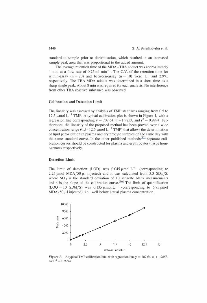
The linearity was assessed by analysis of TMP standards ranging from 0.5 to
12.5 mmol L21 TMP. A typical calibration plot is shown in Figure 1, with a
regression line corresponding y ¼ 707.64 � þ1.9853, and r2 ¼ 0.9994. Fur-
thermore, the linearity of the proposed method has been proved over a wide
concentration range (0.5–12.5 mmol L21 TMP) that allows the determination
of lipid peroxidation in plasma and erythrocyte samples on the same day with
the same standard curve. In the other published methods[11] separate cali-
bration curves should be constructed for plasma and erythrocytes/tissue hom-
ogenates respectively.
Detection Limit
The limit of detection (LOD) was 0.045 mmol L21 (corresponding to
2.25 pmol MDA/50 ml injected) and it was calculated from 3.3 SDbl/S,
where SDbl is the standard deviation of 10 separate blank measurements
and s is the slope of the calibration curve.[16] The limit of quantification
(LOQ ¼ 10 SDbl/S) was 0.135 mmol L21 (corresponding to 6.75 pmol
MDA/50 ml injected), i.e., well below actual plasma concentration.
Figure 1. A typical TMP calibration line, with regression line y ¼ 707.64 � þ1.9853,
and r2 ¼ 0.9994.
Z. A. Sarafinovska et al.2440
Precision
Intra-assay precision was evaluated by 5 repeated, separated measurements on
the same day, of the pooled samples of plasma and erythrocytes obtained from
the control subjects, and was found to be 4.1% for plasma measurement and
2.2% for erythrocytes, respectively. Either 5 repeated, separated measure-
ments on the same day of 0.5 and 5 mmol L21 TMP standard solutions have
been done, and the intra-assay precision was found to be 2.7% and 1.4%,
respectively (Figure 2).
Inter-assay precision was evaluated by five different measurement of the
pooled sample of plasma and erythrocytes on the five consecutive days and was
found to be 9.1% and 8.8% for the samples, respectively. The inter-assay
precision for the 0.5 and 5 mM TMP standard solutions was 7.9% and 5.4%, respect-
ively. These results indicate sufficient reproducibility for clinical application.
Recovery
Recovery of the TBA-MDA adduct was evaluated by addition of known
amounts of standard to the pooled plasma to give concentrations of 0.5, 1.5,
and 2.5 mmol L21 TMP. The average recovery for the tested amounts was
95.6% + 2.5% (n ¼ 15). Also the known amounts of standard stock
solution were added to the pooled erythrocyte lysate to give concentration
of 5, and 10 mmol L21 TMP. The average recovery for the tested amounts
was 97.4% + 1.3% (n ¼ 10).
Applications
MDA is a highly reactive aldehyde, capable of modifying both DNA and
proteins, resulting in mutagenic, genotoxic and cytotoxic events. Some of
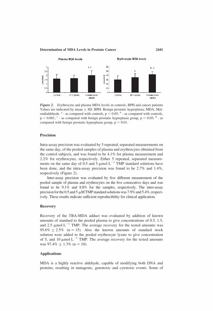
Figure 2. Erythrocyte and plasma MDA levels in controls, BPH and cancer patients
Values are indicated by mean + SD. BPH: Benign prostatic hyperplasia; MDA: Mal-
ondialdehyde. a - as compared with controls, p , 0.05; b - as compared with controls,
p , 0.001; c - as compared with benign prostatic hyperplasia group, p , 0.05; d - as
compared with benign prostatic hyperplasia group, p , 0.01.
Determination of MDA Levels in Prostate Cancer 2441
the identified DNA lesions are known to be pre-mutagenic and may play a role
in carcinogenesis. A possible link between these biochemical alterations and a
risk of developing prostate cancer was suggested.[25] Therefore, high levels of
MDA could explain DNA base modifications found by Olinski et al. (25) in
prostate cancer tissues.
Using this method, we have found elevated lipid peroxidation in the
plasma and erythrocytes of the prostate cancer group as compared to
controls and BPH group. To date, studies that examine the relationship
between lipid peroxidation and cancer have given contradictory results. It is
generally believed that there is an inverse relationship between the concen-
tration of lipid peroxides and the degree of malignancy deviation of the
tumor cells i.e. the higher the rate of lipid peroxidation in the cells
the lower the rate of cell division.[26] Our findings were in agreement
with the reports of Biri et al.;[27] Yilmaz et al.,[28] who have reported
increased TBARS concentrations suggesting oxidative stress and accelerate
peroxidative reactions in the cancerous prostate tissues, even though
antioxidant defense mechanisms were activated. However, Dogru-
Abbasoglu et al.[29] have found no significant change in lipid peroxidation
or antioxidant system parameters in the plasma of patients with BPH and
prostate cancer.
Furthermore, we have found about 20-fold higher MDA concentration in
erythrocyte samples than in plasma. It could be explained by the fact that
erythrocytes are particularly vulnerable to oxidative damage due to: a) con-
tinuous exposure to high oxygen tension, b) the large contents of polyunsatu-
rated fatty acids, major target for peroxidation, and c) the presence of large
amounts of iron, a potent catalyst of oxygen free radical production.[30,31]
We hypothesize that an altered prooxidant-antioxidant balance may lead
to an increase oxidative damage and consequently may play an important role
in the prostate carcinogenesis. The evaluation of oxidative stress involvement
in the etiology of the prostate cancer could contribute in the better understand-
ing of cause and development of this disease.
The further researches should be planned in order to find whether the
oxidative stress related parameters could be used as differential diagnostic
and prognostic tools in prostate cancer and BPH. Moreover, they could help
to improve the sensitivity and specificity of the existing detection techniques.
The improved risk stratification and outcome prediction would enhance the
physician’s ability to counsel patients about treatment option and their associ-
ated risk and benefits.
In conclusion, we have improved and validated an analytical HPLC
method for determination of MDA in plasma and erythrocytes, which is
simple to perform and having high sensitivity, specificity and substantial
improvement in column life. The limit of quantification (0.135 mmol L21 or
6.75 pmol MDA/50 ml injected on the column) is adequate for routine quanti-
fication of MDA in plasma. It could therefore be used for routine clinical
analysis. The method has been successfully applied to the study of the lipid
Z. A. Sarafinovska et al.2442
peroxidation levels in the circulation of prostate cancer patients and offers an
oportunity to further characterize the role of oxidative injury in the pathogen-
esis of this disease.
REFERENCES
1. Jemal, A.; Tiwari, R.C.; Murray, T.; et al. Cancer statistics, 2004. CA Cancer J.Clin. 2004, 54, 8–29.
2. Moul, J.W.; Anderson, J.; Penson, D.F.; Klotz, L.H.; Soloway, M.S.;Schulman, C.C. Early prostate cancer: Prevention, treatment modalities, andquality of life issues. Eur. Urol. 2003, 44, 283–93.
3. Nelson, W.G.; DeMarzo, A.M.; Isaacs, W.B. Prostate cancer. NEJM Mechanismsof Disease 2003, 349, 366–81.
4. Ripple, M.O.; Henry, W.F.; Rago, P.R.; Wilding, G. Prooxidant-antioxidant shiftinduced by androgen treatment of human prostate carcinoma cells. J. Natl. CancerInst. 1997, 89, 40–8.
5. Oberley, T.D.; Oberley, L.W. Oxygen radicals and cancer. In Free Radicals inAging; Yu, B.P. (Ed.); CRC Press: Boca Raton, FL, 1993, 247–6.
6. Dotan, Y.; Lichtenberg, D.; Pinchuk, I. Lipid peroxidation cannot be used as auniversal criterion of oxidative stress. Progress in Lipid Research 2004, 1–28.
7. Draper, H.H.; Hadley, M. Malondialdehyde determination as index of lipid per-oxidation. Methods Enzymol 1990, 186, 421–31.
8. Badcock, N.R.; Zoanetti, G.D.; Martin, E.S. Nonchromatographic assay for malon-dialdehyde-thiobarbituric acid adduct with HPLC equivalence. Clin. Chem. 1997,43, 1655–57.
9. Nielsen, F.; Mikkelsen, B.B.; Nielsen, J.B.; Andersen, H.R.; Grandjean, P. Plasmamalondialdehyde as biomarker for oxidative stress: reference interval and effectsof life-style factors. Clin. Chem. 1997, 43 (7), 1209–1214.
10. Fukunaga, K.; Yoshida, M.; Nakazono, N. A simple, rapid, highly sensitive andreproducible quantification method for plasma malondialdehyde by high-performance liquid chromatography. Biomed. Chromatograph 1998, 12, 300–303.
11. Lykkesfeldt, J. Determination of malondialdehyde as dithiobarbituric acid adductin biological samples by HPLC with fluorescence detection: Comparision withUltraviolet-Visible Spectrophotometry. Clin. Chem. 2001, 47 (9), 1725–1727.
12. Volpi, N.; Tarugi, P. Improvement in the high-performance liquid chromatographymalondialdehyde level determination in normal human plasma. J. Chromat. B,1998, 713, 433–437.
13. Templar, J.; Kon, S.P.; Milligan, T.P.; Newman, D.J.; Raftery, M. Increasedplasma malondialdehyde levels in glomerular disease as determined by a fullyvalidated HPLC method. Nephrol. Dial. Transplant 1999, 14, 946–951.
14. Pilz, J.; Meineke, I.; Gleiter, C.H. Measurement of free and bound malondialde-hyde in plasma by high-performance liquid chromatography as the 2,4-dinitrophenylhydrazine derivative. J. Chromat. B, 2000, 742, 315–325.
15. Claeson, K.; Thorsen, G.; Karlberg, B. Methyl malondialdehyde as an internalstandard for the determination of malondialdehyde. J. Chromat. B, 2001, 751,315–323.
16. Larstad, M.; Ljungkvist, G.; Olin, A.C.; Toren, K. Determination of malondialde-hyde in breath condensate by high-performance liquid chromatography with fluor-escence detection. J. Chromat. B 2001, 766, 107–114.
Determination of MDA Levels in Prostate Cancer 2443
17. Agarwal, R.; Chase, S.D. Rapid fluorimetric-liquid chromatographic determi-nation of malondialdehyde in biological samples. J. Chromat. B 2002, 775,121–126.
18. Karatas, F.; Karatepe, M.; Baysar, A. Determination of free malondialdehyde inhuman serum by high-performance liquid chromatography. Anal. Biochem.2002, 311, 76–79.
19. Li, K.; Shang, X.; Chen, Y. High-performance liquid chromatographic detection oflipid peroxidation in human seminal plasma and its application to male infertility.Clin. Chem. Acta. 2004, 346, 199–203.
20. Lepage, G.; Munoz, G.; Champagne, J.; Roy, C.C. Preparative steps necessary forthe accurate measurement of malondialdehyde by high-performance liquid chrom-atography. Anal. Biochem. 1991, 197, 277–283.
21. Draper, H.H.; Squires, E.J.; Mahmoodi, H.; Wu, J.; Agarwal, S.; Hadley, M. Acomparative evaluation of thiobarbituric acid methods for the determination ofmalondialdehyde in biological materials. Free Radic. Biol. Med. 1993, 15 (4),353–63.
22. Londero, D.; Lo, G.P. Automated high-performance liquid chromatographic sep-aration with spectrofluorometric detection of a malondialdehyde-thiobarbituricacid adduct in plasma. J. Chromatogr. A 1996, 729, 207–210.
23. Diaz-Velez, C.R.; Garcia-Castineiras, S.; Mendoza-ramos, E.; et al. Increased mal-ondialdehyde in periferal blood of patients with congestive heart failure. Am HeartJ. 1996, 131, 146–152.
24. Dasgupta, A.; Hussain, S.; Ahmad, S. Increased lipid peroxidation in patients onmaintenance haemodialysis. Nephron. 1992, 60, 56–59.
25. Olinski, R.; Zastawny, T.H.; Foksinski, M.; Barecki, A.; Dizdaroglu, M. DNA basemodifications and antioxidant enzyme activities in human benign prostate hyper-plasia. Free. Radic. Biol. Med. 1995, 18, 807–813.
26. Das, U.N.; A radical approach to cancer. Med. Sci. Monit. 2002, 8, RA79–92.27. Biri, H.; Ozturk, H.S.; Kacmaz, M.; Karaca, K.; Tokucoglu, H.; Durak, I. Activi-
ties of DNA turnover and free radical metabolizing enzymes in cancerous humanprostate tissue. Cancer Invest. 1999, 17, 314–9.
28. Yilmaz, M.I.; Saglam, K.; Sonmez, A.; et al. Antioxidant system activation inprostate cancer. Biol. Trace. Elem. Res. 2004, 98, 13–9.
29. Dogru-Abassoglu, S.; Aykac-Toker, G.; Kocak, T.; Unluer, E.; Uysal, M. Antiox-idant enzyme activities and lipid peroxides in the plasma of patients with benignprostatic hyperplasia or prostate cancer are not predictive. J. Cancer Res. Clin.Oncol. 1999, 125, 402–4.
30. Kumaraguruparan, R.; Subapriya, R.; Kabalimoorthy, J.; Nagini, S. Antioxidantprofile in the circulation of patients with fibroadenoma and adenocarcinoma ofthe breast. Clin. Biochem. 2002, 35, 275–9.
31. Ripple, M.; Mulcahy, R.T.; Wilding, G. Characteristics of the glutathione/gluta-thione-S-transferase detoxification system in melphalan resistant human prostatecells. J. Urol. 1993, 150, 209–14.
Received February 24, 2007
Accepted April 19, 2007
Manuscript 6071
Z. A. Sarafinovska et al.2444
Related Documents