Rapamycin Inhibits Polyglutamine Aggregation Independently of Autophagy by Reducing Protein Synthesis □ S Matthew A. King, Sarah Hands, Farida Hafiz, Noboru Mizushima, Aviva M. Tolkovsky, and Andreas Wyttenbach Department of Biochemistry, University of Cambridge, Cambridge, United Kingdom (M.A.K., F.H., A.M.T., A.W.); Department of Physiology and Cell Biology, Tokyo Medical and Dental University, Tokyo, Japan (N.M.); and School of Biological Sciences, Neuroscience Group, University of Southampton, Southampton, United Kingdom (S.H., A.W.) Received November 12, 2007; accepted December 31, 2007 ABSTRACT Accumulation of misfolded proteins and protein assemblies is associated with neuronal dysfunction and death in several neu- rodegenerative diseases such as Alzheimer’s, Parkinson’s, and Huntington’s disease (HD). It is therefore critical to understand the molecular mechanisms of drugs that act on pathways that modulate misfolding and/or aggregation. It is noteworthy that the mammalian target of rapamycin inhibitor rapamycin or its analogs have been proposed as promising therapeutic compounds clear- ing toxic protein assemblies in these diseases via activation of autophagy. However, using a cellular model of HD, we found that rapamycin significantly decreased aggregation-prone polyglu- tamine (polyQ) and expanded huntingtin and its inclusion bodies (IB) in both autophagy-proficient and autophagy-deficient cells (by genetic knockout of the atg5 gene in mouse embryonic fibro- blasts). This result suggests that rapamycin modulates the levels of misfolded polyQ proteins via pathways other than autoph- agy. We show that rapamycin reduces the amount of soluble polyQ protein via a modest inhibition of protein synthesis that in turn significantly reduces the formation of insoluble polyQ pro- tein and IB formation. Hence, a modest reduction in huntingtin synthesis by rapamycin may lead to a substantial decrease in the probability of reaching the critical concentration required for a nucleation event and subsequent toxic polyQ aggregation. Thus, in addition to its beneficial effect proposed previously of reducing polyQ aggregation/toxicity via autophagic pathways, rapamycin may alleviate polyQ disease pathology via its effect on global protein synthesis. This finding may have important therapeutic implications. The polyglutamine/CAG disorders comprise a group of neu- rodegenerative diseases that are associated with polyglu- tamine (polyQ) expansion mutations in the respective dis- ease genes that are otherwise unrelated (Cummings and Zoghbi, 2000). Abnormally long polyQ stretches cause pro- teins to misfold and produce intracellular protein aggregates. It is believed that polyQ aggregation follows a stochastic nucleation-dependent process that initiates oligomerization, amyloid-like fibril formation, and the production of struc- tures called inclusion bodies (IBs) (Perutz and Windle, 2001). Because polyQ misfolding/aggregation is associated with cel- lular toxicity (Ross and Poirier, 2004), it is crucial to under- stand the cellular mechanisms that control misfolding/aggre- gation with a view to the development of drugs that modify these pathways and alleviate disease. The accumulation of intracellular IBs points to the inabil- ity of cells to dispose of mutant polyQ proteins using chap- erone-assisted refolding (Muchowski and Wacker, 2005) and proteasome-mediated degradation (Jana and Nukina, 2003). Deciphering the mechanisms of degradation and clearance of polyQ-expanded proteins and how such mechanisms might be targeted using drugs is a major focus of current research. Macroautophagy (here referred to as autophagy) is a process alternative to that of proteasomal degradation by which some long-lived proteins and organelles are cleared (Shintani and Klionsky, 2004). Autophagy may be responsible for clearing polyQ-expanded proteins and their assemblies (Rubinsztein, This study was supported by the Wellcome Trust before October 2006 (to M.A.K., A.M.T.), the Hereditary Disease Foundation (to F.H.), and the Medical Research Council (to A.W., S.H.). Article, publication date, and citation information can be found at http://molpharm.aspetjournals.org. doi:10.1124/mol.107.043398. □ S The online version of this article (available at http://molpharm. aspetjournals.org) contains supplemental material. ABBREVIATIONS: polyQ, polyglutamine; CHX, cycloheximide; GFP, green fluorescent protein; EGFP, enhanced green fluorescent protein; Htt, huntingtin protein; Ex1Htt, huntingtin exon 1; HD, Huntington’s disease; IB, inclusion body; MEF, mouse embryonic fibroblast; mRFP, monomeric red fluorescent protein; mTOR, mammalian target of rapamycin; BafA1, bafilomycin A1; ANOVA, analysis of variance; PAGE, polyacrylamide gel electrophoresis; Rap, rapamycin; FK506, tacrolimus; CCI-779, temsirolimus; ERK, extracellular signal-regulated kinase; tERK, total extracellular signal-regulated kinase. 0026-895X/08/7304-1052–1063$20.00 MOLECULAR PHARMACOLOGY Vol. 73, No. 4 Copyright © 2008 The American Society for Pharmacology and Experimental Therapeutics 43398/3314684 Mol Pharmacol 73:1052–1063, 2008 Printed in U.S.A. 1052 http://molpharm.aspetjournals.org/content/suppl/2008/01/02/mol.107.043398.DC1 Supplemental material to this article can be found at: at ASPET Journals on June 5, 2018 molpharm.aspetjournals.org Downloaded from

Welcome message from author
This document is posted to help you gain knowledge. Please leave a comment to let me know what you think about it! Share it to your friends and learn new things together.
Transcript

Rapamycin Inhibits Polyglutamine Aggregation Independentlyof Autophagy by Reducing Protein Synthesis□S
Matthew A. King, Sarah Hands, Farida Hafiz, Noboru Mizushima, Aviva M. Tolkovsky,and Andreas WyttenbachDepartment of Biochemistry, University of Cambridge, Cambridge, United Kingdom (M.A.K., F.H., A.M.T., A.W.); Department ofPhysiology and Cell Biology, Tokyo Medical and Dental University, Tokyo, Japan (N.M.); and School of Biological Sciences,Neuroscience Group, University of Southampton, Southampton, United Kingdom (S.H., A.W.)
Received November 12, 2007; accepted December 31, 2007
ABSTRACTAccumulation of misfolded proteins and protein assemblies isassociated with neuronal dysfunction and death in several neu-rodegenerative diseases such as Alzheimer’s, Parkinson’s, andHuntington’s disease (HD). It is therefore critical to understandthe molecular mechanisms of drugs that act on pathways thatmodulate misfolding and/or aggregation. It is noteworthy that themammalian target of rapamycin inhibitor rapamycin or its analogshave been proposed as promising therapeutic compounds clear-ing toxic protein assemblies in these diseases via activation ofautophagy. However, using a cellular model of HD, we found thatrapamycin significantly decreased aggregation-prone polyglu-tamine (polyQ) and expanded huntingtin and its inclusion bodies(IB) in both autophagy-proficient and autophagy-deficient cells (bygenetic knockout of the atg5 gene in mouse embryonic fibro-
blasts). This result suggests that rapamycin modulates the levelsof misfolded polyQ proteins via pathways other than autoph-agy. We show that rapamycin reduces the amount of solublepolyQ protein via a modest inhibition of protein synthesis that inturn significantly reduces the formation of insoluble polyQ pro-tein and IB formation. Hence, a modest reduction in huntingtinsynthesis by rapamycin may lead to a substantial decrease inthe probability of reaching the critical concentration required fora nucleation event and subsequent toxic polyQ aggregation.Thus, in addition to its beneficial effect proposed previously ofreducing polyQ aggregation/toxicity via autophagic pathways,rapamycin may alleviate polyQ disease pathology via its effecton global protein synthesis. This finding may have importanttherapeutic implications.
The polyglutamine/CAG disorders comprise a group of neu-rodegenerative diseases that are associated with polyglu-tamine (polyQ) expansion mutations in the respective dis-ease genes that are otherwise unrelated (Cummings andZoghbi, 2000). Abnormally long polyQ stretches cause pro-teins to misfold and produce intracellular protein aggregates.It is believed that polyQ aggregation follows a stochasticnucleation-dependent process that initiates oligomerization,amyloid-like fibril formation, and the production of struc-tures called inclusion bodies (IBs) (Perutz and Windle, 2001).
Because polyQ misfolding/aggregation is associated with cel-lular toxicity (Ross and Poirier, 2004), it is crucial to under-stand the cellular mechanisms that control misfolding/aggre-gation with a view to the development of drugs that modifythese pathways and alleviate disease.
The accumulation of intracellular IBs points to the inabil-ity of cells to dispose of mutant polyQ proteins using chap-erone-assisted refolding (Muchowski and Wacker, 2005) andproteasome-mediated degradation (Jana and Nukina, 2003).Deciphering the mechanisms of degradation and clearance ofpolyQ-expanded proteins and how such mechanisms mightbe targeted using drugs is a major focus of current research.Macroautophagy (here referred to as autophagy) is a processalternative to that of proteasomal degradation by which somelong-lived proteins and organelles are cleared (Shintani andKlionsky, 2004). Autophagy may be responsible for clearingpolyQ-expanded proteins and their assemblies (Rubinsztein,
This study was supported by the Wellcome Trust before October 2006 (toM.A.K., A.M.T.), the Hereditary Disease Foundation (to F.H.), and the MedicalResearch Council (to A.W., S.H.).
Article, publication date, and citation information can be found athttp://molpharm.aspetjournals.org.
doi:10.1124/mol.107.043398.□S The online version of this article (available at http://molpharm.
aspetjournals.org) contains supplemental material.
ABBREVIATIONS: polyQ, polyglutamine; CHX, cycloheximide; GFP, green fluorescent protein; EGFP, enhanced green fluorescent protein; Htt,huntingtin protein; Ex1Htt, huntingtin exon 1; HD, Huntington’s disease; IB, inclusion body; MEF, mouse embryonic fibroblast; mRFP, monomericred fluorescent protein; mTOR, mammalian target of rapamycin; BafA1, bafilomycin A1; ANOVA, analysis of variance; PAGE, polyacrylamide gelelectrophoresis; Rap, rapamycin; FK506, tacrolimus; CCI-779, temsirolimus; ERK, extracellular signal-regulated kinase; tERK, total extracellularsignal-regulated kinase.
0026-895X/08/7304-1052–1063$20.00MOLECULAR PHARMACOLOGY Vol. 73, No. 4Copyright © 2008 The American Society for Pharmacology and Experimental Therapeutics 43398/3314684Mol Pharmacol 73:1052–1063, 2008 Printed in U.S.A.
1052
http://molpharm.aspetjournals.org/content/suppl/2008/01/02/mol.107.043398.DC1Supplemental material to this article can be found at:
at ASPE
T Journals on June 5, 2018
molpharm
.aspetjournals.orgD
ownloaded from

2006). Clearance by autophagy occurs by sequestration of thetarget organelle/protein into double-membrane structurescalled autophagosomes that fuse with endo/lysosomes anddischarge their contents, which are subsequently degraded.The mammalian homolog of Atg8 MAP-LC3 (LC3) is a keymediator of autophagy: after LC3 is C-terminally cleaved (LC3I), phosphatidylethanolamine is added to the C-terminal gly-cine by the Atg5/12 complex, generating LC3 II bound to thenascent autophagosomal membrane (Tanida et al., 2004). Be-cause the Atg5/12 complex catalytically activates the lipidationof LC3, trace amounts of Atg5 can support substantial autoph-agy, whereas Atg5 knockout cells are totally deficient in auto-phagy (Hosokawa et al., 2006). The hallmark of autophagicactivation is the formation of autophagosome puncta containingLC3 II, whereas the biochemical measurement of autophagicactivity is expressed as the amount of LC3 II that accumulatesin the absence or presence of lysosomal activity.
Autophagy was first implicated in the regulation of IBformation and clearance of aggregate-prone proteins basedon the use of chemical activators/inhibitors, including theproautophagic drug rapamycin and knockdown of differentautophagic genes (Rubinsztein, 2006). The finding that rapa-mycin and its analog CCI-779 protect against neurodegen-eration in animal models of misfolding diseases (Ravikumaret al., 2004; Berger et al., 2006) opens up immense hopes fortreating debilitating diseases such as the polyQ disorders.Rapamycin, a macrolytic lactone produced by Streptomyceshygroscopicus, has immunosuppressive, antimicrobial, andantitumor properties. It binds intracellularly to FK506 bind-ing protein 12 and targets the protein kinase mammaliantarget of rapamycin (mTOR). Inhibition of phosphorylation ofmTOR by rapamycin activates autophagy, and it has beensuggested that rapamycin (or analogs) ameliorates neurode-generative proteinopathies via activation of autophagy (Ru-binsztein, 2006). However, mTOR has an impact on variousdownstream targets not necessarily involved in autophagy,including the control of protein synthesis (Dann and Thomas,2006; Wullschleger et al., 2006), and because of these effects,it is currently being evaluated in several phase II clinicaltrials for cancer (Sabatini, 2006). It is therefore unclearwhether rapamycin mediates its protective effects solely viaautophagy.
To probe the actions of rapamycin on the formation andclearance of expanded polyQ proteins and IBs, we have takenadvantage of clonal cell lines of autophagy-proficient (Atg5�/�)and -deficient (Atg5�/�) mouse embryonic fibroblasts (MEFs)that are easily amenable to biochemical and genetic rescueexperiments. Using exon 1 of human Htt containing 97 glu-tamines and fused to enhanced green fluorescent protein(EGFP) (Ex1HttQ97-EGFP) as an aggregation prone modelpolypeptide, we show that autophagy-deficient cells accumulateinsoluble Ex1HttQ97-EGFP more rapidly and form greaternumbers of IBs compared with autophagy-proficient cells. Re-expression of Atg5 in Atg5-deficient cells reversed this pheno-type. Most strikingly, rapamycin reduced the amount of insol-uble Ex1HttQ97-EGFP and IBs to a similar degree in bothAtg5�/� and Atg5�/� cells. The formation of SDS-insolublepolyQ assemblies is a cooperative process that is highly depen-dent on the accumulation of a critical mass of the protein(Scherzinger et al., 1999; Colby et al., 2006). We suggest that amajor effect of rapamycin is the reduction in protein synthesisrequired for polyQ aggregation and IB formation to occur.
Materials and MethodsExpression Vectors. Mammalian expression vectors encoding
exon 1 of the HD gene with 25 or 97 glutamines fused at the Cterminus to an EGFP tag were a gift from Erich Schweitzer and AlanTobin (Brain Research Institute, University of California, Los Ange-les, CA). The mouse Atg5 expression vector and adenovirus mRFP-LC3 have been described previously (Mizushima et al., 2001; Bam-pton et al., 2005).
Cell Culture, Transfection, Inclusion Load Measurement,and Microscopy. SV-40-transformed MEF from Atg5�/� and Atg5�/�
mice (Kuma et al., 2004) were cultured in Dulbecco’s modified Eagle’smedium (Invitrogen, Carlsbad, CA) containing 10% fetal bovine serum(Sigma, St. Louis, MO), 4.5 g/l glucose, 2 mM L-glutamine, 1 mM sodiumpyruvate, 100 U/ml penicillin, and 100 �g/ml streptomycin (Sigma) in5% CO2 at 37°C. Cells were propagated in 75-cm2 flasks and seeded on12-mm poly(L-lysine)-coated glass coverslips in 24-well plates for fluo-rescence analysis or directly onto six-well plates for immunoblot/filter-trap analysis. Cells were trypsinized, counted, and seeded at a densityof 4 � 104 cells/well in 24-well plates and 3 � 105 cells/well in 6-wellplates. After an overnight culture, cells reached 60 to 80% confluenceand were transiently transfected using Lipofectamine 2000 reagent(Invitrogen) according to the manufacturer’s instructions. The trans-fection medium was replaced with fresh medium after 4 h, andcultures were incubated for a further 20 h in the presence or absenceof the following inhibitors: 200 nM rapamycin (Rap; Sigma), 50 nMbafilomycin A1 (BafA1; Sigma), and 0.3 to 0.01 �g/ml cycloheximide(CHX; Sigma). Cultures requiring longer time courses were split 24 hafter transfection and reseeded at lower densities for harvestingafter 48 to 96 h. Cells were fixed in 4% paraformaldehyde, washed inphosphate-buffered saline, and analyzed using epifluorescence mi-croscopy with an Olympus X-170 microscope (Olympus, Tokyo, Ja-pan). Images were collected using an AstraCam camera and Ultra-View software (PerkinElmer Life and Analytical Sciences, Waltham,MA). Inclusion load was calculated as the proportion of EGFP-ex-pressing cells that contained IBs. At least 200 cells were counted percondition.
Protein Synthesis and Cell Counting. Cells were briefly washedfree of methionine to avoid long-term methionine deprivation and la-beled for 1 h in methionine-free RPMI medium (Sigma) containing 10%fetal bovine serum and 1.85 MBq of [35S]methionine (GE Healthcare,Chalfont St. Giles, Buckinghamshire, UK) and the appropriate addi-tives. Cells were washed three times in methionine-containing Dulbec-co’s modified Eagle’s medium, protein was precipitated in ice-cold 20%trichloroacetic acid, and after three washes with 5% trichloroacetic acid,the precipitate was dissolved in 15% SDS and radioactivity was mea-sured by scintillation counting. Little tRNA was found in these pellets.Cells were counted after trypsinization using a hemocytometer.
Immunoblotting, Immunocytochemistry, Filter Trap As-say, and Resolubilization with Formic Acid. Cells were eithercollected with a cell scraper or trypsinized and counted using ahemocytometer before being pelleted and washed in phosphate-buff-ered saline. Material was prepared for immunoblotting and filter-trap detection according to Wanker et al. (1999). In brief, cells werelysed on ice for 30 min in filter-trap lysis buffer [50 mM Tris-HCl, pH8.8, 100 mM NaCl, 5 mM MgCl2 0.5% (w/v) Nonidet P-40, and 1 mMEDTA] in the presence of Complete protease inhibitors (Roche, In-dianapolis, IN). Insoluble material was pelleted by centrifugation at16,000g for 10 min and resuspended in 100 �l of DNaseI buffer (20mM Tris-HCl, 15 mM MgCl2, 0.5 mg/ml DNase I; Sigma) for 2 h at37°C. Protein concentrations of soluble (supernatant fraction) andinsoluble fractions (pellet) were determined using the Bicinchoninicacid kit (Sigma) and bovine serum albumin standards. Between 5and 30 �g of insoluble material was diluted into 200 �l of 2% SDS,boiled for 5 min, and applied to a 96-well dot-blot apparatus (Bio-RadLaboratories, Hercules, CA) containing a cellulose acetate mem-brane with 0.2-�m pore-size (Macherey-Nagel, Bethlehem, PA). Res-olubilization of pellets with formic acid was performed according to
Rapamycin-Mediated Suppression of Polyglutamine Aggregation 1053
at ASPE
T Journals on June 5, 2018
molpharm
.aspetjournals.orgD
ownloaded from

Hazeki et al. (2000). Pellets were treated in 100 �l of 100% formicacid for 1 h at 37°C, vacuum-centrifuged, and solubilized in 1�SDS-PAGE sample buffer (see below). Soluble material was supple-mented with 4� SDS-PAGE sample buffer (1 M Tris-HCl, pH 6.8,400 mM dithiothreitol, 8% SDS, and 40% glycerol), and 30 �g wasused for analysis by SDS-PAGE (8–12.5%). Membranes were blockedin 5% milk for 1 h and probed with the following primary antibodies:mouse monoclonal anti-GFP (8371-1; BD Biosciences, San Jose, CA)at 1:4000, rabbit polyclonal anti-phospho-S6 (2211; Cell SignalingTechnology, Danvers, MA) at 1:1000, mouse monoclonal anti-ERK(M12320; Transduction Laboratories, Lexington, KY) at 1:5000, rab-bit polyclonal anti-actin (A2066; Sigma) at 1:1000, and mouse anti-vimentin (V6630; Sigma, 1:40). Immunocytochemical analysis wasperformed as in Bampton et al. (2005). Rabbit polyclonal anti-LC3antibodies were gifts from Yasuo Uchiyama (Osaka University Grad-uate School of Medicine, Osaka, Japan) and Eiki Kominami (Jun-tendo University School of Medicine, Tokyo, Japan). Rabbit poly-clonal anti-Atg5 antibody was described previously (Mizushima etal., 2001). Blots were subsequently probed with horseradish peroxi-dase-conjugated anti-mouse or anti-rabbit IgG (Jackson Immunore-search Laboratories, West Grove, PA) at 1:5000 and visualized withenhanced chemiluminescence detection reagents (Amersham). Im-munoblots and dot-blot signals were scanned with a flatbed scanner(HP Scanjet 5470c; Hewlett Packard, Palo Alto, CA), and densitom-etry was performed using ImageJ software (National Institutes ofHealth, Bethesda, MD). For ratiometric values, the integrated pixelintensity of each signal was calculated and divided by the signalintensity obtained under control conditions. For each sample, dotplots were repeated at two to three dilutions to ensure that the signalwas not saturated, giving rise to a single value used for statisticalanalysis. Values from several experiments were then used to deter-mine the mean (fold) difference in signal intensity. Input was nor-malized either according to cell number or protein content. ForSDS-insoluble material, in some experiments, a parallel analysis ofprotein loading was conducted by immunoblotting for histone expres-sion (M.A.K., PhD Thesis; data not shown).
Statistical Analysis. The mean value of replicates within anexperiment (duplicates to quadruplicates) was taken as a singlevalue when calculating standard deviations from multiple experi-ments. Multiple comparisons were made using ANOVA followed byTukey’s honestly significant difference post hoc test, pairwise com-parisons were conducted using two-tailed Student’s t test, and onesample t test or 95% confidence intervals were used for calculatingthe significance of ratiometric values. These values and the numberof experiments performed for each result are indicated in the textand figure legends.
ResultsGenetic Ablation of Atg5 Increased Ex1HttQ97-EGFP
Accumulation and Inclusion Body Formation. We firsttested whether the complete genetic ablation of autophagy(Atg5) in cells (MEFs) modulated the accumulation of polyQ-expanded huntingtin (Htt), because previous experiments havebeen performed with RNAi approaches or not under conditionsof Htt synthesis (Iwata et al., 2005b; Shibata et al., 2006).Atg5�/� and Atg�/� MEFs were transfected with cDNA encod-ing exon 1 of human Htt containing either 25 glutamines(Ex1HttQ25) or 97 glutamines (Ex1HttQ97) fused to EGFP. Nodiscernible IBs in cells were found in MEFs of either typetransfected with Ex1HttQ25-EGFP (Fig. 1). However, cytoplas-mic or nuclear IBs were readily formed in Ex1HttQ97-EGFP-expressing cells of both types (Fig. 1). Evidence that Atg5�/�
MEFs were proficient to undergo autophagy whereas Atg5�/�
MEFs were not was obtained by expression of mRFP-LC3.Figure 1 shows that Atg5�/� MEFs contained several mRFP-
LC3 puncta, whereas mRFP-LC3 expression in Atg5�/� MEFswas evenly diffuse, as shown previously (Bampton et al., 2005).We did not observe colocalization of mRFP-LC3 with IBs inwild-type cells at this time point (24 h).
The proportion of EGFP-expressing cells containing IBsincreased over time in both cell types (Fig. 2A). Approxi-mately twice as many Ex1HttQ97-EGFP-positive Atg5�/�
MEFs contained IBs compared with Atg5�/� MEFs after 1 to2 days (Fig. 2A). Nuclear inclusions in approximately 10 to15% of both types of cells were evident from the fact thatnuclear DNA was “vacated” from spots in which the IBs haddeposited (Fig. 1, arrows) (for quantification, see Fig. 4).There was no difference in the transfection rate betweenAtg5�/� and Atg5�/� cells (quantified in Fig. 4), transfectionefficiency varying between 50 and 60% in both types of MEFs(see Supplementary Fig. S1 for low-power fluorescent imagesof cells). It is important to note that at 24 h after transfection,we did not detect any differences in toxicity caused by eitherEx1HttQ25-EGFP or Ex1HttQ97-EGFP expression inAtg5�/� and Atg5�/� cells, as assessed by inspection of nu-clear abnormalities (approximately 5% of EGFP�ve cells
Fig. 1. Atg�/� and Atg5�/� MEF cells form inclusion bodies containingEx1HttQ97-EGFP protein, but Ex1HttQ25-EGFP is homogenously ex-pressed in both cell types. Fluorescent images of Atg5�/� (A) and Atg5�/�
(B) MEFs expressing Ex1HttQ25-EGFP or Ex1HttQ97-EGFP 24 h aftertransfection. The right column shows cells coexpressing mRFP-LC3. Nu-clei (blue) were stained with Hoechst 33342. Cells expressingEx1HttQ25-EGFP do not form IBs and show a homogenous expression(left, green), whereas Ex1HttQ97-EGFP-expressing cells form nuclear(white arrows, middle) and cytoplasmic IBs (right). Note the absence ofDNA where a nuclear IB has been deposited (arrow). Only Atg5�/� cellscoexpressing Ex1HttQ97-EGFP and mRFP-LC3 (red) show LC3 puncta-tion (right, arrowhead). No localization of mRFP-LC3 to IBs wasobserved.
1054 King et al.
at ASPE
T Journals on June 5, 2018
molpharm
.aspetjournals.orgD
ownloaded from
showed baseline toxicity as measured by nuclear fragmenta-tion; see Fig. 4 for a quantitative comparison). Therefore theincrease in IB formation of Ex1HttQ97-EFP in Atg5�/� cellswas due neither to unequal transfection nor to any differen-tial toxicity as a result of IB formation in our experiments.
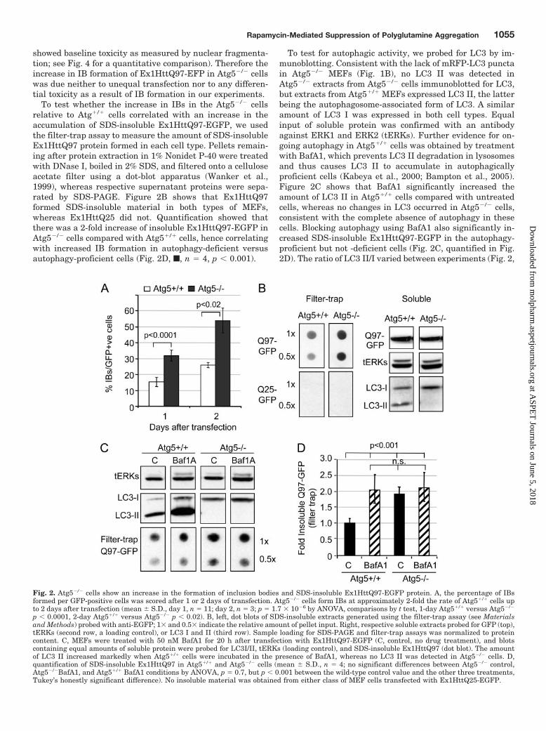
To test whether the increase in IBs in the Atg5�/� cellsrelative to Atg�/� cells correlated with an increase in theaccumulation of SDS-insoluble Ex1HttQ97-EGFP, we usedthe filter-trap assay to measure the amount of SDS-insolubleEx1HttQ97 protein formed in each cell type. Pellets remain-ing after protein extraction in 1% Nonidet P-40 were treatedwith DNase I, boiled in 2% SDS, and filtered onto a celluloseacetate filter using a dot-blot apparatus (Wanker et al.,1999), whereas respective supernatant proteins were sepa-rated by SDS-PAGE. Figure 2B shows that Ex1HttQ97formed SDS-insoluble material in both types of MEFs,whereas Ex1HttQ25 did not. Quantification showed thatthere was a 2-fold increase of insoluble Ex1HttQ97-EGFP inAtg5�/� cells compared with Atg5�/� cells, hence correlatingwith increased IB formation in autophagy-deficient versusautophagy-proficient cells (Fig. 2D, f, n � 4, p � 0.001).
To test for autophagic activity, we probed for LC3 by im-munoblotting. Consistent with the lack of mRFP-LC3 punctain Atg5�/� MEFs (Fig. 1B), no LC3 II was detected inAtg5�/� extracts from Atg5�/� cells immunoblotted for LC3,but extracts from Atg5�/� MEFs expressed LC3 II, the latterbeing the autophagosome-associated form of LC3. A similaramount of LC3 I was expressed in both cell types. Equalinput of soluble protein was confirmed with an antibodyagainst ERK1 and ERK2 (tERKs). Further evidence for on-going autophagy in Atg5�/� cells was obtained by treatmentwith BafA1, which prevents LC3 II degradation in lysosomesand thus causes LC3 II to accumulate in autophagicallyproficient cells (Kabeya et al., 2000; Bampton et al., 2005).Figure 2C shows that BafA1 significantly increased theamount of LC3 II in Atg5�/� cells compared with untreatedcells, whereas no changes in LC3 occurred in Atg5�/� cells,consistent with the complete absence of autophagy in thesecells. Blocking autophagy using BafA1 also significantly in-creased SDS-insoluble Ex1HttQ97-EGFP in the autophagy-proficient but not -deficient cells (Fig. 2C, quantified in Fig.2D). The ratio of LC3 II/I varied between experiments (Fig. 2,
Fig. 2. Atg5�/� cells show an increase in the formation of inclusion bodies and SDS-insoluble Ex1HttQ97-EGFP protein. A, the percentage of IBsformed per GFP-positive cells was scored after 1 or 2 days of transfection. Atg5�/� cells form IBs at approximately 2-fold the rate of Atg5�/� cells upto 2 days after transfection (mean � S.D., day 1, n � 11; day 2, n � 3; p � 1.7 � 10�6 by ANOVA, comparisons by t test, 1-day Atg5�/� versus Atg5�/�
p � 0.0001, 2-day Atg5�/� versus Atg5�/� p � 0.02). B, left, dot blots of SDS-insoluble extracts generated using the filter-trap assay (see Materialsand Methods) probed with anti-EGFP; 1� and 0.5� indicate the relative amount of pellet input. Right, respective soluble extracts probed for GFP (top),tERKs (second row, a loading control), or LC3 I and II (third row). Sample loading for SDS-PAGE and filter-trap assays was normalized to proteincontent. C, MEFs were treated with 50 nM BafA1 for 20 h after transfection with Ex1HttQ97-EGFP (C, control, no drug treatment), and blotscontaining equal amounts of soluble protein were probed for LC3I/II, tERKs (loading control), and SDS-insoluble Ex1HttQ97 (dot blot). The amountof LC3 II increased markedly when Atg5�/� cells were incubated in the presence of BafA1, whereas no LC3 II was detected in Atg5�/� cells. D,quantification of SDS-insoluble Ex1HttQ97 in Atg5�/� and Atg5�/� cells (mean � S.D., n � 4; no significant differences between Atg5�/� control,Atg5�/�BafA1, and Atg5�/� BafA1 conditions by ANOVA, p � 0.7, but p � 0.001 between the wild-type control value and the other three treatments,Tukey’s honestly significant difference). No insoluble material was obtained from either class of MEF cells transfected with Ex1HttQ25-EGFP.
Rapamycin-Mediated Suppression of Polyglutamine Aggregation 1055
at ASPE
T Journals on June 5, 2018
molpharm
.aspetjournals.orgD
ownloaded from

B and C), but BafA1 always increased the amount of LC3 IIby at least 2-fold (Fig. 2D). No SDS-insoluble material wasdetected in extracts of Ex1HttQ25-EGFP transfected cells ofeither genotype (Fig. 2B). We also did not detect EGFP sig-nals on filters when filtrating the supernatant of Ex1HttQ97-EGFP-expressing cells after spinning at 16,000g (data notshown) but without boiling, suggesting that no SDS-insolubleoligomeric Ex1HttQ97-EGFP species of more than 200 nm(pore size of filter) were generated.
We analyzed whether the IBs are ubiquitinylated in bothcell types, because this is a hallmark of all polyQ diseases invivo, including HD. We found colocalization of ubiquitin withIBs in �5% of both Atg5�/� and Atg5�/� cells (Fig. 3 and datanot shown). We also found that lysosome-associated mem-brane protein-1 was associated with IBs in both cell types.Because cytoplasmic IBs are surrounded by intermediatefilaments that form an “aggresome” (Waelter et al., 2001), wefurther probed for the intermediate filament protein vimen-tin. In both Atg5�/� and Atg5�/� cells, IBs were surroundedby vimentin immunoreactivity. These results show that IBsare qualitatively similar in both cell types.
Together, these data show that Atg5-dependent degrada-tion via autophagy plays an important role in determiningthe amount of insoluble polyQ-expanded Ex1Htt protein. Thedecrease in the propensity of cells to form IBs and insolubleEx1HttQ97-EGFP correlates with their ability to performautophagy.
Rapamycin Reduced the Amount of InsolubleEx1HttQ97-EGFP and Inclusion Body Formation inBoth Autophagy-Proficient and -Deficient Cells. We nextinvestigated whether rapamycin requires an Atg5-dependentmechanism to modulate the amount of insoluble Ex1HttQ97-EGFP and IB formation. MEFs were treated with 200 nMrapamycin either 12 h before transfection, to instill high
autophagic activity before onset of polyQ expression and IBformation, or were treated with rapamycin simultaneouslywith transfection. After 18 to 24 h, the percentage of EGFP-positive cells with IBs was determined, whereas insolubleEx1HttQ97-EGFP was measured using the filter-trap assayas described above. To determine that rapamycin was active,we measured S6 phosphorylation. S6 is a ribosomal proteinwhose phosphorylation is regulated by S6 kinase in anmTOR-dependent manner (Nobukini and Thomas, 2004).Figure 4A shows that rapamycin added 12 h before transfec-tion inhibited S6 phosphorylation in both cell types (trans-fected with Ex1HttQ25-EGFP or Ex1HttQ97-EGFP), indicat-ing that rapamycin prevented mTOR activity independentlyof Atg5 activity. There was no difference in the amount ofEx1HttQ25-EGFP or Ex1HttQ97-EGFP expressed in eithercell type treated with rapamycin compared with untreatedcells when the total amount of soluble protein input wasequalized between treatments, thus indicating that there isno differential destruction of the transfected proteins per se(Fig. 4A).
Pretreatment with rapamycin significantly decreased theproportion of EGFP-positive cells containing Ex1HttQ97-EGFP IBs by 40 to 45% in both Atg5�/� and Atg5�/� MEFs(Fig. 4B). To ensure that rapamycin treatment did not affectthe transfection efficiency or toxicity of the Ex1Htt trans-genes in either Atg5 cell type, we monitored both. We andothers have shown previously that analysis of nuclear mor-phology as measured by nuclear fragmentation and conden-sation using DNA stains is a reliable marker of cell toxicityunder these conditions and strongly correlates with othermarkers of cell death (Wyttenbach et al., 2001). Figure 4Cshows that the toxicity associated with the expression ofEx1HttQ25- or Q97-EGFP was at a baseline level (5%) underour experimental conditions (after 18–24 h after transfection)
Fig. 3. Colocalization of LAMP1, ubiq-uitin, and vimentin with IBs. Atg�/� orAtg�/� cells were transfected with aconstruct expressing Ex1HttQ97-EGFPand stained with an anti-ubiquitinantibody (Ubi) and an anti-lysosome-associated membrane protein anti-body (Lamp-1). A separate set of cellswas stained with anti-vimentin. Inboth Atg5�/� and Atg5�/� cells, ubiq-uitin, Lamp-1, and vimentin redistrib-ute toward inclusion bodies formed byEx1HttQ97-EGFP in �5% of the cells,showing that IBs seem to be qualita-tively similar.
1056 King et al.
at ASPE
T Journals on June 5, 2018
molpharm
.aspetjournals.orgD
ownloaded from

and not different in the two Atg5 cell types (�). Furthermore,rapamycin treatment did not modulate cell survival com-pared with control conditions (f), demonstrating that thereduction of IBs in both Atg5 cell lines induced by rapamycin(Fig. 4B) was not due to a differential toxicity (mean � S.D.,n � 3, ANOVA, p � 0.6). To make sure that the differentialincrease in IBs between Atg5�/� and Atg5�/� cells (Figs. 2Band 4B) and the decrease in IB formation by rapamycin wasnot due to unequal transfection rates, we measured thetransfection under the various conditions by counting thenumber of EGFP-positive cells in the total cell populationafter each experiment in parallel with the analysis of toxicityand IB. As shown in Fig. 4D, we obtained transfection effi-ciencies of 50 to 60%. It is noteworthy that neither the celltype nor rapamycin affected the rate of transfection (mean �S.D., n � 3, ANOVA, p � 0.3). Because we observed a minorproportion of IBs in the nuclear compartment (Fig. 1), we alsoquantified the proportion of Atg5�/� and Atg5�/� cells con-taining IB located in the nucleus versus cytoplasmic localiza-tion and whether this distribution is modulated by rapamy-cin. Figure 4D shows that 10 to 15% of nuclear IBs in bothcell types was not changed under rapamycin treatment(mean � S.D., n � 3, ANOVA, p � 0.9).
Having shown that rapamycin treatment reduced IBs in
both cell types, we next investigated whether this reductionwas also observed in the amount of insoluble material. IBsare cellular structures (or aggresomes) that may not providean adequate estimation of the amount of polyQ aggregation.However, we measured a similar reduction in SDS-insolubleEx1HttQ97-EGFP induced by rapamycin when this was as-sayed by filter trap, or after solubilizing the SDS-insolublepellet with formic acid, thereby controlling for equal proteininput and loading between the different conditions (Fig. 5A,quantified in B). There was no significant change in theamount of insoluble Ex1HttQ97-EGFP formed in cells thathad been treated with rapamycin at the time of transfectioncompared with control (see supplementary Fig. S2 for rawdata). Thus, rapamycin can decrease the amount of insolubleEx1HttQ97-EGFP and IB load, but this effect occurs in anAtg5-independent manner and with a considerable delay af-ter its addition.
Ravikumar et al. (2004) found that mTOR was inactivatedin polyQ-expressing cells, was bound to a polyQ-expandedN-terminal portion of Htt, and sequestered into IBs and thussuggested that autophagy is endemically activated in HD(and maybe other polyQ diseases) as a protective response. InMEFs expressing Ex1HttQ97-EGFP, we failed to find anincrease in mTOR immunoreactivity after solubilization of
Fig. 4. Pretreatment with rapamycin re-duces IB load similarly in both Atg5�/� andAtg5�/� MEFs without affecting toxicity,transfection efficiency, or nuclear distribu-tion of IBs. MEFs were seeded and allowed toattach before culturing in the presence orabsence of 200 nM rapamycin overnight (12h), after which MEFs were transfected witheither Ex1HttQ25-EGFP or Ex1HttQ97-EGFP and cultured for a further 20 to 24 hwithout treatment or in the presence of 200nM rapamycin before extraction. A, immuno-blot of soluble fraction of extracts (see Mate-rials and Methods for an explanation of sol-uble versus insoluble) normalized to equalprotein input. Blots were probed for GFP (toprow), tERK (loading control), phospho-serine(235/6)S6, or LC3. No differences wereobserved in the levels of soluble Ex1HttQ25/97, tERK, or LC3 in response to rapamycintreatment; however, the reduction in S6phosphorylation shows that mTOR inhibi-tion was achieved. This experiment was re-peated four times with similar results. B, thepercentage of IBs formed per GFP-positivecell was scored after 1 day (mean � S.D., n �11 (control) or n � 10 (rapamycin); ANOVA,p � 5 � 10�6, Tukey’s post hoc: Atg5�/�
untreated versus rapamycin p � 0.03,Atg5�/� untreated versus rapamycin p �0.003). C, toxicity was determined by scoringabnormal nuclear morphology using Hoechst33342 (mean � S.D., n � 3, ANOVA p �0.32). D, efficiency of transfection was scoredby counting the present of EGFP-positivecells (mean � S.D., n � 3, ANOVA, p � 0.69).E, percentage of EGFP-positive cells contain-ing nuclear IB was determined by analyzingfluorescent images (mean � S.D., n � 3,ANOVA, p � 0.95).
Rapamycin-Mediated Suppression of Polyglutamine Aggregation 1057
at ASPE
T Journals on June 5, 2018
molpharm
.aspetjournals.orgD
ownloaded from

IBs using formic acid. mTOR was also not trapped on thestacking gel in conjunction with Ex1HttQ97-EGFP (data notshown), so the degree of sequestration of mTOR may becell-specific and time-dependent.
Our finding that rapamycin reduced IBs and SDS-insolu-ble material in autophagy-deficient cells was unexpected.Hence, we next investigated through which mechanism rapa-mycin reduced IBs and SDS-insoluble Ex1HttQ97-EGFP.
Rapamycin Reduced the Formation of InsolubleEx1HttQ97-EGFP by Lowering the Amount of SolubleProtein Input. To investigate how rapamycin may be reducingthe load of insoluble Ex1HttQ97-EGFP and IBs in Atg5�/�
MEFs, the kinetics of cell cycle and protein expression wereinvestigated, because rapamycin is well documented to be acell cycle suppressant and an inhibitor of protein synthesis(Dann and Thomas, 2006; Sabatini, 2006). To measure this,we counted the number of cells and calculated the amount ofdecrease due to rapamycin treatment compared with un-treated cells set as the control value (presented as percentageof change). Indeed, approximately 25% fewer cells were gen-erated in rapamycin-treated MEFs over the experimentalperiod (36 h) irrespective of Atg5 genetic background or ex-pression of Ex1HttQ25-EGFP or Ex1HttQ97-EGFP (Fig. 6A,left; 0.001 � p � 0.05 for every condition relative to untreatedcontrol; see Supplementary Fig. S3 for raw data). There wasno statistical difference in rapamycin-induced decrease incell number between untransfected cells or cells transfectedwith Ex1HttQ25-EGFP or Ex1HttQ25-EGFP. The reductionin total cell protein in rapamycin-treated cultures comparedwith untreated cultures was approximately 37%, irrespectiveof Atg5 genetic status or Ex1HttQ25-EGFP or Ex1HttQ97-EGFP expression (Fig. 6A, middle; 0.001 � p � 0.05 for everycondition relative to untreated control; see SupplementaryFig. S3 for raw data), and no difference in the amount of thisreduction between untransfected or transfected cells witheither construct under rapamycin treatment was observed.When the total amount of protein per cell was calculated, theamount of protein was diminished on average by 17 � 1.8%in rapamycin-treated cells compared with that in untreatedcontrols, irrespective of genetic background or expression ofEx1HttQ25-EGFP or Ex1HttQ97-EGFP proteins (Fig. 6A,right). Thus, we conclude that rapamycin caused a signifi-cant reduction in protein per cell (p � 0.001, t test on pooledresults, n � 10 for each genotype).
To test whether the reduction in total protein per cell due
to rapamycin treatment included protein translated fromEx1Htt-expressing plasmid, we examined the amount ofEx1HttQ25-EGFP produced to avoid the uncertainty associ-ated with the insolubility of Ex1HttQ97-EGFP (Ex1HttQ25-EGFP is soluble under all conditions examined, see above).Figure 6B shows that the average reduction in expression ofEx1HttQ25-EGFP per cell as a result of rapamycin was ap-proximately 14% in both Atg5�/� and Atg5�/� cells, similarto the reduction found in total protein per cell. There was nostatistical difference in the reduction of total protein per celland the reduction of Ex1HttQ25-EGFP per cell as a result ofrapamycin treatment. This finding also suggests that Atg5-null cells have no increased protein expression from theplasmids compared with Atg5 wild-type cells because of thelack of the autophagic protein degradation system.
We next measured whether the reduction of 37% of totalprotein per cell as a result of rapamycin treatment (Fig. 6A) wascorrelating with a similar reduction in global protein synthesisas measured by direct [35S]methionine incorporation. Thirtyhours after the addition of rapamycin, we added [35S]methi-onine to Atg5�/� cells for 1 h. We observed a significant reduc-tion in [35S]methionine incorporation (Fig. 6C; mean � S.D.,n � 3; t test, p � 0.001). We then quantified the reduction as apercentage change under rapamycin treatment compared withuntreated cells and found that there was a 39 � 3% reduction in[35S]methionine incorporation (Fig. 6D, left bar). This 39% re-duction was almost identical with the reduction of 37 � 3%found for total soluble protein (Fig. 6D, middle bar) that wecalculated in parallel in each of these experiments. Calculatingthe ratio of change in [35S]methionine incorporation over thereduction of total protein content, this value was no differentfrom 0, indicating that the reduction in the global amount ofprotein (as shown in Fig. 6, A and D) is probably due to aninhibition of protein synthesis.
Inhibition of Protein Synthesis by CycloheximideReduced the Level of Insoluble Ex1HttQ97 and Inclu-sion Body Formation Similar to Rapamycin. To inves-tigate how a decrease in protein synthesis per cell affects thepropensity of cells to form insoluble Ex1HttQ97-EGFP andIBs, a concentration of CHX, an inhibitor of protein synthe-sis, was sought that matches the decrease in the amount ofsoluble protein obtained with rapamycin. Cycloheximide wasused because it binds to ribosomes reversibly, thus ensuringa response that is proportional to ribosome occupancy overextended periods of treatment. In dose-response experiments
Fig. 5. Pretreatment with rapamycin reduces SDS-insol-uble Ex1HttQ97-EGFP in both Atg5�/� and Atg5�/�
MEFs. MEFs were seeded and allowed to attach beforeculturing in the absence or presence of 200 nM rapamycinovernight (�12 h), after which cells were transfected withEx1HttQ97-EGFP and cultured for a further 20 to 24 h.The sample labeled “Rap 0 h” indicates cells to whichrapamycin was added at the time of transfection andmaintained during the subsequent 24 h of culture. TheSDS-insoluble fraction of the cells were analyzed by filter-trap assay or after resolubilization with formic acid. A,representative example of results from filter-trap assay(n � 3) or formic acid resolubilization assay (n � 1). B, thepercentage of change in SDS-insoluble material inducedby rapamycin (mean � S.E.M., n � 6; p � 0.05) for the12-h pretreatment with rapamycin (�12 h) comparedwith rapamycin added at 0 time (Rap 0). See Supplemen-tary Fig. S2 for complete data and statistics. Samplesused for the filter-trap assay were probed for histonelevels, confirming equal loading (data not shown).
1058 King et al.
at ASPE
T Journals on June 5, 2018
molpharm
.aspetjournals.orgD
ownloaded from

using rapamycin, we did not observe a significant differencein the amount of reduction in global protein synthesis be-tween 50 and 200 nM rapamycin (data not shown), andhence, we aimed at a CHX concentration that would matchthat induced by 200 nM rapamycin.
As expected, the amounts of insoluble Ex1HttQ97-EGFPprotein (Fig. 7A) and IB formation (Fig. 7B) were highly depen-dent on the concentration of CHX used in both cell types. Be-tween 0.03 and 0.1 �g/ml CHX, a similar amount of insolubleEx1HttQ97-EGFP was detected in the filter trap assay com-pared with the insoluble material obtained from rapamycin-treated cells (200 nM) irrespective of Atg5 genetic status (Fig.7A). The amount of IBs formed with rapamycin also closelymatched that observed with this concentration range of CHX(Fig. 7B), the amount of insoluble Ex1HttQ97-EGFP and IBformation being equally reduced. Moreover, steady-state pro-tein levels were reduced similarly by the same range of CHXand rapamycin (see Supplementary Fig. S5). Finally, when wecompared the amount of [35S]methionine incorporation over 1 hin cells pretreated with rapamycin (200 nM) and cells preincu-bated with different concentrations of CHX for 24 h, a similarinhibition of [35S]methionine incorporation was found withrapamycin and approximately 0.02 �g/ml CHX (rapamycin,41.1 � 7.2% compared with 49 � 4% with 0.02 �g/ml CHX;mean � range, two independent experiments). This result sug-gests that a concentration of CHX that similarly inhibits pro-tein synthesis compared with 200 nM rapamycin also producesequivalent reduction in insoluble Ex1HttQ97-EGFP and IBsobtained by 200 nM rapamycin. Hence, a small reduction in theinput of Ex1HttQ97-EGFP has major effects on the kinetics offormation of insoluble Ex1HttQ97-EGFP and IBs. It is interest-
ing that at higher concentrations (0.1–0.3 �g/ml), CHX alsoreduced the amounts of LC3 I/II in Atg�/� and LC3 I in Atg5�/�
cells to an equivalent extent by the end of the treatment (Fig.7A), suggesting that LC3 protein is being turned over quitequickly during this time by nonautophagic mechanisms.
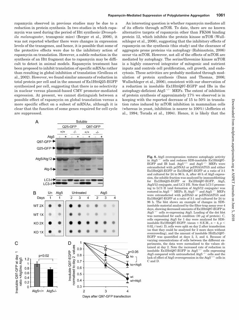
Atg5 Overexpression Partially Restored AutophagicActivity in Atg5�/� Cells and Reduced Insoluble polyQ-Expanded Ex1Htt. The characteristics of cell lines can di-verge rapidly, even when they derive from a common origin.To further investigate the role of autophagy in the control ofinsoluble Ex1HttQ97-EGFP and IB load, Atg5 was expressedin Atg5�/� and Atg5�/� cells by transfection. Atg5 expressionin Atg5�/� cells induced the formation of the Atg5/Atg12conjugate and restored the ability of the cells to produce LC3II to approximately 20% of the levels measured in Atg5�/�
cells (Fig. 8A). It is interesting that we found colocalization ofoverexpressed Atg5 with IBs (see Supplementary Fig. S4A)and a significant amount of Atg5 (but not Atg5/12 conjugate)accumulated in the pellets of both types of MEF cells express-ing Ex1HttQ97-EGFP (Supplementary Fig. S4B shows re-sults for Atg5�/� cells). Whether this Atg5 is disabled fromperforming its function (because no Atg12 accumulated inthis fraction) remains to be resolved. Together, these resultssuggest that some of the overexpressed Atg5 protein inAtg5�/� cells is functional, consistent with the recent findingof Hosokawa et al. (2006) using the same cell clone and DNAplasmids.
When the amount of insoluble material was examined us-ing the filter-trap assay (Fig. 8B), overexpression of Atg5 inAtg5�/� cells reduced the amount of insoluble material thataccumulated each day. Already after 1 day, the amount of
Fig. 6. Rapamycin reduces the amount of protein synthesized per cell. A, Atg5�/� and Atg5�/� MEF cultures were plated for 12 h in the absence orpresence of 200 nM rapamycin, after which two cohorts from each group were transfected with Ex1HttQ25-EGFP or Ex1HttQ97-EGFP and left in therespective media for 24 h. One set of cells was trypsinized and counted to determine cell number (left), whereas total soluble protein was extractedfor the other set, and the amount of total soluble protein was determined (middle). Right, the amount of calculated protein per cell. Results are depictedas the percentage of change between rapamycin-treatment and control. Both cell number and the amount of protein was reduced by rapamycin, leadingto a 17% reduction in protein per cell, irrespective of the expression of the polyQ proteins. Data show mean � S.D.; Q97, n � 5, Q25, n � 2,untransfected, n � 3; cell count, ANOVA p � 0.83; Total soluble protein, ANOVA p � 0.08; pooled results show that there are no significant differencesbetween the two genotypes, p � 0.2). See Supplementary Fig. S3 for complete data and statistics. B, cells were pretreated with rapamycin andtransfected with Ex1HttQ25-EGFP as in A; the amount of Q25-EGFP per cell was determined by measuring the amount of Q25-EGFP byimmunoblotting with an anti-GFP antibody, with the loading normalized to cell number rather than to total protein. Results show mean � range fromtwo independent experiments (t test, p � 0.7) and demonstrate that rapamycin also reduces protein expression from the plasmid. C, [35S]methioninewas added to Atg5�/� cells 30 h after the addition of rapamycin, and total methionine incorporation was determined after 1 h as detailed underMaterials and Methods (mean � S.D., n � 3, t test p � 0.001). D, the percentage of change induced by rapamycin is shown alongside the total amountof protein, which was measured in parallel samples. The right bar shows that the ratio of methionine incorporation to total protein is no different from0, demonstrating that inhibition of translation is the likely cause of the reduction in the amount of protein per cell.
Rapamycin-Mediated Suppression of Polyglutamine Aggregation 1059
at ASPE
T Journals on June 5, 2018
molpharm
.aspetjournals.orgD
ownloaded from

insoluble Ex1HttQ97-EGFP in Atg5�/� cells expressing Atg5decreased by 27 � 8% compared with 2 � 1% change inAtg5�/� cells (mean � S.E.M., n � 4, p � 0.02, t test; Fig. 8C).To compare the rate of reduction of insoluble Ex1HttQ97-EGFP under conditions of Atg5 overexpression over severaldays, we split and replated the transfected cells after 2days of transfection and measured the rate of decrease ofEx1HttQ97-EGFP in both cell types (Fig. 8D). Atg5 over-expression accelerated the reduction of Ex1HttQ97-EGFPin Atg5�/� cells but not in Atg5�/� cells. It is importantto note that overexpression of Atg5 did not reduce theamount of total protein harvested from the Atg5�/� orAtg5�/� cells (unlike rapamycin), suggesting that the de-crease in Ex1HttQ97-EGFP caused by Atg5 is independentof protein synthesis (data not shown). Moreover, the num-ber of cells harvested at each time point was similar be-tween the four conditions, demonstrating that Atg5 over-expression had no effect on the cell cycle. It should benoted that insoluble Ex1HttQ97-EGFP also decreases overtime in cells not overexpressing Atg5 from days 2 to 4 (Fig.8, B and D). This is probably due to continuous cell divi-sion, which reduces the number of plasmids present in
each cell. A differential loss of cells containing insolubleEx1HttQ97-EGFP between days 2 and 4 may also contrib-ute to this finding. Together, these data show that re-expression of Atg5 in autophagy-deficient cells achieves areduction in the amount of insoluble Ex1HttQ97-EGFP.
DiscussionIn the present study, we showed that autophagy-deficient
cells lacking Atg5 expression accumulate more misfoldedinsoluble polyQ protein (Ex1HttQ97-EGFP) and form moreIBs than control cells and that this effect can be partiallyreversed by re-expressing Atg5. These data suggest that thelack of autophagy increases polyQ aggregation and/or re-duces the clearance of aggregation-prone polyQ proteins. Ourresults obtained through a genetic approach (complete ge-netic knockout of Atg5) are consistent with those of recentstudies (Iwata et al., 2005a,b; Kouroku et al., 2007)
Rapamycin is a well characterized activator of autophagyand has been reported previously to alleviate toxicity ofdifferent aggregate-prone proteins (Rubinsztein, 2006). Inthese reports, the authors suggested that the beneficialeffects of rapamycin resulted from its ability to reduceaggregation-prone toxic proteins via autophagy. Hence,one would predict that rapamycin would reduce polyQaggregation in autophagy-proficient cells but not in auto-phagy-deficient cells. We tested this idea by treatingAtg5�/� and Atg5�/� cells with rapamycin and found thatrapamycin reduced soluble and insoluble aggregate-proneEx1Htt fragments independent of autophagic activity. Theevidence for this effect of rapamycin is as follows: 1) rapa-mycin reduced insoluble Ex1HttQ97 and IB load in Atg5-null cells; 2) inhibition by rapamycin, which rapidly inhib-ited phosphorylation of S6 kinase within 1 h, was onlyapparent if the cells were preincubated with rapamycin forapproximately 12 h before expression of Ex1HttQ97 for24 h, a time frame that suggests long-term rather thanshort-term actions of the drug are required for the effect tooccur; 3) rapamycin reduced the amount of soluble proteinper cell within this time frame by approximately 17% inboth Atg5�/� and Atg5�/� cells, and this amount of inhi-bition was correlated with the amount of [35S]methionineincorporated over 1 h at the end of the incubation period,indicating that the reduction in global protein per celloccurred because of a reduction in protein synthesis. Thesimilar extent of rapamycin-induced reduction in theamount of soluble Ex1HttQ25-EGFP per cell indicates thatprotein synthesis from the plasmid was similarly affected;and 4) the reduction in SDS-insoluble polyQ aggregationand IB formation induced by rapamycin could be mimickedby the use of low concentrations of CHX, which also causeda similar partial reduction in the extent of [35S]methionineincorporation. Thus, it seems that treatment with rapamy-cin can have a critical impact on the mass of solubleprotein required for polyQ aggregation and IB formationindependently of autophagy through a relatively modestreduction in protein input.
It is likely that autophagy too can reduce polyQ aggrega-tion not only through clearance of formed polyQ aggregatesbut also by reducing the amount of polyQ protein submittedto these processes. These two mechanisms could be acting inan additive way. It is conceivable that some of the effects of
Fig. 7. The reduction in inclusion bodies and insoluble Ex1HttQ97-EGFPinduced by rapamycin is mimicked with cycloheximide. Atg5�/� andAtg5�/� MEFs were either pretreated with rapamycin for 12 h or leftuntreated before transfection with Ex1HttQ97-EGFP. Cells were thencultured with rapamycin or cycloheximide (CHX, 0.01–0.3 �g/ml) for 20 hbefore protein extraction. A, the soluble fraction was analyzed by immu-noblotting for GFP, tERK, or phospho-serine235/6-S6, whereas the insol-uble fraction was analyzed by filter-trap assay for presence ofEx1HttQ97-EGFP. The data below the dot blot show mean � range ofvalues from two independent experiments. B, EGFP�ve cells were scoredfor IB. C, Cycloheximide treatment caused a similar dose-dependantdecrease in IBs and SDS-insoluble Ex1HttQ97-EGFP in both Atg5�/� andAtg5�/� cells without affecting soluble levels of Ex1HttQ97-EGFP (A andB). Note the similarity between the effect of rapamycin (200 nM) and thereduction caused by 0.02 to 0.1 �g/ml CHX.
1060 King et al.
at ASPE
T Journals on June 5, 2018
molpharm
.aspetjournals.orgD
ownloaded from
rapamycin observed in previous studies may be due to areduction in protein synthesis. In two studies in which rapa-mycin was used during the period of Htt synthesis (Drosoph-ila melanogaster, transgenic mice) (Berger et al., 2006), itwas not reported whether there were changes in expressionlevels of the transgenes, and hence, it is possible that some ofthe protective effects were due to the inhibitory action ofrapamycin on translation. However, a subtle reduction in thesynthesis of an Htt fragment due to rapamycin may be diffi-cult to detect in animal models. Rapamycin treatment hasbeen proposed to inhibit translation of specific mRNAs ratherthan resulting in global inhibition of translation (Grolleau etal., 2002). However, we found similar amounts of reduction intotal protein per cell and in the amount of Ex1HttQ25-EGFPsynthesized per cell, suggesting that there is no selectivityin nuclear versus plasmid-based CMV promoter-mediatedexpression. At present, we cannot distinguish between apossible effect of rapamycin on global translation versus amore specific effect on a subset of mRNAs, although it isclear that the function of some genes required for cell cycleare suppressed.
An interesting question is whether rapamycin mediates allof its effects through mTOR. To date, there are no knownalternative targets of rapamycin other than FK506 bindingprotein 12, which inhibits the protein kinase mTOR (Wull-schleger et al., 2006), suggesting that the inhibitory effects ofrapamycin on the synthesis (this study) and the clearance ofaggregate prone proteins via autophagy (Rubinsztein, 2006)occur via mTOR. However, not all of the effects of mTOR aremediated by autophagy. The serine/threonine kinase mTORis a highly conserved integrator of mitogenic and nutrientinputs and controls cell proliferation, cell growth, and endo-cytosis. These activities are probably mediated through mod-ulation of protein synthesis (Dann and Thomas, 2006;Wullschleger et al., 2006), consistent with our observation ofa reduction in insoluble Ex1HttQ97-EGFP and IBs in theautophagy-deficient Atg5�/� MEFs. The extent of inhibitionof protein synthesis of approximately 17% we observed is inkeeping with the reported decrease of 15 to 50% in transla-tion rates induced by mTOR inhibition in mammalian cells(whereas in yeast, inhibition is nearer to 100%) (Jefferies etal., 1994; Terada et al., 1994). Hence, it is likely that the
Fig. 8. Atg5 overexpression restores autophagic activityin Atg5�/� cells and reduces SDS-insoluble Ex1HttQ97-EGFP and IB load. Atg5�/� and Atg5�/� MEFs werecotransfected with pcDNA3 or pcDNA3ATG5 and eitherEx1HttQ25-EGFP or Ex1HttQ97-EGFP at a ratio of 3:1and cultured for 24 to 96 h. A, after 48 h of Atg5 expres-sion, the soluble fraction was analyzed by immunoblottingfor Ex1HttQ25-EGFP or Ex1HttQ97-EGFP, Atg5,Atg5/12 conjugate, and LC3 I/II. Note that LC3 I process-ing to LC3 II (and formation of Atg5/12 conjugate) wasrestored in Atg5�/� MEFs. B, Atg5�/� and Atg5�/� MEFswere cotransfected with pcDNA3 or pcDNA3ATG5 andEx1HttQ97-EGFP at a ratio of 3:1 and cultured for 24 to96 h. The blot shows an example of changes in SDS-insoluble material analyzed by the filter-trap assay over 4days, showing decreased amounts of Ex1HttQ97-EGFP inAtg5�/� cells re-expressing Atg5. Loading of the dot blotwas normalized for each condition (30 �g of protein). C,cells expressing Atg5 for 1 day were analyzed for SDS-insoluble Ex1HttQ97-EGFP. (mean � S.E.M., n � 4, p �0.02, t test). D, cells were split on day 2 after transfection(so that they could be analyzed for 2 more days withoutovercrowding), and the amount of insoluble HttEx1Q97-EGFP was quantified at days 2, 3, and 4. Because ofvarying concentrations of cells between the different ex-periments, the data were normalized to the values ob-tained at day 2. Note the increased rate of reduction ininsoluble Ex1HttQ97-EGFP in Atg5�/� cells expressingAtg5 compared with untransfected Atg5�/� cells and thelack of effect of Atg5 overexpression in the Atg5�/� cells inC and D.
Rapamycin-Mediated Suppression of Polyglutamine Aggregation 1061
at ASPE
T Journals on June 5, 2018
molpharm
.aspetjournals.orgD
ownloaded from

effect of rapamycin on protein synthesis is cell type-depen-dent. It remains to be seen to what extent this drug affectsprotein synthesis in neurons.
The reduction in insoluble Ex1HttQ97-EGFP achievedwith rapamycin was mimicked by the use of CHX. Severalreports have demonstrated that CHX inhibits autophagy(Kovacs and Seglen, 1981) by preventing fusion of autopha-gosome with endo/lysosomes (Lawrence and Brown, 1993).Consistent with this view, IBs were cleared more quickly inthe presence of rapamycin in an inducible cell model afterexpanded Ex1Htt protein synthesis was turned off, but IBsseemed to accumulate when CHX (10 �g/ml) was presentwith rapamycin (Ravikumar et al., 2002). In the above stud-ies, 2 to 10 �g/ml CHX was used, which inhibits proteinsynthesis and autophagy more than 95%, whereas in ourstudy, the concentration that mimicked rapamycin’s effectwas between 0.01 and 0.03 �g/ml, which elicited only a par-tial block on protein synthesis and presumably on autophagy.However, our important observation is that the same reduc-tion in insoluble material and IBs were elicited by CHX inAtg5�/� MEFs, in which no autophagy can take place. Hence,the phenomenon we observed here is unlikely to be related toautophagy.
Autophagic clearance of Htt fragments may be promotedindependently of mTOR (and S6K/Akt) via a pathway depen-dent on insulin receptor substrate 2 and stimulated via in-sulin and IGF-1 (Yamamoto et al., 2006). The notion thatthere are mTOR-independent pathways of autophagy is sug-gested by the finding that rapamycin inhibited autophagysuppression by insulin but not by regulatory amino acids inliver cells (Kanazawa et al., 2004), and yeast susceptibility toosmotic stress differed when induction of autophagy was bystarvation or rapamycin (Prick et al., 2006). Indeed, it hasbeen shown recently that small-molecule enhancers of mTORcan act independently of rapamycin to enhance the clearanceof mutant huntingtin fragments and A53T �-synuclein (Sarkaret al., 2007). These findings suggest that multiple mechanismsmay regulate autophagic clearance of misfolded/aggregatedproteins. However, irrespective of how the proteins are cleared,a reduction in the critical mass of aggregation-prone proteinssuch as that described in the present study will always bebeneficial in reducing protein aggregation.
Reducing the expression of Htt is, in theory, a promisingstrategy to alleviate disease: a decrease in the expression ofwild-type Htt up to 50% is not detrimental (based on studiesin heterozygotes), whereas decreasing the expression of mu-tant Htt using a short interfering RNA approach has beenfound to significantly reduce HD pathology in mice (Wang etal., 2005; Machida et al., 2006). This idea is supported by therecent findings of Colby et al. (2006), who proposed that anefficient therapeutic strategy for HD lies in reducing the rateof mutant Ex1Htt aggregation by only modestly reducing Httexpression levels. From our study, we cannot conclude thatrapamycin would have a beneficial effect by reducing Httsynthesis in all cases. Future in vivo studies are required toaddress this point.
It is important to note that in HD in vivo, mutant Htt isconstantly produced; hence, the mechanisms by which rapa-mycin regulates the accumulation and turnover of mutantHtt should be studied under condition of continuous Httexpression as we have done in our cellular model. The sto-chastic model of Colby et al. (2006) was applied under condi-
tions in which no further protein synthesis was taking placeduring the misfolding process. If rapamycin can reduce Httconcentrations in HD, it might be beneficial even under con-ditions in which autophagy is already working at its maximalrate.
Acknowledgments
We thank Dr. Yasuo Uchiyama (Osaka University Graduate Schoolof Medicine, Osaka, Japan) and Dr. Eiki Kominami (Juntendo Univer-sity School of Medicine, Tokyo, Japan) for the provision of anti-LC3antibodies, and Drs. Eric Schweitzer and Alan Tobin for the originalconstructs of Ex1HttQ25-EGFP and Ex1HttQ97-EGFP. Most of thiswork was conducted in Aviva M. Tolkovsky’s laboratory under jointsupervision of Andreas Wyttenbach and Aviva M. Tolkovsky.
ReferencesBampton ETW, Goemans CG, Niranjan D, Mizushima N, and Tolkovsky AM (2005)
The dynamics of autophagy visualised in live cells: from autophagosome formationto fusion with endo/lysosomes. Autophagy 1:23–36.
Berger Z, Ravikumar B, Menzies FM, Oroz LG, Underwood BR, Pangalos MN,Schmitt I, Wullner U, Evert BO, O’Kane CJ, et al. (2006) Rapamycin alleviatestoxicity of different aggregate-prone proteins. Hum Mol Genet 15:433–442.
Colby DW, Cassady JP, Lin GC, Ingram VM, and Wittrup KD (2006) Stochastickinetics of intracellular huntingtin aggregate formation. Nat Chem Biol 2:319–323.
Cummings CJ and Zoghbi HY (2000) Trinucleotide repeats: mechanisms and patho-physiology. Annu Rev Genomics Hum Genet 1:281–328.
Dann SG and Thomas G (2006) The amino acid sensitive TOR pathway from yeastto mammals. FEBS Lett 580:2821–2829.
Grolleau A, Bowman J, Pradet-Balade B, Puravs E, Hanash S, Garcia-Sanz JA, andBeretta L (2002) Global and specific translational control by rapamycin in T cellsuncovered by microarrays and proteomics. J Biol Chem 277:22175–22184.
Hazeki N, Tukamoto T, Goto J, and Kanazawa I (2000) Formic acid dissolvesaggregates of an N-terminal huntingtin fragment containing an expanded poly-glutamine tract: applying to quantification of protein components of the aggre-gates. Biochem Biophys Res Commun 277:386–393.
Hosokawa N, Hara Y, and Mizushima N (2006) Generation of cell lines with tetra-cycline-regulated autophagy and a role for autophagy in controlling cell size. FEBSLett 580:2623–2629.
Iwata A, Christianson JC, Bucci M, Ellerby LM, Nukina N, Forno LS, and Kopito RR(2005a) Increased susceptibility of cytoplasmic over nuclear polyglutamine aggre-gates to autophagic degradation. Proc Natl Acad Sci U S A 102:13135–13140.
Iwata A, Riley BE, Johnston JA, and Kopito RR (2005b) HDAC6 and microtubulesare required for autophagic degradation of aggregated huntingtin. J Biol Chem280:40282–40292.
Jana NR and Nukina N (2003) Recent advances in understanding the pathogenesisof polyglutamine diseases: involvement of molecular chaperones and ubiquitin-proteasome pathway. J Chem Neuroanat 26:95–101.
Jefferies HB, Reinhard C, Kozma SC, and Thomas G (1994) Rapamycin selectivelyrepresses translation of the “polypyrimidine tract” mRNA family. Proc Natl AcadSci U S A 91:4441–4445.
Kabeya Y, Mizushima N, Ueno T, Yamamoto A, Kirisako T, Noda T, Kominami E,Ohsumi Y, and Yoshimori T (2000) LC3, a mammalian homologue of yeast Apg8p,is localized in autophagosome membranes after processing. EMBO J 19:5720–5728.
Kanazawa T, Taneike I, Akaishi R, Yoshizawa F, Furuya N, Fujimura S, andKadowaki M (2004) Amino acids and insulin control autophagic proteolysisthrough different signaling pathways in relation to mTOR in isolated rat hepato-cytes. J Biol Chem 279:8452–8459.
King MA (2007) Neurotoxicity and autophagic turnover of hungtingtin exon 1 con-taining expanded polyglutamine, Ph.D. Thesis, University of Cambridge, Cam-bridge, UK.
Kouroku Y, Fujita E, Tanida I, Ueno T, Isoai A, Kumagai H, Ogawa S, Kaufman RJ,Kominami E, and Momoi T (2007) ER stress (PERK/eIF� phosphorylation) medi-ates the polyglutamine-induced LC3 conversion, an essential step for autophagyformation. Cell Death Differ 14:230–239.
Kovacs AL and Seglen PO (1981) Inhibition of hepatocytic protein degradation bymethylaminopurines and inhibitors of protein synthesis. Biochim Biophys Acta676:213–220.
Kuma A, Hatano M, Matsui M, Yamamoto A, Nakaya H, Yoshimuri T, Ohsumi Y,Tokuhisa T, and Mizushima N (2004) The role of autophagy during the earlyneonatal starvation period. Nature 432:1032–1036.
Lawrence BP and Brown WJ (1993) Inhibition of protein synthesis separates auto-phagic sequestration from the delivery of lysosomal enzymes. J Cell Sci 105:473–480.
Machida Y, Okada T, Kurosawa M, Oyama F, Ozawa K, and Nukina N (2006)rAAV-mediated shRNA ameliorated neuropathology in Huntington disease modelmouse. Biochem Biophys Res Commun 343:190–197.
Mizushima N, Yamamoto A, Hatano M, Kobayashi Y, Kabeya Y, Suzuki K, TokuhisaT, Ohsumi Y, and Yoshimori T (2001) Dissection of autophagosome formationusing Apg5-deficient mouse embryonic stem cells. J Cell Biol 152:657–668.
Muchowski PJ and Wacker JL (2005) Modulation of neurodegeneration by molecularchaperones. Nat Rev Neurosci 6:11–22.
Nobukini T and Thomas G (2004) The mTOR/S6K signalling pathway: the role of the
1062 King et al.
at ASPE
T Journals on June 5, 2018
molpharm
.aspetjournals.orgD
ownloaded from

TSC1/2 tumour suppressor complex and the proto-oncogene Rheb. Novartis FoundSymp 262:148–154.
Perutz MF and Windle AH (2001) Cause of neural death in neurodegenerativediseases attributable to expansion of glutamine repeats. Nature 412:143–144.
Prick T, Thumm M, Haussinger D, and Vom Dahl S (2006) Deletion of HOG1 leadsto osmosensitivity in starvation-induced, but not rapamycin-dependent Atg8 deg-radation and proteolysis: further evidence for different regulatory mechanisms inyeast autophagy. Autophagy 2:241–243.
Ravikumar B, Duden R, and Rubinsztein DC (2002) Aggregate-prone proteins withpolyglutamine and polyalanine expansions are degraded by autophagy. Hum MolGenet 11:1107–1117.
Ravikumar B, Vacher C, Berger Z, Davies JE, Luo S, Oroz LG, Scaravilli F, EastonDF, Duden R, O’Kane CJ, et al. (2004) Inhibition of mTOR induces autophagy andreduces toxicity of polyglutamine expansions in fly and mouse models of Hunting-ton disease. Nat Genet 36:585–595.
Ross CA and Poirier MA (2004) Protein aggregation and neurodegenerative disease.Nat Med 10:S10–S17.
Rubinsztein DC (2006) The roles of intracellular protein-degradation pathways inneurodegeneration. Nature 443:780–786.
Sabatini DM (2006) mTOR and cancer: insights into a complex relationship. Nat RevCancer 6:729–734.
Sarkar S, Perlstein EO, Imarisio S, Pineau S, Cordenier A, Maglathlin RL, WebsterJA, Lewis TA, O’Kane CJ, Schreiber SL, et al. (2007) Small molecules enhanceautophagy and reduce toxicity in Huntington’s disease models. Nat Chem Biol3:331–338.
Scherzinger E, Sittler A, Schweiger K, Heiser V, Lurz R, Hasenbank R, Bates GP,Lehrach H, and Wanker EE (1999) Self-assembly of polyglutamine-containinghuntingtin fragments into amyloid-like fibrils: implications for Huntington’s dis-ease pathology. Proc Natl Acad Sci U S A 96:4604–4609.
Shibata M, Lu T, Furuya T, Degterev A, Mizushima N, Yoshimori T, MacDonald M,Yankner B, and Yuan J (2006) Regulation of intracellular accumulation of mutantHuntingtin by Beclin 1. J Biol Chem 281:14474–14485.
Shintani T and Klionsky DJ (2004) Autophagy in health and disease: a double-edgedsword. Science 306:990–995.
Tanida I, Ueno T, and Kominami E (2004) LC3 conjugation system in mammalianautophagy. Int J Biochem Cell Biol 36:2503–2518.
Terada N, Patel HR, Takase K, Kohno K, Nairn AC, and Gelfand EW (1994)Rapamycin selectively inhibits translation of mRNAs encoding elongation factorsand ribosomal proteins. Proc Natl Acad Sci U S A 91:11477–11481.
Waelter S, Boeddrich A, Lurz R, Scherzinger E, Lueder G, Lehrach H, Wanker EE(2001) Accumulation of mutant huntingtin fragments in aggresome-like inclusionbodies as a results of insufficient protein degradation. Mol Biol Cell 12:1393–1407.
Wang YL, Liu W, Wada E, Murata M, Wada K, and Kanazawa I (2005) Clinico-pathological rescue of a model mouse of Huntington’s disease by siRNA. NeurosciRes 53:241–249.
Wanker EE, Scherzinger E, Heiser V, Sittler A, Eickhoff H, and Lehrach H (1999)Membrane filter assay for detection of amyloid-like polyglutamine-containing pro-tein aggregates. Methods Enzymol 309:375–386.
Wullschleger S, Loewith R, and Hall MN (2006) TOR signaling in growth andmetabolism. Cell 124:471–484.
Wyttenbach A, Swartz J, Kita H, Thykjaer T, Carmichael J, Bradley J, Brown R,Maxwell M, Schapira A, Orntoft TF, et al. (2001) Polyglutamine expansions causedecreased CRE-mediated transcription and early gene expression changes prior tocell death in an inducible cell model of Huntington’s disease. Hum Mol Genet10:1829–1845.
Yamamoto A, Cremona ML, and Rothman JE (2006) Autophagy-mediated clearanceof huntingtin aggregates triggered by the insulin-signaling pathway. J Cell Biol172:719–731.
Address correspondence to: Dr. Andreas Wyttenbach, Southampton Neu-roscience Group, School of Biological Sciences, University of Southampton.Bassett Crescent East, Southampton SO16 7PX, UK. E-mail: [email protected]
Rapamycin-Mediated Suppression of Polyglutamine Aggregation 1063
at ASPE
T Journals on June 5, 2018
molpharm
.aspetjournals.orgD
ownloaded from
Related Documents


