16 PICTORIAL ESSAY Ultrasound Characterization of Salivary Lesions Rajesh C Kamble, Alpana N Joshi, Pravin J Mestry ABSTRACT Evolution in ultrasound technology leads to birth of high frequency, high resolution linear transducers which in turn revolutionized the imaging of superficial structures. The majority of cases presenting to clinician in day-to-day practice as facial lumps or neck lump could be evaluated using ultrasound and help in imaging of salivary gland lesions. The age of the clinical presentation and morphological characteristics of lesions on ultrasound helped us in making confident diagnosis which was correlated on fine needle aspiration cytology/histopathology studies. Keywords: Salivary glands, Ultrasound, Parotid glands, Submandibular glands, Sialadenitis. Source of support: Nil Conflict of interest: None declared INTRODUCTION The protocol for imaging of salivary glands in Western countries includes–nonenhanced and contrast-enhanced computed tomography (CT), nonenhanced and contrast- enhanced magnetic resonance (MR) imaging and sialography (also MR sialography). 1,2 In general, CT is considered the best single method for assessment of inflammatory diseases and MR imaging is considered the best single method for assessment of salivary gland tumors. 1-3 According to Yousem et al 2 ultrasono- graphy (US) is not preferred modality in Western countries; but in Europe and Asia, US is widely accepted as the first imaging method for assessment of lymph nodes and soft tissue diseases in the head and neck, including major salivary glands. 4-7 Infact US with US-guided fine needle aspiration cytology (FNAC) of the lesions examination alone may suggest the final diagnosis in most of the clinical settings. As the head and neck region has a complex anatomic structure, a sound knowledge of sonographic anatomy and spatial relationships is crucial for reliable performance of the examination. It is sometimes not possible to visualize and analyze examined lesions completely at US because of their location for example–deep lobe of the parotid gland or area behind the mandible. In these situations, performance of further imaging examinations—CT or MR imaging—is highly recommended. Also, in cases of suspected malignant lesions, further diagnostic methods (i.e. CT or MR imaging) should be applied to assess possible infiltration of bones or deep structures invisible at US (the base of the skull, para- pharyngeal space) and to evaluate deep-lying lymph nodes. 1,3,8,9 On the other hand, dynamic scintigraphy is still the method of choice in functional evaluation of the salivary glands. 10,11 TECHNIQUE The examination should be carried out with the highest frequency transducer possible. Usually, 5 to 12 MHz wide- band linear transducers (median frequency: 7-7.5 MHz or more) are used. 9 In assessment of large tumors and lesions located in deep portions of the glands, 5 to 10 MHz transducers may be useful. 12 Entire salivary glands and all lesions have to be evaluated in at least two perpendicular planes during a US examination. The whole neck should also be scanned to assess lymph nodes and search for concomitant or related disease. NORMAL ULTRASOUND ANATOMY OF SALIVARY GLAND Parotid Gland The parotid gland is located in the retromandibular fossa, anterior to the ear and sternocleidomastoid muscle. Parts of the superficial lobe cover the ramus of the mandible and the posterior part of the masseter muscle. The border between the superficial and deep parotid lobes is created by a plane in which the facial nerve and its branches are located (Figs 1A and B). Branches of the facial nerve are not visible at US. Parts of the trunk of this nerve may be demonstrated only with high frequency probes (above 10 MHz). 13 Therefore, the retromandibular vein, which usually lies directly above the trunk of the facial nerve, 14,15 is used as a US landmark separating the superficial and deep lobes of the parotid gland. The deep parotid lobe can be visualized only partially at US. The echotexture of the major salivary glands, including the parotid gland, is generally homogeneous and hyperechoic in comparison to adjacent muscles. The echogenicity of the parotid gland is based on the amount of intraglandular fatty tissue (Figs 2A and B). After leaving the parotid gland, the main excretory duct (Stensen’s duct) lies on the masseter muscle, about 1 cm below the zygomatic arch, then crosses the buccal muscle

Welcome message from author
This document is posted to help you gain knowledge. Please leave a comment to let me know what you think about it! Share it to your friends and learn new things together.
Transcript
16
Rajesh C Kamble et alPICTORIAL ESSAY
Ultrasound Characterization of Salivary LesionsRajesh C Kamble, Alpana N Joshi, Pravin J Mestry
ABSTRACT
Evolution in ultrasound technology leads to birth of highfrequency, high resolution linear transducers which in turnrevolutionized the imaging of superficial structures. The majorityof cases presenting to clinician in day-to-day practice as faciallumps or neck lump could be evaluated using ultrasound andhelp in imaging of salivary gland lesions. The age of the clinicalpresentation and morphological characteristics of lesions onultrasound helped us in making confident diagnosis which wascorrelated on fine needle aspiration cytology/histopathologystudies.
Keywords: Salivary glands, Ultrasound, Parotid glands,Submandibular glands, Sialadenitis.
How to cite this article: Kamble RC, Joshi AN, Mestry PJ.Ultrasound Characterization of Salivary Lesions. Int JOtorhinolaryngol Clin 2013;5(2):77-90.
Source of support: Nil
Conflict of interest: None declared
INTRODUCTION
The protocol for imaging of salivary glands in Westerncountries includes–nonenhanced and contrast-enhancedcomputed tomography (CT), nonenhanced and contrast-enhanced magnetic resonance (MR) imaging andsialography (also MR sialography).1,2
In general, CT is considered the best single method forassessment of inflammatory diseases and MR imaging isconsidered the best single method for assessment of salivarygland tumors.1-3 According to Yousem et al2 ultrasono-graphy (US) is not preferred modality in Western countries;but in Europe and Asia, US is widely accepted as the firstimaging method for assessment of lymph nodes and softtissue diseases in the head and neck, including major salivaryglands.4-7 Infact US with US-guided fine needle aspirationcytology (FNAC) of the lesions examination alone maysuggest the final diagnosis in most of the clinical settings.
As the head and neck region has a complex anatomicstructure, a sound knowledge of sonographic anatomy andspatial relationships is crucial for reliable performance ofthe examination.
It is sometimes not possible to visualize and analyzeexamined lesions completely at US because of their locationfor example–deep lobe of the parotid gland or area behindthe mandible. In these situations, performance of furtherimaging examinations—CT or MR imaging—is highlyrecommended. Also, in cases of suspected malignant lesions,further diagnostic methods (i.e. CT or MR imaging) should
be applied to assess possible infiltration of bones or deepstructures invisible at US (the base of the skull, para-pharyngeal space) and to evaluate deep-lying lymphnodes.1,3,8,9
On the other hand, dynamic scintigraphy is still themethod of choice in functional evaluation of the salivaryglands.10,11
TECHNIQUE
The examination should be carried out with the highestfrequency transducer possible. Usually, 5 to 12 MHz wide-band linear transducers (median frequency: 7-7.5 MHz ormore) are used.9 In assessment of large tumors and lesionslocated in deep portions of the glands, 5 to 10 MHztransducers may be useful.12 Entire salivary glands and alllesions have to be evaluated in at least two perpendicularplanes during a US examination. The whole neck shouldalso be scanned to assess lymph nodes and search forconcomitant or related disease.
NORMAL ULTRASOUND ANATOMY OFSALIVARY GLAND
Parotid Gland
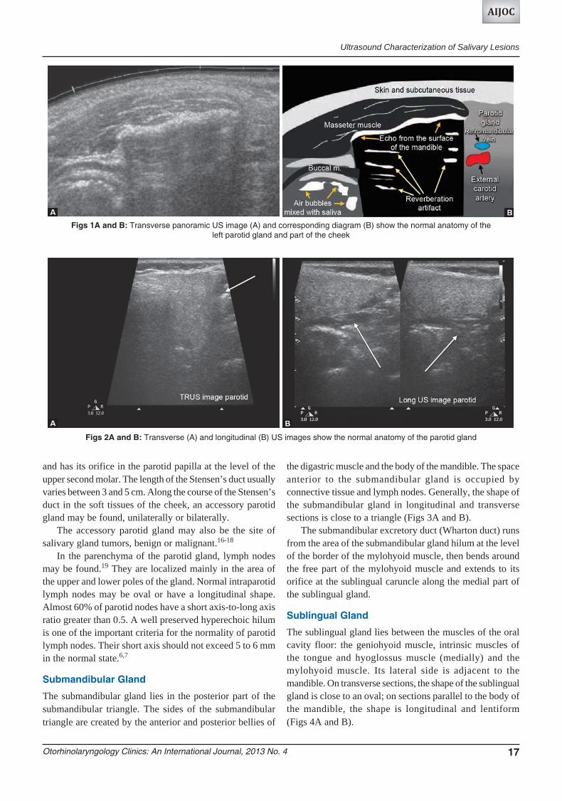
The parotid gland is located in the retromandibular fossa,anterior to the ear and sternocleidomastoid muscle. Parts ofthe superficial lobe cover the ramus of the mandible andthe posterior part of the masseter muscle. The borderbetween the superficial and deep parotid lobes is createdby a plane in which the facial nerve and its branches arelocated (Figs 1A and B). Branches of the facial nerve arenot visible at US. Parts of the trunk of this nerve may bedemonstrated only with high frequency probes (above 10MHz).13 Therefore, the retromandibular vein, which usuallylies directly above the trunk of the facial nerve,14,15 is usedas a US landmark separating the superficial and deep lobesof the parotid gland. The deep parotid lobe can be visualizedonly partially at US.
The echotexture of the major salivary glands, includingthe parotid gland, is generally homogeneous andhyperechoic in comparison to adjacent muscles. Theechogenicity of the parotid gland is based on the amount ofintraglandular fatty tissue (Figs 2A and B).
After leaving the parotid gland, the main excretory duct(Stensen’s duct) lies on the masseter muscle, about 1 cmbelow the zygomatic arch, then crosses the buccal muscle
Otorhinolaryngology Clinics: An International Journal, 2013 No. 4 17
Ultrasound Characterization of Salivary Lesions
AIJOC
and has its orifice in the parotid papilla at the level of theupper second molar. The length of the Stensen’s duct usuallyvaries between 3 and 5 cm. Along the course of the Stensen’sduct in the soft tissues of the cheek, an accessory parotidgland may be found, unilaterally or bilaterally.
The accessory parotid gland may also be the site ofsalivary gland tumors, benign or malignant.16-18
In the parenchyma of the parotid gland, lymph nodesmay be found.19 They are localized mainly in the area ofthe upper and lower poles of the gland. Normal intraparotidlymph nodes may be oval or have a longitudinal shape.Almost 60% of parotid nodes have a short axis-to-long axisratio greater than 0.5. A well preserved hyperechoic hilumis one of the important criteria for the normality of parotidlymph nodes. Their short axis should not exceed 5 to 6 mmin the normal state.6,7
Submandibular Gland
The submandibular gland lies in the posterior part of thesubmandibular triangle. The sides of the submandibulartriangle are created by the anterior and posterior bellies of
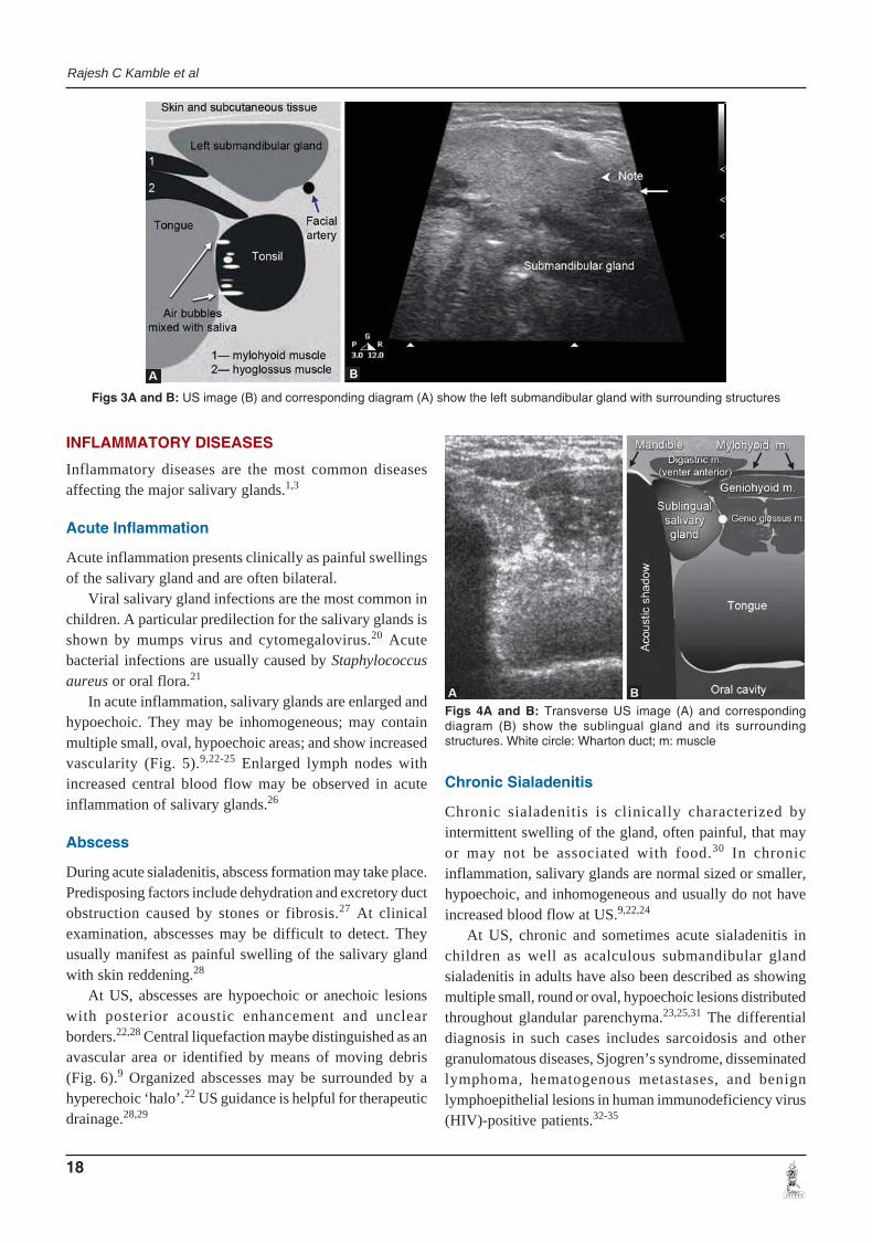
the digastric muscle and the body of the mandible. The spaceanterior to the submandibular gland is occupied byconnective tissue and lymph nodes. Generally, the shape ofthe submandibular gland in longitudinal and transversesections is close to a triangle (Figs 3A and B).
The submandibular excretory duct (Wharton duct) runsfrom the area of the submandibular gland hilum at the levelof the border of the mylohyoid muscle, then bends aroundthe free part of the mylohyoid muscle and extends to itsorifice at the sublingual caruncle along the medial part ofthe sublingual gland.
Sublingual Gland
The sublingual gland lies between the muscles of the oralcavity floor: the geniohyoid muscle, intrinsic muscles ofthe tongue and hyoglossus muscle (medially) and themylohyoid muscle. Its lateral side is adjacent to themandible. On transverse sections, the shape of the sublingualgland is close to an oval; on sections parallel to the body ofthe mandible, the shape is longitudinal and lentiform(Figs 4A and B).
Figs 1A and B: Transverse panoramic US image (A) and corresponding diagram (B) show the normal anatomy of theleft parotid gland and part of the cheek
Figs 2A and B: Transverse (A) and longitudinal (B) US images show the normal anatomy of the parotid gland
18
Rajesh C Kamble et al
Figs 3A and B: US image (B) and corresponding diagram (A) show the left submandibular gland with surrounding structures
Figs 4A and B: Transverse US image (A) and correspondingdiagram (B) show the sublingual gland and its surroundingstructures. White circle: Wharton duct; m: muscle
INFLAMMATORY DISEASES
Inflammatory diseases are the most common diseasesaffecting the major salivary glands.1,3
Acute Inflammation
Acute inflammation presents clinically as painful swellingsof the salivary gland and are often bilateral.
Viral salivary gland infections are the most common inchildren. A particular predilection for the salivary glands isshown by mumps virus and cytomegalovirus.20 Acutebacterial infections are usually caused by Staphylococcusaureus or oral flora.21
In acute inflammation, salivary glands are enlarged andhypoechoic. They may be inhomogeneous; may containmultiple small, oval, hypoechoic areas; and show increasedvascularity (Fig. 5).9,22-25 Enlarged lymph nodes withincreased central blood flow may be observed in acuteinflammation of salivary glands.26
Abscess
During acute sialadenitis, abscess formation may take place.Predisposing factors include dehydration and excretory ductobstruction caused by stones or fibrosis.27 At clinicalexamination, abscesses may be difficult to detect. Theyusually manifest as painful swelling of the salivary glandwith skin reddening.28
At US, abscesses are hypoechoic or anechoic lesionswith posterior acoustic enhancement and unclearborders.22,28 Central liquefaction maybe distinguished as anavascular area or identified by means of moving debris(Fig. 6).9 Organized abscesses may be surrounded by ahyperechoic ‘halo’.22 US guidance is helpful for therapeuticdrainage.28,29
Chronic Sialadenitis
Chronic sialadenitis is clinically characterized byintermittent swelling of the gland, often painful, that mayor may not be associated with food.30 In chronicinflammation, salivary glands are normal sized or smaller,hypoechoic, and inhomogeneous and usually do not haveincreased blood flow at US.9,22,24
At US, chronic and sometimes acute sialadenitis inchildren as well as acalculous submandibular glandsialadenitis in adults have also been described as showingmultiple small, round or oval, hypoechoic lesions distributedthroughout glandular parenchyma.23,25,31 The differentialdiagnosis in such cases includes sarcoidosis and othergranulomatous diseases, Sjogren’s syndrome, disseminatedlymphoma, hematogenous metastases, and benignlymphoepithelial lesions in human immunodeficiency virus(HIV)-positive patients.32-35

Otorhinolaryngology Clinics: An International Journal, 2013 No. 4 19
Ultrasound Characterization of Salivary Lesions
AIJOC
Chronic Sclerosing Sialadenitis
A special form of chronic sialadenitis that may mimic amalignant lesion, both clinically and at imaging, is chronicsclerosing sialadenitis (Kuttner’s tumor).36,37 In Kuttner’stumor, diffuse involvement of the salivary gland (usuallythe submandibular gland) may occur, with multiple smallhypoechoic foci scattered on a heterogeneous backgroundof salivary tissue visible at US.36 In all doubtful cases,verification with FNA/biopsy is recommended.38,39
GRANULOMATOUS SIALADENITIS
Granulomatous sialadenitis occurrence is rare.20 US featuresof granulomatous sialadenitis are nonspecific: single ormultiple hypoechoic areas in an enlarged or normally sizedgland or diffuse hypoechoic areas are noted.9,12,19,40-43
Mycobacterial disease of major salivary glands maymanifest as a salivary gland mass, clinically indistin-guishable from a neoplasm.44 In the parenchymal type oftuberculosis, Chou et al45 described focal, intraparotid,anechoic areas that might have a cavity or cavities withinthem. In necrotic caseous cavities, which appear veryhypoechoic, no color flow can be detected at US, in contrastto most salivary tumors.45 Salivary gland actinomycosis maymimic a malignant tumor at US; it may manifest as ahypoechoic area with ill-defined margins.46
LYMPH NODES IN SIALADENITIS
In acute or chronic inflammation, lymph nodes may beenlarged; however, their normal echo structure(homogeneous cortex and hyperechoic central hilum) ispreserved.
Sialolithiasis
Salivary stones are most often located in the submandibulargland (60-90% of cases) and may be multiple.47-50 Parotid
glands are affected in about 10 to 20% of cases.51 On classicradiographs, intraglandular and small stones may be missed,and only about 20% of sialoliths are radiopaque.52 CT allowsvisualization of large stones but without their preciselocalization and without the possibility of assessment ofthe ducts.53
The standard technique for imaging of the submandi-bular duct and the intraglandular ductal system remainsdigital sialography.54 A novel, noninvasive, promisingmethod appears to be MR sialography, which also givesvery good results in detection of sialoliths.2,54,55
US is a noninvasive method, well-established in casesof clinical suspicion of sialolithiasis, and is used as a primarymodality, particularly in Europe.51,54 Although some authorsclaim that sialoliths smaller than 2 to 3 mm may beoverlooked because of the absence of acoustic shadow, thesearticles are from the 1980s and currently used machineshave better resolution and detection possibilities.56,57
Sialolithiasis causes partial or total mechanical obstruction
Fig. 5: Color Doppler US image shows an acutely inflamed left submandibular gland (arrows).The gland is enlarged and hypoechoic and shows increased blood flow
Fig. 6: B-mode US image shows an enlarged parotid gland withareas of necrosis (arrows) suggestive of an early abscess formation
20
Rajesh C Kamble et al
of the salivary duct, which results in recurrent swelling of asalivary gland during eating and may be complicated bybacterial infection.20,48
Sialoliths in the distal part of the submandibular duct(Wharton duct) may be palpable in the floor of the mouth.However, sialoliths in the proximal portion of the duct orin the parenchyma of salivary glands may be demonstratedonly radiologically.
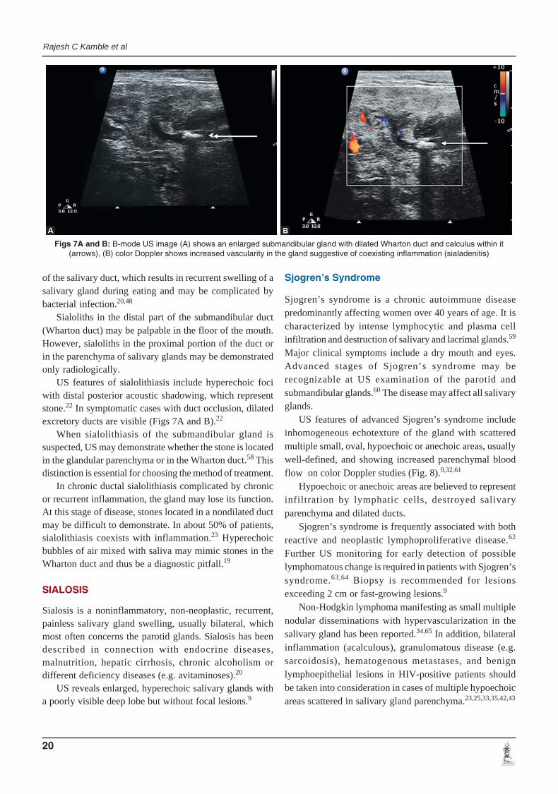
US features of sialolithiasis include hyperechoic fociwith distal posterior acoustic shadowing, which representstone.22 In symptomatic cases with duct occlusion, dilatedexcretory ducts are visible (Figs 7A and B).22
When sialolithiasis of the submandibular gland issuspected, US may demonstrate whether the stone is locatedin the glandular parenchyma or in the Wharton duct.58 Thisdistinction is essential for choosing the method of treatment.
In chronic ductal sialolithiasis complicated by chronicor recurrent inflammation, the gland may lose its function.At this stage of disease, stones located in a nondilated ductmay be difficult to demonstrate. In about 50% of patients,sialolithiasis coexists with inflammation.23 Hyperechoicbubbles of air mixed with saliva may mimic stones in theWharton duct and thus be a diagnostic pitfall.19
SIALOSIS
Sialosis is a noninflammatory, non-neoplastic, recurrent,painless salivary gland swelling, usually bilateral, whichmost often concerns the parotid glands. Sialosis has beendescribed in connection with endocrine diseases,malnutrition, hepatic cirrhosis, chronic alcoholism ordifferent deficiency diseases (e.g. avitaminoses).20
US reveals enlarged, hyperechoic salivary glands witha poorly visible deep lobe but without focal lesions.9
Sjogren’s Syndrome
Sjogren’s syndrome is a chronic autoimmune diseasepredominantly affecting women over 40 years of age. It ischaracterized by intense lymphocytic and plasma cellinfiltration and destruction of salivary and lacrimal glands.59
Major clinical symptoms include a dry mouth and eyes.Advanced stages of Sjogren’s syndrome may berecognizable at US examination of the parotid andsubmandibular glands.60 The disease may affect all salivaryglands.
US features of advanced Sjogren’s syndrome includeinhomogeneous echotexture of the gland with scatteredmultiple small, oval, hypoechoic or anechoic areas, usuallywell-defined, and showing increased parenchymal bloodflow on color Doppler studies (Fig. 8).9,32,61
Hypoechoic or anechoic areas are believed to representinfiltration by lymphatic cells, destroyed salivaryparenchyma and dilated ducts.
Sjogren’s syndrome is frequently associated with bothreactive and neoplastic lymphoproliferative disease.62
Further US monitoring for early detection of possiblelymphomatous change is required in patients with Sjogren’ssyndrome.63,64 Biopsy is recommended for lesionsexceeding 2 cm or fast-growing lesions.9
Non-Hodgkin lymphoma manifesting as small multiplenodular disseminations with hypervascularization in thesalivary gland has been reported.34,65 In addition, bilateralinflammation (acalculous), granulomatous disease (e.g.sarcoidosis), hematogenous metastases, and benignlymphoepithelial lesions in HIV-positive patients shouldbe taken into consideration in cases of multiple hypoechoicareas scattered in salivary gland parenchyma.23,25,33,35,42,43
Figs 7A and B: B-mode US image (A) shows an enlarged submandibular gland with dilated Wharton duct and calculus within it(arrows), (B) color Doppler shows increased vascularity in the gland suggestive of coexisting inflammation (sialadenitis)

Otorhinolaryngology Clinics: An International Journal, 2013 No. 4 21
Ultrasound Characterization of Salivary Lesions
AIJOC
NEOPLASMS
Salivary gland neoplasms are relatively rare. Most of themare benign (70-80%) and found in the parotid glands (80-90%). About 10 to 12% of all salivary gland neoplasms arelocated in the submandibular glands, but almost half of theseneoplasms may be malignant.3,66
Benign Neoplasms
The most common benign neoplasms of major salivaryglands are pleomorphic adenomas (mixed tumor) andWarthin tumors (adenolymphoma, cyst adenolymphoma,papillary cystadenoma lymphomatosum). Clinically, theymanifest as slow growing painless masses.67
Pleomorphic Adenoma
Pleomorphic adenomas occur most often in the parotid gland(60-90%) in people in the fourth and fifth decades of lifebut may arise at any age.3,66,68 There is a slight predominancein women.66
Pleomorphic adenomas are usually solitary andunilateral.3,66,68 They grow slowly and maybe asymptomatic.Nontreated pleomorphic adenomas may undergo malignanttransformation after decades.3,68 In exceptional cases,pleomorphic adenomas may be clinically aggressive; theymay metastasize and even be fatal.20,69,70
At US, pleomorphic adenomas are hypoechoic, well-defined, lobulated tumors with posterior acousticenhancement and may contain calcifications.71-73 The featureof lobulated shape is being emphasized in differentialdiagnosis (Figs 9A and B).73
Vascularization in pleomorphic adenomas is often pooror absent (even when the sensitive power Doppler mode isused) but may be abundant (Figs 9A and B).71,73,74 After
inadequate surgery, pleomorphic adenomas often recur,usually multifocally.75
Warthin Tumor
Warthin tumor is the next most common benign salivaryneoplasm (5-10% of all benign salivary neoplasms).66,68 Itarises most often in men in the fifth and sixth decades oflife.66,68,76 The relationship between smoking anddevelopment of Warthin tumors has been proved.67
Warthin tumor is usually solitary, unilateral, and slowgrowing. In about 10 to 60% of cases, tumors may occurbilaterally or multifocally, sometimes metachronously,growing and manifesting clinically at different times.3,68,77,78
Sporadically, the epithelial component of Warthin tumormay undergo malignant transformation.68,79
At US, Warthin tumors are oval, hypoechoic, well-defined tumors and often contain multiple anechoicareas.25,74,78,80 Warthin tumors are often hypervascularized(Fig. 10).
Diagnosis of a Warthin tumor may be supported byresults of technetium-99m scintigraphy, which reveals a‘hot’ tumor because of the increased uptake of the tracerby the tumor.81
Lobulated shape in pleomorphic adenomas and anechoicareas in Warthin tumors, although common, are notpathognomonic and may be found in many other lesions,including malignancies.73,74
Other Benign Tumors
Other benign tumors (e.g. oncocytoma, basal cell adenoma)occur less frequently in the salivary glands. Their differen-tiation is not possible with US. Among nonepithelial lesions,hemangiomas, lipomas and neurinomas or schwannomas(Fig. 11) may be found in salivary glands.12,87-91
Hemangiomas, the most frequent tumors in infants, maymanifest as heterogeneous lesions with sinusoidal spaces andcalcifications representing phleboliths (Figs 12A and B).88
Hemangioendothelioma (HAE) of the parotid gland isa benign tumor that presents in infancy as a large, rapidlygrowing mass that is either small or not noticed at birth,but becomes apparent soon after88 (Fig. 13).
Lipomas are usually oval and hypoechoic with sharpmargins and hyperechoic linear structures regularlydistributed within the lesion in a striated or featheredpattern.9,92
MALIGNANT NEOPLASMS
The most common malignant neoplasms occurring insalivary glands are mucoepidermoid carcinoma and adenoidcystic carcinoma.94 Squamous cell carcinoma, acinic cellcarcinoma, and adenocarcinoma are less common.
Fig. 8: B-mode US image shows Sjogren’s syndrome in the parotidgland. The gland has an inhomogeneous structure with multiplesmall, oval, hypoechoic areas (arrows)
22
Rajesh C Kamble et al
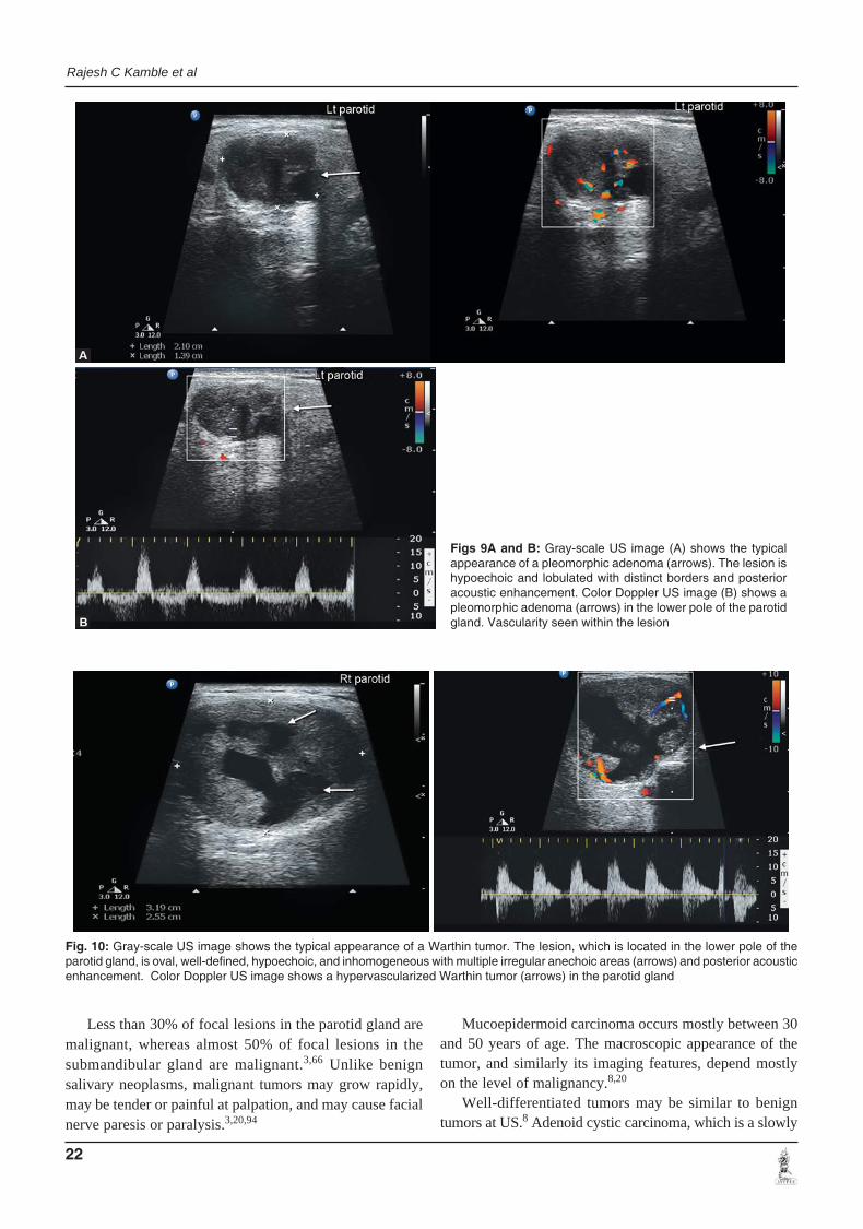
Fig. 10: Gray-scale US image shows the typical appearance of a Warthin tumor. The lesion, which is located in the lower pole of theparotid gland, is oval, well-defined, hypoechoic, and inhomogeneous with multiple irregular anechoic areas (arrows) and posterior acousticenhancement. Color Doppler US image shows a hypervascularized Warthin tumor (arrows) in the parotid gland
Less than 30% of focal lesions in the parotid gland aremalignant, whereas almost 50% of focal lesions in thesubmandibular gland are malignant.3,66 Unlike benignsalivary neoplasms, malignant tumors may grow rapidly,may be tender or painful at palpation, and may cause facialnerve paresis or paralysis.3,20,94
Mucoepidermoid carcinoma occurs mostly between 30and 50 years of age. The macroscopic appearance of thetumor, and similarly its imaging features, depend mostlyon the level of malignancy.8,20
Well-differentiated tumors may be similar to benigntumors at US.8 Adenoid cystic carcinoma, which is a slowly
Figs 9A and B: Gray-scale US image (A) shows the typicalappearance of a pleomorphic adenoma (arrows). The lesion ishypoechoic and lobulated with distinct borders and posterioracoustic enhancement. Color Doppler US image (B) shows apleomorphic adenoma (arrows) in the lower pole of the parotidgland. Vascularity seen within the lesion
Otorhinolaryngology Clinics: An International Journal, 2013 No. 4 23
Ultrasound Characterization of Salivary Lesions
AIJOC
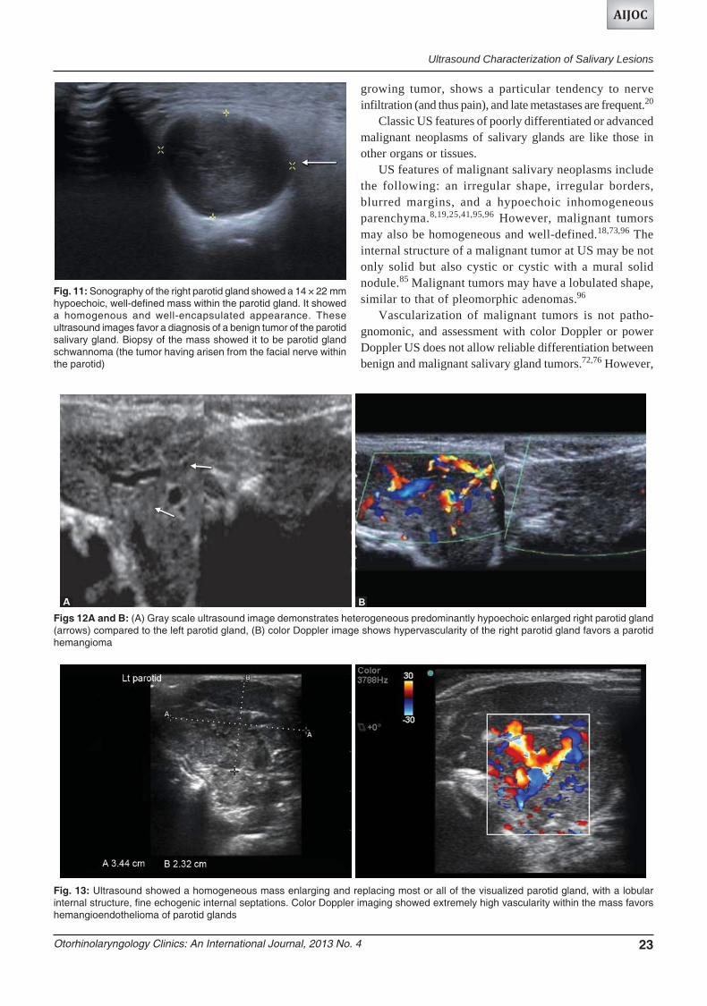
Fig. 11: Sonography of the right parotid gland showed a 14 × 22 mmhypoechoic, well-defined mass within the parotid gland. It showeda homogenous and well-encapsulated appearance. Theseultrasound images favor a diagnosis of a benign tumor of the parotidsalivary gland. Biopsy of the mass showed it to be parotid glandschwannoma (the tumor having arisen from the facial nerve withinthe parotid)
Figs 12A and B: (A) Gray scale ultrasound image demonstrates heterogeneous predominantly hypoechoic enlarged right parotid gland(arrows) compared to the left parotid gland, (B) color Doppler image shows hypervascularity of the right parotid gland favors a parotidhemangioma
Fig. 13: Ultrasound showed a homogeneous mass enlarging and replacing most or all of the visualized parotid gland, with a lobularinternal structure, fine echogenic internal septations. Color Doppler imaging showed extremely high vascularity within the mass favorshemangioendothelioma of parotid glands
growing tumor, shows a particular tendency to nerveinfiltration (and thus pain), and late metastases are frequent.20
Classic US features of poorly differentiated or advancedmalignant neoplasms of salivary glands are like those inother organs or tissues.
US features of malignant salivary neoplasms includethe following: an irregular shape, irregular borders,blurred margins, and a hypoechoic inhomogeneousparenchyma.8,19,25,41,95,96 However, malignant tumorsmay also be homogeneous and well-defined.18,73,96 Theinternal structure of a malignant tumor at US may be notonly solid but also cystic or cystic with a mural solidnodule.85 Malignant tumors may have a lobulated shape,similar to that of pleomorphic adenomas.96
Vascularization of malignant tumors is not patho-gnomonic, and assessment with color Doppler or powerDoppler US does not allow reliable differentiation betweenbenign and malignant salivary gland tumors.72,76 However,

24
Rajesh C Kamble et al
Schick et al72 report that high vascularization and highsystolic peak flow velocity should raise the suspicion ofmalignancy (Figs 14A and B). Bradley et al97 concludedthat tumors demonstrating an increased intratumoralvascular resistance index have an increased risk ofmalignancy.
The presence of metastatic-appearing lymph nodesaccompanying a tumor in the salivary gland stronglysuggests a malignancy.
METASTASES
Salivary glands are very uncommon sites of metastases.Primary tumors metastasizing to salivary glands may belocated in the head and neck region, as well as in moredistant parts of the body. Melanoma spinocellular cancer,breast cancer, and lung cancer may produce metastases tointraparotid lymph nodes.99-103 Extremely rare aremetastases from renal cancer.100,104-106
At US, metastases may be well-defined and oval.9 Itmay be difficult to differentiate multiple metastatic lesionsfrom some patterns of inflammation, Sjogren’s syndromeand granulomatous disease at US.23,25,33,42,43
LYMPHOMA
Salivary glands may also be affected by lymphoma.34
However, primary involvement of salivary glands is rare;they are usually one of the sites of systemic disease.
Clinically, salivary lymphomas most often manifest asa painless, progressive swelling.107,108 They are usuallyassociated with autoimmune disease, most often withSjogren’s syndrome, sometimes also with rheumatoidarthritis.62-64,109
At US of cases of lymphoma in the salivary gland, onemay observe a solitary, hypoechoic, homogeneous orinhomogeneous lesion, which is oval or lobulated or has
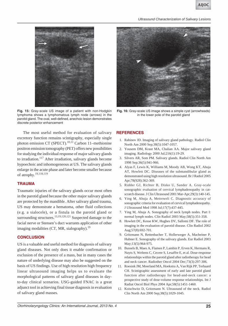
irregular margins and sometimes contains echogenic septaor stripes (Fig. 15).34,110,111 However, these features are notpathognomonic, and lymphoma may not be reliablydifferentiated from other neoplastic or non-neoplasticsalivary gland tumors with US.
A pattern of multiple hypoechoic lesions with increasedblood flow may also be seen.34,111 Such a pattern requiresdifferentiation from inflammation, Sjogren’s syndrome,granulomatous disease (e.g. sarcoidosis), and hematogenousmetastases.23,25,32,33,42,43
In cases of lymphoma, solitary or multiple salivary glandlesions sometimes associated with microcysts may beobserved at CT or MR imaging.63,109
CYSTS
Simple cysts are uncommon in salivary glands. They maybe congenital or acquired. Some acquired cysts develop dueto obstruction of the salivary ducts in the presence of atumor, stones, or inflammation.3 Clinically, they usuallymanifest as a painless swelling but may be tender wheninfected.41
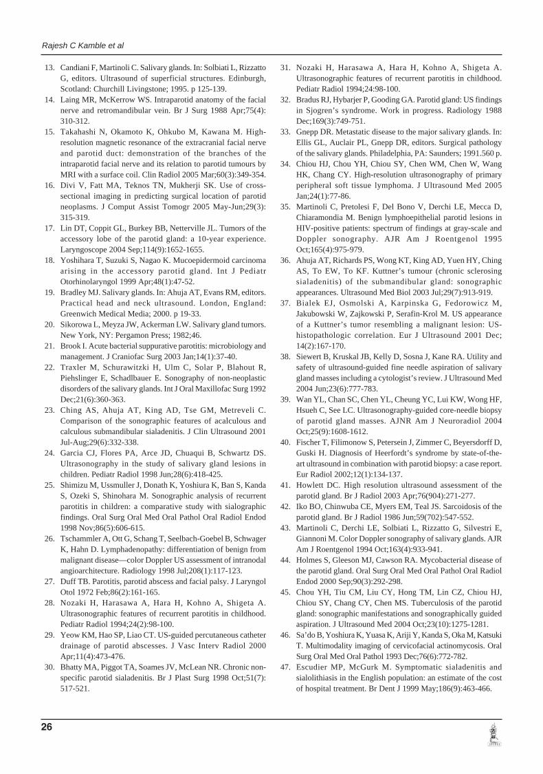
US features of a cyst are well-defined margins, anechoiccontent, posterior acoustic enhancement and avascular atpower Doppler or color Doppler imaging (Fig. 16).22
Benign lymphoepithelial lesions in HIV-positive patientsmay manifest as multiple cysts.35
EFFECTS OF IRRADIATION
The major salivary glands are often irradiated duringradiation therapy of head and neck neoplasms. A majoradverse effect of such treatment is xerostomia caused byfunctional and structural impairment of salivaryparenchyma.11,115 Loss of salivary gland function signifi-cantly diminishes the patient’s quality of life.116
Figs 14A and B: Panoramic gray-scale US image shows an acinic cell carcinoma (arrow) in the left parotid gland. The tumor is well-definedand has regular margins (A) color Doppler reveals increased vascularity within the mass lesion with high peak systolic flow noted (B)
Otorhinolaryngology Clinics: An International Journal, 2013 No. 4 25
Ultrasound Characterization of Salivary Lesions
AIJOC
The most useful method for evaluation of salivaryexcretory function remains scintigraphy, especially singlephoton emission CT (SPECT).10,11 Carbon 11–methioninepositron emission tomography (PET) offers new possibilitiesfor studying the individual response of major salivary glandsto irradiation.117 After irradiation, salivary glands becomehypoechoic and inhomogeneous at US. The salivary glandsenlarge in the acute phase and later become smaller becauseof atrophy.19,118,119
TRAUMA
Traumatic injuries of the salivary glands occur most oftenin the parotid gland because the other major salivary glandsare protected by the mandible. After salivary gland trauma,US may demonstrate a hematoma, other fluid collections(e.g. a sialocele), or a fistula in the parotid gland orsurrounding structures.13,19,120,121 Suspected damage to thefacial nerve or Stensen’s duct warrants application of otherimaging modalities (CT, MR, sialography).19
CONCLUSION
US is a valuable and useful method for diagnosis of salivarygland diseases. Not only does it enable confirmation orexclusion of the presence of a mass, but in many cases thenature of underlying disease may also be suggested on thebasis of US findings. Use of high resolution high frequencylinear ultrasound imaging helps us to evaluate themorphological patterns of salivary gland diseases in day-to-day clinical scenarios. USG-guided FNAC is a greatadjunct tool in achieving final tissue diagnosis in evaluationof salivary gland masses.
REFERENCES
1. Rabinov JD. Imaging of salivary gland pathology. Radiol ClinNorth Am 2000 Sep;38(5):1047-1057.
2. Yousem DM, Kraut MA, Chalian AA. Major salivary glandimaging. Radiology 2000 Jul;216(1):19-29.
3. Silvers AR, Som PM. Salivary glands. Radiol Clin North Am1998 Sep;36(5):941-966.
4. Alyas F, Lewis K, Williams M, Moody AB, Wong KT, AhujaAT, Howlett DC. Diseases of the submandibular gland asdemonstrated using high resolution ultrasound. Br J Radiol 2005Apr;78(928):362-369.
5. Ridder GJ, Richter B, Disko U, Sander A. Gray-scalesonographic evaluation of cervical lymphadenopathy in cat-scratch disease. J Clin Ultrasound 2001 Mar-Apr;29(3):140-145.
6. Ying M, Ahuja A, Metreweli C. Diagnostic accuracy ofsonographic criteria for evaluation of cervical lymphadenopathy.J Ultrasound Med 1998 Jul;17(7):437-445.
7. Ying M, Ahuja A. Sonography of neck lymph nodes. Part I:normal lymph nodes. Clin Radiol 2003 May;58(5):351-358.
8. Howlett DC, Kesse KW, Hughes DV, Sallomi DF. The role ofimaging in the evaluation of parotid disease. Clin Radiol 2002Aug;57(8):692-701.
9. Gritzmann N, Rettenbacher T, Hollerweger A, Macheiner P,Hubner E. Sonography of the salivary glands. Eur Radiol 2003May;13(5):964-975.
10. Bussels B, Maes A, Flamen P, Lambin P, Erven K, Hermans R,Nuyts S, Weltens C, Cecere S, Lesaffre E, et al. Dose-responserelationships within the parotid gland after radiotherapy for headand neck cancer. Radiother Oncol 2004 Dec;73(3):297-306.
11. Roesink JM, Moerland MA, Hoekstra A, Van Rijk PP, TerhaardCH. Scintigraphic assessment of early and late parotid glandfunction after radiotherapy for head-and-neck cancer: aprospective study of dose-volume response relationships. Int JRadiat Oncol Biol Phys 2004 Apr;58(5):1451-1460.
12. Koischwitz D, Gritzmann N. Ultrasound of the neck. RadiolClin North Am 2000 Sep;38(5):1029-1045.
Fig. 15: Gray-scale US image of a patient with non-Hodgkinlymphoma shows a lymphomatous lymph node (arrows) in theparotid gland. The oval, well-defined, anechoic lesion demonstratesdiscrete posterior enhancement
Fig. 16: Gray-scale US image shows a simple cyst (arrowheads)in the lower pole of the parotid gland
26
Rajesh C Kamble et al
13. Candiani F, Martinoli C. Salivary glands. In: Solbiati L, RizzattoG, editors. Ultrasound of superficial structures. Edinburgh,Scotland: Churchill Livingstone; 1995. p 125-139.
14. Laing MR, McKerrow WS. Intraparotid anatomy of the facialnerve and retromandibular vein. Br J Surg 1988 Apr;75(4):310-312.
15. Takahashi N, Okamoto K, Ohkubo M, Kawana M. High-resolution magnetic resonance of the extracranial facial nerveand parotid duct: demonstration of the branches of theintraparotid facial nerve and its relation to parotid tumours byMRI with a surface coil. Clin Radiol 2005 Mar;60(3):349-354.
16. Divi V, Fatt MA, Teknos TN, Mukherji SK. Use of cross-sectional imaging in predicting surgical location of parotidneoplasms. J Comput Assist Tomogr 2005 May-Jun;29(3):315-319.
17. Lin DT, Coppit GL, Burkey BB, Netterville JL. Tumors of theaccessory lobe of the parotid gland: a 10-year experience.Laryngoscope 2004 Sep;114(9):1652-1655.
18. Yoshihara T, Suzuki S, Nagao K. Mucoepidermoid carcinomaarising in the accessory parotid gland. Int J PediatrOtorhinolaryngol 1999 Apr;48(1):47-52.
19. Bradley MJ. Salivary glands. In: Ahuja AT, Evans RM, editors.Practical head and neck ultrasound. London, England:Greenwich Medical Media; 2000. p 19-33.
20. Sikorowa L, Meyza JW, Ackerman LW. Salivary gland tumors.New York, NY: Pergamon Press; 1982;46.
21. Brook I. Acute bacterial suppurative parotitis: microbiology andmanagement. J Craniofac Surg 2003 Jan;14(1):37-40.
22. Traxler M, Schurawitzki H, Ulm C, Solar P, Blahout R,Piehslinger E, Schadlbauer E. Sonography of non-neoplasticdisorders of the salivary glands. Int J Oral Maxillofac Surg 1992Dec;21(6):360-363.
23. Ching AS, Ahuja AT, King AD, Tse GM, Metreveli C.Comparison of the sonographic features of acalculous andcalculous submandibular sialadenitis. J Clin Ultrasound 2001Jul-Aug;29(6):332-338.
24. Garcia CJ, Flores PA, Arce JD, Chuaqui B, Schwartz DS.Ultrasonography in the study of salivary gland lesions inchildren. Pediatr Radiol 1998 Jun;28(6):418-425.
25. Shimizu M, Ussmuller J, Donath K, Yoshiura K, Ban S, KandaS, Ozeki S, Shinohara M. Sonographic analysis of recurrentparotitis in children: a comparative study with sialographicfindings. Oral Surg Oral Med Oral Pathol Oral Radiol Endod1998 Nov;86(5):606-615.
26. Tschammler A, Ott G, Schang T, Seelbach-Goebel B, SchwagerK, Hahn D. Lymphadenopathy: differentiation of benign frommalignant disease—color Doppler US assessment of intranodalangioarchitecture. Radiology 1998 Jul;208(1):117-123.
27. Duff TB. Parotitis, parotid abscess and facial palsy. J LaryngolOtol 1972 Feb;86(2):161-165.
28. Nozaki H, Harasawa A, Hara H, Kohno A, Shigeta A.Ultrasonographic features of recurrent parotitis in childhood.Pediatr Radiol 1994;24(2):98-100.
29. Yeow KM, Hao SP, Liao CT. US-guided percutaneous catheterdrainage of parotid abscesses. J Vasc Interv Radiol 2000Apr;11(4):473-476.
30. Bhatty MA, Piggot TA, Soames JV, McLean NR. Chronic non-specific parotid sialadenitis. Br J Plast Surg 1998 Oct;51(7):517-521.
31. Nozaki H, Harasawa A, Hara H, Kohno A, Shigeta A.Ultrasonographic features of recurrent parotitis in childhood.Pediatr Radiol 1994;24:98-100.
32. Bradus RJ, Hybarjer P, Gooding GA. Parotid gland: US findingsin Sjogren’s syndrome. Work in progress. Radiology 1988Dec;169(3):749-751.
33. Gnepp DR. Metastatic disease to the major salivary glands. In:Ellis GL, Auclair PL, Gnepp DR, editors. Surgical pathologyof the salivary glands. Philadelphia, PA: Saunders; 1991.560 p.
34. Chiou HJ, Chou YH, Chiou SY, Chen WM, Chen W, WangHK, Chang CY. High-resolution ultrasonography of primaryperipheral soft tissue lymphoma. J Ultrasound Med 2005Jan;24(1):77-86.
35. Martinoli C, Pretolesi F, Del Bono V, Derchi LE, Mecca D,Chiaramondia M. Benign lymphoepithelial parotid lesions inHIV-positive patients: spectrum of findings at gray-scale andDoppler sonography. AJR Am J Roentgenol 1995Oct;165(4):975-979.
36. Ahuja AT, Richards PS, Wong KT, King AD, Yuen HY, ChingAS, To EW, To KF. Kuttner’s tumour (chronic sclerosingsialadenitis) of the submandibular gland: sonographicappearances. Ultrasound Med Biol 2003 Jul;29(7):913-919.
37. Bialek EJ, Osmolski A, Karpinska G, Fedorowicz M,Jakubowski W, Zajkowski P, Serafin-Krol M. US appearanceof a Kuttner’s tumor resembling a malignant lesion: US-histopathologic correlation. Eur J Ultrasound 2001 Dec;14(2):167-170.
38. Siewert B, Kruskal JB, Kelly D, Sosna J, Kane RA. Utility andsafety of ultrasound-guided fine needle aspiration of salivarygland masses including a cytologist’s review. J Ultrasound Med2004 Jun;23(6):777-783.
39. Wan YL, Chan SC, Chen YL, Cheung YC, Lui KW, Wong HF,Hsueh C, See LC. Ultrasonography-guided core-needle biopsyof parotid gland masses. AJNR Am J Neuroradiol 2004Oct;25(9):1608-1612.
40. Fischer T, Filimonow S, Petersein J, Zimmer C, Beyersdorff D,Guski H. Diagnosis of Heerfordt’s syndrome by state-of-the-art ultrasound in combination with parotid biopsy: a case report.Eur Radiol 2002;12(1):134-137.
41. Howlett DC. High resolution ultrasound assessment of theparotid gland. Br J Radiol 2003 Apr;76(904):271-277.
42. Iko BO, Chinwuba CE, Myers EM, Teal JS. Sarcoidosis of theparotid gland. Br J Radiol 1986 Jun;59(702):547-552.
43. Martinoli C, Derchi LE, Solbiati L, Rizzatto G, Silvestri E,Giannoni M. Color Doppler sonography of salivary glands. AJRAm J Roentgenol 1994 Oct;163(4):933-941.
44. Holmes S, Gleeson MJ, Cawson RA. Mycobacterial disease ofthe parotid gland. Oral Surg Oral Med Oral Pathol Oral RadiolEndod 2000 Sep;90(3):292-298.
45. Chou YH, Tiu CM, Liu CY, Hong TM, Lin CZ, Chiou HJ,Chiou SY, Chang CY, Chen MS. Tuberculosis of the parotidgland: sonographic manifestations and sonographically guidedaspiration. J Ultrasound Med 2004 Oct;23(10):1275-1281.
46. Sa’do B, Yoshiura K, Yuasa K, Ariji Y, Kanda S, Oka M, KatsukiT. Multimodality imaging of cervicofacial actinomycosis. OralSurg Oral Med Oral Pathol 1993 Dec;76(6):772-782.
47. Escudier MP, McGurk M. Symptomatic sialadenitis andsialolithiasis in the English population: an estimate of the costof hospital treatment. Br Dent J 1999 May;186(9):463-466.
Otorhinolaryngology Clinics: An International Journal, 2013 No. 4 27
Ultrasound Characterization of Salivary Lesions
AIJOC
48. Lustmann J, Regev E, Melamed Y. Sialolithiasis: a survey on245 patients and a review of the literature. Int J Oral MaxillofacSurg 1990 Jun;19(3):135-138.
49. Marchal F, Dulgerov P, Becker M, Barki G, Disant F, LehmannW. Specificity of parotid sialendoscopy. Laryngoscope 2001Feb;111(2):264-271.
50. Marchal F, Dulgerov P, Becker M, Barki G, Disant F, LehmannW. Submandibular diagnostic and interventional sialendoscopy:new procedure for ductal disorders. Ann Otol Rhinol Laryngol2002 Jan;111(1):27-35.
51. Yoshimura Y, Inone Y, Odagana T. Sonographic evaluation ofsialolithiasis. J Oral Maxillofac Surg 1989;47:907-912.
52. Rauch S, Gorlin RJ. Disease of the salivary glands. In: GorlinRJ, Goldmann HM, editors. Thomas’ oral pathology. St Louis,Mo: Mosby; 1970. p 997-1003.
53. Avrahami E, Englender M, Chen E, Shabaty D, Katz R, HarellM. CT of submandibular gland sialolithiasis. Neuroradiology1996 Apr;38(3):287-290.
54. Jager L, Menauer F, Holzknecht N, Scholz V, Grevers G, ReiserM. Sialolithiasis: MR sialography of the submandibular duct—an alternative to conventional sialography and US? Radiology2000 Sep;216(3):665-671.
55. Becker M, Marchal F, Becker CD, Dulguerov P,Georgakopoulos G, Lehmann W, Terrier F. Sialolithiasis andsalivary ductal stenosis: diagnostic accuracy of MR sialographywith a three-dimensional extended-phase conjugate-symmetryrapid spinecho sequence. Radiology 2000 Nov;217(2):347-358.
56. Gritzmann N. Sonography of the salivary glands. AJR Am JRoentgenol 1989 Jul;53(1):161-166.
57. Rinast E, Gmelin E, Hollands-Thorn B. Digital subtractionsialography, conventional sialography, high-resolutionultrasonography and computed tomography in the diagnosis ofsalivary gland diseases. Eur J Radiol 1989 Nov;9(4):224-230.
58. Iro H, Schneider T, Nitsche N, Waitz G, Marienhagen J, Ell C.Extracorporeal shock wave lithotripsy of a salivary stone. DtschMed Wochenschr 1990 Jan;115(1):12-14.
59. Kumar V, Cotran RS, Robbins SL. Disorders of the immunesystem. In: Kumar V, editor. Basic pathology. 6th ed.Philadelphia, PA: Saunders; 1997. p 111-112.
60. Makula E, Pokorny G, Kiss M, Vörös E, Kovács L, Kovács A,Csernay L, Palkó A. The place of magnetic resonance andultrasonographic examinations of the parotid gland in thediagnosis and follow-up of primary Sjogren’s syndrome.Rheumatology (Oxford) 2000 Jan;39(1):97-104.
61. Niemela RK, Takalo R, Paakko E, Suramo I, Päivänsalo M,Salo T, Hakala M. Ultrasonography of salivary glands in primarySjogren’s syndrome: a comparison with magnetic resonanceimaging and magnetic resonance sialography of parotid glands.Rheumatology (Oxford) 2004 Jul;43(7):875-879.
62. McCurley TL, Collins RD, Ball E, Collins RD. Nodal andextranodal lymphoproliferative disorders in Sjogren’s syndrome:a clinical and immunopathologic study. Hum Pathol 1990May;21(5):482-492.
63. Tonami H, Matoba M, Kuginuki Y, Yokota H, Higashi K,Yamamoto I, Sugai S. Clinical and imaging findings oflymphoma in patients with Sjogren’s syndrome. J Comput AssistTomogr 2003 Jul-Aug;27(4):517-524.
64. Masaki Y, Sugai S. Lymphoproliferative disorders in Sjogren’ssyndrome. Autoimmun Rev 2004 Mar;3(3):175-182.
65. Matsushita T, Takashima S, Takayama F, Momose M, Wang J,Ishiyama T. Sonographic detection of secondary MALTlymphoma of the submandibular gland. J Clin Ultrasound 2001Oct;29(8):462-465.
66. Renehan A, Gleave EN, Hancock BD, Smith P, McGurk M.Long-term follow-up of over 1000 patients with salivary glandtumours treated in a single centre. Br J Surg 1996 Dec;83(12):1750-1754.
67. Yoo GH, Eisele DW, Askin FB, Driben JS, Johns ME. Warthin’stumor: a 40-year experience at the Johns Hopkins Hospital.Laryngoscope 1994 Jul;104(7):799-803.
68. Ellis GL, Auclair PL, Gnepp DR, editors. Surgical pathologyof the salivary glands. Philadelphia, PA: Saunders 1991:10-25.7.
69. Joe VQ, Westesson PL. Tumors of the parotid gland: MRimaging characteristics of various histologic types. Am JRoentgenol 1994 Aug;163(2):433-438.
70. Klijanienko J, El-Naggar AK, Servois V, Rodriguez J, ValidireP, Vielh P. Clinically aggressive metastasizing pleomorphicadenoma: report of two cases. Head Neck 1997 Oct;19(7):629-633.
71. Bialek EJ, Jakubowski W, Karpinska G. Role of ultrasonographyin diagnosis and differentiation of pleomorphic adenomas: workin progress. Arch Otolaryngol Head Neck Surg 2003Sep;129(9):929-933.
72. Schick S, Steiner E, Gahleitner A, Böhm P, Helbich T, Ba-Ssalamah A, Mostbeck G. Differentiation of benign andmalignant tumors of the parotid gland: value of pulsed Dopplerand color Doppler sonography. Eur Radiol 1998;8(8):1462-1467.
73. Shimizu M, Ussmuller J, Hartwein J, Donath K, Kinukawa N.Statistical study for sonographic differential diagnosis oftumorous lesions in the parotid gland. Oral Surg Oral Med OralPathol Oral Radiol Endod 1999 Aug;88(2):226-233.
74. Zajkowski P, Jakubowski W, Bia³ek EJ, Wysocki M, OsmólskiA, Serafin-Król M. Pleomorphic adenoma and adenolymphomain ultrasonography. Eur J Ultrasound 2000 Sep;12(1):23-29.
75. Laskawi R, Schott T, Schröder M. Recurrent pleomorphicadenomas of the parotid gland: clinical evaluation and long-term follow-up. Br J Oral Maxillofac Surg 1998 Feb;36(1):48-51.
76. Dumitriu D, Dudea SM, Botar-Jid C, B|ciuÛ G. Ultrasonographicand sonoelastographic features of pleomorphic adenomas of thesalivary glands. Med Ultrason 2010 Sep;12(3):175-183.
77. Zajkowski P, Jakubowski W, Bia?ek EJ, Wysocki M, OsmólskiA, Serafin-Król M. Pleomorphic adenoma and adenolymphomain ultrasonography.Eur J Ultrasound 2000 Sep;12(1):23-29.
78. Yu GY, Ma DQ, Zhang Y, Peng X, Cai ZG, Gao Y, Chen Y.Multiple primary tumours of the parotid gland. Int J OralMaxillofac Surg 2004 Sep;33(6):531-534.
79. Podlesák T, Dolecková V, Sibl O. Malignancy of a cystadeno-lymphoma of the parotid gland. Eur Arch Otorhinolaryngol1992;249(4):233-235.
80. Kim J, Kim EK, Park CS, Choi YS, Kim YH, Choi EC.Characteristic sonographic findings of Warthin’s tumor in theparotid gland. J Clin Ultrasound 2004 Feb;32(2):78-81.
81. Canbay AE, Knorz S, Heimann KD, Hildmann H, Tiedjen KU.Sonography and scintigraphy in the diagnosis ofcystadenolymphomas (warthin tumor). Laryngorhinootologie2002 Nov;81(11):815-819.
28
Rajesh C Kamble et al
82. Auclair PL, Ellis GL. Mucoepidermoid carcinoma. In: Ellis GL,Auclair PL, Gnepp DR, editors. Surgical pathology of thesalivary glands. Philadelphia, PA: Saunders; 1991. p 269-298.
83. Khadaroo RG, Walton JM, Ramsay JA, Hicks MJ, ArchibaldSC. Mucoepidermoid carcinoma of the parotid gland: a rarepresentation in a young child. J Pediatr Surg 1998 Jun;33(6):893-895.
84. Nishimura T, Furukawa M, Kawahara E. Pleomorphic adenomaof parotid gland with cystic degeneration. J Laryngol Otol 1994May;108(5):446-448.
85. Suh SI, Seol HY, Kim TK, Lee NJ, Kim JH, Kim KA, Woo JS,Lee JH. Acinic cell carcinoma of the head and neck: radiologic-pathologic correlation. J Comput Assist Tomogr 2005 Jan-Feb;29(1):121-126.
86. Takeshita T, Tanaka H, Harasawa A, Kaminaga T, Imamura T,Furui S. Benign pleomorphic adenoma with extensive cysticdegeneration: unusual MR findings in two cases. Radiat Med2004 Sep-Oct;22(5):357-361.
87. Wu CW, Chi HP, Chiang FY, Hsu YC, Chan LP, Kuo WR.Giant lipoma arising from deep lobe of the parotid gland. WorldJ Surg Oncol 2006 Jun;4:28.
88. Wong KT, Ahuja AT, King AD, Yuen EH, Yu SC. Vascularlesions of parotid gland in adult patients: diagnosis with high-resolution ultrasound and MRI. Br J Radiol 2004Jul;77(919):600-606.
89. Chong KW, Chung YF, Khoo ML, Lim DT, Hong GS, SooKC. Management of intraparotid facial nerve schwannomas.Aust N Z J Surg 2000 Oct;70(10):732-734.
90. Hehar SS, Dugar J, Sharp J. The changing faces of a parotidmass. J Laryngol Otol 1999 Oct;113(10):938-941.
91. Oncel S, Onal K, Ermete M, Uluc E. Schwannoma(neurilemmoma) of the facial nerve presenting as a parotid mass.J Laryngol Otol 2002 Aug;116(8):642-643.
92. Chikui T, Yonetsu K, Yoshiura K, Miwa K, Kanda S, Ozeki S,Shinohara M. Imaging findings of lipomas in the orofacial regionwith CT, US and MRI. Oral Surg Oral Med Oral Pathol OralRadiol Endod 1997 Jul;84(1):88-95.
93. Roebuck DJ, Ahuja AT. Hemangioendothelioma of the parotidgland in infants: sonography and correlative MR imaging. AJNRAm J Neuroradiol 2000 Jan;21(1):219-223.
94. Bruneton JN, Caramella E, Roux P, Fenart D, Manzino JJ.Comparison of ultrasonographic and histological findings formultinodular lesions of the salivary glands. Eur J Radiol 1985Nov;5(4):295-296.
95. Goto TK, Yoshiura K, Nakayama E, Yuasa K, Tabata O, NakanoT, Kawazu T, Tanaka T, Miwa K, Shimizu M, et al. Thecombined use of US and MR imaging for the diagnosis of massesin the parotid region. Acta Radiol 2001 Jan;42(1):88-95.
96. Hardee PS, Carter JL, Piper KM, Ng SY. Metachronous bilateralprimary adenocarcinoma of the submandibular glands. Oral SurgOral Med Oral Pathol Oral Radiol Endod 2001 Apr;91(4):455-461.
97. Bradley MJ, Durham LH, Lancer JM. The role of colour flowDoppler in the investigation of the salivary gland tumour. ClinRadiol 2000 Oct;55(10):759-762.
98. Eneroth CM, Hamberger CA, Jakobsson PA. Malignancy ofacinic cell carcinoma. Ann Otol Rhinol Laryngol 1966Sep;75(3):780-792.
99. Wittich GR, Scheible WF, Haget PC. Ultrasonography of thesalivary glands. Radiol Clin North Am 1985 Mar;23(1):29-37.
100. Malata CM, Camilleri IG, McLean NR, Piggott TA, SoamesJV. Metastatic tumours of the parotid gland. Br J Oral MaxillofacSurg 1998 Jun;36(3):190-195.
101. Pisani P, Krengli M, Ramponi A, Guglielmetti R, Pia F.Metastases to parotid gland from cancers of the upper airwayand digestive tract. Br J Oral Maxillofac Surg 1998 Feb;36(1):54-57.
102. Raut V, Sinnathuray AR, Primrose WJ. Aggressive treatmentof metastasis to the parotid. Ulster Med J 2004 Nov;73(2):85-88.
103. Seifert G, Hennign SK, Caselitz J. Metastatic tumours to theparotid and submandibular glands. Pathol Res Pract 1986Dec;181(6):684-694.
104. Gogus C, Kilic O, Tulunay O, Tulunay O, Beduk Y. Solitarymetastasis of renal cell carcinoma to the parotid gland 10 yearsafter radical nephrectomy. Int J Urol 2004 Oct;11(10):894-896.
105. Park YW, Hlivko TJ. Parotid gland metastasis from renal cellcarcinoma. Laryngoscope 2002 Mar;112(3):453-456.
106. Seijas BP, Franco FL, Sastre RM, Garcia AA, Lopez-CedrunCembranos JL. Metastatic renal cell carcinoma presenting as aparotid tumor. Oral Surg Oral Med Oral Pathol Oral RadiolEndod 2005 May;99(5):554-557.
107. Dunn P, Kuo TT, Shih LY, Lin TL, Wang PN, Kuo MC, TangCC. Primary salivary gland lymphoma: a clinicopathologic studyof 23 cases in Taiwan. Acta Haematol 2004;112(4):203-208.
108. Tiplady CW, Taylor PR, White J, Arullendran P, Proctor SJ;Scotland and Newcastle Lymphoma Group. Lymphomapresenting as a parotid tumour: a population-based study ofdiagnosis, treatment and outcome on behalf of the Scotland andNewcastle Lymphoma Group. Clin Oncol (R Coll Radiol) 2004Sep;16(6):414-419.
109. Rodallec M, Guermazi A, Brice P, Attal P, Zagdanski AM, FrijaJ, de Kerviler E. Imaging of MALT lymphomas. Eur Radiol2002 Feb;12(2):348-356.
110. Eichhorn KW, Arapakis I, Ridder GJ. Malignant non-Hodgkin’slymphoma mimicking a benign parotid tumor: sonographicfindings. J Clin Ultrasound 2002 Jan;30(1):42-44.
111. Yasumoto M, Yoshimura R, Sunaba K, Shibuya H. Sonographicappearances of malignant lymphoma of the salivary glands. JClin Ultrasound 2001 Nov-Dec;29(9):491-498.
112. Ahuja AT, Ying M, Yuen YH, Metreveli C. ‘Pseudocystic’appearance of non-Hodgkin’s lymphomatous nodes: aninfrequent finding with high-resolution transducers. Clin Radiol2001 Feb;56(2):111-115.
113. Giovagnorio F, Galluzzo M, Andreoli C, De Cicco ML, DavidV. Color Doppler sonography in the evaluation of superficiallymphomatous lymph nodes. J Ultrasound Med 2002Apr;21(4):403-408.
114. Kessler A, Rappaport Y, Blank A, Marmor S, Weiss J, Graif M.Cystic appearance of cervical lymph nodes is characteristic ofmetastatic papillary thyroid carcinoma. J Clin Ultrasound 2003Jan;31(1):21-25.
115. Harrison LB, Zelefsky MJ, Pfister DG, Carper E, Raben A, KrausDH, Strong EW, Rao A, Thaler H, Polyak T, et al. Detailedquality of life assessment in patients treated with primaryradiotherapy for squamous cell cancer of the base of the tongue.Head Neck 1997 May;19(3):169-175.

Otorhinolaryngology Clinics: An International Journal, 2013 No. 4 29
Ultrasound Characterization of Salivary Lesions
AIJOC
116. Lin A, Kim HM, Terrell JE, Dawson LA, Ship JA, Eisbruch A.Quality of life after parotid-sparing IMRT for head-and-neckcancer: a prospective longitudinal study. Int J Radiat Oncol BiolPhys 2003 Sep;57(1):61-70.
117. Buus S, Grau C, Munk OL, Bender D, Jensen K, Keiding S.11C-methionine PET, a novel method for measuring regionalsalivary gland function after radiotherapy of head and neckcancer. Radiother Oncol 2004 Dec;73(3):289-296.
118. Howlett DC, Alyas F, Wong KT, Lewis K, Williams M, MoodyAB, Ahuja AT. Sonographic assessment of the submandibularspace. Clin Radiol 2004;59:1070-1078.
119. Nomayr A, Lell M, Sweeney R, Bautz W, Lukas P. MRIappearance of radiation-induced changes of normal cervicaltissues. Eur Radiol 2001;11(9):1807-1817.
120. Capaccio P, Paglia M, Minorati D, Manzo R, Ottaviani F.Diagnosis and therapeutic management of iatrogenic parotidsialocele. Ann Otol Rhinol Laryngol 2004 Jul;113(7):562-564.
121. Gritzmann N. Sonography of the salivary glands. AJR Am JRoentgenol 1989;153:161-166.
ABOUT THE AUTHORS
Rajesh C KambleHead, Department of Radiology, Karuna Hospital, Jeevan Bima NagarBorivali (West), Mumbai, Maharashtra, IndiaCorrespondence Address: Shobha Diagnostic Centre, Shop No. 2A Wing, Dhiraj CHSL, Poddar Road, Opp Shanti Park, Malad (East)Mumbai-400097; Shobha Diagnostic Centre, Flat No. 4, Gr. FloorNilanajana Chsl, Marve Road, Opp. Choksi Hospital, Marve RoadMalad (West), Mumbai-400064, Maharashtra, India, Phone:9820543898, 284460606, e-mail: [email protected],[email protected]
Alpana N JoshiConsultant Sonologist, Surya Children’s Hospital, Khar (West)Mumbai, Maharashtra, India
Pravin J MestryConsultant Sonologist, Jeevan Vikas Kendra, Andheri (East), MumbaiMaharashtra, India
Related Documents