Page 1/27 Radiomics for Precision Medicine in Glioblastoma Kiran Aftab Aga Khan University Faiqa Binte Aamir Aga Khan University Saad Mallick Aga Khan University Fatima Mubarak Aga Khan University Whitney B. Pope University of California Los Angeles Tom Mikkelsen Henry Ford Health System Jack P. Rock Henry Ford Health System Ather Enam ( [email protected] ) Aga Khan University https://orcid.org/0000-0003-2194-0723 Research Article Keywords: Glioblastoma, neuro-oncology, radiomics, radiogenomics, primary brain tumor Posted Date: October 11th, 2021 DOI: https://doi.org/10.21203/rs.3.rs-905421/v1 License: This work is licensed under a Creative Commons Attribution 4.0 International License. Read Full License Version of Record: A version of this preprint was published at Journal of Neuro-Oncology on January 12th, 2022. See the published version at https://doi.org/10.1007/s11060-021-03933-1.

Welcome message from author
This document is posted to help you gain knowledge. Please leave a comment to let me know what you think about it! Share it to your friends and learn new things together.
Transcript
Page 1/27
Radiomics for Precision Medicine in GlioblastomaKiran Aftab
Aga Khan UniversityFaiqa Binte Aamir
Aga Khan UniversitySaad Mallick
Aga Khan UniversityFatima Mubarak
Aga Khan UniversityWhitney B. Pope
University of California Los AngelesTom Mikkelsen
Henry Ford Health SystemJack P. Rock
Henry Ford Health SystemAther Enam ( [email protected] )
Aga Khan University https://orcid.org/0000-0003-2194-0723
Research Article
Keywords: Glioblastoma, neuro-oncology, radiomics, radiogenomics, primary brain tumor
Posted Date: October 11th, 2021
DOI: https://doi.org/10.21203/rs.3.rs-905421/v1
License: This work is licensed under a Creative Commons Attribution 4.0 International License. Read Full License
Version of Record: A version of this preprint was published at Journal of Neuro-Oncology on January12th, 2022. See the published version at https://doi.org/10.1007/s11060-021-03933-1.
Page 2/27
AbstractIntroduction: Being the most common primary brain tumor, glioblastoma presents as an extremelychallenging malignancy to treat with dismal outcomes despite treatment. Varying molecularepidemiology of glioblastoma between patients and intra-tumoral heterogeneity explains the failure ofcurrent one-size-�ts-all treatment modalities. Radiomics uses machine-learning to identify salient featuresof the tumor on brain imaging and promises patient speci�c management in glioblastoma patients.
Methods: We performed a comprehensive review of the available literature on studies investigating therole of radiomics and radiogenomics models for the diagnosis, strati�cation, prognostication as well astreatment planning and monitoring of glioblastoma.
Results: Classi�ers based on combination of various MRI sequences, genetic information and clinicaldata can predict non-invasive tumor diagnosis, overall survival and treatment response with reasonableaccuracy. However, the use of radiomics for glioblastoma treatment remains in infancy as larger samplesizes, standardized image acquisition and data extraction techniques are needed to develop machinelearning models that can be translated effectively into clinical practice.
Conclusion: Radiomics has the potential to transform the scope of glioblastoma management throughpersonalized medicine.
Glioblastoma:Glioblastoma has an incidence of 3.22 per 100,000 and a median overall survival (OS) of 14.6 monthsfollowing standard treatment, which includes a combination of surgical resection, radiation therapy andchemotherapy. [1] This “one-size-�ts-all” model for the treatment of glioblastoma is now beingquestioned following research on various pathways implied in intratumoral heterogeneity, arising as aresult of genetic and epigenetic makeup, levels of protein expression, metabolic or bioenergetic behavior,microenvironment biochemistry and structural composition.[2] Consequently, features differ onhistopathology and imaging across patients as well as spatially throughout a single tumor.[3,4,5]Personalized treatment protocols targeting individual patient’s tumor characteristics are thus beingincreasingly advocated for improved success rates in glioblastoma management.[4,6,7]
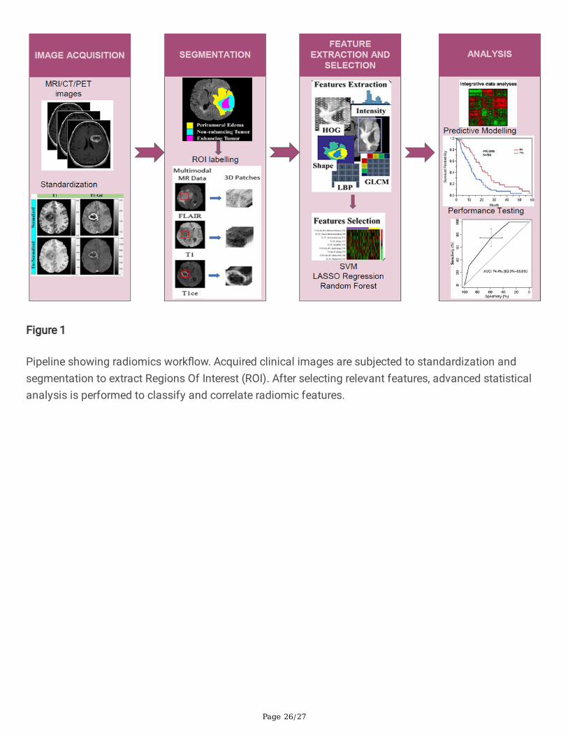
Radiomics And Radiogenomics:Radiomics is an emerging application of neuroimaging where advanced computational methods areused to quantitatively extract characteristics from clinical images that are too complex for a human eyeto appreciate.[8,9] These imaging characteristics, called “features” re�ect tumor characteristics and innerorganization as well as the tumor microenvironment.[9]Radiomics is a multi-step process including theacquisition and preprocessing of images, segmentation, feature extraction and selection, and advancedstatistics using machine learning (ML) algorithms (Figure 1). The pipeline of radiomics is highly
Page 3/27
collaborative and involves contributions from clinicians, molecular biologists, statisticians, andbioengineers. [8]
Radiomics-derived imaging phenotypes are associated with molecular markers to create ‘radiogenomics’models.[5] It is a rapid and reproducible tool to evaluate tumor subtype, mutation status and intratumoralheterogeneity; and non-invasively predicts tumor progression, survival and response to targeted therapiesusing these characteristics.[5,8]Radiogenomics offers more information as opposed to surgical biopsy inview of spatial tumor heterogeneity,[8] especially useful for genomic pro�ling in recurrent glioblastomawhich is driven by different clonal populations with varying hypermutations and evasion mechanisms.[10]Thus, clinical decision support systems using radiomics will form the base for precision medicine.[9]
Applications Of Radiomics In Glioblastoma Management:Radiomics analysis has been widely studied for its use in subtyping brain tumors, predicting prognosisand treatment planning. Combining radiomics with clinical and genetic prognostic factors yields superiorcan result in predicting the survival of patients than using each component alone.
Diagnosis and classi�cation of glioblastoma:
Simple features on structural MRI such as tumor size, location and enhancement patterns have beenassociated with various histopathological subtypes of glioblastoma. Incorporating complex radiologicalfeatures derived using image processing software and combining advanced MRI modalities can furtherimprove the accuracy of these models (Table 1).
Tumor location:
It is well known that location of the tumor affects the outcomes in patients with glioblastoma. A“probabilistic radiographic atlas” of more than 500 glioblastoma patients showed associations betweenstereospeci�c frequency of tumor occurrence with age, extent of resection, genetic expression, andsurvival data. Interestingly, regions closer to subventricular zone were seen to have MGMT unmethylated,mesenchymal, and EGFR-ampli�ed tumors,[11] supporting their invasive nature and poor prognosis.[12] Acomparison between solitary and multicentric glioblastoma revealed distinct gene expression pro�lingand outcomes between the two types, with upregulation of genes responsible for tumor cell motility andinvasiveness in the multicentric type.[13]Another study showed correlation of tumor phenotypeswith spatial distribution of tumors.[14]Thus, tumor location provides important information on the cell oforigin and tumor behavior.
Tumor size and contrast enhancement patterns:
The volumes of both contrast enhancement and necrosis at the time of initial diagnosis were foundhigher in tumors with the mesenchymal gene expression signature compared with those having proneuralor proliferative signatures. A ratio of the T2/FLAIR hyperintense volume to the volume of contrastenhancement plus necrosis of less than 2.3 could predict the mesenchymal subtype with 82% sensitivity
Page 4/27
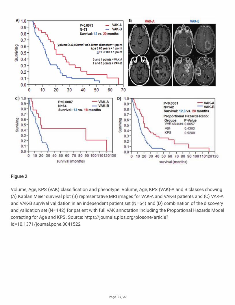
and 87% speci�city.[15] VAK Classi�cation, a scoring system developed to create phenotypes based ontumor Volumetry, Age, KPS annotation, was combined with P53 activation, MGMT promoter methylationand a group of genes and microRNAs in The Cancer Genome Atlas (TCGA) glioblastoma dataset topredict patient survival and facilitate genomics-based personalized therapy for glioblastoma patients.(Figure 2).[16]‘VASARI’, a semi-quantitative feature set named was designed to measure tumor size andvolumes of components with enhancement, non-enhancement, necrosis and edema, which correlatedwith survival rates and subtypes.[17]Speci�c invasive imaging signatures including ependymalinvolvement, deep white matter tract involvement and enhancement across the midline predicted adecrease in OS, MYC oncogene activation and inhibition of NF-KB inhibitor-alpha.[18] These patients werefound to have mitochondrial dysfunction,[18] consistent with the ‘”Warburg effect”,[19] where cancer cellsrely on aerobic glycolysis facilitated by MYC oncogene upregulation.
Volumetry was combined with DNA microarray analysis to train classi�ers that can predict gene-expression patterns and survival. Tumor contrast enhancement and mass effect was associated with up-regulation of speci�c hypoxia and proliferation gene-expression programs such as VEGF, ADM, PLAUR,SERPINE1, CA12, TOPA, CDC2, and BUB1B.[20] In another radiogenomic study based on TCGA,strati�cation into high and low FLAIR radio-phenotypes re�ected underlying edema and cellular invasionin glioblastoma, as they were associated with genes and microRNAs involved in cancer and cellularmigration.[21]MRI volumetric features are predictive of several cancer-relevant, drug-targetable DNAmutations in glioblastoma. TP53, RB1, NF1, EGFR, and PDGFRA mutations could each be signi�cantlypredicted by at least one imaging feature.[22]These studies provide a basis for genomic pro�ling andnon-invasively selecting patients for personalized therapies using tumor volumetry.
Radiomics was used to distinguish brain metastasis and glioblastoma using contrast-enhancing andperitumoralhyperintense masks in T2-weighted (T2W) MRI. In this model, deep learning showed bestperformance (area under curve AUC 0.956) compared to the traditional machine learning model (AUC0.890) and human readers (AUC 0.774).[23]Similar performance, AUC 0.96 for support vector machine(SVM), was observed in another study which used post-contrast T1 weighted (T1CE) MRI instead.However, performance decreased when subtypes of brain metastasis were attempted to classify.[24]
Texture:
Texture is a chief radiomic feature utilized for glioblastoma phenotyping. In one study, a gray-level co-occurrence matrix (GLCM) approach was employed for extracting phenotypic texture features fornecrosis, active tumor, and edema on structural MRI. Features were signi�cant predictors (p value <0.01)of prognosis but in areas of active tumor only.[25] Another study was able to predict MGMT methylationstatus using space-frequency texture analysis based on the S-transform in T2W MRI, albeit with anaccuracy of 71%, requiring better algorithms.26Other studies on texture features were able to predictMGMT methylation status with reasonable accuracy.[27,28]
Occasionally, high‐grade gliomas (WHO Grade III and glioblastoma) may have the same MRI appearanceas low-grade gliomas. A radiomic analysis using texture along with size, shape, intensity, and histogram
Page 5/27
features was used to differentiate low-grade from high-grade gliomas, reaching a prediction performancein the cross-validation as high as AUC value of 0.932 with support vector machine. However, the accuracydecreased to 0.75 in the independent validation dataset.[29] In a similar study, Random Forest gave thehighest AUC in the training cohort compared to test cohort, re�ecting variation in accuracy acrossdifferent ML classi�ers.[30]
Advanced MRI sequences and multimodal analyses:
Perfusion MRI has been extensively used in brain tumors to evaluate angiogenesis and tumor behavior.Raw tumor features from structural MRI and delta-radiomic features from Dynamic susceptibility contrast(DSC) perfusion MRI were extracted to differentiate low-grade gliomas from high-grade gliomas, thisclassi�er reached an AUC of 0.94.[31] A Cochrane meta-analysis on 7 studies to differentiate untreatedsolid and non‐enhancing low-grade from high-grade gliomas using DSC MRI features (rCBV and Ktrans)reported wide range of estimates for both sensitivity and speci�city, making these parameters lessreliable.[32] Diffusion MRI was employed to compare the expression of various genes between the high-versus low- Apparent Diffusion Coe�cient (ADC) tumors in a subset of patients. High-ADC tumors werefound to have higher expression of 13 genes, 6 of which encode for extracellular matrix (ECM) moleculesincluding collagen or collagen-binding proteins, suggesting a role of these genes in pro-invasivephenotype.[33] In another study, physiologic MRI was correlated with stereotactic image-guided biopsiesto differentiate contrast-enhancing and non-enhancing tumor areas. DSC MRI was useful for identifyingtissue specimens with higher tumor proliferation, necrosis, and vascular hyperplasia in the contrast-enhancing component of the lesion, while Diffusion MRI may be used to detect in�ltrating tumors in thenon-enhancing region. This is of particular interest for de�ning tumor burden in non-enhancing regions,where distinguishing reactive edema from biologically active in�ltrative tumor is clinically important.Accuracy of these results could be confounded by the misregistration arising as a result of brain shift.[34]
MR imaging features of Primary CNS Lymphoma (PCNSL) and glioblastoma overlap, with differingsurvival outcomes and treatment options. In one study, perfusion and diffusion-weighted MRI were usedto differentiate glioblastoma from lymphoma. Mean ADC and plasma volume (rVp) were higher in theglioblastoma compared to PCNSL. Moreover, mean ADC was superior (AUC 0.83) to rVp and permeabilitytransfer constant (Ktrans). This was true for contrast-enhancing regions only, possibly due to increases intumor cellularity, microvascular permeability, and vascular proliferation.35 In another study, ADC wasoutperformed by a multi-parametric (T1-weighted post contrast T1WCE, post-contrast T2Wand T2 FLAIR)and multiregional radiomics classi�er with AUC 0.921.[36]
Studies have used multiparametric MRI to create more accurate radiomic models for tumor subtyping.Rathore et al. used 267 multiparametric MRI based radiomic features, extracted from T1-weighted (T1W),T2W, T1WCE, T2 FLAIR, DSC, and DTI to train classi�ers to subtype de novo glioblastoma into threeimaging phenotypes. For example, the solid subtype was characterized by highly uniform vascularization,highest cell densities, small-sized edema, moderately spherical and well-circumscribed appearance withperitumoral edematous tissue having signs of heterogeneous neovascularization. This subtype had a
Page 6/27
predilection for the right temporal lobe and was associated with the worst prognosis. A personalizedtreatment regimen would involve very aggressive peritumoral resection and radiation dose escalation inthese tumors.[14] combining various MRI sequences can improve classi�cation accuracy for tumorgrading. 37,38Accuracy also increased using MRI features from multiregional and multiparametricstructural MRI to predict MGMT methylation status in glioblastoma.[39,40] Similarly, IDH 1 mutationstatus was predicted using radiomic features on multiparametric MRI with enhanced accuracy when ageand multiple regions were included.[41]
Prognostication of Glioblastoma:
It is increasingly important for physicians to understand an individual patient’s prognosis and adjust theirtherapy accordingly. Radiomics alone and augmented with clinical data, genomics, and proteomics canbe used to predict outcomes (Table 2).
Conventional MRI features:
Studies have used various features extracted from conventional MRI to predict patient outcomes inglioblastoma. Longer median survival was associated with higher sphericity, surface-to-volume ratio andedge enhancement on T1W MRI.[42]Lao et. al divided features into ‘handcrafted features’ and ‘deepfeatures’ to create a feature signature, which when coupled with clinical risk factors such as age andKarnofsky Performance Score, was able to predict overall survival (OS). Compared with the predictiveability of traditional risk factors, the proposed feature signature achieved a superior prediction of OS (C-index = 0.739).[43]Similar combined models reached C-index of 0.974.[44]
Texture, tumor shape and volumetric features were extracted, and combined with patient age to produce amodel that would predict short-term, mid-term, and long-term OS.[44,45] Zhou et al went one step furtherand identi�ed spatial-based characteristics from tumor sub-regions that can be used to predict survivaltime in patients.[46] Similarly, Chaddad et al found three texture features extracted from active part of thetumors that signi�cantly predicted survival outcomes compared to necrotic and edematous parts.[25]Moreover, these radiomic models could predict survival in different molecular subtypes as well.[47]
Advanced MRI features:
Advanced MRI modalities have also been also explored to predict glioblastoma patient outcomes.[48] Itwas seen that high rCBV in the non-enhancing region of tumor was predictive of worsening OS andProgression-free Survival (PFS).[49]Pre-treatment ADC histogram analysis was useful to predict PFS inbevacizumab-treated patients with newly diagnosed as well as recurrent glioblastoma.33,50 In thesestudies, low ADC predicted poor outcomes.
Radiogenomics and proteomics:
MGMT promoter hypermethylation, associated with better prognosis and response to therapy, has beencombined with radiomic features from structural MRI to stratify patients based on overall survival.
Page 7/27
Adding MGMT and IDH1 mutation status resulted in more robust radiomics-based prognostic models.[51,52] Zinn et al strati�ed VAK annotated cases further with molecular signatures and found a 10.5months’ additional survival bene�t for the group with MGMT promoter methylation.[16]In another study,glioblastomas were divided into groups based on vascularization (rCBV values). It was seen that MGMTmethylation was a positive predictive factor for OS (p = 0.003, AUC = 0.70) in the moderately vascularizedtumors. However, there was no signi�cant effect of MGMT methylation in the highly vascularized tumors(p = 0.10, AUC = 0.56).[53]Other studies did not �nd any signi�cant association of prognosis with MGMTpromoter hypermethylation.[42,54] This could be due to insu�cient feature selection methods.
Integrative models promise a reduction in prediction errors.[51,55]Chaddad et al created multi-omicintegrative model using radiomic, clinical, protein expression and genetic features to predict the outcomefor IDH1 wild-type glioblastoma patients which reached AUC of 78.24%.[56]Liao et al. us extracted Firstorder and multi-dimensional features from segmented lesions on T2‐FLAIR MRI and gave a featureimportance score for feature selection.[57] When combined with genetic expression, the GradientBoosting Decision Tree model gave a 0.81 accurate prediction of both short-term and long-term survival.Six metagenes showed signi�cant interactive effects with image features. However, this study waslimited by unavailability of complete genomic data.[57]
Immunophenotypes in glioblastoma are important as they predict response to immunotherapy andoutcomes. Hsu et al. used radiomic immunophenotyping models to predict patient prognosis.[58]Thephenotype with the worst prognosis comprised highly enriched myeloid-derived suppressor cells andlowly enriched Cytotoxic T lymphocytes.[58]
Treatment of glioblastoma:
Studies have shown the bene�t of radiomics analysis in planning surgical procedures, evaluating thedose of radiotherapy, predicting the effective dose of chemotherapeutic agents and stratifying patientswho will bene�t from therapy. After initiating therapies, radiomics can be used to differentiate mimickingentities like true progression, pseudoprogression and radionecrosis(Table 3).
Surgical Resection:
A recent study examined the correlation of tumor surface regularity on T1W MRI and OS of 165glioblastoma patients who underwent surgical resection and highlighted that patients with surface-regular tumors had a higher survival rate and bene�t from total tumor resection as compared to surface-irregular tumor patients.[59]Gaw et al used machine learning models to better predict tumor cell invasionbefore resection was conducted.[60] Their aim was to allow for more effective surgery and radiationplanning and created a hybrid model with Proliferation-Invasion (PI) model of glioma growth and pre-operative MRIs.[60] Thus, radiomics can help plan a targeted and personalized surgical treatment.
Radiation Therapy (RT) planning:
Page 8/27
Radiomics shows immense potential to guide precision radiotherapy. Prediction models can estimate theextent of tumor in�ltration and can help identify areas that are at a higher risk of tumor recurrence fortargeted RT.[14,61]Rathore et al. worked on a method for estimating peritumoral edema in�ltration usingradiomics by testing on pre- and post-operative multiomodal MRI sequences in 90 de novo glioblastomapatients and found that recurrent tumor regions revealed higher vascularity and cellularity whencompared with the non-recurrent regions.[14] A similar study done on 31 de novo glioblastoma patientscon�rmed these �ndings and also highlighted the importance of using multiparametric pattern analysismethods for planning a focused treatment approach to decrease recurrence rate.[61] Thus, radiomics canguide in planning radiation therapy dose escalation in areas with higher risk of tumor recurrence as wellas increasing gross total resection. This method can also help prevent dose-related toxicities seen withRT, salvaging the neural tissue at lower risk areas from damage.[62]
Chemotherapy with Temozolomide (TMZ):
Chemotherapy with TMZ along with adjuvant RT increases median OS.[63] However, TMZ resistancearises due to tumor heterogeneity. Yan et al. con�rmed the importance of radiomics analysis in predictingdisease progression in 57 glioblastoma patients treated with TMZ post-surgery using structural, diffusionand perfusion MR. The study found lower ADC, higher FLAIR and contrast enhanced T1 signals in areaswith a higher risk of tumor progression.[64] In another study assessing the e�cacy of using a deep-learning based survival-prediction model of 118 patients undergoing concurrent chemoradiotherapy withtemozolomide post-surgery, radiomics features including T1W with and without contrast, T2 FLAIR andADC images were used to assess the OS. While there was no difference observed between the twogroups, it highlighted that both clinical and radiomic features should be used hand in hand to predict OSof glioblastoma patients.[65] This reiterates the importance of radiomic models predictive of treatmentresponse to identify suitable treatment regimens.
Therapy with Bevacizumab:
However, variations in genetic makeup of VEGF among individuals can lead to resistance to bevacizumaband limiting its use.[66]Radiomics analysis can provide important biomarkers for selecting patients andto predict the treatment outcome. T1W and T1WCE MRI of 172 patients with recurrent glioblastoma priorto treatment with bevacizumab were used to develop radiomics-based survival predictor as a low-costinstrument for identifying treatment response in these patients.[66] In patients with recurrentglioblastoma receiving bevacizumab treatment, radiomics features from T1WCE obtained at baseline andpost-treatment showed prognostic value for survival and progression.[67]Using ADC and CBV of 54patients with recurrent glioblastoma that were treated with RT and temozolomide, and subsequentlytreated with bevacizumab, was effective in segregating patients into responders and non-responders tobevacizumab treatment.[68] To predict which patients will bene�t from bevacizumab therapy for brainnecrosis after radiotherapy, a strati�cation model was created which integrated the pre-treatment MRIradiomics signature, the interval between radiotherapy and diagnosis of brain necrosis, and the interval
Page 9/27
between diagnosis of brain necrosis and treatment with bevacizumab. This model achieved AUC 0.912 inthe validation set.[69]
Evaluating response to Radiation Therapy (RT):
Texture features derived from enhancing component and peri-lesional edema on structural MRI were usedto differentiate pseudoprogression from true progression in glioblastoma.[70] Another model achievedhigh sensitivity and moderate speci�city; incorporating the MGMT status further increased accuracy.[71]While these studies were based on post-RT MRI, pre-RT MRI scans to predict the development of futurepseudoprogression in glioblastoma patients gave an AUC of 0.82.[72]Recent studies show thatincorporating diffusion and perfusion-weighted MRI, which re�ect hypercellularity and hypervascularity oftumor, improves the accuracy in detecting pseudoprogression than conventional MRI alone.[73-75]
Radiation necrosis, another post-RT effect that is di�cult to differentiate from true progression, can bedetected using ML classi�ers based on traditional and delta radiomic features derived from MRI.[76]
Challenges In The Clinical Application Of Radiomics ForGlioblastoma:Despite the proven potential of radiomics in various aspects of glioblastoma management, thesemethods are yet to be introduced in mainstream clinical practice. Obstacles to translation include limitedreproducibility of algorithms and less robust machine models. Formation of bodies to recommendstandardization methods such as QIBA and QIN offer hope.[77,78]
Data availability and sharing:
Majority of the studies exploring radiomics in glioblastoma are limited by small sample sizes. Biologicalvariability of the tumor among patients explains why radiomics is still in infancy. Promoting collaborativestudies, sharing of data across institutions and making more high-quality datasets publicly available(such as Huiyihuiying Inc., BraTS, TCGA[79-81]) will result in more robust as well as reproducible models.This also requires overcoming the administrative and regulatory barriers to large-scale data sharing. Inaddition, clearly documenting the analysis and making original codes and data available will allow otherinvestigators to replicate the results.[59]
Image acquisition:
The inclusion of retrospectively collected, multi-center data for clinical trials on radiomics is limited byvariations across institutions in image acquisition such as the protocol de�ned by physicians, resolution,slice thickness, and washout period for contrast imaging of the acquired images.[82]Features extractedfrom MRI images can be in�uenced by �eld of view, �eld strength and slice thickness.[83] To combat thevariability in the data collected, standardized steps are recommended following the image acquisition likeintensity normalization, voxel re-slicing, use of a speci�c anatomical plane for multiparametric data,
Page 10/27
standardization of signal intensity prior to image listing, and developing algorithms for multiple MR-modalities for image registration.[82,84,85]
Segmentation and feature extraction:
Although considered the highest standard for segmentation, manual segmentation of images is labor-intensive and increases risk of observer bias. In contrast, semi- and fully-automated methods canimprove robustness and reproducibility.[86] Extracted features are dependent on the segmented regionand tumor margins therefore segmentation is the key step.[83] While automated feature extraction haslower degree of variation in the scoring of semantic features,[82]these methods can still lead to site-speci�c variations when obtaining imaging.[82]
Machine learning models:
Accuracy of ML models is limited by over�tting and under�tting. Over�tting of data occurs when doingfeature extraction on high-dimensional, large scale data.[79] However, it can be reduced by featureselection methods such as principal components analysis (PCA), sparse PCA, auto-encoders, etc.[87,88]Under�tting, due to small sample sizes, can be addressed using techniques like SMOTE.[89]
Conclusion:Radiomics offers revolutionary changes in the scope of glioblastoma management through facilitating apersonalized approach at various stages. Integrative models that include clinical, genetic and othermolecular data can enhance the accuracy. The main limitation seen in most studies is the small samplesize and the retrospective nature of these projects. Besides, variability in methods to generate data acrossinstitutions limits the generalizability in different patient populations. Whilst the results of these studiesare promising, a key goal moving forward is to make these models more reproducible in a wide array ofsettings. Multicenter clinical trials are needed to translate these models and provide actual bene�t toglioblastoma patients.
DeclarationsCompliance with Ethical Standards:
Funding: none
Con�icts of interest/Competing interests: none
Code availability: not applicable
Ethical approval: This article does not contain any studies with human participants or animals performedby any of the authors.
Page 11/27
Data availability:
Availability of data and material: not applicable
Authors' contributions:
Kiran Aftab – conceptualization, drafting, revising, reviewing, supervision
FaiqaBinteAamir – drafting, revising
Saad Mallick – drafting, revising
Fatima Mubarak – revising, reviewing, supervision
Syed Ather Enam – conceptualization, revising, reviewing, supervision
Whitney B. Pope – revising, reviewing
Tom Mikkelsen – revising, reviewing
Jack P. Rock – revising, reviewing
References1. Tykocki T, Eltayeb M (2018 Aug) Ten-year survival in glioblastoma. A systematic review. J
ClinNeurosci 54:7–13
2. Hobbs SK, Shi G, Homer R, Harsh G, Atlas SW, Bednarski MD (2003 Nov) Magnetic resonance image-guided proteomics of human glioblastoma multiforme. J MagnReson Imaging 18(5):530–536
3. Ellingson BM (2015 Jan) Radiogenomics and imaging phenotypes in glioblastoma: novelobservations and correlation with molecular characteristics. CurrNeurolNeurosci Rep 15(1):506
4. ElBanan MG, Amer AM, Zinn PO, Colen RR (2015 Feb) Imaging genomics of Glioblastoma: state ofthe art bridge between genomics and neuroradiology. Neuroimaging Clin N Am 25(1):141–153
5. Verduin M, Compter I, Steijvers D, Postma AA, Eekers DBP, Anten MM et al (2018) NoninvasiveGlioblastoma Testing: Multimodal Approach to Monitoring and Predicting Treatment Response. DisMarkers 2018:2908609
�. Olar A, Aldape KD (2014 Jan) Using the molecular classi�cation of glioblastoma to informpersonalized treatment. J Pathol 232(2):165–177
7. Sotoudeh H, Shafaat O, Bernstock JD, Brooks MD, Elsayed GA, Chen JA et al (2019) Arti�cialIntelligence in the Management of Glioma: Era of Personalized Medicine. Front Oncol 9:768
�. Chaddad A, Kucharczyk MJ, Daniel P, Sabri S, Jean-Claude BJ, Niazi T, Abdulkarim B (2019)Radiomics in glioblastoma: current status and challenges facing clinical implementation. Frontiers inoncology. May 21;9:374
Page 12/27
9. Lambin P, Leijenaar RTH, Deist TM, Peerlings J, de Jong EEC, van Timmeren J et al (2017 Dec)Radiomics: the bridge between medical imaging and personalized medicine. Nature Reviews ClinicalOncology 14(12):749–762
10. Pinker K, Shitano F, Sala E, Do RK, Young RJ, Wibmer AG et al (2018 Mar) Background, current role,and potential applications of radiogenomics. J MagnReson Imaging 47(3):604–620
11. Ellingson BM, Lai A, Harris RJ, Selfridge JM, Yong WH, Das K et al (2013 Mar) Probabilisticradiographic atlas of glioblastoma phenotypes. AJNR Am J Neuroradiol 34(3):533–540
12. Lim DA, Cha S, Mayo MC, Chen M-H, Keles E, VandenBerg S, Berger MS (2007) Relationship ofglioblastoma multiforme to neural stem cell regions predicts invasive and multifocal tumorphenotype. Neurooncology 9(4):424–429
13. Kong D-S, Kim J, Lee I-H, Kim ST, Seol HJ, Lee J-I et al. Integrative radiogenomic analysis formulticentricradiophenotype in glioblastoma. Oncotarget. 2016 Mar 8;7(10):11526–38
14. Rathore S, Akbari H, Rozycki M, Abdullah KG, Nasrallah MP, Binder ZA et al (2018 Mar) Radiomic MRIsignature reveals three distinct subtypes of glioblastoma with different clinical and molecularcharacteristics, offering prognostic value beyond IDH1. Sci Rep 23(1):5087 8(
15. Naeini KM, Pope WB, Cloughesy TF, Harris RJ, Lai A, Eskin A et al (2013 May) Identifying themesenchymal molecular subtype of glioblastoma using quantitative volumetric analysis ofanatomic magnetic resonance images. Neuro Oncol 15(5):626–634
1�. Zinn PO, Sathyan P, Mahajan B, Bruyere J, Hegi M, Majumder S et al (2012) A novel volume-age-KPS(VAK) glioblastoma classi�cation identi�es a prognostic cognate microRNA-gene signature. PLoSOne 7(8):e41522
17. Gutman DA, Cooper LAD, Hwang SN, Holder CA, Gao J, Aurora TD et al (2013 May) MR imagingpredictors of molecular pro�le and survival: multi-institutional study of the TCGA glioblastoma dataset. Radiology 267(2):560–569
1�. Colen RR, Vangel M, Wang J, Gutman DA, Hwang SN, Wintermark M et al (2014 Jun) Imaginggenomic mapping of an invasive MRI phenotype predicts patient outcome and metabolicdysfunction: a TCGA glioma phenotype research group project. BMC Med Genomics 2:7:30
19. Warburg O. On the origin of cancer cells. Science. 1956 Feb 24;123(3191):309–14
20. Diehn M, Nardini C, Wang DS, McGovern S, Jayaraman M, Liang Y et al. Identi�cation of noninvasiveimaging surrogates for brain tumor gene-expression modules. Proc Natl AcadSci U S A. 2008 Apr1;105(13):5213–8
21. Zinn PO, Mahajan B, Majadan B, Sathyan P, Singh SK, Majumder S et al (2011) Radiogenomicmapping of edema/cellular invasion MRI-phenotypes in glioblastoma multiforme. PLoS One6(10):e25451
22. Gutman DA, Dunn WD, Grossmann P, Cooper LAD, Holder CA, Ligon KL et al (2015 Dec) Somaticmutations associated with MRI-derived volumetric features in glioblastoma. Neuroradiology57(12):1227–1237
Page 13/27
23. Bae S, An C, Ahn SS, Kim H, Han K, Kim SW et al. Robust performance of deep learning fordistinguishing glioblastoma from single brain metastasis using radiomic features: modeldevelopment and validation. Sci Rep. 2020 Jul 21;10
24. Artzi M, Bressler I, Bashat DB (2019) Differentiation between glioblastoma, brain metastasis andsubtypes using radiomics analysis. J Magn Reson Imaging 50(2):519–528
25. Chaddad A, Tanougast C (2016 Nov) Extracted magnetic resonance texture features discriminatebetween phenotypes and are associated with overall survival in glioblastoma multiforme patients.Med BiolEngComput 54(11)(1):1707–1718
2�. Drabycz S, Roldán G, de Robles P, Adler D, McIntyre JB, Magliocco AM et al. An analysis of imagetexture, tumor location, and MGMT promoter methylation in glioblastoma using magnetic resonanceimaging. Neuroimage. 2010 Jan 15;49(2):1398–405
27. Kor�atis P, Kline TL, Coufalova L, Lachance DH, Parney IF, Carter RE et al (2016 Jun) MRI texturefeatures as biomarkers to predict MGMT methylation status in glioblastomas. Med Phys43(6):2835–2844
2�. Hajianfar G, Shiri I, Maleki H, Oveisi N, Haghparast A, Abdollahi H et al (2019 Dec) Noninvasive O6Methylguanine-DNA Methyltransferase Status Prediction in Glioblastoma Multiforme Cancer UsingMagnetic Resonance Imaging Radiomics Features: Univariate and Multivariate RadiogenomicsAnalysis. World Neurosurgery 132(1):e140–e161
29. Nakamoto T, Takahashi W, Haga A, Takahashi S, Kiryu S, Nawa K et al (2019 Dec) Prediction ofmalignant glioma grades using contrast-enhanced T1-weighted and T2-weighted magneticresonance images based on a radiomic analysis. Sci Rep 19(1):19411 9(
30. Cho HH, Lee SH, Kim J, Park H (2018 Nov) Classi�cation of the glioma grading using radiomicsanalysis. PeerJ 22;6:e5982
31. Jeong J, Wang L, Ji B, Lei Y, Ali A, Liu T et al (2019 Jul) Machine-learning based classi�cation ofglioblastoma using delta-radiomic features derived from dynamic susceptibility contrast enhancedmagnetic resonance images: Introduction. Quant Imaging Med Surg 9(7):1201–1213
32. Abrigo JM, Fountain DM, Provenzale JM, Law EK, Kwong JS, Hart MG et al (2018 Jan) Magneticresonance perfusion for differentiating low-grade from high-grade gliomas at �rst presentation.Cochrane Database Syst Rev 22:1:CD011551
33. Pope WB, Mirsadraei L, Lai A, Eskin A, Qiao J, Kim HJ et al (2012 Jun) Differential gene expression inglioblastoma de�ned by ADC histogram analysis: relationship to extracellular matrix molecules andsurvival. AJNR Am J Neuroradiol 33(6):1059–1064
34. Barajas RF, Phillips JJ, Parvataneni R, Molinaro A, Essock-Burns E, Bourne G et al (2012 Jul) Regionalvariation in histopathologic features of tumor specimens from treatment-naive glioblastomacorrelates with anatomic and physiologic MR Imaging. Neuro Oncol 14(7):942–954
35. Lin X, Lee M, Buck O, Woo KM, Zhang Z, Hatzoglou V et al (2017 Mar) Diagnostic Accuracy of T1-Weighted DCE-MRI and DWI-ADC for Differentiation of Glioblastoma and Primary CNS Lymphoma.AJNR Am J Neuroradiol 38(3):485–491
Page 14/27
3�. Suh HB, Choi YS, Bae S, Ahn SS, Chang JH, Kang S-G et al (2018) Primary central nervous systemlymphoma and atypical glioblastoma: Differentiation using radiomics approach. EurRadiol. Sep1;28(9):3832–9
37. Tian Q, Yan L-F, Zhang X, Zhang X, Hu Y-C, Han Y et al (2018 Dec) Radiomics strategy for gliomagrading using texture features from multiparametric MRI. J MagnReson Imaging 48(6):1518–1528
3�. Zhang X, Yan L-F, Hu Y-C, Li G, Yang Y, Han Y et al. Optimizing a machine learning based gliomagrading system using multi-parametric MRI histogram and texture features. Oncotarget. 2017 Jul18;8(29):47816–30
39. Xi Y, Guo F, Xu Z, Li C, Wei W, Tian P et al (2018) Radiomics signature: A potential biomarker for theprediction of MGMT promoter methylation in glioblastoma. J Magn Reson Imaging 47(5):1380–1387
40. Li Z-C, Bai H, Sun Q, Li Q, Liu L, Zou Y et al (2018) Multiregional radiomics features frommultiparametric MRI for prediction of MGMT methylation status in glioblastoma multiforme: Amulticentre study. EurRadiol. Sep 1;28(9):3640–50
41. Lee MH, Kim J, Kim S-T, Shin H-M, You H-J, Choi JW et al (2019 May) Prediction of IDH1 MutationStatus in Glioblastoma Using Machine Learning Technique Based on Quantitative Radiomic Data.World Neurosurg 125:e688–e696
42. Tixier F, Um H, Bermudez D, Iyer A, Apte A, Graham MS et al. Preoperative MRI-radiomics featuresimprove prediction of survival in glioblastoma patients over MGMT methylation status alone.Oncotarget. 2019 Jan 18;10(6):660–72
43. Lao J, Chen Y, Li Z-C, Li Q, Zhang J, Liu J et al. A Deep Learning-Based Radiomics Model forPrediction of Survival in Glioblastoma Multiforme. Sci Rep. 2017 Sep 4;7
44. Zhang X, Lu H, Tian Q, Feng N, Yin L, Xu X et al (2019) A radiomics nomogram based onmultiparametric MRI might stratify glioblastoma patients according to survival. EurRadiol. Oct1;29(10):5528–38
45. Sanghani P, Ang BT, King NKK, Ren H (2018 Dec) Overall survival prediction in glioblastomamultiforme patients from volumetric, shape and texture features using machine learning. SurgOncol27(4):709–714
4�. Zhou M, Chaudhury B, Hall LO, Goldgof DB, Gillies RJ, Gatenby RA (2017 Jul) Identifying spatialimaging biomarkers of glioblastoma multiforme for survival group prediction. J MagnReson Imaging46(1):115–123
47. Yang D, Rao G, Martinez J, Veeraraghavan A, Rao A (2015 Nov) Evaluation of tumor-derived MRI-texture features for discrimination of molecular subtypes and prediction of 12-month survival statusin glioblastoma. Med Phys 42(11):6725–6735
4�. Park JE, Kim HS, Jo Y, Yoo R-E, Choi SH, Nam SJ et al (2020) Radiomics prognostication model inglioblastoma using diffusion- and perfusion-weighted MRI. Scienti�c Reports. Mar 6;10(1):4250
49. Jain R, Poisson LM, Gutman D, Scarpace L, Hwang SN, Holder CA et al (2014 Aug) OutcomePrediction in Patients with Glioblastoma by Using Imaging, Clinical, and Genomic Biomarkers: Focus
Page 15/27
on the Nonenhancing Component of the Tumor. Radiology 272(2):484–493
50. Pope WB, Lai A, Mehta R, Kim HJ, Qiao J, Young JR et al (2011 May) Apparent diffusion coe�cienthistogram analysis strati�es progression-free survival in newly diagnosed bevacizumab-treatedglioblastoma. AJNR Am J Neuroradiol 32(5):882–889
51. Choi Y, Nam Y, Jang J, Shin N-Y, Lee YS, Ahn K-J et al (2020) Radiomics may increase the prognosticvalue for survival in glioblastoma patients when combined with conventional clinical and geneticprognostic models. EurRadiol. Oct 2
52. Molitoris JK, Rao YJ, Patel RA, Kane LT, Badiyan SN, Gittleman H et al (2017) Multi-institutionalexternal validation of a novel glioblastoma prognostic nomogram incorporating MGMT methylation.J Neurooncol. Sep 1;134(2):331–8
53. Fuster-Garcia E, LorenteEstellés D, Álvarez-Torres M, del M, Juan-Albarracín, Chelebian J, Rovira E A,et al (2021) MGMT methylation may bene�t overall survival in patients with moderately vascularizedglioblastomas. EurRadiol. Mar 1;31(3):1738–47
54. Sasaki T, Kinoshita M, Fujita K, Fukai J, Hayashi N, Uematsu Y et al (2019) Radiomics and MGMTpromoter methylation for prognostication of newly diagnosed glioblastoma. Scienti�c Reports. Oct8;9(1):14435
55. Beig N, Patel J, Prasanna P, Hill V, Gupta A, Correa R et al. Radiogenomic analysis of hypoxiapathway is predictive of overall survival in Glioblastoma. Scienti�c Reports. 2018 Jan 8;8(1):7
5�. Chaddad A, Daniel P, Sabri S, Desrosiers C, Abdulkarim B (2019 Aug) Integration of radiomic andmulti-omic analyses predicts survival of newly diagnosed IDH1 wild-type glioblastoma. Cancers11(8):1148
57. Liao X, Cai B, Tian B, Luo Y, Song W, Li Y (2019 Jun) Machine-learning based radiogenomics analysisof MRI features and metagenes in glioblastoma multiforme patients with different survival time. JCell Mol Med 23(6):4375–4385
5�. Hsu JB-K, Lee GA, Chang T-H, Huang S-W, Le NQK, Chen Y-C et al. Radiomic Immunophenotyping ofGSEA-Assessed Immunophenotypes of Glioblastoma and Its Implications for Prognosis: A FeasibilityStudy. Cancers (Basel). 2020 Oct 19;12(10)
59. Pérez-Beteta J, Molina-García D, Ortiz-Alhambra JA, Fernández-Romero A, Luque B, Arregui E et al.Tumor Surface Regularity at MR Imaging Predicts Survival and Response to Surgery in Patients withGlioblastoma. Radiology. 2018 Apr 3;288(1):218–25
�0. Gaw N, Hawkins-Daarud A, Hu LS, Yoon H, Wang L, Xu Y et al (2019) Integration of machine learningand mechanistic models accurately predicts variation in cell density of glioblastoma usingmultiparametric MRI. Sci Rep. Jul 11;9(1):10063
�1. Akbari H, Macyszyn L, Da X, Bilello M, Wolf RL, Martinez-Lage M et al (2016 Apr) Imaging Surrogatesof In�ltration Obtained Via Multiparametric Imaging Pattern Analysis Predict Subsequent Location ofRecurrence of Glioblastoma. Neurosurgery 78(4):572–580
�2. De Ruysscher D, Niedermann G, Burnet NG, Siva S, Lee AWM, Hegi-Johnson F (2019 Feb)Radiotherapy toxicity. Nature Reviews Disease Primers 21(1):1–20 5(
Page 16/27
�3. Lee SY. Temozolomide resistance in glioblastoma multiforme. Genes & diseases. 2016 Sep1;3(3):198-210
�4. Yan J-L, Li C, van der Hoorn A, Boonzaier NR, Matys T, Price SJ (2020 Jun) A Neural NetworkApproach to Identify the Peritumoral Invasive Areas in Glioblastoma Patients by Using MRRadiomics. Sci Rep 16(1):9748 10(
�5. Yoon HG, Cheon W, Jeong SW, Kim HS, Kim K, Nam H et al. Multi-Parametric Deep Learning Modelfor Prediction of Overall Survival after Postoperative Concurrent Chemoradiotherapy in GlioblastomaPatients. Cancers (Basel) [Internet]. 2020 Aug 14 [cited 2021 Feb 4];12(8)
��. Kickingereder P, Götz M, Muschelli J, Wick A, Neuberger U, Shinohara RT et al. Large-scale RadiomicPro�ling of Recurrent Glioblastoma Identi�es an Imaging Predictor for Stratifying Anti-AngiogenicTreatment Response. Clin Cancer Res. 2016 Dec 1;22(23):5765–71
�7. Grossmann P, Narayan V, Chang K, Rahman R, Abrey L, Reardon DA et al. Quantitative imagingbiomarkers for risk strati�cation of patients with recurrent glioblastoma treated with bevacizumab.Neuro-Oncology. 2017 Nov 29;19(12):1688–97
��. Petrova L, Kor�atis P, Petr O, LaChance DH, Parney I, Buckner JC et al. Cerebral blood volume andapparent diffusion coe�cient - Valuable predictors of non-response to bevacizumab treatment inpatients with recurrent glioblastoma. J Neurol Sci. 2019 Oct 15;405:116433
�9. Cai J, Zheng J, Shen J, Yuan Z, Xie M, Gao M et al. A Radiomics Model for Predicting the Responseto Bevacizumab in Brain Necrosis after Radiotherapy. Clin Cancer Res. 2020 Oct 15;26(20):5438–47
70. Patel MD, Zhan J, Natarajan K, Flintham R, Davies N, Sanghera P et al. Radiomic evaluation oftreatment response in patients with glioblastoma: a preliminary study. ECR 2019 EPOS. EuropeanCongress of Radiology - ECR 2019; 2019
71. Bani-Sadr A, Eker OF, Berner L-P, Ameli R, Hermier M, Barritault M et al (2019) Conventional MRIradiomics in patients with suspected early- or pseudo-progression. Neuro-Oncology Advances. May1;1(vdz019)
72. Baine M, Burr J, Du Q, Zhang C, Liang X, Krajewski L et al (2021 Feb) The Potential Use of Radiomicswith Pre-Radiation Therapy MR Imaging in Predicting Risk of Pseudoprogression in GlioblastomaPatients. Journal of Imaging 7(2):17
73. Kim JY, Park JE, Jo Y, Shim WH, Nam SJ, Kim JH et al (2019) Incorporating diffusion- and perfusion-weighted MRI into a radiomics model improves diagnostic performance for pseudoprogression inglioblastoma patients. Neuro Oncol. Feb 19;21(3):404–14
74. Elshafeey N, Kotrotsou A, Hassan A, Elshafei N, Hassan I, Ahmed S et al (2019 Jul) Multicenter studydemonstrates radiomic features derived from magnetic resonance perfusion images identifypseudoprogression in glioblastoma. Nat Commun 18(1):3170 10(
75. Elshafeey N, Kotrotsou A, GiniebraCamejo D, Abrol S, Hassan I, El Salek K et al (2017) Multicenterstudy to demonstrate radiomic texture features derived from MR perfusion images ofpseudoprogression compared to true progression in glioblastoma patients. JCO. May20;35(15_suppl):2016–2016
Page 17/27
7�. Zhang Z, Yang J, Ho A, Jiang W, Logan J, Wang X et al (2018) A predictive model for distinguishingradiation necrosis from tumour progression after gamma knife radiosurgery based on radiomicfeatures from MR images. EurRadiol. Jun 1;28(6):2255–63
77. About the Quantitative Imaging Network (QIN) | Quantitative Imaging Network (QIN) | CIP Grant-supported Networks | Programs & Resources | Cancer Imaging Program (CIP) [Internet]. [cited 2021Feb 20]. Available from:https://imaging.cancer.gov/programs_resources/specialized_initiatives/qin/about/teams.htm
7�. Shukla-Dave A, Obuchowski NA, Chenevert TL, Jambawalikar S, Schwartz LH, Malyarenko D et al(2019 Jun) Quantitative imaging biomarkers alliance (QIBA) recommendations for improvedprecision of DWI and DCE-MRI derived biomarkers in multicenter oncology trials. J MagnResonImaging 49(7):e101–e121
79. Medical Image Arti�cial Intelligence Cloud Platform - Huiyihuiying-Medical Image Arti�cialIntelligence Cloud Platform [Internet]. [cited 2021 Feb 22]. Available from: http://en.huiyihuiying.com/
�0. Menze BH, Jakab A, Bauer S, Kalpathy-Cramer J, Farahani K, Kirby J et al (2015 Oct) The MultimodalBrain Tumor Image Segmentation Benchmark (BRATS). IEEE Trans Med Imaging 34(10):1993–2024
�1. The Cancer Genome Atlas Program - National Cancer Institute [Internet] (2018) [cited 2021 Feb 22].Available from: https://www.cancer.gov/about-nci/organization/ccg/research/structural-genomics/tcga
�2. Narang S, Lehrer M, Yang D, Lee J, Rao A (2016 Aug) Radiomics in glioblastoma: current status,challenges and potential opportunities. Transl Cancer Res 5(4):383–397
�3. Avanzo M, Stancanello J, El Naqa I (2017 Jun) Beyond imaging: The promise of radiomics. PhysMed 38:122–139
�4. Bidgood WD, Horii SC, Prior FW et al (1997) Understanding and using DICOM, the data interchangestandard for biomedical imaging. J Am Med Inform Assoc 4:199–212
�5. Hoebel KV, Patel JB, Beers AL, Chang K, Singh P, Brown JM et al (2020) Radiomics RepeatabilityPitfalls in a Scan-Rescan MRI Study of Glioblastoma. Radiology: Arti�cial Intelligence. Nov4;3(1):e190199
��. Zaidi H, El Naqa I (2010) PET-guided delineation of radiation therapy treatment volumes: a survey ofimage segmentation techniques. Eur J Nucl Med Mol Imaging 37:2165–2187
�7. Mishra D, Dash R, Rath AK et al (2011) Feature selection in gene expression data using principalcomponent analysis and rough set theory. AdvExp Med Biol 696:91–100
��. Kumar D, Wong A, Clausi D (2015) Lung nodule classi�cation using deep features in CT images.Computer Robot Vision 327:110–116
�9. Chawla NV, Bowyer KW, Hall LO, Kegelmeyer WP. SMOTE: Synthetic Minority Over-samplingTechnique. Journal of Arti�cial Intelligence Research. 2002 Jun 1;16:321–57
Tables

Page 18/27
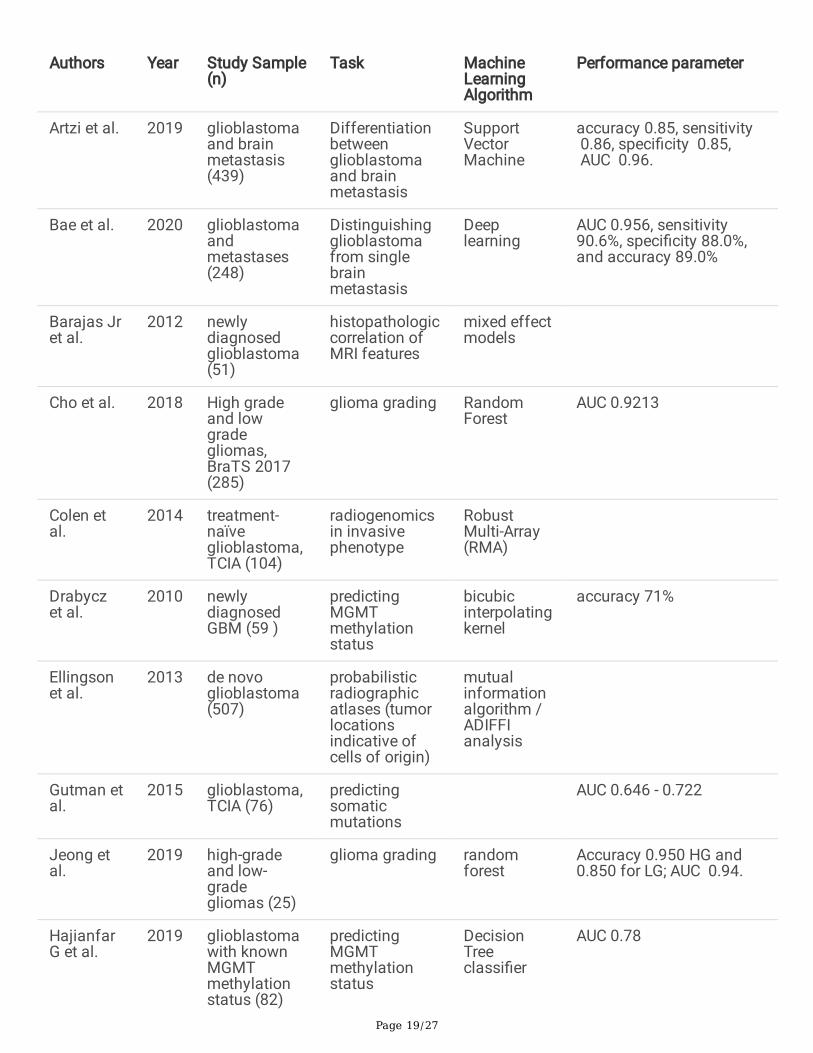
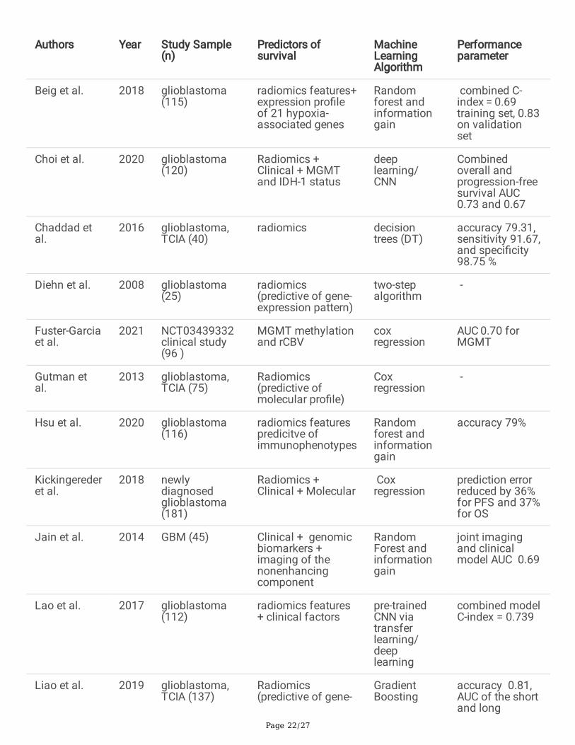
Table 1. Application of radiomics in glioblastoma diagnosis and classi�cation
Page 19/27
Authors Year Study Sample(n)
Task MachineLearningAlgorithm
Performance parameter
Artzi et al. 2019 glioblastomaand brainmetastasis(439)
Differentiationbetweenglioblastomaand brainmetastasis
SupportVectorMachine
accuracy 0.85, sensitivity 0.86, speci�city 0.85, AUC 0.96.
Bae et al. 2020 glioblastomaandmetastases(248)
Distinguishingglioblastomafrom singlebrainmetastasis
Deeplearning
AUC 0.956, sensitivity90.6%, speci�city 88.0%,and accuracy 89.0%
Barajas Jret al.
2012 newlydiagnosedglioblastoma(51)
histopathologiccorrelation ofMRI features
mixed effectmodels
Cho et al. 2018 High gradeand lowgradegliomas,BraTS 2017(285)
glioma grading RandomForest
AUC 0.9213
Colen etal.
2014 treatment-naïveglioblastoma,TCIA (104)
radiogenomicsin invasivephenotype
RobustMulti-Array(RMA)
Drabyczet al.
2010 newlydiagnosedGBM (59 )
predictingMGMTmethylationstatus
bicubicinterpolatingkernel
accuracy 71%
Ellingsonet al.
2013 de novoglioblastoma(507)
probabilisticradiographicatlases (tumorlocationsindicative ofcells of origin)
mutualinformationalgorithm /ADIFFIanalysis
Gutman etal.
2015 glioblastoma,TCIA (76)
predictingsomaticmutations
AUC 0.646 - 0.722
Jeong etal.
2019 high-gradeand low-gradegliomas (25)
glioma grading randomforest
Accuracy 0.950 HG and0.850 for LG; AUC 0.94.
HajianfarG et al.
2019 glioblastomawith knownMGMTmethylationstatus (82)
predictingMGMTmethylationstatus
DecisionTreeclassi�er
AUC 0.78
Page 20/27
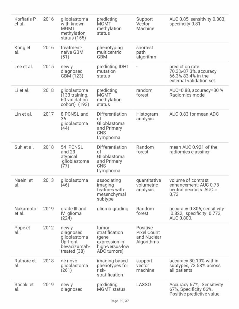
Kor�atis Pet al.
2016 glioblastomawith knownMGMTmethylationstatus (155)
predictingMGMTmethylationstatus
SupportVectorMachine
AUC 0.85, sensitivity 0.803,speci�city 0.81
Kong etal.
2016 treatment-naïve GBM(51)
phenotypingmulticentricGBM
shortestpathalgorithm
Lee et al. 2015 newlydiagnosedGBM (123)
predicting IDH1mutationstatus
- prediction rate70.3%-87.3%, accuracy66.3%-83.4% in theexternal validation set.
Li et al. 2018 glioblastoma(133 training,60 validationcohort) (193)
predictingMGMTmethylationstatus
randomforest
AUC=0.88, accuracy=80 %Radiomics model
Lin et al. 2017 8 PCNSL and36glioblastoma(44)
DifferentiationofGlioblastomaand PrimaryCNSLymphoma
Histogramanalysis
AUC 0.83 for mean ADC
Suh et al. 2018 54 PCNSLand 23atypical glioblastoma(77)
DifferentiationofGlioblastomaand PrimaryCNSLymphoma
Randomforest
mean AUC 0.921 of theradiomics classi�er
Naeini etal.
2013 glioblastoma(46)
associatingimagingfeatures withmesenchymalsubtype
quantitativevolumetricanalysis
volume of contrastenhancement: AUC 0.78central necrosis: AUC =0.73
Nakamotoet al.
2019 grade III andIV glioma(224)
glioma grading Randomforest
accuracy 0.806, sensitivity 0.822, speci�city 0.773,AUC 0.800.
Pope etal.
2012 newlydiagnosedglioblastomaUp-frontbevacizumab-treated (38)
tumorstrati�cation(geneexpression inhigh-versus-lowADC tumors)
PositivePixel Countand NuclearAlgorithms
Rathore etal.
2018 de novoglioblastoma(261)
imaging basedphenotypes forrisk-strati�cation
supportvectormachine
accuracy 80.19% withinsubtypes, 73.58% acrossall patients
Sasaki etal.
2019 newlydiagnosed
predictingMGMT status
LASSO Accuracy 67%, Sensitivity67%, Speci�city 66%,Positive predictive value
Page 21/27
GBM patients(201)
67%, Negative predictivevalue 67%, Prevalence ofpMGMT methylation 50%
Tian et al. 2018 Grades II, III,and IVgliomas (153)
glioma grading supportvectormachine
accuracy 96.8%, AUC0.987 LGGs vs HGGs; accuracy 98.1%, AUC0.992 for grades III vs IV
Xi et al. 2018 GBM patients(98)
predictingMGMTmethylationstatus
supportvectormachine
Training: accuracy 86.59%
validation: accuracy 80%.
Zhang etal.
2017 glioblastomawith knownMGMTmethylationstatus (98)
predictingMGMTmethylationstatus
supportvectormachine
training: accuracy 86.59%,validation: accuracy of80%
Zhang etal.
2017 High gradeand lowgradegliomas (120)
glioma grading supportvectormachine
Accuracy 0.945
Zinn et al. 2012 glioblastoma,TCIA (142)
patientsstrati�cation
Zinn et al. 2011 glioblastoma,TCIA (78)
RadiogenomicMapping ofEdema/CellularInvasion
IngenuityPathwayAnalysis(IPA).
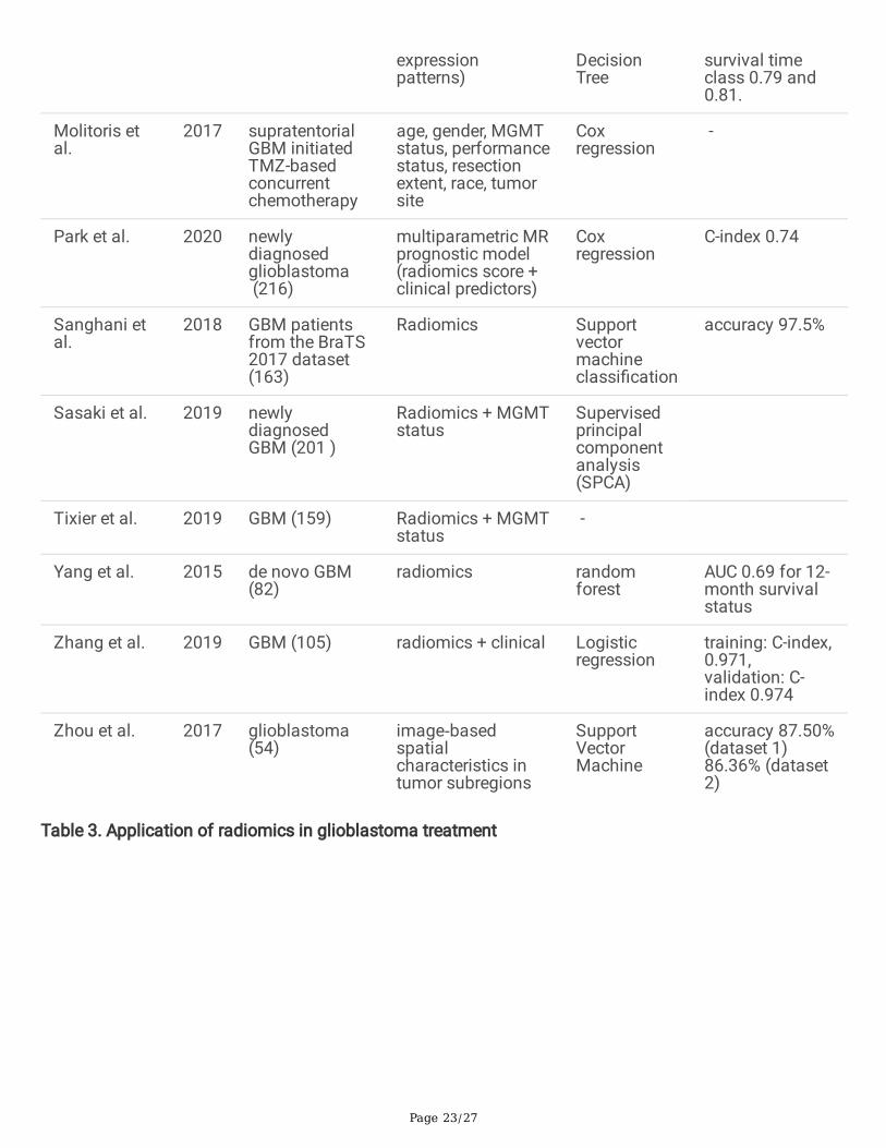
Table 2. Application of radiomics in glioblastoma survival prediction
Page 22/27
Authors Year Study Sample(n)
Predictors ofsurvival
MachineLearningAlgorithm
Performanceparameter
Beig et al. 2018 glioblastoma(115)
radiomics features+expression pro�leof 21 hypoxia-associated genes
Randomforest andinformationgain
combined C-index = 0.69training set, 0.83on validationset
Choi et al. 2020 glioblastoma(120)
Radiomics +Clinical + MGMTand IDH-1 status
deeplearning/CNN
Combinedoverall andprogression-freesurvival AUC0.73 and 0.67
Chaddad etal.
2016 glioblastoma,TCIA (40)
radiomics decisiontrees (DT)
accuracy 79.31,sensitivity 91.67,and speci�city98.75 %
Diehn et al. 2008 glioblastoma(25)
radiomics(predictive of gene-expression pattern)
two-stepalgorithm
-
Fuster-Garciaet al.
2021 NCT03439332clinical study(96 )
MGMT methylationand rCBV
coxregression
AUC 0.70 forMGMT
Gutman etal.
2013 glioblastoma,TCIA (75)
Radiomics(predictive ofmolecular pro�le)
Coxregression
-
Hsu et al. 2020 glioblastoma(116)
radiomics featurespredicitve ofimmunophenotypes
Randomforest andinformationgain
accuracy 79%
Kickingerederet al.
2018 newlydiagnosedglioblastoma(181)
Radiomics +Clinical + Molecular
Coxregression
prediction errorreduced by 36%for PFS and 37%for OS
Jain et al. 2014 GBM (45) Clinical + genomicbiomarkers +imaging of thenonenhancingcomponent
RandomForest andinformationgain
joint imagingand clinicalmodel AUC 0.69
Lao et al. 2017 glioblastoma(112)
radiomics features+ clinical factors
pre-trainedCNN viatransferlearning/deeplearning
combined modelC-index = 0.739
Liao et al. 2019 glioblastoma,TCIA (137)
Radiomics(predictive of gene-
GradientBoosting
accuracy 0.81,AUC of the shortand long
Page 23/27
expressionpatterns)
DecisionTree
survival timeclass 0.79 and0.81.
Molitoris etal.
2017 supratentorialGBM initiatedTMZ-basedconcurrentchemotherapy
age, gender, MGMTstatus, performancestatus, resectionextent, race, tumorsite
Coxregression
-
Park et al. 2020 newlydiagnosedglioblastoma (216)
multiparametric MRprognostic model(radiomics score +clinical predictors)
Coxregression
C-index 0.74
Sanghani etal.
2018 GBM patientsfrom the BraTS2017 dataset(163)
Radiomics Supportvectormachineclassi�cation
accuracy 97.5%
Sasaki et al. 2019 newlydiagnosedGBM (201 )
Radiomics + MGMTstatus
Supervisedprincipalcomponentanalysis(SPCA)
Tixier et al. 2019 GBM (159) Radiomics + MGMTstatus
-
Yang et al. 2015 de novo GBM(82)
radiomics randomforest
AUC 0.69 for 12-month survivalstatus
Zhang et al. 2019 GBM (105) radiomics + clinical Logisticregression
training: C-index,0.971,validation: C-index 0.974
Zhou et al. 2017 glioblastoma(54)
image‐basedspatialcharacteristics intumor subregions
SupportVectorMachine
accuracy 87.50%(dataset 1)86.36% (dataset2)
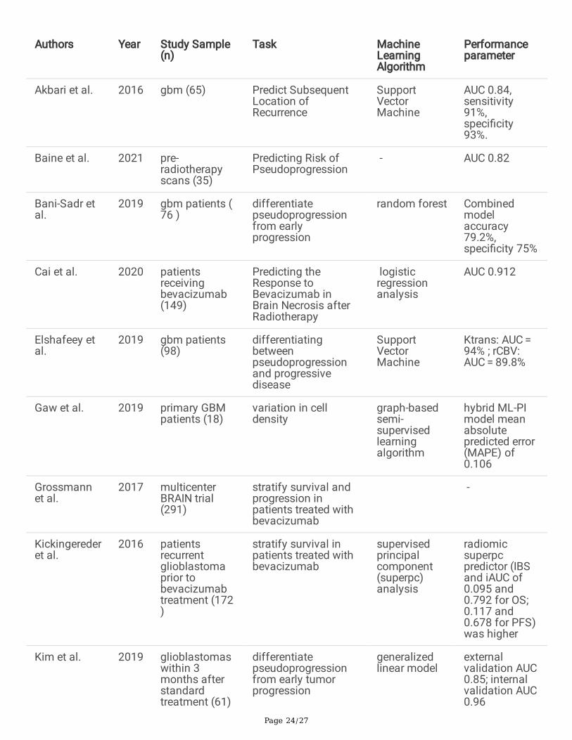
Table 3. Application of radiomics in glioblastoma treatment
Page 24/27
Authors Year Study Sample(n)
Task MachineLearningAlgorithm
Performanceparameter
Akbari et al. 2016 gbm (65) Predict SubsequentLocation ofRecurrence
SupportVectorMachine
AUC 0.84,sensitivity91%,speci�city93%.
Baine et al. 2021 pre-radiotherapyscans (35)
Predicting Risk ofPseudoprogression
- AUC 0.82
Bani-Sadr etal.
2019 gbm patients (76 )
differentiatepseudoprogressionfrom earlyprogression
random forest Combinedmodelaccuracy79.2%,speci�city 75%
Cai et al. 2020 patientsreceivingbevacizumab(149)
Predicting theResponse toBevacizumab inBrain Necrosis afterRadiotherapy
logisticregressionanalysis
AUC 0.912
Elshafeey etal.
2019 gbm patients(98)
differentiatingbetweenpseudoprogressionand progressivedisease
SupportVectorMachine
Ktrans: AUC = 94% ; rCBV:AUC = 89.8%
Gaw et al. 2019 primary GBMpatients (18)
variation in celldensity
graph-basedsemi-supervisedlearningalgorithm
hybrid ML-PImodel meanabsolutepredicted error(MAPE) of0.106
Grossmannet al.
2017 multicenterBRAIN trial(291)
stratify survival andprogression inpatients treated withbevacizumab
-
Kickingerederet al.
2016 patientsrecurrentglioblastomaprior tobevacizumabtreatment (172)
stratify survival inpatients treated withbevacizumab
supervisedprincipalcomponent(superpc)analysis
radiomicsuperpcpredictor (IBSand iAUC of0.095 and0.792 for OS;0.117 and0.678 for PFS)was higher
Kim et al. 2019 glioblastomaswithin 3months afterstandardtreatment (61)
differentiatepseudoprogressionfrom early tumorprogression
generalizedlinear model
externalvalidation AUC0.85; internalvalidation AUC0.96
Page 25/27
Pérez-Betetaet al.
2018 glioblastoma,TCIA (116)
Predicts Survival andResponse to Surgery
Coxproportionalhazardsregressionanalysis
discovery: Cindex 0.76; validation: Cindex 0.74
Petrova et al. 2019 patients withrecurrentglioblastoma(54)
Response totreatment
supportvectormachine
Accuracy0.78% (OS)0.82 % (PFS6)
Pope et al. 2011 Up-frontbevacizumab-treated +controlpatients withnewlydiagnosedGBM (121)
stratify survival inpatients treated withbevacizumab
- -
Yan et al. 2020 newlydiagnosedcerebralglioblastoma (57)
to identifyperitumouralprogression areas inpatients treated withsurgery andconcomitantchemoradiotherapy
convolutionalneuralnetwork
training:accuracy92.6%;validation:accuracy78.5%.MultimodalMR radiomics
Yoon et al. 2020 GBM patients(118)
Prediction of OverallSurvival afterPostoperativeConcurrentChemoradiotherapy
deeplearning/CNN
combinedmodel C-index0.768 , iAUC0.790
Zhang et al. 2018 pathologicallycon�rmednecrosis orprogression(87)
distinguishingradiation necrosisfrom tumourprogression aftergamma kniferadiosurgery
RUSBoostensembleclassi�er
overallpredictiveaccuracy73.2%; AUC0.73.
Figures
Page 26/27
Figure 1
Pipeline showing radiomics work�ow. Acquired clinical images are subjected to standardization andsegmentation to extract Regions Of Interest (ROI). After selecting relevant features, advanced statisticalanalysis is performed to classify and correlate radiomic features.
Page 27/27
Figure 2
Volume, Age, KPS (VAK) classi�cation and phenotype. Volume, Age, KPS (VAK)-A and B classes showing(A) Kaplan Meier survival plot (B) representative MRI images for VAK-A and VAK-B patients and (C) VAK-Aand VAK-B survival validation in an independent patient set (N = 64) and (D) combination of the discoveryand validation set (N = 142) for patient with full VAK annotation including the Proportional Hazards Modelcorrecting for Age and KPS. Source: https://journals.plos.org/plosone/article?id=10.1371/journal.pone.0041522
Related Documents


