Radiology of the Vomiting Child Steven T Welch, MD Children’s Mercy Hospital April 30, 2011

Welcome message from author
This document is posted to help you gain knowledge. Please leave a comment to let me know what you think about it! Share it to your friends and learn new things together.
Transcript

Radiology of the Vomiting Child
Steven T Welch, MDChildren’s Mercy Hospital
April 30, 2011

Vomiting/ Regurgitation in young
children
• Most common cause of vomiting and regurgitation in infants is gastroesophageal reflux.
• These patients typically maintain normal weight and developmental milestones.
Natural history of infant regurgitation
• 47% of 1 month old infants had 1 or more daily episodes of vomiting/regurgitation, decreased to 29% at 4 mos, and 6% at 7 mos.– Miyazawa et al, “International Pediatrics”, 2002.
• Spilling of feeds reached peak prevalence of 41% between 3 and 4 mos and decreased to less than 5% by 13 mos.– Martin et al, “Pediatrics”, 2002.

Upper GI study
Stomach
Reflux in esophagus
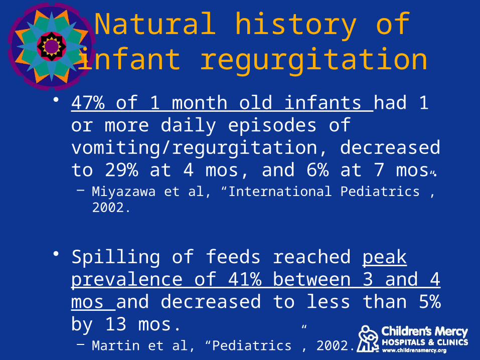
Vomiting/ Regurgitation in young
children• Most patients do NOT require imaging with an upper
GI exam as this study defines anatomy, and reflux may or may not be seen.
• In cases of persistent or severe regurgitation, pH probe monitoring may be helpful.
• Imaging should be considered if there are airway symptoms or bloody or bilious emesis.

pH probe in esophagus
What test should be ordered for a child with bilious emesis?
1. Esophagram
2. Upper GI/small bowel follow-through
3. OPM (oropharyngeal motility)
4. Upper GI study
Vomiting/ Regurgitation in young
children
• It is NOT necessary to order a small bowel follow-through examination to exclude malrotation because the pediatric upper GI examination includes imaging of the ligament of Treitz.
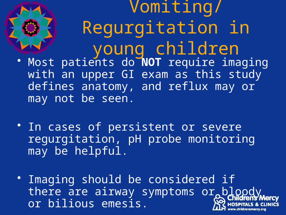
Newborn Bilious Emesis
• Bilious emesis in a newborn is an emergency which should be promptly evaluated with an upper GI examination to exclude malrotation and volvulus.
• Patients should have an NG or OG tube placed to confirm the presence of bilious material as well as facilitating the UGI exam.

Supine abdomenLeft lateral decubitus view
stomach
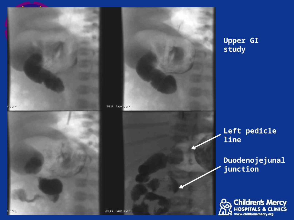
Left pedicle line
Duodenojejunal junction
Upper GI study

Upper GI study
Volvulus

Normal duodenojejunal junction (ligament of Treitz)
Pyloric Stenosis
• Common cause of early infantile intestinal obstruction.
• Also known as Hypertrophic Pyloric Stenosis (HPS).• Multifactorial causes suggested including:
– Hereditary– Exposure to macrolide antibiotics (erythromycin)– Abnormal myenteric plexus innervation– Infantile hypergastrinemia
Pyloric Stenosis
• 2-4 cases/ 1000 live births in U.S., male:female ratio 4:1
• 95% diagnosed between 3 and 12 weeks of age.
• Nonbilious emesis which becomes projectile.
• May have a palpable “olive” on exam.
In cases of suspected pyloric stenosis, the best radiology study to
order is:
1. Upper GI study
2. KUB (abd Xray)
3. Ultrasound
4. Computed tomography (CT)
Pyloric Stenosis
• A limited abdominal ultrasound is the diagnostic study of choice.– Highly sensitive and specific– No radiation– No sedation

Pyloric Ultrasound
Elongated pyloric channel
Thickened pyloric muscular wall

Pyloric Stenosis
• Individual wall thickness > 3mm
• Elongated pyloric channel >18mm
• Mucosal hypertrophy• Absence of fluid or gas
in the pyloric channel during the US study.
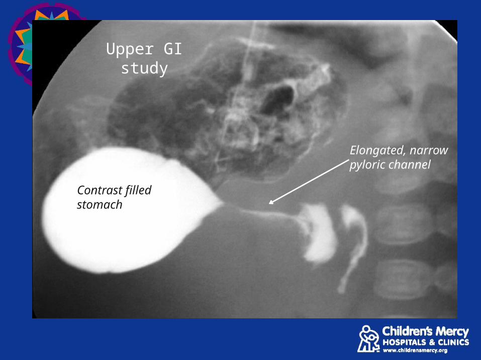
Upper GI study
Elongated, narrow pyloric channel
Contrast filled stomach
Intussusception
• Most common cause of intestinal obstruction in children aged 3 to 36 mos., 60% < 1 y.o., 80% < 2 y.o.
• Majority are idiopathic.• Seasonal patterns associated with gastroenteritis,
possibly due to hypertrophy of lymphoid tissue in the terminal ileum.
• Increased incidence after some forms of rotavirus vaccine.
Intussusception
• Pathologic lead point in some cases:– Meckel’s diverticulum– Enteric duplication cyst– Lymphoma– Polyps– Henoch-Schonlein purpura (intramural
hemorrhage)
Intussusception
• Present with sudden onset of crampy, intermittent abdominal pain with drawing-up of legs and inconsolable crying.
• May develop vomiting and currant-jelly stools.• Diagnostic work-up includes abdominal radiographs
and ultrasound.• Treated with air enema reduction.

Why order plain x-rays in suspected cases of intussusception?
1. Look for obstruction.
2. Exclude free air.
3. May suggest an alternative diagnosis.
4. All of the above.

Supine abdomen X-ray Left decubitus X-ray
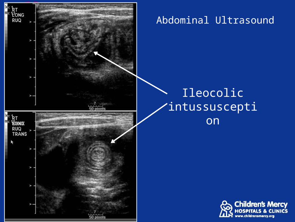
Abdominal Ultrasound
Ileocolic intussusception
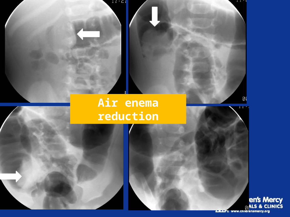
Air enema reduction
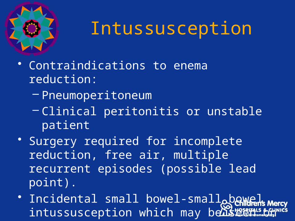
Intussusception
• Contraindications to enema reduction:– Pneumoperitoneum– Clinical peritonitis or unstable patient
• Surgery required for incomplete reduction, free air, multiple recurrent episodes (possible lead point).
• Incidental small bowel-small bowel intussusception which may be seen on US or CT is typically transient and asymptomatic.
Less common causes of obstruction
Newborn presentation:– Meconium ileus– Small bowel atresia– Meconium plug (small left colon) syndrome– Hirschprung’s disease

Multiple dilated bowel loops
Stomach

Filling defects in terminal ileum on contrast enema
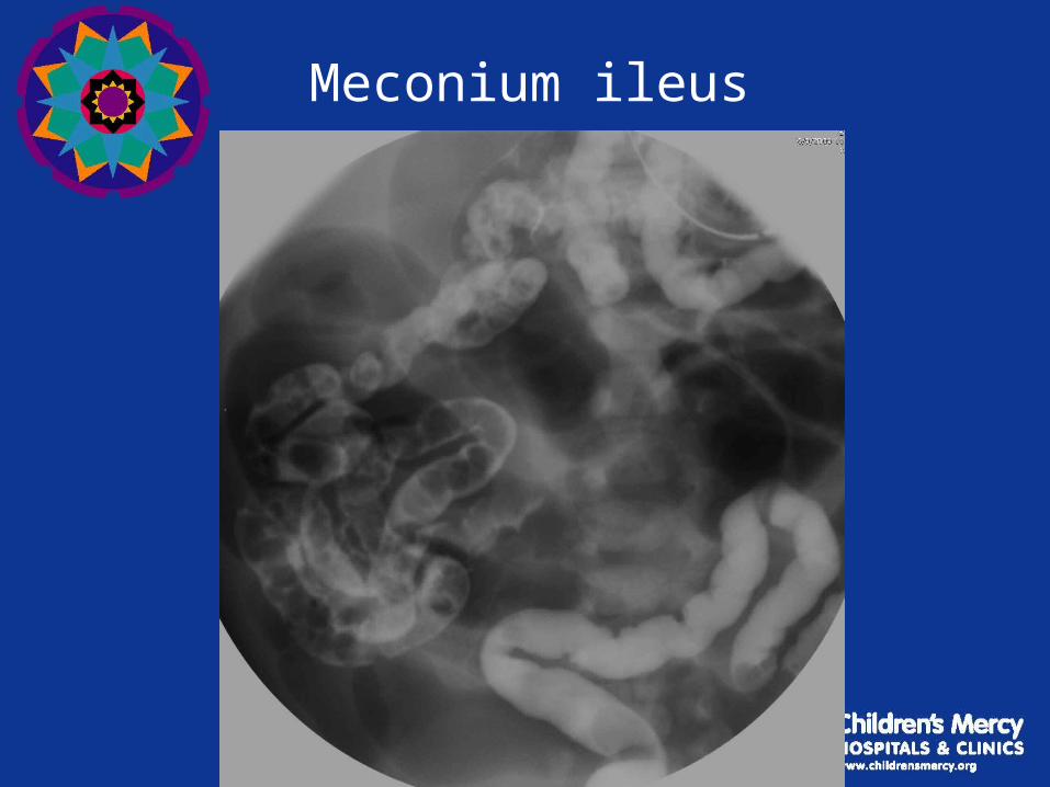
Meconium ileus
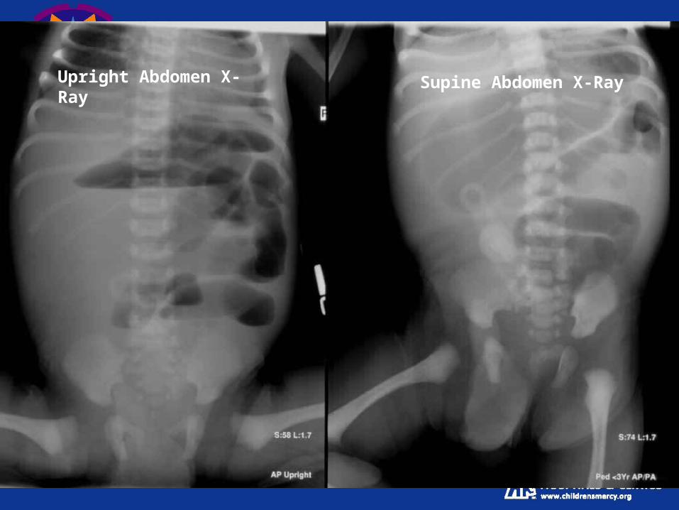
Upright Abdomen X-Ray Supine Abdomen X-Ray
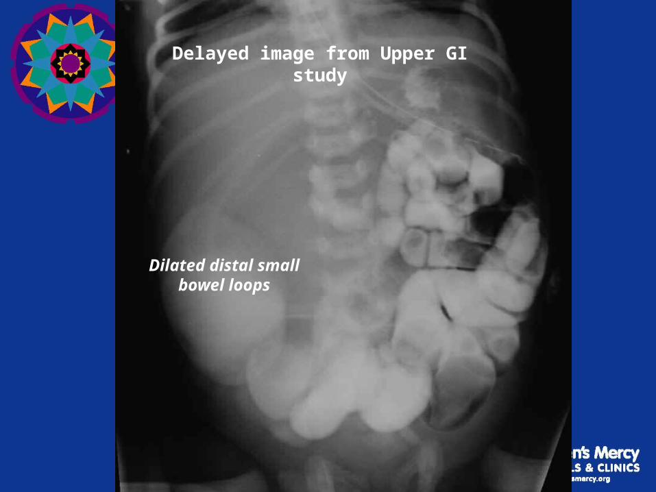
Delayed image from Upper GI study
Dilated distal small bowel loops
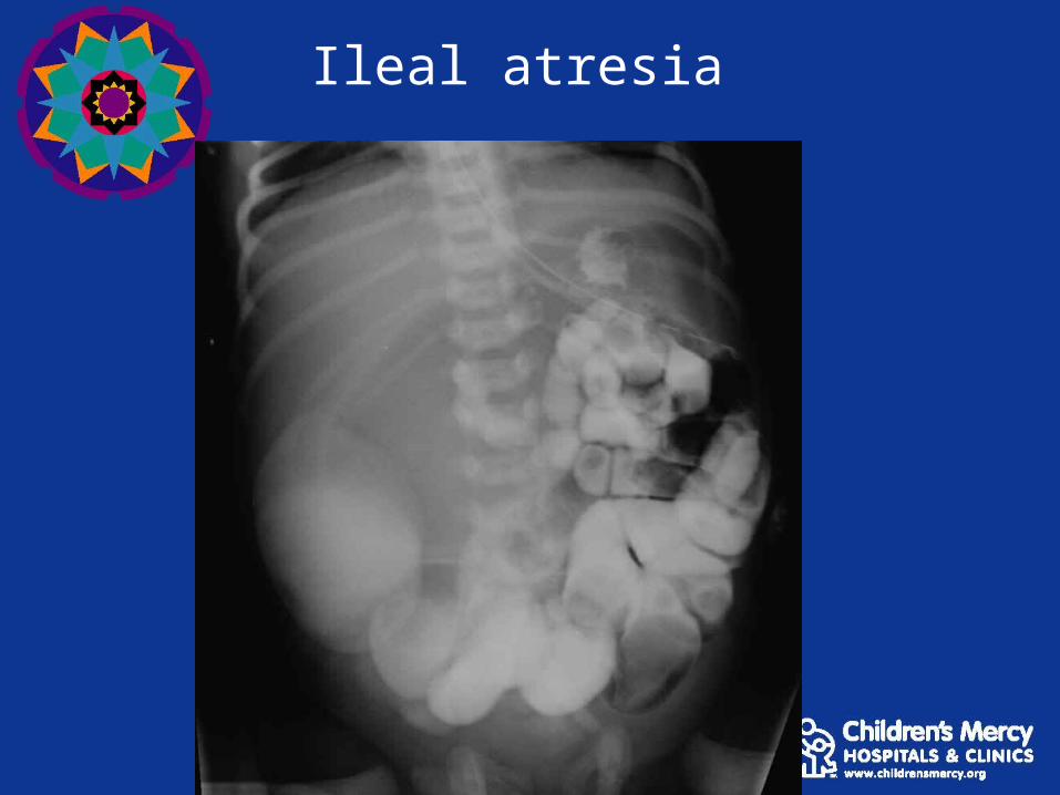
Ileal atresia

Multiple dilated bowel loops suggesting distal bowel pathology
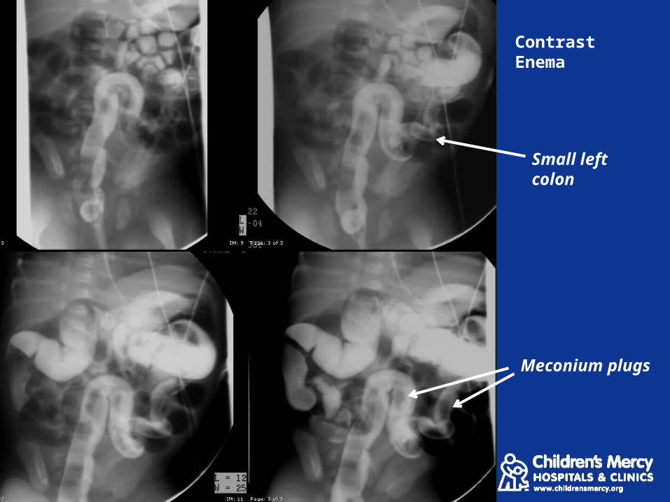
Small left colon
Meconium plugs
Contrast Enema

Meconium Plug Syndrome
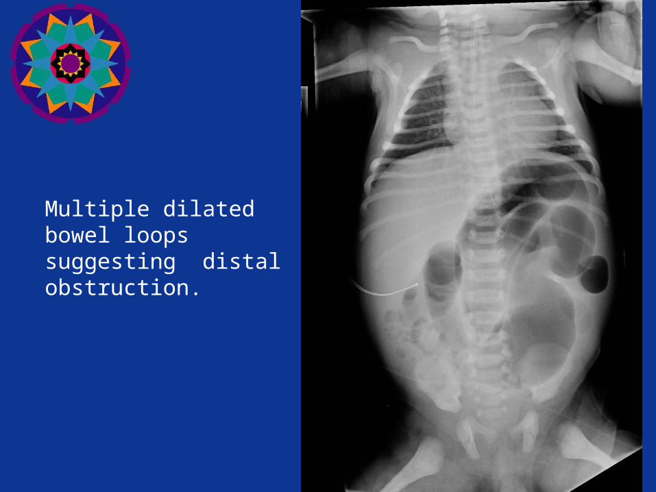
Multiple dilated bowel loops suggesting distal obstruction.

Lateral view from a contrast enema
Dilated sigmoid colon
Narrowed, irregular rectum

Hirschprung’s disease
Additional causes of obstruction
• Older infants and children:– Appendicitis– Adhesions– Incarcerated hernia– Meckel’s diverticulum
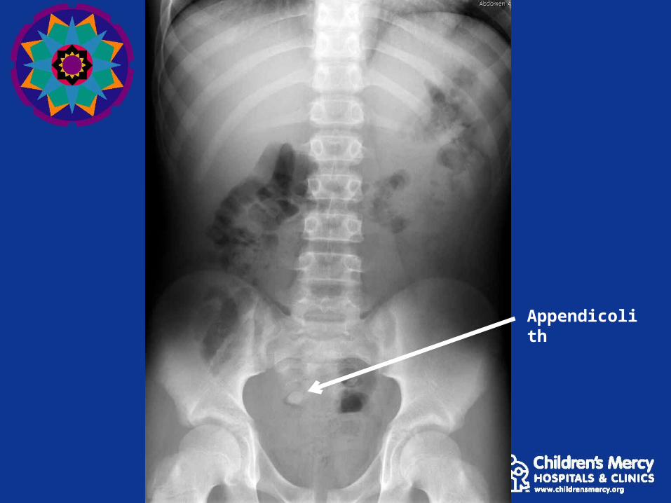
Appendicolith
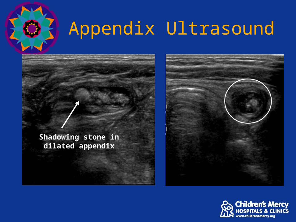
Appendix Ultrasound
Shadowing stone in dilated appendix
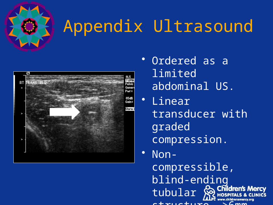
Appendix Ultrasound
• Ordered as a limited abdominal US.
• Linear transducer with graded compression.
• Non-compressible, blind-ending tubular structure, >6mm
• Often surrounded by edema/inflammation.

Appendicitis CTStone within an inflamed appendix
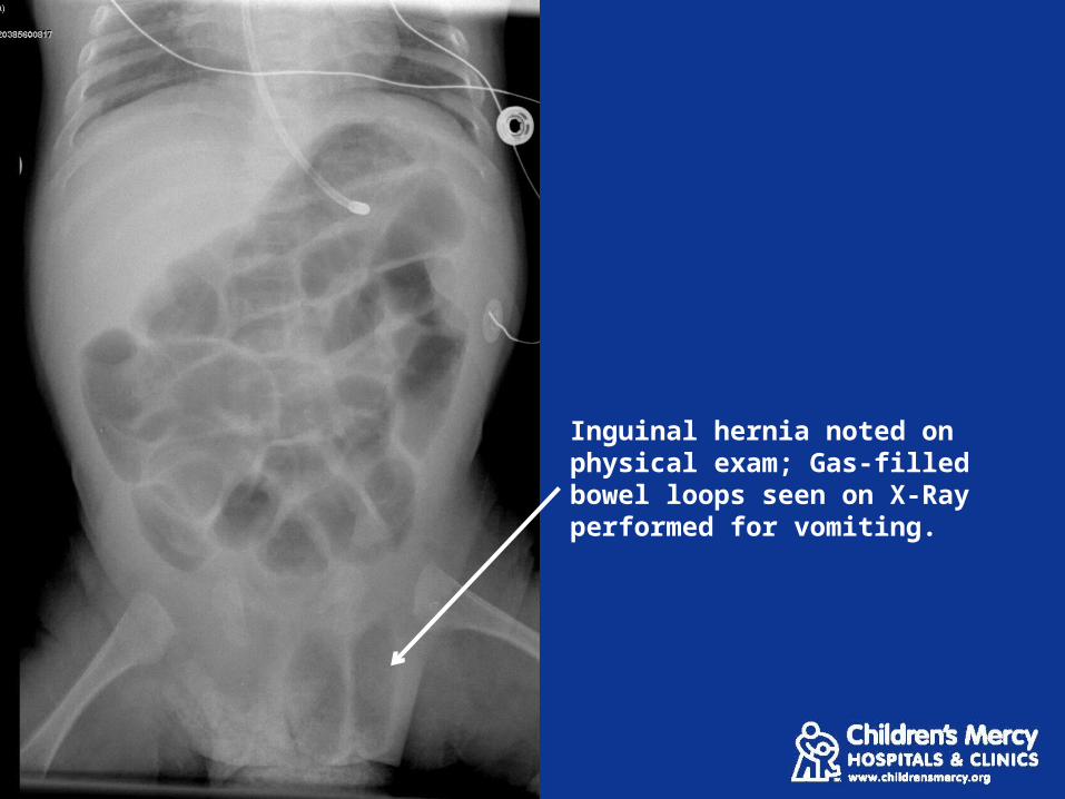
Inguinal hernia noted on physical exam; Gas-filled bowel loops seen on X-Ray performed for vomiting.
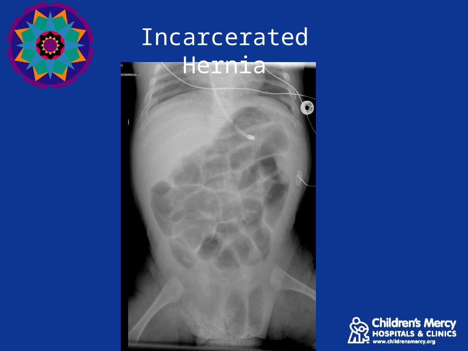
Incarcerated Hernia
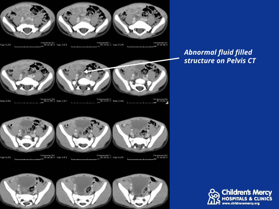
Abnormal fluid filled structure on Pelvis CT
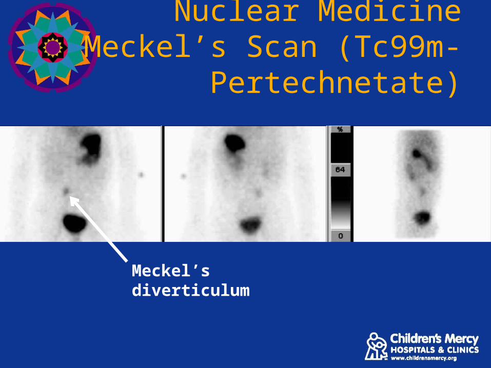
Nuclear Medicine Meckel’s Scan (Tc99m-
Pertechnetate)
Meckel’s diverticulum
Summary
• Most vomiting/ regurgitation in infants is due to reflux and does not require imaging.

Summary
• Most vomiting/ regurgitation in infants is due to reflux and does not require imaging.
• Bilious emesis is an emergency which should be evaluated by an upper GI study.
Summary
• Most vomiting/ regurgitation in infants is due to reflux and does not require imaging.
• Bilious emesis is an emergency which should be evaluated by an upper GI study.
• Ultrasound is an important tool in the diagnosis of pyloric stenosis and intussusception.
Summary
• Most vomiting/ regurgitation in infants is due to reflux and does not require imaging.
• Bilious emesis is an emergency which should be evaluated by an upper GI study.
• Ultrasound is an important tool in the diagnosis of pyloric stenosis and intussusception.
• When in doubt about the imaging work-up, consult your radiology colleagues at CMH.
Thank you for your attention
Related Documents