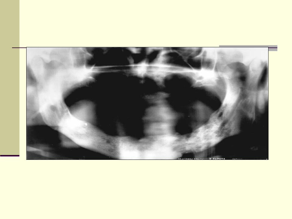
Radiology of inflammatory lesions of the jaws Dr. Ghaida’ AlJamal, BDS., MS., Dip (ABOMR)

Radiology of inflammatory lesions of the jaws
Aug 12, 2015
Welcome message from author
This document is posted to help you gain knowledge. Please leave a comment to let me know what you think about it! Share it to your friends and learn new things together.
Transcript

Radiology of inflammatory lesions of the jaws
Dr. Ghaida’ AlJamal, BDS., MS., Dip (ABOMR)

Most common pathologic conditions of jaws
Body responds to chemical, physical, or microbiologic injury with inflammation
Homeostasis
Balance of osteoclastic bone resorption and osteoblastic bone production
Mediators of inflammation tip this balance to favor either bone resorption or bone formation

Periapical inflammatory lesion
Source is necrotic pulp
Lesion restricted to the region of the tooth

Example

Osteomyelitis
Infection spreads in bone marrow
Lesion is no longer contained

Example
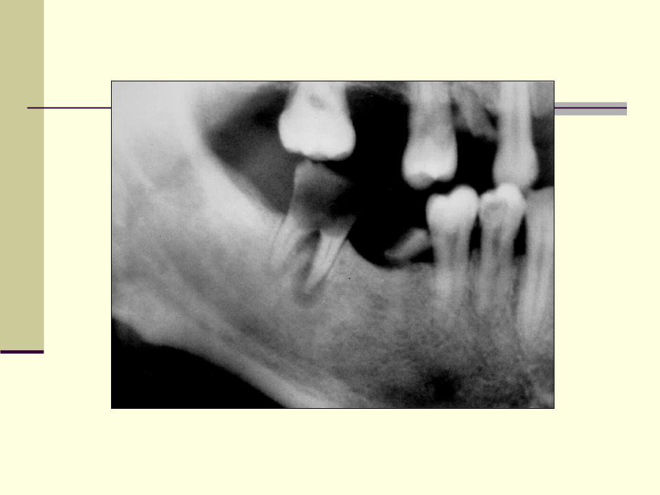
Periodontal lesions & pericoronitis
Lesion extended into overlying soft tissues
Arises in the tissues surrounding the crown of the PE tooth

Radiographic features
Location
Periphery
Internal structure
Effects on surrounding structures

Periapical inflammatory lesions
Acute apical periodontitis
Chronic apical periodontitis
Periapical abscess
Periapical granuloma

Periapical inflammatory lesions
Rarefying oseitis/sclerosing osteitis

Periapical inflammatory lesions
Def: local response of bone around apex of tooth that occurs 2° to necrosis of pulp or destruction of PA tissues by extensive periodontal disease

Caries acute
Necrotic pulp apical periodontitis
Trauma chronic
Periapical abcess
Periapical granuloma
Osteomyelitis
Periapical cyst

Clinical features
Asymptomatic …….severe pain w, w/o
facial swelling, fever, lymphadenopathy

Radiographic features
Location
Apex of involved tooth
Cervically up the tooth root

Radiographic features
Periphery
Ill defined
Well defined

Radiographic features
Internal structure Early …..no changes
Loss of bone density (widening of PDL at apex)
Larger diameter involvement
Mixture of sclerosis and rarefaction


Differential diagnosis
Periapical cemental dysplasia
Enostosis, osteosclerosis


Small, radiolucent periapical lesions with well defined periphery…….
granuloma or cyst

Differential diagnosis
Surgical scar
Mets & leukemia


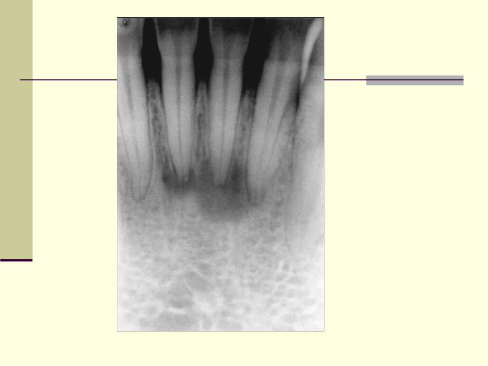
Pericoronitis
Inflammation of the tissues surrounding the crown of a partially erupted tooth
Radiographic features
No changes……
localized rarefaction and sclerosis……..
osteomyelitis

Radiographic features
Enlargement of follicular space with ill defined periphery and gradual transition of the normal trabecular pattern

Radiographic features
Surrounding bone rarefaction or sclerosis
Periosteal new bone formation

Differential diagnosis
Enostosis
FD
Oseosarcoma
SCC
Osteomyelitis
Inflammation of bone (marrow, cortex, cancellous portion & periosteum)
Source: pyogenic
hematogenous

Osteomyelitis
Hallmark is sequestra
It is a segment of bone that has become necrotic because of ischemic injury caused by inflammation

Acute Osteomyelitis
Predominantly neutrophils
From non vital teeth most commonly

Radiographic features
Very early ……no change
Ill defined periphery
Decrease in density
Loss of sharpness of trabeculae

Radiographic features
Maxilla is rare
Bone destruction area of radiolucency
Sclerotic regions
Sequestra maybe present….

Radiographic features
Cortical bone may be resorbed
Bone formation (involucrum)….
Onion-skin appearance (proliferative periostitis
Effects on teeth &lamina dura….

Differential diagnosis
FD
Malignancies (OS, SCC)
LCH
Lymphoma, luekemia

Chronic Osteomyelitis
De novo
Or sequela of inadequately treated acute Osteomyelitis

Diffuse sclerosing osteomyelitis
Bone metabolism shifts toward increased bone formation…..
Symptoms are less severe ….

Radiographic features
Posterior mandible most commonly
Periphery better defined…..
Regions of greater and lesser radiopacity…
More chronic lesions are exceedingly radiopaque…

Radiographic features
Sequestrum….
Periosteal new bone (similar to onion skin)
Outer contour of mandible altered
External resorption, LD less apparent, PDL enlarged
May develop draining fistula


Differential diagnosis
FD
Paget disease of bone
OS
LCH, leukemia, lymphoma

Osteoradionecrosis
Inflammatory condition of bone that occurs after bone has been exposed to therapeutic doses of radiation

Osteoradionecrosis
Radiation causes damage to bone w hypoxia, hypocellularity and hypovascularity
Delayed or lack of healing….
Clinical and radiographic features
Posterior mandible…
Bone exposure…
Pathologic fracture
Pain or no pain

Radiographic features
Similar to chronic osteomyelitis
Ill defined periphery
More bone formation sclerotic appearance…

Radiographic features
Scattered regions of radiolucency w, w/o central sequestra
Uncommon inflammatory new bone….
Rare bone formation on outer cortex

Differential diagnosis
Malignant neoplasm
Chronic osteomyelitis (history)

Related Documents
![ORAL ~RADIOLOGY - dent.zaums.ac.irdent.zaums.ac.ir/uploads/1_296_chapter1.pdf · tal Disturbances of the Face and Jaws. ... 19 Inflammatory l esions of the Jaws, ]66 ... 29 Developmental](https://static.cupdf.com/doc/110x72/5a97a0aa7f8b9ab6188ced90/oral-radiology-dentzaumsac-disturbances-of-the-face-and-jaws-19-inflammatory.jpg)