Katrina McGinty MD Department of Radiology, UNC-CH Abdominal Imaging Group August 14, 2018 RIGHT IMAGE FOR THE RIGHT PATIENT

Welcome message from author
This document is posted to help you gain knowledge. Please leave a comment to let me know what you think about it! Share it to your friends and learn new things together.
Transcript

Katrina McGinty MD
Department of Radiology, UNC-CH
Abdominal Imaging Group
August 14, 2018
RIGHT IMAGE FOR THE RIGHT PATIENT

OVERVIEW
• Overutilization
• Review of general scan limitations
• Review of each modality, pros and cons, common indications

GROWTH OF IMAGING
• In the past decade, imaging services and their cost have grown at twice the rate of other
technologies in the health care industry
• Radiation dose!
• $$$
Hendee, W. et al. Addressing Overutilization in Medical Imaging. Radiology. August 2010.
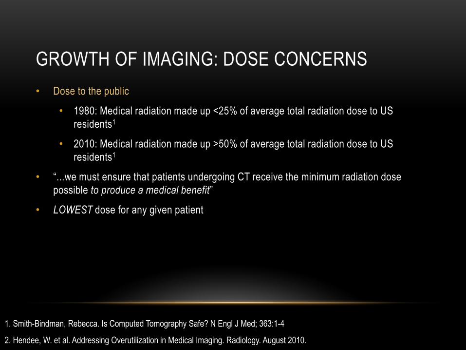
GROWTH OF IMAGING: DOSE CONCERNS
• Dose to the public
• 1980: Medical radiation made up <25% of average total radiation dose to US
residents1
• 2010: Medical radiation made up >50% of average total radiation dose to US
residents1
• “...we must ensure that patients undergoing CT receive the minimum radiation dose
possible to produce a medical benefit”
• LOWEST dose for any given patient
1. Smith-Bindman, Rebecca. Is Computed Tomography Safe? N Engl J Med; 363:1-4
2. Hendee, W. et al. Addressing Overutilization in Medical Imaging. Radiology. August 2010.

GROWTH OF IMAGING: DOSE CONCERNS
• Dose to the public
• 1980: Medical radiation made up <25% of average total radiation dose to US
residents1
• 2010: Medical radiation made up >50% of average total radiation dose to US
residents1
• “…we must ensure that patients undergoing CT receive the minimum radiation dose
possible to produce a medical benefit”2
• LOWEST dose for any given patient
• APPROPRIATE dose for any given patient
1. Smith-Bindman, Rebecca. Is Computed Tomography Safe? N Engl J Med; 363:1-4
2. Hendee, W. et al. Addressing Overutilization in Medical Imaging. Radiology. August 2010.

GROWTH OF IMAGING: DOSE CONCERNS
• Radiology is measured in effective dose (millisieverts: mSv)
• Refers to radiation risk averaged over the entire body
• Background radiation (cosmic radiation, radon): 3 mSv/year
• Effective dose may be used to estimated risk of cancer/cancer related death
• Risk levels: Additional risk of fatal cancer from an examination
• Negligible: less than 1 in 1,000,000
• Minimal: 1 in 1,000,000 to 1 in 100,000
• Very low: 1 in 100,000 to 1 in 10,000
• Low: 1 in 10,000 to 1 in 1,000
• Moderate: 1 in 1,000 to 1 in 500
• These risk levels represent a very small addition to the 1 in 5 chance we all have of dying from cancer
Radiologyinfo.org American college of radiology and Radiologic Society of North America

GROWTH OF IMAGING: DOSE CONCERNS
Procedure Effective dose Comparable for natural
background radiation for:
Additional life risk of
fatal cancer
Intra-oral XR 0.005 mSv 1 day Negligible
Extremity XR 0.001 mSv 3 hours Negligible
Chest XR 0.1 mSv 10 days Minimal
Spine XR 1.5 mSv 6 months Very low
Head CT 2-4 mSv 8-16 months Low
Chest CT 1.5-7 mSv 6 months-2years Very low to low
Abdominopelvic CT 10-20 mSv 3-7 years Low to moderate
PET-CT 25 mSv 8 years Moderate
Radiologyinfo.org American college of radiology and Radiologic Society of North America
• New software and dose reduction protocols are continually evolving
• Doses vary with scan technique and patient size

GROWTH OF IMAGING: COST OF EXAMS
• Conventional Radiography (X-ray): $149-$388
• Two view chest x ray: $207
• 4 views of the knee: $266
• Ultrasound: $386-1360
• Breast ultrasound: $386
• Abdominal ultrasound: $783
• Carotid Doppler: $1360
• CT: $1072- $1832 per body part!
• CT CAP w/wo contrast: $5322!
• MR: $1555 - $4547
• Brain MR: $2189
• Abdominal MRI w/wo contrast: $4547

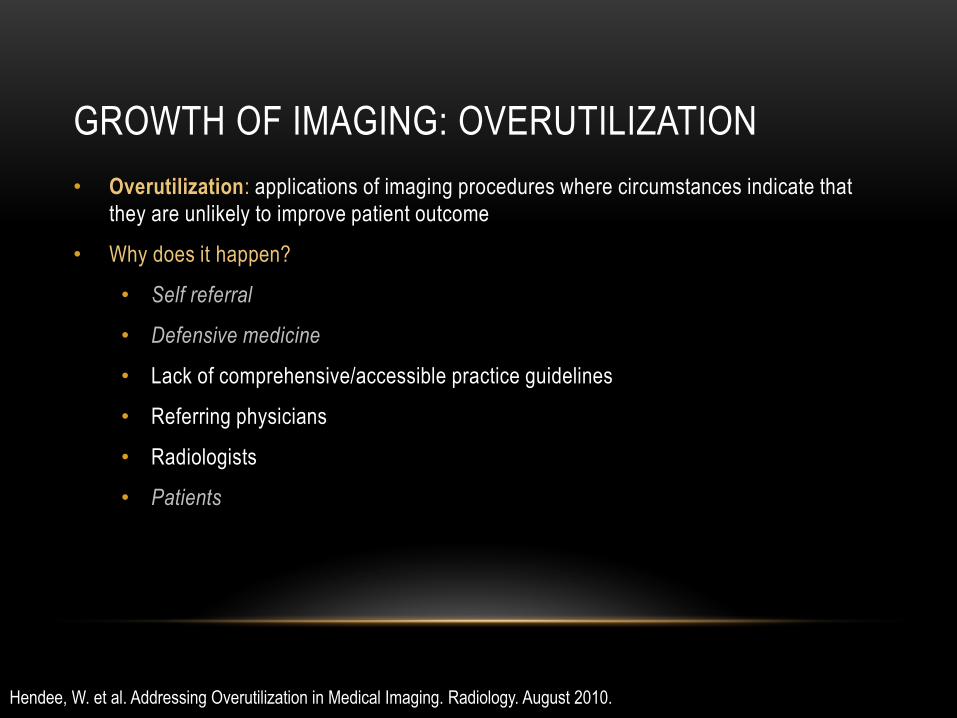
GROWTH OF IMAGING: OVERUTILIZATION
• Overutilization: applications of imaging procedures where circumstances indicate that
they are unlikely to improve patient outcome
• Why does it happen?
• Self referral
• Defensive medicine
• Lack of comprehensive/accessible practice guidelines
• Referring physicians
• Radiologists
• Patients
Hendee, W. et al. Addressing Overutilization in Medical Imaging. Radiology. August 2010.
GROWTH OF IMAGING: OVERUTILIZATION
Hendee, W. et al. Addressing Overutilization in Medical Imaging. Radiology. August 2010.
• Overutilization: applications of imaging procedures where circumstances indicate that
they are unlikely to improve patient outcome
• Why does it happen?
• Self referral
• Defensive medicine
• Lack of comprehensive/accessible practice guidelines
• Referring physicians
• Radiologists
• Patients

YOUR OPTIONS…
X ray Ultrasound CT MR

YOUR OPTIONS…
X ray
What views?
Ultrasound CT MR

YOUR OPTIONS…
X ray Ultrasound
Limitations?
CT MR
YOUR OPTIONS…
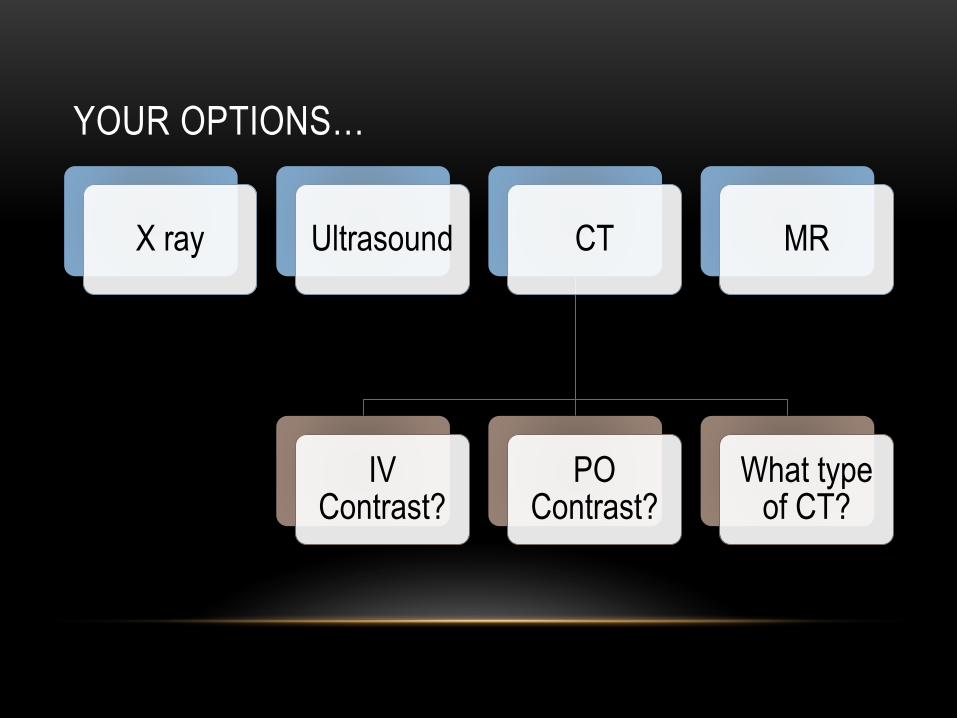
X ray Ultrasound CT
IV Contrast?
PO Contrast?
What type of CT?
MR
YOUR OPTIONS…
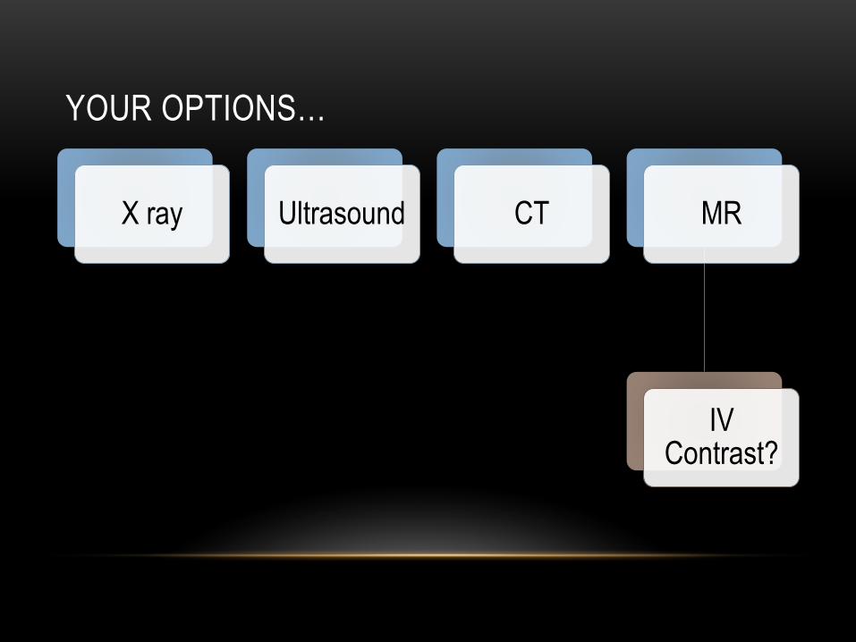
X ray Ultrasound CT MR
IV Contrast?

YOUR OPTIONS…
X ray Ultrasound CT MR
IV Contrast?
NSFGadolinium deposition
Renal function
YOUR OPTIONS…
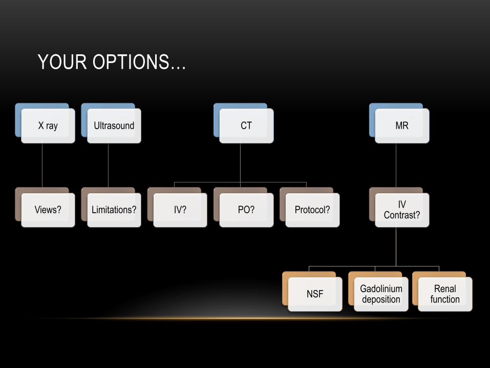
X ray
Views?
Ultrasound
Limitations?
CT
IV? PO? Protocol?
MR
IV Contrast?
NSFGadolinium deposition
Renal function

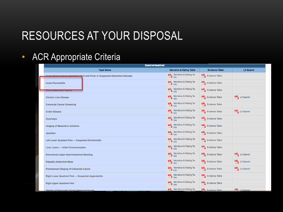
RESOURCES AT YOUR DISPOSAL
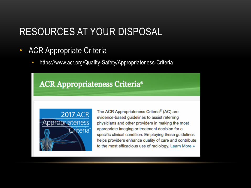
• ACR Appropriate Criteria
• https://www.acr.org/Quality-Safety/Appropriateness-Criteria
RESOURCES AT YOUR DISPOSAL
• ACR Appropriate Criteria
• https://www.acr.org/Quality-Safety/Appropriateness-Criteria

RESOURCES AT YOUR DISPOSAL
• ACR Appropriate Criteria
RESOURCES AT YOUR DISPOSAL
• ACR Appropriate Criteria

RESOURCES AT YOUR DISPOSAL
• ACR Appropriate Criteria

RESOURCES AT YOUR DISPOSAL
• ACR Appropriate Criteria
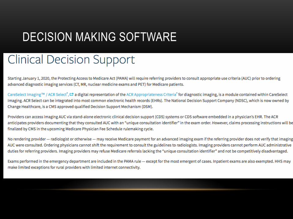
DECISION MAKING SOFTWARE

DECISION MAKING SOFTWARE
RESOURCES AT YOUR DISPOSAL
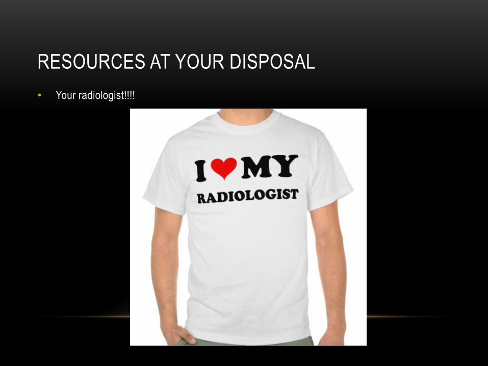
• Your radiologist!!!!

RIGHT SCAN FOR THE RIGHT PATIENT
• Ultrasound
• BODY HABITUS!
• Will we be able to image the area in question?
• CT
• Can the patient lie flat for several minutes on their back
• Can the patient hold their breath for ~15 second
• MR
• Is the patient able to lie in an enclosed space for up to one hour?
• Can the patient lie on their back?
• Can the patient hold their breath for 20 second?
• Can the patient tolerate premedication (anxiety) and still follow breathing instructions?
• Can the patient tolerate loud noises?

CONVENTIONAL RADIOGRAPH (X-RAY)
Cheap
Relatively low radiation dose
Readily accessible
Clinician friendly
Limited sensitivity
Possibly over utilized
False positives
Unsatisfying reports!
Pro
sC
ons
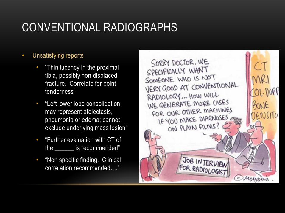
CONVENTIONAL RADIOGRAPHS
• Unsatisfying reports
• “Thin lucency in the proximal
tibia, possibly non displaced
fracture. Correlate for point
tenderness”
• “Left lower lobe consolidation
may represent atelectasis,
pneumonia or edema; cannot
exclude underlying mass lesion”
• “Further evaluation with CT of
the ______ is recommended”
• “Non specific finding. Clinical
correlation recommended….”

CONVENTIONAL RADIOGRAPH
Non specific lobulated left lower lobe mass, possibly loculated fluid, pleural based mass,
neoplasm, infection….. Recommend correlation with CT of the chest.

CONVENTIONAL RADIOGRAPH
Multiloculated collection in the pleural space consistent with empyema

CONVENTIONAL RADIOGRAPH
• Specific views and patient
positioning may be helpful
• Upright chest x ray:
Pneumoperitoneum
• Expiratory upright film:
Pneumothorax
• Decubitus films: Layering
effusion
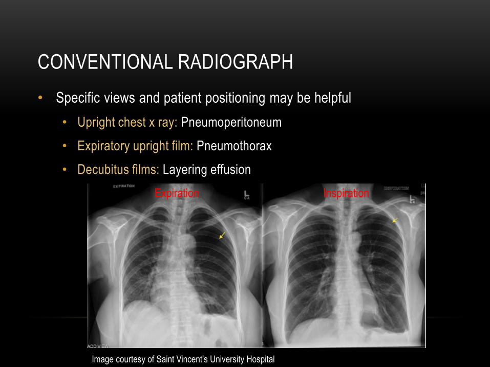
CONVENTIONAL RADIOGRAPH
• Specific views and patient positioning may be helpful
• Upright chest x ray: Pneumoperitoneum
• Expiratory upright film: Pneumothorax
• Decubitus films: Layering effusion
Image courtesy of Saint Vincent’s University Hospital
InspirationExpiration

CONVENTIONAL RADIOGRAPH
• Easily accessible
• Not always sensitive or specific
• Specific views may answer specific clinical questions
• You get what you pay for!

ULTRASOUND
No radiation
“Real time” imaging- blood flow, peristalsis, etc
Cheap (relatively speaking
Operator dependent
Patient dependent
• Body habitus
• Positioning
• Breath hold
Pro
sC
ons

ULTRASOUND LIMITATIONS: BODY HABITUS
BMI 24 BMI 49

ULTRASOUND LIMITATIONS: BOWEL GAS
ULTRASOUND: LIMITATIONS
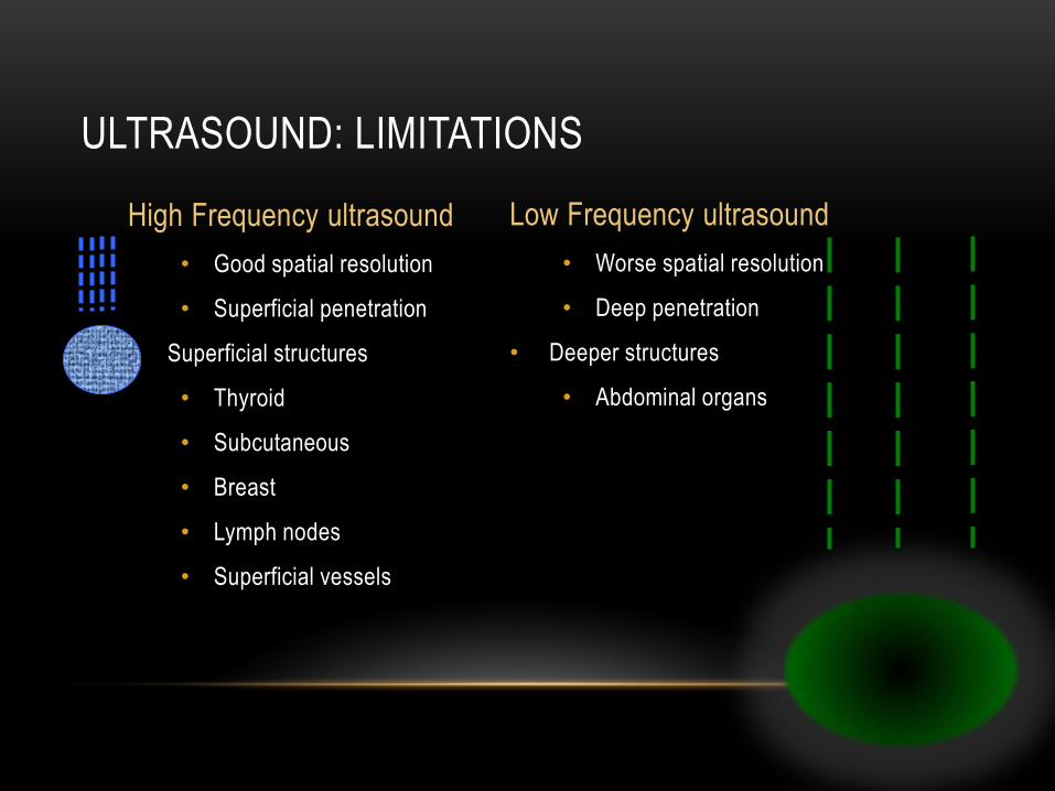
High Frequency ultrasound
• Good spatial resolution
• Superficial penetration
• Superficial structures
• Thyroid
• Subcutaneous
• Breast
• Lymph nodes
• Superficial vessels
Low Frequency ultrasound
• Worse spatial resolution
• Deep penetration
• Deeper structures
• Abdominal organs
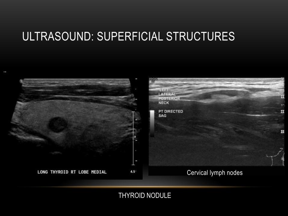
ULTRASOUND: SUPERFICIAL STRUCTURES
THYROID NODULE
Cervical lymph nodes
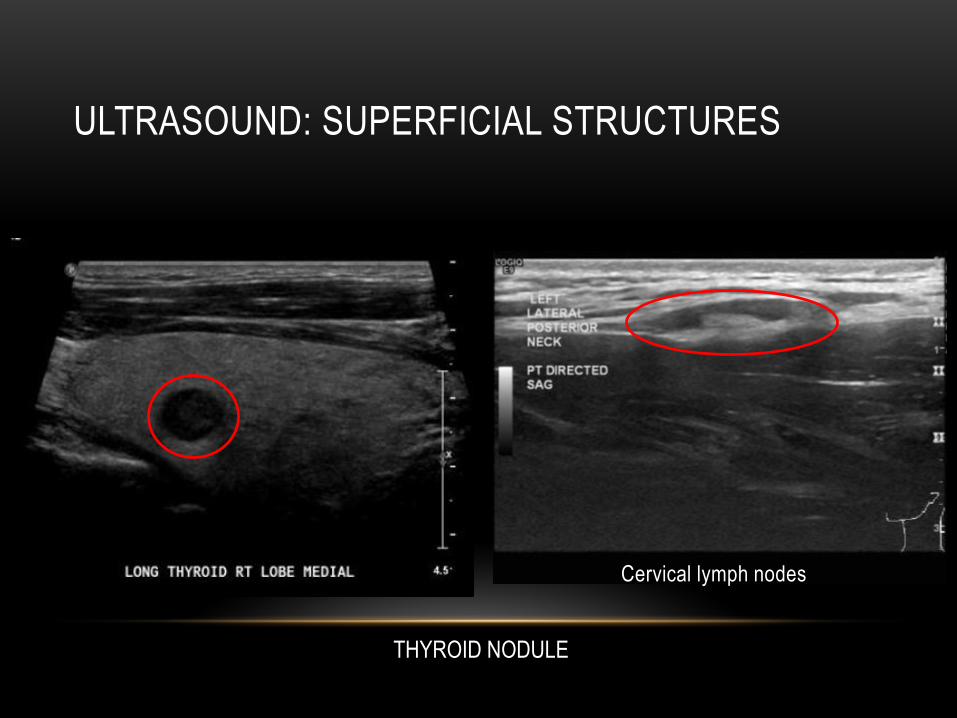
ULTRASOUND: SUPERFICIAL STRUCTURES
THYROID NODULE
Cervical lymph nodes

ULTRASOUND: DYNAMIC IMAGING
Left inguinal hernia with Valsalva

ULTRASOUND: DYNAMIC IMAGING
Left inguinal hernia with Valsalva

ULTRASOUND: DYNAMIC IMAGING
Left inguinal hernia with Valsalva

ULTRASOUND: DYNAMIC IMAGING
Left inguinal hernia with Valsalva

ULTRASOUND: DYNAMIC IMAGING
Left inguinal hernia with Valsalva

ULTRASOUND: DYNAMIC IMAGING
Left inguinal hernia with Valsalva

ULTRASOUND: DYNAMIC IMAGING
Left inguinal hernia with Valsalva

ULTRASOUND: DYNAMIC IMAGING
Left inguinal hernia with Valsalva

ULTRASOUND: DYNAMIC IMAGING
Left inguinal hernia with Valsalva

ULTRASOUND: VASCULAR EVALUATION
• Cirrhotic patient: TIPS evaluation
Elevated velocities through TIPS indicative of stent malfunction

ULTRASOUND: BILIARY TREE
Intrahepatic biliary ductal dilatation Normal caliber common bile duct

ULTRASOUND: BILIARY TREE
Intrahepatic biliary ductal dilatation Normal caliber common bile duct

ULTRASOUND: GALLBLADDER/BILIARY TREE
• Incidental gallstone

ULTRASOUND: GU IMAGING
• First line modality:
• Uterus
• Ovaries
• Testicles
• Superficial structures
• Why?
• Good soft tissue contrast
• No radiation

ULTRASOUND: GU EVALUATION: UTERUS
• Post menopausal bleeding
Ill defined uterine mass, possibly leiyomyoma
although neoplasm cannot be excluded:
Recommend ultrasound for further evaluation…

ULTRASOUND: GU EVALUATION: UTERUS
• Post menopausal bleeding
Ill defined uterine mass, possibly leiyomyoma
although neoplasm cannot be excluded:
Recommend ultrasound for further evaluation…

ULTRASOUND: GU EVALUATION: UTERUS
FIGO grade II endometrial adenocarcinoma involving 81% of the myometrium

ULTRASOUND: GU EVALUATION: UTERUS
FIGO grade II endometrial adenocarcinoma involving 81% of the myometrium
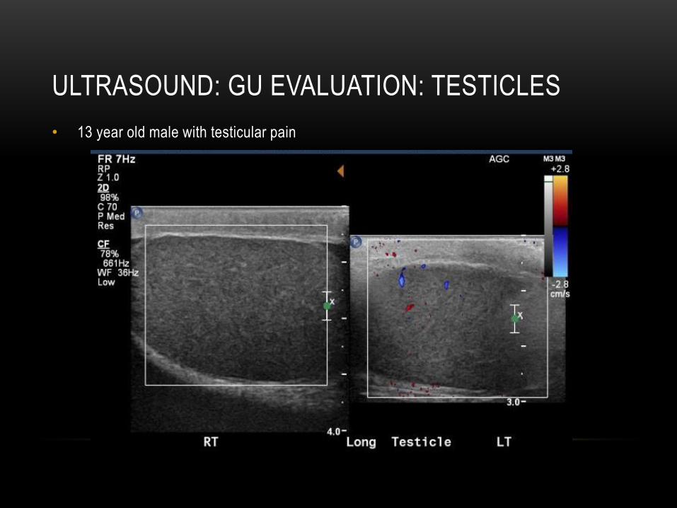
ULTRASOUND: GU EVALUATION: TESTICLES
• 13 year old male with testicular pain

ULTRASOUND: GU EVALUATION: TESTICLES
• 13 year old male with testicular pain
Right sided testicular torsion

ULTRASOUND: GU EVALUATION: KIDNEYS
• Useful:
• Stones (sometimes)
• Hydronephrosis
• Cysts (sometimes)
• Not useful:
• Characterizing solid renal masses
• Ureteral stones (sometimes)
• *Coming soon: Ultrasound contrast! Stay tuned!

ULTRASOUND: HYDRONEPHROSIS

ULTRASOUND: NEPHROLITHIASIS

ULTRASOUND: SOLID VERSUS CYSTIC
Multiple simple renal cysts
Characteristics of a cyst
1. Anechoic = “black”
2. Posterior acoustic enhancement =
“bright shadow”
3. No blood flow
4. Nothing in it
Simple cyst

ULTRASOUND: SOLID VERSUS CYSTIC
Multiple simple renal cysts
Characteristics of a cyst
1. Anechoic = “black”
2. Posterior acoustic enhancement =
“bright shadow”
3. No blood flow
4. Nothing in it
Simple cyst

ULTRASOUND: SOLID VERSUS CYSTIC
Characteristics of a cyst
1. Anechoic = “black”
2. Posterior acoustic enhancement =
“bright shadow”
3. No blood flow
4. Nothing in it
Cyst!Cyst???
Mildly complex cystsSimple cyst

ULTRASOUND: SOLID VERSUS CYSTIC
Characteristics of a cyst
1. Anechoic = “black”
2. Posterior acoustic enhancement =
“bright shadow”
3. No blood flow
4. Nothing in it
Cyst!Cyst???
Mildly complex cystsSimple cyst

ULTRASOUND: GU: KIDNEYS
• 79 year old female with acute kidney injury
Characteristics of a cyst
1. Anechoic = “black”
2. Posterior acoustic enhancement =
“bright shadow”
3. No blood flow
4. Nothing in it
Mildly complex cystsSimple cyst

ULTRASOUND: GU: KIDNEYS
• 79 year old female with acute kidney injury
Characteristics of a cyst
1. Anechoic = “black”
2. Posterior acoustic enhancement =
“bright shadow”
3. No blood flow
4. Nothing in it
Mildly complex cystsSimple cyst

ULTRASOUND: GU: KIDNEYS
• 79 year old female with acute kidney injury
Characteristics of a cyst
1. Anechoic = “black”
2. Posterior acoustic enhancement =
“bright shadow”
3. No blood flow
4. Nothing in it
Mass
MassKidney
Mildly complex cystsSimple cyst

ULTRASOUND: GU: KIDNEYS
• 79 year old female with AKI
Renal cell carcinoma

ULTRASOUND: GU: EVALUATION OF KIDNEY
• 71 year old male with right upper quadrant pain
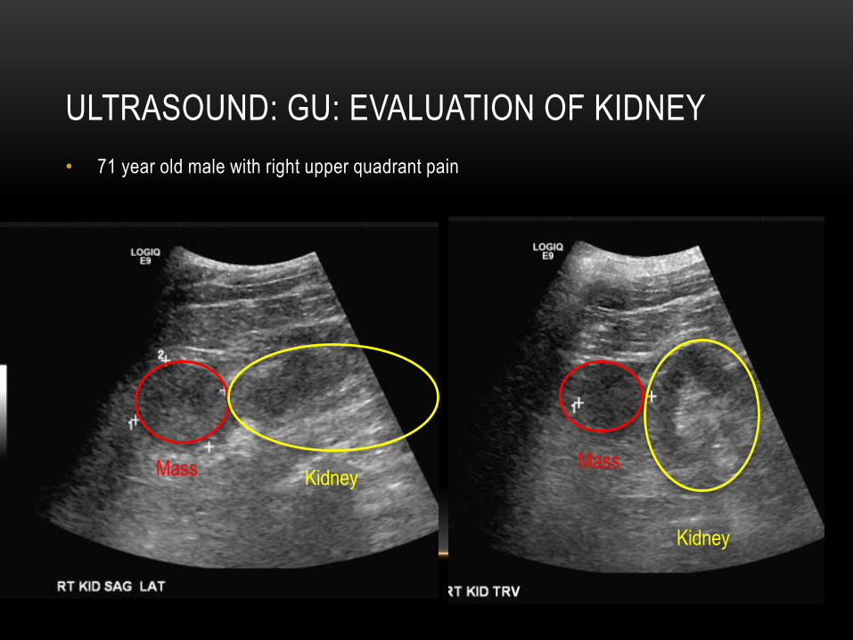
ULTRASOUND: GU: EVALUATION OF KIDNEY
• 71 year old male with right upper quadrant pain
Mass Mass
Kidney
Kidney

ULTRASOUND: GU: EVALUATION OF KIDNEY
• 71 year old male with right upper quadrant pain
Clear cell renal cell carcinoma: 5 cm

ULTRASOUND: GU EVALUATION: KIDNEYS
• Uses
• Hydronephrosis: Very good
• Calculi: Good (renal caluli, not necessarily ureteral calculi)
• Cysts: Okay (simple cysts, non obese patient)
• Masses (detection and characterization): Poor

ULTRASOUND
• First line imaging modality
• Vascular pathology
• Dynamic ”real time” imaging
• Biliary pathology
• Uterus, ovaries, testicle
• Kidneys (sometimes)
• NOT useful for
• Characterizing solid lesions
• Detection of occult pathology outside of the probe’s range
• Penetrating extensive fat/gas

RECENTLY ARRIVED!
CONTRAST ENHANCED ULTRASOUND!!!
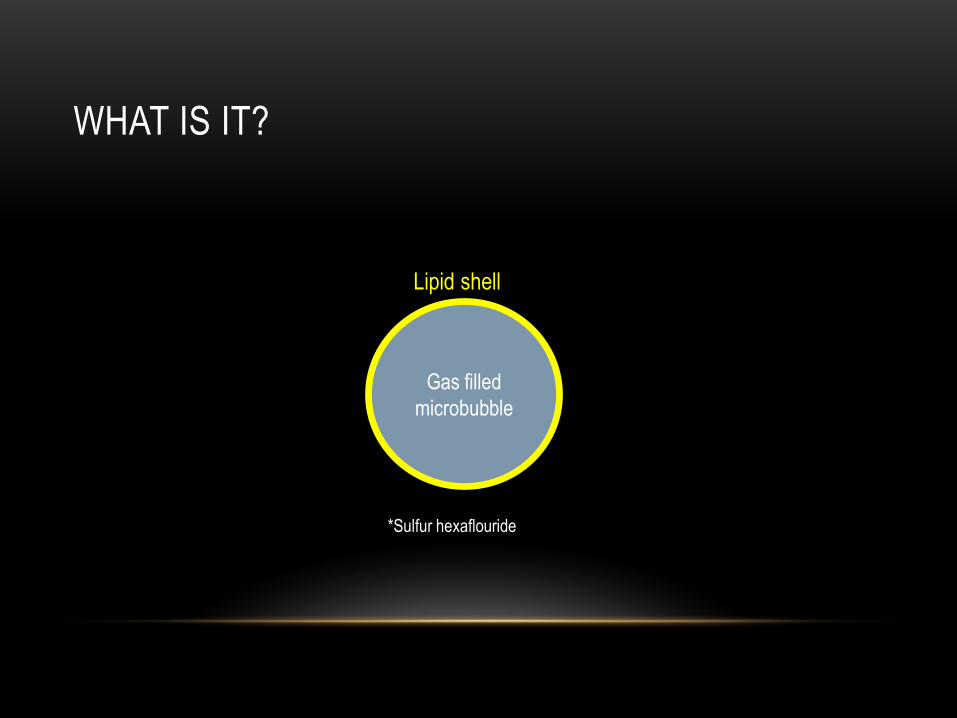
WHAT IS IT?
Gas filled
microbubble
Lipid shell
*Sulfur hexaflouride

WHAT IS IT?
Gas filled
microbubble
Lipid shell
Same size as RBC
6-8 microns2-8 microns

WHAT IS IT?
Gas filled
microbubble
Lipid shell
Oscillates when in US field
Ç√
WHY DO WE NEED IT?
WHY DO WE NEED IT?
• Metabolism: Gas is exhaled, lipids broken down in liver

WHY DO WE NEED IT?
• Metabolism
• NO renal excretion!

WHY DO WE NEED IT?
• Metabolism
• NO renal excretion!
• Basically no hepatic excretion!
WHY DO WE NEED IT?
• Metabolism
• NO renal excretion!
• Basically no hepatic excretion!
• Repeated injection
WHY DO WE NEED IT?
• Metabolism
• NO renal excretion!
• Basically no hepatic excretion!
• Repeated injection
• Dose is ~2 mL (or less)
• Bubbles last 5-10 minutes
• Can destroy bubbles using ultrasound to reinject if needed

WHY DO WE NEED IT?
• Metabolism
• NO renal excretion!
• Basically no hepatic excretion!
• Repeated injection
• Dose is ~2 mL (or less)
• Bubbles last 5-10 minutes
• Can destroy bubbles using ultrasound to reinject if needed
• Have given up to 161 mL without adverse effects!

WHY DO WE NEED IT?
• Renal impaired patients
• Patients who cannot tolerate MRI
• Patients with contrast allergies

HOW DOES IT LOOK?
• Real time enhancement of lesion
• Pattern of enhancement: Central vs. peripheral
• Washout
• Presence or absence
• Timing of washout
• Opportunity for repeat injections if uncertain of pattern

HOW DOES IT LOOK?
• Hemangioma

HOW DOES IT LOOK?
• HCC
HOW ACCURATE IS IT?

HOW ACCURATE IS IT?
CEUS CECT CEMR

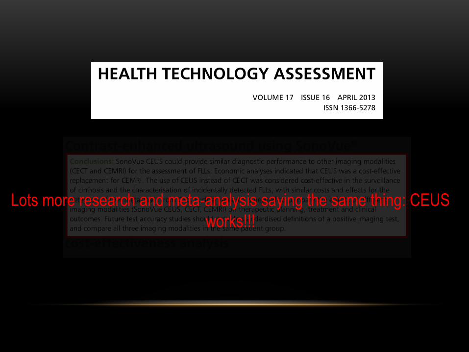
Lots more research and meta-analysis saying the same thing: CEUS
works!!!
HOW DOES IT WORK
• Lesion localized on US

HOW DOES IT WORK
• Lesion localized on US
• Contrast injected

HOW DOES IT WORK
• Lesion localized on US
• Contrast injected
• Lesion watched in real time
• Cine clips
• Intermittent observation for 5+ minutes
• Radiologist in room (for now)

HOW DOES IT WORK
• Lesion localized on US
• Contrast injected
• Lesion watched in real time
• Cine clips
• Intermittent observation for 5+ minutes
• Radiologist in room (for now)
• If lesion characterized, exam is done and patient can go
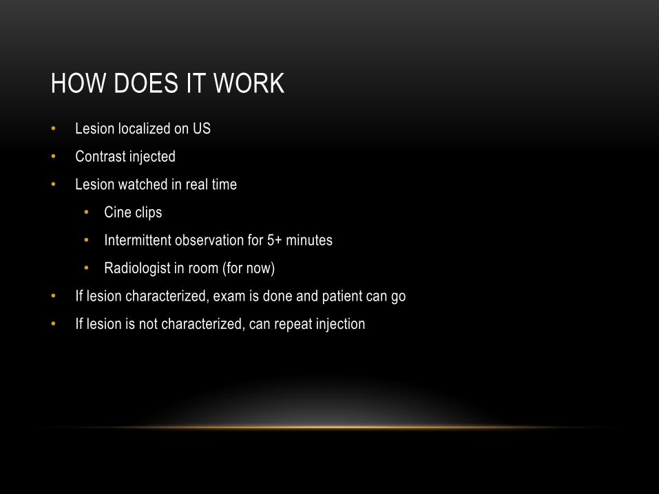
HOW DOES IT WORK
• Lesion localized on US
• Contrast injected
• Lesion watched in real time
• Cine clips
• Intermittent observation for 5+ minutes
• Radiologist in room (for now)
• If lesion characterized, exam is done and patient can go
• If lesion is not characterized, can repeat injection

LIMITATIONS
• Lesion must be visualized on ultrasound
• Patient size
• Gas
• Limited penetration of cirrhotic/steatotic livers

LIMITATIONS
• Lesion must be visualized on ultrasound
• Patient size
• Gas
• Limited penetration of cirrhotic/steatotic livers
• Staffing
• Attending in room
• 1 hour for exam (procedure slot)

CONTRAINDICATIONS
• Prior allergic reaction to ultrasound contrast (1 in ~12,000)
• Unstable cardiac disease
WHERE TO GO FROM HERE?
• Be patient
• Role in interventions
• LOTS of possibilities!!!
QUESTIONS?

COMPUTED TOMOGRAPHY
Quick
Easily accessible
“Screening test”
Radiation: doses are 100-500x those of conventional radiograph
IV contrast
Pro
sC
ons

CT CONTRAST AGENTS
• At risk patients: BUN/Creatinine recommended within 30 days of the exam IF…
• > 60 year old
• History of renal disease
• Dialysis
• Renal transplant
• Single kidney
• Renal cancer
• Renal surgery
• Hypertension requiring medical therapy
• History of diabetes
• Metformin use
• No universal cutoff- will vary with institution
• Range of serum creatinine 1.5-2.0
ACR Manual on Contrast Media, Version 10.2, 2016
CT CONTRAST AGENTS: WHEN TO AVOID IT
• Risk factors for contrast induced nephropathy…
• Repeated doses (20 hours to clear contrast from system)
• Acute renal injury
• Dehydration
• Radiologist is consulted to determine if contrast is needed or if situation can be optimized
• DIALYSIS
• If the patient is on hemodialysis AND anuiric, IV contrast can be given
• If the patient is still making urine, proceed cautiously
• PREVENTION
• Hydration: oral or IV, no ideal rate
• Sodium bicarbonate and N acetylecysteine (mucormyst) not validated
• Acute renal failure is a contraindication to IV contrast
ACR Manual on Contrast Media, Version 10.2, 2016
CT CONTRAST AGENTS: PREMEDICATION
• Reactions to contrast agents
• Mild (no treatment): 5-8% of patients (flushing, nausea, vomiting)
• Moderate (require treatment): 1% of patients (severe nausea/vomiting, hives,
swelling)
• Severe (require treatment): 0.1% of patients (anaphylaxis)
• Expected death rate of 1 in 75,0001
• “Pseudo-allergy”: No allergic antibody- IV contrast causes histamine release from mast
cells
M. Saijoughlan. Intravenous Radiocontrast Media: A review of Allergic Reactions. US Pharm. 2012;37 (5): HS-14-HS-16
ACR Manual on Contrast Media, Version 10.2, 2016

CT CONTRAST AGENTS: PREMEDICATION
• Contrast reaction: At risk patients
• Prior reaction
• Shellfish allergy does not necessitate premedication1
• Premedication:
• 13 hours prior: Prednisone 50 mg (IV or po)
• 7 hours prior: Prednisone 50 mg (IV or po)
• 1 hour prior: Prednisone 50 mg (IV or po) and Diphenhydramine (Benadryl) 50 mg po2
• “Emergency” premedication
• Q4 hours until injection: 40 mg Methylprednisonedosium succinate (Solu-medrol) or 200 mg hydrocortison sodium succinate (Solu-Cortef)
• 1 hour prior: 50 mg diphenhydramine (Benadryl)
• Steroid less effective when given less than 4-6 hours prior to exam
1. M. Saijoughlan. Intravenous Radiocontrast Media: A review of Allergic Reactions. US Pharm. 2012;37 (5): HS-14-HS-16
2. ACR Manual on Contrast Media, Version 10.2, 2016

CT CONTRAST: METFORMIN, BREASTFEEDING
• Metformin
• Acute renal failure caused by IV contrast can lead to an accumulation of metformin,
resulting in lactate accumulation/lactic acidosis
• Hold metformin for 48 hours post injection
• Breastfeeding
• >1% of the dose is excreted in breast milk
• >1% of the contrast in breast milk is absorbed from the GI tract
• 0.01% of dose ingested by infant
• If the mother is concerned, she may abstain from breast feeding for 24 hours
ACR Manual on contrast media. Version 10.2, 2016

CT: WHEN AND WHY OF CONTRAST AGENTS
• Principle: Increased attenuation (brightness) from the iodine atom in contrast = “enhancement”
• Magnitude of enhancement is related to amount of contrast deposited in a target organ or in the intravascular blood pool
• Variables in enhancement
• Rate of injection
• Cardiac output of the patient
• Organ perfusion (i.e. single versus dual blood supply)
• Timing of imaging
• When do we use it?
• Vascular imaging
• Infectious/inflammatory processes
• Neoplasm
Herman, S. Computed Tomorgraphy Contrast Enhancement Principles and the Use of High Concentration Contrast Media. J Comput
Assist Tomogr 2004; 28: S7-S11

CT: WHEN AND WHY OF CONTRAST AGENTS
• Getting a diagnostic scan…
• Appropriate use of IV contrast
• Appropriate timing of IV contrast
• Based on clinical history, a scan protocol is
chosen to optimize the diagnostic yield
• Precontrast imaging?
• Multiple phases of imaging?

CT CONTRAST AGENTS: TIMING IS EVERYTHING
Same patient: Two lesions
Precontrast Late arterial Portal venous Equilibrium

CT CONTRAST AGENTS: TIMING IS EVERYTHING
Same patient: Two lesions
Precontrast Late arterial Portal venous Equilibrium
HEMANGIOMA
METASTATIC
DISEASE

CT CONTRAST: VASCULAR IMAGING

CT CONTRAST: VASCULAR IMAGING
Bowed interventricular septum = Right heart strain
Filling defect = pulmonary emboli

CT CONTRAST: INFECTION/INFLAMMATION
• Inflammed small bowel: No contrast
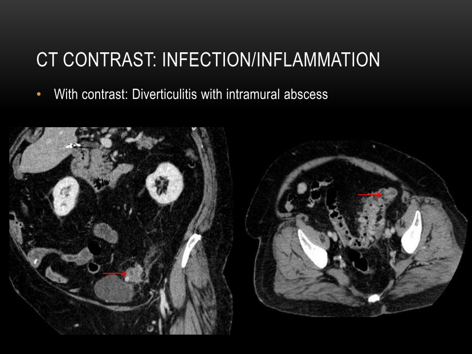
CT CONTRAST: INFECTION/INFLAMMATION
• With contrast: Diverticulitis with intramural abscess

CT CONTRAST: NEOPLASM
• Pancreatic neuroendocrine tumor: Without and with contrast
No contrast: No tumor! Pancreatic protocol CT: TUMOR

CT: IV CONTRAST: WHEN DON’T WE WANT IT?
• What is bright on CT?
• Blood
• Calcium
• Iron
• Foreign bodies

CT: IV CONTRAST: WHEN DON’T WE WANT IT?
• What is bright on CT?
• Blood
• Calcium
• Iron
• Foreign bodies
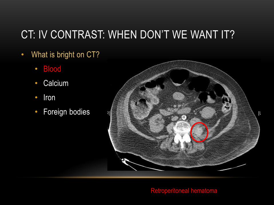
CT: IV CONTRAST: WHEN DON’T WE WANT IT?
• What is bright on CT?
• Blood
• Calcium
• Iron
• Foreign bodies
Retroperitoneal hematoma

CT: IV CONTRAST: WHEN DON’T WE WANT IT?
• What is bright on CT?
• Blood
• Calcium
• Iron
• Foreign bodies
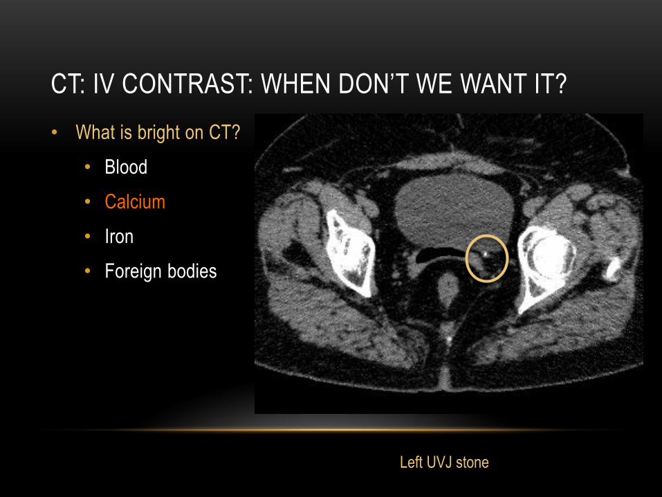
CT: IV CONTRAST: WHEN DON’T WE WANT IT?
• What is bright on CT?
• Blood
• Calcium
• Iron
• Foreign bodies
Left UVJ stone

CT: IV CONTRAST: WHEN DON’T WE WANT IT?
• What is bright on CT?
• Blood
• Calcium
• Iron
• Foreign bodies

CT: IV CONTRAST: WHEN DON’T WE WANT IT?
• What is bright on CT?
• Blood
• Calcium
• Iron
• Foreign bodies
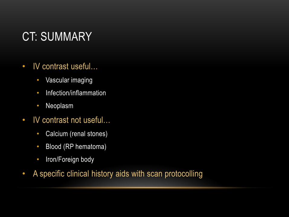
CT: SUMMARY
• IV contrast useful…
• Vascular imaging
• Infection/inflammation
• Neoplasm
• IV contrast not useful…
• Calcium (renal stones)
• Blood (RP hematoma)
• Iron/Foreign body
• A specific clinical history aids with scan protocolling

MRI: PROS AND CONS
• Pros
• No radiation
• Highly diagnostic modality
• Excellent soft tissue contrast
• Histologic information: fat, water, iron, fibrosis
• Functional information: perfusion, peristalsis, cardiac output
• Improvements
• Decreasing scan times
• Emergency medicine
• New sequences

MRI: PROS AND CONS
• Limitations: Patient
• Enclosed space for up to one hour?
• Lying on their back
• Loud noises
• Limitations: Radiologist and system
• Subspecialized reading
• Longer scan times
• Limited availability/varying magnets
• Solutions
• Stereovision
• Gentle use of anxiolytics
• More MR trained radiologist/subspecialized reads

MRI: CATEGORIES AND CONTRAST AGENTS
• Multiple types of MRI
• Neurologic: Brain, neck and spine
• Abdominopelvic
• Musculoskeletal
• Vascular imaging
• Cardiac imaging
• And more!
• Pelvic MRI: Be specific!
• Prostate MRI?
• Rectal MRI?
• MSK MRI?

MRI: CATEGORIES AND CONTRAST AGENTS
• Multiple types of MRI
• Neurologic: Brain, neck and spine
• Abdominopelvic
• Musculoskeletal
• Vascular imaging
• Cardiac imaging
• And more!
• Pelvic MRI: Be specific!
• Prostate MRI?
• Rectal MRI?
• MSK MRI?
All three are ordered
as a pelvic MRI

MRI: CATEGORIES AND CONTRAST AGENTS
• Multiple types of MRI
• Neurologic: Brain, neck and spine
• Abdominopelvic
• Musculoskeletal: Bones, joints, soft tissues, spine
• Vascular imaging
• Cardiac imaging
• And more!
• Every MRI order is reviewed by a radiologist to protocol appropriately
• Rigorously protocolled
• Tailored to the patient and clinical question
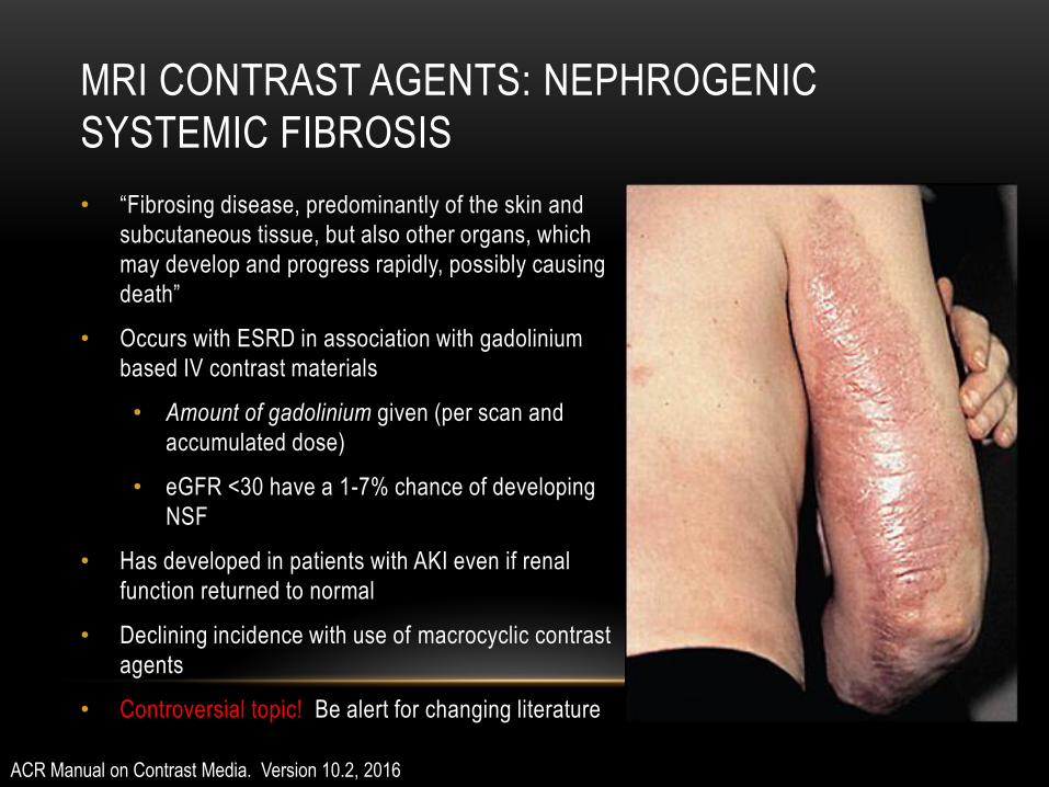
MRI CONTRAST AGENTS: NEPHROGENIC
SYSTEMIC FIBROSIS
• “Fibrosing disease, predominantly of the skin and
subcutaneous tissue, but also other organs, which
may develop and progress rapidly, possibly causing
death”
• Occurs with ESRD in association with gadolinium
based IV contrast materials
• Amount of gadolinium given (per scan and
accumulated dose)
• eGFR <30 have a 1-7% chance of developing
NSF
• Has developed in patients with AKI even if renal
function returned to normal
• Declining incidence with use of macrocyclic contrast
agents
• Controversial topic! Be alert for changing literature
ACR Manual on Contrast Media. Version 10.2, 2016

MRI CONTRAST AGENTS: NSF
• When can we give contrast?
• ESRD on chronic HD:
• Is CT possible instead of MR?
• If MR must be performed, we choose least offensive contrast agent and lower
dose
• Consider hemodialysis ASAP
• ESRD (GFR <15), not on HD
• Avoid both MR and CT contrast agents if at all possible
• If must be given, lower dose, etc
ACR Manual on Contrast Media. Version 9, 2013

MRI CONTRAST AGENTS: NSF
• Screening requirements: require BUN/Creatinine within 30 days of exam1
• Age >60 years
• Hypertension
• Renal disease
• GFR Guidelines
• GFR <15: No IV contrast
• GFR 15-30: Use a lower risk contrast agent (Doderone, Multihance)
• GFR >30: No problem!
• Certain contrast agents have few, if any reported cases of NSF
• Multihance
• Dotarem
• Gadavist
• Prohance
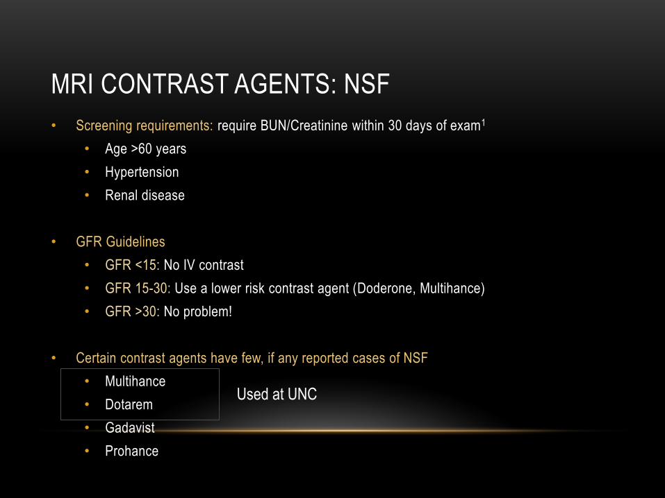
Used at UNC
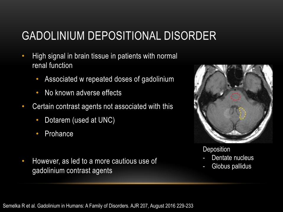
GADOLINIUM DEPOSITIONAL DISORDER
• High signal in brain tissue in patients with normal
renal function
• Associated w repeated doses of gadolinium
• No known adverse effects
• Certain contrast agents not associated with this
• Dotarem (used at UNC)
• Prohance
• However, as led to a more cautious use of
gadolinium contrast agents
Deposition
- Dentate nucleus
- Globus pallidus
Semelka R et al. Gadolinium in Humans: A Family of Disorders. AJR 207, August 2016 229-233

MRI CONTRAST AGENTS: PREGNANCY AND
BREASTFEEDING
• Pregnancy:
• “Present data has not conclusively document any deleterious effects of MR imaging
on the developing fetus”1
• Avoid in first trimester (not evidence based)
• IV contrast DOES cross the placenta and is not given at our institution in pregnancy
• Breastfeeding:
• > 0.04% of the IV dose in breast milk 2
• > 1% of the contrast in breast milk is absorbed across the GI tract2
• Expected dose to infant <0.0004% of IV dose2
• If the mother is concerned, she may abstain from breastfeeding for 24 hours
1. Kanal et al. ACR Guidance Document on MR Safe Practices: 2013. JMRI 37: 501-530 (2013)
2. ACR Manual on Contrast Media. Version 10.2, 2016
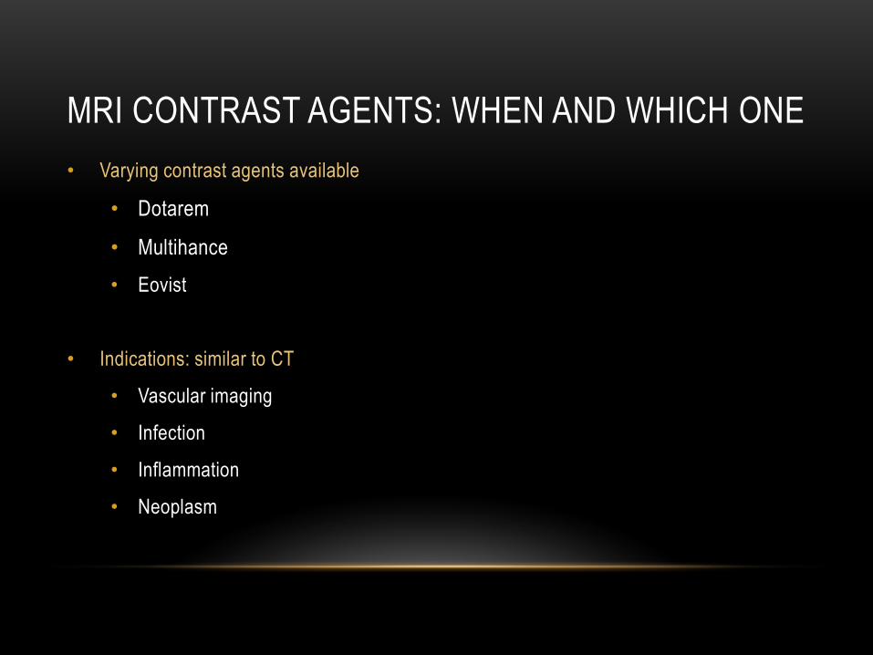
MRI CONTRAST AGENTS: WHEN AND WHICH ONE
• Varying contrast agents available
• Dotarem
• Multihance
• Eovist
• Indications: similar to CT
• Vascular imaging
• Infection
• Inflammation
• Neoplasm
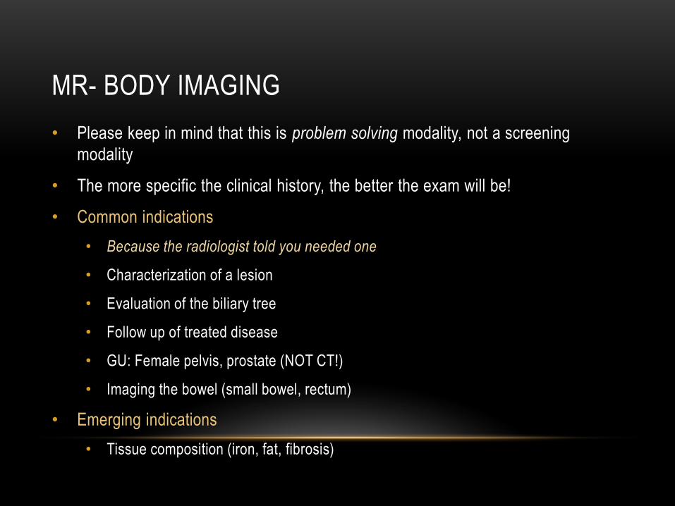
MR- BODY IMAGING
• Please keep in mind that this is problem solving modality, not a screening
modality
• The more specific the clinical history, the better the exam will be!
• Common indications
• Because the radiologist told you needed one
• Characterization of a lesion
• Evaluation of the biliary tree
• Follow up of treated disease
• GU: Female pelvis, prostate (NOT CT!)
• Imaging the bowel (small bowel, rectum)
• Emerging indications
• Tissue composition (iron, fat, fibrosis)
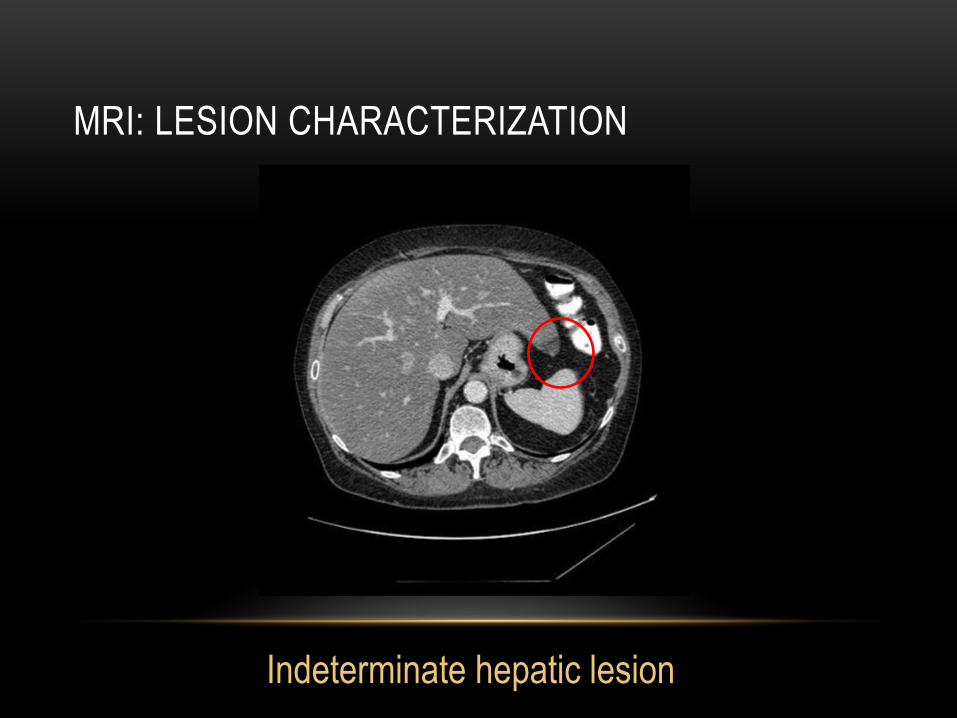
MRI: LESION CHARACTERIZATION
Indeterminate hepatic lesion

MRI: LESION CHARACTERIZATION
Focal steatosis

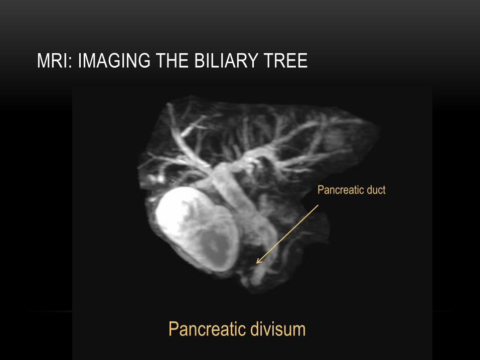
MRI: IMAGING THE BILIARY TREE
Pancreatic duct
Pancreatic divisum

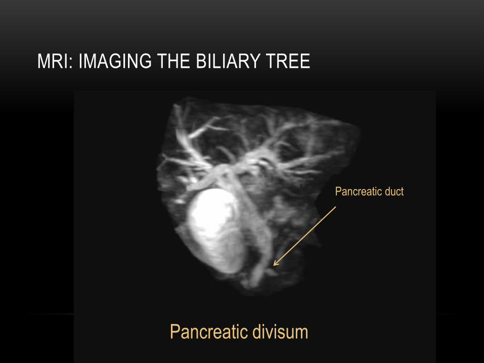
MRI: IMAGING THE BILIARY TREE
Pancreatic duct
Pancreatic divisum

MRI: IMAGING THE BILIARY TREE
Pancreatic duct
Pancreatic divisum
MRI: IMAGING THE BILIARY TREE
Pancreatic duct
Pancreatic divisum

MRI: IMAGING THE BILIARY TREE
Pancreatic duct
Pancreatic divisum

MRI: IMAGING THE BILIARY TREE
Pancreatic duct
Pancreatic divisum

MRI: IMAGING THE BILIARY TREE
Pancreatic duct
Pancreatic divisum
MRI: IMAGING THE BILIARY TREE
Pancreatic duct
Pancreatic divisum

MRI: IMAGING THE BILIARY TREE
Pancreatic duct
Pancreatic divisum

MRI: IMAGING THE BILIARY TREE
Pancreatic duct
Pancreatic divisum

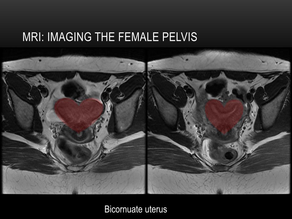
MRI: IMAGING THE FEMALE PELVIS
Bicornuate uterus
MRI: IMAGING THE FEMALE PELVIS
Bicornuate uterus

MRI: IMAGING THE MALE PELVIS
PROSTATE CANCER

MRI: IMAGING THE MALE PELVIS
PROSTATE CANCER

MRI IMAGING: TISSUE COMPOSITION
• Liver
• Fat content
• Iron content
• Fibrosis
• Bowel
• MR enterography
• Rectal MR
• MRI is a rapidly expanding and changing field. If you want to know if
we can do it- just ask!!!
YOUR RADIOLOGIST
• Clinician feedback
• Reports
• Relevant? Unclear?
• Imaging problems
• Patient complaints?
• Didn’t give you an answer
• Pathology and/or clinical follow up
• Were we right or wrong?
• You are our target population with our imaging and reports- let us
know how we can improve and make your life easier!
TAKE HOME POINTS
• Overutilization is a real but solvable problem if a partnership exists between the clinician
and radiologist
• There are many different imaging modalities at your disposal with varied resources to help
advise you
• ACR appropriateness criteria
• Radiologist
• Appropriate scan for each patient
• Appropriate radiation dose
• Scan limitations
• Patient limitations
• Clinician feedback is critical for imaging and service improvement.

THANK YOU!

WORKS CITED
• ACR Manual on Contrast Media, Version 10.2, 2016
• Radiologyinfo.org American college of radiology and Radiologic Society of North America
• DiLeo, R. and Spinelli, R. Strategies of teach non radiologist physicians the appropriate use of imaging studies: Use of Radiology Seminars. J Med Pract Manage. 2006 May-Jun; 21(6): 362-6
• Hendee, W. et al. Addressing Overutilization in Medical Imaging. Radiology. August 2010.
• Herman, S. Computed Tomorgraphy Contrast Enhancement Principles and the Use of High Concentration Contrast Media. J Comput Assist Tomogr 2004; 28: S7-S11
• Kanal et al. ACR Guidance Document on MR Safe Practices: 2013. JMRI 37: 501-530 (2013)
• Kellow, Z et al. The Role of Abdominal Radiography in Evaluation of the Nontrauma emergency patient. September 2008, Radiology, 248, 887-893
• McDonald, R et al. Intravenous contrast material induced nephropathy: Causal or coincident phenomenon? April 2013, Radiology, 267, 106-118
• M. Saijoughlan. Intravenous Radiocontrast Media: A review of Allergic Reactions. US Pharm. 2012;37 (5): HS-14-HS-16
• Semelka R et al. Gadolinium in Humans: A Family of Disorders. AJR 207, August 2016 229-233
• Smith-Bindman, Rebecca. Is Computed Tomography Safe? N Engl J Med; 363:1-4
• Stoker et al. Imaging patient with acute abdominal pain. October 2009. Radiology 253,31-46
Related Documents