1 liiidawi Publishing Corpomtion Mediators of IniLimmation Volume 2007, Article ID 50180, 5 pages doiilO.1155/2007/50180 Research Article Radioiodine Labeled Anti-MIF McAb: A Potential Agent for Inflammation Imaging Chao Zhang,^- ^ Gui-hua Hou,^ Jian-kui Han,^ Jing Song,^ and Ting Liang^ ' Di'parlmcnl of Nuclear Medicine.. Qilu Hospital ofShiwdong University, jinau 250012, China -'Institute of llxpcrimaiiai Nuclear Medicine, School of Medicine, Shandong University, jinun 2500t2, China Correspondence should be addressed to Jian-kui Han, [email protected] Received 2! Iune 2007; Accepted 22 August 2007 Macrophage migration inhibitory factor (MIH) is a proinflammatory cytokine that may play a role in the palhogenesis of inflam- mation. Radio labeled anti-MIF McAb can be used to detect in vivo inflammatory changes. The objective of this study was to investigate in vivo biology of radioiodinated anti MIF McAb using the inflammation model mite. Anti-MlI- McAb was radioiod- inated with Na'^^^I by lodogen method. Animal models were induced in the mice by intramuscular injection of S. aureus, E. colit and turpentine oil. The biodislribution studies with radioiodinated anti-MIF McAb were performed on inflammation mice. The relationship between inflammatory lesions and anii-MlF McAb binding was investigated using the percent of injected dose per gram tissue (% IP/g) of tissue samples and whole-body autoradiography. The radioactivity of '^"'I-anti-MIF McAh in ihe intlain- matory ti.ssue increased gradually for three inflammation models. The highest uptake was found in S. nureus group and the lowest was in E. coli group. The uptake in turpentine oil group was average. Whole-body autoradiography showed that ali infUitnmotion foci could be visualized clearly from 24 hours after injection, but 48 hours images were much clearer in accordance with the high I/NT ratio. These results demon.strate the ability of radioiodinated anti-MIF McAb to measure in vivo intlatiimatory events rep- resented by high expression of MIF and suggests lhat radiolabeled anti-MIF McAb warrants further investigation as a potenlJa! inflammation-seeking agent for imaging to detect inflammatory disorders. Copyright & 2007 Chao Zhang et al. This is an open access article distributed under the t.reative Comtnons Attribution License, which permits unrestricted use, distribution, and reproduction in any medium, provided the original work js properly cited. 1. INTRODUCTION Macrophage migration inhibitor factor {MIF) was originally discovered as a kind of lymphokines involved in delayed type hypersensitivity and various macrophage functions [1-3]. However, its descriptive name was shown to be rather im- precise as MIF can also promote macrophage rolling and transmigration by upregulating P-selection expression in en- dothehal cells lining the site of inflammation |4, 5]. Numer- ous animal studies have revealed the critical role of MIF in acute and chronic inflammation [6, 7]. The increased levels of MIF in certain pathological conditions may be indicative of its involvement in those diseases. Indeed, increased MIF plasma or serum levels were identified in patients with severe sepsis [8], Crohn's disease and ulcerative colitis [9], acute pancreatitis [10], rheumatoid arthritis (RA) [11], type 2 di- abetes (T2I)) [12], GuiUain-Barre syndrome [13], or multi- ple sclerosis [14]. Consequently, MIF's activity has become a potential target for treating these various disorders. In this study, we labeled anti-MIF monoclonal antibody (McAb) with radioiodine Na'-^I and investigated its biodistribution and pharmacokinetics in vivo in animal models with inflam- mation. 2. MATERIALS AND METHODS 2.7. Radioiodination of anti-MIF McAb All commercially available chemicals were of analytic grade and anti-MIF McAb (R&D Systems) was pharmaceutical grade. Anti-MIF McAb was iodinated with Na''^I (specific activity 37 MBq/mg, China Institute of Atomic Energy) us- ing the lodogen technique (Pierce). Radioiodinated antibody was separated from free iodine using a size exclusion column (Sephadex G-25, Pharmacia). The specific activity of radioio- dinated antibody is 29.56 GBq//jmol. The radiochemical pu- rity is >95% (paper chromatography). 2.2. Preparation of inflammation animal model The animal experiments were carried out in accordance with institutional, national, and international guidelines for

Welcome message from author
This document is posted to help you gain knowledge. Please leave a comment to let me know what you think about it! Share it to your friends and learn new things together.
Transcript
1 liiidawi Publishing CorpomtionMediators of IniLimmationVolume 2007, Article ID 50180, 5 pagesdoiilO.1155/2007/50180
Research Article
Radioiodine Labeled Anti-MIF McAb:A Potential Agent for Inflammation Imaging
Chao Zhang,^- Gui-hua Hou,^ Jian-kui Han,^ Jing Song,^ and Ting Liang^
' Di'parlmcnl of Nuclear Medicine.. Qilu Hospital ofShiwdong University, jinau 250012, China-'Institute of llxpcrimaiiai Nuclear Medicine, School of Medicine, Shandong University, jinun 2500t2, China
Correspondence should be addressed to Jian-kui Han, [email protected]
Received 2! Iune 2007; Accepted 22 August 2007
Macrophage migration inhibitory factor (MIH) is a proinflammatory cytokine that may play a role in the palhogenesis of inflam-mation. Radio labeled anti-MIF McAb can be used to detect in vivo inflammatory changes. The objective of this study was toinvestigate in vivo biology of radioiodinated anti MIF McAb using the inflammation model mite. Anti-MlI- McAb was radioiod-inated with Na' ^ I by lodogen method. Animal models were induced in the mice by intramuscular injection of S. aureus, E. colitand turpentine oil. The biodislribution studies with radioiodinated anti-MIF McAb were performed on inflammation mice. Therelationship between inflammatory lesions and anii-MlF McAb binding was investigated using the percent of injected dose pergram tissue (% IP/g) of tissue samples and whole-body autoradiography. The radioactivity of '^"'I-anti-MIF McAh in ihe intlain-matory ti.ssue increased gradually for three inflammation models. The highest uptake was found in S. nureus group and the lowestwas in E. coli group. The uptake in turpentine oil group was average. Whole-body autoradiography showed that ali infUitnmotionfoci could be visualized clearly from 24 hours after injection, but 48 hours images were much clearer in accordance with the highI/NT ratio. These results demon.strate the ability of radioiodinated anti-MIF McAb to measure in vivo intlatiimatory events rep-resented by high expression of MIF and suggests lhat radiolabeled anti-MIF McAb warrants further investigation as a potenlJa!inflammation-seeking agent for imaging to detect inflammatory disorders.
Copyright & 2007 Chao Zhang et al. This is an open access article distributed under the t.reative Comtnons Attribution License,which permits unrestricted use, distribution, and reproduction in any medium, provided the original work js properly cited.
1. INTRODUCTION
Macrophage migration inhibitor factor {MIF) was originallydiscovered as a kind of lymphokines involved in delayed typehypersensitivity and various macrophage functions [1-3].However, its descriptive name was shown to be rather im-precise as MIF can also promote macrophage rolling andtransmigration by upregulating P-selection expression in en-dothehal cells lining the site of inflammation |4, 5]. Numer-ous animal studies have revealed the critical role of MIF inacute and chronic inflammation [6, 7]. The increased levelsof MIF in certain pathological conditions may be indicativeof its involvement in those diseases. Indeed, increased MIFplasma or serum levels were identified in patients with severesepsis [8], Crohn's disease and ulcerative colitis [9], acutepancreatitis [10], rheumatoid arthritis (RA) [11], type 2 di-abetes (T2I)) [12], GuiUain-Barre syndrome [13], or multi-ple sclerosis [14]. Consequently, MIF's activity has become apotential target for treating these various disorders. In thisstudy, we labeled anti-MIF monoclonal antibody (McAb)with radioiodine Na'-^I and investigated its biodistribution
and pharmacokinetics in vivo in animal models with inflam-mation.
2. MATERIALS AND METHODS
2.7. Radioiodination of anti-MIF McAb
All commercially available chemicals were of analytic gradeand anti-MIF McAb (R&D Systems) was pharmaceuticalgrade. Anti-MIF McAb was iodinated with Na''^I (specificactivity 37 MBq/mg, China Institute of Atomic Energy) us-ing the lodogen technique (Pierce). Radioiodinated antibodywas separated from free iodine using a size exclusion column(Sephadex G-25, Pharmacia). The specific activity of radioio-dinated antibody is 29.56 GBq//jmol. The radiochemical pu-rity is >95% (paper chromatography).
2.2. Preparation of inflammation animal model
The animal experiments were carried out in accordancewith institutional, national, and international guidelines for
Mediators of Inflammation
humane use of animals for research. Fourty eight mice(BALB/c, 18 - 22 g, Animal Center of Shandong Univer-sity) were divided into three groups, each group consistingof 16 mice, respectively. The first and second groups were in-duced inflammation by intramuscularly injecting 2 x 10''-10"colony forming units (CFU) of S. aureus and E. coU in 0.2mL,respectively, into the left thigh muscle [15]. The third groupof mice were induced sterile inflammation by intramuscularinjection of 0.2 mL turpentine oil [ 15]. Twenty four hours af-ter inoculation, focal inflammation occurred. Those inflam-mation models were proved by histological studies (data notshowed).
2.3. Biodistribution of ^"l-anti-MIF McAb
Mice with the left thigh inflammation were intraperitoneallyinjected with 3.7MBq '-^I-anti-MIF McAb in 0.2mL PBS.Three mice of each group were sacrificed by cervical disloca-tion at 30 minutes, 4 hours, 24 hours, 48 hours, and 72 hoursafter injection, respectively. A sample of 1 mL blood wascollected at the time of decapitation. Samples of two thighmuscles (left as target, right as control), lungs, heart, liver,spleen, kidney, and bone were removed and weighted. Thetissue radioactivity was measured with a wipe test counter(CAPRAC). The percent of injected dose per gram tissue (%ID/g) was calculated by comparison with samples to standarddilutions of the initial dose.
2.4. Whole-body autoradiography
Three groups of mice inflammatory models were establishedby the same method like biodistribution study. Each groupconsists of 4 mice. ' -^I-anti-MIF McAb (3.7 MBq in 0.2 mLPBS) were injected intravenously via the tail vein. Serial im-ages were performed at 24 hours, 48 hours, and 72 hours afterinjection. The anesthetized mice were placed on the storage-phosphor screen plate with the ventral side facing the plate,in subdued light. The plate was exposed to a mouse for 45minutes. At cessation of exposure, the plate was immediatelycovered with an opaque plastic sheet, then transferred to thescanner, and scanned by typhoon trio + (laser red 633 nm,pixel size 200 mcrons, phosphor mode: best sensitivity).
2.5. Statistics
Dates were expressed as the x ± s. The dates were analyzedusing SPSS 11.0 software.
3. RESULTS
3.1. Antibody clearance from the blood
'^^I-anti-MIF McAb shortly transited from the peritonealcavity to the circulation after intraperitoneal injection. Atfirst 30 minutes, the activity of '-^I-anti-MIF McAb in theblood increased rapidly up to 45.00% ID, 36.66% ID, and45.66% ID, respectively, in S. aureus, E. coli, and turpentinegroup. Then, it went up and reached a zenith at 4 hours postinjection. Levels were 60.03% ID, 38.59% ID, and 54.42% ID,
S. aureusTurpentineL coli
K I: Clearance of ''' I-anti-MIF McAb from the blood in S.aureus group, ii. co/(group, and turpentine oil group (% lD/g,x±s).
.10 min 4h 24 h 48 h 72 h
- • - S. aureus-m~ F. coli-A- Turpentine
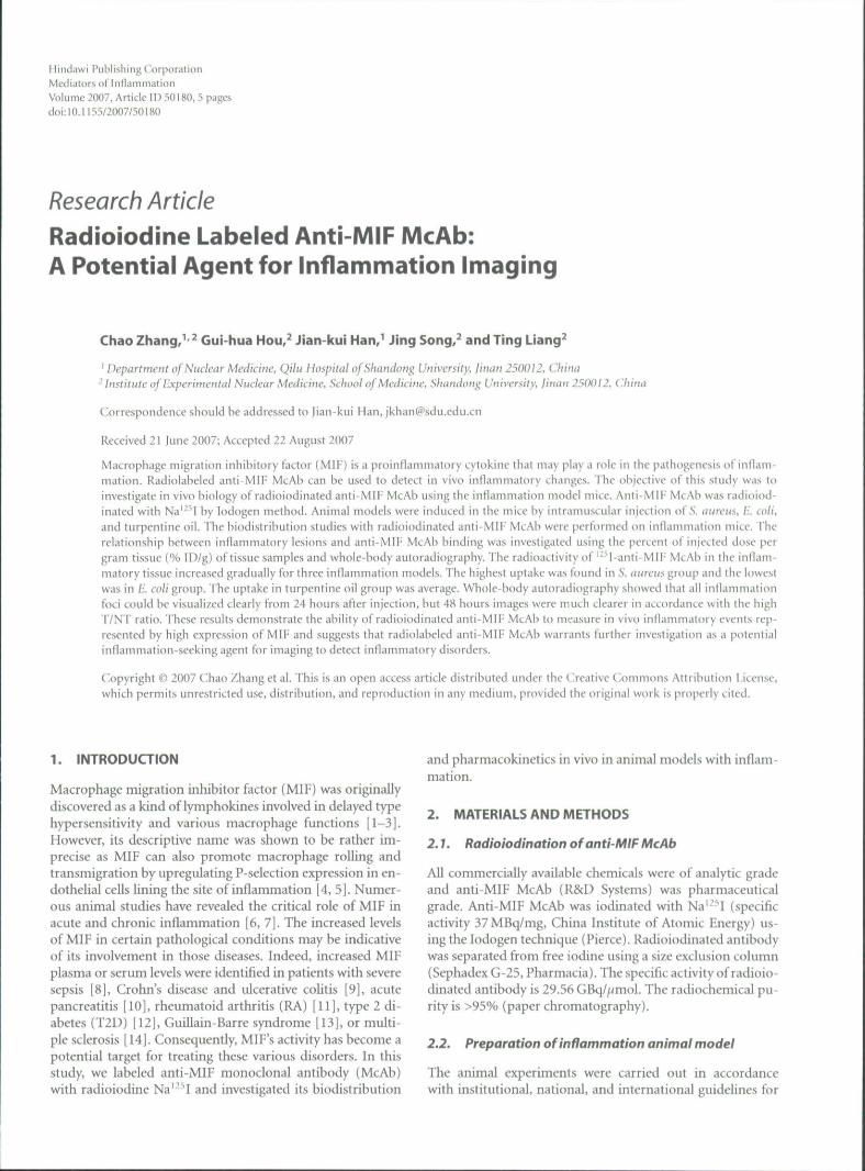
FIGURE 2: Accumulation of '"I-anti-MIF McAb in the inflamma-tory tissue of 5. aureus group, E. coli group, and turpentine oil group(%ID/g,x±s).
respectively, in three groups. After that point, the activity inthe blood went down quickly (Figure 1).
3.2. Accumulation in the inflammatory tissue
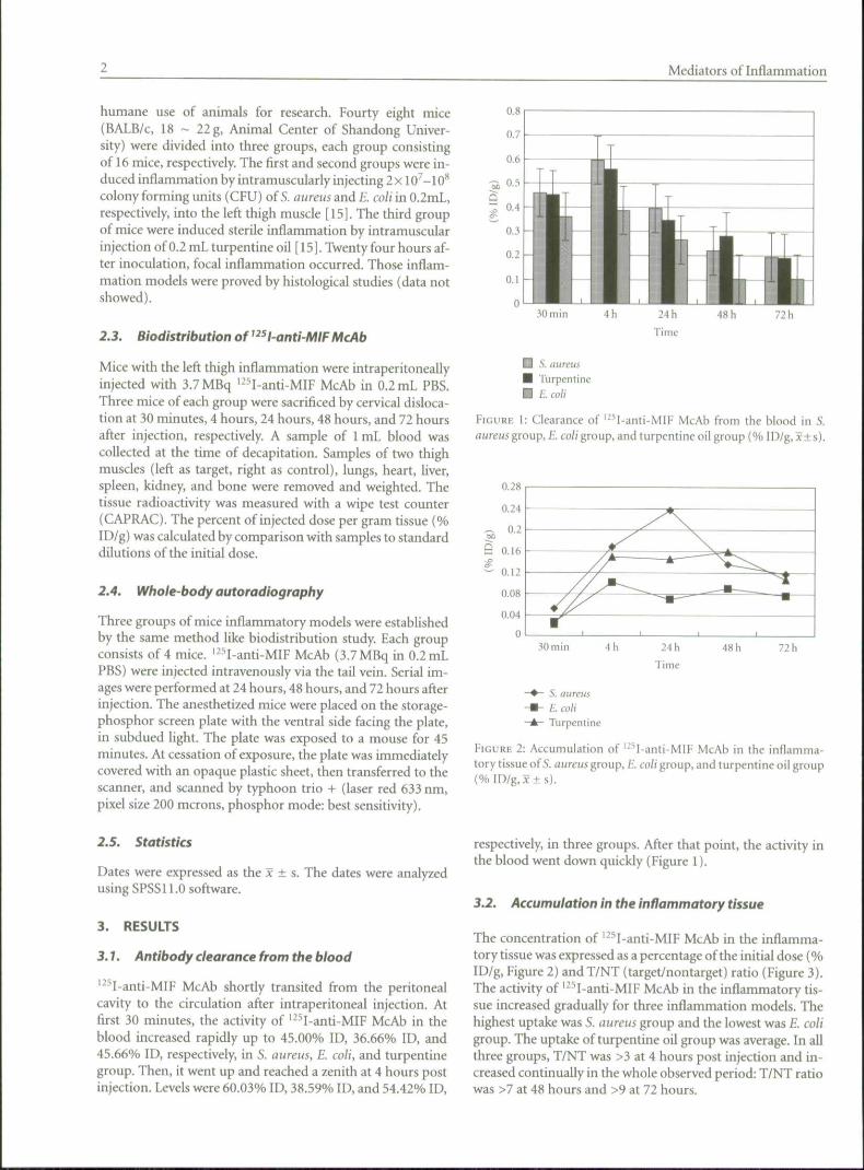
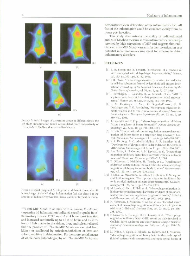
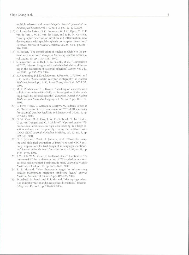
The concentration of '-^I-anti-MIF McAb in the inflamma-tory tissue was expressed as a percentage of the initial dose (%ID/g, Figure 2) and T/NT (target/nontarget) ratio (Figure 3).The activity of '^^I-anti-MIF McAb in the inflammatory tis-sue increased gradually for three inflammation models. Thehighest uptake was S. aureus group and the lowest was E. coligroup. The uptake of turpentine oil group was average. In allthree groups, T/NT was >3 at 4 hours post injection and in-creased continually in the whole observed period: T/NT ratiowas >7 at 48 hours and >9 at 72 hours.
Chao Zhang et al.
-2 10
72 h
- • - S. aureus- • - £ cod'-* - Turpentine
FIGURE 3: Change of T/NT in the S. aureus, E. coU, and turpentineoil group.
3.3. Biodistribution of ^^^ I anti-MIF McAb
As expected, the biodistribution of '-"'I-anti-MIF McAbshowed the highest uptake and the lowest decrease in the in-flammatory tissue. The activity in the blood was higher thanthe kidney, liver, spleen, heart, and lung. The change of activ-ity in the heart and lung was the same as blood. Peak uptakeinthekidney (0.2575+0.1640% ID/g, 0.2452 + 0.0612% ID/g,and 0.2909 ± 0.0856% ID/g, respectively, in the S. aureus,E. coliy and turpentine oil group), liver (0.2271 ± 0.1345%ID/g, 0.1682 ± 0.0028% ID/g, and 0.1828 + 0.0955% ID/g, re-spectively in the S. aureus, E. coli, and turpentine oil group),spleen (0.1450 ± 0.1621% ID/g, 0.0882 + 0.0799% ID/g,0.1704 ± 0.1351% ID/g, respectively in the 5. aureus, E. coli,and turpentine oil group) occurred around 30 minutes, fol-lowed by gradual clearance over time. This indicated that theproduct of '-'^I-anti-MlF McAb was excreted from kidney orswallowed by reticuloendothelium of liver and spleen, result-ing in deiodination.
3.4. Imaging of the inflammatory foci
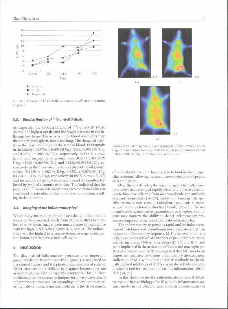
Whole-body autoradiography showed that all inflammationfoci could be visualized clearly from 24 hours after injection,but after 48 hours images were much clearer in accordancewith the high T/NT ratio (Figures 4, 3, and 6). The radioac-tivity was the highest in S. aureus lesion, average in turpen-tine lesion, and the lowest in E. coli lesion.
4. DISCUSSION
The diagnosis of inflammatory processes is an importantgoal in medicine. In some cases the diagnosis is easy, based onthe clinical history and the physical examination of patient.Other cases are more difficult to diagnose because they areasymptomatic or with nonspecific symptoms. Thus, nuclearmedicine provides several techniques for in vivo detection ofinflammatory processes. An expanding and even more inter-esting field of modern nuclear medicine is the development
48 h
(b)
72 h
(c)
¥\cvK\i4: Serial images of 5. aureus group at different times: the leftthigh intlammation foci accumulated much more radioactivily of'^^l-anti-MIF McAb; the highest was at 48 hours.
of radiolabelled receptor ligands, able to bind in vivo to spe-cific receptors, allowing the noninvasive detection of specificcells and tissues.
Over the last decades, the imaging agents for inflamma-tion have been developed rapidly: from nonbioactive chemi-cals to bioactive cell, and from macromolecule and antibodysegments to peptides [16-20], and so on. Amongst the spe-cific tracers, a new class of radiopharmaceuticals is repre-sented by monoclonal antibodies (MoAb) [21-23]. The useof antibodies against surface granulocyte or lymphocyte anti-gens may improve the ability to detect inflammatory pro-cesses compared to the use of radiolabeled leukocytes.
The inflammatory response is rapid and includes secre-tion of cytokines and proinflammatory mediators that caninduce an inflammatory response. MIF is believed to initiateinflammation by release of a number of proinflammatory cy-toldnes including TNF-a, interleukin(IL)-I/^, and IL-6, andto be implicated in the activation of T cells and macrophages.Recent reevaluation of MIF has suggested that MIF may be animportant mediator of various inflammatory diseases, neu-tralization of MIF with either anti-MIF antibody or chemi-cally derived inhibitors of MIF's enzymatic activity could bea valuable tool for treatment of various inflammatory disor-ders [24,25].
In this study, we use the radioiodinated anti-MIF McAbto evaluate in vivo biology of MIF with the inflammation an-imal model in the BALB/c mice. Biodistribution studies of
Mediators of Inflammation
48 h
(b)
72 h
FIGURE 5: Serial images of turpentine group at different times: theleft thigh inflammation lesion accumulated more radioactivity of'^^1-anti-MIF McAb and was visualized clearly.
24 h 48 h
(b)
FIGURE 6: Serial images of E coli group at different times: after 48hours image of the left thigh inflammation loci was clear, but theamount of radioactivity was less than S. (iHreus or turpentine lesion.
McAb in animals with S. aureus, E. coli, andturpentine oil inflammation indicated specific uptake in in-flammatory tissues: T/NT was >3 at 4 hours post injectionand increased continually up to >7 at 48 hours and >9 at 72hours. High uptake in the kidney, liver, and spleen reflectedthat the product of '^''I-anti-MIF McAb was excreted fromkidney or swallowed by reticuloendothelium of liver andspleen, resulting in deiodination. Furthermore, serial imagesof whole-body autoradiography of '^^I-anti-MIF McAb also
demonstrated clear delineation of the inflammatory foci. Allfoci of the inflammation could be visualized clearly from 24hours post injection.
This study demonstrates the ability of radioiodinatedanti-MIF McAb to measure in vivo inflammatory events rep-resented by high expression of MIF and suggests that radi-olabeled anti-MIF McAb warrants further investigation as apotential inflammation-seeking agent for imaging to detectinflammatory disorders.
REFERENCES
[1] B. R. Bloom and B. Bennett, "Mechanism of a reaction invitro associated with delayed-type hypersensitivity," Science,vol. 153, no. 3731, pp. 80-82, 1966.
[2] J. R. David, "Delayed hypersensitivity in vitro: its mediationby cell-free substances formed by lymphoid cell-antigen inter-action," Proceedings of the National Academy of Sciences of theUnited States of America, vol. 56, no. I, pp. 72-77, 1966.
[3] I. Bernhagen, T. Calandra, R. A. Mitchell, et al., "MIF isa pituitary-derived cytokine that potentiates lethal endotox-aemia," Nature, vol. 365, no. 6448, pp. 756-759, 1993.
(4] C. M. Denkinger, C. Metz, G. Fingerle-Rowson, M. D.Denkinger, and T. G. Forsthuber, "Macrophage migration in-hibitory factor and its role in autoimmune diseases," ArchivumImmunologiae et Therapiae Experimentalis, vol. 52, no. 6, pp.389-400, 2004.
[5] T Calandra and T. Roger, "Macrophage migration inhibitoryfactor: a regulator of innate immunity," Nature Reviews Im-munology, vol. 3, no. 10, pp. 79l-80n, 2003.
[6] E. Lolis, "Glucocorticoid counter regulation: macrophage mi-gration inhibitory factor as a target for drug discovery," Cur-rent Opinion in Pharmacology, vol. 1, no. 6, pp. 662-668, 2001.
[7] Y. P. De Jong, A. C. Abadia-Molina, A. R. Satoskar, et al.,"Development of chronic colitis is dependent on the cytokineMl¥," Nature Immunology, vol 2, no. I I , p p . 1 0 6 1 - 1 0 6 6 , 2 0 0 1 .
[8] F. A. Bozza, R. N. Gomes, A. M. Japiassij, et al., "Macrophagemigration inhibitory factor levels correlate with fatal outcomein sepsis," Shock, vol. 22, no. 4, pp. 309-313, 2004.
19) T. Ohkawara, ). Nishihira, H. Takeda, et al., "Ameliorationof dextran sulfate sodium-induced colitis by anti-macrophagemigration inhibitory factor antibody in mice," Gastroenterol-ogy, vol. 123, no. 1, pp. 256-270, 2002.
[10] Y. Sakai, A. Masamune, A. Satoh, J. Nishihira, T. Yamagiwa,and T. Shimosegawa, "Macrophage migration inhibitory fac-tor is a critical mediator of severe acute pancreatitis," Gastroen-terology, vol. 124, no. 3, pp. 725-736, 2003.
111] M. Leech, C. Metz, P. Hall, et al., "Macrophage migration in-hibitory factor in rheumatoid arthritis: evidence of proinflam-matory function and regulation by glucocorticoids," Arthritisand Rheumatism, vol. 42, no. R, pp. 1601-1608, 1999.
[I2[ N. Yabunaka, J. Nishihira, Y. Mizuc, et al., "Elevated serumcontent of macrophage migration inhibitory factor in patientswith type 2 diabetes," Diabetes Care, vol. 23, no. 2, pp. 256-258, 2000.
[13] F. Nicoletti, A. Greange, D. Oriikowski, et al., "Macrophagemigration inhibitory factor (MIF) seems crucially involved inGuiliain-Barre syndrome and experimental allergic neuritis,"lournal of Neuroimmunology, vol. 168, no. 1-2, pp. 168-174,2003.
114] M. Niino, A. Ogata, S. Kikuchi, K. Tashiro, and J. Nishihira,"Macrophage migration inhibitory factor in the cerehrospinalfluid of patients with conventional and optic-spinal forms of
Chao Zhang et al.
multiple sclerosis and neuro-Beh(^et',s disease," journal of theNeurological Sciences, vol 179, no. 1-2, pp. 127-131,2000.
15] G. I. van der Uken, O. G. Boerman, W. ]. G. Oyen, M. T P.van de Ven, I. W. M. van der Meer, and F. H. M. Gorstens,"Scintigraphic detection of infection and inflammation: newdevelopments with special emphasis on receptor interaction,"European journal of Nuclear Medicine, vol. 25, no. 5, pp. 535-546, 1998.
16] W. Becker, "The contribution of nuclear medicine to the pa-tient with infection," F.ttropean lournal of Nuclear Medicine,vol. 22, no. 10, pp. 1195-1211, 1995.
171 S. Vinjamuri, A. V. Hall, K. K. Solanki, et al., "(;t)mparison(jf-wnrj-j. infecton imaging with radiolabelled white-cell imag-ing in the evaluation of bacterial infection," Lancet, vol. 347,no. 8996, pp. 233-235, 1996.
18] H. P. Krenning, D. I. Kwekkebomm, S. Pauwcis, L. K. Kvols, andI. C Reubi, "Somatostatin receptor .scintigraphy," in NuclearMedicine Annual, pp. 1-50, Raven Press, New York, NY, USA,1995.
19] M. R. Plucher and P. |. Blower, "Labelling of lekocytes withcolloidal tecnetium-99m-SnF2: an investigation of the label-ing process by autoradiography," European journal of NuclearMedicine and Molecular Imaging, vol. 22, no. 2, pp. 101-107,1995.
[20] G. Ferro-Flores, G. Arteaga de Murphy, M. Pedraza-L6pe?., etal., "In vitro and in vivo a.ssessment of''"'Tc-UBI specificityfor bacteria," Nuclear Medicine and Biology, vol. 30, no. 6, pp.597-603, 2003.
|21t G. W. Visser, R. P Klok, J. W. K. Gebbinck, T. Ter Linden,CJ. A. van Dongen, and G. F. Molthoff, "Optimal quality '' ' I-nionoclonai antibodies on high-dose labeling in a large re-action volume and temporarily coating the antibody withIODO-(iHN," journal of Nuclear Medicine, vol. 42, no. 3, pp.509-519,2001.
|22[ Ci. C;. Jayson, 1. Zweit, A. Jackson, el al., "Molecular imag-ing and biological evaluation of HuMV833 an(i-VE(;F anti-body: implications for trial design of antiangiogenic antibod-ies," lournal o{the National Cancer institute, vol. 94, no. 19, pp.1484-1493,2002.
123 [ I. Verel, (.!. W. M. Visser, R. Boellaard, et al., "Quantitative '' Zrimniuno-PF.T for in vivo scouting of ""Y-labeled monoclonalantibodies in xenograft-bearing nude mice," journal of NuclearMedicine, vol. 44, no. 10, pp. 1663-1670, 2003.
I24[ E. F. Morand, "New therapeutic target in inflammatorydisease: macrophage migration inhibitory factor," InternalMedicine jourtiat, vol. 35, no. 7, pp. 419^26, 2005.
[25[ D. Aeberli, M. Leech, and F'. F. Morand, "Macrophage migra-tion inhibitory factor and glucocorticoid sensitivity," Rheuma-tology, vol. 45, no. 8, pp. 937-943, 2006.

Related Documents