CONTINUING EDUCATION Radioguided Sentinel Lymph Node Biopsy in Malignant Cutaneous Melanoma* Giuliano Mariani, MD 1,2 ; Marco Gipponi, MD 3 ; Luciano Moresco, MD 4 ; Giuseppe Villa, MD 5 ; Mirco Bartolomei, MD 6 ; Giovanni Mazzarol 7 ; Maria Claudia Bagnara, MSc 8 ; Antonella Romanini, MD 9 ; Ferdinando Cafiero, MD 3 ; Giovanni Paganelli, MD 6 , and H. William Strauss, MD 10 1 Regional Center of Nuclear Medicine, University of Pisa Medical School, Pisa, Italy; 2 Institute of Clinical Physiology, National Research Council, Pisa, Italy; 3 Division of Surgical Oncology, National Cancer Institute, Genoa, Italy; 4 Division of Surgical Oncology, Department of Oncology, Biology and Genetics, University of Genoa and National Cancer Institute, Genoa, Italy; 5 Nuclear Medicine Service, Department of Internal Medicine and Medical Specialties, University of Genoa Medical School, Genoa, Italy; 6 Division of Nuclear Medicine, European Institute of Oncology, Milan, Italy; 7 Department of Pathology, European Institute of Oncology, Milan, Italy; 8 Health Physics Unit, San Martino Hospital, Genoa, Italy; 9 Medical Oncology Unit, Santa Chiara Hospital, Pisa, Italy; and 10 Division of Nuclear Medicine, Department of Radiology, Memorial Sloan-Kettering Cancer Center, New York, New York The procedure of sentinel lymph node biopsy in patients with malignant cutaneous melanoma has evolved from the notion that the tumor drains in a logical way through the lymphatic system, from the first to subsequent levels. As a consequence, the first lymph node encountered (the sentinel node) will most likely be the first affected by metastasis; therefore, a negative sentinel node makes it highly unlikely that other nodes in the same lymphatic basin are affected. Although the long-term therapeutic benefit of the sentinel lymph node biopsy per se has not yet been ascer- tained, this procedure distinguishes patients without nodal metas- tases, who can avoid nodal basin dissection with its associated risk of lymphedema, from those with metastatic involvement, who may benefit from additional therapy. Sentinel lymph node biopsy would represent a significant advantage as a minimally invasive procedure, considering that an average of only 20% of melanoma patients with a Breslow thickness between 1.5 and 4 mm harbor metastasis in their sentinel node and are therefore candidates for elective lymph node dissection. Furthermore, histologic sampling errors (amounting to approximately 12% of lymph nodes in the conventional routine) can be reduced if one assesses a single (sentinel) node extensively rather than assessing the standard few histologic sections in a high number of lymph nodes per patient. The cells from which cutaneous melanomas originate are located between the dermis and the epidermis, a zone that drains to the inner lymphatic network in the reticular dermis and, in turn, to larger collecting lymphatics in the subcutis. Therefore, the optimal route for interstitial administration of radiocolloids for lymphoscin- tigraphy and subsequent radioguided sentinel lymph node biopsy is intradermal or subdermal injection. 99m Tc-Labeled colloids in various size ranges are equally adequate for radioguided sentinel lymph node biopsy in patients with cutaneous melanoma, depend- ing on local experience and availability. For melanomas along the midline of the head, neck, and trunk, particular consideration should be given to ambiguous lymphatic drainage, which fre- quently requires interstitial administration virtually all around the tumor or surgical scar from prior excision of the melanoma. Lym- phoscintigraphy is an essential part of radioguided sentinel lymph node biopsy because images are used to direct the surgeon to the sites of the nodes. The sentinel lymph node should have a signif- icantly higher count than that of the background (at least 10:1 intraoperatively). After removal of the sentinel node, the surgical bed must be reexamined to ensure that all radioactive sites are identified and removed for analysis. Virtually the entire sentinel lymph node should be processed for histopathology, including both conventional hematoxylin– eosin staining and immune stain- ing with antibodies to the S-100 and HMB-45 antigens. The suc- cess rate of radioguidance in localizing the sentinel lymph node in melanoma patients is approximately 98% in institutions that per- form a high number of procedures and approaches 99% when combined with the vital blue-dye technique. Growing evidence of the high correlation between a sentinel lymph node biopsy nega- tive for cancer and a negative status for the lymphatic basin— evidence, therefore, of the high prognostic value of sentinel node biopsy— has led to the procedure’s being included in the most recent version of the TNM staging system and starting to become the standard of care for patients with cutaneous melanoma. Key Words: sentinel lymph node; malignant cutaneous mela- noma; radiocolloid; interstitial administration; lymphoscintigra- phy; intraoperative -probe guidance J Nucl Med 2002; 43:811– 827 T he incidence of malignant cutaneous melanoma has increased 3-fold since the early 1970s (1), and melanoma is now the sixth most common cancer in the United States (2). The National Cancer Institute Surveillance, Epidemiology and End Results Survey found that, in 1993, approximately Received Dec. 6, 2001; revision accepted Feb. 28, 2002. For correspondence or reprints contact: Giuliano Mariani, MD, Regional Center of Nuclear Medicine, University of Pisa Medical School, Via Roma 67, I-56126 Pisa, Italy. E-mail: [email protected] *NOTE: FOR CE CREDIT, YOU CAN ACCESS THIS ACTIVITY THROUGH THE SNM WEB SITE (http://www.snm.org/education/ce_online.html) UNTIL JUNE 2003. SENTINEL NODE BIOPSY IN MELANOMA • Mariani et al. 811

Welcome message from author
This document is posted to help you gain knowledge. Please leave a comment to let me know what you think about it! Share it to your friends and learn new things together.
Transcript
CONTINUING EDUCATION
Radioguided Sentinel Lymph Node Biopsy inMalignant Cutaneous Melanoma*Giuliano Mariani, MD1,2; Marco Gipponi, MD3; Luciano Moresco, MD4; Giuseppe Villa, MD5;Mirco Bartolomei, MD6; Giovanni Mazzarol7; Maria Claudia Bagnara, MSc8; Antonella Romanini, MD9;Ferdinando Cafiero, MD3; Giovanni Paganelli, MD6, and H. William Strauss, MD10
1Regional Center of Nuclear Medicine, University of Pisa Medical School, Pisa, Italy; 2Institute of Clinical Physiology, NationalResearch Council, Pisa, Italy; 3Division of Surgical Oncology, National Cancer Institute, Genoa, Italy; 4Division of SurgicalOncology, Department of Oncology, Biology and Genetics, University of Genoa and National Cancer Institute, Genoa, Italy;5Nuclear Medicine Service, Department of Internal Medicine and Medical Specialties, University of Genoa Medical School, Genoa,Italy; 6Division of Nuclear Medicine, European Institute of Oncology, Milan, Italy; 7Department of Pathology, European Institute ofOncology, Milan, Italy; 8Health Physics Unit, San Martino Hospital, Genoa, Italy; 9Medical Oncology Unit, Santa Chiara Hospital,Pisa, Italy; and 10Division of Nuclear Medicine, Department of Radiology, Memorial Sloan-Kettering Cancer Center, New York,New York
The procedure of sentinel lymph node biopsy in patients withmalignant cutaneous melanoma has evolved from the notion thatthe tumor drains in a logical way through the lymphatic system,from the first to subsequent levels. As a consequence, the firstlymph node encountered (the sentinel node) will most likely be thefirst affected by metastasis; therefore, a negative sentinel nodemakes it highly unlikely that other nodes in the same lymphaticbasin are affected. Although the long-term therapeutic benefit ofthe sentinel lymph node biopsy per se has not yet been ascer-tained, this procedure distinguishes patients without nodal metas-tases, who can avoid nodal basin dissection with its associatedrisk of lymphedema, from those with metastatic involvement, whomay benefit from additional therapy. Sentinel lymph node biopsywould represent a significant advantage as a minimally invasiveprocedure, considering that an average of only 20% of melanomapatients with a Breslow thickness between 1.5 and 4 mm harbormetastasis in their sentinel node and are therefore candidates forelective lymph node dissection. Furthermore, histologic samplingerrors (amounting to approximately 12% of lymph nodes in theconventional routine) can be reduced if one assesses a single(sentinel) node extensively rather than assessing the standard fewhistologic sections in a high number of lymph nodes per patient.The cells from which cutaneous melanomas originate are locatedbetween the dermis and the epidermis, a zone that drains to theinner lymphatic network in the reticular dermis and, in turn, tolarger collecting lymphatics in the subcutis. Therefore, the optimalroute for interstitial administration of radiocolloids for lymphoscin-tigraphy and subsequent radioguided sentinel lymph node biopsyis intradermal or subdermal injection. 99mTc-Labeled colloids invarious size ranges are equally adequate for radioguided sentinel
lymph node biopsy in patients with cutaneous melanoma, depend-ing on local experience and availability. For melanomas along themidline of the head, neck, and trunk, particular considerationshould be given to ambiguous lymphatic drainage, which fre-quently requires interstitial administration virtually all around thetumor or surgical scar from prior excision of the melanoma. Lym-phoscintigraphy is an essential part of radioguided sentinel lymphnode biopsy because images are used to direct the surgeon to thesites of the nodes. The sentinel lymph node should have a signif-icantly higher count than that of the background (at least 10:1intraoperatively). After removal of the sentinel node, the surgicalbed must be reexamined to ensure that all radioactive sites areidentified and removed for analysis. Virtually the entire sentinellymph node should be processed for histopathology, includingboth conventional hematoxylin–eosin staining and immune stain-ing with antibodies to the S-100 and HMB-45 antigens. The suc-cess rate of radioguidance in localizing the sentinel lymph node inmelanoma patients is approximately 98% in institutions that per-form a high number of procedures and approaches 99% whencombined with the vital blue-dye technique. Growing evidence ofthe high correlation between a sentinel lymph node biopsy nega-tive for cancer and a negative status for the lymphatic basin—evidence, therefore, of the high prognostic value of sentinel nodebiopsy—has led to the procedure’s being included in the mostrecent version of the TNM staging system and starting to becomethe standard of care for patients with cutaneous melanoma.
Key Words: sentinel lymph node; malignant cutaneous mela-noma; radiocolloid; interstitial administration; lymphoscintigra-phy; intraoperative �-probe guidance
J Nucl Med 2002; 43:811–827
The incidence of malignant cutaneous melanoma hasincreased 3-fold since the early 1970s (1), and melanoma isnow the sixth most common cancer in the United States (2).The National Cancer Institute Surveillance, Epidemiologyand End Results Survey found that, in 1993, approximately
Received Dec. 6, 2001; revision accepted Feb. 28, 2002.For correspondence or reprints contact: Giuliano Mariani, MD, Regional
Center of Nuclear Medicine, University of Pisa Medical School, Via Roma 67,I-56126 Pisa, Italy.
E-mail: [email protected]*NOTE: FOR CE CREDIT, YOU CAN ACCESS THIS ACTIVITY THROUGH
THE SNM WEB SITE (http://www.snm.org/education/ce_online.html) UNTILJUNE 2003.
SENTINEL NODE BIOPSY IN MELANOMA • Mariani et al. 811
54,300 new cases of melanoma were diagnosed and 9,400patients died of melanoma (3). If present trends continue,the lifetime incidence of cutaneous malignant melanomamay reach 1 in 75 persons by 2005. Major risk factors formelanoma include increased age, a large number of moles,a family history of melanoma, immunosuppression, and sunsensitivity (4). Death is more likely when the disease isdiagnosed at an advanced stage; in fact, the 5-y mortalityaverages approximately 20% in stages I–II cumulatively butis higher in stage III (35%) and stage IV (90%) (3).
Melanoma can be cured by surgery alone when the dis-ease is localized, but advanced disease is almost invariablyfatal, even after aggressive antitumor combination therapy.The main prognostic factors for primary melanoma of theskin are the thickness of the lesion (in millimeters), thepresence of ulceration (absence of intact epidermis overly-ing the melanoma, assessed histologically) (5), and meta-static involvement of the sentinel lymph node (6). Amongthese, the number of metastatic lymph nodes and whetherthey are microscopically or macroscopically involved is byfar the most important prognostic factor (7–11). The ratio-nale for histopathologic evaluation of the first lymph nodedraining the tumor, the sentinel lymph node, stems from thepredictable lymphatic spread of melanoma (12). Lympho-scintigraphy provides the road map to identify the nodes tointerrogate (13,14).
Although the importance of regional lymph nodes in thedissemination of cutaneous melanoma was recognized at thebeginning of the 20th century (15,16), use of radiolabeledcolloids injected interstitially (17–20) to identify thesenodes did not begin until approximately 10 y ago. Thisapplication of radiocolloids is an extension of the observa-tions of Cabanas (21), who applied the technique to patientswith penile cancer, and of Bennett and Lago (22), who usedthe technique to locate the major nodal basins drainingcutaneous melanomas.
Metastatic spread of cancer through the lymphatic systemoccurs as a sequence of events (23) starting with penetrationof neoplastic cells through the basal lamina into the con-nective tissue spaces. Neoplastic cells then approach andpenetrate the lymphatic endothelial barrier, are carried bylymph flow to the lymph node, proliferate in the sinusoids ofthe node but are mostly confined to the subcapsular space,and destroy the lymph node through further proliferation,occasionally followed by penetration into surrounding tis-sue. Finally, seeding of secondary nodes occurs.
Neoplastic cells leaving the metastatic first-tier lymphnode can follow lymph flow up the lymphatic chain, even ata relatively early stage of tumor growth. At any stage, tumorcells can enter the bloodstream by penetrating venous cap-illaries in the tumor bed, following lymph flow through thethoracic duct, or traversing lymphovenous anastomoses inthe metastatic lymph node. The common embryologic ori-gin of the lymphatic and venous systems inextricably linksneoplastic spread through these conduits. In fact, cancercells can invade the lymphatics or the blood vessels directly,
readily passing from blood to lymphatic channels and backagain through venolymphatic communications through theinterstitial space of lymph nodes or of other tissues (24,25).
When viewed using lymphoscintigraphy and radioguidedsurgery, a sentinel lymph node is not necessarily the nodewith the highest level of radioactivity. If only the hottestnode were considered, approximately 13% of nodal metas-tases would be missed, especially if the lesion involves thecervical nodes. These metastases would be identified if allradioactive nodes were harvested for immunohistochemicalevaluation (26). The very high positive and negative pre-dictive value of sentinel node biopsy has led to wide accep-tance of the procedure as an integral part of tumor staging.This acceptance was summarized by Ell (27): “Sentinellymph node biopsy is calling into question the practice ofwide lymph node basin dissections for staging cancer, andchallenging not only the established concepts of dissemina-tion of cancer via lymphatics but also the present standardsof histopathological examination of lymph nodes, the func-tional significance of micrometastases and the conventionalwisdom and practice of cancer staging.”
Sentinel lymph node evaluation distinguishes patientswithout nodal metastases, who can avoid nodal basin dis-section and its associated risk of lymphedema, from thosewith metastatic involvement, who might benefit from addi-tional therapy such as complete lymphadenectomy and ad-juvant therapy (28). Patients with negative results fromsentinel lymph node biopsy also benefit psychologically(29). The possible therapeutic benefit of the sentinel lymphnode biopsy procedure per se and of sentinel node–basedtherapeutic decisions is currently under investigation inseveral prospective clinical trials (especially the so-calledSunbelt Melanoma Trial) (30).
CLINICAL PROBLEM
Patients with early-stage malignant cutaneous melano-mas � 1 mm thick have an excellent prognosis, with a 10-ysurvival � 90% and a recurrence rate � 5% (31,32). Con-versely, only 40% of patients with a melanoma thicker than1.5 mm survive 5 y after primary treatment (dropping to10% if distant metastases are present at the time of initialdiagnosis). Patients with cutaneous melanomas thicker than4 mm have an ominous prognosis, with a high risk of bothlocoregional lymph node recurrence (in more than one thirdof the patients) (33) and systemic disease in more than threefourths of the patients (68% overall 10-y survival) (34).
The level of invasion is a strong prognostic factor onlyfor thin melanomas. In patients with melanomas thickerthan 4 mm, the clinical benefit of prophylactic lymph nodedissection is questionable, even though the 5-y survival ratefor patients with stage III cutaneous melanoma with 1positive node is 40% (decreasing to 24% with involvementof 4 nodes). Lymphatic metastasis lowers the survival rateof these patients by approximately half. It has also beenreported that the prognostic impact of Breslow thickness is
812 THE JOURNAL OF NUCLEAR MEDICINE • Vol. 43 • No. 6 • June 2002
less important when adjusted for status of regional lymphnodes (35).
Patients with intermediate-thickness cutaneous mela-noma (between 1 and 4 mm) constitute the population mostlikely to benefit from elective lymph node dissection whenbiopsy of the sentinel lymph node shows metastatic involve-ment. In fact, this surgical treatment ensures significanttherapeutic benefit and increased survival (36), especially inpatients with micrometastases only (37,38).
The incidence of sentinel lymph node metastasis is ap-proximately 5% for melanomas between 0.76 and 1.5 mmthick but increases to almost 20% for melanomas between1.5 and 4 mm thick (33). Several other reports describe theincidence of sentinel lymph node involvement as 12%–36%, with an average value of approximately 20%(12,17,39–41).
Drainage of the tumor is usually predictable when thelesion is located in the extremities. Lower-extremity mela-nomas drain toward the homolateral groin, and upper-ex-tremity lesions drain toward the homolateral axilla; if themelanoma is located in the hand/forearm or in the foot/leg,intercalated lymph nodes in epitrochlear or popliteal re-gions, respectively, can be imaged at lymphoscintigraphy.The pattern of lymphatic drainage from lesions of the head,neck, and trunk is much less predictable (14,22,42–46).
Sentinel lymph node biopsy with detailed histologic eval-uation of the node is necessary to define metastatic involve-ment, because noninvasive methods, including CT (47–49),cannot provide this information. An alternative staging mo-dality is to consider lymph node dissection of the drainingbasin in all patients. This approach is not advisable, how-ever, because metastases will be found in a minority ofpatients (approximately 20%), and all patients will be at riskof the immediate and long-term complications of surgery,including chronic lymphedema and nerve injury. Absenceof tumor in the sentinel lymph node has a very high negativepredictive value, because skip metastases (involvement ofsecond- or third-tier lymph nodes with sparing of the sen-tinel node) have been reported as very low (�2% of thepatients) in melanoma (17,50–52). This factor strongly sup-ports the concept of orderly progression of lymph nodemetastases (12) and therefore validates the clinical role ofsentinel lymph node biopsy, especially in patients withmelanoma (53). On the other hand, prior sentinel lymphnode biopsy does not compromise regional nodal basincontrol (54) and is associated with a very low incidence(�2%) of late lymphedema (55).
LYMPHATICS IN RELATION TO MELANOMA
As reviewed earlier (56), the lymphatic vessels follow adistribution that tends to accompany the routes of bloodsupply because of a common embryologic origin with thevenous structures that creates the potential for lym-phovenous anastomoses under certain conditions of lymphflow and pressure (57,58). The review (56) provides details
on the embryologic origin of the lymphatic system and onlymphatic neoangiogenesis in tumors and in the peritumoralspace, with special reference to the main topic of this article,radioguided biopsy of the sentinel lymph node.
The epidermis, consisting of epithelial cells arrangedfrom the basal to the keratinocyte layer, is virtually devoidof lymphatic structures. In contrast, the dermis has abundantlymph vessels. Dermal lymph vessels are subdivided into 2networks, a superficial papillary and subpapillary fine-meshed network and a deeper network filling the reticulardermis. Under physiologic conditions of flow and pressure,lymph flows from the superficial to the deeper network. Theinner network in turn drains lymph into somewhat largervessels that take a curving course to penetrate into thesubcutis, where they merge into larger collecting lymphatics(Fig. 1A). This subcutaneous plexus and the deep fascialplexus communicate efficiently along fibrous strands spacedapproximately 1 mm apart. The density of communicatinglymphatic vessels, as well as of the overall lymphatic sys-tem, varies in different regions of the body. Lymphaticvessels are rarely detectable in a region up to 50 �m belowthe epidermis. The next 3 layers, up to 200 �m, exhibit thehighest density of lymphatic vessels in the dermis. Theuppermost quarter of the dermis depthwise contains 42%–64% of all lymphatic vessels observed in the skin (59).
The density of lymphatic vessels does not always mirrorthe rate of lymph flow or the capacity of lymphatic drainage.The highest values of lymph flow are observed in the distalregions of the limbs. This may be a physiologic mechanismensuring good return of fluid and protein leaked by theblood capillaries back to the systemic circulation, alongwith metabolic waste products and cell debris. Conversely,the lowest lymph flow values are observed in the head andneck, with intermediate values being found in the upperarms and shoulders (60,61).
There are some remarkable regional differences in lym-phatic density (59). The highest overall lymphatic vesseldensity is observed in the skin of the head and neck,whereas the lowest is in the extremities. Intermediate levelsare observed in the trunk, with the exception of the chest,where lymphatic vessel density is somewhat lower. Theseregional differences may account for the poorer prognosisof cutaneous melanoma originating in the head, where thedensity of lymphatic vessels is highest, compared withlesions arising elsewhere in the body.
Factors that influence the amount of lymph flow include,at least, the volume of lymph produced per unit time (whichcan be higher in the distal limbs because of gravitationaleffects on hydrostatic pressure within the blood capillaries),mechanical pump effects caused by skeletal muscle contrac-tions, intrinsic rhythmic pulsation of the lymph vessels, andtemperature.
Not only does lymph travel faster from the periphery thanfrom central regions, but the lymph nodes themselves ap-pear to be more “porous” to radiocolloids when the lymphchannels originate in the leg, foot, thigh, hand, or forearm.
SENTINEL NODE BIOPSY IN MELANOMA • Mariani et al. 813

This effect is shown by more frequent lymphoscintigraphicvisualization of second-tier lymph nodes after ready passageof the tracer through the sentinel lymph node in theseregions of the body (62).
Lymphatic mapping with radiocolloids has revealed un-suspected routes of lymphatic drainage that contradictSappey’s anatomic drawings. The main advantage of lym-phoscintigraphy over classic anatomic notions is that thelymphoscintigraphic pattern reflects the functional status oflymphatic drainage specifically in each patient and is moreaccurate than the typical distribution defined by the averageanatomic appearance in a series of patients. Primary mela-nomas located in the trunk show a pattern of ambiguousdrainage that is greatly expanded in comparison with theclassic Sappey’s watershed areas (14,43,61) (Fig. 1B).
These considerations explain why lymphoscintigraphymust be considered an essential part of radioguided sentinel
lymph node biopsy. In fact, the imaging phase performed oninterstitial injection of the radiocolloid tracer should provide3 pieces of information. The first is the identification of alllymph node basins at risk for metastatic disease, includingthose in unexpected locations (14,46,63). If the primary siteis a watershed area of lymphatic drainage to more than onenodal basin (64), preoperative lymphoscintigraphy providesthe surgeon with a map of lymphatic flow from the primarysite, so that all (and only) the nodal basins at risk formetastatic disease will be dissected. The second piece ofinformation is the precise location of the sentinel lymphnode (or nodes), so that biopsy can be performed using avery limited skin incision and local anesthesia. The third isthe identification of in-transit (or intercalated) nodes, de-fined as lymph nodes along the channel from the primarysite to the regional basin, and marking of these for possibleharvesting and histologic evaluation.
FIGURE 1. (A) Low-magnification view ofhistologic section (hematoxylin–eosin stain-ing) extending from epidermis to underlyingmuscle (modified from Ackerman (157)).Locations of papillary–subpapillary plexus,dermal lymphatics, subcutaneous lym-phatics, communicating lymphatics alongfibrous strands, and deep epifascial lym-phatics are indicated. (B) Schematic repre-sentation of regions with ambiguous lym-phatic drainage around midline of bodyas derived from classic anatomic notions(Sappey’s concept, dark gray) and fromlymphoscintigraphic, functional studies (lightgray). In Sappey’s notion, ambiguous lym-phatic drainage (i.e., possibility of drainingtoward either left- or right-sided lymphnodes) corresponds to restricted area ex-tending only approximately 5 cm on eachside of midline of body. More recent lym-phoscintigraphic studies show instead thatambiguous lymphatic drainage can extendto much wider area: Head, neck, andshoulders are virtually entirely included inthis area, as is relevant abdominal areaaround waist.
814 THE JOURNAL OF NUCLEAR MEDICINE • Vol. 43 • No. 6 • June 2002
RADIOPHARMACEUTICALS
In radioguided surgery for sentinel lymph node biopsy,the radiopharmaceutical should ensure good visualization ofthe lymphatic channels leading from the site of interstitialadministration to the corresponding lymph node while at thesame time being preferentially retained in the first lymphnode (or nodes) encountered along such a lymphatic path-way (56). Tracer is retained in the lymph nodes because ofthe clearing function of macrophages based on active, sat-urable phagocytosis (65).
The biologic clearing mechanism of lymph nodes islost when a node is massively involved with metastases.When few normal cells remain in the node, lymphoscin-tigraphy does not show that node. Attempts to visualizetumor-involved nodes with interstitial injection of specifictumor-seeking radioactive tracers (such as an antimelanomamonoclonal antibody) have, unfortunately, so far been dis-appointing (66,67).
A colloidal particle size of 2.5–1,000 nm is a prerequisitefor efficient uptake of radiolabeled particles by macro-
phages, as are a net negative surface charge and opsoniza-tion of such micellae, thanks to the action of complementcomponents C3, C4B, and C5 and of some �- and �-glob-ulins (68,69). Several formulations (such as 198Au-colloid,99mTc-antimony sulfide, 99mTc-sulfur colloid, 99mTc-stan-nous fluoride, 99mTc-rhenium sulfide, and nanocolloid ormicrocolloid of human serum albumin) undergo opsoniza-tion.
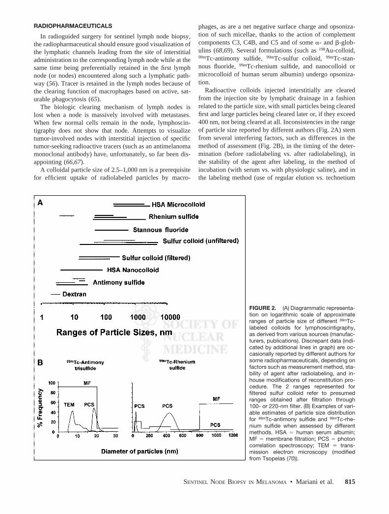
Radioactive colloids injected interstitially are clearedfrom the injection site by lymphatic drainage in a fashionrelated to the particle size, with small particles being clearedfirst and large particles being cleared later or, if they exceed400 nm, not being cleared at all. Inconsistencies in the rangeof particle size reported by different authors (Fig. 2A) stemfrom several interfering factors, such as differences in themethod of assessment (Fig. 2B), in the timing of the deter-mination (before radiolabeling vs. after radiolabeling), inthe stability of the agent after labeling, in the method ofincubation (with serum vs. with physiologic saline), and inthe labeling method (use of regular elution vs. technetium
FIGURE 2. (A) Diagrammatic representa-tion on logarithmic scale of approximateranges of particle size of different 99mTc-labeled colloids for lymphoscintigraphy,as derived from various sources (manufac-turers, publications). Discrepant data (indi-cated by additional lines in graph) are oc-casionally reported by different authors forsome radiopharmaceuticals, depending onfactors such as measurement method, sta-bility of agent after radiolabeling, and in-house modifications of reconstitution pro-cedure. The 2 ranges represented forfiltered sulfur colloid refer to presumedranges obtained after filtration through100- or 220-nm filter. (B) Examples of vari-able estimates of particle size distributionfor 99mTc-antimony sulfide and 99mTc-rhe-nium sulfide when assessed by differentmethods. HSA � human serum albumin;MF � membrane filtration; PCS � photoncorrelation spectroscopy; TEM � trans-mission electron microscopy (modifiedfrom Tsopelas (70)).
SENTINEL NODE BIOPSY IN MELANOMA • Mariani et al. 815
elution after a long interval of ingrowth). The pore size ofany filters used (as with 99mTc-sulfur colloid) and in-housemodifications of the reconstitution procedures represent ad-ditional variables to be considered (71–75).
Although sufficient for lymphoscintigraphic visualizationand for �-probe identification, the amount of radioactivityretained in the sentinel lymph node 15–18 h after interstitialinjection of radiocolloid is generally low. Depending on thedifferent radiocolloids used, approximately 0.34%–0.92%of the injected dose is retained per node when the tracer isinjected subdermally in patients with cutaneous melanoma(76,77), although values have ranged from as low as0.0013% to as high as 6.8% in a single study (78).
99mTc-Sulfur colloid is most commonly used in theUnited States. Microfiltration through 100- or 220-nm filtershas been proposed, with a goal of using particles in therange of approximately 50–100 or 50–200 nm. Filtered99mTc-sulfur colloid is generally preferred for sentinellymph node studies (79), although some authors still claimthe superiority of the unfiltered agent (80,81). The highlymph flow in the skin (particularly of the extremities) andthe somewhat higher porosity of the lymphatic system(61,62,82) suggest that radiocolloids of larger particle size(between 200 and 300 nm) may yield satisfactory results inpatients with cutaneous melanoma.
Most European investigators have used 99mTc-nanocol-loid human serum albumin with particles of between 4 andapproximately 100 nm (95% of the particles � 80 nm). Thisradiopharmaceutical offers the additional benefits of instantlabeling at room temperature and stability both in vitro andin vivo.
Besides particle size, the number of particles injected isan important parameter in radioguided sentinel lymph nodelocalization. This feature, which has to do with saturation ofthe clearing capacity of the sentinel lymph node based onactive phagocytosis by macrophages, was earlier discussedat length (56). In particular, the specific activity of thepreparation is important for administration of a preparationwith the fewest particles. Therefore, methods of high-spe-cific-activity labeling should be sought.
TECHNIQUES
Tracer InjectionA general consensus has developed over the last few
years on the main parameters that define the optimal tech-niques for injecting radiocolloid for lymphatic mapping andsentinel lymph node biopsy in patients with malignant cu-taneous melanoma (50,69,83–91).
The cells that give rise to cutaneous melanomas, mela-nocytes, are located between the dermis and the epidermis.Transformed melanocytes initially grow in this space, andfrom it they spread elsewhere. Therefore, there is no doubtthat lymphatic drainage from these tumors follows the gen-eral pattern of lymph flow from the papillary and subpap-illary plexus to the inner network in the reticular dermis and,
in turn, to the larger collecting lymphatics in the subcutis.The subcutaneous plexus (terminal component of the over-all superficial lymphatic circulation) communicates with thedeep, epifascial lymphatic circulation collecting lymphfrom muscles, bones, and so forth (Fig. 1A).
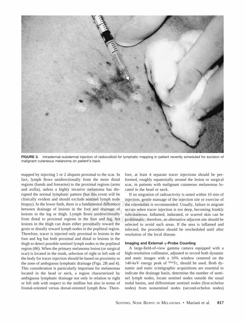
These considerations explain why the concept of intra-dermal or subdermal injection of radiocolloid for lymphaticmapping in patients with cutaneous melanoma is generallyaccepted and applied routinely. Intradermal or subdermalinjection is performed using 25- or 27-gauge needles, in-serting the needle in a direction as tangent as possible to theskin surface for a few millimeters inside the skin; thismodality of interstitial administration entails small volumesof injectate, just enough to produce a visible wheal in theskin (Fig. 3). Small volumes (0.05–0.2 mL, depending onthe thickness of the skin in a specific anatomic region) arerecommended in order not to collapse lymphatics (85);when the doses for injection are prepared (radioactive prep-aration containing 37 MBq/mL, or 1 mCi/mL), 0.1–0.2 mLof air is drawn into the syringe behind the radiocolloid toensure that no tracer is left in the dead space of the syringeand needle. Care must be taken to avoid contamination ofthe patient’s skin during the procedure, because interstitialpressure builds up on intradermal or subdermal injection;contamination should be prevented by placement of a swabover the needle before it is withdrawn from the skin.
The number and specific site of radiocolloid aliquots toinject varies according to the anatomic region being ex-plored and whether the primary lesion is still present or hasalready been excised, always observing the general rule ofinjecting the tracer approximately 0.5–1 cm away from thescar or the tumor margin. One or 2 injections close to thecenter of the surgical scar (corresponding to the approxi-mate original location of the excised tumor) can be suffi-cient for reliably visualizing the pattern of lymphatic drain-age encountered by tumor cells leaving the melanoma; incases of wide excisions or of a large melanoma still presentin the skin, a total of 4, 6, or even 8 separate injections canbe necessary, depending on different factors.
Often, we study patients 2–3 wk after tumor excision,when the histologic findings indicate a thin or intermediate-thickness cutaneous melanoma. Size and orientation of thesurgical scar are dictated by both location and size of thetumor. If the melanoma is not exceedingly large, the sur-geon makes the incision along the predominant lines ofcontraction of skeletal muscles directly underneath the tu-mor (Fig. 4). This recommended pattern may occasionallynot be followed in cases of wide lesions, which may forcethe surgeon to adapt the incision so that an adequate 1- to2-cm margin of apparently healthy skin is removed on eachside of the melanoma (1 cm for lesions � 1 mm thick, 2 cmfor lesion thicker than 1 mm).
Keeping as a reference the orientation of the surgical scar,one should inject the tracer either on each side or on onlyone side of the scar, depending on location. If the scar islocated in the upper limbs, the lymphatics can reliably be
816 THE JOURNAL OF NUCLEAR MEDICINE • Vol. 43 • No. 6 • June 2002
mapped by injecting 1 or 2 aliquots proximal to the scar. Infact, lymph flows unidirectionally from the more distalregions (hands and forearms) to the proximal regions (armsand axilla), unless a highly invasive melanoma has dis-rupted the normal lymphatic pattern (but this event will beclinically evident and should exclude sentinel lymph nodebiopsy). In the lower limb, there is a fundamental differencebetween drainage of lesions in the foot and drainage oflesions in the leg or thigh. Lymph flows unidirectionallyfrom distal to proximal regions in the foot and leg, butlesions in the thigh can drain either proximally toward thegroin or distally toward lymph nodes in the popliteal region.Therefore, tracer is injected only proximal to lesions in thefoot and leg but both proximal and distal to lesions in thethigh to detect possible sentinel lymph nodes in the poplitealregion (86). When the primary melanoma lesion (or surgicalscar) is located in the trunk, selection of right or left side ofthe body for tracer injection should be based on proximity tothe zone of ambiguous lymphatic drainage (Figs. 2B and 4).This consideration is particularly important for melanomaslocated in the head or neck, a region characterized byambiguous lymphatic drainage not only in relation to rightor left side with respect to the midline but also in terms offrontal-oriented versus dorsal-oriented lymph flow. There-
fore, at least 4 separate tracer injections should be per-formed, roughly equatorially around the lesion or surgicalscar, in patients with malignant cutaneous melanomas lo-cated in the head or neck.
If no migration of radioactivity is noted within 10 min ofinjection, gentle massage of the injection site or exercise ofthe extremities is recommended. Usually, failure to migrateoccurs when tracer injection is too deep, becoming franklysubcutaneous. Inflamed, indurated, or scarred skin can beproblematic; therefore, an alternative adjacent site should beselected to avoid such areas. If the area is inflamed andinfected, the procedure should be rescheduled until afterresolution of the local disease.
Imaging and External �-Probe CountingA large-field-of-view gamma camera equipped with a
high-resolution collimator, adjusted to record both dynamicand static images with a 10% window centered on the140-keV energy peak of 99mTc, should be used. Both dy-namic and static scintigraphic acquisitions are essential toindicate the drainage basin, determine the number of senti-nel lymph nodes, locate sentinel nodes outside the usualnodal basins, and differentiate sentinel nodes (first-echelonnodes) from nonsentinel nodes (second-echelon nodes)
FIGURE 3. Intradermal–subdermal injection of radiocolloid for lymphatic mapping in patient recently scheduled for excision ofmalignant cutaneous melanoma on patient’s back.
SENTINEL NODE BIOPSY IN MELANOMA • Mariani et al. 817

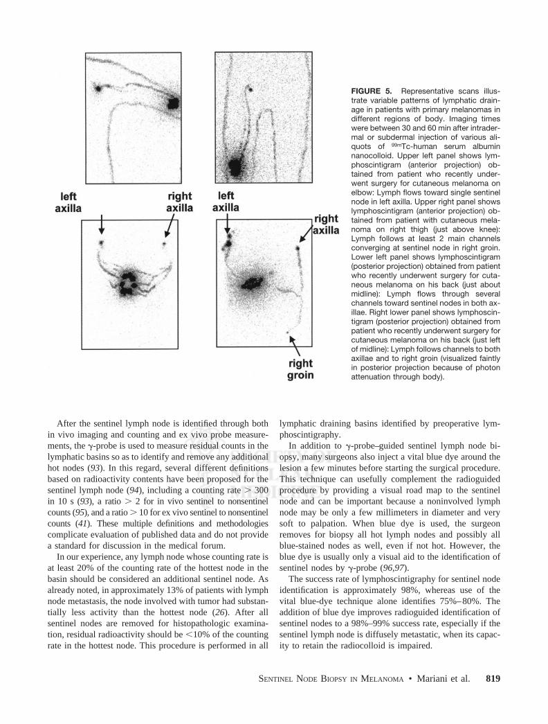
(92). Starting immediately after radiotracer injection, dy-namic acquisition over 20–30 min (20 s per frame, with a128 � 128 matrix) serves to reveal the progression oflymphatic flow (drainage pattern) and to distinguish first-tier from second-tier lymph nodes. The draining lymphaticbasins are outlined, and the number and position of sentinelnodes are depicted (Fig. 5); the site of these nodes can bemarked on the overlying skin by the nuclear medicinephysician, thus localizing the nodes for the surgeon. Ifnecessary, acquisition is continued until a total of 45 min toadequately visualize the sentinel lymph nodes. Subse-quently, orthogonal (anterior or posterior) and oblique (asneeded) static images are acquired (300 s, with a 256 � 256matrix) to localize the nodes in 3 dimensions. To facilitatetopographic localization, a 57Co flood source (or, whenavailable, a CT scan) can be used for simultaneous trans-mission imaging, or the body silhouette can be outlined bymoving a radioactive point source along the contour of thebody while recording the scan. This procedure helps toidentify sentinel lymph nodes and to mark their cutaneousprojection in each draining basin. Static images (spot viewsor whole-body scans) can be repeated at 2 h and, whennodes are not clearly depicted, again at 4–6 h after injectionor even just before operation. The study in the nuclearmedicine department is completed by further confirming the
exact location of the sentinel lymph nodes by externalcounting with the handheld �-probe detector.
Intraoperative �-Probe CountingWith the patient positioned on the operating table, exter-
nal �-probe counting should be repeated to confirm thelocation of the sentinel lymph nodes before the procedure.Depending on the site of the primary lesion, different lym-phatic basins may have been identified by preoperativelymphoscintigraphy. In each site, the area with highestactivity (counts per second) is identified and topographi-cally correlated with the intradermal tattoo that has beenpositioned during lymphoscintigraphy. Furthermore, countsare taken over the sites of tracer injection and along thelymphatic channels draining to the lymphatic basins. Allsuch external measurements are recorded for subsequentcomparison with intraoperative count rates (which shouldobviously be correspondingly higher than external countrates). The patient is then prepared and draped in the usualsterile fashion. Attention is turned to all the lymphaticbasins that have been identified as at risk for metastasisaccording to the lymphoscintigraphic pattern. Surgeons usu-ally search for the sentinel lymph nodes first and thenproceed to remove the primary melanoma (if it is still inplace) or to excise a wider skin margin around the previoussurgical scar (if this indication has emerged from the histo-pathology of the primary lesion).
In vivo radioactivity (counts per second) is measured inthe surgical bed opened by making a 2- to 4-cm incisionover the area with highest activity as determined by externalcounting. Intraoperatively, the �-probe is moved carefullyand slowly, because the counting rate changes almost in-stantly from hundreds of counts per second to nearly zero onminimal movement of the probe. Sentinel lymph nodes maybe only a few millimeters in diameter, and the radiocolloidis retained in the sinusoidal spaces near the afferent lymphchannels. Thus, recognition of the node may be difficult ifthe probe is moved too quickly.
Hot nodes are usually identified by comparison withbackground radioactivity, which is defined as the averagecount rates of the surrounding nonsentinel nodes and lymphnode basin. Ratios of sentinel lymph node counts to back-ground counts are typically on the order of 10:1, 15:1, oreven 20:1. This wide range of values depends on the doseinjected, the type of tracer injected, the elapsed time be-tween tracer injection and surgery, and the type of �-probeused. In particular, if a small-particle radiocolloid is in-jected, lymphatic vessels are visualized within a few min-utes after injection and radioactive lymph quicklyprogresses to show second-tier or third-tier nodes as well. Inthis case, the ratio of sentinel counts to nonsentinel countswill be relatively lower than that observed after injection ofa larger-particle radiocolloid (which is preferentially re-tained in the true sentinel node). This factor should beconsidered to avoid unnecessary biopsy of radioactive butnonsentinel lymph nodes.
FIGURE 4. Diagrammatic representation of recommended in-jection sites for lymphatic mapping with radiocolloid accordingto region of body and orientation of surgical scar.
818 THE JOURNAL OF NUCLEAR MEDICINE • Vol. 43 • No. 6 • June 2002
After the sentinel lymph node is identified through bothin vivo imaging and counting and ex vivo probe measure-ments, the �-probe is used to measure residual counts in thelymphatic basins so as to identify and remove any additionalhot nodes (93). In this regard, several different definitionsbased on radioactivity contents have been proposed for thesentinel lymph node (94), including a counting rate � 300in 10 s (93), a ratio � 2 for in vivo sentinel to nonsentinelcounts (95), and a ratio � 10 for ex vivo sentinel to nonsentinelcounts (41). These multiple definitions and methodologiescomplicate evaluation of published data and do not providea standard for discussion in the medical forum.
In our experience, any lymph node whose counting rate isat least 20% of the counting rate of the hottest node in thebasin should be considered an additional sentinel node. Asalready noted, in approximately 13% of patients with lymphnode metastasis, the node involved with tumor had substan-tially less activity than the hottest node (26). After allsentinel nodes are removed for histopathologic examina-tion, residual radioactivity should be �10% of the countingrate in the hottest node. This procedure is performed in all
lymphatic draining basins identified by preoperative lym-phoscintigraphy.
In addition to �-probe–guided sentinel lymph node bi-opsy, many surgeons also inject a vital blue dye around thelesion a few minutes before starting the surgical procedure.This technique can usefully complement the radioguidedprocedure by providing a visual road map to the sentinelnode and can be important because a noninvolved lymphnode may be only a few millimeters in diameter and verysoft to palpation. When blue dye is used, the surgeonremoves for biopsy all hot lymph nodes and possibly allblue-stained nodes as well, even if not hot. However, theblue dye is usually only a visual aid to the identification ofsentinel nodes by �-probe (96,97).
The success rate of lymphoscintigraphy for sentinel nodeidentification is approximately 98%, whereas use of thevital blue-dye technique alone identifies 75%–80%. Theaddition of blue dye improves radioguided identification ofsentinel nodes to a 98%–99% success rate, especially if thesentinel lymph node is diffusely metastatic, when its capac-ity to retain the radiocolloid is impaired.
FIGURE 5. Representative scans illus-trate variable patterns of lymphatic drain-age in patients with primary melanomas indifferent regions of body. Imaging timeswere between 30 and 60 min after intrader-mal or subdermal injection of various ali-quots of 99mTc-human serum albuminnanocolloid. Upper left panel shows lym-phoscintigram (anterior projection) ob-tained from patient who recently under-went surgery for cutaneous melanoma onelbow: Lymph flows toward single sentinelnode in left axilla. Upper right panel showslymphoscintigram (anterior projection) ob-tained from patient with cutaneous mela-noma on right thigh (just above knee):Lymph follows at least 2 main channelsconverging at sentinel node in right groin.Lower left panel shows lymphoscintigram(posterior projection) obtained from patientwho recently underwent surgery for cuta-neous melanoma on his back (just aboutmidline): Lymph flows through severalchannels toward sentinel nodes in both ax-illae. Right lower panel shows lymphoscin-tigram (posterior projection) obtained frompatient who recently underwent surgery forcutaneous melanoma on his back (just leftof midline): Lymph follows channels to bothaxillae and to right groin (visualized faintlyin posterior projection because of photonattenuation through body).
SENTINEL NODE BIOPSY IN MELANOMA • Mariani et al. 819

The criteria for �-probe identification of a sentinel lymphnode are based on detecting a focal zone of radioactivityaccumulation with a ratio of counts (hot spot to background)� 10 (typically in the 10–100 range). The operative siteshould then be reexamined to ensure that the area of radio-activity has been removed and that a second node is not alsoactive; if one is, it should be removed and the surgical bedreexamined until no areas of increased counts are found.Complete removal of sentinel lymph nodes is confirmed byreduction of the counting rate in the surgical area to back-ground levels. Histopathologic examination is performed onthe lymph node with the highest counting rate and on anyadditional lymph node with a counting rate at least 20% ofthat in the hottest node.
Histopathologic ExaminationThe detection of tumor cells in the lymph node requires
special stains and detailed evaluation of the sentinel node. Incontrast to routine histopathology, in which only a fewsections stained with hematoxylin–eosin are evaluated, sen-tinel node evaluation should include 10–20 sections (if notthe entire node) and specific immune staining to identifytumor involvement. The importance of detailed histologicexamination is apparent from retrospective studies on nodalrecurrences in patients whose sentinel nodes were reportedas uninvolved and negative for tumor on conventionalpathologic examination. When additional sections wereevaluated and special stains were applied, tumor cells wereshown in approximately 12% of the patients (51–53).
These observations stress the importance of detailed his-tologic examination of sentinel node biopsy samples. Froma clinical perspective, application of this time-consuming,rigorous approach to all biopsies is difficult, but it is nowapparent that patients with other tumors, such as breastcancer, also benefit when evaluated by this compulsiveapproach (98).
Histopathologic assessment of the excised sentinel nodesshould be as complete as possible (99,100). The harvestednode should be bisected along its main axis, starting at thehilus, and multiple sections should be taken from each half.Some sections are used for hematoxylin–eosin staining,whereas others are processed by immunohistochemistry;additional sections can be kept unstained for additionalprocessing, as needed (99–102).
The need for high-quality histopathologic examinationhas led to reconsideration of the role of frozen sections inthese patients. The possibility of performing both a selectivelymphadenectomy and, if necessary, a full excision of thedraining basin has prompted surgeons to ask for intraoper-ative histopathology. However, examination of frozen sec-tions is not recommended for nonpalpable nodes because ofthe relatively poor quality of frozen-section histology (102).Permanent sections allow more accurate morphologic eval-uation; moreover, immunohistochemistry can be used toidentify the melanocytic cells. Immunohistology is consid-ered crucial by most groups, particularly to detect subtle
micrometastases (103). In fact, immunohistochemistry en-ables differentiation between melanized and nonmelanizedmacrophages, which may closely resemble melanoma cells(102).
Several monoclonal and polyclonal antibodies are nowcommercially available and can consistently be applied toformalin-fixed and paraffin-embedded tissues. Because ofthe large experience acquired by pathologists worldwidewith antibodies to the S-100 protein and the HMB-45 anti-gen, these are especially reliable. Antibodies to the MelanA/Mart1 and NKC1 antigens are additional reagents that seemparticularly useful for increasing the effectiveness of immu-nohistochemistry for detecting micrometastases (102).
However, each antibody has a different specificity andsensitivity; therefore, accurate assessment of the classicmorphologic features of the cells is required to avoid erro-neous results. Despite its utility, immunohistochemistryshould not be used as the fundamental technique to examinesentinel lymph nodes for melanoma metastases. In particu-lar, dendritic cells—and, occasionally, even macrophagesand normal structures such as nerves, if cut transversely orif close to the capsule—can be misdiagnosed for metastaticmelanoma because of their strong immunoreactivity for theS-100 antibody (104). Additional benign lesions stainingpositive for the S-100 protein include nodal nevi inclusions(105). Although these structures are found more frequentlyin the capsule, they can also be found in the internal tra-becula; moreover, they have been found in lymph nodesdraining from malignant cutaneous melanoma, more fre-quently (up to 20%) in the sentinel lymph node than innonsentinel nodes (105,106).
HMB-45 staining can be observed in areas with calcifi-cations, which are easily found in the trabeculae of theinguinal and pelvic lymph nodes, whereas staining is neg-ative in up to 20%–30% of melanomas (102). Thus, immu-nohistochemistry (possibly performed with both antibodies,S-100 and HMB-45 (107)) combined with careful conven-tional histopathology based on step or multiple sections iscurrently the most reliable method to assess sentinel lymphnode status and predict the nodal status of melanoma pa-tients.
Interest in detecting occult metastases in sentinel nodeshas prompted investigators to explore the most sensitivetools available, including primary cell culture and amplifi-cation of messenger RNA for specific biologic markers bythe reverse transcription polymerase chain reaction. In prin-ciple, these techniques can detect even a very few or singleand isolated metastatic cells among 1–10 million normalcells (108). Such paramount sensitivity is in comparisonwith a detection limit of approximately 1 malignant cell in10,000 for conventional hematoxylin–eosin staining andapproximately 1 malignant cell in 100,000 for immunohis-tochemistry (27). Candidate markers for malignant mela-noma include the tyrosinase gene (encoding an enzymeassociated with melanin synthesis) and messenger RNA
820 THE JOURNAL OF NUCLEAR MEDICINE • Vol. 43 • No. 6 • June 2002
present in T lymphocytes infiltrating the melanoma(MART-1) (109–120).
A significant fraction of patients whose sentinel nodes arenegative by careful histopathology and immunohistochem-istry are polymerase chain reaction positive. However, thereis no general consensus on the clinical significance of thesefindings. Furthermore, it is also generally recognized thatthese ultrasensitive methods are fraught with a relativelyhigh false-positive rate (115), linked basically to nodal neviinclusions and nerve structures (102,105) and even to frag-ments of tyrosinase messenger RNA that survive in macro-phages and can only be differentiated from metastatic cellsby examining cell morphology.
One problem with the current techniques using con-ventional reverse transcription polymerase chain reaction(more feasible than in situ reverse transcription polymer-ase chain reaction, which is also applicable to formalin-fixed and paraffin-embedded sections) is the requirementthat a sizable fraction of the lymph node (approximatelyhalf) be processed for tissue homogenization and DNA orRNA extraction. A fraction of this size is not availablefor regular histology and immunohistochemistry (theonly validated procedure). This large tissue requirementraises concern about the possibility of losing some diag-nostic information.
A highly specific molecular marker of malignancy iscurrently not available for melanoma, and only a compari-son of histology and molecular biology performed on thesame tissue or cells may enable a comparative assessment ofthe actual sensitivity of each method. Although studiesunder way will likely increase the specificity of molecularbiology techniques, no clinical decisions should yet bebased on polymerase chain reaction determinations, in orderto avoid unnecessary lymphadenectomy procedures and fur-ther medical treatment.
BIOPSY
The search for micrometastases in the sentinel lymphnode is now consistently accepted for patients with cu-taneous melanoma whose lesions are �1 mm thick oreven �1 mm but at Clark level IV or V or with ulceration(31,32). A positive node identifies patients with a worseprognosis, even among those with cutaneous lesionsthicker than 4 mm. In fact, among patients with 4-mm-thick lesions, the 5-y survival rate is 61% for those withnegative sentinel lymph nodes but only 30% for thosewith positive sentinel lymph nodes (121). After a positivesentinel node is identified, the only widely recommendedtreatment is to complete the lymphadenectomy of thelymphatic basin containing the metastatic sentinel node(122). Lymphadenectomy alone has improved the 5-ysurvival rate from 27% to 48% (123).
To determine the incidence of false-negative studies ofsentinel nodes (so-called skip metastasis), investigatorshave evaluated patients through use of sentinel lymph
node biopsy followed by complete lymphadenectomy.Extensive histopathologic examination of all removednodes showed that fewer than 2% of patients had nega-tive sentinel nodes with metastases in other lymph nodes(8,12,34,40,41,50,52,76,86). Other studies based onlong-term recurrences in previously mapped negativelymphatic basins have suggested that the incidence offalse-negative sentinel node studies may be higher, rang-ing from 4% (i.e., 10/243 patients) (51) to 8% or 9%(50,124), 11% (125), 16% (126), 19% (127), and even27% (128). Many of these studies used either frozensections or incomplete sampling of the node or failed toexamine the sections immunohistologically.
In addition to the tumor status of the sentinel node,several prognostic indices have been developed (129).Cochran et al. (122,130) combined Breslow thickness ofthe primary melanoma, volume of the tumor in the sen-tinel lymph node, and density of interdigitating cells inthe sentinel node (as an index of downregulation in thelymph node) to predict outcome and tumor status of thenonsentinel nodes in melanoma patients. Microstaging ofthe sentinel lymph node based on accurate histomorpho-metric evaluation takes on an increasingly relevant role(11,131,132).
Uncertainties about the actual incidence of false-negativesentinel node biopsies and about the impact of such false-negative findings on long-term clinical outcome are onlypart of the controversy still surrounding the issue of whethersentinel node biopsy should be considered the standard ofcare for patients with malignant cutaneous melanoma(26,133–136). One reason for the debate is the lack ofeffective adjuvant therapy (in addition to regional lymphnodal dissection) once it has been ascertained that the sen-tinel lymph nodes harbor metastases (35,36,123,137,138).
The only positive trial in terms of overall survival anddisease-free survival concerned the use of high-dose inter-feron versus no therapy in patients with stage III melanoma(28). The favorable outcome in these patients has beenmaintained after a median follow-up of more than 10 y buthas not been confirmed by a subsequent trial comparinghigh-dose and low-dose interferon versus no therapy (139).However, a recent short-term trial comparing high-doseinterferon with ganglioside vaccine supported the beneficialeffects of interferon therapy (140), thus leaving the clinicianwith a therapeutic option rather than a standard treatment.Many ongoing multicenter and multinational randomizedtrials are assessing the impact of sentinel lymph node biopsyon the long-term survival of patients with cutaneous mela-noma.
Although investigators are still debating about evi-dence that sentinel lymph node biopsy followed by earlyregional node dissection can improve regional control(and survival), lymphatic mapping definitely providesimportant prognostic information (8), as shown by anoverall 93% 3-y survival in patients with a tumor-nega-tive sentinel node versus 67% in patients with a tumor-
SENTINEL NODE BIOPSY IN MELANOMA • Mariani et al. 821
positive sentinel node (125). This concept has become soaccepted (137) that the tumor status of the sentinel lymphnode is now becoming an essential parameter in the newstaging system for patients with cutaneous melanoma(31,32,141–143) (Tables 1 and 2).
A new exciting perspective on the sentinel lymph nodebiopsy procedure is that it will lead to timely diagnosis ofmicrometastasis in regional lymph nodes. This possibilitynot only makes sentinel node biopsy useful as a stagingprocedure but also opens new windows of opportunity toassess micrometastasis per se and its evolution within thesentinel node. In fact, sentinel nodes may be the initial sitesof antigen presentation leading to an immune response, thusimplying that sentinel lymph nodes without micrometastasismay be activated by submicroscopic cell debris or by sol-uble tumor antigens, whereas cytokine production may besuppressed by micrometastasis (144).
It has not yet been definitely proved that application ofthe sentinel lymph node biopsy improves the overall clinicaloutcome and long-term survival of patients with cutaneousmelanoma. However, the procedure is certainly becomingthe standard of care for these patients as a modality oflymphatic mapping, at least for prognosis, because eitherthe number or the percentage of metastatic lymph nodes isthe strongest predictor of outcome (31,32,141–143).
The indications for sentinel lymph node biopsy in pa-tients with malignant cutaneous melanoma are now some-what wide, encompassing any patient who has an intactmelanoma or has had an incisional or excisional biopsy ofcutaneous melanoma (143). Biopsy is contraindicated inpatients who have already had a wide excision with orwithout closure by a rotation skin flap, because lymphaticdrainage of the primary site can be deeply modified by sucha surgical procedure. Although patients with primary mel-anomas between 1 and 4 mm thick are, in principle, themost likely to benefit from sentinel lymph node biopsy, it isquickly becoming a required standard of care for staging(31,32,141,143).
LEARNING CURVE
The learning curve for sentinel lymph node biopsyreflects the time required to form a team of specialists in
nuclear medicine, surgery, and pathology to work to-gether (56). A major difference in the case of melanomaversus breast cancer concerns the role of the pathologist.Usually, when performing sentinel lymph node biopsy ona patient with breast cancer, the team in the operatingroom relies on the histopathologic results for the excisedsentinel node in deciding impromptu whether to extendthe surgery and dissect the axillary lymph node. There-fore, histopathologic evaluation of the sentinel node hasto be as fast and yet as accurate as possible. Because ofthis stringent requirement, several approaches have beendeveloped to optimize the speed and sensitivity of his-topathologic examination (56). Moreover, the examina-tion usually occurs near the operating room, in an envi-ronment and manner quite different from those of aregular histopathology laboratory. However, even breastcancer patients are receiving the more rigorous his-topathologic examination that is applied to melanomapatients, with fixed specimens being examined by immu-nohistochemistry and reverse transcription polymerasechain reaction. Because this procedure takes time, agrowing number of patients are undergoing sentinelnode biopsy followed later by surgery for the primarytumor.
Although some controversy exists about the number ofprocedures that a team must complete to qualify as a routineperformer of lymphatic mapping in patients with cutaneousmelanoma, we agree with Morton’s suggestion of 30–50procedures, considerably fewer than the 60–80 proceduresconsidered necessary for breast cancer (143). We also be-lieve that the learning phase will be shorter if all or some ofthe team members have prior experience with intraoperativelymphatic mapping in other clinical applications (mostlybreast cancer), either under radioguidance or under visualblue-dye guidance.
As is the case for breast cancer teams (56), multidisci-plinary teams for sentinel lymph node biopsy in melanomapatients must meet 2 important performance requirements tobe considered as having completed their learning phase: Thesentinel lymph node must be successfully identified in at
TABLE 1Final Version of TNM Staging System of American Joint
Committee on Cancer for Malignant Cutaneous Melanoma
Tumorthickness (mm)
No. of nodemetastases
Site ofmetastasis
SerumLDH
T1A/B, �1 N1, 1 1a, soft tissues NormalT2A/B, 1.01–2.0 N2, 2 or 3 1b, lung NormalT3A/B, 2.01–4.0 In-transit metastasis 1c, any other NormalT4A/B, �4 N3, �4 Any other Elevated
LDH � lactate dehydrogenase; A � without ulceration; B � withulceration.
TABLE 2Survival Rates of Patients with Malignant Cutaneous
Melanoma According to Initial Pathology Staging
Stage2-y survival
(%)5-y survival
(%)
P forstages II–IVvs. stage I
I 99 92II 88 67 �0.0001III 71 46 �0.0001IV 11 0 �0.0001
Stage I � T1a N0 M0 through T2a N0 M0; stage II � T2b N0 M0through T4b N0 M0; stage III � T1–4a N1a M0 through any T N3M0; stage IV � any T any N M1.
822 THE JOURNAL OF NUCLEAR MEDICINE • Vol. 43 • No. 6 • June 2002
least 97% of patients, and metastasis must be found in thesentinel node of between 12% and 20% of patients whosemelanoma is �1 mm thick.
RADIATION PROTECTION
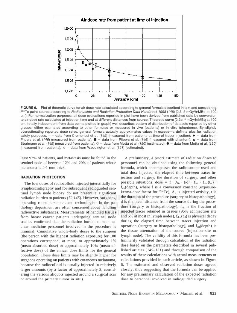
The low doses of radiocolloid injected interstitially forlymphoscintigraphy and for subsequent radioguided sen-tinel lymph node biopsy do not present a significantradiation burden to patients (72,145). However, surgeons,operating room personnel, and technologists in the pa-thology department are often concerned about handlingradioactive substances. Measurements of handled tissuesfrom breast cancer patients undergoing sentinel nodestudies confirmed that the radiation burden to non–nu-clear medicine personnel involved in the procedure isminimal. Cumulative whole-body doses to the surgeon(the person with the highest radiation exposure) for 100operations correspond, at most, to approximately 1%(mean absorbed dose) or approximately 10% (mean ef-fective dose) of the annual dose limits for the generalpopulation. These dose limits may be slightly higher forsurgeons operating on patients with cutaneous melanoma,because the radiocolloid is usually injected in relativelylarger amounts (by a factor of approximately 3, consid-ering the various aliquots injected around a surgical scaror around the primary tumor in situ).
A preliminary, a priori estimate of radiation doses topersonnel can be obtained using the following generalformula, which encompasses the radioisotope used andtotal dose injected, the elapsed time between tracer in-jection and surgery, the duration of surgery, and otherpossible situations: dose � f � A0 � t/d2 � fret � fdec(tel) �fatt(depth), where f is a conversion constant (exposure-kerma-dose factor for 99mTc), A0 is injected activity, t isthe duration of the procedure (surgery or histopathology),d is the mean distance from the source during the proce-dure (surgery or histopathology), fret is the fraction ofinjected tracer retained in tissues (95% at injection siteand 5% at most in lymph nodes), fdec(tel) is physical decayduring the elapsed time between tracer injection andoperation (surgery or histopathology), and fatt(depth) isthe tissue attenuation of the source (injection site orlymph node). The validity of this formula has been pre-liminarily validated through calculation of the radiationdose based on the parameters described in several pub-lished articles (145–151) and through comparison of theresults of these calculations with actual measurements orcalculations provided in each article, as shown in Figure6. The estimated and observed radiation doses agreedclosely, thus suggesting that the formula can be appliedfor any preliminary calculation of the expected radiationdose to personnel involved in radioguided surgery.
FIGURE 6. Plot of theoretic curve for air dose rate calculated according to general formula described in text and considering99mTc point source according to Radionuclide and Radiation Protection Data Handbook 1998 (148) (2.5–5 mGy/h/MBq at 100cm). For normalization purposes, all dose evaluations reported in plot have been derived from published data by conversionto air dose rate calculated at injection time and at different distances from source. Theoretic curve (2.3e5 mGy/h/MBq at 100cm, totally independent from data points plotted in graph) well describes pattern of distribution of datasets reported by othergroups, either estimated according to other formulas or measured in vivo (patients) or in vitro (phantoms). By slightlyoverestimating reported dose rates, general formula actually approximates values in excess—a definite plus for radiationsafety purposes. – � data from Cremonesi et al. (145) (measured from patients at time of tracer injection); � � data fromPijpers et al. (146) (measured from patients); ■ � data from Pijpers et al. (146) (measured with phantom); Œ � data fromStratmann et al. (149) (measured from patients); E � data from Motta et al. (150) (estimated); F � data from Motta et al. (150)(measured from patients); � � data from Waddington et al. (151) (estimated).
SENTINEL NODE BIOPSY IN MELANOMA • Mariani et al. 823

CONCLUSION
Despite some differences in the way radioguided sentinellymph node biopsy is being performed around the world, theexcellent results that have been reported univocally point tothis procedure as being the most safe, efficient, and cost-effective modality for lymphatic mapping in patients withprimary cutaneous melanoma (143,152–154). This conclu-sion, besides being based on the high reported success ratefor identification of sentinel lymph nodes, is also based onthe accuracy of the procedure and, therefore, its ability tospare melanoma patients from unnecessarily extensive sur-gery (138,155,156).
Although the reliability of sentinel lymph node biopsy inpatients with malignant melanoma is widely recognizedboth for staging and for prognosis, some issues are still openfor discussion. The correct procedure for performing ra-dioguided sentinel lymph node biopsy has now been clari-fied, from patient inclusion to choice of radiocolloid, tracerinjection, acquisition of lymphoscintigraphy, intraoperative�-probe searching, and, finally, histopathologic evaluationof the removed node. Nevertheless, the long-term impact ofsentinel node biopsy (with the ensuing elective lymph nodedissection of a metastatic basin) on clinical outcome has yetto be unequivocally determined. Critical metaanalysis of alllong-term follow-up and survival studies published so farwill likely help in the assessment of actual benefit to pa-tients with cutaneous melanoma.
The encouraging results reported for sentinel lymph nodebiopsy, especially in patients with melanoma, have renewedthe interest of investigators in defining a given therapeuticstrategy based on the pathophysiology of each phase in thenatural history of a disease. These considerations makesentinel lymph node biopsy ideal for implementation inother types of cancer as well.
ACKNOWLEDGMENTS
The authors thank all their coworkers who, at variousinstitutions, have helped to develop programs for radio-guided sentinel lymph node biopsy and to accumulate theexperience on which this article was based. In particular, theauthors thank Drs. Pietro Bianchi, Ferdinando Buffoni, andGiuseppe Agnese (Nuclear Medicine Service, University ofGenoa); Drs. Carmine Di Somma, Franca Carli, and GuidoNicolo (National Cancer Institute of Genoa); Drs. GianPiero Manca and Paola Erba (Regional Center of NuclearMedicine, University of Pisa); Drs. Concetta De Cicco andMarco Chinol (Nuclear Medicine Service, European Insti-tute of Oncology, Milan); Dr. Alessandro Testori (Divisionof Surgery, European Institute of Oncology, Milan); andDr. Roberto Gennari (Division of Surgery, “FondazioneMaugeri,” Pavia). Research was supported in part by theItalian Ministry of Education, University and Research, theItalian Ministry of Health, and the Italian Association forCancer Research.
REFERENCES
1. Gloecker-Reis LA, Miller BA, Hankey BF, et al. SEER Cancer StatisticsReview, 1973–1991. Bethesda, MD: National Cancer Institute; 1994. NationalInstitutes of Health publication 94:2789.
2. Landis SH, Murray T, Bolden S, Wingo PA. Cancer statistics, 1999. CA CancerJ Clin. 1999;49:8–31.
3. Rigel DS, Friedman RJ, Kopf AW. The incidence of malignant melanoma in theUnited States: issues as we approach the 21st century. J Am Acad Dermatol.1996;34:839–847.
4. Swetter SM, Smoller BR, Bauer EA. Cutaneous cancer and malignant mela-noma. In: Abeloff MD, Armitage JO, Lichter AS, Niederhuber JE, eds. ClinicalOncology. New York, NY: Churchill Livingston; 1995:1027–1033.
5. Balch CM, Wilkerson JA, Murad TM, Soong SJ, Ingalls AL, Maddox WA. Theprognostic significance of ulceration of cutaneous melanoma. Cancer. 1980;45:3012–3019.
6. Morton DL, Wanek L, Nizze JA, et al. Improved long-term survival afterlymphadenectomy of melanoma metastatic to regional nodes: analysis of prog-nostic factors in 1134 patients from the John Wayne Cancer Clinic. Ann Surg.1991;214:491–499.
7. Cochran AJ, Lana AMA, Wen D-R. Histomorphometry in the assessment ofprognosis in stage II malignant melanoma. Am J Surg Pathol. 1989;13:600–604.
8. Gershenwald JE, Thompson W, Mansfield PF, et al. Multi-institutional mela-noma lymphatic mapping experience: the prognostic value of sentinel lymphnode status in 612 stage I or II melanoma patients. J Clin Oncol. 1999;17:967–983.
9. Cascinelli N, Belli F, Santinami M, et al. Sentinel lymph node biopsy incutaneous melanoma: the WHO Melanoma Program experience. Ann SurgOncol. 2000;7:469–474.
10. Chan AD, Essner R, Wanek LA, Morton DL, Reintgen D. Judging the thera-peutic value of lymph node dissections for melanoma. J Am Coll Surg. 2000;19:16–23.
11. Cherpelis BS, Haddad F, Messina J, et al. Sentinel lymph node micrometastasisand other histologic factors that predict outcome in patients with thicker mel-anomas. J Am Acad Dermatol. 2001;44:762–766.
12. Reintgen D, Cruse CW, Wells K, et al. The orderly progression of melanomanodal metastases. Ann Surg. 1994;220:759–767.
13. Holmes EC, Moseley HS, Morton DL, et al. A rational approach to the surgicalmanagement of melanoma. Ann Surg. 1977;186:481–490.
14. Uren RF, Howman-Giles RB, Shaw HM, Thompson JF, McCarthy WH. Lym-phoscintigraphy in high-risk melanoma of the trunk: predicting draining nodegroups, defining lymphatic channels and locating the sentinel node. J Nucl Med.1993;34:1435–1440.
15. Handley WS. The pathology of melanotic growth in relation to their operativetreatment. Lancet. 1907;1:996.
16. Pringle JH. A method of operation in cases of melanotic tumours of the skin.Edinb Med J. 1908;23:496.
17. Morton DL, Wen D-R, Wong JH, et al. Technical details of intraoperativelymphatic mapping for early stage melanoma. Arch Surg. 1992;127:392–399.
18. Alex JC, Krag DN. Gamma probe guided lymph node localization in malignantmelanoma. Surg Oncol. 1993;2:137–143.
19. Alex JC, Weaver DL, Fairbank JT, Rankin BS, Krag DN. Gamma-probe-guidedlymph node localization in malignant melanoma. Surg Oncol. 1993;2:303–308.
20. Krag D, Weaver D, Alex JC, Fairbank JT. Surgical resection and radiolocal-ization of the sentinel lymph node in breast cancer using a gamma probe. SurgOncol. 1993;2:335–340.
21. Cabanas RM. An approach to the treatment of penile carcinoma. Cancer.1977;39:456–466.
22. Bennett LR, Lago G. Cutaneous lymphoscintigraphy in malignant melanoma.Semin Nucl Med. 1983;13:61–69.
23. Carr I. Lymphatic metastasis. Cancer Metastasis Rev. 1983;2:307–317.24. Malek P, Belan A, Kole J. In vivo evidence of lymphovenous communications
in the popliteal region. Acta Radiol. 1965;3:344–352.25. DeVita V, Hellman S, Rosenberg S. Molecular biology of cancer. In: Fidler IJ,
ed. Principles and Practice of Oncology. 5th ed. Philadelphia, PA: LippincottRaven Press; 1997:140–142.
26. McMasters KM, Reintgen DS, Ross MI, et al. Sentinel lymph node biopsy formelanoma: controversy despite widespread agreement. J Clin Oncol. 2001;19:2851–2855.
27. Ell PJ. A revolution in surgical oncology: sentinel lymph node biopsy. In:Proceedings of the continuing education courses of the SNM; 2001; Toronto,Ontario, Canada: 72–79.
28. Kirkwood JM, Strawderman MH, Ernstaff MS, Smith TJ, Bordern EC, Blum
824 THE JOURNAL OF NUCLEAR MEDICINE • Vol. 43 • No. 6 • June 2002
RH. Interferon alfa-2b adjuvant therapy of high risk resected cutaneous mela-noma: the Eastern Cooperative Oncology Group Trial EST 1684. J Clin Oncol.1996;14:7–17.
29. Wagner JD, Gordon MS, Chuang T-Y, et al. Predicting sentinel and residuallymph node basin disease after sentinel lymph node biopsy for melanoma.Cancer. 2000;89:453–462.
30. McMasters KM. The Sunbelt Melanoma Trial. Ann Surg Oncol. 2001;8(suppl9):41S–43S.
31. Balch CM. The revised melanoma staging system: its use in the design andinterpretation of melanoma clinical trials. J Clin Oncol. 2001;19:82–87.
32. Balch CM, Buzaid AC, Soong SJ, et al. Final version of the American JointCommittee on Cancer staging system for cutaneous melanoma. J Clin Oncol.2001;19:3635–3648.
33. Ross M. Sentinel node mapping for early-stage melanoma. In: Perry MC, ed.Educational Book. Alexandria, VA: ASCO; 1997:326–330.
34. Gershenwald JE, Mansfield PF, Lee JE, Ross MI. Role for lymphatic mappingand sentinel lymph node biopsy in patients with thick (� 4 mm) primarymelanoma. Ann Surg Oncol. 2000;7:160–165.
35. Cascinelli N, Morabito A, Santinami M, Mackie RM, Belli F. Immediate ordelayed dissection of regional lymph nodes in patients with melanoma of thetrunk: a randomised trial—WHO Melanoma Programme. Lancet. 1998;351:793–796.
36. Balch CM, Soong SJ, Bartolucci AA, et al. Efficacy of an elective regionallymph node dissection of 1 to 4 mm thick melanomas for patients 60 years ofage and younger. Ann Surg. 1996;224:255–263.
37. Balch CM, Soong SJ, Milton GW, et al. A comparison of prognostic factors andsurgical results in 1,786 patients with localized (stage I) melanoma treated inAlabama, USA, and New South Wales, Australia. Ann Surg. 1982;196:677–684.
38. Reintgen DS, Cox EB, McCarthy KSJ, Vollmer RT, Seigler HF. Efficacy ofelective lymph node dissection in patients with intermediate thickness primarymelanoma. Ann Surg. 1983;198:379–385.
39. Morton DL, Wen D-R, Foshag LJ, Essner R, Cochran AJ. Intraoperativelymphatic mapping and selective cervical lymphadenectomy for early stagemelanomas of the head and neck. J Clin Oncol. 1993;11:1751–1756.
40. Krag DN, Meijer SJ, Weaver DL, et al. Minimal-access surgery for stagingmalignant melanoma. Arch Surg. 1995;130:654–658.
41. Albertini JJ, Cruse CW, Rapaport D, et al. Intraoperative radio-lympho-scintig-raphy improves sentinel lymph node identification for patients with melanoma.Ann Surg. 1996;223:217–224.
42. Eberbach MA, Whal RL, Argenta LC, Froelich J, Niederhuber JE. Utility oflymphoscintigraphy in directing surgical therapy for melanomas of the head,neck and upper thorax. Surgery. 1987;102:433–442.
43. Norman J, Cruse CW, Espinosa C, et al. Redefinition of cutaneous lymphaticdrainage with the use of lymphoscintigraphy for malignant melanoma. Am JSurg. 1991;162:432–437.
44. Fisher EB, Lewis VL Jr, Griffith BH, Spies W. The role of cutaneous lympho-scintigraphy in determining regional lymph node drainage of truncal melano-mas. Ann Plast Surg. 1992;28:506–510.
45. Berger DH, Feig BW, Podoloff D, et al. Lymphoscintigraphy as a predictor oflymphatic drainage from cutaneous melanoma. Ann Surg Oncol. 1997;4:247–251.
46. Roozendaal GK, De Vries JDH, Van Poll D, et al. Sentinel nodes outside lymphnode basins in melanoma patients. Br J Surg. 2000;88:305–308.
47. Buzaid AC, Sandler AB, Maani S, et al. Role of computer tomography in thestaging of melanoma. J Clin Oncol. 1991;11:638–643.
48. Gritters LS, Francis IR, Zasadny KR, Whal R. Initial assessment of positronemission tomography using 2-fluorine-18–2-deoxy-d-glucose in the imaging ofmalignant melanoma. J Nucl Med. 1993;34:1420–1427.
49. Crippa F, Leutner M, Belli F, et al. Which kind of lymph node metastases canFDG PET detect? A clinical study in melanoma. J Nucl Med. 2000;41:1491–1494.
50. Thompson JF, McCarthy WH, Bosch CM, et al. Sentinel lymph node status asan indicator of the presence of metastatic melanoma in regional lymph nodes.Melanoma Res. 1995;5:255–260.
51. Gershenwald JE, Colome MI, Lee JE, et al. Patterns of recurrence following anegative sentinel lymph node biopsy in 243 patients with stage I or II melanoma.J Clin Oncol. 1998;16:2253–2260.
52. Gershenwald JE, Tseng CH, Thompson W, et al. Improved sentinel lymph nodelocalization in patients with primary melanoma with the use of radiolabeledcolloid. Surgery. 1998;124:203–210.
53. Jansen L, Nieweg OE, Peterse JL, Hoefnagel CA, Valdes Olmos RA, KroonBBR. Reliability of sentinel lymph node biopsy for staging melanoma. Br JSurg. 2000;87:484–489.
54. Gershenwald JE, Berman RS, Porter G, Mansfield PF, Lee JE, Ross MI.Regional nodal basin control is not compromised by previous sentinel lymphnode biopsy in patients with melanoma. Ann Surg Oncol. 2000;7:226–231.
55. Wrone DA, Tanabe KK, Cosimi AB, et al. Lymphedema after sentinel lymphnode biopsy for cutaneous melanoma: a report of 5 cases. Arch Dermatol.2000;136:511–514.
56. Mariani G, Moresco L, Viale G, et al. Radioguided sentinel lymph node biopsyin breast cancer surgery. J Nucl Med. 2001;42:1198–1215.
57. Shields JW. Normal and tumor angiogenesis related to flow. Lymphology.1999;32:118–122.
58. Jonsson K, Wallace S, Jing BS. The clinical significance of lympho-venousanastomoses in malignant disease. Lymphology. 1982;15:95–99.
59. Lubach D, Ludemann W, Berens D, von Rautenfeld D. Recent findings on theangioarchitecture of the lymph vessel system of human skin. Br J Dermatol.1996;135:733–737.
60. Fisher M, Franzeck UK, Herring I, et al. Flow velocity of single lymphaticcapillaries in human skin. Am J Physiol. 1996;270:358–363.
61. Uren RF, Howman-Giles RB, Thompson JF, Roberts J, Bernard E. Variabilityof cutaneous lymphatic flow rates in melanoma patients. Melanoma Res. 1998;8:279–282.
62. Uren RF, Howman-Giles RB, Thompson JF. Demonstration of second-tierlymph nodes during preoperative lymphoscintigraphy for melanoma: incidencevaries with primary tumor site. Ann Surg Oncol. 1998;5:517–521.
63. Leong SP, Achtem TA, Habib FA, et al. Discordancy between clinical predic-tions versus lymphoscintigraphic and intraoperative mapping of sentinel lymphnode drainage of primary melanoma. Arch Dermatol. 1999;135:1472–1476.
64. McMasters KM. Multiple nodal basin drainage in truncal melanomas. Ann SurgOncol. 2000;7:249–250.
65. Bergqvist L, Stundberg R, Ryden S, Strand S-E. The “critical colloid dose” instudies of reticuloendothelial function. J Nucl Med. 1987;28:1424–1429.
66. Limouris GS, Voliotopoulos V, Kondi Paphiti A, Stavraka A, Vlahos L. Twophase scintigraphic mapping of lymphatic drainage in cutaneous melanomausing 99mTc-sulfur microcolloid/99mTc antimelanoma antibody. Anticancer Res.1997;17:1667–1669.
67. Bartolomei M, Testori A, Chinol M, et al. Sentinel node localization in cuta-neous melanoma: lymphoscintigraphy with colloids and antibody fragmentsversus blue dye mapping. Eur J Nucl Med. 1998;25:1489–1494.
68. Moghimi SM, Patel HM. Differential properties of organ-specific serum opso-nins for liver and spleen macrophages. Biochim Biophys Acta. 1989;984:379–383.
69. Moghimi SM, Hawley AE, Christy NM, Gray T, Illum L, Davis SS. Surfaceengineered nanospheres with enhanced drainage into lymphatics and uptake bymacrophages of the regional lymph nodes. FEBS Lett. 1994;344:25–30.
70. Tsopelas C. Particle size analysis of 99mTc-labeled and unlabeled antimonytrisulfide and rhenium sulfide colloids intended for lymphoscintigraphic appli-cation. J Nucl Med. 2001;42:460–466.
71. Alazraki NP, Styblo T, Grant SF, Cohen C, Larsen T, Aarsvold JN. Sentinelnode staging of early breast cancer using lymphoscintigraphy and the intraop-erative gamma-detecting probe. Semin Nucl Med. 2000;30:56–64.
72. Bergqvist L, Strand S-E, Persson B. Particle sizing and biokinetics of interstitiallympho-scintigraphic agents. Semin Nucl Med. 1983;12:9–19.
73. Eckelman WC, Steigman J, Paik CH. Radiopharmaceutical chemistry. In: Har-bert JC, Eckelman WC, Neumann RD, eds. Nuclear Medicine: Diagnosis andTherapy. New York, NY: Thieme Medical Publishers; 1996:213–265.
74. Wilhelm AJ, Mijnhout GS, Franssen EJF. Radiopharmaceuticals in sentinellymph-node detection: an overview. Eur J Nucl Med. 1999;26(suppl):S36–S42.
75. Eshima D, Fauconnier T, Eshima L, Thornback JR. Radiopharmaceuticals forlymphoscintigraphy: including dosimetry and radiation considerations. SeminNucl Med. 2000;30:25–32.
76. Kapteijn BA, Nieweg OE, Liem I, et al. Sentinel node biopsy in melanomapatients: gamma probe detection versus blue dye. Ann Surg Oncol. 1997;4:156–160.
77. Pijpers R, Borgstein PJ, Meijer S, et al. Transport and retention of colloidaltracers in regional lymphoscintigraphy in melanoma: influence on lymphaticmapping and sentinel node biopsy. Melanoma Res. 1998;8:413–418.
78. Kapteijn BAE, Nieweg OE, Muller SH, et al. Validation of gamma probedetection of the sentinel node in melanoma. J Nucl Med. 1997;38:362–366.
79. Goldfarb LR, Alazraki NP, Eshima D, Eshima LA, Herda SC, Halkar RK.Lymphoscintigraphic identification of sentinel lymph nodes: clinical evaluationof 0.22-micron filtration of Tc-99m sulfur colloid. Radiology. 1998;208:505–509.
80. Cody HS III, Borgen PI. State-of-the-art approaches to sentinel node biopsy forbreast cancer: study design, patient selection, technique, and quality control atMemorial Sloan-Kettering Cancer Center. Surg Oncol. 1999;8:85–91.
SENTINEL NODE BIOPSY IN MELANOMA • Mariani et al. 825
81. Hill AD, Tran KN, Yeung H, Yeh SD, Borgen PI, Cody HS III. Sentinel lymphnode biopsy in breast cancer: unfiltered radioisotope is superior to filtered. J AmColl Surg. 1999;188:377–381.
82. Joseph E, Brobeil A, Cruse CW, Wells K, Costello D, Reintgen DS. Lymphaticmapping for melanomas of the extremity. J Hand Surgery [Am]. 1999;24:675–681.
83. Alazraki NP, Eshima D, Eshima LA, et al. Lymphoscintigraphy, the sentinelnode concept, and the intraoperative gamma probe in melanoma, breast cancer,and other potential cancers. Semin Nucl Med. 1997;27:55–67.
84. Borgstein P, Meijer S. Historical perspective of lymphatic tumour spread andthe emergence of the sentinel node concept. Eur J Surg Oncol. 1998;24:85–89.
85. Keshtgar MRS, Ell PJ. Sentinel lymph node detection and imaging. Eur J NuclMed. 1999;26:57–67.
86. Krag D, Harlow S, Weaver D, Ashikaga T. Technique of sentinel node resectionin melanoma and breast cancer: probe-guided surgery and lymphatic mapping.Eur J Surg Oncol. 1998;24:89–93.
87. Borgstein PJ. The Sentinel Node Concept: Consequences of Lymphatic TumourSpread in Melanoma and Breast Cancer [thesis]. Amsterdam, The Netherlands:Free University of Amsterdam; 1999.
88. Gennari R, Stoldt HS, Bartolomei M, et al. Sentinel node localisation: a newprospective in the treatment of nodal melanoma metastases. Int J Oncol.1999;15:25–32.
89. Gennari R, Bartolomei M, Testori A, et al. Sentinel node localization in primarymelanoma: preoperative dynamic lymphoscintigraphy, intraoperative gammaprobe, and vital dye guidance. Surgery. 2000;127:19–25.
90. Keshtgar MRS, Waddington WA, Lakhani SR, Ell PJ. The Sentinel Node inSurgical Oncology. Berlin, Germany: Springer Verlag; 1999.
91. Freeman LM, Blaufox D, eds. Seminars in Nuclear Medicine. Vol 30. Phila-delphia, PA: WB Saunders; 2000.
92. Pijpers R, Collet GJ, Meijer S, Hoekstra OS. The impact of dynamic lympho-scintigraphy and gamma probe guidance on sentinel node biopsy in melanoma.Eur J Nucl Med. 1995;22:1238–1241.
93. McMasters KM, Reintgen DS, Ross MI, et al. Sentinel lymph node biopsy formelanoma: how many radioactive nodes should be removed? Ann Surg Oncol.2001;8:192–197.
94. Nieweg OE, Tanis PJ, Kroon BBR. The definition of a sentinel node. Ann SurgOncol. 2001;8:538–541.
95. Mudun A, Murray DR, Herda SC, et al. Early stage melanoma: lymphoscintig-raphy, reproducibility of sentinel node detection, and effectiveness of theintraoperative gamma probe. Radiology. 1996;199:171–175.
96. Glass LF, Messina JL, Cruse W, et al. The use of intraoperative radiolym-phoscintigraphy for sentinel node biopsy in patients with malignant melanoma.Dermatol Surg. 1996;22:715–720.
97. Bostick P, Essner R, Sarantou T, et al. Intraoperative lymphatic mapping forearly-stage melanoma of the head and neck. Am J Surg. 1997;174:536–539.
98. Viale G, Maiorano E, Mazzarol G, et al. Histologic detection and clinicalimplications of micrometastases in axillary sentinel lymph nodes for patientswith breast carcinoma. Cancer. 2001;92:1378–1384.
99. Goscin C, Glass LF, Messina JL. Pathologic examination of the sentinel lymphnode in melanoma. Surg Oncol Clin North Am. 1999;8:427–434.
100. Messina JL, Glass LF, Cruse CW, Berman C, Ku NK, Reintgen DS. Pathologicexamination of the sentinel lymph node in malignant melanoma. Am J SurgPathol. 1999;23:686–690.
101. Cochran AJ, Wen D-R, Morton DL. Occult tumor cells in the lymph nodes ofpatients with pathological stage I malignant melanoma: an immunohistologicalstudy. Am J Surg Pathol. 1988;12:612–618.
102. Cochran AJ, Balda B-R, Starz H, et al. The Augsburg Consensus: techniques oflymphatic mapping, sentinel lymphadenectomy and completion lymphadenec-tomy in cutaneous malignancies. Cancer. 2000;89:237–241.
103. Messina JL, Glass F. Pathologic examination of the sentinel node. J Fla MedAssoc. 1997;84:153–156.
104. Yu LL, Flotte TJ, Tanabe KK, et al. Detection of microscopic melanomametastases in sentinel lymph nodes. Cancer. 1999;86:617–627.
105. Carson KF, Wen D-R, Li P-X, et al. Nodal nevi and cutaneous melanomas. Am JSurg Pathol. 1996;20:834–840.
106. Ridolfi RL, Rosen PP, Thaler H. Nevus cell aggregates associated with lymphnodes: estimated frequency and clinical significance. Cancer. 1977;39:164–171.
107. Lingam MK, Mackie RM, McKay AJ. Intraoperative identification of sentinellymph node in patients with malignant melanoma. Br J Cancer. 1997;75:1505–1508.
108. Palmieri G, Ascierto PA, Cossu A, et al. Detection of occult melanoma cells inparaffin-embedded histological negative sentinel lymph nodes using a reversetranscriptase polymerase chain reaction assay. J Clin Oncol. 2001;19:1437–1443.
109. van der Velde-Zimmermann D, Roijiers JF, Bouwens-Rombouts A, et al.Molecular test for the detection of tumor cells in blood and sentinel nodes ofmelanoma patients. Am J Pathol. 1996;149:759–764.
110. Li P-X, Cheng L, Wen D-R, et al. Demonstration of cytoplasmic tyrosinasemRNA in tissue cultured cells by reverse transcriptase (RT) in situ polymerasechain reaction (PCR) and RT PCR in situ hybridization. Diagn Mol Pathol.1997;6:26–33.
111. Blaheta HJ, Schittek B, Breuninger H, et al. Lymph node micrometastases ofcutaneous melanoma: increased sensitivity of molecular diagnosis in compari-son to immunohistochemistry. Int J Cancer. 1998;79:318–323.
112. Goydos JS, Ravikumar TS, Germino FJ, Yudd A, Bancila E. Minimally invasivestaging of patients with melanoma: sentinel lymphadenectomy and detection ofthe melanoma-specific proteins MART-1 and tyrosinase by reverse transcriptasepolymerase chain reaction. J Am Coll Surg. 1998;187:182–190.
113. Guo J, Cheng L, Wen D-R, Huang RR, Cochran AJ. Demonstration of tyrosi-nase mRNA in formalin fixed paraffin-embedded archival sections of mela-noma, using the reverse transcriptase in situ polymerase chain reaction. DiagnMol Pathol. 1998;7:10–15.
114. Shivers SC, Wang X, Li W, et al. Molecular staging of malignant melanoma:correlation with clinical outcome. JAMA. 1998;280:1410–1415.
115. Bieligk SC, Ghossein R, Bhattacharya S, Coit DG. Detection of tyrosinasemRNA by reverse transcription-polymerase chain reaction in melanoma sentinelnodes. Ann Surg Oncol. 1999;6:232–240.
116. Bostick PJ, Morton DL, Turner RR, et al. Prognostic significance of occultmetastases detected by sentinel lymphadenectomy and reverse transcriptase-polymerase chain reaction in early-stage melanoma patients. J Clin Oncol.1999;17:3238–3244.
117. Blaheta HJ, Ellwanger U, Schittek B, et al. Examination of regional lymphnodes by sentinel node biopsy and molecular analysis provides new stagingfacilities in primary cutaneous melanoma. J Invest Dermatol. 2000;114:637–642.
118. Pellegrino D, Bellina CR, Manca G, et al. Detection of melanoma cells inperipheral blood and sentinel lymph nodes by RT-PCR analysis: a comparativestudy with immunohisto-chemistry. Tumori. 2000;86:336–338.
119. van der Velde-Zimmermann D, Schipper ME, de Weger RA, Hennipman A,Borel Rinkes IH. Sentinel node biopsies in melanoma patients: a protocol foraccurate, efficient, and cost effective analysis by preselection for immunohis-tochemistry on the basis of Tyr-PCR. Ann Surg Oncol. 2000;7:51–54.
120. Shivers SC, Li W, Lin J, et al. The clinical relevance of molecular staging formelanoma. Recent Results Cancer Res. 2001;158:187–199.
121. Zettersten E, Sagabiel RW, Miller J III, Tallapureddy S, Leong S, Kshani-SabetM. Prognostic factors in thick cutaneous melanoma (� 4 mm) [abstract]. MelRes. 2001;11(suppl 1):S74.
122. Cochran AJ, Morton DL, Johnson TD, et al. Prediction of outcome and tumor-status of the non-sentinel node in melanoma patients with a positive sentinelnode [abstract]. Modern Pathol. 2000;13:61A.
123. Thompson JF. Surgical management of occult lymph node metastases [abstract].Melanoma Res. 2001;11(suppl 1):S12.
124. Jacobs IA, Chevinsky AH, Magidson JG, Britto EJ, Smith TJ. Gamma-probe-directed lymphatic mapping and sentinel lymphoadenectomy in primary mela-noma: reliability of the procedure and analysis of failures after long-termfollow-up. J Surg Oncol. 2001;77:157–164.
125. Jansen L, Nieweg OE, Kapteijn AE, et al. Reliability of lymphoscintigraphy inindicating the number of sentinel nodes in melanoma patients. Ann Surg Oncol.2000;7:624–630.
126. Clary BM, Brady MS, Lewis JJ, Coit DG. Sentinel lymph node biopsy in themanagement of patients with cutaneous melanoma: review of a large single-institutional experience with emphasis on recurrence. Ann Surg. 2001;233:250–258.
127. Harlow SP, Krag DN, Ashikaga T, et al. Gamma probe guided biopsy of thesentinel node in malignant melanoma: a multicentre study. Melanoma Res.2001;11:45–55.
128. Cascinelli N. WHO melanoma programme: results of the sentinel node study[abstract]. In: Proceedings of Sentinel Node 2000; December 1–4, 2000; SantaMonica, CA: 19.
129. Hoon DSB, Bostick P, Kuo C, et al. Molecular markers in blood as surrogateprognostic indicators of melanoma recurrence. Cancer Res. 2000;60:2253–2257.
130. Cochran AJ, Wen D-R, Farzad Z, et al. Immune suppression by melanoma cellsas a factor in the generation of metastatic disease. Anticancer Res. 1989;9:859–864.
131. Starz H, Balda BR, Kramer KU, Buchels H, Wang H-J. A micromorphometrybased concept for routine staging classification of sentinel lymph node metas-
826 THE JOURNAL OF NUCLEAR MEDICINE • Vol. 43 • No. 6 • June 2002

tases and its clinical relevance in melanoma patients. Cancer. 2001;91:2110–2121.
132. Starz H, De Donno A, Balda BR. The Augsburg experience: histological aspectsand patient outcomes. Ann Surg Oncol. 2001;8(suppl 9):48S–51S.
133. Emilia JCD, Lawrence W. Sentinel lymph node biopsy in malignant melanoma:the standard of care? J Surg Oncol. 1997;65:153–154.
134. Morton DL. Intraoperative lymphatic mapping and sentinel lymphadenectomy:community standard care or clinical investigation? Cancer J Sci Am. 1997;3:328–330.
135. Nieweg OE, Kapteijn BAE, Thompson JF, Kroon BBR. Lymphatic mappingand selective lymphadenectomy for melanoma: not yet the standard therapy[editorial]. Eur J Surg Oncol. 1997;23:397–398.
136. Nieweg OE, Tanis PJ, de Vries JDH, Valdes Olmos RA, Kroon BBR, HoefnagelCA. The sentinel node in melanoma: present controversies. In: proceedings ofthe continuing education courses of the SNM; 2001; Toronto, Ontario, Canada:94–103.
137. Soong SJ, Harrison RA, McCarthy WH, Urist MM, Balch CM. Factors affectingsurvival following local, regional, or distant recurrence from localized mela-noma. J Surg Oncol. 1998;67:228–233.
138. Nieweg OE, Tanis PJ, Rutgers EJT. Summary of the second internationalsentinel node conference. Eur J Nucl Med. 2001;28:646–649.
139. Kirkwood JM, Ibrahim JG, Sondak VK, et al. High- and low-dose interferonalfa-2b in high-risk melanoma: first analysis of intergroup trial E1690/S9111/C9190. J Clin Oncol. 2000;18:2444–2458.
140. Kirkwood JM, Ibrahim JG, Sosman JA, et al. High-dose interferon alfa-2bsignificantly prolongs relapse-free and overall survival compared with theGM2-KLH/QS-21 vaccine in patients with resected stage IIB-II melanoma:results of intergroup trial E1694/S9512/C509801. J Clin Oncol. 2001;19:2370–2380.
141. Kroon BBR, Nieweg OE, Hoekstra HJ, Lejeune FJ. Principles and guidelines forsurgeons: management of cutaneous malignant melanoma. Eur J Surg Oncol.1998;23:550–558.
142. Balch CM, Buzaid AC, Atkins MB, et al. A new American Joint Committee onCancer Staging system for cutaneous melanoma. Cancer. 2000;88:1484–1491.
143. Cochran AJ. The pathologist’s role in sentinel lymph node evaluation. SeminNucl Med. 2000;30:11–17.
144. Leong SP, Peng M, Zhou YM, et al. Cytokine profiles of sentinel lymph nodesdraining the primary melanoma. Ann Surg Oncol. 2002;9:82–87.
145. Cremonesi M, Ferrari M, Sacco E, et al. Radiation protection in radioguidedsurgery of breast cancer. Nucl Med Commun. 1999;20:919–924.
146. Pijpers R, Peijer S, Dignum P, et al. The sentinel node procedure: aspects ofradiation safety of a new surgical technique. Tijdschr Nucl Geneesk. 1997;19:144–147.
147. Glass EC, Essner R, Giuliano AE. Sentinel node localization in breast cancer.Semin Nucl Med. 1999;29:57–68.
148. Delacroix D, Guerre JP, Leblanc P, Hickman C. Radionuclide and radiationprotection data handbook 1998. Rad Prot Dosim. 1998;76:7–16.
149. Stratmann SL, McCarthy TM, Kuhn JA. Radiation safety with breast sentinelnode biopsy. Am J Surg. 1999;178:454–457.
150. Motta C, Turra A, Farina B, Ostan A, Ramella S, Cartia GL. Radioguidedsurgery of breast cancer: radiation protection survey. Tumori. 2000;86:372–374.
151. Waddington WA, Keshtgar MRS, Taylor I, Lakhani SR, Short MD, Ell PJ.Radiation safety of the sentinel lymph node technique in breast cancer. EurJ Nucl Med. 2000;27:377–391.
152. Reintgen D, Balch CM, Kirkwood J, Ross M. Recent advances in the care of thepatient with malignant melanoma. Ann Surg. 1997;1:1–14.
153. Reintgen DS, Albertini J, Millotes G, et al. Investment in new technologyresearch can save future health care dollars. J Fla Med Assoc. 1997;84:175–181.
154. Brobeil A, Cruse CW, Messina JL, et al. Cost analysis of sentinel lymph nodebiopsy as an alternative to elective lymph node dissection in patients withmalignant melanoma. Surg Oncol Clin North Am. 1999;8:435–445.
155. Reintgen D. More rational and conservative surgical strategies for malignantmelanoma using lymphatic mapping and sentinel node biopsy techniques. CurrOpin Oncol. 1996;8:152–158.
156. Balch CM, Ross MI. Sentinel lymphadenectomy for melanoma: is it a substitutefor elective lymphadenectomy? Ann Surg Oncol. 1999;6:416–417.
157. Ackerman AB. Histologic Diagnosis of Inflammatory Skin Diseases: A Methodby Pattern Analysis. Philadelphia, PA: Lea & Febiger; 1978: 30.
SENTINEL NODE BIOPSY IN MELANOMA • Mariani et al. 827
Related Documents