1 Radiography Protocols Upper Limb Second through Fifth Digits (Standard 3 views) First Digit (Thumb) (Standard 3 views) Hand (Standard 3 views) Wrist (Standard 4 views) Forearm (Standard 2 views) Elbow (Standard 4 views) Humerus (Standard 2 views) Shoulder (Standard 3 views) Acromioclavicular Articulation (Standard 2 views) Clavicle (Standard 2 views) Scapula (Standard 2 views) Lower Limb Toes (Standard 3 views) Foot (Standard 3 views) Calcaneus (Standard 2 views) Ankle (Standard 4 views) Tib/Fib (Standard 2 projections) Knee (Standard 4 views) Femur (Standard 2 projections) Hip (Standard 2 Projections) Pelvis (Standard 1 projection) Vertebral Column Cervical Spine (Standard 3 views) Thoracic Spine (Standard 2 projections) Lumbar Spine (Standard 2 views) Sacroiliac Joints (Standard 2 projections) Sacrum (Standard 2 views) Coccyx (Standard 2 views) Scoliosis (Standard 1 projection) Thorax Routine Chest (Standard 2 projections) Portable Chest (Standard 1 projection) Nonroutine Chest Views Ribs (Standard 2 projections)

Welcome message from author
This document is posted to help you gain knowledge. Please leave a comment to let me know what you think about it! Share it to your friends and learn new things together.
Transcript

1
Radiography Protocols
Upper Limb
Second through Fifth Digits (Standard 3 views)
First Digit (Thumb) (Standard 3 views)
Hand (Standard 3 views)
Wrist (Standard 4 views)
Forearm (Standard 2 views)
Elbow (Standard 4 views)
Humerus (Standard 2 views)
Shoulder (Standard 3 views)
Acromioclavicular Articulation (Standard 2
views)
Clavicle (Standard 2 views)
Scapula (Standard 2 views)
Lower Limb
Toes (Standard 3 views)
Foot (Standard 3 views)
Calcaneus (Standard 2 views)
Ankle (Standard 4 views)
Tib/Fib (Standard 2 projections)
Knee (Standard 4 views)
Femur (Standard 2 projections)
Hip (Standard 2 Projections)
Pelvis (Standard 1 projection)
Vertebral Column
Cervical Spine (Standard 3 views)
Thoracic Spine (Standard 2 projections)
Lumbar Spine (Standard 2 views)
Sacroiliac Joints (Standard 2 projections)
Sacrum (Standard 2 views)
Coccyx (Standard 2 views)
Scoliosis (Standard 1 projection)
Thorax
Routine Chest (Standard 2 projections)
Portable Chest (Standard 1 projection)
Nonroutine Chest Views
Ribs (Standard 2 projections)

2
Sternum (Standard 2 views)
Sternoclavicular Articulations (Standard 2
projections)
Cranium
Facial Bones (Standard 3 views)
Mandible (Standard 2 projections)
Nasal Bones (Standard 2 projections)
Abdomen
Abdomen (Standard 2 projection)

3
4
Upper Limb
Second through Fifth Digits (Standard 3 views) Views
1. PA 2. Lateral 3. Oblique
Notes: Please separate the finger of interest from other fingers on all views.
First Digit (Thumb) (Standard 3 views) Views
1. AP 2. Lateral 3. Oblique
Hand (Standard 3 views) Views
1. PA 2. PA oblique 3. Lateral
Wrist (Standard 4 views) Views
1. PA 2. Lateral 3. PA external oblique 4. Pisiform oblique
Optional 5. PA ulnar deviation (optional to evaluate for scaphoid fracture) 6. PA axial STRECHER METHOD (optional for carpal bone evaluation) 7. Tangential GAYNOR-HART METHOD (optional for carpal bone evaluation)
Forearm (Standard 2 views) Views
1. AP 2. Lateral
Elbow (Standard 4 views) Views
1. AP 2. Lateral 3. AP oblique- medial rotation
5
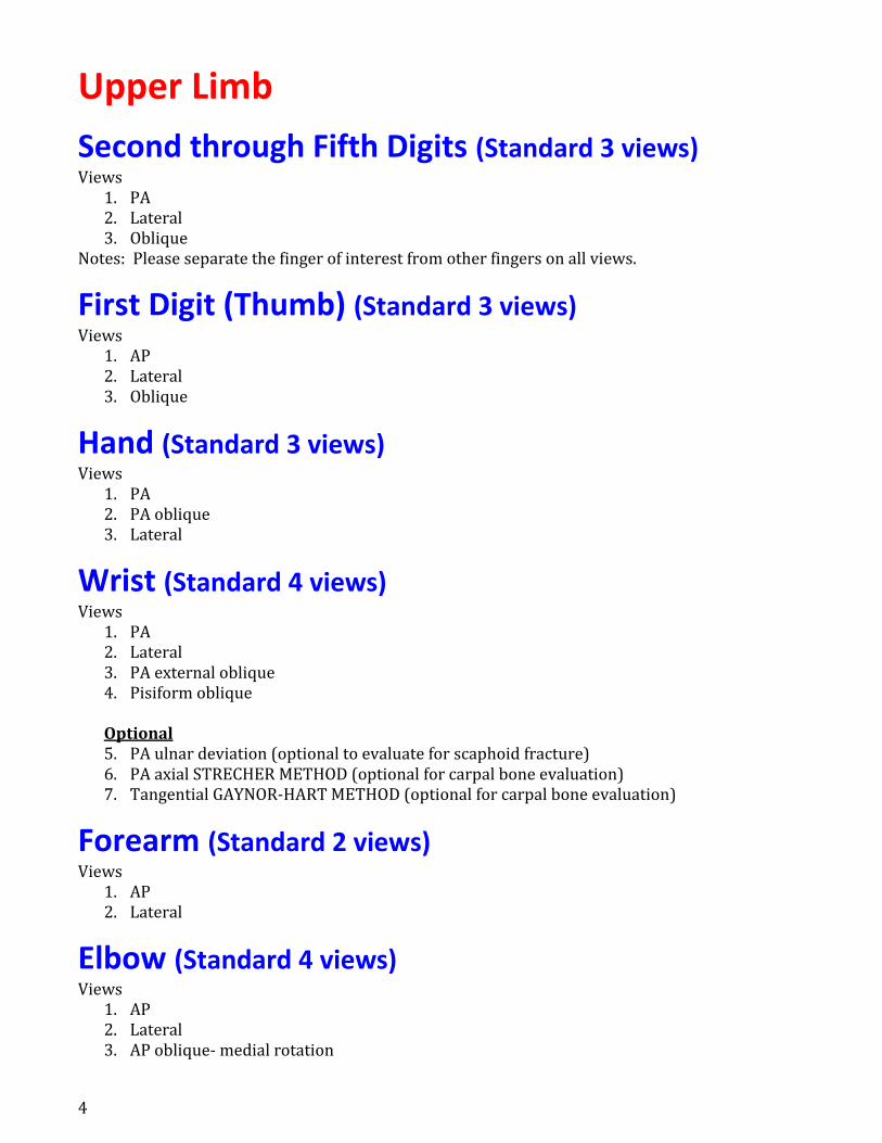
4. AP oblique- lateral rotation Optional
5. AP partial flexion (optional)
Humerus (Standard 2 views) Views
1. AP 2. Lateral
Shoulder (Standard 3 views) Views
1. AP internal rotation 2. AP external rotation 3. Scapular Y Optional 4. AP oblique GRASHEY METHOD 5. Inferosuperior axial LAWRENCE METHOD (optional to look for dislocation) 6. Transthoracic lateral LAWRENCE METHOD (optional)
Acromioclavicular Articulation (Standard 2 views) Views
1. AP PEARSON METHOD (with weights) 2. AP PEARSON METHOD (without weights)
Notes: Get both AC joints in field of view.
Clavicle (Standard 2 views) Views
1. AP 2. AP axial
Scapula (Standard 2 views) Views
1. AP 2. Lateral
6
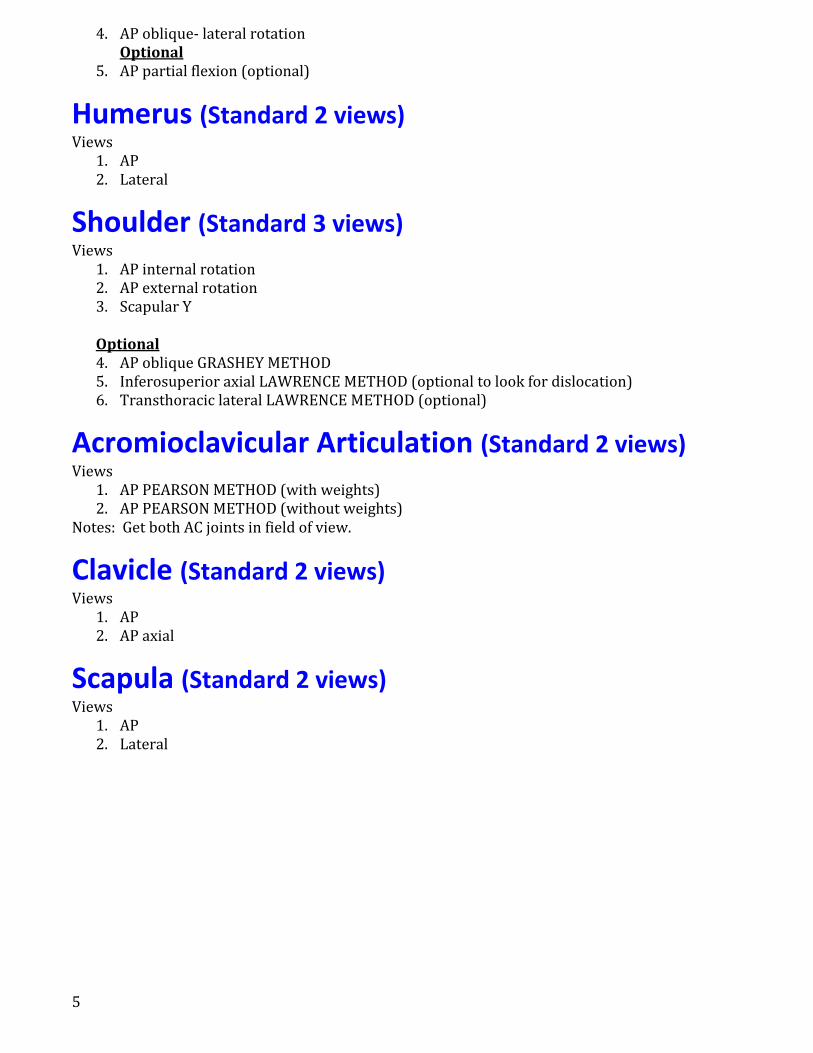
Lower Limb
Toes (Standard 3 views) Views
1. AP 2. AP oblique 3. Lateral
Notes: Separate the toe of interest from the other toes
Foot (Standard 3 views) Views
1. AP or AP axial 2. AP oblique- medial rotation 3. Lateral- mediolateral
Calcaneus (Standard 2 views) Views
1. Axial- plantar dorsal 2. Lateral
Ankle (Standard 4 views) Views
1. AP 2. Lateral- mediolateral 3. AP oblique- medial rotation ankle mortise 4. AP oblique- lateral rotation Optional 5. AP stress studies (optional)
Tib/Fib (Standard 2 projections) Views
1. AP 2. Lateral
Knee (Standard 4 views) Views
1. AP 2. Lateral 3. AP oblique

7
4. Patella sunrise view Optional 5. Patella: Tangential SETTEGAST METHOD 6. Intercondylar fossa: PA axial HOLMBLAD MEHTOD (optional) 7. Intercondylar fossa: PA axial CAMP-COVENTRY METHOD (optional) 8. Patella: PA (optional) 9. Patella: Lateral- mediolateral (optional)
Femur (Standard 2 projections) Views
1. AP 2. Lateral Optional 3. Femoral Neck: AP oblique MODIFIED CLEAVES METHOD (optional)
Hip (Standard 2 Projections) Views
1. AP 2. Frog Lateral LAUENSTEIN and HICKEY METHODS or 3. Axiolateral DANELIUS-MILLER METHOD
Pelvis (Standard 1 projection) Views
1. AP Optional 2. Inlet (optional 3. Outlet (optional) 4. Judet Obliques

8
Vertebral Column
Cervical Spine (Standard 3 views) Views
1. Atlas and axis: AP- open mouth 2. AP axial 3. Lateral GRANDY METHOD Optional 4. Dens: AP FUCHS METHOD (optional) 5. Lateral- SWIMMER’S TECHNIQUE (if needed) 6. Hyperflexion and hyperextension lateral (optional) 7. AP axial oblique- RPO and LPO (optional- of little clinical value) 8. PA axial oblique- RAO and LAO (optional- of little clinical value) 9. Trauma lateral- dorsal decubitus (if trauma) 10. Trauma AP axial oblique (optional)
Thoracic Spine (Standard 2 projections) Views
1. AP 2. Lateral Optional 3. Lateral- SWIMMER’S TECHNIQUE (if needed)
Lumbar Spine (Standard 2 views) Views
1. AP 2. Lateral Optional 3. L5-S1 Junction: Lateral 4. Hyperflexion and hyperextension lateral (optional) 5. Zygapophyseal Joints: AP oblique- RPO and LPO (optional- of little clinical value)
Sacroiliac Joints (Standard 2 projections) Views
1. AP axial FERGUSON METHOD 2. AP oblique- RPO AND LPO
Sacrum (Standard 2 views) Views
1. AP angled 2. Lateral

9
Coccyx (Standard 2 views) Views
1. AP angled 2. Lateral
Scoliosis (Standard 1 projection) Pasting AP, PA or lateral FERGUSON METHOD

10
Thorax
Routine Chest (Standard 2 projections) Views
1. PA 2. Lateral
Portable Chest (Standard 1 projection) View: AP Note: A PA view should be taken whenever possible as an AP view has significantly less clinical value.
Nonroutine Chest Views Views
1. AP lordotic LINDBLOM METHOD (optional) 2. AP or PA lateral decubitus (optional) 3. Lateral dorsal or ventral decubitus (optional)
Ribs (Standard 2 projections) Views
1. PA or AP upper and lower 2. PA or AP oblique upper and lower Notes: It is helpful if a BB is placed at the site of pain.
Sternum (Standard 2 views) Views
1. PA oblique- RAO 2. Lateral
Sternoclavicular Articulations (Standard 2 projections) Views
1. PA 2. PA oblique- RAO or LAO BODY ROTATION METHOD

11
Cranium
Facial Bones (Standard 3 views) Views
1. Lateral 2. Parietocanthial (WATERS) 3. Acanthoparietal (Reverse WATERS)
Notes: These views have little clinical value.
Mandible (Standard 2 projections) Views
1. AP Towne 2. Mandible – axiolateral obliques
Nasal Bones (Standard 2 projections) Views
1. Parietocanthial (WATERS) 2. Lateral (both)

12
Abdomen
Abdomen (Standard 2 projection) Views
1. AP- supine (get enough views to get entire abd-pelvis in field of view) 2. AP-Upright (visualized hemidiaphragms; this view is optional if the clinical indication is to
evaluate for renal stones) Optional 3. AP- upright 4. AP- lateral decubitus (optional) 5. Lateral (optional) 6. Lateral- dorsal decubitus (optional)
Related Documents