A Study of forensic radiography in South Africa, Argentina and the USA Mark Viner FCR, HDCR, MSc, DipHSM, DipFMS, Cert RP Fellow 2005 A report for the Winston Churchill Memorial Trust December 2005 – March 2006

Welcome message from author
This document is posted to help you gain knowledge. Please leave a comment to let me know what you think about it! Share it to your friends and learn new things together.
Transcript

A Study of forensic radiography in South Africa, Argentina and the USA
Mark Viner
FCR, HDCR, MSc, DipHSM, DipFMS, Cert RP
Fellow 2005
A report for the Winston Churchill Memorial Trust
December 2005 – March 2006

Radiography & Forensic Medical Investigation
Contents
Acknowledgments 3 Section 1 Domestic Aspects 5 Section 2 Introduction 8
My Background 8 What is Forensic Radiography? 9 Forensic Radiography in the United Kingdom 9 Aim of the fellowship 11 Why South Africa, Argentina and the USA? 11
The Fellowship
Itinerary 12 South Africa 13 South Africa Summary 17 Australia 18 Argentina 19 Argentina Summary 21 United States 22 United States Summary 22
Conclusions 22
Lessons Learned 23 Future Challenges 24 Outcomes and Follow-up 24
Acknowledgments I would like to thank both the Winston Churchill Memorial Trust for giving me a unique opportunity to travel and study forensic radiography in South Africa, Argentina and the United States, and for Barts and The London NHS Trust for allowing me to take advantage of this opportunity. I would especially like to thank Jilly Bowen for supporting my travelling fellowship at what was a particularly difficult time for her.
September 2006 2 Mark Viner
Radiography & Forensic Medical Investigation
I would also like to thank all of my hosts in South Africa, Australia, Argentina and the United States of America for their generosity and enthusiasm and in particular to the following for their hospitality;
In South Africa: Mr Aladdin Speelman and Professor Lorna Martin of Cape Town, Professor Shabbir Waddee and Dr Johann Dempers of Stellenbosch, Professor Hendrik Scholtz, of Johannesburg, Dr Servas Roussouw and Professor Nazeema Hoosain of Pretoria and Dr Segaran Aiyer and Mrs Leonie Munro of Durban and especially Professor Steve Naidoo and Mrs Poonitha Naidoo of Durban for all their help with my arrangements both before, during and after my travels.
In Australia: Dr & Mrs Peter Ellis of Sydney & Ms Rebecca Ellen of Melbourne
In Argentina: Dr Cynthia Urroz, Dr Luis Fondebrieder and Capt Osvaldo d’Amuri of Buenos Aires, Dr Alfonso Piccardi and Mr Dario Olmo of Cordoba
In the USA: Dr Anthony Falsetti of Gainsville, Florida, Prof Gil Brogdon of Alabama, Mrs Nancy Adams of Tupelo Mississppi, Dr Dwayne Wolf, Dr Yvonne Milewski and Ms Ann Ribera of Houston, Texas, Mr Paul Laudicina and Dr Geoff Harkey of Ilinois, Dr Brad Adams, Dr Barbara Sampson and Mr Bob & Mrs Rose Lippincott of New York.
I would also like to thank the following for supplying some of the images used in this report; Mr Chris Gosling of Siemens Medical plc, Ms Virginia Sanders of Lodox Ltd and Mr Paul Laudicina. Finally I would like to thank my wife Kim for all her support and encouragement and for giving up her time to join me on some of my travels Introduction I became aware of Winston Churchill Travelling Fellowships some years ago on seeing an advertisement in a professional journal. Having had a long-standing professional interest in forensic radiography, I thought that a fellowship would offer a great opportunity to further my understanding of the role and scope of forensic radiography by travelling and studying in other countries. At that time I was not able to devote the necessary time to a fellowship and filed the information away. Having seen first hand from a colleague how a fellowship had widened his horizons and contributed to a much greater understanding of clinical practice within our profession in the field of trauma care, I was determined to pursue an application for a fellowship when the timing was right. And so it was that, having drafted an initial itinerary and completing the necessary application forms, I was selected to attend for an interview. A few weeks later I was delighted to find that I had been awarded the chance of a lifetime to travel to South Africa, Argentina and the USA to study my chosen subject. I took full advantage of my colleague’s previous experience and questioned him endlessly for help with planning my fellowship. Having established key contacts in each of my target countries, I began to plan my itinerary and book flights and accommodation whilst maintaining some flexibility in my arrangements. I was aiming to cover a lot of ground in a relatively short time and quickly became an amateur expert on airline schedules and ticket regulations as I tried to co-ordinate my itinerary to fit in with the availability of the experts whom I hoped to meet. Thanks to one or two key contacts in my target countries, everything began to fall into place and departure was planned for 3rd December 2005. My Background I trained as a diagnostic radiographer and worked in a number of hospitals in London, spending the majority of my working life at The Royal London Hospital where I became lead superintendent radiographer and latterly radiology services manager for St Bartholomew’s and the Royal London Hospitals. I have long had an interest in forensic and trauma radiography, which developed during my time at the Royal London and Westminster Hospitals. As well as the Diploma and Higher Diploma of the
September 2006 3 Mark Viner

Radiography & Forensic Medical Investigation
College of Radiographers, an MSc in Health Services Management and the Diploma of the Institute of Health Service Managers, I also hold a Diploma in Forensic Medical Sciences. In 1996 I was approached to assist the United Nations International Criminal Tribunal for the former Yugoslavia and co-ordinated the forensic radiography teams in Bosnia, Croatia and Kosovo from 1996 – 2001. I worked extensively on these investigations in the Balkans and have also worked as part of investigating teams in Sierra Leone and the Republic of Ireland as well as on a number of cases in the UK working with forensic pathologists, dentists and anthropologists in their investigations. I am a founder member and current chair of the Association of Forensic Radiographers (AFR), a voluntary professional organisation that works with and on behalf of the College of Radiographers to improve training and standards in forensic radiography. The AFR has been instrumental in establishing training courses at two UK universities and has established an effective forensic radiography response team which was part of the government response to both the tsunami and London suicide bombing incidents. I am an advisor to the International Criminal Court, the UK Home Office and the Government Office for London on forensic radiography and emergency planning and earlier this year I was awarded the Fellowship of The College of Radiographers. Since completing my fellowship, I have taken up the position of Director of Operations and Programmes for the Inforce Foundation, a charity dedicated to improving standards of forensic investigation of war crimes and mass fatality incidents
September 2006 4 Mark Viner

Radiography & Forensic Medical Investigation
What is forensic radiography? Radiography is the creation of radiographs; photographs made by exposing a photographic film or other image receptor to X-rays. Since X-rays penetrate solid objects, but are slightly attenuated by them, the picture resulting from the exposure reveals the internal structure of the object. It is thus an examination of the structure of materials by non-destructive methods. Forensic radiography is the creation of radiographs for the purpose of assisting with legal investigations, and forensic radiology is the interpretation of these images to assist such investigations. The most common use of radiography is in the medical field (where it is now known as medical imaging and includes imaging using technologies other than ionising radiation), but veterinarians and engineers also use radiography. Almost every use of radiography has a potential forensic application and these include: examination of fine arts to detect forgeries, examination of aircraft and vehicle parts in accident investigation or investigation of smuggling, and examination of suspect packages.
Figure 1: Abdomen radiograph of a narcotics trafficker showing packages of cocaine in the bowel
It is however in the field of forensic medical investigation that forensic radiography is most widely employed. Its main uses are to determine and document the cause of injury or death, to detect and locate hazardous and/or illegal material concealed about the person, and to identify individuals. It is used on both living and deceased subjects and can provide evidence of assault, systematic abuse and narcotics concealed in body cavities during trafficking. It can be used to locate and document ballistic or other evidential material in the body following penetrating injury, to determine the cause of death following surgical intervention, accidental death and homicide, and to positively identify an individual for legal purposes both before and after death. Forensic radiography is particularly important in the investigation process and identification of victims following mass fatality incidents such as transportation crashes, terrorist incidents and natural disasters. Forensic Radiography in the United Kingdom In the United Kingdom, the professionals undertaking medical imaging examinations are diagnostic radiographers. Radiography training is validated by the College of Radiographers and the Health Professions Council (HPC), and radiographers practising within the National Health Service are required to hold state registration with the HPC. Forensic examinations of live subjects are most frequently undertaken in hospitals by diagnostic radiographers. However, studies have shown that, in many cases, radiographers undertaking forensic examinations are inadequately trained for such work, do not have a good understanding of the legal principles underpinning forensic investigation and fail to follow the correct procedures.1
Forensic examinations of deceased subjects are undertaken in hospital or public mortuaries by diagnostic radiographers. Mortuaries are generally ill-equipped for such examinations with few (if any) having access to X-ray equipment on site and often having to rely on old mobile X-ray equipment being donated by a hospital or on equipment being specifically brought in to undertake the examination. This significantly affects the ability of the forensic pathologist to achieve a timely radiographic investigation and hampers the investigative process. In some cases, due to university re-organisation, departments of forensic medicine associated with hospitals have closed, and the
1 Baker M & Hughes N (1997); The Provision of Forensic Radiography: A Research Survey Report, University of Central England, Birmingham
September 2006 5 Mark Viner
Radiography & Forensic Medical Investigation
service is provided by Home Office pathologists working in a variety of public mortuaries, further exacerbating the situation and making service provision and research collaboration even more difficult. The organisation of Forensic Pathology Services has been the subject of a review by the Home Office which recommended the creation of regional centres of forensic pathology equipped with appropriate X-ray facilities.2 The provision of radiographers and equipment for the investigation of mass fatality incidents has been recognised as a weakness in emergency planning since the Lockerbie disaster in 1988, with many plans relying on local hospitals to provide staff and equipment when requested. With standards of evidence and forensic procedure under the spotlight in a number of high-profile cases during the last 10-15 years, coupled with a much greater awareness of cultural, religious and ethical issues surrounding the examination of the deceased, the recommendations of a number of public enquiries and government reports have culminated in legislative changes including: The Human Tissue Act3, a reform of the coronial system4 and the creation of the Council for Registration of Forensic Practitioners. The Association of Forensic Radiographers, in conjunction with the Society and College of Radiographers has led the profession’s response to these changes, producing professional guidelines, developing post-graduate training programmes at two UK universities and creating a team of trained radiographers who have successfully responded to mass fatality incidents including the Tsunami and London Suicide Bombings. Against the background of seemingly inadequate training and provision in forensic radiography, medical imaging as a whole has seen enormous development and expansion in the last 20 years. Rapid technological advancement now allows computed tomography (CT) scanning with multi-planar imaging and 3-dimensional reconstructions of skeletal and soft tissue structures. Magnetic resonance (MR) scanning allows the clinician to visualise the soft tissues and especially the muscular structures and nervous system in great detail permitting the diagnosis and treatment of disease at much earlier stages. The profession of radiography in the UK has also made enormous advances, with radiographers expanding roles to include image interpretation and reporting, dynamic imaging studies, administration of drugs, etc., and the creation of “Consultant Radiographer” grades within the NHS. Despite the obvious advantages offered by the newer technologies to the forensic pathologist, the forensic community in the UK has been slow to explore the potential of these new techniques for post-mortem examination. This is partly due to limited access to the new equipment which is located in hospitals and clinics and simply not available for post-mortem examination, and partly due to the dislocation of departments of forensic medicine from teaching hospitals over the past 10-15 years.
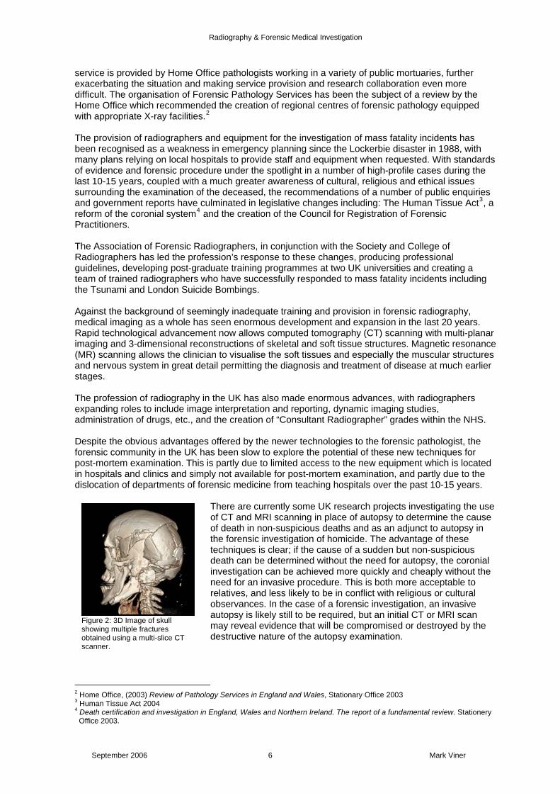
Figure 2: 3D Image of skull showing multiple fractures obtained using a multi-slice CT scanner.
There are currently some UK research projects investigating the use of CT and MRI scanning in place of autopsy to determine the cause of death in non-suspicious deaths and as an adjunct to autopsy in the forensic investigation of homicide. The advantage of these techniques is clear; if the cause of a sudden but non-suspicious death can be determined without the need for autopsy, the coronial investigation can be achieved more quickly and cheaply without the need for an invasive procedure. This is both more acceptable to relatives, and less likely to be in conflict with religious or cultural observances. In the case of a forensic investigation, an invasive autopsy is likely still to be required, but an initial CT or MRI scan may reveal evidence that will be compromised or destroyed by the destructive nature of the autopsy examination.
2 Home Office, (2003) Review of Pathology Services in England and Wales, Stationary Office 2003 3 Human Tissue Act 2004 4 Death certification and investigation in England, Wales and Northern Ireland. The report of a fundamental review. Stationery Office 2003.
September 2006 6 Mark Viner

Radiography & Forensic Medical Investigation
With such developments and the huge increase in public interest in forensic investigation and rapidly changing legislative and social background to these issues within the UK, it is likely that radiography and radiographers will play a key role in forensic medical examinations in the future. Yet despite a high level of interest amongst radiographers, a firm commitment to development on the part of the professional body, and the introduction of key legislation by government, the use of radiography in forensic medical investigation within the UK appears to be very low. The purpose of my travelling fellowship was therefore to learn something of how radiography was used in support of forensic medical investigation, and particularly in forensic pathology, in other countries. I wanted to evaluate the scope of practice, the technologies used, the training available, and determine anticipated future trends and hopefully to use this to further develop forensic radiography in the UK. Aim of the fellowship The aim of my Winston Churchill Travelling Fellowship was thus to research and evaluate:
• the contribution of radiography to forensic medical investigation • the technologies used • the training available to practitioners
I aimed to achieve this by visiting medico-legal laboratories, institutes and universities in South Africa, Argentina and the USA, meeting leading experts and work-shadowing them to observe current practice, and interviewing them to test my understanding and determine anticipated future trends. I propose to apply the knowledge and experience gained to further develop forensic radiography in the United Kingdom, and specifically to develop core standards and key learning outcomes for radiography training programmes Why South Africa, Argentina and the United States? I wanted to visit countries and centres which provided a range of different factors which could potentially affect the levels of demand and provision. South Africa has well developed medical and legal systems with similarities to the UK. The training and regulation of radiographers is similar with good links between professional organisations. However, levels of crime involving penetrating injury (particularly gunshot wounds) are much higher than in the UK and I anticipated that this would result in a greater demand for post mortem radiography. I therefore hoped that it would provide an interesting comparison with the situation in the UK. In Argentina, the forensic medical service is organised in a similar manner to the European model with institutes of legal medicine combining judicial and forensic functions in one institution. Training and regulation of imaging technicians/radiographers is significantly different to that of the UK and I hoped that it would provide a contrasting background against which to evaluate the role of radiography. Argentina also has a recent history of human rights abuses during the military dictatorship of the 1970s, with large numbers of “disappeared” who are still unaccounted for. In the 1980s it was one of the first countries to establish a forensic anthropology team to locate and identify the remains of the disappeared for both legal and humanitarian purposes. As radiography can be of great value in the identification of skeletalised human remains, I was interested to see the extent to which radiography played a role in this process. The USA has a variety of legal systems and regulatory frameworks which differ widely between states. It has a well developed medical system with extensive resources and well developed and regulated forensic sciences. The training of radiologic technologists differs from that of the UK and the role of the radiographer is not yet as broad reaching. However, there is a range of published literature on the use of radiography in forensic medicine and anthropology and I was particularly interested to learn more about the Disaster Mortuary Response Team (DMORT) which investigates and identifies victims of mass fatality incidents such as 9/11 and Hurricane Katrina.
September 2006 7 Mark Viner

Radiography & Forensic Medical Investigation
The Fellowship
Itinerary: December 2005 – March 2006 Countries and Centres Visited
Countries and centres visited South Africa 3rd – 26th December 2005 Cape Town & Stellenbosch Johannesburg & Pretoria: Durban Holiday in Australia & Chile 27th December – 11th January
Melbourne, Australia Argentina 12th – 30th January 2006 Buenos Aires Mar Del Plata Cordoba United Status 1st February – 1st March 2006 Gainesville, Florida: Mobile, Alabama Jackson Mississippi & Memphis Tennessee: Houston & Galveston, Texas Wheaton, Illinois New York & Connecticut Seattle, Washington (American Academy of Forensic Sciences Meeting) New York
September 2006 8 Mark Viner

Radiography & Forensic Medical Investigation
South Africa Cape Town & Stellenbosch The first stop on my itinerary was Cape Town, where I had arranged to meet with Professor Lorna Martin, head of department of forensic medicine at the University of Cape Town, and Mr Aladdin Speelman, radiography lecturer and vice-president of the South African Society of Radiographers The department of forensic medicine at Cape Town is one of two within the Western Cape region, the other being at the University of Stellenbosch. Between them, the two departments undertake nearly 6000 forensic autopsies per annum and all are carried out in city mortuaries which are managed by the Police Service. Outside the greater Cape Town area, forensic work is undertaken by the district surgeon along with other clinical duties. I toured the Salt River mortuary in Cape Town and spent time discussing with Professor Martin and her staff the different systems in the UK and South Africa. The volume and pressure of work here is very high and this has led to differences in training in forensic pathology, with doctors able to specialise at an earlier stage. Due to the high levels of gunshot injury, the mortuary has recently been equipped with a mobile C-Arm fluoroscopy unit, which enables ballistic fragments to be located and removed very rapidly instead of taking repeated X-ray films. It is now becoming common in South Africa to use fluoroscopy in the mortuary in place of X-ray film examinations and most major centres are due to be equipped with a fluoroscope in the near future. I was introduced to Dr Yolande Van Heide, forensic pathologist, and worked with her on a homicide case using the fluoroscope to examine the body of a young male with a gunshot wound to the chest. The X-ray examination revealed two bullets, one in the abdominal cavity, and one in the shoulder. The latter turned out to be due to a previous injury, and I later learned that this is not an uncommon finding at autopsy. On average, the department undertakes 2-3 cases per day requiring radiography – usually gunshot cases. Radiography at the mortuary used to be undertaken by police technicians. I asked Professor Martin whether any radiographers assist with forensic work and learn that following the breakdown of the old x-ray machine and prior to delivery of the new fluoroscopy unit, all cases requiring X-rays had to be transferred to the hospital for scanning on a revolutionary new digital x-ray scanner - the Lodox Statscan. Use of hospital equipment is far from ideal, and whilst hospital radiographers were willing to assist in using the new fluoroscopy unit, the volume of work would require the presence of a radiographer at the mortuary each day. Both money and radiographers are scarce commodities here and the pathologists now undertake their own fluoroscopy although they have received only limited training on the machine.
Figure 3; Dr Van Heide with the fluoroscopy machine
The Department of Forensic Medicine is co-located with the Groot Schuur Hospital, a vast 1000 bed hospital, famous for the first heart transplant in the world, undertaken by Christian Barnard. Following a tour of the Christian Barnard Museum, I was introduced to Miss Gillian Bowie, Chief Radiographer, who took me on a tour of the hospital radiology department.
Figure 4: Groote Schuur Hospital, Cape Town
I was interested to see the “Satscan” machine that Professor Martin had referred to and the consultant trauma surgeon explained how major trauma victims are examined using the machine. He explained that this digital X-ray scanning unit had been developed by the South African company, Lodox, using technology originally designed for scanning miners for concealed diamonds. It is in routine use for trauma patients at Groote Schuur and also at the Red Cross Children’s Hospital in Cape Town. Using low dose X-rays, it scans the complete body in two planes (the AP and lateral positions). Each scan takes 13 seconds.
September 2006 9 Mark Viner

Radiography & Forensic Medical Investigation
The trauma surgeons use it to assess the patient, localise bullets and inject contrast media to check arterial blood flow and renal function. I can see its value for forensic work, and the forensic
pathology team are keen to install one in the mortuary, if funding becomes available. I was later introduced to Ms Virginia Sanders of Lodox and spent some time viewing and discussing forensic radiography images and her research undertaken in conjunction with the department of anatomy. I spent an interesting morning at Tygerburg mortuary as the guest of Professor Shabbir Wadee and observed several cases there. These were a mixture of road traffic accidents and homicide, and radiography (including ante-mortem CT scanning in one case) plays an important role in the investigation. Unfortunately, the fluoroscope here had broken several months prior to my visit and a replacement was awaited. Until then, any cases requiring X-ray are taken to the hospital department early in the morning for radiography. I discussed this with pathologist Dr Johann Dempers and some of his colleagues, and discovered that they are currently sending very few cases for X-ray as it is difficult and time consuming to arrange. I explained that this is a very similar situation to the UK and we calculated that on-site X-ray provision would increase its usage at Tygerburg by almost 100% I was introduced to many members of the pathology team in both centres, and conducted interviews with pathologists, anthropologists, odontologists and radiographers. The anthropologists and odontologists make regular use of X-ray
examinations and utilise equipment in local hospitals and medical schools. There is little radiographer involvement in these cases. At present forensic medicine is the responsibility of the police service, although from April it transfers to the Health Department. Pathologists are hopeful of change following this transfer, as they will take over responsibility for management of the mortuaries, together with staffing and equipment. Professor Martin and Professor Wadee would like to use this opportunity to develop better links with the department of radiology.
Figure 5: Full body digital x-ray image using the “Statscan”
I was keen to explore the views of radiographers regarding forensic radiography and to find out what training and preparation was open to them. I learned that some radiographers in outlying hospitals are asked to assist with forensic x-ray examinations and whilst many are keen, none have received any specific training for this work. The radiographers at Groote Schuur are interested in working more closely with the department of forensic medicine and miss the opportunity to undertake examinations on the Lodox unit at the hospital. At the Cape Peninsula University of Technology department of radiography I met with senior lecturer, Mrs Penny Engel-Hills, and talked to her and Mr Aladdin Speelman about the structure and syllabus of radiography training in South Africa. In common with UK training, students receive little exposure to forensic issues at undergraduate level. At present there is no post-graduate training, although this is under review. Johannesburg & Pretoria The next stop on my itinerary was Johannesburg where I had arranged to meet Professor Hendrik Scholtz of the department of forensic medicine at the University of the Witswatersrand and Dr Servas Russouw of the department of forensic pathology, University of Pretoria, Professor Scholtz explained that these two university departments served Guateng Province together with the department at the Medical University of South Africa (MEDUNSA). The Province has 15,000 cases requiring forensic autopsy per annum (pa) and the largest mortuaries are: Pretoria = 2300-2500 cases pa, and four in Johannesburg: Hill Brow 2800-3000 cases pa and three others, one undertaking 4000-4500 cases and the other two 2500 cases per annum. The MEDUNSA department undertakes around 500 cases pa. There are several other mortuaries which come under Professor Scholtz's authority undertaking around 200 cases per annum and most are on the periphery of Pretoria & Johannesburg where 80% of the workload is road traffic incidents.
September 2006 10 Mark Viner

Radiography & Forensic Medical Investigation
Professor Scholtz showed be around the Hillbrow mortuary, which has 15 pathology staff including four registrars. The number of cases requiring X-ray averages 2-3 per day, mainly for gunshot or other penetrating injury and suspicious deaths of infants or children. Johannesburg has a new fluoroscopy unit similar to that in Cape Town, and I talked at length to Professor Scholtz about it unique design. He explained that he drew up the specifications himself, with advice from a radiologist. The unit has a 39cm image intensifier to give wide coverage and allow the body to be examined in a single sweep. It has a flat screen monitor mounted on the unit to permit ease of viewing by the operator (pathologist), a rotating anode tube, allowing a greater heat loading, and a CD-ROM writer for recording all cases. This unit has been ordered for all the major departments of forensic medicine in South Africa, with the exception of Durban. He believes that the pathologists are not currently realising the full potential of the new unit and that there is room for further training. As a gold standard he would prefer regular radiographer support and like his colleagues in the Western Cape thinks that the transfer of responsibilities of the mortuaries from the police to the health service will enable the situation to be improved, offering a greater opportunity for radiographers to become involved. Prof Scholtz was involved in the identification of victims following the tsunami and we discussed the use of radiography during this process and for the investigation of the London suicide bombings. We also discussed his work in investigating hyperbaric death in divers in which all victims are routinely given a CT scan to examine for air emboli indicating pulmonary barotraumas. These scans are performed at private hospitals prior to an autopsy being performed. The Pretoria department of forensic medicine is associated with Pretoria Academic Hospital, University of Pretoria. The University also has a department of forensic anthropology. I toured the department with Dr Russouw and observed a number of cases. All of these were pedestrians killed in road traffic accidents with multiple injuries. Unfortunately, the X-ray equipment here had been out of action for several months and the delivery of the new fluoroscope was eagerly awaited. Cases requiring X-rays were being referred to the department of radiology at the hospital, but pressure on resources is great and cases were usually restricted to gunshot injuries. Dr Russouw admitted that that they use radiography for less cases than they would like and, of the cases that I observed, he would have preferred to have used radiology to investigate and document bony injuries in preference to more invasive methods. We estimated that only 50% of cases requiring radiology were being referred to the hospital. The department used to employ a radiographer to undertake forensic radiography but there has been nobody in post for some years. Pathologists will operate their new fluoroscope, but have not yet received training and would prefer to have radiographer support. I discussed this issue with Professor Nazeema Ebrahim a radiographer and associate dean of the department of allied health & nursing and Mr Hoosain Ebrahim a forensic photographer and deputy dean both at the University of Limpopo (formerly known as the Medical University of South Africa). Professor Ebrahim supports the view that radiographers should take responsibility for forensic examinations, a view now shared by the South African Society of Radiographers. As yet, little training is available but she has developed training material for post-graduate level courses in examination of non-accidental injury in children and in the forensic examination of gunshot wounds. Mr Ebrahim stressed the importance of training and robust procedures for forensic examination of all types and cited several examples of photographic evidence being ruled as inadmissible due to poor technique and documentation. We discussed whether there should be greater emphasis on the legal aspects of medicine for radiographers at undergraduate level and Professor Ebrahim explained that this is being progressed as part of a national initiative through the South African Society of Radiographers.
Figure 6: Mr Hoosain and Prof Nazeema Ibrahim with Mark Viner
September 2006 11 Mark Viner

Radiography & Forensic Medical Investigation
Durban In Durban I was the guest of forensic pathologist, Professor Steve Naidoo, and radiographer, Mrs Poonitha Naidoo. I had worked with Professor Naidoo as part of a United Nations forensic team in Croatia and Kosovo and am indebted to him for helping to arrange many of my contacts in South Africa. I spent several days with Professor Naidoo and his team at the department of pathology at the University of Kwazulu Natal, observing cases in the mortuary, attending case reviews and visiting outlying mortuaries and laboratories in the region. The police mortuary in Durban has opted to remain with plain film X-ray rather than use fluoroscopy and a new unit was installed nine months previously. It is a C-Arm tube & bucky arrangement,similar to the equipment used in some hospital trauma rooms. Professor Naidoo explained to me that Durban has resisted the move towards fluoroscopy as they prefer to have a full record of the examination. They are however interested in the prospect of direct digital imaging as a replacement for film, and we talked at length on this subject. All gunshot cases have X-rays here irrespective of the number of entry & exit wounds. On busy days this can be 8-10 cases and the film processor with its three minute cycle time is quite a limitation. Pathology technicians undertake X-rays, having received some basic training, and I noted that both films and radiation protection measures are excellent.
Professor Naidoo and I visited other mortuaries in the Kwazulu-Natal region, which he explains is South Africa’s most rural region. I discovered that, outside of the main academic centres, resources are poor and, whilst an X-ray service is usually available, it is quite often reliant on very old equipment or a restricted access to hospital facilities. I also had chance to visit the regional ballistics laboratory in Durban and spoke with Captain Van der Sant, deputy head of the unit. The unit is one of three, the others being in Pretoria and Cape Town, and last year undertook investigations on some 11,500 cases. He stressed the importance of retrieving all ballistic material from a cadaver and explained that, even if there is an exit wound, key forensic evidence from a bullet fragment or jacket can still be retrieved from the body. During my time in South Africa I had the opportunity to meet with many
radiographers. Many of them expressed reluctance to take X-rays of the dead but many others were keen to become involved but did not know how. I also spoke at length with radiography educators in each centre. All were keen to develop core standards and embrace forensic issues within the undergraduate course, although some felt that this should be a post-graduate course. Opportunities for post-graduate education are limited here but interest was expressed in developing courses similar to those in the UK.
Figure 7: X-ray machine in use at a rural mortuary
The issue of radiographers undertaking forensic work has recently become an issue under discussion by the South African Society of Radiographers and I spoke at length with their President, Jenny Motto. Jenny explained that both the Society and the Health Professions Council of South Africa had decided that radiography of cadavers does come under the scope of practice of diagnostic radiographers; i.e. it is to be considered to be part of a radiographer’s routine duties and not a voluntary examination. They have taken this decision after much ethical debate and concluded that a radiographer’s responsibility as a health professional does not end when the patient dies. This raises three important issues for them:
i) Forensic radiography of the deceased will need to become part of the undergraduate curriculum
ii) At the moment, most forensic radiography of the deceased in South Africa takes place in the absence of a radiographer.
iii) Some radiographers are reluctant to undertake such work I was asked to share my experiences of forensic radiography in the UK and delivered presentations at the University of Cape Town & the Nelson Mandela School of Medicine in Durban. The multi-
September 2006 12 Mark Viner

Radiography & Forensic Medical Investigation
professional audiences were keen to learn about forensic radiography in general and about experiences of the London suicide bombings. In the discussion that followed it was clear that we share many of the same issues and that future collaboration and communication between us will be beneficial to both countries. All the people that I met in South Africa were incredibly generous and friendly and I am indebted to so many of them for many excursions and fantastic hospitality. I was able to renew some old friendships and formed many new ones and look forward to continued communication with them.
South Africa Summary:
• All major centres are either equipped with good x-ray equipment or will be shortly • Levels of provision in rural areas are variable and often poor • The most common application of radiography is for firearm fatalities and when necessary non-
accidental injury in children • The high rate of gun crime and scarce resources limits the use of radiography to these
indications in many cases • Where on-site provision is available, much greater use of radiography is made. • In most cases, radiography is performed by police technicians or pathologists with very little
training and there is little involvement of radiographers. • Where technicians have received some basic training, image quality is improved and
radiography is used in a greater percentage of cases. • No use is made of CT scanning or MRI scanning as facilities are not available. A request to
large hospital departments with this equipment would likely be refused. • Research using the “Statscan” shows very promising results and it is well suited to forensic
applications • Pathologists keen to involve radiographers more, especially when control of the mortuaries
passes from the Police to the Health Service in March 2006. • There is little or no training in forensic issues within the undergraduate radiography
programme although this issue is under discussion • No guidelines or post-graduate training courses in forensic radiography exist at present. • Radiography is not well integrated into mass fatality plans
September 2006 13 Mark Viner

Radiography & Forensic Medical Investigation
Australia Melbourne Although not in the original itinerary for the study, I met up with forensic colleagues during a short stopover in Australia. Following the tsunami last winter, Australian forensic teams took part in the investigations, along with some of the South African pathologists I had met in South Africa. Reports of their work indicated that the Australian teams were highly organised and well equipped. So, with such recent hands-on experience, I wanted to find out how radiography featured in their investigations. With only ten days in Australia, based in Sydney, I arranged to meet the senior forensic pathologist in Sydney, Dr Peter Ellis, who coordinated the Australian tsunami teams, and Dr Ellis kindly arranged for me to visit the Victorian Institute of Forensic Medicine in Melbourne. In Melbourne, the pathologists, coroner, scenes of crime officers, toxicology and DNA lab work, and forensic physicians work on an Institute model; all are based together on one site. There is only one centre like this in the UK, and it provides the model proposed within the Home Office reviews of Forensic Pathology and the Coronial system. I was therefore interested to tour the centre and see how forensic radiography was integrated into the investigative process.
Figure 8: Victorian Institute of Forensic Medicine, Melbourne
I was shown around by Rebecca Ellen, Senior Technician. It is a modern facility with on site computed X-ray facilities, which have long been an essential part of their investigations. This year they have been fortunate enough to receive additional funding as the Commonwealth Games are taking place in Melbourne. This has enabled them to purchase a 16 slice CT scanner, which was only three months old when I visited. All cases have full body CT scans prior to autopsy and they are getting some interesting results.
Figure 9: CT Scanner in situ in the mortuary
The scanner is operated by autopsy technicians, rather than radiographers. All technicians are degree-trained as laboratory officers and are multiskilled - assisting at autopsy, undertaking laboratory tests and post-mortem X-rays, for which they are state registered. I am only aware of four other centres in the world undertaking CT in this way. It was a unique opportunity to see the benefits of this approach first hand and it will be interesting to see what their research shows – perhaps CT could replace autopsy in some circumstances in the future.
September 2006 14 Mark Viner

Radiography & Forensic Medical Investigation
Argentina Buenos Aires The next phase of my study took me to the Institute of Forensic Science in Buenos Aires, Argentina, where I spent almost three weeks working alongside the radiology team.
The institute and mortuary is housed in a beautiful nineteenth century building in the centre of Buenos Aires’ University district. Uniquely I found that the Institute has a dedicated three- roomed medico-legal X-ray department. It is staffed by a team of nine full trained radiographers, under the direction of consultant forensic radiologist, Dr Cynthia Urroz, and two other radiologists.
Figure 10: Institute of Legal Medicine, Buenos Aires
The centre is very busy, undertaking about 5000 forensic X-ray examinations a year, covering the whole of the Buenos Aires Province. Their forensic work not only includes post mortem work but they also undertake radiological examinations for all medico-legal cases, such as assaults or industrial injury, insurance claims and non accidental injury cases in the living; bringing together all the expertise in one place. This is excellent for the training of forensic radiologists and radiographers; they get to see many more cases than they would normally be exposed to if the work was undertaken in different centres.
Dr Urroz arranged several visits to departments within the institute and I had the opportunity to discuss cases and interview anthropologists, toxicologists, fingerprint experts and medical examiners, as well as radiology and radiography staff. I spent several days observing and discussing cases with Dr Urroz who has an enormous range and depth of experience of forensic radiology. It was apparent that the on-site provision of a dedicated X-ray facility with specially trained radiographers and radiologists added significantly to the scope of the investigative process. As a result, radiography was widely employed at post mortem and used to investigate many types injury. It is used to document peri-mortem fractures (those caused at or around the time of death), confirm fractures of the hyoid bone in the neck due to hanging, and is regularly employed to identify individuals by determining the age and sex of human remains. It also aids positive identification by means of comparison of post-mortem and ante-mortem films. I calculated that radiography is employed in around 28% of cases in this centre. Dr Urroz has developed training programmes in forensic radiography for radiography technicians. We discussed these at some length and I noted several important points for inclusion in the syllabuses of such courses and we agreed that forensic radiography is “50% radiography and 50% documentation”. We discussed the use of radiography in the investigation of mass fatality incidents in which Dr Urroz has significant experience. I shared our experiences of forensic radiography in the UK and delivered a presentation to Dr Urroz and her colleagues about our experiences of the London bomb blasts. In the discussion that followed it was clear that we share many of the same issues and that future communication between us will be beneficial to both countries. I knew that Argentina had a well developed team of forensic anthropologists based in Buenos Aires. The team (EAAF) work throughout Latin America and beyond but most of their work is in Argentina
investigating the disappearance of opponents of a past military regime. I spent time with Luis Fondebreider, senior anthropologist, who has led the team since it was set up over twenty years ago. The team was originally set up as a charity but now receives some government funding. They locate and examine human remains from this period, and also act as the investigating team, presenting cases to the prosecuting judge. Because they are not a government
September 2006 15 Mark Viner Figure 11: Dr Luis Fondebrieder & Mark Viner
Radiography & Forensic Medical Investigation
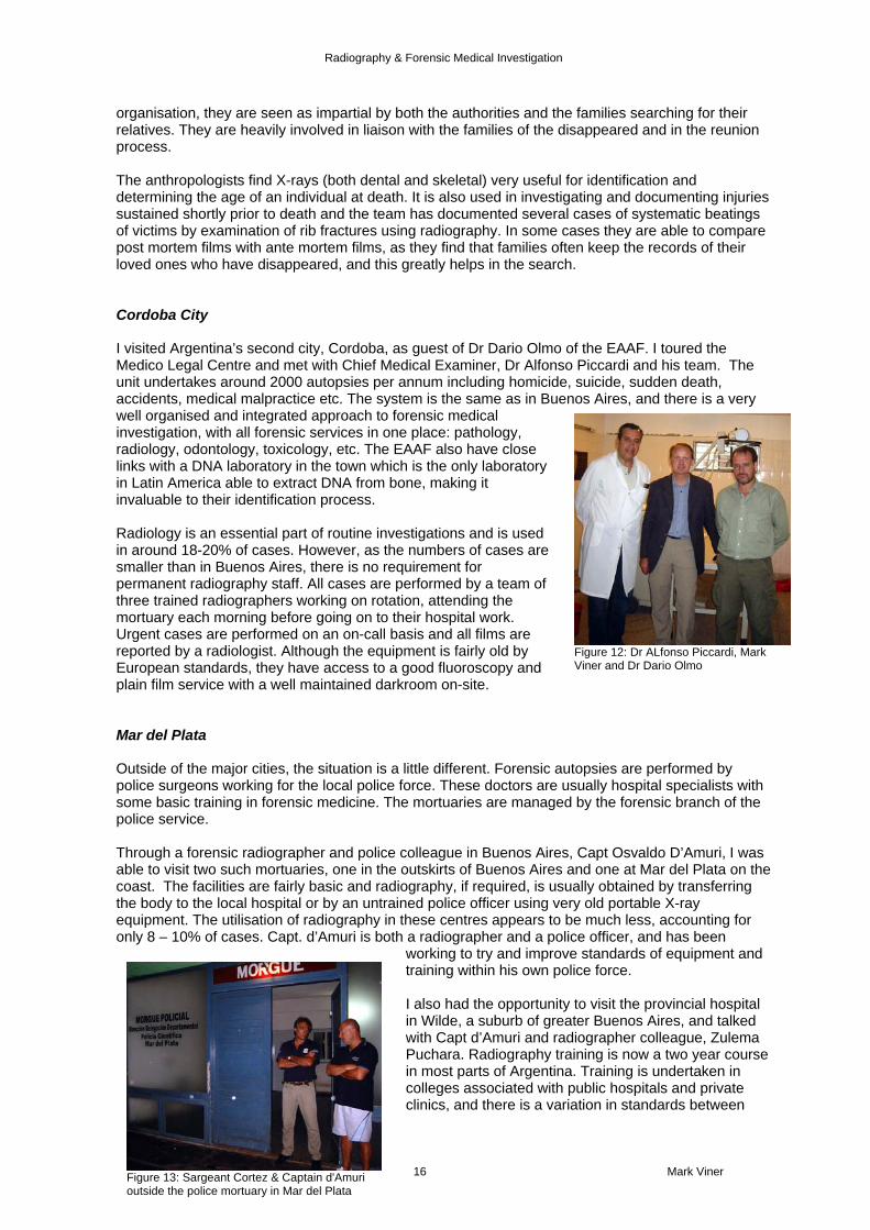
organisation, they are seen as impartial by both the authorities and the families searching for their relatives. They are heavily involved in liaison with the families of the disappeared and in the reunion process. The anthropologists find X-rays (both dental and skeletal) very useful for identification and determining the age of an individual at death. It is also used in investigating and documenting injuries sustained shortly prior to death and the team has documented several cases of systematic beatings of victims by examination of rib fractures using radiography. In some cases they are able to compare post mortem films with ante mortem films, as they find that families often keep the records of their loved ones who have disappeared, and this greatly helps in the search. Cordoba City I visited Argentina’s second city, Cordoba, as guest of Dr Dario Olmo of the EAAF. I toured the Medico Legal Centre and met with Chief Medical Examiner, Dr Alfonso Piccardi and his team. The unit undertakes around 2000 autopsies per annum including homicide, suicide, sudden death, accidents, medical malpractice etc. The system is the same as in Buenos Aires, and there is a very well organised and integrated approach to forensic medical investigation, with all forensic services in one place: pathology, radiology, odontology, toxicology, etc. The EAAF also have close links with a DNA laboratory in the town which is the only laboratory in Latin America able to extract DNA from bone, making it invaluable to their identification process. Radiology is an essential part of routine investigations and is used in around 18-20% of cases. However, as the numbers of cases are smaller than in Buenos Aires, there is no requirement for permanent radiography staff. All cases are performed by a team of three trained radiographers working on rotation, attending the mortuary each morning before going on to their hospital work. Urgent cases are performed on an on-call basis and all films are reported by a radiologist. Although the equipment is fairly old by European standards, they have access to a good fluoroscopy and plain film service with a well maintained darkroom on-site.
Figure 12: Dr ALfonso Piccardi, Mark Viner and Dr Dario Olmo
Mar del Plata Outside of the major cities, the situation is a little different. Forensic autopsies are performed by police surgeons working for the local police force. These doctors are usually hospital specialists with some basic training in forensic medicine. The mortuaries are managed by the forensic branch of the police service. Through a forensic radiographer and police colleague in Buenos Aires, Capt Osvaldo D’Amuri, I was able to visit two such mortuaries, one in the outskirts of Buenos Aires and one at Mar del Plata on the coast. The facilities are fairly basic and radiography, if required, is usually obtained by transferring the body to the local hospital or by an untrained police officer using very old portable X-ray equipment. The utilisation of radiography in these centres appears to be much less, accounting for only 8 – 10% of cases. Capt. d’Amuri is both a radiographer and a police officer, and has been
working to try and improve standards of equipment and training within his own police force. I also had the opportunity to visit the provincial hospital in Wilde, a suburb of greater Buenos Aires, and talked with Capt d’Amuri and radiographer colleague, Zulema Puchara. Radiography training is now a two year course in most parts of Argentina. Training is undertaken in colleges associated with public hospitals and private clinics, and there is a variation in standards between
September 2006 16 Mark Viner Figure 13: Sargeant Cortez & Captain d'Amuri outside the police mortuary in Mar del Plata

Radiography & Forensic Medical Investigation
centres, especially outside the major cities, where facilities are generally poor in comparison with UK standards. There are no forensic aspects covered in the basic training, and the course run by Dr Urroz appears to be the only post-graduate training available. Many people in Argentina have at least two jobs. Capt. D’Amuri works as a radiographer and as a policeman, as well as undertaking voluntary work with local underprivileged children. I don’t know where he gets the time – although it may explain why the Argentines don’t eat dinner until 11pm! The warmth of welcome that I received from everyone I met was tremendous and I am I am indebted to them for showing me some of their beautiful country and for fantastic hospitality. In conclusion, The UK can learn a lot from Argentina’s co-ordinated approach to forensic radiography. Although working in adverse circumstances with cultural values and norms very different from ours, their training and use of resources is exemplary. They set an example for us to follow and I look forward to continued dialogue with our colleagues in South America.
Argentina Summary:
• Very well developed forensic radiology service serving the major cities • Centralisation of all medico-legal x-ray examinations in one centre justifies employment of
specialist dedicated staff. • Good imaging protocols exist and radiography examinations are widely used for a range of
investigations. • Radiography makes a significant contribution to identifying skeletalised human remains of the
disappeared. • Little provision or co-ordination of forensic radiography services in provincial police mortuaries• Where on-site provision is available, much greater use of radiography is made. • In main centres, radiography is performed by radiographers who have received additional
post-graduate training in forensic imaging. • Image quality is good and examinations contribute valuable information to the investigations. • No use is made of CT scanning or MRI scanning as facilities are not available. A request to
large hospital departments with this equipment would likely be refused. • There is a good post-graduate training programme for radiography technicians in Buenos
Aires but none available in other areas • No national guidelines in forensic radiography exist at present. • Radiography is not well integrated into mass fatality plans
September 2006 17 Mark Viner

Radiography & Forensic Medical Investigation
United States Gainesville, Florida
Figure 14: Alligators, insects and humidity contribute to rapid decomposition of human remains in Florida
From Buenos Aires, I travelled, via Miami, to Gainsville to commence the next stage of my fellowship in the USA. One of the main teaching centres of forensic anthropology is based at the William Maples C A Pound Laboratory of Human Identification at the University of Florida. Here I had arranged to meet Dr Tony Falsetti, senior lecturer in forensic anthropology. The laboratory investigates cases that cannot be identified by any other means, or that are so decomposed that they require anthropological examination to investigate manner of death, etc. In Florida, due to the climate and insect activity, bodies decompose very rapidly and thus the case rate is much higher than the rest of the USA. The laboratory undertakes approximately 120 cases per annum with most cases being referred from Florida and New York State. I toured the laboratory and talked to Dr Falsetti and his colleagues about the use of radiography and a number of their cases. On arrival all cases are X-rayed to look for any identifying features, trauma, etc. Following the anthropological examination, bones are again X-rayed for trauma, identifying features and dental work. Radiography is very useful in determining the nature of traumatic injury, presence of ballistic material (indicating previous gunshot injuries), documenting ante-mortem injuries, pathologies, variants etc. Around 50% of all cases that are sent to the laboratory for identification are identified, and radiography contributes to positive a positive identification in around 25-30% of cases. All X-rays are taken by the anthropologists using a Faxitron machine. The results and detail are generally excellent, but there are sometimes problems with replicating the original positioning when comparing a post-mortem to an ante mortem film. The anthropologists have received no training in radiography and this can sometimes cause problems; in some cases help is requested from the radiology department but direct assistance from the radiologic technologists can be difficult to obtain. Mobile, Alabama From Gainsville I travelled through the Florida panhandle to Mobile, Alabama. Here I was privileged to meet Dr Gil Brogdon, Emeritus Professor of Radiology at the University of South Alabama, and author of the definitive text on forensic radiology. We discussed the role of radiography in forensic medicine at length and I put it to him that there appeared to be a very wide variation in the level of usage and standards of medical imaging in support of forensic investigation, not only within the United States, but also world-wide. He agreed that the use of radiology was sporadic and that quality was often poor, due to lack of knowledge and training on the part of those undertaking procedures. He felt that, in many cases, the development of forensic radiology was being hampered by lack of awareness of basic imaging principles and the opportunities offered by modern technology. Even the introduction of simple quality control procedures for imaging and processing equipment could have a significant effect on the information obtained and the forensic investigator’s confidence in using radiology as a valuable tool. We discussed the role of radiographers and radiologic technologists in improving this situation and he confirmed that, in his view, their input to the process was essential in order to achieve consistency of results and increase the utilisation of this valuable tool.
September 2006 18 Mark Viner

Radiography & Forensic Medical Investigation
Jackson Mississippi & Memphis Tennessee In Tupelo Mississippi, I had arranged to meet one of the few technologists who have not only become involved in working with forensic teams, but through their dedication and enthusiasm have been changing the perception and use of medical imaging as a forensic tool amongst other forensic professionals. Ms Nancy Adams is the Clinical Practice Co-ordinator for the Radiologic Technology Programme at the Itawamba Community College, Mississippi. She is involved in the DMORT (Disaster Mortuary Response Team) system and worked as part of a team of radiographers in identifying the deceased from the Hurricane Katrina disaster. Her experience helped prepare the regional DMORT team to respond effectively to the crisis and ensure that radiography was employed safely and effectively. Ms Adams, She is now hoping to work more closely with local Medical Examiner’s offices to set up training courses in forensic radiography.
Figure 15: Nancy Adams and Mark Viner outside the birthplace of Elvis Presley in Tupelo Mississippi
I spent three days with Nancy, discussing her work with DMORT and the role of forensic imaging and the training of radiographers. Together we visited the regional forensic laboratories in Memphis, Tennessee and Jackson, Mississippi. In Memphis, we met with Dr Jennifer Love, forensic anthropologist at the forensic laboratory. Dr Love showed us around the facility which covers Shelby County, Tennessee and also takes referrals from North Mississippi. Around 1500 cases (suspicious & sudden death) are referred pa, of which around 250 are homicides. Around 10% of cases are examined by radiography which is performed by pathology technicians with no radiography training. Equipment includes a plain film X-ray room and fluoroscopy, although this is not used.
In Jackson, we met Sam Howell, director of the Crime Laboratory. The laboratory is designed on an institute model with all facilities on site - DNA, toxicology, fire investigation, trace evidence, fingerprinting, documents, ballistics, narcotics, etc., and we were given an extensive and very interesting tour of these units, having the opportunity to talk with staff about their work. The laboratories cover the whole of Mississippi State with four pathologists performing around 1800 autopsies per annum. At present, X-rays can only be undertaken at the crime laboratory, not in the mortuary where cases are examined and cases have to be transferred for X-ray, if necessary. As a result, the facility is little used. I learned that, when X-ray facilities were on the same site,
radiography accounted for around 15% of cases.
Figure 16: Mortuary X-Ray Room at the Memphis forensic laboratory
I discussed the issues of forensic radiography training with Ms Adams and had the opportunity to visit the university department and North Mississippi Medical Centre hospital where she works. Forensic issues are not covered in the basic training programme for radiologic technologists and there is very little post-graduate education. It seems that there are only a few technologists who regularly undertake work for the medical examiner and coroners and Ms Adams is keen to improve standards and training.
September 2006 19 Mark Viner

Radiography & Forensic Medical Investigation
Houston & Galveston, Texas From Jackson, I travelled through Louisiana to Houston, Texas for my next appointment with the radiography lecturing team at Houston Community College. Following a tour of the department and training facilities, I talked with the team. As in Mississippi, I learned that the radiography programme here did not include forensic issues, with the exception of non-accidental injuries in children. My visit had stimulated their investigation of the situation at the city examiner’s office and they were surprised to find that radiography examinations there were undertaken by untrained personnel. I learned that, in Texas, radiographers do not need to be state registered, as long as they are working under the supervision of a registered medical practitioner but it does not matter whether or not the medical practitioner has training in the use of ionising radiation. We discussed this issue at length, and the possibility of devising a training course for mortuary technicians. I had also arranged to visit the Medical Examiner’s Office, Harris County, where I was the guest of Dr Dwayne Wolf, deputy chief medical examiner, and his staff. Following the morning case conference, I toured the facility, which undertakes around 3000 autopsies per annum. The unit has all laboratories on site including DNA and toxicology laboratories, a ballistics unit, trace evidence unit, as well as the mortuary. X-Ray facilities consist of a dedicated x-ray room and film processor. There is a routine protocol for examination of gunshot wounds, burns cases, decomposition cases, stabbing cases and non accidental injury (paediatric) cases. Around 10-15% of cases are examined by radiography with cadavers being X-rayed on arrival at the mortuary. X-rays are performed by pathology technicians who have no training in radiography.
Figure 17: Dental x-ray machine for human identification at Harris County Medical Examiner's Laboratories
I had the opportunity to discuss issues with the team and interview several pathologists, an odontologist, crime scene investigators and anthropologist, and presented a lecture on the use of radiography in the investigation of the London suicide bombings. Whilst in Texas, I also visited the Medical Examiners Office in Galveston County, as the guest of Dr Yvonne Milewski. This is a smaller facility than Houston, undertaking around 500 autopsies per annum. Once again, X-rays are require in around 10-15% of cases and are undertaken by the pathology technicians using a mobile unit. Again, I found that the technicians have received no formal training for use of X-rays. The examination protocol is decided by the pathologist and usually all gunshot deaths, stabbings, decomposed and burned victims and children are X-rayed. Wheaton, Illinois From Texas I flew to Chicago and spent two days with Paul Laudicina, a radiologic technologist and lecturer who has been working with the DuPage Coroner’s Office for many years and has established an excellent radiography service. Having clearly demonstrated the value of on-site radiography to the Coroner’s Office, Paul was instrumental in helping to design a new facility which incorporates a specially designed X-ray suite and facilities for coping with mass fatality incidents. Along with his team of trained and experienced radiographers, he provides a 24 hour call-out service and ensures
that the facilities are maintained in optimum condition to respond to any emergency. I toured the facility and talked at length with Mr Laudicina, the Coroner, and Dr Geoff Harkey the chief pathologist. It is clear that they find the on-site access to X-rays and expert advice invaluable in their investigations. The unit undertakes around 300- 400 autopsies per annum and, until recently, nearly 55% of the cases were examined using radiography. Financial restrictions have currently limited this workload,
September 2006 20 Mark Viner
Figure 18: Wall mounted x-ray unit within the high infection risk autopsy area in the DuPage Coroner’s Office

Radiography & Forensic Medical Investigation
but Dr Harkey is keen to utilise radiography as he finds it to be an invaluable tool, not only in his investigations but also in demonstrating evidence visually in the courts. Paul lectures throughout the USA to radiologic technologists on the subject of Trauma and Forensic Radiography. Later I talked with Mr Laudicina about the training that he has devised, and we discussed the use of radiography in mass incidents. New Haven, Connecticut From Chicago I flew to New York as a guest of radiography educator, Professor Bob Lippincott, and radiology manager Mrs Rose Lippincott. I wanted to meet with a radiologic technologist in New Haven, with whom Professor Brogdon had put me in touch. Gerry Conlogue has had a long standing interest in the examination of the deceased and is perhaps best known for his appearances on “The Mummy Road show”. Gerry also works with the Medical Examiner’s Office in Connecticut. This office undertakes around 5,000 autopsies per annum and radiography is performed in just 5% of cases. Until Gerry started to offer the service the radiography was performed by mortuary technicians who have received little or no training in X-ray procedures. Gerry now runs several courses in forensic radiography at Quinnipiac University. He is keen to ensure that radiographers should be more involved with forensic work and should educate themselves to understand the forensic context in which they work. We spent an interesting morning comparing notes and I learned a great deal about the structure of his courses and the learning outcomes. Seattle, Washington I interrupted my time in New York to travel to Seattle for the American Academy of Forensic Sciences’ annual meeting. This meeting gave me an opportunity to talk with other pathologists, scientists and investigators about the role of radiography and to meet up with some of the people that I had already met on my travels whilst I presented a proffered paper on the London suicide bombings. New York Back in New York I visited the City of New York Medical Examiner’s Office where I was a guest of Dr Brad Adams, forensic Anthropologist and Dr Barbara Sampson, deputy medical examiner. 20,000 cases are reported to Medical Examiner’s Office and about 5,000 of these will have an autopsy. These are performed across five mortuaries; the daily workload is as follows:
o 11 per day in Bronx - mostly violent deaths o 18 per day in Brooklyn and Staten Island - mostly violent deaths o 14 per day in Manhattan - mostly hospital deaths
Figure 19: Office of the Chief Medical Examiner, City of New York, Scene of the World Trade Center Investigation
o 11 per day in Queens - mostly road traffic accidents Each mortuary has X-ray equipment and staffed by radiologic technologists. The radiology workload is high and utilisation ranges from between 18 – 35% of cases across the five sites. Only plain film X-ray is used and all violent deaths, all traffic accidents (drivers, passengers and pedestrians), all sudden infant death and non accidental injury cases are examined using radiography. I spent several days at the office, observing cases and interviewing Dr Adams and Dr Sampson, as well as radiologic technologists, radiologists pathologists and investigators and assisting Dr Adams and Dr Sampson in their decision making for replacement of their X-ray equipment. We also discussed the management of Mass Fatality
September 2006 21 Mark Viner

Radiography & Forensic Medical Investigation
incidents and I gained some insight into the complex investigations following the 9/11 attacks. In most of the centres that I visited in the USA, radiologic technologists do not have an opportunity to become involved in the examination of the deceased, and their only exposure to forensic cases comes through clinical cases resulting from assault or non accidental injury in the living. In New York, I was also able to visit several hospitals and talk with radiologic technologists and university lecturers about radiography training and forensic radiography. I discovered a very similar pattern to that encountered in Houston, with little training available for radiographers or others in forensic issues, but a great interest and willingness to become involved. Throughout the USA I was treated to the most fantastic hospitality by my hosts and look forward to continuing friendships and professional collaboration with them in the future. Sadly, New York was my last port of call and on 1st March I returned to the UK.
USA Summary:
• The majority of centres have on-site provision of radiography facilities with modern equipment• Very wide variations in organisation of services between states • Well established role for forensic anthropology relies extensively on radiography • In some centres, good imaging protocols exist and radiography examinations are widely used
for a range of investigations. • In others, radiography is restricted mainly to gunshot injuries and non-accidental deaths in
children • Some use is made of CT scanning but this is in its early stages of development as a forensic
tool • Very few radiologic technologists are involved with forensic work. However, in centres where
they provide the service, utilisation is much higher. • Variations in legislation between states appears to be the underlying reason behind these
variations • There are no post-graduate training programme for radiologic technologists at present • No national guidelines in forensic radiography exist at present. • Radiography is integrated into mass fatality plans, but the extent to which this applies varies
widely between states
September 2006 22 Mark Viner

Radiography & Forensic Medical Investigation
Conclusions
Lessons Learned The fellowship has enabled me to gain a unique and valuable insight into the application of radiography in support of forensic medicine, and in particular forensic pathology. In all the countries that I visited, radiography is used regularly as a vital tool in forensic investigations. However, whilst the levels of utilisation clearly differ from country to country owing to demographic, epidemiological and social variations, significant variations were found between sites in each country. In some cases, this was due to lack of physical resources in terms of equipment, but in many cases there appeared to be a correlation between the level of training given to those undertaking the radiography examination and its level of utilisation and perceived benefit to the investigation. Those centres which (in common with many centres in the United Kingdom) are reliant upon use of mobile or local hospital facilities are often considerably restricting their use of radiography in investigations even in cases where it is clearly indicated. In each of the countries visited the difference in levels of utilisation between those sites with on site provision and those without was in the order of a 2:1 ratio. Furthermore, in centres where qualified radiographers, radiologic technologists or radiography technicians undertook examinations and/or had direct input into the planning, co-ordination and training programmes for the service, radiography was employed in much greater proportion and for a much broader range of investigations, and this was further increased where regular radiological input was available. There are clearly significant opportunities for radiographers and radiologic technologists to become more involved in forensic work, either by providing the service or by planning, co-ordinating and overseeing it and training others to do so. However, the current situation in many (particularly rural) parts of the countries that I visited is that this work is not being undertaken by trained personnel. In such places, standards of imaging technique, processing quality control, radiation safety and health and hygiene are compromised and it is almost inevitable that the forensic investigation is less thorough as a result. In the United Kingdom, on-site provision of imaging services is rare, and whilst the mobile or hospital service that is used utilises qualified radiographers, there are often inadequately prepared for this type of work.
Future Challenges Many of the investigators and pathologists that I interviewed predict a much greater utilisation of radiography and medical imaging techniques in the future, owing to technological and legislative changes, a greater emphasis on cultural issues and increased public awareness. Some centres that I visited are undertaking research using these new technologies with exciting results. Radiographers and Radiologic Technologists are the trained, regulated experts in the use of medical imaging and have a responsibility to educate others to ensure that those using ionising radiations do so safely and effectively and have received appropriate training in both imaging techniques and forensic practice. With the increasing use of more advanced imaging techniques their involvement becomes even more critical and it is clear that the profession must move swiftly to prepare for the challenges ahead. Outcomes & Follow Up I hope to use the knowledge and experience gained from this opportunity in order to further develop partnership working between UK radiography and forensic science organisations and those in other parts of the world through the International Society of Radiographers and Radiologic Technologists. A key aim of this collaboration will be to agree core standards for forensic radiography and develop guidelines & key learning outcomes for radiography organisations seeking to develop training programmes in the United Kingdom and other countries.
September 2006 23 Mark Viner
Radiography & Forensic Medical Investigation
Both during and since my fellowship I have been sharing my findings within the profession of radiography and the wider forensic community. During my travels I regularly updated a web-log and produced a series of reports for the magazine of the Society and College of Radiographers and the newsletter of the Association of Forensic Radiographers. I am continuing to collect data on the use of forensic radiography in the UK and Europe and am preparing a report of my findings and recommendations for the International Society of Radiographers and Radiologic Technologists, The College of Radiographers and The Forensic Science Society and papers for submission to radiography and forensic science journals, and forensic science meetings, including the American Academy of Forensic Sciences. As a specialist advisor and development team member for the post-graduate courses in forensic imaging at City University London and the University of Central England in Birmingham, I am already using the knowledge gained through the fellowship to further develop the curriculum for these courses and to develop new modules. As a committee member of the Association of Forensic Radiographers, and College of Radiographers advisor to the Home Office and Government Office for London, I am working to improve the co-ordination of the radiography response for Mass Fatality Incidents and am also working with LGC forensics and the Forensic Pathologists in London to try and improve radiography services in the capital through better education, training and more effective use of resources. My fellowship has also generated a lot of interest in the countries that I visited and I have written an article for the American Society of Radiologic Technologists and am preparing another one for the journal of the South African Society of Radiographers. Radiographers in the USA and in South Africa are interested in forming branches of the Association of Forensic Radiographers in their countries. I am also arranging for the donation, transportation and installation of a replacement x-ray film processor for the department of forensic medicine in Durban South Africa, with the kind support of Agfa Ltd. I have also been asked to give many presentations and have delivered lectures at the International Society of Radiologists in Cape Town, the UK Radiological Congress in Birmingham, South Bank University, the Association of Anatomical Pathology Technologists in Manchester and others for radiographer, pathology and forensic science groups. I have also been asked to give an eponymous lecture at the UK Radiological Congress in 2007 and to present at a conference of radiographers in Dublin in November 2006. Since returning from my fellowship, I have taken up a new role as Director of Operations and Programmes for the Inforce Foundation, a charity dedicated to training forensic practitioners to investigate atrocity crimes and mass fatality incidents on an international basis. The experience that I have gained through the fellowship, not only of forensic radiography but of the legal context and investigative methods used in other countries, together with the professional contacts and lasting friendships that I have made will make an enormous contribution to this work. None of this would have been possible without the unique opportunity provided to me by the Winston Churchill Memorial Trust to travel and study in South Africa, Argentina and the United States. I have benefited enormously from this experience in many more ways than I had ever anticipated and hope to be able to use my experience for the improvement of forensic services in the United Kingdom and overseas in the years to come.
September 2006 24 Mark Viner
Related Documents