Int.J.Curr.Microbiol.App.Sci (2021) 10(05): 489-501 489 Original Research Article https://doi.org/10.20546/ijcmas.2021.1005.056 Radiographic Evaluation of Long Bone Fractures by using Antibiotic Loaded Bone Cement (ALBC) and Biosynthetic Bone Graft in dogs P. S. Dakhane 1 , D. U. Lokhande 1 , G. S. Khandekar 1 , G. U. Yadav 1* , S. D. Tripathi 1 , S. D. Ingole 2 and D. P. Kadam 3 1 Department of Vet. Surgery and Radiology, 2 Department of Vet. Physiology, 3 Department of Vet. Pathology, Mumbai Veterinary College, Parel, Mumbai – 12, India Maharashtra Animal and Fisheries Sciences University, Nagpur – 06, India *Corresponding author ABSTRACT Introduction Dog has been considered as “Man‟s best friend” and an increasing interest has been observed among people of urban as well as rural areas of India to keep them for companionship. A fracture is a break in the continuity of hard tissues like bone or cartilage. The incidence of musculoskeletal injuries has been increasing in recent times. Among the small animal surgical cases, the incidence of fracture is documented to be International Journal of Current Microbiology and Applied Sciences ISSN: 2319-7706 Volume 10 Number 05 (2021) Journal homepage: http://www.ijcmas.com A total of 24 clinical cases of dogs with long bone fractures were selected for study. All the dogs were divided into 3 groups, viz. Group I, Group II and Group III consisting of 8 dogs in each group. Group I will be treated as control group whereas Group II will be treated with antibiotic loaded bone cement and immobilization of fragments will be done by locking compression plate alone and group III will be treated with locking Compression Plate along with biosynthetic bone graft. The secondary bone fragments will be kept in position by use of wire. Fracture healing was evaluated by radiographic examination in all cases before surgery and just after surgery and also on 2 nd , 4 th and 8 th week of post operative period. An increase in weight bearing while standing, walking and running was observed, however maximum weight bearing was observed from 30 th & 60 th post-operative day in group III. Comparison between groups revealed better weight bearing in group III. Keywords Fracture, Long bone, Canine, Antibiotic loaded bone cement (ALBC), Biosynthetic Bone Graft, Radiographic evaluation Accepted: 14 April 2021 Available Online: 10 May 2021 Article Info

Welcome message from author
This document is posted to help you gain knowledge. Please leave a comment to let me know what you think about it! Share it to your friends and learn new things together.
Transcript
Int.J.Curr.Microbiol.App.Sci (2021) 10(05): 489-501
489
Original Research Article https://doi.org/10.20546/ijcmas.2021.1005.056
Radiographic Evaluation of Long Bone Fractures by using Antibiotic
Loaded Bone Cement (ALBC) and Biosynthetic Bone Graft in dogs
P. S. Dakhane1, D. U. Lokhande
1, G. S. Khandekar
1, G. U. Yadav
1*,
S. D. Tripathi1, S. D. Ingole
2 and D. P. Kadam
3
1Department of Vet. Surgery and Radiology,
2Department of Vet. Physiology,
3Department of Vet. Pathology, Mumbai Veterinary College, Parel, Mumbai – 12, India
Maharashtra Animal and Fisheries Sciences University, Nagpur – 06, India
*Corresponding author
A B S T R A C T
Introduction
Dog has been considered as “Man‟s best
friend” and an increasing interest has been
observed among people of urban as well as
rural areas of India to keep them for
companionship. A fracture is a break in the
continuity of hard tissues like bone or
cartilage. The incidence of musculoskeletal
injuries has been increasing in recent times.
Among the small animal surgical cases, the
incidence of fracture is documented to be
International Journal of Current Microbiology and Applied Sciences ISSN: 2319-7706 Volume 10 Number 05 (2021) Journal homepage: http://www.ijcmas.com
A total of 24 clinical cases of dogs with long bone fractures were selected
for study. All the dogs were divided into 3 groups, viz. Group I, Group II
and Group III consisting of 8 dogs in each group. Group I will be treated as
control group whereas Group II will be treated with antibiotic loaded bone
cement and immobilization of fragments will be done by locking
compression plate alone and group III will be treated with locking
Compression Plate along with biosynthetic bone graft. The secondary bone
fragments will be kept in position by use of wire. Fracture healing was
evaluated by radiographic examination in all cases before surgery and just
after surgery and also on 2nd
, 4th
and 8th
week of post operative period. An
increase in weight bearing while standing, walking and running was
observed, however maximum weight bearing was observed from 30th
&
60th
post-operative day in group III. Comparison between groups revealed
better weight bearing in group III.
K e y w o r d s
Fracture, Long
bone, Canine,
Antibiotic loaded
bone cement
(ALBC),
Biosynthetic Bone
Graft, Radiographic
evaluation
Accepted:
14 April 2021
Available Online:
10 May 2021
Article Info
Int.J.Curr.Microbiol.App.Sci (2021) 10(05): 489-501
490
about 17.80%, out of which fracture in dogs
constituted to 67%. Among all the fractures,
the incidence of long bone fractures
constituted to 84.48% (Ali 2013).
The type of fracture and degree of soft tissue
trauma depends upon direction and magnitude
of the force that is applied to the bone (Burns,
2010). Fracture repair depends on the fracture
configuration and the biological environment
of the bone. Advances in fracture fixation
have reduced the mortality and morbidity
associated with these fractures (Ragunath and
Singh, 2008). Locking compression plating
system is a recent concept of fracture
reduction for the management of unstable
diaphyseal and metaphyseal fractures.
Locking intemal fixators allow forcallus
formation through increased flexibility in
stabilization (Egol et al., 2004). The Locking
Compression Plate (LCP) offers the possibility
of inserting conventional and locking head
screws into specially designed combination
holes. This new plate hole design permitted
the use of both of standard screws and locking
head screws (LHS) resulting in fixed-angle
stability.
The healing of fracture is evaluated by
conventional radiography; however to use of
computed radiography provide better
resolution of callus & fracture. However many
times radiograph shows bone healing but
animal does not bear weight properly. In the
recent past, interest has shifted to finding out
the efficacy of some of the degradable and
absorbable implants which aid in reduction
and get degraded or absorbed after some time
resulting in gradual reduction of rigid fixation
and allowing fast bone healing. However, still
such procedures have not given desirable
results. As animal patient is different from
human, various work on orthopedic is still in
progress. Bioceramics (hydroxyapatite,
tricalcium phosphate, dicalcium phosphate,
bioactive glass, calcium sulphate), polymers
(Polylactic acid, polyglycolic acid and
polymethylmethacrylate), metals (stainless
steel, titanium and titanium alloys, cobalt-
chromium) and composites (ceramic metal,
ceramic-polymer, ceramic-ceramic) have been
tried by several researchers to overcome this
(Vardhan et al., 2017).
Polymethylmethacrylate (PMMA)-based bone
cement is the most common, commercially
available material used in the orthopaedic field
to fix cemented prostheses to the hosting bone
(Juszczyk et al., 2008). Bone cement or
Polymethylmethacrylate (PMMA), has been
used in surgical fixation of artificial joints for
over 50 years. The primary function of bone
cement is to transfer forces from bone to
prosthesis (Arora et al., 2013).
Treatment of bone defects is a continuous
challenge in orthopaedic surgery. Large
defects that result from trauma, infection,
resection of tumours, or other causes usually
do not heal spontaneously, and surgical
intervention is often required. The most
widely used technique for the reconstruction
of a bone defect is the use of autogenous bone
graft. However, the disadvantage of this
technique is its limited availability and
morbidity at donor site. These disadvantages
led to the use allograft and xenograft. But the
usage of these materials for bone repair has
been associated with the risk of rejection and
transfer diseases. To overcome these
drawbacks of endogenous and exogenous
bone graft, several synthetic bone grafts have
been proposed. Particularly, hydroxyapatite
(HA) is currently used worldwide in practical
applications as a bone substitute due to its
close similarities with bone and tooth tissue.
HA has been used as a filler for periodontal,
periapical defects, alveolar ridge
augmentation, and maxillofacial
reconstruction. Beta tricalcium phosphate (β-
TCP) was one of the earliest calcium
Int.J.Curr.Microbiol.App.Sci (2021) 10(05): 489-501
491
phosphate compounds to be used as a bone
graft substitute.
Materials and Methods
A total of 24 clinical cases of dogs with long
bone fractures were selected for study. The
dogs were divided into 3 groups, viz. Group I,
Group II and Group III consisting of 8 dogs
each. Group I will be treated as control group
whereas Group II will be treated with
antibiotic loaded bone cement and
immobilization of fragments will be done by
locking compression plate alone and group III
will be treated with locking Compression Plate
along with biosynthetic bone graft. The
secondary bone fragments will be kept in
position by use of wire.
Post-operative radiographic examination
Post operative Radiographs were taken
immediately after surgery and subsequently on
day 2nd
, 4th
& 8th
wk post-operatively and
healing of fracture was evaluated on the basis
of following radiographic scoring system as
per Lane and Sandhu (1987) as follows:
Results and Discussion
In group I, four cases shows simple transverse
fracture at mid shaft region. One case each
showed transverse fracture at distal third
region, oblique mid shaft fracture, transverse
fracture at upper third region and midshaft
overriding fracture, respectively.
In group II, three cases showed simple
transverse fracture at mid shaft region. Two
cases each showed oblique mid shaft fracture,
distal third mid shaft. One case showed coolis
fracture respectively.
In group III, five cases shows simple
transverse fracture at mid shaft region. Two
cases showed distal third mid shaft, one case
showed oblique spiral midshaft. Coutinho
(2012) and Chavan (2013) also reported that
the preoperative radiographs were useful for
the evaluation of fracture as well as for
selection of the proper technique and its
repair.
In the present study, fracture was seen, more
in male dogs (75%) than that of female dogs
(25%). Several authors also reported higher
incidence of fractures in male than in female
dogs (Kolata et al., 1974; Phillips, 1979;
Balagopalan et al., 1995; Aithal et al., 1999
and Simon et al., 2011) which coincides with
the findings of the present study.
Out of the 24 dogs, 50% dogs were between
one to two years of age, 29.16% were between
two to four years of age and 20.83% were
above four years of age.
In breed wise distribution, the percentage of
non-descript breed having fracture was
recorded to be 83.33%, 4.16% was golden
retriever, 4.16% was Labrador, 4.16% was
Labrador mix while 4.16% were Lyssa Apso.
Maala and Celo (1975) and Aithal et al.,
(1999) noted that the „local‟ dogs or non
descript dogs are usually let loose to roam
outside freely and thus are more likely to
succumb to road accidents.
The most common etiology for long bone
fractures was an automobile accident which
was seen in 66.0% dogs. While 20.83 % case
of dogs was resulted due to fallen from height
while in 12.5% dogs fracture resulted due to
jump from the table or height.
Femur and tibia-fibula were the most
commonly affected bones with fracture and
each comprising of 25% and 37.5% while
37.5% dogs had fracture, in radius and ulna.
Aithal et al., (1999) recorded that of all the
long bones fracture, highest number of
fractures were seen in femur (38.56%),
Int.J.Curr.Microbiol.App.Sci (2021) 10(05): 489-501
492
followed by tibia/fibula (17.16%), radius/ulna
(16.92%) and humerus (7.71%). According to
Hansen (2003), femur is the bone, that
fractured most often in dogs and cats.
Time elapsed since long bone fracture with
bone loss was found to be two to four days in
case of 62.5%dogs while five to nine days in
case of 37.5% dogs. It is attributed that when
the case is fresh and immobilized as soon as
possible, the outcome will be good. Earlier
immobilization of fracture will lead to less
complications and better results (Xu et al.,
1998).
Post operative Radiological Union score by
computed radiography was studied after
surgery (day 0) and subsequently on 2nd
, 4th
and 8th
week post-operatively and were
compared. In group I, II & III according to
RUS all case after surgery shows score 1 i.e
no fracture line visible and no callus
formation.
Group I
On 2nd
week, radiographic examination
revealed perfect fixation of the plate with one
case of implant failure. In five cases, i.e. in
62.5 % of cases, no periosteal reaction was
observed while two cases i.e. 25% of cases
depicted initiation of periosteal reaction at a
distance from the fracture line with trace
callus formation and one case found bending
of plate on 3rd
day of post operative due to dog
was uncooperative and very aggressive. In all
the cases of this group, full fracture line was
visible (score 0). According to Rajhans (2013)
and Kumar (2016), there may be mild
periosteal reaction around the fracture site and
the area of bone loss with trace callus
formation. On 2nd
week of post-operative,
three cases i.e. 37.5% of cases showed score 0,
four cases i.e. 50% of cases depicted score 0
and one cases i.e. 12.5% of cases revealed
primary callus formation between the
periosteal and intercortical space.
Bridging callus was beginning to form
between the fractured fragments and the area
of bone loss. The fracture gap had reduced and
the fracture line was partially visible (score 2).
Inconsistent and asymmetric formation of
periosteal callus was formed due to
stabilization of distal femur fractures with
periarticular locking plates (Lujan et al.,
2010). According to Gupta (2015), primary
soft callus was formed on 15th
post operative
day and then this primary callus was
transformed into secondary callus on 30th
day
of post operatively without any evidence of
bridging in goats. On 8th
week of post-
operative period, two cases i.e. 25% of cases
showed score 0, five cases showed score 2 and
in one case i.e. in 12.5% of case showed score
2. Gupta (2015) and Kumar (2016) observed
that bridging of fracture line and complete
union was seen on 8th
week of post operative
period. Similar finding also reported by
Johnson et al., (1996), Nadkarni et al., (2008),
Raghunath and Singh (2008), Manjunatha et
al., (2011), Coutinho (2012), Sirin et al.,
(2013).
Group II
On 2nd
week of post-operative period, no
evidence of callus formation was seen in four
cases i.e. 50 % of cases (score 0) except in
four cases showed score 1. Formation of
endosteal callus has not been prominent
however minimal periosteal callus bridging
fractured segments was noted which was
correlated clinically with presence of rigid
stability and absence of crepitation on 2nd
week of post operative period (Patel et al.,
2018). On 4th
week of post-operative period,
two cases i.e. 25 % of cases showed score 1
and six cases i.e. 75 % of cases showed score
2.
Int.J.Curr.Microbiol.App.Sci (2021) 10(05): 489-501
493
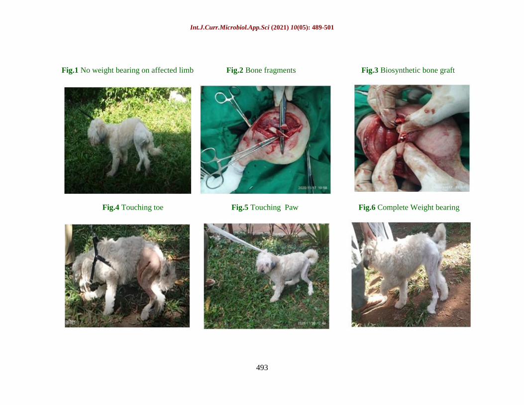
Fig.1 No weight bearing on affected limb Fig.2 Bone fragments Fig.3 Biosynthetic bone graft
Fig.4 Touching toe Fig.5 Touching Paw Fig.6 Complete Weight bearing
Int.J.Curr.Microbiol.App.Sci (2021) 10(05): 489-501
494
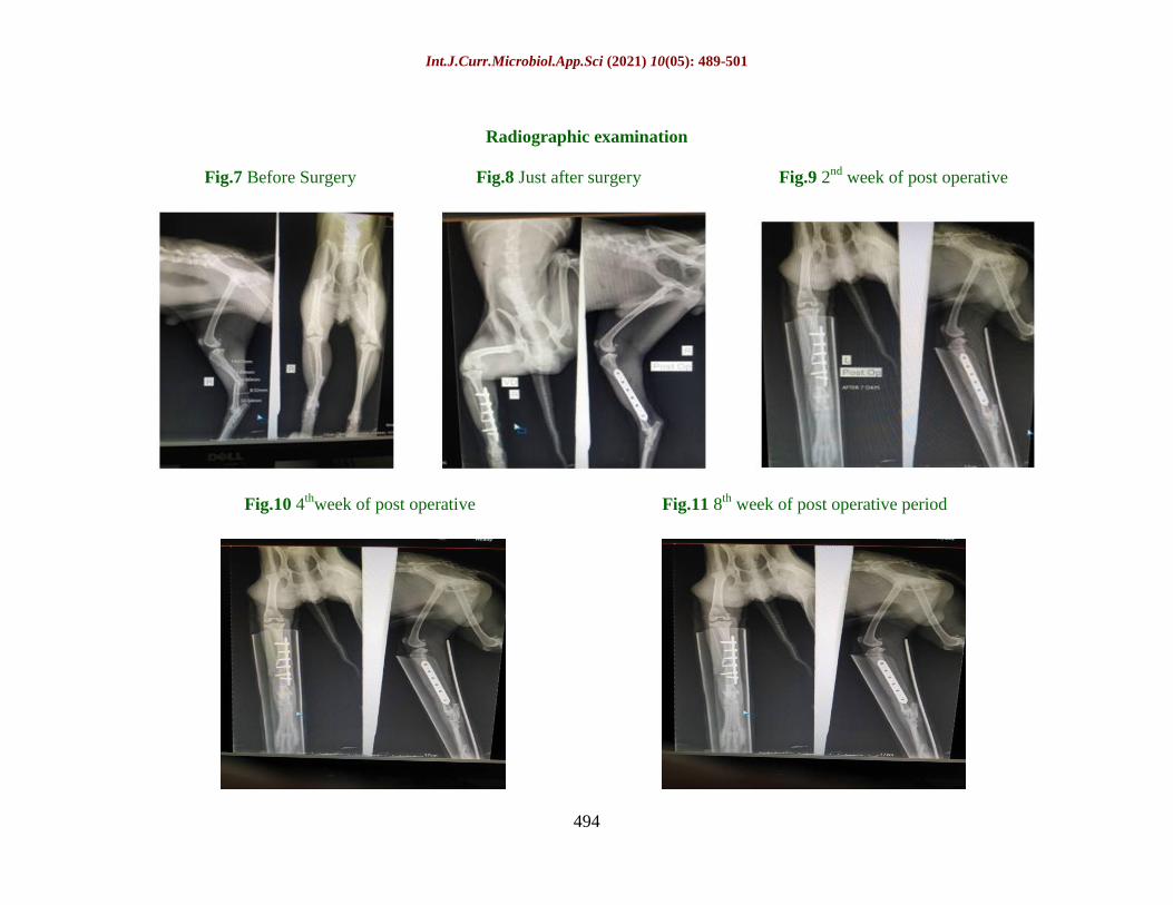
Radiographic examination
Fig.7 Before Surgery Fig.8 Just after surgery Fig.9 2nd
week of post operative
Fig.10 4th
week of post operative Fig.11 8th
week of post operative period
Int.J.Curr.Microbiol.App.Sci (2021) 10(05): 489-501
495
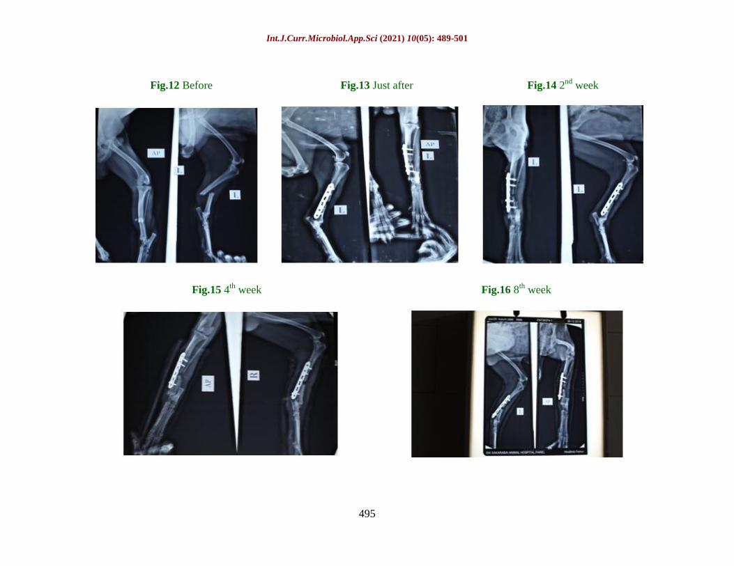
Fig.12 Before Fig.13 Just after Fig.14 2nd
week
Fig.15 4th
week Fig.16 8th
week
Int.J.Curr.Microbiol.App.Sci (2021) 10(05): 489-501
496
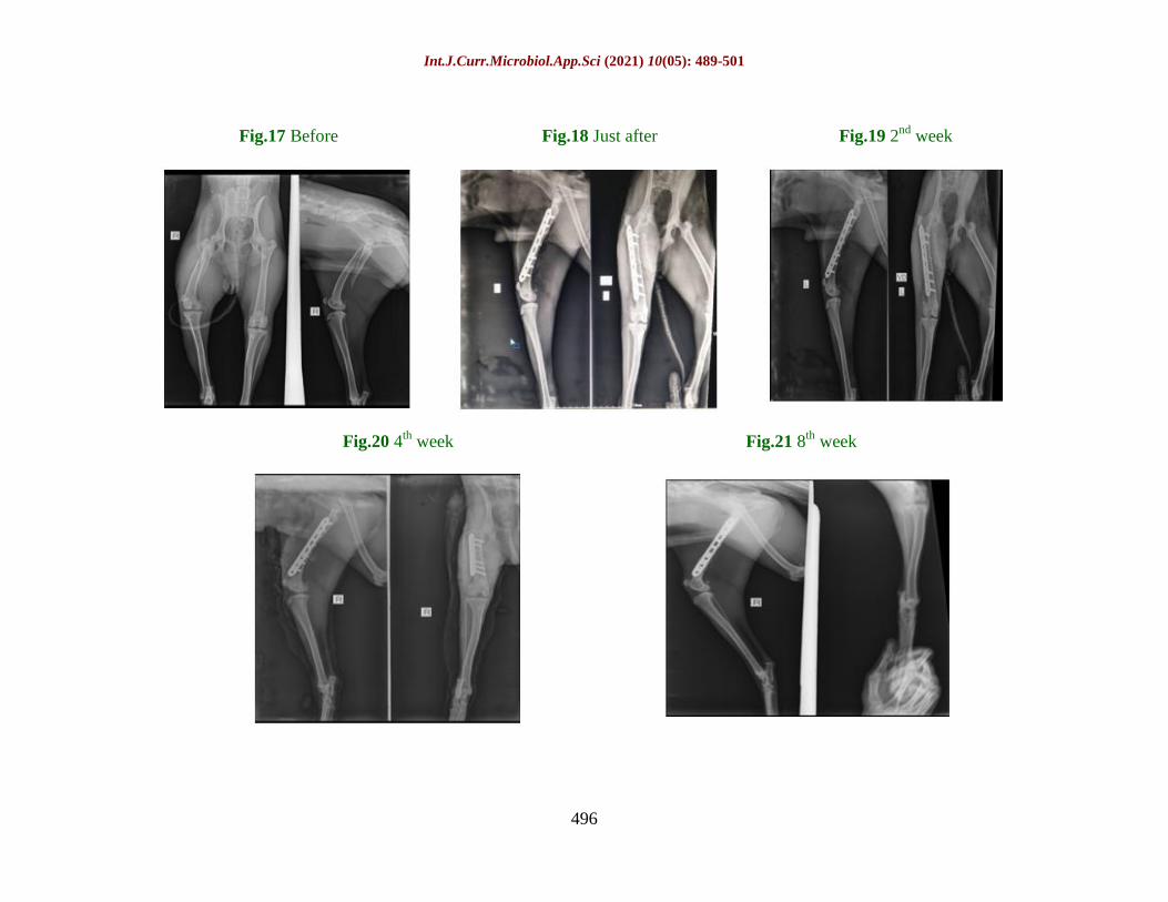
Fig.17 Before Fig.18 Just after Fig.19 2nd
week
Fig.20 4th
week Fig.21 8th
week
Int.J.Curr.Microbiol.App.Sci (2021) 10(05): 489-501
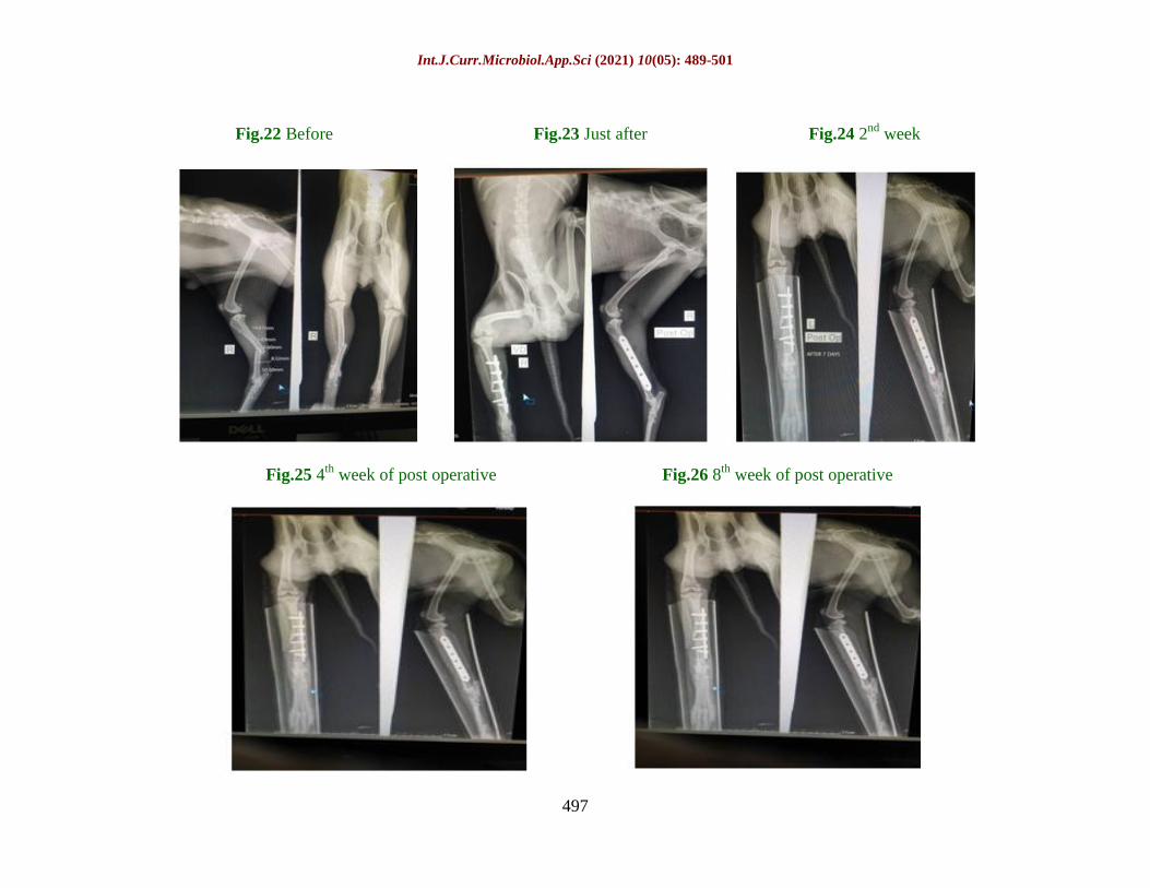
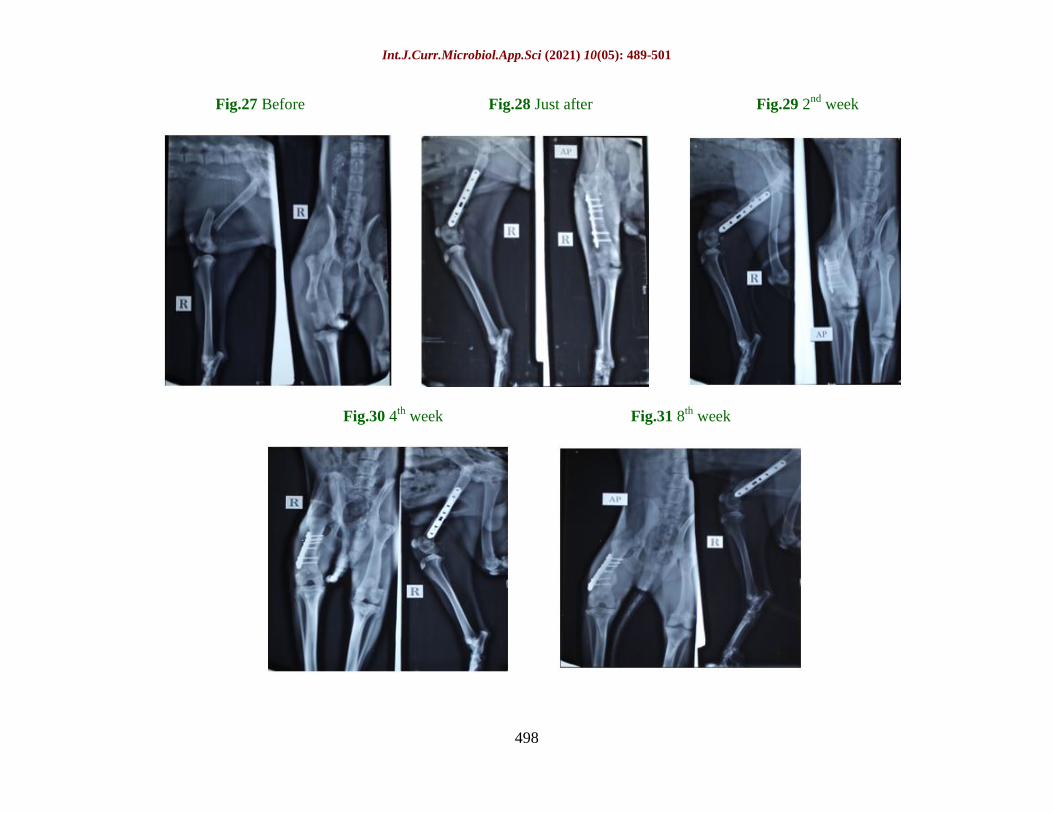
497
Fig.22 Before Fig.23 Just after Fig.24 2nd
week
Fig.25 4th
week of post operative Fig.26 8th
week of post operative
Int.J.Curr.Microbiol.App.Sci (2021) 10(05): 489-501
498
Fig.27 Before Fig.28 Just after Fig.29 2nd
week
Fig.30 4th
week Fig.31 8th
week
Int.J.Curr.Microbiol.App.Sci (2021) 10(05): 489-501
499
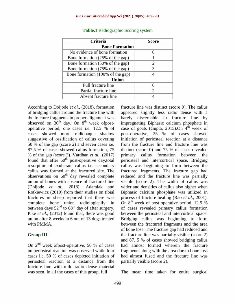
Table.1 Radiographic Scoring system
Criteria Score
Bone Formation
No evidence of bone formation 0
Bone formation (25% of the gap) 1
Bone formation (50% of the gap) 2
Bone formation (75% of the gap) 3
Bone formation (100% of the gap) 4
Union
Full fracture line 0
Partial fracture line 2
Absent fracture line 4
According to Doijode et al., (2018), formation
of bridging callus around the fracture line with
the fracture fragments in proper alignment was
observed on 30th
day. On 8th
week ofpost-
operative period, one cases i.e. 12.5 % of
cases showed more radiopaque shadow
suggestive of ossification of callus covering
50 % of the gap (score 2) and seven cases i.e.
87.5 % of cases showed callus formation, 75
% of the gap (score 3). Vardhan et al., (2017)
found that after 60th
post-operative day,total
resorption of exuberant callus i.e. secondary
callus was formed at the fractured site. The
observations on 60th
day revealed complete
union of bones with absence of fractured line
(Doijode et al., 2018). Adamiak and
Rotkiewicz (2010) from their studies on tibial
fractures in sheep reported that there was
complete bone union radiologically in
between days 52nd
to 68th
day of after surgery.
Pike et al., (2012) found that, there was good
union after 8 weeks in 6 out of 13 dogs treated
with PMMA.
Group III
On 2nd
week ofpost-operative, 50 % of cases
no periosteal reaction was observed while four
cases i.e. 50 % of cases depicted initiation of
periosteal reaction at a distance from the
fracture line with mild radio dense material
was seen. In all the cases of this group, full
fracture line was distinct (score 0). The callus
appeared slightly less radio dense with a
barely discernable in fracture line by
impregnating Biphasic calcium phosphate in
case of goats (Gupta, 2015).On 4th
week of
post-operative, 25 % of cases showed
initiation of periosteal reaction at a distance
from the fracture line and fracture line was
distinct (score 0) and 75 % of cases revealed
primary callus formation between the
periosteal and intercortical space. Bridging
callus was beginning to form between the
fractured fragments. The fracture gap had
reduced and the fracture line was partially
visible (score 2). The width of callus was
wider and densities of callus also higher when
Biphasic calcium phosphate was utilized in
process of fracture healing (Rao et al., 2001).
On 8th
week of post-operative period, 12.5 %
of cases revealed primary callus formation
between the periosteal and intercortical space.
Bridging callus was beginning to form
between the fractured fragments and the area
of bone loss. The fracture gap had reduced and
the fracture line was partially visible (score 2)
and 87. 5 % of cases showed bridging callus
had almost formed wherein the fracture
fragments along with the area due to bone loss
had almost fused and the fracture line was
partially visible (score 2).
The mean time taken for entire surgical
Int.J.Curr.Microbiol.App.Sci (2021) 10(05): 489-501
500
procedure starting from initial skin incision to
application of last skin suture for group I, II
and III was 86.25 minutes, 99.37 minutes and
95.62 minutes, respectively.
References
Aithal H. P., G. R. Singh, and G. S. Bisht
(1999) Fractures in dogs: survey of
402 cases. Indian Journal of Veterinary
Surgery 20: 15-21.
Ali L. M. (2013) Incidence, Occurrence,
Classification and Outcome of Small
Animal Fractures: A Retrospective
Study (2005-2010). International
Journal of Biological, Biomolecular,
Agricultural, Food and
Biotechnological Engineering 7(3):
191-196
Arora Manit, Edward K S Chan, Sunil Gupta,
Ashish D Diwan (2013)
Polymethylmethacrylate bone cements
and additives: A review of the
literature, World J Orthop; 4(2): 67-74,
doi:10.5312/wjo.v4.i2.67
Balagopalan T. P., C. B. Devanand, K.
Rajankutty, T. Sarada, S. R. Nayar, C.
A. Varkey, A. M. Jalaluddin, K. N.
Nayar and P. O. George (1995)
Fracture in dogs- A review of 208
cases. Indian Journal of Veterinary
Surgery 16:41-43.
Burns C. G. (2010) Influence of Locking Bolt
Location on the Mechanical Properties
of an Interlocking Nail in the Canine
Femur. Master‟s Thesis, Graduate
School of The Ohio State University,
USA
Chavan S. R. (2013) Evaluation of locking
compression plate system for long
bone fracture repair in dogs: A clinical
study. M. V. Sc. thesis submitted to
Maharashtra Animal and Fishery
Sciences University, Nagpur, India.
Coutinho N. R. (2012) Evaluation of
interlocking nail for long bone fracture
repair in dogs: a clinical study.
Unpublished M. V. Sc. thesis
submitted to Maharashtra Animals and
Fishery Sciences University, Nagpur,
Maharashtra
Doijode Vinit, Dilip Kumar D and B. V.
Shivaprakash (2018) Comparative
Evaluation of Veterinary Cuttable
Plate and Polypropylene Mesh
Impregnated PMMA Plate for Fracture
Repair of Tibia Bone in Goats,
International Journal of Livestock
Research, 8(6), 160-169, doi:
10.5455/ijlr.20170731051307
Egol K. A., E. N. Kubiak, E. Fulkerson, F. J.
Kummer and K.J. Koval (2004)
Biomechanics of Locked Plates and
Screws. Journal of Orthopaedic
Trauma 18: 488-493
Gupta P. (2015) Use of interlocking nails for
repair of humeral and tibial shaft
fractures under image intensifier in
dogs. M. V. Sc. thesis submitted to
Anand Agricultural University
Hansen B. (2003) Assessment of Pain in
Dogs: Veterinary Clinical Studies.
ILAR JOURNAL 44(3): 197-205
Johnson K. D., K. E. Frierson, T. S. Keller, C.
Cook, R. Scheinberg and J.Zerwekh
(1996) Porous ceramics as bone graft
substitutes in long bone defects: a
biomechanical, histological and
radiographic analysis. The Journal of
Orthopedic Research 14: 351-369.
Kolata R. J., N. H. Kraut and D. E. Johnston
(1974) Patterns of trauma in urban
dogs and cats: A study of 1000 cases.
Journal of American Veterinary
Medical Association 164:499-502
Kumar D. (2016) Efficacy of bone substitutes
for fracture healing in goats. Ph. D.
thesis (Surgery and Radiology)
submitted to Nanaji Deshmukh
Veterinary Science University,
Jabalpur.
Lane J. M. and H. S. Sandhu (1987) Current
Int.J.Curr.Microbiol.App.Sci (2021) 10(05): 489-501
501
approaches to experimental bone
grafting. Orthop. Clin. North. Am.
18(2): 213-25.
Lujan T. J., C. E. Henderson, S. M. Madey, D.
C. Fitzpatrick, J. L. Marsh and M.
Bottlang (2010) Locked plating of
distal femur fractures leads to
inconsistent and asymmetric callus
formation. Journal of Orthopaedic
trauma 24: 156-162
Maala C. P. and E. M. Celo (1975) A study on
the anatomical locations, incidence and
causes of fractures in dogs. Phillipine
Journal of Veterinary Medicine
14:137-143.
Manjunatha D. R., B. N. Nagaraja, S. Rao, S.
Yathiraj and Ranganath, L. (2011)
Evaluation of closed and open
interlocking nailing for femoral
fracture repair in dogs. Indian Journal
of Veterinary Surgery 32: 65-66.
Nadkarni B., S. Srivastav, V. Mittal and S.
Agarwal (2008) Use of locking
compression plates for long bone
nonunion without removing existing
intramedullary nail: A review of
literature and our experience. J.
Trauma. 65(2): 485-486.
Phillips I.R. (1979) A survey of bone fractures
in the dog and cat. Journal of Small
Animal Practice, 20: 661-674.
Raghunath M. and S. S. Singh (2008)
Intramedullary interlocking nailing for
management of long bone fractures in
dogs: A study of 17 clinical cases.
Indian J. Vet. Surg. 29 (2): 106-109.
Rajhans M. (2013) Stabilisation of splinters of
long bone fracture in dogs. M.V.Sc. &
A.H. thesis (Surgery and Radiology)
submitted to Nanaji Deshmukh
Veterinary Science University,
Jabalpur.
Rao T. M., G. V. Lakshmipathi, T. P. Sastry
and O. Ramakrishna (2001)
Biochemical changes following ulnar
segmental defect repair with fibrillar
collagen-hydroxyappatite and porous
hydroxyappatite implants in canines.
Indian Journal of Animal Research 35:
112-115.
Simon M. S., R. Ganesh, S. Ayyappan and R.
S. Kumar, V. Kundave and B. Das
(2011) Incidences of pelvic limb
fractures in dogs: A survey of
478cases. Veterinary World 3: 120-
121
Sirin O. S., U. Kaya and B. Olcay (2013)
Clinical and Radiological Outcomes of
Locking Compression Plate System in
Dogs with Diaphyseal Fractures: 32
Cases. Journal of the faculty of
Veterinary Medicine, Kafkas
University 19: 13-18
Vardhan H. K., V. Devi Prasad, Makkena
Sreenu and N. Syaama Sundar (2017)
Radiographic evaluation of poly
methylmethacrylate and hydroxy
apatite implants for fracture healing in
rabbits, International Journal of
Science, Environment and
Technology, Vol. 6, No 5, 3168 – 3173
Xu X., A. Jin, Y. Liu, J. Liu, X. Zhanq and D.
Qin (1998) New concepts and
advances of immobilization of long
bones. Chinese Medical Journal 111:
174-9.
How to cite this article:
Dakhane, P. S., D. U. Lokhande, G. S. Khandekar, G. U. Yadav, S. D. Tripathi, S. D. Ingole
and Kadam, D. P. 2021. Radiographic Evaluation of Long Bone Fractures by using Antibiotic
Loaded Bone Cement (ALBC) and Biosynthetic Bone Graft in dogs.
Int.J.Curr.Microbiol.App.Sci. 10(05): 489-501. doi: https://doi.org/10.20546/ijcmas.2021.1005.056
Related Documents

![The Radiographic Characterization of Burst Fractures of ... · The Radiographic Characterization of Burst Fractures of the Spine ... 678 ATLAS ET AL. ... 10-12]. Axial com pressive](https://static.cupdf.com/doc/110x72/5b083b3c7f8b9a5f6d8c1772/the-radiographic-characterization-of-burst-fractures-of-radiographic-characterization.jpg)