International Journal of Physics and Research (IJPR) ISSN(P): 2250-0030; ISSN(E): 2319-4499 Vol. 3, Issue 5, Dec 2013, 11-20 © TJPRC Pvt. Ltd. RADIATION DOSIMETRIC PROPERTIES OF NEW OXA-, THIADIAZOLE, TRIAZOLE AND THIAZOLE QUINAZOLINE DERIVATIVES HASSAN M. DIAB 1 , HALA SOLIMAN 2 & ARAFA I. ABD EL-HAFEZ 3 1,2,3 National Institute for Standards, Ionizing Radiation Metrology Laboratory, Egypt 1 Physics Department, Science College, Northern Boarder University, Arar, Northern Borders, Saudi Arabia ABSTRACT There is a need to synthesis a new TL material has a simple glow curve, glow peak and tissue equivalent suitable to study the biological effects of ionizing radiation. New 1,3,4-oxadiazole, 1,3,4-thiadiazole and 1,2,4-triazole derivatives of 4-thiomethyl-quinazoline have been synthesized by cyclization of hydrazide, amidrazone and thiosemicarbazide derivatives via their treatments with carbon disulfide, sulfuric acid, sodium hydroxide, and mercuric oxide. 1,3-Thiazole derivatives were prepared from thiosemicarbazide and arylidene derivative after its treatment with ethylchloroactate, phenacyl bromide and mercaptoacetic acid. The radiation dosimetric properties of new 1,3,4-oxadiazole, 1,3,4-thiadiazole, 1,2,4-triazole and 1,3-thiazole derivatives of 4-thiomethyl-quinazoline have been investigated. The TL-radiation response was found to be sensitive to the compound composition quantitatively and qualitatively due to different created trap centers according to the type of doping oxides. The therapeutic range (0.5 to 2 Gy) of the TL response versus the delivered dose without reaching saturation level provides a possibility to use this material in a phantom for checking the treatment plans of the dose delivery in the near future. The TL enhancement response to gamma radiation makes the substance doped with 11system a promising material for gamma detection dosimetry. These properties may have important applications in developing selective detectors and dosimeter suitable to study the biological effects of exposure to ionizing radiation. KEYWORDS: Biomaterial, TL-Dosimetry, Therapeutic Doses INTRODUCTION Thermoluminescence (TL) studies have received considerable attention especially, the structures of the defects giving rise to TL glow peaks [1,2]. On the other hand, the previous observations have shown that LiF:Mg,Ti really presents a rather complex TL mechanism from a solid state point of view [3]. Therefore, there is no general agreement on the authentic structure of defects in LiF:Mg,Ti and there is a still lack of understanding of TL mechanism of this material [4]. The dosimetric and TL properties of LiF:Mg,Ti used as a reference material for comparison are disreputable variable and non-universal [5,6]. This is in part due to its very complex glow curve with its many glow peaks reported between room temperature and 400 0 C under various conditions of dose, annealing and storage parameters, LET of the radiation field, etc. Each glow peak may have distinctly different dosimetric characteristics and the relative intensity of the various glow peaks depends on a great many factors. However, the dosimetric characteristics of the glow peaks rather than the glow curve (integration over a number of glow peaks) will surely lead to better understanding of TLD and contribute to the adoption of better dosimetric technique. So, there is a need to synthesis a new TL material has a simple glow curve and glow peak. A great deal of research work on electron rich nitrogen heterocycles has been done to identify new compounds, having potential applications in photonics. Such compounds class is very attractive since they could be prepared in different

Welcome message from author
This document is posted to help you gain knowledge. Please leave a comment to let me know what you think about it! Share it to your friends and learn new things together.
Transcript
International Journal of Physics
and Research (IJPR)
ISSN(P): 2250-0030; ISSN(E): 2319-4499
Vol. 3, Issue 5, Dec 2013, 11-20
© TJPRC Pvt. Ltd.
RADIATION DOSIMETRIC PROPERTIES OF NEW OXA-, THIADIAZOLE, TRIAZOLE
AND THIAZOLE QUINAZOLINE DERIVATIVES
HASSAN M. DIAB1, HALA SOLIMAN
2 & ARAFA I. ABD EL-HAFEZ
3
1,2,3National Institute for Standards, Ionizing Radiation Metrology Laboratory, Egypt
1Physics Department, Science College, Northern Boarder University, Arar, Northern Borders, Saudi Arabia
ABSTRACT
There is a need to synthesis a new TL material has a simple glow curve, glow peak and tissue equivalent suitable
to study the biological effects of ionizing radiation. New 1,3,4-oxadiazole, 1,3,4-thiadiazole and 1,2,4-triazole derivatives
of 4-thiomethyl-quinazoline have been synthesized by cyclization of hydrazide, amidrazone and thiosemicarbazide
derivatives via their treatments with carbon disulfide, sulfuric acid, sodium hydroxide, and mercuric oxide. 1,3-Thiazole
derivatives were prepared from thiosemicarbazide and arylidene derivative after its treatment with ethylchloroactate,
phenacyl bromide and mercaptoacetic acid. The radiation dosimetric properties of new 1,3,4-oxadiazole, 1,3,4-thiadiazole,
1,2,4-triazole and 1,3-thiazole derivatives of 4-thiomethyl-quinazoline have been investigated. The TL-radiation response
was found to be sensitive to the compound composition quantitatively and qualitatively due to different created trap centers
according to the type of doping oxides. The therapeutic range (0.5 to 2 Gy) of the TL response versus the delivered dose
without reaching saturation level provides a possibility to use this material in a phantom for checking the treatment plans of
the dose delivery in the near future. The TL enhancement response to gamma radiation makes the substance doped with
11system a promising material for gamma detection dosimetry. These properties may have important applications in
developing selective detectors and dosimeter suitable to study the biological effects of exposure to ionizing radiation.
KEYWORDS: Biomaterial, TL-Dosimetry, Therapeutic Doses
INTRODUCTION
Thermoluminescence (TL) studies have received considerable attention especially, the structures of the defects
giving rise to TL glow peaks [1,2]. On the other hand, the previous observations have shown that LiF:Mg,Ti really presents
a rather complex TL mechanism from a solid state point of view [3]. Therefore, there is no general agreement on the
authentic structure of defects in LiF:Mg,Ti and there is a still lack of understanding of TL mechanism of this material [4].
The dosimetric and TL properties of LiF:Mg,Ti used as a reference material for comparison are disreputable
variable and non-universal [5,6]. This is in part due to its very complex glow curve with its many glow peaks reported
between room temperature and 4000C under various conditions of dose, annealing and storage parameters, LET of the
radiation field, etc. Each glow peak may have distinctly different dosimetric characteristics and the relative intensity of the
various glow peaks depends on a great many factors. However, the dosimetric characteristics of the glow peaks rather than
the glow curve (integration over a number of glow peaks) will surely lead to better understanding of TLD and contribute to
the adoption of better dosimetric technique. So, there is a need to synthesis a new TL material has a simple glow curve and
glow peak.
A great deal of research work on electron rich nitrogen heterocycles has been done to identify new compounds,
having potential applications in photonics. Such compounds class is very attractive since they could be prepared in different
12 Hassan M. Diab, Hala Soliman & Arafa I. Abd El-Hafez
shapes and sizes and can accept rare earth ions without inducing any crystallization. These classes are very attractive since
they could be prepared in different shapes and sizes and can accept rare earth ions without inducing any crystallization.
They are promising materials for photonics applications because of wide transmissions window, good stability and
durability, high-refractive index, higher non-linear optical properties, and relatively low-phonon energies. Although the
benefits of radiation are enormous and continuously increasing, it is well known that ionizing radiation can induce cancer
and genetic defect. Such compounds have good ability to host luminescent activators. It was reported that
thermoluminescence TL for derivatives of such classes has been archived [7]. Recently, some studies achievedfor uses of
solar cells in dosimetry ؛monocrystalline silicon solar cell of the construction n+pp++ Passivated Emitter Solar Cell (PESC)
was irradiated by 60
Co gamma ray doses. The TL enhancement response to gamma radiation makes the monocrystalline
silicon solar cell system a promising material for gamma detection dosimetry[8]. inorganic dosimetry showed Ruthenium
phthalocyanineterial of gamma-ray thermoluminscence dosimeterhas a potential use as a material for gamma –
raythermoluminscence dosimeter(TLD) for clinical dosimetery[9].
Also, Dosimetric properties of the quaternary tellurite glasses have been measured as a function of Different
compositions of the glassy system in different rare earth oxides concentration by using thermoluminescence(TL) detection
technique.The experiment results showed that tellurite investigation of thermoluminescence has a potential use as the
materials of gamma-ray thermoluminescence [10]. Besides that, The behavior of the different types of tellurite glasses is
analyzed regarding to their kinetic parameters and luminescence emission which enhances the claim of tellurite glasses for
use as TLD material at therapeutic radiation doses [11]. Sulfonated grafted polymers also provided a better understanding
of the response to 208
pb ions irradiation to determine the importance of grafting conversionin accurate dosimetric properties
measurement [12].
On the other hand, quinazoline derivatives are an important class of nitrogen-containing heterocycles which
display a wide variety of biological activities [13] and the quinazoline moiety is an important part of many natural
alkaloids. Compounds with diverse biological activities (hypotonic, antiallergic, antibacterial and anthelminthic) have been
found among quinazoline derivatives [14]. Most biologically active quinazolines possess substituents at C-2 and N-3
positions. The anti fungal, antibacterial, anti-HIV activities of Schiff and Mannich bases derived from isatin and N-[4-(4-
chlorophenyl)thiazol-2-yl]thiosemicarbazide [15], antimicrobial activity of fluorinated hydroquinazoline derivatives [16]
and 6-chloro-2-morpholino 4-quinazolyl-5-nitro-2-furyl hydrazone [17] were reported. On this basis, we have synthesized
some derivatives of quinazoline semicarbazone. It has been reported that, certain compounds bearing 1,3,4-oxa-,
thiadiazole, and 1,2,4-triazole nucleus possess significant anti-inflammatory activity [18,19]. Some 1,2,4-triazole
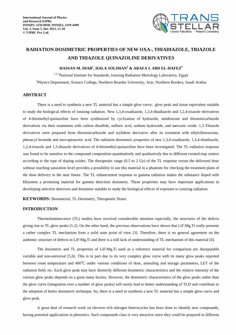
derivatives incorporating Schiff base structure were synthesized as antitumor agents [20]. Compounds MKT 077 [21] and
HP-236 [22] are thiazole compounds and have been reported as a registered antitumour and antipsychotic agents,
respectively (Figure 1).
N
S
S
N N
CH3 Cl
O CH3
CH3
S
N
NN
S
F
CH3
CH3
CH3
O
[MKT o77] [HP-236]
Figure 1: Structure of MKT077 and HP-236
Radiation Dosimetric Properties of New Oxa-, Thiadiazole, Triazole and Thiazole Quinazoline Derivatives 13
Owing to the above facts and in continuation of our research program [17-20] in the synthesis of biologically
active 1,3,4-oxadiazol, 1,2,4-triazol, 1,3,4-thiadiazol, our goal here in the synthesis of such heterocyclic compounds and
thiazolidinone nuclei attached to the quinazoline-thiomethyl derivative and investigation of the luminescence spectra of the
prepared compounds in addition to structure correlation.
EXPERIMENTAL
The reading out process was performed using a TLD reader (Model 4500, Bicron/Harshaw) equipped with two
photomultiplier tubes, which could record luminescence independently. The reader was controlled by WinREMS Software
supplied with the spectrometer and running on a Windows® computer. The thermoluminescence detectors TLD-100 were
used with dimensions of 3.2 X 3.2 X 0.89 mm3 and doped with titanium (11.5 ppm) and magnesium (300 ppm) purchased
from Bicorn / Harshow Chemical Company. Solvents and reagents were obtained from Acros (Geel, Belgium), Fluka
(Taufkirchen, Germany) or Sigma (Steinheim, Germany). All melting points were measured on Electro thermal IA 9000
series digital melting Point apparatus. The IR spectra were recorded in potassium bromide discs on a Pye Unicam SP 3300
and Shimadzu FT IR 8101 PC infrared spectrophotometer. The NMR Spectra were recorded at 270 MHz on a Varian
Mercury VX-300 NMR spectrometer. 1H NMR (300 MHz) and
13C NMR (75.5 MHz) were run in deuterated chloroform
(CDCl3) or dimethylsulphoxide (DMSO-d6). Chemical shifts were related to that of the solvent. Mass Spectra were
recorded on a Shimadzu GCMS-QP1000 EX mass spectrometer at 70 eV. Elemental analyses were carried out at the Micro
analytical Centre of Cairo University, Giza, Egypt. All reactions were followed by TLC (Silica gel, Aluminum Sheets 60
F254, Merck).
RESULTS AND DISCUSSIONS
Sample Synthesis
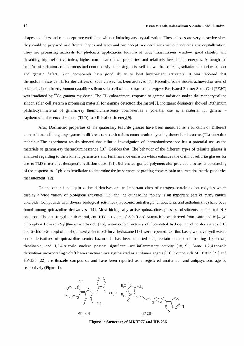
The quinazoline-4-thiol (1), which is used as starting material in this study, was prepared according to the reported
method [15]. The thiol 1 was alkylated with ethyl 2-chloroacetate and 2-chloroacetonitrile, in anhydrous DMF containing
anhydrous potassium carbonate, to afford ethyl 2-(quinazolin-4-ylthio)acetate (2) and 2-(quinazolin-4-ylthio)acetonitrile (3)
in good yields 87%, 77% respectively. Compounds 2 and 3 were treated with hydrazine hydrate, in ethanol, to give acid
hydrazide 4 and amidrazone 5. Cyclization of acid hydrazide 4 with CS2 and KOH resulted to the formation of 5-
((quinazolin-4-ylthio)methyl)-1,3,4-oxadiazole-2-thiol (6). Amidrazone derivative 5 was also treated with CS2 in methanol
to the thiol 7. The structure of compounds 6 and 7 were established under the basis of their elemental analysis and spectral
data (scheme 1).
N
N
SH
N
N
S COOEt
N
N
S CN
N
SNH
NHNH2N
N
N
S CONHNH2
N
N
S
SN
N
SHN
N
S
ON
N
SH
DMF/K2CO3
ClCH2CO2EtClCH2CN
N2H4N2H4
CS2/KOHCS2/MeOH
1
234
5
67
Scheme 1. Alkylation and formation of oxa- and thiadiazol of quinazoline-thiomethyl
14 Hassan M. Diab, Hala Soliman & Arafa I. Abd El-Hafez
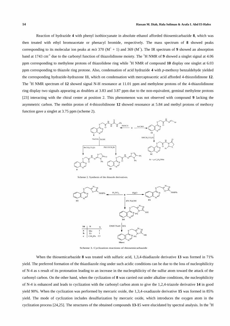
Reaction of hydrazide 4 with phenyl isothiocyanate in absolute ethanol afforded thiosemicarbazide 8, which was
then treated with ethyl bromoacetate or phenacyl bromide, respectively. The mass spectrum of 8 showed peaks
corresponding to its molecular ion peaks at m/z 370 (M+ + 1) and 369 (M
+). The IR spectrum of 9 showed an absorption
band at 1743 cm-1
due to the carbonyl function of thiazolidinone moiety. The 1H NMR of 9 showed a singlet signal at 4.06
ppm corresponding to methylene protons of thiazolidene ring while 1H NMR of compound 10 display one singlet at 6.03
ppm corresponding to thiazole ring protone. Also, condensation of acid hydrazide 4 with p-methoxy benzaldehyde yielded
the corresponding hydrazide-hydrazone 11, which on condensation with mercaptoacetic acid afforded 4-thiozolidinone 12.
The 1H NMR spectrum of 12 showed signal N-H resonance at 11.01 ppm and methylene protons of the 4-thiazolidinone
ring display two signals appearing as doublets at 3.83 and 3.87 ppm due to the non-equivalent, geminal methylene protons
[23] interacting with the chiral center at position 2. This phenomenon was not observed with compound 9 lacking the
asymmetric carbon. The methin proton of 4-thiozolidinone 12 showed resonance at 5.84 and methyl protons of methoxy
function gave a singlet at 3.75 ppm (scheme 2).
N
N
S
O
NH
NH
NH
S
Ph
N
N
S
O
NH
N N
S
O
Ph
N
N
S
O
NH
N N
S
Ph
Ph
N
N
S
O
NH
N Ar
N
N
S
O
NH
N
S
O
Ar
4
PhNCS
EtOHNaOAc
BrCH2CO2Et PhCOCH2Br
ArCHO
8
9 10
11
12
Ar = p-C6H4OMe
SHCH2CO2Et
Scheme 2. Synthesis of the thiazole derivatives.
N
NN
N
N
S
PhSH
S
NN
N
N
S
NHPh
O
NN
N
N
S
NHPh
N
NN
N
N
S
PhSR
16 R X
b
a
c
Me
Et
CH2Ph
I
I
Cl
8
4N-NaOH
H2SO4 HgO
DMF/NaH RX
13
14
15
16a-c
Scheme 3. Cyclization reactions of thiosemicarbazide
When the thiosemicarbazide 8 was treated with sulfuric acid, 1,3,4-thiadiazole derivative 13 was formed in 71%
yield. The preferred formation of the thiazdiazole ring under such acidic conditions can be due to the loss of nucleophilicity
of N-4 as s result of its protonation leading to an increase in the nucleophilicity of the sulfur atom toward the attack of the
carbonyl carbon. On the other hand, when the cyclization of 8 was carried out under alkaline conditions, the nucleophilicity
of N-4 is enhanced and leads to cyclization with the carbonyl carbon atom to give the 1,2,4-triazole derivative 14 in good
yield 90%. When the cyclization was performed by mercuric oxide, the 1,3,4-oxadiazole derivative 15 was formed in 85%
yield. The mode of cyclization includes desulfurization by mercuric oxide, which introduces the oxygen atom in the
cyclization process [24,25]. The structures of the obtained compounds 13-15 were elucidated by spectral analysis. In the 1H
Radiation Dosimetric Properties of New Oxa-, Thiadiazole, Triazole and Thiazole Quinazoline Derivatives 15
NMR spectra, the signal due to the S˗CH2 methylene protons present in all compounds 13-15 at 4.03-4.53 ppm, as singlets.
NH and SH protons were observed at 10.06-10.58 and 14.01 ppm as broad singlets respectively. The thiol 14 was
alkylated, after its treatment with 60% sodium hydride in anhydrous DMF, with methyl-, ethyl iodide and benzyl chloride to
give the corresponding S-alkyl derivatives 16a-c. Structures of the latter compounds 16a-c were established on the basis of
1H NMR spectra, which showed the absence of the characteristic SH peak at 14.01 ppm and the presence of signals, singlet
at 2.49 ppm for S˗Me, triplet at 1.33 ppm, quartet at 3.11 ppm due to the ethyl group, and singlet at 4.40 ppm due to the
benzyl group respectively (scheme 3).
Radiation Dosimetric Activity
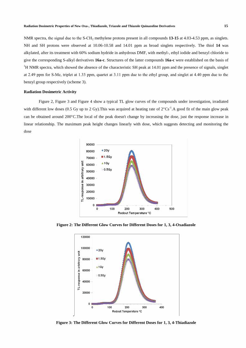
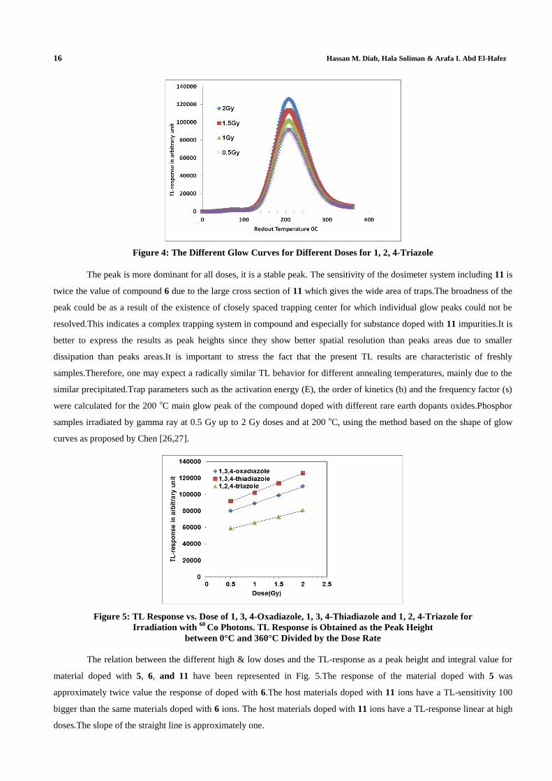
Figure 2, Figure 3 and Figure 4 show a typical TL glow curves of the compounds under investigation, irradiated
with different low doses (0.5 Gy up to 2 Gy).This was acquired at heating rate of 2Cs-1
.A good fit of the main glow peak
can be obtained around 200C.The local of the peak doesn't change by increasing the dose, just the response increase in
linear relationship. The maximum peak height changes linearly with dose, which suggests detecting and monitoring the
dose
Figure 2: The Different Glow Curves for Different Doses for 1, 3, 4-Oxadiazole
.
Figure 3: The Different Glow Curves for Different Doses for 1, 3, 4-Thiadiazole
16 Hassan M. Diab, Hala Soliman & Arafa I. Abd El-Hafez
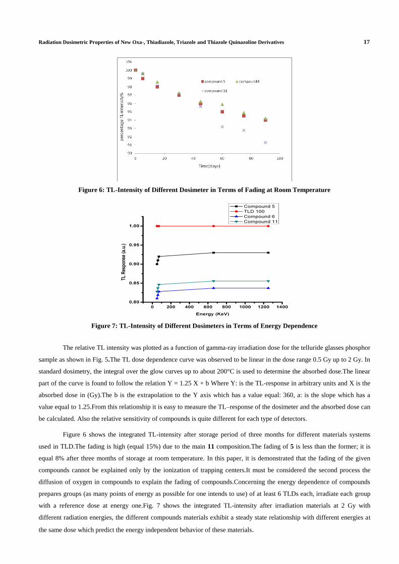
Figure 4: The Different Glow Curves for Different Doses for 1, 2, 4-Triazole
The peak is more dominant for all doses, it is a stable peak. The sensitivity of the dosimeter system including 11 is
twice the value of compound 6 due to the large cross section of 11 which gives the wide area of traps.The broadness of the
peak could be as a result of the existence of closely spaced trapping center for which individual glow peaks could not be
resolved.This indicates a complex trapping system in compound and especially for substance doped with 11 impurities.It is
better to express the results as peak heights since they show better spatial resolution than peaks areas due to smaller
dissipation than peaks areas.It is important to stress the fact that the present TL results are characteristic of freshly
samples.Therefore, one may expect a radically similar TL behavior for different annealing temperatures, mainly due to the
similar precipitated.Trap parameters such as the activation energy (E), the order of kinetics (b) and the frequency factor (s)
were calculated for the 200 oC main glow peak of the compound doped with different rare earth dopants oxides.Phosphor
samples irradiated by gamma ray at 0.5 Gy up to 2 Gy doses and at 200 oC, using the method based on the shape of glow
curves as proposed by Chen [26,27].
Figure 5: TL Response vs. Dose of 1, 3, 4-Oxadiazole, 1, 3, 4-Thiadiazole and 1, 2, 4-Triazole for
Irradiation with 60
Co Photons. TL Response is Obtained as the Peak Height
between 0°C and 360°C Divided by the Dose Rate
The relation between the different high & low doses and the TL-response as a peak height and integral value for
material doped with 5, 6, and 11 have been represented in Fig. 5.The response of the material doped with 5 was
approximately twice value the response of doped with 6.The host materials doped with 11 ions have a TL-sensitivity 100
bigger than the same materials doped with 6 ions. The host materials doped with 11 ions have a TL-response linear at high
doses.The slope of the straight line is approximately one.
Radiation Dosimetric Properties of New Oxa-, Thiadiazole, Triazole and Thiazole Quinazoline Derivatives 17
Figure 6: TL-Intensity of Different Dosimeter in Terms of Fading at Room Temperature
Figure 7: TL-Intensity of Different Dosimeters in Terms of Energy Dependence
The relative TL intensity was plotted as a function of gamma-ray irradiation dose for the telluride glasses phosphor
sample as shown in Fig. 5.The TL dose dependence curve was observed to be linear in the dose range 0.5 Gy up to 2 Gy. In
standard dosimetry, the integral over the glow curves up to about 200°C is used to determine the absorbed dose.The linear
part of the curve is found to follow the relation Y = 1.25 X + b Where Y: is the TL-response in arbitrary units and X is the
absorbed dose in (Gy).The b is the extrapolation to the Y axis which has a value equal: 360, a: is the slope which has a
value equal to 1.25.From this relationship it is easy to measure the TL–response of the dosimeter and the absorbed dose can
be calculated. Also the relative sensitivity of compounds is quite different for each type of detectors.
Figure 6 shows the integrated TL-intensity after storage period of three months for different materials systems
used in TLD.The fading is high (equal 15%) due to the main 11 composition.The fading of 5 is less than the former; it is
equal 8% after three months of storage at room temperature. In this paper, it is demonstrated that the fading of the given
compounds cannot be explained only by the ionization of trapping centers.It must be considered the second process the
diffusion of oxygen in compounds to explain the fading of compounds.Concerning the energy dependence of compounds
prepares groups (as many points of energy as possible for one intends to use) of at least 6 TLDs each, irradiate each group
with a reference dose at energy one.Fig. 7 shows the integrated TL-intensity after irradiation materials at 2 Gy with
different radiation energies, the different compounds materials exhibit a steady state relationship with different energies at
the same dose which predict the energy independent behavior of these materials.
18 Hassan M. Diab, Hala Soliman & Arafa I. Abd El-Hafez
The therapeutic dose range (0.5-2 Gy) of the TL response versus the delivered dose without reaching saturation
level provides a possibility to use this material in a phantom for checking the treatment plans of the dose delivery in the
near future.The results showed that the TL-radiation response was found to be sensitive to the compounds composition
quantitatively and qualitatively due to different created trap centers according to the type of doping oxides.
CONCLUSIONS
New azole heterocycles were synthesized and their dosimetric properties of were investigated. From the obtained
results it could be concluded that:
The TL-radiation response was found to be sensitive to the compound composition quantitatively and qualitatively
due to different created trap centers according to the type of doping oxides.
The therapeutic range (0.5 to 2 Gy) of the TL response versus the delivered dose without reaching saturation level
provides a possibility to use this material in a phantom for checking the treatment plans of the dose delivery in the
near future.
The TL enhancement response to gamma radiation makes the substance doped with 11system a promising material
for gamma detection dosimetry. These properties may have important applications in developing selective
detectors and dosimeter suitable to study the biological effects of ionizing radiation exposure.
The experiment results showed that for compounds investigation of thermoluminescence, the compound potential
use as the materials of gamma-ray thermoluminescence.
REFERENCES
1. Pradhan , S.M., Sneha,C., Adtani, M.M., Radiation Protection Dosimetry 144(1-4), 195-198, 2011.
2. Fuks, E., Horowitz,Y.S., Horowitz, A., Oster, L., Marino, S., Rainer, M., Rosenfeld,A., Datz, H., Radiation
Protection Dosimetry, Vol. 143, No. 2–4, pp. 416–426, 2011.
3. Furetta ,C., “Handbook of thermoluminescence(2nd
edition)”,World Scientific Publishing Co.Pte.Ltd, 2010.
4. Yazici, A.N., J. Phys. D: Appl. Phys. 36, 1418, 2003.
5. Harvey,J.A., Haverland,N.P., Kearfott,K.J., Applied Radiation and Isotopes 68 ,1988–2000, 2010.
6. Bakshi, A.K., Chatterjee, S., Selvam, T. P., Joshi, V.J., Chougaonkar, M.P., Nuclear Instruments and Methods in
Physics Research B 269 , 2107–2110, 2011.
7. M. Elkholy, Materials Chem. Phys., 77, 321 (2002).
8. H.M.Diab, A.Ibrahim and R.EL-mallawany "Silicon solar cell as a gamma ray dosimeter" Measurments , No.4
accepted for published (2013).
9. H. M. Diab, Tamer Ezzat Youssef and Hany A. Shousha “Investigation of Thermoluminescence Properties of
Ruthenium phthalocyanine phosphor for low gamma radiation dosimetry” Egyptian Journal of Biophysics, Vol.
12, No. 2, 119 – 129. July, 2006
10. R. El-Mallawany, and H. M. Diab, Improving dosimetric properties of tellurite glasses, Physica B,407 pp.3580-
3585 (2012).
Radiation Dosimetric Properties of New Oxa-, Thiadiazole, Triazole and Thiazole Quinazoline Derivatives 19
11. R. El-Mallawany and H. M. Diab Effect of pre-readout annealing treatments on TL mechanism in Tellurite
Glasses at therapeutic radiation doses level.mesurments journal46 pp 1722-1725. (2013).
12. H. M. Diab and Abd EL-Hafez I. A., Thermoluminescence dosimetric properties of sulfonated grafted LDPP
incorporating high linear energy transfer dependency for dose verification purposes. Isotope & Rad. Res.,
43(4),1185-1193.(2011)
13. R. A. Smits, I. J. P. De Esch, O. P. Zuiderv, J. Broeker, K. Sansuk, E., Guaita, G. Coruzzi, M. Adami, E.
Haaksma, R. Leurs, J. Med. Chem., 51, 7855 (2008).
14. S. Johne, Pharmazie, 36, 583 (1981).
15. S. N. Pandeya, D. Sriram, G. Nath, E. De-Clercq, Eur J Pharm Sci., 9, 25 (1999).
16. M. M. Ghorab, S. M. Abdel-Gawad, M.S.A. El-Gaby, Farmaco Sci 55, 249 (2000).
17. S. Jantova, D. Hudecova, S. Stankovsky, K. Spirkova, L. Ruzekova, Folia Microbiol (Praha), 40, 611 (1995).
18. M. Amir, K. Shikha, Eur. J. Med. Chem. 39, 535 (2004).
19. N. Demirbas, S. Alpay Karaoglu, A. Demirbas, K. Sancak Eur J. Med. Chem., 39, 793 (2004).
20. N. Demirbas, A. Ugurluoglu, A. Demirbas, Bioorg. Med. Chem., 10, 3717 (2002).
21. M. Kawakami, K. Koya, T. Ukai, N. Tatsuta, A. Ikegawa, K. Ogawa, T. Shishido, L. B. Chen, J. Med. Chem. 41,
130 (1998).
22. N. J. Hrib, J. G. Juracak, D. E. Bregna, R. W. Dunn, H. M. Geyer, H. B. Hartman, J. E. Roehr, K. L. Rogers, D.
K. Rush, A. M. Szczepanik, M. R. Szewezak, C. A. Wilmot, P. G. Conway, J. Med. Chem., 35, 2712 (1992)..
23. M. S. Karthikeyan, Eur. J. Med. Chem. 43, 309 (2008).
24. G. Capan, N. Ulusoy, N. Ergenc, M. Kiraz, Monats. Chemie 130, 1399 (1999).
25. IAEA (International Atomic Energy Agency), absorbed dose determination in photon and electron beams an
international code of practice. 1997, Technical Report Series No. 277, IAEA, Vienna,
26. C. Furetta, G. Kitis, A. Brambilla, C. P. F. Jany Bergonzo Foulon, Rad. Prot. Dosim, 84, 201 (1999).
27. M. S. Preet, V. K. Bedi, P. M. Mohinder, Bioorg Med Chem Lett 14, 20, 521 (2004).

Related Documents


![TriggeringEmission with the HelicalTurn in Thiadiazole-Helicenes · 2017. 1. 13. · Acopper(II) complex with two thiadiazole-[5]helicene ligandswas structurally characterized, and](https://static.cupdf.com/doc/110x72/60bfd9b6d124d074e95f846c/triggeringemission-with-the-helicalturn-in-thiadiazole-2017-1-13-acopperii.jpg)