Fall 2011, Copyright UW Imaging Research Laboratory Radiation Detection Tom Lewellen, PhD [email protected] Nuclear Medicine Basic Science Lectures http://www.rad.washington.edu/research/our-research/groups/irl/education/ basic-science-resident-lectures September 2011 Tuesday, September 27, 11

Welcome message from author
This document is posted to help you gain knowledge. Please leave a comment to let me know what you think about it! Share it to your friends and learn new things together.
Transcript

Fall 2011, Copyright UW Imaging Research Laboratory
Radiation Detection
Tom Lewellen, PhD
[email protected] Medicine Basic Science Lectures
http://www.rad.washington.edu/research/our-research/groups/irl/education/basic-science-resident-lectures
September 2011
Tuesday, September 27, 11

Fall 2011, Copyright UW Imaging Research Laboratory
Main points of last week’s lecture:
Charged particles• Short ranged in tissue
~ mm for betas (predictable, continuously slowing path)~ µm for alphas (more sporadic path)
• Interactions Types: Excitation, Ionization, Bremsstrahlung• Linear energy transfer (LET) - nearly continual energy transfer• Bragg ionization peak (LET peaks as particle slows down)
Photons• Relatively long ranged (~cm)• Local energy deposition - photon deposits much or all of their energy each interaction• Interactions Types: Rayleigh, Photoelectric, Compton, Pair Production• Compton - dominant process in tissue-equivalent materials for Nuc. Med. energies• Beam hardening - polychromatic photon beam• Buildup factors - narrow vs. wide beam attenuation• Secondary ionization - useful for photon detection
Now we shall discuss …How interaction of radiation can lead to detection
Tuesday, September 27, 11
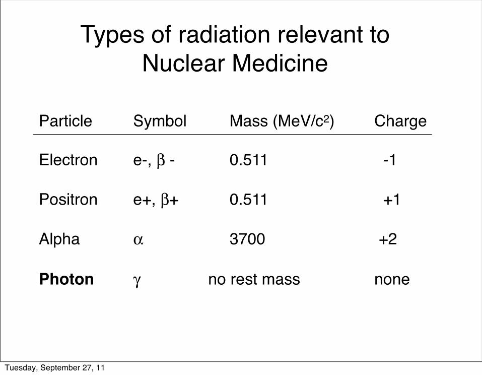
Types of radiation relevant to Nuclear Medicine
Particle! Symbol! Mass (MeV/c2) ! Charge
Electron! e-, β -! ! 0.511 !! ! -1
Positron! e+, β+!! 0.511 !! ! +1
Alpha!! α! ! 3700 ! ! ! +2
Photon! γ! no rest mass! ! none
Tuesday, September 27, 11

• Loses energy in a more or less continuous slowing down process as it travels through matter.
• The distance it travels (range) depend only upon its initial energy and its average energy loss rate in the medium.
• The range for an α particle emitted in tissue is on the order of µm’s.
α Particle Range in Mattermono-energetic
- - - - - - - - - - - - - - - - - - -+ + + + + + + + + + + + + + + + +
µm’s
α
Tuesday, September 27, 11

• β particle ranges vary from one electron to the next, even for βs of the same energy in the same material.
• This is due to different types of scattering events the β encounters (i.e., scattering events, bremsstrahlung-producing collisions, etc.).
• The β range is often given as the maximum distance the most energetic β can travel in the medium.
• The range for β particles emitted in tissue is on the order of mm’s.
β Particle Range in Mattercontinuous energy spectrum
mm’s-
β±
Tuesday, September 27, 11

Interactions of Photons with MatterExponential Penetration: N=N0e-λx
Photoelectric effect! photon is absorbed!Compton scattering! part of the energy of the photon is absorbed! scattered photon continues on with lower energy
Pair production! positron-electron pair is created! requires photons above 1.022 MeV!
Coherent (Rayleigh) scattering! photon deflected with very little energy loss! only significant at low photon energies (<50 keV)
λ
cm’s
NN0
x
Tuesday, September 27, 11

Basic Radiation Detector System
Pulse or
Current storedto disk
incoming radiation
Analog-to-
digitalAmplify
& condition
Tuesday, September 27, 11

Basic Radiation Detector SystemsWhat do you want to know about the radiation?
Energy?Position (where did it come from)?How many / how much?
Important properties of radiation detectors
(depends on application)Energy resolutionSpatial resolutionSensitivityCounting Speed
Tuesday, September 27, 11

Pulse Mode versus Current Mode
• Pulse mode– Detect individual photons– Required for NM imaging applications
• Current mode– Measures average rates of photon flux– Avoids dead-time losses
Tuesday, September 27, 11

Types of Radiation Detectorsdetection modes / functionality
• Counters– Number of interactions– Pulse mode
• Spectrometers– Number and energy of interactions– Pulse mode
• Dosimeters– Net amount of energy deposited– Current mode
• Imaging Systems– CT = current mode– NM = pulse mode
Tuesday, September 27, 11

Types of Radiation Detectorsphysical composition
• Gas-filled detectors
• Solid-state (semiconductor) detectors
• Organic scintillators (liquid & plastic)
• Inorganic scintillators
scintillators operate with a photo-sensor
(i.e. another detector)Tuesday, September 27, 11
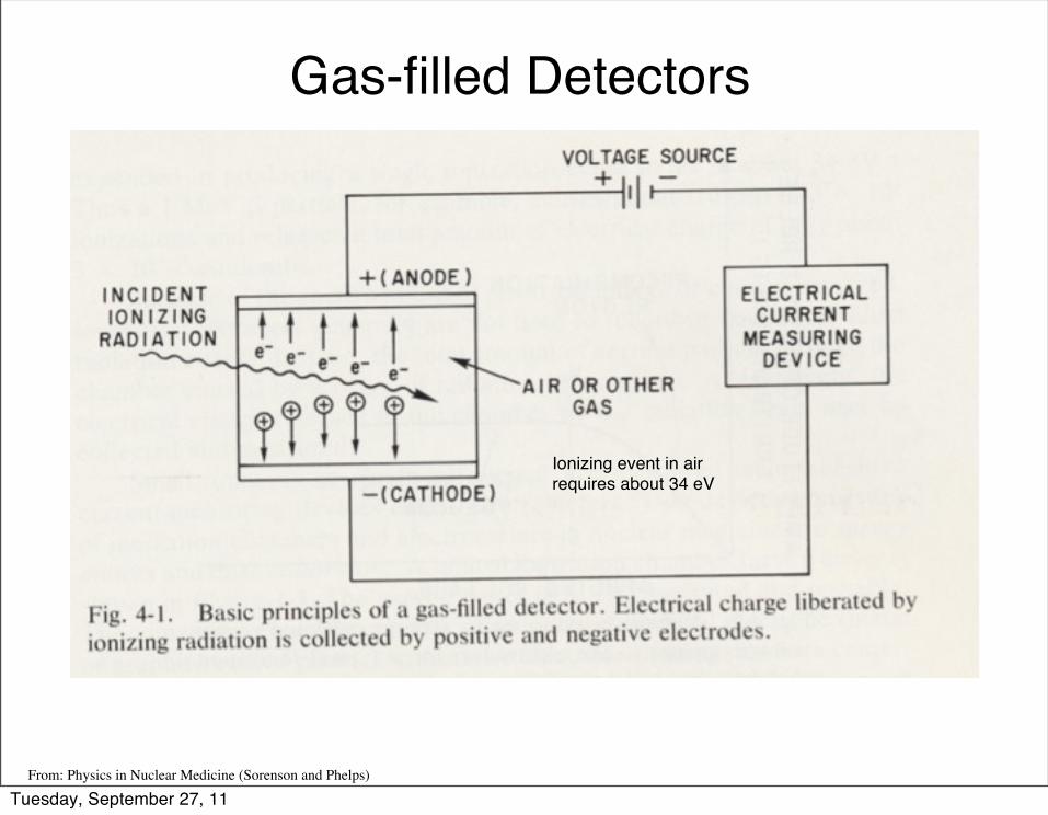
Gas-filled Detectors
Ionizing event in airrequires about 34 eV
From: Physics in Nuclear Medicine (Sorenson and Phelps)
Tuesday, September 27, 11

Gas-filled detectors(operates in three ranges)
Geiger-Muller counters
Proportional counters
Ionization chambers– Radiation survey meters– Dosimeters (dose calibrator)
From: Radiation Detection and Measurement (Knoll, GF)
Tuesday, September 27, 11

Ionization Chambers
From: Physics in Nuclear Medicine (Sorenson and Phelps)
ATOMLAB 200 Dose Calibrator
No amplificationNo dead-timeSignal = liberated chargeSettings for different isotopesCalibrations
Ionization chamber region
Tuesday, September 27, 11

Geiger-Muller counters
From: Physics in Nuclear Medicine (Sorenson and Phelps)
No energy infoLong dead-timeThin window probe
Tuesday, September 27, 11

Dose calibratorFrom: The Essential Physics of Medical Imaging (Bushberg, et al)
A) Film packB) Black (opaque) envelopeC) FilmD) Plastic film badgeF) Teflon filterG) Lead filterH) Copper filterI) Aluminum filterJ) “Open window”
Dosimeter - Film Badge
Fall 2011, Copyright UW Imaging Research Laboratory
Tuesday, September 27, 11

Dose calibratorFrom: The Essential Physics of Medical Imaging (Bushberg, et al)
Pocket Dosimeter
Fall 2011, Copyright UW Imaging Research Laboratory
Tuesday, September 27, 11

Semiconductor Detectors
• Works on same principle as gas-filled detectors (i.e., production of electron-hole pairs in semiconductor material)
• Only ~3 eV required for ionization (~34 eV, air)• Usually needs to be cooled (thermal noise)• Usually requires very high purity materials or
introduction of “compensating” impurities that donate electrons to fill electron traps caused by other impurities
Tuesday, September 27, 11
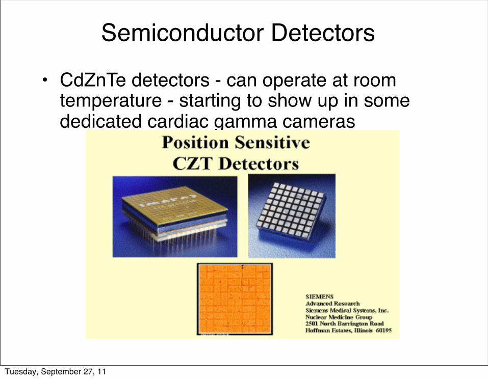
Semiconductor Detectors
• CdZnTe detectors - can operate at room temperature - starting to show up in some dedicated cardiac gamma cameras
Tuesday, September 27, 11

Organic Liquid Scintillators(liquid scintillator cocktail)
• Organic solvent - must dissolve scintillator material and radioactive sample
• Primary scintillator (p-terphenyl and PPO)• Secondary solute (wave-shifter)• Additives (e.g., solubilizers)• Effective for measuring beta particles (e.g., H-3, C-14).
Tuesday, September 27, 11

Inorganic Scintillators(physical characteristics)
Absorption of radiation lifts electrons from valence to conduction band
Impurities (activators) create energy levels within the band gap permitting visible light scintillations
Tuesday, September 27, 11

Inorganic Scintillators(physical characteristics)
NaI(Tl) BGO LSO(Ce) GSO(Ce)
Density (gm/cm3) 3.67 7.13 7.4 6.71
EffectiveAtomic Number 51 75 66 59
AttenuationCoefficient(@ 511 keV, cm-1 ) 0.34 0.955 0.833 0.674
Light Output(photons/Mev) 40K ~8K ~30K ~20K
Decay Time 230 ns 300 ns 12 ns 60 ns40 ns
Wavelength 410 nm 480 nm 420 nm 430 nm
Index of Refraction 1.85 2.15 1.82 1.85
Hygroscopy yes no no no
Rugged no yes yes no
sensitivity
energy & spatial resol.counting speed
photo-sensor matchingmanufacturing / cost
relevant detector property
Tuesday, September 27, 11

Photomultiplier Tube (PMT) - most common photo-sensor currently in use for Nuclear medicine
From: Physics in Nuclear Medicine (Sorenson and Phelps)
photo-sensor needed with scintillators
Tuesday, September 27, 11
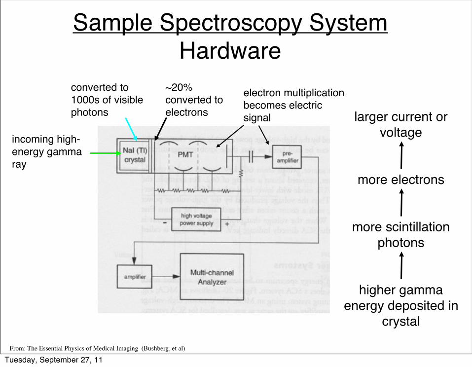
Sample Spectroscopy SystemHardware
From: The Essential Physics of Medical Imaging (Bushberg, et al)
incoming high-energy gamma ray
converted to 1000s of visible photons
~20% converted to electrons
electron multiplicationbecomes electric signal larger current or
voltage
more electrons
more scintillation photons
higher gamma energy deposited in
crystal
Tuesday, September 27, 11

Silicon Photomultipliers(Geiger-mode APDs)
Fall 2011, Copyright UW Imaging Research Laboratory
Tuesday, September 27, 11

GM-APDs Arrays at the UW
Zecotek PhotonicsType 3-N 8x8 array with 3.3 mm square elements
SensL 4x4 array with 3 mm square elements
Fall 2011, Copyright UW Imaging Research Laboratory
Tuesday, September 27, 11

Interactions of Photons with a Spectrometer
A. PhotoelectricB. Compton + PhotoelectricC. ComptonD. Photoelectric with characteristic
x-ray escapeE. Compton scattered photon from
lead shieldF. Characteristic x-ray from lead
shield
From: The Essential Physics of Medical Imaging (Bushberg, et al)
Tuesday, September 27, 11

Sample Spectroscopy SystemOutput
From: Physics in Nuclear Medicine (Sorenson and Phelps)
From: The Essential Physics of Medical Imaging (Bushberg, et al)
counting mode
Ideal Energy Spectrum
Remember - you are looking at the energy deposited in the detector!
Tuesday, September 27, 11

Energy Resolution
From: Physics in Nuclear Medicine (Sorenson and Phelps)
Realistic Energy Spectrum
Tuesday, September 27, 11
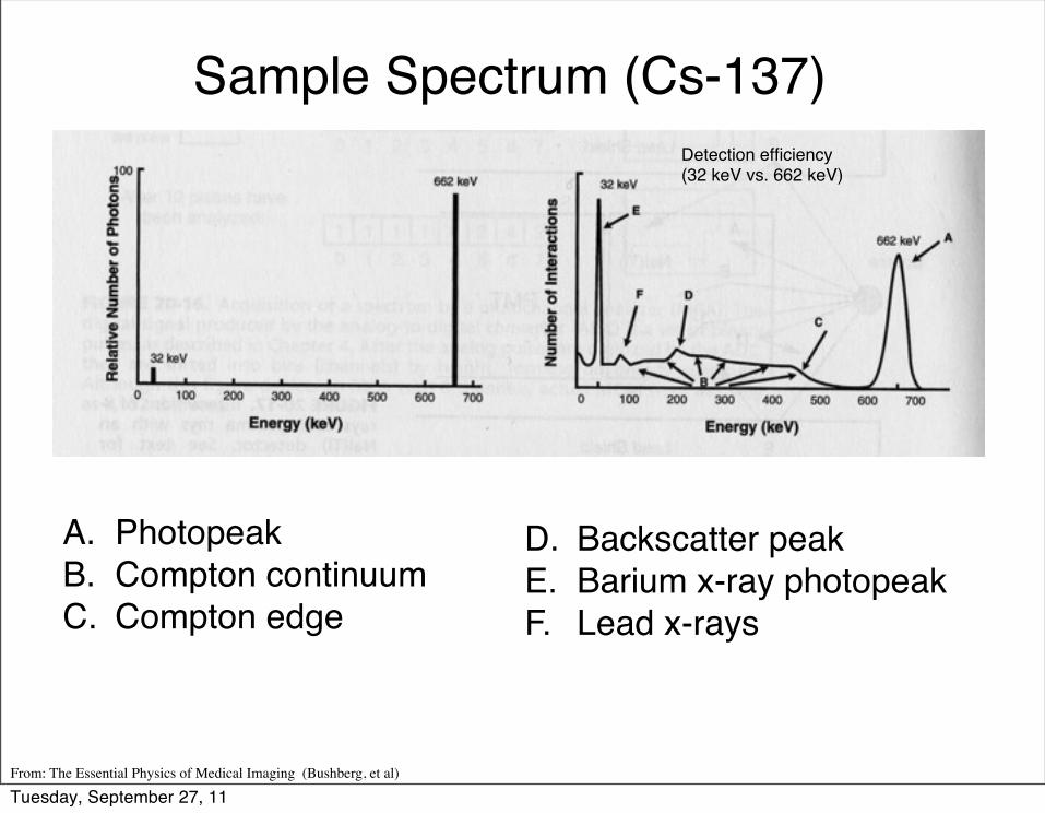
Sample Spectrum (Cs-137)
A. PhotopeakB. Compton continuumC. Compton edge
D.! Backscatter peakE.! Barium x-ray photopeakF.! Lead x-rays
Detection efficiency (32 keV vs. 662 keV)
From: The Essential Physics of Medical Imaging (Bushberg, et al)
Tuesday, September 27, 11

Sample Spectrum (Tc-99m)
A. PhotopeakB. Photoelectric with
iodine K-shell x-ray escape
C. Absorption of lead x-rays from shield
From: The Essential Physics of Medical Imaging (Bushberg, et al)
Tuesday, September 27, 11

Sample Spectrum (In-111)
source detector
From: Physics in Nuclear Medicine (Sorenson and Phelps)
Tuesday, September 27, 11

Effects of Pulse Pileup (count rate)
From: Physics in Nuclear Medicine (Sorenson and Phelps)
Tuesday, September 27, 11
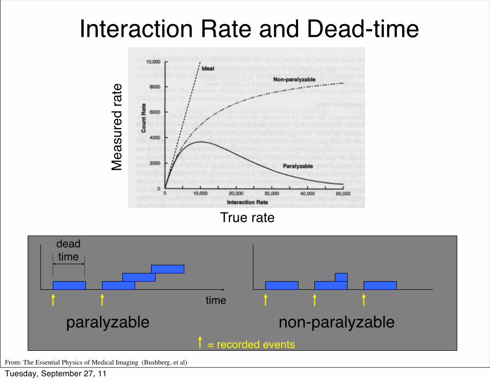
Interaction Rate and Dead-time
paralyzable non-paralyzable
From: The Essential Physics of Medical Imaging (Bushberg, et al)
True rate
Mea
sure
d ra
te
time
deadtime
= recorded events
Tuesday, September 27, 11

Calibrations• Energy calibration (imaging systems/spectroscopy)
– Adjust energy windows around a known photopeak– Often done with long-lives isotopes for convenience ! Cs-137:
Eγ= 662 keV (close to PET 511 keV), T1/2=30yr! Co-57: Eγ= 122 keV (close to Tc99m 140 keV ), T1/2=272d
• Dose calibration (dose calibrator)– Measure activity of know reference samples (e.g., Cs-137 and
Co-57)– Linearity measured by repeated measurements of a decaying
source (e.g., Tc-99m)
Tuesday, September 27, 11

Raphex Question
D58. The window setting used for Tc-99m is set with the center at 140 keV with a width of +/-14 keV i.e., 20%. The reason for this is: A. The energy spread is a consequence of the statistical broadening when
amplifying the initial energy deposition event in the NaI(Tl) crystal. B. The 140 keV gamma ray emission of Tc-99m is not truly monoenergetic but
the center of a spectrum of emissions. C. The higher and lower Gaussian tails are a consequence of compton scattering
within the patient. D. The result of additional scattered photons generated in the collimator. E. A consequence of patient motion during scanning.
Tuesday, September 27, 11

Raphex Answer
D58. The window setting used for Tc-99m is set with the center at 140 keV with a width of +/-14 keV i.e., 20%. The reason for this is: A . Photons, which impinge upon the crystal, lose energy by Compton
scattering and the photoelectric effect. Both processes convert the gamma ray energy into electron energy. On average approximately one electron hole pair is produced per 30 eV of g amma ray e nergy deposited in the crystal. These electrons result in the release of visible ligh t when trapped in the crystal. These light quanta are collected and amplified by photomultiplier tubes. The statistical fluctuation in the number of light quanta collected and their amplification is what causes the spread in the detected energy peak, even when most of the Tc-99m photons deposit exactly 140 keV in the NaI(Tl) crystal.
Tuesday, September 27, 11

The count rate for a 1 µCi source is measured as 25 kcps by a well counter. Assuming no corrections are applied, the measured count rate for a 10 µCi source will be:
a. 250 kcpsb. Less than 250 kcpsc. Greater than 250 kcps
Because of deadtime effects
Question
Fall 2011, Copyright UW Imaging Research Laboratory
Tuesday, September 27, 11

How many peaks would you expect for a99m-Tc sample placed outside a well counter?
1
1
At higher doses you will get distortion of the photopeak and a high end tail on the energy spectra due to pileup. See slide 31 from lecture.
What about inside a well counter?
Is your answer dose dependent?
Question
Fall 2011, Copyright UW Imaging Research Laboratory
Tuesday, September 27, 11

How many peaks would you expect for a 68-Ge sample placed outside a well counter? What about inside a well counter?
Outside, 1Inside, 2
68-Ge is a positron emitter (e.g., PET)
Question
Fall 2011, Copyright UW Imaging Research Laboratory
Tuesday, September 27, 11

Of the following, the most efficient detector for x-rays is:
From: The Essential Physics of Medical Imaging (Bushberg, et al)
NaI(Tl) is an inorganic scintillator and is much more efficient at detecting x-rays
than gas filled detectors.
a. Geiger counterb. NaI(Tl) detectorc. Single channel analyzerd. Ionization chambere. Pocket (self-reading) dosimeter
Question
Fall 2011, Copyright UW Imaging Research Laboratory
Tuesday, September 27, 11

Gas multiplication occurs in:
From: The Essential Physics of Medical Imaging (Bushberg, et al)
a. Geiger-Mueller countersb. Scintillation detectorsc. Semiconductor detectorsd. Ionization chamberse. Dose calibrators
Question
Fall 2011, Copyright UW Imaging Research Laboratory
Tuesday, September 27, 11

In a pho tomu l t i p l i e r t ube , t he photocathode is at a positive voltage with respect to the first dynode.
From: The Essential Physics of Medical Imaging (Bushberg, et al)
False
False
Small changes to the voltage applied to an ionization chamber have a large effect upon the charge collected from each interaction with ionizing radiation.
Question (True or False)
Fall 2011, Copyright UW Imaging Research Laboratory
Tuesday, September 27, 11

A 1 MeV beta particle produces a pulse of the same amplitude in a G-M detector as a 200 keV beta particle.
From: The Essential Physics of Medical Imaging (Bushberg, et al)
True
Question (True or False)
Fall 2011, Copyright UW Imaging Research Laboratory
Tuesday, September 27, 11

Which detector system is most appropriate and accurate for the measurement of a pure beta source:
From: Raphex
a. Ionization chamberb. Geiger Muller tubec. NaI(Tl) well scintillation counterd. Thermoluminescent dosimetere. Liquid scintillation counter
Question
Fall 2011, Copyright UW Imaging Research Laboratory
Tuesday, September 27, 11

a. Identify the energy of a radionuclideb. Reject Compton scattered photonsc. Separate a mixture of radionuclidesd. Alter the sensitivity or resolution of the
systeme. All of the above
From: Raphex
A pulse height analyzer (PHA) window can be used to:
Question
Fall 2011, Copyright UW Imaging Research Laboratory
Tuesday, September 27, 11
Related Documents


