rac-1-Acetyl-5-benzyl-2-thioxoimidazo- lidin-4-one Mary C. Uzca ´tegui, a Gerzon E. Delgado, a * Asiloe ´ J. Mora, a Teresa Gonza ´lez b and Alexander Bricen ˜o b a Laboratorio de Cristalografı ´a, Departamento de Quı ´mica, Facultad de Ciencias, Universidad de Los Andes, Me ´rida 5101, Venezuela, and b Centro de Quı ´mica, Instituto Venezolano de Investigaciones, Cientı ´ficas (IVIC), Apartado 21827, Caracas 1020-A, Venezuela Correspondence e-mail: [email protected] Received 1 December 2008; accepted 9 December 2008 Key indicators: single-crystal X-ray study; T = 298 K; mean (C–C) = 0.004 A ˚ ; R factor = 0.045; wR factor = 0.124; data-to-parameter ratio = 15.1. In the title compound, C 12 H 12 N 2 O 2 S, the molecules have a wing-like conformation, with a distance of 3.797 (2) A ˚ between the centroids of the five- and six-membered rings. In the crystal structure, molecules are linked by N—HO hydrogen bonds, forming infinite one-dimensional zigzag chains, running along [001], with a C(4) graph-set motif. Related literature For related compounds, see: Seijas et al. (2006, 2007); Delgado et al. (2007); Sulbaran et al. (2007). For racemization of amino acids, see: Yamada et al. (1983); Yoshioka (2007). For refer- ence structural data, see: Allen et al. (2002). For hydrogen- bond motifs in graph-set notation, see Etter (1990). Experimental Crystal data C 12 H 12 N 2 O 2 S M r = 248.30 Monoclinic, P2 1 =c a = 11.696 (5) A ˚ b = 13.479 (6) A ˚ c = 7.767 (4) A ˚ = 94.41 (1) V = 1220.8 (9) A ˚ 3 Z =4 Mo K radiation = 0.26 mm 1 T = 298 (2) K 0.4 0.3 0.2 mm Data collection Rigaku AFC-7S Mercury diffractometer Absorption correction: multi-scan (Jacobson, 1998) T min = 0.900, T max = 0.950 12945 measured reflections 2349 independent reflections 2065 reflections with I >2(I) R int = 0.026 Refinement R[F 2 >2(F 2 )] = 0.045 wR(F 2 ) = 0.124 S = 1.05 2349 reflections 156 parameters H-atom parameters constrained Á max = 0.24 e A ˚ 3 Á min = 0.27 e A ˚ 3 Table 1 Hydrogen-bond geometry (A ˚ , ). D—HA D—H HA DA D—HA N3—H3O4 i 0.86 1.98 2.834 (2) 175 Symmetry code: (i) x; y þ 1 2 ; z þ 1 2 . Data collection: CrystalClear (Rigaku, 2002); cell refinement: CrystalClear; data reduction: CrystalStructure (Rigaku/MSC, 2004); program(s) used to solve structure: SHELXS97 (Sheldrick, 2008); program(s) used to refine structure: SHELXL97 (Sheldrick, 2008); molecular graphics: DIAMOND (Brandenburg, 1999); software used to prepare material for publication: PLATON (Spek, 2003) and publCIF (Westrip, 2009). This work was supported by Consejo de Desarrollo Cien- tı ´fico, Humanı´stico y Tecnolo ´ gico de la Universidad de Los Andes, CDCHT-ULA (grants C-1616–08-A and C-1617–08-F) and Fondo Nacional de Ciencia, Tecnologı´a e Innovacio ´ n, FONACIT (grant LAB-97000821). Supplementary data and figures for this paper are available from the IUCr electronic archives (Reference: CV2495). References Allen, F. H. (2002). Acta Cryst. B58, 380–388. Brandenburg, K. (1999). DIAMOND. Crystal Impact GbR, Bonn, Germany. Delgado, G. E., Mora, A. J., Uzca ´tegui, J., Bahsas, A. & Bricen ˜ o, A. (2007). Acta Cryst. C63, o448–o450. Etter, M. C. (1990). Acc. Chem. Res. 23, 120–126. Jacobson, R. (1998). Private communication to Rigaku Corporation, Tokyo, Japan. Rigaku (2002). CrystalClear. Rigaku Corporation, Tokyo, Japan. Rigaku/MSC (2004). CrystalStructure. Rigaku/MSC, The Woodlands, Texas, USA. Seijas, L. E., Delgado, G. E., Mora, A. J., Bahsas, A. & Bricen ˜ o, A. (2007). Acta Cryst. C63, o303–o305. Seijas, L. E., Delgado, G. E., Mora, A. J., Bahsas, A. & Uzca ´tegui, J. (2006). Av. Quı ´m. 1, 3–7. Sheldrick, G. M. (2008). Acta Cryst. A64, 112–122. Spek, A. L. (2003). J. Appl. Cryst. 36, 7–13. Sulbaran, M. E., Delgado, G. E., Mora, A. J., Bahsas, A., Novoa de Armas, H. & Blaton, N. (2007). Acta Cryst. C63, o543–o545. Westrip, S. P. (2009). publCIF. In preparation. Yamada, S., Hongo, C., Yoshioka, R. & Chibata, I. (1983). J. Org. Chem. 48, 843–846. Yoshioka, R. (2007). Top. Curr. Chem. 269, 83–132. organic compounds o104 Uzca ´tegui et al. doi:10.1107/S1600536808041883 Acta Cryst. (2009). E65, o104 Acta Crystallographica Section E Structure Reports Online ISSN 1600-5368

Welcome message from author
This document is posted to help you gain knowledge. Please leave a comment to let me know what you think about it! Share it to your friends and learn new things together.
Transcript
rac-1-Acetyl-5-benzyl-2-thioxoimidazo-lidin-4-one
Mary C. Uzcategui,a Gerzon E. Delgado,a* Asiloe J.
Mora,a Teresa Gonzalezb and Alexander Bricenob
aLaboratorio de Cristalografıa, Departamento de Quımica, Facultad de Ciencias,
Universidad de Los Andes, Merida 5101, Venezuela, and bCentro de Quımica,
Instituto Venezolano de Investigaciones, Cientıficas (IVIC), Apartado 21827, Caracas
1020-A, Venezuela
Correspondence e-mail: [email protected]
Received 1 December 2008; accepted 9 December 2008
Key indicators: single-crystal X-ray study; T = 298 K; mean �(C–C) = 0.004 A;
R factor = 0.045; wR factor = 0.124; data-to-parameter ratio = 15.1.
In the title compound, C12H12N2O2S, the molecules have a
wing-like conformation, with a distance of 3.797 (2) A
between the centroids of the five- and six-membered rings.
In the crystal structure, molecules are linked by N—H� � �O
hydrogen bonds, forming infinite one-dimensional zigzag
chains, running along [001], with a C(4) graph-set motif.
Related literature
For related compounds, see: Seijas et al. (2006, 2007); Delgado
et al. (2007); Sulbaran et al. (2007). For racemization of amino
acids, see: Yamada et al. (1983); Yoshioka (2007). For refer-
ence structural data, see: Allen et al. (2002). For hydrogen-
bond motifs in graph-set notation, see Etter (1990).
Experimental
Crystal data
C12H12N2O2SMr = 248.30Monoclinic, P21=ca = 11.696 (5) Ab = 13.479 (6) Ac = 7.767 (4) A� = 94.41 (1)�
V = 1220.8 (9) A3
Z = 4Mo K� radiation� = 0.26 mm�1
T = 298 (2) K0.4 � 0.3 � 0.2 mm
Data collection
Rigaku AFC-7S Mercurydiffractometer
Absorption correction: multi-scan(Jacobson, 1998)Tmin = 0.900, Tmax = 0.950
12945 measured reflections2349 independent reflections2065 reflections with I > 2�(I)Rint = 0.026
Refinement
R[F 2 > 2�(F 2)] = 0.045wR(F 2) = 0.124S = 1.052349 reflections
156 parametersH-atom parameters constrained��max = 0.24 e A�3
��min = �0.27 e A�3
Table 1Hydrogen-bond geometry (A, �).
D—H� � �A D—H H� � �A D� � �A D—H� � �A
N3—H3� � �O4i 0.86 1.98 2.834 (2) 175
Symmetry code: (i) x;�yþ 12; zþ 1
2.
Data collection: CrystalClear (Rigaku, 2002); cell refinement:
CrystalClear; data reduction: CrystalStructure (Rigaku/MSC, 2004);
program(s) used to solve structure: SHELXS97 (Sheldrick, 2008);
program(s) used to refine structure: SHELXL97 (Sheldrick, 2008);
molecular graphics: DIAMOND (Brandenburg, 1999); software used
to prepare material for publication: PLATON (Spek, 2003) and
publCIF (Westrip, 2009).
This work was supported by Consejo de Desarrollo Cien-
tıfico, Humanıstico y Tecnologico de la Universidad de Los
Andes, CDCHT-ULA (grants C-1616–08-A and C-1617–08-F)
and Fondo Nacional de Ciencia, Tecnologıa e Innovacion,
FONACIT (grant LAB-97000821).
Supplementary data and figures for this paper are available from theIUCr electronic archives (Reference: CV2495).
References
Allen, F. H. (2002). Acta Cryst. B58, 380–388.Brandenburg, K. (1999). DIAMOND. Crystal Impact GbR, Bonn, Germany.Delgado, G. E., Mora, A. J., Uzcategui, J., Bahsas, A. & Briceno, A. (2007).
Acta Cryst. C63, o448–o450.Etter, M. C. (1990). Acc. Chem. Res. 23, 120–126.Jacobson, R. (1998). Private communication to Rigaku Corporation, Tokyo,
Japan.Rigaku (2002). CrystalClear. Rigaku Corporation, Tokyo, Japan.Rigaku/MSC (2004). CrystalStructure. Rigaku/MSC, The Woodlands, Texas,
USA.Seijas, L. E., Delgado, G. E., Mora, A. J., Bahsas, A. & Briceno, A. (2007). Acta
Cryst. C63, o303–o305.Seijas, L. E., Delgado, G. E., Mora, A. J., Bahsas, A. & Uzcategui, J. (2006). Av.
Quım. 1, 3–7.Sheldrick, G. M. (2008). Acta Cryst. A64, 112–122.Spek, A. L. (2003). J. Appl. Cryst. 36, 7–13.Sulbaran, M. E., Delgado, G. E., Mora, A. J., Bahsas, A., Novoa de Armas, H.
& Blaton, N. (2007). Acta Cryst. C63, o543–o545.Westrip, S. P. (2009). publCIF. In preparation.Yamada, S., Hongo, C., Yoshioka, R. & Chibata, I. (1983). J. Org. Chem. 48,
843–846.Yoshioka, R. (2007). Top. Curr. Chem. 269, 83–132.
organic compounds
o104 Uzcategui et al. doi:10.1107/S1600536808041883 Acta Cryst. (2009). E65, o104
Acta Crystallographica Section E
Structure ReportsOnline
ISSN 1600-5368
supplementary materials
supplementary materials
sup-1
Acta Cryst. (2009). E65, o104 [ doi:10.1107/S1600536808041883 ]
rac-1-Acetyl-5-benzyl-2-thioxoimidazolidin-4-one
M. C. Uzcátegui, G. E. Delgado, A. J. Mora, T. González and A. Briceño
Comment
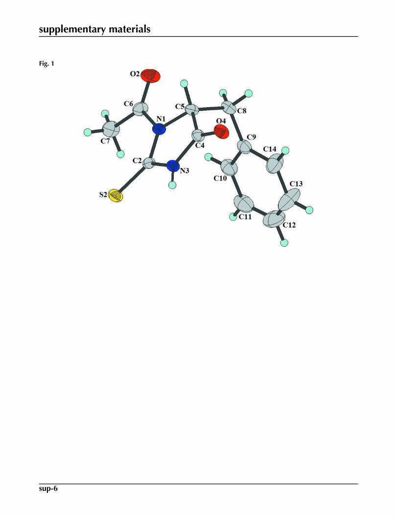
In continuation of our study of N-carbamoyl, hydantoin and thiohydantoin derivatives of α-amino acids (Seijas et al., 2006,2007; Delgado et al., 2007; Sulbaran et al., 2007), we report here the structure of the title compound (I) - the N-acetyl-thiohydantoin derivative of the α-amino acid L-phenylalanine.
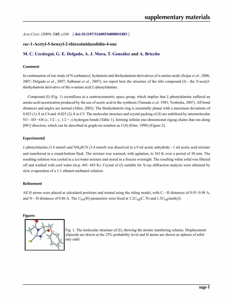
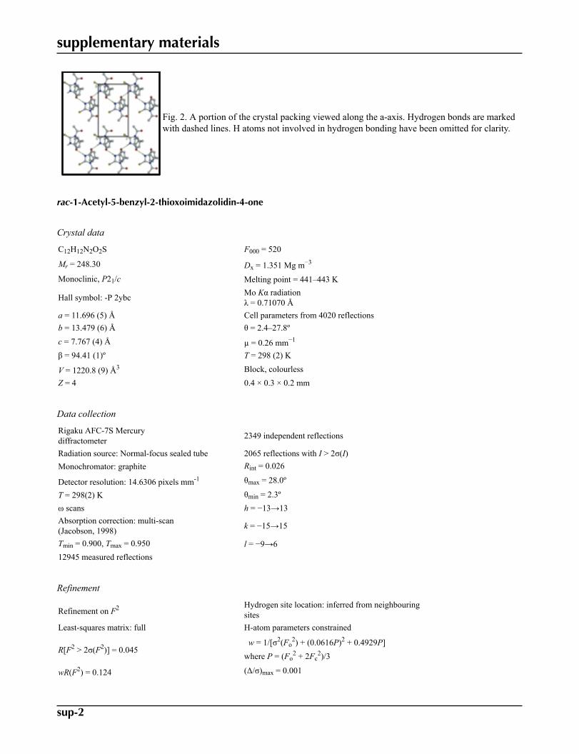
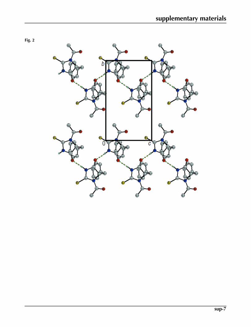
Compound (I) (Fig. 1) crystallizes in a centrosymmetric space group, which implies that L-phenylalanine suffered anamino acid racemization produced by the use of acetic acid in the synthesis (Yamada et al. 1983; Yoshioka, 2007). All bonddistances and angles are normal (Allen, 2002). The thiohydantoin ring is essentially planar with a maximum deviations of0.023 (1) Å in C4 and -0.025 (2) Å in C5. The molecular structure and crystal packing of (I) are stabilized by intermolecularN3—H3···O4 (x, 1/2 - y, 1/2 + z) hydrogen bonds (Table 1), forming infinite one-dimentional zigzag chains that run along[001] direction, which can be described in graph-set notation as C(4) (Etter, 1990) (Figure 2).
Experimental
L-phenylalanine (3.4 mmol) and NH4SCN (3.4 mmol) was dissolved in a 9 ml acetic anhydride - 1 ml acetic acid mixture
and transferred in a round-bottom flask. The mixture was warmed, with agitation, to 363 K over a period of 30 min. Theresulting solution was cooled in a ice/water mixture and stored in a freezer overnight. The resulting white solid was filteredoff and washed with cool water (m.p. 441–443 K). Crystal of (I) suitable for X-ray diffraction analysis were obtained byslow evaporation of a 1:1 ethanol-methanol solution.
Refinement
All H atoms were placed at calculated positions and treated using the riding model, with C—H distances of 0.93–0.98 A,and N—H distances of 0.86 A. The Uiso(H) parameters were fixed at 1.2Ueq(C, N) and 1.5Ueq(methyl).
Figures
Fig. 1. The molecular structure of (I), showing the atomic numbering scheme. Displacementelipsoids are drawn at the 25% probability level and H atoms are shown as spheres of arbit-rary radii.
supplementary materials
sup-2
Fig. 2. A portion of the crystal packing viewed along the a-axis. Hydrogen bonds are markedwith dashed lines. H atoms not involved in hydrogen bonding have been omitted for clarity.
rac-1-Acetyl-5-benzyl-2-thioxoimidazolidin-4-one
Crystal data
C12H12N2O2S F000 = 520
Mr = 248.30 Dx = 1.351 Mg m−3
Monoclinic, P21/c Melting point = 441–443 K
Hall symbol: -P 2ybc Mo Kα radiationλ = 0.71070 Å
a = 11.696 (5) Å Cell parameters from 4020 reflectionsb = 13.479 (6) Å θ = 2.4–27.8ºc = 7.767 (4) Å µ = 0.26 mm−1
β = 94.41 (1)º T = 298 (2) K
V = 1220.8 (9) Å3 Block, colourlessZ = 4 0.4 × 0.3 × 0.2 mm
Data collection
Rigaku AFC-7S Mercurydiffractometer 2349 independent reflections
Radiation source: Normal-focus sealed tube 2065 reflections with I > 2σ(I)Monochromator: graphite Rint = 0.026
Detector resolution: 14.6306 pixels mm-1 θmax = 28.0º
T = 298(2) K θmin = 2.3ºω scans h = −13→13Absorption correction: multi-scan(Jacobson, 1998) k = −15→15
Tmin = 0.900, Tmax = 0.950 l = −9→612945 measured reflections
Refinement
Refinement on F2 Hydrogen site location: inferred from neighbouringsites
Least-squares matrix: full H-atom parameters constrained
R[F2 > 2σ(F2)] = 0.045 w = 1/[σ2(Fo
2) + (0.0616P)2 + 0.4929P]where P = (Fo
2 + 2Fc2)/3
wR(F2) = 0.124 (Δ/σ)max = 0.001
supplementary materials
sup-3
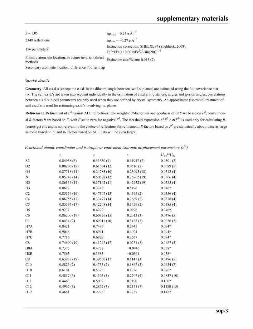
S = 1.05 Δρmax = 0.24 e Å−3
2349 reflections Δρmin = −0.27 e Å−3
156 parametersExtinction correction: SHELXL97 (Sheldrick, 2008),Fc*=kFc[1+0.001xFc2λ3/sin(2θ)]-1/4
Primary atom site location: structure-invariant directmethods Extinction coefficient: 0.013 (2)
Secondary atom site location: difference Fourier map
Special details
Geometry. All e.s.d.'s (except the e.s.d. in the dihedral angle between two l.s. planes) are estimated using the full covariance mat-rix. The cell e.s.d.'s are taken into account individually in the estimation of e.s.d.'s in distances, angles and torsion angles; correlationsbetween e.s.d.'s in cell parameters are only used when they are defined by crystal symmetry. An approximate (isotropic) treatment ofcell e.s.d.'s is used for estimating e.s.d.'s involving l.s. planes.
Refinement. Refinement of F2 against ALL reflections. The weighted R-factor wR and goodness of fit S are based on F2, convention-
al R-factors R are based on F, with F set to zero for negative F2. The threshold expression of F2 > σ(F2) is used only for calculating R-
factors(gt) etc. and is not relevant to the choice of reflections for refinement. R-factors based on F2 are statistically about twice as largeas those based on F, and R- factors based on ALL data will be even larger.
Fractional atomic coordinates and isotropic or equivalent isotropic displacement parameters (Å2)
x y z Uiso*/Ueq
S2 0.84958 (5) 0.53330 (4) 0.61947 (7) 0.0501 (2)O2 0.88296 (18) 0.61004 (12) 0.0516 (2) 0.0689 (5)O4 0.87718 (14) 0.24785 (10) 0.23095 (18) 0.0512 (4)N1 0.85248 (14) 0.50588 (12) 0.26762 (19) 0.0366 (4)N3 0.86134 (14) 0.37142 (11) 0.42952 (19) 0.0383 (4)H3 0.8622 0.3343 0.5196 0.046*C2 0.85359 (16) 0.47307 (13) 0.4365 (2) 0.0356 (4)C4 0.86755 (17) 0.33477 (14) 0.2669 (2) 0.0378 (4)C5 0.85594 (17) 0.42208 (14) 0.1459 (2) 0.0385 (4)H5 0.9237 0.4272 0.0796 0.046*C6 0.86200 (19) 0.60326 (15) 0.2013 (3) 0.0476 (5)C7 0.8434 (2) 0.69011 (16) 0.3128 (3) 0.0626 (7)H7A 0.8421 0.7495 0.2445 0.094*H7B 0.9046 0.6941 0.4024 0.094*H7C 0.7716 0.6829 0.3637 0.094*C8 0.74690 (19) 0.41292 (17) 0.0231 (3) 0.0487 (5)H8A 0.7375 0.4732 −0.0446 0.058*H8B 0.7565 0.3585 −0.0561 0.058*C9 0.63988 (19) 0.39550 (17) 0.1147 (3) 0.0496 (5)C10 0.5823 (2) 0.4733 (2) 0.1867 (3) 0.0634 (7)H10 0.6101 0.5376 0.1786 0.076*C11 0.4837 (3) 0.4563 (3) 0.2707 (4) 0.0837 (10)H11 0.4463 0.5092 0.3190 0.100*C12 0.4967 (3) 0.2862 (3) 0.2141 (7) 0.1180 (15)H12 0.4681 0.2223 0.2237 0.142*
supplementary materials
sup-4
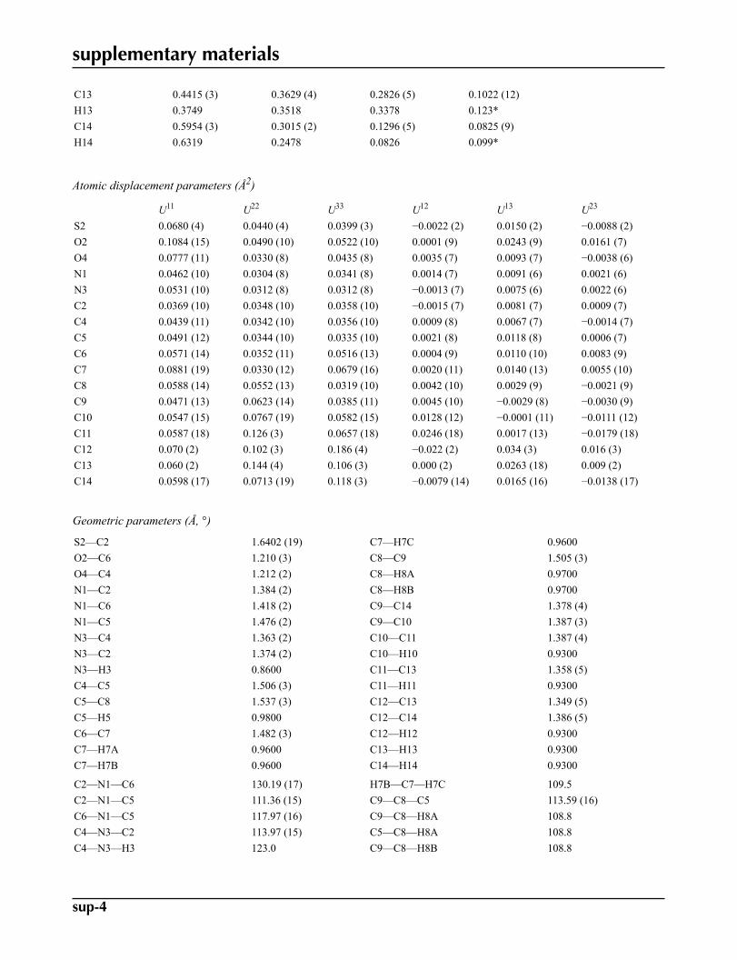
C13 0.4415 (3) 0.3629 (4) 0.2826 (5) 0.1022 (12)H13 0.3749 0.3518 0.3378 0.123*C14 0.5954 (3) 0.3015 (2) 0.1296 (5) 0.0825 (9)H14 0.6319 0.2478 0.0826 0.099*
Atomic displacement parameters (Å2)
U11 U22 U33 U12 U13 U23
S2 0.0680 (4) 0.0440 (4) 0.0399 (3) −0.0022 (2) 0.0150 (2) −0.0088 (2)O2 0.1084 (15) 0.0490 (10) 0.0522 (10) 0.0001 (9) 0.0243 (9) 0.0161 (7)O4 0.0777 (11) 0.0330 (8) 0.0435 (8) 0.0035 (7) 0.0093 (7) −0.0038 (6)N1 0.0462 (10) 0.0304 (8) 0.0341 (8) 0.0014 (7) 0.0091 (6) 0.0021 (6)N3 0.0531 (10) 0.0312 (8) 0.0312 (8) −0.0013 (7) 0.0075 (6) 0.0022 (6)C2 0.0369 (10) 0.0348 (10) 0.0358 (10) −0.0015 (7) 0.0081 (7) 0.0009 (7)C4 0.0439 (11) 0.0342 (10) 0.0356 (10) 0.0009 (8) 0.0067 (7) −0.0014 (7)C5 0.0491 (12) 0.0344 (10) 0.0335 (10) 0.0021 (8) 0.0118 (8) 0.0006 (7)C6 0.0571 (14) 0.0352 (11) 0.0516 (13) 0.0004 (9) 0.0110 (10) 0.0083 (9)C7 0.0881 (19) 0.0330 (12) 0.0679 (16) 0.0020 (11) 0.0140 (13) 0.0055 (10)C8 0.0588 (14) 0.0552 (13) 0.0319 (10) 0.0042 (10) 0.0029 (9) −0.0021 (9)C9 0.0471 (13) 0.0623 (14) 0.0385 (11) 0.0045 (10) −0.0029 (8) −0.0030 (9)C10 0.0547 (15) 0.0767 (19) 0.0582 (15) 0.0128 (12) −0.0001 (11) −0.0111 (12)C11 0.0587 (18) 0.126 (3) 0.0657 (18) 0.0246 (18) 0.0017 (13) −0.0179 (18)C12 0.070 (2) 0.102 (3) 0.186 (4) −0.022 (2) 0.034 (3) 0.016 (3)C13 0.060 (2) 0.144 (4) 0.106 (3) 0.000 (2) 0.0263 (18) 0.009 (2)C14 0.0598 (17) 0.0713 (19) 0.118 (3) −0.0079 (14) 0.0165 (16) −0.0138 (17)
Geometric parameters (Å, °)
S2—C2 1.6402 (19) C7—H7C 0.9600O2—C6 1.210 (3) C8—C9 1.505 (3)O4—C4 1.212 (2) C8—H8A 0.9700N1—C2 1.384 (2) C8—H8B 0.9700N1—C6 1.418 (2) C9—C14 1.378 (4)N1—C5 1.476 (2) C9—C10 1.387 (3)N3—C4 1.363 (2) C10—C11 1.387 (4)N3—C2 1.374 (2) C10—H10 0.9300N3—H3 0.8600 C11—C13 1.358 (5)C4—C5 1.506 (3) C11—H11 0.9300C5—C8 1.537 (3) C12—C13 1.349 (5)C5—H5 0.9800 C12—C14 1.386 (5)C6—C7 1.482 (3) C12—H12 0.9300C7—H7A 0.9600 C13—H13 0.9300C7—H7B 0.9600 C14—H14 0.9300
C2—N1—C6 130.19 (17) H7B—C7—H7C 109.5C2—N1—C5 111.36 (15) C9—C8—C5 113.59 (16)C6—N1—C5 117.97 (16) C9—C8—H8A 108.8C4—N3—C2 113.97 (15) C5—C8—H8A 108.8C4—N3—H3 123.0 C9—C8—H8B 108.8
supplementary materials
sup-5
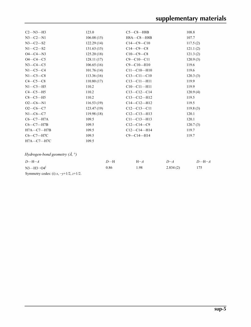
C2—N3—H3 123.0 C5—C8—H8B 108.8N3—C2—N1 106.08 (15) H8A—C8—H8B 107.7N3—C2—S2 122.29 (14) C14—C9—C10 117.5 (2)N1—C2—S2 131.63 (15) C14—C9—C8 121.1 (2)O4—C4—N3 125.20 (18) C10—C9—C8 121.3 (2)O4—C4—C5 128.11 (17) C9—C10—C11 120.9 (3)N3—C4—C5 106.65 (16) C9—C10—H10 119.6N1—C5—C4 101.76 (14) C11—C10—H10 119.6N1—C5—C8 113.36 (16) C13—C11—C10 120.3 (3)C4—C5—C8 110.80 (17) C13—C11—H11 119.9N1—C5—H5 110.2 C10—C11—H11 119.9C4—C5—H5 110.2 C13—C12—C14 120.9 (4)C8—C5—H5 110.2 C13—C12—H12 119.5O2—C6—N1 116.53 (19) C14—C12—H12 119.5O2—C6—C7 123.47 (19) C12—C13—C11 119.8 (3)N1—C6—C7 119.98 (18) C12—C13—H13 120.1C6—C7—H7A 109.5 C11—C13—H13 120.1C6—C7—H7B 109.5 C12—C14—C9 120.7 (3)H7A—C7—H7B 109.5 C12—C14—H14 119.7C6—C7—H7C 109.5 C9—C14—H14 119.7H7A—C7—H7C 109.5
Hydrogen-bond geometry (Å, °)
D—H···A D—H H···A D···A D—H···A
N3—H3···O4i 0.86 1.98 2.834 (2) 175Symmetry codes: (i) x, −y+1/2, z+1/2.
supplementary materials
sup-6
Fig. 1
supplementary materials
sup-7
Fig. 2
Related Documents


![Index [ ] · PDF fileacetylation 962–964 – one-pot 981 ... benzyl alcohol 822–823, 1177 benzylamines 155 – substituted 828 benzyl chloride 253](https://static.cupdf.com/doc/110x72/5a85803e7f8b9a14748c25ad/index-962964-one-pot-981-benzyl-alcohol-822823-1177-benzylamines.jpg)


