Rhabdoviruses and reoviruses Rabies and rotavirus www.freelivedoctor.com

Welcome message from author
This document is posted to help you gain knowledge. Please leave a comment to let me know what you think about it! Share it to your friends and learn new things together.
Transcript

Rhabdoviruses and reoviruses
Rabies and rotavirus
www.freelivedoctor.com

Rhabdoviruses
Rabies
www.freelivedoctor.com

Rabies in the US
Distribution of rabies in the US, 2001. (From http://www.cdc.gov/ncidod/dvrd/rabies/Epidemiology/Epidemiology.htm)www.freelivedoctor.com

Human pathogens in the order Mononegavirales
Family Subfamily Genus Human pathogensRhabdoviridae
Lyssavirus Rabies virus
Filoviridae
Marburghvirus Marburgh virus
Ebolavirus Ebola virus
Paramyxoviridae
Paramyxovirinae
Rubulavirus Mumps virus, Parainfluenzavirus 2,4
Respirovirus Parainfluenza virus 1,3
Henipavirus Hendra virus, Nipah virus
Morbillivirus Measles virus
Pneumovirinae
Pneumovirus Respiratory syncytial virus
Metaneumovirus Human metapneumovirus
www.freelivedoctor.com
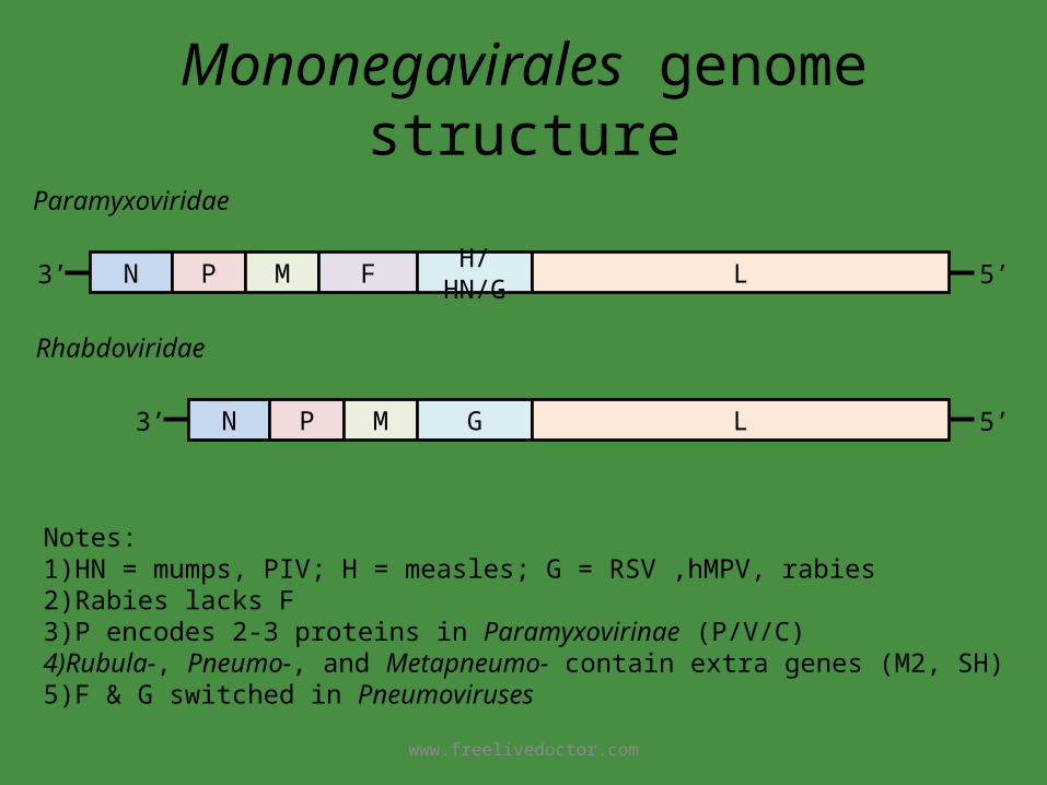
Mononegavirales genome structure
N P M F H/HN/G L3’ 5’
N P M G L3’ 5’
Paramyxoviridae
Rhabdoviridae
Notes:1)HN = mumps, PIV; H = measles; G = RSV ,hMPV, rabies2)Rabies lacks F3)P encodes 2-3 proteins in Paramyxovirinae (P/V/C)4)Rubula-, Pneumo-, and Metapneumo- contain extra genes (M2, SH)5)F & G switched in Pneumoviruses
www.freelivedoctor.com

Rhabdovirus gene functionGene product Virion location Function
Nucleoprotein (N) Nucleocapsid Protects RNA genome
Polymerase phosphoprotein (P)
Associated with nucleocapsid
RNA polymerase subunit
Matrix (M) Between nucleocapsid and envelope
Virion assembly
glycoprotein (G) Transmembrane envelope glycoprotein
Viral attachment protein
Large protein (L) Associated with nucleocapsid
RNA polymerase
www.freelivedoctor.com
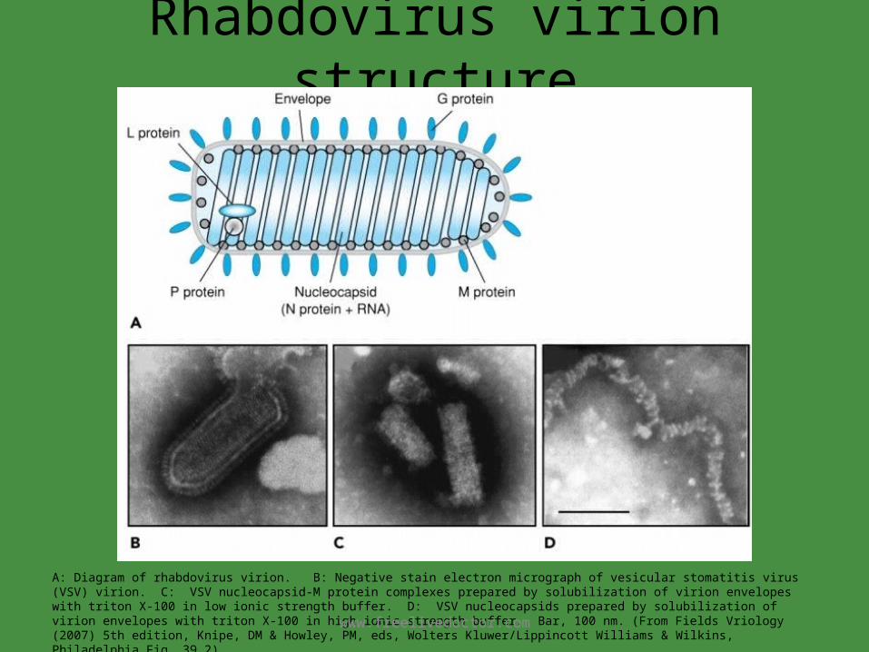
Rhabdovirus virion structure
A: Diagram of rhabdovirus virion. B: Negative stain electron micrograph of vesicular stomatitis virus (VSV) virion. C: VSV nucleocapsid-M protein complexes prepared by solubilization of virion envelopes with triton X-100 in low ionic strength buffer. D: VSV nucleocapsids prepared by solubilization of virion envelopes with triton X-100 in high ionic strength buffer. Bar, 100 nm. (From Fields Vriology (2007) 5th edition, Knipe, DM & Howley, PM, eds, Wolters Kluwer/Lippincott Williams & Wilkins, Philadelphia Fig. 39.2) www.freelivedoctor.com

Rhabdovirus replicationReplication of rhabdoviruses: a simple enveloped (-) RNA virus. 1, Rhabdoviruses bind to the cell surface and are (2) endocytosed. The envelope fuses with the endosome vesicle membrane to deliver the nucleocapsid to the cytoplasm. The virion must carry a polymerase, which (3) produces five individual messenger RNAs (mRNAs) and a full-length (+) RNA template. 4, Proteins are translated from the mRNAs, including one glycoprotein (G), which is co-translationally glycosylated in the endoplasmic reticulum (ER), processed in the Golgi apparatus, and delivered to the cell membrane. 5, The genome is replicated from the (+) RNA template, and N, L, and NS proteins associate with the genome to form the nucleocapsid. 6, The matrix protein associates with the G protein-modified membrane, which is followed by assembly of the nucleocapsid. 7, The virus buds from the cell in a bullet-shaped virion. (From Medical Microbiology, 5th ed., Murray, Rosenthal & Pfaller, Mosby Inc., 2005, Fig. 6-14.)
www.freelivedoctor.com

Rabies distribution
Distribution of animal rabies in the United States, 1999. The percentages relate to the total number of cases of animal rabies. (From Medical Microbiology, 5th ed., Murray, Rosenthal & Pfaller, Mosby Inc., 2005, Fig. 61-3.)
Note: Dogs are the primary source of human rabies in urban settings worldwide.
www.freelivedoctor.com

Rabies pathogenesis
Pathogenesis of rabies virus infection. Numbered steps describe the sequence of events. (From Medical Microbiology, 5th ed., Murray, Rosenthal & Pfaller, Mosby Inc., 2005, Fig. 61-2.)
www.freelivedoctor.com

Rabies disease time course
Progression of rabies disease. (From Medical Microbiology, 5th ed., Murray, Rosenthal & Pfaller, Mosby Inc., 2005, Table 61-1.)
www.freelivedoctor.com
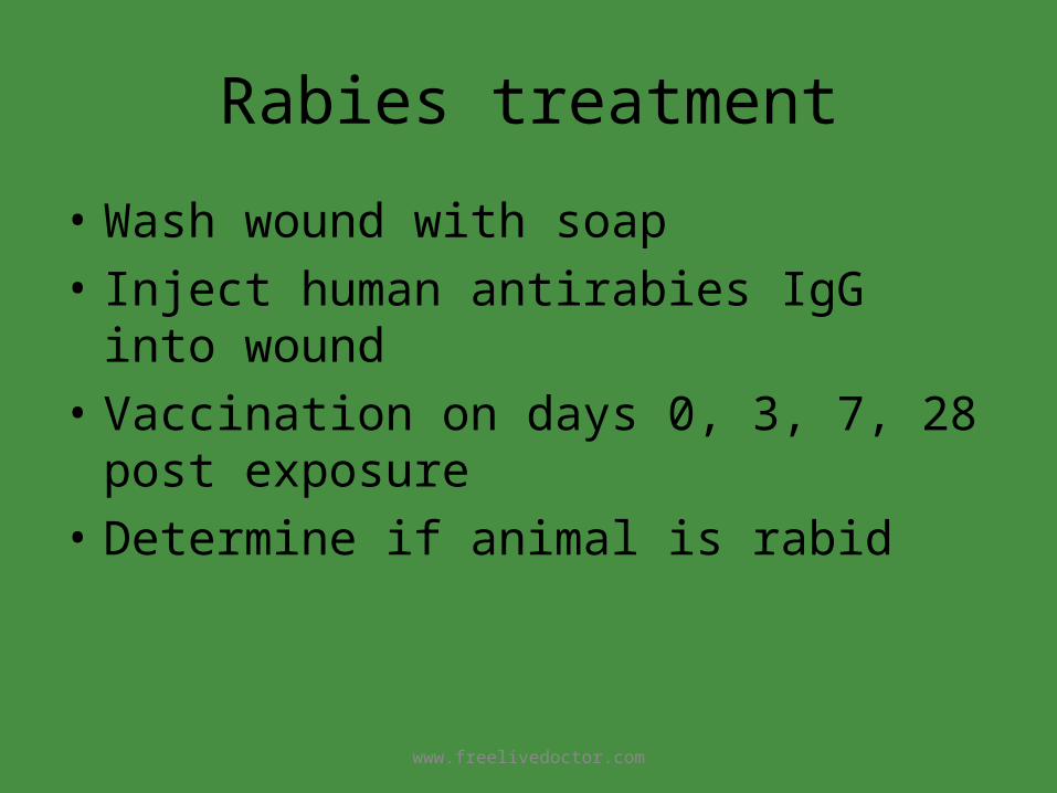
Rabies treatment
• Wash wound with soap• Inject human antirabies IgG into wound• Vaccination on days 0, 3, 7, 28 post exposure• Determine if animal is rabid
www.freelivedoctor.com

Negri bodies
Immunohistochemical staining of intra-cytoplasmic viral inclusions in the neuron of a human rabies patient. (Fields Vriology (2007) 5th edition, Knipe, DM & Howley, PM, eds, Wolters Kluwer/Lippincott Williams & Wilkins, Philadelphia Fig. 39.9)www.freelivedoctor.com

Control of rabies in wild animalsthrough bait based vaccination
The bait Lunch time
http://www.cdc.gov/ncidod/dvrd/rabies/prevention&control/ovalvacc.htm
www.freelivedoctor.com

Rabies summary
• Structure– Negative sense ssRNA, helical nucleocapsid, envelope
• Pathogenesis– Transmitted by bite of rabid animal– Replication in cytoplasm; budding– Spread by axonal transport to brain; long incubation period– Fever, nausea, hydrophobia, coma– Almost always fatal
• Diagnosis– Viral antigen or nucleic acid, Negri bodies
• Treatment/prevention– Inactivated viral vaccine for humans after exposure, live virus
vaccine for animals
www.freelivedoctor.com
Reoviruses
Rotavirus
www.freelivedoctor.com

Rotavirus disease burden
Annual burden of rotavirus disease worldwide and in the United States in infants and young children under 5 years of age. (From Fields Vriology (2007) 5th edition, Knipe, DM & Howley, PM, eds, Wolters Kluwer/Lippincott Williams & Wilkins, Philadelphia Fig. 53.sup8)
www.freelivedoctor.com

Rotavirus disease distribution
Distribution of rotavirus disease-associated deaths worldwide by region. (From Fields Vriology (2007) 5th edition, Knipe, DM & Howley, PM, eds, Wolters Kluwer/Lippincott Williams & Wilkins, Philadelphia Fig. 53.sup9)
www.freelivedoctor.com

Human reoviruses
Reoviridae responsible for human disease (From Medical Microbiology, 5th ed., Murray, Rosenthal & Pfaller, Mosby Inc., 2005, Table 62-1)
www.freelivedoctor.com

Rotavirus structure
Genome Segments: Shows PAGE separation of the 11 segments of the rotavirus SA11 genome. Encoded proteins: Shows the PAGE separation of the intracellular proteins synthesized by SA11 and the genome segment in which they are encoded. Six structural (VP) and six nonstructural (NSP) proteins are synthesized in the infected cell. Note that NSP6 runs far below NSP5 and is not shown on the gel presented here. Viron Schematic: Shows the locations of the various structural proteins within the rotavirus virion. Note the arrangement of the proteins into 3 concentric capsid layers, and the location of the VP1/VP3 complexes at the 5-fold verticies of the icosahedral structure. Reconstruction: A 3D reconstruction of the SA11 virion (23Å resolution) with the genome computationally removed. The color scheme is the same as the schematic, to indicate locations of the various proteins. (http://www.iah.bbsrc.ac.uk/dsRNA_virus_proteins/rotavirus%20figure.htm)
www.freelivedoctor.com

Rotavirus gene function
Functions of rotavirus gene products. (From Medical Microbiology, 5th ed., Murray, Rosenthal & Pfaller, Mosby Inc., 2005, Table 62-3)
www.freelivedoctor.com

Rotavirus structure
Electron micrograph of negatively stained rotavirus virus particles. (From Schaechter’s Mechanisms of Microbial Disease; 4th ed.; Engleberg, DiRita & Dermody; Lippincott, Williams & Wilkins; 2007; Fig. 37-1)
www.freelivedoctor.com

Rotavirus replication
The rotavirus replication cycle . (From Schaechter’s Mechanisms of Microbial Disease; 4th ed.; Engleberg, DiRita & Dermody; Lippincott, Williams & Wilkins; 2007; Fig. 37-2) www.freelivedoctor.com

Rotavirus pathogenesis• Transmission by fecal oral route; fomites
– 1012 particles/ml in stool; infection can result from 10 particles• Major cause of epidemic diarrhea in infants and young children• Highly infectious, 90% of children are seropositive by age 3• Incubation period less than 48 hr• Fever, vomiting, watery diarrhea dehydration
– No blood or leukocytes in stool– Virus replicates in epitheal cells of villi in small intestine– Damage to epithelium major cause of diarrhea– One virus gene product is an enterotoxin– Causes loss of electrolytes and prevents readsorption of water
• Self limiting; can be fatal in malnourished or dehydrated children• Long term immunity
www.freelivedoctor.com

Rotavirus vaccines
• First live vaccine (bovine and monkey viruses, 1999)– Withdrawn from market because of
intussusception
• New live, oral vaccine RotaTeq licensed Feb. 2006.– Contains five live reassortant viruses of human
and bovine origin
www.freelivedoctor.com

Rotavirus• Structure
– Naked double shell capsid– 11 segment double stranded RNA genome
• Pathogenesis– Fecal oral transmission– Replication in cytoplasm– Fever, vomiting, diarrhea in infants and young children – Incubation period less than 48 hr, highly infectious– Infection of intestinal epithelium causes loss of electrolytes and
prevents readsorption of water– Long term immunity; asymptomatic infection in adults
• Diagnosis– viral antigen detection
• Treatment/prevention– RotaTeq live, oral vaccine
www.freelivedoctor.com
Related Documents


