REVIEW Promising developments in neuropsychological approaches for the detection of preclinical Alzheimer’ s disease: a selective review Dorene M Rentz 1* , Mario A Parra Rodriguez 2 , Rebecca Amariglio 1 , Yaakov Stern 3 , Reisa Sperling 1 and Steven Ferris 4 Abstract Recently published guidelines suggest that the most opportune time to treat individuals with Alzheimer’s disease is during the preclinical phase of the disease. This is a phase when individuals are defined as clinically normal but exhibit evidence of amyloidosis, neurodegeneration and subtle cognitive/behavioral decline. While our standard cognitive tests are useful for detecting cognitive decline at the stage of mild cognitive impairment, they were not designed for detecting the subtle cognitive variations associated with this biomarker stage of preclinical Alzheimer’s disease. However, neuropsychologists are attempting to meet this challenge by designing newer cognitive measures and questionnaires derived from translational efforts in neuroimaging, cognitive neuroscience and clinical/experimental neuropsychology. This review is a selective summary of several novel, potentially promising, approaches that are being explored for detecting early cognitive evidence of preclinical Alzheimer’s disease in presymptomatic individuals. Introduction Over the next 30 years, more than 20 million Americans are at risk for developing Alzheimer’ s disease (AD) de- mentia and there is no effective disease-modifying treat- ment. Recently published guidelines suggest that the best time to intervene in AD might be during a preclin- ical phase [1], when the underlying pathophysiological changes are occurring up to 15 years in advance of clin- ical symptoms [2]. With the advent of in vivo amyloid imaging, approximately 20 to 30% of all cognitively nor- mal older individuals harbor a significant burden of amyloid pathology, a hallmark of AD [3-6], and these in- dividuals are now the target population for planned sec- ondary prevention trials in the treatment of AD [7]. As these treatment trials are being contemplated, they will focus on recruiting individuals who have no detect- able cognitive impairment on our standardized instru- ments but instead display biomarker evidence as being at risk for progressing to AD dementia. AD treatment trials in the past have used cognitive tests and questionnaires that may not be optimal for these cur- rently planned studies, which are aimed at detecting subtle cognitive changes in clinically normal individuals and reliably tracking treatment change over time. The field has therefore been challenged to modify and im- prove the sensitivity of our current standardized tests through the use of sophisticated psychometric tech- niques or to develop a new generation of cognitive in- struments and questionnaires that can be used in these prevention trials. Cognitive and behavioral assessments have long been considered the gold standard for the diagnosis and pre- diction of AD progression. However, numerous studies have failed to find a relationship between cognitive per- formance and biomarker evidence of AD in clinically asymptomatic at-risk individuals [8-11]. In part, the rela- tionship between early amyloid-beta (Aβ) deposition and performance on traditional standardized cognitive tests was found to be influenced by cognitive reserve (CR) [12-16]. The concept of CR was initially introduced as a possible explanation for the delayed onset of dementia among individuals who had high occupational or educational attainment. One theory is that those with high CR may tolerate AD pathology for longer before * Correspondence: [email protected] 1 Center for Alzheimer Research and Treatment, Departments of Neurology, Brigham and Women’s Hospital and Massachusetts General Hospital, Harvard Medical School, 221 Longwood Avenue, Boston, MA 02115, USA Full list of author information is available at the end of the article © BioMed Central Ltd. Rentz et al. Alzheimer's Research & Therapy 2013 2013, 5:58 http://alzres.com/content/5/6/58

Welcome message from author
This document is posted to help you gain knowledge. Please leave a comment to let me know what you think about it! Share it to your friends and learn new things together.
Transcript
Rentz et al. Alzheimer's Research & Therapy 2013, 5:58http://alzres.com/content/5/6/58
REVIEW
Promising developments in neuropsychologicalapproaches for the detection of preclinicalAlzheimer’s disease: a selective reviewDorene M Rentz1*, Mario A Parra Rodriguez2, Rebecca Amariglio1, Yaakov Stern3, Reisa Sperling1 and Steven Ferris4
Abstract
Recently published guidelines suggest that the most opportune time to treat individuals with Alzheimer’s disease isduring the preclinical phase of the disease. This is a phase when individuals are defined as clinically normal butexhibit evidence of amyloidosis, neurodegeneration and subtle cognitive/behavioral decline. While our standardcognitive tests are useful for detecting cognitive decline at the stage of mild cognitive impairment, they were notdesigned for detecting the subtle cognitive variations associated with this biomarker stage of preclinical Alzheimer’sdisease. However, neuropsychologists are attempting to meet this challenge by designing newer cognitivemeasures and questionnaires derived from translational efforts in neuroimaging, cognitive neuroscience andclinical/experimental neuropsychology. This review is a selective summary of several novel, potentially promising,approaches that are being explored for detecting early cognitive evidence of preclinical Alzheimer’s disease inpresymptomatic individuals.
IntroductionOver the next 30 years, more than 20 million Americansare at risk for developing Alzheimer’s disease (AD) de-mentia and there is no effective disease-modifying treat-ment. Recently published guidelines suggest that thebest time to intervene in AD might be during a preclin-ical phase [1], when the underlying pathophysiologicalchanges are occurring up to 15 years in advance of clin-ical symptoms [2]. With the advent of in vivo amyloidimaging, approximately 20 to 30% of all cognitively nor-mal older individuals harbor a significant burden ofamyloid pathology, a hallmark of AD [3-6], and these in-dividuals are now the target population for planned sec-ondary prevention trials in the treatment of AD [7].As these treatment trials are being contemplated, they
will focus on recruiting individuals who have no detect-able cognitive impairment on our standardized instru-ments but instead display biomarker evidence as beingat risk for progressing to AD dementia. AD treatmenttrials in the past have used cognitive tests and
* Correspondence: [email protected] for Alzheimer Research and Treatment, Departments of Neurology,Brigham and Women’s Hospital and Massachusetts General Hospital, HarvardMedical School, 221 Longwood Avenue, Boston, MA 02115, USAFull list of author information is available at the end of the article
© BioMed Central Ltd.2013
questionnaires that may not be optimal for these cur-rently planned studies, which are aimed at detectingsubtle cognitive changes in clinically normal individualsand reliably tracking treatment change over time. Thefield has therefore been challenged to modify and im-prove the sensitivity of our current standardized teststhrough the use of sophisticated psychometric tech-niques or to develop a new generation of cognitive in-struments and questionnaires that can be used in theseprevention trials.Cognitive and behavioral assessments have long been
considered the gold standard for the diagnosis and pre-diction of AD progression. However, numerous studieshave failed to find a relationship between cognitive per-formance and biomarker evidence of AD in clinicallyasymptomatic at-risk individuals [8-11]. In part, the rela-tionship between early amyloid-beta (Aβ) deposition andperformance on traditional standardized cognitive testswas found to be influenced by cognitive reserve (CR)[12-16]. The concept of CR was initially introduced as apossible explanation for the delayed onset of dementiaamong individuals who had high occupational oreducational attainment. One theory is that those withhigh CR may tolerate AD pathology for longer before
Rentz et al. Alzheimer's Research & Therapy Page 2 of 102013, 5:58http://alzres.com/content/5/6/58
demonstrating declines on cognitive testing [17], andthus may interfere with our ability to detect subtlecognitive changes thought to be associated with preclin-ical AD [1]. Therefore, as we develop tests sensitive tothe early cognitive changes associated with biomarkerevidence of preclinical AD, we will need to use betterapproaches for measuring CR or our new tests will needto take into account age, education, sex and race/ethnicity effects on performance, all of which willprovide some relative anchoring of an individual’s per-formance vis-à-vis CR.Ultimately, these newly developed measures will need
to be simple, cost-effective, and capable of capturing thesubtle changes that can differentiate healthy aging frompreclinical AD. These tests also need to be useful acrossall ethnicities and educational strata as well as provingsensitive to change over the short timeframe of a clinicaltrial. Given the significant advances made toward thein vivo detection of biomarkers in preclinical AD (thatis, amyloid imaging, cerebral spinal fluid (CSF) amyloid/tau, magnetic resonance imaging (MRI) volume loss)[6,18,19], a recent meta-analysis indicated that earlyamyloid pathology, a biomarker of preclinical AD, ap-pears to have a greater influence on memory-related sys-tems [20] in clinically normal older adults than othercognitive domains. The authors therefore selected the
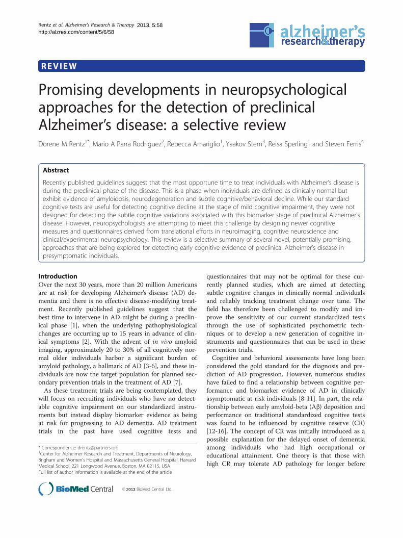
Table 1 Tests showing sensitivity to subtle cognitive changesAlzheimer’s disease
Test Memory component Valida
Memory Capacity Test [13] Verbal associative binding 34 HC
Face Name AssociativeMemory Exam (FNAME) [22,31]
Cross-model associativebinding
45 HCburden
210 Hsumm
129 Hhippo
Short-Term MemoryBinding test [41]
Visual recognition, changedetection, feature binding
30 asyshort-tpreclin
Behavioral PatternSeparation-Object test [49,50]
Visual recognition,pattern separation
31 HCweakeIn 23 aexpos
Spatial Pattern Separationtask [51,56]
Visual recognition, patternseparation, spatial discrimination
37 HCbilaterrecall
Discrimination andTransfer task [62,63]
Spatial discrimination 37 HChippocPerform
Dual tasking task [76,77] Spatial discrimination 39 E28performemofamilia
Aβ, amyloid beta; AD, Alzheimer’s disease; aMCI, amnestic mild cognitive impairmenHC, healthy older controls; RAVLT, Rey auditory verbal learning test.
instruments presented here because they were essentiallytests of memory that were derived from knowledgegained through these translational efforts in neuroimag-ing, neuroscience, and clinical and experimental neuro-psychology. The instruments were also designed totarget the neural pathways involved in the basic compo-nents of memory including encoding (that is, learning ofnew information), retrieval (that is, accessing information)and storage (that is, recognition of information), aswell as additional features including associative binding,semantic encoding, pattern separation and spatial dis-crimination that may be vulnerable in preclinical AD co-horts. We included tests of executive function because themeta-analysis suggested that these tests [20] also had asignificant although weaker association with amyloid de-position than episodic memory. No other cognitive do-mains were related to biomarker evidence of preclinicalAD. Finally, we have included a section on patient-oriented outcome measures related to subjective cognitiveconcerns because there is recent interest in and supportfor these very early complaints in clinically normal indi-viduals perhaps also heralding evidence of preclinical ADand risk of subsequent cognitive decline.Hence, the overall goal of this article is to present
a selective summary of just some of the newer, po-tentially promising approaches for detecting cognitive
associated with biomarker evidence of preclinical
tion
, decrements in second-list learning associated with amyloid burden
, decrements in face name versus face occupation associated with amyloid
C, good test–retest and discriminate validity for name, occupation andary scores, useful across all educational strata
C, FNAME performance summary scores were associated with reducedcampal volume and APOE4 carrier status
mptomatic carriers with E280A mutation showed impairment in visualerm memory binding, suggesting short-term memory binding may be aical marker for familial AD
, impairments in pattern separation were noted in those withr RAVLT Delayed Recall Scores. Recognition Memory was normal.MCI individuals, pattern separation deficits improved in thoseed to drug treatment during a clinical trial
, Spatial Pattern Separation performance was associated with reducedal hippocampal volume and with the CSF Aβ42/pTau181 ratio. Paragraphwas not sensitive to these biomarker correlates
, reduced transfer performance was associated with mild-to-moderateampal atrophy in CN and associated with clinical impairment 2 years later.ance also correlated with CSF Aβ42 and the Aβ42/pTau181 ratio
0A mutation carriers showed dual tasking impairments despite normalmances on other standard neuropsychological tests of cognition andry. Dual tasking performance discriminates asymptomatic carriers withl AD from healthy controls
t; APOE4, apolipoprotein E4; CN, cognitively normal; CSF, cerebrospinal fluid;

Rentz et al. Alzheimer's Research & Therapy Page 3 of 102013, 5:58http://alzres.com/content/5/6/58
evidence of preclinical AD in presymptomatic individuals(see Table 1). Presenting an exhaustive list of all tests andmeasures that have demonstrated the ability to detect cog-nitive decline at the stage of mild cognitive impairment(MCI) is beyond the scope of this review, but rather we se-lect a few tests/questionnaires that have shown promisefor having an association with biomarker evidence of pre-clinical AD. Some of these tests were developed recently,and therefore are not fully validated, and some are unpub-lished. However, these tests are included here because theyshow promise in targeting neural pathways that might beuseful in early detection studies.
Tests of memory associated with evidence ofpreclinical Alzheimer’s diseaseMemory Capacity TestThe Memory Capacity Test from Herman Buschke wasrecently published as showing sensitivity to Aβ depos-ition on amyloid imaging in normal older adults [13].Similar to the Free and Cued Selective Reminding Test,the Memory Capacity Test improves encoding specificityby means of pairing the word to be remembered with acategory/semantic cue [21], inducing deep semantic en-coding to maximize learning and retrieval.The Memory Capacity Test uses two 16-item word lists
(32 pairs) to be remembered from the same category cuesas the first list. The test measures associative binding withthe objective that decrements in the second-list recallwould be more sensitive to the early pathological changesassociated with preclinical AD. In fact, individuals withgreater Aβ burden on amyloid imaging showed normal re-call on the first list but lower than normal binding scoreson the second list despite performing normally on otherstandardized tests of memory [13].Tests that challenge the associative binding system, such
as the Memory Capacity Test, show promise for being ableto distinguish older adults in the preclinical phase of AD.
Face Name Associative Memory ExamThe Face Name Associative Memory Exam (FNAME)[22] is a cross-modal associative memory test based on a
Figure 1 Example of Face Name Associative Memory Exam stimuli.
functional magnetic resonance imaging (fMRI) task thatpairs pictures of unfamiliar faces with common firstnames. Previous fMRI work from multiple groups hassuggested that the successful formation and retrieval offace–name pairs requires the coordinated activity of adistributed memory network [23-25]. This network in-cludes not only the hippocampus and related structuresin the medial temporal lobe, but a distributed set of cor-tical regions, collectively known as the default mode net-work [26]. The face–name fMRI task has shownsensitivity to longitudinal clinical decline in MCI [27] aswell as in those at genetic risk for AD [23,28,29], and isassociated with Aβ burden in clinically normal older in-dividuals [30].The FNAME is a short behavioral version of the fMRI
task that requires the individual to remember 16 un-familiar face–name pairs and 16 face–occupation pairs,for a total of 32 cross-modal paired associates to be re-membered (see Figure 1).The distribution of scores on the FNAME in clinically
normal adults did not exhibit the same ceiling effects, incontrast to other standardized tests of memory in thisasymptomatic cohort [22], and is useful over a range ofeducation and CR. Performance for face–names as well asa summary score of names and occupations showed goodtest–retest reliability [31] and was correlated with Aβ bur-den on amyloid imaging [22], reduced hippocampal vol-ume on MRI, as well as APOE4 carrier status incognitively normal older adults [32]. A simpler 12-itemversion of the FNAME was developed for face–name pairsonly, similar to the original fMRI task. A computerizedversion of the FNAME is being developed on the iPad(Apple, Cupertino, CA, USA) in conjunction withCogStateW (Cogstate Ltd, Melbourne, Australia) and willbe used as a secondary outcome measure in the Domin-antly Inherited Alzheimer Network and the Anti-AmyloidTreatment in Asymptomatic AD secondary preventiontrials, purposely designed to treat presymptomatic individ-uals at risk for AD. Further validation work is being doneto determine whether this alternate version of theFNAME will be useful for measuring clinical change over
Rentz et al. Alzheimer's Research & Therapy Page 4 of 102013, 5:58http://alzres.com/content/5/6/58
the course of a clinical trial in asymptomatic individuals atrisk for AD.
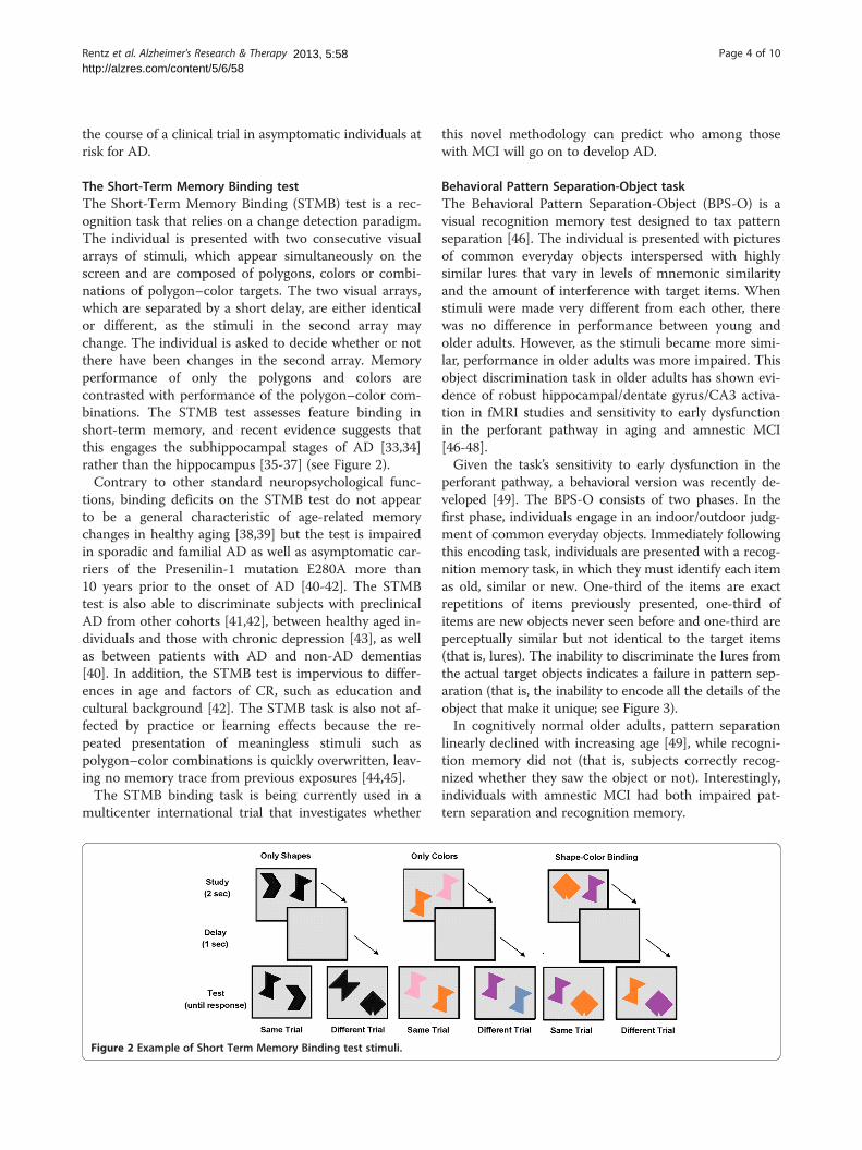
The Short-Term Memory Binding testThe Short-Term Memory Binding (STMB) test is a rec-ognition task that relies on a change detection paradigm.The individual is presented with two consecutive visualarrays of stimuli, which appear simultaneously on thescreen and are composed of polygons, colors or combi-nations of polygon–color targets. The two visual arrays,which are separated by a short delay, are either identicalor different, as the stimuli in the second array maychange. The individual is asked to decide whether or notthere have been changes in the second array. Memoryperformance of only the polygons and colors arecontrasted with performance of the polygon–color com-binations. The STMB test assesses feature binding inshort-term memory, and recent evidence suggests thatthis engages the subhippocampal stages of AD [33,34]rather than the hippocampus [35-37] (see Figure 2).Contrary to other standard neuropsychological func-
tions, binding deficits on the STMB test do not appearto be a general characteristic of age-related memorychanges in healthy aging [38,39] but the test is impairedin sporadic and familial AD as well as asymptomatic car-riers of the Presenilin-1 mutation E280A more than10 years prior to the onset of AD [40-42]. The STMBtest is also able to discriminate subjects with preclinicalAD from other cohorts [41,42], between healthy aged in-dividuals and those with chronic depression [43], as wellas between patients with AD and non-AD dementias[40]. In addition, the STMB test is impervious to differ-ences in age and factors of CR, such as education andcultural background [42]. The STMB task is also not af-fected by practice or learning effects because the re-peated presentation of meaningless stimuli such aspolygon–color combinations is quickly overwritten, leav-ing no memory trace from previous exposures [44,45].The STMB binding task is being currently used in a
multicenter international trial that investigates whether
Figure 2 Example of Short Term Memory Binding test stimuli.
this novel methodology can predict who among thosewith MCI will go on to develop AD.
Behavioral Pattern Separation-Object taskThe Behavioral Pattern Separation-Object (BPS-O) is avisual recognition memory test designed to tax patternseparation [46]. The individual is presented with picturesof common everyday objects interspersed with highlysimilar lures that vary in levels of mnemonic similarityand the amount of interference with target items. Whenstimuli were made very different from each other, therewas no difference in performance between young andolder adults. However, as the stimuli became more simi-lar, performance in older adults was more impaired. Thisobject discrimination task in older adults has shown evi-dence of robust hippocampal/dentate gyrus/CA3 activa-tion in fMRI studies and sensitivity to early dysfunctionin the perforant pathway in aging and amnestic MCI[46-48].Given the task’s sensitivity to early dysfunction in the
perforant pathway, a behavioral version was recently de-veloped [49]. The BPS-O consists of two phases. In thefirst phase, individuals engage in an indoor/outdoor judg-ment of common everyday objects. Immediately followingthis encoding task, individuals are presented with a recog-nition memory task, in which they must identify each itemas old, similar or new. One-third of the items are exactrepetitions of items previously presented, one-third ofitems are new objects never seen before and one-third areperceptually similar but not identical to the target items(that is, lures). The inability to discriminate the lures fromthe actual target objects indicates a failure in pattern sep-aration (that is, the inability to encode all the details of theobject that make it unique; see Figure 3).In cognitively normal older adults, pattern separation
linearly declined with increasing age [49], while recogni-tion memory did not (that is, subjects correctly recog-nized whether they saw the object or not). Interestingly,individuals with amnestic MCI had both impaired pat-tern separation and recognition memory.
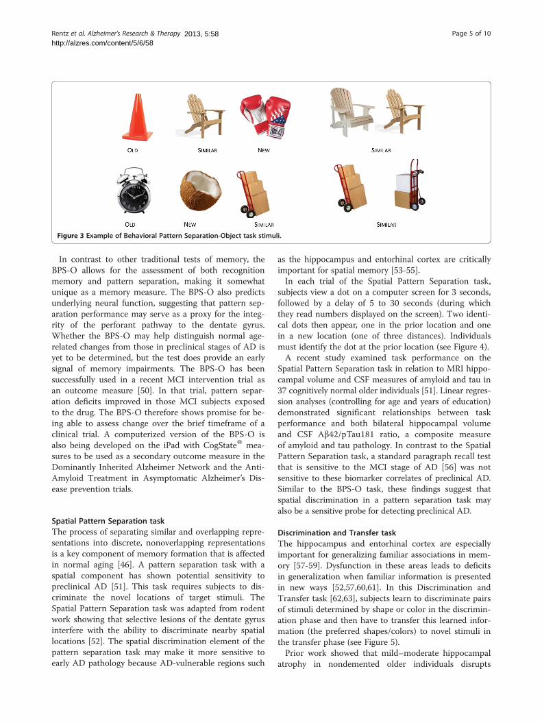
Figure 3 Example of Behavioral Pattern Separation-Object task stimuli.
Rentz et al. Alzheimer's Research & Therapy Page 5 of 102013, 5:58http://alzres.com/content/5/6/58
In contrast to other traditional tests of memory, theBPS-O allows for the assessment of both recognitionmemory and pattern separation, making it somewhatunique as a memory measure. The BPS-O also predictsunderlying neural function, suggesting that pattern sep-aration performance may serve as a proxy for the integ-rity of the perforant pathway to the dentate gyrus.Whether the BPS-O may help distinguish normal age-related changes from those in preclinical stages of AD isyet to be determined, but the test does provide an earlysignal of memory impairments. The BPS-O has beensuccessfully used in a recent MCI intervention trial asan outcome measure [50]. In that trial, pattern separ-ation deficits improved in those MCI subjects exposedto the drug. The BPS-O therefore shows promise for be-ing able to assess change over the brief timeframe of aclinical trial. A computerized version of the BPS-O isalso being developed on the iPad with CogStateW mea-sures to be used as a secondary outcome measure in theDominantly Inherited Alzheimer Network and the Anti-Amyloid Treatment in Asymptomatic Alzheimer’s Dis-ease prevention trials.
Spatial Pattern Separation taskThe process of separating similar and overlapping repre-sentations into discrete, nonoverlapping representationsis a key component of memory formation that is affectedin normal aging [46]. A pattern separation task with aspatial component has shown potential sensitivity topreclinical AD [51]. This task requires subjects to dis-criminate the novel locations of target stimuli. TheSpatial Pattern Separation task was adapted from rodentwork showing that selective lesions of the dentate gyrusinterfere with the ability to discriminate nearby spatiallocations [52]. The spatial discrimination element of thepattern separation task may make it more sensitive toearly AD pathology because AD-vulnerable regions such
as the hippocampus and entorhinal cortex are criticallyimportant for spatial memory [53-55].In each trial of the Spatial Pattern Separation task,
subjects view a dot on a computer screen for 3 seconds,followed by a delay of 5 to 30 seconds (during whichthey read numbers displayed on the screen). Two identi-cal dots then appear, one in the prior location and onein a new location (one of three distances). Individualsmust identify the dot at the prior location (see Figure 4).A recent study examined task performance on the
Spatial Pattern Separation task in relation to MRI hippo-campal volume and CSF measures of amyloid and tau in37 cognitively normal older individuals [51]. Linear regres-sion analyses (controlling for age and years of education)demonstrated significant relationships between taskperformance and both bilateral hippocampal volumeand CSF Aβ42/pTau181 ratio, a composite measureof amyloid and tau pathology. In contrast to the SpatialPattern Separation task, a standard paragraph recall testthat is sensitive to the MCI stage of AD [56] was notsensitive to these biomarker correlates of preclinical AD.Similar to the BPS-O task, these findings suggest thatspatial discrimination in a pattern separation task mayalso be a sensitive probe for detecting preclinical AD.
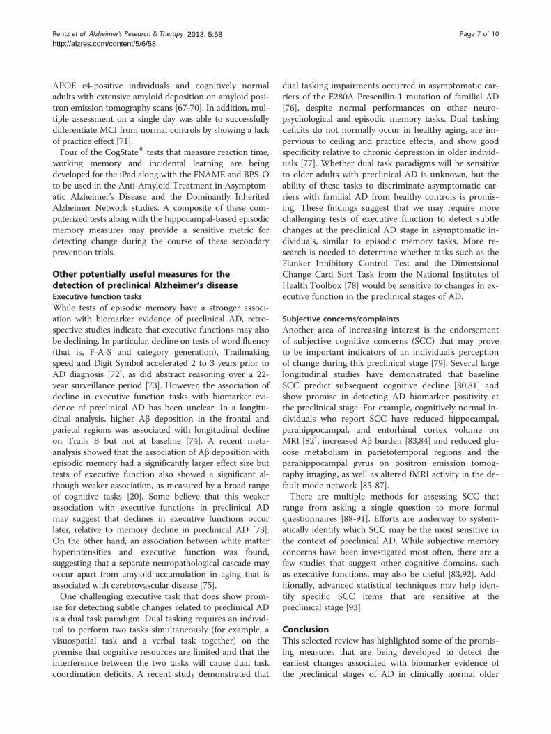
Discrimination and Transfer taskThe hippocampus and entorhinal cortex are especiallyimportant for generalizing familiar associations in mem-ory [57-59]. Dysfunction in these areas leads to deficitsin generalization when familiar information is presentedin new ways [52,57,60,61]. In this Discrimination andTransfer task [62,63], subjects learn to discriminate pairsof stimuli determined by shape or color in the discrimin-ation phase and then have to transfer this learned infor-mation (the preferred shapes/colors) to novel stimuli inthe transfer phase (see Figure 5).Prior work showed that mild–moderate hippocampal
atrophy in nondemented older individuals disrupts
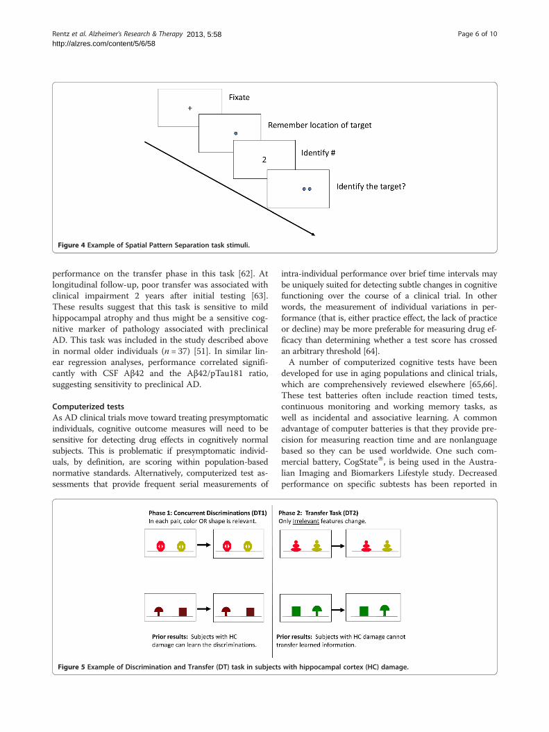
Figure 4 Example of Spatial Pattern Separation task stimuli.
Rentz et al. Alzheimer's Research & Therapy Page 6 of 102013, 5:58http://alzres.com/content/5/6/58
performance on the transfer phase in this task [62]. Atlongitudinal follow-up, poor transfer was associated withclinical impairment 2 years after initial testing [63].These results suggest that this task is sensitive to mildhippocampal atrophy and thus might be a sensitive cog-nitive marker of pathology associated with preclinicalAD. This task was included in the study described abovein normal older individuals (n = 37) [51]. In similar lin-ear regression analyses, performance correlated signifi-cantly with CSF Aβ42 and the Aβ42/pTau181 ratio,suggesting sensitivity to preclinical AD.
Computerized testsAs AD clinical trials move toward treating presymptomaticindividuals, cognitive outcome measures will need to besensitive for detecting drug effects in cognitively normalsubjects. This is problematic if presymptomatic individ-uals, by definition, are scoring within population-basednormative standards. Alternatively, computerized test as-sessments that provide frequent serial measurements of
Figure 5 Example of Discrimination and Transfer (DT) task in subjects
intra-individual performance over brief time intervals maybe uniquely suited for detecting subtle changes in cognitivefunctioning over the course of a clinical trial. In otherwords, the measurement of individual variations in per-formance (that is, either practice effect, the lack of practiceor decline) may be more preferable for measuring drug ef-ficacy than determining whether a test score has crossedan arbitrary threshold [64].A number of computerized cognitive tests have been
developed for use in aging populations and clinical trials,which are comprehensively reviewed elsewhere [65,66].These test batteries often include reaction timed tests,continuous monitoring and working memory tasks, aswell as incidental and associative learning. A commonadvantage of computer batteries is that they provide pre-cision for measuring reaction time and are nonlanguagebased so they can be used worldwide. One such com-mercial battery, CogStateW, is being used in the Austra-lian Imaging and Biomarkers Lifestyle study. Decreasedperformance on specific subtests has been reported in
with hippocampal cortex (HC) damage.
Rentz et al. Alzheimer's Research & Therapy Page 7 of 102013, 5:58http://alzres.com/content/5/6/58
APOE ε4-positive individuals and cognitively normaladults with extensive amyloid deposition on amyloid posi-tron emission tomography scans [67-70]. In addition, mul-tiple assessment on a single day was able to successfullydifferentiate MCI from normal controls by showing a lackof practice effect [71].Four of the CogStateW tests that measure reaction time,
working memory and incidental learning are beingdeveloped for the iPad along with the FNAME and BPS-Oto be used in the Anti-Amyloid Treatment in Asymptom-atic Alzheimer’s Disease and the Dominantly InheritedAlzheimer Network studies. A composite of these com-puterized tests along with the hippocampal-based episodicmemory measures may provide a sensitive metric fordetecting change during the course of these secondaryprevention trials.
Other potentially useful measures for thedetection of preclinical Alzheimer’s diseaseExecutive function tasksWhile tests of episodic memory have a stronger associ-ation with biomarker evidence of preclinical AD, retro-spective studies indicate that executive functions may alsobe declining. In particular, decline on tests of word fluency(that is, F-A-S and category generation), Trailmakingspeed and Digit Symbol accelerated 2 to 3 years prior toAD diagnosis [72], as did abstract reasoning over a 22-year surveillance period [73]. However, the association ofdecline in executive function tasks with biomarker evi-dence of preclinical AD has been unclear. In a longitu-dinal analysis, higher Aβ deposition in the frontal andparietal regions was associated with longitudinal declineon Trails B but not at baseline [74]. A recent meta-analysis showed that the association of Aβ deposition withepisodic memory had a significantly larger effect size buttests of executive function also showed a significant al-though weaker association, as measured by a broad rangeof cognitive tasks [20]. Some believe that this weakerassociation with executive functions in preclinical ADmay suggest that declines in executive functions occurlater, relative to memory decline in preclinical AD [73].On the other hand, an association between white matterhyperintensities and executive function was found,suggesting that a separate neuropathological cascade mayoccur apart from amyloid accumulation in aging that isassociated with cerebrovascular disease [75].One challenging executive task that does show prom-
ise for detecting subtle changes related to preclinical ADis a dual task paradigm. Dual tasking requires an individ-ual to perform two tasks simultaneously (for example, avisuospatial task and a verbal task together) on thepremise that cognitive resources are limited and that theinterference between the two tasks will cause dual taskcoordination deficits. A recent study demonstrated that
dual tasking impairments occurred in asymptomatic car-riers of the E280A Presenilin-1 mutation of familial AD[76], despite normal performances on other neuro-psychological and episodic memory tasks. Dual taskingdeficits do not normally occur in healthy aging, are im-pervious to ceiling and practice effects, and show goodspecificity relative to chronic depression in older individ-uals [77]. Whether dual task paradigms will be sensitiveto older adults with preclinical AD is unknown, but theability of these tasks to discriminate asymptomatic car-riers with familial AD from healthy controls is promis-ing. These findings suggest that we may require morechallenging tests of executive function to detect subtlechanges at the preclinical AD stage in asymptomatic in-dividuals, similar to episodic memory tasks. More re-search is needed to determine whether tasks such as theFlanker Inhibitory Control Test and the DimensionalChange Card Sort Task from the National Institutes ofHealth Toolbox [78] would be sensitive to changes in ex-ecutive function in the preclinical stages of AD.
Subjective concerns/complaintsAnother area of increasing interest is the endorsementof subjective cognitive concerns (SCC) that may proveto be important indicators of an individual’s perceptionof change during this preclinical stage [79]. Several largelongitudinal studies have demonstrated that baselineSCC predict subsequent cognitive decline [80,81] andshow promise in detecting AD biomarker positivity atthe preclinical stage. For example, cognitively normal in-dividuals who report SCC have reduced hippocampal,parahippocampal, and entorhinal cortex volume onMRI [82], increased Aβ burden [83,84] and reduced glu-cose metabolism in parietotemporal regions and theparahippocampal gyrus on positron emission tomog-raphy imaging, as well as altered fMRI activity in the de-fault mode network [85-87].There are multiple methods for assessing SCC that
range from asking a single question to more formalquestionnaires [88-91]. Efforts are underway to system-atically identify which SCC may be the most sensitive inthe context of preclinical AD. While subjective memoryconcerns have been investigated most often, there are afew studies that suggest other cognitive domains, suchas executive functions, may also be useful [83,92]. Add-itionally, advanced statistical techniques may help iden-tify specific SCC items that are sensitive at thepreclinical stage [93].
ConclusionThis selected review has highlighted some of the promis-ing measures that are being developed to detect theearliest changes associated with biomarker evidence ofthe preclinical stages of AD in clinically normal older
Rentz et al. Alzheimer's Research & Therapy Page 8 of 102013, 5:58http://alzres.com/content/5/6/58
adults. While this is not meant to be an exhaustive sur-vey of potential measures, the review is meant to high-light some promising recent efforts and to emphasizethe critical need to develop a new collection of cognitivetasks that are cost-effective, available within the publicdomain, useful across all levels of CR and derived fromtranslational efforts in cognitive neuroscience. Many ofthe tests described here will require additional valid-ation, but with the start of secondary prevention trialswe now have a unique opportunity to use these trials todetermine whether these measures will be useful fordetecting preclinical AD and tracking cognitive changeover time.In addition, future efforts are exploring whether more
complex activities of daily living (that is, financial com-petency, navigating complex telephone trees) and othersubjective and informant questionnaires related to en-gagement (that is, physical activities, cognitive stimulation)or emotional states (that is, social isolation, apathy,withdrawal) may be associated with biomarker evidenceof preclinical AD. More advanced psychometric tech-niques, such as Item Response Theory [93-95] and Con-firmatory Factor Analysis [75], are now being used tocreate more sensitive instruments from our current stan-dardized tests as well as composite measures that maycapture the individual variance of change in clinicallynormal individuals [11]. Home assessments and the useof iPad and tablet technologies are also being exploredto determine whether more frequent/multiple cognitiveassessments may provide a more reliable estimate ofchange and whether they can be performed without theaid of a technician or extensive travel to a clinic. Whilemany of these efforts are still in development, clinical re-searchers are attempting to meet the challenge of devel-oping instruments and questionnaires that may besensitive to biomarker evidence of early AD. Finally, itwill probably be a combination of these objective andsubjective measures/questionnaires that will be the mostvaluable for tracking cognitive change over time.
AbbreviationsAD: Alzheimer’s disease; APOE: Apolipoprotein E allele;APOE4: Apolipoprotein E4 carrier status; Aβ: Amyloid beta; BPS-O: BehavioralPattern Separation-Object; CR: Cognitive reserve; CSF: Cerebral spinal fluid;fMRI: Functional magnetic resonance imaging; FNAME: Face NameAssociative Memory Exam; MCI: Mild cognitive impairment; MRI: Magneticresonance imaging; SCC: Subjective cognitive concerns; STMB: Short-TermMemory Binding; ε4: Carriers of the E4 apolipoprotein E gene.
Competing interestsThe authors declare that they have no competing interests.
AcknowledgementsThe authors wish to thank all of the subjects who make research possible.MAPR is currently a Fellow of Alzheimer’s Society, UK, Project Grant NumberRF165.
Author details1Center for Alzheimer Research and Treatment, Departments of Neurology,Brigham and Women’s Hospital and Massachusetts General Hospital, HarvardMedical School, 221 Longwood Avenue, Boston, MA 02115, USA.2Department of Psychology, University of Edinburgh, Centre for CognitiveAging and Cognitive Epidemiology, Alzheimer Scotland Dementia ResearchCentre and Scottish Dementia Clinical Research Network, 7 George Square,Edinburgh EH8 9JZ, UK. 3Cognitive Neuroscience Division, Department ofNeurology, Taub Institute for Research on Alzheimer’s Disease and the AgingBrain, Columbia University College of Physicians and Surgeons, 630 West168th Street, New York, NY 10032, USA. 4Alzheimer’s Disease Center,Comprehensive Center for Brain Aging, Department of Psychiatry, NYULangone Medical Center, 145 East 32nd Street, New York, NY 10016, USA.
Published:
References1. Sperling RA, Aisen PS, Beckett LA, Bennett DA, Craft S, Fagan AM, Iwatsubo
T, Jack CR Jr, Kaye J, Montine TJ, Park DC, Reiman EM, Rowe CC, Siemers E,Stern Y, Yaffe K, Carrillo MC, Thies B, Morrison-Bogorad M, Wagster MV,Phelps CH: Toward defining the preclinical stages of Alzheimer’s disease:recommendations from the National Institute on Aging–Alzheimer’sAssociation workgroups on diagnostic guidelines for Alzheimer’s disease.Alzheimers Dement 2011, 7:280–292.
2. Rowe CC, Ellis KA, Rimajova M, Bourgeat P, Pike KE, Jones G, Fripp J,Tochon-Danguy H, Morandeau L, O’Keefe G, Price R, Raniga P, Robins P,Acosta O, Lenzo N, Szoeke C, Salvado O, Head R, Martins R, Masters CL,Ames D, Villemagne VL: Amyloid imaging results from the AustralianImaging, Biomarkers and Lifestyle (AIBL) study of aging. Neurobiol Aging2010, 31:1275–1283.
3. Johnson KA: Amyloid imaging of Alzheimer’s disease using PittsburghCompound B. Curr Neurol Neurosci Rep 2006, 6:496–503.
4. Mintun MA, Larossa GN, Sheline YI, Dence CS, Lee SY, Mach RH, Klunk WE,Mathis CA, Dekosky ST, Morris JC: [11C]PIB in a nondemented population:potential antecedent marker of Alzheimer disease. Neurology 2006,67:446–452.
5. Rowe CC, Ng S, Ackermann U, Gong SJ, Pike K, Savage G, Cowie TF,Dickinson KL, Maruff P, Darby D, Smith C, Woodward M, Merory J, Tochon-Danguy H, O’Keefe G, Klunk WE, Mathis CA, Price JC, Masters CL, VillemagneVL: Imaging beta-amyloid burden in aging and dementia. Neurology 2007,68:1718–1725.
6. Fagan AM, Mintun MA, Mach RH, Lee SY, Dence CS, Shah AR, Larossa GN,Spinner ML, Klunk WE, Mathis CA, DeKosky ST, Morris JC, Holtzman DM:Inverse relation between in vivo amyloid imaging load andcerebrospinal fluid Aβ42 in humans. Ann Neurol 2006, 59:512–519.
7. Sperling RA, Jack CR Jr, Aisen PS: Testing the right target and right drugat the right stage. Sci Transl Med 2011, 3:111–133.
8. Aizenstein HJ, Nebes RD, Saxton JA, Price JC, Mathis CA, Tsopelas ND, ZiolkoSK, James JA, Snitz BE, Houck PR, Bi W, Cohen AD, Lopresti BJ, DeKosky ST,Halligan EM, Klunk WE: Frequent amyloid deposition without significantcognitive impairment among the elderly. Arch Neurol 2008, 65:1509–1517.
9. Jack CR Jr, Lowe VJ, Senjem ML, Weigand SD, Kemp BJ, Shiung MM,Knopman DS, Boeve BF, Klunk WE, Mathis CA, Petersen RC: 11C PiB andstructural MRI provide complementary information in imaging ofAlzheimer’s disease and amnestic mild cognitive impairment. Brain 2008,131:665–680.
10. Mormino EC, Kluth JT, Madison CM, Rabinovici GD, Baker SL, Miller BL,Koeppe RA, Mathis CA, Weiner MW, Jagust WJ: Episodic memory loss isrelated to hippocampal-mediated beta-amyloid deposition in elderlysubjects. Brain 2009, 132:1310–1323.
11. Storandt M, Mintun MA, Head D, Morris JC: Cognitive decline and brainvolume loss as signatures of cerebral amyloid-beta peptide depositionidentified with Pittsburgh compound B: cognitive decline associatedwith Aβ deposition. Arch Neurol 2009, 66:1476–1481.
12. Kemppainen NM, Aalto S, Karrasch M, Nagren K, Savisto N, Oikonen V, ViitanenM, Parkkola R, Rinne JO: Cognitive reserve hypothesis: Pittsburgh CompoundB and fluorodeoxyglucose positron emission tomography in relation toeducation in mild Alzheimer’s disease. Ann Neurol 2008, 63:112–118.
13. Rentz DM, Locascio JJ, Becker JA, Moran EK, Eng E, Buckner RL, Sperling RA,Johnson KA: Cognition, reserve, and amyloid deposition in normal aging.Ann Neurol 2010, 67:353–364.
21 Nov 2013
Rentz et al. Alzheimer's Research & Therapy Page 9 of 102013, 5:58http://alzres.com/content/5/6/58
14. Roe CM, Mintun MA, D’Angelo G, Xiong C, Grant EA, Morris JC: Alzheimerdisease and cognitive reserve: variation of education effect with carbon11-labeled Pittsburgh Compound B uptake. Arch Neurol 2008, 65:1467–1471.
15. Roe CM, Mintun MA, Ghoshal N, Williams MM, Grant EA, Marcus DS, MorrisJC: Alzheimer disease identification using amyloid imaging and reservevariables: proof of concept. Neurology 2010, 75:42–48.
16. Yaffe K, Weston A, Graff-Radford NR, Satterfield S, Simonsick EM, Younkin SG,Younkin LH, Kuller L, Ayonayon HN, Ding J, Harris TB: Association of plasmabeta-amyloid level and cognitive reserve with subsequent cognitivedecline. JAMA 2011, 305:261–266.
17. Stern Y: Cognitive reserve in ageing and Alzheimer’s disease. LancetNeurol 2012, 11:1006–1012.
18. Dickerson BC, Stoub TR, Shah RC, Sperling RA, Killiany RJ, Albert MS, HymanBT, Blacker D, Detoledo-Morrell L: Alzheimer-signature MRI biomarkerpredicts AD dementia in cognitively normal adults. Neurology 2011,76:1395–1402.
19. Morris JC, Roe CM, Grant EA, Head D, Storandt M, Goate AM, Fagan AM,Holtzman DM, Mintun MA: Pittsburgh compound B imaging andprediction of progression from cognitive normality to symptomaticAlzheimer disease. Arch Neurol 2009, 66:1469–1475.
20. Hedden T, Oh H, Younger AP, Patel TA: Meta-analysis of amyloid–cognition relations in cognitively normal older adults. Neurology 2013,80:1341–1348.
21. Dubois B, Feldman HH, Jacova C, Dekosky ST, Barberger-Gateau P,Cummings J, Delacourte A, Galasko D, Gauthier S, Jicha G, Meguro K,O’brien J, Pasquier F, Robert P, Rossor M, Salloway S, Stern Y, Visser PJ,Scheltens P: Research criteria for the diagnosis of Alzheimer’s disease:revising the NINCDS-ADRDA criteria. Lancet Neurol 2007, 6:734–746.
22. Rentz DM, Amariglio RE, Becker JA, Frey M, Olson LE, Frishe K, Carmasin J,Maye JE, Johnson KA, Sperling RA: Face–name associative memoryperformance is related to amyloid burden in normal elderly.Neuropsychologia 2011, 49:2776–2783.
23. Miller SL, Celone K, DePeau K, Diamond E, Dickerson BC, Rentz D,Pihlajamaki M, Sperling RA: Age-related memory impairment associatedwith loss of parietal deactivation but preserved hippocampal activation.Proc Natl Acad Sci U S A 2008, 105:2181–2186.
24. Rugg MD, Otten LJ, Henson RN: The neural basis of episodic memory:evidence from functional neuroimaging. Philos Trans R Soc London B BiolSci 2002, 357:1097–1110.
25. Vannini P, Hedden T, Becker JA, Sullivan C, Putcha D, Rentz D, Johnson KA,Sperling RA: Age and amyloid-related alterations in default networkhabituation to stimulus repetition. Neurobiol Aging 2012, 33:1237–1252.
26. Buckner RL, Snyder AZ, Shannon BJ, LaRossa G, Sachs R, Fotenos AF, ShelineYI, Klunk WE, Mathis CA, Morris JC, Mintun MA: Molecular, structural, andfunctional characterization of Alzheimer’s disease: evidence for arelationship between default activity, amyloid, and memory. J Neurosci2005, 25:7709–7717.
27. O’Brien JL, O’Keefe KM, LaViolette PS, DeLuca AN, Blacker D, Dickerson BC,Sperling RA: Longitudinal fMRI in elderly reveals loss of hippocampalactivation with clinical decline. Neurology 2010, 74:1969–1976.
28. Celone KA, Calhoun VD, Dickerson BC, Atri A, Chua EF, Miller SL, DePeau K,Rentz DM, Selkoe DJ, Blacker D, Albert MS, Sperling RA: Alterations inmemory networks in mild cognitive impairment and Alzheimer’s disease:an independent component analysis. J Neurosci 2006, 26:10222–10231.
29. Sperling RA, Chua E, Cocchiarella A, Rand-Giovannetti E, Poldrack R, SchacterDL, Albert M: Putting names to faces: successful encoding of associativememories activates the anterior hippocampal formation. Neuroimage2003, 20:1400–1410.
30. Sperling RA, Laviolette PS, O’Keefe K, O’Brien J, Rentz DM, Pihlajamaki M,Marshall G, Hyman BT, Selkoe DJ, Hedden T, Buckner RL, Becker JA, JohnsonKA: Amyloid deposition is associated with impaired default networkfunction in older persons without dementia. Neuron 2009, 63:178–188.
31. Amariglio RE, Frishe K, Olson LE, Wadsworth LP, Lorius N, Sperling RA, RentzDM: Validation of the Face Name Associative Memory Exam in cognitivelynormal older individuals. J Clin Exp Neuropsychol 2012, 34:580–587.
32. Bamfo R, Armariglio R, Rentz D: Early detection of preclinical Alzheimer’sdisease with the Face Name Associative Memory Exam. Senior thesis. HarvardUniversity, Neurobiology; 2013.
33. Didic M, Barbeau EJ, Felician O, Tramoni E, Guedj E, Poncet M, Ceccaldi M:Which memory system is impaired first in Alzheimer’s disease? JAlzheimers Dis 2011, 27:11–22.
34. Staresina BP, Davachi L: Object unitization and associative memoryformation are supported by distinct brain regions. J Neurosci 2010,30:9890–9897.
35. Baddeley A, Allen R, Vargha-Khadem F: Is the hippocampus necessary forvisual and verbal binding in working memory? Neuropsychologia 2010,48:1089–1095.
36. Baddeley A, Jarrold C, Vargha-Khadem F: Working memory and thehippocampus. J Cogn Neurosci 2011, 23:3855–3861.
37. Piekema C, Rijpkema M, Fernandez G, Kessels RP: Dissociating the neuralcorrelates of intra-item and inter-item working-memory binding. PLoSONE 2010, 5:e10214.
38. Brockmole JR, Parra MA, Della Sala S, Logie RH: Do binding deficitsaccount for age-related decline in visual working memory? Psychon BullRev 2008, 15:543–547.
39. Parra MA, Abrahams S, Logie RH, Sala SD: Age and binding within-dimension features in visual short-term memory. Neurosci Lett 2009,449:1–5.
40. Della Sala S, Parra MA, Fabi K, Luzzi S, Abrahams S: Short-term memorybinding is impaired in AD but not in non-AD dementias.Neuropsychologia 2012, 50:833–840.
41. Parra MA, Abrahams S, Logie RH, Mendez LG, Lopera F, Della Sala S: Visualshort-term memory binding deficits in familial Alzheimer’s disease. Brain2010, 133:2702–2713.
42. Parra MA, Sala SD, Abrahams S, Logie RH, Mendez LG, Lopera F: Specificdeficit of colour-colour short-term memory binding in sporadic andfamilial Alzheimer’s disease. Neuropsychologia 2011, 49:1943–1952.
43. Parra MA, Abrahams S, Logie RH, Della Sala S: Visual short-term memorybinding in Alzheimer’s disease and depression. J Neurol 2010, 257:1160–1169.
44. Colzato LS, Raffone A, Hommel B: What do we learn from bindingfeatures? Evidence for multilevel feature integration. J Exp Psychol HumPercept Perform 2006, 32:705–716.
45. Logie RH, Brockmole JR, Vandenbroucke ARE: Bound feature combinationsin visual short term memory are fragile but influence long-term learning.Vis Cogn 2009, 17:160–179.
46. Stark SM, Yassa MA, Stark CE: Individual differences in spatial patternseparation performance associated with healthy aging in humans. LearnMem 2010, 17:284–288.
47. Yassa MA, Mattfeld AT, Stark SM, Stark CE: Age-related memory deficitslinked to circuit-specific disruptions in the hippocampus. Proc Natl AcadSci U S A 2011, 108:8873–8878.
48. Yassa MA, Stark SM, Bakker A, Albert MS, Gallagher M, Stark CE: High-resolution structural and functional MRI of hippocampal CA3 anddentate gyrus in patients with amnestic mild cognitive impairment.Neuroimage 2010, 51:1242–1252.
49. Stark SM, Yassa MA, Lacy JW, Stark CE: A task to assess behavioral patternseparation (BPS) in humans: data from healthy aging and mild cognitiveimpairment. Neuropsychologia 2013 [Epub ahead of print].
50. Bakker A, Krauss GL, Albert MS, Speck CL, Jones LR, Stark CE, Yassa MA,Bassett SS, Shelton AL, Gallagher M: Reduction of hippocampalhyperactivity improves cognition in amnestic mild cognitive impairment.Neuron 2012, 74:467–474.
51. Lau H, Karantzoulis S, Myers C, Pirraglia E, Li Y, Gurnani A, Glodzik L,Scharfman H, Kesner RP, de Leon M, Ferris S: Cognitive detection ofpreclinical AD. Alzheimers Dement 2012, 8:P358.
52. Gilbert PE, Kesner RP: Role of the rodent hippocampus in paired-associatelearning involving associations between a stimulus and a spatiallocation. Behav Neurosci 2002, 116:63–71.
53. Brun VH, Leutgeb S, Wu HQ, Schwarcz R, Witter MP, Moser EI, Moser MB:Impaired spatial representation in CA1 after lesion of direct input fromentorhinal cortex. Neuron 2008, 57:290–302.
54. Nadel L, MacDonald L: Hippocampus: cognitive map or working memory?Behav Neural Biol 1980, 29:405–409.
55. Oswald CJ, Good M: The effects of combined lesions of the subicularcomplex and the entorhinal cortex on two forms of spatial navigation inthe water maze. Behav Neurosci 2000, 114:211–217.
56. Kluger A, Ferris SH, Golomb J, Mittelman MS, Reisberg B:Neuropsychological prediction of decline to dementia in nondementedelderly. J Geriatr Psychiatry Neurol 1999, 12:168–179.
57. Eichenbaum H, Mathews P, Cohen NJ: Further studies of hippocampalrepresentation during odor discrimination learning. Behav Neurosci 1989,103:1207–1216.
Rentz et al. Alzheimer's Research & Therapy Page 10 of 102013, 5:58http://alzres.com/content/5/6/58
58. Gluck MA, Myers CE, Nicolle MM, Johnson S: Computational models of thehippocampal region: implications for prediction of risk for Alzheimer’sdisease in non-demented elderly. Curr Alzheimer Res 2006, 3:247–257.
59. Winocur G, Olds J: Effects of context manipulation on memory andreversal learning in rats with hippocampal lesions. J Comp Physiol Psychol1978, 92:312–321.
60. Buckmaster CA, Eichenbaum H, Amaral DG, Suzuki WA, Rapp PR: Entorhinalcortex lesions disrupt the relational organization of memory in monkeys.J Neurosci 2004, 24:9811–9825.
61. Cermak LS, Uhly B, Reale L: Encoding specificity in the alcoholic Korsakoffpatient. Brain Lang 1980, 11:119–127.
62. Myers CE, Kluger A, Golomb J, Ferris S, de Leon MJ, Schnirman G, Gluck MA:Hippocampal atrophy disrupts transfer generalization in nondementedelderly. J Geriatr Psychiatry Neurol 2002, 15:82–90.
63. Myers CE, Kluger A, Golomb J, Gluck MA, Ferris S: Learning andgeneralization tasks predict short-term cognitive outcome innondemented elderly. J Geriatr Psychiatry Neurol 2008, 21:93–103.
64. Darby D, Brodtmann A, Woodward M, Budge M, Maruff P: Using cognitivedecline in novel trial designs for primary prevention and early disease-modifying therapy trials of Alzheimer’s disease. Int Psychogeriatr 2011,23:1376–1385.
65. Snyder PJ, Jackson CE, Petersen RC, Khachaturian AS, Kaye J, Albert MS,Weintraub S: Assessment of cognition in mild cognitive impairment: acomparative study. Alzheimers Dement 2011, 7:338–355.
66. Wild K, Howieson D, Webbe F, Seelye A, Kaye J: Status of computerizedcognitive testing in aging: a systematic review. Alzheimers Dement 2008,4:428–437.
67. Stonnington CM, Locke DE, Dueck AC, Caselli RJ: Anxiety affects cognitiondifferently in healthy apolipoprotein E ε4 homozygotes and non-carriers.J Neuropsychiatry Clin Neurosci 2011, 23:294–299.
68. Darby DG, Brodtmann A, Pietrzak RH, Fredrickson J, Woodward M,Villemagne VL, Fredrickson A, Maruff P, Rowe C: Episodic memory declinepredicts cortical amyloid status in community-dwelling older adults. JAlzheimers Dis 2011, 27:627–637.
69. Lim YY, Pietrzak RH, Ellis KA, Jaeger J, Harrington K, Ashwood T, Szoeke C,Martins RN, Bush AI, Masters CL, Rowe CC, Villemagne VL, Ames D, Darby D,Maruff P: Rapid decline in episodic memory in healthy older adults withhigh amyloid-beta. J Alzheimers Dis 2013, 33:675–679.
70. Lim YY, Ellis KA, Pietrzak RH, Ames D, Darby D, Harrington K, Martins RN,Masters CL, Rowe C, Savage G, Szoeke C, Villemagne VL, Maruff P, AIBLResearch Group: Stronger effect of amyloid load than APOE genotype oncognitive decline in healthy older adults. Neurology 2012, 79:1645–1652.
71. Darby D, Maruff P, Collie A, McStephen M: Mild cognitive impairment canbe detected by multiple assessments in a single day. Neurology 2002,59:1042–1046.
72. Elias MF, Beiser A, Wolf PA, Au R, White RF, D’Agostino RB: The preclinicalphase of Alzheimer’s disease: a 22 year prospective study of theFramingham cohort. Arch Neurol 2000, 57:808–813.
73. Grober E, Hall CB, Lipton RB, Zonderman AB, Resnick SM, Kawas C: Memoryimpairment, executive dysfunction, and intellectual decline in preclinicalAlzheimer’s disease. J Int Neuropsychol Soc 2008, 14:266–278.
74. Resnick SM, Sojkova J, Zhou Y, An Y, Ye W, Holt DP, Dannals RF, Mathis CA,Klunk WE, Ferrucci L, Kraut MA, Wong DF: Longitudinal cognitive decline isassociated with fibrillar amyloid-beta measured by [11C]PiB. Neurology2010, 74:807–815.
75. Hedden T, Mormino EC, Amariglio RE, Younger AP, Schultz AP, Becker JA,Buckner RL, Johnson KA, Sperling RA, Rentz DM: Cognitive profile ofamyloid burden and white matter hyperintensities in cognitively normalolder adults. J Neurosci 2012, 32:16233–16242.
76. MacPherson SE, Parra MA, Moreno S, Lopera F, Della Sala S: Dual taskabilities as a possible preclinical marker of Alzheimer’s disease in carriersof the E280A presenilin-1 mutation. J Int Neuropsychol Soc 2012, 18:234–241.
77. Kaschel R, Logie RH, Kazen M, Della Sala S: Alzheimer’s disease, but notageing or depression, affects dual-tasking. J Neurol 2009, 256:1860–1868.
78. Weintraub S, Dikmen SS, Heaton RK, Tulsky DS, Zelazo PD, Bauer PJ, CarlozziNE, Slotkin J, Blitz D, Wallner-Allen K, Fox NA, Beaumont JL, Mungas D,Nowinski CJ, Richler J, Deocampo JA, Anderson JE, Manly JJ, Borosh B, Havlik R,Conway K, Edwards E, Freund L, King JW, Moy C, Witt E, Gershon RC:Cognition assessment using the NIH Toolbox. Neurology 2013, 80:S54–S64.
79. Reisberg B, Prichep L, Mosconi L, John ER, Glodzik-Sobanska L, Boksay I,Monteiro I, Torossian C, Vedvyas A, Ashraf N, Jamil IA, de Leon MJ: The pre-mild cognitive impairment, subjective cognitive impairment stage ofAlzheimer’s disease. Alzheimers Dement 2008, 4:S98–S108.
80. Dik MG, Jonker C, Comijs HC, Bouter LM, Twisk JW, van Kamp GJ, Deeg DJ:Memory complaints and APOE-ε4 accelerate cognitive decline incognitively normal elderly. Neurology 2001, 57:2217–2222.
81. Schofield PW, Marder K, Dooneief G, Jacobs DM, Sano M, Stern Y:Association of subjective memory complaints with subsequent cognitivedecline in community-dwelling elderly individuals with baselinecognitive impairment. Am J Psychiatry 1997, 154:609–615.
82. Saykin AJ, Wishart HA, Rabin LA, Santulli RB, Flashman LA, West JD, McHughTL, Mamourian AC: Older adults with cognitive complaints show brainatrophy similar to that of amnestic MCI. Neurology 2006, 67:834–842.
83. Amariglio RE, Becker JA, Carmasin J, Wadsworth LP, Lorius N, Sullivan C,Maye JE, Gidicsin C, Pepin LC, Sperling RA, Johnson KA, Rentz DM:Subjective cognitive complaints and amyloid burden in cognitivelynormal older individuals. Neuropsychologia 2012, 50:2880–2886.
84. Perrotin A, Mormino EC, Madison CM, Hayenga AO, Jagust WJ: Subjectivecognition and amyloid deposition imaging: a Pittsburgh Compound Bpositron emission tomography study in normal elderly individuals. ArchNeurol 2012, 69:223–229.
85. Hafkemeijer A, Altmann-Schneider I, Oleksik AM, van de Wiel L, MiddelkoopHA, van Buchem MA, van der Grond J, Rombouts SA: Increased functionalconnectivity and brain atrophy in elderly with subjective memorycomplaints. Brain Connect 2013, 3:353–362.
86. Mosconi L, De Santi S, Brys M, Tsui WH, Pirraglia E, Glodzik-Sobanska L, RichKE, Switalski R, Mehta PD, Pratico D, Zinkowski R, Blennow K, de Leon MJ:Hypometabolism and altered cerebrospinal fluid markers in normalapolipoprotein E E4 carriers with subjective memory complaints. BiolPsychiatry 2008, 63:609–618.
87. Wang Y, Risacher SL, West JD, McDonald BC, Magee TR, Farlow MR, Gao S,O’Neill DP, Saykin AJ: Altered default mode network connectivity in olderadults with cognitive complaints and amnestic mild cognitiveimpairment. J Alzheimers Dis 2013, 35:751–760.
88. Amariglio RE, Townsend MK, Grodstein F, Sperling RA, Rentz DM: Specificsubjective memory complaints in older persons may indicate poorcognitive function. J Am Geriatr Soc 2011, 59:1612–1617.
89. Farias ST, Mungas D, Reed BR, Cahn-Weiner D, Jagust W, Baynes K, Decarli C:The measurement of everyday cognition (ECog): scale development andpsychometric properties. Neuropsychology 2008, 22:531–544.
90. Gilewski MJ, Zelinski EM, Schaie KW: The Memory FunctioningQuestionnaire for assessment of memory complaints in adulthood andold age. Psychol Aging 1990, 5:482–490.
91. Reisberg B, v SH, de Leon MJ, Crook T: The Global Deterioration Scale forassessment of primary degenerative dementia. Am J Psychiatry 1982,139:1136–1139.
92. van Norden AG, Fick WF, de Laat KF, van Uden IW, van Oudheusden LJ,Tendolkar I, Zwiers MP, de Leeuw FE: Subjective cognitive failures andhippocampal volume in elderly with white matter lesions. Neurology2008, 71:1152–1159.
93. Snitz BE, Yu L, Crane PK, Chang CC, Hughes TF, Ganguli M: Subjectivecognitive complaints of older adults at the population level: an itemresponse theory analysis. Alzheimer Dis Assoc Disord 2012, 26:344–351.
94. Crane PK, Narasimhalu K, Gibbons LE, Mungas DM, Haneuse S, Larson EB,Kuller L, Hall K, van Belle G: Item response theory facilitated cocalibratingcognitive tests and reduced bias in estimated rates of decline. J ClinEpidemiol 2008, 61:1018–1027.
95. Crane PK, Narasimhalu K, Gibbons LE, Pedraza O, Mehta KM, Tang Y, ManlyJJ, Reed BR, Mungas DM: Composite scores for executive function items:demographic heterogeneity and relationships with quantitativemagnetic resonance imaging. J Int Neuropsychol Soc 2008, 14:746–759.
Cite this article as: Rentz et al.: Promising developments inneuropsychological approaches for the detection of preclinicalAlzheimer’s disease: a selective review. Alzheimer's Research & Therapy
10.1186/alzrt222
2013, 5:58
Related Documents