Welcome message from author
This document is posted to help you gain knowledge. Please leave a comment to let me know what you think about it! Share it to your friends and learn new things together.
Transcript


Quick Review Cards for MedicalLaboratory Science Second Edition
Valerie Dietz Polansky, MEd, MLS(ASCP)CM
Program DirectorMedical Laboratory Technology ProgramSt. Petersburg CollegeSt. Petersburg, Florida
2956_FM_i-xxii 29/01/14 11:54 AM Page i

F. A. Davis Company1915 Arch StreetPhiladelphia, PA 19103www.fadavis.com
Copyright © 2014 by F. A. Davis Company
Copyright © 2014 by F. A. Davis Company. All rights reserved. This product is protected by copyright. No part of it may be reproduced, stored in a retrieval system, or transmitted in any form or by any means, electronic, mechanical, photocopying, recording, or otherwise, without written permission from the publisher.
Printed in the United States of America
Last digit indicates print number: 10 9 8 7 6 5 4 3 2 1
Senior Acquisitions Editor: Christa FratantoroDevelopmental Editor: Angela NortonManager of Content Development: George LangDesign and Illustration Manager: Carolyn O’Brien
As new scientific information becomes available through basic and clinical research, recommended treatments and drug therapies undergo changes.The author(s) and publisher have done everything possible to make this book accurate, up to date, and in accord with accepted standards at the timeof publication. The author(s), editors, and publisher are not responsible for errors or omissions or for consequences from application of the book, andmake no warranty, expressed or implied, in regard to the contents of the book. Any practice described in this book should be applied by the reader inaccordance with professional standards of care used in regard to the unique circumstances that may apply in each situation. The reader is advised always to check product information (package inserts) for changes and new information regarding dose and contraindications before administeringany drug. Caution is especially urged when using new or infrequently ordered drugs.
ISBN: 978-0-8036-2956-1
Authorization to photocopy items for internal or personal use, or the internal or personal use of specific clients, is granted by F. A. Davis Company forusers registered with the Copyright Clearance Center (CCC) Transactional Reporting Service, provided that the fee of $.25 per copy is paid directly toCCC, 222 Rosewood Drive, Danvers, MA 01923. For those organizations that have been granted a photocopy license by CCC, a separate system of payment has been arranged. The fee code for users of the Transactional Reporting Service is: 978080362956-1/14 0 + $.25.
2956_FM_i-xxii 29/01/14 11:54 AM Page ii

Dedicated to my husband, Gary, for his support and encouragement through another long project, and in lovingmemory of my parents, Bill and Lee Dietz, who provided me
with the education that was the foundation of my career.
2956_FM_i-xxii 29/01/14 11:54 AM Page iii

2956_FM_i-xxii 29/01/14 11:54 AM Page iv

Preface
Quick Review Cards for Medical Laboratory Science were developed as a study aid to improve student performanceon Board examinations in medical laboratory science(medical technology) at both the technician and technol-ogist levels. (Technician candidates may skip the sectionon management and education.)
This card deck is the product of more than 30 years ofexperience teaching hundreds of students who have suc-cessfully passed Board examinations at both levels. Thecard format allows for easy sorting and portability, makingthem ideal for quick reviews and last-minute studying. Useof these cards alone, however, does not guarantee a pass-ing score; they are intended to be used as an adjunct totraditional textbooks. Students are encouraged to high-light unfamiliar information and to refer to textbooks andclass notes to supplement their study of those topics. Theuse of a multiple-choice review book and practice examsalso will help to round out a student’s preparation for theBoard exam.
The review cards will also be beneficial to MSL and MLTstudents before graduation as they prepare for course exam-inations. Professionals who are cross-training or reenteringthe workplace will find these cards useful as well.
No review of this type can include all topics. This reviewfocuses on common procedures and disorders, otherknowledge that entry-level laboratory professionals are ex-pected to have, and topics that are frequently included onBoard exams. The review cards are written in an informalnote-taking style, using abbreviations, symbols, and shortphrases to maximize the amount of information included.A list of abbreviations is found in the frontmatter.
New to the second edition are a chapter on moleculardiagnostics and graphics for select topics. Space did notallow for inclusion of drawings of all cells/organisms. Stu-dents are encouraged to refer to textbooks to supplementtheir review with additional pictures and diagrams. Furtherbenefit could be derived from making their own drawings,diagrams, and flow charts. Active and frequent review willlead to higher scores.
Every effort was made to ensure the accuracy of thecontent. In some cases, discrepancies were found withinand among references; then information was either se-lected from the most recent publication or confirmed inanother source. Please let the publisher know if you havesuggestions for improving future editions.
v
2956_FM_i-xxii 29/01/14 11:54 AM Page v

2956_FM_i-xxii 29/01/14 11:54 AM Page vi

Bibliography
Arneson, W., & Brickell, J. Clinical Chemistry A Laboratory Perspective.Philadelphia, PA: F.A. Davis, 2007.
Bishop, M. L., Fody, E. P., & Schoeff, L. E. Clinical Chemistry: Principles, Procedures,Correlations (6th ed.). Philadelphia, PA:Lippincott Williams & Wilkins, 2010.
Bruns, D. E., Ashwood, E. R., & Burtis, C.A. Fundamentals of Molecular Diagnostics. St. Louis, MO: SaundersElsevier, 2007.
Buckingham, L. & Flaws, M. L. MolecularDiagnostics Fundamentals, Methods,& Clinical Applications. Philadelphia,PA: F.A. Davis, 2007.
Ciesla, B., Hematology in Practice (2nd ed.). Philadelphia: F.A. DavisCompany, 2012.
Harmening, D. M., Clinical Hematol-ogy and Fundamentals of Hemosta-sis (5th ed.). Philadelphia, PA: F.A. Davis, 2009.
Harmening, D. M., Laboratory Manage-ment Principles and Processes (2nd ed.). St. Petersburg, FL: D. H. Publishing & Consulting, 2007.
Harmening, D. M., Modern Blood Bankingand Transfusion Practices (6th ed.).Philadelphia, PA: F. A. Davis, 2012.
Kern, M. E. & Blevins, K. S. Medical Mycol-ogy, A Self-Instructional Text (2nd ed.).Philadelphia, PA: F.A. Davis, 1997.
Kiser, K. M., Payne, W. C., & Taff, T. A.Clinical Laboratory Microbiology APractical Approach. Upper SaddleRiver, NJ: Pearson Education, 2011.
Leventhal, R., & Cheadle, R.F. MedicalParasitology: A Self-Instructional Text(5th ed.). Philadelphia, PA: F.A. Davis,2002.
Mahon, C. R., Lehman, D. C., &Manuselis, G. Textbook of DiagnosticMicrobiology (4th ed.). Philadelphia,PA: W B. Saunders, 2011.
McKenzie, S. B., & Williams, J. L. ClinicalLaboratory Hematology. Upper SaddleRiver, NJ: Pearson Education, 2010.
Mundt, L. A., & Shanahan, K. Graff’s Text-book of Urinalysis and Body Fluids (2nd ed.). Philadelphia, PA: LippincottWilliams & Wilkins, 2011.
Quinley, E. D. Immunohematology Principles & Practice (3rd ed.).Philadelphia, PA: Lippincott Williams & Wilkins, 2011.
Rodak, B. F., Fritsma, G. A., & Koehane, E.M. Hematology Clinical Principles andApplications (4th ed.). St. Louis, MO: Elsevier Saunders, 2012.
Standards for Blood Banks and TransfusionServices (27th ed.). Bethesda: AmericanAssociation of Blood Banks, 2011.
Stevens, C. D. Clinical Immunologyand Serology: A Laboratory Perspec-tive (3rd ed.). Philadelphia, PA: F.A. Davis, 2010.
vii
continued...
2956_FM_i-xxii 29/01/14 11:54 AM Page vii

Bibliography continued viii
Strasinger, S. K., & Di Lorenzo, M. S. The Phlebotomy Textbook (3rd ed.).Philadelphia, PA: F.A. Davis, 2011.
Strasinger, S. K., & Di Lorenzo, M. S., Urinalysis and Body Fluids (5th ed.).Philadelphia, PA: F.A. Davis, 2008.
Sunheimer, R. L., & Graves, L. ClinicalLaboratory Chemistry. Upper SaddleRiver, NJ: Pearson Education, 2011.
Technical Manual (17th ed.). Bethesda:American Association of Blood Banks, 2011.
Turgeon, M. L. Clinical HematologyTheory and Procedures (5th ed.).Philadelphia, PA: Lippincott Williams & Wilkins, 2012.
Turgeon, M. L. Immunology & Serology in Laboratory Medicine (4th ed.). St. Louis, MO: Mosby Elsevier, 2009.
Turgeon, M. L. Linne & Ringsrud’s Clinical Laboratory Science (5th ed.). St. Louis, MO: Mosby Elsevier, 2007.
2956_FM_i-xxii 29/01/14 11:54 AM Page viii
Reviewers
Eileen Carreiro-Lewandowski, CLSProfessorUniversity of MassachusettsDartmouth, MassachusettsTerry Dunkel, MS, MT(ASCP)Program Director, Assistant ProfessorPresentation CollegeAberdeen, South DakotaKathleen Engelmann, PhD, MLS(ASCP)Associate ProfessorUniversity of BridgeportBridgeport, ConnecticutDavid M. Falleur, Med, MT(ASCP), CLSChair, Associate ProfessorSouthwest Texas State UniversitySan Marcos, TexasAbraham Furman, PhDAssociate ProfessorOregon Institute of TechnologyKlamath Falls, OregonMichelle L. Gagan, MSHS, MLS(ASCP)Education CoordinatorYork Technical CollegeRock Hill, South Carolina
Andrea R. Hoffmann, MT(ASCP)CMInstructorDelgado Community CollegeNew Orleans, LouisianaStephen M. Johnson, MS, MT(ASCP)Program DirectorSaint Vincent Health Center,Erie, PennsylvaniaKathy Kenwright, MS, MT(ASCP)SI, MBAssociate ProfessorUniversity of Tennessee Health
Science CenterMemphis, TennesseeKristi Lew, BSc (MLS), MSc, MLT,
MLS(ASCP)CMAssistant ProfessorUniversity of AlbertaEdmonton, Alberta, CanadaAmy M. McCarty, MA, BS(ASCP)Program DirectorWashington Hospital CenterWashington, DC
Sonja Nehr-Kanet, MS, MLS(ASCP)CMClinical Associate ProfessorIdaho State UniversityMeridian, IdahoMaura Pieretti, PhD, HCLDScientific DirectorBayCare LaboratoriesTampa, FloridaEllen F. Romani, MHSA, MLS(ASCP)CM,
BB, DLMDepartment ChairSpartanburg Community CollegeSpartanburg, South CarolinaAnchalee D. Steele, MT(ASCP)Program DirectorSpencerian CollegeLexington, KentuckyAmy Sutton, BS, MT(ASCP)Laboratory SupervisorSchryver MedicalPhoenix, Arizona
ix
2956_FM_i-xxii 29/01/14 11:54 AM Page ix

2956_FM_i-xxii 29/01/14 11:54 AM Page x

Contents
Section 1 Laboratory Operations Review, 1Credentialing, 2Agencies That Issue Guidelines/
Standards, 3Federal Regulatory Agencies, 4Federal Regulations, 5CLIA ‘88 Test Complexities, 6Bloodborne Pathogens Standard, 7Specimen Infectivity, 8Packaging of Biologics for Shipping, 9Hazard Communication Standard (HCS),
10Occupational Exposures to Hazardous
Chemicals in Laboratories Standard,11
Hazard Categories of Chemicals, 12Hazard Identification System, 14Storage of Chemicals, 16Fire Safety, 17Commonly Used Anticoagulants/
Additives, 18Recommended Order for Drawing
Evacuated Tubes & Filling Tubes Froma Syringe, 19
Recommended Order for FillingMicrocollection Tubes from CapillaryPunctures, 20
Special Situations in Phlebotomy, 21Special Test Requirements, 22Phlebotomy Sources of Error, 23Guidelines for Specimen Handling &
Processing, 25Centrifuges, 26Examples of Criteria for Specimen
Rejection, 27Types of Glass, 28Types of Plastic, 29Glassware Inscriptions, 30Volumetric Glassware, 31Glass Pipets, 32Mechanical Micropipets, 33Grades of Chemicals, 34Purified Water, 35CAP Reagent Labeling Requirements,
36Brightfield Microscopy, 37Other Types of Microscopy, 39Informatics, 41Computer Hardware, 42
Computer Software, 43Information Systems, 44Computer Networks, 45Quality Assessment, 46Quality Control, 47Quality Control Statistics, 48Interpretation of Quality Control Data,
50Westgard Multirules, 53Typical Steps Taken When a Control
Is Outside Acceptable Range, 54
Calibration, 55Test Performance Specifications and
Verification, 56Diagnostic Value of a Test, 58Other Components of a QA
Program, 59CLIA Requirements for Procedure
Manuals, 61Ethical and Legal Issues, 62Commonly Used Prefixes in the Metric
System, 64General Laboratory Calculations,
65
xi
continued...
2956_FM_i-xxii 29/01/14 11:54 AM Page xi

Section 2 Clinical Chemistry Review, 67Comparison of Conventional and
SI Units for Selected ReferenceRanges, 68
Examples of Patient Variables That MayAffect Chemistry Values, 69
Examples of Preanalytical Factors That May Affect Chemistry Results, 70
Differences in Analyte Concentrations,72
Photometric Methods, 73Visible Light, 75Wavelengths Used in
Spectrophotometry, 76Chromatography, 77Other Analytic Techniques, 78Steps in Automated Analysis, 79Chemistry Panels, 80Carbohydrates, Lipids, and
Proteins, 81Regulation of Glucose, 83Diabetes Mellitus, 84
Tests for Diabetes Mellitus, 85Typical Laboratory Findings in
Uncontrolled Diabetes Mellitus, 86Metabolic Syndrome, 87Aminoacidopathies, 88Protein Electrophoresis, 89Common Serum Protein Electrophoresis
Patterns, 90Nonprotein Nitrogen Compounds, 92Major Electrolytes, 93Other Electrolytes, 95Iron and Related Tests, 97Factors That Influence Enzymatic
Reactions, 98Enzymes of Clinical Significance, 99Summary of Diagnostic Enzymology, 101Cardiac Markers for Diagnosis of Acute
Myocardial Infarction, 102Other Cardiac Tests, 103Bilirubin Metabolism, 104Types of Bilirubin, 105Unconjugated versus Conjugated
Bilirubin, 106
Differential Diagnosis of Jaundice, 107Pituitary Hormones, 108Thyroid and Parathyroid Hormones, 110Thyroid Function Testing, 111Adrenal Hormones, 112Reproductive Hormones, 113Pancreatic Hormones, 114Therapeutic Drug Monitoring (TDM),
115Therapeutic Drug Groups, 116Toxic Agents, 117Drugs of Abuse Urine Screen, 118Common Tumor Markers, 119Acid-Base Balance Terminology, 121Acid-Base Imbalances, 123Arterial Blood Gases Terminology, 124Blood Gas Parameters, 125Blood Gas Instrumentation, 126Sources of Error in Arterial Blood Gases,
127Calculated Chemistry Values, 128Chemistry Calculations, 131
Contents continued xii
continued...
2956_FM_i-xxii 29/01/14 11:54 AM Page xii

Contents continued xiii
Section 3 Clinical Microbiology Review, 135Biosafety Levels, 136CDC Classification of Biological Agents,
137Biological Safety Cabinets, 138Sterilization and Disinfection, 139Bacterial Toxins, 141Specimen Collection Guidelines, 142Specimen Preservation and
Storage, 143Fragile Organisms, 144Criteria for Rejection of Specimens in
Microbiology, 145Gram Stain, 146Staining Properties of Gram-Positive
and Gram-Negative Bacteria, 147Types of Media, 148Routine Media for Aerobes and
Facultative Anaerobes, 149Selective Media for Isolation of Neisseria
gonorrhoeae and Neisseriameningitidis, 152
Special Bacteriologic Media, 153Aerotolerance Test, 155
Organisms Requiring Incubation inIncreased CO2, 156
Hemolytic Reactions on Sheep BloodAgar, 157
Staphylococci, 158Summary of Tests for Identification of
Staphylococci, 160Streptococci/Enterococci, 161Tests for Identification of Beta-Hemolytic
Streptococci, 165Tests for Identification of Alpha-Hemolytic
Streptococci, 167Tests for Identification of Nonhemolytic
Streptococci/Enterococci, 168Antibiograms of Gram-Positive Cocci, 169Aerobic Spore-Forming
Gram-Positive Rods, 170Aerobic Non–Spore-Forming
Gram-Positive Rods, 171Neisseria and Moraxella, 173Characteristics of Enterobacteriaceae,
176Biochemical Tests for Identification of
Enterobacteriaceae, 177
Antigens of Enterobacteriaceae, 181Commonly Isolated Enterobacteriaceae,
182Summary of Key Reactions for
Enterobacteriaceae, 185Appearance of Enterobacteriaceae on
Selected Media, 186Diarrheagenic Escherichia coli, 187Characteristics of Nonfermenting Gram-
Negative Rods, 189Commonly Isolated Nonfermenting
Gram-Negative Rods, 190Campylobacter and Helicobacter, 191Vibrio and Related Organisms, 192Haemophilus, 194Speciation of Haemophilus, 195Miscellaneous Gram-Negative
Rods, 196Specimens for Anaerobic Culture, 198Media for Culture of Anaerobes, 199Anaerobic Environment, 200Methods to Identify Anaerobes, 201Anaerobic Gram-Positive Cocci, 203Anaerobic Gram-Positive Rods, 204
continued...
2956_FM_i-xxii 29/01/14 11:55 AM Page xiii

Contents continued xiv
Gram-Negative Anaerobes, 206Laboratory Identification of
Mycobacteria, 207Acid-Fast Stains, 209Classification of Mycobacteria Based on
Pathogenicity, 210Classification of Nontuberculous
Mycobacteria Based on Physiology,211
Medically Important Mycobacteria, 212Chlamydia and Chlamydophila, 215Spirochetes, 216Mycoplasma/Ureaplasma, 217Rickettsiae, 218Routine Culture Setup and
Interpretation, 219Fecal Pathogens, 223Major Classes of Antibiotics, 224Disk Diffusion Susceptibility Method
(Kirby Bauer), 227Other Susceptibility Tests, 229Automated Identification an
Susceptibility Testing, 231Examples of Quality Control in
Microbiology Labs, 232
Stool Specimens for Ova and Parasites,234
Ova and Parsite Examiniation, 235Intestinal Amebae, 236Flagellates of the Intestinal and
Urogentital Tracts, 239Intestinal Ciliates, 243Intestinal Sporozoans, 244Intestinal Nematodes, 245Intestinal Cestodes, 251Trematodes, 254Blood and Tissue Protozoa, 258Differentiation of Plasmodium, 262Blood and Tissue Helminths, 265Stains Used in Mycology, 266Fungal Culture Media, 267Dernatophytes, 268Dimorphic Fungi, 269Yeast, 271Contaminants/Opportunistic
Fungi, 273Fungal Pathogens by Site, 276Viral Structure, 277Viral Replication, 278Human DNA Viruses, 279
Human RNA Viruses, 280Common Viruses by Site, 282Viral Speciment Collection and
Transport, 283Methods for Diagnosis of Viral
Infections, 284Cell Cultures, 285Comparison of Microorganisms, 286
Section 4 Hematology Review, 287Blood Cells, 288Comparison of Conventional and
SI Units for Adult Reference Ranges,289
Reference Ranges for Red Blood CellParameters, 290
Reference Ranges for Leukocytes andPlatelets, 291
Hematopoietic Cell Differentiation, 292Erythropoiesis, 293Changes During Cell Maturation, 294Erythrocytic Developmental Series, 295Asynchronous Erythropoiesis, 296Hemoglobin, 297Hemoglobin Electrophoresis, 298
continued...
2956_FM_i-xxii 29/01/14 11:55 AM Page xiv
Contents continued xv
Hemoglobin Derivatives, 299RBC Morphology, 300RBC Inclusions, 303Staining of RBC Inclusions, 305Erythrocyte Indices, 306Hemoglobinopathy versus Thalassemia,
307Normocytic Anemias, 308Macrocytic Anemias, 310Microcytic, Hypochromic Anemias, 311Differentiation of Microcytic
Hypochromic Anemias, 312Acute versus Chronic Blood Loss, 313Granulocytic Maturation, 314Normal Leukocytes of the Peripheral
Blood, 315Leukocyte Abnormalities, 316Quantitative Abnormalities of
Leukocytes, 317Hematopoietic Neoplasms, 318Classification of Hematopoietic
Neoplasms, 320Acute versus Chronic Leukemia, 321Common Leukemias, 322
Cytochemical Stains for Differentiation ofAcute Leukemia, 324
Leukemoid Reaction versus ChronicMyelogenous Leukemia, 325
Plasma Cell Disorders, 326Manual Hematology Procedures, 327Changes in Blood at Room Temperature,
329Methods of Automated Cell Counting &
Differentiation, 330Graphic Representations of Cell
Populations, 331Technologies Used in Automated
Hematology Analyzers, 333Automated CBC, 334QA/QC for Automated Hematology
Analyzers, 336Flow Cytometry, 337Hematology Calculations, 338Overview of Hemostasis, 341Quantitative Platelet Disorders, 342Qualitative Platelet Disorders, 343Tests of Platelet Function, 344Coagulation Factors, 345
Functional Classification of CoagulationFactors, 347
Summary of Coagulation Factors, 348Coagulation Theories, 350Cell-Based Model of Hemostasis, 352Prothrombin Time (PT) and Activated
Partial Thromboplastin Time (APTT),353
Interpretation of PT/APTT, 354Other Coagulation Tests, 355Most Common Inherited Coagulation
Disorders, 356Acquired Factor Deficiencies, 357Tests of Fibrinolytic System, 358Disseminated Intravascular Coagulation
vs. Primary Fibrinolysis, 359Tests to Assess Risk of Thrombosis, 360Anticoagulant (Antithrombotic)
Therapy, 361Coagulation Instrumentation, 362Examples of Criteria for Specimen
Rejection in Coagulation Testing, 363Sources of Error in Coagulation Testing,
364
continued...
2956_FM_i-xxii 29/01/14 11:55 AM Page xv

Contents continued xvi
Section 5 Immunology Review, 367Immunology Terminology, 368Branches of the Immune System, 372Types of Immunity, 373Adaptive Immunity, 374Cells of the Natural Immune System,
375Cells of the Acquired Immune System,
377Subpopulations of Lymphocytes, 378Lymphoid Organs, 379Isolation & Identification of
Lymphocytes, 380Immunoglobulin (Ig) Structure, 381Immunoglobulins, 383Complement, 385Hypersensitivity Reactions, 387Agglutination Methods, 388Precipitation Methods, 389Immunoassay Terminology, 390Enzyme Immunoassay (EIA)
Terminology, 391Enzyme Immunoassays (EIA) Formats,
392Fluorescent Immunoassays (FIA), 394
Comparison of Labeled Immunoassays,395
Nontreponemal Tests for Syphilis, 397Treponemal Tests for Syphilis, 398Interpretation of Syphilis Test Results,
400Serological Tests for Other Bacterial
Infections, 401Serological Tests for Infectious
Mononucleosis (IM), 402Hepatitis Tests, 403Hepatitis Serological Profiles, 405Appearance of HIV Markers, 406HIV Screening Tests, 407False Positives and Negatives With HIV-
Antibody ELISA Testing, 408HIV Confirmatory/Supplemental Tests,
409Tests to Stage and Monitor HIV, 410Screening Tests for Systemic Lupus
Erythematosus (SLE), 411Tests for Specific Antinuclear Antibodies
(ANA), 412Serological Tests for Rheumatoid
Arthritis (RA), 413
Interpretation of Serological Tests, 414
Serology Calculations, 415
Section 6 Immunohematology Review, 417Criteria for Whole Blood Donors (AABB),
418Donor Deferrals (AABB), 419Collection of Whole Blood, 421Apheresis, 422Donor Testing Required by AABB and/or
FDA, 423Anticoagulant/Preservative Solutions,
424Additive Solutions, 425Open and Closed Systems, 426Blood Components—RBCs, 427Blood Components—Plasma and
Derivatives, 429Blood Components—Platelets, 430Labeling Requirements for Blood and
Components, 431Leukocyte Reduction (Leukoreduction),
432RBC Storage Lesion, 433
continued...
2956_FM_i-xxii 29/01/14 11:55 AM Page xvi

Contents continued xvii
Primary versus Secondary Response,434
IgG versus IgM, 435Factors That Affect Agglutination in
Tube Testing, 436Comparison of Tube, Gel, and Solid
Phase Testing, 437Grading Reactions—Tube versus Gel,
438Comparison of Tube, Gel, and Solid
Phase Reactions, 439ABO Genotypes and Phenotypes, 440Using Punnett Square to Predict ABO
Type, 440Frequency of ABO Types, 441ABO System, 442ABO Typing, 443ABO Discrepancies, 444Rh Genotypes and Phenotypes, 445Using Punnett Square to Predict Rh
Type, 445Rh Antigens, 446Frequency of Rh Antigens, 447Frequency of Rh Genes, 448Breaking the Rh Code, 449
Rh Typing Sera, 450Interpretation of Rh Typing, 451Causes of False Rh Typing Results, 451Weak D Testing and Interpretation, 452Selection of Rh Type for Transfusion,
453Frequency of Other Selected Blood
Group Antigens, 454I System, 455Antibody Characteristics, 456Antigen-Antibody Enhancement, 458Antihuman Globulin Serum, 459Antiglobulin Testing, 460Antibody Identification, 461Interpreting Antibody Panels, 462Cold Antibodies, 463Compatibility Testing, 464Crossmatches, 465The Major Crossmatch, 466Examples of Incompatible
Crossmatches, 467Transfusion of Non-Group-Specific RBCs,
468Pretransfusion Testing, 469Conditions for Reissue of RBCs, 470
Emergency Transfusions, 470Transfusion-Associated Infections, 471Acute Immunologic Transfusion
Reactions, 473Acute Nonimmunologic Transfusion
Reactions, 475Delayed Transfusion Reactions, 476Transfusion Reaction Investigation, 478Testing of Neonates, 480Hemolytic Disease of the Fetus and
Newborn (HDFN), 481Rh Immune Globulin (RhIG) Workup,
482Rh Immune Globulin (RhIG), 483Examples of Equipment/Reagent
Quality Control, 484
Section 7 Urinalysis and Body Fluids Review, 485Urine Specimens, 486Urine Volume, 487Urine Color, 488Changes in Unpreserved Urine at Room
Temperature, 489Chemical Urinalysis by Reagent Strip, 490
continued...
2956_FM_i-xxii 29/01/14 11:55 AM Page xvii
Contents continued xviii
General Sources of Error With ReagentStrip Testing, 492
Specific Sources of Error with ReagentStrip Testing, 494
Other Urine Chemistry Tests, 496Glucose Oxidase versus Copper
Reduction, 498Epithelial Cells in the Urine Sediment,
499Blood Cells in the Urine Sediment, 501Normal Crystals Found in Acid or
Neutral Urine, 502Normal Crystals Found in Alkaline Urine,
503Abnormal Crystals, 505Casts, 507Miscellaneous Urine Sediment, 509Renal Disorders, 511Urinalysis Correlations, 512Cerebrospinal Fluid, 514Differential Diagnosis of Meningitis, 516Body Fluids, 517Differentiation of Transudates and
Exudates, 518Synovial Fluid, 519
Synovial Fluid Crystals, 520Semen Analysis, 521Amniotic Fluid Tests, 522
Section 8 Molecular Diagnostics (MDx) Review, 523Nucleic Acid Terminology, 524Comparison of DNA and RNA, 527Nitrogen Bases, 529Nucleotides, 530DNA Replication Terminology, 532DNA Replication, 534Primer Extension, 536Discontinuous Synthesis of DNA on
Lagging Strand, 537Gene Expression Terminology, 538Protein Synthesis, 540Basic MDx Terminology, 541Overview of MDx, 543Blood Collection Tubes for MDx, 544Specimens for MDx, 545Specimen Processing, 547Isolation of DNA, 548Comparison of RNA & DNA Isolation,
549
Assessment of Nucleic AcidYield/Quality, 550
Amplification Terminology, 551Components of Polymerase Chain
Reaction (PCR), 554Steps in PCR, 555PCR Controls, 558Other Target Amplification Methods,
559Other Amplification Methods, 560Hybridization Assays, 561Gel Electrophoresis, 563Blotting, 564DNA Sequencing/Variation
Terminology, 565Sanger Dideoxy Chain Termination
Method, 567Setup of Sanger Dideoxy Chain
Termination Method, 569Strengths and Limitations of Molecular
Testing, 570Causes and Prevention of False Results
in MDx, 571Clinical Applications of Molecular
Diagnosis, 573continued...
2956_FM_i-xxii 29/01/14 11:55 AM Page xviii

Contents continued xix
Section 9 Management & Education Review, 575Management Skills and Styles, 576Management Principles, 577Laboratory Management, 578Maslow’s Hierarchy of Needs, 579Personnel Required in High-Complexity
Laboratories Under CLIA ‘88, 580Employee Performance Appraisal, 581
Testing Personnel CompetencyAssessment, 582
Laboratory Operating Costs, 583Break-Even Points, 584Quality Management, 585Sentinel Events: The Joint Commission
(TJC), 587Point-of-Care Testing (POCT), 588Competency-Based Instruction, 589
ABCs of Writing Behavioral Objectives,590
VAK Learning Style Model, 590Domains of Learning, 591Bloom’s Cognitive Taxonomy, 592Instructional Methods, 593Testing at Different Cognitive Levels,
594Test-Taking Tips, 595
2956_FM_i-xxii 29/01/14 11:55 AM Page xix

Partial List of Abbreviations Used xx
Ab AntibodyAg AntigenAHG Antihuman globulinAIDS Acquired immunodeficiency syndromeASAP As soon as possibleAT AntithrombinBBP Bloodborne pathogensBP Blood pressureCAP College of American PathologistsCDC Centers for Disease Control and PreventionCIA Chemiluminescent immunoassayCLIA ‘88 Clinical Laboratory Improvement Amendments
of 1988CLSI Clinical Laboratory and Standards InstituteCMS Centers for Medicare and Medicaid ServicesCMV CytomegalovirusCNS Coagulase-negative staphylococciCNS Central nervous systemCk CheckCSF Cerebrospinal fluidCV Coefficient of variationDAT Direct antiglobulin testDiff DifferentialDTaP Diphtheria, tetanus, pertussis vaccineDx Diagnosis
EBV Epstein-Barr virusEIA Enzyme immunoassayELISA Enzyme-linked immunosorbent assayESR Erythrocyte sedimentation rateFDA Food and Drug AdministrationFFP Fresh frozen plasmaGPC Gram-positive cocciGAS Group A SreptococcusGBS Group B SreptococcusGI GastrointestinalGN Gram negativeGNCB Gram-negative coccobacilliGNDC Gram-negative diplococciGNR Gram-negative rodsGP Gram positiveGPC Gram-positive cocciGPR Gram-positive rodsGU GenitourinaryHAV Hepatitis A virusHBIG Hepatitis B immune globulinHBV Hepatitis B virusHCB Hepatitis C virusHCT HematocritHDFN Hemolytic disease of the fetus & newbornHgb Hemoglobin
continued...
2956_FM_i-xxii 29/01/14 11:55 AM Page xx

Partial List of Abbreviations Used continued
xxi
Hib Haemophilus influenzae type bHIPAA Health Insurance Portability and Accountability
Act of 1996HIV Human immunodeficiency virusHLA Human leukocyte antigenHPF High power fieldHr Hour(s)IAT Indirect antiglobulin testID Identify, identificationIFA Indirect fluorescent antibodyIg ImmunoglobulinIM Infectious mononucleosisIS Immediate spinLPF Low power fieldLF Lactose fermenterMin Minute(s)MMR Measles, mumps, rubella vaccineMo Month(s)MRSA Methicillin-resistant Stapylococcus aureusMW Molecular weightN NormalNAT Nucleic acid testingN:C Nucleus to cytoplasmNeg Negative
NLF Nonlactose fermenterNRBC Nucleated red blood cellOIF Oil immersion fieldOSHA Occupational Safety and Health AdministrationPCR Polymerase chain reactionPHI Protected health informationPlt Platelet(s)Poly Polymorphonuclear leukocyte, granulocytePos PositivePPD Purified protein derivativePt PatientQA Quality assurance or assessmentQC Quality controlRBC Red blood cellsRE ReticuloendothelialRhIG Rh immune globulinRT Room temperature (20°–24°C)RTI Respiratory tract infectionRxn ReactionSD Standard deviationSec Second(s)SG Specific gravitySOP Standard operating proceduresTemp Temperature
continued...
2956_FM_i-xxii 29/01/14 11:55 AM Page xxi

Partial List of Abbreviations Used continued
xxii
Tf Transfuse, transfusionUTI Urinary tract infectionVRE Vancomycin-resistant enterococciWB Whole bloodWBC White blood cellsWk Week(s)Xmatch CrossmatchYr Year(s)
# Number↑ Increase(s), increased
↓ Decrease(s), decreased> Greater than≥ Greater than or equal to < Less than≤ Less than or equal to = Equals
Other abbreviations are defined in the text.
2956_FM_i-xxii 29/01/14 11:55 AM Page xxii

1S E C T I O N
Laboratory OperationsReview
2956_Ch01_001-066 30/01/14 3:07 PM Page 1

Credentialing Laboratory Operations Review 2
PROCESS DEFINITION EXAMPLES
Accreditation
Certification
Licensure
Recognition granted by nongovernmental agency to institutions that meet certain standards.Voluntary.
Recognition granted by nongovernmental agency to individuals who meet education requirements &demonstrate entry-level competency by passing exam.Voluntary.
Permission granted by state to individuals/organiza-tions to engage in certain professions/businesses.Mandatory. Illegal to practice/operate in that statewithout license.
AABB (formerly American Association of Blood Banks)College of American Pathologists (CAP)The Joint Commission (formerly JCAHO)National Accrediting Agency for Clinical Laboratory
Sciences (NAACLS)
American Society for Clinical Pathology (ASCP)American Association of Bioanalysts (AAB)American Medical Technologists (AMT)
Licensure of laboratory personnel is required in CA, FL, HI, LA, MT, NV, NY, ND, RI, TN, WV. Many states require licensure of clinical labs.
2956_Ch01_001-066 30/01/14 3:07 PM Page 2
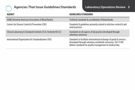
Agencies That Issue Guidelines/Standards Laboratory Operations Review 3
AGENCY GUIDELINES/STANDARDS
AABB (formerly American Association of Blood Banks)
Centers for Disease Control & Prevention (CDC)
Clinical Laboratory & Standards Institute (CLSI, formerly NCCLS)
International Organization for Standardization (ISO)
Technical standards & accreditation of blood banks.
Standards & guidelines primarily related to infection control & safework practices.
Standards on all aspects of lab practice developed through voluntary consensus.
Standards to facilitate international exchange of goods & services.Developed through voluntary worldwide consensus. ISO 15189 defines standards for quality management in medical labs.
2956_Ch01_001-066 30/01/14 3:07 PM Page 3

Federal Regulatory Agencies Laboratory Operations Review 4
AGENCY AUTHORITY
Centers for Medicare & Medicaid Services (CMS)
Department of Health & Human Services (HHS)
Department of Transportation (DOT)
Environmental Protection Agency (EPA)
Food & Drug Administration (FDA)
Nuclear Regulatory Commission (NRC)
Occupational Safety & Health Administration (OSHA)
Substance Abuse and Mental Health Services Administration (SAMHSA)
Writes regulations for & enforces Clinical Laboratory ImprovementAmendments of 1988 (CLIA ‘88).
Interprets & implements federal regulations related to health care.Oversees CDC, CMS, FDA, SAMSHA.
Regulates packaging, labeling, & transportation of biological products.
Regulates disposal of toxic chemical & biohazardous wastes.
Regulates market entry of instruments/reagents & production ofdonor blood & components. Licenses blood banks.
Licenses labs that use radionucleotides.
Regulates employee safety in the workplace.
Certifies laboratories to conduct forensic drug testing for federalagencies.
2956_Ch01_001-066 30/01/14 3:07 PM Page 4
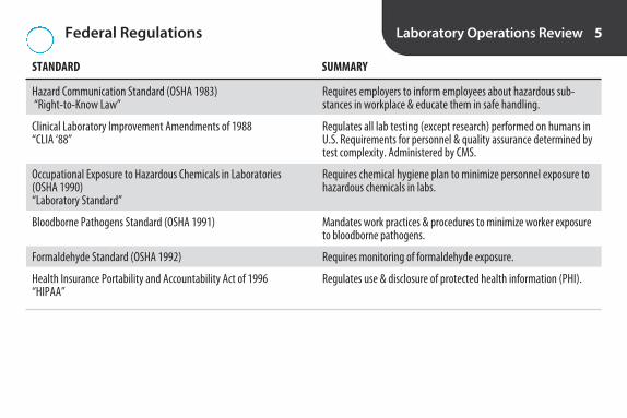
Federal Regulations Laboratory Operations Review 5
STANDARD SUMMARY
Hazard Communication Standard (OSHA 1983)“Right-to-Know Law”
Clinical Laboratory Improvement Amendments of 1988“CLIA ‘88”
Occupational Exposure to Hazardous Chemicals in Laboratories (OSHA 1990)“Laboratory Standard”
Bloodborne Pathogens Standard (OSHA 1991)
Formaldehyde Standard (OSHA 1992)
Health Insurance Portability and Accountability Act of 1996“HIPAA”
Requires employers to inform employees about hazardous sub-stances in workplace & educate them in safe handling.
Regulates all lab testing (except research) performed on humans inU.S. Requirements for personnel & quality assurance determined bytest complexity. Administered by CMS.
Requires chemical hygiene plan to minimize personnel exposure tohazardous chemicals in labs.
Mandates work practices & procedures to minimize worker exposureto bloodborne pathogens.
Requires monitoring of formaldehyde exposure.
Regulates use & disclosure of protected health information (PHI).
2956_Ch01_001-066 30/01/14 3:07 PM Page 5

CLIA ‘88 Test Complexities Laboratory Operations Review 6
PROFICIENCY TESTING PERSONNEL COMPLEXITY CRITERIA QUALITY CONTROL TESTING (PT) (MINIMUM QUALIFICATIONS)
Waived
Provider-Performed Microscopy (PPM)*
Moderate Complexity
High Complexity
*PPM is a subcategory of moderate complexity.**Criteria used to evaluate text complexity: knowledge, training/experience, reagent/material preparation, characteristics of operational steps, calibration/qualitycontrol/proficiency testing materials, test system troubleshooting, interpretation/judgment. Each of the 7 criteria is rated 1–3 (lowest to highest), & scores are totaled.
None specified
Physician, midlevel practitioner,or dentist
High school diploma or equivalent& training for testing performed
Associate degree in medical lab-oratory technology or equivalent
Tests cleared by FDA forhome use, negligiblelikelihood of erroneousresults, or no reasonablerisk of harm to patient ifperformed incorrectly
Certain microscopicexams performed byprovider during patient’svisit, e.g., direct wetmount, KOH prep, urinesediment
Score ≤ 12 on 7 criteria**
Score > 12 on 7 criteria**
None required otherthan to follow manu-facturers’ directions
Required when controlsare available; otherwise,reference materials(e.g., photomicrographs)fulfill requirement
2 levels of external con-trols each day of testing
2 levels of external con-trols each day of testing
Not required
PT not specifically required, but labsmust verify accuracyof testing twice annu-ally. Can be throughPT, split sampling, orblind testing.
Required
Required
2956_Ch01_001-066 30/01/14 3:07 PM Page 6

Bloodborne Pathogens Standard Laboratory Operations Review 7
History
Purpose
Primary Requirements
Published in 1991. Revised in 2001 following passage of Needlestick Safety & Prevention Act to includestronger requirements for employers to evaluate & adopt safer medical devices.
To protect health-care workers from occupational exposure to bloodborne pathogens (BBP; e.g., HIV,HBV, HCV)
Exposure control plan: Determination of employees’ risk of exposure & implementation of methods tocontrol exposure. Plan must be reviewed & updated annually to reflect new technologies. Documenta-tion of evaluation & adoption of safer devices is required. Nonmanagerial employees must be involved inevaluation & selection of devices.Universal precautions: All blood & certain body fluids are to be handled as if known to be infectious forbloodborne pathogens.Engineering controls: Control measures that isolate or remove a hazard from workplace, e.g., sharpscontainers, self-sheathing needles, plastic capillary tubes, Plexiglas shields.Work practice controls: e.g., hand washing, disposal of needles with safety device activated & holderattached, ban on eating/drinking/smoking in lab.Personal protective clothing & equipment: e.g., lab coats, gloves, face shields. Employer must pro-vide & must launder lab coats.Housekeeping: e.g., proper disposal of biohazardous waste, decontamination of work surfaces.Training: On assignment & annually thereafter.Medical surveillance: Postexposure evaluation & follow-up at no cost to employee.Hepatitis B vaccine: Provided by employer within 10 days of assignment at no cost to employee.Hazard communication: e.g., biohazard labels, red bags.Sharps injury log: Must include description & location of incident, device involved. Employee privacymust be protected.
2956_Ch01_001-066 30/01/14 3:07 PM Page 7
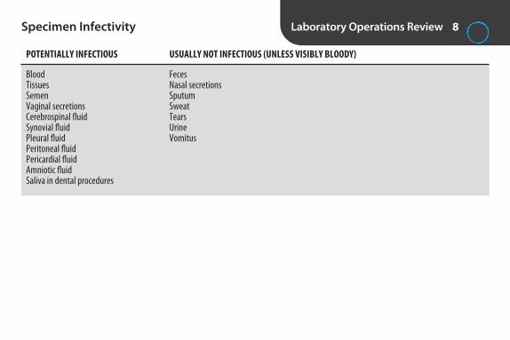
Specimen Infectivity Laboratory Operations Review 8
POTENTIALLY INFECTIOUS USUALLY NOT INFECTIOUS (UNLESS VISIBLY BLOODY)
BloodTissuesSemenVaginal secretionsCerebrospinal fluidSynovial fluidPleural fluidPeritoneal fluidPericardial fluidAmniotic fluidSaliva in dental procedures
FecesNasal secretionsSputumSweatTearsUrineVomitus
2956_Ch01_001-066 30/01/14 3:07 PM Page 8

Packaging of Biologics for Shipping Laboratory Operations Review 9
REQUIREMENT EXPLANATION
Primary container
Secondary container
Mailing container
Labeling
Training
Test tube, vial, etc. containing etiologic agent. Must be securely closed, watertight, surrounded by absorbentmaterial, & placed in secondary container.
Must be watertight, sealed, & placed in approved mailing container.
Must be made of fiberboard.
Biohazard label required on primary & mailing containers.
Employees must be trained & retrained every 2–3 yr or when regulations change.
2956_Ch01_001-066 30/01/14 3:07 PM Page 9

Hazard Communication Standard (HCS) Laboratory Operations Review 10
History
Also Known As
Purpose
Primary Requirements
Issued by OSHA in 1983. Written for manufacturing industry, but courts expanded jurisdiction to clinical labs.
“Right-to-Know Law”; “HAZCOM”
To inform employees about chemical hazards in workplace & protective measures
Written hazard communication planInventory of hazardous chemicals on siteHazard labelingMaterial safety data sheet (MSDS) for each chemical readily accessible to employees on each shift.Training on initial assignment & when new hazard introduced.
2956_Ch01_001-066 30/01/14 3:07 PM Page 10

Occupational Exposures to Hazardous Chemicals in Laboratories Standard
Laboratory Operations Review 11
History
Also Known As
Purpose
Primary Requirements
Issued by OSHA in 1990. Extension of HCS written specifically for labs.
“Laboratory Standard”; “Chemical Hygiene Standard”
To limit employee exposure to hazardous chemicals to levels at or below permissible exposure levels (PELs).
Written chemical hygiene plan outlining standard operating procedures for use, storage, exposure control, &disposal of hazardous chemicals.
Designation of chemical hygiene officer.Hazard identification & labeling.Material safety data sheet (MSDS) for each chemical readily accessible to employees on each shift.Use of personal protective equipment.Proper maintenance of fume hoods & other protective equipment.Monitoring of employee exposure to hazardous chemicals. Medical exams at no cost in cases of suspected overexposure.Training on initial assignment & before assignments involving new exposures.
2956_Ch01_001-066 30/01/14 3:07 PM Page 11

Hazard Categories of Chemicals Laboratory Operations Review 12
CLASSIFICATION EXAMPLE EFFECT COMMENTS
Corrosives
Toxic substances
Carcinogens
Mutagens & teratogens
Ignitables
Chemicals with pH <2 or >12. Separate in-organic acids from organic acids. Concen-trated acids & bases can generate largeamounts of heat when mixed with water.
Threshold limit values (TLVs) = safe level of exposure.
OSHA requires monitoring of formaldehydeexposure.
Special precautions during pregnancy.
Flashpoint = lowest temp that produces ignitable vapor. Flammables <100°F; combustibles ≥ 100°F.
Glacial acetic acid, hydrochloricacid, sodium hydroxide
Cyanides, sulfides
Benzidine, formaldehyde
Benzene, lead, mercury, ra-dioactive material, toluene
Acetone, alcohols, ether, xylene
Visible destruction ofhuman tissue on contact.Can cause injury on inhala-tion or contact.
Interfere with metabolicprocesses when ingested,inhaled, or absorbedthrough skin.
Capable of causing cancer.
Mutagens induce geneticmutations; teratogenscause defects in embryo.
Fire
continued...
2956_Ch01_001-066 30/01/14 3:07 PM Page 12

Hazard Categories of Chemicals continued
CLASSIFICATION EXAMPLE EFFECT COMMENTS
Reactives Ether forms explosive peroxides on exposureto air or light; store in explosion-proof refrig-erator. Perchloric acid may react explosivelywith organic compounds; separate fromother acids. Picric acid is shock sensitivewhen dehydrated; more powerful than TNT.Sodium azide solutions can form explosivelead or copper azides in drains.
Ether, perchloric acid, picricacid, sodium azide
Explosion
Laboratory Operations Review 13
2956_Ch01_001-066 30/01/14 3:07 PM Page 13
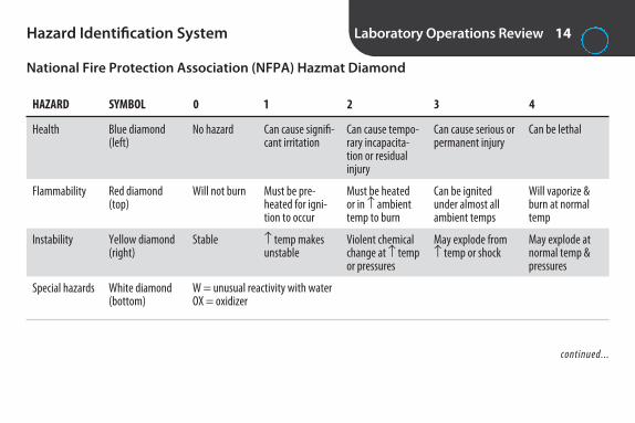
National Fire Protection Association (NFPA) Hazmat Diamond
Hazard Identification System Laboratory Operations Review 14
HAZARD SYMBOL 0 1 2 3 4
Health
Flammability
Instability
Special hazards
Can be lethal
Will vaporize &burn at normaltemp
May explode atnormal temp &pressures
Blue diamond(left)
Red diamond(top)
Yellow diamond(right)
White diamond(bottom)
No hazard
Will not burn
Stable
Can cause signifi-cant irritation
Must be pre-heated for igni-tion to occur
↑ temp makesunstable
Can cause tempo-rary incapacita-tion or residualinjury
Must be heatedor in ↑ ambienttemp to burn
Violent chemicalchange at ↑ tempor pressures
Can cause serious orpermanent injury
Can be ignitedunder almost allambient temps
May explode from↑ temp or shock
W = unusual reactivity with waterOX = oxidizer
continued...
2956_Ch01_001-066 30/01/14 3:07 PM Page 14

Hazard Identification System continued
Red area =flammability
Blue area =health
Yellow area =reactivity
White area =special dangers
Safety diamond. Colored areas within the diamond indicate types of danger: red area (top) = flammability; blue area(left) = health; yellow area (right) = reactivity; and white area (bottom) = special dangers. (From Arneson W, Brickell J. ClinicalChemistry: A Laboratory Perspective. Philadelphia: FA Davis; 2007:5.)
Laboratory Operations Review 15
2956_Ch01_001-066 30/01/14 3:07 PM Page 15

Storage of Chemicals Laboratory Operations Review 16
CHEMICAL CATEGORY EXAMPLES STORAGE GUIDELINES
Acids
Bases
Flammables
Oxidizers
Water-reactive chemicals
Store below counter level or in acid cabinets. Separatefrom flammable & combustible material, bases, & activemetals (e.g., sodium, potassium, magnesium). Separateorganic acids from inorganic acids. Separate oxidizingacids from organic acids.
Separate from acids. Store inorganic hydroxides in polyethylene containers.
Limit amount in work area. Store in approved safety cansor cabinets. Separate from oxidizing acids & oxidizers.
Separate from reducing agents (e.g., zinc, alkaline metals,formic acid), flammable & combustible materials.
Keep away from water. Store in a dry, cool place.
Organic: formic, glacial acetic, citric Inorganic: hydrochloric, nitric, sulfuric Oxidizing: chromic, nitric, perchloric, sulfuric
Ammonium hydroxide, potassium hydroxide,sodium hydroxide
Acetone, alcohols, xylene
Nitric acid, perchloric acid, sulfuric acid, aceticacid, potassium chloride, hydrogen peroxide
Sodium, potassium
2956_Ch01_001-066 30/01/14 3:07 PM Page 16

Fire Safety Laboratory Operations Review 17
CLASS OF FIRE COMBUSTIBLE MATERIAL EXTINGUISHERS TO USE COMMENTS
A
B
C
D
Don’t use water on electrical fires orburning liquids.
Never use water. Dry chemical maydamage electrical equipment. CO2leaves no residue; good choice forcomputers, analyzers.
Cloth, wood, paper
Flammable or combustible liquids
Electrical equipment
Combustible metals
Pressurized water (A) Dry chemical (ABC)
Dry chemical (ABC) CO2 (BC)
Dry chemical (ABC) CO2 (BC)
Leave to professional firefighters.
2956_Ch01_001-066 30/01/14 3:07 PM Page 17

Commonly Used Anticoagulants/Additives Laboratory Operations Review 18
ANTICOAGULANT/ADDITIVE STOPPER COLOR MODE OF ACTION EXAMPLES OF USE COMMENTS
EDTA
Heparin
Sodium citrate
Sodium fluoride
Prevents platelets from clumping. Minimal morphologic changes to WBCs.Tube should be at least 1/2 full.
Best anticoagulant for prevention of hemolysis. Don’t use for diffs (blue background).
Preserves labile clotting factors. Tube mustbe full for 9:1 blood-to-anticoagulantratio or coag results falsely ↑. To ensureproper ratio when drawing with butterfly,use discard tube to clear air from tubing.Discard tube not required in other situa-tions. Reduce anticoagulant when HCT>55%.
Preserves glucose for 24 hr. Combinedwith K oxalate if anticoagulationneeded. Oxalate binds Ca2+.
CBC, diff, sed rate
Many chemistries, os-motic fragility, plasmahgb, blood gases
Most coagulation tests
Glucose, lactic acid,blood alcohol
Lavender
Green
Light blue
Gray
Prevents clotting bychelating Ca2+
Prevents clotting byneutralizing thrombin
Prevents clotting bybinding Ca2+
Inhibits glycolysis (not an anticoagulant)
2956_Ch01_001-066 30/01/14 3:07 PM Page 18

Recommended Order for Drawing EvacuatedTubes & Filling Tubes From a Syringe
Laboratory Operations Review 19
TUBE STOPPER OR CLOSURE COMMENTS
Blood culture
Coagulation (citrate)
Serum (with/without clot activator; with/without gel)
Heparin (with/without gel)
EDTA
Glycolytic inhibitor (Na fluoride/K oxalate)
Drawing 1st avoids bacterial contamination from needle that haspierced other stoppers.
Drawing before other anticoagulant & clot activator tubes avoidscontamination with additives that can affect coag results.
Drawing before green avoids contamination with sodium heparin(↑ Na+) or lithium heparin (↑ Li+). Drawing before lavender avoidscontamination from K2EDTA ( ↓ Ca2+, Mg2+; ↑ K+). Drawing beforegray avoids contamination with sodium fluoride/potassium oxalate(↓ Ca2+, ↑ Na+, ↑ K+, interference with some enzyme assays).
Drawing before lavender avoids contamination from K2EDTA ( ↓ Ca2+, Mg2+; ↑ K+). Drawing before gray avoids contaminationwith sodium fluoride/potassium oxalate (↓ Ca2+, ↑ Na+, ↑ K+).
Drawing before gray avoids contamination with oxalate, which alters cellular morphology
Yellow (SPS) or blood culturebottle
Light blue
Red, gold, speckled
Green
Lavender, pink, white
Gray
(CLSI H3-A6, 2007)
2956_Ch01_001-066 30/01/14 3:07 PM Page 19

Recommended Order for Filling MicrocollectionTubes From Capillary Punctures
Laboratory Operations Review 20
TEST/TUBE RATIONALE FOR ORDER
Blood gases Minimizes exposure to air
EDTA Minimizes clumping of platelets
Other additive tubes Minimizes clotting
Serum tubes Clotting is not a concern
(CLSI H4-A6, 2008)
2956_Ch01_001-066 30/01/14 3:07 PM Page 20
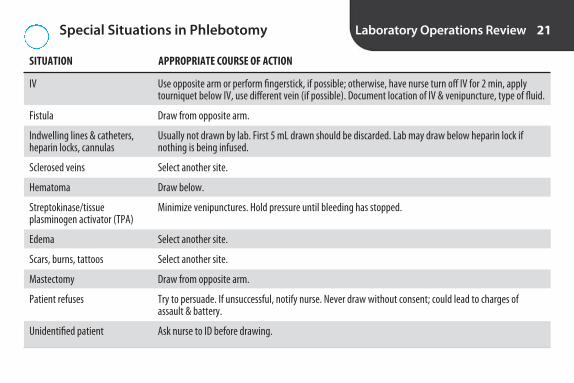
Special Situations in Phlebotomy Laboratory Operations Review 21
SITUATION APPROPRIATE COURSE OF ACTION
IV
Fistula
Indwelling lines & catheters, heparin locks, cannulas
Sclerosed veins
Hematoma
Streptokinase/tissue plasminogen activator (TPA)
Edema
Scars, burns, tattoos
Mastectomy
Patient refuses
Unidentified patient
Use opposite arm or perform fingerstick, if possible; otherwise, have nurse turn off IV for 2 min, applytourniquet below IV, use different vein (if possible). Document location of IV & venipuncture, type of fluid.
Draw from opposite arm.
Usually not drawn by lab. First 5 mL drawn should be discarded. Lab may draw below heparin lock if nothing is being infused.
Select another site.
Draw below.
Minimize venipunctures. Hold pressure until bleeding has stopped.
Select another site.
Select another site.
Draw from opposite arm.
Try to persuade. If unsuccessful, notify nurse. Never draw without consent; could lead to charges of assault & battery.
Ask nurse to ID before drawing.
2956_Ch01_001-066 30/01/14 3:07 PM Page 21

Special Test Requirements Laboratory Operations Review 22
REQUIREMENT EXAMPLES* COMMENTS
Fasting
Chilling
Warming
Protection from light
Chain of custody
*Follow laboratory’s established procedures.
Nothing to eat or drink (except water) for at least 8 hr
Place in slurry of crushed ice & water. Don’t use ice cubesalone because RBCs may lyse.
Use 37ºC heat block, heel warmer, or hold in hand.
Wrap in aluminum foil.
Chain of custody form. Lock box may be required.
Fasting blood sugar, triglycerides, lipid panel,gastrin, insulin
ACTH, acetone, ammonia, gastrin, glucagon, lactic acid, pyruvate, PTH, renin
Cold agglutinins, cryoglobulins
Bilirubin, carotene, erythrocyte protoporphyrin,vitamin A, vitamin B12
Any test used as evidence in legal proceedings;e.g., blood alcohol, drug screens, DNA analysis
2956_Ch01_001-066 30/01/14 3:07 PM Page 22

Phlebotomy Sources of Error Laboratory Operations Review 23
ERROR POSSIBLE EFFECT
Misidentification of patient
Drawing at incorrect time
Improper skin disinfection
Drawing from edematous site
Fist pumping during venipuncture
Tourniquet >1 min
IV fluid contamination
Expired collection tubes
Incorrect anticoagulant or contamination from incorrect order of draw
Failure to hold bottom of tube lower than top during collection
Treatment errors, possibility of transfusion fatality
Treatment errors if samples for certain tests aren’t drawn at appropriate time, e.g.,therapeutic drug monitoring, analytes that exhibit diurnal variation, analytes thatare affected by recent eating/drinking
Infection at site of puncture. Contamination of blood cultures & blood components.Isopropyl alcohol wipes can contaminate samples for blood alcohol.
Dilution of sample with tissue fluid
↑ K+, lactic acid, Ca2+, phosphorus; ↓ pH
↑ K+, total protein, lactic acid
↑ glucose, electrolytes (depending on IV)
↓ vacuum, failure to obtain specimen
K2EDTA before serum or heparin tube: ↓ Ca2+, Mg2+, ↑ K+
Contamination of citrate tube with clot activator: erroneous coag results.
Carryover from one tube to another. Possible additive contamination.
continued...
2956_Ch01_001-066 30/01/14 3:07 PM Page 23

Phlebotomy Sources of Error continued
ERROR POSSIBLE EFFECT
Short draws
Inadequate mixing of anticoagulant tube
Hemolysis from alcohol contamination, “milking” site of capillary puncture, probing with needle, vigorous shaking of tubes, exposure of samples to extremes of temperature
Incorrect blood: anticoagulant ratio affects some results, e.g., coag tests.
Micro-clots, fibrin, platelet clumping can lead to erroneous results.
↑ K+, Mg2+, LD, iron
Laboratory Operations Review 24
2956_Ch01_001-066 30/01/14 3:07 PM Page 24

Guidelines for Specimen Handling & Processing*
Laboratory Operations Review 25
• Transport blood specimens carefully to avoid hemolysis.
• Protect tubes for bili, carotene from light.• Transport samples for ACTH, lactic acid, ammonia,
blood gases in ice slurry.• Maintain tubes in vertical position to promote
complete clotting.• Allow serum & gel separator tubes to clot for
30–60 min before centrifugation to avoid fibrinstrands.
• Centrifuge within 2 hr of collection.• Spin most tubes at 1,000–1,300 RCF for 10–15 min.• Spin citrate tubes at 1,500 RCF for 15 min to produce
platelet-poor plasma.• Keep tubes capped during centrifugation to avoid
loss of CO2, change of pH, evaporation, or aerosol formation.
• Don’t re-spin primary tubes. Can cause hemolysis. Ifrecentrifuging is necessary, transfer serum/plasma toanother tube.
• Don’t re-spin serum separator tubes. Serum in contactwith RBCs under gel can be expressed & ↑ K+.
• Separate serum or plasma from cells within 2 hr ofcollection (exception: centrifuged gel tubes).
• When transferring samples to secondary containers,aspirate to avoid cellular contamination. Don’t pour.
• Lipemic specimens can be ultracentrifuged at 105 x gto remove chylomicrons (triglycerides).
• Separated serum/plasma may be kept at RT for 8 hror at 2–8°C for 48 hr. For longer storage, freeze at –20°C. Avoid repeated freezing & thawing.
• Don’t freeze whole blood.
*Always follow laboratory’s established procedures.
2956_Ch01_001-066 30/01/14 3:07 PM Page 25

Centrifuges Laboratory Operations Review 26
TERM EXPLANATION
Relative centrifugal force (RCF)
rpm
Radius (r)
Horizontal-head centrifuge (swinging-bucket)
Angle-head centrifuge
Ultra centrifuge
Always make sure centrifuge is balanced. Don’t open while spinning. Keep tubes capped.
Force acting on sample being centrifuged. Gravities (g). Function of rpm and radius. RCF = 1.12 × 10⁻⁵ × r × rpm2.
Revolutions per minute. Speed of centrifugation. Determined by tachometer.
Distance in cm from center of rotation to bottom of tube when rotating.
Tubes are in horizontal position when rotating. Produces a tightly packed, flat sediment surface. Recommended for serum separator tubes.
Tubes are at fixed angle (25°–40°) when rotating. Capable of higher speeds. Produces aslanted sediment surface that isn’t tightly packed. Decantation is not recommended.
High-speed. Capable of 100,000 rpm. Refrigerated to reduce heat.
2956_Ch01_001-066 30/01/14 3:07 PM Page 26

Examples of Criteria for Specimen Rejection* Laboratory Operations Review 27
• Missing or inadequate label• Collected at wrong time• Collected in wrong tube• Insufficient specimen• Inadequate volume of blood in anticoagulant tube• Exposure to temperature extremes• Prolonged transit• Clots in CBC tube• Hemolysis (depending on test ordered)• Lipemia (depending on test ordered)
*Follow lab’s written policies.
2956_Ch01_001-066 30/01/14 3:07 PM Page 27

Types of Glass Laboratory Operations Review 28
TYPE CHARACTERISTICS
Borosilicate glass (Kimax, Pyrex)
Aluminosilicate glass (Corex)
Boron free
High silica
Flint glass
Low actinic
High resistance to thermal shock & chemical attack. Heavy walls to minimize breakage. Used formost beakers, flasks, & pipets. Minimal contamination of liquids by elements in glass. Can be heated& autoclaved.
6 times stronger than borosilicate. Better able to resist clouding due to alkali & scratching.
Used for highly alkaline solutions. Alkali resistant. Poor heat resistance.
Heat, chemical, & electrical tolerance. Excellent optical properties. Used for high-precision analyticwork, optical reflectors, mirrors.
Soda-lime glass containing oxides of sodium, silicon, & calcium. Least expensive but poor resistanceto high temp & sudden changes of temp. Only fair resistance to chemicals. Can release alkali & affectsome determinations. Used for some disposable glassware.
Amber or red. Used to ↓ exposure to light, e.g., bilirubin standards.
2956_Ch01_001-066 30/01/14 3:07 PM Page 28

Types of Plastic Laboratory Operations Review 29
TYPE CHARACTERISTICS
Polypropylene
Polyethylene
Polycarbonate
Polystyrene
Polyvinyl chloride
Teflon
Relatively inert chemically. Resistant to most acids, alkalis, & salts. Can be autoclaved. Used for pipet tips, testtubes.
Relatively inert chemically. Resistant to most acids (except concentrated H2SO4), alkalis, & salts. Used for testtubes, bottles, disposable transfer pipets, test tube racks. Can’t be autoclaved.
Stronger than polypropylene & better temp tolerance, but chemical resistance not as good. Clear. Resistant toshattering. Used for centrifuge tubes, graduated cylinders.
Rigid, clear. Shouldn’t be autoclaved. Will crack & splinter. Used for test tubes, graduated tubes.
Soft & flexible but porous. Frequently used as tubing.
Extremely inert. Excellent temp tolerance & chemical resistance. Used for stir bars, stopcocks, tubing.
2956_Ch01_001-066 30/01/14 3:07 PM Page 29

Glassware Inscriptions Laboratory Operations Review 30
INSCRIPTION EXPLANATION
A
20°C
TC
TD
Class A. Meets high standards for accuracy.
Temp of calibration. Temp glassware & solutions should be for maximum accuracy.
To contain. Vessel calibrated to hold specified volume (e.g., volumetric flask).
To deliver. Vessel calibrated to deliver specified volume (e.g., graduated cylinder).
2956_Ch01_001-066 30/01/14 3:07 PM Page 30

Volumetric Glassware Laboratory Operations Review 31
GLASSWARE DESCRIPTION/USE
Beaker
Erlenmeyer flask
Florence flask
Volumetric flask
Graduated cylinder (graduates)
Wide-mouthed, straight-sided jar with pouring spout. Not accurate enough for critical measurements.
Sloping sides. Graduated markings. Used to hold liquids, mix solutions, measure noncritical volumes.
Spherical base with long cylindrical neck. Single calibration mark. Only for noncritical measurements.
Pear shaped. Long neck with single calibration mark. Manufactured to strict standards. Glassware &solutions should be at RT. Used to prepare standards & reagents. Shouldn’t be used to store solutions.
Upright, straight-sided tube with flared base. Used for noncritical measurements. Most are TD.Shouldn’t be used to measure <5 mL or <10% of capacity. Use graduate closest in size to volume tobe measured.
2956_Ch01_001-066 30/01/14 3:07 PM Page 31

Glass Pipets Laboratory Operations Review 32
PIPET DESCRIPTION/USE
Volumetric
Ostwald-Folin
Serological
Mohr
Micropipet
Transfer pipet. Single calibration mark. Calibrated to accurately deliver fixed volume of nonviscous samples &standards. Touch off last drop against wall of receiving vessel.
Transfer pipet. Similar to volumetric pipet, but bulb closer to tip. Etched ring means blowout. Used for accuratemeasurement of viscous fluids, e.g., whole blood. Not widely used.
Graduated or measuring pipet. Graduation marks down to tip. Etched ring means blowout. Can use for serial dilutions & measuring reagents. Not accurate enough for measuring samples or standards.
Graduated or measuring pipet. Doesn’t have graduation marks all the way to tip or frosted band near upper end.Delivery is made “point to point.” Not widely used.
Disposable pipet for volumes ranging from 1–1,000 µ L. Single calibration mark. Filled by capillary action. TC. Must be rinsed out with diluent to deliver exact amount. Small pipetting bulb is used.
2956_Ch01_001-066 30/01/14 3:07 PM Page 32

Mechanical Micropipets Laboratory Operations Review 33
TypesAir displacement
Positive displacement
Calibration
Uses suction to aspirate & dispense sample through polypropylene tip. 1- or 2-stop. With 2-stop, button isdepressed to 2nd stop to “blow out.” Tips can only be used once. Seals require periodic lubrication. Followmanufacturer’s instructions for use.
Uses a glass capillary tip fitted with Teflon-tipped plunger. No carryover. Tips are reusable. Plunger settingmust be checked & Teflon tip replaced periodically. Follow manufacturer’s instructions for use.
Verify accuracy & precision on receipt, after service or repair, & on regular schedule. Most accurate method for calibration is gravimetric method (weight of distilled water delivered). Secondary method is spectropho-tometric (absorbance of potassium dichromate or p-nitrophenol delivered).
2956_Ch01_001-066 30/01/14 3:07 PM Page 33
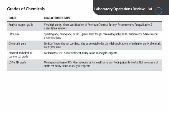
Grades of Chemicals Laboratory Operations Review 34
GRADE CHARACTERISTICS/USE
Analytic reagent grade
Ultra pure
Chemically pure
Practical, technical, or commercial grade
USP or NF grade
Very high purity. Meets specifications of American Chemical Society. Recommended for qualitative & quantitative analysis.
Spectrograde, nanograde, or HPLC grade. Used for gas chromatography, HPLC, fluorometry, & trace metaldeterminations.
Limits of impurities not specified. May be acceptable for some lab applications when higher purity chemicalsaren’t available.
For industrial use. Not of sufficient purity to use as analytic reagents.
Meet specifications of U.S. Pharmacopeia or National Formulary. Not injurious to health. Not necessarily ofsufficient purity to use as analytic reagents.
2956_Ch01_001-066 30/01/14 3:07 PM Page 34
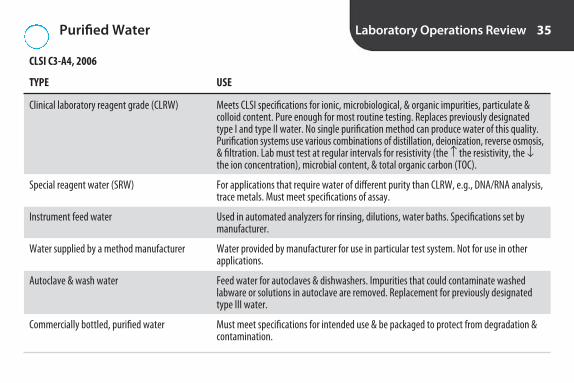
Purified Water Laboratory Operations Review 35
CLSI C3-A4, 2006
TYPE USE
Clinical laboratory reagent grade (CLRW)
Special reagent water (SRW)
Instrument feed water
Water supplied by a method manufacturer
Autoclave & wash water
Commercially bottled, purified water
Meets CLSI specifications for ionic, microbiological, & organic impurities, particulate & colloid content. Pure enough for most routine testing. Replaces previously designatedtype I and type II water. No single purification method can produce water of this quality.Purification systems use various combinations of distillation, deionization, reverse osmosis,& filtration. Lab must test at regular intervals for resistivity (the ↑ the resistivity, the ↓the ion concentration), microbial content, & total organic carbon (TOC).
For applications that require water of different purity than CLRW, e.g., DNA/RNA analysis,trace metals. Must meet specifications of assay.
Used in automated analyzers for rinsing, dilutions, water baths. Specifications set bymanufacturer.
Water provided by manufacturer for use in particular test system. Not for use in other applications.
Feed water for autoclaves & dishwashers. Impurities that could contaminate washed labware or solutions in autoclave are removed. Replacement for previously designatedtype III water.
Must meet specifications for intended use & be packaged to protect from degradation &contamination.
2956_Ch01_001-066 30/01/14 3:07 PM Page 35
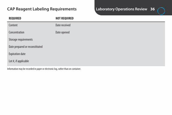
CAP Reagent Labeling Requirements Laboratory Operations Review 36
REQUIRED NOT REQUIRED
Content
Concentration
Storage requirements
Date prepared or reconstituted
Expiration date
Lot #, if applicable
Information may be recorded in paper or electronic log, rather than on container.
Date received
Date opened
2956_Ch01_001-066 30/01/14 3:07 PM Page 36

Brightfield Microscopy Laboratory Operations Review 37
TERM EXPLANATION
Achromatic objective
Aperture diaphragm
Binocular microscope
Blue filter
Brightfield microscope
Compound microscope
Condenser
Depth of focus
Field diaphragm
Field of view
Immersion oil
Kohler illumination
Least expensive objective. Partially corrects for chromatic & spherical aberrations.
Controls angle & amount of light sent to objective.
One with 2 oculars.
Used to eliminate yellow color emitted by tungsten.
Uses transmitted light & lenses. Objects appear dark against white background. Used for most routine clinical work.
One with 2 lens systems—objectives & oculars.
Focuses light on specimen.
Distance throughout which all parts of specimen are in focus simultaneously.
Limits area of illumination to image field.
Area of specimen that can be seen.
Used to help objective gather light from a wide numerical aperture. Provides high resolution. Type B (highviscosity) is commonly used.
Method of focusing & centering light path & spreading light uniformly. Ensures optimum contrast & resolution.
continued...
2956_Ch01_001-066 30/01/14 3:07 PM Page 37

Brightfield Microscopy continued
TERM EXPLANATION
Magnification, total
Numerical aperture (NA)
Objectives
Ocular
Parcentric
Parfocal
Planachromatic objective
Resolution
Rheostat
Tungsten-halogen bulb
Virtual image
Working distance
Magnification of ocular × magnification of objective. 1,000× is highest magnification achievable withbrightfield microscope.
Mathematical expression of light admitted by lens. The higher the NA, the greater the resolution.
Lenses attached to revolving nosepiece. Most commonly used are low power (10×), high power (40×), &oil immersion (50× or 100×).
Eye piece. Usually 10×.
Object in center of field at 1 magnification will be in center of field at other magnifications
Object remains in focus from 1 magnification to another
More expensive objective that corrects for curvature of field. Results in flat field with uniform focus.
Ability to reveal fine detail & distinguish between 2 close points.
Light control knob. Light intensity shouldn’t be regulated by condenser or diaphragms.
Type of bulb used for brightfield microscopy.
Image seen through microscope. Upside down & reversed.
Distance between slide & objective. Decreases with higher magnification objectives.
Laboratory Operations Review 38
2956_Ch01_001-066 30/01/14 3:07 PM Page 38
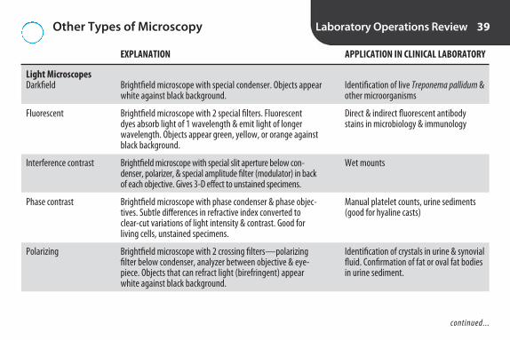
Other Types of Microscopy Laboratory Operations Review 39
EXPLANATION APPLICATION IN CLINICAL LABORATORY
Light Microscopes Darkfield
Fluorescent
Interference contrast
Phase contrast
Polarizing
Identification of live Treponema pallidum &other microorganisms
Direct & indirect fluorescent antibodystains in microbiology & immunology
Wet mounts
Manual platelet counts, urine sediments(good for hyaline casts)
Identification of crystals in urine & synovialfluid. Confirmation of fat or oval fat bodiesin urine sediment.
Brightfield microscope with special condenser. Objects appearwhite against black background.
Brightfield microscope with 2 special filters. Fluorescent dyes absorb light of 1 wavelength & emit light of longerwavelength. Objects appear green, yellow, or orange againstblack background.
Brightfield microscope with special slit aperture below con-denser, polarizer, & special amplitude filter (modulator) in backof each objective. Gives 3-D effect to unstained specimens.
Brightfield microscope with phase condenser & phase objec-tives. Subtle differences in refractive index converted to clear-cut variations of light intensity & contrast. Good for living cells, unstained specimens.
Brightfield microscope with 2 crossing filters—polarizing filter below condenser, analyzer between objective & eye-piece. Objects that can refract light (birefringent) appearwhite against black background.
continued...
2956_Ch01_001-066 30/01/14 3:07 PM Page 39

Other Types of Microscopy continued
EXPLANATION APPLICATION IN CLINICAL LABORATORY
Electron Microscopes Transmission
Scanning
Virology, cells (organelles)
Virology, cells (surface)
Beam of electrons passes through specimen, focused onto fluorescent screen or photographic plate. Magnification>100,000×.
Beam of electrons strikes surface of specimen, focused onto photographic film or cathode ray tube. 3-D image. Magnification >1,000×.
Laboratory Operations Review 40
2956_Ch01_001-066 30/01/14 3:07 PM Page 40

Informatics Laboratory Operations Review 41
TERM EXPLANATION
Informatics Information science. Science of processing data for storage, retrieval, & use.
Laboratory informatics Use of computers & information systems to process & communicate information generated in clinical lab
Electronic medical record (EMR) Computerized medical record. American Recovery & Reinvestment Act of 2009 (ARRA) calls for EMRs for all patients by 2014 to help ↓ cost & ↑ efficiency of health-care delivery.
2956_Ch01_001-066 30/01/14 3:07 PM Page 41

Computer Hardware Laboratory Operations Review 42
TERM EXPLANATION
Hardware
CPU
RAM
ROM
Hard drive
Disk drive
Optical disks
Peripheral devices
Input devices
Output devices
Modem
Physical parts of computer
Central processing unit. Executes software instructions.
Random access memory. Working memory used for temporary storage of programs & data. Content is lost eachtime computer is turned off.
Read-only memory. Part of memory that is permanently protected from being modified, erased, or written over.Not affected by power loss. Used for boot-level & other system instructions.
Magnetic-coated metal plate inside CPU for storing data
Device that reads data stored on magnetic or optical disk & writes data onto disk for storage
CDs, DVDs. Store data.
Input/output & information storage components
Devices that deliver data to computer, e.g., keyboards, barcode readers, computer links (interfaces)
Devices by which computer delivers data, e.g., printers, monitors
Input/output device that allows computers to communicate over telephone lines
2956_Ch01_001-066 30/01/14 3:07 PM Page 42

Computer Software Laboratory Operations Review 43
TERM EXPLANATION
Software
Operating system software
Application software
Word processor
Spreadsheet
Database
Browser
Programs that tell computer what to do
Program that controls basic functions of computer, e.g., Microsoft Windows
Programs designed to meet specific needs of users, e.g., word processing
Application program that allows for manipulation of text. Used to write letters, reports, etc.
Application program to manipulate numbers & perform mathematical calculations. Used to prepare financial statements, budgets, etc.
Application program to organize, store, sort, & retrieve data (words or numbers).
Program that provides access to Internet, e.g., Microsoft Internet Explorer
2956_Ch01_001-066 30/01/14 3:07 PM Page 43

Information Systems Laboratory Operations Review 44
TERM EXPLANATION
Laboratory information system (LIS)
Hospital information system (HIS)
Interface
Unidirectional interface
Bidirectional interface
Middleware
System validation
System of hardware, software, connections, & communication protocols to handle all informa-tional needs of lab, from intake of requests to delivery of results. Can provide patient informa-tion, test information, collection lists, work lists, test results, financial functions, productivity/workload monitoring, quality management, & interface with other computer systems.
Information system to handle all informational needs of hospital, both clinical & administrative.
Hardware & software that allow for electronic communication between 2 computer systems,even if they use different programming languages. The LIS is typically interfaced to HIS & automated analyzers.
Interface that transmits electronic information in 1 direction, e.g., a point-of-care analyzerdownloads test results to LIS.
Interface that transmits electronic information in 2 directions, e.g., the LIS downloads ordersfrom the HIS & uploads results to the HIS.
Interface between an analyzer and LIS. Can apply rules to automate processes, e.g., autoverifica-tion (automatic release of results without tech review when certain criteria are met).
Documentation that LIS functions as expected. Required by regulatory agencies.
2956_Ch01_001-066 30/01/14 3:07 PM Page 44

Computer Networks Laboratory Operations Review 45
TERM EXPLANATION
Local area network (LAN)
Wide area network (WAN)
Internet
Intranet
Extranet
Protocol
Ethernet
TCP/IP
Health Level 7 standard (HL7)
Computer network that connects computers in close geographic proximity (e.g., building, campus)
Computer network that connects computers over larger geographic area (e.g., multisite health-carefacility, Internet)
Global system of interconnected computer networks
Computer network within an organization. Access is usually restricted to employees.
Extension of a private network onto the Internet where it can be accessed by authorized clients, suppliers, etc.
Common set of signals & rules that network uses for communication
One of the 1st protocols developed for connecting computers
Transmission Control Protocol/Internet Protocol. Originally developed as transfer protocol for Internet; adapted for transmission in LANs.
Standardized message protocol that facilitates exchange of medical data among computer systems
2956_Ch01_001-066 30/01/14 3:07 PM Page 45
Quality Assessment Laboratory Operations Review 46
TERM EXPLANATION
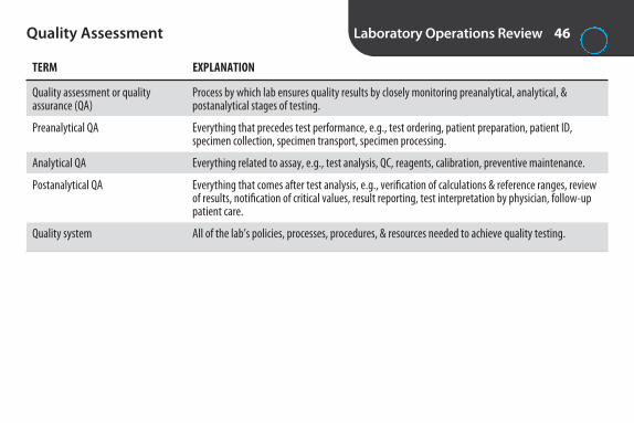
Quality assessment or quality assurance (QA)
Preanalytical QA
Analytical QA
Postanalytical QA
Quality system
Process by which lab ensures quality results by closely monitoring preanalytical, analytical, & postanalytical stages of testing.
Everything that precedes test performance, e.g., test ordering, patient preparation, patient ID, specimen collection, specimen transport, specimen processing.
Everything related to assay, e.g., test analysis, QC, reagents, calibration, preventive maintenance.
Everything that comes after test analysis, e.g., verification of calculations & reference ranges, reviewof results, notification of critical values, result reporting, test interpretation by physician, follow-uppatient care.
All of the lab’s policies, processes, procedures, & resources needed to achieve quality testing.
2956_Ch01_001-066 30/01/14 3:07 PM Page 46
Quality Control Laboratory Operations Review 47
TERM EXPLANATION
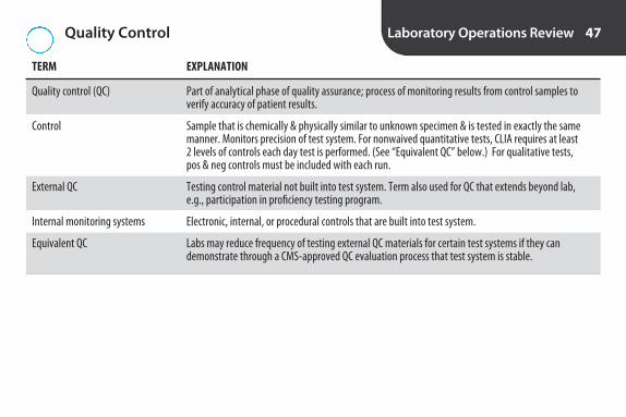
Quality control (QC)
Control
External QC
Internal monitoring systems
Equivalent QC
Part of analytical phase of quality assurance; process of monitoring results from control samples toverify accuracy of patient results.
Sample that is chemically & physically similar to unknown specimen & is tested in exactly the samemanner. Monitors precision of test system. For nonwaived quantitative tests, CLIA requires at least 2 levels of controls each day test is performed. (See “Equivalent QC” below.) For qualitative tests, pos & neg controls must be included with each run.
Testing control material not built into test system. Term also used for QC that extends beyond lab, e.g., participation in proficiency testing program.
Electronic, internal, or procedural controls that are built into test system.
Labs may reduce frequency of testing external QC materials for certain test systems if they can demonstrate through a CMS-approved QC evaluation process that test system is stable.
2956_Ch01_001-066 30/01/14 3:07 PM Page 47

Quality Control Statistics Laboratory Operations Review 48
TERM EXPLANATION
Measures of dispersion
Range
Mean
Standard deviation (SD)
Coefficient of variation (CV)
Statistical parameters describing spread of data about mean, e.g., standard deviation,coefficient of variation, range. Measurements of precision.
Difference between highest & lowest values in data set.
Sum of all observations divided by number of observations. Average of all observations.
Statistical expression of dispersion of values around mean. Requires a minimum of 20 values.
SD = where Σ = sum, x = individual value, x– = mean, n = number of values
Expresses standard deviation as percentage. CV % = (SD ÷ mean) × 100. The ↓ the CV,the ↑ the precision.
∑( x x)n 1
-2
� �
continued...
2956_Ch01_001-066 30/01/14 3:07 PM Page 48

Quality Control Statistics continued
TERM EXPLANATION
Normal distribution The Gaussian bell-shaped curve:
68% of values fall within ±1 SD of mean95% of values fall within ±2 SD of mean (95% confidence interval)99.7% of values fall within ±3 SD of mean
68%
95%
–2s –1s +1s +2sX
Gaussian frequency distribution with95% limits indicated. (From Arneson W,Brickell J. Clinical Chemistry: A Laboratory Perspective. Philadelphia: FA Davis; 2007:54.)
Laboratory Operations Review 49
2956_Ch01_001-066 30/01/14 3:07 PM Page 49

Interpretation of Quality Control Data Laboratory Operations Review 50
TERM EXPLANATION
Control limits
Levey-Jennings chart
Range within which control values must fall for assay to be considered valid. Many labs use mean ±2 SD. 1 determination in 20 will fall outside ±2 SD. This is anticipated part of normal variation.
Normal distribution curve lying on its side, marked with mean, ±1, ±2, ±3 SD.
2
+ 2 S.D.+ 1 S.D.
X– 1 S.D.– 2 S.D.
4 6 8 10 12Day
14 16 18 20
Levey-Jennings chart showing in-control results. (From Strasinger SK, Di Lorenzo MS. Urinalysis andBody Fluids, 5th ed. Philadelphia: FA Davis; 2008:133.)
continued...
2956_Ch01_001-066 30/01/14 3:07 PM Page 50

Interpretation of Quality Control Data continued
TERM EXPLANATION
Outlier
Shift
A control result outside established limits. May be due to chance or may indicate problem in test system. If itoccurs more than once in 20 successive runs, investigation must be carried out.
6 consecutive control values on same side of mean.
2
+ 2 S.D.+ 1 S.D.
– 1 S.D.– 2 S.D.
4 6 8 10 12Day
14 16 18 20
X
Levey-Jennings chart showing outlier. (Modified from Strasinger SK, Di Lorenzo MS. Urinalysis andBody Fluids, 5th ed. Philadelphia: FA Davis; 2008:133.)
2 4 6 8 10 12Day
14 16 18 20
+ 2 S.D.+ 1 S.D.
– 1 S.D.– 2 S.D.
X
Levey-Jennings chart showing shift. (From Strasinger SK, Di Lorenzo MS. Urinalysis and Body Fluids,5th ed. Philadelphia: FA Davis; 2008:133.)
continued...
Laboratory Operations Review 51
2956_Ch01_001-066 30/01/14 3:07 PM Page 51

Interpretation of Quality Control Data continued
TERM EXPLANATION
Trend
Error, random
Error, systematic
False rejection
Control values increasing or decreasing for 6 consecutive runs.
Error that doesn’t recur in regular pattern, e.g., error due to dirty glassware, use of wrong pipet, voltage fluctuation, sampling error, anticoagulant or drug interference. Indicated by control value significantly different from others on Levey-Jennings chart, or violation of the 13S or R4S Westgard rules. Usually a 1-timeerror, & controls & samples can be rerun with success.
Recurring error inherent in test procedure, e.g., dirty photometer, faulty ISE, evaporation or contamination ofstandards or reagents. Affects all results. Indicated by trend or shift on Levey-Jennings chart, or violation of 22S,41S, or 10× Westgard rules (see following). Requires investigation to determine cause.
Rejection of run because QC results indicate problem when none is present. Use of Westgard rules minimizesfalse rejections.
2 4 6 8 10 12Day
14 16 18 20
+ 2 S.D.+ 1 S.D.
– 1 S.D.– 2 S.D.
X
Levey-Jennings chart showing trend. (From Strasinger SK, Di Lorenzo MS. Urinalysis and Body Fluids,5th ed. Philadelphia: FA Davis; 2008:133.)
Laboratory Operations Review 52
2956_Ch01_001-066 30/01/14 3:07 PM Page 52

Westgard Multirules Laboratory Operations Review 53
RULE EXPLANATION TYPE OF ERROR COMMENTS
12S
13S
22S
R4S
41S
10x
Initiates testing of other rules. If no violation ofother rules, run is considered in control.
Rejection rule
Rejection rule
Rejection rule
Rejection rule
Rejection rule
Random
Systematic
Random
Systematic
Systematic
1 control >±2s from mean. Warning flag of possiblechange in accuracy or precision.
1 control >±3s from mean
2 consecutive controls >2s from mean on same side
2 consecutive controls differ by >4s
4 consecutive controls >1s from mean on same side
10 consecutive controls on same side of mean
2956_Ch01_001-066 30/01/14 3:07 PM Page 53

Typical Steps Taken When a Control Is Outside Acceptable Range*
Laboratory Operations Review 54
STEP RATIONALE
Hold patient results until problem is resolved.
Rerun control (1 time only).
If control is still out, run a new vial of control or another lot #.
If control is still out, look for & correct any problems, then run control.
If control is still out, recalibrate, then run control.
If control is still out, get assistance.
Once resolved, document corrective action.
Evaluate all patient results in rejected run & since last run with acceptable QC. Repeat tests & issue corrected reports, as needed.
*Follow laboratory’s written protocol.
If a control value is inaccurate, patient values might be inaccurate.
Value might have been due to expected random error. (1 in 20 resultswill be outside ±2 SD.)
Control might have been outdated, improperly stored, contaminated.
Consider reagents (low, outdated, improperly stored, contaminated,change in lot #), preventive maintenance (overdue), mechanical problems,clots, etc.
Calibration may have shifted.
Supervisor or service rep may be able to determine problem.
Provides a record for future reference, points out repetitive problems.
Ensure accuracy of reported results.
2956_Ch01_001-066 30/01/14 3:07 PM Page 54
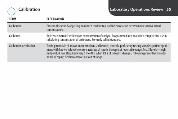
Calibration Laboratory Operations Review 55
TERM EXPLANATION
Calibration Process of testing & adjusting analyzer’s readout to establish correlation between measured & actual concentrations.
Calibrator Reference material with known concentration of analyte. Programmed into analyzer’s computer for use incalculating concentration of unknowns. Formerly called standard.
Calibration verification Testing materials of known concentrations (calibrators, controls, proficiency testing samples, patient speci-mens with known values) to ensure accuracy of results throughout reportable range. Test 3 levels—high,midpoint, & low. Required every 6 months, when lot # of reagents changes, following preventive mainte-nance or repair, & when controls are out of range.
2956_Ch01_001-066 30/01/14 3:07 PM Page 55

Test Performance Specifications and Verification
Laboratory Operations Review 56
CLIA requires labs to verify manufacturer’s analytical performance claims for # 1–4 below on new methods introducedafter April 24, 2003.
PARAMETER EXPLANATION VERIFICATION
1. Accuracy
2. Precision
3. Reportable range
4. Reference interval
Lab tests samples of known values (controls, calibrators,proficiency samples, previously tested patient speci-mens) to see how close results are to known value.
Lab repeatedly tests same samples (on same day &different days) to see how close the results are.
Lab tests samples with known values at highest &lowest levels claimed to be accurate by manufacturer.
If manufacturer’s or published reference ranges areused, lab must test specimens from normal subjects toverify ranges. Ranges may need to be adjusted to fitlab’s patient population.
How close measurement is to true value.
Reproducibility. How close results are when samesample is tested multiple times. (Note: A procedurecan be precise but not accurate.)
Range of values over which lab can verify accuracyof test system. Also known as linearity.
Formerly called normal value. Can vary for differentpatient populations (age, gender, race). Establishedby testing minimum of 120 healthy subjects & de-termining range in which 95% fall. (Note: 5% ofhealthy population falls outside of reference range.)Labs may use manufacturer’s reference ranges orpublished reference ranges, if appropriate for theirpatient population.
continued...
2956_Ch01_001-066 30/01/14 3:07 PM Page 56

Test Performance Specifications and Verification continued
PARAMETER EXPLANATION VERIFICATION
5. Analytical sensitivity
6. Analytical specificity
Determined by manufacturer. For unmodified FDA-approved tests, verification isn’t required.
Determined by manufacturer. For unmodified FDA-approved tests, verification isn’t required.
Same as detection limit. Lowest concentration ofsubstance that can be detected by test method. ↑ sensitivity means ↓ false negs. Desirable in screening tests.
Ability of method to measure only analyte it’s sup-posed to measure & not other related substances. ↑ specificity means ↓ false pos, ↓ cross-reactivity. Desirable in confirmatory tests.
Laboratory Operations Review 57
2956_Ch01_001-066 30/01/14 3:07 PM Page 57

Diagnostic Value of a Test Laboratory Operations Review 58
CHARACTERISTIC EXPLANATION
True positive (TP) Pos result in patient who has the disease
False positive (FP) Pos result in patient who doesn’t have the disease
True negative (TN) Neg result in patient who doesn’t have the disease
False negative (FN) Neg result in patient who does have the disease
Diagnostic sensitivity * % of population with the disease that test posTP / (TP + FN) × 100
Diagnostic specificity * % of population without the disease that test negTN / (TN + FP) × 100
Positive predictive value * (PPV) % of time that a pos result is correctTP / (TP + FP) × 100
Negative predictive value * (NPV) % of time that a neg result is correctTN / (TN + FN) × 100
*Values are determined by manufacturer. Information helps physicians interpret results.
2956_Ch01_001-066 30/01/14 3:07 PM Page 58

Other Components of a QA Program Laboratory Operations Review 59
ACTIVITY EXPLANATION
Correlation study
Preventive maintenance
Function checks
Delta checks
Critical values
Study to verify accuracy of new method. Split patient samples are analyzed by existing method& new method. Requires a minimum of 40 patient samples representing wide range of concen-trations. Reference values (existing method) are plotted on x axis, values from new method on y axis. Perfect correlation is straight line passing through zero at 45º angle. The correlationcoefficient (r) can be derived mathematically. Values range from −1 to +1. 0 = no correlationbetween methods. +1 = perfect direct correlation. ≥ 0.95 = excellent correlation.
Schedule of maintenance to keep equipment in peak operating condition. Maintenance mustbe documented & must follow manufacturer’s specifications & frequencies.
Procedures specified by manufacturer to evaluate critical operating characteristics of test system, e.g., stray light, background counts. Must be within manufacturer’s established limitsbefore patient testing is conducted. Documentation required.
Comparison of patient data with previous results. Detects specimen mix-up & other errors.When limit is exceeded, must determine if due to medical change in patient or lab error.
Test results that indicate a potentially life-threatening situation. List typically includes glucose,Na+, K+, total CO2, Ca2+, Mg2+, phosphorus, total bili (neonates), blood gases. Patient care per-sonnel must be notified immediately. The Joint Commission requires “read-back” policy. Personreceiving critical values must record & read back patient’s name & critical values. Lab must document person who received information & time of notification.
continued...
2956_Ch01_001-066 30/01/14 3:07 PM Page 59

Other Components of a QA Program continued
ACTIVITY EXPLANATION
Personnel competency assessment
Proficiency testing (PT)
Standard operating procedure (SOP)
CLIA requires documentation of competency assessment on hire, at 6 months, & then annually.
Testing of unknowns submitted by outside agency, e.g., CAP. Unknowns must not receive pref-erential treatment. Results reported to agency, which compares them to results from otherlabs. CLIA requires all labs performing nonwaived tests (moderate or high complexity) to participate in PT.
Set of instructions for methods used in the laboratory. Also known as procedure manual.
Laboratory Operations Review 60
2956_Ch01_001-066 30/01/14 3:07 PM Page 60
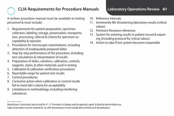
CLIA Requirements for Procedure Manuals Laboratory Operations Review 61
A written procedure manual must be available to testingpersonnel & must include:
1. Requirements for patient preparation, specimen collection, labeling, storage, preservation, transporta-tion, processing, referral & criteria for specimen ac-ceptability & rejection
2. Procedures for microscopic examinations, includingdetection of inadequately prepared slides
3. Step-by-step performance of the procedure, includingtest calculations & interpretation of results
4. Preparation of slides, solutions, calibrators, controls,reagents, stains, & other materials used in testing
5. Calibration & calibration verification procedures6. Reportable range for patient test results7. Control procedures8. Corrective action when calibration or control results
fail to meet lab’s criteria for acceptability9. Limitations in methodology, including interfering
substances
10. Reference intervals11. Imminently life-threatening laboratory results (critical
values)12. Pertinent literature references13. System for entering results in patient record & report-
ing (including protocol for critical values)14. Action to take if test system becomes inoperable
Manufacturer’s instructions may be used for #1–12. Procedures & changes must be approved, signed, & dated by director before use. Copies of procedures must be retained for 2 yr after discontinuance & must include dates of initial use & discontinuance.
2956_Ch01_001-066 30/01/14 3:07 PM Page 61

Ethical and Legal Issues Laboratory Operations Review 62
TERM EXPLANATION
Confidentiality
Protected health information (PHI)
Invasion of privacy
Informed consent
Expressed consent
Implied consent
Informed refusal
Negligence
Malpractice
The right of a patient to have his/her medical information kept private. Restriction of access to information to those who have authorization and a need to know.
All individually identifiable health information, including lab results. HIPAA requires health-careproviders to establish extensive security measures to ensure privacy. Unauthorized disclosure ofmedical information could lead to charges of breach of confidentiality or invasion of privacy.
The wrongful intrusion into a person’s private affairs, e.g., unauthorized disclosure of confidentialmedical information.
Consent for a medical procedure given by patient after procedure & possible risks have been explained. May be expressed or implied.
Written or verbal consent.
Consent that is inferred from action or signs; e.g., a patient sits in phlebotomy chair & extendsarm. Action implies consent for venipuncture.
Patients have right to refuse medical treatment/procedures.
Violation of duty to exercise reasonable skill & care.
Misconduct or negligence that results in injury to patient.
continued...
2956_Ch01_001-066 30/01/14 3:07 PM Page 62

Ethical and Legal Issues continued
TERM EXPLANATION
Assault & battery
Chain of custody
Touching another person without his/her consent. Drawing blood against a patient’s wishes couldlead to charges of assault & battery.
Procedure to guarantee integrity of specimen to court, e.g., legal blood alcohol, drug test. Eachperson handling specimen must sign chain-of-custody form that accompanies specimen & docu-ments custody of specimen at all times. Specimen may be transported in locked box to preventtampering.
Laboratory Operations Review 63
2956_Ch01_001-066 30/01/14 3:07 PM Page 63

Commonly Used Prefixes in the Metric System
Laboratory Operations Review 64
PREFIX DEFINITION
Deci- 10−1
Centi- 10−2
Milli- 10−3
Micro- 10−6
Nano- 10−9
Pico- 10−12
Femto- 10−15
2956_Ch01_001-066 30/01/14 3:07 PM Page 64
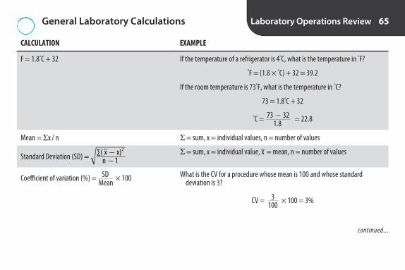
General Laboratory Calculations Laboratory Operations Review 65
CALCULATION EXAMPLE
F = 1.8°C + 32
Mean = Σ x / n
Standard Deviation (SD) =
Coefficient of variation (%) = SD × 100Mean
∑( x x)n 1
2� �
If the temperature of a refrigerator is 4°C, what is the temperature in °F?
°F = (1.8 × °C) + 32 = 39.2
If the room temperature is 73°F, what is the temperature in °C?
73 = 1.8°C + 32
°C = 73 � 32 = 22.81.8
Σ = sum, x = individual values, n = number of values
Σ = sum, x = individual value, x– = mean, n = number of values
What is the CV for a procedure whose mean is 100 and whose standard deviation is 3?
CV = 3 × 100 = 3%100
continued...
2956_Ch01_001-066 30/01/14 3:07 PM Page 65

General Laboratory Calculations continued
CALCULATION EXAMPLE
Dilution = Vol. of specimen Vol. of specimen + vol. of diluent
Correcting for a dilution: Value obtained for diluted specimen × reciprocal of dilution
What is the dilution if 0.1 mL of serum is diluted with 0.4 mL of saline?
0.1 = 0.1 = 1 0.1 + 0.4 0.5 5
How would you prepare a 1:10 dilution of urine?1 part of urine + 9 parts of diluent
A specimen for glucose is diluted 1:5. The value of the diluted specimen is 100 mg/dL. What value should be reported?
100 mg/dL × 5 = 500 mg/dL
Laboratory Operations Review 66
2956_Ch01_001-066 30/01/14 3:07 PM Page 66

2S E C T I O N
Clinical Chemistry Review
2956_Ch02_067-134 30/01/14 4:24 PM Page 67

Comparison of Conventional and SI Units for Selected Reference Ranges
Clinical Chemistry Review 68
ANALYTE CONVENTIONAL UNITS SI UNITS
Bilirubin, total 0.2–1 mg/dL 3.4–17.1 μmol/L
BUN 6–20 mg/dL 2.1–7.1 mmol/L
Calcium, total 8.6–10 mg/dL 2.15–2.5 mmol/L
Chloride 98–107 mEq/L 98–107 mmol/L
Creatinine M: 0.9–1.2 mg/dL, F: 0.6–1.1 mg/dL M: 80–106 mmol/L, F: 53–97 mmol/L
Glucose, fasting 70–99 mg/dL 3.9–5.5 mmol/L
Potassium 3.5–5.1 mEq/L 3.5–5.1 mmol/L
Sodium 136–145 mEq/L 136–145 mmol/L
Total protein 6.4–8.3 g/dL 64–83 g/L
Uric acid M: 3.5–7.2 mg/dL, F: 2.6–6 mg/dL M: 208–428 μmol/L, F: 155–357 μmol/L
SI = Système International d’Unités (international system of units).
2956_Ch02_067-134 27/02/14 10:52 AM Page 68

Examples of Patient Variables That May Affect Chemistry Values
Clinical Chemistry Review 69
VARIABLE ANALYTES AFFECTED
Diurnal variation ↑ in am: ACTH, cortisol, iron ↑ in pm: growth hormone, PTH, TSH
Day-to-day variation ≥ 20% for ALT, bili, CK, steroid hormones, triglycerides
Recent food ingestion ↑ glucose, insulin, gastrin, triglycerides, Na+, uric acid, iron, LD, Ca2+; ↓ chloride, phosphate, K+
Fasting required: fasting glucose, triglycerides, lipid panel
Alcohol ↓ glucose; ↑ triglycerides, GGT
Posture ↑ albumin, cholesterol, Ca2+ when standing
Activity ↑ in ambulatory patients: creatinine kinase (CK)↑ with exercise: K+, phosphate, lactic acid, creatinine, protein, CK, AST, LD
Stress ↑ ACTH, cortisol, catecholamines
Age, gender, race, drugs Various
2956_Ch02_067-134 30/01/14 4:24 PM Page 69

Examples of Preanalytical Factors That May Affect Chemistry Results
Clinical Chemistry Review 70
FACTOR EFFECT
Use of isopropyl alcohol wipes to disinfect venipuncture site
Squeezing site of capillary puncture
Pumping fist during venipuncture
Tourniquet >1 min
IV fluid contamination
Incorrect anticoagulant or contamination from incorrect order of draw
Hemolysis
Exposure to light
Temperature between collection & testing
Can compromise blood alcohol determination
↑ K+
↑ K+, lactic acid, Ca2+, phosphorus; ↓ pH
↑ K+, total protein, lactic acid
↑ glucose, K+, Na+, Cl– (depending on IV). Possible dilution of other analytes.
K2EDTA: ↓ Ca2+, Mg2+; ↑ K+
Sodium heparin: ↑ Na+ if tube not completely filledLithium heparin: ↑ lithium Gels: Some interfere with trace metals & certain drugs
↑ K+, Mg2+, phosphorus, LD, AST, iron, ammonia (May be method dependent. Refer to reagent package inserts.)
↓ bilirubin, carotene
Chilling required for lactic acid, ammonia, blood gases
continued...
2956_Ch02_067-134 30/01/14 4:24 PM Page 70
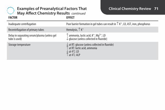
Examples of Preanalytical Factors That May Affect Chemistry Results continued
Clinical Chemistry Review 71
Inadequate centrifugation
Recentrifugation of primary tubes
Delay in separating serum/plasma (unless gel tube is used)
Storage temperature
FACTOR EFFECT
Poor barrier formation in gel tubes can result in ↑ K+, LD, AST, iron, phosphorus
Hemolysis, ↑ K+
↑ ammonia, lactic acid, K+, Mg2+, LD↓ glucose (unless collected in fluoride)
↓ at RT: glucose (unless collected in fluoride)↑ at RT: lactic acid, ammonia↓ at 4ºC: LD↑ at 4ºC: ALP
2956_Ch02_067-134 30/01/14 4:24 PM Page 71

Differences in Analyte Concentrations Clinical Chemistry Review 72
DIFFERENCE ANALYTE(S)
Higher in plasma than serum Total protein, LD, Ca2+
Higher in serum than plasma K+, phosphate, glucose, CK, bicarbonate, ALP, albumin, AST, triglycerides
Higher in plasma than whole blood Glucose
Higher in capillary blood than venous blood Glucose (in postprandial specimen), K+
Higher in venous blood than capillary blood Ca2+, total protein
Higher in RBCs than plasma K+, phosphate, Mg2+
Higher in plasma than RBCs Na+, chloride
2956_Ch02_067-134 30/01/14 4:24 PM Page 72
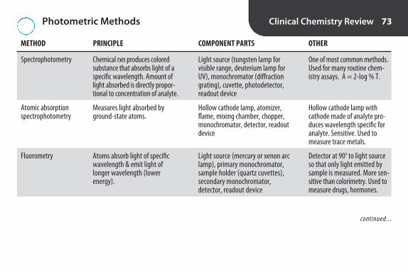
Photometric Methods Clinical Chemistry Review 73
METHOD PRINCIPLE COMPONENT PARTS OTHER
Spectrophotometry
Atomic absorption spectrophotometry
Fluorometry
Chemical rxn produces coloredsubstance that absorbs light of aspecific wavelength. Amount oflight absorbed is directly propor-tional to concentration of analyte.
Measures light absorbed byground-state atoms.
Atoms absorb light of specificwavelength & emit light oflonger wavelength (lower energy).
Light source (tungsten lamp forvisible range, deuterium lamp forUV), monochromator (diffractiongrating), cuvette, photodetector,readout device
Hollow cathode lamp, atomizer,flame, mixing chamber, chopper,monochromator, detector, readoutdevice
Light source (mercury or xenon arclamp), primary monochromator,sample holder (quartz cuvettes),secondary monochromator, detector, readout device
One of most common methods.Used for many routine chem-istry assays. A = 2-log % T.
Hollow cathode lamp withcathode made of analyte pro-duces wavelength specific foranalyte. Sensitive. Used tomeasure trace metals.
Detector at 90º to light sourceso that only light emitted bysample is measured. More sen-sitive than colorimetry. Used tomeasure drugs, hormones.
continued...
2956_Ch02_067-134 30/01/14 4:24 PM Page 73

Photometric Methods continued Clinical Chemistry Review 74
METHOD PRINCIPLE COMPONENT PARTS OTHER
Chemiluminescence
Turbidimetry
Nephelometry
Chemical rxn that produces light.Usually involves oxidation of luminol, acridinium esters, or dioxetanes.
Measures reduction in lighttransmission by particles in suspension.
Similar to turbidity, but light ismeasured at angle from lightsource.
Reagent probes, sample & reagentcuvette, photomultiplier tube,readout device
Light source, lens, cuvette, photodetector, readout device
Light source, collimator, mono-chromator, cuvette, photodetector,readout device
Doesn’t require excitation radia-tion or monochromators like fluorometry. Extremely sensitive.Used for immunoassays.
Used to measure proteins inurine & CSF.
Used to measure ag-ab rxn.
2956_Ch02_067-134 30/01/14 4:24 PM Page 74

Visible Light Clinical Chemistry Review 75
WAVELENGTH (nm) COLOR ABSORBED COLOR TRANSMITTED (COLOR SEEN)
350–430 Violet Yellow
430–475 Blue Orange
475–495 Blue-green Red-orange
495–505 Green-blue Orange-red
505–555 Green Red
555–575 Yellow-green Violet-red
575–600 Yellow Violet
600–650 Orange Blue
670–700 Red Green
2956_Ch02_067-134 30/01/14 4:24 PM Page 75

Wavelengths Used in Spectrophotometry Clinical Chemistry Review 76
WAVELENGTH (nm) RANGE COMMON LIGHT SOURCE CUVETTE
220–380 Near-ultraviolet Deuterium or mercury arc Quartz (silica)
380–750 Visible Incandescent tungsten or tungsten-iodide Borosilicate
750–2,000 Near-infrared Incandescent tungsten or tungsten-iodide Quartz (silica)
2956_Ch02_067-134 30/01/14 4:24 PM Page 76

Chromatography* Clinical Chemistry Review 77
TYPE COMPONENTS USE OTHER
Thin-layer chromatography (TLC)
High-performance liquid chromatography (HPLC)
Gas chromatography (GC)
Sorbent-coated glass or plasticplate, closed container, solvent
Solvent, pump, injection port,column, detector, recorder
Gas, injection port, column,oven, detector, recorder
Screening test for drugs ofabuse in urine
Separation of thermolabilecompounds
Separation of volatile com-pounds or compounds that can be made volatile, e.g.,therapeutic & toxic drugs
Substances identified by Rf value(distance traveled by compound/distance traveled by solvent).
Concentration determined bypeak height ratio (height of analyte peak/height of internalstandard peak). Mass spectrome-try (MS) can be used as detectorfor definitive ID (LC/MS).
Compounds identified by reten-tion time. Area of peak is propor-tional to concentration. MS can be used as detector for definitiveID (GC/MS).
*Separation of compounds based on differential distribution between mobile phase & stationary phase.
2956_Ch02_067-134 30/01/14 4:24 PM Page 77

Other Analytic Techniques Clinical Chemistry Review 78
METHOD PRINCIPLE COMPONENT PARTS USE
Ion-selective electrodes
Osmometry
Electrophoresis
Potential difference between 2 electrodesdirectly related to concentration of analyte.
Determines osmolality (measurement of # of dissolved particles in solution, irrespectiveof molecular weight, size, density, or type)based on freezing-point depression. (Vapordepression osmometers not widely used inclinical labs. Don’t measure volatile solutes.)
Separation of charged particles in electricalfield. Anions move to positively chargedpole (anode); cations to negatively chargedpole (cathode). The greater the charge, thefaster the migration.
Reference electrode, indicatorelectrode, liquid junction,measuring device
Cooling bath, thermistorprobe, stirring wire, galvanometer
Power supply, supportmedium, buffer, stain, densitometer
pH, PCO2, PO2, Na+, K+,Ca2+, Li+, Cl−
Serum & urine osmolality
Serum protein elec-trophoresis, hemoglobinelectrophoresis
2956_Ch02_067-134 30/01/14 4:24 PM Page 78

Steps in Automated Analysis Clinical Chemistry Review 79
STEP COMMENTS
Sample ID Usually by bar code reader
Test selection Usually communicated by LIS
Sampling Usually closed-tube sampling from primary collection tubes. Some analyzers have short sample & clot detection
Reagent delivery Usually by syringes, pumps, or pressurized reagent bottles. Vitros uses dry slides. Some offer reagent inventory
Chemical reaction Mixing & incubation
Measurements Visible & UV spectrophotometry, ion selective electrodes, fluorescence polarization, chemiluminescence, bioluminescence. Most offer automatic dilution & retesting when linearity is exceeded
Data handling Concentration derived from calibration curve stored in analyzer
Reporting Usually reported to LIS through interface
Troubleshooting Can be done remotely by modem on many analyzers
2956_Ch02_067-134 30/01/14 4:24 PM Page 79

Chemistry Panels Clinical Chemistry Review 80
PANEL TESTS
Basic metabolic panel Na+, K+, chloride, CO2, glucose, creatinine, BUN, Ca2+
Comprehensive metabolic panel Na+, K+, chloride, CO2, glucose, creatinine, BUN, albumin, total protein, ALP, AST, bilirubin, Ca2+
Electrolyte panel Na+, K+, Cl–, CO2
Hepatic function panel Albumin, ALT, AST, ALP, bilirubin (total & direct), total protein
Lipid panel Total cholesterol, HDL cholesterol, LDL cholesterol, triglycerides
Renal function panel Na+, K+, CO2, glucose, creatinine, BUN, Ca2+, albumin, phosphate
2956_Ch02_067-134 30/01/14 4:24 PM Page 80

Carbohydrates, Lipids, and Proteins Clinical Chemistry Review 81
ANALYTE REFERENCE RANGE CLINICAL SIGNIFICANCE OTHER
Glucose, fasting
Cholesterol, total
HDL cholesterol
LDL cholesterol
Triglycerides
70–99 mg/dL
Desirable: <200 mg/dL
Desirable: ≥ 60 mg/dL
Optimal: <100 mg/dL
Desirable: <150 mg/dL
↑ (hyperglycemia): diabetes mel-litus, other endocrine disorders,acute stress, pancreatitis
↓ (hypoglycemia): insulinoma, insulin-induced hypoglycemia,hypopituitarism
Limited value for predicting risk ofcoronary artery disease (CAD) byitself. Used in conjunction withHDL & LDL cholesterol
Appears to be inversely related to CAD
Risk factor for CAD
Risk factor for CAD
Major source of cellular energy. Levels ↓ at RT. Use sodium fluoride to prevent glycolysis. Glucoseoxidase & hexokinase are most common methods.Hexokinase considered more accurate, fewer interfering substances.
Enzymatic methods most common.
Homogeneous assays don’t require pretreatmentto remove non-HDL. 1st reagent blocks non-HDL,2nd reacts with HDL.
May be calculated from Friedewald formula (iftriglycerides not >400 mg/dL) or measured bydirect homogeneous assays.
Main form of lipid storage. Enzymatic methodsusing lipase. Requires fasting specimen.
continued...
2956_Ch02_067-134 30/01/14 4:24 PM Page 81

Carbohydrates, Lipids, and Proteins continued Clinical Chemistry Review 82
ANALYTE REFERENCE RANGE CLINICAL SIGNIFICANCE OTHER
Total protein
Albumin
Microalbumin (on urine)
6.4–8.3 g/dL
3.5–5 g/dL
50–200 mg/24 hrpredictive of diabeticnephropathy
↑ dehydration, chronic inflamma-tion, multiple myeloma
↓ nephrotic syndrome, malab-sorption, overhydration, hepaticinsufficiency, malnutrition,agammaglobulinemia
↑ dehydration↓ malnutrition, liver disease,
nephrotic syndrome, chronic inflammation
↑ in diabetics at risk of nephropathy
<4.5 g/dL associated with peripheral edema. Biuret method. Alkaline copper reagent reactswith peptide bonds.
Largest fraction of plasma proteins. Synthesizedby liver. Regulates osmotic pressure. Measure bydye binding, e.g., bromocresol green (BCG),bromocresol purple (BCP).
Detects albumin in urine earlier than dipstick protein. Strict control of glucose & blood pressure can prevent progression to end-stagerenal disease. Immunoassays on 24-hr urine. Alternative is albumin-to-creatinine ratio on random sample. 30–300 mg albumin/g creatinine = microalbuminuria. Urine dipsticks available for albumin & albumin-tocreatinine ratio.
2956_Ch02_067-134 30/01/14 4:24 PM Page 82

Regulation of Glucose Clinical Chemistry Review 83
HORMONE ACTION RELATIVE IMPORTANCE
Decreases glucose levelsInsulin Responsible for entry of glucose into cells. Increases glycogenesis. Primary
Increases glucose levels Glucagon Stimulates glycogenolysis & gluconeogenesis. Inhibits glycolysis. Primary
Cortisol Insulin antagonist. Increases gluconeogenesis. Secondary
Epinephrine Promotes glycogenolysis & gluconeogenesis. Secondary
Growth hormone Insulin antagonist. Secondary
Thyroxine Increases glucose absorption from GI tract. Stimulates glycogenolysis. Negligible
2956_Ch02_067-134 30/01/14 4:24 PM Page 83

Diabetes Mellitus Clinical Chemistry Review 84
TYPE CAUSE CHARACTERISTICS
Type 1 (formerly type I juvenile-onset diabetes, insulin-dependent diabetes mellitus)
Type 2(formerly type II, adult-onset diabetes, non–insulin-dependent diabetes mellitus)
Gestational diabetes mellitus (GDM)
Autoimmune destruction of beta cells. Absolute insulin deficiency. Genetic predis-position (HLA-DR 3/4).
Insulin resistance in peripheral tissue. Insulin secretory defect of beta cells. Associated with obesity.
Placental lactogen inhibits action of insulin.
Acute onset. Most develop before age 25 yr. Dependency on injected insulin. Prone to ketoacidosis & diabetic complications.
Most common type. Usual onset was after age 40 yrbut being seen in obese youth. Not dependent onexogenous insulin. Not prone to ketoacidosis or diabetic complications.
Usually Dx during latter half of pregnancy. Somedevelop type 2 diabetes years later. Risk of intrauterine death or neonatal complications(macrosomia, hypoglycemia, hypocalcemia, polycythemia, hyperbilirubinemia).
2956_Ch02_067-134 30/01/14 4:24 PM Page 84

Tests for Diabetes Mellitus Clinical Chemistry Review 85
TEST PATIENT PREPARATION DIABETES MELLITUS COMMENTS
Random plasma glucose
Fasting plasma glucose (FPG)
2-hr plasma glucose
Oral glucose tolerance test (OGTT)
Hemoglobin A1c
None
Fast of at least 8 hr
75-g glucose load
Fast of at least 8 hr; 75-gglucose load
None; fasting not required
≥ 200 mg/dL
≥ 126 mg/dL on 2 occasions
≥ 200 mg/dL on 2 occasions
Fasting ≥ 92 mg/dL, or 1 hr≥ 180, or 2 hr ≥ 153
≥ 6.5%
Collected any time of day without regard to time since last meal. Only for use in patients with symptoms of hyperglycemia.
Only for Dx of gestational diabetes mellitus.Performed at 24–28 wk of gestation.
Gives estimate of glucose control over previous 2–3 months. Originally only used to monitor therapy. Now accepted for Dx except in patients with hemoglo-binopathies or abnormal RBC turnover.Should be performed using method certified by National GlycohemoglobinStandardization Program. Point-of-care assays currently not accurate enough for diagnosis.
2956_Ch02_067-134 30/01/14 4:24 PM Page 85

Typical Laboratory Findings in UncontrolledDiabetes Mellitus
Clinical Chemistry Review 86
INCREASED DECREASED
Blood glucose Bicarbonate Urine glucose Blood pHUrine specific gravityGlycohemoglobinKetones (blood & urine)Anion gapBUNOsmolality (serum & urine)CholesterolTriglycerides
2956_Ch02_067-134 30/01/14 4:24 PM Page 86

Metabolic Syndrome Clinical Chemistry Review 87
Definition Group of risk factors that seem to promote development of atherosclerotic cardiovascular disease & type2 diabetes mellitus
Risk factors ↓ HDL-C ↑ LDL-C ↑ triglycerides↑ blood pressure ↑ blood glucose
2956_Ch02_067-134 30/01/14 4:24 PM Page 87
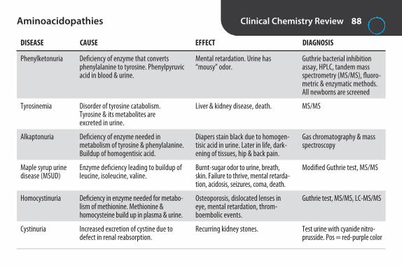
Aminoacidopathies Clinical Chemistry Review 88
DISEASE CAUSE EFFECT DIAGNOSIS
Phenylketonuria
Tyrosinemia
Alkaptonuria
Maple syrup urine disease (MSUD)
Homocystinuria
Cystinuria
Deficiency of enzyme that convertsphenylalanine to tyrosine. Phenylpyruvicacid in blood & urine.
Disorder of tyrosine catabolism. Tyrosine & its metabolites are excreted in urine.
Deficiency of enzyme needed in metabolism of tyrosine & phenylalanine.Buildup of homogentisic acid.
Enzyme deficiency leading to buildup ofleucine, isoleucine, valine.
Deficiency in enzyme needed for metabo-lism of methionine. Methionine & homocysteine build up in plasma & urine.
Increased excretion of cystine due to defect in renal reabsorption.
Mental retardation. Urine has“mousy” odor.
Liver & kidney disease, death.
Diapers stain black due to homogen-tisic acid in urine. Later in life, dark-ening of tissues, hip & back pain.
Burnt-sugar odor to urine, breath,skin. Failure to thrive, mental retarda-tion, acidosis, seizures, coma, death.
Osteoporosis, dislocated lenses ineye, mental retardation, throm-boembolic events.
Recurring kidney stones.
Guthrie bacterial inhibitionassay, HPLC, tandem mass spectrometry (MS/MS), fluoro-metric & enzymatic methods.All newborns are screened
MS/MS
Gas chromatography & massspectroscopy
Modified Guthrie test, MS/MS
Guthrie test, MS/MS, LC-MS/MS
Test urine with cyanide nitro-prusside. Pos = red-purple color
2956_Ch02_067-134 30/01/14 4:24 PM Page 88

Protein Electrophoresis Clinical Chemistry Review 89
Rate of migration Depends on size, shape, & charge of molecule
Support medium Cellulose acetate or agarose
Buffer Barbital buffer, pH 8.6
Stains Ponceau S, amido blue, bromphenol blue, Coomassie brilliant blue
Charge At pH 8.6, proteins are negatively charged & move toward anode
Order of migration (fastest to slowest) Albumin, alpha-1 globulin, alpha-2 globulin, beta globulin, gamma globulin
Largest fraction Albumin
Electroendosmosis Buffer flow toward cathode. Causes gamma region to be cathodic to point of application
Urine Must be concentrated first because of low protein concentration. Bence Jones proteins migrateto gamma region in urine electrophoresis
CSF Must be concentrated first because of low protein concentration. CSF has a prealbumin band
2956_Ch02_067-134 30/01/14 4:24 PM Page 89
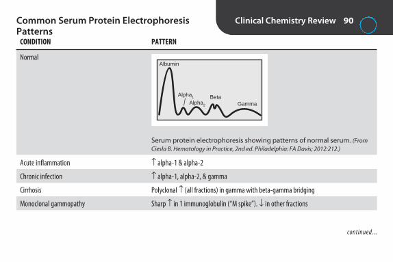
Common Serum Protein Electrophoresis Patterns
Clinical Chemistry Review 90
CONDITION PATTERN
Normal
Acute inflammation ↑ alpha-1 & alpha-2
Chronic infection ↑ alpha-1, alpha-2, & gamma
Cirrhosis Polyclonal ↑ (all fractions) in gamma with beta-gamma bridging
Monoclonal gammopathy Sharp ↑ in 1 immunoglobulin (“M spike”). ↓ in other fractions
Albumin
Alpha1
Alpha2
BetaGamma
Serum protein electrophoresis showing patterns of normal serum. (FromCiesla B. Hematology in Practice, 2nd ed. Philadelphia: FA Davis; 2012:212.)
continued...
2956_Ch02_067-134 30/01/14 4:24 PM Page 90

Common Serum Protein Electrophoresis Patterns continued
Clinical Chemistry Review 91
CONDITION PATTERN
Polyclonal gammopathy Diffuse ↑ in gamma
Hypogammaglobulinemia ↓ gamma
Nephrotic syndrome ↓ albumin, ↑ alpha-2
Alpha-1-antitrypsin deficiency ↓ alpha-1
Hemolyzed specimen ↑ beta or unusual band between alpha-2 & beta
Plasma Extra band (fibrinogen) between beta & gamma
2956_Ch02_067-134 30/01/14 4:24 PM Page 91

ANALYTE REFERENCE RANGE CLINICAL SIGNIFICANCE OTHER
BUN
Creatinine
Uric acid
Ammonia
Nonprotein Nitrogen Compounds Clinical Chemistry Review 92
8–26 mg/dL
0.7–1.5 mg/dL
M: 3.5–7.2F: 2.6-6 mg/dL
19–60 μg/dL
↑ kidney disease↓ overhydration or liver disease
↑ kidney disease
↑ gout, renal failure, ketoacido-sis, lactate excess, high nucleoprotein diet, leukemia,lymphoma, polycythemia
↓ administration of ACTH, renaltubular defects
↑ liver disease, hepatic coma,renal failure, Reye’s syndrome
Synthesized by liver from ammonia. Excreted by kidneys.Urease reagent. Don’t use sodium fluoride, EDTA, citrate, orammonium heparin. Test isn’t sensitive. Dilute urine 1:20 or1:50 & refrigerate or acidify.
Waste product from dehydration of creatine (mainly in muscles). Jaffe’s reaction (alkaline picrate) is nonspecific.Enzymatic methods are more specific. Tests aren’t sensitive.Normal BUN: creatinine ratio = 12–20. Dilute urine 1:100.
Increased = risk of renal calculi & joint tophi. Uricasemethod. EDTA & fluoride interfere. Adjust urine pH to 7.5–8 to prevent precipitation.
Produced in GI tract. High levels are neurotoxic. Collect inEDTA or heparin. Serum may cause ↑ levels as NH3 is gener-ated during clotting. Chill immediately. Analyze ASAP. Avoidcontamination from ammonia in detergents or water.
2956_Ch02_067-134 30/01/14 4:24 PM Page 92

Major Electrolytes Clinical Chemistry Review 93
ANALYTE REFERENCE RANGE CLINICAL SIGNIFICANCE OTHER
Sodium (Na+)
Potassium (K+)
136–145 mmol/L
3.5–5.1 mmol/L
↑ (hypernatremia): Due to ↑ intake or IV administration, hyperaldosteronism, excessive sweating, burns, diabetes insipidus.Causes tremors, irritability, confusion, coma.
↓ (hyponatremia): Due to renal or extrarenalloss (vomiting, diarrhea, sweating, burns) or↑ extracellular fluid volume. Causes weak-ness, nausea, altered mental status.
↑ (hyperkalemia): Due to ↑ intake, ↓ excretion, crush injuries, metabolic acido-sis. Can cause muscle weakness, confusion,cardiac arrhythmia, cardiac arrest.
↓ (hypokalemia): Due to ↑ GI or urinary loss,use of diuretics, metabolic alkalosis. Cancause muscle weakness, paralysis, breathingproblems, cardiac arrhythmia, death.
Major extracellular cation. Contributes almosthalf to plasma osmolality. Maintains normaldistribution of water & osmotic pressure. Levelsregulated by aldosterone. Ion-selective electrode(ISE) is most common method. NormalNa+/K+ ratio in serum approximately 30:1.
Major intracellular cation. Artifactual ↑ due tosqueezing site of capillary puncture, prolongedtourniquet, pumping fist during venipuncture,contamination with IV fluid, hemolysis, prolonged contact with RBCs, leukocytosis,thrombocytosis. Serum values 0.1–0.2 mmol/Lhigher than plasma due to release fromplatelets during clotting. Most commonmethod is ISE with valinomycin membrane.
continued...
2956_Ch02_067-134 30/01/14 4:24 PM Page 93

Major Electrolytes continued Clinical Chemistry Review 94
ANALYTE REFERENCE RANGE CLINICAL SIGNIFICANCE OTHER
Chloride (Cl–)
CO2, total
98–107 mmol/L
23–29 mmol/L
↑ (hyperchloremia): Due to same conditions as↑ Na+ & excess loss of HCO3
–.↓ (hypochloremia): From prolonged vomiting,
diabetic ketoacidosis, aldosterone deficiency,salt-losing renal diseases, metabolic alkalosis,compensated respiratory acidosis
↑ in metabolic alkalosis, compensated respiratory acidosis
↓ in metabolic acidosis, compensated respiratory alkalosis
Major extracellular ion. Helps maintain osmolality, blood volume, electric neutrality.Passively follows Na+. Most commonmethod is ISE. Sweat chloride test for Dx of cystic fibrosis.
>90% is bicarbonate (HCO3–); remainder
is carbonic acid (H2CO3) & dissolved CO2. HCO3–
important in maintaining acid-base balance.Keep sample capped to prevent loss of CO2. Measured by ISE or enzymatic method.
2956_Ch02_067-134 30/01/14 4:24 PM Page 94

Other Electrolytes Clinical Chemistry Review 95
ANALYTE REFERENCE RANGE CLINICAL SIGNIFICANCE OTHER
Magnesium (Mg2+)
Calcium (Ca2+)
1.6–2.6 mg/dL
Total: 8.6–10 mg/dLIonized: 4.60–5.08 mg/dL
↑ due to renal failure, ↑ intake (e.g.,antacids), dehydration, bone cancer, endocrine disorders. Can cause cardiac abnormalities, paralysis, respiratory arrest, coma.
↓ due to severe illness, GI disorders, en-docrine disorders, renal loss. Can lead tocardiac arrhythmias, tremors, tetany,paralysis, psychosis, coma. Rare in non-hospitalized patients.
↑ with primary hyperparathyroidism, cancer, multiple myeloma. Can causeweakness, coma, GI symptoms, renal calculi.
↓ with hypoparathyroidism, malabsorption,vitamin D deficiency, renal tubular acidosis. Leads to tetany (muscle spasms),seizures, cardiac arrhythmias.
Essential cofactor for many enzymes. 10× more concentrated in RBCs. Avoid hemolysis. EDTA, citrate, oxalate bind Mg2+.Colorimetric methods are most common.
Most abundant mineral in body. 99% inbones. Regulated by parathyroid hormone(PTH), vitamin D, calcitonin. Anticoagu-lants other than heparin bind Ca2+. Colorimetric methods for total Ca2+. Ionized (free) Ca2+ is biologically activeform, better indicator of Ca2+ status. Measured by ISE. Affected by pH & temp.
continued...
2956_Ch02_067-134 30/01/14 4:24 PM Page 95

Other Electrolytes continued Clinical Chemistry Review 96
ANALYTE REFERENCE RANGE CLINICAL SIGNIFICANCE OTHER
Phosphorus, inorganic (phosphate)
Lactate (lactic acid)
2.5–4.5 mg/dL
4.5–19.8 mg/dL
↑ with renal disease, hypoparathyroidism.↓ with hyperparathyroidism, vitamin D
deficiency, renal tubular acidosis.
Sign of ↓ O2 to tissues.
Major intracellular anion. Mostly in bones.Component of nucleic acids, many coen-zymes. Important reservoir of energy(ATP). Limited value alone. Should be correlated with Ca2+ (normally reciprocalrelationship). Higher in children. Citrate,oxalate, EDTA interfere. More in RBCs thanplasma. Avoid hemolysis. Separatepromptly.
By product of anaerobic metabolism. Bestnot to use tourniquet. Patient shouldn’tmake fist. Collect in heparin & put on iceor use fluoride to inhibit glycolysis. Enzymatic methods.
2956_Ch02_067-134 30/01/14 4:24 PM Page 96

Iron and Related Tests Clinical Chemistry Review 97
ANALYTE REFERENCE RANGE CLINICAL SIGNIFICANCE OTHER
Iron
Total iron binding capacity (TIBC)
% saturation or transferrin saturation
Transferrin
Ferritin
M: 65–175 F: 50–170 μg/dL
250–425 μg/dL
20%–50%
200–360 mg/dL
M: 20–250F: 10–120 μg/L
↑ iron overdose, hemochromatosis,sideroblastic anemia, hemolyticanemia, liver disease
↓ iron deficiency anemia
↑ iron deficiency anemia↓ iron overdose, hemochromatosis
↑ iron overdose, hemochromatosis,sideroblastic anemia
↓ iron deficiency anemia
↑ iron deficiency anemia↓ iron overdose, hemochromatosis,
chronic infections, malignancies
↑ iron overload, hemochromatosis,chronic infections, malignancies
↓ iron deficiency anemia
Necessary for hgb synthesis. Transportedby transferrin. Hemolysis interferes. Oxalate, citrate, & EDTA bind iron. Early-morning specimen preferred because ofdiurnal variation. Colorimetric methods.
Iron added to saturate transferrin. Excess removed. Iron content determined.
Calculated value. 100× serum iron/TIBC.
Complex of apotransferrin (protein thattransports iron) & iron. Immunoassay.
Storage form of iron. Rough estimate ofbody iron content. Immunoassay.
2956_Ch02_067-134 30/01/14 4:24 PM Page 97

Factors That Influence Enzymatic Reactions Clinical Chemistry Review 98
FACTOR EXPLANATION OTHER
Substrate concentration
Enzyme concentration
pH
Temperature
Cofactors
Inhibitors
First-order kinetics: [enzyme] > [substrate].Reaction rate proportional to [substrate].
Zero-order kinetics: [substrate] > [enzyme].Reaction rate proportional to [enzyme].
Velocity of rxn proportional to [enzyme] as long as[substrate] > [enzyme].
Extremes of pH may denature enzymes.
Increase of 10ºC doubles rate of rxn until around40º–50ºC; then denaturation of enzyme may occur.
Nonprotein molecules that participate in rxn. Mustbe present in excess.
Inorganic cofactors (e.g., Cl–, Mg2+) called activa-tors. Either required for or enhance rxn.
Organic cofactors (e.g., nicotinamide adenine dinucleotide) called coenzymes. May serve as 2nd substrate in rxn.
Interfere with rxn.
Assays are zero-order (excess substrate).
Unit of measure is international unit (IU). Amountof enzyme that will catalyze 1 μmol of substrateper min under standardized conditions.
Most rxn occur at pH 7–8. Use buffers to maintainoptimal pH.
37°C is most commonly used in U.S.
Rxn commonly used in enzyme determinations:Nicotinamide adenine dinucleotide (NAD) ⇔
nicotinamide adenine dinucleotide, reducedform (NADH). NADH has absorbance at 340 nm;NAD does not.
2956_Ch02_067-134 30/01/14 4:24 PM Page 98

Enzymes of Clinical Significance Clinical Chemistry Review 99
ENZYME TISSUE(S) CLINICAL SIGNIFICANCE OTHER
Acid phosphatase (ACP)
Alkaline phosphatase(ALP)
Aspartate aminotrans-ferase (AST)
Alanine aminotrans-ferase (ALT)
Gamma glutamyl transferase (GGT)
Prostate
Almost all
Many. Highest inliver, heart, skeletalmuscle.
Liver, RBCs
Liver, kidneys, pancreas
↑ in prostate cancer
↑ liver & bone disease. Levelshigher in biliary tract obstruc-tion than in hepatocellular disorders (hepatitis, cirrhosis)
↑ with liver disease (marked↑ with viral hepatitis), acutemyocardial infarction (AMI),muscular dystrophy
↑ with liver disease
↑ in all hepatobiliary disor-ders, chronic alcoholism
Limited value today. PSA is more specific.
↑ in children, adolescents, pregnant women, & with healing bone fractures. Optimum pH = 9–10.
Avoid hemolysis.
More specific for liver disease than AST. Marked↑ with viral hepatitis.
Most sensitive enzyme for all types of liver dis-ease. Highest levels with obstructive disorders.Treatment centers use to monitor abstentionfrom alcohol.
continued...
2956_Ch02_067-134 30/01/14 4:24 PM Page 99

Enzymes of Clinical Significance continued Clinical Chemistry Review 100
Lactate dehydrogenase (LD)
Creatine kinase (CK)
Amylase (AMS)
Lipase (LPS)
Glucose-6-phosphate dehydrogenase (G6PD)
ENZYME TISSUE(S) CLINICAL SIGNIFICANCE OTHER
All. Highest in liver,heart, skeletal muscle, RBCs
Cardiac muscle, skele-tal muscle, brain
Salivary glands, pancreas
Pancreas
RBCs
↑ with AMI, liver disease, per-nicious anemia
↑ with AMI, muscular dystrophy
↑ in acute pancreatitis, otherabdominal diseases, mumps
↑ in acute pancreatitis
Inherited deficiency can leadto drug-induced hemolyticanemia
Catalyzes lactic acid ⇔ pyruvic acid. Avoid hemolysis. Unstable. Store at 25ºC, not 4ºC.Highest levels with pernicious anemia. Some anticoagulants interfere.
Catalyzes phosphocreatine + ADP ⇔ creatine+ ATP. Most sensitive enzyme for skeletal muscle disease. Highest levels with musculardystrophy. Inhibited by all anticoagulants ex-cept heparin. ↑ with physical activity, IM injec-tions. CK-MB isoenzyme used in Dx of AMI.
Breaks down starch to simple sugars. In acutepancreatitis, levels ↑ 2–12 hr after attack, peak at 24 hr, return to normal in 3–5 days.
Breaks down triglycerides into fatty acids & glycerol. Levels usually parallel amylase, butmay stay ↑ longer. More specific than amylasefor pancreatic disease.
Measured in hemolysate of whole blood.
2956_Ch02_067-134 30/01/14 4:24 PM Page 100

Summary of Diagnostic Enzymology Clinical Chemistry Review 101
CARDIAC DISORDERS HEPATIC DISORDERS SKELETAL MUSCLE DISORDERS BONE DISORDERS ACUTE PANCREATITIS
CK-MB Hepatocellular disorders: CK, AST, LD, aldolase ALP Amylase, lipaseAST, ALT, LD
Biliary tract obstruction: ALP, GGT
2956_Ch02_067-134 30/01/14 4:24 PM Page 101

Cardiac Markers for Diagnosis of Acute Myocardial Infarction
Clinical Chemistry Review 102
CK-MB MYOGLOBIN CARDIAC TROPONINS (cTn)
Elevation after chest pain 4–6 hr 1–4 hr 4–10 hr
Duration of elevation 2–3 days 18–24 hr 4–10 days
Sensitivity/specificity Not entirely specific for AMI Sensitive but not specific High sensitivity & specificity
Methods Immunoassay Immunoassay Immunoassay
Comments Used to be “gold standard.” Negative predictive marker. If not Considered definitive marker for AMIUse declining because of ↑ within 8 hr of chest pain, newer tests AMI ruled out
Testing recommendations Use 2 biomarkers—1 that is ↑ within 6 hr (CK-MB or myoglobin) & 1 with high sensitivity & specificity that is ↑ within 6–9 hr & remains elevated for several days (troponin). Draw blood at admission, at 6–9 hr, & at 12–24 hr, if previous results were not ↑
2956_Ch02_067-134 30/01/14 4:24 PM Page 102

Other Cardiac Tests Clinical Chemistry Review 103
* National Cholesterol Education Program Adult Treatment Panel III.Coronary artery disease (CAD, atherosclerosis) = coronary heart disease (CHD).
TEST CLINICAL SIGNIFICANCE
Tests for Heart FailureB-type natriuretic peptide (BNP) Released from heart muscle of left ventricle when fluid builds from heart failure. Acts on kidneys
to ↑ excretion of fluid.
Tests to Assess Risk of Coronary Artery Disease (CAD)Cardiac C-reactive protein (cCRP) High-sensitivity CRP (hs-CRP) to ID individuals at risk of cardiovascular disease. Nonspecific marker
of inflammation. Best single biomarker for predicting cardiovascular events. Test on 2 occasions because of individual variability. Methods: nephelometry, immunoassay.
Total cholesterol Limited value for predicting risk of CAD by itself. Used in conjunction with HDL & LDL cholesterol.Desirable: <200 mg/dL.*
HDL cholesterol Inversely related to risk of CAD. Low levels are risk factor. Desirable: ≥ 60 mg/dL.*
LDL cholesterol Major cause of CAD. Primary target of therapy. Optimal: <100 mg/dL.*
Triglycerides Independent risk factor for CAD. Desirable level: <150 mg/dL.*
2956_Ch02_067-134 30/01/14 4:24 PM Page 103

Bilirubin Metabolism Clinical Chemistry Review 104
Rec
ticu
loen
do
thel
ial S
yste
m
RB
Cs
HG
B
Hem
e 1
iron
1
glo
bin
Blo
od
Unc
onju
gate
d (in
dire
ct)
bilir
ubin
Po
rtal
blo
od
Uro
bilin
ogen
Liv
er
Inte
stin
es
Kid
ney
s
Fec
al u
robi
linog
en1
uro
bilin
Urin
eur
obili
noge
n
Unc
onju
gate
d bi
lirub
in
Con
juga
ted
(dire
ct)
bilir
ubin
Glu
curo
nyl t
rans
fera
se
Bac
teria
Con
juga
ted
bilir
ubin
Uro
bilin
ogen
Uro
bilin
Normal metabolism of bilirubin.
2956_Ch02_067-134 30/01/14 4:24 PM Page 104

Types of Bilirubin Clinical Chemistry Review 105
TYPE REFERENCE RANGE CLINICAL SIGNIFICANCE OTHER
Total bilirubin
Conjugated bilirubin (direct bilirubin)
Unconjugated bilirubin (indirect bilirubin)
0.2–1 mg/dL
<0.2 mg/dL
<0.8 mg/dL
↑ liver disease, hemolysis,hemolytic disease of newborn.In infants, >20 mg/dL associ-ated with brain damage (kernicterus)
↑ liver disease, obstructivejaundice
↑ prehepatic, posthepatic, & some types of hepatic jaundice
Sum of conjugated, unconjugated, & deltabilirubin. Avoid hemolysis. Protect samplefrom light. Jendrassik-Grof method. Diazoreagent. Accelerator added so conjugatedbilirubin reacts. Bilirubinometry forneonates only. Measures reflected lightfrom skin using 2 wavelengths.
Bilirubin monoglucuronide, bilirubin diglucuronide, & delta bilirubin (bound toalbumin; only seen with significant hepaticobstruction). Avoid hemolysis. Protect fromlight. Jendrassik-Grof method. Diazoreagent. No accelerator required.
Calculated value. Total bili – direct bili.
2956_Ch02_067-134 28/02/14 11:27 AM Page 105

Unconjugated Versus Conjugated Bilirubin Clinical Chemistry Review 106
UNCONJUGATED BILIRUBIN CONJUGATED BILIRUBIN
Structure Bilirubin Bilirubin monoglucuronide, bilirubindiglucuronide, & delta bilirubin
Bound to protein Yes (albumin) No (except delta bilirubin)
Type of compound Nonpolar Polar
Soluble in water? No Yes
Present in urine? No Yes
Reaction with diazotized sulfanilic acid Indirect (only reacts in presence of accelerator) Direct (reacts without accelerator)
Affinity for brain tissue High Low
2956_Ch02_067-134 30/01/14 4:24 PM Page 106
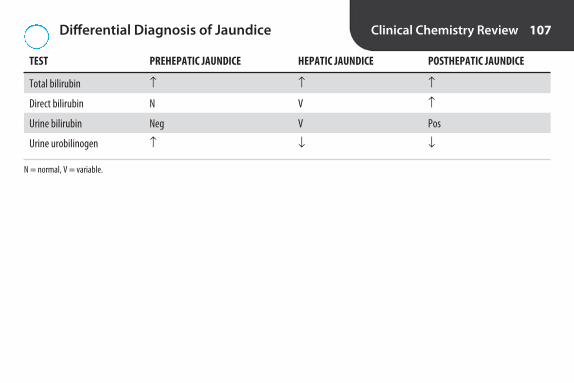
Differential Diagnosis of Jaundice Clinical Chemistry Review 107
TEST PREHEPATIC JAUNDICE HEPATIC JAUNDICE POSTHEPATIC JAUNDICE
Total bilirubin ↑ ↑ ↑
Direct bilirubin N V ↑
Urine bilirubin Neg V Pos
Urine urobilinogen ↑ ↓ ↓
N = normal, V = variable.
2956_Ch02_067-134 30/01/14 4:24 PM Page 107

Pituitary Hormones Clinical Chemistry Review 108
HORMONE REGULATES COMMENTS
Anterior pituitaryACTH
FSH
Growth hormone (GH)
LH
Prolactin (PRL)
TSH
Production of adrenocortical hormones by adrenal cortex
Sperm & egg production
Protein synthesis, cell growth, &division
Maturation of follicles, ovulation,production of estrogen, proges-terone, testosterone
Lactation
Production of T3 & T4 by thyroid
Regulated by corticotropin-releasing hormone (CRH) from hypo-thalamus. Diurnal variation: highest levels in early am, lowest inlate afternoon. ↑ Cushing’s disease. Collect on ice. Store frozen.
Regulated by gonadotropin-releasing hormone (GnRH) from hypothalamus. Sharp ↑ just before ovulation.
Regulated by growth-hormone releasing hormone (GHRH) & somatostatin from hypothalamus. ↑ gigantism, acromegaly. ↓ dwarfism.
Regulated by GnRH from hypothalamus. Sharp ↑ just before ovulation. Home ELISA kits to detect ovulation.
Regulated by prolactin-releasing factor (PRF) & prolactin-inhibiting factor (PIF) from hypothalamus.
Regulated by thyrotropin-releasing hormone (TRH) from hypothalamus. ↑ hypothyroidism. ↓ hyperthyroidism.
continued...
2956_Ch02_067-134 30/01/14 4:24 PM Page 108

Pituitary Hormones continued Clinical Chemistry Review 109
HORMONE REGULATES COMMENTS
Posterior pituitaryADH
Oxytocin
Reabsorption of water in distalrenal tubules
Uterine contractions during childbirth, lactation
Produced in hypothalamus. Stored in posterior pituitary. Releasestimulated by ↑ osmolality, ↓ blood volume or blood pressure. ↓ in diabetes insipidus.
Produced in hypothalamus. Stored in posterior pituitary. Not clinically useful.
2956_Ch02_067-134 30/01/14 4:24 PM Page 109

Thyroid and Parathyroid Hormones Clinical Chemistry Review 110
HORMONE REGULATES COMMENTS
ThyroidThyroxine (T4)
Triiodothyronine (T3)
Calcitonin
Parathyroid Parathyroid hormone (PTH)
Principle thyroid hormone. 50 × more concentrated thanT3. Contains 4 atoms of I. Regulated by TSH. Most boundto thyroxine-binding globulin (TBG). ↑ in hyperthy-roidism, ↓ in hypothyroidism
Most formed from deiodination of T4 by tissues. Contains 3 atoms of I.4×–5× more potent than T4. Regulated by TSH. ↑ in hyperthyroidism, ↓ in hypothyroidism
Important in diagnosis of thyroid cancer
Primary hyperthyroidism = ↑ PTH, ↑ Ca2+, ↓ phosphateHypoparathyroidism = ↓ PTH, ↓ Ca2+, ↑ phosphate
Metabolism, growth, & development
Metabolism, growth, & development
Inhibition of Ca2+ resorption
Regulation of Ca2+ & phosphate
2956_Ch02_067-134 30/01/14 4:24 PM Page 110

Thyroid Function Testing Clinical Chemistry Review 111
PRIMARY SECONDARY TEST HYPOTHYROIDISM HYPOTHYROIDISM HYPERTHYROIDISM T3 THYROTOXICOSIS COMMENTS
Thyroid-stimulating hormone (TSH)
Free T4 (FT4)
Free T3 (FT3)
↑
↓
↓
↓
↓
↓
↓
↑
↑
↓
N
↑
1st test for screen-ing. If normal, nofurther testing.
Biologically activeform of T4. 2ndstep in screening ifTSH abnormal.
Biologically activeform of T3. Usuallynot helpful in Dxof hypothyroidism because last testto become abnor-mal. Usually onlytested when TSH is↓ & FT4 isn’t ↑.
Primary hypothyroidism = thyroid insufficiency. Secondary hypothyroidism = pituitary insufficiency. Graves’ disease (type of autoimmune disease) is most common cause ofhyperthyroidism. Thyroid tests are immunoassays.
2956_Ch02_067-134 30/01/14 4:24 PM Page 111

Adrenal Hormones Clinical Chemistry Review 112
HORMONE REGULATES COMMENTS
Adrenal cortex Aldosterone
Cortisol
Adrenal medullaEpinephrine, norepinephrine (adrenaline, noradrenaline)
Reabsorption of Na+ in renaltubules
Carbohydrate, fat, & proteinmetabolism. Water & electrolytebalance. Suppresses inflamma-tory & allergic reactions
“Fight or flight syndrome.”Stimulation of sympatheticnervous system
↑ causes hypertension due to water & Na+ retention. ↓ leads tosevere water & electrolyte abnormalities.
Regulated by ACTH. Diurnal variation. Highest in am.↑and loss of diurnal variation in Cushing’s syndrome, ↓ in
Addison’s disease.
Epinephrine is primary hormone of adrenal medulla. Epinephrine &norepinephrine = catecholamines. Metabolites are metanephrines &VMA. ↑ with pheochromocytoma (rare catecholamine producingtumor). Tests: plasma & urine catecholamines & metanephrines, urine VMA.
2956_Ch02_067-134 30/01/14 4:24 PM Page 112

Reproductive Hormones Clinical Chemistry Review 113
HORMONE REGULATES COMMENTS
Ovaries Estrogens
Progesterone
Placenta Estrogen (estriol)
Progesterone
HCG
Human placental lactogen (HPL)
Testes Testosterone
Development of female reproductive organs &secondary sex characteristics. Regulation ofmenstrual cycle. Maintenance of pregnancy
Preparation of uterus for ovum implantation,maintenance of pregnancy
No hormonal activity
See above
Progesterone production by corpus luteum duringearly pregnancy. Development of fetal gonads
Estrogen & progesterone production by corpusluteum. Development of mammary glands
Development of male reproductive organs &secondary sex characteristics
Estradiol (E2) is major estrogen produced by ovaries; most potent estrogen. Also produced in adrenal cortex.
Also produced by placenta. Metabolite is pregnanediol.Useful in infertility studies & to assess placental function.
Used to monitor fetal growth & development.
Used to detect pregnancy, gestational trophoblastic dis-ease (e.g., hydatidiform mole), testicular tumor, & otherHCG-producing tumors.
Used to assess placental function.
Also produced in adrenal cortex.
2956_Ch02_067-134 30/01/14 4:24 PM Page 113

Pancreatic Hormones Clinical Chemistry Review 114
HORMONE REGULATES COMMENTS
Insulin Carbohydrate metabolism Produced in beta cells of islets of Langerhans. Causes ↑ movement of glucose into cells for metabolism. Decreases plasma glucose levels. ↓ indiabetes mellitus, ↑ with insulinoma, hypoglycemia.
Glucagon Glycogenolysis, gluconeogenesis, lipolysis Produced in alpha cells of islets of Langerhans. Increases plasma glucoselevels. ↑ with glucagonoma, diabetes mellitus, pancreatitis, trauma.
2956_Ch02_067-134 30/01/14 4:24 PM Page 114

Therapeutic Drug Monitoring (TDM) Clinical Chemistry Review 115
TERM EXPLANATION
Minimum effective concentration (MEC) Lowest concentration of drug in blood that will produce desired effect
Minimum toxic concentration (MTC) Lowest concentration of drug in blood that will produce adverse response
Therapeutic index Ratio of MTC to MEC
Trough Lowest concentration of drug measured in blood. Reached just before next scheduled dose.Shouldn’t fall below MEC
Peak Highest concentration of drug measured in blood. Drawn immediately on achievement ofsteady state. Should not exceed MTC
Steady state Amount of drug absorbed & distributed = amount of drug metabolized & excreted. Usuallyreached after 5–7 half-lives
Half-life Time required for concentration of drug to be ↓ by half.
Pharmacokinetics Rates of absorption, distribution, biotransformation, & excretion
Most common methods: immunoassay, chromatography.
2956_Ch02_067-134 30/01/14 4:24 PM Page 115

Therapeutic Drug Groups Clinical Chemistry Review 116
GROUP REPRESENTATIVE DRUGS
Analgesics Salicylates, acetaminophen
Antiepileptics Phenobarbital, phenytoin, valproic acid, carbamazepine, ethosuximide, felbamate, gabapentin, lamotrigine
Antineoplastics Methotrexate
Antibiotics Aminoglycosides (amikacin, gentamicin, kanamycin, tobramycin), vancomycin
Cardioactives Digoxin, disopyramide, procainamide, quinidine
Psychoactives Tricyclic antidepressants, lithium
Immunosuppressants Cyclosporine, tacrolimus (FK-506)
2956_Ch02_067-134 30/01/14 4:24 PM Page 116

Toxic Agents Clinical Chemistry Review 117
SUBSTANCE ANALYTIC METHOD
Ethanol Gas chromatography, enzymatic methods
Carbon monoxide Differential spectrophotometry (co-oximeter), gas chromatography
Arsenic Atomic absorption
Lead Atomic absorption
Pesticides Measurement of serum pseudocholinesterase
2956_Ch02_067-134 30/01/14 4:24 PM Page 117

Drugs of Abuse Urine Screen Clinical Chemistry Review 118
Drugs routinely tested Amphetamines, barbiturates, benzodiazepines, cannabinoids, cocaine, methadone, opiates, phencyclidine, tricyclic antidepressants
Adulterated urine* Value outside physiological range or presence of substance that isn’t found in human urine, e.g., pH <3 or ≥ 11; nitrite ≥ 500 mg/dL; presence of chromium, halogens (bleach, iodine, fluoride), glutaraldehyde, pyridine, or surfactant
Substituted urine Values that aren’t consistent with normal human urine, e.g., creatinine <2 mg/dL & specific gravity≤ 1.0010 or ≥ 1.0200
Diluted urine Creatinine & specific gravity lower than expected for normal human urine, e.g., creatinine ≥ 2 mg/dL but ≤ 20 mg/dL & specific gravity ≥ 1.0010 but ≤ 1.0030
Method Immunoassay
Confirmation Method using different measurement principle
*Urine adulteration test strips are available.
2956_Ch02_067-134 30/01/14 4:25 PM Page 118

Common Tumor Markers* Clinical Chemistry Review 119
TUMOR TYPE OF CANCER FOR WHICH MARKER MARKER IS MOST OFTEN USED CLINICAL USE COMMENTS
α -Fetoprotein (AFP)
Cancer antigen 15-3 (CA 15-3) & cancer antigen 27.29 (CA 27.29)
Cancer antigen 19-9 (CA 19-9)
Cancer antigen 125 (CA 125)
Carcinoembryonic antigen (CEA)
Human chorionic gonadotropin (hCG)
Liver
Breast
Pancreatic
Ovarian
Colorectal
Ovarian & testicular. Also gesta-tional trophoblastic diseases
Aid Dx, monitor therapy, detectrecurrence
Stage disease, monitor therapy,detect recurrence
Stage disease, monitor therapy,detect recurrence
Aid Dx, monitor therapy, detectrecurrence
Monitor therapy, detect recurrence
Aid Dx, monitor therapy, detectrecurrence
Produced by fetal liver; re-expressed incertain tumors. Also ↑ in hepatitis,pregnancy.
Two different assays for same marker.Can be ↑ with other cancers & non-cancerous conditions.
Can be ↑ with other cancers & non-cancerous conditions.
Can be ↑ with other cancers & gyneco-logical conditions.
Fetal antigen re-expressed in tumors.Can be ↑ with other cancers, non-cancerous conditions, & in smokers.
↑ in pregnancy.
continued...
2956_Ch02_067-134 30/01/14 4:25 PM Page 119

Common Tumor Markers* continued Clinical Chemistry Review 120
TYPE OF CANCER FOR WHICH MARKER IS MOST OFTEN USED CLINICAL USE COMMENTS
Prostate-specific antigen (PSA)
Thyroglobulin
Prostate
Thyroid
Screening, aid Dx, monitor therapy, detect recurrence
Monitor therapy, detect recurrence
Currently most widely used tumormarker. Screening asymptomatic men is controversial. Some men withprostate cancer don’t have ↑ PSA. PSAcan be ↑ in other conditions. Measure-ment of free PSA may be helpful whenPSA is borderline.
↑ in other thyroid diseases. Antithyroglobulin antibodies should be measured at same time. Can inter-fere with assays.
* Tumor markers alone cannot Dx cancer. Most are not useful for screening. Tumor markers are nonspecific & can be elevated in noncancerous conditions. Some patientswith cancer do not have elevated tumor markers. Serial testing is more useful than a single test. With successful treatment, tumor marker levels should decline & return tonormal. Increasing levels following treatment might indicate a recurrence. All of the tumor markers above are measured by immunoassay.
2956_Ch02_067-134 30/01/14 4:25 PM Page 120

TERM EXPLANATION
pH
Acid
Base
Buffer
Bicarbonate
Carbonic acid
Total CO2
PCO2
Henderson-Hasselbalch equation
Acid-Base Balance Terminology Clinical Chemistry Review 121
− Log [H+] or log 1[H+]
Chemical that can yield H+. Proton donor. pH <7
Chemical that can accept H+ or yield OH−. pH >7
Weak acid & its salt or conjugate base. Minimizes changes in pH. Most important 1 for maintainingblood pH is bicarbonate/carbonic acid. (H+ + HCO3
− ⇔ H2CO3 ⇔ H2O + CO2). Others: phosphates,proteins, hemoglobin
HCO3−. Second largest fraction of anions. Proton acceptor or base. Equal to total CO2 − 1.
Regulated by kidneys
H2CO3. Proton donor or weak acid. Equal to PCO2 × 0.03. Regulated by lungs
All forms of CO2. (HCO3− + H2CO3 + dissolved CO2 )
Partial pressure of CO2. Directly related to amount of dissolved CO2.
pH = 6.1 + log [HCO3
−] or 6.1 + log
HCO3−
× 0.03[H2CO3] PCO2
continued...
2956_Ch02_067-134 30/01/14 4:25 PM Page 121

Acid-Base Balance Terminology continued Clinical Chemistry Review 122
Acidosis (acidemia)
Alkalosis (alkalemia)
Compensated acidosis or alkalosis
TERM EXPLANATION
Blood pH <7.38. ↓ HCO3−: H2CO3 ratio. (Normal 20:1). May be due to ↓ in HCO3
− (metabolic acidosis) or ↑ in H2CO3 (respiratory acidosis)
Blood pH >7.42. ↑ HCO3−: H2CO3 ratio. May be due to ↑ in HCO3
− (metabolic alkalosis) or ↓ inH2CO3 (respiratory alkalosis)
When compensatory mechanisms have succeeded in restoring the 20:1 ratio & pH returns to nor-mal. Kidneys compensate for respiratory problem; lungs compensate for metabolic problem
2956_Ch02_067-134 30/01/14 4:25 PM Page 122

Acid-Base Imbalances Clinical Chemistry Review 123
CONDITION pH PCO2 HCO3− COMPENSATION TO RE-ESTABLISH 20:1 RATIO
Respiratory acidosis ↓ ↑ N Kidneys retain HCO3−, excrete H+
Metabolic acidosis ↓ N ↓ Hyperventilation (blow off CO2)
Respiratory alkalosis ↑ ↓ N Kidneys excrete HCO3−, retain H+
Metabolic alkalosis ↑ N ↑ Hypoventilation (retain CO2)
2956_Ch02_067-134 30/01/14 4:25 PM Page 123
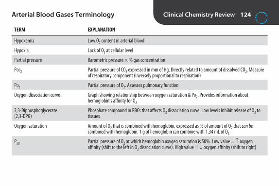
Arterial Blood Gases Terminology Clinical Chemistry Review 124
TERM EXPLANATION
Hypoxemia Low O2 content in arterial blood
Hypoxia Lack of O2 at cellular level
Partial pressure Barometric pressure × % gas concentration
PCO2 Partial pressure of CO2 expressed in mm of Hg. Directly related to amount of dissolved CO2. Measureof respiratory component (inversely proportional to respiration)
PO2 Partial pressure of O2. Assesses pulmonary function
Oxygen dissociation curve Graph showing relationship between oxygen saturation & PO2. Provides information about hemoglobin’s affinity for O2
2,3-Diphosphoglycerate Phosphate compound in RBCs that affects O2 dissociation curve. Low levels inhibit release of O2 to (2,3-DPG) tissues
Oxygen saturation Amount of O2 that is combined with hemoglobin, expressed as % of amount of O2 that can becombined with hemoglobin. 1 g of hemoglobin can combine with 1.34 mL of O2
P50 Partial pressure of O2 at which hemoglobin oxygen saturation is 50%. Low value = ↑ oxygen affinity (shift to the left in O2 dissociation curve). High value = ↓ oxygen affinity (shift to right)
2956_Ch02_067-134 30/01/14 4:25 PM Page 124
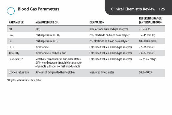
Blood Gas Parameters Clinical Chemistry Review 125
REFERENCE RANGE PARAMETER MEASUREMENT OF: DERIVATION (ARTERIAL BLOOD)
pH [H+] pH electrode on blood gas analyzer 7.35–7.45
PCO2 Partial pressure of CO2 PCO2 electrode on blood gas analyzer 35–45 mm Hg
PO2 Partial pressure of O2 PO2 electrode on blood gas analyzer 80–100 mm Hg
HCO3– Bicarbonate Calculated value on blood gas analyzer 22–26 mmol/L
Total CO2 Bicarbonate + carbonic acid Calculated value on blood gas analyzer 23–27 mmol/L
Base excess* Metabolic component of acid-base status. Calculated value on blood gas analyzer –2 to +2 mEq/LDifference between titratable bicarbonate of sample & that of normal blood sample
Oxygen saturation Amount of oxygenated hemoglobin Measured by oximeter 94%–100%
*Negative values indicate base deficit.
2956_Ch02_067-134 30/01/14 4:25 PM Page 125

Blood Gas Instrumentation Clinical Chemistry Review 126
DEVICE DESCRIPTION MEASURES CALIBRATION
pH electrode
PCO2 electrode (Severinghaus electrode)
PO2 electrode (Clark electrode)
Co-oximeter
H+-sensitive glass electrode containing Ag/AgCl wirein electrolyte of known pH & reference (calomel)electrode (Hg/Hg2Cl2). Measurement is potentiomet-ric (change in voltage indicates activity of analyte).
pH electrode covered with membrane permeable toCO2, with bicarbonate buffer between membrane &electrode. Measurement is potentiometric.
Platinum cathode & Ag/AgCl anode covered withsemipermeable membrane. Measurement is amperometric (amount of current flow is indicationof O2 present).
Spectrophotometer that reads absorbance or reflectance at isobestic point (wavelength where reduced & oxyhemoglobin have same absorbance or reflectance, e.g., 805 nm) & differential point(wavelength where reduced & oxyhemoglobin havedifferent absorbance or reflectance, e.g., 650 nm).
[H+]
Dissolved CO2
Dissolved O2
Oxygen saturation. Somealso measure carboxyhe-moglobin, methemoglobin,& sulfhemoglobin by usingadditional wavelengths.
2 phosphate buffersof known pH. (Storeat RT; don’t expose to air)
2 gases of known PCO2
2 gases of known PO2
Calibration curve pre-pared from specimenswith 0% & 100% O2 saturation
2956_Ch02_067-134 30/01/14 4:25 PM Page 126

Sources of Error in Arterial Blood Gases Clinical Chemistry Review 127
ERROR EFFECT
Hyperventilation ↓ PCO2,↑ pH, ↑ PO2
Specimen exposed to air ↓ PCO2, ↑ pH, ↑ PO2
Specimen at RT >30 minutes ↓ PO2, ↓ pH, ↑ PCO2
2956_Ch02_067-134 30/01/14 4:25 PM Page 127

Calculated Chemistry Values Clinical Chemistry Review 128
VALUE CALCULATION NORMAL RANGE CLINICAL SIGNIFICANCE
A/G ratio
Amylase: creatinine clearance ratio
Anion gap
AlbuminTotal protein − albumin
Urine amylase (U/L) × serum creatinine (mg/L)Serum amylase (U/L) × urine creatinine (mg/L)
(Na+ + K+) – (Cl– + HCO3–)
orNa+ – (Cl– + HCO3
–)
1–2.5
2%–5%
10–20or 7–16
Reversed A/G ratio with multiplemyeloma, liver disease.
↑ acute pancreatitis. ↓ macroamylasemia.
Difference between unmeasuredanions & unmeasured cations. ↑ in renal failure; diabetic acido-sis; lactic acidosis; methanol,ethanol, ethylene glycol, or salicylate poisoning; laboratoryerror. Useful QC check. Can’t be a negative number. If all deter-minations are ↑ or ↓, possibleinstrument error in 1 of the determinations.
continued...
2956_Ch02_067-134 30/01/14 4:25 PM Page 128

Calculated Chemistry Values continued Clinical Chemistry Review 129
VALUE CALCULATION NORMAL RANGE CLINICAL SIGNIFICANCE
BUN-to-creatinine ratio
Creatinine clearance
Indirect (unconjugated) bilirubin
LDL cholesterol
BUNCreatinine
urine creatinine (mg/dL) × urine mL per 24hr/1,440plasma creatinine (mg/dL)
× 1.73body surface area
Total bilirubin – direct (conjugated) bilirubin
Friedewald formula: LDL-C = total cholesterol − [HDL-C + (triglycerides/5)]
10–20
M: 97–137 mL/min F: 88–128 mL/min
<0.2 mg/dL
Desirable level <130 mg/dL
Normal ratio with renal disease.Prerenal conditions: ↑ ratio with↑ BUN & normal CR. Postrenalconditions: ↑ ratio with ↑ CR.Ratio ↓ with ↓ urea production(e.g., severe liver disease, ↓ pro-tein intake).
↓ renal disease (early indicator).
↑ prehepatic, posthepatic, &some types of hepatic jaundice.
↑ LDL cholesterol associatedwith ↑ risk of CAD. Not valid iftriglycerides >400 mg/dL. Directmeasurements now available.
continued...
2956_Ch02_067-134 30/01/14 4:25 PM Page 129
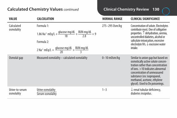
Calculated Chemistry Values continued Clinical Chemistry Review 130
VALUE CALCULATION NORMAL RANGE CLINICAL SIGNIFICANCE
Calculated osmolality
Osmolal gap
Urine-to-serum osmolality
Formula 1:
1.86 Na+ mEq/L + glucose mg/dL + BUN mg/dL + 918 2.8
Formula 2:
2 Na+ mEq/L + glucose mg/dL + BUN mg/dL 20 3
Measured osmolality – calculated osmolality
Urine osmolalitySerum osmolality
275–295 Osm/kg
0–10 mOsm/kg
1–3
Concentration of solute. Electrolytescontribute most. One of colligativeproperties. ↑ dehydration, uremia,uncontrolled diabetes, alcohol orsalicylate intoxication, excessiveelectrolyte IVs. ↓ excessive waterintake.
Similar to anion gap but based onosmotically active solute concen-tration rather than concentrationof ions. >10 indicates abnormalconcentration of unmeasuredsubstance (ex: isopropanol,methanol, acetone, ethylene glycol). Used to Dx poisonings.
↓ renal tubular deficiency, diabetes insipidus.
2956_Ch02_067-134 30/01/14 4:25 PM Page 130

Chemistry Calculations
FORMULA EXAMPLE
Beer’s law: Concentration of unknown =
Absorbance of unknown × concentration of standardAbsorbance of standard
mEq/L = mg/dL × 10GEW*
mmol/L = mg/dL × 10GMW†
Clinical Chemistry Review 131
A manual glucose assay gave the following results: Absorbanceof 100 mg/dL standard = 0.3.Absorbance of patient = 0.4. What is the glucose concentration of the patient?
Concentration = 0.4 × 100 = 133 mg/dL0.3
(Note: If a dilution is run, multiply answer by reciprocal of dilution.)
A calcium is reported as 10 mg/dL. What is the concentration in mEq/L?(Atomic weight of calcium = 40. Valence of calcium = 2+.)
mEq/L = 10 × 10 = 520
A calcium is reported as 10 mg/dL. What is the concentration in mmol/L?(Atomic weight of calcium = 40. Valence of calcium = 2+.)
mmol/L = 10 × 10 = 2.540
continued...
*GEW = gram equivalent weight (gram molecular weight ÷ valence).†GMW = gram molecular weight
2956_Ch02_067-134 30/01/14 4:25 PM Page 131

Chemistry Calculations continued Clinical Chemistry Review 132
FORMULA EXAMPLE
mmol/L = mEq/Lvalence
Molarity (M) = grams per literGMW
Normality (N) = grams per literGEW
A calcium is reported as 5 mEq/L. What is the concentration in mmol/L?(Atomic weight of calcium = 40. Valence of calcium = 2+.)
mmol/L = 5 = 2.52
What is the molarity of a solution that contains 45 grams of NaCl per liter?(Atomic weights: Na = 23, Cl = 35.5.)
M = 45 = 0.7758.5
What is the normality of a solution that contains 98 grams of H2SO4 per 500 mL? (Atomic weights: H = 1, S = 32, O = 16.)
N = 196 = 449
continued...
2956_Ch02_067-134 30/01/14 4:25 PM Page 132

Chemistry Calculations continued Clinical Chemistry Review 133
FORMULA EXAMPLE
% concentration = grams or milliliters per 100 mL
N = M × valence
V1C1 = V2C2
What is the concentration in % of a solution that contains 8.5 grams of NaCl per liter?
8.5 g = x1,000 mL 100 mL
1,000 x = (8.5) × 100x = 0.85%
What is the normality of a 3 M H2SO4 solution?
N = 3 × 2 = 6
What is the molarity of a 0.3 N H2SO4 solution?
M = 0.3 = 0.152
How many mL of 95% alcohol are needed to prepare 100 mL of 70% alcohol?
(x)(95) = (100)(70)x = 73.7 mL
V = volume, C = concentration.
2956_Ch02_067-134 30/01/14 4:25 PM Page 133

2956_Ch02_067-134 30/01/14 4:25 PM Page 134

3S E C T I O N
Clinical MicrobiologyReview
2956_Ch03_135-286 29/01/14 12:17 PM Page 135

Biosafety Levels Clinical Microbiology Review 136
LEVEL RISK TYPES OF AGENTS EXAMPLES OF AGENTS PRECAUTIONS
I
II
III
IV
Standard microbiological practices. No special equipment.
Biological safety cabinet (BSC) I or II.Personal protective equipment (PPE).Autoclave must be available. Limitedaccess. Most micro labs fall in this category.
Same as above plus negative air flow,sealed windows.
Requires use of class III BSC; full-body,air-supplied positive pressure suit; independent unit with specializedventilation & waste management toprevent release into environment.
Those not known to causedisease in healthy adults
Common humanpathogens
Those that may cause serious or lethal diseasevia inhalation. Effectivetreatment available
Those that pose high riskof life-threatening dis-ease. May be transmittedby aerosols. No vaccine or therapy
Minimal
Moderate
High
Extreme
Bacillus subtilis, Mycobacterium gordonae, soil microbes
E. coli, Salmonella, HIV, HBV, influenza
Bacillus anthracis, Francisella, Brucella,Mycobacterium tuberculosis, Rickettsiarickettsii, Coxiella burnetii, mold stagesof systemic fungi
Ebola virus, Lassa virus, others thatcause hemorrhagic fevers
2956_Ch03_135-286 29/01/14 12:17 PM Page 136

Centers for Disease Control and PreventionClassification of Biological Agents
Clinical Microbiology Review 137
CATEGORY PRIORITY SPREAD IMPACT EXAMPLES
A
B
C
Bacillus anthracis, Yersinia pestis, Francisellatularensis, Clostridium botulinum toxin, smallpox, hemorrhagic fever viruses (Ebola,Marburg, Lassa, Machupo)
Brucella, Salmonella, Shigella, E. coli O157:H7,Burkholderia mallei, Burkholderia pseudomallei,Chlamydia psittaci, Coxiella burnetii, Clostridiumperfringens toxin, ricin toxin, staphylococcalenterotoxin B, Rickettsia prowazekii, viral encephalitis viruses, Vibrio cholerae, Cryptosporidium parvum
Nipah virus, hantavirus
Easily disseminated ortransmitted from personto person
Moderately easy to disseminate
Could be engineered formass dissemination
Highest
2nd highest
3rd highest
High mortality, potential formajor public health impact
Moderate illness, low death rate
High morbidity/mortality,major public health impact
2956_Ch03_135-286 29/01/14 12:17 PM Page 137
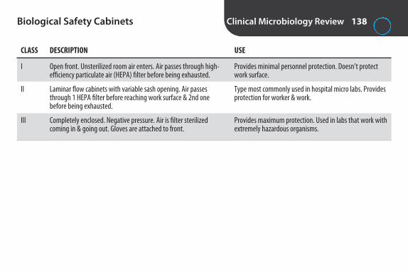
Biological Safety Cabinets Clinical Microbiology Review 138
CLASS DESCRIPTION USE
I
II
III
Provides minimal personnel protection. Doesn’t protectwork surface.
Type most commonly used in hospital micro labs. Providesprotection for worker & work.
Provides maximum protection. Used in labs that work withextremely hazardous organisms.
Open front. Unsterilized room air enters. Air passes through high-efficiency particulate air (HEPA) filter before being exhausted.
Laminar flow cabinets with variable sash opening. Air passesthrough 1 HEPA filter before reaching work surface & 2nd one before being exhausted.
Completely enclosed. Negative pressure. Air is filter sterilizedcoming in & going out. Gloves are attached to front.
2956_Ch03_135-286 29/01/14 12:17 PM Page 138

Sterilization and Disinfection Clinical Microbiology Review 139
Temperature
Steam under pressure
Boiling
Pasteurization
Hot air sterilization
Incineration
Filtration
Radiation
ChemicalsAlcohols
Chlorine
Formalin
Most reliable
Autoclave. Most practical & dependable method. 15 lb of pressure for 15 min. 121°C. Kills spores.
Not reliable. Spores may not be killed.
Used in food industry to kill foodborne pathogens. Doesn’t sterilize. Liquid is heated to 71.7°C for 15 sec.
Used when steam may damage or fail to penetrate. 2 hours at 170°C. Kills spores.
Used to sterilize inoculating loops & biomedical wastes.
Used to sterilize liquids that are thermolabile (e.g., urea broth). HEPA air filters are used in BSC.
UV light. Used in BSC.
Ethyl & isopropyl (70%–80%) frequently used as antiseptics & disinfectants. Don’t kill spores.
Sodium hypochlorite (household bleach). One of most effective agents against HIV & HBV. 10% solution. Prepare daily.
Aqueous solution of formaldehyde. 5%–10% formalin used to preserve & fix specimens. Exposure must be monitored.
continued...
2956_Ch03_135-286 29/01/14 12:17 PM Page 139

Sterilization and Disinfection continued Clinical Microbiology Review 140
Glutaraldehyde
Hydrogen peroxide
Iodine & iodophors
Phenolics
Quaternary ammoniumcompounds
GasesEthylene oxide
Effective against most vegetative cells other than mycobacteria. Used for cold sterilization of items damaged byheat, e.g., inhalation therapy equipment, equipment with optical lenses such as endoscopes.
3% solution used as antiseptic
Iodophors (e.g., povidone-iodine) release iodine slowly & are less irritating & nonstaining. Effective skin antiseptics.
Don’t kill spores. Other disinfectants are evaluated by comparing effectiveness to phenol. “Phenol coefficient.”
Effective against wide range of vegetative bacteria. Not effective against spores, mycobacteria, or nonenvelopedviruses. Used to disinfect floors, walls, furniture.
Widely used in hospitals to sterilize materials that can’t withstand steam.
2956_Ch03_135-286 29/01/14 12:17 PM Page 140

Bacterial Toxins Clinical Microbiology Review 141
EXOTOXIN ENDOTOXIN
Produced by Gram positives Gram negatives
Location in cell Within & without Within
Composition Protein Lipopolysaccharide
Effect Systemic Local
Toxicity High Low
Stability to heating Unstable Stable
Antigenicity High Low
Stimulates antitoxin productions? Yes No
Converted to toxoid? Yes No
2956_Ch03_135-286 29/01/14 12:17 PM Page 141

Specimen Collection Guidelines Clinical Microbiology Review 142
• Obtain during acute phase of infection (within 2–3 daysfor viruses).
• Collect before antibiotics are administered.• Sample appropriate site.• Aspirates or tissues are preferred to swabs.• Use swabs with Dacron or polyester tips & plastic
shafts. Wood, cotton, & calcium alginate may betoxic.
• For anaerobes, aspirates preferred to swabs.• Avoid contamination with environmental or normal
flora.
• Obtain sufficient quantity.• Use sterile, leak-proof container.• Properly label container, not lid.• Transport in secondary container (plastic bag) marked
with biohazard symbol.• Protect requisition from contamination.• Syringes with needles attached should never be
transported.• Deliver to lab within 30 min of collection.• For prolonged transit, use special preservatives or
holding media.
2956_Ch03_135-286 29/01/14 12:17 PM Page 142

Specimen Preservation and Storage Clinical Microbiology Review 143
SPECIMEN PRESERVATIVE OR TRANSPORT DEVICE STORAGE TEMPERATURE (UNPRESERVED)
Anaerobes
CSF
Neisseria gonorrhoeae
Sputum
Stool for culture
Stool for ova & parasites
Urine
Viruses
25°C. Don’t refrigerate
25°C or 35°C. Don’t refrigerate
25°C. Don’t refrigerate
4°C
4°C. Best to process without delay. Refrigera-tion may kill Shigella
Can hold formed specimens at 4°C. Soft or liquid specimens should be examined immediately or preserved
4°C for not more than 24 hr
4°C. For delay >24 hr, freeze at −70°C
Anaerobic transport systems to eliminate O2
None
Amies transport medium with charcoal, Transgrow, JEMBEC plates, Gono-Pak, BioBag (best to inoculatemedium directly)
None
Transport media, e.g., Cary-Blair transport medium ifspecimen can’t be cultured within 2 hr of collection
Depending on procedures to be performed: polyvinyl alcohol (PVA), 10% formalin, sodium acetate–aceticacid–formalin (SAF), merthiolate-iodine-formalin (MIF),others without formaldehyde or mercury (e.g., Ecofix,Parasafe)
Transport tubes with boric acid–glycerol if specimen can’t be processed within 2 hr of collection
Viral transport medium
2956_Ch03_135-286 29/01/14 12:17 PM Page 143

Fragile Organisms Clinical Microbiology Review 144
• Anaerobes• Chlamydia• Haemophilus influenzae• Neisseria gonorrhoeae• Neisseria meningitidis
• Salmonella• Shigella• Streptococcus pneumoniae• Viruses• Parasites
2956_Ch03_135-286 29/01/14 12:17 PM Page 144

Criteria for Rejection of Specimens in Microbiology
Clinical Microbiology Review 145
• Unlabeled or improperly labeled specimen• Improper collection site• Prolonged transit (over 2 hr without preservation)• Improper temperature during transport or storage• Leaking specimens• Specimens in nonsterile containers• Improper transport medium• Culturette ampule not broken, swab dried out• Improper swab, e.g., wood or calcium alginate for
viruses or Chlamydia
• Syringes with needles attached• Culture for anaerobes requested on inappropriate
sources• Specimen received in formalin (other than stool for ova
& parasites)• Saliva instead of sputum• Foley catheter tip• Insufficient quantity
Follow lab’s written policies.
2956_Ch03_135-286 29/01/14 12:17 PM Page 145

Gram Stain Clinical Microbiology Review 146
REAGENT FUNCTION OTHER
Crystal violet Basic dye Stains all bacteria purple
Iodine Mordant Fixes crystal violet to cells
95% ethyl alcohol or acetone or combination Decolorizer Removes crystal violet from gram neg
Safranin Counterstain Stains gram neg pink
2956_Ch03_135-286 29/01/14 12:17 PM Page 146

Staining Properties of Gram-Positive and Gram-Negative Bacteria
Clinical Microbiology Review 147
GRAM REACTION CELL WALL STAINED BY COLOR IN GRAM STAIN
Gram positive Thick peptidoglycan layer with teichoic acid & lipoteichoic Crystal violet Purpleacid. Teichoic acid cross-links prevent decolorization in Gram stain.
Gram negative Thin peptidoglycan layer covered with proteins, phospholipids, Safranin Pink& lipopolysaccharides. Decolorizer causes ↑ permeability of lipid-rich cell wall. Primary stain (crystal violet) washes out.
2956_Ch03_135-286 29/01/14 12:17 PM Page 147

Types of Media Clinical Microbiology Review 148
TYPE EXPLANATION EXAMPLES
Supportive
Enrichment
Selective
Differential
Note: Media may be of more than 1 type, e.g., selective & differential.
Nutrient agar, trypticase soy agar
Sheep blood agar, chocolate agar, brain-heart infusion,buffered charcoal-yeast extract agar
Columbia colistin–nalidixic acid (CNA) agar, eosinmethylene blue (EMB), MacConkey, Hektoen enteric(HE), xylose lysine deoxycholate (XLD), Thayer-Martin
EMB, MacConkey, HE, XLD
Supports growth of most nonfastidious bacteria
Contains added growth factors, e.g., blood, vitamins, yeastextract
Contains additives such as dyes, bile salts, alcohols, acids, or antibiotics to inhibit growth of certain bacteria (e.g.,gram pos)
Formulated to provide distinct colonial appearances basedon certain biochemical rxn (e.g., lactose fermentation, hydrogen sulfide [H2S] production)
2956_Ch03_135-286 29/01/14 12:17 PM Page 148
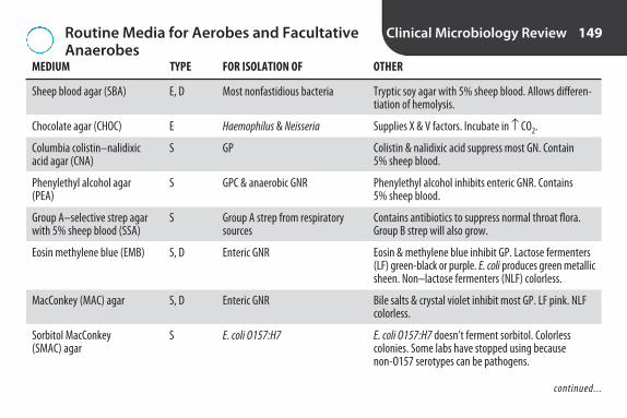
Routine Media for Aerobes and FacultativeAnaerobes
Clinical Microbiology Review 149
MEDIUM TYPE FOR ISOLATION OF OTHER
Sheep blood agar (SBA)
Chocolate agar (CHOC)
Columbia colistin–nalidixic acid agar (CNA)
Phenylethyl alcohol agar (PEA)
Group A–selective strep agar with 5% sheep blood (SSA)
Eosin methylene blue (EMB)
MacConkey (MAC) agar
Sorbitol MacConkey (SMAC) agar
Tryptic soy agar with 5% sheep blood. Allows differen-tiation of hemolysis.
Supplies X & V factors. Incubate in ↑ CO2.
Colistin & nalidixic acid suppress most GN. Contain 5% sheep blood.
Phenylethyl alcohol inhibits enteric GNR. Contains 5% sheep blood.
Contains antibiotics to suppress normal throat flora.Group B strep will also grow.
Eosin & methylene blue inhibit GP. Lactose fermenters(LF) green-black or purple. E. coli produces green metallicsheen. Non–lactose fermenters (NLF) colorless.
Bile salts & crystal violet inhibit most GP. LF pink. NLFcolorless.
E. coli O157:H7 doesn’t ferment sorbitol. Colorlesscolonies. Some labs have stopped using because non-O157 serotypes can be pathogens.
E, D
E
S
S
S
S, D
S, D
S
Most nonfastidious bacteria
Haemophilus & Neisseria
GP
GPC & anaerobic GNR
Group A strep from respiratorysources
Enteric GNR
Enteric GNR
E. coli O157:H7
continued...
2956_Ch03_135-286 29/01/14 12:17 PM Page 149

Routine Media for Aerobes and FacultativeAnaerobes continued
Clinical Microbiology Review 150
MEDIUM TYPE FOR ISOLATION OF OTHER
Hektoen enteric (HE) agar
Xylose lysine deoxycholate (XLD)
Salmonella-Shigella (SS) agar
Gram-negative broth (GN)
Bile salts, bromothymol blue, & acid fuchsin inhibit normal GI flora. Nonpathogens orange to salmon pink.NLF green to blue-green. H2S-pos colonies have blackprecipitate.
Deoxycholate inhibits many GNR & GP. 4 types of colonies:yellow (e.g., E. coli), yellow with black centers (e.g.,some Proteus species), colorless or red colonies (e.g.,Shigella), red colonies with black centers (e.g., Salmonella).(Some shigellae may be inhibited. Some salmonellaemay not produce H2S.)
Brilliant green & bile salts inhibit other enterics. Salmonella & Shigella don’t ferment lactose (colorlesscolonies). Salmonella produces H2S (black center).
Deoxycholate & citrate salts retard growth of GP. Sub-culture onto selective differential agar after 6–8 hr &18–24 hr incubation. Use of enrichment broths for stoolcultures is decreasing.
S, D
S, D
S
E, S
Salmonella & Shigella in stool
Salmonella & Shigella in stool
Salmonella & Shigella in stool
Salmonella & Shigella fromstools & rectal swabs
continued...
2956_Ch03_135-286 29/01/14 12:17 PM Page 150

Routine Media for Aerobes and FacultativeAnaerobes continued
Clinical Microbiology Review 151
MEDIUM TYPE FOR ISOLATION OF OTHER
Deoxycholate-citrate agar
Campylobacter broth
Campylobacter blood agar (Campy BAP)
E = enriched; S = selective; D = differential; GP = gram positive; GN = gram negative; C = cocci; R = rods.
Other enterics inhibited.
Subculture to Campy-selective agar after overnight incubation at 4°C.
Incubate plates in ↑ CO2 at 42°C.
S
E
E, S
Salmonella & Shigella
Campylobacter from stool
Campylobacter from stool
2956_Ch03_135-286 29/01/14 12:17 PM Page 151

Selective Media for Isolation of Neisseria gonorrhoeae and Neisseria meningitidis
Clinical Microbiology Review 152
MEDIUM COMMENTS
Modified Thayer-Martin (TM)
Martin-Lewis
New York City medium (NYC)
GC-LECT
JEMBEC plates
Used for specimens from sites with normal flora.
Vancomycin, colistin, nystatin, & trimethoprim inhibit growth of other bacteria & fungi. Incubate in↑ CO2. Some N. gonorrhoeae may be inhibited.
Similar to Thayer-Martin, but different antibiotics. Inhibits yeast better. Incubate in ↑ CO2.
Incubate in ↑ CO2. Some N. gonorrhoeae are inhibited by antibiotics. Genital mycoplasmas will grow.
Antibiotics to inhibit GN & GP bacteria & yeast.
For transportation & growth of N. gonorrhoeae. Plates contain Neisseria-selective medium & comewith resealable polyethylene bag & CO2-generating tablet. No need to transfer to culture plate.
2956_Ch03_135-286 29/01/14 12:17 PM Page 152

Special Bacteriologic Media Clinical Microbiology Review 153
MEDIUM USE OTHER
Cystine-tellurite blood agar
Loeffler medium
Tindale agar
Bismuth sulfite agar
Cefsulodin-irgasan-novobiocin (CIN) agar
Alkaline peptone water (APW)
C. diphtheriae produces black colonies.
Promotes development of metachromatic granules.
Corynebacterium spp produce gray to black colonies due toreduction of tellurite. C. diphtheriae colonies are surroundedby a brown halo.
Bismuth sulfite & brilliant green inhibit most others. S. typhicolonies are black, surrounded by metallic sheen. Others arelight green. (Some salmonellae may be inhibited.)
Crystal violet inhibits most GN. Novobiocin inhibits GPC. Cefsulodin inhibits most GP & GN. Y. enterocolitica fermentsmannitol, appears as red “bull’s-eye” colonies surrounded bycolorless halo.
Alkaline pH suppresses commensals. Subcultured to TCBS.
Differential medium for isolation ofCorynebacterium diphtheriae
Enrichment medium for C. diphtheriae
Selective differential medium for C.diphtheriae
Selective for Salmonella
Selective medium for Yersinia entero-colitica, Aeromonas, & Plesiomonasshigelloides
Enrichment medium for recovery ofVibrio from stool
continued...
2956_Ch03_135-286 29/01/14 12:17 PM Page 153

Special Bacteriologic Media continued Clinical Microbiology Review 154
MEDIUM USE OTHER
Thiosulfate citrate bile salts sucrose (TCBS) agar
Bordet-Gengou agar
Regan-Lowe agar
Buffered charcoal-yeast extract (BCYE) agar
Human blood bilayer Tween (HBT) agar
High pH inhibits most bacteria. V. cholerae ferments sucrose,produces yellow colonies. V. parahaemolyticus & V. vulnificusdon’t ferment sucrose; usually produce blue-green colonies.
Potato-glycerol-based medium enriched with blood. Contam-inants inhibited by methicillin. “Cough plate.” Bordetellacolonies resemble mercury droplets.
Charcoal agar supplemented with horse blood, cephalexin, &hotericin B.
Yeast extract & L-cysteine enhance growth of Legionella.Charcoal absorbs toxic compounds.
Incubate in ↑ CO2 for 48 hr. Colonies are beta hemolytic.
Selective for Vibrio
Selective enrichment medium for isolation of Bordetella pertussis
Selective for B. pertussis
Enrichment medium for isolation ofLegionella
Selective & differential for G. vaginalis
2956_Ch03_135-286 29/01/14 12:17 PM Page 154
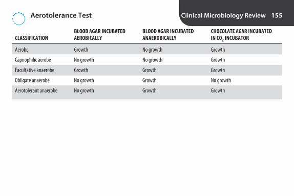
Aerotolerance Test Clinical Microbiology Review 155
BLOOD AGAR INCUBATED BLOOD AGAR INCUBATED CHOCOLATE AGAR INCUBATED CLASSIFICATION AEROBICALLY ANAEROBICALLY IN CO2 INCUBATOR
Aerobe Growth No growth Growth
Capnophilic aerobe No growth No growth Growth
Facultative anaerobe Growth Growth Growth
Obligate anaerobe No growth Growth No growth
Aerotolerant anaerobe No growth Growth Growth
2956_Ch03_135-286 29/01/14 12:17 PM Page 155

Organisms Requiring Incubation in Increased CO2
Clinical Microbiology Review 156
• Campylobacter• Haemophilus• Helicobacter• Moraxella catarrhalis• Mycobacterium• Pathogenic Neisseria
2956_Ch03_135-286 29/01/14 12:17 PM Page 156

Hemolytic Reactions on Sheep Blood Agar Clinical Microbiology Review 157
HEMOLYSIS DESCRIPTION EXPLANATION EXAMPLES
Alpha
Beta
Gamma (nonhemolytic)
Streptococcus pneumoniae, viridansstreptococci, some enterococci
Group A strep, group B strep, Listeriamonocytogenes
Some enterococci
Green zone around colony.May be narrow or wide.
Clear zone around colony.May be narrow or wide.
No zone of hemolysis.
Partial lysis of RBCs
Complete lysis of RBCs
No lysis of RBCs
2956_Ch03_135-286 29/01/14 12:17 PM Page 157
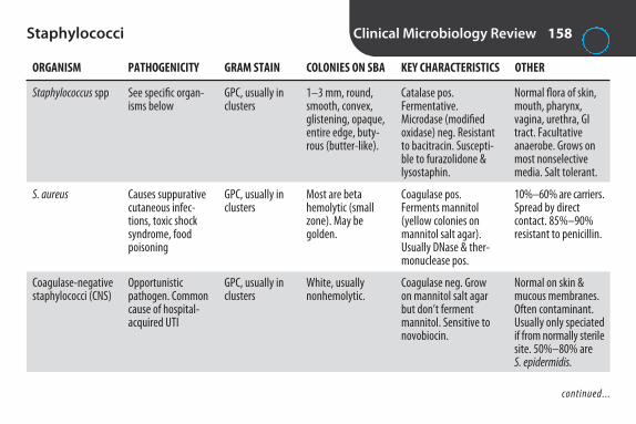
Staphylococci Clinical Microbiology Review 158
ORGANISM PATHOGENICITY GRAM STAIN COLONIES ON SBA KEY CHARACTERISTICS OTHER
Staphylococcus spp
S. aureus
Coagulase-negative staphylococci (CNS)
Normal flora of skin,mouth, pharynx,vagina, urethra, GItract. Facultativeanaerobe. Grows onmost nonselectivemedia. Salt tolerant.
10%–60% are carriers.Spread by direct contact. 85%–90%resistant to penicillin.
Normal on skin & mucous membranes.Often contaminant.Usually only speciatedif from normally sterilesite. 50%–80% are S. epidermidis.
GPC, usually inclusters
GPC, usually inclusters
GPC, usually inclusters
1–3 mm, round,smooth, convex,glistening, opaque,entire edge, buty-rous (butter-like).
Most are beta hemolytic (smallzone). May begolden.
White, usually nonhemolytic.
See specific organ-isms below
Causes suppurativecutaneous infec-tions, toxic shocksyndrome, foodpoisoning
Opportunisticpathogen. Commoncause of hospital-acquired UTI
Catalase pos. Fermentative.Microdase (modifiedoxidase) neg. Resistantto bacitracin. Suscepti-ble to furazolidone &lysostaphin.
Coagulase pos. Ferments mannitol(yellow colonies onmannitol salt agar).Usually DNase & ther-monuclease pos.
Coagulase neg. Growon mannitol salt agarbut don’t ferment mannitol. Sensitive tonovobiocin.
continued...
2956_Ch03_135-286 29/01/14 12:17 PM Page 158

Staphylococci continued Clinical Microbiology Review 159
ORGANISM PATHOGENICITY GRAM STAIN COLONIES ON SBA KEY CHARACTERISTICS OTHER
S. saprophyticus
Micrococcus
UTI = urinary tract infection.
Novobiocin only performed when CNSisolated from urine offemale.
Not commonly iso-lated. Must differenti-ate from staph.Usually only growaerobically.
GPC, usually inclusters
Large GPC inpairs, tetrads(predominantarrangement),clusters
White to slightlyyellow. Non-hemolytic.
Often pigmented(bright yellow, orange, pink, tan).High-domedcolonies.
UTI in young sexu-ally active females,urethritis & prosta-titis in males
Usually nonpatho-genic. Found in environment & on skin, mucousmembranes,oropharynx
Coagulase neg. Resistantto novobiocin. May ferment mannitol.
Catalase pos. Coagulaseneg. Oxidative. Microdase (modifiedoxidase) pos. Suscepti-ble to bacitracin. Resis-tant to furazolidone &lysostaphin.
2956_Ch03_135-286 29/01/14 12:17 PM Page 159

Summary of Tests for Identification of Staphylococci
Clinical Microbiology Review 160
TEST PRINCIPLE KEY REACTIONS OTHER
Catalase
Coagulase
Slide agglutination tests for S. aureus
Mannitol salt agar (MSA)
Novobiocin susceptibility
Bubbles after 20–30 sec aren’t considered pos rxn. Catalase fromRBCs in blood agar may produceweak bubbles.
Slide test is screening test. Detectsbound coagulase (clumping factor).If neg, tube test should be performed.Detects free coagulase. Largely re-placed by latex agglutination tests.
Most labs report pos organisms as S. aureus.
7.5% salt inhibits most organismsother than staph. All staph can grow on MSA. Rarely used for ID of S. aureus today because otherspecies ferment mannitol.
Performed on CNS isolated fromurine.
Enzyme catalase converts 3% hydrogen peroxide (H2O2) to oxygen & water. Immediate bubbling.
Enzyme coagulase causes coagula-tion (tube test) or agglutination(slide test) in plasma.
Agglutination of latex beads coatedwith fibrinogen & abs to protein A(protein in cell wall of S. aureus).
Fermentation of mannitol results incolor change from pink to yellow.
Organisms resistant to novobiocingrow to edge of disk.
Staphylococci pos. Strepto-cocci & enterococci neg
S. aureus pos
S. aureus pos
S. aureus pos
S. saprophyticus is resistant.Other CNS susceptible
2956_Ch03_135-286 29/01/14 12:17 PM Page 160

Streptococci/Enterococci Clinical Microbiology Review 161
ORGANISM PATHOGENICITY GRAM STAIN COLONIES ON SBA KEY CHARACTERISTICS OTHER
Streptococcus spp
Group A streptococci (GAS)
Facultative anaerobes.Require enrichedmedia. Chaining bestin broth cultures.
Most common isS. pyogenes. Hemolysisdue to O2-stable streptolysin S & O2-labile streptolysin O.To detect species thatproduce streptolysin O only, stab into agar,place coverslip overinoculum, or incubateanaerobically.
Oval GPC in chains& pairs
Oval GPC in chains
<1 mm, white togray, translucent,or semiopaque.Variable hemolysis.
Pinpoint. Grayishwhite. Translucent.Usually beta hemolytic (widezone).
See specific organ-isms below
Causes 90% of strep infections.Strep sore throat,rheumatic fever,glomerulonephritis,scarlet fever (scar-latina), erysipelas,puerperal sepsis,impetigo
Catalase neg
Sensitive to bacitracin.Resistant to SXT. PYR pos
continued...
2956_Ch03_135-286 29/01/14 12:17 PM Page 161

Streptococci/Enterococci continued Clinical Microbiology Review 162
ORGANISM PATHOGENICITY GRAM STAIN COLONIES ON SBA KEY CHARACTERISTICS OTHER
Group B streptococci (GBS)
Group D streptococci, nonenterococci
Enterococcus
S. agalactiae. Vaginal &rectal swabs collectedfrom pregnant womenat 35–37 wk gestation.Inoculated in selectivebroth, e.g., LIM, GBSbroth, StrepB CarrotBroth.
Most common isS. gallolyticus (formerlyS. bovis).
Lancefield group D.80% are E. faecalis,15% E. faecium.
Oval GPC in chains
Oval GPC in chains
Oval GPC in pairs &chains
Slightly larger thanGAS. Gray-white.Narrow zone of diffuse beta hemolysis. May be nonhemolytic.
Usually non-hemolytic. May bealpha hemolytic.
Usually alpha ornonhemolytic.Rarely beta.
Normal flora of fe-male genital tract.Most commoncause of neonatalsepticemia &meningitis
Normal in GI tract.Causes nosocomialUTI, wound infec-tions, bacteremia
Normal in mouth,GI tract, femalegenital tract. Causesnosocomial UTI,wound infections,bacteremia
Resistant to SXT & baci-tracin. Sodium hippu-rate pos. CAMP pos
Hydrolyzes esculin
Hydrolyzes esculin.Grows in 6.5% NaClbroth. PYR pos
continued...
2956_Ch03_135-286 29/01/14 12:17 PM Page 162

Streptococci/Enterococci continued Clinical Microbiology Review 163
ORGANISM PATHOGENICITY GRAM STAIN COLONIES ON SBA KEY CHARACTERISTICS OTHER
Streptococcus pneumoniae
Viridans streptococci
No Lancefield group.>80 serotypes basedon capsular antigens.
Football-shaped(lancet-shaped)GPC. Usually inpairs. May be single or in shortchains. Frequentlyencapsulated
GPC in chains
Round, translucent,glistening, domeshaped whenyoung. Central depression withage (umbilicate)due to autolysis.Alpha hemolytic.Encapsulatedstrains are mucoid
Usually alpha he-molytic, but may bebeta hemolytic ornonhemolytic.
Normal in upperrespiratory tract of some. Mostcommon cause of community-acquired pneumo-nia. Major cause of otitis media,meningitis inadults. Infects sinuses, eyes
Normal in oral,respiratory, GI mu-cosa. Opportunisticpathogens. Fre-quent cause ofsubacute bacterialendocarditis. Gingivitis & dentalcaries (cavities)
Bile solubility pos. Sensitive to optochin
Resistant to optochin.Bile solubility neg
continued...
2956_Ch03_135-286 29/01/14 12:17 PM Page 163

Streptococci/Enterococci continued Clinical Microbiology Review 164
ORGANISM PATHOGENICITY GRAM STAIN COLONIES ON SBA KEY CHARACTERISTICS OTHER
Microaerophilic Streptococcus spp (formerly S. milleri group)
3 species: S. anginosus,S. constellatus, S. inter-medius. In viridansgroup. Require 10%CO2. Grow betteranaerobically.
GPC in pairs, chains Colonies are halfthe size of otherstrep. Usually betahemolytic but canbe alpha hemolyticor nonhemolytic.Butterscotch orcaramel odor.
Normal flora ofmouth & GI, GUtracts. Infectionsassociated withtrauma or surgery.Most commonsites: abdomen,head, neck
Usually Lancefieldgroup F. PYR neg. VP pos
2956_Ch03_135-286 29/01/14 12:17 PM Page 164
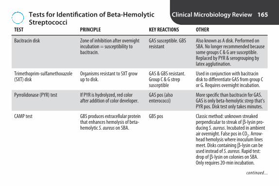
Tests for Identification of Beta-HemolyticStreptococci
Clinical Microbiology Review 165
TEST PRINCIPLE KEY REACTIONS OTHER
Bacitracin disk
Trimethoprim-sulfamethoxazole (SXT) disk
Pyrrolidonase (PYR) test
CAMP test
Also known as A disk. Performed onSBA. No longer recommended becausesome groups C & G are susceptible. Replaced by PYR & serogrouping bylatex agglutination.
Used in conjunction with bacitracindisk to differentiate GAS from group Cor G. Requires overnight incubation.
More specific than bacitracin for GAS.GAS is only beta-hemolytic strep that’sPYR pos. Disk test only takes minutes.
Classic method: unknown streakedperpendicular to streak of β -lysin pro-ducing S. aureus. Incubated in ambientair overnight. False pos in CO2. Arrow-head hemolysis where inoculum linesmeet. Disks containing β -lysin can beused instead of S. aureus. Rapid test:drop of β -lysin on colonies on SBA.Only requires 20-min incubation.
Zone of inhibition after overnightincubation = susceptibility tobacitracin.
Organisms resistant to SXT growup to disk.
If PYR is hydrolyzed, red colorafter addition of color developer.
GBS produces extracellular proteinthat enhances hemolysis of beta-hemolytic S. aureus on SBA.
GAS susceptible. GBSresistant
GAS & GBS resistant.Group C & G strepsusceptible
GAS pos (also enterococci)
GBS pos
continued...
2956_Ch03_135-286 29/01/14 12:17 PM Page 165
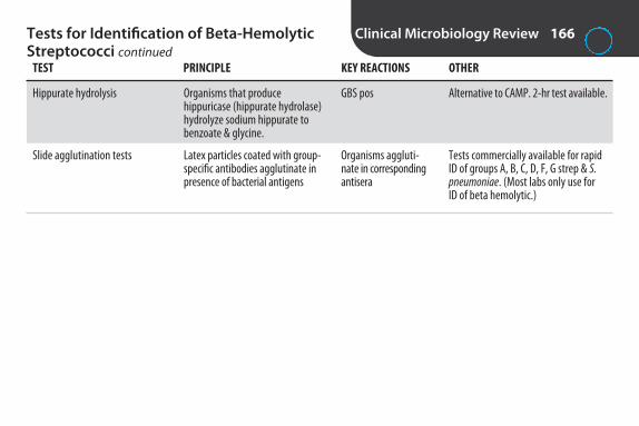
Tests for Identification of Beta-HemolyticStreptococci continued
Clinical Microbiology Review 166
TEST PRINCIPLE KEY REACTIONS OTHER
Hippurate hydrolysis
Slide agglutination tests
Alternative to CAMP. 2-hr test available.
Tests commercially available for rapidID of groups A, B, C, D, F, G strep & S.pneumoniae. (Most labs only use for ID of beta hemolytic.)
Organisms that produce hippuricase (hippurate hydrolase)hydrolyze sodium hippurate tobenzoate & glycine.
Latex particles coated with group-specific antibodies agglutinate inpresence of bacterial antigens
GBS pos
Organisms aggluti-nate in correspondingantisera
2956_Ch03_135-286 29/01/14 12:17 PM Page 166

Tests for Identification of Alpha-HemolyticStreptococci
Clinical Microbiology Review 167
TEST PRINCIPLE KEY REACTIONS OTHER
Optochin disk
Bile solubility
Also known as P disk. Contains ethylhydrocupreinehydrochloride. Placed on lawn of inoculum on SBA.Plates incubated overnight in ↑ CO2. If zone of in-hibition <14 mm, ID organism as S. pneumoniaeonly if bile soluble.
Can be performed in broth or on colony. Pos =clearing of broth or disappearance of colony. Results in 30 min or less.
Zone of inhibition ≥ 14 mm with6-mm disk or ≥ 16 mm with 10-mm disk = susceptibility tooptochin
Bile salts (e.g., sodium deoxy-cholate) cause lysis of some organisms
S. pneumoniae susceptible.Viridans strep resistant
S. pneumoniae pos
2956_Ch03_135-286 29/01/14 12:17 PM Page 167

Tests for Identification of NonhemolyticStreptococci/Enterococci
Clinical Microbiology Review 168
TEST PRINCIPLE KEY REACTIONS OTHER
Pyrrolidonase (PYR) test
Bile-esculin agar
Esculin test
6.5% NaCl broth
Bile inhibits GPs other than group Dstrep & enterococci.
Doesn’t test for bile tolerance.
Requires overnight incubation.
See “Tests for Identification ofBeta-Hemolytic Streptococci”
Organisms that can grow in 40% bile & produce esculinasehydrolyse esculin, producingblack precipitate
Organisms that produce esculi-nase hydrolyze esculin on disk,producing dark spot
Organisms that can grow in6.5% NaCl produce turbidity
Enterococci pos (also GAS)
Group D strep & enterococci pos
Group D strep & enterococci pos
Enterococci pos (also GBS). Group D strep neg
2956_Ch03_135-286 29/01/14 12:17 PM Page 168
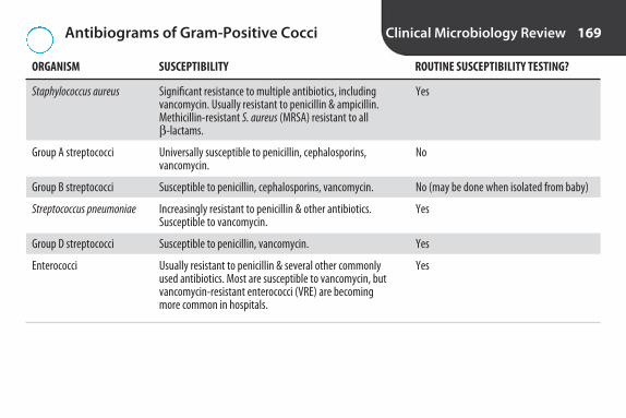
Antibiograms of Gram-Positive Cocci Clinical Microbiology Review 169
ORGANISM SUSCEPTIBILITY ROUTINE SUSCEPTIBILITY TESTING?
Staphylococcus aureus
Group A streptococci
Group B streptococci
Streptococcus pneumoniae
Group D streptococci
Enterococci
Yes
No
No (may be done when isolated from baby)
Yes
Yes
Yes
Significant resistance to multiple antibiotics, includingvancomycin. Usually resistant to penicillin & ampicillin.Methicillin-resistant S. aureus (MRSA) resistant to all β -lactams.
Universally susceptible to penicillin, cephalosporins, vancomycin.
Susceptible to penicillin, cephalosporins, vancomycin.
Increasingly resistant to penicillin & other antibiotics. Susceptible to vancomycin.
Susceptible to penicillin, vancomycin.
Usually resistant to penicillin & several other commonlyused antibiotics. Most are susceptible to vancomycin, butvancomycin-resistant enterococci (VRE) are becomingmore common in hospitals.
2956_Ch03_135-286 29/01/14 12:17 PM Page 169
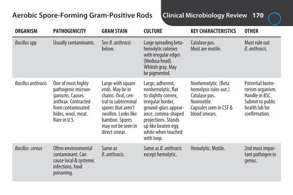
Aerobic Spore-Forming Gram-Positive Rods Clinical Microbiology Review 170
ORGANISM PATHOGENICITY GRAM STAIN CULTURE KEY CHARACTERISTICS OTHER
Bacillus spp
Bacillus anthracis
Bacillus cereus
Must rule out B. anthracis.
Potential bioter-rorism organism.Handle in BSC.Submit to publichealth lab forconfirmation.
2nd most impor-tant pathogen ingenus.
Large spreading beta-hemolytic colonieswith irregular edges(Medusa head).Whitish gray. May be pigmented.
Large, adherent,nonhemolytic, flat to slightly convex, irregular border,ground-glass appear-ance, comma-shapedprojections. Standsup like beaten eggwhite when touchedwith loop.
Same as B. anthracisexcept hemolytic.
Usually contaminants.
One of most highlypathogenic microor-ganisms. Causes anthrax. Contractedfrom contaminatedhides, wool, meat.Rare in U.S.
Often environmentalcontaminant. Cancause local & systemicinfections, food poisoning.
See B. anthracisbelow.
Large with squareends. May be inchains. Oval, cen-tral to subterminalspores that aren’tswollen. Looks likebamboo. Sporesmay not be seen indirect smear.
Same as B. anthracis.
Catalase pos.Most are motile.
Nonhemolytic. (Beta hemolysis rules out.) Catalase pos.Nonmotile.Capsules seen in CSF &blood smears.
Hemolytic. Motile.
2956_Ch03_135-286 29/01/14 12:17 PM Page 170

Aerobic Non–Spore-Forming Gram-Positive Rods
Clinical Microbiology Review 171
ORGANISM PATHOGENICITY GRAM STAIN CULTURE KEY CHARACTERISTICS OTHER
Corynebacterium spp
Corynebacterium diphtheriae
Called diphtheroidsbecause resembleC. diphtheriae.
ID requiresdemonstration oftoxin production.Modified Elek immunoprecipita-tion test. PCR forTOX gene.
Facultative anaer-obes. Grow onmost media.
Gray-blackcolonies withbrown halos onTindale agar.Black colonies oncystine tellurite.Loeffler mediumstimulates growth& production ofmetachromaticgranules.
Normal on skin &mucous membranes.Opportunisticpathogens.
Diphtheria. Rare inU.S.
Irregular, slightlycurved, nonparallelsides, club-shapedends. Chinese letteror picket fence for-mation (palisades).
Same as above.
Catalase pos. Nonmotile.
Catalase pos. Nonmotile.Toxin producing.
continued...
2956_Ch03_135-286 29/01/14 12:17 PM Page 171

Aerobic Non–Spore-Forming Gram-Positive Rods continued
Clinical Microbiology Review 172
ORGANISM PATHOGENICITY GRAM STAIN CULTURE KEY CHARACTERISTICS OTHER
Corynebacterium jeikeium
Listeria monocytogenes
Nocardia
Most commonlyisolated diph-theroid. Highlyresistant to antibiotics.
Grows from 0.5°–45°C. Coldenrichment maybe used. Catalase differentiates from GBS. Motilitydifferentiates fromdiphtheroids.
N. brasiliensis mostcommon speciesto cause skin infec-tions. N. asteroidesmost commonspecies to causelung infections.
Same as othercommensalcorynebacteria.
Tiny colonieswith narrowzone of indistinctbeta hemolysis.Translucent, gray.
Slow-growing.On SBA wrinkled,dry, crumbly,chalky white toorange-tan, betahemolytic.
Hospital-acquiredpathogen.
Meningitis & septicemia in new-borns & immuno-compromised. Food poisoning.
Immunocompetent:skin infections. Im-munocompromised:invasive pulmonary& disseminated infections.
Same as above.
Parallel sides,rounded ends, coccobacillary. Singles, chains, ordiphtheroidarrangement.
Fine branching filaments with fragmentation.Often beaded.
Catalase pos. Rapid sucrose urea (RSU) neg.
Catalase pos. Hippuratehydrolysis pos. Esculinpos. CAMP pos (hemolysislooks like shovel, not arrowhead). Tumblingmotility on wet mount.Umbrella growth inmotility agar at RT butnot at 35°C.
Catalase pos. Partiallyacid fast. Sulfur granules.
2956_Ch03_135-286 29/01/14 12:17 PM Page 172

Neisseria and Moraxella Clinical Microbiology Review 173
ORGANISM PATHOGENICITY GRAM STAIN CULTURE KEY CHARACTERISTICS OTHER
Neisseria spp
N. gonorrhoeae Susceptible to drying& cold. Don’t refrig-erate specimens.Molecular methodsfor genital speci-mens. Culture preferred for non-genital specimens &those from children.Should confirm bydifferent method.
Aerobic or faculta-tive. Pathogens arecapnophilic.
Requires ↑ CO2. Usu-ally doesn’t grow onSBA. Grows on CHOC& Neisseria-selectivemedia. Colonies aresmall, grayish whiteor tan. 5 differentcolony types. Maylook like mixed culture.
See individual organisms below
Gonorrhea, salpingi-tis, ophthalmia ofthe newborn
GNDC, coffee-bean shaped
Intracellular & ex-tracellular GNDC.Diagnostic in urethral dischargefrom sympto-matic males. Culture confirma-tion required forfemales
Catalase pos. Oxidase pos.
Carbohydrate utilization:glucose only. Superoxolpos. Can ID with mono-clonal antibodies.
continued...
2956_Ch03_135-286 29/01/14 12:17 PM Page 173

Neisseria and Moraxella continued Clinical Microbiology Review 174
ORGANISM PATHOGENICITY GRAM STAIN CULTURE KEY CHARACTERISTICS OTHER
N. meningitidis
Commensal Neisseria spp
Don’t refrigerate.Handle in BSC.Serogrouping byslide agglutination.Bacterial antigentest available for detection but poorsensitivity. Shouldn’treplace culture. Vaccine available.
Not speciated if from respiratoryspecimens. Must differentiate frompathogenic Neisseriawhen isolated on selective media orfrom normally sterilebody site.
Grows on SBA,CHOC, & Neisseria-selective media.Colonies are bluishgray or tan. May bemucoid.
Grow on SBA &CHOC at RT, nutrientagar at 35°C. Somegrow on Neisseria-selective media.Don’t require ↑ CO2.May be pigmented.
Can be normal florain upper respiratorytract. Common causeof meningitis inyoung adults
Normal in upper respiratory tract.Rarely cause disease
Intracellular &extracellularGNDC
GNDC
Carbohydrate utilization:glucose & maltose. ONPG neg.
Varies with species.
continued...
2956_Ch03_135-286 29/01/14 12:17 PM Page 174

Neisseria and Moraxella continued Clinical Microbiology Review 175
ORGANISM PATHOGENICITY GRAM STAIN CULTURE KEY CHARACTERISTICS OTHER
N. lactamica
Moraxella catarrhalis
GNDC = gram-negative diplococci.Note: Gram-negative rods Kingella & Acinetobacter may grow on Neisseria-selective media.
One of commensalNeisseria. Easilymisidentified as N. meningitidis. Mayreact with meningo-coccal typing sera.ONPG differentiates.
DNase & butyrate esterase differentiatefrom Neisseria spp.
Will grow on Neisseria-selectivemedia. Resembles N. meningitidis butsmaller.
Grows on SBA &CHOC. Some maygrow at RT &/or onNeisseria-selectivemedia. “Hockeypuck colonies”(colony can bepushed over agarsurface with loop).
Normal in upper respiratory tract ofchildren. Rare inadults. Rarely causesdisease
Normal in upper res-piratory tract. Causesrespiratory infectionsin young, old, &compromised; otitismedia & sinusitis inchildren
GNDC
GNDC
Carbohydrate utilization:glucose, maltose, & lac-tose (slow). ONPG pos.
Catalase pos. Oxidase pos.Carbohydrate utilization:neg for all sugars & ONPG. DNase pos. Butyrate esterase pos.
2956_Ch03_135-286 29/01/14 12:17 PM Page 175

Characteristics of Enterobacteriaceae Clinical Microbiology Review 176
Gram stain Non–spore-forming gram-negative rods
Growth characteristics Facultative anaerobes
Colonies on sheep blood agar Most are large, dull, gray, nonhemolytic
Colonies on MacConkey Lactose fermenters pinkNon–lactose fermenters colorless
Biochemicals Ferment glucoseOxidase negMost reduce nitrates to nitritesMost are catalase pos
2956_Ch03_135-286 29/01/14 12:17 PM Page 176

Biochemical Tests for Identification of Enterobacteriaceae
Clinical Microbiology Review 177
TEST PRINCIPLE INTERPRETATION OTHER
Oxidase
Nitrate reduction
Carbohydrate fermentation
ONPG
Good test to differentiate Enterobacteriaceaefrom nonfermenters. False pos from iron-containing wire. Use platinum wire orwooden stick.
If no color develops, zinc dust added. Reduces nitrates. Red color after zinc dustindicates presence of residual nitrates, i.e.,neg rxn. No color after zinc dust means nitrates reduced to N2 or NO2, i.e., pos rxn.
Frequently tested carbohydrates are glucose,lactose, sucrose, mannose, sorbitol, manni-tol, xylose, adonitol, cellobiose, dulcitol,trehalose. All Enterobacteriaceae fermentglucose.
Test for slow lactose fermentation. Helpfulin differentiating Citrobacter (pos) frommost Salmonella (neg).
Tetramethyl-para-phenylene-diamine dihydrochloride reactswith cytochrome C to produce blue or purple color.
If organism reduces nitrates to nitrites, red color develops whensulfanilic acid & N,N-dimethyl-L-naphthylamine added.
When carbohydrate fermented,acidic end products cause colorchange in pH indicator.
ONPG is changed to orthonitro-phenol by beta-galactosidase.
Pos = blue or purple
Pos = red, or no color after addition of zinc dust
With phenol red indicator,change from red to yellow
Pos = yellow
continued...
2956_Ch03_135-286 29/01/14 12:17 PM Page 177

Biochemical Tests for Identification of Enterobacteriaceae continued
Clinical Microbiology Review 178
TEST PRINCIPLE INTERPRETATION OTHER
H2S production
Triple sugar iron (TSI) agar
Indole
Sulfur-containing compounds = sodiumthiosulfate, cystine, methionine. Good testto differentiate Salmonella (H2S pos) fromShigella (H2S neg).
Contains 0.1% glucose, 1% lactose, 1% sucrose, phenol red, sodium thiosulfate,iron salt. Leave cap slightly loose. Record as slant/butt, e.g., K/A. (Some record NC[no change] for pink butt.) Kligler’s iron agar (KIA) is same except nosucrose.
Spot indole test commercially available. Need source of tryptophan. Use colonies fromSBA or CHOC agar, not MacConkey. Pos =blue. E. coli & Proteus vulgaris are pos.
Organisms that possess H2S-producing enzymes produce color-less H2S gas from sulfur-containingcompounds. H2S reacts with ironsalt in medium to form black ferrous sulfide.
Sugar fermentation produces acid,changes color of pH indicator.
Tryptophanase deaminates trypto-phan. Indole produced. Kovacs’sreagent (paradimethylamino-benzaldehyde) forms pink-coloredcomplex with indole.
Black precipitate
Yellow = acid (A). Pink = alka-line (K). Pink butt = glucose not fermented. Yellow butt =glucose fermented. Pink slant =lactose/sucrose not fermented.Yellow slant = lactose and/orsucrose fermented. Black precipitate = H2S produced.Bubbles = gas production
Pos = pink
continued...
2956_Ch03_135-286 29/01/14 12:17 PM Page 178

Biochemical Tests for Identification of Enterobacteriaceae continued
Clinical Microbiology Review 179
TEST PRINCIPLE INTERPRETATION OTHER
Methyl red (MR)
Voges-Proskauer (VP)
Citrate
Urease
Phenylalanine deaminase (PD)
Little acid produced by this pathway. Organisms that are VP pos are usuallymethyl red neg & vice versa.
False neg if cap not loose. Klebsiella & Enterobacter are pos.
Proteus & Morganella are rapid urease producers.
Proteus, Providencia, Morganella are pos.
Acid products formed when glucoseis metabolized by mixed acid fermentation pathway. Colorchange in pH indicator.
Acetoin is produced from alternatepathway for glucose metabolism.
If organism can use citrate as solesource of carbon, pH ↑, pH indica-tor changes color.
Urease breaks down urea. Ammoniareleased, pH ↑, pH indicatorchanges color.
Phenylalanine deaminase deaminates phenylalanine tophenylpyruvic acid, which reactswith ferric chloride to producegreen color.
Pos = red color after additionof methyl red (pH indicator)
Pos = red color when alpha-naphthol & KOH added
Pos = green to blue, or growth
Pos = yellow to pink
Pos = green color after addition of ferric chloride
continued...
2956_Ch03_135-286 29/01/14 12:17 PM Page 179

Biochemical Tests for Identification of Enterobacteriaceae continued
Clinical Microbiology Review 180
TEST PRINCIPLE INTERPRETATION OTHER
Decarboxylase reactions
Motility
Ornithine decarboxylase (ODC) differentiatesKlebsiella (neg) & Enterobacter (pos).
Most Enterobacteriaceae are pos, exceptKlebsiella & Shigella. Differentiates Klebsiella(neg) & Enterobacter (pos).
If organism has enzyme to decarboxylate amino acid (e.g., ornithine, lysine, arginine), pH ↑,pH indicator changes color.
Motile organisms grow away fromstab line in motility medium.
Pos = yellow to purple
Pos = movement away fromstab line or hazy appearancethroughout medium afterovernight incubation
2956_Ch03_135-286 29/01/14 12:17 PM Page 180

Antigens of Enterobacteriaceae Clinical Microbiology Review 181
ANTIGEN ALTERNATE NAME LOCATION CHARACTERISTICS OTHER
O antigen Somatic antigen Cell wall Lipopolysaccharide, heat stable Used for serological grouping of Salmonella & Shigella.
H antigen Flagellar antigen Flagella Proteins, heat labile Used to serotype Salmonella.
K antigen Capsular antigen Capsule Polysaccharide, heat labile, may Role in preventing phagocytosis. mask O antigen. Removed by ↑ virulence. Vi antigen is K antigen heating produced by S. typhi.
2956_Ch03_135-286 29/01/14 12:17 PM Page 181

Commonly Isolated Enterobacteriaceae Clinical Microbiology Review 182
KEY REACTIONS
ORGANISM PATHOGENICITY POSITIVE NEGATIVE OTHER
Escherichia coli
Shigella
Edwardsiella tarda
Predominant aerobe in GI tract. Most common cause of UTI. Greenmetallic sheen on EMB. PresumptiveID: lactose pos, dry colony on Mac-Conkey, oxidase neg, indole pos. E. coli O157:H7 doesn’t ferment sorbitol; colorless colonies on SMAC.
Blood, mucus, polys in stool. Rarely disseminates. Fragile organ-ism. S. dysenteriae most severe.Shigella sonnei most common in U.S. Serogrouped by O antigens.Serogroups A, B, C, D. Closely relatedto Escherichia on molecular basis.
Chief reservoirs are reptiles & fresh-water fish. Infections often involveaquatic environments. Pos indoledifferentiates from Salmonella.
UTI, septicemia, neonatalsepsis & meningitis, diarrhea (some)
Dysentery (shigellosis).Most communicable of bacterial diarrheas.Found primarily incrowded or substandardconditions, e.g., day-carecenters, jails, prisons
Opportunistic. Bacteremia,wound infections
Lactose, gas, indole,MR, motility
MR
Gas, H2S, indole, MR,motility
H2S, VP, citrate,PD, urease
Lactose, gas, H2S,VP, citrate, PD,urease, motility
Lactose, VP, citrate, PD, urease
continued...
2956_Ch03_135-286 29/01/14 12:17 PM Page 182

Commonly Isolated Enterobacteriaceae continued
Clinical Microbiology Review 183
KEY REACTIONS
ORGANISM PATHOGENICITY POSITIVE NEGATIVE OTHER
Salmonella
Citrobacter freundii
Klebsiella pneumoniae
Enterobacter aerogenes & cloacae
Found in poultry. May be transmittedby reptiles. S. typhi has Vi antigen,only trace H2S, citrate neg. Groupedby O antigens (e.g., A, B, C), serotypedby H antigens (e.g., 1, 2).
Lactose variable. ONPG & LDC differentiate from Salmonella.
Encapsulated. Colonies usually mucoid. Some strains hydrolyzeurea slowly. K. oxytoca is similar toK. pneumoniae except indole pos.Motility & ODC differentiate fromEnterobacter.
Colonies may be mucoid. SameIMViC reactions as Klebsiella.
Typhoid (enteric) fever,bacteremia, enterocolitis
Nosocomial infections
Pneumonia, UTI, septicemia
Opportunistic & nosoco-mial infections. UTI, RTI,& wound infections
H2S, MR, motility, lysine decarboxylase(LDC)
Gas, H2S, MR, citrate,motility, ONPG
Lactose, gas, VP, citrate, urease (slow)
Lactose, gas, VP, citrate, motility, ODC
Lactose, indole,VP, PD, urease,ONPG
VP, PD, LDC
H2S, indole, MR,PD, motility, ornithine decar-boxylase (ODC)
H2S, indole, MR,PD
continued...
2956_Ch03_135-286 29/01/14 12:17 PM Page 183
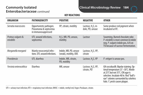
Commonly Isolated Enterobacteriaceae continued
Clinical Microbiology Review 184
KEY REACTIONS
ORGANISM PATHOGENICITY POSITIVE NEGATIVE OTHER
Serratia marcescens
Proteus vulgaris & mirabilis
Morganella morganii
Providencia
Yersinia enterocolitica
UTI = urinary tract infection; RTI = respiratory tract infection; IMViC = indole, methyl red, Voges-Proskauer, citrate.
Some produce red pigment whenincubated at RT.
Swarming. Burned chocolate odor.P. mirabilis is most common & indoleneg. P. vulgaris indole pos, A/A onTSI because of sucrose fermentation.
P. rettgeri is urease pos.
GN coccobacilli. Bipolar staining. Op-timal temperature 25°–30°C. Motileat 25°C but not 35°C. CIN agar is selective. Incubate 48 hr. Red “bull’s-eye” colonies surrounded by colorlesshalo. Y. pestis causes plague.
Opportunistic pathogen.Pneumonia & septicemiain immunosuppressed
UTI, wound infections,septicemia
Mainly nosocomial infec-tions. UTI, wound infections.
UTI, diarrhea
Diarrhea
VP, citrate, motility
H2S, MR, PD, urease,motility
Indole, MR, PD, urease(weak), motility, ODC
Indole, MR, citrate,PD, motility
MR, urease
Lactose, H2S, in-dole, PD, urease
Lactose
Lactose, H2S, VP,citrate
Lactose, H2S, VP
Lactose, H2S, VP,citrate, PD
2956_Ch03_135-286 29/01/14 12:17 PM Page 184

Summary of Key Reactions for Enterobacteriaceae
Clinical Microbiology Review 185
LACTOSE NEGATIVE H2S POSITIVE VP POSITIVE PD POSITIVE UREASE POSITIVE NONMOTILE AT 35°C
ShigellaEdwardsiellaSalmonellaCitrobacter (some)SerratiaProteusMorganellaProvidenciaYersinia
ShigellaKlebsiellaYersinia (motile at 22°C)
KlebsiellaEnterobacterSerratia
EdwardsiellaSalmonellaCitrobacterProteus
ProteusMorganellaProvidenciaKlebsiella (slow)
Proteus MorganellaProvidencia rettgeri
2956_Ch03_135-286 29/01/14 12:17 PM Page 185

Appearance of Enterobacteriaceae on Selected Media
Clinical Microbiology Review 186
ORGANISM TSI MacCONKEY HEKTOEN ENTERIC XLD
Escherichia coli
Shigella
Edwardsiella
Citrobacter
Salmonella
Klebsiella
Enterobacter
Serratia
Proteus
Morganella
Providencia
Yersinia
Yellow
Colorless
Red, yellow, or colorless withor without black centers
Red, yellow, or colorless withor without black centers
Red with black center
Yellow
Yellow
Yellow or colorless
Yellow or colorless, with orwithout black centers
Red or colorless
Yellow or colorless
Yellow or colorless
A/A, gas
K/A
K/A, gas, H2S
A/A or K/A, gas, withor without H2S
K/A, gas, H2S
A/A, gas
A/A, gas
K/A
K/A (mirabilis) A/A(vulgaris), gas, H2S
K/A, gas
K/A
Yellow/orange
Flat, dry pink colony with darker pink halo
Colorless
Colorless
Colorless at 24 hr. May become pink at 48 hr
Colorless
Pink, mucoid
Pink. May be mucoid
Colorless at first, turning pink. S. marcescensmay have red pigment at RT
Colorless. May swarm
Colorless
Colorless
Colorless to peach
Yellow
Green
Colorless
Colorless
Green
Yellow
Yellow
Colorless
Colorless
Colorless
Colorless
Salmon
2956_Ch03_135-286 29/01/14 12:17 PM Page 186

Diarrheagenic Escherichia coli Clinical Microbiology Review 187
DISEASE GRAM STAIN GROUP PATHOGENICITY TRANSMISSION MECHANISM OF STOOL OTHER
Enterohemorrhagic (EHEC). Also known as Shiga toxin–producing (STEC) or verotoxin-producing (VTEC)
Enterotoxigenic (ETEC)
Enteroinvasive (EIEC)
E. coli O157:H7 is mostcommon isolate ofgroup & pathogen most often isolatedfrom bloody stools.Non-O157 STEC alsocauses disease. DNAprobes can ID genesthat code for toxins. Report to public health.
Profuse, watery stool.DNA probes to detecttoxins or toxin genes.
Undercookedmeat, raw milk,apple cider
Contaminatedfood or water
Contaminatedfood or water
Diarrhea, hemorrhagiccolitis, hemolytic uremicsyndrome (HUS). Mostcommon cause of renalfailure in children in U.S.May be fatal, especiallyin young or elderly
Traveler’s diarrhea, diarrhea in infants
Bloody diarrhea. Dysentery-like. Usually in young children inareas of poor sanitation.
Toxins (vertox-ins or Shigatoxins)
Toxins
Invasiveness
RBCs but usuallyno polys
No polys or RBCs
Polys, RBCs, mucus
continued...
2956_Ch03_135-286 29/01/14 12:17 PM Page 187

Diarrheagenic Escherichia coli continued Clinical Microbiology Review 188
DISEASE GRAM STAIN GROUP PATHOGENICITY TRANSMISSION MECHANISM OF STOOL OTHER
Enteropathogenic (EPEC)
Enteroaggregative(EAEC)
Diffusely adherent (DAEC)
Watery diarrhea withmucus.
Most labs can’t detect.
Not well studied.
Formula & foodcontaminated withfecal material
Nosocomial &community acquired
Little known aboutepidemiology
Diarrhea in infants. Majorpathogen in infants indeveloping countries
Diarrhea in developingcountries. Chronic diarrheain HIV-infected patients
Diarrhea & UTI. Mostcommon in children indeveloping countries
Adherence-attachment
Adherence-attachment
Adherence-attachment
No polys or RBCs
2956_Ch03_135-286 29/01/14 12:17 PM Page 188
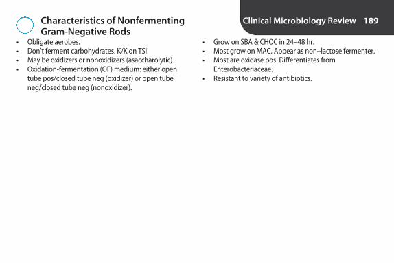
Characteristics of Nonfermenting Gram-Negative Rods
Clinical Microbiology Review 189
• Obligate aerobes.• Don’t ferment carbohydrates. K/K on TSI.• May be oxidizers or nonoxidizers (asaccharolytic). • Oxidation-fermentation (OF) medium: either open
tube pos/closed tube neg (oxidizer) or open tubeneg/closed tube neg (nonoxidizer).
• Grow on SBA & CHOC in 24–48 hr.• Most grow on MAC. Appear as non–lactose fermenter.• Most are oxidase pos. Differentiates from
Enterobacteriaceae.• Resistant to variety of antibiotics.
2956_Ch03_135-286 29/01/14 12:17 PM Page 189
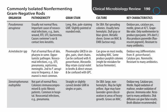
Commonly Isolated Nonfermenting Gram-Negative Rods
Clinical Microbiology Review 190
ORGANISM PATHOGENICITY GRAM STAIN CULTURE KEY CHARACTERISTICS
Pseudomonas aeruginosa
Acinetobacter spp
Stenotrophomonas maltophilia
Oxidase pos, catalase pos,motile, grows at 42°C. Grape-like odor. Only nonfermenter toproduce pyocyanin. (4% don’t.)Also produces pyoverdin (fluo-rescent pigment). Resistant tomany antibiotics.
Oxidase neg (differentiatesfrom N. gonorrhoeae). Catalase pos. Nonmotile. Resistant to many antibiotics.
Oxidase neg. Catalase pos.Motile. Rapid oxidation ofmaltose, weaker oxidation ofglucose. Ammonia odor. Resis-tant to many antibiotics. Diskdiffusion can give false results.Broth dilution recommended.
Usually not normal flora.Important cause of nosoco-mial infections, e.g., burn,wound, RTI, UTI, bacteremia.Causes swimmer’s ear &contact lens keratitis.
Part of normal flora of skin,pharynx in some. Oppor-tunistic pathogen. Nosoco-mial infections, e.g., UTI,pneumonia, septicemia,meningitis. 2nd to P. aerugi-nosa in frequency. A. bau-mannii is most common.
Not part of normal flora. Colonizes immunocompro-mised & cystic fibrosis patients. Common in hospi-tal. Nosocomial infections,e.g., pneumonia.
Long, thin, pale-stainingGNR. Slightly pointed orrounded ends.
Pleomorphic GNCB in sin-gles, pairs, short chains.Can be confused with N.gonorrhoeae, Moraxella.May retain crystal violetin broths & direct smears& be confused with GPC.
Straight or slightlycurved slender GNR insingles or pairs.
On SBA: flat spreadingcolonies. Usually betahemolytic. Dull gray orblue-green. Metallicsheen. Grows on MAC &EMB (lactose neg).
Can grow on most media,including MAC. Someproduce purplish colonies(might be mistaken forlactose fermenter).
On SBA: large, non-hemolytic. May be lightyellow. Agar may havelavender-green discol-oration in areas of heavygrowth. Grows on MAC.
2956_Ch03_135-286 29/01/14 12:17 PM Page 190

Campylobacter and Helicobacter Clinical Microbiology Review 191
ORGANISM PATHOGENICITY GRAM STAIN CULTURE KEY CHARACTERISTICS
Campylobacter jejuni
Campylobacter coli
Campylobacter fetus
Helicobacter pylori
Darting corkscrew motility. Oxidase, catalase, & hippurate hydrolysis pos.
Rarely differentiatedfrom C. jejuni. Hippuratehydrolysis neg.
Oxidase & catalase pos.Hippurate hydrolysis neg.
Rarely cultured. Rapidurease tests on gastricbiopsy, urea breath test,histology, PCR, serology.
Most common cause of bacterial diarrhea. Sources of infection: chickens, rawmilk, pets
Similar to C. jejuni, but lesssevere. Usually foodborne
Causes bacteremia in im-munocompromised & elderly.Uncommon stool isolate
Gastritis, duodenal & pepticulcers. Possible risk factor forgastric carcinoma
Curved, slender, GNR.“Seagulls,” loose spirals, & S shaped.Stain faintly.
Same as above.
Same as above.
Curved, slender, GNR.
Microaerophilic &capnophilic. Grown onCampy-BAP at 42°C in ↑CO2. (Can grow at 37°C;normal enteric flora in-hibited by 42°C incuba-tion.) Slow growing. Holdplates 3 days.
Same as above.
Most often isolated inblood cultures. Hold for 2 wk. Inhibited on Campyagar. Grows on routinemedia at 37°C, not at 42°C.
Grows on nonselectivemedia incubated at 37°Cin same atmosphere asCampy. Doesn’t grow at42°C. Slow growing.
2956_Ch03_135-286 29/01/14 12:17 PM Page 191

Vibrio and Related Organisms Clinical Microbiology Review 192
ORGANISM PATHOGENICITY GRAM STAIN CULTURE KEY CHARACTERISTICS
Vibrio cholerae
Vibrio vulnificus
“Rice water” stools. Oxidase pos. Motile.Serological ID with antisera to O ag. O1 &O139 strains cause epidemics. El Tor bio-type causes most casesworldwide. Notify pub-lic health department,send for confirmation.
Oxidase pos. Motile.
Cholera (acute diarrhea, dehydration, electrolyte im-balance). Transmitted by contaminated water, seafood.Uncommon in U.S., but maybe seen in coastal areas.
2nd most serious type of vibrio infection. Immuno-compromised or individualswith liver disease: septicemiafollowing consumption ofraw oysters, water-associatedwound infections. Healthy individuals: gastroenteritis.Seen in U.S.
Small comma-shapedGNR in direct smears;straight pleomorphicGNR in culture.
Straight or curvedGNR.
Nonhalophilic (doesn’trequire NaCl for growth).Grows on SBA, CHOC,MAC (NLF). Large yellowcolonies on TCBS (fer-ments sucrose). Alkalinepeptone water (APW) canbe used for enrichment.
Halophilic (salt loving, requires addition of Na+).Most are green on TCBS;some are yellow. Maylook like enteric on MAC because some arelactose pos.
continued...
2956_Ch03_135-286 29/01/14 12:17 PM Page 192

Vibrio and Related Organisms continued Clinical Microbiology Review 193
ORGANISM PATHOGENICITY GRAM STAIN CULTURE KEY CHARACTERISTICS
Vibrio parahaemolyticus
Aeromonas spp
Plesiomonas shigelloides(now included inEnterobacteriaceae)
Oxidase pos. Motile.
Oxidase pos (differenti-ates from Enterobacte-riaceae). Motile.
Biochemical & antigenicsimilarities to Shigella.Oxidase pos. Motile.
2nd most common Vibrioto cause gastroenteritis. Con-taminated seafood. Water-associated wound infections.Seen in U.S.
Gastroenteritis & wound infections, often related to aquatic exposure. Sep-ticemia, meningitis.
Gastroenteritis from contam-inated water or seafood. Bacteremia & meningitis inimmunocompromised &neonates.
Straight or curvedGNR.
Straight or curvedGNR.
Pleomorphic GNR insingles, pairs, shortchains, or long filaments.
Requires 1% NaCl forgrowth. Grows on SBA,MAC (NLF). Blue-greencolonies on TCBS (doesn’tferment sucrose).
Grows on routine media.Most are beta hemolyticon SBA, NLF on MAC.Doesn’t grow on TCBS.CIN & APW can be usedfor selective isolation.
Grows on SBA, CHOC.Most grow on MAC, appear as NLF. Doesn’tgrow on TCBS.
2956_Ch03_135-286 29/01/14 12:17 PM Page 193

Haemophilus Clinical Microbiology Review 194
ORGANISM PATHOGENICITY GRAM STAIN CULTURE OTHER
H. influenzae
H. influenzae biotype aegyptius & H. aegyptius
H. parainfluenzae, H. haemolyticus, &H. parahaemolyticus
H. ducreyi
Should be serotyped.
Characteristics are similar to H. influenzae.Difficult to differentiate.
Can ID by PCR.
Normal flora of upper respi-ratory tract. Causes sinusitis,otitis media, pneumonia,bronchitis, often in elderly orcompromised. Type b com-mon cause of pneumonia &meningitis in children whereHib vaccine not available.
Both cause conjunctivitis(pink eye). H. influenzae bio-type aegyptius also causesBrazilian purpuric fever.
Normal flora of upper respi-ratory tract. Low incidence ofpathogenicity.
Never normal flora. Causes chancroid (sexuallytransmitted disease).
Small, pleomorphicGNCB to long fila-ments. Capsules may be seen.
Same as above.
Small, pleomorphicGNCB to long fila-ments.
Small GNCB, bipolarstaining. May resem-ble schools of fish orrailroad tracks.
Grows on CHOC in 5%–10%CO2. Translucent, moist,tannish colonies. Encapsu-lated strains form larger &more mucoid colonies.Mousy or bleach-like odor.May demonstrate satel-litism with staph on SBA.
Same as above.
Colonies are larger, dry, & tannish.
Difficult to culture.
2956_Ch03_135-286 29/01/14 12:17 PM Page 194
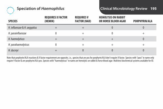
Speciation of Haemophilus Clinical Microbiology Review 195
REQUIRES X FACTOR REQUIRES V HEMOLYSIS ON RABBIT SPECIES (HEMIN) FACTOR (NAD) OR HORSE BLOOD AGAR PORPHYRIN/ALA
H. influenzae & H. aegyptius + + 0 0
H. parainfluenzae 0 + 0 +
H. haemolyticus + + + 0
H. parahaemolyticus 0 + + +
H. ducreyi + 0 0 0
Note that porphyrin/ALA reactions & X factor requirement are opposite, i.e., species that are pos for porphyrin/ALA don’t require X factor. Species with “para” in name onlyrequire V factor & are porphyrin/ALA pos. Species with “haemolyticus” in name are hemolytic on rabbit & horse blood agar. Multitest biochemical systems available for ID.
2956_Ch03_135-286 29/01/14 12:17 PM Page 195

Miscellaneous Gram-Negative Rods Clinical Microbiology Review 196
ORGANISM PATHOGENICITY GRAM STAIN CULTURE KEY CHARACTERISTICS
Bartonella
Bordetella pertussis
Brucella
Francisella
Dx usually by serological ormolecular methods.
Fluorescent antibody stain.DTaP vaccine (diphtheria,tetanus, pertussis).
Oxidase & urease pos. Level 3 pathogen. Potentialbioterrorism agent. Re-portable disease. Serologicaltests are primary means of Dx.
Level 3 pathogen. Directfluorescent ab methods forID. Send to public healthlab for confirmation. Serological tests.
Trench fever, relapsing fever,bacteremia, endocarditis,cat-scratch disease
Whooping cough in children& adults
Brucellosis (undulant fever).From unpasteurized milk orcontact with infected goats,cows, hogs, dogs
Tularemia (rabbit fever). Maninfected by tick or handlinginfected animal
Intracellular GNCB
Small GNCB
Tiny, faintly stainingGNCB
Pale-staining, small,pleomorphic, intra-cellular GNCB withbipolar staining
Not practical. Takes 9–40 daysto grow.
Grows on Bordet-Gengou & Regan-Lowe (charcoal,horse’s blood) after 3–7 days.
Most often isolated from bloodcultures or biopsies of reticu-loendothelial (RE) tissue. Bloodcultures incubated in ↑ CO2 for3 wk. Culture not sensitive.
Grows on special media en-riched with glucose & cystine(e.g., blood cysteine glucoseagar), TM, & BCYE. Small,transparent colonies after 3 days.
continued...
2956_Ch03_135-286 29/01/14 12:17 PM Page 196

Miscellaneous Gram-Negative Rods continued Clinical Microbiology Review 197
ORGANISM PATHOGENICITY GRAM STAIN CULTURE KEY CHARACTERISTICS
Gardnerella
Legionella
Pasteurella multocida
TM = Thayer-Martin; BCYE = buffered charcoal yeast extract.
Catalase & oxidase neg.Tests for Dx of BV: clue cells(vaginal epithelial cellscovered with gram-variablerods), whiff test (10% KOHadded to vaginal secretion → fish-like odor).
Oxidase pos. ID by immuno-fluorescent stain. Serology.
Oxidase, catalase, & indole pos.
Normal flora of female genital tract. Associated with bacterial vaginosis (BV) when it & anaerobic GNR arepredominant & Lactobacillusis absent. Linked to maternal& neonatal infections, bac-teremia, rarely UTI
Legionnaire’s disease, Pontiacfever. Found in water (e.g., airconditioners, showerheads,whirlpools). Contracted by inhalation of aerosol
Contracted by bite or scratchof cat or dog or contact withinfected carcass. Wound &respiratory tract infection
Small, pleomorphicgram-variable rods
Small, pleomorphic,weakly staining GNR
Pleomorphic GNCBwith bipolar staining
Not recommended for Dx ofBV. Grows on SBA, PEA, CNA,human blood Tween (HBT)agar, V agar. Produces diffusebeta hemolysis only on mediacontaining human blood. Requires ↑ CO2 & 48–72 hrincubation.
Grows on BCYE in 3–4 days.Pale yellow-green fluores-cence with Wood’s lamp.
Grows on SBA & CHOC, but notMAC. Musty odor.
2956_Ch03_135-286 29/01/14 12:17 PM Page 197

Specimens for Anaerobic Culture Clinical Microbiology Review 198
ACCEPTABLE UNACCEPTABLE
Bile Expectorated sputumBlood FecesBody fluids Gastric juiceBone marrow SwabsPercutaneous lung aspirate or biopsy Voided or catheterized urineSuprapubic bladder aspirates Bronchial washings (unless obtained with a double-lumen plugged catheter)TissueTranstracheal aspirateWound
2956_Ch03_135-286 29/01/14 12:17 PM Page 198

Media for Culture of Anaerobes Clinical Microbiology Review 199
MEDIUM USE
Anaerobic blood agar (CDC)
Bacteroides bile–esculin (BBE) agar
Brucella blood agar
Colistin–nalidixic acid (CNA) blood agar
Cycloserine cefoxitin fructose egg yolk (CCFA) agar
Egg-yolk agar (EYA)
Kanamycin-vancomycin laked blood (KVLB) agar
Phenylethyl alcohol (PEA) agar
Thioglycolate (THIO) broth
Nonselective enrichment medium that grows obligate & facultative anaerobes. Containsyeast extract, L-cysteine, hemin, & vitamin K.
Selective differential medium for Bacteroides fragilis. Bile salts & gentamicin act as inhibitors.B. fragilis colonies black with dark halos due to esculin hydrolysis.
Enriched medium that grows obligate & facultative anaerobes.
Selective medium that grows obligate anaerobes & GP facultative anaerobes.
Selective & differential for Clostridium difficile. Colonies are yellow due to fermentation offructose. Chartreuse fluorescence.
For determination of lecithinase & lipase production by clostridia & fusobacteria.
Also known as laked blood kanamycin-vancomycin (LKV) agar. Most commonly used selectivemedium for isolation of anaerobic GNRs, especially Bacteroides & Prevotella.
Selective medium that inhibits enteric GNRs & grows obligate anaerobes & GP facultativeanaerobes.
All-purpose medium that supports growth of most aerobes & anaerobes. Can be used asbackup broth to detect organisms present in small numbers or anaerobes. Thioglycolate actsas reducing agent. Aerobes grow at top, strict anaerobes at bottom, facultative anaerobesthroughout. Store at RT. Boil & cool before use.
2956_Ch03_135-286 29/01/14 12:17 PM Page 199

Anaerobic Environment Clinical Microbiology Review 200
Ideal atmosphere
Methods Anaerobic transport media
Self-contained anaerobic agar
Anaerobic generating systems
Anaerobic chamber
Quality controlMethylene blue strip
Resazurin
Oxygen analyzers
80%–90% N2, 5% H, 5%–10% CO2
Tubes of O2-reduced agar, tubes with anaerobic environment inside, or gas-impermeable bags withgas-generating environment. Most have indicator that changes color in presence of O2. Can maintainanaerobes for 24–72 hr.
Brucella-based blood & LKV agars contain enzyme that reduces O2. Lid creates airtight seal.
Plates in bag or container. Packet or ampule added. Chemical reaction removes O2.
“Glove box.” Gold standard. Workbench in gas-tight cabinet. Anaerobic atmosphere created/maintainedby gas tank, palladium catalysts, & desiccants. Work done through gloved or gloveless openings.
Blue = O2 present, white or colorless = no O2.
Pink = O2 present, colorless = no O2.
More expensive. Real-time monitoring.
2956_Ch03_135-286 29/01/14 12:17 PM Page 200

Methods to Identify Anaerobes Clinical Microbiology Review 201
METHOD EXPLANATION
Gram stain
Growth on media
Special-potency antimicrobial disks
Rapid tests
Conventional tubed biochemicals
Biochemical multitest systems
Preformed enzyme-based systems
Gram reaction; morphology; presence, location, & shape of spores provide clues to ID. Some GN anaerobes stain faintly with safranin. Recommended to extend time of counterstaining to3–5 min or use 0.1% basic fuchsin. Some GP anaerobes, e.g., Clostridium, may stain pink.
Which media organism grows on, pigmentation, hemolysis, & colonial morphology provideclues to ID.
Kanamycin, vancomycin, & colistin disks can be used to differentiate anaerobes & ensure thatover-decolorized Clostridium is not misidentified as GNR. Disks placed on 1st quadrant of plate.After incubation, observe if organism is susceptible or resistant.
For presumptive ID, e.g., fluorescence; catalase; spot indole; urease; motility; SPS, nitrate, &bile disks; lecithinase, lipase, & proteolytic rxn on egg-yolk agar.
Test tubes containing variety of media inoculated & incubated in anaerobic environment. Rxnleads to change in pH. Expensive & time consuming. Largely replaced by multitest systems.
Trays or strips are inoculated & read after 24–48 hr incubation in anaerobic environment. Codenumber is obtained & ID determined from codebook. Only contains codes for most commonlyisolated anaerobes.
Detect preexisting enzymes. Panels or cards are inoculated & incubated in room air. Colorchanges are read in 4 hr. Code number obtained & ID determined from codebook. Only containscodes for most commonly isolated anaerobes.
continued...
2956_Ch03_135-286 29/01/14 12:17 PM Page 201

Methods to Identify Anaerobes continued Clinical Microbiology Review 202
METHOD EXPLANATION
Gas-liquid chromatography (GLC)
16S ribosomal RNA gene sequencing
Analysis of metabolic end products or cellular fatty acids.
DNA extracted from organism, amplified by PCR, sequenced on automated sequencer. Nucleotide sequence compared with known sequences in database.
2956_Ch03_135-286 29/01/14 12:17 PM Page 202

Anaerobic Gram-Positive Cocci Clinical Microbiology Review 203
ORGANISM PATHOGENICITY GRAM STAIN OTHER
Finegoldia magna(formerly Peptostreptococcus magnus)
Peptostreptococcus anaerobius
Peptoniphilus asaccharolyticus (formerly Peptostreptococcus asaccharolyticus)
Small colonies. May take 48 hrto grow. Catalase & indole neg.Resistant to SPS.
Small gray-white colonies in 24-48 hr. Sweet odor. Sensitiveto SPS.
Yellow colonies. Musty odor. Resistant to SPS. Indole pos.
Normal flora on skin & in mouth,intestines, female genital tract.Most commonly isolated & mostpathogenic anaerobic GPC. Associ-ated with skin infections, decubitusulcers, septic arthritis, bone infec-tion following orthopedic surgery,oral & female genital tract infec-tions, bacteremia.
Normal on skin & in mouth, GI, & GU tracts. Mixed infections ofskin, soft tissues, GI tract, femalegenital tract, bones, joints, lungs,brain.
Normal on skin & in GI & GUtracts. Associated with obstetric & gynecological infections.
GPC in singles, pairs, tetrads,clusters. Resembles staph.
Tiny GPC in chains. Resemblesstrep.
GPC in pairs, short chains,tetrads, clusters.
2956_Ch03_135-286 29/01/14 12:17 PM Page 203

Anaerobic Gram-Positive Rods Clinical Microbiology Review 204
PATHOGENICITY GRAM STAIN OTHER
Actinomyces
Clostridium botulinum
Clostridium difficile
Clostridium perfringens
Sulfur granules may be seen indischarge. Crush & stain to re-veal characteristic Gram-stainmorphology. “Molar tooth”colonies.
Reportable disease. Toxin testing at public health labs.
Yellow ground-glass colonies oncycloserine cefoxitin fructoseagar (CCFA). Usually not cultured.Must demonstrate toxin produc-tion. Toxins A & B. Tissue cultureor EIA.
Most commonly isolatedClostridium. Double zone of beta hemolysis on SBA.
Infects brain, face, lungs, genitals
Botulism due to ingestion of toxin in inadequately cooked or improperlycanned foods. Infant botulism due toingestion of spores in honey. Woundbotulism from injection drug use
Antibiotic-associated diarrhea,pseudomembranous colitis
Normal in GI tract. Causes gas gangrene, food poisoning
Short or long GPR. Branchedor unbranched. Banded ap-pearance. Can break intoclub-shaped rods resemblingdiphtheroids.
GPR with oval subterminalspores.
Thin GPR. May form chains.Rare oval subterminal spores.
Large GPR with blunt ends inchains. “Box cars.” Tendencyto stain gram neg. Usually nospores seen.
continued...
2956_Ch03_135-286 29/01/14 12:17 PM Page 204

Anaerobic Gram-Positive Rods continued Clinical Microbiology Review 205
PATHOGENICITY GRAM STAIN OTHER
Clostridium tetani
Propionibacterium
Lactobacillus
Rare in U.S. because of DTaPvaccine. Lab confirmation rarely required.
Most common anaerobic GPR.Catalase & indole pos.
Aerotolerant anaerobes. Growbetter under anaerobic condi-tions. Colonies vary greatly. May resemble S. viridans. Catalase neg.
Causes tetanus. Deep wounds infectedwith soil
Normal skin flora. Common contaminantof blood cultures. Causes acne; infec-tions associated with artificial joints,catheters, shunts, artificial heart valves;keratitis; bacteremia; endocarditis
Normal in mouth, GI tract. Predominantflora of vagina during reproductiveyears. Produces lactic acid, maintainsvaginal pH. Absence in vagina predis-poses to bacterial vaginosis & yeast in-fections. Rare cause of endocarditis,septicemia in immunocompromised
GPR with swollen terminalspores. “Drumstick.” “Tennisracquet.” Becomes gram negafter 24 hr.
Club-shaped, pleomorphicGPR. Diphtheroid-like.
Pleomorphic, long, thin, non–spore-forming GPR, often inchains. Chains of rods in THIO.
2956_Ch03_135-286 29/01/14 12:17 PM Page 205

Gram-Negative Anaerobes Clinical Microbiology Review 206
ORGANISM PATHOGENICITY GRAM STAIN OTHER
Gram-negative cocciVeillonella
Gram-negative rods Bacteroides fragilis
Fusobacterium
Porphyromonas
Prevotella
May show weak red fluorescence underUV light. Doesn’t grow on KVLB. Usuallydoesn’t reduce nitrates.
Most common anaerobe isolated. Resistant to bile. Hydrolyses esculin.Turns BBE brown.
Indole pos. Most fluoresce chartreuse(green-yellow) under UV light.
Slow growing. Brown to black on SBA.Some fluoresce brick-red to orangeunder UV light. Doesn’t grow on KVLB.
Slow growing. Some are pigmented.Brown to black on SBA & KVLB. Somefluoresce brick-red to orange under UVlight. Doesn’t grow on BBE.
Normal flora of upper respiratorytract, GI & GU tracts. Usually inmixed culture. Can cause infec-tions in immunocompromised
Mixed infections below diaphragm
Pulmonary infections, brain abscesses, oral lesions
Head, neck, & pleuropulmonaryinfections
Head, neck, & pleuropulmonaryinfections
Tiny GNDC in pairs, clusters,short chains. Can resembleNeisseria
Pleomorphic, pale, irregu-larly staining GNR
GNR. F. nucleatum is long &thin with tapered ends(spindle shaped)
GNCB
GNCB
2956_Ch03_135-286 29/01/14 12:17 PM Page 206

Laboratory Identification of Mycobacteria Clinical Microbiology Review 207
Lab safety
Specimens requiring digestion/decontamination
Specimens not requiring digestion/decontamination
Digestion/decontamination methods
Staining
Media
Mycobacterium tuberculosis transmitted by inhalation of aerosols. Low infective dose. Separate room,if possible, with non-recirculating ventilation system & negative air pressure. BS-2 practices. Use ofBSC for all procedures that might generate aerosols. Electric incinerators instead of flames to sterilizewire loops. Slide-warming trays instead of flames to fix slides. Tuberculocidal disinfectants to cleanequipment & BSC, e.g., 1:10 dilution of household bleach made fresh daily. UV light in BSC (only whennot in use). Annual testing of employees for exposure, e.g., Mantoux skin test with PPD.
Sputum & other specimens with normal flora such as gastric lavage, urine, feces.
Tissue or body fluids collected aseptically.
Sodium hydroxide (NaOH), N-acetyl-L-cysteine (NALC)/NaOH, benzalkonium chloride, oxalic acid.NaOH is digestant & decontaminating agent. NALC is liquefying agent.
Cell walls have high lipid content (mycolic acids). Difficult to stain. Resist decolorization by acid alcohol (acid-fast). Gram stain poorly. Use carbolfuchsin or fluorochrome acid-fast stains.
Agar-based (Middlebrook 7H10 & 7H11), egg-based (Löwenstein-Jensen, Petragani, American Thoracic Society), liquid (Middlebrook 7H9). Combination of a solid-based medium & a liquid-basedmedium recommended for primary isolation.
continued...
PPD = purified protein derivative
2956_Ch03_135-286 29/01/14 12:17 PM Page 207

Laboratory Identification of Mycobacteria continued
Clinical Microbiology Review 208
Incubation
Rate of growth
Automated systems for recovery
Methods for identification
35°C in 5%–10% CO2. (Exception: If M. marinum, M. ulcerans, or M. haemophilum suspected, incubateat 25°–30°C.)
Slow. On solid media, most require 2–6 wk incubation; rapid growers 2–3 days. More rapid growth inliquid media.
Liquid broth inoculated, placed in blood culture instrument for automatic or continuous monitoring.Growth indicated by consumption of O2 or production of CO2. Earlier detection than manual methods.
Colony morphology, growth rate, optimum temp for growth, photoreactivity, biochemical tests, chromatography, nucleic acid hybridization, PCR, automated DNA sequencing.
2956_Ch03_135-286 29/01/14 12:17 PM Page 208

Acid-Fast Stains Clinical Microbiology Review 209
STAIN PRIMARY STAIN DECOLORIZER COUNTERSTAIN APPEARANCE OF AFB OTHER
Ziehl-Neelsen
Kinyoun
Fluorochrome
AFB = acid fast bacilli. OI = oil immersion.
Requires heat. Examine atleast 300 OI fields. Wipe lens after pos smears toavoid cross-contamination & false pos.
Cold stain. Examine at least300 OI fields. Wipe lensafter pos smears to avoidcross-contamination &false pos.
More sensitive than carbol-fuchsin. Faster to read. Can examine at 250×.Read immediately or storeat 2°C–8°C in dark to avoidfading. Examine at least300 fields. Pos should beconfirmed with carbol-fuchsin stain. Rapid grow-ers might not stain.
Methylene blue
Methylene blue
Potassium per-manganate oracridine orange
Carbolfuchsin
Carbolfuchsin
Auramine- rhodamine
Acid alcohol
Acid alcohol
Acid alcohol
Red, slightly curved,beaded rods (2–8 μm).Blue background.
Red, slightly curved,beaded rods (2–8 μm).Blue background.
Yellow-orange rodsagainst dark background.
2956_Ch03_135-286 29/01/14 12:17 PM Page 209

Classification of Mycobacteria Based on Pathogenicity
Clinical Microbiology Review 210
GROUP PATHOGENICITY SPECIES
Mycobacterium tuberculosiscomplex (“tubercle bacilli”)
Atypical mycobacteria, nontuberculous mycobacteria (NTM), or mycobacteria other than tubercle (MOTT) bacilli
M. tuberculosis, M. bovis, M. africanum, M. microti, M. canetti
M. avium, M. intracellulare, M. kansasii, M. malmoense,M. chelonae, M. xenopi, M. gordonae
Cause human tuberculosis (TB).
Some cause pulmonary infection similar toTB but not transmitted person to person.Usually in immunocompromised. Contractedfrom environment (soil, water).
2956_Ch03_135-286 29/01/14 12:17 PM Page 210

Classification of Nontuberculous Mycobacteria Based on Physiology
Clinical Microbiology Review 211
RUNYOUN GROUP CHARACTERISTICS EXAMPLES
Photochromogens
Scotochromogens
Nonphotochromogens
Rapid growers
M. marinum, M. kansasii
M. gordonae
M. avium complex, M. ulcerans
M. smegmatis
Yellow pigment when exposed to light after being grown in dark. >7 days to appear on solid media.
Yellow pigment when grown in light or dark. >7 days to appear on solid media.
No pigment produced in light or dark. >7 days to appear on solid media.
<7 days to appear on solid media.
2956_Ch03_135-286 29/01/14 12:17 PM Page 211
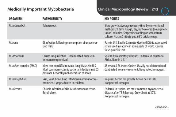
Medically Important Mycobacteria Clinical Microbiology Review 212
ORGANISM PATHOGENICITY KEY POINTS
M. tuberculosis
M. bovis
M. africanum
M. avium complex (MAC)
M. hemophilum
M. ulcerans
Slow growth. Average recovery time by conventionalmethods 21 days. Rough, dry, buff-colored (no pigmen-tation) colonies. Serpentine cording on smear from culture. Niacin & nitrate pos. 68°C catalase neg.
Rare in U.S. Bacille Calmette-Guérin (BCG) is attenuatedstrain used in vaccine in some parts of world. Causesfalse-pos PPD test.
Spread by respiratory droplets. Endemic in equatorialAfrica. Rare in U.S.
M. avium & M. intracellulare. Usually not differentiated.Contracted from environment. Nonphotochromogens.
Requires hemin for growth. Grows best at 30°C. Nonphotochromogen.
Endemic in tropics. 3rd most common mycobacterialdisease after TB & leprosy. Grows best at 30°C. Nonphotochromogen.
Tuberculosis
GI infection following consumption of unpasteur-ized milk
Causes lung infection. Disseminated disease inimmunocompromised
Most common NTM to cause lung disease in U.S.Most common systemic bacterial infection in AIDSpatients. Cervical lymphadenitis in children
Skin, joint, bone, lung infections in immunocom-promised. Lymphadenitis in children
Chronic infection of skin & subcutaneous tissue.Buruli ulcers
continued...
2956_Ch03_135-286 29/01/14 12:17 PM Page 212
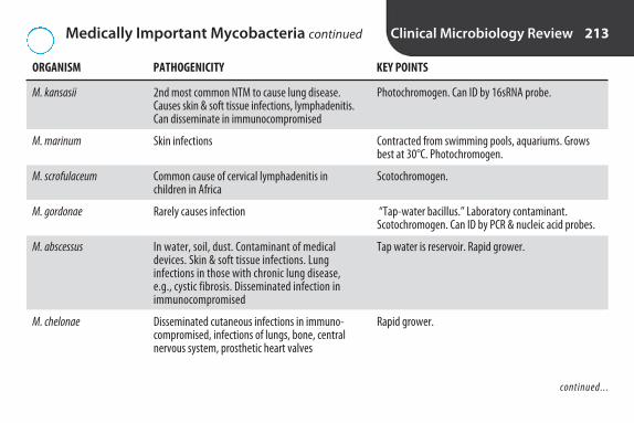
Medically Important Mycobacteria continued Clinical Microbiology Review 213
ORGANISM PATHOGENICITY KEY POINTS
M. kansasii
M. marinum
M. scrofulaceum
M. gordonae
M. abscessus
M. chelonae
Photochromogen. Can ID by 16sRNA probe.
Contracted from swimming pools, aquariums. Growsbest at 30°C. Photochromogen.
Scotochromogen.
“Tap-water bacillus.” Laboratory contaminant. Scotochromogen. Can ID by PCR & nucleic acid probes.
Tap water is reservoir. Rapid grower.
Rapid grower.
2nd most common NTM to cause lung disease.Causes skin & soft tissue infections, lymphadenitis.Can disseminate in immunocompromised
Skin infections
Common cause of cervical lymphadenitis in children in Africa
Rarely causes infection
In water, soil, dust. Contaminant of medical devices. Skin & soft tissue infections. Lung infections in those with chronic lung disease,e.g., cystic fibrosis. Disseminated infection inimmunocompromised
Disseminated cutaneous infections in immuno-compromised, infections of lungs, bone, centralnervous system, prosthetic heart valves
continued...
2956_Ch03_135-286 29/01/14 12:17 PM Page 213

Medically Important Mycobacteria continued Clinical Microbiology Review 214
ORGANISM PATHOGENICITY KEY POINTS
M. fortuitum
M. leprae
Common in environment. Rapid grower. Weakly grampos. Weakly acid fast. Stains with carbolfuchsin, butmay not with fluorescent stain.
Endemic in Southern hemisphere. <100 cases per yearin U.S. Most in TX, CA, LA, HI, PR. Armadillos may bereservoir. Doesn’t grow on artificial media. Can begrown in footpads of mice & armadillos. Dx by acid-faststain of tissue. Less acid fast than M. tuberculosis.
Infections of skin, soft tissues, IV & injection sites,surgical wounds
Leprosy (Hansen’s disease)
2956_Ch03_135-286 29/01/14 12:17 PM Page 214
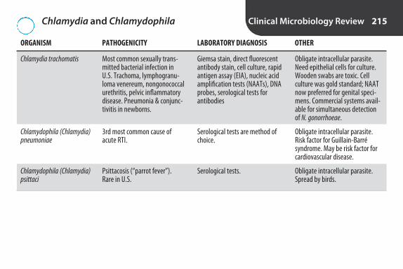
Chlamydia and Chlamydophila Clinical Microbiology Review 215
ORGANISM PATHOGENICITY LABORATORY DIAGNOSIS OTHER
Chlamydia trachomatis
Chlamydophila (Chlamydia) pneumoniae
Chlamydophila (Chlamydia) psittaci
Obligate intracellular parasite.Need epithelial cells for culture.Wooden swabs are toxic. Cell culture was gold standard; NAATnow preferred for genital speci-mens. Commercial systems avail-able for simultaneous detection of N. gonorrhoeae.
Obligate intracellular parasite.Risk factor for Guillain-Barré syndrome. May be risk factor forcardiovascular disease.
Obligate intracellular parasite.Spread by birds.
Most common sexually trans-mitted bacterial infection inU.S. Trachoma, lymphogranu-loma venereum, nongonococcalurethritis, pelvic inflammatorydisease. Pneumonia & conjunc-tivitis in newborns.
3rd most common cause ofacute RTI.
Psittacosis (“parrot fever”). Rare in U.S.
Giemsa stain, direct fluorescentantibody stain, cell culture, rapidantigen assay (EIA), nucleic acidamplification tests (NAATs), DNAprobes, serological tests for antibodies
Serological tests are method ofchoice.
Serological tests.
2956_Ch03_135-286 29/01/14 12:17 PM Page 215

Spirochetes Clinical Microbiology Review 216
ORGANISM PATHOGENICITY LABORATORY DIAGNOSIS OTHER
Borrelia recurrentis
Borrelia burgdorferi
Treponema pallidumsubspecies pallidum
Leptospira interrogans
Transmitted by ticks & lice.
Most commonly reported tick-borneinfection in U.S. Vector = Ixodes tick.
Worldwide.
Zoonotic disease. Transmitted by urineof infected animal. Organism in blood& CSF during first week, then urine.
Relapsing fever
Lyme disease (erythemachronicum migrans, neurological & cardiac abnormalities, arthritis)
Syphilis
Weil’s disease (infectiousjaundice, leptospirosis)
Giemsa- or Wright-stained bloodsmears.
Grows on modified Kelly medium.Serology is most common method.
Doesn’t grow on artificial media.Darkfield microscopy, fluorescentstain. Serology is most commonmethod.
Culture. Growth = turbidity belowsurface of semisolid Fletcher’smedium. Hold cultures for 6 wk.Serological tests, PCR.
2956_Ch03_135-286 29/01/14 12:17 PM Page 216

Mycoplasma/Ureaplasma Clinical Microbiology Review 217
ORGANISM PATHOGENICITY OTHER
Mycoplasma pneumoniae
Mycoplasma hominis
Ureaplasma
Smallest free-living cells. Lack cell wall. Pleomorphic. Not visibleby Gram staining. Difficult to grow. Culture rarely performed. Maytake 21 days or more. Usually Dx by serology.
Only species that will grow on SBA & CHOC but may require 4 days. Pinpoint translucent colonies easily overlooked. “Fried-egg”colonies seen with stereomicroscope after staining with methyleneblue. Serological methods available.
Granular brown appearance on A8 agar because of urease produc-tion. Serological methods available.
Primary atypical pneumonia(walking pneumonia)
Urogenital tract disease
Urogenital tract disease
2956_Ch03_135-286 29/01/14 12:17 PM Page 217

Rickettsiae Clinical Microbiology Review 218
GENUS PATHOGENICITY LABORATORY DIAGNOSIS OTHER
Rickettsia
Coxiella
Ehrlichia
Obligate intracellular parasite. Transmitted byticks, mites, lice, fleas. Rocky Mountain spot-ted fever is most common rickettsial infectionin U.S. Caused by R. rickettsii. BSL-3 biohazard.
Obligate intracellular parasite. Zoonotic disease. Found in cattle, sheep, goats. Highlycontagious. BSL-3 biohazard. Reportable disease. Potential bioterrorism agent.
Obligate intracellular parasite. Infects WBCs.Transmitted by ticks. Disease similar to RockyMountain spotted fever.
Rocky Mountain spotted fever,rickettsial pox, epidemic typhus,murine typhus, scrub typhus
Q fever
Ehrlichiosis
Don’t grow on artificial media.Grow in lice, ticks, tissue culture,eggs. Usually Dx by serology. Immunohistologic & molecularmethods available.
Grows in cell culture. Usual Dx byserology. Immunohistologic &molecular methods available.
Morulae (clusters of organismsthat resemble blackberry) inWBCs. Usual Dx by serology.
2956_Ch03_135-286 29/01/14 12:17 PM Page 218

Routine Culture Setup and Interpretation Clinical Microbiology Review 219
SPECIMEN MEDIA NORMAL FLORA COMMON PATHOGENS OTHER
Throat/nasopharynx
Sputum, bronchial washings/aspirates, transtracheal aspirates
Urine
Stab streaking to detect betastrep that only produce strep-tolysin O. Direct antigen tests for GAS. Negs should be cultured.
Gram stain. Acceptable spu-tum: <10 epithelial cells/LPF,>25 polys/LPF. Note: Neu-tropenic patients may notproduce polys.
Rapid screen for UTI: 1 ormore bacteria/OI field onGram stain of uncentrifugedurine. Interpretive guidelinesvary. Low counts are signifi-cant in catheterized urines &aspirates. 3 or more organismswith none predominant =probable contamination.
Alpha & gamma strep,commensal Neisseria,CNS, diphtheroids, S. pneumoniae, Candida
CNS, non–beta-hemolytic strep, diphtheroids, com-mensal Neisseria,Haemophilus, yeast.Aspirates: none
Clean catch: few skinflora. Catheterized oraspirate: none
SBA, CHOC. Streptococcus-selective agar may be used
SBA, CHOC, MAC
SBA, MAC. Use 0.01-mL or 0.001-mLcalibrated loop
GAS (S. pyogenes)
S. pneumoniae, H. influenzae,S. aureus, Enterobacteriaceae,Pseudomonas, M. catarrhalis,C. albicans
E. coli, Klebsiella, Enterobacter,Proteus, Enterococcus,Pseudomonas, S. aureus, S. saprophyticus, CNS, GBS
continued...
2956_Ch03_135-286 29/01/14 12:17 PM Page 219

Routine Culture Setup and Interpretation continued
Clinical Microbiology Review 220
SPECIMEN MEDIA NORMAL FLORA COMMON PATHOGENS OTHER
Stool
Genital
CSF
Polys = invasive diarrhea;e.g., Salmonella, Shigella,Campylobacter, EIEC, C. difficile.No polys = enterotoxin-mediated diarrhea; e.g., ETEC,Vibrio. RBCs with EHEC.
Gram stain
Use tube 2. Handle stat. Don’trefrigerate. Use of cytocen-trifuge to concentrate speci-men increases sensitivity ofGram stain. Use slides that
Anaerobes, Enterobac-teriaceae, enterococci,strep, yeast
Urethra: CNS, diph-theroids. Vagina, pre-pubescent/postmeno-pausal: yeast, GNR,GPC. Vagina, reproduc-tive years: Lactobacillus,GPC. Surgical/aspirates:none
None
SBA, MAC or EMB,XLD or HE, Campyblood. Some labsalso use SMAC, CIN,and/or TCBS
SBA, CHOC, MAC,Thayer-Martin. AddTHIO & anaerobicmedia for tissue &aspirates
SBA, CHOC. MAC.Some add THIO
Salmonella, Shigella, Campy-lobacter, diarrheagenic E. coli,Aeromonas, Plesiomonas, Y.enterocolitica, Vibrio
N. gonorrhoeae, C. albicans,GBS, S. aureus. Surgical/aspirates: also anaerobes
GBS (infants), Listeria (infants,elderly), H. influenzae (unim-munized children), N. menin-gitidis, S. pneumoniae, GNR.Any isolate significant
continued...
2956_Ch03_135-286 29/01/14 12:17 PM Page 220

Routine Culture Setup and Interpretation continued
Clinical Microbiology Review 221
SPECIMEN MEDIA NORMAL FLORA COMMON PATHOGENS OTHER
Blood
have been alcohol dipped &flamed or autoclaved. If pos,call. ID all isolates. Directantigen tests of limited value.
Skin prep: 80%–95% ethanolor isopropyl alcohol followedby povidone-iodine or tinctureof iodine. Optimum volume:adult, 20 mL; child, 1 mL peryr of life. blood-to-broth ratio1:5 to 1:10. Usually draw 2 setsin 24 hr. Automated systemswidely used for monitoring.Growth indicated by consump-tion of O2 or production of CO2.Gram stain & subculture stillrequired for ID.
NoneCommon contaminants:CNS, Micrococcus, alphastrep, diphtheroids, P. acnes
Most labs inoculatean aerobic & anaero-bic bottle. Some use2 aerobic bottles, noanaerobic
Any isolate potentially signifi-cant. GPC are most common
continued...
2956_Ch03_135-286 29/01/14 12:17 PM Page 221

Routine Culture Setup and Interpretation continued
Clinical Microbiology Review 222
SPECIMEN MEDIA NORMAL FLORA COMMON PATHOGENS OTHER
Wound
Eye
Ear
Practices vary. Follow laboratory’s SOP.
Gram stain.
Gram stain. If pos, call. Keratitis is emergency. Loss of eye can occur.
Gram stain.
CNS, diphtheroids, P. acnes. Surgical, aspi-rate, or tissue: none
CNS, diphtheroids, P. acnes
Normal skin flora
SBA, CHOC, MAC.Surgical, aspirate, ortissue: add anaero-bic blood, KVLB,THIO
SBA, CHOC
SBA, CHOC, MAC
S. aureus, beta-hemolyticstrep, Enterobacteriaceae, P. aeruginosa. Surgical, aspi-rate, or tissue: also anaerobes.Human bite: alpha-hemolyticstrep, S. aureus, S. pyogenes,Eikenella corrodens, anaerobes
H. influenzae, S. pneumoniae,S. aureus, Enterobacteriaceae,Pseudomonas
S. pneumoniae, H. influenzae,GAS, M. catarrhalis, S. aureus,Pseudomonas
2956_Ch03_135-286 29/01/14 12:17 PM Page 222

Fecal Pathogens Clinical Microbiology Review 223
OXIDASE POSITIVE OXIDASE NEGATIVE
Campylobacter Escherichia coli
Vibrio Salmonella
Aeromonas Shigella
Plesiomonas Yersinia
2956_Ch03_135-286 29/01/14 12:17 PM Page 223

Major Classes of Antibiotics Clinical Microbiology Review 224
REPRESENTATIVE CLASS ANTIBIOTICS MODE OF ACTION SPECTRUM OF ACTIVITY OTHER
Natural penicillins
Synthetic penicillins
Cephalosporins
Carbapenems
Monobactams
β -Lactam. Bactericidal. Allergic rxn common side effect.
β -Lactam. Bactericidal. Penicillinase-resistant penicillins = oxacillin, methi-cillin, nafcillin, cloxacillin, dicloxacillin. Drug class ofchoice for staph infections.
β -Lactam. Bactericidal.
β -Lactam. Bactericidal. Reserved for IV use in seriousinfections.
β -Lactam. Bactericidal.
Inhibit cell wall synthesis
Inhibit cell wall synthesis
Inhibit cell wall synthesis
Inhibit cell wall synthesis
Inhibit cell wall synthesis
Penicillin V, penicillin G
Methicillin, oxacillin,ampicillin, carbeni-cillin, piperacillin
Cephalexin, cefo-toxin, ceftriaxone,cefepime
Imipenem
Aztreonam
Narrow. GPs other thanstaph, some GN.
Broader spectrum of activitythan natural penicillins.GPs (not MRSA) & GNs.
GPs & GNs. Each genera-tion has broader spectrumof activity than previous.
GPs & GNs, including P. aeruginosa & anaerobes,but not MRSA or VRE.
P. aeruginosa & other GNR.Low activity against GPs &anaerobes.
continued...
2956_Ch03_135-286 29/01/14 12:17 PM Page 224

Major Classes of Antibiotics continued Clinical Microbiology Review 225
REPRESENTATIVE CLASS ANTIBIOTICS MODE OF ACTION SPECTRUM OF ACTIVITY OTHER
Glycopeptides
Macrolides
Tetracyclines
Aminoglycosides
Bactericidal. Used for seriousinfections.
Bacteriostatic.
Bacteriostatic. Used less oftenthan β -lactams. Not given tochildren or pregnant womendue to staining of teeth, abnormal bone growth.
Bactericidal. Toxic to kidneys& ears. Only used to treat serious GN infections.
Inhibit cell wall synthesis
Inhibit protein synthesis
Inhibit protein synthesis
Inhibit protein synthesis
Vancomycin
Erythromycin, clarithromycin,azithromycin
Tetracycline, doxycycline
Gentamycin, to-bramycin, amikacin
GPs. Drug of choice forMRSA. Some strains of Enterococcus are now resistant (VRE).
GPs & some GNs, intracel-lular organisms, atypicalmycobacteria.
GPs & GNs, intracellular organisms, spirochetes,MRSA, VRE.
Used primarily for GNs. Ineffective against anaerobes.
continued...
2956_Ch03_135-286 29/01/14 12:17 PM Page 225
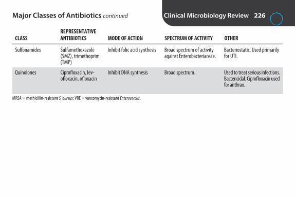
Major Classes of Antibiotics continued Clinical Microbiology Review 226
REPRESENTATIVE CLASS ANTIBIOTICS MODE OF ACTION SPECTRUM OF ACTIVITY OTHER
Sulfonamides
Quinolones
MRSA = methicillin-resistant S. aureus; VRE = vancomycin-resistant Enterococcus.
Bacteriostatic. Used primarilyfor UTI.
Used to treat serious infections.Bactericidal. Ciprofloxacin usedfor anthrax.
Inhibit folic acid synthesis
Inhibit DNA synthesis
Sulfamethoxazole(SMZ), trimethoprim(TMP)
Ciprofloxacin, lev-ofloxacin, ofloxacin
Broad spectrum of activityagainst Enterobacteriaceae.
Broad spectrum.
2956_Ch03_135-286 29/01/14 12:17 PM Page 226

Disk Diffusion Susceptibility Method (Kirby Bauer)
Clinical Microbiology Review 227
Organisms
Inoculum
Medium
Disks
Incubation
Modifications for fastidious bacteria
Reading
Situations/actions
Rapidly growing aerobes & facultative anaerobes. Not for slow growers, anaerobes, or fastidious organisms (except with modifications).
18–24 hr pure culture. 1.5 × 10⁸ CFU/mL. Compare to 0.5 McFarland standard. Too heavy = smallerzones/false resistant. Too light = larger zones/false susceptible.
Mueller-Hinton agar. Swab entire surface evenly to achieve solid lawn of growth.
Store in refrig or freezer in sealed, desiccated container. Warm to RT before use. Check expirationdates. Place within 15 min of inoculation. No more than 12 per 150-mm plate.
Within 15 min of disk placement. Inverted. Ambient air. 35°C for 16–18 hr. <16 hr = ↑ zone, falsesusceptible. >18 hr = ↓ zone, false resistant. (Exception: MRSA may require 24 hr.)
Streptococcus: Use MH supplemented with 5% sheep blood. Incubate in 5%–7% CO2. Haemophilus:Use Haemophilus test medium (HTM). Incubate in 5%–7% CO2.
Routine: Measure zones from back side of plate using ruler or calipers. Media containing blood: Readfrom top with lid removed.
Sparse growth: Repeat with standardized inoculum. Mixed culture: Repeat with pure inoculum.Colonies within zone: Subculture & retest. Slight growth in zone for trimethoprim & sulfonamides:Disregard. Swarming of Proteus into zone: Ignore.
continued...
2956_Ch03_135-286 29/01/14 12:17 PM Page 227

Disk Diffusion Susceptibility Method (Kirby Bauer) continued
Clinical Microbiology Review 228
Reporting
Quality control
CFU = colony-forming units; CLSI = Clinical and Laboratory Standards Institute.
Resistant, intermediate, or susceptible based on zone of inhibition in mm. Refer to CLSI interpretivetables.
Test QC strains of E. coli, S. aureus, P. aeruginosa, Enterococcus faecalis for 20–30 consecutive days. Ifresults are acceptable, frequency can be reduced to weekly.
2956_Ch03_135-286 29/01/14 12:17 PM Page 228
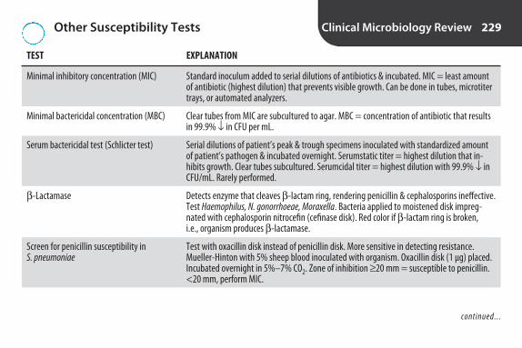
Other Susceptibility Tests Clinical Microbiology Review 229
TEST EXPLANATION
Minimal inhibitory concentration (MIC)
Minimal bactericidal concentration (MBC)
Serum bactericidal test (Schlicter test)
β -Lactamase
Screen for penicillin susceptibility in S. pneumoniae
Standard inoculum added to serial dilutions of antibiotics & incubated. MIC = least amountof antibiotic (highest dilution) that prevents visible growth. Can be done in tubes, microtitertrays, or automated analyzers.
Clear tubes from MIC are subcultured to agar. MBC = concentration of antibiotic that resultsin 99.9% ↓ in CFU per mL.
Serial dilutions of patient’s peak & trough specimens inoculated with standardized amountof patient’s pathogen & incubated overnight. Serumstatic titer = highest dilution that in-hibits growth. Clear tubes subcultured. Serumcidal titer = highest dilution with 99.9% ↓ inCFU/mL. Rarely performed.
Detects enzyme that cleaves β -lactam ring, rendering penicillin & cephalosporins ineffective.Test Haemophilus, N. gonorrhoeae, Moraxella. Bacteria applied to moistened disk impreg-nated with cephalosporin nitrocefin (cefinase disk). Red color if β -lactam ring is broken, i.e., organism produces β -lactamase.
Test with oxacillin disk instead of penicillin disk. More sensitive in detecting resistance.Mueller-Hinton with 5% sheep blood inoculated with organism. Oxacillin disk (1 μg) placed.Incubated overnight in 5%–7% CO2. Zone of inhibition ≥ 20 mm = susceptible to penicillin.<20 mm, perform MIC.
continued...
2956_Ch03_135-286 29/01/14 12:17 PM Page 229

Other Susceptibility Tests continued Clinical Microbiology Review 230
TEST EXPLANATION
Tests for oxacillin- (methicillin-) resistant S. aureus (MRSA)
Vancomycin screen
E test
D test
Oxacillin used as class representative for penicillinase-resistant penicillins. Organism resistantto it is resistant to all. (Oxacillin-resistant S. aureus referred to as methicillin-resistant for his-torical reasons.) Oxacillin screen plate: Mueller-Hinton with 4% NaCl & oxacillin (6 μg /mL)inoculated & incubated overnight. Any growth = resistant. Not suitable for testing coag-negstaph (CNS). CLSI recommends cefoxitin disk diffusion method for both S. aureus & CNS. Manylabs use PCR. Can be done directly on specimen. Faster results.
For detection of vancomycin-resistant enterococci (VRE). Brain-heart infusion agar plus 6 μg vancomycin/mL inoculated & incubated overnight. Growth = resistance. Can also beused for S. aureus.
Plastic strip containing antibiotic concentration gradient placed on inoculum lawn on Mueller-Hinton plate & incubated overnight. MIC = point where border of growth inhibition intersectsstrip. Combines convenience of disk diffusion with ability to generate MIC data. Useful for testing fastidious organisms such as S. pneumoniae, other strep, H. influenzae, & anaerobes.
To detect inducible clindamycin resistance in MRSA isolates that are resistant to erythromycin& susceptible to clindamycin on initial testing. Erythromycin & clindamycin disks placed 15–26 mm apart on Mueller-Hinton agar inoculated with organism. After overnight incuba-tion, flattened zone between disks (D-shaped zone of inhibition around clindamycin disk)means erythromycin induces clindamycin resistance. Clindamycin reported as resistant. Similar procedure for beta-hemolytic strep except Mueller-Hinton with sheep blood used &disks placed 12 mm apart.
2956_Ch03_135-286 29/01/14 12:17 PM Page 230
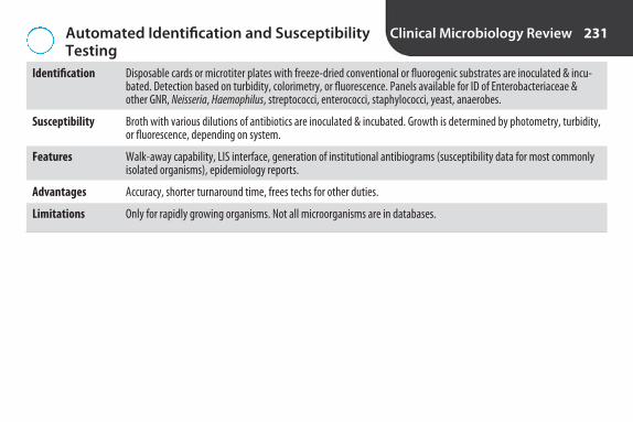
Automated Identification and SusceptibilityTesting
Clinical Microbiology Review 231
Identification
Susceptibility
Features
Advantages
Limitations
Disposable cards or microtiter plates with freeze-dried conventional or fluorogenic substrates are inoculated & incu-bated. Detection based on turbidity, colorimetry, or fluorescence. Panels available for ID of Enterobacteriaceae & other GNR, Neisseria, Haemophilus, streptococci, enterococci, staphylococci, yeast, anaerobes.
Broth with various dilutions of antibiotics are inoculated & incubated. Growth is determined by photometry, turbidity,or fluorescence, depending on system.
Walk-away capability, LIS interface, generation of institutional antibiograms (susceptibility data for most commonlyisolated organisms), epidemiology reports.
Accuracy, shorter turnaround time, frees techs for other duties.
Only for rapidly growing organisms. Not all microorganisms are in databases.
2956_Ch03_135-286 29/01/14 12:17 PM Page 231

Examples of Quality Control in MicrobiologyLabs
Clinical Microbiology Review 232
PARAMETER REQUIREMENT(S)
Autoclave
Balances
Biosafety cabinet
Centrifuges
CO2 incubator
Gas pack
Microscopes
Temperatures
Thermometers
Instrument logs
Gram stain
Reagents
Spore test weekly (B. stearothermophilus). Must reach 121°C/15 psi.
Check for accuracy annually.
Check airflow annually.
Check rpm every 6 months.
Check CO2 & temp daily.
Check for anaerobiosis with methylene blue strip each use (white = no O2, blue = O2).
Clean & adjust 4 times/yr.
Daily checks of incubators, heating blocks, water baths, refrigerators, freezers.
Must be checked against reference thermometer from National Bureau of Standards.
Document routine function checks, preventive maintenance service. Maintain for life of instrument.
Control slide weekly (E. coli & S. aureus).
Most are tested each day of use with pos & neg controls.
continued...
2956_Ch03_135-286 29/01/14 12:17 PM Page 232
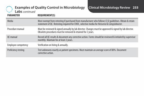
Examples of Quality Control in MicrobiologyLabs continued
Clinical Microbiology Review 233
PARAMETER REQUIREMENT(S)
Media
Procedure manual
QC manual
Employee competency
Proficiency testing
Most exempt from retesting if purchased from manufacturer who follows CLSI guidelines. Obtain & retainstatement of QC. Retesting required for CHOC, selective media for Neisseria & Campylobacter.
Must be reviewed & signed annually by lab director. Changes must be approved & signed by lab director.Obsolete procedures must be removed & retained for 2 years.
Record all QC results & document any corrective action. Forms should be reviewed & initialed by supervisormonthly. Maintain for at least 2 years.
Verification on hiring & annually.
Test unknowns exactly as patient specimens. Must maintain an average score of 80%. Document corrective action.
2956_Ch03_135-286 29/01/14 12:17 PM Page 233

Stool Specimens for Ova and Parasites Clinical Microbiology Review 234
MAXIMUM TIME BETWEEN COLLECTION CONSISTENCY AND EXAMINATION PARASITES RECOVERED
Liquid 30 minutes Trophozoites, helminth eggs, larvae
Semisolid 1 hour Trophozoites, cysts, helminth eggs, larvae
Formed 24 hours Cysts, helminth eggs, larvae
2956_Ch03_135-286 29/01/14 12:17 PM Page 234
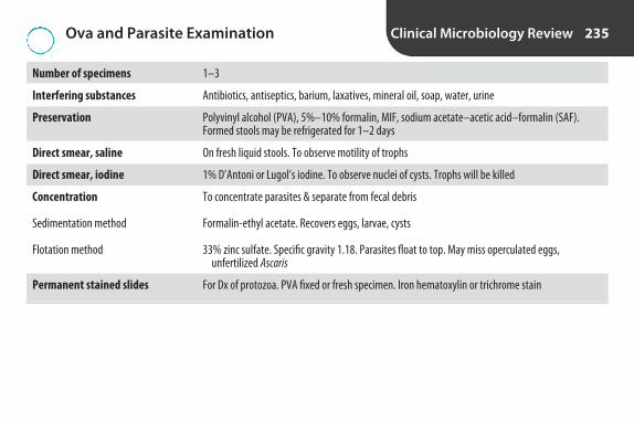
Ova and Parasite Examination Clinical Microbiology Review 235
Number of specimens
Interfering substances
Preservation
Direct smear, saline
Direct smear, iodine
Concentration
Sedimentation method
Flotation method
Permanent stained slides
1–3
Antibiotics, antiseptics, barium, laxatives, mineral oil, soap, water, urine
Polyvinyl alcohol (PVA), 5%–10% formalin, MIF, sodium acetate–acetic acid–formalin (SAF).Formed stools may be refrigerated for 1–2 days
On fresh liquid stools. To observe motility of trophs
1% D’Antoni or Lugol’s iodine. To observe nuclei of cysts. Trophs will be killed
To concentrate parasites & separate from fecal debris
Formalin-ethyl acetate. Recovers eggs, larvae, cysts
33% zinc sulfate. Specific gravity 1.18. Parasites float to top. May miss operculated eggs, unfertilized Ascaris
For Dx of protozoa. PVA fixed or fresh specimen. Iron hematoxylin or trichrome stain
2956_Ch03_135-286 29/01/14 12:17 PM Page 235
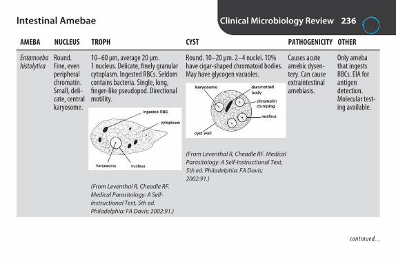
Intestinal Amebae Clinical Microbiology Review 236
AMEBA NUCLEUS TROPH CYST PATHOGENICITY OTHER
Entamoeba histolytica
Only amebathat ingestsRBCs. EIA forantigen detection. Molecular test-ing available.
Round. Fine, evenperipheralchromatin.Small, deli-cate, centralkaryosome.
10–60 μm, average 20 μm. 1 nucleus. Delicate, finely granularcytoplasm. Ingested RBCs. Seldomcontains bacteria. Single, long, finger-like pseudopod. Directionalmotility.
Round. 10–20 μm. 2–4 nuclei. 10%have cigar-shaped chromatoid bodies.May have glycogen vacuoles.
Causes acuteamebic dysen-tery. Can causeextraintestinalamebiasis.
(From Leventhal R, Cheadle RF.Medical Parasitology: A Self-Instructional Text, 5th ed. Philadelphia: FA Davis; 2002:91.)
(From Leventhal R, Cheadle RF. MedicalParasitology: A Self-Instructional Text,5th ed. Philadelphia: FA Davis;2002:91.)
continued...
2956_Ch03_135-286 29/01/14 12:17 PM Page 236
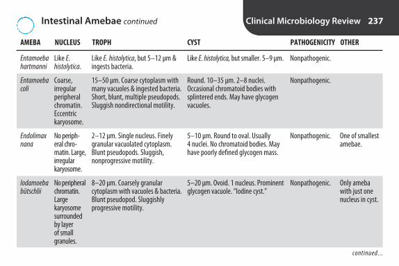
Intestinal Amebae continued Clinical Microbiology Review 237
AMEBA NUCLEUS TROPH CYST PATHOGENICITY OTHER
Entamoeba hartmanni
Entamoeba coli
Endolimax nana
Iodamoeba bütschlii
One of smallestamebae.
Only amebawith just onenucleus in cyst.
Like E. histolytica.
Coarse, irregular peripheralchromatin.Eccentrickaryosome.
No periph-eral chro-matin. Large,irregularkaryosome.
No peripheralchromatin.Largekaryosomesurroundedby layer of smallgranules.
Like E. histolytica, but 5–12 μm &ingests bacteria.
15–50 μm. Coarse cytoplasm withmany vacuoles & ingested bacteria.Short, blunt, multiple pseudopods.Sluggish nondirectional motility.
2–12 μm. Single nucleus. Finelygranular vacuolated cytoplasm.Blunt pseudopods. Sluggish, nonprogressive motility.
8–20 μm. Coarsely granular cytoplasm with vacuoles & bacteria.Blunt pseudopod. Sluggishly progressive motility.
Like E. histolytica, but smaller. 5–9 μm.
Round. 10–35 μm. 2–8 nuclei. Occasional chromatoid bodies withsplintered ends. May have glycogenvacuoles.
5–10 μm. Round to oval. Usually 4 nuclei. No chromatoid bodies. Mayhave poorly defined glycogen mass.
5–20 μm. Ovoid. 1 nucleus. Prominentglycogen vacuole. “Iodine cyst.”
Nonpathogenic.
Nonpathogenic.
Nonpathogenic.
Nonpathogenic.
continued...
2956_Ch03_135-286 29/01/14 12:17 PM Page 237

Intestinal Amebae continued Clinical Microbiology Review 238
AMEBA NUCLEUS TROPH CYST PATHOGENICITY OTHER
Blastocystis hominis
All amebae are transmitted by ingestion of fecally contaminated food or water.
Should bequantitated.Formerly clas-sified as yeast;now consideredan ameba.
Up to 4,pushed tothe side.
Round. 6–40 μm. Large centralbody.
Uncertain.
(From Leventhal R, Cheadle RF.Medical Parasitology: A Self-Instructional Text, 5th ed. Philadelphia: FA Davis; 2002:95.)
None
2956_Ch03_135-286 29/01/14 12:17 PM Page 238
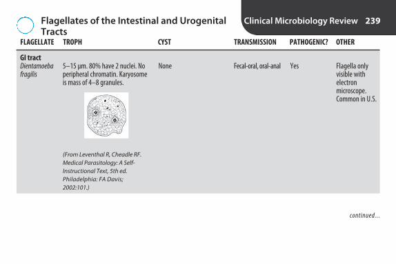
Flagellates of the Intestinal and UrogenitalTracts
Clinical Microbiology Review 239
FLAGELLATE TROPH CYST TRANSMISSION PATHOGENIC? OTHER
GI tractDientamoeba fragilis
Flagella onlyvisible withelectron microscope.Common in U.S.
5–15 μm. 80% have 2 nuclei. Noperipheral chromatin. Karyosomeis mass of 4–8 granules.
None Fecal-oral, oral-anal Yes
(From Leventhal R, Cheadle RF.Medical Parasitology: A Self-Instructional Text, 5th ed.Philadelphia: FA Davis;2002:101.)
continued...
2956_Ch03_135-286 29/01/14 12:17 PM Page 239

Flagellates of the Intestinal and UrogenitalTracts continued
Clinical Microbiology Review 240
FLAGELLATE TROPH CYST TRANSMISSION PATHOGENIC? OTHER
Giardia lamblia (also known as G. intestinalis or G. duodenalis)
Common in U.S.Enterotest: Patient swal-lows capsuleattached tostring. Stringwithdrawnafter 4 hr.Mucus exam-ined. Antigendetection.
Pear-shaped. 10–20 μm. 2 nucleiwith large central karyosomes. 4 pairs of flagella. Anterior & ven-tral sucking disks. 2 axostyles. 2 median bodies. “Old man inglasses.” Falling leaf motility.
Ovoid. 8–19 μm. Thickdouble wall. Cytoplasmshrinks away from cellwall. 2–4 nuclei at ante-rior end. Median bodies &axostyles.
Ingestion of cystin contaminatedwater, oral-anal,pets
Yes
(From Leventhal R, Cheadle RF. Medical Parasitology: A Self-Instructional Text, 5th ed. Philadelphia: FA Davis; 2002:100.)
(From Leventhal R, CheadleRF. Medical Parasitology: ASelf-Instructional Text, 5thed. Philadelphia: FA Davis;2002:100.)
continued...
2956_Ch03_135-286 29/01/14 12:17 PM Page 240

Flagellates of the Intestinal and UrogenitalTracts continued
Clinical Microbiology Review 241
FLAGELLATE TROPH CYST TRANSMISSION PATHOGENIC? OTHER
Chilomastix mesnili
Trichomonas hominis
Easier to ID living organism.Rotary motion.
6–24 μm. Broad anterior, taperingposterior. 3 flagella at anterior,one at posterior. Spiral grooverunning length of body. Promi-nent cytostome bordered by fibrils. Single nucleus at anterior.Stiff rotary motility.
6–14 μm. Prominent axostyle extending through posterior. 4 anterior flagella. Undulatingmembrane entire length of bodyalong costa. 1 oval nucleus withsmall karyosome.
6–10 μm. Round withbulge at anterior. Singlelarge nucleus. Hourglass-shaped cytostome &prominent fibrils. “Lemon cyst.”
None
Ingestion of cyst
Ingestion of fecallycontaminated material
No
No
continued...
2956_Ch03_135-286 29/01/14 12:17 PM Page 241

Flagellates of the Intestinal and UrogenitalTracts continued
Clinical Microbiology Review 242
FLAGELLATE TROPH CYST TRANSMISSION PATHOGENIC? OTHER
Urogenital tractTrichomonas vaginalis
Only flagellatein urogenitaltract. Dx by wet mount(jerky motility),culture, rapidantigen tests,PCR.
5–18 μm. 4 anterior flagella, undulating membrane that extends half the body length.
None Sexual Yes
(From Leventhal R, Cheadle RF. Medical Parasitology: A Self-Instructional Text, 5th ed. Philadelphia: FA Davis; 2002:103.)
2956_Ch03_135-286 29/01/14 12:17 PM Page 242

Intestinal Ciliates Clinical Microbiology Review 243
CILIATE TROPH CYST TRANSMISSION PATHOGENIC? OTHER
Balantidium coli Only ciliate pathogenicto humans. Largest protozoan of humans.Common in pigs.
Ingestion of cystOvoid. Usually 40–50 μm,but can reach 200 μm. Covered with cilia. Funnel-shaped cytostome. Macronu-cleus & micronucleus.Vacuoles. Rapid rotary motion.
Round. 45–65 μm.Thick refractile cellwall. No cilia.
Yes
2956_Ch03_135-286 29/01/14 12:17 PM Page 243

Intestinal Sporozoans Clinical Microbiology Review 244
ORGANISM TRANSMISSION DIAGNOSIS OTHER
Isospora belli
Cryptosporidium parvum
Cyclospora cayentanensis
Microsporidia
Human is definitive host. Causes anorexia,nausea, abdominal pain, diarrhea, possi-ble malabsorption.
Important opportunistic infection in AIDSpatients. Causes chronic diarrhea in immunosuppressed, acute self-limiteddiarrhea in immunocompetent.
Prolonged diarrhea.
Obligate intracellular parasites. Oppor-tunistic pathogens, mainly in AIDS patients. Cause prolonged diarrhea. Can disseminate. Molecular studies indi-cate related to fungi.
Ingestion of oocysts in fecally contaminated food or water
Ingestion of oocysts fromfood or water contami-nated with animal feces,oral/anal route, directcontact with infected individual or animal
Contaminated food &water
Ingestion of spores
Oocysts in feces. 30 × 12 μm.Transparent. 1–2 sporoblasts.
Modified acid-fast stain of feces.Red spherical bodies 3–6 μm. 4 sporozoites. Immunoassay kits.
Oocysts in direct wet mount. 8–10 μm. Variable staining withmodified acid-fast stain. Autoflu-orescence under UV light. Brightblue at 365 nm, mint green at450–490 nm.
Chromotrope or calcofluor whitestain of formalin-preserved stool.Spores are 1.5–4.0 μm.
2956_Ch03_135-286 29/01/14 12:17 PM Page 244

Intestinal Nematodes Clinical Microbiology Review 245
NEMATODE ROUTE OF INFECTION DIAGNOSTIC STAGE OTHER
Trichuris trichiura Whip worm. Eosinophilia. Eggs requiredevelopmental period in warm soil before infective.
Ingestion of infective egg. Barrel-shaped ovum. 50 × 23 μm.Brown or yellow. Clear plug at either end.
(From Leventhal R, Cheadle RF. Medical Parasitology: A Self-Instructional Text, 5th ed. Philadelphia: FA Davis;2002:17.)
continued...
2956_Ch03_135-286 29/01/14 12:17 PM Page 245

Intestinal Nematodes continued Clinical Microbiology Review 246
NEMATODE ROUTE OF INFECTION DIAGNOSTIC STAGE OTHER
Enterobius vermicularis Pinworm. Common in U.S. Common inchildren. Resistant eggs in home caninfect other family members. Itchingwhen adult female migrates out ofanus at night to deposit eggs. Diagnoseby pinworm paddle or cellophane tape prep.
Ingestion of infective egg. Ovoid ovum. 50 × 20 μm. 1 sideflattened. Colorless. Larva inside.
(From Leventhal R, Cheadle RF. Medical Parasitology: A Self-Instructional Text, 5th ed. Philadelphia: FA Davis;2002:15.)
continued...
2956_Ch03_135-286 29/01/14 12:17 PM Page 246
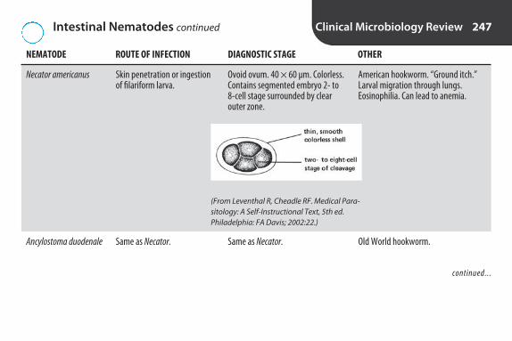
Intestinal Nematodes continued Clinical Microbiology Review 247
NEMATODE ROUTE OF INFECTION DIAGNOSTIC STAGE OTHER
Necator americanus
Ancylostoma duodenale
American hookworm. “Ground itch.”Larval migration through lungs.Eosinophilia. Can lead to anemia.
Old World hookworm.
Skin penetration or ingestionof filariform larva.
Same as Necator.
Ovoid ovum. 40 × 60 μm. Colorless.Contains segmented embryo 2- to8-cell stage surrounded by clearouter zone.
Same as Necator.
(From Leventhal R, Cheadle RF. Medical Para-sitology: A Self-Instructional Text, 5th ed.Philadelphia: FA Davis; 2002:22.)
continued...
2956_Ch03_135-286 29/01/14 12:17 PM Page 247
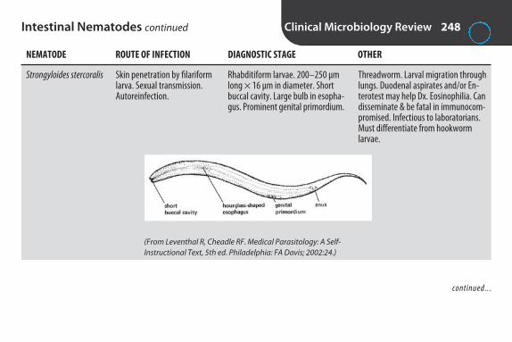
Intestinal Nematodes continued Clinical Microbiology Review 248
NEMATODE ROUTE OF INFECTION DIAGNOSTIC STAGE OTHER
Strongyloides stercoralis Threadworm. Larval migration throughlungs. Duodenal aspirates and/or En-terotest may help Dx. Eosinophilia. Candisseminate & be fatal in immunocom-promised. Infectious to laboratorians.Must differentiate from hookworm larvae.
Skin penetration by filariformlarva. Sexual transmission.Autoreinfection.
Rhabditiform larvae. 200–250 μmlong × 16 μm in diameter. Shortbuccal cavity. Large bulb in esopha-gus. Prominent genital primordium.
(From Leventhal R, Cheadle RF. Medical Parasitology: A Self-Instructional Text, 5th ed. Philadelphia: FA Davis; 2002:24.)
continued...
2956_Ch03_135-286 29/01/14 12:17 PM Page 248
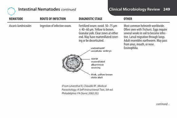
Intestinal Nematodes continued Clinical Microbiology Review 249
NEMATODE ROUTE OF INFECTION DIAGNOSTIC STAGE OTHER
Ascaris lumbricoides Most common helminth worldwide.Often seen with Trichuris. Eggs requireseveral weeks in soil to become infec-tive. Larval migration through lungs.Adult resembles earthworm. May passfrom anus, mouth, or nose.Eosinophilia.
Ingestion of infective ovum. Fertilized ovum: ovoid. 50–75 μm× 40–60 μm. Yellow to brown.Granular yolk. Clear zones at eitherend. May have mammillated cover-ing or be decorticated.
(From Leventhal R, Cheadle RF. Medical Parasitology: A Self-Instructional Text, 5th ed.Philadelphia: FA Davis; 2002:20.)
continued...
2956_Ch03_135-286 29/01/14 12:17 PM Page 249

Intestinal Nematodes continued Clinical Microbiology Review 250
NEMATODE ROUTE OF INFECTION DIAGNOSTIC STAGE OTHER
Nonfertilized ovum: larger & moreelongated. Thinner shell. No concentric clear zones. Can bemammillated or decorticated.
(From Leventhal R, Cheadle RF. Medical Parasitology:A Self-Instructional Text, 5e. Philadelphia: FA Davis;2002:20.
2956_Ch03_135-286 29/01/14 12:17 PM Page 250

Intestinal Cestodes Clinical Microbiology Review 251
GRAVID INTERMEDIATE DIAGNOSTIC CESTODE SCOLEX PROGLOTTID OVUM HOST TRANSMISSION STAGE OTHER
Taenia saginata
Beef tape-worm. Ovaare infectivefor cattle, butnot humans.
Longer thanwide. 15–30uterinebranches perside. Irregularlyalternatinggenital pores.
Tetragonal. 4cup-shapedsuckers. Nohooklets.
Spherical. 35–45 μm.Yellow orbrown. Thickradially striatedshell.
Ingestion oflarva in under-cooked beef
Cow Ova in feces
(From Leventhal R, Cheadle RF. Medical Parasitology: A Self-Instructional Text, 5th ed. Philadelphia: FA Davis; 2002:49.)
continued...
2956_Ch03_135-286 29/01/14 12:17 PM Page 251
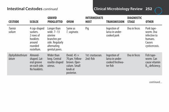
Intestinal Cestodes continued Clinical Microbiology Review 252
GRAVID INTERMEDIATE DIAGNOSTIC CESTODE SCOLEX PROGLOTTID OVUM HOST TRANSMISSION STAGE OTHER
Taenia solium
Diphyllobothrium latum
Pork tape-worm. Ova infective tohumans.Causes cysticercosis.
Fish tape-worm. Cancause vitaminB12 deficiency.
Longer thanwide. 7–13uterinebranches perside. Regularlyalternatinggenital pores.
Wider thanlong. Centralrosette-shapeduterus.
4 cup-shapedsuckers. 2 rows ofhookletsaroundrounded rostellum.
Almond-shaped. Lat-eral grooveon each side.No hooklets.
Same as T. saginata.
Ovoid. 45 ×70 μm. Yellow-brown. Oper-culum. Smallknob at posterior.
Ingestion oflarva in under-cooked pork
Ingestion oflarva in under-cooked freshwa-ter fish
Pig
1st: crustacean.2nd: fish
Ova in feces
Ova in feces
continued...
2956_Ch03_135-286 29/01/14 12:17 PM Page 252
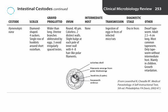
Intestinal Cestodes continued Clinical Microbiology Review 253
GRAVID INTERMEDIATE DIAGNOSTIC CESTODE SCOLEX PROGLOTTID OVUM HOST TRANSMISSION STAGE OTHER
Hymenolepis nana
Dwarf tape-worm. Adult2.5–4 cmlong. Mostcommontapeworm.Only tape-worm withoutintermediatehost. Mainlyin children.Growth retardation.
Wider thanlong. Uterinebranchesobliterated byeggs. 3 ovoidirregularlyspaced testes.
Diamond-shaped. 4 suckers.Single row ofhookletsaround shortrostellum.
Round. 40 μm.Colorless. 2distinct walls.Slight bulge ateach pole ofinner wallwith 4–8 hair-like polarfilaments.
Ingestion ofeggs in feces ofinfectedmice/rats
None Ova in feces
(From Leventhal R, Cheadle RF. MedicalParasitology: A Self-Instructional Text, 5th ed. Philadelphia: FA Davis; 2002:47.)
2956_Ch03_135-286 29/01/14 12:17 PM Page 253

Trematodes Clinical Microbiology Review 254
ROUTE OF LOCATION TREMATODE INFECTION OF ADULT DIAGNOSTIC STAGE OTHER
Fasciolopsis buski
Fasciola hepatica
Clonorchis sinensis
Largest trematode.
Sheep liver fluke.
Chinese liver fluke.One of the mostfrequent infectionsseen in Asianrefugees. Egg is oneof smallest passedby humans.
Ingestion of metacercariae onaquatic plants
Ingestion of metacer-cariae on aquatic plants
Ingestion of metacer-cariae in raw, undercooked, dried,salted, or pickled fish
Duodenum
Bile duct
Bile ducts, gallblad-der, pancreaticducts
Ova in feces. 135 × 80 μm. Yellow-brown. Thinshell. Small operculum. Granular contents evenlydistributed. Clear zone between shell & yolk.
Similar to F. buski.
Ova in feces. 29 × 16 μm. Bulbous. Opercularshoulders. Small comma-shaped protuberanceat posterior. Thick shell with tiny spines. Ciliated miracidium inside.
operculum
thickened rimaround operculum(called shoulders)
developedmiracidium
small knob
(From Leventhal R, Cheadle RF. Medical Parasitology: A Self-Instructional Text, 5th ed. Philadelphia: FA Davis; 2002:67.)
continued...
2956_Ch03_135-286 29/01/14 12:17 PM Page 254

Trematodes continued Clinical Microbiology Review 255
ROUTE OF LOCATION TREMATODE INFECTION OF ADULT DIAGNOSTIC STAGE OTHER
Paragonimus westermani
Schistosoma mansoni
Blood fluke
Ingestion of under-cooked crabs or crayfish
Skin penetration
Lungs
Intestinal venules
Ova in feces or sputum. 80–120 μm × 48–60μm. Yellow-brown. Thick shelled. Flattened operculum. “Chinese vase.”
Ova in feces. Elongated. 155 × 65 μm. Pronounced lateral spine.
(From Leventhal R, CheadleRF. Medical Parasitology: ASelf-Instructional Text, 5th ed.Philadelphia: FA Davis;2002:74.)
continued...
2956_Ch03_135-286 29/01/14 12:17 PM Page 255

Trematodes continued Clinical Microbiology Review 256
ROUTE OF LOCATION TREMATODE INFECTION OF ADULT DIAGNOSTIC STAGE OTHER
Schistosoma japonicum
Blood fluke.Skin penetration Intestinal venules Ova in feces. Ovoid. 90 × 70 μm. Minute lateralspine.
(From Leventhal R, Cheadle RF. Medical Parasitology: A Self-Instructional Text, 5th ed.Philadelphia: FA Davis; 2002:74.)
continued...
2956_Ch03_135-286 29/01/14 12:17 PM Page 256

Trematodes continued Clinical Microbiology Review 257
ROUTE OF LOCATION TREMATODE INFECTION OF ADULT DIAGNOSTIC STAGE OTHER
Schistosoma haematobium
Note: All trematodes have a snail as an intermediate host.
Blood fluke. Onlyparasite of urinarysystem.
Skin penetration Bladder venules Ova in urine. Elongated. 140 × 60 μm. Terminalspine.
(From Leventhal R, Cheadle RF. Med-ical Parasitology: A Self-InstructionalText, 5th ed. Philadelphia: FA Davis;2002:75.)
2956_Ch03_135-286 29/01/14 12:17 PM Page 257

Blood and Tissue Protozoa Clinical Microbiology Review 258
ORGANISM CLASSIFICATION TRANSMISSION DIAGNOSIS OTHER
Acanthamoeba
Naegleria fowleri
Plasmodium
Causes granulomatousamebic encephalitis(GAE) & ulcerative acan-thamoeba keratitis incontact lens wearers.Usually in immunocom-promised or debilitated.
Causes primary amebicmeningoencephalitis(PAM).
Causes malaria. Parasitesin RBCs.
Ameba
Ameba
Sporozoan
Swimming in contam-inated water, inade-quately disinfectedcontact lenses
Swimming in contami-nated ponds or streams
Anopheles mosquito
Stained smears of culture material. Histological examination of brain. Trophs & cysts in corneal scrapings.
Stained smears of culture material. Trophsin CSF.
Wright’s-stained thick & thin blood smears.Draw blood just before paroxysm.
continued...
2956_Ch03_135-286 29/01/14 12:17 PM Page 258

Blood and Tissue Protozoa continued Clinical Microbiology Review 259
ORGANISM CLASSIFICATION TRANSMISSION DIAGNOSIS OTHER
Babesia Symptoms resemblemalaria.
Sporozoan Tick bite, blood transfusion
Wright’s-stained thick & thin bloodsmears.Parasites in RBCs. 2–4 μm. Pear-shaped.Usually in pairs or tetrads (Maltese cross).
(From Leventhal R,Cheadle RF. MedicalParasitology: A Self-Instructional Text, 5th ed. Philadelphia: FA Davis; 2002:121.)
(From Leventhal R,Cheadle RF. MedicalParasitology: A Self-Instructional Text, 5th ed. Philadelphia: FA Davis; 2002:121.)
continued...
2956_Ch03_135-286 29/01/14 12:17 PM Page 259
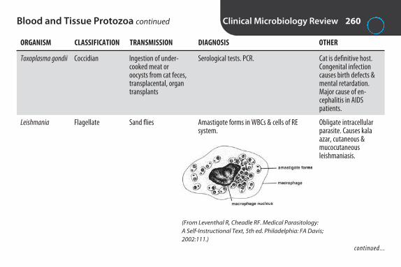
Blood and Tissue Protozoa continued Clinical Microbiology Review 260
ORGANISM CLASSIFICATION TRANSMISSION DIAGNOSIS OTHER
Toxoplasma gondii
Leishmania
Cat is definitive host.Congenital infectioncauses birth defects &mental retardation.Major cause of en-cephalitis in AIDS patients.
Obligate intracellularparasite. Causes kalaazar, cutaneous & mucocutaneous leishmaniasis.
Coccidian
Flagellate
Ingestion of under-cooked meat oroocysts from cat feces,transplacental, organtransplants
Sand flies
Serological tests. PCR.
Amastigote forms in WBCs & cells of REsystem.
continued...
(From Leventhal R, Cheadle RF. Medical Parasitology: A Self-Instructional Text, 5th ed. Philadelphia: FA Davis;2002:111.)
2956_Ch03_135-286 29/01/14 12:17 PM Page 260

Blood and Tissue Protozoa continued Clinical Microbiology Review 261
ORGANISM CLASSIFICATION TRANSMISSION DIAGNOSIS OTHER
Trypanosoma T. gambiense & rhode-siense: Sleeping sickness.T. cruzi: Chagas disease;found in southern U.S.
Flagellate T. gambiense & rhode-siense: tsetse fly. T. cruzi: reduviid orkissing bug.
T. gambiense & rhodesiense: trypomastigotein blood, lymph nodes, CSF. T. cruzi: trypo-mastigote in blood; amastigote in RE cells,myocardium, CNS.
(From Leventhal R, Cheadle RF. MedicalParasitology: A Self-Instructional Text,5th ed. Philadelphia: FA Davis; 2002:109.)
2956_Ch03_135-286 29/01/14 12:18 PM Page 261

Differentiation of Plasmodium Clinical Microbiology Review 262
P. FALCIPARUM P. MALARIAE P. OVALE P. VIVAX
Name of disease
Paroxysm cycle
Infected RBCs
Stages seen
Merozoites per mature schizont
Tertian malaria
44–48 hr
Enlarged. Schüffner’s dots
All
12–24
Malignant malaria
36–48 hr
Not enlarged
Ring forms (trophs) & gametocytes
Not seen in peripheralblood
Quartan malaria
72 hr
Not enlarged
All
6–12
Ovale malaria
48 hr
Sometimes enlarged. Fre-quently oval with raggedmargins. Schüffner’s dots
All
8–12
(From Leventhal R, Cheadle RF. MedicalParasitology: A Self-Instructional Text,5th ed. Philadelphia: FA Davis; 2002:119.)
continued...
2956_Ch03_135-286 29/01/14 12:18 PM Page 262

Differentiation of Plasmodium continued Clinical Microbiology Review 263
P. FALCIPARUM P. MALARIAE P. OVALE P. VIVAX
Shape of gametocytes
RoundSausage- or crescent-shaped
Ovoid Round
(From Leventhal R,Cheadle RF. MedicalParasitology: A Self-Instructional Text, 5th ed. Philadelphia: FA Davis; 2002:118.)
(From Leventhal R, Cheadle RF. Medical Parasitology: A Self-Instructional Text, 5th ed.Philadelphia: FA Davis;2002:118.)
continued...
2956_Ch03_135-286 29/01/14 12:18 PM Page 263
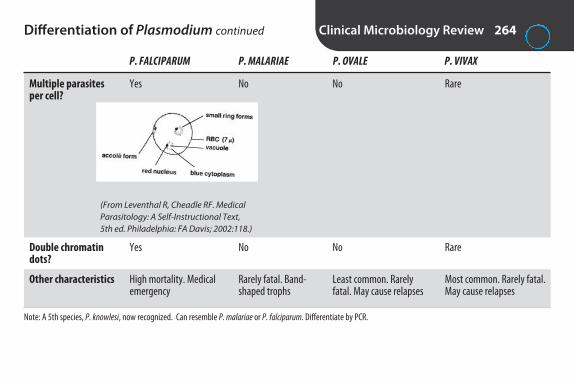
Differentiation of Plasmodium continued Clinical Microbiology Review 264
P. FALCIPARUM P. MALARIAE P. OVALE P. VIVAX
Multiple parasites per cell?
Double chromatin dots?
Other characteristics
Note: A 5th species, P. knowlesi, now recognized. Can resemble P. malariae or P. falciparum. Differentiate by PCR.
Rare
Rare
Most common. Rarely fatal.May cause relapses
Yes
Yes
High mortality. Medicalemergency
No
No
Rarely fatal. Band-shaped trophs
No
No
Least common. Rarelyfatal. May cause relapses
(From Leventhal R, Cheadle RF. MedicalParasitology: A Self-Instructional Text,5th ed. Philadelphia: FA Davis; 2002:118.)
2956_Ch03_135-286 29/01/14 12:18 PM Page 264

Blood and Tissue Helminths Clinical Microbiology Review 265
ORGANISM CLASSIFICATION TRANSMISSION DIAGNOSIS OTHER
Toxocara
Trichinella spiralis
Wuchereria bancrofti
Brugia malayi
Loa loa
Onchocerca volvulus
Echinococcus granulosus
Humans are accidental hosts.Larvae in tissues. Visceral & ocu-lar larva migrans (toxocariasis).
Eosinophilia, edema of uppereyelids, fever, myalgia.
Elephantiasis (lymphatic filariasis).
Elephantiasis. Nocturnal periodicity.
Loiasis. Nocturnal periodicity.
River blindness.
Humans are accidental interme-diate host. Cysts develop in liveror lungs.
Nematode (in dogs/cats)
Nematode
Nematode
Nematode
Nematode
Nematode
Cestode
Ingestion of eggsfrom dog/cat feces
Undercooked pork,walrus, bear
Mosquitoes
Mosquitoes
Mango fly (Chrysops)
Black fly
Ingestion of fecalmaterial from infected dog
Serological tests.
Larvae encysted in muscle.Serological tests.
Microfilariae in blood. Giemsa-stained thick & thin smears.
Microfilariae in blood. Giemsa-stained thick & thin smears.
Microfilariae in blood. Giemsa-stained thick & thin smears.
Microfilariae from skin snips oraspirates of nodules
Radiologic ID of cysts or ID ofscoleces in needle biopsy mate-rial from cyst (hydatid sand).Serological methods available.
2956_Ch03_135-286 29/01/14 12:18 PM Page 265

Stains Used in Mycology Clinical Microbiology Review 266
STAIN USED FOR OTHER
KOH
Calcofluor white
Lactophenol cotton blue
Gram stain
Acid-fast
India ink
Wright/Giemsa
Dissolves keratin. Makes fungal elements more visible.
Fluorescent stain.
Lactic acid preserves. Phenol kills. Cotton blue stains.
Fungi stain gram pos.
Nocardia is partially acid-fast.
Halos around yeast due to wide capsule. Insensitive. Directantigen test preferred.
Useful for Histoplasma.
Skin, hair, nails
Tissues, sputum, body fluids, skin & cornealscrapings
Microscopic exam of fungal culture
Yeast
Nocardia
CSF sediment if Cryptococcus is suspected
Blood & bone marrow
2956_Ch03_135-286 29/01/14 12:18 PM Page 266

Fungal Culture Media Clinical Microbiology Review 267
MEDIUM USE
For isolationSabouraud dextrose agar (SDA)
Sabouraud dextrose agar with antibiotics
Brain-heart infusion agar
Brain-heart infusion agar with antibiotics
Inhibitory mold agar
Dermatophyte test medium
For identificationPotato dextrose agar
Cornmeal agar with Tween 80
Urea agar
Birdseed agar (niger seed or caffeic agar)
General medium. pH 5.6 inhibits bacteria. Grows most molds & yeast.
Antibiotics inhibit fungal contaminants & bacteria. Dermatophytes & most fungalpathogens grow.
For isolation & conversion of dimorphic fungi from mold to yeast phase. Used for normallysterile specimens.
Selective medium used for isolation of pathogenic fungi from specimens contaminatedwith bacteria.
For recovery of fungi from specimens contaminated with bacteria. Contains chloram-phenicol & gentamicin to inhibit bacteria.
For recovery of dermatophytes from skin, hair, nails. Turn agar from yellow to red. Antibiotics inhibit bacteria.
Stimulates sporulation of molds. Good for slide cultures.
To differentiate Candida species. C. albicans produces chlamydospores.
Detection of urease production by C. neoformans.
Isolation of C. neoformans. Black-brown colonies in 4–7 days.
2956_Ch03_135-286 29/01/14 12:18 PM Page 267

Dermatophytes Clinical Microbiology Review 268
OTHER MOST COMMON DERMATOPHYTE INFECTS MACROCONIDIA MICROCONIDIA CHARACTERISTICS SPECIES
Microsporum
Trichophyton
Epidermophyton
M. caris, M. gypseum,M. audouinii
T. mentagrophytes, T. rubrum, T. tonsurans(primary cause oftinea capitis in U.S.)
E. floccosum is onlyspecies
Skin, hair. Rarelynails.
Skin, hair, nails.
Skin, sometimesnails. Rarely hair.
Large, spindle orcylinder shaped,thick walled, multi-septate, rough, spiny.Borne singly on shortconidiophores.
Rare. Pencil shaped,multiseptate, thinwalled, smooth,borne singly on conidiophore.
Club shaped, septate,thin walled, smooth.Borne in singles orclusters of 2–3 onconidiophore.
Ectothrix hair invasion.Some species causehair to fluoresce.
Hair infections en-dothrix or ectothrix.Hair usually doesn’tfluoresce. May havespirals, nodular bodies,chlamydospores, faviform mycelia. T. rubrum colonies arered on reverse side.
Numerous chlamydospores.
Few or absent.Small, clubshaped.
Predominant.Spherical, tearshaped, orclavate.
Absent.
2956_Ch03_135-286 29/01/14 12:18 PM Page 268

Dimorphic Fungi Clinical Microbiology Review 269
FUNGUS INFECTION MOLD PHASE YEAST PHASE OTHER
Blastomyces dermatitidis
Paracoccidioides brasiliensis
Coccidioides immitis
Endemic to Ohio & MississippiRiver valleys. Usually in farmerswho contract from soil. Begins inlungs. May become systemic.
Mainly in Brazil, Argentina, S. Mexico. Begins in lungs. Canbecome systemic.
Found in desert of SouthwestU.S. More common in dark-skinned men. Spores are inhaledor contaminate injured skin. Affects respiratory tract 1st. Canspread to other organs. Caution:Arthrospores highly infectious.Serological tests available.
White to gray-browncolony. Hyaline, septatehyphae with small ovalconidia borne singly attips of conidiophores.“Lollipops.”
Resembles Blastomyces
Fluffy or powdery whiteto gray-tan colonies.Barrel-shapedarthrospores.
North Americanblastomycosis(Gilchrist’s disease)
South Americanblastomycosis
Valley fever
8–15 μm. Round, thickwalled. Single bud con-nected by wide neck.
10–30 μm. Round, doublewalled. Multiple budspinched at attachment.“Mariner’s wheel.”
Spherule 20–80 μm containing numerous endospores.
continued...
2956_Ch03_135-286 29/01/14 12:18 PM Page 269

Dimorphic Fungi continued Clinical Microbiology Review 270
FUNGUS INFECTION MOLD PHASE YEAST PHASE OTHER
Histoplasma capsulatum
Sporothrix schenckii
Endemic in Ohio & MississippiRiver valleys. Soil saprophyte.Found in manure of chickens, pigeons, bats. Acute pulmonarydisease that can be self-limitedor spread. Predilection for RE system. Tissue phase may beconfused with Leishmania. Serological tests & nucleic acidprobes available.
Found in Mississippi & MissouriRiver valleys. Found on rosebushes, barberry bushes, sphag-num moss, mulch. Usually infectsfarmers or florists. Introduced by trauma, usually to hand. Cutaneous lesions spread alonglymphatics.
Silky, white to gray-tancolonies. Thick-walledspherical macroconidia,7–16 μm, surrounded by finger-like projec-tions. “Tuberculatechlamydospores”
White to yellow colonies.Clusters of pear-shapedconidia at tips of conid-iophores. “Flowers”
Histoplasmosis (spelunker’s disease,Darling’s disease)
Sporotrichosis(rose gardener’sdisease)
1–4 μm round to oval. In-tracellular in phagocytesof blood or bone marrow.
Gram pos cigar-shapedcells. Usually not seen indirect smears unless byimmunofluorescence.
2956_Ch03_135-286 29/01/14 12:18 PM Page 270

Yeast Clinical Microbiology Review 271
YEAST INFECTIONS IMPORTANT CHARACTERISTICS OTHER
Candida albicans
Candida tropicalis
Geotrichum candidum
Cryptococcus neoformans
Most frequently isolated yeast. Normalflora in GI tract, mucocutaneousareas. Serious infections most oftenin immunocompromised. Onychomy-cosis, esophagitis common in AIDS.
May produce constricted germ tubes.(True germ tubes lack constrictions.)Usually infects immunosuppressed.
No blastoconidia.
In bird & bat droppings, decayingvegetation, fruit, milk. Capsule seenwith India ink prep in about 50% ofcases. Cryptococcal antigen test pre-ferred. Disseminated cryptococcosiscommon in AIDS.
Thrush (mouth), vulvovaginitis,diaper rash, onychomycosis(nails), paronychomycosis (cuticles), endocarditis,meningitis, UTI, pulmonaryinfections, fungemia
Vaginitis, UTI, GI, pulmonary, & systemic infections
Uncommon cause of wound infections & oral thrush
Lung infection that can disseminate to brain
Grows on SBA, EMB (spider-likeprojections), SDA. Gram-pos cells,2–4 μm. Blastoconidia or pseudo-hyphae (no constrictions). Producesgerm tubes in serum, round terminalchlamydospores on cornmeal agar.
Blastoconidia produced randomly along pseudohyphae on cornmeal agar.
Forms hockey stick–shaped arthro-conidia on cornmeal agar.
Irregularly sized, spherical cells surrounded by capsule. Maroon to brown-black colonies on nigerseed agar. Urease pos, phenol oxidase pos.
continued...
2956_Ch03_135-286 29/01/14 12:18 PM Page 271
Yeast continued Clinical Microbiology Review 272
YEAST INFECTIONS IMPORTANT CHARACTERISTICS OTHER
Malassezia furfur
Pneumocystis jiroveci
Yeasts can be identified by carbohydrate assimilation.
In skin infections, usually not cultured.Identified by KOH prep of skin scrap-ings. “Spaghetti & meatball” appear-ance. In systemic infections, recoveredfrom blood cultures. Will only grow on media overlaid with fatty acid, e.g., sterile mineral or olive oil.
Can’t be cultured. Dx by histopatholog-ical staining. Opportunistic infection inAIDS & other immunocompromisedpatients. Formerly P. carinii & classifiedas a protozoan.
Tinea versicolor. Systemic infection in children givenlipid replacement therapy
Atypical interstitial plasmacell pneumonia (PCP)
Oval or bottle-shaped buddingyeast, 3–8 μm in diameter. Charac-teristic collar between mother &daughter cells. Chains of short,slightly curved septate hyphae maybe seen in skin scrapings.
Cysts are 4–12 μm spheres with intracystic bodies.
2956_Ch03_135-286 29/01/14 12:18 PM Page 272
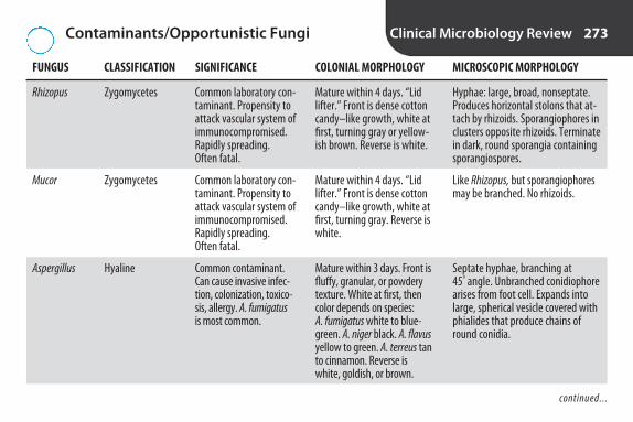
Contaminants/Opportunistic Fungi Clinical Microbiology Review 273
FUNGUS CLASSIFICATION SIGNIFICANCE COLONIAL MORPHOLOGY MICROSCOPIC MORPHOLOGY
Rhizopus
Mucor
Aspergillus
Hyphae: large, broad, nonseptate.Produces horizontal stolons that at-tach by rhizoids. Sporangiophores inclusters opposite rhizoids. Terminatein dark, round sporangia containingsporangiospores.
Like Rhizopus, but sporangiophoresmay be branched. No rhizoids.
Septate hyphae, branching at 45° angle. Unbranched conidiophorearises from foot cell. Expands intolarge, spherical vesicle covered withphialides that produce chains ofround conidia.
Common laboratory con-taminant. Propensity toattack vascular system ofimmunocompromised.Rapidly spreading. Often fatal.
Common laboratory con-taminant. Propensity toattack vascular system ofimmunocompromised.Rapidly spreading. Often fatal.
Common contaminant.Can cause invasive infec-tion, colonization, toxico-sis, allergy. A. fumigatusis most common.
Zygomycetes
Zygomycetes
Hyaline
Mature within 4 days. “Lidlifter.” Front is dense cottoncandy–like growth, white atfirst, turning gray or yellow-ish brown. Reverse is white.
Mature within 4 days. “Lidlifter.” Front is dense cottoncandy–like growth, white atfirst, turning gray. Reverse iswhite.
Mature within 3 days. Front isfluffy, granular, or powderytexture. White at first, thencolor depends on species: A. fumigatus white to blue-green. A. niger black. A. flavusyellow to green. A. terreus tanto cinnamon. Reverse iswhite, goldish, or brown.
continued...
2956_Ch03_135-286 29/01/14 12:18 PM Page 273
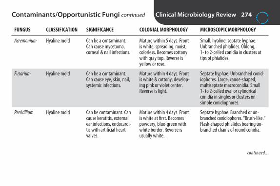
Contaminants/Opportunistic Fungi continued Clinical Microbiology Review 274
FUNGUS CLASSIFICATION SIGNIFICANCE COLONIAL MORPHOLOGY MICROSCOPIC MORPHOLOGY
Acremonium
Fusarium
Penicillium
Small, hyaline, septate hyphae. Unbranched phialides. Oblong, 1- to 2-celled conidia in clusters attips of phialides.
Septate hyphae. Unbranched conid-iophores. Large, canoe-shaped, multiseptate macroconidia. Small 1- to 2-celled oval or cylindrical conidia in singles or clusters on simple conidiophores.
Septate hyphae. Branched or un-branched conidiophores. “Brush-like.”Flask-shaped phialides bearing un-branched chains of round conidia.
Can be a contaminant.Can cause mycetoma,corneal & nail infections.
Can be a contaminant.Can cause eye, skin, nail,systemic infections.
Can be contaminant. Cancause keratitis, externalear infections, endocardi-tis with artificial heartvalves.
Hyaline mold
Hyaline mold
Hyaline mold
Mature within 5 days. Frontis white, spreading, moist,colorless. Becomes cottonywith gray top. Reverse is yellow or rose.
Mature within 4 days. Frontis white & cottony, develop-ing pink or violet center. Reverse is light.
Mature within 4 days. Frontis white at first. Becomespowdery, blue-green withwhite border. Reverse is usually white.
continued...
2956_Ch03_135-286 29/01/14 12:18 PM Page 274

Contaminants/Opportunistic Fungi continued Clinical Microbiology Review 275
FUNGUS CLASSIFICATION SIGNIFICANCE COLONIAL MORPHOLOGY MICROSCOPIC MORPHOLOGY
Alternaria
Cladosporium
Curvularia
Dark septate hyphae. Conidiophoresof variable length, sometimesbranched. Large brown, drumstick-shaped conidia with transverse &longitudinal septations, in singles or chains.
Dark septate hyphae. Dark branchingconidiophores producing 2 or morechains of oval brown conidia.
Dark septate hyphae. Simple orbranched conidiophores, bent where conidia attach. Large, 4-celled,curved conidia. Central cell is larger& darker.
Can be contaminant. Can cause subcutaneousinfection.
Nonpathogenic.
Can be a contaminant.Can cause sinusitis, keratitis.
Dematiaceous
Dematiaceous
Dematiaceous
Mature within 5 days. Frontis gray-white & wooly atfirst. Becomes green-black or brown with light border.Reverse is black.
Mature within 7 days. Frontis green-brown or black withvelvety nap. Becomesheaped & slightly folded. Reverse is black.
Mature within 5 days. Front isdark olive green to brown orblack with pink-gray woolysurface. Reverse is black.
2956_Ch03_135-286 29/01/14 12:18 PM Page 275

Fungal Pathogens by Site Clinical Microbiology Review 276
SITE POSSIBLE PATHOGENS
Blood/bone marrow
Cerebrospinal fluid
Hair
Nail
Sputum, bronchial washings, transtracheal aspirates
Skin
Throat
Urine
Vaginal/cervical
Histoplasma capsulatum, Cryptococcus neoformans, Candida albicans, Blastomyces dermatitidis,Malassezia furfur
Cryptococcus neoformans, Coccidioides immitis, Histoplasma capsulatum, Candida spp
Trichophyton, Microsporum
Trichophyton, Epidermophyton, Candida, Aspergillus
Candida, Aspergillus, Rhizopus, Mucor, Penicillium, Blastomyces dermatitidis, Coccidioides immitis, Paracoccidioides brasiliensis, Histoplasma capsulatum, Sporothrix schenckii
Candida, Trichophyton, Microsporum, Epidermophyton
Candida albicans, Geotrichum candidum
Candida, Blastomyces dermatitidis, Coccidioides immitis, Histoplasma capsulatum, Cryptococcus neoformans
Candida
2956_Ch03_135-286 29/01/14 12:18 PM Page 276
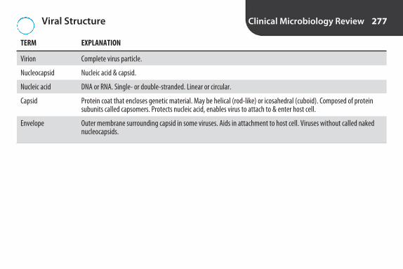
Viral Structure Clinical Microbiology Review 277
TERM EXPLANATION
Virion
Nucleocapsid
Nucleic acid
Capsid
Envelope
Complete virus particle.
Nucleic acid & capsid.
DNA or RNA. Single- or double-stranded. Linear or circular.
Protein coat that encloses genetic material. May be helical (rod-like) or icosahedral (cuboid). Composed of proteinsubunits called capsomers. Protects nucleic acid, enables virus to attach to & enter host cell.
Outer membrane surrounding capsid in some viruses. Aids in attachment to host cell. Viruses without called nakednucleocapsids.
2956_Ch03_135-286 29/01/14 12:18 PM Page 277

Viral Replication Clinical Microbiology Review 278
TERM EXPLANATION
Adsorption
Penetration
Uncoating
Eclipse/synthesis
Maturation/release
Attachment of virus to host cell receptor.
Virus enters host cell by direct penetration, endocytosis (entering in a vacuole), or fusion with cell membrane.
Loss of capsid. Genome enters cytoplasm (most RNA viruses) or nucleus (most DNA viruses).
Eclipse: several hr during which virions can’t be detected. Synthesis: mRNA is produced. Directs synthesis of viralparticles.
Genetic material assembled into protein coat. Virions migrate to cytoplasmic membrane. Released by budding off,leaking out, or lysing host cell with enzymes.
2956_Ch03_135-286 29/01/14 12:18 PM Page 278

Human DNA Viruses Clinical Microbiology Review 279
COMMON FAMILY NAME REPRESENTATIVE VIRUSES INFECTION(S)
Adenoviruses
Hepadnaviruses
Herpes viruses
Papillomaviruses
Parvoviruses
Poxviruses
Respiratory, urinary tract, GI, & eye infections
Hepatitis B
Oral, genital, neonatal, & ocular herpes, HSV encephalitisChicken pox (varicella), shingles (zoster)Infectious mononucleosisInfections in newborns & immunocompromisedRoseola, Kaposi sarcoma
Warts, including genital warts that are linked to cervical cancer
Fifth disease (erythema infectiosum)
Smallpox
Adenoviruses serotypes 1–489
Hepatitis B virus (HBV)
Herpes simplex viruses (HSV-1, HSV-2)Varicella-zoster virus (VZV)Epstein-Barr virus (EBV)Cytomegalovirus (CMV)Human herpesviruses 6–8
Human papilloma virus (HPV)
Parvovirus B–19
Variola
2956_Ch03_135-286 29/01/14 12:18 PM Page 279

Human RNA Viruses Clinical Microbiology Review 280
COMMON FAMILY NAME REPRESENTATIVE VIRUSES INFECTION(S)
Arenaviruses
Astroviruses
Bunyaviruses
Caliciviruses
Coronaviruses
Filoviruses
Flaviviruses
Orthomyxoviruses
Aseptic meningitis or meningoencephalitisLassa fever
Gastroenteritis in children
Encephalitis, hepatitis
Hantavirus pulmonary syndrome (HPS)
Most common cause of infectious gastroenteritis in U.S.
Severe acute respiratory syndrome (SARS), cold-like infections, pediatric diarrhea
Hemorrhagic fever
Yellow fever, St. Louis encephalitis, dengue fever, WestNile virus infection
Hepatitis C
Influenza
Lymphocyte choriomeningitis virus (LCM)Lassa fever virus
Astrovirus
Arboviruses (California encephalitis, LaCrosse virus)
Hantaviruses (Sin Nombre virus)
Noroviruses
Coronavirus
Marburg & Ebola viruses
Arboviruses (yellow fever virus, St. Louis encephalitis virus, dengue virus, West Nile virus)
Hepatitis C virus (HCV)
Influenza A, B, & C
continued...
2956_Ch03_135-286 29/01/14 12:18 PM Page 280

Human RNA Viruses continued Clinical Microbiology Review 281
COMMON FAMILY NAME REPRESENTATIVE VIRUSES INFECTION(S)
Paramyxoviruses
Picornaviruses
Reoviruses
Retroviruses
Rhabdoviruses
Togaviruses
RTI = respiratory tract infection.
Measles (rubeola)MumpsRTI in childrenRTI in infants, elderly, immunocompromisedRTI
Polio, hand-foot-mouth disease, aseptic meningitis,others
Hepatitis ACommon cold
Most common cause of gastroenteritis in infants & children
AIDS
T-cell leukemia & lymphoma, tropical spastic paraparesis
Rabies
Rubella (German measles)Eastern, Western, & Venezuelan encephalitis
Measles virusMumps virusParainfluenza virusRespiratory syncytial virus (RSV)Human metapneumovirus (HMPV)
Enteroviruses (polioviruses, coxsackieviruses A& B, echoviruses, enteroviruses)
Hepatitis A virus (HAV)Rhinovirus
Rotavirus
Human immunodeficiency viruses (HIV-1, HIV-2)
Human T-lymphotropic viruses (HTLV-1, HTLV-2)
Rabies virus
Rubella virus Eastern, Western, & Venezuelan equine
encephalitis viruses
2956_Ch03_135-286 29/01/14 12:18 PM Page 281

Common Viruses by Site Clinical Microbiology Review 282
SITE OF INFECTION SPECIMENS COMMON VIRUSES
Central nervous system
Eye
Genital tract
GI tract
Respiratory tract
Skin
Urinary tract
Enteroviruses, HSV, arboviruses
HSV, adenoviruses
HSV, HPV
Adults: noroviruses, adenoviruses, enteroviruses.Infants/children: rotavirus, adenoviruses
Influenza A & B, parainfluenza virus, adenoviruses, RSV,HMPV, rhinovirus, enteroviruses
HSV, VZV, measles, rubella, enterovirus, parvovirus B19
Adenovirus, HSV, CMV
CSF, throat swab, stool, brain tissue, blood
Conjunctival swab, corneal scraping
Genital swab, vesicle swab or fluid, lesionbiopsy
Stool, rectal swab
Nasal aspirate, throat swab, nasopharyngealswab, bronchoalveolar lavage, lung biopsy
Vesicle fluid or scrapings
Urine
2956_Ch03_135-286 29/01/14 12:18 PM Page 282
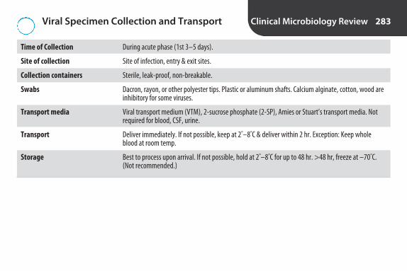
Viral Specimen Collection and Transport Clinical Microbiology Review 283
Time of Collection
Site of collection
Collection containers
Swabs
Transport media
Transport
Storage
During acute phase (1st 3–5 days).
Site of infection, entry & exit sites.
Sterile, leak-proof, non-breakable.
Dacron, rayon, or other polyester tips. Plastic or aluminum shafts. Calcium alginate, cotton, wood areinhibitory for some viruses.
Viral transport medium (VTM), 2-sucrose phosphate (2-SP), Amies or Stuart’s transport media. Not required for blood, CSF, urine.
Deliver immediately. If not possible, keep at 2°–8°C & deliver within 2 hr. Exception: Keep wholeblood at room temp.
Best to process upon arrival. If not possible, hold at 2°–8°C for up to 48 hr. >48 hr, freeze at –70°C.(Not recommended.)
2956_Ch03_135-286 29/01/14 12:18 PM Page 283

Methods for Diagnosis of Viral Infections Clinical Microbiology Review 284
Cytology/histology
Electron microscopy
Direct fluorescent antibody stain
Antigen detection
Cell culture
Shell vial culture
Molecular methods
Serology
Microscopic examination of specimen for viral cytopathic effect (CPE).
Rarely used. Labor intensive, expensive.
Fluorescent-labeled antibody added to patient cells fixed to slide. If viral antigen present, antibody binds. Fluorescence seen with fluorescent microscope.
Solid-phase & membrane ELISAs.
Different viruses grow in different cell lines. Growth may take 1–28 days. Examine microscopi-cally for cytopathic effects (CPE): cell rounding, clumping, vacuolation, granulation, giant multi-nucleate cells, cell fusion, syncytial formation, cell lysis, plaques (groups of killed cells), inclusionbodies. Not all viruses produce CPE. Immunofluorescent stains may be used for confirmation.
Rapid modification of conventional cell culture. Detection in 1–2 days. Specimen centrifugedonto monolayer of cells growing on coverslip. Coverslips stained with viral-specific immunofluo-rescent conjugate. Used primarily for viruses that are slow to produce CPE.
PCR, real-time PCR, branched DNA, nucleic acid hybridization. Faster & more sensitive than cellculture. Can detect viruses that can’t be cultured, multiple viruses simultaneously.
Detects antibodies in serum. Useful in evaluating immune status or diagnosing viral infectionswhere culture is difficult or impossible. Presence of antibodies isn’t always indicative of currentinfection.
2956_Ch03_135-286 29/01/14 12:18 PM Page 284

Cell Cultures Clinical Microbiology Review 285
CELL LINE DERIVATION EXAMPLES FOR ISOLATION OF
Primary Cells from mammalian tissue Primary monkey kidney (PMK) cells Most viruses
Finite (diploid) Cultures of connective tissue cells Human neonatal lung (HNL) CMV (only type of cell culture that can be used)
Continuous (immortal, Malignant or transformed cells Hep2, A549, Vero HSV, RSV, enteroviruses, heteroploid) adenoviruses
No single cell type grows all viruses. Several types should be inoculated.
2956_Ch03_135-286 29/01/14 12:18 PM Page 285

Comparison of Microorganisms Clinical Microbiology Review 286
BACTERIA CHLAMYDIA MYCOPLASMA RICKETTSIAE VIRUSES
Size
DNA & RNA?
Ribosomes?
Reproduce by binary fission?
Cell wall?
Growth on nonliving medium?
Obligate intracellular parasite?
Gram stain?
Susceptible to antibiotics?
0.4–2 μm
Both
Yes
Yes
Yes
Yes
Most are not
Yes
Yes
0.3–0.5 μm
Both
Yes
Yes
Yes
No
Yes
No
Yes
300 nm
Both
Yes
Yes
No
Yes
No
No
Yes
0.3–0.5 μm
Both
Yes
Yes
Yes
No
Yes
No
Yes
25–300 nm
Either
No
No
No
No
Yes
No
No
2956_Ch03_135-286 29/01/14 12:18 PM Page 286

4S E C T I O N
Hematology Review
2956_Ch04_287-366 30/01/14 4:25 PM Page 287

Blood Cells Hematology Review 288
CELL FUNCTION SITE OF PRODUCTION
Erythrocytes O2 transport Bone marrow
Granulocytes Defense against bacterial infection Bone marrow
Lymphocytes Cellular & humoral immunity Lymphoid tissue
Platelets Coagulation Bone marrow
2956_Ch04_287-366 30/01/14 4:25 PM Page 288

Comparison of Conventional and SI Units for Adult Reference Ranges
Hematology Review 289
CONVENTIONAL UNITS SI UNITS
WBC 4.5–11.5 × 10³/�L 4.5–11.5 × 10⁹/L
RBC Male: 4.6–6 × 10⁶/�L Male: 4.6–6 × 10¹²/L Female: 4–5.4 × 10⁶/�L Female: 4–5.4 × 10¹²/L
HGB Male: 14–18 g/dL Male: 140–180 g/LFemale: 12–15 g/dL Female: 120–150 g/L
HCT Male: 40%–54% Male: 0.40–0.54 L/LFemale: 35%–49% Female: 0.35–0.49 L/L
MCV 80–100 fL 80–100 fL
MCH 27–31 pg 27–31 pg
MCHC 32%–36% 32–36 g/dL
PLT 150–450 × 103/�L 150–450 × 109/L
SI = Syst‘ me International d’Unit”s (international system of units).
2956_Ch04_287-366 30/01/14 4:25 PM Page 289

Reference Ranges for Red Cell Parameters Hematology Review 290
BIRTH 1–2 MO 1–3 YR 8–13 YR ADULT COMMENTS
RBCs (× 1012/L) 4.10–6.10 3.4–5 3.4–5.2 4–5.4 M: 4.6–6F: 4–5.4
HGB (g/dL) 16.5–21.5 10.6–16.4 9.6–15.6 12–15 M: 14–18 Preterm infants: About 1 g F: 12–15 lower than full-term
HCT (%) 48–68 32–50 38–48 35–49 M: 40–54F: 35–49
MCV (fL) 95–125 83–107 78–94 80–94 80–100 Macrocytes 1st 5 days. MCV higher in preterm infants
RDW (%) 14.2–19.9 11.4–14.5 11.5–14.5 11.5–14.5
Retic (%) 1.5–5.8 0.8–2.8 0.5–1.5 0.5–1.5 0.5–1.5 Newborns: ↑ polychromasia
NRBCs (/100 WBCs) 2–24 0 0 0 0 Preterm infants: Up to 25 for >1 wk
Note trends and comparisons to adult values.
2956_Ch04_287-366 30/01/14 4:25 PM Page 290

Reference Ranges for Leukocytes andPlatelets
Hematology Review 291
BIRTH 1–2 MO 1–3 YR 8–13 YR ADULT COMMENTS
WBCs (× 109/L) 9–37 6–18 5.5–17.5 4.5–13.5 4.5–11.5
Segs (%) 37–67 20–40 22–46 23–53 50–70
Bands (%) 3–11 0–5 0–5 0–5 0–5 Newborns: Occasional metas &myelos. More immature grans inpreterm infants.
Lymphs (%) 18–38 42–72 37–73 23–53 18–42 Newborns: A few benign immature B cells may be seen(“baby” or “kiddie” lymphs).
PLT (× 109/L) 150–450 150–450 150–450 150–450 150–450 Newborns: Variation in size &shape.
Note trends and comparisons to adult values.
2956_Ch04_287-366 30/01/14 4:25 PM Page 291

Hematopoietic Cell Differentiation Hematology Review 292
Note that this algorithm does not show all differentiation steps.
Pluripotential Hematopoietic Stem Cell(HSC)
Multipotential Progenitor Cell(MPP)
Common Myeloid Progenitor(CMP)
Granulocyte Erythrocyte Monocyte Megakaryocyte T-lymph NK cell B-lymph
Common Lymphoid Progenitor(CLP)
2956_Ch04_287-366 30/01/14 4:25 PM Page 292

Erythropoiesis Hematology Review 293
PERIOD SITE(S) COMMENTS
1–2 mo of gestation Yolk sac & aorta-gonads- Primitive erythroblasts. Embryonic hemoglobin (Gower I, Gower II, mesonephros (AGM) region Portland).
3–6 mo of gestation Liver, spleen Liver is primary site.
7 mo of gestation—age 4 yr Bone marrow All marrow is active.
Adult Bone marrow Only active sites are pelvis, vertebrae, ribs, sternum, skull. Shafts of longbones filled with fat. Fatty marrow may be reactivated to compensate for anemia. Liver & spleen may be reactivated (extramedullaryhematopoiesis) if bone marrow fails to keep up with demand.
2956_Ch04_287-366 30/01/14 4:25 PM Page 293

Changes During Cell Maturation Hematology Review 294
CHARACTERISTIC CHANGE(S) WITH MATURATION
Size Becomes smaller.
N:C ratio Becomes smaller.
Cytoplasm Less basophilic due to loss of RNA. Granulocytes produce granules. Erythrocytes become pink due to Hgb production.
Nucleus Becomes smaller. Nuclear chromatin condenses. Nucleoli disappear. In granulocytic series, nucleus indents,then segments. In erythrocytic series, nucleus is extruded.
N:C ratio = nucleus to cytoplasm ratio
2956_Ch04_287-366 30/01/14 4:26 PM Page 294

Erythrocytic Developmental Series Hematology Review 295
RUBRIBLAST TERMINOLOGY NORMOBLAST TERMINOLOGY KEY CHARACTERISTICS
Rubriblast
Prorubricyte
Rubricyte
Metarubricyte
Reticulocyte
Mature erythrocyte
Pronormoblast
Basophilic normoblast
Polychromatophilic normoblast
Orthochromic normoblast
Polychromatophilic erythrocyte
Mature erythrocyte
14–24 �m. N:C ratio 8:1. Royal blue cytoplasm. Fine chromatin.1–2 nucleoli. Normally confined to bone marrow.
12–17 �m. N:C ratio 6:1. Chromatin is coarser with slightly visible parachromatin. Nucleoli usually not visible. Normally confined to bone marrow.
10–15 �m. N:C ratio 4:1. Cytoplasm is polychromatophilic due to hemoglobin production. Chromatin is clumped with distinctareas of parachromatin. Last stage to divide. Normally confinedto bone marrow.
8–12 �m. N:C ratio 1:2. Nucleus is pyknotic. Last nucleated stage.Normally confined to bone marrow.
7–10 �m. No nucleus. Cytoplasm is diffusely basophilic (bluishtinge). Reticulum seen with supravital stain. 0.5%–1.5% of RBCs in adult peripheral blood.
7–8 �m. Biconcave disk. Reddish-pink cytoplasm with area ofcentral pallor 1/3 diameter of cell.
2956_Ch04_287-366 30/01/14 4:26 PM Page 295
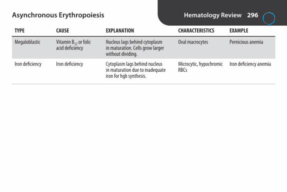
Asynchronous Erythropoiesis Hematology Review 296
TYPE CAUSE EXPLANATION CHARACTERISTICS EXAMPLE
Megaloblastic Vitamin B12 or folic Nucleus lags behind cytoplasm Oval macrocytes Pernicious anemiaacid deficiency in maturation. Cells grow larger
without dividing.
Iron deficiency Iron deficiency Cytoplasm lags behind nucleus Microcytic, hypochromic Iron deficiency anemiain maturation due to inadequate RBCsiron for hgb synthesis.
2956_Ch04_287-366 30/01/14 4:26 PM Page 296

Hemoglobin Hematology Review 297
HEMOGLOBIN MOLECULAR STRUCTURE ADULT REFERENCE VALUE NEWBORN REFERENCE VALUE
A 2 α , 2 β chains >95% 20%
A2 2 α , 2 δ chains 1.5%–3.7% <1%
F 2 α , 2 γ chains <2% 50%–85%
S Valine substituted for glutamic 0 0acid in 6th position of β chain
C Lysine substituted for glutamic 0 0acid in 6th position of β chain
2956_Ch04_287-366 30/01/14 4:26 PM Page 297

Hemoglobin Electrophoresis Hematology Review 298
Cathode (–)
Origin “Crawl” “Slow” “Fast” “Accelerated”
Cellulose Acetate pH 8.6Anode (+)
A2C
S F A
Cathode (–)
Origin
Citrate Agar pH 6.2Anode (+)
AA2
SF C
2956_Ch04_287-366 30/01/14 4:26 PM Page 298

Hemoglobin Derivatives Hematology Review 299
NORMAL % OF HEMOGLOBIN CAUSE EFFECT TOTAL HGB OTHER
Methemoglobin Iron oxidized to ferric (Fe3+) Can’t bind O2. Cyanosis, ≤ 1% Heinz bodies. Treat with state. Usually acquired from possibly death. methylene blue.exposure to oxidants. Rarely inherited.
Sulfhemoglobin Sulfur bound to heme. O2 affinity 1/100th 0 Can’t be converted back to Acquired from exposure to normal. Cyanosis. normal hemoglobin. Not drugs & chemicals. detected in cyanmethemo-
globin method.
Carboxyhemoglobin Carbon monoxide bound ↓ O2 to tissues. Can <1 % Affinity of hgb for CO is to heme. be fatal. 200× greater than for O2.
Skin turns cherry red.
Hemoglobin derivatives are quantitated by differential spectrophotometry.
2956_Ch04_287-366 30/01/14 4:26 PM Page 299

RBC Morphology Hematology Review 300
ABNORMALITY DESCRIPTION SIGNIFICANCE
SizeAnisocytosis
Macrocytes
Microcytes
ShapePoikilocytosis
Elliptocytes/ovalocytes
Crenated RBCs
Burr cells (echinocytes)
Acanthocytes (spur cells)
Variation in size.
RBCs >9 µm.
RBCs <6 µm.
Variation in shape.
Oval or pencil/cigar shaped.
Round cell with knobby, uniform projections.
Round cell with evenly spaced blunt orpointed projections.
Small, dense cells with irregularlyspaced projections of varying length.
Seen in many anemias.
Megaloblastic anemias, liver disease, reticulocytosis. Normalin newborns.
Iron deficiency anemia, thalassemia, anemia of chronic infections.
Seen in many anemias.
Membrane defect. Hereditary ovalocytosis, various anemias.
Osmotic imbalance. If seen in most cells in thin part of smear,don’t report. Probably artifact due to excess anticoagulant orslow drying.
Membrane defect. Uremia, pyruvate kinase deficiency. Maybe drying artifact. A few can be present in healthy individuals.
Membrane defect. Severe liver disease, abetalipoproteinemia.
continued...
2956_Ch04_287-366 30/01/14 4:26 PM Page 300

RBC Morphology continued Hematology Review 301
ABNORMALITY DESCRIPTION SIGNIFICANCE
Schistocytes
Sickle cells (drepanocytes)
Hemoglobin C crystals
Hemoglobin SC crystals
Teardrops (dacryocytes)
StainingHypochromia
Anisochromia
Polychromasia
RBC fragments
Crescent, S or C shaped, boat shaped,oat shaped
Blunt, 6-sided, dark-staining projection.“Bar of gold.” “Washington monument”
Glove-like intracellular crystals
Teardrop shaped
Central pallor >1/3 cell diameter
Mixture of normochromic &hypochromic RBCs
Bluish-gray color
RBCs split by fibrin strands. Microangeopathic hemolyticanemias (DIC, TTP, HUS), prosthetic heart valves.
Sickle cell anemia.
Hemoglobin C disease.
Hemoglobin SC disease.
Myelofibrosis, thalassemia & other anemias.
Iron deficiency anemia, thalassemia.
Dimorphic anemia, post-transfusion.
Young RBCs. Retics with supravital stain. Sign of active erythropoiesis. 1%–2% in normal adult. ↑ with acute blood loss, hemolytic anemia, following treatment for irondeficiency or pernicious anemia.
continued...
2956_Ch04_287-366 30/01/14 4:26 PM Page 301
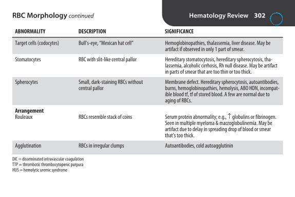
RBC Morphology continued Hematology Review 302
ABNORMALITY DESCRIPTION SIGNIFICANCE
Target cells (codocytes)
Stomatocytes
Spherocytes
ArrangementRouleaux
Agglutination
Bull’s-eye, “Mexican hat cell”
RBC with slit-like central pallor
Small, dark-staining RBCs withoutcentral pallor
RBCs resemble stack of coins
RBCs in irregular clumps
Hemoglobinopathies, thalassemia, liver disease. May be artifact if observed in only 1 part of smear.
Hereditary stomatocytosis, hereditary spherocytosis, tha-lassemia, alcoholic cirrhosis, Rh null disease. May be artifactin parts of smear that are too thin or too thick.
Membrane defect. Hereditary spherocytosis, autoantibodies,burns, hemoglobinopathies, hemolysis, ABO HDN, incompat-ible blood tf, tf of stored blood. A few are normal due toaging of RBCs.
Serum protein abnormality; e.g., ↑ globulins or fibrinogen.Seen in multiple myeloma & macroglobulinemia. May be artifact due to delay in spreading drop of blood or smearthat’s too thick.
Autoantibodies, cold autoagglutinin
DIC = disseminated intravascular coagulationTTP = thrombotic thrombocytopenic purpuraHUS = hemolytic uremic syndrome
2956_Ch04_287-366 30/01/14 4:26 PM Page 302

RBC Inclusions Hematology Review 303
INCLUSION STAIN DESCRIPTION EXPLANATION SIGNIFICANCE CONDITIONS
Basophilic stippling
Howell-Jolly bodies
Cabot rings
Pappenheimer bodies
Wright’s & newmethylene blue
Wright’s & newmethylene blue
Wright’s
Wright’s (sideroticgranules withPrussian blue stain)
Multiple, irregularpurple inclusionsevenly distributedin cell
Round, purple, 1–2 µm in diameter. Usuallyonly 1 per cell
Reddish purplerings or figure-8s
Small purplish bluegranules. Vary insize, shape, #. Usually in clustersat periphery
Aggregation of RNA(ribosomes)
Nuclear remnants(DNA)
May be part of mitoticspindle, remnant ofmicrotubules, or fragment of nuclearmembrane
Iron particles
Coarse: exposureto lead. Fine:young RBC
Usually pitted byspleen. Seen withaccelerated or abnormal erythropoiesis
Rapid blood regen-eration, abnormalerythropoiesis
Faulty iron utilization
Exposure to lead, accelerated or abnor-mal hemoglobin synthesis, thalassemia
Postsplenectomy, thalassemia, hemolytic& megaloblastic anemias, sickle cellanemia
Megaloblastic anemia,thalassemia, postsplenectomy
Sideroblastic anemias,postsplenectomy, thalassemia, sickle cell anemia, hemochromatosis
continued...
2956_Ch04_287-366 30/01/14 4:26 PM Page 303

RBC Inclusions continued Hematology Review 304
Siderotic granules
Reticulocytes
Heinz bodies
INCLUSION STAIN DESCRIPTION EXPLANATION SIGNIFICANCE CONDITIONS
Prussian blue
New methyleneblue (polychromasiaon Wright’s stain)
Supravital stain,e.g., crystal violet,brilliant cresyl blue,methylene blue
Blue granules ofvarying size &shape
Blue-staining network
Round blue inclusions, varyingsizes, close to cellmembrane. Maybe >1
Aggregates of ironparticles
Residual RNA (ribosomes)
Precipitated, oxidized, denaturedhemoglobin
Faulty iron utilization in hgb synthesis
>2% = ↑erythropoiesis<0.1% = ↓erythropoiesis
Normal duringaging but pitted byspleen
Sideroblastic anemias,postsplenectomy, thalassemia, sickle cell anemia, hemochromatosis
Hemolytic anemia,blood loss, followingtreatment for iron defi-ciency or megaloblasticanemia
G6PD deficiencies, unstable hemoglobins,chemical injury toRBCs, drug-inducedhemolytic anemia
2956_Ch04_287-366 30/01/14 4:26 PM Page 304

Staining of RBC Inclusions Hematology Review 305
INCLUSION WRIGHT’S STAIN NEW METHYLENE BLUE STAIN PRUSSIAN BLUE STAIN
Reticulum Cell appears polychromatophilic Yes No
Howell-Jolly bodies Yes Yes No
Pappenheimer bodies Yes Yes Yes
Siderotic granules Yes, but called Pappenheimer bodies Yes Yes
Heinz bodies No Yes No
2956_Ch04_287-366 30/01/14 4:26 PM Page 305
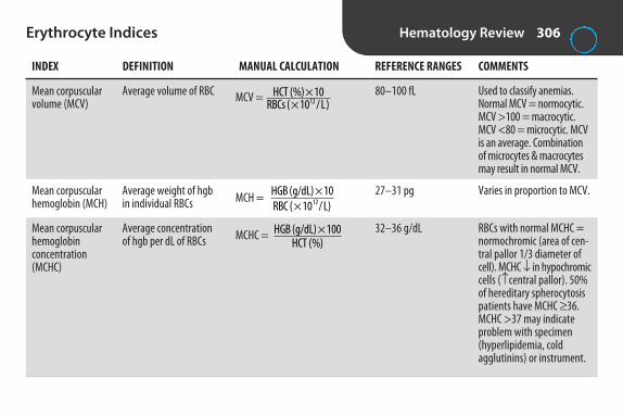
Erythrocyte Indices Hematology Review 306
INDEX DEFINITION MANUAL CALCULATION REFERENCE RANGES COMMENTS
Mean corpuscular volume (MCV)
Mean corpuscular hemoglobin (MCH)
Mean corpuscular hemoglobin concentration (MCHC)
Average volume of RBC
Average weight of hgbin individual RBCs
Average concentrationof hgb per dL of RBCs
MCV =
MCH =
MCHC =
HCT (%) 10RBCs ( 10 L)12
×× /
HGB (g/dL) 10RBC ( 10 L)12
×× /
HGB (g/dL) 100HCT (%)
×
80–100 fL
27–31 pg
32–36 g/dL
Used to classify anemias.Normal MCV = normocytic.MCV >100 = macrocytic.MCV <80 = microcytic. MCVis an average. Combination of microcytes & macrocytesmay result in normal MCV.
Varies in proportion to MCV.
RBCs with normal MCHC = normochromic (area of cen-tral pallor 1/3 diameter ofcell). MCHC ↓ in hypochromiccells (↑central pallor). 50%of hereditary spherocytosispatients have MCHC ≥ 36.MCHC >37 may indicateproblem with specimen (hyperlipidemia, cold agglutinins) or instrument.
2956_Ch04_287-366 30/01/14 4:26 PM Page 306

Hemoglobinopathy Versus Thalassemia Hematology Review 307
HEMOGLOBINOPATHY THALASSEMIA
Abnormality Qualitative abnormality. Abnormality in amino acid Quantitative abnormality. Amino acid sequence of globin sequence of globin chain, not in amount of globin chains is normal, but underproduction of 1 or more globin produced. chains.
Examples Sickle cell anemia & trait, hemoglobin C disease & trait. β -thalassemia major & minor.
Note: Some hematologists refer to all qualitative & quantitative hemoglobin abnormalities as hemoglobinopathies.
2956_Ch04_287-366 30/01/14 4:26 PM Page 307

Normocytic Anemias Hematology Review 308
HEMOGLOBIN ANEMIA ETIOLOGY BLOOD SMEAR ELECTROPHORESIS OTHER
Sickle cell anemia (SS)
Sickle cell trait (AS)
Hemoglobin C disease (CC)
Inheritance of sickle cellgene from both parents.Valine substituted forglutamic acid in 6th position of β chain.
Inheritance of sickle cellgene from 1 parent.
Inheritance of gene forHgb C from both parents.Lysine substituted forglutamic acid in 6th position of β chain.
Aniso, poik, sickle cells, target cells, nRBCs, HJ bodies, basophilic stippling, siderotic gran-ules, polychromasia.
Occasional target cells. Nosickle cells unless hypoxic.
Many target cells, foldedcells, occasional Hgb C crystals.
≥ 80% S, 1%–20%F, normal A2, no A
50%–65% A,35%–45% S, normal F, N toslightly ↑ A2
>90 C, <7% F, no A
Hgb S polymerizes under ↓ O2& ↓ blood pH. Disease not evident in newborn because of ↑Hgb F. Pos solubility test.Retics 10%–20%. May have ↑ WBC with shift to left & ↑ PLT. Moderate to severeanemia.
No anemia. Pos solubility test.Important to Dx for geneticcounseling.
Mild to moderate anemia.
continued...
2956_Ch04_287-366 30/01/14 4:26 PM Page 308

Normocytic Anemias continued Hematology Review 309
Hemoglobin C trait (AC)
SC disease (SC)
Hereditary spherocytosis
Autoimmune hemolytic anemia
HEMOGLOBIN ANEMIA ETIOLOGY BLOOD SMEAR ELECTROPHORESIS OTHER
Inheritance of gene forHgb C from 1 parent.
Inheritance of 1 sickle cellgene & 1 Hgb C gene.
Defect of cell membrane.
Autoantibodies.
Many target cells.
Many target cells. Folded &boat-shaped cells, occa-sional SC crystals (finger-likeprojections, “WashingtonMonument” crystals).
Spherocytes, polychromasia.
Polychromasia, sphero-cytes, nRBCs.
60%–70% A,30%–40% C
>S than C, normalto 7% F, no A
Normal
Normal
Pos solubility test. Mild tomoderate anemia.
MCHC usually >36 g/dL. ↑ retics, ↑ osmotic fragility.
↑ retics, ↑ indirect bili, ↓ haptoglobin, pos DAT.
N = normal, nRBCs = nucleated red blood cells, DAT = direct antiglobulin test.
2956_Ch04_287-366 30/01/14 4:26 PM Page 309

Macrocytic Anemias Hematology Review 310
HEMOGLOBIN ANEMIA ETIOLOGY BLOOD SMEAR ELECTROPHORESIS OTHER
MegaloblasticFolate deficiency
Vitamin B12deficiency
Nonmegaloblastic
Nutritional deficiency, ↑ cellreplication (e.g., hemolytic anemias, myeloproliferative dis-eases, pregnancy), malabsorp-tion, drug inhibition. Deficiencyimpairs DNA synthesis.
Nutritional deficiency, malab-sorption, impaired utilization,parasites. Deficiency impairsDNA synthesis.
Alcoholism, liver disease, ↑ erythropoiesis.
Oval macrocytes,Howell-Jolly bodies,hypersegmentation,aniso, poik
Same
Round macrocytes, nohypersegmentation
Normal
Normal
Normal
Pancytopenia, ↑ LD
Same. Pernicious anemia is most common type. Autoimmune disease. Gastric atrophy leads to ↓ intrinsic factor neededfor B12 absorption
WBC & PLT normal
2956_Ch04_287-366 30/01/14 4:26 PM Page 310

Microcytic, Hypochromic Anemias Hematology Review 311
ANEMIA ETIOLOGY BLOOD SMEAR OTHER
Iron deficiency anemia (IDA)
Sideroblastic anemia
β –thalassemia major
β –thalassemia minor
Anemia of chronic inflammation*
Insufficient iron for hgb synthesis.
Enzymatic defect in heme synthesis.
↓ β -chain production.
↓ β -chain production.
Acute phase reactants (e.g.,hepcidin) affect iron absorp-tion & release. Iron in bonemarrow macrophages is notreleased to developing RBCs.
Aniso, poik, hypochromic microcytes
Dual population of RBCs (normocytic & microcytic), Pappenheimer bodies, basophilic stippling
Marked aniso & poik,hypochromic microcytes,target cells, ovalocytes,nRBCs, basophilic stippling
Aniso, poik, hypochromic microcytes, target cells, basophilic stippling
60%–70% of cases havenormocytic normochromicRBCs; 30%–40% microcytichypochromic
Most common anemia.
RBC indices usually normal. Ringed sideroblasts in marrow.
Homozygous. Little or no Hgb A, 95%–98% F, 2%–5% A2.Severe anemia. MCV <67 fL.
Heterozygous. >90%–95% Hgb A,3.5%–7% A2, 2%–5% F.Mild anemia.
Associated with chronic infections & inflammation, malignancies, autoim-mune diseases. 2nd most common anemia after IDA. Most common anemia in hospitalized pts.
*Formerly known as anemia of chronic disease. More often normocytic normochromic but included here because must be considered in differential Dx of microcytic anemia.
2956_Ch04_287-366 30/01/14 4:26 PM Page 311

Differentiation of Microcytic Hypochromic Anemias
Hematology Review 312
ANEMIA RBCS RDW SERUM IRON TIBC SERUM FERRITIN HGB A2
Iron deficiency anemia ↓ ↑ ↓ ↑ ↓ N
Sideroblastic anemia ↓ ↑ ↑ N ↑ N
β -thalassemia minor ↑ N N N N ↑
Anemia of chronic inflammation ↓ N ↓ ↓ ↑ N
TIBC = total iron binding capacity.
2956_Ch04_287-366 30/01/14 4:26 PM Page 312
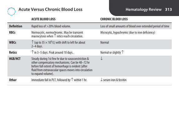
Acute Versus Chronic Blood Loss Hematology Review 313
ACUTE BLOOD LOSS CHRONIC BLOOD LOSS
Definition Rapid loss of >20% blood volume. Loss of small amounts of blood over extended period of time
RBCs Normocytic, normochromic. May be transient Microcytic, hypochromic (due to iron deficiency)macrocytosis when ↑ retics reach circulation.
WBCs ↑ (up to 35 × 109/L) with shift to left for about Normal2–4 days.
Retics ↑ in 3–5 days. Peak around 10 days. Normal or slightly ↑
HGB/HCT Steady during 1st few hr due to vasoconstriction & ↓other compensatory mechanisms. Can be 48–72 hr before full extent of hemorrhage is evident (after fluid from extravascular spaces moves into circulation to expand volume).
Other Immediate fall in PLT, followed by ↑ within 1 hr. ↓ serum iron & ferritin
2956_Ch04_287-366 30/01/14 4:26 PM Page 313

Granulocytic Maturation Hematology Review 314
STAGE KEY CHARACTERISTICS
Myeloblast 15–20 �m. Small amount of dark blue cytoplasm. Usually no granules. Nucleus has delicate chromatin with nucleoli.
Promyelocyte 12–24 �m. Similar to myeloblast but has primary (nonspecific) granules.
Myelocyte 10–18 �m. Secondary (specific) granules (eosinophilic, basophilic, or neutrophilic). Last stage to divide.
Metamyelocyte 10–18 �m. Nucleus begins to indent.
Band 10–16 �m. Nuclear indentation is more than half.
Segmented neutrophil 10–16 �m. 2–5 nuclear lobes connected by thin strands of chromatin.
2956_Ch04_287-366 30/01/14 4:26 PM Page 314
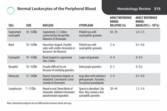
Normal Leukocytes of the Peripheral Blood Hematology Review 315
ADULT REFERENCE ADULT REFERENCE RANGE: RANGE:
CELL SIZE NUCLEUS CYTOPLASM RELATIVE (%) ABSOLUTE (× 109/L)
Segmented neutrophil
Band
Eosinophil
Basophil
Monocyte
Lymphocyte
10–16 �m
10–16 �m
10–16 �m
10–16 �m
12–18 �m
7–15 �m
50–70
2–6
0–4
0–2
2–9
20–44
2.4–7.5
0.1–0.6
0–0.4
0–0.2
0.1–0.9
1.2–3.4
Segmented. 2–5 lobes connected by thread-like filament of chromatin.
Horseshoe shaped. Parallelsides with visible chromatin inbetween. No filament.
Band shaped or segmentedinto 2 lobes.
Usually difficult to see because of overlying granules.
Round, horseshoe shaped, orlobulated. Convoluted. Loosestrands of chromatin.
Round or oval. Dense blocks ofchromatin. Indistinct chromatin/parachromatin separation.
Pinkish tan with neutrophilic granules
Pinkish tan with neutrophilic granules
Large red granules
Dark purple granules
Gray-blue with indistinctpink granules. Vacuoles.Occasional pseudopods
Sparse to abundant. Skyblue. May contain a fewazurophilic granules
Note: Automated analyzers do not differentiate between bands and segs.
2956_Ch04_287-366 30/01/14 4:26 PM Page 315
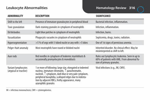
Leukocyte Abnormalities Hematology Review 316
ABNORMALITY DESCRIPTION SIGNIFICANCE
Shift to the left
Toxic granulation
Döhle bodies
Vacuolization
Hypersegmentation
Pelger-Huët anomaly
Auer rods
Variant lymphocytes (atypical or reactive)
Presence of immature granulocytes in peripheral blood
Dark-staining granules in cytoplasm of neutrophils
Light blue patches in cytoplasm of neutrophils
Phagocytic vacuoles in cytoplasm of neutrophils
>5 % of segs with 5-lobed nuclei or any with >5 lobes
Most neutrophils have round or bilobed nuclei
Red needles in cytoplasm of leukemic myeloblasts & occasionally promyelocytes & monoblasts
1 or more of following: large size, elongated or indentednucleus, immature chromatin, ↑ parachromatin, nucleoli, ↑ cytoplasm, dark blue or very pale cytoplasm,peripheral basophilia, scalloped edges due to indenta-tion by adjacent RBCs, frothy appearance, manyazurophilic granules
Bacterial infection, inflammation.
Infection, inflammation.
Infection, burns.
Septicemia, drugs, toxins, radiation.
One of 1st signs of pernicious anemia.
Inherited disorder. No clinical effect. May bemisinterpreted as shift to left.
Rules out lymphocytic leukemia. Seen in up to60% of patients with AML. From abnormal fu-sion of primary granules.
Viral infections (e.g., IM, CMV).
IM = infectious mononucleosis, CMV = cytomegalovirus.
2956_Ch04_287-366 30/01/14 4:26 PM Page 316

Quantitative Abnormalities of Leukocytes Hematology Review 317
ABNORMALITY ASSOCIATIONS
Neutrophilia Bacterial infection, inflammation, hemorrhage, hemolysis, stress
Neutropenia Acute infection, antibodies, drugs, chemicals, radiation
Lymphocytosis IM, CMV, whooping cough, acute infectious lymphocytosis
Monocytosis Convalescence from viral infections, chronic infections, TB, subacute bacterial endocarditis, parasitic infec-tions, rickettsial infections
Eosinophilia Allergies, skin diseases, parasitic infections, CML
Basophilia Chronic myelogenous leukemia, polycythemia vera
2956_Ch04_287-366 30/01/14 4:26 PM Page 317

Hematopoietic Neoplasms* Hematology Review 318
DISORDER EXPLANATION EXAMPLE(S) OTHER
Myeloproliferative neoplasms (MPN) or myeloproliferative disorders (MPD)
Myelodysplastic syndromes (MDS)
Myelodysplastic/myeloproliferative disorders (MDS/MPN)
Premalignant hematopoietic stemcell disorders involving overproduc-tion of 1 or more myeloid (nonlym-phocytic) cell lines. Bone marrow &peripheral blood show ↑ RBCs, gran-ulocytes, &/or platelets, with 1 cellline usually predominate. Normalmaturation & morphology.
Premalignant hematopoietic stemcell disorders involving ineffectivehematopoiesis in 1 or more myeloidcell lines. Hypercellular bone marrow with maturation abnormali-ties (dysplasias). Peripheral bloodcytopenias (↓ counts) & morpho-logic abnormalities.
Premalignant neoplasms with bothmyeloproliferative & myelodysplasticfeatures.
Polycythemia vera,chronic myelogenousleukemia, essentialthrombocythemia, primary myelofibrosis
Refractory anemia, refractory neutrope-nia, refractory thrombocytopenia
Chronic myelomono-cytic leukemia (CMML)
Usually in older adults. Caused bymutations in hematopoietic stemcells. Primarily chronic but can transform into acute leukemia.Splenomegaly, extramedullaryhematopoiesis common.
More common in elderly. May be dueto exposure to chemicals, radiation,chemotherapy, viral infections. Cantransform into acute leukemia.
continued...
2956_Ch04_287-366 30/01/14 4:26 PM Page 318

Hematopoietic Neoplasms* continued Hematology Review 319
DISORDER EXPLANATION EXAMPLE(S) OTHER
Leukemia
Lymphoma
Malignant neoplasms involving unregulated proliferation ofhematopoietic stem cells. Abnormalcells in bone marrow & peripheralblood.
Malignant neoplasm of lymphoidcells in lymphatic tissues or lymphnodes.
Acute lymphoblasticleukemia (ALL),chronic lymphocyticleukemia (CLL)
Hodgkin lymphoma,non-Hodgkin lymphoma
Classified as acute or chronic, & lymphoid or myelogenous. See“Common Leukemias” card for more detail.
Solid tumors. Classified as B or T cell.Can spread to bone marrow, thenmalignant cells can be present in peripheral blood (leukemic phase).
*Neoplasm = new growth; unregulated growth of a single transformed cell; may be benign or malignant. A benign neoplasm can progress to a malignant neoplasm.
2956_Ch04_287-366 30/01/14 4:26 PM Page 319
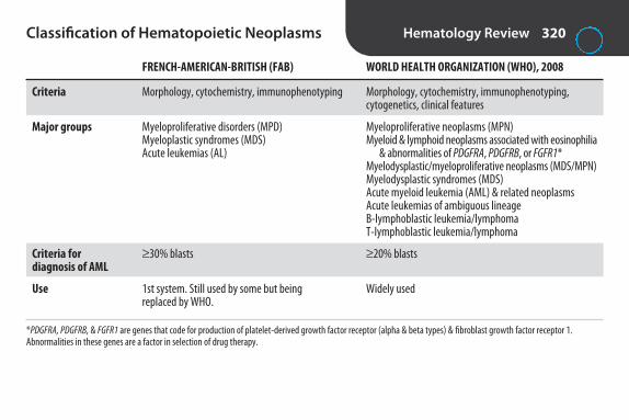
Classification of Hematopoietic Neoplasms Hematology Review 320
FRENCH-AMERICAN-BRITISH (FAB) WORLD HEALTH ORGANIZATION (WHO), 2008
Criteria Morphology, cytochemistry, immunophenotyping Morphology, cytochemistry, immunophenotyping, cytogenetics, clinical features
Major groups Myeloproliferative disorders (MPD) Myeloproliferative neoplasms (MPN)Myeloplastic syndromes (MDS) Myeloid & lymphoid neoplasms associated with eosinophilia Acute leukemias (AL) & abnormalities of PDGFRA, PDGFRB, or FGFR1*
Myelodysplastic/myeloproliferative neoplasms (MDS/MPN)Myelodysplastic syndromes (MDS)Acute myeloid leukemia (AML) & related neoplasmsAcute leukemias of ambiguous lineageB-lymphoblastic leukemia/lymphomaT-lymphoblastic leukemia/lymphoma
Criteria for ≥ 30% blasts ≥ 20% blastsdiagnosis of AML
Use 1st system. Still used by some but being Widely usedreplaced by WHO.
*PDGFRA, PDGFRB, & FGFR1 are genes that code for production of platelet-derived growth factor receptor (alpha & beta types) & fibroblast growth factor receptor 1. Abnormalities in these genes are a factor in selection of drug therapy.
2956_Ch04_287-366 30/01/14 4:26 PM Page 320

Acute Versus Chronic Leukemia Hematology Review 321
ACUTE CHRONIC
Age All ages, with peaks in 1st decade & after 50 yr Adults
Onset Sudden Insidious
Median survival time, untreated Weeks to months Months to years
WBC ↑, N, or ↓ ↑ (may be >50,000)
Differential Blasts usually present More mature cells
Anemia Mild to severe Mild
Platelets Mild to severe ↓ Usually N
Other Usually lymphoid in children, myeloid in adults Myeloid mostly in young to middle-aged, lym-phoid in older adults. Most go into blast crisis
Methods used to diagnose Peripheral blood smear, bone marrow Same but less use of cytochemical stains examination, cytochemical stains, immunophenotyping, cytogenetics, molecular genetics
2956_Ch04_287-366 30/01/14 4:26 PM Page 321

Common Leukemias Hematology Review 322
TYPE OTHER NAMES WBC (× 109/L) BLOOD SMEAR OTHER
Acute myeloid leukemia (AML)
Acute lymphoblastic leukemia (ALL)
Chronic myelogenous leukemia (CML)
Acute myelocytic,acute myeloge-nous, acute nonlymphocyticleukemia (ANLL)
Acute lymphocytic
Chronic granulo-cytic, chronicmyeloid
Usually 5–30but can rangefrom 1–200
↑ in 50% ofpatients. Canbe N or ↓
Usually >100
≥ 20% blasts. May have Auerrods, pseudo-Pelger-Huëtcells, Howell-Jolly bodies, Pappenheimer bodies, basophilic stippling, nRBCs,hypogranular or giant PLT.
Small, homogeneous blasts in children; larger, heteroge-neous blasts in adults. Manydo not have circulating blasts.
All stages of granulocytic mat-uration. Segs & myelocytespredominant. ↑ eos & basos.Pseudo-Pelger-Huët cells (hyposegmentation of neutrophil nuclei), NRBCs, abnormal PLT may be seen.
Most common type in children<1 yr & adults. Rare in olderchildren & teens. ↑ uric acid &LD from ↑ cell turnover.
Peak incidence 2–5 yr. Smallerpeak in elderly. ↑ uric acid &LD. Spreads to central nervoussystem. Immunophenotypingto determine lineage (T or B).Cytogenetics & molecularanalysis for prognosis.
Most common MPD. Mostcommon after age 55 yr.Philadelphia (Ph) chromo-some. ↓ LAP. Eventually becomes AML or ALL.
continued...
2956_Ch04_287-366 30/01/14 4:26 PM Page 322

Common Leukemias continued Hematology Review 323
TYPE OTHER NAMES WBC (× 109/L) BLOOD SMEAR OTHER
Chronic lymphocytic leukemia (CLL)
30–200 80%–90% small, mature-looking lymphs. May have hypercondensed chromatin &light-staining parachromatin(“soccer ball appearance”),few prolymphocytes. Smudge cells.
Most common type ofleukemia in older adults. Proliferation of B lymphs.
2956_Ch04_287-366 30/01/14 4:26 PM Page 323

Cytochemical Stains for Differentiation of Acute Leukemia
Hematology Review 324
STAIN AML ALL
Myeloperoxidase Pos Neg
Sudan black Pos Neg
Napthol AS-D chloroacetate esterase (specific esterase) Pos Neg
Periodic acid–Schiff (PAS) Neg or diffusely pos Pos (coarse granular or block-like)
2956_Ch04_287-366 30/01/14 4:26 PM Page 324

Leukemoid Reaction Versus Chronic Myelogenous Leukemia
Hematology Review 325
LEUKEMOID REACTION CML
WBC count High High
Peripheral blood smear Shift to left (blasts rare), toxic Shift to left with blasts, eosinophilia, granulation, Döhle bodies basophilia
Leukocyte alkaline phosphatase (LAP) High Low
Ph chromosome Neg Pos
2956_Ch04_287-366 30/01/14 4:26 PM Page 325

Plasma Cell Disorders Hematology Review 326
DISORDER KEY CHARACTERISTICS
Multiple myeloma Malignant plasma cells in marrow. Normocytic, normochromic anemia. Rouleaux on blood smear. ↑ ESR due to ↑ globulins. M spike on serum protein electrophoresis (monoclonal gammopathy).May have Bence Jones proteinuria. Lytic bone disease.
Plasma cell leukemia Form of multiple myeloma. Plasma cells in peripheral blood. Pancytopenia. Rouleaux. Monoclonalgammopathy.
Waldenström’s macroglobulinemia Malignant lymphocyte–plasma cell proliferative disorder. Monoclonal gammopathy due to ↑ IgM.Rare plasmacytoid lymphocytes or plasma cells on peripheral smear. Rouleaux. May have BenceJones proteinuria & cryoglobulins.
ESR = erythrocyte sedimentation rate.
2956_Ch04_287-366 30/01/14 4:26 PM Page 326

Manual Hematology Procedures Hematology Review 327
TEST PURPOSE METHOD COMMENTS
Manual WBC count, CSF
Microhematocrit (packed cell volume, PCV)
Reticulocyte count
Erythrocyte sedimentation rate (ESR)
Differential Dxof meningitis
Screening foranemia
Assess rate oferythropoiesis
Screen for inflammation
CSF loaded into Neubauer hemacytome-ter. WBCs counted in all 9 squares ofeach side under 10× .
Microhematocrit tubes centrifuged at10,000-15,000 rpm for 5 min. % of totalvolume occupied by RBCs determined.
Blood smear stained with new methylene blue. 1,000 RBCs counted. % containing reticulum determined.
Whole blood added to Westergren tube& placed in vertical rack. Height of RBCcolumn read after 1 hr.
Acetic acid can be used to lyse RBCs, if necessary. Disposable 1-piece hemacytome-ters available. Most labs perform counts onhematology analyzers today. Manual countsare no longer performed on blood.
Values may be slightly higher than calcu-lated values from automated analyzers.
Miller ocular can be used to facilitate count-ing. Adult reference range = 0.5%–1.5%. ↑ with ↑ erythropoiesis, e.g., blood loss, hemolytic anemia, following treatment ofpernicious or iron deficiency anemia. Mostretic counts are performed on automated analyzers today.
Nonspecific. CRP preferred. Referenceranges: males 0–15 mm/hr; females 0–20. ↑ with inflammation. Automatedmethods available with results in <60 min.
continued...
2956_Ch04_287-366 30/01/14 4:26 PM Page 327

Manual Hematology Procedures continued Hematology Review 328
TEST PURPOSE METHOD COMMENTS
Tube solubility screening test for Hemoglobin S
Osmotic fragility
Donath-Landsteiner (DL) test
Screening forHgb S
Dx of hereditaryspherocytosis
Dx of paroxys-mal cold hemoglobinuria
Blood mixed with reducing agent, e.g.,sodium dithionite. Hgb S is insoluble,produces turbid solution that obscuresblack lines behind tube.
Blood added to serial dilutions of NaCl &incubated. Amount of hemolysis deter-mined by reading absorbance of super-natant from each tube.
Blood collected in 2 clot tubes. Tube 1incubated at 4°C, then 37°C. Tube 2 in-cubated at 37°C only. Pos = hemolysis inTube 1, none in Tube 2.
Not specific for Hgb S. Doesn’t differentiateSS from AS. Follow up with hgb elec-trophoresis.
↑ in hereditary spherocytosis. ↓ with targetcells, sickle cell anemia, iron deficiency ane-mia, thalassemia.
Rare autoimmune hemolytic anemia due tobiphasic antibody (autoanti-P) that bindscomplement to RBCs in capillaries at <20°C& elutes off at 37°C. Complement remainsattached & lyses cells.
CRP = C-reactive protein.
2956_Ch04_287-366 30/01/14 4:26 PM Page 328

Changes in Blood at Room Temperature Hematology Review 329
MCV ↑ due to RBC swelling
HCT ↑ due to ↑ MCV
MCHC ↓ due to ↑ HCT
ESR ↓ (swollen RBCs don’t rouleaux)
Osmotic fragility ↑
WBC ↓
WBC morphology Necrobiotic cells, karyorrhexis (nuclear disintegration), degranulation, vacuolization
PARAMETER CHANGE
2956_Ch04_287-366 30/01/14 4:26 PM Page 329

Methods of Automated Cell Counting & Differentiation
Hematology Review 330
METHOD PRINCIPLE APPLICATION
Electrical impedance(Coulter principle)
Radio frequency (RF)
Optical light scattering (flow cytometry)
Low-voltage direct current (DC) resistance. ↑ resistance (impedance)when nonconductive particles suspended in electrically conductive diluentpass through aperture. Height of pulses indicates cell volume, # pulses indicates count.
High-frequency electromagnetic probe measures conductivity. Change inRF signal provides information about nucleus-to-cytoplasm ratio, nucleardensity, granularity.
Hydrodynamically focused stream of cells passes through quartz flow cellpast light source (tungsten halogen lamp or laser light). Scattered light ismeasured at different angles. Provides information about cell volume &complexity, e.g., granularity.
Cell counting & sizing
WBC differential
Cell counting & sizing, WBC differential
2956_Ch04_287-366 30/01/14 4:26 PM Page 330

Graphic Representations of Cell Populations Hematology Review 331
GRAPH INFORMATION USE
Histogram Size distribution graph that plots cell size (x axis) RBC, WBC, & PLTvs. relative number (y axis). Size thresholds separate cell populations.
Lymphocytes
Mononuclear cellsGranulocytes
50 100 200 300 400Femtoliters
Rel
ativ
e nu
mbe
r
(From Ciesla B. Hematology in Practice, 2nd ed. Philadelphia: FA Davis; 2012:323.)
continued...
2956_Ch04_287-366 30/01/14 4:26 PM Page 331

Graphic Representations of Cell Populations continued
Hematology Review 332
Scatterplot or cytogram Cells are plotted based on 2 characteristics, WBC differentiale.g., volume vs. light scatter. Separates cells into distinct populations.
GRAPH INFORMATION USE
(From Ciesla B. Hematology in Practice, 2nd ed.Philadelphia: FA Davis; 2012:329.)
2956_Ch04_287-366 30/01/14 4:26 PM Page 332

Technologies Used in Automated Hematology Analyzers
Hematology Review 333
MANUFACTURER TECHNOLOGIES
Impedance instrumentsBeckman Coulter Cell counting & sizing: electrical impedance (LH Series) WBC differential: VCS technology
PARAMETER MEASUREMENT INFORMATION
Volume Direct current (DC) impedance Cell volumeConductivity (opacity) Radiofrequency (RF) Cell size & internal structure Scatter Light scatter as cells pass through Cell surface structure & cellular
laser beam granularity
Sysmex Impedance, radiofrequency, absorption spectrophotometry, & flow cytometry with fluorescent dyes(X-Series)
Abbott Impedance, fluorescence staining, flow cytometry, multiangle polarized scatter separation (MAPSS) (CELL-DYN)
Light-scattering instrumentsSiemens Light scattering & cytochemical analysis(Advia)
2956_Ch04_287-366 30/01/14 4:26 PM Page 333

Automated CBC Hematology Review 334
PARAMETER VARIOUS METHODS USED
Cell counts ImpedanceLight scatter
WBC differential VCS technology (volume, conductivity, scatter)Fluorescent flow cytometry & light scatter MAPSS technology (multiangle polarized scatter separation)Cytochemistry (peroxidase) & optical flow cytometry
HGB Cyanmethemoglobin method Modified cyanide-free cyanmethemoglobin method Sodium lauryl sulphate (SLS-hgb) method
HCT Calculated from RBC & MCVCumulative pulse heights detection
MCV Mean of RBC volume histogramCalculated from HCT & RBC
MCH Calculated from HGB & RBC
MCHC Calculated from HGB & HCT
continued...
2956_Ch04_287-366 30/01/14 4:26 PM Page 334

Automated CBC continued Hematology Review 335
PARAMETER VARIOUS METHODS USED
RDW CV of RBC histogram
Retics Staining with new methylene blue; VCS technologyStaining with auramine O; fluorescence detectionStaining with fluorescent dye; light scatter & fluorescence detectionStaining with oxazine-750; optical scatter & absorbance
2956_Ch04_287-366 30/01/14 4:26 PM Page 335

QA/QC for Automated Hematology Analyzers
Hematology Review 336
BEFORE IMPLEMENTATION AFTER IMPLEMENTATION
Verification of accuracy & precisionLinearity studies to verify analytic measurement rangeCorrelation studies to compare new method to current method
Periodic calibration with stabilized whole blood calibratorsPeriodic calibration verificationAnalysis of at least 2 levels of control material each day of testingInstrument maintenanceParticipation in proficiency testing program
2956_Ch04_287-366 30/01/14 4:26 PM Page 336

Flow Cytometry Hematology Review 337
Principle
Measurements
Applications
Measurement of physical, antigenic, functional properties of cells suspended in fluid
Fluorescence: Cells stained with antibodies conjugated to specific fluorochrome pass 1 by 1 in front of laser lightsource. Electrons of fluorochrome raised to higher energy state; emit light of specific wavelength as they returnto ground state. Emitted light detected by photodetectors for specific wavelengths.
Forward scatter (FS): Photodetector in line with laser beam measures forward scatter (FS). Proportional to volume or size.
Side scatter (SS): Photodetector to side measures side scatter (SS). Reflects surface complexity & internal structures.FS, SS, fluorescence displayed simultaneously on screen. Cell populations with similar characteristics form clusters
on dot plot. Specific populations can be selected with cursor (gating).
Immunophenotyping: Differentiating cells on basis of surface & cytoplasmic markers. Can determine lineage &maturity of cells in hematologic malignancies. Useful for Dx, follow-up, & prognosis. Certain immunopheno-types associated with specific cytogenetic abnormalities.
Dx & monitoring of immunodeficienciesDx of paroxysmal nocturnal hemoglobinuriaEnumeration of stem cellsQuantitation of fetal hemoglobin
2956_Ch04_287-366 30/01/14 4:26 PM Page 337

Hematology Calculations Hematology Review 338
FORMULA EXAMPLE CALCULATION
Retic % =
Reticulocyte % using Miller Disc =
Absolute retic count (ARC) (× 109/L) =
Corrected retic count (CRC) =
retic %
retics per 1,000RBCs10
retics in square A 100RBCs in squareB 9
××
retic % RBC (10 /L) 1,00012× ×100
× HCT(%)45
What is the retic count if reticulum is observed in 15 of 1,000 RBCs?
Retic % = = 1.5
What is the retic count if 60 retics are counted in square A & 300 RBCs are counted insquare B?
Retic % = = 2.2
What is the absolute retic count if the retic count is 2% & the RBC is 5.2 × 10¹²/L?
ARC = = 104 × 109/L
What is the corrected retic count if the uncorrected retic count is 5% & the HCT is 36%?
CRC = 5 × = 4%
1510
60300
××100
9
2 5.2 1,000× ×100
3645
continued...
2956_Ch04_287-366 30/01/14 4:26 PM Page 338
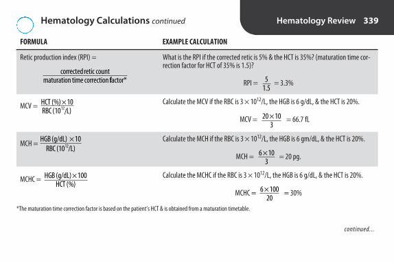
Hematology Calculations continued Hematology Review 339
FORMULA EXAMPLE CALCULATION
Retic production index (RPI) =
MCV =
MCH =
MCHC =
HGB (g/dL) 10RBC (10 /L)12
×
HGB (g/dL) 100HCT (%)
×
HCT (%) 10RBC (10 /L)12
×
corrected retic countmaturation time correction ffactor*
What is the RPI if the corrected retic is 5% & the HCT is 35%? (maturation time cor-rection factor for HCT of 35% is 1.5)?
RPI = = 3.3%
Calculate the MCV if the RBC is 3 × 10¹²/L, the HGB is 6 g/dL, & the HCT is 20%.
MCV = = 66.7 fL
Calculate the MCH if the RBC is 3 × 10¹²/L, the HGB is 6 gm/dL, & the HCT is 20%.
MCH = = 20 pg.
Calculate the MCHC if the RBC is 3 × 10¹²/L, the HGB is 6 g/dL, & the HCT is 20%.
MCHC = = 30%
6 103
×
6 100×20
20 10×3
51.5
continued...
*The maturation time correction factor is based on the patient’s HCT & is obtained from a maturation timetable.
2956_Ch04_287-366 30/01/14 4:26 PM Page 339

Hematology Calculations continued Hematology Review 340
FORMULA EXAMPLE CALCULATION
Rules of Three:
RBC × 3 = HGB ± 0.5HGB × 3 = HCT ± 3%
Manual Cell Count:
Cells/mm3 (µL) = # cells counted × depth factor(always 10) × reciprocal of dilution × reciprocal of
area counted (mm2)
Absolute WBC = total WBC × relative count (% from differential)
Corrected WBC =
uncorrected WBC 100+ NRBCs per 100 WBCs
×100
What should the HGB be if the RBC is 4.1 × 10¹²/L?
HGB = 4.1 × 3 = 12.3 ± 0.5 = 11.8–12.8 g/dL
What should the HCT be if HGB is 12.3 g/dL?
HCT = 12.3 × 3 = 36.9 ± 3 = 33.9%–39.9%
Calculate the CSF WBC count if 18 WBCs are counted in 9 mm2 on 1 side of aNeubauer hemacytometer, using undiluted CSF.
WBCs/µL = 18 × 10 × 1 × 1/9 = 20
Calculate the absolute lymphocyte count if the total WBC is 10 × 109/L & there are70% lymphocytes.
Absolute count = 10 × 0.70 = 7 × 10⁹/L
The automated hematology analyzer reports a WBC of 30 × 109/L. The technologistcounts 115 NRBCs per 100 WBCs while performing the differential. What is the cor-rected WBC?
Corrected WBC = = 14 × 109/L30 100+115×
100
2956_Ch04_287-366 30/01/14 4:26 PM Page 340

Overview of Hemostasis Hematology Review 341
Primary hemostasis
Secondary hemostasis
Fibrinolysis
• Vasoconstriction• Platelet adhesion • Platelet aggregation to form primary hemostatic plug at injury site
• Interaction of coag factors to produce fibrin (secondary hemostatic plug)• Fibrin stabilization by factor XIII
• Release of tissue plasminogen activator• Conversion of plasminogen to plasmin• Conversion of fibrin to fibrin degradation products
2956_Ch04_287-366 30/01/14 4:26 PM Page 341

Quantitative Platelet Disorders Hematology Review 342
DISORDER EXPLANATION CLINICAL MANIFESTATIONS LAB TESTS
Thrombocytopenia
Primary thrombocytosis
Secondary or reactive thrombocytosis
↓ production (e.g., aplastic ane-mia, myelodysplastic syn-dromes), ↑ destruction (e.g.,immune thrombocytopenic pur-pura, drugs, DIC, mechanical destruction by artificial heartvalves), splenic sequestration,massive transfusion (dilutioneffect)
Unregulated production ofmegakaryocytes in bone marrow, e.g., essential thrombo-cythemia, other myeloprolifera-tive disorders
↑ PLT due to another condition,e.g., hemorrhage, surgery,splenectomy
<30 × 109/L: petechiae, menorrha-gia, spontaneous bruising.
<10 × 109/L: severe spontaneousbleeding
Thrombosis or hemorrhage
Thrombosis or hemorrhage infrequent
PLT <150 × 109/L
PLT usually >1,000 × 109/L.Platelet aggregation maybe abnormal
PLT >450 × 109/L but usually <1,000 × 109/L
2956_Ch04_287-366 30/01/14 4:26 PM Page 342

Qualitative Platelet Disorders Hematology Review 343
DISORDER EXPLANATION LABORATORY TESTS
Inherited Bernard-Soulier syndrome
Glanzmann’s thrombasthenia
δ -storage pool disorder
Acquired
All can result in serious bleeding.VWF = Von Willebrand factor.
Lack of functional glycoprotein (GP) Ib/IX/V onplt surface prevents interaction with VWF. Abnormal plt adhesion to collagen.
Deficiency or abnormality of plt membrane GPIIb/IIIa. Fibrinogen can’t attach to plt surface &initiate plt aggregation.
Dense granule deficiency. Lack of ADP release.
Functional plt disorders occur with chronicrenal failure, myeloproliferative disorders, car-diopulmonary bypass, use of aspirin & otherdrugs. Mechanisms vary.
Giant plts with dense granulation. ↑ closuretime (PFA). Abnormal aggregation with ristocetin.
↑ closure time (PFA). Abnormal aggregationwith ADP, collagen, epinephrine.
Abnormal secondary aggregation with ADP &epinephrine.
Abnormal plt aggregation.
2956_Ch04_287-366 30/01/14 4:26 PM Page 343

Tests of Platelet Function Hematology Review 344
TEST METHOD CLINICAL SIGNIFICANCE
Platelet aggregation
Platelet function assay (PFA)
VWF:Ag
Aggregating agent (e.g., ADP, collagen, risto-cetin, epinephrine) added to plt suspension.As plts aggregate, ↑ in light transmittance. Pltaggregation curves generated (time vs. %transmittance).
Citrated whole blood drawn through capillarytubes coated with ADP/collagen or epineph-rine/collagen. Plts adhere & aggregate whenexposed to collagen. Closure time = length oftime for plts to form platelet plug & closeaperture of capillary tube.
Immunologic tests (e.g., EIA) using mono-clonal antibodies to VWF.
Abnormal curves with plt dysfunctions such asvon Willebrand disease, Bernard-Soulier syn-drome, plt storage pool defects, idiopathicthrombocytopenia purpura, drugs.
Screening test for qualitative plt defects. Re-places bleeding time. Von Willebrand disease:prolonged with collagen/ADP & collagen/epinephrine. Defects related to drugs (e.g., aspirin): normal with collagen/ADP, prolongedwith collagen/epinephrine.
VWF connects plts to collagen. ↓ in von Willebrand disease, so plts don’t function normally.
2956_Ch04_287-366 30/01/14 4:26 PM Page 344

Coagulation Factors Hematology Review 345
NAME PATHWAY INHERITED DEFICIENCY OTHER
I
II
III
IV
V
VII
VIII
Fibrinogen
Prothrombin
Tissue factor (TF)
Ca2+
Labile factor (proaccelerin)
Stable factor (proconvertin)
Antihemophilic factor
I, E, C
I, E, C
E
I, E, C
I, E, C
E
I
Rare
Rare
Rare
Rare
Common (hemophilia A)
Converted to fibrin by thrombin.
Precursor of thrombin.
Phospholipid released from injured vessel wall. Not normallyin blood.
Bound by anticoagulant sodiumcitrate. In assays using citratedplasma, must be supplied byreagents.
Deteriorates rapidly.
Circulates in association with vonWillebrand factor (VWF). VIII:C = coagulant portion. Extremelylabile.
continued...
2956_Ch04_287-366 30/01/14 4:26 PM Page 345

Coagulation Factors continued Hematology Review 346
NAME PATHWAY* INHERITED DEFICIENCY OTHER
IX
X
XI
XII
XIII
HMWK
PK
*I = intrinsic, E = extrinsic, C = common
Christmas factor (plasma thromboplas-tin component)
Stuart factor
Plasma thromboplastin antecedent
Hageman factor (contact factor)
Fibrin stabilizing factor
High molecular weight kininogen(Fitzgerald factor)
Prekallikrein (Fletcher factor)
I
I, E,C
I
I
I, E, C
I
I
Common (hemophilia B).
Rare.
Rare (hemophilia C).May or may not causebleeding.
No bleeding.
Rare.Poor wound healing.
Rare.No bleeding.
No bleeding.
Glass activation factor. Not part ofin vivo coagulation.
Stabilizes fibrin clot.
Not part of in vivo coagulation.
Not part of in vivo coagulation.
2956_Ch04_287-366 30/01/14 4:26 PM Page 346

Functional Classification of Coagulation Factors
Hematology Review 347
EXPLANATION FACTORS
Substrate Substance changed by an enzyme. Fibrinogen
Cofactor Protein that accelerates enzymatic reactions. No enzymatic V, VIII (V is cofactor for Xa; VIII is cofactor for IXa)activity of its own.
Enzyme Protein that catalyzes a change in specific substrate. Serine proteases: thrombin (IIa), VIIa, IXa, Xa, XIa, Secreted in inactive form (proenzyme, zymogen). Must be XIIa, prekallikreinactivated to function.
Transglutaminase: XIIIa
Letter “a” following Roman numeral indicates activated form of enzyme.
2956_Ch04_287-366 30/01/14 4:26 PM Page 347

Summary of Coagulation Factors Hematology Review 348
FACTORS EXPLANATION
Contact group
Prothrombin group
Fibrinogen group
Factors in extrinsic pathway
Factors in intrinsic pathway
Factors in common pathway
Extrinsic tenase complex
Intrinsic tenase complex
Prothrombinase complex
Factor VIII complex
Produced in liver
PK, HMWK, XII, XI
II, VII, IX, X
I, V, VIII, XIII
TF, VII
PK, HMWK, XII, XI, IX, VIII
X, V, II, I
VIIa/TF
IXa/VIIIa
Xa/Va
VIII:C & von Willebrand factor (VWF)
All
Factors involved in initiation of intrinsic pathway.
Vitamin K–dependent factors.
Factors acted on by thrombin (V, VIII, & XIII are acti-vated; I is converted to fibrin). All are high molecularweight proteins.
Acts on X.
Acts on X.
Acts on prothrombin.
VIII:C is the procoagulant; VWF is the carrier protein.
continued...
2956_Ch04_287-366 30/01/14 4:26 PM Page 348

Summary of Coagulation Factors continued Hematology Review 349
FACTORS EXPLANATION
Require vitamin K for synthesis
Affected by Coumadin (warfarin)
Consumed during clotting
Labile factors
II, VII, IX, X
II, VII, IX, X
I, II, V, VIII, XIII
V, VIII
All that require vitamin K. Warfarin is a vitamin K antagonist.
Not present in serum.
2956_Ch04_287-366 30/01/14 4:26 PM Page 349
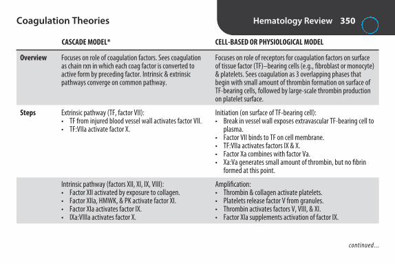
Coagulation Theories Hematology Review 350
CASCADE MODEL* CELL-BASED OR PHYSIOLOGICAL MODEL
Overview
Steps
Focuses on role of coagulation factors. Sees coagulationas chain rxn in which each coag factor is converted toactive form by preceding factor. Intrinsic & extrinsicpathways converge on common pathway.
Extrinsic pathway (TF, factor VII): • TF from injured blood vessel wall activates factor VII.• TF:VIIa activate factor X.
Intrinsic pathway (factors XII, XI, IX, VIII): • Factor XII activated by exposure to collagen.• Factor XIIa, HMWK, & PK activate factor XI. • Factor XIa activates factor IX.• IXa:VIIIa activates factor X.
Focuses on role of receptors for coagulation factors on surfaceof tissue factor (TF)–bearing cells (e.g., fibroblast or monocyte)& platelets. Sees coagulation as 3 overlapping phases thatbegin with small amount of thrombin formation on surface ofTF-bearing cells, followed by large-scale thrombin productionon platelet surface.
Initiation (on surface of TF-bearing cell):• Break in vessel wall exposes extravascular TF-bearing cell to
plasma.• Factor VII binds to TF on cell membrane. • TF:VIIa activates factors IX & X.• Factor Xa combines with factor Va.• Xa:Va generates small amount of thrombin, but no fibrin
formed at this point.
Amplification: • Thrombin & collagen activate platelets.• Platelets release factor V from granules.• Thrombin activates factors V, VIII, & XI.• Factor XIa supplements activation of factor IX.
continued...
2956_Ch04_287-366 30/01/14 4:26 PM Page 350

Coagulation Theories continued Hematology Review 351
CASCADE MODEL* CELL-BASED OR PHYSIOLOGICAL MODEL
Comments
*For simplicity, Ca2+ & platelet factor 3 not shown.
Common pathway (factors X, V, II, I): • Xa:Va converts prothrombin (II) to thrombin (IIa). • Thrombin cleaves fibrinogen (I) into fibrin & acti-
vates factor XIII to stabilize clot.
“Classic” theory. Explains in vitro coag (PT & APTTtests) & helps ID factor deficiencies, but doesn’t fit cur-rent understanding of coag in vivo. Pathways don’toperate independently.
Propagation (on surface of activated platelet): • Factor Xa binds to factor VIIIa on platelet.• IXa:VIIIa activates factor X. • Xa:Va converts prothrombin (II) to thrombin (IIa). • Thrombin cleaves fibrinogen (I) into fibrin & activates factor
XIII to stabilize clot.
To compare models, think of extrinsic pathway as occurring onTF-bearing cell & intrinsic pathway (without factor XII, HMPK,PK) on platelet surface. (Factor XII, HMPK, PK aren’t needed invivo because factor XI is activated by thrombin produced in initiation phase.)
2956_Ch04_287-366 30/01/14 4:26 PM Page 351
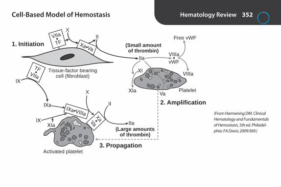
Cell-Based Model of Hemostasis Hematology Review 352
Xa•Va
VIIa•TF
Tissue-factor bearingcell (fibroblast)
TF•VIIa
IXa•VIIIa Xa•Va
(Small amountof thrombin)
Free vWF
VIIIa
VIIIa•
vWF
1. Initiation
3. Propagation
2. Amplification
PlateletVa
XIa
XI
IIa
II
II
IIa(Large amounts
of thrombin)
X
X
IXa
IX
IXXIa
Activated platelet
(From Harmening DM. ClinicalHematology and Fundamentalsof Hemostasis, 5th ed. Philadel-phia: FA Davis; 2009:569.)
2956_Ch04_287-366 30/01/14 4:26 PM Page 352

Prothombin Time (PT) and Activated PartialThromboplastin Time (APTT)
Hematology Review 353
PT APTT
Purpose
Reagent(s)
Prolonged results
Other
To detect deficiencies in extrinsic & common pathways& to monitor coumadin (warfarin) therapy.
Thromboplastin reagent (thromboplastin, phospholipid, Ca2+).
Coumadin therapy; deficiency of VII, X, V, II, or I; circulating inhibitors.
Report INR (international normalized ratio). INR =[patient PT/mean normal PT]ISI. ISI = internationalsensitivity index. Supplied by manufacturer. Be sure touse ISI for current lot of thromboplastin & analyzerbeing used. Therapeutic range for most situations is2–3. For patients with artificial heart valves, 2.5–3.5.
To detect deficiencies in intrinsic & common pathways & tomonitor unfractionated heparin therapy.
Activated partial thromboplastin reagent (phospholipid, activator), CaCl2.
Heparin therapy; deficiency of HMWK, PK, XII, XI, IX, VIII, X, V,II, or I; circulating inhibitors.
2956_Ch04_287-366 30/01/14 4:26 PM Page 353

Interpretation of PT/APTT Hematology Review 354
PT APTT POSSIBLE DEFICIENCY
Prolonged Normal VII
Normal Prolonged HMWK, PK, XII, XI, IX, VIII
Prolonged Prolonged X, V, II, I
2956_Ch04_287-366 30/01/14 4:26 PM Page 354

Other Coagulation Tests Hematology Review 355
TEST DISCUSSION
Mixing studies
Activated clotting time (ACT)
Thrombin time (TT)
Reptilase time
Fibrinogen
Factor assays
Factor XIII screening test
Anti-Factor Xa assay
Follow up to abnormal PT or APTT. Test is repeated on 1:1 mixture of pt plasma & normal plasma. If pt has factor defi-ciency, time will be corrected because normal plasma supplies missing factor. If time is not corrected, an inhibitor ispresent, e.g., antibody or anticoagulant.
Whole blood clotting method using point-of-care analyzer. Used in cardiac care units & during cardiac surgery tomonitor heparin.
Measures time required for thrombin to convert fibrinogen to fibrin. Prolonged with hypo- or dysfibrinogenemia, heparin, FDPs.
Similar to TT except uses reptilase (snake venom enzyme) instead of thrombin. Prolonged results with afibrinogenemia &most congenital dysfibrinogenemias. Variable results with hypofibrinogenemia. Not affected by presence of heparin.
Estimation of fibrinogen level by modified TT. Thrombin added to dilutions of pt plasma. Results obtained from calibration curve prepared from testing dilutions of a fibrinogen standard. Normal: 200–400 mg/dL.
% of factor activity determined by amount of correction of PT or APTT when dilutions of pt plasma are added to factor-deficient plasma.
Pt’s platelet-rich plasma mixed with CaCl2. Clot placed in urea or monochloroacetic acid & incubated at 37°C. With XIIIdeficiency, clot dissolves within 24 hr.
Test to monitor therapy with low molecular weight heparin. Can also be used instead of APTT to monitor therapy withunfractionated heparin. Pt plasma added to excess factor Xa & substrate specific for factor Xa. Heparin in sample formscomplex with AT & inhibits factor Xa. Residual factor Xa cleaves substrate to produce colored product whose intensityis inversely proportional to concentration of heparin.
2956_Ch04_287-366 30/01/14 4:26 PM Page 355

Most Common Inherited Coagulation Disorders
Hematology Review 356
DISEASE DEFICIENCY CLINICAL FINDINGS LABORATORY FINDINGS OTHER
Von Willebrand disease
Hemophilia A
Hemophilia B (Christmas disease)
VWF
Factor VIII
Factor IX
Mucocutaneous bleed-ing ranging from mild to severe
Varies from asympto-matic to crippling bleed-ing into joints, muscles,& fatal intracranial hemorrhage
Same as hemophilia A
PLT: NClosure time (PFA): N or ↑Platelet aggregation: abnormal
with ristocetinPT: NAPTT: N or ↑Factor VIII: N or ↓VWF:Ag: ↓
PLT: NPT: NAPTT: ↑Factor VIII: ↓
PLT: NPT: NAPTT: ↑Factor IX: ↓
Most common inheritedbleeding disorder. Autoso-mal dominant. Both sexesaffected. Plts can’t adhereto collagen to form plt plug.Lab results vary.
2nd most common inher-ited bleeding disorder. Sex-linked recessive. Occursprimarily in males. Mothersare carriers.
Sex-linked recessive.
2956_Ch04_287-366 30/01/14 4:26 PM Page 356
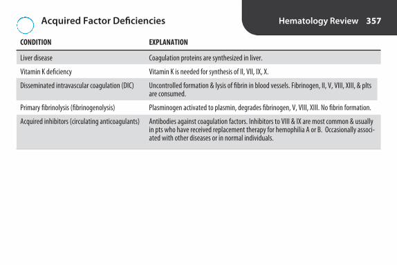
Acquired Factor Deficiencies Hematology Review 357
CONDITION EXPLANATION
Liver disease Coagulation proteins are synthesized in liver.
Vitamin K deficiency Vitamin K is needed for synthesis of II, VII, IX, X.
Disseminated intravascular coagulation (DIC) Uncontrolled formation & lysis of fibrin in blood vessels. Fibrinogen, II, V, VIII, XIII, & pltsare consumed.
Primary fibrinolysis (fibrinogenolysis) Plasminogen activated to plasmin, degrades fibrinogen, V, VIII, XIII. No fibrin formation.
Acquired inhibitors (circulating anticoagulants) Antibodies against coagulation factors. Inhibitors to VIII & IX are most common & usuallyin pts who have received replacement therapy for hemophilia A or B. Occasionally associ-ated with other diseases or in normal individuals.
2956_Ch04_287-366 30/01/14 4:26 PM Page 357

Tests of Fibrinolytic System Hematology Review 358
TEST EXPLANATION METHOD(S) CLINICAL SIGNIFICANCE
D-dimer Fragment that results from Latex agglutination using Marker for DIC. Also pos with deep vein lysis of fibrin by plasmin monoclonal antibodies against thrombosis, pulmonary embolism, & after
D-dimer, ELISA lytic therapy. Neg in primary fibrinolysis.
Fibrin(ogen) Product of action of plasmin Latex agglutination using Sign of ↑ fibrinolytic activity. Doesn’t degradation on fibrin or fibrinogen antibodies against FDP differentiate between fibrin degradation products (FDP) products & fibrinogen degradation products.
Present in DIC, primary fibrinolysis, deep vein thrombosis, pulmonary embolism, & after lytic therapy.
2956_Ch04_287-366 30/01/14 4:26 PM Page 358

Disseminated Intravascular Coagulation vs.Primary Fibrinolysis
Hematology Review 359
DIC PRIMARY FIBRINOLYSIS
PT Prolonged Prolonged
APTT Prolonged Prolonged
Fibrinogen ↓ ↓
Platelets ↓ Normal
FSP Present Present
D-Dimer Pos Neg
RBC morphology Schistocytes Normal
2956_Ch04_287-366 30/01/14 4:26 PM Page 359

Tests to Assess Risk of Thrombosis Hematology Review 360
TEST SIGNIFICANCE ASSAYS
Antithrombin (AT, formerly antithrombin III)
Protein C
Protein S
Factor V Leiden
Lupus anticoagulants
Plasma inhibitor that neutralizes all serine proteases,including thrombin. Deficiencies associated with ↑ risk of thrombosis.
Coagulation inhibitor. Inactivates Va & VIIIa. Deficien-cies associated with ↑ risk of thrombosis.
Cofactor for protein C.
Most common cause of hereditary activated protein Cresistance (APC). Mutation that makes V resistant toactivity of activated protein C. ↑ risk of thrombosis.
Risk factor for thrombosis & recurrent spontaneousabortion. Acquired antiphospholipid antibodies that interact with phospholipid in APTT reagent & prolongtime. In vitro phenomenon. Patient doesn’t have factordeficiency or bleeding. Present in patients with lupus,other autoimmune diseases, neoplasms, infections,drugs. Also present in some normal individuals.
Chromogenic substrate assay, immunologicassay, nephelometry.
Immunologic assay, chromogenic substrateassay, clot based assay.
Clotting assay, immunologic assay.
APC resistance assay is most frequent screeningtest. Patient plasma diluted in V-deficientplasma. Activated protein C added. APTT or diluteRussell viper venom time (dRVVT) performed.Abnormals must be confirmed by molecular test-ing (e.g., PCR, restriction fragment length polymorphism).
Detected by unexplained prolongation of APTTthat isn’t corrected by addition of equal volumeof normal plasma. No definitive assay.
2956_Ch04_287-366 30/01/14 4:26 PM Page 360
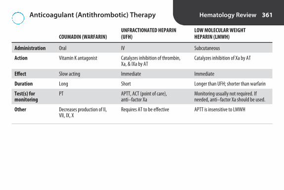
Anticoagulant (Antithrombotic) Therapy Hematology Review 361
UNFRACTIONATED HEPARIN LOW MOLECULAR WEIGHT COUMADIN (WARFARIN) (UFH) HEPARIN (LMWH)
Administration Oral IV Subcutaneous
Action Vitamin K antagonist Catalyzes inhibition of thrombin, Catalyzes inhibition of Xa by ATXa, & IXa by AT
Effect Slow acting Immediate Immediate
Duration Long Short Longer than UFH; shorter than warfarin
Test(s) for PT APTT, ACT (point of care), Monitoring usually not required. If monitoring anti–factor Xa needed, anti–factor Xa should be used.
Other Decreases production of II, Requires AT to be effective APTT is insensitive to LMWHVII, IX, X
2956_Ch04_287-366 30/01/14 4:26 PM Page 361

Coagulation Instrumentation Hematology Review 362
ENDPOINT DETECTION PRINCIPLE
Mechanical Change in electrical conductivity between 2 probes or change in movement of steel ball when clot forms
Photo-optical ↓ in light transmittance as fibrin forms
Chromogenic ↑ in light absorbance at 405 nm as para-nitroaniline (pNA) is cleaved from synthetic substrate by coag enzyme
Immunologic ↑ in light absorbance as latex particles coated with specific antibody are agglutinated by antigen
Some analyzers have multiple detection methods.
2956_Ch04_287-366 30/01/14 4:26 PM Page 362

Examples of Criteria for Specimen Rejectionin Coagulation Testing
Hematology Review 363
• Improper labeling
• Delay in delivery to lab
• Exposure to extremes of temperature
• Tube <90% full
• Specimen clotted
• Specimen hemolyzed
2956_Ch04_287-366 30/01/14 4:26 PM Page 363
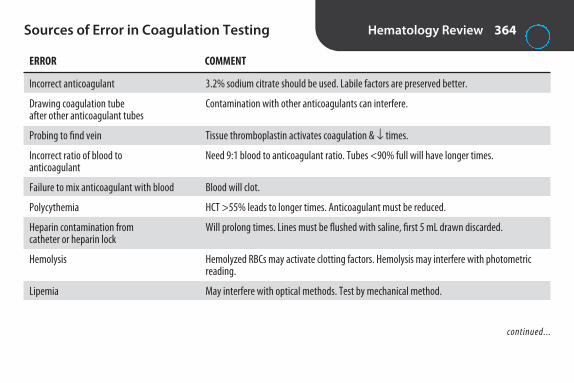
Sources of Error in Coagulation Testing Hematology Review 364
ERROR COMMENT
Incorrect anticoagulant
Drawing coagulation tube after other anticoagulant tubes
Probing to find vein
Incorrect ratio of blood to anticoagulant
Failure to mix anticoagulant with blood
Polycythemia
Heparin contamination from catheter or heparin lock
Hemolysis
Lipemia
3.2% sodium citrate should be used. Labile factors are preserved better.
Contamination with other anticoagulants can interfere.
Tissue thromboplastin activates coagulation & ↓ times.
Need 9:1 blood to anticoagulant ratio. Tubes <90% full will have longer times.
Blood will clot.
HCT >55% leads to longer times. Anticoagulant must be reduced.
Will prolong times. Lines must be flushed with saline, first 5 mL drawn discarded.
Hemolyzed RBCs may activate clotting factors. Hemolysis may interfere with photometricreading.
May interfere with optical methods. Test by mechanical method.
continued...
2956_Ch04_287-366 30/01/14 4:26 PM Page 364

Sources of Error in Coagulation Testing continued
Hematology Review 365
ERROR COMMENT
Improper storage of specimen
Improper storage or reconstitution of reagents
Equipment malfunction, e.g., temperature, timer, detector, volumes dispensed
Should be stored in vertical position at RT with stopper on to prevent change in pH. Specimens for PT must be tested within 24 hr of collection, APTT within 4 hr. (If APTT is for monitoring heparin, must be centrifuged within 1 hr of collection.)
Run normal & abnormal controls every 8 hr & with each change of reagents to verify systemperformance.
Run normal & abnormal controls every 8 hr & with each change of reagents to verify systemperformance.
2956_Ch04_287-366 30/01/14 4:26 PM Page 365

2956_Ch04_287-366 30/01/14 4:26 PM Page 366

5S E C T I O N
Immunology Review
2956_Ch05_367-416 30/01/14 4:26 PM Page 367
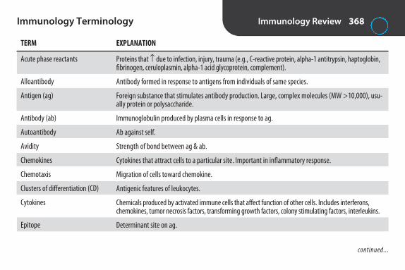
Immunology Terminology Immunology Review 368
TERM
Acute phase reactants
Alloantibody
Antigen (ag)
Antibody (ab)
Autoantibody
Avidity
Chemokines
Chemotaxis
Clusters of differentiation (CD)
Cytokines
Epitope
EXPLANATION
Proteins that ↑ due to infection, injury, trauma (e.g., C-reactive protein, alpha-1 antitrypsin, haptoglobin,fibrinogen, ceruloplasmin, alpha-1 acid glycoprotein, complement).
Antibody formed in response to antigens from individuals of same species.
Foreign substance that stimulates antibody production. Large, complex molecules (MW >10,000), usu-ally protein or polysaccharide.
Immunoglobulin produced by plasma cells in response to ag.
Ab against self.
Strength of bond between ag & ab.
Cytokines that attract cells to a particular site. Important in inflammatory response.
Migration of cells toward chemokine.
Antigenic features of leukocytes.
Chemicals produced by activated immune cells that affect function of other cells. Includes interferons,chemokines, tumor necrosis factors, transforming growth factors, colony stimulating factors, interleukins.
Determinant site on ag.
continued...
2956_Ch05_367-416 30/01/14 4:26 PM Page 368

Immunology Terminology continued Immunology Review 369
TERM
Hapten
Histamine
Hypersensitivity
Immunity
Immunogen
Immunoglobulin (Ig)
Immunology
Inflammation
Interferons
Interleukins
Ligand
EXPLANATION
Low molecular weight substance that can bind to ab once it’s formed, but is incapable of stimulating abproduction unless bound to larger carrier molecule.
Vasoactive amine released from mast cells & basophils during allergic rxn.
Heightened state of immune responsiveness that causes tissue damage in host.
Resistance to infection.
Any substance capable of inducing immune response.
Antibody.
Study of reactions of host when exposed to foreign substances.
Cellular & humoral mechanisms involved in reaction to injury or infection.
Cytokines with antiviral properties. Also active against certain tumors & inflammatory processes.
Cytokines produced by leukocytes that affect inflammatory response through ↑ in soluble factors or cells.
Molecule that binds to another molecule of complementary configuration; the substance being measuredin an immunoassay.
continued...
2956_Ch05_367-416 30/01/14 4:26 PM Page 369

Immunology Terminology continued Immunology Review 370
TERM
Lysozyme
Major histocompatibility complex (MHC)
Monoclonal antibody
Opsonin
Phagocytosis
Plasma cells
Polyclonal antibody
Postzone
Prozone
Seroconversion
EXPLANATION
Enzyme found in tears & saliva that attacks cell walls of microorganisms.
System of genes that control expression of MHC molecules found on all nucleated cells; originallyreferred to as human leukocyte antigens (HLA).
Ab derived from a single B-cell clone.
Serum proteins that attach to foreign substance & enhance phagocytosis.
Engulfment of cells or particulate matter by neutrophils & macrophages.
Transformed B cells that secrete ab.
Ab produced by many B-cell clones.
Reduced ag/ab complexes due to ag excess. Can cause false neg in serological test for ab. Repeat test in 1–2 wk.
Reduced ag/ab complexes due to ab excess. Can cause false neg in serological test for ab. Dilute serum& retest.
Change of serological test from neg to pos due to development of detectable ab.
continued...
2956_Ch05_367-416 30/01/14 4:26 PM Page 370

Immunology Terminology continued Immunology Review 371
TERM
Serum sickness
Thymus
Titer
Vaccination
Zone of equivalence
EXPLANATION
Type III hypersensitivity reaction that results from buildup of abs to animal serum used in some passiveimmunizations.
Small, flat bilobed organ found in thorax; site of T-lymph development. One of the primary lymphoidorgans.
Means of expressing ab concentration; reciprocal of highest dilution with pos rxn.
Injection of immunogenic material to induce immunity.
When # of multivalent sites of ag & ab are approximately equal. Results in optimal precipitation.
2956_Ch05_367-416 30/01/14 4:26 PM Page 371

Branches of the Immune System Immunology Review 372
BRANCH DEFINITION DEFENSE AGAINST CELLS INVOLVED EXAMPLES
Cellular
Humoral
Graft rejection, hypersensitivityrxn, elimination of tumor cells
Ab production
T lymphs, macrophages
B lymphs, plasma cells
Cell mediated
Antibody mediated
Viruses, fungi, mycobacteria,other intracellular pathogens,tumor cells
Bacteria (extracellular)
2956_Ch05_367-416 30/01/14 4:26 PM Page 372

Types of Immunity Immunology Review 373
TYPE EXPLANATION COMPONENTS MEMORY?
Natural or innate
Acquired or adaptive
No
Yes
Defense mechanisms presentat birth. Not ag specific.
Defense mechanisms that areantigen specific.
External defense system: intact skin, mucous membranes, cilia& mucus in respiratory tract, stomach acid, flushing of urine,lactic acid in vagina, lysozyme in tears & saliva, normal flora
Internal defense system: neutrophils, macrophages, acutephase reactants, complement
T cells, B cells, plasma cells, antibodies, cytokines
2956_Ch05_367-416 30/01/14 4:26 PM Page 373

Adaptive Immunity Immunology Review 374
TYPE EXPLANATION EXAMPLE SPECIFIC? IMMEDIATE? LONG-TERM?
Naturally acquired active immunity
Artificially acquired active immunity
Naturally acquired passive immunity
Artificially acquired passive immunity
Individual infected with microorganism produces ab
Individual exposed to agthrough vaccine developsimmunity without having infection
Individual protected by absproduced by another person
Individual receives immuneglobulin containing abs pro-duced by another person
Clinical or subclinicalinfection
DTaP, MMR, polio,tetanus, Hib vaccine
Maternal abs thatcross placenta & arepresent in breast milk
Rh immune globulin,HBIG, antitoxins
Yes
Yes
Yes
Yes
No
No
Yes
Yes
Yes
Yes
No
No
2956_Ch05_367-416 30/01/14 4:26 PM Page 374

Cells of the Natural Immune System Immunology Review 375
CELL
GranulocytesNeutrophils
Eosinophils
Basophils
MononuclearsMonocytes
Tissue cellsMast cells
Macrophages
Dendritic cells
FUNCTION
Phagocytosis, inflammatory response
Neutralization of basophil & mast cell productsDestruction of some helminths
Hypersensitivity rxn
Phagocytosis
Hypersensitivity rxn
Phagocytosis; elimination of bacteria, intra -cellular parasites, tumor cells; secretion ofcell mediators; ag presentation
Phagocytosis, presentation of ag to helper Tlymphs in blood & lymphoid organs
COMMENTS
Respond to chemotaxins. Granules contain bactericidal enzymes.
Some phagocytic ability.
Granules contain histamine, heparin, eosinophil chemotactic factor A.In allergic rxn, bind IgE. Granules release contents in presence of ag.
Migrate to tissues, become macrophages. Respond to chemotaxins.
Connective tissue cells. Resemble basophils but larger & more granules. Bind IgE.
Activated by contact with microorganisms or cytokines from Tlymphs.
Initiate acquired immune response.
continued...
2956_Ch05_367-416 30/01/14 4:26 PM Page 375
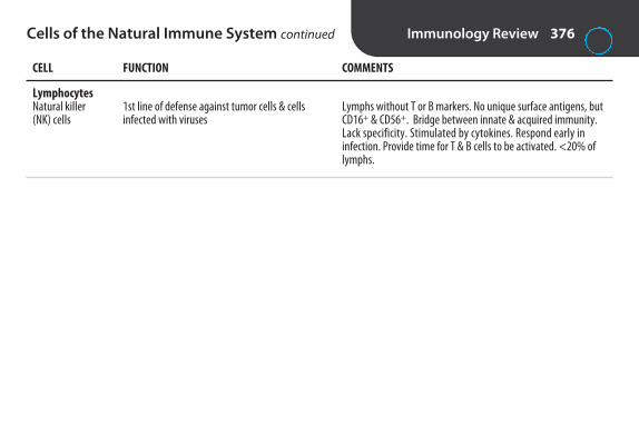
Cells of the Natural Immune System continued Immunology Review 376
CELL
LymphocytesNatural killer (NK) cells
FUNCTION
1st line of defense against tumor cells & cellsinfected with viruses
COMMENTS
Lymphs without T or B markers. No unique surface antigens, butCD16+ & CD56+. Bridge between innate & acquired immunity.Lack specificity. Stimulated by cytokines. Respond early in infection. Provide time for T & B cells to be activated. <20% oflymphs.
2956_Ch05_367-416 30/01/14 4:26 PM Page 376

Cells of the Acquired Immune System Immunology Review 377
CELL
T lymphs
Helper/inducer T cells
Cytotoxic/suppressor T cells
T regulatory cells
B lymphs
Plasma cells
Memory cells
FUNCTION
Cell-mediated immunity.
Orchestrate cell-mediated immunity. Activate Bcells, cytotoxic cells, & NK cells.
Suppressor cells inhibit helper T cells. Cytotoxiccells kill other cells.
Suppress immune response to self.
After antigenic challenge, transform into blaststhat give rise to plasma cells & memory cells.
Ab production.
Respond to ag when encountered again with ↑ speed & intensity.
COMMENTS
Derived from cells in bone marrow. Develop T-cell–specific surface ags in thymus. 60%–80% of lymphs.
CD4+. 2/3 of peripheral T cells. Normal CD4 = 1,000/μL.In AIDS, <200/μL.
CD8+. 1/3 of peripheral T cells. (Normal CD4/CD8 ratio2:1. In AIDS, <0.5:1).
CD4+ & CD25+.
Develop in bone marrow. When mature, have surfaceIgs (IgM, IgD) that act as receptors for ags. 10%–20%of lymphs.
In peripheral lymphoid organs. Nondividing. Die in afew days.
In peripheral lymphoid organs. Live months to years.
2956_Ch05_367-416 30/01/14 4:26 PM Page 377

Subpopulations of Lymphocytes Immunology Review 378
Thymus Bone marrow
T Cells60-80%
B Cells10-20%
NKLymphocytes
< 20%
Lymphocytestem cell
T-helper T-suppressor
(From Ciesla B. Hematology in Practice, 2nd ed. Philadelphia: FA Davis; 2012:136.)
2956_Ch05_367-416 30/01/14 4:26 PM Page 378

Lymphoid Organs Immunology Review 379
PRIMARY SECONDARY
Bone marrow SpleenThymus Lymph nodes
TonsilsAppendixCutaneous-associated lymphoid tissueMucosal-associated lymphoid tissue (MALT), including Peyer’s patches in lower ileum
2956_Ch05_367-416 30/01/14 4:26 PM Page 379

Isolation & Identification of Lymphocytes Immunology Review 380
Isolation Density gradient centrifugation with Ficoll-Hypaque. Layers from top to bottom: plasma, mononuclear cells,Ficoll-Hypaque, RBCs, & granulocytes.
Identification Flow cytometry. Fluorescent-labeled monoclonal abs against specific surface ags. Each ab has different fluorescenttag. Light scattering measured as cells flow through laser beam. Common T-cell ags tested: CD2, CD3, CD4, CD7,CD8; B-cell ags: CD19, CD20, CD22, surface Ig.
2956_Ch05_367-416 30/01/14 4:26 PM Page 380
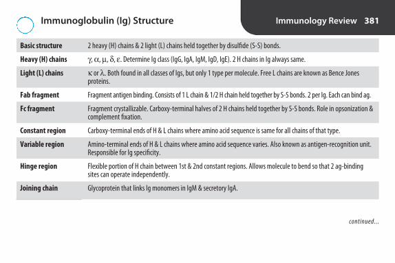
Immunoglobulin (Ig) Structure Immunology Review 381
2 heavy (H) chains & 2 light (L) chains held together by disulfide (S-S) bonds.
γ , α , µ , δ , ε . Determine Ig class (IgG, IgA, IgM, IgD, IgE). 2 H chains in Ig always same.
κ or λ . Both found in all classes of Igs, but only 1 type per molecule. Free L chains are known as Bence Jones proteins.
Fragment antigen binding. Consists of 1 L chain & 1/2 H chain held together by S-S bonds. 2 per Ig. Each can bind ag.
Fragment crystallizable. Carboxy-terminal halves of 2 H chains held together by S-S bonds. Role in opsonization &complement fixation.
Carboxy-terminal ends of H & L chains where amino acid sequence is same for all chains of that type.
Amino-terminal ends of H & L chains where amino acid sequence varies. Also known as antigen-recognition unit.Responsible for Ig specificity.
Flexible portion of H chain between 1st & 2nd constant regions. Allows molecule to bend so that 2 ag-bindingsites can operate independently.
Glycoprotein that links Ig monomers in IgM & secretory IgA.
Basic structure
Heavy (H) chains
Light (L) chains
Fab fragment
Fc fragment
Constant region
Variable region
Hinge region
Joining chain
continued...
2956_Ch05_367-416 30/01/14 4:26 PM Page 381

Immunoglobulin (Ig) Structure continued Immunology Review 382
Light chainhypervariable
regions
Intrachaindisulfide bonds
Interchaindisulfide bonds
Fab
Fc
Amino terminal
Carboxy terminal
Antigenbinding
Biologicalactivitymediation
Heavy chainhypervariableregions
Light chain
Heavy chain
Hinge region
Complement binding region
VL
VH C
L
CH 1
CH 2
CH 3
Carbohydrate
Structure of the basic immunoglobulin unit. VH = variable region heavy chain; VL = variable region light chain; CH = constantregion heavy chain; CL = constant region light chain. (From Harmening DM. Clinical Hematology and Fundamentals of Hemostasis, 5th ed.Philadelphia: FA Davis; 2009:502.)
2956_Ch05_367-416 30/01/14 4:26 PM Page 382

Immunoglobulins Immunology Review 383
Form
Molecularweight (daltons)
Heavy chain
Light chain
% of total Ig
Serum concentration(mg/dL)
Antigen-binding sites
Complement fixation
IgG
Monomer
150,000
Gamma (γ )
Kappa (κ ) or lambda (λ )
70–75
800–1,600
2
Yes
IgM
Pentamer
900,000
Mu (µ )
κ or λ
10
120–150
10
Yes
IgA
Monomer & dimer
160,000 or 400,000
Alpha (α )
κ or λ
10–15
70–350
2 or 4
No
IgD
Monomer
180,000
Delta (δ )
κ or λ
<1
1–3
2
No
IgE
Monomer
190,000
Epsilon (ε )
κ or λ
0.002
0.005
2
No
continued...
2956_Ch05_367-416 30/01/14 4:26 PM Page 383

Immunoglobulins continued Immunology Review 384
Crosses placenta?
Role(s)
Other
IgG
Yes
Defense against bacteria & viruses.Neutralizes toxins.Opsonin. Passive im-munity in newborns.
More efficient at precipitation thanagglutination
IgM
No
Neutralizes toxins. Opsonin.
First Ig produced in im-mune response. Only Igproduced by newborn.Most efficient Ig at ini-tiating complementcascade. More efficientat agglutination thanIgG. Destroyed bysulfhydryl compounds.
IgA
No
1st line of defense.Patrols mucosalsurfaces. Preventsadherence of bac-teria and neutral-izes toxins.
In tears, sweat,saliva, respiratory& GI mucosa,breast milk.
IgD
No
May play role in B-cell maturation.
On surface of Blymphs.
IgE
No
Role in allergicrxn. Binds to basophils & mastcells. When 2 ad-jacent moleculeson mast cell bindag, degranulationof cell with releaseof histamine & heparin.
Type I immediatehypersensitivityrxn.
2956_Ch05_367-416 30/01/14 4:26 PM Page 384

Complement Immunology Review 385
Definition
Functions
Classical pathway*
Alternative pathway*
Lectin pathway*
Present in highest concentration in plasma
Key component of all pathways
Ions required
Group of >30 proteins involved in phagocytosis & clearance of foreign antigens. Most are inactive enzymeprecursors that are converted to active enzymes in precise order (cascade).
Inflammation, opsonization, chemotaxis, cell lysis.
Triggered by ag/ab rxn. IgM is most efficient activator. Single molecule attached to 2 adjacent ags can initiate cascade. IgG1, 2, & 3 can activate complement but at least 2 molecules required.
Recognition unit: C1 (first to bind).Activation unit: C4, C2, C3.Membrane attack complex: C5, C6, C7, C8, C9 (cell lysis).
Ab independent. Activated by bacteria, fungi, viruses, tumor cells, some parasites.
Ab independent. Initiated by mannose-binding lectin (MBL). Nonspecific recognition of sugars on microor-ganisms. Important defense mechanism in infancy.
C3
C3
Ca2+, Mg2+
continued...
2956_Ch05_367-416 30/01/14 4:27 PM Page 385

Complement continued Immunology Review 386
Deficiencies
Inactivation
↑ susceptibility to infection. Accumulation of immune complexes, which can lead to glomerulonephritis.
56°C for 30 min.
*All 3 pathways converge at point of cleavage of C3.
2956_Ch05_367-416 30/01/14 4:27 PM Page 386

Hypersensitivity Reactions Immunology Review 387
TYPE I: TYPE II: TYPE III: IMMUNE TYPE IV: T-CELL ANAPHYLACTIC CYTOTOXIC COMPLEX DEPENDENT
Key reactant(s)
Mechanism
Onset of symptoms
Examples
T cells
Release of cytokines
Delayed (sensitization after1st contact with ag, symp-toms upon reexposure).
Contact dermatitis, hypersen-sitivity pneumonitis, tuber-culin skin test
IgG, IgM, complement,soluble antigens
Deposits of ag-ab com-plexes in tissues
Immediate
Arthus rxn, serum sick-ness, systemic lupus erythematosus (SLE),rheumatoid arthritis (RA)
IgE
Release of mediatorsfrom mast cells &basophils
Immediate
Anaphylaxis, hayfever, asthma, foodallergies
IgG, IgM, complement,cellular antigens
Cytolysis due to ab &complement
Immediate
Transfusion reactions,hemolytic disease ofnewborn, autoimmunehemolytic anemia
2956_Ch05_367-416 30/01/14 4:27 PM Page 387

Agglutination Methods Immunology Review 388
METHOD
Direct agglutination
Hemagglutination
Passive (indirect) agglutination
Reverse passive agglutination
Agglutination inhibition
Hemagglutination inhibition
Coagglutination
Antiglobulin-mediated agglutination
PRINCIPLE
Naturally occurring ags on particles (e.g., bacterial ags). Particles agglutinate in presence of corresponding ab.
Ag-ab rxn that results in clumping of RBCs.
Soluble ags bound to particles, e.g., latex. Particles agglutinate inpresence of corresponding ab.
Ab attached to carrier particles. Particles agglutinate in presence ofcorresponding ag.
Competition between particulate ag (reagent) & soluble ag (in spec-imen) for sites on reagent ab. Lack of agglutination is pos result.
Detects abs to certain viruses that agglutinate RBCs. In presence ofab, virus is neutralized & hemagglutination doesn’t occur.
Reagent ab attached to carrier bacteria. (Staphylococcus aureus mostfrequently used as carrier. Protein A binds Fc portion of reagent ab.)Visible agglutination in presence of corresponding ag.
Detection of nonagglutinating ab by coupling with 2nd ab (antihumanglobulin).
APPLICATION
Widal test for typhoid fever. SalmonellaO & H ags used to detect abs in patientserum. Test no longer common in U.S.
ABO slide typing.
Rheumatoid factor, antinuclear antibody.
Rapid ID of bacteria.
Detection of illicit drugs.
Rubella & other viruses.
Rapid ID of bacteria.
Direct & indirect antiglobulin tests.
Notes: Rheumatoid factor can cause false-pos rxn in agglutination tests because it reacts with any IgG. Heterophile antibodies can cause false-pos rxn in hemagglutination tests.
2956_Ch05_367-416 30/01/14 4:27 PM Page 388
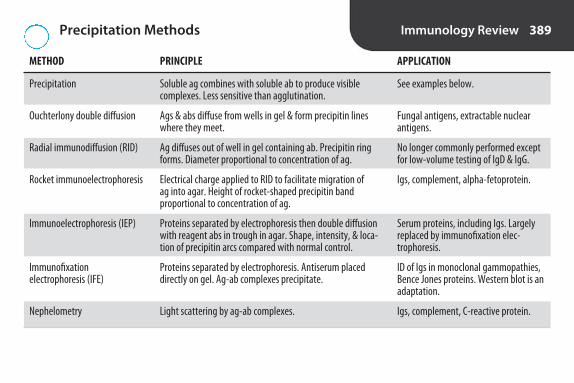
Precipitation Methods Immunology Review 389
METHOD
Precipitation
Ouchterlony double diffusion
Radial immunodiffusion (RID)
Rocket immunoelectrophoresis
Immunoelectrophoresis (IEP)
Immunofixation electrophoresis (IFE)
Nephelometry
PRINCIPLE
Soluble ag combines with soluble ab to produce visible complexes. Less sensitive than agglutination.
Ags & abs diffuse from wells in gel & form precipitin lineswhere they meet.
Ag diffuses out of well in gel containing ab. Precipitin ringforms. Diameter proportional to concentration of ag.
Electrical charge applied to RID to facilitate migration of ag into agar. Height of rocket-shaped precipitin band proportional to concentration of ag.
Proteins separated by electrophoresis then double diffusionwith reagent abs in trough in agar. Shape, intensity, & loca-tion of precipitin arcs compared with normal control.
Proteins separated by electrophoresis. Antiserum placed directly on gel. Ag-ab complexes precipitate.
Light scattering by ag-ab complexes.
APPLICATION
See examples below.
Fungal antigens, extractable nuclearantigens.
No longer commonly performed exceptfor low-volume testing of IgD & IgG.
Igs, complement, alpha-fetoprotein.
Serum proteins, including Igs. Largelyreplaced by immunofixation elec-trophoresis.
ID of Igs in monoclonal gammopathies,Bence Jones proteins. Western blot is anadaptation.
Igs, complement, C-reactive protein.
2956_Ch05_367-416 30/01/14 4:27 PM Page 389
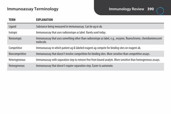
Immunology Review 390Immunoassay Terminology
TERM EXPLANATION
Ligand Substance being measured in immunoassay. Can be ag or ab.
Isotopic Immunoassay that uses radioisotope as label. Rarely used today.
Nonisotopic Immunoassay that uses something other than radioisotope as label, e.g., enzyme, fluorochrome, chemiluminescentmolecule.
Competitive Immunoassay in which patient ag & labeled reagent ag compete for binding sites on reagent ab.
Noncompetitive Immunoassay that doesn’t involve competition for binding sites. More sensitive than competitive assays.
Heterogeneous Immunoassay with separation step to remove free from bound analyte. More sensitive than homogeneous assays.
Homogeneous Immunoassay that doesn’t require separation step. Easier to automate.
2956_Ch05_367-416 30/01/14 4:27 PM Page 390

Immunology Review 391Enzyme Immunoassay (EIA) Terminology
TERM EXPLANATION
EIA Any immunoassay that uses an enzyme as label. A substrate is added to measure enzyme activity.
Direct EIA 1st type of EIA developed. Competitive. Enzyme-labeled reagent is part of initial ag-ab rxn. All reactants added at sametime. 1 incubation & 1 wash.
Indirect EIA Noncompetitive EIA. Enzyme-labeled reagent isn’t involved in initial ag-ab rxn. 2 incubations & 2 washes. More sensitivethan direct assays. Also known as enzyme-linked immunosorbent assay (ELISA). See next card for more details.
Solid phase Reagent ag or ab bound to support medium, e.g., polystyrene test tubes, microtiter plates, cellulose membranes, glassbeads.
2956_Ch05_367-416 30/01/14 4:27 PM Page 391

Immunology Review 392Enzyme Immunoassays (EIA) Formats
METHOD
EIA
Enzyme-linked immunosorbent assay (ELISA)
Sandwich ELISA or capture assay
OTHER
Original EIA. Used to measure smallrelatively pure ags, e.g., insulin, estrogen.
Used to detect abs to viruses, e.g.,HIV, HAV, HCV, EBV.
Ags must have multiple determinants.Used to measure Igs, hormones, pro-teins & detect tumor markers, viruses,parasites, fungi. High concentration ofag can cause hook effect. Too much agfor binding sites so undiluted samplehas lower absorbance than dilutions.
DESCRIPTION
Heterogeneous,competitive, direct
Heterogeneous,noncompetitive,indirect
Heterogeneous,noncompetitive,indirect
PRINCIPLE
Enzyme-labeled ligand & unlabeled patientligand compete for binding sites on ab at-tached to solid phase. Free labeled ligand re-moved by washing. Substrate added. Colorinversely proportional to concentration ofligand in specimen.
Ag attached to solid phase. Ab in specimenattaches. Unbound ab removed by washing.Enzyme-labeled antiglobulin added. At-taches to ab on solid phase. Substrate added.Color directly proportional to ab concentra-tion. More sensitive than competitive EIA.One of most common immunoassays.
Ab attached to solid phase. Ag in specimenattaches. Enzyme-labeled ab added, at-taches to different determinant. Enzymaticactivity directly proportional to amount of agin sample.
continued...
2956_Ch05_367-416 30/01/14 4:27 PM Page 392

Enzyme Immunoassays (EIA) Formats continued
Immunology Review 393
METHOD
Rapid ELISA
Enzyme-multiplied immunoassay technique (EMIT)
OTHER
May have built in control. Usuallyqualitative.
Used for determination of low mo-lecular weight analytes not readilymeasured by other methods, e.g.,hormones, therapeutic drugs, drugsof abuse. Automated.
DESCRIPTION
Membrane based
Homogeneous
PRINCIPLE
Reagent ag or ab bound to membrane in sin-gle use cassette. Sample added. Presence ofag-ab complex indicated by colored rxn.
Ag in specimen & enzyme-labeled ag com-pete for binding sites on reagent ab. Whenenzyme-labeled ag binds, enzyme activityinhibited. Enzyme activity is directly propor-tional to concentration of ag in specimen.
2956_Ch05_367-416 30/01/14 4:27 PM Page 393

Fluorescent Immunoassays (FIA) Immunology Review 394
METHOD
Direct fluorescent antibody (DFA) staining
Indirect fluorescent antibody (IFA) staining
Fluorescence polarization immunoassay (FPIA)
EXAMPLES OF ANALYTES
Bacterial, viral antigens
Fluorescent antinuclear antibody (FANA), fluorescenttreponemal antibody (FTA)
Therapeutic drugs, hormones
PRINCIPLE
Specimen on glass slide overlaid with fluorescein-labeled ab. If corresponding ag present, labeled ab binds. Fluorescence observed with fluorescentmicroscope.
Reagent ag on glass slide overlaid with patientserum. If corresponding ab present in serum, at-taches to ag. When fluorescein-labeled antihumanglobulin added, attaches to ab. Fluorescence observed with fluorescent microscope.
Labeled ag competes with ag in specimen for siteson reagent ab. Free labeled ag rotates rapidly,emits little polarized light. Bound labeled ag ro-tates more slowly, emits more polarized light.Amount of polarized light is inversely proportionalto concentration of ag in specimen.
OTHER
Detects ags. Fluorescentlabels: fluorescein isothio-cyanate or rhodamine Bisothiocyanate.
“Sandwich technique.”Detects abs in serum.
Competitive. Homoge-neous. Automated.
2956_Ch05_367-416 30/01/14 4:27 PM Page 394
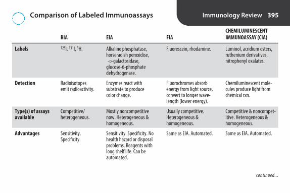
Comparison of Labeled Immunoassays Immunology Review 395
Labels
Detection
Type(s) of assays available
Advantages
CHEMILUMINESCENT IMMUNOASSAY (CIA)
Luminol, acridium esters,ruthenium derivatives, nitrophenyl oxalates.
Chemiluminescent mole-cules produce light fromchemical rxn.
Competitive & noncompet-itive. Heterogeneous & homogeneous.
Same as EIA. Automated.
FIA
Fluorescein, rhodamine.
Fluorochromes absorbenergy from light source,convert to longer wave-length (lower energy).
Usually competitive. Heterogeneous & homogeneous.
Same as EIA. Automated.
RIA
125I, 131I, 3H.
Radioisotopesemit radioactivity.
Competitive/heterogeneous.
Sensitivity. Specificity.
EIA
Alkaline phosphatase,horseradish peroxidise, -D-galactosidase,
glucose-6-phosphate dehydrogenase.
Enzymes react with substrate to produce color change.
Mostly noncompetitivenow. Heterogeneous &homogeneous.
Sensitivity. Specificity. Nohealth hazard or disposalproblems. Reagents withlong shelf life. Can be automated.
continued...
2956_Ch05_367-416 30/01/14 4:27 PM Page 395

Comparison of Labeled Immunoassays continued
Immunology Review 396
Disadvantages
Use
CHEMILUMINESCENT IMMUNOASSAY (CIA)
Quenching of light emis-sion by some biologicalmaterials.
Common.
FIA
Autofluorescence fromorganic substances inserum. Nonspecific bind-ing to substances inserum. Expensive, dedi-cated instrumentation.
Common.
RIA
Radiation hazard.Short shelf life ofreagents. Disposalproblem. Licens-ing & federal regulations.
Not common.
EIA
Natural inhibitors in somespecimens. Nonspecificprotein binding.
Common.
2956_Ch05_367-416 30/01/14 4:27 PM Page 396

Nontreponemal Tests for Syphilis Immunology Review 397
Method
Detects
Antigen
Positive reaction
Specimen(s)
Reactivity during disease
False positives
Other
VDRL
Flocculation
Reagin (ab against cardiolipin that is in serum of pts with syphilis)
Cardiolipin
Microscopic clumps
Inactivated serum, CSF
May be neg in primary stage. Titers usually peak during secondaryor early late stages. Titers in late stage, even when untreated.More rapid decline with treatment. Becomes nonreactive in 1–2 yr following successful treatment.
Biologic false pos with infectious mononucleosis (IM), infectioushepatitis, malaria, leprosy, lupus erythematosus, rheumatoidarthritis, advanced age, pregnancy. Reactive in other treponemalinfections such as yaws & pinta.
Screening test. Reactives should be confirmed by treponemal test.Replaced by RPR for serum. Still performed on CSF for Dx of neu-rosyphilis.
RPR
Flocculation.
Reagin.
Cardiolipin with charcoal.
Macroscopic agglutination.
Serum (inactivation not required),plasma.
Same as VDRL.
Same as VDRL.
Used for screening & treatmentmonitoring. Screening test. Reactives should be confirmed by treponemal test.
2956_Ch05_367-416 30/01/14 4:27 PM Page 397

Treponemal Tests for Syphilis Immunology Review 398
Detects
Reagent(s)
Positive reaction
Specimen(s)
Reactivity during disease
ANTIBODY CAPTURE ENZYME-LINKED IMMUNOSORBENT ASSAY (ELISA)
Antibody to T. pallidum.
Enzyme-labeled treponemal ag.
Color development following additionof substrate.
Serum
High sensitivity in primary syphilis.Decreases in later stages.
FLUORESCENT TREPONEMALANTIBODY ABSORPTION(FTA-ABS)
Antibody to T. pallidum
Sorbent (nonpathogenic tre-ponemes—Reiter strain),slides with Nichols strain of T.pallidum, fluorescein-labeledantihuman globulin
Fluorescence
Serum, CSF
Usually pos before reagin tests.Some false negs in primarysyphilis. Usually pos for life
TREPONEMA PALLIDUMPARTICLE AGGLUTINATION(TP-PA)
Antibody to T. pallidum
Colored gelatin particlescoated with treponemal ag
Agglutination of sensitizedgel particles. Smooth matover surface of well
Serum
Not as sensitive in primarysyphilis as FTA. Sensitivityclose to 100% in secondarysyphilis. Usually pos in latestages
continued...
2956_Ch05_367-416 30/01/14 4:27 PM Page 398

Treponemal Tests for Syphilis continued Immunology Review 399
False positives
Other
ANTIBODY CAPTURE ENZYME-LINKED IMMUNOSORBENT ASSAY (ELISA)
Similar to other treponemal tests.
Can be automated. Used for screening& for confirmation of reactive nontre-ponemal test. When used for screen-ing, reactives should be confirmedwith nontreponemal test (reverse sequence screening), followed by TP-PA or FTA-ABS if nontreponemaltest is nonreactive. IgM capture assayfor Dx of congenital syphilis.
FLUORESCENT TREPONEMALANTIBODY ABSORPTION(FTA-ABS)
Fewer than nontreponemaltests. Reactive with other treponemal diseases, e.g.,yaws, pinta
Sorbent removes nonspecificantibodies. Used to confirm re-active nontreponemal test. Notgood for treatment monitoring
TREPONEMA PALLIDUMPARTICLE AGGLUTINATION(TP-PA)
Fewer than nontreponemaltests
Sorbent removes nonspecificantibodies. Used to confirmreactive nontreponemal test. Not good for treatmentmonitoring
2956_Ch05_367-416 30/01/14 4:27 PM Page 399

Interpretation of Syphilis Test Results Immunology Review 400
RESULTS INTERPRETATION
RPR reactive Pos for syphilisFTA reactive
RPR reactive Neg for syphilisFTA nonreactive
ELISA reactive Pos for syphilisRPR reactive
ELISA reactive Late, latent, or previous syphilisRPR nonreactiveFTA-ABS reactive
2956_Ch05_367-416 30/01/14 4:27 PM Page 400

Serological Tests for Other Bacterial Infections
Immunology Review 401
TEST
Anti-streptolysin O (ASO)
Anti-DNase B
Streptozyme
Helicobacter pylori antibodies
Mycoplasma pneumoniaeantibodies
Rickettsial antibodies
COMMENTS
Uses recombinant streptolysin ag. If ab pres-ent, ag-ab complexes form & ↑ light scatter.Replaces classic neutralization method.
Highly specific. May be pos when ASO is neg.
Uses sheep RBCs coated with several strepto-coccal ags. More false pos & false negs. Shouldbe used in conjunction with ASO & anti-DNase.Serial titers should be performed.
Most tests detect IgG. 25% ↓ in titer = successful treatment. Abs remain for years. Pos rapid tests should be confirmed by ELISA.
Can test for IgM & IgG abs. Replaces cold agglutinin, which was nonspecific.
Organism specific assays replace Weil-Felixrxn, which was nonspecific.
DIAGNOSIS
Sequelae of group A strep (GAS)infection: rheumatic fever, post-streptococcal glomerulonephritis
Sequelae of GAS infection: rheu-matic fever, glomerulonephritisfollowing skin infection, i.e., impetigo
Sequelae of GAS infection
Gastric & duodenal ulcers causedby H. pylori
Primary atypical pneumonia(PAP)
Typhus, Rocky Mountain spottedfever, other rickettsial infections
COMMON METHOD(S)
Nephelometry.
EIA, nephelometry.
Slide agglutination.
Method of choice: ELISA.Rapid tests, PCR available.
Most common: EIA. Alsoagglutination, IFA. Molec-ular methods available.
Gold standard: IFA, micro-IF. PCR available.
2956_Ch05_367-416 30/01/14 4:27 PM Page 401

Serological Tests for Infectious Mononucleosis (IM)
Immunology Review 402
Specificity
Occurrence
Tests
HETEROPHILE ANTIBODIES*
Nonspecific abs that agglutinate horse, sheep, &bovine RBCs. Heterophile abs are abs that reactwith similar ags from different species.
90% of patients develop in 1st month of illness. Canpersist for 1 yr. Neg in 10% of adults & up to 50% ofchildren with IM. If symptomatic & heterophile neg,test for EBV-specific abs.
Rapid latex agglutination, solid-phase immunoas-says. Ag is purified bovine RBC extract. Screeningtests.
EBV-SPECIFIC ANTIBODIES
Specific abs against Epstein-Barr virus (EBV) ags present in differ-ent phases of infection—early: early ag (EA), late: viral capsid ag(VCA), latent: EBV nuclear ag (EBNA).
Anti-VCA IgM appears at onset of symptoms, disappears in 3 mo.Anti-VCA IgG appears at onset of symptoms, persists for life.Anti-EBNA present during convalescence.Acute infection: Anti-VCA IgM, anti-VCA IgG, anti-EA.Past infection: Anti-EBNA, anti-VCA IgG, neg anti-VCA IgM.
Indirect immunofluorescent assays (IFA), ELISA, chemilumines-cent assays. IFA is gold standard but time consuming & harder tointerpret. Molecular tests can be used for immunocompromisedpatients who don’t produce abs.
2956_Ch05_367-416 30/01/14 4:27 PM Page 402

Hepatitis Tests Immunology Review 403
TEST
Hepatitis A Total anti-HAV
IgM anti-HAV
HAV RNA
Hepatitis B HBsAg
HBeAg
Total anti-HBc
IgM anti-HBc
Anti-HBe
Anti-HBs
SIGNIFICANCE
Past infection & immunity
Acute infection
Current infection
Acute or chronic infection, infectivity
Acute or chronic infection
Current or past infection or carrier
Current or recent infection
Recovery, reduced infectivity
Recovery & immunity
COMMENTS
Used to detect HAV in food & water.
1st serological marker to appear. Used to screen donor blood.Pos should be confirmed by repeat testing & another assaysuch as HBsAg neutralization or HBV DNA.
Indicates high degree of infectivity.
Predominantly IgG, which persists for life.
1st ab to appear. Useful for detecting HBV infection whenHBsAg is no longer detectable (“window period”). Used toscreen blood donors.
Ab that develops following immunization.
continued...
2956_Ch05_367-416 30/01/14 4:27 PM Page 403

Hepatitis Tests continued Immunology Review 404
TEST
HBV DNA
Hepatitis C Anti-HCV
HCV RNA
Hepatitis D (delta hepatitis)IgM anti-HDV
IgG anti-HDV
HDV RNA
Hepatitis E
SIGNIFICANCE
Current infection
Acute, chronic, or previous infection
Current infection
Acute or chronic infection
Recovery or chronic infection
Current infection
COMMENTS
Detectable 21 days before HBsAg. Used to monitor viral load &therapy.
Pos should be confirmed by recombinant immunoblot assay(RIBA) or molecular method.
Used for viral load testing, blood/organ donor screening. HCVgenotyping to determine optimal treatment.
HDV is a defective virus that can only occur in presence of HBV.
Marker of active viral replication. Used to monitor therapy.
Tests are currently not approved by FDA for use in U.S.
Most serological tests for hepatitis are by EIA or CIA. Molecular methods detect infections earlier.
2956_Ch05_367-416 30/01/14 4:27 PM Page 404

Hepatitis Serological Profiles Immunology Review 405
ACUTE HEPATITIS A
IgM anti-HAV +
HEPATITIS BIMMUNIZATION
HBsAg –Anti-HBc –Anti-HBs +
RECOVERY FROMHEPATITIS A
Total anti-HAV +
ACUTE HEPATITIS B
HBsAg +Total anti-HBc +IgM anti-HBc + Anti-HBs –
RECOVERY FROM HEPATITIS B
HBs Ag –Total anti-HBc + Anti-HBs +
CHRONIC HEPATITIS B/CARRIER
HBsAg +Total anti-HBc + IgM anti-HBc –Anti-HBs –
SYMPTOMSALT
HBsAg
HBeAg
HBV DNA
IgM Anti HBc
IgG Anti HBc
Anti HBsAnti HBe
0 1 2 3 4 5 6 12Months after infection
--------
Clinical indications of hepatitis B virus over time. (From Arneson W, Brickell J. Clinical Chemistry: A Laboratory Perspective. Philadelphia:FA Davis; 2007:258.)
2956_Ch05_367-416 30/01/14 4:27 PM Page 405

Immunology Review 406Appearance of HIV Markers
MARKER COMMENTS
Viral RNA Detectable within days of infection.
p24 ag Core coat for nucleic acids. Detectable in 2–3 wk. Becomes undetectable as abs develop, then detectable again in late stagesas immune system fails & virus replicates.
IgM ab Usually detectable in 2–8 wk. Transient. Peaks in about 1–2 wk, undetectable about 1–2 wk later.
IgG ab Detectable shortly after IgM. Gradual ↑ in titer over several months. Long lasting.
0 10 20 30 40 50 60 70Days After Exposure
*By 3rd generation EIA. IgM first, then IgG.
HIV-RNA
HIV p24 Ag
Anti-HIV Ab*
Appearance of HIV markers after exposure (exact times vary with virus, patient immune response, and tests used).
2956_Ch05_367-416 30/01/14 4:27 PM Page 406

HIV Screening Tests Immunology Review 407
TEST
EIA/ELISA1st generation2nd generation3rd generation4th generation
Rapid tests
Nucleic acid amplification testing (NAAT)
COMMENTS
EIA or CIA. P24 ag without HIV ab = acute infec-tion. P24 ag & HIV ab = established infection.Currently not approved for screening blooddonors.
Immunochromatographic assays. Can be per-formed on whole blood, serum, oral fluid.
Not cost effective for screening in most settings.Used for screening blood donors & infants. (Abtests aren’t reliable in children <18 months ofage. Abs from infected mother can be presenteven if child isn’t infected.)
DETECTS
IgG ab to HIV-1IgG ab to HIV-1/2 IgG & IgM ab to HIV-1/2IgG & IgM ab to HIV-1/2 & p24 ag
IgG & IgM ab to HIV
HIV RNA
WINDOW PERIOD*
6–12 wk6–12 wk3–4 wk2 wk
4–12 wk
5 days
All specimens with pos screening tests must be tested again by same or different screening test. If repeatedly pos, a confirmatory/supplemental test must be done.*Time between exposure & pos test result.
2956_Ch05_367-416 30/01/14 4:27 PM Page 407
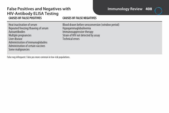
False Positives and Negatives with HIV-Antibody ELISA Testing
Immunology Review 408
CAUSES OF FALSE POSITIVES
Heat inactivation of serumRepeated freezing/thawing of serumAutoantibodiesMultiple pregnanciesLiver diseaseAdministration of immunoglobulinsAdministration of certain vaccinesSome malignancies
CAUSES OF FALSE NEGATIVES
Blood drawn before seroconversion (window period)HypogammaglobulinemiaImmunosuppressive therapyStrain of HIV not detected by assayTechnical errors
False neg infrequent. False pos more common in low-risk populations.
2956_Ch05_367-416 30/01/14 4:27 PM Page 408

Immunology Review 409HIV Confirmatory/Supplemental Tests
TEST
Western blot (WB)
Indirect immunofluorescent assays (IFA)
NAAT
DETECTS
Ab to HIV
Ab to HIV
HIV RNA
COMMENTS
Traditional confirmatory test but not as sensitive as 4th-gen EIA or NAAT. Inter-pretation is controversial but most labs report pos if at least 2 of the following3 bands are present: p24, gp41, gp120/160. NAAT required following neg orindeterminate results. Time-consuming, difficult to interpret.
Sensitivity & specificity comparable to Western blot. Not frequently used. Expensive, subjective.
Qualitative test used for confirmation.
More specific than screening tests. Fewer false pos.
2956_Ch05_367-416 30/01/14 4:27 PM Page 409

Tests to Stage and Monitor HIV Immunology Review 410
TEST
CD4 T-cell count
HIV-1 viral load assays
COMMENTS
HIV infects CD4 cells. # declines as disease progresses. <200/μL defines AIDS according to CDC. Alsoused to monitor therapy. Perform every 3–6 mo. Flow cytometry is gold standard.
Quantitative NAAT to determine plasma HIV RNA. Used to predict disease progression, determine whento start antiretroviral therapy, & monitor response to therapy. Test 2–8 wk after start of therapy & thenevery 3–4 mo. Same assay should be used in order to assess changes.
2956_Ch05_367-416 30/01/14 4:27 PM Page 410

Immunology Review 411Screening Tests for Systemic Lupus Erythematosus (SLE)
TESTS
Fluorescent antinuclear antibody (FANA)
EIA antinuclear antibody (EIA-ANA)
COMMENTS
Indirect immunofluorescence (IIF). Sub-strate is human epithelial tumor cell line(HEp-2). Detects abs to >100 autoantigens.Staining patterns reported but not consid-ered as significant as in past. Labor inten-sive. Subjective. Still considered goldstandard by rheumatologists.
Easier, less expensive. Can be automated.Interpretation not subjective. Not as manyantigens as IIF.
SENSITIVITY
High. 95%–100% ofSLE patients test pos.
May not be as sensitive as IIF.
SPECIFICITY
Low. Abs present in other autoimmunediseases, 2% of healthy individuals, 75%of elderly. Dilutions tested to eliminatelow titer rxn in normal population. Cutoffdilution to report pos usually ≥ 1:80. End-point titer may be reported. Generallyhigher in SLE.
Low.
2956_Ch05_367-416 30/01/14 4:27 PM Page 411

Immunology Review 412Tests for Specific Antinuclear Antibodies (ANA)
TESTS
Anti-dsDNA
Anti-Sm
Antihistone, anti-DNP, anti-SS-A/Ro, anti–SS-B/La, anti-nRNP
Extractable nuclear antigen (ENA) antibiotics
COMMENTS
Most specific ab for SLE. Titers correlate with diseaseactivity. Peripheral or homogeneous fluorescent pat-tern with indirect immunofluorescence (IIF). Othermethods: EIA, immunoblotting, immunodiffusion.
Coarsely speckled pattern with IIF. Other methods:EIA, immunoblotting, immunodiffusion.
Generally not useful for Dx of SLE. Used to Dx other connective tissue diseases. Methods: IIF, EIA,immunoblotting, immunodiffusion.
Immunodiffusion (Ouchterlony double diffusion) testpanel that typically tests for abs to Sm, SS-A/Ro, SS-B/La, RNP. Precipitin lines of identity/nonidentity.New method: multiplex bead assay. Immunoassayusing specific ag-coated beads & flow cytometry to detect multiple (currently 6–13) ANAs simultaneously.
SENSITIVITYFOR SLE
Low
Low
Low
Low
SPECIFICITY FOR SLE
High. Uncommon in other diseases or normalindividuals.
High. Uncommon in other diseases or normalindividuals.
Low.
Anti-Sm is specific forSLE.
2956_Ch05_367-416 30/01/14 4:27 PM Page 412

Serological Tests for Rheumatoid Arthritis (RA)
Immunology Review 413
TEST
Rheumatoid factor (RF)
Anti–cyclic citrullinated peptide antibody (Anti-CCP)
COMMON METHOD(S)
Agglutination, ELISA,nephelometry
ELISA
COMMENTS
Autoantibody (usually IgM) against IgG. Pos in 70%–80% of patientswith RA. Not specific for RA. Present with other autoimmune diseases,infections, & in some normal individuals. Agglutination tests only detectIgM RF. ELISA & nephelometry can also detect IgA & IgG classes of RF.Automated methods more common.
More specific for RA than RF. Associated with more severe form of RA.
2956_Ch05_367-416 30/01/14 4:27 PM Page 413
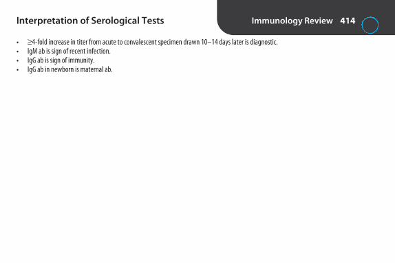
Interpretation of Serological Tests Immunology Review 414
• ≥ 4-fold increase in titer from acute to convalescent specimen drawn 10–14 days later is diagnostic.• IgM ab is sign of recent infection.• IgG ab is sign of immunity.• IgG ab in newborn is maternal ab.
2956_Ch05_367-416 30/01/14 4:27 PM Page 414

Serology Calculations Immunology Review 415
1. How would you prepare a 5% suspension of human group O RBCs?5% = 5 mL per 100 mLMix 5 mL of packed RBCs + 95 mL of buffer, or for a smaller amount, 0.5 mL of packed RBCs + 9.5 mL buffer. Any 1:20 dilution could be used.
(5:100 = 1:20)
2. How would you prepare 5 mL of a 1:10 dilution of serum?1 x
10 = 510x = 5x = 0.5 mL 0.5 mL diluted to 5 mL is a 1:10 dilution, so mix 0.5 mL of serum + 4.5 mL of buffer.
3. How would you prepare 10 mL of a 1:100 dilution from a 1:10 dilution?A. Determine the dilution factor to make a 1:100 dilution from a 1:10 dilution:
1/10 × 1/x = 1/1001/10x = 1/10010x = 100x = 10
A 1:10 dilution of a 1:10 dilution yields a 1:100 dilution. (1/10 × 1/10 = 1/100).B. Determine how to make 10 mL of a 1:10 dilution:
1:10 dilution is 1 part + 9 parts. To make 10 mL, mix 1 mL of solution + 9 mL of buffer.To make 10 mL of a 1:100 dilution from a 1:10 dilution, mix 1 mL of the 1:10 dilution + 9 mL of buffer.
4. What is the dilution in tube 4 of a twofold serial dilution, if tube 1 is undiluted?1 × 1/2 × 1/2 × 1/2 = 1/8
2956_Ch05_367-416 30/01/14 4:27 PM Page 415

2956_Ch05_367-416 30/01/14 4:27 PM Page 416

6S E C T I O N
ImmunohematologyReview
2956_Ch06_417-484 29/01/14 12:21 PM Page 417

Criteria for Whole Blood Donors (AABB) Immunohematology Review 418
ALLOGENEIC AUTOLOGOUS
Age
HGB/HCT
Temperature
Venipuncture site
Current as of date of publication. Check current AABB Technical Manual for updates.Autologous–No donations within 72 hr of surgery.
As determined by medical director.
HGB ≥ 11.0 g/dL or HCT ≥ 33%.
As determined by medical director. Bacteremia is cause for deferral.
As determined by medical director.
≥ 16 or as allowed by state law
HGB ≥ 12.5g/dL or HCT ≥ 38%
≤ 37.5°(99.5°F)
No infectious skin disease or scars indicative ofdrug use
2956_Ch06_417-484 29/01/14 12:21 PM Page 418

Donor Deferrals (AABB) Immunohematology Review 419
DEFERRAL CONDITION
2 days
2 weeks
4 weeks
6 weeks
8 weeks
12 months
3 years
Aspirin, if donor is sole source of platelets
Measles (rubeola), mumps, polio, typhoid, yellow fever vaccines
Rubella, chicken pox (varicella-zoster) vaccine
Pregnancy
Whole blood donation
SyphilisGonorrheaMucous membrane exposure to bloodSkin penetration with sharp contaminated with blood or body fluids Household or sexual contact with individual with hepatitisSexual contact with individual with HIV or at high riskIncarceration in correctional facility for >72 consecutive hrTravel to Iraq or area endemic for malaria Recipient of blood, blood components, plasma-derived clotting factor concentrates, or transplant
Malaria, or from an area endemic for malaria
continued...
2956_Ch06_417-484 29/01/14 12:21 PM Page 419

Donor Deferrals (AABB) continued Immunohematology Review 420
DEFERRAL CONDITION
Permanent
Not all inclusive. Current as of date of publication. Check current AABB Technical Manual for updates.
Parenteral drug useFamily history of Creutzfeldt-Jakob diseaseTreatment with pituitary growth hormone of human originViral hepatitis after 11th birthdayConfirmed positive HBsAgRepeatedly reactive anti-HBc on >1 occasionRepeatedly reactive HTLV on >1 occasion Present or past clinical or laboratory evidence of infection with HIV, HCV, HTLVHistory of babesiosis or Chagas’ disease
2956_Ch06_417-484 29/01/14 12:21 PM Page 420

Collection of Whole Blood Immunohematology Review 421
Skin preparation
Volume of blood routinely collected
Maximum volume
Low-volume collections
Volumes of anticoagulant
Time of collection
Samples for testing
Storage temperature of unit between collection & processing
Aseptic method, e.g., povidone-iodine scrub & prep solution.
450 mL ± 10% or 500 mL ± 10%, depending on collection bag.
10.5 mL of blood per kg of donor’s weight, including samples for testing (e.g., 473 mLfrom 100 lb [45 kg] donor).
300–404 mL in 450-mL bag or 333–449 mL in 500-mL bag. Labeled “low-volume.” RBCsmay be transfused, but other components shouldn’t be prepared because of abnormal anticoagulant-to-plasma ratio.
63 mL anticoagulant for 450-mL collection, 70 mL for 500-mL collection.
Usually <10 min. If >15–20 min, unit may not be suitable for preparation of platelets orplasma.
From diversion pouch or by 2nd phlebotomy.
20°–24°C if platelets are to be prepared; otherwise 1°–6°C.
2956_Ch06_417-484 29/01/14 12:21 PM Page 421

Apheresis Immunohematology Review 422
Explanation
Advantages
Donor requirements
Components collected
Therapeutic uses
Automated blood collection system that allows removal of 1 or more components from blood & return ofremainder to donor.
Allows collection of larger volume of specific components. Can reduce # of donors to which pt is exposed.
Vary with procedure.
RBCs (2 units can be collected at same time from donors who are larger & have higher HCT. 16 wk between donations).
Platelets. (Plateletpheresis. Can collect HLA matched for pts who are refractory to random plts. Can beleukoreduced during collection. Contain ≥ 3 x 10¹¹ plt.)
Plasma (Plasmapheresis). Granulocytes. (Leukapheresis. Not widely used to date.)Stem cells. (For bone marrow reconstitution in pt with cancer, leukemia, lymphoma. Autologous or
HLA matched.)
Therapeutic plasmapheresis (plasma exchange) used to remove abnormal plasma proteins & replacewith crystalloid, albumin, or FFP. Therapeutic cytapheresis used to remove cellular elements, e.g., ab-normal # platetets (to ↓ risk of hemorrhage or thrombosis), leukemic WBCs, lymphocytes (to induce immunosuppression).
2956_Ch06_417-484 29/01/14 12:21 PM Page 422

Donor Testing Required by AABB and/or FDA
Immunohematology Review 423
TypingABORh (including weak D)
Antibody screen
Syphilis testingAntibodies to Treponema pallidum or nontreponemal serological test for syphilis, e.g., RPR
Hepatitis testingHBsAgAnti-HBcAnti-HCVHCV RNA (NAT)
HIV testingAnti-HIV-1/2HIV-1 RNA (NAT)
Other infectious disease testingAnti-HTLV-I/IIWest Nile virus RNA (NAT)Anti–Tryanosoma cruzi (FDA recommends 1-time donor screening)
Test to detect bacterial contamination of plateletsCulture of platelets or FDA-approved rapid test (e.g., Pan Genera Detection [PGD] test)
For autologous donations, complete donor testing required if tf will occur outside collection facility; otherwise only ABO & Rh required.
2956_Ch06_417-484 29/01/14 12:21 PM Page 423

Anticoagulant/Preservative Solutions Immunohematology Review 424
NAME ABBREVIATION RBC SHELF LIFE COMMENTS
Acid citrate-dextrose (Formula A) ACD-A 21 days Citrate prevents coagulation by chelating Ca2+.Dextrose (glucose) supports ATP generation.Used for apheresis.
Citrate-phosphate-dextrose CPD 21 days Higher pH preserves 2,3-DPG better. Better O2delivery.
Citrate-phosphate-double-dextrose CP2D 21 days Contains 100% more glucose than CPD.
Citrate-phosphate-dextrose with adenine CPDA-1 35 days Adenine increases ADP, which increases synthesis of ATP. Contains more glucose to sustain cells during longer storage.
2956_Ch06_417-484 29/01/14 12:21 PM Page 424
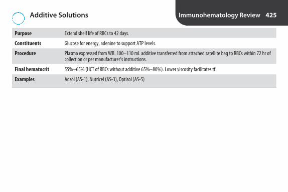
Additive Solutions Immunohematology Review 425
Purpose Extend shelf life of RBCs to 42 days.
Constituents Glucose for energy, adenine to support ATP levels.
Procedure Plasma expressed from WB. 100–110 mL additive transferred from attached satellite bag to RBCs within 72 hr ofcollection or per manufacturer’s instructions.
Final hematocrit 55%–65% (HCT of RBCs without additive 65%–80%). Lower viscosity facilitates tf.
Examples Adsol (AS-1), Nutricel (AS-3), Optisol (AS-5)
2956_Ch06_417-484 29/01/14 12:21 PM Page 425

Open and Closed Systems Immunohematology Review 426
EXPLANATION EFFECT ON EXPIRATION DATE OF COMPONENT
Open system
Closed system
Components stored at 1°–6°C must be used within 24 hrafter system opened; components stored at 20°–24°C within 4 hr.
No change.
Seal on unit is broken to attach external transferbag. Exposure to air poses threat of bacterialcontamination.
Sterility maintained through use of attachedsatellite bags or sterile connecting device thatwelds tubing from 1 bag to another. No exposureto air.
2956_Ch06_417-484 29/01/14 12:21 PM Page 426
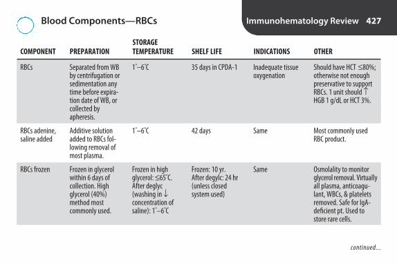
Blood Components—RBCs Immunohematology Review 427
STORAGE COMPONENT PREPARATION TEMPERATURE SHELF LIFE INDICATIONS OTHER
RBCs
RBCs adenine, saline added
RBCs frozen
Should have HCT ≤ 80%;otherwise not enoughpreservative to supportRBCs. 1 unit should ↑HGB 1 g/dL or HCT 3%.
Most commonly usedRBC product.
Osmolality to monitorglycerol removal. Virtuallyall plasma, anticoagu-lant, WBCs, & plateletsremoved. Safe for IgA-deficient pt. Used tostore rare cells.
Separated from WBby centrifugation orsedimentation anytime before expira-tion date of WB, orcollected byapheresis.
Additive solutionadded to RBCs fol-lowing removal ofmost plasma.
Frozen in glycerolwithin 6 days of collection. Highglycerol (40%)method most commonly used.
1°–6°C
1°–6°C
Frozen in highglycerol: ≤ 65°C.After deglyc (washing in ↓concentration ofsaline): 1°–6°C
35 days in CPDA-1
42 days
Frozen: 10 yr. After degylc: 24 hr(unless closed system used)
Inadequate tissueoxygenation
Same
Same
continued...
2956_Ch06_417-484 29/01/14 12:21 PM Page 427

Blood Components—RBCs continued Immunohematology Review 428
STORAGE COMPONENT PREPARATION TEMPERATURE SHELF LIFE INDICATIONS OTHER
Washed RBCs
RBCs leukocytes reduced
RBCs irradiated
Not a substitute for leuko-reduced RBCs. About 20%of RBCs lost in process.
Must retain 85% of original RBCs. <5 × 10⁶
WBCs.
For prevention of graft-vs.-host disease.Kills donor T cells.
RBCs washed withsaline.
Filtration or aphere-sis processing.
Irradiation at 2,500 cGy.
1°–6°C
1°–6°C
1°–6°C
24 hr after washing
Closed system:same as RBCs. Opensystem: 24 hr
Original outdate or28 days from irradi-ation, whichevercomes 1st
History of severeallergic rxn (e.g.,IgA, other plasmaproteins)
History of febrilerxn
Immunodeficiency,malignancy, bonemarrow transplant,tf with blood fromblood relative, in-trauterine &neonatal tf
2956_Ch06_417-484 29/01/14 12:21 PM Page 428

Blood Components—Plasma and Derivatives Immunohematology Review 429
STORAGE COMPONENT PREPARATION TEMPERATURE SHELF LIFE INDICATIONS OTHER
Fresh frozen plasma (FFP)
Cryoprecipitate
Contains all coag factors.Check for evidence ofthawing & refreezing.Thawed at 30°–37°C or byFDA-approved microwave.
Used for hemophilia A &von Willebrand diseaseonly if factor VIII concen-trate or recombinant factor preparations notavailable. Should contain≥ 80 IU of factor VIII &≥ 150 mg of fibrinogen.
Plasma separatedfrom WB & frozenwithin 8 hr of collection
Prepared by thaw-ing FFP at 1°–6°C,removing plasma,& refreezing within1 hr
Frozen: ≤ –18°C.After thawing:1°–6°C
Frozen: ≤ –18°C.After thawing: RT
Frozen: 12 mo. After thawing: 24 hr
Frozen: 12 mo. After thawing: singleunits 6 hr, pools 6 hr if sterile connecting device used, other-wise 4 hr
Deficiency of coagfactors
Fibrinogen & factorXIII deficiencies
2956_Ch06_417-484 29/01/14 12:21 PM Page 429
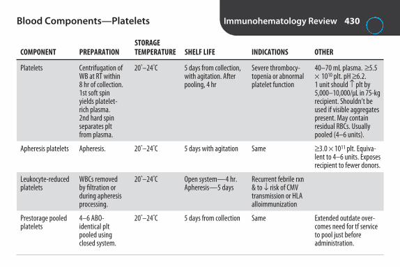
Blood Components—Platelets Immunohematology Review 430
STORAGE COMPONENT PREPARATION TEMPERATURE SHELF LIFE INDICATIONS OTHER
Platelets
Apheresis platelets
Leukocyte-reduced platelets
Prestorage pooled platelets
40–70 mL plasma. ≥ 5.5× 10¹⁰ plt. pH ≥ 6.2. 1 unit should ↑ plt by5,000–10,000/μL in 75-kgrecipient. Shouldn’t beused if visible aggregatespresent. May containresidual RBCs. Usuallypooled (4–6 units).
≥ 3.0 × 10¹¹ plt. Equiva-lent to 4–6 units. Exposesrecipient to fewer donors.
Extended outdate over-comes need for tf serviceto pool just before administration.
Centrifugation ofWB at RT within8 hr of collection.1st soft spinyields platelet-rich plasma. 2nd hard spinseparates pltfrom plasma.
Apheresis.
WBCs removedby filtration orduring apheresisprocessing.
4–6 ABO-identical pltpooled usingclosed system.
20°–24°C
20°–24°C
20°–24°C
20°–24°C
5 days from collection,with agitation. Afterpooling, 4 hr
5 days with agitation
Open system—4 hr.Apheresis—5 days
5 days from collection
Severe thrombocy-topenia or abnormalplatelet function
Same
Recurrent febrile rxn& to ↓ risk of CMVtransmission or HLAalloimmunization
Same
2956_Ch06_417-484 29/01/14 12:21 PM Page 430

Labeling Requirements for Blood and Components
Immunohematology Review 431
• ISBT 128 bar-code symbology • Name of product• Method of preparation (whole blood, apheresis)• Storage temperature • Preservatives/anticoagulant • Number of units in pooled components • Name, address, registration number, & license number
of collection & processing facility• Expiration date (& time if applicable). When expiration
time not indicated, expiration is at midnight.
• Identification number for unit or pool• Donor category (volunteer, autologous)• ABO group & Rh type, if applicable.• Special handling information• Statements regarding recipient identification, Circular
of Information, infectious disease risk, & prescription requirement
• Autologous units: “For Autologous Use Only.” Biohazard label if any infectious disease markers arepos or “Donor Untested” if testing not performed.
2956_Ch06_417-484 29/01/14 12:21 PM Page 431

Leukocyte Reduction (Leukoreduction) Immunohematology Review 432
Purpose
WBCs
Methods
To ↓ WBCs to ↓ febrile nonhemolytic transfusion reactions, transmission of CMV, & HLA alloimmunization
<5 × 10⁶
• By apheresis processing• By filtration during manufacture of components or after storage. Prestorage leukocyte reduction is most effective.
WBCs removed before they release cytokines• Use of filter during infusion
2956_Ch06_417-484 29/01/14 12:21 PM Page 432

RBC Storage Lesion Immunohematology Review 433
INCREASED DECREASED OTHER CHANGES
Lactic acidPlasma K+
Plasma hemoglobinMicroaggregates
Shift to left of O2 dissociation curve (↑ HGB/O2 affinity, ↓ O2 delivery to tissues)ATP2,3-DPGpHGlucoseViable cellsLabile coagulation factors
2956_Ch06_417-484 29/01/14 12:21 PM Page 433

Primary Versus Secondary Response Immunohematology Review 434
PRIMARY SECONDARY (ANAMNESTIC)
Stimulus 1st exposure to ag Subsequent exposure to ag
Lag phase Days to months Hours
Type of antibody IgM at first. May switch to IgG after 2–3 weeks IgG(isotype switching)
Titer Rises slowly, peaks, then declines Rises faster & higher, stays elevated longer
PRIMARY SECONDARY
Steady state
Total AbLogarithmicincrease
IgM
IgG
Inductive period
Ant
ibod
y C
once
ntra
tion
in S
erum
Decline
Total Ab
IgG
IgMNegativephase
Primaryimmunogenicstimulation
Secondaryimmunogenicstimulation
Schematic representation of primary and secondary antibody responses. Note the enhanced antibody production and expanded antibody-producing cell population during the secondary antibody response. (From Herscowitz HB. Immunophysiology. In Bellanti JA [ed]: Immunology III. Philadelphia: WB Saunders; 1985:117, with permission.)
2956_Ch06_417-484 29/01/14 12:21 PM Page 434

IgG Versus IgM Immunohematology Review 435
IgG IgM
Structure Monomer Pentamer
Number of ag-binding sites 2 10
Type of ab Immune Naturally occurring
Optimum temperature of reactivity 37°C 25°C or lower
Reacts in saline? No Yes
Reacts best by IAT? Yes No
Complement fixation Moderate Strong
Causes transfusion reactions? Yes Not usually, except ABO
Crosses placenta? Yes No
Causes hemolytic disease of the newborn/fetus? Yes No
Destroyed by sulfhydryl compounds (dithiothreitol [DTT], No Yes2-mercaptoethanol [2-ME])?
2956_Ch06_417-484 29/01/14 12:21 PM Page 435

Factors That Affect Agglutination in Tube Testing
Immunohematology Review 436
FACTOR COMMENTS
Sensitization stage
Temperature
pH
Ionic strength
Ag/ab ratio
Incubation time
Agglutination stage
Type of ab molecule
Density of ags & location on RBC surface
Zeta potential
Attachment of ab to ag
Clinically significant abs react best at 37°C.
Most abs react at pH 5.5–8.5.
Reducing ionic strength of medium facilitates interaction of ab with ag (e.g., low ionicstrength solution [LISS]).
Too much ab can cause prozone (false neg). Optimum serum-to-cell ratio is 80:1. Usually 2 drops serum to 1 drop of 2%–5% RBCs. Follow manufacturer’s directions.
Depends on medium. Usually 10–30 min. Follow manufacturer’s directions.
Formation of ag-ab bridges between RBCs
IgM is larger, can span distance between RBCs more easily.
Affects ease of attachment of abs.
Difference in charge between neg-charged RBC surface & cloud of pos ions that surroundRBCs. Reducing zeta potential allows RBCs to move closer together (e.g., enzyme treatmentof test cells).
2956_Ch06_417-484 29/01/14 12:21 PM Page 436

Comparison of Tube, Gel, and Solid-PhaseTesting
Immunohematology Review 437
TUBE TESTING GEL TESTING SOLID-PHASE TESTING
Reaction container
Principle
Pos rxn
Neg rxn
Adaptable to automation?
Advantages
Note: A cell button in tube testing is a solid agglutinate—4+ rxn. A cell button in solid phase is unagglutinated cells—neg rxn.
Microplate with RBC membranes bound tosurface of wells.
Abs in sample attach to RBC ags on surfaceof wells. After incubation, unbound ab re-moved by washing. Anti-IgG-labeled indi-cator RBCs added. Attach to abs bound toreagent RBC ags during centrifugation.
Indicator RBCs adhere diffusely to surfaceof well.
No adherence of RBCs. Button of RBCs inbottom of well.
Yes
Standardized. More sensitive than tubetesting. Rxn stable 2 days.
Glass test tubes
Abs attach to correspon-ding ags on RBCs, form-ing bridges betweencells. RBCs agglutinate.
Agglutinated RBCs orhemolysis
No agglutinated RBCs orhemolysis
No
Low cost
Plastic microtube containing dextran-acrylamide gel.
Ag-ab rxn results in agglutinated RBCs.Gel acts as sieve. Large agglutinatescan’t pass through, remain at top.Small agglutinates pass into gel. Unagglutinated cells go to bottom.
Agglutinated RBCs suspended in gel.Position indicates strength of rxn.Larger agglutinates at top.
Button of unagglutinated RBCs inbottom of microtube.
Yes
Standardized. More sensitive thantube testing. Rxn stable 2–3 days; canbe captured electronically. AHG testsdon’t require washing or control cells.
2956_Ch06_417-484 29/01/14 12:21 PM Page 437

Grading Reactions—Tube Versus Gel Immunohematology Review 438
TUBE* GEL
4+
3+
2+
1+
Mixed field
Neg
*In tube testing, hemolysis is also a pos rxn.
Solid band of agglutinated RBCs at top
Band of agglutinated RBCs near top with a few staggered below
Agglutinates throughout
Agglutinates predominantly in lower half of column with someRBCs at bottom
Layer of agglutinated RBCs at top & pellet of unagglutinated RBCsat bottom
Well-defined pellet of unagglutinated RBCs at bottom
One solid agglutinate
Several large agglutinates
Medium-sized agglutinates, clear background
Small agglutinates, turbid background
Some agglutinated RBCs in sea of free RBCs
No agglutinates
2956_Ch06_417-484 29/01/14 12:21 PM Page 438

Comparison of Tube, Gel, and Solid-Phase Reactions
Immunohematology Review 439
Tub
e
4+ 3+ 2+ 1+
Neg
So
lid P
has
eG
el
2956_Ch06_417-484 29/01/14 12:21 PM Page 439

ABO Genotypes and Phenotypes Immunohematology Review 440
PHENOTYPE GENOTYPE(S)
A AA, AO
B BB, BO
AB AB
O OO
Mother’s genotype
A O
Father’s genotype B AB BO
O AO OO
Using Punnett Square to Predict ABO Type
2956_Ch06_417-484 29/01/14 12:21 PM Page 440

Frequency of ABO Types Immunohematology Review 441
TYPE WHITES (%) BLACKS (%) HISPANICS (%) ASIANS (%)
O 45 49 57 40
A 40 27 31 27
B 11 19 10 25
AB 4 4 2 7
2956_Ch06_417-484 29/01/14 12:21 PM Page 441

ABO System Immunohematology Review 442
GROUP ANTIGEN(S) ON RBC ANTIBODY(S) IN SERUM
O Neither Anti-A, Anti-B
A A Anti-B
B B Anti-A
AB A & B Neither anti-A nor anti-B
2956_Ch06_417-484 29/01/14 12:21 PM Page 442

ABO Typing Immunohematology Review 443
FORWARD GROUPING REVERSE GROUPING TYPE
ANTI-A ANTI-B A CELLS B CELLS
0 0 + + O
+ 0 0 + A
0 + + 0 B
+ + 0 0 AB
Use of anti-A,B & A2 cells optional.
2956_Ch06_417-484 29/01/14 12:21 PM Page 443

ABO Discrepancies Immunohematology Review 444
ANTI-A ANTI-B A1 CELLS B CELLS POSSIBLE CAUSE* RESOLUTION
0
4+
4+
4+
3+
4+
*Other explanations may be possible.
Incubate reverse grouping at RT for 30 min. If still neg, incubate at 4°C for 15–30 min. Include controls: pt RBCs in 4% albumin, pt serum with O cells.
Type RBCs with anti-A1 (Dolichos biflorus lectin). Test serumwith several additional A1, A2, & O cells.
Use washed RBCs suspended in saline for forward grouping.Perform saline replacement technique in reverse grouping.
Perform ab panel at RT. If cold alloantibody identified, repeat reverse grouping with A1 & B cells that lack corresponding ag.
Type cells with anti-A1 (Dolichos biflorus lectin). Test serumwith several additional A1, A2, & O cells.
Check medical history for infection by GI bacteria (some haveenzymes that convert A ag to B-like ag.) Retype RBCs withdifferent monoclonal anti-B or acidified human anti-B (pH 6.0; doesn’t react with acquired B ag).
0
1+
2+
2+
1+
0
0
4+
2+
2+
0
4+
Missing isoagglu-tinins in group O
A2 with anti-A1
Rouleaux
AB with cold alloantibody
A2B with anti-A1
Acquired B antigen
0
0
4+
4+
4+
2+
2956_Ch06_417-484 29/01/14 12:21 PM Page 444

Rh Genotypes and Phenotypes Immunohematology Review 445
PHENOTYPE GENOTYPE(S)
Rh positive DD, Dd
Rh negative dd
Mother’s genotype
D d
Father’s genotype d Dd dd
d Dd dd
Using Punnett Square to Predict Rh Type
2956_Ch06_417-484 29/01/14 12:21 PM Page 445

Rh Antigens Immunohematology Review 446
FISHER-RACE WEINER ROSENFIELD
D Rho Rh1
C rh’ Rh2
E rh” Rh3
c hr’ Rh4
e hr” Rh5
2956_Ch06_417-484 29/01/14 12:21 PM Page 446
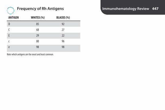
Frequency of Rh Antigens Immunohematology Review 447
ANTIGEN WHITES (%) BLACKS (%)
D 85 92
C 68 27
E 29 22
c 80 96
e 98 98
Note which antigens are the most and least common.
2956_Ch06_417-484 29/01/14 12:21 PM Page 447
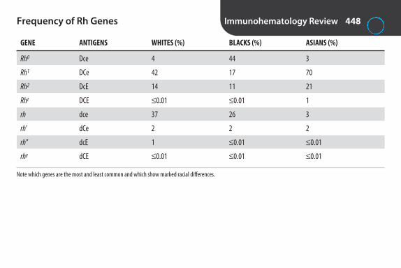
Frequency of Rh Genes Immunohematology Review 448
GENE ANTIGENS WHITES (%) BLACKS (%) ASIANS (%)
Rh0 Dce 4 44 3
Rh1 DCe 42 17 70
Rh2 DcE 14 11 21
Rhz DCE ≤ 0.01 ≤ 0.01 1
rh dce 37 26 3
rh’ dCe 2 2 2
rh” dcE 1 ≤ 0.01 ≤ 0.01
rhy dCE ≤ 0.01 ≤ 0.01 ≤ 0.01
Note which genes are the most and least common and which show marked racial differences.
2956_Ch06_417-484 29/01/14 12:21 PM Page 448

Breaking the Rh Code Immunohematology Review 449
SEE THINK EXAMPLE(S)
r before h “big” rh’ = C
h before r “little” hr’ = c
R presence of D Rh1 = DCe
r absence of D rh’ = dCe
1 or ’ C Rh1 = DCe(if no 1 or ’, then c) rh’ = dCe
Rh0 = Dce
2 or ” E Rh2 = DcE(if no 2 or ”, then e) rh” = dcE
Rh0 = Dce
0 c + e Rh0 = Dce
Z or y C + E rhy = dCE
Notes: Italics & superscripts denote genes, e.g., Rh’. Standard type is used for agglutinogens (antigens). Subscripts are used with R, e.g., Rho. Superscripts are used with r, e.g., rh’. The h’s are left out in shorthand notation, e.g., Rho = Ro.
2956_Ch06_417-484 29/01/14 12:21 PM Page 449

Rh Typing Sera Immunohematology Review 450
HIGH-PROTEIN ANTI-D LOW-PROTEIN ANTI-D
Source
Control
Comments
Mixture of monoclonal IgM & monoclonal or polyclonal IgG.
Any neg typing rxn serves as control, e.g., neg rxn with anti-A oranti-B. When RBCs react with all antisera (i.e., AB pos), run controlrecommended by manufacturer. (Usually pt RBCs with autologousserum or 6% or 8% albumin.)
Most widely used. Lower rate of false pos with Ig-coated RBCs.
Prepared from pools of human sera (immunized Rh-neg individuals).
Same ingredients as reagent, except noanti-D. Should be purchased from samemanufacturer as anti-D.
More false-pos than low-protein reagents,e.g., RBCs with pos DAT.
2956_Ch06_417-484 29/01/14 12:21 PM Page 450

Interpretation of Rh Typing Immunohematology Review 451
ANTI-D CONTROL Rh TYPE
+ 0 Pos
0 0 Neg*
+ + Invalid
*Test for weak D if donor or infant of mother being evaluated for RhIG.
FALSE POSITIVE FALSE NEGATIVE
Warm or cold autoagglutinins
Rouleaux
Polyagglutinable RBCs
Nonspecific agglutination due to ingredient in reagent, e.g., dye, preservative
Contaminated or incorrect reagent
Failure to add reagent
RBC suspension too heavy
Resuspending cell button too vigorously
Contaminated or incorrect reagent
Blocking of ag sites by abs, e.g., severe HDFN due to anti-D
Causes of False Rh Typing Results
2956_Ch06_417-484 29/01/14 12:21 PM Page 451
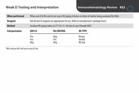
Weak D Testing and Interpretation Immunohematology Review 452
When performed When anti-D & Rh control are neg in Rh typing of donor or infant of mother being evaluated for RhIG.
Reagent Not all anti-D reagents are appropriate for use. Refer to manufacturer’s package insert.
Method Incubate Rh typing tubes at 37°C for 15–60 min & carry through AHG.*
Interpretation ANTI-D Rh CONTROL Rh TYPE
Pos Neg Rh posPos Pos InvalidNeg Neg Rh neg
*RBCs with pos DAT will react in any IAT test.
2956_Ch06_417-484 29/01/14 12:21 PM Page 452

Selection of Rh Type for Transfusion Immunohematology Review 453
RECIPIENT TYPE Rh TYPE PATIENT CAN RECEIVE
Rh pos Rh pos or Rh neg
Weak D Rh pos or Rh neg
Rh neg Rh neg only, especially women of childbearing age. (If Rh pos must be given in emergency, RhIG can be given toprevent immunization.)
2956_Ch06_417-484 29/01/14 12:21 PM Page 453

Frequency of Other Selected Blood GroupAntigens
Immunohematology Review 454
ANTIGEN WHITES (%) BLACKS (%)
K 9 2
k 99.8 100
Fya 68 13
Fyb 80 23
Jka 76 92
Jkb 74 48
M 79 74
N 70 75
S 55 30
s 90 92
Lea 22 23
Leb 72 55
P1 79 94
Note which antigens are the most and least common and which show marked racial differences.
2956_Ch06_417-484 29/01/14 12:21 PM Page 454
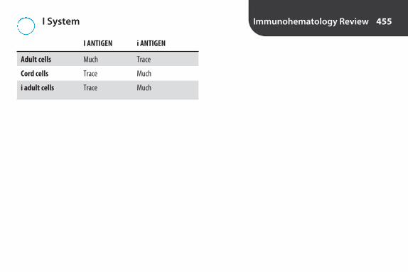
I System Immunohematology Review 455
I ANTIGEN i ANTIGEN
Adult cells Much Trace
Cord cells Trace Much
i adult cells Trace Much
2956_Ch06_417-484 29/01/14 12:21 PM Page 455

Antibody Characteristics Immunohematology Review 456
Naturally occurring
Clinically significant
Warm antibodies
Cold antibodies
Usually only react in AHG
Can react in any phase of testing
Detection enhanced by enzyme treatment of test cells
Not detected with enzyme treatment of test cells
Enhanced by acidification
Show dosage
Bind complement
Cause in vitro hemolysis
Labile in vivo & in vitro
Common cause of anamnestic response (delayed tf rxn)
ABO, Lewis, P1, MN, Lua
ABO, Rh, Kell, Duffy, Kidd, SsU
Rh, Kell, Duffy, Kidd
M, N, P1
Kell, Duffy, Kidd
Lewis
Rh, Lewis, Kidd, P1
M, N, Duffy
M
Rh other than D, MNS, Duffy, Kidd
I, Kidd, Lewis
ABO, Lewis, Kidd, Vell, some P1
Kidd
Kidd
continued...
2956_Ch06_417-484 29/01/14 12:21 PM Page 456

Antibody Characteristics continued Immunohematology Review 457
Associated with paroxysmal nocturnal hemoglobinuria
Associated with cold agglutinin disease & Mycoplasma pneumoniae infections
Associated with infectious mononucleosis
Anti-P
Anti-I
Anti-i
2956_Ch06_417-484 29/01/14 12:21 PM Page 457

Antigen-Antibody Enhancement Immunohematology Review 458
REAGENT EXPLANATION
Albumin
Low ionic strength solution (LISS)
Polythyleneglycol (PEG)
Enzymes
22% bovine serum albumin. Reduces net neg charge of RBCs, allowing them to come closer together.
Lowers ionic strength of suspending medium, allowing ags & abs to move closer together more rapidly. Reduces incubation time for IAT.
Increases ab uptake. Used for detection & ID of weak IgG abs.
Ficin & papain most commonly used. Reduce RBC surface charge by cleaving sialic acid molecules. M, N, S, Fya, Fyb antigens destroyed.
2956_Ch06_417-484 29/01/14 12:21 PM Page 458
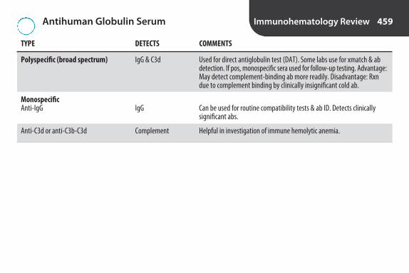
Antihuman Globulin Serum Immunohematology Review 459
TYPE DETECTS COMMENTS
Polyspecific (broad spectrum)
Monospecific Anti-IgG
Anti-C3d or anti-C3b-C3d
Used for direct antiglobulin test (DAT). Some labs use for xmatch & abdetection. If pos, monospecific sera used for follow-up testing. Advantage:May detect complement-binding ab more readily. Disadvantage: Rxndue to complement binding by clinically insignificant cold ab.
Can be used for routine compatibility tests & ab ID. Detects clinically significant abs.
Helpful in investigation of immune hemolytic anemia.
IgG & C3d
IgG
Complement
2956_Ch06_417-484 29/01/14 12:21 PM Page 459

Antiglobulin Testing Immunohematology Review 460
DIRECT (DAT) INDIRECT (IAT)
Detects
Specimen
Incubation
Application
False positives
False negatives
In vitro sensitization of RBCs by IgG ab
Serum, plasma, RBCs
Serum or plasma with reagent RBCs or RBCs withreagent antiserum
Ab screen, xmatch, RBC phenotyping, weak D testing
Cells with pos DATOvercentrifugation
Same as DAT, plus over- or underincubation
In vivo sensitization of RBCs by IgG ab
EDTA red cells preferred
None required
HDFN, tf rxn, autoimmune hemolytic anemia, drug-induced hemolytic anemia
Complement binding in vitro if RBCs are taken fromred-top tube & broad-spectrum AHG used
SepticemiaContamination of specimenWharton’s jelly in cord bloodOver-readingOvercentrifugation
Interruption in testingContamination, improper storage, or outdating of AHGFailure to add AHGNeutralization of AHG from inadequate washingDilution of AHG by residual salineOver- or undercentrifugation
2956_Ch06_417-484 29/01/14 12:21 PM Page 460

Antibody Identification Immunohematology Review 461
REACTIONS POSSIBLE EXPLANATION
Same strength & in 1 phase only
Varying strength
In different phases
All cells in AHG, autocontrol negative
All cells in AHG, autocontrol positive
All cells at 37ºC, neg in AHG, autocontrol positive
Suggestive of single ab
Multiple abs, abs exhibiting dosage, ags of differing strength
Combination of warm & cold abs, ab with wide thermal range
Multiple abs, ab to high frequency ag
Warm autoantibody
Rouleaux
2956_Ch06_417-484 29/01/14 12:21 PM Page 461

Interpreting Antibody Panels Immunohematology Review 462
Rule out (cross out)
Testing with selected cells
Confirmation
1. On row of ags at top of antigram, cross out those that are present on cells that didn’t react in any phase. (Some blood bankers only cross out homozygous ags to avoid missing a weak ab thatdisplays dosage.)
2. Circle ags that aren’t crossed out. An ab to 1 or more of these is present.3. Look for matching patterns, e.g., if serum only reacted with cells # 3 & # 5, & E ag is only on cells #
3 & # 5, ab is most likely anti-E.An ab will react with all cells that possess the corresponding ag (except for abs that demonstratedosage & only react with homozygous cells). An ab won’t react with any cells that lack the correspon-ding ag.
If other abs can’t be ruled out, further testing with selected cells might be required. Cells selected for testing should be pos for only 1 ag corresponding to abs in question, e.g., if pattern of reactivitymatches anti-Jka, but anti-K & anti-S can’t be ruled out, serum should be tested with cells of the following type:• Jk(a–), K+, S–• Jk(a–), K–, S+• Jk(a+), K–, S–Homozygous cells are preferred to avoid missing weak abs that demonstrate dosage.
Conclusive ID requires testing sufficient # of cells that are pos & neg for corresponding ag, e.g., 3 pos &3 neg. Some labs use other combinations that provide same probability (p) value of ≤ 0.05 (i.e., 95%confidence level that ab ID is correct). Once ab is identified, type pt RBCs for corresponding ag. Shouldbe neg.
2956_Ch06_417-484 29/01/14 12:21 PM Page 462

Cold Antibodies Immunohematology Review 463
Anti-A1 Only found in subgroups of A. Agglutinates A1 & A1B cells, but not A2 or O.
Anti-I Agglutinates all adult cells, except i adult. Doesn’t agglutinate cord cells.
Anti-i Agglutinates cord cells more strongly than adult cells.
Anti-H Most common in A1 & A1B. Agglutinates O cells most strongly, followed by A2 & B; then A1 & A1B.
Anti-IH Most common in A1 & A1B. Agglutinates cells that possess both I & H. Agglutinates adult O cells most strongly. Weaker rxnwith A1 cells. Doesn’t agglutinate cord cells.
2956_Ch06_417-484 29/01/14 12:21 PM Page 463

Compatibility Testing Immunohematology Review 464
• Specimen collected within 3 days of tf if pt has beenpregnant or transfused in preceding 3 months
• Confirmation of identifying information on requestform & specimen
• Check of blood bank records• Repeat ABO type on donor• Repeat Rh type on donor if unit is labeled Rh neg
(weak D not required)
• ABO type on recipient• Rh type on recipient (weak D not required)• Antibody screen on recipient• Xmatch recipient serum & donor RBCs• Retain pt specimen & unit segment at 1°–6°C for 7 days
after tf
2956_Ch06_417-484 29/01/14 12:21 PM Page 464

Crossmatches Immunohematology Review 465
EXPLANATION WHEN PERFORMED COMMENTS
Antiglobulin xmatch
Immediate spin (IS) xmatch
Computer (electronic) xmatch
Detects ABO incompatibility. Abscreen carried through AHG mustbe performed & must be neg.
Detects ABO incompatibility. ABOtyping of recipient must be done× 2. Ab screen carried throughAHG must be performed & mustbe neg. Computer system mustbe validated to prevent release ofABO-incompatible blood & mustalert user to discrepancies & incompatibilities.
Recipient serum & donorRBCs tested through AHG
Recipient serum & donorRBCs tested in IS only
Computer check of donorABO & Rh type & recipientABO & Rh type
If recipient has or had clinically significant ab
If recipient doesn’t have & never had clinically significant ab
If recipient doesn’t have & never had clinically significant ab
2956_Ch06_417-484 29/01/14 12:21 PM Page 465

The Major Crossmatch* Immunohematology Review 466
WILL WILL NOT
Detect ABO incompatibility Detect all ABO typing errorsDetect most abs against donor cells Detect most Rh typing errors
Detect all absDetect platelet or leukocyte antibodiesPrevent immunizationEnsure normal survival of RBCs
*Recipient serum & donor RBCs.
2956_Ch06_417-484 29/01/14 12:21 PM Page 466

Examples of Incompatible Crossmatches Immunohematology Review 467
REACTIONS POSSIBLE CAUSE* RESOLUTION
Neg ab screen, incompatible IS xmatch
1 ab screening cell & 1 donor pos in AHG
Ab screening cells & all donors except 1 neg at 37°C & in AHG. 1 donor pos in AHG only
Ab screening cells, donors, & autocontrol pos in AHG
Ab screening cells, donors, & autocontrol pos at 37°C, neg in AHG
*Other causes are possible.
Retype donor & recipient. Xmatch with ABO-compatible donor.
Identify ab. Xmatch units neg for corresponding ag.
Perform DAT on unit. If pos, return to collecting facility.
Best not to tf. If unavoidable, find “least incompatible” unit.
Saline replacement technique.
ABO incompatibility
Alloantibody
Positive DAT on donor
Warm autoantibody
Rouleaux
2956_Ch06_417-484 29/01/14 12:21 PM Page 467

Transfusion of Non-Group-Specific RBCs Immunohematology Review 468
PATIENT TYPE RBC TYPE PATIENT CAN RECEIVE PLASMA TYPE PATIENT CAN RECEIVE
O O only O, A, B, AB
A A, O A, AB
B B, O B, AB
AB AB, A, B, O AB
2956_Ch06_417-484 29/01/14 12:21 PM Page 468

Pretransfusion Testing Immunohematology Review 469
COMPONENT CROSSMATCH? OTHER
RBCs Yes Can be computer xmatch if neg ab screen & no record of previously detected abs.
Plasma No Must be ABO compatible with recipient RBCs.
Cryoprecipitate No All ABO groups acceptable. Rh type not considered.
Platelets No* Any ABO group acceptable. Compatible with recipient RBCs recommended. ABO-identical preferred.
*For apheresis platelets containing >2 mL RBCs, ABO compatibility & xmatch required.
2956_Ch06_417-484 29/01/14 12:21 PM Page 469

Conditions for Reissue of RBCs Immunohematology Review 470
• Maintained at 1°–10°C. (RBCs at RT reach 10°C in 30 min.)• Closure wasn’t broken.• At least 1 sealed segment remains attached to unit.• Unit is inspected before release.• Records indicate that blood has been inspected & is acceptable
for reissue.
Emergency Transfusions
• If time allows for typing, give ABO & Rh compatible;otherwise give O-neg RBCs.
• Label must indicate that xmatch wasn’t completed.• Physician must sign emergency release.• Routine testing must be completed & physician
notified immediately of any incompatibility.
2956_Ch06_417-484 29/01/14 12:21 PM Page 470

Transfusion-Associated Infections Immunohematology Review 471
INFECTION PREVENTION COMMENTS
HIV
Hepatitis B
Hepatitis C
HTLV-I and -II
Syphilis
Malaria
Babesiosis
Chagas’ disease
Cytomegalovirus
Estimated risk: 1 in 1,467,000 transfusions. With NAT,window period 9 days.
Estimated risk: 1 in 280,000 transfusions.
Estimated risk: 1 in 1,149,000 transfusions. With NAT,window period 7.4 days.
Rare in U.S.
Limited risk in refrigerated or frozen components. Spiro-chetes can’t survive in cold. Highest risk from platelets.
Plasmodium is transmitted in RBCs.
Babesia is transmitted in RBCs.
Trypanosoma cruzi is transmitted in blood. Potential riskin donors from Central & S. America.
CMV transmitted in WBCs. Risk to CMV-neg immunocom-promised pts & premature infants of CMV-neg mothers.
Donor testing
Donor testing
Donor testing
Donor testing
Donor testing
Donor history
Donor history
Donor history
Selected donor testing (not routine). Use of CMV-negor leukoreduced components for pts at risk.
continued...
2956_Ch06_417-484 29/01/14 12:21 PM Page 471

Transfusion-Associated Infections continued Immunohematology Review 472
INFECTION PREVENTION COMMENTS
Sepsis Highest risk from platelets because of RT storage. Donor screening (history, temp), aseptic technique,use of diversion pouches (1st 30–45 mL of bloodgoes into pouch so that skin plug doesn’t enter unit),tests to detect bacterial contamination of platelets(e.g., culturing 24 hr after collection)
2956_Ch06_417-484 29/01/14 12:21 PM Page 472

Acute Immunologic Transfusion Reactions* Immunohematology Review 473
TYPE CLINICAL SIGNS CAUSE LABORATORY FINDINGS OTHER
Hemolytic, intravascular
Febrile
Allergic
Anaphylactic
Most serious rxn. May be fatal. Usuallydue to tf of ABO-incompatible blood.
Common. Most often in multiply transfused pts or women with multiplepregnancies. Future tfs should be withleukoreduced components. Antipyretics(aspirin, acetaminophen) can be usedto premedicate.
Common. Treat with antihistamines. Tf rxn investigation not required.
Rare, but dangerous. Treated with epinephrine. Transfuse with washedproducts.
Immediate destructionof donor RBCs by recipient ab
Anti-leukocyte abs or cytokines
Foreign plasma proteins
Anti-IgA in IgA-deficient recipient
Fever; chills; shock;renal failure; DIC;pain in chest, back,or flank
Temp ↑ ≥ 1ºC or 2ºF during or shortlyafter tf, with noother explanation
Hives (urticaria),wheezing
Pulmonary edema,bronchospasms
In post-tf specimens: HGBin urine & serum; mixed-field DAT (unless donorcells are all destroyed); ↓haptoglobin, HGB, & HCT
None
None
None
continued...
2956_Ch06_417-484 29/01/14 12:21 PM Page 473

Acute Immunologic Transfusion Reactions* continued
Immunohematology Review 474
TYPE CLINICAL SIGNS CAUSE LABORATORY FINDINGS OTHER
Transfusion-related acute lung injury (TRALI)
*Occurring <24 hr after tf.
Most common cause of tf-relateddeaths in U.S. Related to infusion ofplasma. All components have been implicated. Reduced use of plasmafrom female donors appears to be reducing TRALI fatalities.
Unknown. Possiblydonor abs to WBC ags
Fever, chills, cough-ing, respiratory distress, fluid inlungs, ↓ BP within 6 hr of tf. Life-threatening.
None
2956_Ch06_417-484 29/01/14 12:21 PM Page 474
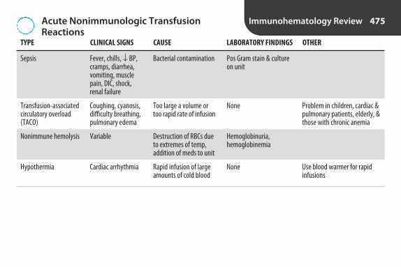
Acute Nonimmunologic Transfusion Reactions
Immunohematology Review 475
TYPE CLINICAL SIGNS CAUSE LABORATORY FINDINGS OTHER
Sepsis
Transfusion-associated circulatory overload (TACO)
Nonimmune hemolysis
Hypothermia
Problem in children, cardiac &pulmonary patients, elderly, &those with chronic anemia
Use blood warmer for rapid infusions
Bacterial contamination
Too large a volume ortoo rapid rate of infusion
Destruction of RBCs dueto extremes of temp,addition of meds to unit
Rapid infusion of largeamounts of cold blood
Fever, chills, ↓ BP,cramps, diarrhea,vomiting, musclepain, DIC, shock,renal failure
Coughing, cyanosis,difficulty breathing,pulmonary edema
Variable
Cardiac arrhythmia
Pos Gram stain & cultureon unit
None
Hemoglobinuria, hemoglobinemia
None
2956_Ch06_417-484 29/01/14 12:21 PM Page 475

Delayed Transfusion Reactions* Immunohematology Review 476
TYPE CLINICAL SIGNS CAUSE LABORATORY FINDINGS OTHER
ImmunologicHemolytic, extravascular
Alloimmunization
Transfusion-associated graft-vs.-host disease (TA-GVHD)
May be due to anamnestic response. Kidd antibodiesmost common cause. Usuallynot life-threatening.
Use leukoreduced componentsfor pts with WBC abs.
Irradiate components for pre-mature infants, intrauterineor exchange transfusion, stem cell or bone marrowtransplants, recipients ofblood from a 1st-degree relative, immunocompro-mised, pts with leukemia or lymphoma.
Donor RBCs sensitizedby recipient IgG ab & removed from circulation.
Development of abs toforeign RBCs, WBCs,platelets, plasma pro-teins following tf.
Viable T lymphs indonor blood attack recipient.
Fever, anemia, mildjaundice 2–14 daysafter tf.
None, unless subse-quently exposed tosame foreign ags.
Rash, nausea, vomit-ing, diarrhea, fever,pancytopenia. Usually fatal.
↑ bili, mixed-field DAT,↓ haptoglobin, ↓ HGB & HCT, pos ab screen
Ab to RBCs detected inab screen. Others requirespecialized testing.
None
continued...
2956_Ch06_417-484 29/01/14 12:21 PM Page 476

Delayed Transfusion Reactions* continued Immunohematology Review 477
TYPE CLINICAL SIGNS CAUSE LABORATORY FINDINGS OTHER
NonimmunologicIron overload
*Occurring >24 hr after tf.
Problem for pts receiving repeated tf over long period oftime, e.g., pts with thalassemia,sickle cell anemia, other chronicanemias.
Build up of iron inbody.
Diabetes, cirrhosis,cardiomyopathy
↑ serum ferritin
2956_Ch06_417-484 29/01/14 12:21 PM Page 477

Transfusion Reaction Investigation Immunohematology Review 478
Signs & symptoms of possible transfusion reaction
Specimens needed
Immediate steps
Signs of hemolytic reaction
Further steps if signs of possible hemolytic reaction
Fever; chills; respiratory distress; hyper- or hypotension; back, flank, chest, or abdominalpain; pain at site of infusion; hives (urticaria)*; jaundice; hemoglobinuria; nausea/vomiting;abnormal bleeding; oliguria/anuria.
Pre-tf blood.Post-tf blood.Post-tf urine.Segment from unit.Blood bag with administration set & attached IV solutions.
Stop tf.Check all IDs & labels.Repeat ABO on post-tf sample.Visual check of pre- & post-tf samples for hemolysis.DAT on post-tf sample. If pos, perform on pre-tf sample.
Hemolysis in post-tf sample, but not in pre-tf sample. Mixed field agglutination in DAT onpost-tf sample, but not on pre-tf sample.
Check HGB in first voided urine after tf.Repeat ABO & Rh on pre- & post-tf samples & unit.Repeat antibody screen on pre- & post-tf samples.AHG xmatch with pre- & post-tf samples.
continued...
2956_Ch06_417-484 29/01/14 12:22 PM Page 478

Transfusion Reaction Investigation continued Immunohematology Review 479
Additional tests that may be performed
Reporting of transfusion-related fatalities
*If symptoms dissipate after treatment with antihistamines, tf may be resumed, & investigation isn’t required.
Haptoglobin (↓ with hemolysis).Gram stain & culture of unit.Bilirubin 5–7 hr after tf (sign of extravascular hemolysis).BUN & creatinine (sign of renal involvement).
Must be reported to FDA Center for Biologics Evaluation & Research (CBER) by phone or e-mail ASAP.
2956_Ch06_417-484 29/01/14 12:22 PM Page 479
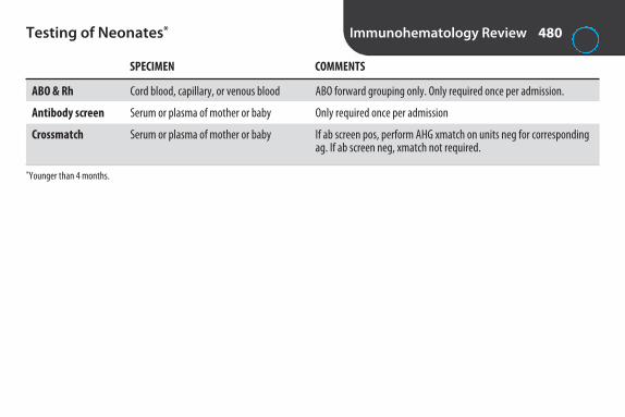
Testing of Neonates* Immunohematology Review 480
SPECIMEN COMMENTS
ABO & Rh
Antibody screen
Crossmatch
*Younger than 4 months.
ABO forward grouping only. Only required once per admission.
Only required once per admission
If ab screen pos, perform AHG xmatch on units neg for correspondingag. If ab screen neg, xmatch not required.
Cord blood, capillary, or venous blood
Serum or plasma of mother or baby
Serum or plasma of mother or baby
2956_Ch06_417-484 29/01/14 12:22 PM Page 480

Hemolytic Disease of the Fetus and Newborn(HDFN)
Immunohematology Review 481
ABO Rh
Mothers at risk Usually group O Rh neg
First child affected? Yes Not usually
Frequency Common Uncommon
Severity Mild Can be severe
DAT* Weak pos or neg Strong pos
Spherocytes? Yes Rare
Predictable? No Yes (maternal ab screen)
Preventable? No Yes (RhIG)
*Strength of rxn doesn’t correlate with severity of disease.
2956_Ch06_417-484 29/01/14 12:22 PM Page 481

Rh Immune Globulin (RhIG) Workup Immunohematology Review 482
Prenatal evaluation
Postpartum evaluation
ABO & Rh (weak D not required). If Rh pos, woman isn’t candidate for RhIG.
Ab screen. (Don’t use IS or RT incubation or AHG with anticomplement activity.) If pos, ID antibody. If anti-Dpresent, woman isn’t candidate.
ABO & Rh, including weak D, on baby. If baby is Rh neg, mother isn’t candidate.
If baby is Rh pos, draw mother’s blood after delivery & perform rosette test to screen for large fetal bleed.Mother’s RBCs incubated with anti-D. Anti-D coats fetal D-pos RBCs. Indicator D-pos RBCs added. Attach toanti-D on fetal D-pos RBCs, forming rosettes.
If rosette test pos, quantitate fetal bleed by flow cytometry or Kleihauer-Betke acid-elution test. Fetal cellsresist acid elution; stain pink. Adult cells lose hgb; appear as “ghosts.”
2956_Ch06_417-484 29/01/14 12:22 PM Page 482

Rh Immune Globulin (RhIG) Immunohematology Review 483
Composition
Purpose
Administration
Dose
Anti-D derived from pools of human plasma
Prevent immunization to D
Antepartum: To Rh neg woman at 28 wk of gestationPostpartum: Within 72 hr of delivery when Rh-neg woman delivers Rh-pos babyOther obstetric events: To Rh-neg woman after spontaneous or therapeutic abortion, ectopic pregnancy,
amniocentesis, chorionic villus sampling, antepartum hemorrhage, or fetal deathNote: May also be administered to Rh-neg recipients of Rh-pos blood or components
1 dose per 15 mL of D-pos fetal RBCs (30 mL of fetal WB). In calculating dose, if # to right of decimal point is ≥ 0.5, round up to next whole # & add 1 vial, e.g., 1.6 vials calculated = 2 + 1 = 3. If # to right of decimalpoint is <0.5, don’t round up; just use whole # & add 1 vial, e.g., 1.4 vials calculated = 1 + 1 = 2.
2956_Ch06_417-484 29/01/14 12:22 PM Page 483

Examples of Equipment/Reagent Quality Control
Immunohematology Review 484
Blood storage refrigerators & freezers, platelet incubators
Temperature recorder
Alarms
Waterbaths
Heat blocks
Centrifuges
Cell washers
Pipettes
Antisera
Reagent cells
Antihuman globulin
Not all inclusive. Follow manufacturer’s guidelines & lab’s Standard Operating Procedures Manual.
System for continuous temp monitoring & audible alarm.
Compare against thermometer daily. Calibrate as necessary.
Check high & low temp of activation quarterly.
Check temp daily.
Check temp daily. Periodically check each well.
Determine optimum speed & time for different procedures upon receipt, after repairs,& periodically. Check timer every 3 mo, RPM every 6 mo (with tachometer).
Check tube fill level daily, AHG volume monthly. Verify time & speed quarterly.
Calibrate quarterly.
Test with pos & neg controls each day of use. Use heterozygous cells for pos controls.
Check for hemolysis. Test each day of use with pos & neg controls.
Check anti-IgG activity each day of use by testing Rh-pos cells sensitized with anti-D.
2956_Ch06_417-484 29/01/14 12:22 PM Page 484

7S E C T I O N
Urinalysis and BodyFluids Review
2956_Ch07_485-522 29/01/14 12:23 PM Page 485

Urine SpecimensUrinalysis and Body
Fluids Review 486
TYPE USE COLLECTION COMMENTS
Random
First am
2-hr postprandial
24-hr
Clean catch
Catheterized
Suprapubic aspiration
Routine urinalysis (UA)
Routine UA
Diabetes mellitus monitoring
Quantitative chemical tests
Routine, culture
Culture
Culture
Anytime
Upon awakening
2 hr after eating
Discard 1st void on day 1 & notetime. Collect all urine for next 24 hr, including first void at same time on day 2
Cleanse external genitalia & collectmidstream in sterile container
Catheter inserted into urethra
Needle inserted through abdomeninto bladder
Not ideal since urine may be dilute & contaminated.
Best for screening. Most concentrated.
Best for detecting glycosuria.
Improper collection is commonsource of error. Refrigerate orkeep on ice. Preservatives required for some tests.
Less contamination. Do culturebefore UA.
Avoids contamination.
Avoids contamination.
2956_Ch07_485-522 29/01/14 12:23 PM Page 486
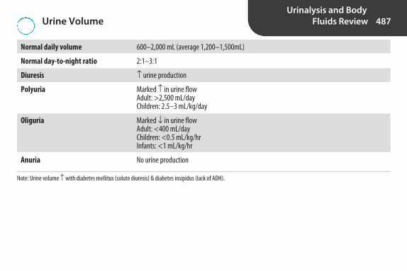
Urine VolumeUrinalysis and Body
Fluids Review 487
Normal daily volume 600–2,000 mL (average 1,200–1,500mL)
Normal day-to-night ratio 2:1–3:1
Diuresis ↑ urine production
Polyuria Marked ↑ in urine flowAdult: >2,500 mL/day Children: 2.5–3 mL/kg/day
Oliguria Marked ↓ in urine flowAdult: <400 mL/dayChildren: <0.5 mL/kg/hrInfants: <1 mL/kg/hr
Anuria No urine production
Note: Urine volume ↑ with diabetes mellitus (solute diuresis) & diabetes insipidus (lack of ADH).
2956_Ch07_485-522 29/01/14 12:23 PM Page 487

Urine Color Urinalysis and Body
Fluids Review 488
Normal Yellow due to urochrome
Dilute urine Colorless, pale yellow
Concentrated urine Dark yellow, amber
Bilirubin Amber, orange, yellow-green; yellow foam on shaking
Urobilin Amber, orange; no yellow foam on shaking
Homogentisic acid Normal on voiding; brown or black on standing
Melanin Brown or black on standing
Methemoglobin Brown or black
Myoglobin Red; brown on standing
Blood/hemoglobin Pink or red when fresh; brown on standing
Porphyrin Port-wine
Drugs, medications, food Green, blue, red, orange
Pseudomonas infection Green, blue-green
2956_Ch07_485-522 29/01/14 12:23 PM Page 488
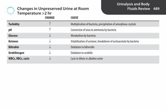
Changes in Unpreserved Urine at Room Temperature >2 hr
Urinalysis and Body
Fluids Review 489
CHANGE CAUSE
Turbidity ↑ Multiplication of bacteria, precipitation of amorphous crystals
pH ↑ Conversion of urea to ammonia by bacteria
Glucose ↓ Metabolism by bacteria
Ketones ↓ Volatilization of acetone, breakdown of acetoacetate by bacteria
Bilirubin ↓ Oxidation to biliverdin
Urobilinogen ↓ Oxidation to urobilin
WBCs, RBCs, casts ↓ Lysis in dilute or alkaline urine
2956_Ch07_485-522 29/01/14 12:23 PM Page 489

Chemical Urinalysis by Reagent StripUrinalysis and Body
Fluids Review 490
TEST NORMAL PRINCIPLE SIGNIFICANCE COMMENTS
pH
Protein
Glucose
Ketones
Blood
Acid with protein/meat diet; alkalinewith vegetarian diet. pH 9 = im-properly preserved specimen.
Buffered to pH 3. Most sensitive toalbumin. Orthostatic proteinuria—benign condition, protein is neg in1st am specimen, pos after standing.
Specific for glucose. More sensitive &specific than copper reduction. Normalrenal threshold = 160–180 mg/dL.
Most sensitive to acetoacetic acid.Less sensitive to acetone. Doesn’treact with beta-hydroxybutyric acid.
Uniform color = hgb or myoglobin.Speckled = RBCs.
First am: 5–6Random: 4.5–8
Neg–trace
Neg
Neg
Neg
Double indicator system
Protein error of indicator
Glucose oxidase/peroxidase
Sodium nitroprusside rxn
Pseudoperoxidase activity of hgb
Acid-base balance, man-agement of urinary tractinfection (UTI)/renal calculi
Possible renal disease
Possible diabetes mellitus
↑ fat metabolism (uncontrolled diabetesmellitus, vomiting, star-vation, low carb diet,strenuous exercise)
Hematuria, hemoglobinuria,myoglobinuria
continued...
2956_Ch07_485-522 29/01/14 12:23 PM Page 490

Chemical Urinalysis by Reagent Strip continuedUrinalysis and Body
Fluids Review 491
TEST NORMAL PRINCIPLE SIGNIFICANCE COMMENTS
Bilirubin
Urobilinogen
Nitrite
Leukocyte esterase
Specific gravity (SG)
Only conjugated bilirubin is excretedin urine.
Reagent strips don’t detect absenceof urobilinogen, only ↑.
Some bacteria reduce nitrates to nitrites. 1st am specimen best. ↑ sensitivity when urine in bladderat least 4 hr.
Longest rxn time. Detects intact &lysed grans & monos, not lymphs.Can be used with nitrite to screenurines for culture.
Only measures ionic solute. Not affected by urea, glucose, radi-ographic contrast media, plasmaexpanders. Not always same as SG by refractometer.
Neg
1 Ehrlich unit or 1mg/dL
Neg
Neg
Random specimen:1.003–1.030
Diazo reaction
Ehrlich’s aldehyde rxn ordiazo rxn
Greiss reaction
Leukocyte esterase rxn
pKa change of polyelectrolyte
Liver disease, biliary obstruction
Liver disease, hemolyticdisorders
UTI
UTI
Indication of kidney’s concentrating ability &state of hydration. ↑ in diabetes mellitus due toglucose. ↓ in diabetes insipidus due to ↓ ADH
2956_Ch07_485-522 29/01/14 12:23 PM Page 491

General Sources of Error with Reagent StripTesting
Urinalysis and Body
Fluids Review 492
POSSIBLE EFFECT COMMENTS
Failure to test within 2 hr of collection or to preserve correctly
Failure to bring refrigerated specimens to RT before testing
Failure to mix specimen well
Failure to follow manufacturer’s instructions
Failure to dip all test pads in urine
Prolonged dipping
Failure to remove excess urine from strip
Failure to read at recommended time
Failure to compare to color chart or read in good light
Failure to store strips properly
WBCs, RBCs settle out.
Reagents may leach from pads.
Store in capped original container at RT.
Changes in chemical composition
False-neg enzymatic rxns
False-neg leukocyte, blood
Erroneous results
False-neg rxns
False-neg rxns
Runover of chemicals to adjacentpads, distortion of colors
Erroneous results
Erroneous results
Erroneous results
continued...
2956_Ch07_485-522 29/01/14 12:23 PM Page 492

General Sources of Error with Reagent StripTesting continued
Urinalysis and Body
Fluids Review 493
POSSIBLE EFFECT COMMENTS
Failure to perform quality control
Expired strips
Highly pigmented urine
Run pos & neg control every 24 hr & when openingnew container.
Pigment masks true rxns. Test by alternate method.
Erroneous results
Erroneous results
Atypical colors, false-pos rxns
2956_Ch07_485-522 29/01/14 12:23 PM Page 493

Specific Sources of Error with Reagent StripTesting
Urinalysis and Body
Fluids Review 494
INCREASED OR FALSE POSITIVE DECREASED OR FALSE NEGATIVE
pH
Protein
Glucose
Ketones
Blood
Bilirubin
Urobilinogen
Nitrite
Acid runover from protein square.
Proteins other than albumin.
Unpreserved specimen, ↑ascorbic acid, ↑SG, ↓temp.
Improper storage. Acetone is volatile. Bacteria break down acetoacetic acid.
↑ascorbic acid, ↑nitrite, ↑SG (crenated RBCs), unmixed specimen.
Exposure to light, ↑ascorbic acid, ↑nitrite.
Improperly preserved specimen (oxidation to urobilin), formalin.
Non-nitrate-reducing bacteria, inadequate time in bladder,reduction of nitrites to N2, ↓dietary nitrate, antibiotics,↑ascorbic acid, ↑SG.
Improperly preserved specimen
Highly buffered alkaline urine, prolongeddipping, contaminated container, ↑SG
Contamination with peroxide or bleach
Red pigments, dyes, some meds
Menstruation, oxidizing agents, bacterialperoxidase
Highly pigmented urine
Highly pigmented urine
Highly pigmented urine, improperly preserved specimen (contaminating bacteria produce nitrites)
continued...
2956_Ch07_485-522 29/01/14 12:23 PM Page 494

Specific Sources of Error with Reagent StripTesting continued
Urinalysis and Body
Fluids Review 495
INCREASED OR FALSE POSITIVE DECREASED OR FALSE NEGATIVE
Leukocyte esterase
Specific gravity
Sources of error vary with brand of reagent strip. Refer to manufacturer’s package insert.
↑glucose, ↑protein, ↑ascorbic acid, ↑SG; antibiotics; reading too soon.
Alkaline urine. (Add 0.005 if pH is 6.5 or higher. Correctionis made by automated readers.)
Highly pigmented urine, oxidizing agents, formalin, nitrofurantoin, vaginaldischarge
↑ protein
2956_Ch07_485-522 29/01/14 12:23 PM Page 495

Other Urine Chemistry TestsUrinalysis and Body
Fluids Review 496
TEST DETECTS METHOD(S) SOURCES OF ERROR COMMENTS
Microalbumin
Sulfosalicylic acid (SSA)
Clinitest
Not detected by most urine dip-sticks. 50-200 mg/24 hr or ACR≥ 2.8 for males, ≥ 2.0 for femalespredictive of diabetic nephropathy.Strict control of glucose & bloodpressure can prevent progressionto end-stage renal disease.
Centrifuge & test supernatant. No longer used to confirm posprotein by dipstick.
Watch rxn to avoid missing passthrough. Not as sensitive or spe-cific for glucose as reagent strip.Reacts with other reducing sug-ars. Used to detect galactosemiain children <2 yr.
Immunoassay on 24-hrurine or albumin-to-creatinine ratio (ACR) on random sample. Dipsticks available for ACR.
Acid precipitation
Copper reduction
Albumin in low concentration
All proteins, in-cluding BenceJones proteins
Reducing substances
False pos: radiographic dyes,tolbutamide, some antibiotics,turbid urineFalse neg: highly buffered alkaline urine
False pos: ↑ascorbic acidFalse neg: “pass through”(Due to ↑ concentration. Goesthrough orange, returns togreen. Repeat using 2-dropmethod)
continued...
2956_Ch07_485-522 29/01/14 12:23 PM Page 496

Other Urine Chemistry Tests continuedUrinalysis and Body
Fluids Review 497
TEST DETECTS METHOD(S) SOURCES OF ERROR COMMENTS
Acetest
Ictotest
Most sensitive to acetoaceticacid. Can be used on urine,serum, plasma.
More sensitive than reagentstrip. Less affected by interferingsubstances.
Sodium nitroprusside reaction
Diazo reaction
Ketones
Bilirubin
False neg: Improperly storedspecimen (Acetone is lost into airif sample is left at RT. Bacteriabreak down acetoacetic acid.)
False pos: Urine pigmentsFalse neg: Exposure to light,improperly stored specimen,↑ascorbic acid, ↑nitrite
2956_Ch07_485-522 29/01/14 12:23 PM Page 497

Glucose Oxidase Versus Copper ReductionUrinalysis and Body
Fluids Review 498
GLUCOSE OXIDASE (REAGENT STRIP) COPPER REDUCTION (CLINITEST) MOST LIKELY INTERPRETATION*
+ + Glucose
+ 0 Glucose below sensitivity of Clinitest
0 + Non-glucose-reducing substance
0 0 No glucose or other reducing substances
*Defective reagents and presence of interfering substances should be ruled out.
2956_Ch07_485-522 29/01/14 12:23 PM Page 498

Epithelial Cells in the Urine SedimentUrinalysis and Body
Fluids Review 499
CELL DESCRIPTION ORIGIN CLINICAL SIGNIFICANCE COMMENTS
Squamous epithelial cell
Transitional epithelial cell
↑ numbers usually seen inurine from females. Mayobscure RBCs & WBCs. Reduced by collecting midstream clean-catchspecimen.
May form syncytia (clumps)
40–50 μm. Flat.Prominent round nucleus.
20–30μm. Spherical,pear-shaped, or polyhedral. Roundcentral nucleus.
Lower urethra,vagina
Renal pelvis,ureters, bladder,upper urethra
Usually none
Seldom significant
continued...
2956_Ch07_485-522 29/01/14 12:23 PM Page 499
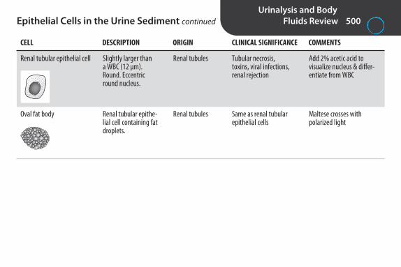
Epithelial Cells in the Urine Sediment continuedUrinalysis and Body
Fluids Review 500
CELL DESCRIPTION ORIGIN CLINICAL SIGNIFICANCE COMMENTS
Renal tubular epithelial cell
Oval fat body
Add 2% acetic acid to visualize nucleus & differ-entiate from WBC
Maltese crosses with polarized light
Slightly larger than a WBC (12 μm).Round. Eccentricround nucleus.
Renal tubular epithe-lial cell containing fatdroplets.
Renal tubules
Renal tubules
Tubular necrosis, toxins, viral infections,renal rejection
Same as renal tubularepithelial cells
2956_Ch07_485-522 29/01/14 12:23 PM Page 500

Blood Cells in the Urine SedimentUrinalysis and Body
Fluids Review 501
CELL DESCRIPTION ORIGIN CLINICAL SIGNIFICANCE COMMENTS
White blood cell (WBC)
Glitter cell
Red blood cell (RBC)
Normal: 0–8/HPF. Clumpsof WBCs associated withacute infection.
Seen in hypotonic urine.
Normal: 0–3/HPF. Crenatein hypertonic urine. Lyse inhypotonic urine & with 2%acetic acid.
Usually polys. About 12 μm.Granular appearance.
WBC with Brownian move-ment of granules. Stainfaintly or not at all.
Biconcave disk, about 7 μm.Smooth. Non-nucleated.
Kidney, bladder,or urethra
Same as WBC
Kidney, bladder,or urethra
Cystitis, pyelonephritis, tumors, renal calculi.
Same as WBC.
Infection, trauma, tumors,renal calculi. DysmorphicRBCs indicate glomerularbleeding.
2956_Ch07_485-522 29/01/14 12:23 PM Page 501

Normal Crystals Found in Acid or NeutralUrine
Urinalysis and Body
Fluids Review 502
CRYSTAL DESCRIPTION COMMENTS
Amorphous urates
Uric acid
Calcium oxalate
Form pink precipitate in bottom of tube. Mayobscure significant sediment. Dissolve bywarming to 60ºC.
Birefringent. Polarizes light.
Occasionally found in slightly alk urine. Mono-hydrate form may be mistaken for RBCs. Mostcommon constituent of renal calculi. From oxalate-rich foods.
Irregular granules.
Pleomorphic. 4-sided, 6-sided, star-shaped,rosettes, spears, plates. Colorless, red-brown,or yellow.
Octahedral (8-sided) envelope form is mostcommon. Also dumbbell & ovoid forms.
From Strasinger SK, Di Lorenzo MS. Urinalysis and Body Fluids, 5th ed. Philadelphia: FA Davis; 2008:113.
2956_Ch07_485-522 29/01/14 12:23 PM Page 502

Normal Crystals Found in Alkaline UrineUrinalysis and Body
Fluids Review 503
CRYSTAL DESCRIPTION COMMENTS
Amorphous phosphates
Triple phosphate
Ammonium biurate
Form white precipitate in bottom of tube. Dissolve with 2% acetic acid.
Seen in old specimens.
Irregular granules
“Coffin-lid” crystal
Yellow-brown “thorn apples” & spheres
From Strasinger SK, Di Lorenzo MS. Urinalysis and Body Fluids, 5th ed. Philadelphia: FA Davis; 2008:113.continued...
2956_Ch07_485-522 29/01/14 12:23 PM Page 503
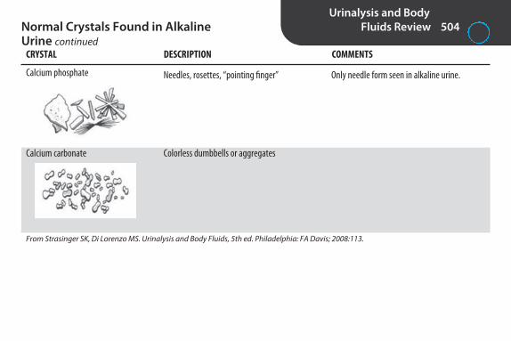
Normal Crystals Found in Alkaline Urine continued
Urinalysis and Body
Fluids Review 504
Calcium phosphate
Calcium carbonate
Only needle form seen in alkaline urine.Needles, rosettes, “pointing finger”
Colorless dumbbells or aggregates
CRYSTAL DESCRIPTION COMMENTS
From Strasinger SK, Di Lorenzo MS. Urinalysis and Body Fluids, 5th ed. Philadelphia: FA Davis; 2008:113.
2956_Ch07_485-522 29/01/14 12:23 PM Page 504

Abnormal Crystals*
Urinalysis and Body
Fluids Review 505
CRYSTAL DESCRIPTION SIGNIFICANCE COMMENTS
Leucine
Tyrosine
Cystine
Often seen with tyrosine.
Often seen with leucine.
Must differentiate from uric acid.Doesn’t polarize light. Confirm bycyanide-nitroprusside test.
Yellow, oily-looking spheres.Radial & concentric striations.
Fine yellow needles in sheavesor rosettes.
Hexagonal (6-sided).
Severe liver disease
Severe liver disease
Cystinuria
From Strasinger SK, Di Lorenzo MS. Urinalysis and Body Fluids, 5th ed. Philadelphia: FA Davis; 2008:117.
continued...
2956_Ch07_485-522 29/01/14 12:23 PM Page 505
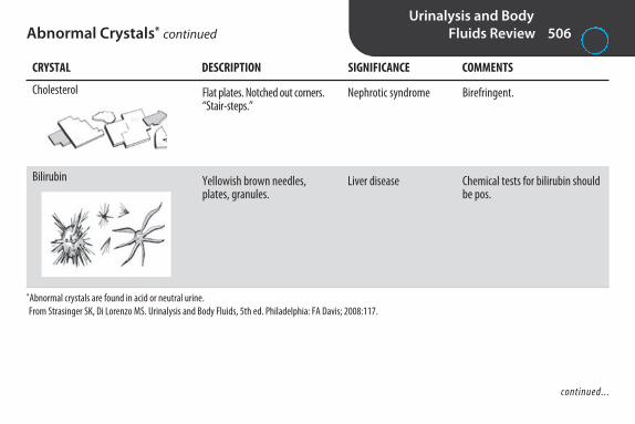
Abnormal Crystals* continuedUrinalysis and Body
Fluids Review 506
Cholesterol
Bilirubin
*Abnormal crystals are found in acid or neutral urine.From Strasinger SK, Di Lorenzo MS. Urinalysis and Body Fluids, 5th ed. Philadelphia: FA Davis; 2008:117.
Birefringent.
Chemical tests for bilirubin shouldbe pos.
Flat plates. Notched out corners.“Stair-steps.”
Yellowish brown needles,plates, granules.
Nephrotic syndrome
Liver disease
continued...
CRYSTAL DESCRIPTION SIGNIFICANCE COMMENTS
2956_Ch07_485-522 29/01/14 12:24 PM Page 506

CastsUrinalysis and Body
Fluids Review 507
TYPE DESCRIPTION SIGNIFICANCE COMMENTS
Hyaline
Granular
RBC
Blood
WBC
Epithelial cell
Most common type. Least significant.Tamm-Horsfall protein only. Dissolve in alkurine. Same refractive index as urine; may beoverlooked with bright light.
From disintegration of cellular casts.
IDs kidneys as source of bleeding. Most fragilecast. Often in fragments.
From disintegration of RBC casts.
IDs kidneys as site of infection.
Transitional & squamous epithelial cell aren’tseen in casts. Distal to renal tubules & collect-ing ducts where casts are formed.
Homogeneous with parallelsides, rounded ends.
Same as hyaline, but withgranules.
RBCs in cast matrix. Yellowish to orange color.
Contain hemoglobin. Yellowish to orange color.
WBCs in cast matrix. Irregular in shape.
Renal tubular epithelial cellsin cast matrix.
Normal: 0–2/LPF. ↑ with stress,fever, trauma, exercise, renaldisease.
Normal: 0–1/LPF. ↑ with stress,exercise, glomerulonephritis,pyelonephritis.
Acute glomerulonephritis,strenuous exercise.
Same as RBC cast.
Pyelonephritis.
Renal tubular damage.
continued...
2956_Ch07_485-522 29/01/14 12:24 PM Page 507

Casts continuedUrinalysis and Body
Fluids Review 508
TYPE DESCRIPTION SIGNIFICANCE COMMENTS
Waxy
Fatty
Broad
From degeneration of cellular & granularcasts. Unfavorable sign.
Maltese crosses with polarized light if lipid is cholesterol. Sudan III & Oil Red O stain triglycerides & neutral fats.
Formed in dilated distal tubules & collectingducts. “Renal failure casts.”
Homogeneous, opaque,notched edges, broken ends.
Casts containing lipiddroplets.
Wide. May be cellular, granular, or waxy.
Urinary stasis.
Nephrotic syndrome.
Advanced renal disease.
2956_Ch07_485-522 29/01/14 12:24 PM Page 508

Miscellaneous Urine Sediment Urinalysis and Body
Fluids Review 509
STRUCTURE DESCRIPTION SIGNIFICANCE COMMENTS
Bacteria
Yeast
Sperm
If clinically significant, WBCs present(unless patient is neutropenic).
Add 2% acetic acid to differentiate fromRBCs. RBCs lyse; yeast don’t. Pseudohy-phae indicate severe infection. WBCsare present in true yeast infections.
Rods, cocci.
5–7 μm. Ovoid, colorless,smooth, refractile. May bud & form pseudohyphae.
4–6 μm head, 40–60 μm tail.
UTI or contaminants.
Usually due to vaginal orfecal contamination. Maybe due to kidney infection.Often in urine of diabetics.
Usually not significant inadult. May be sign of sexual abuse in child.
continued...
2956_Ch07_485-522 29/01/14 12:24 PM Page 509

Miscellaneous Urine Sediment continuedUrinalysis and Body
Fluids Review 510
STRUCTURE DESCRIPTION SIGNIFICANCE COMMENTS
Trichomonas
Mucus
More common in females. Don’t reportunless motile.
May be mistaken for hyaline casts.
Resembles WBC. Flagella &undulating membrane. Rapid,jerky, nondirectional motility.
Transparent, long, thin, ribbon-like structure with tapering ends.
Parasitic infection of genital tract.
None.
2956_Ch07_485-522 29/01/14 12:24 PM Page 510

Renal DisordersUrinalysis and Body
Fluids Review 511
DISORDER CAUSE REAGENT STRIP SEDIMENT OTHER
Acute glomerulonephritis
Nephrotic syndrome
Pyelonephritis
Cystitis
Frequently follows untreated group A strepinfection
Hypoproteinemia, hyperlipidemia
Inflammation & damage to glomeruli
Increased glomerularpermeability
Kidney infection
Bladder infection
Protein, blood
Protein (large amount)
Protein, leukocyte esterase, nitrite
Leukocyte esterase, nitrite
RBCs (some dysmorphic),WBCs, hgb casts.
Casts (all kinds), free fat &oval fat bodies.
WBCs, WBC casts, bacteria.
WBCs, bacteria, possiblyRBCs. No casts.
2956_Ch07_485-522 29/01/14 12:24 PM Page 511

Urinalysis CorrelationsUrinalysis and Body
Fluids Review 512
PARAMETERS RELATIONSHIPS/CONSIDERATIONS
pH/microscopic
pH/nitrite/leukocyte esterase (LE)/microscopic
Protein/microscopic
Protein/blood/microscopic
Protein/specific gravity (SG)
Glucose/ketones
Glucose/microscopic
Glucose/protein/microscopic
Blood/microscopic
Ck pH when identifying crystals.RBCs, WBCs, & casts lyse at alk pH.
With bacterial UTI, usually have pos nitrite, pos LE. Ck for bacteria & WBCs. Bacteriaconvert urea to ammonia, ↑ pH.
Protein may indicate renal disease. Ck for casts.
Large amounts of blood or myoglobin can cause pos protein. Ck for RBCs. Present withhematuria, not with hemoglobinuria or myoglobinuria.
↑SG can cause false-pos trace protein. Trace protein more significant in dilute urine.
Ketones present with uncontrolled diabetes mellitus.
Yeast thrives in ↑glucose. WBCs should be present if true yeast infection.
Renal disease is common complication of diabetes mellitus. Ck for casts.
Pos blood, no RBCs: Blood rxn could be due to hemoglobin or myoglobin, or false posdue to bacterial peroxidase. Ck for bacteria.
Neg blood, RBCs seen: Could be false-neg blood due to ↑ascorbic acid. Yeast, or monohydrate calcium oxalate crystals could be misidentified as RBCs.
continued...
2956_Ch07_485-522 29/01/14 12:24 PM Page 512

Urinalysis Correlations continuedUrinalysis and Body
Fluids Review 513
PARAMETERS RELATIONSHIPS/CONSIDERATIONS
Bilirubin/urobilinogen
LE/microscopic
Nitrite/LE/microscopic
SG/microscopic
Liver disease: bili pos or neg, urobili ↑.Biliary obstruction: bili pos, urobili N.Hemolytic disorder: bili neg, urobili ↑.
Can have pos LE without WBCs (WBCs lysed).Can have pos LE & WBCs without bacteria (Trichomonas).
With pos nitrite, usually have pos LE, WBCs, & bacteria.Can have pos LE, WBCs, bacteria, & neg nitrite (non-nitrate-reducing bacteria).
RBCs & WBCs lyse in dilute urine. With ↑SG, RBCs & WBCs may crenate, cause false-neg blood & LE.
2956_Ch07_485-522 29/01/14 12:24 PM Page 513

Cerebrospinal FluidUrinalysis and Body
Fluids Review 514
NORMAL ABNORMALITIES COMMENTS
Color
Clarity
WBC
RBC
Glucose
Examine within 1 hr of collection to avoidfalse pos from lysis of RBCs. Centrifuge CSF & examine against white background.
Cells begin to lyse within 1 hr. Perform cellcount within 30 min of collection. Traditionallyperformed in hemacytometer, following lysis of RBCs with 3% glacial acetic acid. Can be performed on automated analyzers of sufficientsensitivity. Differential on stained smear follow-ing concentration (cytocentrifugation).
Limited diagnostic value.
Blood glucose method.
Colorless
Clear
Adult: 0–5/μLNewborn: 0–30/μL (mononuclear)
0
60%–70% of blood glucose
Xanthochromia = slight pink, orange,or yellow due to oxyhemoglobin orbilirubin. Seen with subarachnoidhemorrhage.
Traumatic tap = red or pink, decreas-ing from tube 1–3.
Cloudy with infection or bleeding.
↑ in meningitis.
↑ with subarachnoid hemorrhage,traumatic tap.
↓ in bacterial meningitis.
continued...
2956_Ch07_485-522 29/01/14 12:24 PM Page 514

Cerebrospinal Fluid continuedUrinalysis and Body
Fluids Review 515
NORMAL ABNORMALITIES COMMENTS
Protein
Cells
Biuret method not sensitive enough. Usetrichloracetic acid.
Nucleated RBCs may be seen due to bonemarrow contamination. Cartilage cells may be seen.
15–45 mg/dL
Lymphs, monos, ependymalcells, choroid plexus cells
↑ in meningitis & with traumatic tap.
Siderophages with subarachnoidhemorrhage. Blasts with leukemia.
2956_Ch07_485-522 29/01/14 12:24 PM Page 515

Differential Diagnosis of MeningitisUrinalysis and Body
Fluids Review 516
BACTERIAL VIRAL MYCOBACTERIAL FUNGAL
WBC
Differential
Protein
Glucose
Lactate
Other
↑
Lymphs, monos
↑
N or ↑
↑
Pos India Ink and/or latex aggluti-nation test with Cryptococcusneoformans
↑
Polys
↑
↑
↑
Pos Gram stain,bacterial antigen
↑
Lymphs
↑
N
N
↑
Lymphs, monos
↑
↑
↑
Weblike clot or pellicle
2956_Ch07_485-522 29/01/14 12:24 PM Page 516
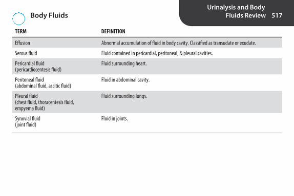
Body FluidsUrinalysis and Body
Fluids Review 517
TERM DEFINITION
Effusion
Serous fluid
Pericardial fluid (pericardiocentesis fluid)
Peritoneal fluid(abdominal fluid, ascitic fluid)
Pleural fluid(chest fluid, thoracentesis fluid, empyema fluid)
Synovial fluid(joint fluid)
Abnormal accumulation of fluid in body cavity. Classified as transudate or exudate.
Fluid contained in pericardial, peritoneal, & pleural cavities.
Fluid surrounding heart.
Fluid in abdominal cavity.
Fluid surrounding lungs.
Fluid in joints.
2956_Ch07_485-522 29/01/14 12:24 PM Page 517

Differentiation of Transudates and ExudatesUrinalysis and Body
Fluids Review 518
TRANSUDATE EXUDATE
Etiology
Type of process
Color
Clarity
Specific gravity
Protein
Fluid-to-serum protein ratio
Glucose
Spontaneous clotting
LD
WBC
Differential
Condition involving membranes within body cavity(infection, malignancy, inflammation, hemorrhage)
Inflammatory
Yellow, brown, red, green
Cloudy
>1.015
>3 g/dL
>0.5
30 mg or more < than serum level
Yes
>60% of serum
>1,000/μL
Predominantly polys
Systemic disorder affecting fluid filtration& reabsorption (congestive heart failure,hypoalbuminemia, cirrhosis). Problemoriginating outside body cavity.
Noninflammatory
Colorless
Clear
<1.015
<3 g/dL
<0.5
Equal to serum level
No
<60% of serum
<1,000/μL
Predominantly mononuclears
2956_Ch07_485-522 29/01/14 12:24 PM Page 518
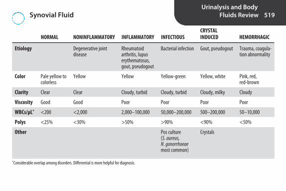
Synovial Fluid Urinalysis and Body
Fluids Review 519
CRYSTAL NORMAL NONINFLAMMATORY INFLAMMATORY INFECTIOUS INDUCED HEMORRHAGIC
Etiology
Color
Clarity
Viscosity
WBCs/μL*
Polys
Other
*Considerable overlap among disorders. Differential is more helpful for diagnosis.
Trauma, coagula-tion abnormality
Pink, red, red-brown
Cloudy
Poor
50–10,000
<50%
Degenerative joint disease
Yellow
Clear
Good
<2,000
<30%
Pale yellow tocolorless
Clear
Good
<200
<25%
Gout, pseudogout
Yellow, white
Cloudy, milky
Poor
500–200,000
<90%
Crystals
Rheumatoidarthritis, lupuserythematosus,gout, pseudogout
Yellow
Cloudy, turbid
Poor
2,000–100,000
>50%
Bacterial infection
Yellow-green
Cloudy, turbid
Poor
50,000–200,000
>90%
Pos culture (S. aureus, N. gonorrhoeaemost common)
2956_Ch07_485-522 29/01/14 12:24 PM Page 519

Synovial Fluid CrystalsUrinalysis and Body
Fluids Review 520
CRYSTAL DESCRIPTION SIGNIFICANCE
Monosodium urate
Calcium pyrophosphate
Cholesterol
Gout
Pseudogout
Chronic effusions (rheumatoid arthritis)
1–30 μm long needles. Intra- or extracellular.Strongly birefringent.
Yellow when long axis of crystal is parallel toslow wave of red compensator; blue whenperpendicular.
1–20 μm long, 4 μm wide. Rod-shaped, rectangular, or rhomboid.
Intracellular. Weakly birefringent. Blue whenlong axis of crystal is parallel to slow wave ofred compensator; yellow when perpendicular.
Large rectangle with notched-out corner. Extracellular.
From Strasinger SK, Di Lorenzo MS. Urinalysis and Body Fluids, 5th ed. Philadelphia: FA Davis; 2008:215.
2956_Ch07_485-522 29/01/14 12:24 PM Page 520
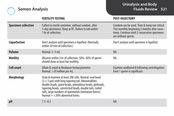
Semen AnalysisUrinalysis and Body
Fluids Review 521
FERTILITY TESTING POST-VASECTOMY
Specimen collection
Liquefaction
Volume
Motility
Cell count
Morphology
pH
Condom can be used. Time & temp not critical.Test monthly beginning 2 months after vasec-tomy. Continue until 2 consecutive specimensare without sperm.
Don’t analyze until specimen is liquified.
NA.
NA.
Examine undiluted & following centrifugation.Even 1 sperm is significant.
NA.
Collect in sterile container, without condom, after 3-day abstinence. Keep at RT. Deliver to lab within 1 hr of collection.
Don’t analyze until specimen is liquified. (Normallywithin 30 min of collection.)
Normal: 2–5 mL
Observe within 3 hr of collection. 50%–60% of spermshould show at least fair motility.
Dilute & count in Neubauer hemacytometer. Normal: >20 million per mL.
Stain & examine at least 200 cells. Normal: oval head (3 × 5 μm) with long tapering tail. Abnormalities: double heads, giant heads, amorphous heads, pinheads,tapering heads, constricted heads, double tails, coiledtails, large numbers of spermatids (immature forms).Normal = <30% abnormal forms.
7.3–8.3
2956_Ch07_485-522 29/01/14 12:24 PM Page 521

Amniotic Fluid TestsUrinalysis and Body
Fluids Review 522
TEST ASSESSMENT/DIAGNOSIS OF METHOD COMMENTS
Lecithin-to-sphingomyelin( L/S) ratio
Phosphatidylglycerol (PG)
Foam stability index(shake test)
Lamellar body count
Amniotic fluid bilirubin
Alpha-fetoprotein (AFP)
Reference method. L/S ratio ≥ 2.0 signifiesmaturity. False ↑ with blood or meconiumcontamination.
Last surfactant to rise. Not affected by presence of blood or meconium.
Index is highest concentration of ethanolthat supports ring of foam after shaking.Index of 0.48 is comparable to L/S ratio of2.0. Blood and meconium interfere.
Number correlates with amount of phos-pholipid present in fetal lungs. Samplemust be free of blood or meconium.
Bilirubin has peak absorbance at 450 nm.Baseline is drawn connecting 365- & 550-nm points. ΔA450 compared to gesta-tional age to determine severity of disease.Specimen must be protected from light.Blood & meconium interfere.
High levels with anencephaly, spina bifida.
Fetal lung maturity
Fetal lung maturity
Fetal lung maturity
Fetal lung maturity
Hemolytic disease of thenewborn/fetus
Neural tube defects
Thin-layer chromatography
Immunologic agglutination
Shake with increasingamounts of 95% ethanol
Count in platelet channel of hematology analyzers
Direct spectrophotometricscan from 365–550 nm
Immunologic method
2956_Ch07_485-522 29/01/14 12:24 PM Page 522

8S E C T I O N
Molecular Diagnostics(MDx) Review
2956_Ch08_523-574 29/01/14 12:24 PM Page 523

Nucleic Acid TerminologyMolecular Diagnostics
(MDx) Review 524
TERM EXPLANATION
Nucleic acid
Deoxyribonucleic acid (DNA)
dsDNA
ssDNA
Genomic DNA
Mitochondrial DNA
Chromosome
Gene
Exons
DNA & RNA
Nucleic acid that carries genetic information. In eukaryotes, located in chromosomes in nucleus & smallamount in mitochondria. Also present in bacteria & DNA viruses.
Double-stranded DNA.
Single-stranded DNA.
DNA in chromosomes.
Circular piece of DNA found in mitochondria of eukaryotes. Codes for polypeptides involved in oxidativephosphorylation. Transmitted by maternal inheritance (mitochondria in ova, not sperm). Used in forensics, lineage & population studies.
Piece of coiled DNA containing many genes. Humans have 23 pairs of chromosomes. 1 set from mother,1 from father. 22 pairs of autosomes, 1 pair sex chromosomes (X,Y).
Specific sequence of nucleotides (1,000–4,000) at particular location on chromosome. Starts at 5’ endwith promoter region that initiates transcription & ends at 3’ end with terminator sequence that endstranscription. Humans have about 30,000 genes. DNA sequence in genes is 99.9% the same in all people.
DNA sequences on chromosomes that code for amino acids/proteins.
continued...
2956_Ch08_523-574 29/01/14 12:24 PM Page 524

Nucleic Acid Terminology continuedMolecular Diagnostics
(MDx) Review 525
TERM EXPLANATION
Introns
Genome
Genetic code
Ribonucleic acid (RNA)
Nucleoside
Nucleotide
Pentose
Ribose
Deoxyribose
Noncoding regions between exons.
All hereditary information for an individual.
Relationship between sequence of bases in DNA or its RNA transcript & sequence of amino acids in proteins.
Nucleic acid that converts genetic information from DNA into specific proteins. Some has regulatory or structural function. Present in nucleus & in cytoplasm where it’s associated with ribosomes (free orattached to endoplasmic reticulum). Source of genetic information in RNA viruses (retroviruses).
Pentose sugar with nitrogen base attached. Dephosphorylated nucleotide. Adenosine, guanosine, cytidine, thymidine.
Building blocks of DNA & RNA. A pentose sugar with nitrogen base attached to 1’C & 1–3 phosphate groupsattached to 5’C. Nucleotide sequences always written in 5’ to 3’ direction, e.g., 5’ATCGAACAGTAC 3’
Sugar with 5 carbons (C). Cs are numbered 1’–5’ starting with C to right of oxygen & going clockwise.Superscript (prime) differentiates Cs in sugar from Cs in bases, which are numbered 1–9.
5-C sugar found in RNA.
5-C sugar found in DNA. Similar to ribose, but with 1 less oxygen.
continued...
2956_Ch08_523-574 29/01/14 12:24 PM Page 525

Nucleic Acid Terminology continuedMolecular Diagnostics
(MDx) Review 526
TERM EXPLANATION
Nitrogen bases
Base pairs
Complementary
Pyrimidine
Purine
Phosphodiester bonds
Carbon-nitrogen ring structures attached to 1’C of sugar in DNA & RNA: adenine (A), guanine (G), cytosine (C), thymine (T), & uracil (U). A, G, C, T are in DNA. U replaces T in RNA. T & U are similar except that U lacks methyl group.
Purine from 1 strand of nucleic acid & pyrimidine from another strand joined by hydrogen (H) bonds. A forms 2 H bonds with T or U. G forms 3 H bonds with C.
Opposite or partner base in base pair, e.g., A is complementary to T or U, G is complementary to C.
Nitrogen bases with single C-N ring (C, T, U).
Nitrogen bases with 2 C-N rings (A, G).
Bonds that join nucleotides in nucleic acid. 5’ phosphate group of 1 sugar attaches to 3’ hydroxyl groupof adjacent sugar. A molecule of H2O splits off.
2956_Ch08_523-574 29/01/14 12:24 PM Page 526

Comparison of DNA and RNAMolecular Diagnostics
(MDx) Review 527
DNA RNA
Function
Location
Composition
Sugar
Pyrimidines
Purines
Converts genetic information fromDNA into proteins. Some has regula-tory or structural function. Source ofgenetic information in RNA viruses.
Nucleus & cytoplasm.
Repeating nucleotides linked byphosphodiester bonds between 5’ phosphate group of 1 sugar & 3’ hydroxyl group of next.
Ribose.
C, U.
A, G.
Carries genetic information. Serves as template for synthesis of RNA.
Nucleus (except mitochondrial DNA).
Repeating nucleotides linked by phosphodiester bonds between 5’phosphate group of 1 sugar & 3’ hydroxyl group of next.
Deoxyribose.
C, T.
A, G.
continued...
2956_Ch08_523-574 29/01/14 12:24 PM Page 527

DNA RNA
Usual structure Single stranded. Shorter than DNA.Irregular 3-D structure.
Double stranded. Each strand has 5’ end (phosphate group attached to 5’C)& 3’ end (OH group attached to 3’C). Strands are antiparallel, i.e., one in 3’to 5’ direction, other 5’–3’. 2 strands coil around each other to form doublehelix with alternating sugar & P groups on exterior & bases on interior.Strands held together by hydrogen (H) bonds between complementarybases.
Comparison of DNA and RNA continuedMolecular Diagnostics
(MDx) Review 528
C
C C
CC
C
C C
CC C
C
C
C
A AA
A
AA A
T T
T
T
T
TT
G
G
G
G
G
GG
GG
G
GG
5′
Sugar-phosphate backbone
5′3′
3′
Nitrogen bases
DNA double helix
(From Buckingham L. Molecular Diagnostics: Fundamentals, Methods, andClinical Applications, 2nd ed. Philadelphia: FA Davis; 2012:3.)
2956_Ch08_523-574 29/01/14 12:24 PM Page 528

Nitrogen BasesMolecular Diagnostics
(MDx) Review 529
PURINES PYRIMIDINES BASE PAIRING # H BONDS IN BASE PAIR
DNA & RNA A, G C G-C 3
DNA only T A-T 2
RNA only U A-U 2
A = adenine, G = guanine, C = cytosine, T = thymine, U = uracil.
2956_Ch08_523-574 29/01/14 12:24 PM Page 529
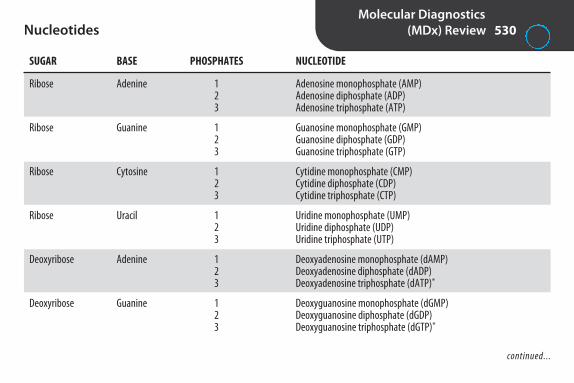
NucleotidesMolecular Diagnostics
(MDx) Review 530
SUGAR BASE PHOSPHATES NUCLEOTIDE
Ribose Adenine 1 Adenosine monophosphate (AMP)2 Adenosine diphosphate (ADP)3 Adenosine triphosphate (ATP)
Ribose Guanine 1 Guanosine monophosphate (GMP)2 Guanosine diphosphate (GDP)3 Guanosine triphosphate (GTP)
Ribose Cytosine 1 Cytidine monophosphate (CMP)2 Cytidine diphosphate (CDP)3 Cytidine triphosphate (CTP)
Ribose Uracil 1 Uridine monophosphate (UMP)2 Uridine diphosphate (UDP)3 Uridine triphosphate (UTP)
Deoxyribose Adenine 1 Deoxyadenosine monophosphate (dAMP)2 Deoxyadenosine diphosphate (dADP)3 Deoxyadenosine triphosphate (dATP)*
Deoxyribose Guanine 1 Deoxyguanosine monophosphate (dGMP)2 Deoxyguanosine diphosphate (dGDP)3 Deoxyguanosine triphosphate (dGTP)*
continued...
2956_Ch08_523-574 29/01/14 12:25 PM Page 530

Nucleotides continuedMolecular Diagnostics
(MDx) Review 531
SUGAR BASE PHOSPHATES NUCLEOTIDE
Deoxyribose Cytosine 1 Deoxycytidine monophosphate (dCMP)2 Deoxycytidine diphosphate (dCDP)3 Deoxycytidine triphosphate (dCTP)*
Deoxyribose Thymine 1 Deoxythymidine monophosphate (dTMP)2 Deoxythymidine diphosphate (dTDP)3 Deoxythymidine triphosphate (dTTP)*
*One of the 4 dNTPs used in PCR.
2956_Ch08_523-574 29/01/14 12:25 PM Page 531

DNA Replication TerminologyMolecular Diagnostics
(MDx) Review 532
TERM EXPLANATION
Replication
Semiconservative replication
Helicase
Replication fork
Template
Primase
RNA primer
DNA polymerase
Continuous synthesis
Reproduction of DNA during cell division.
Newly synthesized strand of DNA consists of 1 strand of original DNA & new daughter strand. OriginalDNA is conserved.
Enzyme that unwinds section of DNA by breaking bases’ hydrogen bonds to form 2 single-strandedtemplates for replication.
Location where dsDNA separates into ssDNA & synthesis of DNA begins.
Strand of DNA that serves as pattern for new strand of DNA or RNA.
RNA polymerase that synthesizes RNA primers needed to initiate replication.
Segment of RNA formed on DNA template to start replication. Provides 3’OH needed for phosphodi-ester bond formation with incoming nucleotide.
Enzyme that synthesizes daughter DNA by reading code on DNA template. Can only synthesize DNAfrom 5’ end to 3’ end because 3’OH needed to receive incoming nucleotides & form phosphodiesterbonds. Can only extend a nucleotide chain; can’t start one. Needs primer to start.
Synthesis of daughter strand of DNA from 3’–5’ target strand. DNA polymerase proceeds in linearfashion, assembling series of joined nucleotides in leading strand.
continued...
2956_Ch08_523-574 29/01/14 12:25 PM Page 532

DNA Replication Terminology continuedMolecular Diagnostics
(MDx) Review 533
TERM EXPLANATION
Discontinuous synthesis
Leading strand
Lagging strand
Okazaki fragments
RNase H
Ligase
Synthesis of daughter strand of DNA from 5’–3’ target strand. DNA polymerase must read target in 3’ to 5’ direction, so must proceed in nonlinear fashion, reading a section, assembling appropriatenucleotides, then moving back upstream to read another section in 3’ to 5’ direction. New strandcalled lagging strand. Consists of unconnected DNA fragments called Okazaki fragments separatedby RNA primers.
Daughter strand synthesized continuously from 3’–5’ template.
Daughter strand synthesized discontinuously from 5’–3’ template.
Short fragments of unconnected DNA formed during replication of 5’–3’ template.
Enzyme that removes RNA primers from daughter strands of DNA.
Enzyme that joins DNA fragments together.
2956_Ch08_523-574 29/01/14 12:25 PM Page 533

DNA ReplicationMolecular Diagnostics
(MDx) Review 534
STEP EXPLANATION
Strand separation
Synthesis of primers
Extension of primers
Removal of primers
Replacement of primers
Joining of Okazaki fragments
Helicase unwinds section of dsDNA to form replication forks. Both original strands of DNA will serve astemplates. Replication will proceed simultaneously on both strands, in opposite directions.
Primase reads section of code on DNA, attaches complementary nucleotides to form RNA primer thatprovides 3’OH end needed to grow nucleotide chain.
DNA polymerase reads template, attaches complementary nucleotides starting at 3’ OH of primer. Codeis read in 3’ to 5’ direction. Synthesis on 3’–5’ template is continuous, forming leading strand. Synthesison 5’–3’ template is discontinuous, forming lagging strand of disconnected Okazaki fragments.
RNase H removes RNA primers.
DNA polymerase places nucleotides where primers used to be.
DNA ligase joins DNA fragments together.
continued...
2956_Ch08_523-574 29/01/14 12:25 PM Page 534
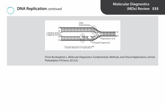
DNA Replication continuedMolecular Diagnostics
(MDx) Review 535
Okazaki fragments
Replication fork
Overall direction of replication
DNApolymerase
Leading strandLagging strand
5′3′
3′
5′
5′5′
3′ 3′
(From Buckingham L. Molecular Diagnostics: Fundamentals, Methods, and Clinical Applications, 2nd ed.Philadelphia: FA Davis; 2012:9.)
2956_Ch08_523-574 29/01/14 12:25 PM Page 535

Primer ExtensionMolecular Diagnostics
(MDx) Review 536
Template DNA: 3’ AGCA 5’→ (DNA polymerase reads template, attaches nucleotides in this direction
Daughter strand: 5’ TCGT 3’
Polymerase reads template in 3’ to 5’ direction. New strand grows in 5’ to 3’ direction
In example above:1. Polymerase reads A on template, adds T to daughter strand2. Polymerase reads G on template, adds C to daughter strand3. Polymerase reads C on template, adds G to daughter strand4. Polymerase reads A on template, adds T to daughter strand
5’ P of incoming nucleotide attaches to 3’ OH of last nucleotide on strand, forming phosphodiester bond
2956_Ch08_523-574 29/01/14 12:25 PM Page 536

Discontinuous Synthesis of DNA on Lagging Strand
Molecular Diagnostics(MDx) Review 537
Nucleotide position 1 2 3 4 5 6 7 8Template DNA 5’ A G C T T G A C 3’
¬ polymerase ¬ polymerasefirst pass second pass
Daughter DNA 3’ T C G A A C T G 5’
5’–3’ DNA template is read in 3’ to 5’ direction. DNA daughter strand is synthesized in 5’ to 3’ direction.
First pass: polymerase reads positions 4 to 1 on template—TCGA, assembles AGCT on daughter strand.
Second pass: polymerase reads positions 8 to 5 on template—CAGT, assembles GTCA on daughter strand.
2956_Ch08_523-574 29/01/14 12:25 PM Page 537

Gene Expression TerminologyMolecular Diagnostics
(MDx) Review 538
TERM EXPLANATION
Gene expression
Messenger RNA (mRNA)
Ribosomal RNA (rRNA)
Transfer RNA (tRNA)
Micro RNA (miRNA)
Transcription
Translation
Sense strand
Antisense strand
Promoter
Synthesis of specific proteins based on DNA code.
RNA that transcribes DNA code in nucleus & carries it to cytoplasm where it’s translated into protein. About1%–2% of total cellular RNA.
RNA associated with ribosomes, site of protein synthesis.
RNA that reads code in mRNA & transports amino acids to growing polypeptide chain.
Tiny regulatory RNAs involved in control of gene expression.
Conversion of genetic information from DNA into complementary strand of mRNA. If DNA sequence isAGCT, mRNA sequence will be UCGA.
Conversion of genetic information from mRNA into specific sequence of amino acids to form specific protein. Takes place on ribosomes in cytoplasm. tRNA reads mRNA & assembles amino acids.
Strand of DNA that doesn’t serve as template for synthesis of mRNA. Its sequence is same as mRNA that istranslated into protein (except it has T where RNA has U).
Strand of DNA that serves as template for synthesis of mRNA.
Region of DNA that binds RNA polymerase, initiating transcription.
continued...
2956_Ch08_523-574 29/01/14 12:25 PM Page 538

Gene Expression Terminology continuedMolecular Diagnostics
(MDx) Review 539
TERM EXPLANATION
Codon
Start codon
Termination codons
Anticodon
Ribosomes
3 nucleotides that code for an amino acid, e.g., GCC codes for alanine. 64 codons code for 21 amino acids.2–6 codons for each, except methionine & tryptophan which only have 1.
AUG, codes for methionine.
UAA, UAG, UGA. Not associated with any amino acid. Signal to stop protein synthesis.
3-nucleotide sequence on tRNA that attaches to codon on mRNA, bringing with it specified amino acids.
Organelles in cytoplasm & on surface of rough endoplasmic reticulum. Made of rRNA & protein. Site of protein synthesis.
2956_Ch08_523-574 29/01/14 12:25 PM Page 539

Protein SynthesisMolecular Diagnostics
(MDx) Review 540
STEP EXPLANATION
Transcription
Translation
1. RNA polymerase binds to promoter region of gene.2. DNA separates into 2 single strands.3. RNA polymerase adds complementary nucleotides to produce strand of mRNA. If DNA sequence is 3’ ATTCGA 5’,
mRNA will be 5’ UAAGCU 3’.4. mRNA is released from DNA. 5. DNA strands reanneal.
1. mRNA associates with ribosomes in cytoplasm.2. Ribosome moves along mRNA, reading genetic code. Always begins with codon AUG.3. tRNA with complementary 3-nucleotide sequence (anticodon) attaches to codon on mRNA, bringing with it amino
acid specified by codon, e.g., anticodon UAC on tRNA attaches to codon AUG on mRNA, bringing with it amino acidmethionine.
4. As ribosome moves along mRNA, other tRNAs bring in other amino acids.5. Peptide bonds form between amino acids. 6. Process continues until ribosome reaches stop codon. 7. Protein is released.
2956_Ch08_523-574 29/01/14 12:25 PM Page 540
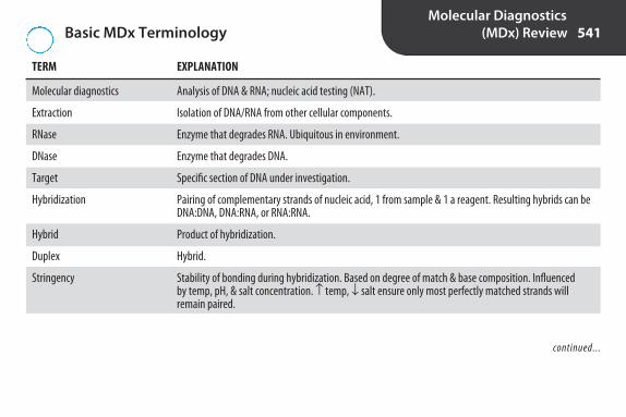
Basic MDx TerminologyMolecular Diagnostics
(MDx) Review 541
Molecular diagnostics
Extraction
RNase
DNase
Target
Hybridization
Hybrid
Duplex
Stringency
Analysis of DNA & RNA; nucleic acid testing (NAT).
Isolation of DNA/RNA from other cellular components.
Enzyme that degrades RNA. Ubiquitous in environment.
Enzyme that degrades DNA.
Specific section of DNA under investigation.
Pairing of complementary strands of nucleic acid, 1 from sample & 1 a reagent. Resulting hybrids can beDNA:DNA, DNA:RNA, or RNA:RNA.
Product of hybridization.
Hybrid.
Stability of bonding during hybridization. Based on degree of match & base composition. Influenced by temp, pH, & salt concentration. ↑ temp, ↓ salt ensure only most perfectly matched strands will remain paired.
continued...
TERM EXPLANATION
2956_Ch08_523-574 29/01/14 12:25 PM Page 541

Basic MDx Terminology continuedMolecular Diagnostics
(MDx) Review 542
Nucleic acid probe
Homologous
Short strand of DNA or RNA with known base sequence. Isolated from organisms, cloned in bacteria, orsynthesized. Labeled with fluorescent or chemiluminescent dyes, enzymes, or radioisotopes to producevisible sign of hybridization. Used in many molecular methods, e.g., Southern blot, Northern blot, in situhybridization, restriction fragment length polymorphism, conventional & real time PCR. If complemen-tary base sequence is present, probe attaches to target.
Matching, e.g., a homologous nucleic acid segment is 1 whose nucleotides are in correct order to hybridize with another nucleic acid segment, such as a primer or probe. The base pairs of 1 strand arecomplementary to the base pairs of the other. Term also used in reference to chromosome pairs, e.g., maternal copy of chromosome 17 and homologous paternal copy.
TERM EXPLANATION
2956_Ch08_523-574 29/01/14 12:25 PM Page 542

Overview of MDxMolecular Diagnostics
(MDx) Review 543
Specimen collection
Specimen processing
Nucleic acid extraction
Nucleic acid amplification (optional)
Nucleic acid detection/analysis by:• Gel electrophoresis• Hybridization with labeled probes• Southern or Northern blotting• Restriction enzyme mapping• DNA sequencing• Real-time PCR
2956_Ch08_523-574 29/01/14 12:25 PM Page 543

Blood Collection Tubes for MDxMolecular Diagnostics
(MDx) Review 544
STOPPER COLOR CONTENT USE
Lavender EDTA Isolation of DNA & detection of viruses. 1 of preferred anticoagulants forblood & bone marrow.
White K2EDTA & gel barrier Isolation of plasma. Gel forms barrier between plasma & cells.
Blue/black Sodium citrate, gel, density Isolation of mononuclear cells. Gel forms barrier between mononuclears gradient fluid in plasma & RBCs/granulocytes.
Yellow Acid citrate dextrose (ACD) Enhanced recovery of WBCs for several days after collection. 1 of preferredanticoagulants for blood & bone marrow.
Green Heparin Generally not recommended. Heparin inhibits polymerase; unacceptablefor testing that involves PCR.
2956_Ch08_523-574 29/01/14 12:25 PM Page 544

Specimens for MDxMolecular Diagnostics
(MDx) Review 545
SPECIMEN USE/COMMENTS
Whole blood/plasma
Bone marrow
Urine
Feces
Cerebrospinal, synovial, pleural, pericardial, ascetic fluids
Amniotic fluid, choronic villus sampling (CVS)
Buccal cells
Nucleated cells are source of DNA. Some recent techniques can detect non-nuclear DNA in plasma.Nucleic acid of infectious agents often detected in plasma, sometimes in host DNA. Hemolysis caninterfere with amplification.
DNA obtained from nucleated cells.
Detection of Chlamydia, CMV, & BK virus (associated with rejection of transplanted kidney).
Detection of intestinal pathogens. Analysis of stool DNA proposed for detection of colorectal cancer.
Detection of infectious agents & cancer cells.
Dx of sickle cell anemia, Tay-Sachs disease, thalassemia, other chromosomal abnormalities & inherited disorders. Usually grown in tissue culture to ↑ # of cells for DNA extraction.
Good source of DNA, especially for genotyping bone marrow recipients & patients who have beenrecently transfused. Avoid use of mouthwash with phenol or ethanol. Use sterile Dacon or rayonswabs with plastic shafts. Calcium alginate swabs & wooden shafts may contain substances thatinhibit PCR.
continued...
2956_Ch08_523-574 29/01/14 12:25 PM Page 545
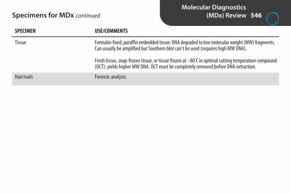
Specimens for MDx continuedMolecular Diagnostics
(MDx) Review 546
SPECIMEN USE/COMMENTS
Tissue
Hair/nails
Formalin-fixed, paraffin embedded tissue: DNA degraded to low molecular weight (MW) fragments.Can usually be amplified but Southern blot can’t be used (requires high MW DNA).
Fresh tissue, snap-frozen tissue, or tissue frozen at –80°C in optimal cutting temperature compound(OCT): yields higher MW DNA. OCT must be completely removed before DNA extraction.
Forensic analysis.
2956_Ch08_523-574 29/01/14 12:25 PM Page 546

Specimen ProcessingMolecular Diagnostics
(MDx) Review 547
SAMPLE PREPARATION
Blood or bone marrow
Plasma
Tissue
Bacteria/fungi
*Avoid frost-free freezers because of temp variation. Thawed samples should be thoroughly mixed before testing.
Whole blood can be stored at 2°–25°C for 24–48 hr after collection.Separate WBCs by:
1. Differential lysis in hypotonic buffer or water (RBCs lysed, WBCs precipitated by centrifugation) or 2. Density gradient centrifugation in Ficoll to isolate mononuclears
WBCs can be stored at –70°C for up to a year*
Remove from RBCs promptly & hold at –20°C or below. *
Fresh or frozen: grind, homogenize, or minceEmbedded: deparaffinize in xylene, rehydrate in ↓ concentrations of ethanol
Break cell walls (enzymes, detergents, NaOH, boiling, or glass beads). Enzyme treatment—less damageto chromosomal DNA. NaOH & boiling yield ssDNA. Not suitable for restriction enzyme analysis. OK foramplification.
2956_Ch08_523-574 29/01/14 12:25 PM Page 547

Isolation of DNAMolecular Diagnostics
(MDx) Review 548
STEP EXPLANATION
Isolation of nucleated cells (if necessary)
Release of DNA from cells
Separation of DNA from cellular debris, protein, lipids
Concentration
Storage
Centrifugation of whole blood to obtain buffy coat, differential lysis with water or hypotonicsolution to separate WBCs from RBCs, density gradient centrifugation in Ficoll to obtainmononuclear cells (settle below plasma, above polys/RBCs).
Cells ruptured by mechanical or chemical methods.
Liquid-phase extraction: use of solvents. Multiple manual steps. Limited sample throughput.Phenol & chloroform are hazardous.
Solid-phase extraction: DNA adsorption onto silica on membrane filters or magnetic particles.More commonly used. Easier, can be automated.
Precipitation in ethanol or isopropanol in high salt concentration. Resuspension in H2O or TEbuffer. Handle with care to avoid breaking DNA.
Extracted DNA can be stored at RT for several months, refrigerated for 1 yr, or frozen at–20°C to –70°C for up to 10 yr.
2956_Ch08_523-574 29/01/14 12:25 PM Page 548

Comparison of RNA & DNA IsolationMolecular Diagnostics
(MDx) Review 549
Method Similar. Lysis of cells, isolation by phenol-chloroform extraction or binding to silica, precipitation in alcohol.
Stability RNA less stable. Process specimens promptly. Can be stored suspended in ethanol for several months at –20°C orlong term at –70°C.
Contamination RNA more susceptible to degradation. Use RNase decontamination solution to clean gloves, benchtops, equipment;RNase-free reagents & plasticware; nuclease-free water; DNase I to digest DNA.
2956_Ch08_523-574 29/01/14 12:25 PM Page 549

Assessment of Nucleic Acid Yield/Quality Molecular Diagnostics
(MDx) Review 550
METHOD EXPLANATION ASSESSMENT
Yield
Purity
DNA μg/mL: A260 × 50RNA μg/mL: A260 × 40
Brighter bands = higher yield
DNA A260/A280 ratio: 1.6–2.0 = good quality <1.6 = protein contamination, speci-
men must be reprocessed >2 = possible contamination with RNA
RNA A260/A280 ratio:≥ 2 = good quality
DNA: high MW fragments = good quality
RNA 28S/18S ratio:2 = good quality<2 = smaller fragments due to RNase
degradation
Spectrophotometry: Absorbance@ 260 nm
Gel electrophoresis/densitometry
Spectrophotometry: Absorbance@ 260 nm/absorbance @ 280 nm
Gel electrophoresis/densitometry
Nucleic acids absorb light at 260 nm.
Intensity of bands is proportionalto nucleic acid concentration.Compare to standard.
Nucleic acids absorb light at 260 nm. Absorption peak for protein is 280 nm.
Position of bands is related toMW. Lower MW fragments migrate furthest from point of application.
2956_Ch08_523-574 29/01/14 12:25 PM Page 550

Amplification TerminologyMolecular Diagnostics
(MDx) Review 551
TERM EXPLANATION
Amplification
Target amplification
Probe amplification
Signal amplification
Nucleic acid amplification testing (NAAT)
Polymerase chain reaction (PCR)
Denaturation
Melting
Technique to ↑ (amplify) amount of nucleic acid in sample, probe, or signal so that verysmall amounts of nucleic acid can be detected.
Technique to ↑ amount of target nucleic acid in sample through in vitro replication, e.g., polymerase chain reaction (PCR), transcription mediated amplification (TMA).
Technique to ↑ amount of probe bound to target so very small amounts of nucleic acid canbe detected, e.g., ligase chain reaction.
Technique to ↑ signal generated so that very small amounts of nucleic acid can be detected,e.g., branched chain signal amplification (bDNA), hybrid capture assay (HCA).
Term used in blood banks to describe tests that detect bloodborne pathogens by presence ofnucleic acids rather than antibodies. Provides earlier detection.
1st & most widely used amplification method. Specific primers & thermostable DNA polymerase make copies of target DNA by repeated cycles of denaturation, annealing, & extension. Can produce a million copies in less than an hour.
Separation of ds DNA into 2 strands using heat or alkali.
Use of heat to separate strands of DNA.
continued...
2956_Ch08_523-574 29/01/14 12:25 PM Page 551

Amplification Terminology continuedMolecular Diagnostics
(MDx) Review 552
TERM EXPLANATION
Melting temperature (Tm)
Anneal
Polymerase
DNA polymerase
Taq polymerase
Oliogonucleotides
Primers
Deoxynucleotide triphosphates (dNTPs)
Temperature required to denature 50% of dsDNA. Depends in part on GC content. G-C pairharder to break than A-T pair because 3 H-bonds instead of 2.
Binding to complementary strand of nucleic acid.
Enzyme that assembles nucleotides to produce new strand of nucleic acid.
Enzyme that synthesizes DNA using existing strand of DNA as template. Can only add to existing piece of DNA so needs primer to start.
Heat-stable DNA polymerase used to catalyze synthesis of DNA in PCR. Originally isolatedfrom bacterium Thermus aquaticus in hot springs of Yellowstone National Park. Can with-stand high temp required for denaturation.
Short fragments of ssDNA that are synthesized for specific purpose, e.g., oligonucleotideprimers.
Short segments of DNA designed to anneal to DNA target in PCR. Provide free 3’-OH group towhich DNA polymerase can add dNTP.
Building blocks from which DNA polymerase synthesizes new strands of DNA. Deoxyadenosinetriphosphate (dATP), deoxyguanosine triphosphate (dGTP), deoxythymidine triphosphate(dTTP), deoxycytidine triphosphate (dCTP).
continued...
2956_Ch08_523-574 29/01/14 12:25 PM Page 552

Amplification Terminology continuedMolecular Diagnostics
(MDx) Review 553
TERM DEFINITION
Thermocycler
Amplicon
Melting curve analysis
Reverse transcriptase
Copy or complementary DNA (cDNA)
Ligate
DNA ligase
Instrument that rapidly changes temperatures during amplification cycle.
Product of amplification.
PCR amplicons are slowly heated in presence of dyes specific for dsDNA. At low temp, signal is high. As temp ↑, DNA strands separate & signal ↓. Fluorescent signal patterns varydepending on nucleotide sequence.
Enzyme used in vitro to make DNA from RNA. Isolated from retroviruses.
DNA copied from RNA by reverse transcriptase.
To join together by chemical process.
Enzyme that links DNA strands by joining 5’ end of one to 3’ end of another.
2956_Ch08_523-574 29/01/14 12:25 PM Page 553
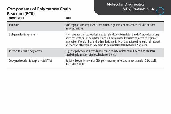
Components of Polymerase Chain Reaction (PCR)
Molecular Diagnostics(MDx) Review 554
COMPONENT ROLE
Template DNA region to be amplified. From patient’s genomic or mitochondrial DNA or from microorganisms.
2 oligonucleotide primers Short segments of ssDNA designed to hybridize to template strands & provide startingpoint for synthesis of daughter strands. 1 designed to hybridize adjacent to region of interest on 3’ end of 1 strand, other designed to hybridize adjacent to region of interest on 3’ end of other strand. Segment to be amplified falls between 2 primers.
Thermostable DNA polymerase E.g., Taq polymerase. Extends primers on each template strand by adding dNTPs & catalyzing formation of phosphodiester bonds.
Deoxynucleotide triphosphates (dNTPs) Building blocks from which DNA polymerase synthesizes a new strand of DNA: dATP,dGTP, dTTP, dCTP.
2956_Ch08_523-574 29/01/14 12:25 PM Page 554

Steps in PCRMolecular Diagnostics
(MDx) Review 555
STEP TEMP/TIME* EXPLANATION
1. Denaturation (melting) dsDNA separated into 2 strands by heat. Each strand serves as template forsynthesis of new strand.
90°–96°C/20–60 sec.
3′
3′
DNARegion to beamplified
5′
5′
Primers
(From Buckingham L. Molecular Diagnostics: Fundamentals, Methods, andClinical Applications, 2nd ed. Philadelphia: FA Davis; 2012:133.)
continued...
2956_Ch08_523-574 29/01/14 12:25 PM Page 555

Steps in PCR continuedMolecular Diagnostics
(MDx) Review 556
STEP TEMP/TIME* EXPLANATION
2. Annealing (hybridization) Primers attach to both template strands by binding with complementarybases on 3’ side of region to be amplified.
50°–70°C/20–90 sec.
3′
3′
DNA5′
5′
PrimerPrimer
Region of interest
(From Buckingham L. Molecular Diagnostics: Fundamentals, Methods, andClinical Applications, 2nd ed. Philadelphia: FA Davis; 2012:134.)
continued...
2956_Ch08_523-574 29/01/14 12:25 PM Page 556

Steps in PCR continuedMolecular Diagnostics
(MDx) Review 557
STEP TEMP/TIME* EXPLANATION
3. Extension (polymerization)
Cycle repeated 15–40 times
*Exact temperatures & times vary.
DNA polymerase attaches to ends of primers, adds dNTPs that are complementary to template, synthesizing new strands of DNA.
Each cycle results in doubling of DNA, yielding millions of copies of originalDNA segment. Amplicon can be analyzed by variety of methods, e.g., gelelectrophoresis, hybridization.
68°–75°C/10–60 sec.
3′
3′
DNA5′
5′
(From Buckingham L. Molecular Diagnostics: Fundamentals, Methods, andClinical Applications, 2nd ed. Philadelphia: FA Davis; 2012:134.)
2956_Ch08_523-574 29/01/14 12:25 PM Page 557

PCR ControlsMolecular Diagnostics
(MDx) Review 558
CONTROL DESCRIPTION PURPOSE EXPECTED REACTION
Positive control
Negative control (contamination control, reagent blank, or no target control)
Negative template control
Internal control (amplification control)
Target sequence detected
Target sequence not detected
Target sequence not detected
Housekeeping gene detected
Contains nucleic acid sequence of interest
Contains all PCR componentsexcept DNA
Contains DNA but not target DNA
Contains 2nd set of primers& unrelated target (house-keeping gene or other nucleicacid target introduced insample before nucleic acidextraction or during PCR)
Ensures that polymerase is active,primers & thermocycler workingproperly.
Checks for contamination withtarget DNA or amplicon from previous amplification.
Ensures that primers aren’t bind-ing to wrong DNA sequences.
Demonstrates that PCR is working.Differentiates true neg from falseneg due to amplification failure.Detects PCR inhibitors.
2956_Ch08_523-574 29/01/14 12:25 PM Page 558

Other Target Amplification MethodsMolecular Diagnostics
(MDx) Review 559
METHOD EXPLANATION
PCR modificationsNested PCR
Real-time PCR
Multiplex PCR
Reverse transcriptase PCR (RT-PCR)
Transcription-based amplification systems (TAS)Self-sustaining sequence replication (3SR)Transcription-mediated amplification (TMA)Nucleic acid sequence-based amplification (NASBA)
Uses 2 pairs of primers & 2nd round of amplification to ↑ sensitivity &specificity.
Simultaneous amplification & detection (fluorescence) in sealed tube.Less subject to contamination. Eliminates need for analysis of product by gel electrophoresis. Quantitative. Commonly used for detection of microorganisms.
Uses more than 1 set of primers so multiple targets can be amplified insame tube.
Method to amplify RNA (mRNA or microbial RNA). Same procedure as PCR except RNA is 1st converted to cDNA by reverse transcriptase. Used todetect HIV & HCV, measure viral loads in HIV & HCV infections, measuregene expression.
Non-PCR methods to amplify target RNA. Similar methods, different manufacturers. cDNA is formed from target RNA. Millions of copies of RNAproduced by transcription of cDNA. Assays to detect RNA viruses, Mycobac-terium tuberculosis, Chlamydia trachomatis. TMA & NASBA are isothermalrxn. Don’t require thermocycler.
2956_Ch08_523-574 29/01/14 12:25 PM Page 559
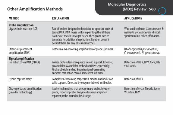
Other Amplification MethodsMolecular Diagnostics
(MDx) Review 560
METHOD EXPLANATION APPLICATIONS
Probe amplification Ligase chain reaction (LCR)
Strand-displacement amplification (SDA)
Signal amplification Branched chain DNA (bDNA)
Hybrid capture assay
Cleavage-based amplification (Invader technology)
Was used to detect C. trachomatis &Neisseria gonorrhoeae in clinicalspecimens but taken off market.
ID of Legionella pneumophila, C. trachomatis, N. gonorrhoeae.
Detection of HBV, HCV, CMV; HIVviral loads.
Detection of HPV.
Detection of cystic fibrosis, factor V Leiden, HPV.
Pair of probes designed to hybridize to opposite ends oftarget DNA. DNA ligase will join pair together if there is an exact match to target bases, then probe acts astemplate for additional replication. Ligation doesn’toccur if there are any base mismatches.
Isothermal rxn involving amplification of probes/primers.
Probes capture target sequence to solid support. Extender,preamplifier, & amplifier probes hybridize sequentially.Final probe is branched & carries signal-generating enzymes that act on chemiluminescent substrate.
Complexes containing target DNA bind to antibodies onsolid support. Detected by enzyme-labeled antibodies.
Isothermal method that uses primary probe, invaderprobe, reporter probe. Enzyme cleavage amplifies reporter probe bound to DNA target.
2956_Ch08_523-574 29/01/14 12:25 PM Page 560

Hybridization AssaysMolecular Diagnostics
(MDx) Review 561
ASSAY PRINCIPLE APPLICATION
Solid-phase hybridization
Liquid-phase hybridization
Detection of mutations associatedwith genetic diseases, e.g., sicklecell anemia
Infectious diseases, e.g., HCV geno-typing, Mycobacteria speciation
Detection of bacterial rRNA in clinical specimens or cultures
Dot/slot blot. Sample applied to membrane. Membraneheated to denature DNA. Labeled probe added. Unhy-bridized probe washed away. Hybridized probe visualizedby autoradiography or enzyme assay. Pos rxn indicatespresence of target sequence.
Sandwich hybridization. Unlabeled probe bound to mem-brane or well surface (capture probe). Sample applied. If target sequence is present, anneals to capture probe. Labeledprobe (signal-generating probe) that anneals to different siteon target added. More specific than dot blot. Can be carriedout in microtiter plates, so adaptable to automation.
Line probe assays (LiPA). Probes attached to strips, amplifiedtarget added. Unbound target washed away. Hybridizedcomplexes visualized with Biotin-Streptavidin method.
Target nucleic acid & probe interact in aqueous solution.Rapid & more sensitive than solid phase. Uses smallersample size. Adaptable to automation.
continued...
2956_Ch08_523-574 29/01/14 12:25 PM Page 561

Hybridization Assays continuedMolecular Diagnostics
(MDx) Review 562
ASSAY PRINCIPLE APPLICATION
In situ hybridization
DNA chip technology (microarrays)
Cytogenetic studies, e.g., HER2/neuoncogenes in breast cancer, chromo-some translocations in leukemia
Gene profiles associated with certaincancers/diseases, classification ofleukemias, tumor staging, determi-nation of drug resistance in HIV
Target nucleic acid detected in intact cells. Tissue (fresh,frozen, or paraffin-embedded) or cells fixed on glassslide. Probe added. Attaches to complementary DNA sequences. If fluorescent-labeled probe used, known asfluorescent in situ hybridization (FISH). Viewed with fluorescent microscope.
Multiple probes (up to 105–106) immobilized at specificlocations on solid support (glass slide, silicone chip). Afteramplification, sample & control nucleic acids labeled with2 different fluorescent dyes & loaded onto chip. Sample &control compete to hybridize to chip based on relativeamounts. Unbound strands of target washed away. Hybridized nucleic acid detected with fluorescent detector. Relative colors measured. Thousands of reac-tions can be performed at same time.
2956_Ch08_523-574 29/01/14 12:25 PM Page 562
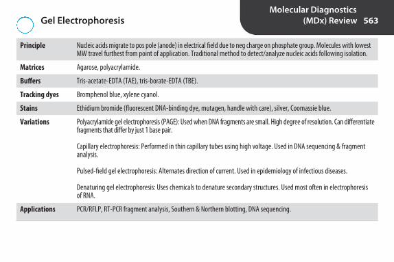
Gel ElectrophoresisMolecular Diagnostics
(MDx) Review 563
Principle
Matrices
Buffers
Tracking dyes
Stains
Variations
Applications
Nucleic acids migrate to pos pole (anode) in electrical field due to neg charge on phosphate group. Molecules with lowestMW travel furthest from point of application. Traditional method to detect/analyze nucleic acids following isolation.
Agarose, polyacrylamide.
Tris-acetate-EDTA (TAE), tris-borate-EDTA (TBE).
Bromphenol blue, xylene cyanol.
Ethidium bromide (fluorescent DNA-binding dye, mutagen, handle with care), silver, Coomassie blue.
Polyacrylamide gel electrophoresis (PAGE): Used when DNA fragments are small. High degree of resolution. Can differentiatefragments that differ by just 1 base pair.
Capillary electrophoresis: Performed in thin capillary tubes using high voltage. Used in DNA sequencing & fragmentanalysis.
Pulsed-field gel electrophoresis: Alternates direction of current. Used in epidemiology of infectious diseases.
Denaturing gel electrophoresis: Uses chemicals to denature secondary structures. Used most often in electrophoresis of RNA.
PCR/RFLP, RT-PCR fragment analysis, Southern & Northern blotting, DNA sequencing.
2956_Ch08_523-574 29/01/14 12:25 PM Page 563

BlottingMolecular Diagnostics
(MDx) Review 564
Principle
Southern blot
Northern blot
Nucleic acids that have been separated by electrophoresis are transferred to solid support membrane.
Used to analyze DNA. Useful for identification of DNA fragments that don’t amplify well, for polymorphisms &structural alterations.
1. DNA extracted from sample.2. DNA cut into fragments with restriction endonucleases. 3. Fragments separated by gel electrophoresis. 4. dsDNA fragments denatured into single strands by treatment with base. 5. Single-stranded fragments transferred (blotted) to solid support medium by capillary action. (Newer methods
use vacuum & pressure to ↑ speed of transfer.)6. DNA immobilized by baking or UV light. 7. Labeled DNA probe applied.8. Excess probe washed away.9. Visible bands seen where probe hybridized to complementary DNA fragments.
Labor intensive. Rarely used.
Similar method used to analyze RNA. Digestion step isn’t required because RNA is short. Used to determine if agene is being expressed by measuring amount of a specific mRNA. Primarily a research tool.
2956_Ch08_523-574 29/01/14 12:25 PM Page 564
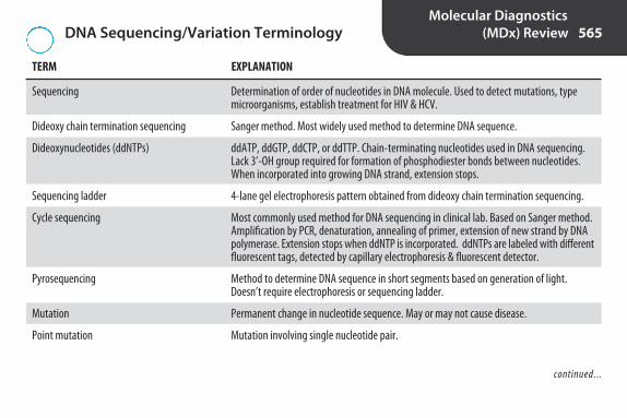
DNA Sequencing/Variation TerminologyMolecular Diagnostics
(MDx) Review 565
TERM EXPLANATION
Sequencing
Dideoxy chain termination sequencing
Dideoxynucleotides (ddNTPs)
Sequencing ladder
Cycle sequencing
Pyrosequencing
Mutation
Point mutation
Determination of order of nucleotides in DNA molecule. Used to detect mutations, type microorganisms, establish treatment for HIV & HCV.
Sanger method. Most widely used method to determine DNA sequence.
ddATP, ddGTP, ddCTP, or ddTTP. Chain-terminating nucleotides used in DNA sequencing.Lack 3’-OH group required for formation of phosphodiester bonds between nucleotides.When incorporated into growing DNA strand, extension stops.
4-lane gel electrophoresis pattern obtained from dideoxy chain termination sequencing.
Most commonly used method for DNA sequencing in clinical lab. Based on Sanger method.Amplification by PCR, denaturation, annealing of primer, extension of new strand by DNApolymerase. Extension stops when ddNTP is incorporated. ddNTPs are labeled with differentfluorescent tags, detected by capillary electrophoresis & fluorescent detector.
Method to determine DNA sequence in short segments based on generation of light.Doesn’t require electrophoresis or sequencing ladder.
Permanent change in nucleotide sequence. May or may not cause disease.
Mutation involving single nucleotide pair.
continued...
2956_Ch08_523-574 29/01/14 12:25 PM Page 565

DNA Sequencing/Variation Terminology continued
Molecular Diagnostics(MDx) Review 566
TERM EXPLANATION
Polymorphism
Single nucleotide polymorphism (SNP)
Restriction endonucleases
Restriction site (recognition site)
Palindrome
Restriction digest
Restriction pattern (RFLP profile)
Restriction fragment length polymorphisms (RFLPs)
Variation in DNA sequence that occurs in at least 1% of population. Some cause suscepti-bility to disease, affect response to drugs.
“Snip.” Variation in single base. Most common type of variation in human genome. May ormay not cause disease.
Bacterial enzymes used to cut DNA at specific sites. Used to produce short fragments forelectrophoresis.
Nucleotide sequence recognized by restriction endonuclease. Most are palindromes.
Nucleotide sequence that reads same in both directions, e.g., the complementary strand to5’-CATG-3’ is 3’-GTAC-5’. Since reading is from 5’ to 3’ direction, both are read as CATG.
DNA fragments formed by action of restriction endonucleases on DNA.
Pattern of DNA fragments produced following digestion with restriction endonucleases.Fragments separated by gel electrophoresis. Stained with ethidium bromide & viewedunder UV light or blotted onto nitrocellulose membrane & detected with labeled probe(Southern blot).
Differences in restriction patterns due to variations in nucleotide sequences that changewhere restriction enzymes cleave DNA. Useful in forensic & paternity testing, detection ofgenes associated with specific diseases, ID of microorganisms.
2956_Ch08_523-574 29/01/14 12:25 PM Page 566

Sanger Dideoxy Chain Termination MethodMolecular Diagnostics
(MDx) Review 567
Principle
Components
Procedure
Modification of DNA replication that incorporates labeled dideoxynucleotides (ddNTPs, chain terminatingnucleotides) in reaction mixture. Similar to dNTPs except lack 3’ OH needed for phosphodiester bond formation. When incorporated into growing DNA chain, elongation is terminated.
Template, primer, DNA polymerase, dNTPs (dATP, dCTP, dGTP, dTTP), ddNTPs (ddATP, ddCTP, ddGTP, ddTTP)
1. DNA template amplified.2. 4 reaction tubes containing:
a. template b. primerc. DNA polymerased. all 4 dNTPse. only 1 ddNTP (ddATP, ddCTP, ddGTP, or ddTTP)
3. Amplicon denatured.4. Primer hybridizes to target.5. DNA polymerase extends primer, occasionally incorporating ddNTP that stops further extension.
Resulting fragments are of various lengths. All fragments in tube end with same labeled ddNTP.6. Products electrophoresed in 4 separate lanes labeled A, C, G, T, corresponding with the ddNTP in tube.
Fragments separate according to size. 7. Gel dried & exposed to x-ray film, producing sequencing ladder. Band furthest from origin is smallest,
fastest migrating fragment & ends in the 1st nucleotide in the sequence, e.g., if band furthest fromorigin is in lane A (lane from tube that contained ddATP), then 1st nucleotide in sequence is A. Ladderis read from bottom to top to determine entire nucleotide sequence.
continued...
2956_Ch08_523-574 29/01/14 12:25 PM Page 567


Setup of Sanger Dideoxy Chain Termination Method
Molecular Diagnostics(MDx) Review 569
TUBE A TUBE C TUBE G TUBE T
Template � � � �
Primer � � � �
Polymerase � � � �
4 dNTPs � � � �
ddATP �
ddCTP �
ddGTP �
ddTTP �
Terminal ddNTP of all ddATP ddCTP ddGTP ddTTPresulting fragments
2956_Ch08_523-574 29/01/14 12:25 PM Page 569

Strengths and Limitations of Molecular Testing
Molecular Diagnostics(MDx) Review 570
Strengths
Limitations
High sensitivityHigh specificityAbility to predict some diseases before they are manifested in patientAbility to detect mutations that predict response to some therapiesAbility to detect nonviable, fastidious, slow-growing, & nonculturable microorganismsAdaptability to automation (high throughput, rapid turnaround time)
Potential for nucleic acid contaminationData not always clinically significant, e.g., presence of microbial nucleic acid doesn’t always indicate disease, unable
to differentiate viable & dead microorganismsCost
2956_Ch08_523-574 29/01/14 12:25 PM Page 570

Causes and Prevention of False Results in MDx
Molecular Diagnostics(MDx) Review 571
CAUSES PREVENTION
False positives Contamination from other specimens
Contamination with amplicons from previous target amplification
Environmental contamination
Contamination by testing personnel
False negatives Inhibitors in sample, e.g., heparin, hemoglobin, lactoferrin
Degradation of nucleic acids during transport/handling
Use of plugged pipet tips & screw cap tubes to minimize sample aerosols
Use of closed tube (real-time) PCR or signal or probe amplification methods;separate areas for sample prep & amplification with no movement ofequipment or reagents from amplification area to sample prep area; useof dUTP-UNG system to destroy amplicons from previous amplifications
Use of 10% bleach or alcohol to clean benches, hoods; UV light to decon-taminate sample prep areas (interferes with DNA replication by breakingsugar-phosphate backbone)
Use of lab coats, gloves
Proper specimen collection (correct anticoagulant, no hemolysis), use ofinternal control or split sample testing (1 aliquot with target added)
Use of transport media, ice, dry ice, or freezing (depending on specimen),collection tubes designed to stabilize RNA (PAXgene Blood RNA System,BD Diagnostics), prompt processing or preservation, inhibition of DNases& RNases
continued...
2956_Ch08_523-574 29/01/14 12:25 PM Page 571

Causes and Prevention of False Results in MDx continued
Molecular Diagnostics(MDx) Review 572
CAUSES PREVENTION
Degradation of nucleic acids during processing
Degradation of nucleic acids during storage
Use of gloves when handling samples, standards, controls to protectagainst nuclease degradation. RNase-free conditions for isolation of RNA
Storage at proper temp:• Specimens: snap freeze & store at –70°C• DNA: RT a few months or 1 yr in refrig, –20°C to –70°C for 10 yr• RNA: –20°C few months, –70°C longer. Avoid repeated freeze/thaw cycles & frost-free refrigerators. Can damage DNA.
2956_Ch08_523-574 29/01/14 12:25 PM Page 572

Clinical Applications of Molecular DiagnosisMolecular Diagnostics
(MDx) Review 573
DISCIPLINE APPLICATIONS
Microbiology
Blood banking
Coagulation
Hematology
Immunology
Cytogenetics
Histocompatibility
Detection of microorganisms, e.g., Mycobacterium tuberculosis, Legionella, Chlamydia trachomatis, N. gonorrhoea,Gardnerella, Trichomonas vaginalis, Candida, groups A and B streptococci, HIV, HBV, HCV, CMV, HPV, HSV
Viral load testing, e.g., HIV, HCV, HBV, CMV, EBVGenotyping to determine antimicrobial resistance, e.g., methicillin-resistant Staphylococcus aureus (MRSA),
vancomycin-resistant enterococci (VRE), M. tuberculosis, HIV, HCVEpidemiologyClassification
Screening donors for bloodborne pathogens: HIV, HBV, HCV, West Nile virus
Detection of factor V Leiden, von Willebrand’s disease
Dx & classification of B- & T-cell malignancies, non-Hodgkin’s lymphomas; monitoring of therapy & remission
Dx of HIV, HBV, HCV, CMV infections, measurement of cytokine activity
Dx of genetic diseases, e.g., sickle cell anemia, cystic fibrosis, Huntington’s chorea, Duchenne-type musculardystrophy, hemochromatosis.
Dx of carrier state, e.g., Duchenne-type muscular dystrophy, von Willebrand’s disease.
HLA typing, detection of polymorphisms in major histocompatibility complex (MHC) genes
2956_Ch08_523-574 29/01/14 12:25 PM Page 573

Clinical Applications of Molecular Diagnosis continued
Molecular Diagnostics(MDx) Review 574
DISCIPLINE APPLICATIONS
Oncology
Other
Dx of cancer, e.g., breast, bladder, lung, colon. KRAS mutation in colon cancer predicts lack of response to certain therapies.
Paternity testing, forensics (DNA fingerprinting), pharmacogenomics (“personalized medicine,” selection of optimal drug therapy based on patient’s genotype), fetomaternal medicine (circulating DNA & RNA)
2956_Ch08_523-574 29/01/14 12:25 PM Page 574

9S E C T I O N
Management & Education Review
2956_Ch09_575-596 30/01/14 4:28 PM Page 575

Management Skills and StylesManagement &
Education Review 576
SKILLS
Organizational Ability to apply management process, systematize workflow, make decisions, communicate withcoworkers.
People Understanding theories of human needs & work motivation.
Financial Effective use of & accounting for company’s monetary assets.
Technical Skills to transform resources into products/services.
STYLES
Authoritarian Manager makes all decisions without input from others. Quick decision-making. Least acceptance &commitment from staff. Poorest quality decisions.
Democratic Manager makes decisions after polling staff. Better quality & acceptance. Decisions take longer. Thosein minority might feel ignored.
Consensus Manager tries to get at least partial agreement from all staff. Everyone has input. Highest quality decisions. Good acceptance & commitment. Time-consuming.
Laissez-faire Manager leaves decision to staff. Least effective approach. Manager abdicates responsibility.
2956_Ch09_575-596 30/01/14 4:28 PM Page 576

Management &Education Review 577Management Principles
FOUNDATIONS
Mission Organization’s purpose
Goals Organization’s broad, long-term ambitions
Objectives Directives that describe how a goal will be achieved. Should be SMAART:Specific Measurable Achievable Agreed-upon Realistic Time-bound
PROCESSES
Planning Establishing goals & objectives, formulating policies to carry out objectives.
Organizing Coordinating resources to achieve plans. Defining working relationships, including line of authority & workflow.
Directing Communicating, motivating, delegating, & coaching. Creating a climate that meets the needs of individuals & the organization.
Controlling Defining standards of performance, developing a reporting system, & taking corrective action when necessary.
2956_Ch09_575-596 30/01/14 4:28 PM Page 577
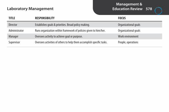
Management &Education Review 578Laboratory Management
TITLE RESPONSIBILITY FOCUS
Director Establishes goals & priorities. Broad policy making. Organizational goals
Administrator Runs organization within framework of policies given to him/her. Organizational goals
Manager Oversees activity to achieve goal or purpose. Work environment
Supervisor Oversees activities of others to help them accomplish specific tasks. People, operations
2956_Ch09_575-596 30/01/14 4:28 PM Page 578

Maslow’s Hierarchy of NeedsManagement &
Education Review 579
NEED DEFINITION WORKPLACE COUNTERPART(S)
Physiological
Safety
Social
Esteem
Self-actualization
Income
Insurance, safe work environment, job security
Social relationships with coworkers
Job title, privileges, respect of colleagues
Challenging work, autonomy, professional growth
Survival needs. Food, water, air, rest
Physical & psychological security
Sense of belonging, acceptance, affection
Respect, independence, appreciation, recognition
Realization of full potential
2956_Ch09_575-596 30/01/14 4:28 PM Page 579

Personnel Required in High-Complexity Laboratories Under CLIA ’88
Management &Education Review 580
TITLE RESPONSIBILITIES
Laboratory director Overall operation & administration of lab.
Technical supervisor Technical & scientific oversight of lab. Must be available on as-needed basis to provide on-site, telephone, orelectronic consultation.
Clinical consultant Consultation regarding appropriateness & interpretation of tests.
General supervisor Day-to-day supervision of lab.
Testing personnel Specimen processing, test performance, & reporting of test results.
2956_Ch09_575-596 30/01/14 4:28 PM Page 580

Employee Performance AppraisalManagement &
Education Review 581
COMPONENTS
Job description Basis for evaluation.
Standards/criteria What is expected. Should be objective & measurable.
Measurement instrument Instrument to compare actual performance with desired performance.
Evaluator Person trained in use of instrument, familiar with intricacies of job, time to dedicate to process.
Feedback mechanism Plan for sharing results of review, taking corrective action, planning for future.
EVALUATION ERRORS
Error of central tendency Everyone is rated toward middle of scale.
Contrast error An individual is rated lower than justified because of comparison with another exceptional individual.(The opposite may also occur.)
Error of leniency Everyone is rated high. (The opposite may also occur.)
Halo effect Good performance in one area influences evaluation in other areas.
Reverse halo effect Poor performance in one area influences evaluation in other areas.
Recency phenomenon Judgments are made based on recent events or unusual incidents.
2956_Ch09_575-596 30/01/14 4:28 PM Page 581

Testing Personnel Competency AssessmentManagement &
Education Review 582
Frequency
Methods
Required by CLIA ’88 for moderate & high complexity labs.
Semiannually during 1st yr, annually thereafter, & whenever there’s a change in test methodology or instrumentation
Direct observation of routine patient test performanceMonitoring of recording/reporting of test resultsReview of intermediate test results or worksheets, QC records, proficiency testing results, preventive maintenance
recordsDirect observation of performance of instrument maintenance/function checksAssessment of test performance through testing of previously analyzed specimens, internal blind testing samples,
or external proficiency testing samplesAssessment of problem-solving skills
2956_Ch09_575-596 30/01/14 4:28 PM Page 582

Laboratory Operating CostsManagement &
Education Review 583
DEFINITION EXAMPLES
Fixed costs
Variable costs
Direct costs
Indirect costs
Unit cost/cost per test
Instrument leases, maintenance contracts, computer services,equipment costs, facilities upkeep, management salaries, custodial salaries, employee benefits, depreciation, lease payments, rent, taxes
Labor costs, supplies, reagents, disposables
Supplies, reagents, controls, standards, disposables, equipmentcosts, equipment maintenance contracts, equipment deprecia-tion, technical & supervisory labor
Administration, plant maintenance, security, utilities, buildingdepreciation, rent, taxes, insurance, housekeeping, purchasing,billing, regulatory expenses, laboratory information system(LIS) expenses
Expenses that don’t fluctuate whenvolume of work changes over shortterm
Expenses that fluctuate directly withchange in work load
Costs associated with performance ofa test
Overhead
Total of direct & indirect expenses ofproducing a test result
2956_Ch09_575-596 30/01/14 4:28 PM Page 583

Management &Education Review 584Break-Even Points
ANALYSIS FORMULA
Break-even price per test
Break-even test volume
Break-even revenue
Annual fixed costs + variable costsTest volume
Total fixed costsAverage revenue per test variab− lle cost per test
Total fixed costs(Average revenue per test varia− bble cost per test)/average revenue per test
2956_Ch09_575-596 30/01/14 4:28 PM Page 584

Quality ManagementManagement &
Education Review 585
SYSTEM HIGHLIGHTS
GP26-A4: Quality Management System: A Model for Laboratory Services
ISO 15189: Medical laboratories-Particular requirements for quality & competence
Lean
Quality management system developed by Clinical & Laboratory Standards Institute (CLSI)to organize all policies, processes, & procedures for preanalytic, analytic, & postanalyticactivities. Based on 12 quality systems essentials (QSEs): organization, customer focus, facilities & safety, personnel, purchasing & inventory, equipment, process management,documents & records, information management, nonconforming event management, assessments, continual improvement. Similar to ISO 15189.
Quality management system developed specifically for clinical labs by the InternationalOrganization for Standardization (ISO). Based on ISO 17025 (General requirements for thecompetence of testing and calibration laboratories) and ISO 9001 (Quality management systems-Requirements). Accreditation to ISO 15189 is mandatory in some countries, butcurrently voluntary in U.S. The College of American Pathologists (CAP) offers an ISO 15189accreditation program. It doesn’t replace the CAP Laboratory Accreditation Program.
System developed by Toyota to improve quality by improving workflow & eliminatingwaste. Focuses on equipment layout, standardization of processes, cross-training, inventorymanagement. Turnaround times are improved by grouping automated analyzers in core lab& replacing batch processing with single-piece flow.
continued...
2956_Ch09_575-596 30/01/14 4:28 PM Page 585
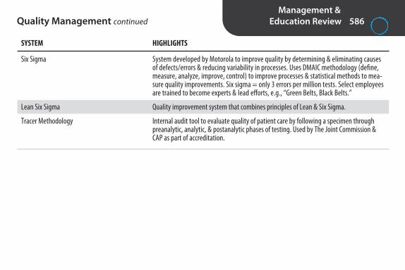
Management &Education Review 586Quality Management continued
SYSTEM HIGHLIGHTS
Six Sigma
Lean Six Sigma
Tracer Methodology
System developed by Motorola to improve quality by determining & eliminating causesof defects/errors & reducing variability in processes. Uses DMAIC methodology (define,measure, analyze, improve, control) to improve processes & statistical methods to mea -sure quality improvements. Six sigma = only 3 errors per million tests. Select employeesare trained to become experts & lead efforts, e.g., “Green Belts, Black Belts.”
Quality improvement system that combines principles of Lean & Six Sigma.
Internal audit tool to evaluate quality of patient care by following a specimen throughpreanalytic, analytic, & postanalytic phases of testing. Used by The Joint Commission &CAP as part of accreditation.
2956_Ch09_575-596 30/01/14 4:28 PM Page 586

Sentinel Events: The Joint Commission (TJC)Management &
Education Review 587
Definition
Example
Goals
Requirements
Reporting
Unexpected event involving death or serious physical or psychological injury, or risk thereof
Administration of ABO-incompatible blood
Improved patient care
1. Root cause analysis: Analysis of why event happened. Examines proximate causes, e.g., personnel, equipment,environment, leadership, corporate culture, communication, external factors. Focuses on systems/processes,not individuals.
2. Action plan: Establishes risk reduction strategies & measures of effectiveness. Should delineate responsibilitiesfor implementation/oversight & establish time lines.
3. Implementation.4. Monitoring.
Reporting to TJC is optional but encouraged so that event can be added to database & used as educational tool tohelp others avoid similar events. Confidentiality is maintained.
2956_Ch09_575-596 30/01/14 4:28 PM Page 587

Management &Education Review 588Point-of-Care Testing (POCT)
Definition
Goal
Common POCT tests
Regulations
Optimal staffing
Considerations
Testing performed at site of patient care. Also known as decentralized, bedside, or near-patient testing.
Provide rapid test results where immediate medical action is required, e.g., emergency department, intensivecare units.
Urine reagent strips, glucose, electrolytes, blood gases, activated clotting time (ACT), PT, APTT, hemoglobin.
Determined by test complexity. May operate under clinical laboratory’s CLIA certificate or separate CLIA certificate.
Director: MLS or pathologist responsible for administrative, financial, & technical decisions.Point-of-Care Coordinator (POCC): Oversees testing & responsible for compliance, training, QC, proficiency
testing (if required). Designated contact/trainer at each testing site: Liaison between POCC & testing personnel, assists with
training/competency assessment.Testing personnel: Qualifications determined by test complexity of testing. Usually phlebotomists, lab
assistants, nurses, respiratory therapists.
Cost, performance specifications, ease of use, turnaround time, impact on quality & cost of patient care, datamanagement/connectivity/interface capabilities.
2956_Ch09_575-596 30/01/14 4:28 PM Page 588

Management &Education Review 589Competency-Based Instruction
Body of Knowledge Task Analysis(competencies required
to do job)
Objectives
Instruction
Feedback
Evaluation basedon objectives
Certification ExamContent Online
2956_Ch09_575-596 30/01/14 4:28 PM Page 589

ABCs of Writing Behavioral Objectives Management &
Education Review 590
COMPONENT QUESTION(S) ANSWERED EXAMPLES
A
B
C
D
The MLT student; the phlebotomist; the laboratory assistant
Perform; demonstrate; explain; list; outline; label
Following the directions in the Blood Bank Procedure Manual; after a lecture, demonstration, & practice session;without use of notes; within 30 min; without coaching
10 normal & 3 abnormal differentials; in complete agree-ment with the instructor; with 90% accuracy; within ±15%of reported value; within ±2 SD of instructor
Audience
Behavior
Criteria
Degree
Who?
What?
Under what conditions? When? How?
Expected standard of performance.How many? How much? How well?
VAK Learning Style Model
STYLE PREFERENCE EFFECTIVE TECHNIQUES
Visual Seeing Reading assignments, pictures, slides, diagrams, drawings, graphs, videos, demonstrations.
Auditory Hearing Lectures, tapes, discussions. Stimulated by changes in vocal tone, pitch, pacing.
Kinesthetic Doing Laboratories, role-play, group work.
2956_Ch09_575-596 30/01/14 4:28 PM Page 590

Management &Education Review 591Domains of Learning
DOMAIN ELEMENTS KNOWLEDGE LEVEL APPLICATION LEVEL PROBLEM-SOLVING LEVEL
Cognitive
Psychomotor
Affective
Manipulate information in newsituation or context. Analyze,synthesize, evaluate.
Adapt existing skills to meet new demands or originate newprocedures.
Organize & internalize valuesinto system that guides behavior.
Recall & comprehendfacts/information
Observe & imitate askill
Receive & respond toinformation about attitudes/feelings
Facts, knowledge
Physical skills
Attitudes, feelings,values
Apply information in concretesituations
Practice a skill
Assess attitudes/feelings
2956_Ch09_575-596 30/01/14 4:28 PM Page 591

Bloom’s Cognitive TaxonomyManagement &
Education Review 592
LEVEL ABILITY TO EXAMPLES OF VERBS FOR OBJECTIVES AT DIFFERENT LEVELS
Knowledge
Comprehension
Application
Analysis
Synthesis
Evaluation
Cite, define, identify, label, list, match, name, pronounce, quote,recite, reproduce, state
Change, describe, explain, give examples, give main ideas, illustrate, interpret, paraphrase, rephrase, summarize
Apply, classify, compute, demonstrate, predict, prepare, present,show, solve, utilize
Analyze, associate, conclude, determine, diagnose, diagram, differentiate, discriminate, distinguish, examine, infer, outline
Combine, compile, compose, create, design, develop, devise,generalize, invent, modify, originate, plan, propose, project, revise, rewrite, synthesize, theorize
Appraise, assess, compare, conclude, contrast, critique, deduce,evaluate, judge, weigh
Recall specific facts
Grasp meaning of material
Use material in new & concrete situations
Break down material into component parts
Put elements together to form new whole
Judge value of material for given purpose
2956_Ch09_575-596 30/01/14 4:28 PM Page 592

Instructional MethodsManagement &
Education Review 593
INSTRUCTOR CENTERED STUDENT CENTERED
LecturesDemonstrations
LabsDiscussions Role playing Simulations Case studies Cooperative learning groups Writing assignments Presentations
2956_Ch09_575-596 30/01/14 4:28 PM Page 593

Testing at Different Cognitive LevelsManagement &
Education Review 594
LEVEL DEFINITION EXAMPLE
Recall
Application
Analysis
Which enzymes are elevated with liver disease?
15 nRBCs per 100 WBCs were observed on a differential. Theautomated analyzer reported the total WBC as 15 × 106/L.What is the corrected WBC count?
A patient’s RBCs agglutinated in anti-A, but not in anti-B. Hisserum agglutinated A1 cells & B cells. What might account forthese results & how should you proceed?
Recognizing or remembering isolated information
Interpreting or applying limited data
Evaluating data, solving problems, or fitting together a variety of elements into a meaningfulwhole
2956_Ch09_575-596 30/01/14 4:28 PM Page 594
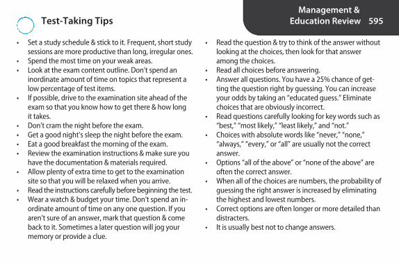
Test-Taking TipsManagement &
Education Review 595
• Set a study schedule & stick to it. Frequent, short studysessions are more productive than long, irregular ones.
• Spend the most time on your weak areas.• Look at the exam content outline. Don’t spend an
inordinate amount of time on topics that represent alow percentage of test items.
• If possible, drive to the examination site ahead of theexam so that you know how to get there & how long it takes.
• Don’t cram the night before the exam.• Get a good night’s sleep the night before the exam.• Eat a good breakfast the morning of the exam.• Review the examination instructions & make sure you
have the documentation & materials required.• Allow plenty of extra time to get to the examination
site so that you will be relaxed when you arrive.• Read the instructions carefully before beginning the test.• Wear a watch & budget your time. Don’t spend an in-
ordinate amount of time on any one question. If youaren’t sure of an answer, mark that question & comeback to it. Sometimes a later question will jog yourmemory or provide a clue.
• Read the question & try to think of the answer withoutlooking at the choices, then look for that answeramong the choices.
• Read all choices before answering.• Answer all questions. You have a 25% chance of get-
ting the question right by guessing. You can increaseyour odds by taking an “educated guess.” Eliminatechoices that are obviously incorrect.
• Read questions carefully looking for key words such as“best,” “most likely,” “least likely,” and “not.”
• Choices with absolute words like “never,” “none,” “always,” “every,” or “all” are usually not the correct answer.
• Options “all of the above” or “none of the above” areoften the correct answer.
• When all of the choices are numbers, the probability ofguessing the right answer is increased by eliminatingthe highest and lowest numbers.
• Correct options are often longer or more detailed thandistracters.
• It is usually best not to change answers.
2956_Ch09_575-596 30/01/14 4:28 PM Page 595
Nikki
Typewriter
uploaded by [stormrg]

2956_Ch09_575-596 30/01/14 4:28 PM Page 596
Related Documents