METHODOLOGY ARTICLE Open Access Quantitative fluorescence loss in photobleaching for analysis of protein transport and aggregation Daniel Wüstner 1* , Lukasz M Solanko 1 , Frederik W Lund 1 , Daniel Sage 2 , Hans J Schroll 3 and Michael A Lomholt 4 Abstract Background: Fluorescence loss in photobleaching (FLIP) is a widely used imaging technique, which provides information about protein dynamics in various cellular regions. In FLIP, a small cellular region is repeatedly illuminated by an intense laser pulse, while images are taken with reduced laser power with a time lag between the bleaches. Despite its popularity, tools are lacking for quantitative analysis of FLIP experiments. Typically, the user defines regions of interest (ROIs) for further analysis which is subjective and does not allow for comparing different cells and experimental settings. Results: We present two complementary methods to detect and quantify protein transport and aggregation in living cells from FLIP image series. In the first approach, a stretched exponential (StrExp) function is fitted to fluorescence loss (FL) inside and outside the bleached region. We show by reaction–diffusion simulations, that the StrExp function can describe both, binding/barrier–limited and diffusion-limited FL kinetics. By pixel-wise regression of that function to FL kinetics of enhanced green fluorescent protein (eGFP), we determined in a user-unbiased manner from which cellular regions eGFP can be replenished in the bleached area. Spatial variation in the parameters calculated from the StrExp function allow for detecting diffusion barriers for eGFP in the nucleus and cytoplasm of living cells. Polyglutamine (polyQ) disease proteins like mutant huntingtin (mtHtt) can form large aggregates called inclusion bodies (IB’s). The second method combines single particle tracking with multi-compartment modelling of FL kinetics in moving IB’s to determine exchange rates of eGFP-tagged mtHtt protein (eGFP-mtHtt) between aggregates and the cytoplasm. This method is self-calibrating since it relates the FL inside and outside the bleached regions. It makes it therefore possible to compare release kinetics of eGFP-mtHtt between different cells and experiments. Conclusions: We present two complementary methods for quantitative analysis of FLIP experiments in living cells. They provide spatial maps of exchange dynamics and absolute binding parameters of fluorescent molecules to moving intracellular entities, respectively. Our methods should be of great value for quantitative studies of intracellular transport. Keywords: Mathematical model, Crowding, Protein aggregation, Fractal kinetics, Rate coefficient, Multi-compartment, Neurodegeneration Background Quantitative fluorescence microscopy witnesses an in- creasing demand for computational methods allowing for interpretation of the complex data generated by this imaging technique. To determine intracellular transport dynamics of proteins and lipids tagged with suitable fluorophores, one often relies on perturbing the steady state distribution of the probe and following the dynamics of re-establishing a steady state. One example for this ap- proach are pulse-chase experiments, where a fluorescent ligand, like a dye-tagged transferrin (Tf) or fluorescent lipoprotein binds to its receptor at the cell surface at the start of the experiment (t = 0), and the transport of the fluorescent-ligand-receptor complex to the target organelle during the course of endocytosis is followed over time by quantitative fluorescence microscopy [1-3]. Pre-labeling of the target organelle with a suitable marker with different spectral characteristics allows, together with appropriate * Correspondence: [email protected] 1 Department of Biochemistry and Molecular Biology, University of Southern Denmark, Campusvej 55, Odense M DK-5230, Denmark Full list of author information is available at the end of the article © 2012 Wustner et al.; licensee BioMed Central Ltd. This is an Open Access article distributed under the terms of the Creative Commons Attribution License (http://creativecommons.org/licenses/by/2.0), which permits unrestricted use, distribution, and reproduction in any medium, provided the original work is properly cited. Wüstner et al. BMC Bioinformatics 2012, 13:296 http://www.biomedcentral.com/1471-2105/13/296

Welcome message from author
This document is posted to help you gain knowledge. Please leave a comment to let me know what you think about it! Share it to your friends and learn new things together.
Transcript
METHODOLOGY ARTICLE Open Access
Quantitative fluorescence loss in photobleachingfor analysis of protein transport and aggregationDaniel Wüstner1*, Lukasz M Solanko1, Frederik W Lund1, Daniel Sage2, Hans J Schroll3 and Michael A Lomholt4
Abstract
Background: Fluorescence loss in photobleaching (FLIP) is a widely used imaging technique, which providesinformation about protein dynamics in various cellular regions. In FLIP, a small cellular region is repeatedlyilluminated by an intense laser pulse, while images are taken with reduced laser power with a time lag betweenthe bleaches. Despite its popularity, tools are lacking for quantitative analysis of FLIP experiments. Typically, the userdefines regions of interest (ROIs) for further analysis which is subjective and does not allow for comparing differentcells and experimental settings.
Results: We present two complementary methods to detect and quantify protein transport and aggregation inliving cells from FLIP image series. In the first approach, a stretched exponential (StrExp) function is fitted tofluorescence loss (FL) inside and outside the bleached region. We show by reaction–diffusion simulations, that theStrExp function can describe both, binding/barrier–limited and diffusion-limited FL kinetics. By pixel-wise regressionof that function to FL kinetics of enhanced green fluorescent protein (eGFP), we determined in a user-unbiasedmanner from which cellular regions eGFP can be replenished in the bleached area. Spatial variation in theparameters calculated from the StrExp function allow for detecting diffusion barriers for eGFP in the nucleus andcytoplasm of living cells. Polyglutamine (polyQ) disease proteins like mutant huntingtin (mtHtt) can form largeaggregates called inclusion bodies (IB’s). The second method combines single particle tracking withmulti-compartment modelling of FL kinetics in moving IB’s to determine exchange rates of eGFP-tagged mtHttprotein (eGFP-mtHtt) between aggregates and the cytoplasm. This method is self-calibrating since it relates the FLinside and outside the bleached regions. It makes it therefore possible to compare release kinetics of eGFP-mtHttbetween different cells and experiments.
Conclusions: We present two complementary methods for quantitative analysis of FLIP experiments in living cells.They provide spatial maps of exchange dynamics and absolute binding parameters of fluorescent molecules tomoving intracellular entities, respectively. Our methods should be of great value for quantitative studies ofintracellular transport.
Keywords: Mathematical model, Crowding, Protein aggregation, Fractal kinetics, Rate coefficient,Multi-compartment, Neurodegeneration
BackgroundQuantitative fluorescence microscopy witnesses an in-creasing demand for computational methods allowingfor interpretation of the complex data generated by thisimaging technique. To determine intracellular transportdynamics of proteins and lipids tagged with suitablefluorophores, one often relies on perturbing the steady
state distribution of the probe and following the dynamicsof re-establishing a steady state. One example for this ap-proach are pulse-chase experiments, where a fluorescentligand, like a dye-tagged transferrin (Tf) or fluorescentlipoprotein binds to its receptor at the cell surface at thestart of the experiment (t = 0), and the transport of thefluorescent-ligand-receptor complex to the target organelleduring the course of endocytosis is followed over time byquantitative fluorescence microscopy [1-3]. Pre-labeling ofthe target organelle with a suitable marker with differentspectral characteristics allows, together with appropriate
* Correspondence: [email protected] of Biochemistry and Molecular Biology, University of SouthernDenmark, Campusvej 55, Odense M DK-5230, DenmarkFull list of author information is available at the end of the article
© 2012 Wustner et al.; licensee BioMed Central Ltd. This is an Open Access article distributed under the terms of the CreativeCommons Attribution License (http://creativecommons.org/licenses/by/2.0), which permits unrestricted use, distribution, andreproduction in any medium, provided the original work is properly cited.
Wüstner et al. BMC Bioinformatics 2012, 13:296http://www.biomedcentral.com/1471-2105/13/296
image analysis tools, to measure transport kinetics of themolecule of interest by multicolour fluorescence micros-copy. Compartment modelling based on ordinary differen-tial equations can be used to determine inter-organelletransport kinetics. For example, trafficking kinetics offluorescent Tf and lipoproteins to sorting endosomes andthe endocytic recycling compartment (ERC) have beenmeasured in this way in various cell types [1-8]. Similarly,using a temperature-sensitive folding mutant, export of aviral protein tagged with eGFP has been analyzed by thisapproach [9].An alternative way to perturb the steady state does not
rely on pulse-labeling molecules in a particular compart-ment and is often used to elucidate transport dynamicsalong the secretory pathway or between nucleus andcytoplasm [10-16]. Selective photodestruction of fluores-cence of the molecule of interest in one organelle andmeasuring fluorescence recovery after photobleaching(FRAP) of unbleached molecules into the area revealstransport kinetics and provides information about a pos-sible immobile fraction of the labeled molecules in thetarget organelle [17]. A variant of FRAP is continuousphotobleaching (CP) introduced by Peters et al. in 1981[18]. In this approach, a small area of the cell is continu-ously illuminated, and the fluorescence decay in thisregion due to photobleaching and transport is moni-tored. Similar as in FRAP, diffusion constants, and withextensive modelling efforts, binding parameters or flowcomponents can be estimated from CP measurements[18-20]. Both techniques, however, infer the transportparameters solely from fitting a time-dependent functionto experimental data. Thus, any locally varying transportproperties in the cell cannot be directly monitored, sinceFRAP and CP curves do not depend explicitly on spatialcoordinates. Consequently, one often finds model redun-dancy; i.e., that various mathematical models can describea measured FRAP curve equally well [21]. Another compli-cation in both, FRAP and CP is that the user has only verylimited impact on the time hierarchy of the experiment.While varying the size of the bleach area allows for con-trolling the diffusion time, if binding and diffusion takeplace on different time scales, the user cannot isolate oneof the processes by adjusting the time resolution of themeasurement.A method related to FRAP is fluorescence loss in
photobleaching (FLIP). In FLIP, a region is repeatedlyilluminated by an intense laser pulse, while images aretaken with reduced laser power between the bleaches. Apause between the laser pulses allows for some recoveryin the bleached region. The duration of the pause can beset by the user, who thereby can control the time reso-lution of the experiment. Repeating this protocol severaltimes creates a sink for the fluorescent molecules in thelocal environment being in continuous exchange with
the bleached region [17]. A decrease of fluorescence ofdye-tagged molecules outside the bleached area allowsfor assessing continuity between intracellular compart-ments, and in principal, for measuring the kinetics of re-cruitment to the bleached region from various cellularareas. Accordingly, FLIP has the potential to includespatial information in the computational analysis. How-ever, only very few FLIP analysis efforts published so fartry to include the whole image information into analysisof FLIP image sets: In one study, van Gemert et al.(2009) used independent component analysis to decom-pose FLIP image series into static and dynamic compo-nents [22]. Very recently, van de Giessen et al. (2012)used a monoexponential decay model on a pixel basiscombined with image registration and denoising tomeasure FL in FLIP image series [23]. The widely usedstandard approach for analysis of FLIP data is to definerectangular regions of interest (ROI) at different sites inthe cell and to quantify the mean fluorescence in theseROI as function of time [17]. The user chooses the num-ber, location and size of the ROI’s based on visual in-spection of the image sets. This generates a verysubjective element in the data analysis.We present two new approaches to quantify FLIP
experiments reliably; either on an image basis to detectareas of different probe mobility, or by physical model-ling of FL from moving entities. In the first section, wedemonstrate that a stretched exponential (StrExp) func-tion can be fitted to the decaying intensity at each pixelposition in the bleached and non-bleached region of thecell. This is used to detect diffusion-limited depletionzones around the bleached area in cells expressingenhanced green fluorescent protein (eGFP). Pixel-wisefitting of the StrExp function to FLIP image sets alsoallows us to determine local heterogeneity in nucleocyto-plasmic transport of eGFP. In the second section, we meas-ure FL kinetics in moving inclusion bodies (IB’s) bycombining FLIP with single-particle tracking. From thatdata, we infer exchange parameters of mutant Huntingtintagged with eGFP (eGFP-mtHtt) by multi-compartment(MC) modelling of binding/release and fluorescence at-tenuation due to photobleaching. The presented methodsshould find wide application in quantitative cell biologyand especially in analysis of protein aggregation in neuro-degenerative diseases.
ResultsThe stretched exponential function as empirical decaylaw for transport studiesThe StrExp function is an empirical decay function withbroad applications in modelling of physical, photochem-ical and biophysical data [24-28]. It can be considered asa generalization of the exponential function, since thestretching parameter h gives a time-dependent rate
Wüstner et al. BMC Bioinformatics 2012, 13:296 Page 2 of 21http://www.biomedcentral.com/1471-2105/13/296
constant, called a rate coefficient (see Equation 1 inMethods and Additional file 1: Figure S1B and EquationS4) [27]. Rudolf Kohlrausch first introduced this func-tion in 1854 to describe the discharge of a capacitator,which is why it is sometimes also called the Kohlrauschfunction [29]. It has been demonstrated that many phys-ical relaxation processes can be described by a StrExpfunction including frequency-dependent dielectric con-stants of polymeric systems and glasses [30,31], lumines-cence decays of macromolecules in heterogeneousenvironments [27,28], luminescence quenching and res-onance energy transfer in disordered media [24,27],spin-relaxation [24] and mechanical stress relaxation incrystals [32]. All of these processes have in common that aphysical system is perturbed for a short time followed byrelaxation towards a new equilibrium or steady state value,respectively. In some cases, the StrExp function can beused to discern a physical mechanism underlying theobserved relaxation process [24]. More often, it is used asempirical fitting function to describe an experiment inquantitative terms [26-28,32]. Here, we use the StrExpfunction as empirical function to describe FLIP image setson a pixel basis. In a classical FLIP experiment, the per-turbation is caused by a series of intense bleach pulses inthe selected ROI. We will consider two limiting cases of aFLIP experiment and demonstrate that the StrExp functionconcurs with simulated time courses for both situations.
Analysis of diffusion-limited FLIP experiments using theStrExp functionWhen the pause between individual bleaches is shortcompared to the molecular transport rates, the mea-sured FL becomes limited by diffusion of the moleculestowards the bleached area. Developing a physical modelfor this situation requires taking the exact location ofthe bleach ROI into account. Assuming that binding/dis-sociation events are very fast compared to molecular dif-fusion the process is governed by Equation 6 which canbe solved (see Methods and Appendix 1). The diffusionmodel assumes cylindrical cell geometry and cylindricalbleach in the centre of the cell within 3 μm around theorigin performed over the whole cell height (Figure 1A).Under these assumptions, we have a 2D-problem, whichis reasonable for bleaching experiments in confocalmicroscopy due to the large depth of field of the fo-cussed laser beam [33]. The radial symmetry of the dif-fusion problem implies that the FL is only a function of
the magnitude of the radius vector r→
but not of its direc-tion. Thus, it is sufficient to analyze some points withvarying distance from the centre. Note that this model isan oversimplification of a real FLIP experiment (i.e., cy-lindrical versus conical bleach profile, radial symmetriccell etc.), with the sole intention to assess the ability of
StrExp function for describing diffusion-limited bleach-ing processes. We have experimentally verified that thebleach profile of the laser is in fact only a cylinder, whenusing an objective with low numerical aperture (i.e., 40xwith NA=0.7). For the high resolution objectives usedhere (i.e., 40x or 63x with NA=1.2), the bleach profilegives a double cone, as verified using fixed cells expres-sing eGFP (see Additional file 1: Figure S2). We found inadditional FLIP experiments with cells expressing eGFPthat the bleach profile has no impact on the ability ofthe StrExp function to fit the data. The lower NA object-ive, however, gave much worse signal-to-noise ratio forthe same scanning intensity, which deteriorates the fit-ting performance (not shown). Additional simulationsusing a 3D geometry showed that the StrExp can fit syn-thetic FLIP image sets with either a conical or a cylin-drical bleach profile equally well (see Additional file 1:Figure S3). Fast bleaching causes the generation of a de-pletion zone surrounding the bleached area (i.e., for r1 <r < r2 in Figure 1B). The depletion zone is characterizedby faster FL for positions close to the bleached regionthan for positions further away. This is illustrated in Fig-ure 1 for two selected points located outside thebleached region of the cell; i.e., 5 μm (red dot and redcurves) and 10 μm (blue dot and blue curves). The de-pletion zone is more pronounced, the slower the diffu-sion coefficient is compared to the bleach rate(Figure 1C-E). We used the StrExp function to empiric-ally fit the simulated FL at 5 and 10 μm radial distances(Figure 1C-E). The StrExp fits diffusion-limited FL inboth positions well, as inferred by low residuals betweendata and regression. The regression result becomes bet-ter the larger the distances of the analyzed point fromthe bleached area (i.e., at 10 μm). Closer to the bleachedarea, i.e. at 5 μm, the StrExp function cannot accountfor the almost biphasic decay (see large residuals up to10 sec). Moreover, close to the bleached region the fit isbetter for smaller diffusion constants (compare residualsin red in Figure 1C-E).Diffusion is a process in space and time, while the
StrExp function provides kinetic parameters only at onelocation (i.e., it does not depend on position; we just
map the function over all pixel positions r→
= (x, y), seeEquation 1). Thus, we need to make sure that there is amonotonic relationship between the parameters of thefit function at various positions and the output of thediffusion model for a given parameter combination. Wefound that both, the rate constant and the stretchingparameter of the StrExp function decline monotonicallyfor increasing distance, r, from the center of the cell(Figure 1F, G). Moreover, for increasing distances fromthe cell centre, the StrExp function switches from astretched (h > 1) to a compressed exponential (h < 1)(Figure 1G). The bleaching process in these FLIP
Wüstner et al. BMC Bioinformatics 2012, 13:296 Page 3 of 21http://www.biomedcentral.com/1471-2105/13/296
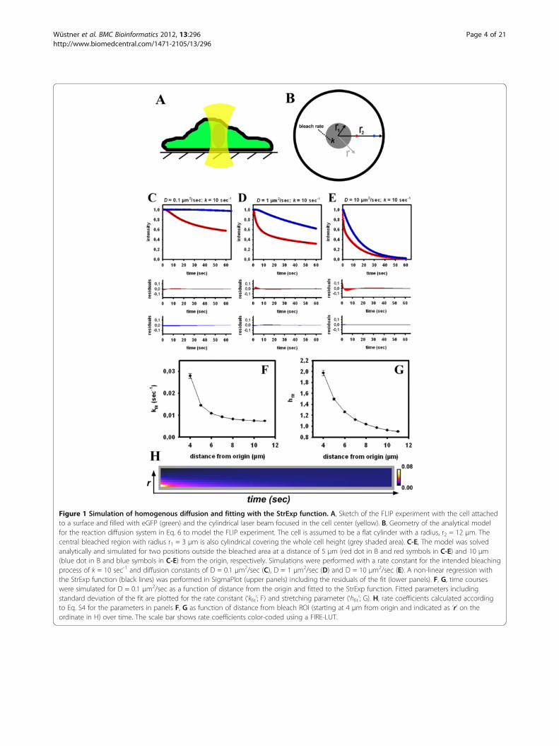
Figure 1 Simulation of homogenous diffusion and fitting with the StrExp function. A, Sketch of the FLIP experiment with the cell attachedto a surface and filled with eGFP (green) and the cylindrical laser beam focused in the cell center (yellow). B, Geometry of the analytical modelfor the reaction diffusion system in Eq. 6 to model the FLIP experiment. The cell is assumed to be a flat cylinder with a radius, r2 = 12 μm. Thecentral bleached region with radius r1 = 3 μm is also cylindrical covering the whole cell height (grey shaded area). C-E, The model was solvedanalytically and simulated for two positions outside the bleached area at a distance of 5 μm (red dot in B and red symbols in C-E) and 10 μm(blue dot in B and blue symbols in C-E) from the origin, respectively. Simulations were performed with a rate constant for the intended bleachingprocess of k = 10 sec-1 and diffusion constants of D = 0.1 μm2/sec (C), D = 1 μm2/sec (D) and D = 10 μm2/sec (E). A non-linear regression withthe StrExp function (black lines) was performed in SigmaPlot (upper panels) including the residuals of the fit (lower panels). F, G, time courseswere simulated for D = 0.1 μm2/sec as a function of distance from the origin and fitted to the StrExp function. Fitted parameters includingstandard deviation of the fit are plotted for the rate constant (‘kfit’; F) and stretching parameter (‘hfit’; G). H, rate coefficients calculated accordingto Eq. S4 for the parameters in panels F, G as function of distance from bleach ROI (starting at 4 μm from origin and indicated as ‘r’ on theordinate in H) over time. The scale bar shows rate coefficients color-coded using a FIRE-LUT.
Wüstner et al. BMC Bioinformatics 2012, 13:296 Page 4 of 21http://www.biomedcentral.com/1471-2105/13/296
simulations can be considered as diffusion-limitedchemical reaction destroying fluorophores selectively inthe bleached ROI. The number of fluorophores, n(t),drops due to localized bleaching, for which one can setdn(t)/dt = − k(t) · n(t). The time-dependent rate coeffi-cient, k(t), models the diffusion-limited destruction offluorophores [34-36]. In the StrExp function, we have
k tð Þ ¼ 1h:τ0
: tτ0
� �1h�1
with h > 1 in the bleached ROI and
the depletion zone and h < 1 at longer distances from thebleach spot (see Additional file 1: Equations. S1-S4). Ac-cordingly, the rate coefficient describing the kinetics of FLdecreases for the stretched case (in the bleached ROI andthe depletion zone) and increases for the compressed caseof the fitting function (distant from the bleach spot). This isillustrated in Figure 1H, where the rate coefficients werecalculated using the fitting parameters of the StrExp func-tion (compare Figure 1F, G). In and close to the bleach spot,the bleaching reaction slows down over time, since thefluorescent molecules being located further apart musttravel longer distances to reach the bleach ROI. This isequivalent to a decreasing rate coefficient and h > 1, re-spectively. Vice versa, for regions more distant from thebleached ROI, there is a delay before FL takes place, whichis characterized by compressed exponentials (h < 1) and anincreasing rate coefficient. If bleaching is slow compared tothe probe diffusion, the intensity decay at each position out-side the bleached ROI is solely governed by the bleach rateconstant, and we recover a mono-exponential decay at eachposition (i.e., bleach-limited FLIP, not shown). Table 1
summarizes the possible scenarios in diffusion-limited FLIPexperiments and the possible outcome of the StrExp fit tothe data.To account for heterogeneous intracellular diffusion
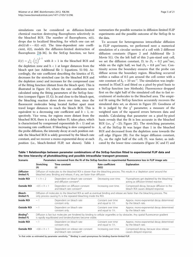
in FLIP experiments, we performed next a numericalsimulation of a circular section of a cell with 2 differentdiffusion constants (Figure 2 and Additional file 2:Movie S1). On the left half of this 2-μm-sized unit cell,we set the diffusion constant, D, to D1 = 0.2 μm2/sec,while on the right half, we had D2 = 0.8 μm2/sec. Con-tinuity across the boundary ensures that the probe candiffuse across the boundary region. Bleaching occurredwithin a radius of 0.5 μm around the cell center with arate constant of kb = 10 sec-1. The simulation was imple-mented in FEniCS and fitted on a pixel-by-pixel basis toa StrExp function (see Methods). Fluorescence droppedfirst on the right half of the simulated cell due to fast re-cruitment of molecules to the bleached area. The empir-ical fit using the StrExp function accurately recovers thesimulated data set, as shown in Figure 2D. Goodness offit is judged by the χ2 parameter, a measure of theweighted sum of the squared errors between data andmodels. Calculating that parameter on a pixel-by-pixelbasis reveals that the fit is less accurate in the bleachedROI (i.e., χ2 ~25; Figure 2E). The stretching parameter,h, of the StrExp fit was larger than 1 in the bleachedROI and decreased from the depletion zone towards thecell edge (Figure 2B). For the larger diffusion constant,D2, on the right half of the cell, FL was faster, as indi-cated by the lower time constants (Figure 2C and F) and
Table 1 Relationships between parameter combinations of the StrExp function fitted to experimental FLIP data andthe time hierarchy of photobleaching and possible intracellular transport processes
Scenario Parameters recovered from the fit of the StrExp function to experimental fluorescence loss in FLIP image sets
Stretching(h)
Time constant(τ)
Rate coefficient(k(t))
Explanation
Diffusion-limited FL
Diffusion of molecules to the bleached ROI is slower than the bleaching process. This results in a ’depletion zone’ around thebleached area. Binding and release, if any, are faster than diffusion.
Inside ROI 1 < h < 2 Dependent on bleach rate constantand diffusion constant*.
Decreasing over time. Fluorophores get depleted by the bleachingacting as diffusion-limited reaction.
Outside ROI ~0.5 < h < 1 Dependent on diffusion constantand bleach rate constant*.
Increasing over time. Compressed decay, because diffusion to thebleach ROI causes delayed response.
Bleach-limited FL
Diffusion of molecules to the bleached ROI as well as eventual binding and release are faster than the bleaching process. Theonly process causing FL is the repeated bleaching inside the ROI.
Inside ROI ~ 1 Dependent on bleach rateconstant only.
Constant over timeand equal to 1/τ.
Approx. mono-exponential decay determinedby the bleach rate.
Outside ROI ~ 1 Dependent on bleach rateconstant only.
Constant over timeand equal to 1/τ.
Approx. mono-exponential decay determinedby the bleach rate.
Binding#-limited FL
Diffusion is fast but molecules are hindered by binding to cellular organelles or by obstacles. Any spatial fluorescence gradientis rapidly equilibrated and binders/barriers become visible.
Inside ROI ~ 1 Dependent on bleach rateconstant only.
Constant over timeand equal to 1/τ.
Approx. mono-exponential decay determinedby the bleach rate.
Outside ROI ~0.8 < h < 1 Dependent on release rate constantand bleach rate constant*.
Increasing over time. Compressed decay, because slow releasecauses delayed response.
*In that order as estimated by parameter sensitivity. # ‘Binding’ is used synonymous for binding-/barrier-limited FLIP.
Wüstner et al. BMC Bioinformatics 2012, 13:296 Page 5 of 21http://www.biomedcentral.com/1471-2105/13/296
higher rate coefficients (Figure 2G-L) compared to theleft half of the cell. The slowest and most delayed inten-sity decay is found on the left cell edge, where h ~ 0.65,compared to the right cell border with h ~ 0.85. Bothvalues describe compressed exponentials and therebyincreasing rate coefficents for the FL with time (com-pare box 1 and 4 in Figure 2I, L). The spatial gradientof FL kinetics from close to the bleached area towardsthe cell border is more pronounced for the left halfwith slow diffusion (Figure 2F). Together, the simula-tions show that the StrExp function can ‘catch’ all es-sential elements of diffusion-limited FLIP experiments.From the estimated parameter set, one can recover thecomplete spatiotemporal map of the FL process. Themain characteristics of diffusion-limited FLIP experi-ments is revealed in the h-map of the regression with
the StrExp function, i.e., a gradual transition from astretched decay inside the bleach ROI with h > 1 to acompressed decay outside the ROI with h < 1. Theslower the diffusion is compared to the bleach rate, thelarger are the differences of h-values along this gradi-ent. Additional simulations for 3D diffusion can beequally well described by the StrExp function (see Add-itional file 1: Figure S3).
Diffusion-limited FLIP of eGFP in the cytoplasm of McAcellsWe aimed for an experimental realization of these predic-tions in a physiologically relevant setting. Enhanced greenfluorescent protein (eGFP) is a small 27 kDa proteinwhich does not bear a nuclear targeting sequence andshould not bind to any cellular structures. While its
Figure 2 Simulation of heterogenous diffusion and fitting with the StrExp function. A 2-dimensional bleaching experiment was simulatedon a disk with a circular bleached area of radius r1=0.5 μm using FeniCS, an automated computational modelling suite (www.fenicsproject.org).The bleaching rate is set to k=10 sec-1 and the diffusion coefficient is d1=0.2 μm2/sec and d2=0.8 μm2/sec on the left and right half disk,respectively (A). Fluorescence loss inside and outside the bleached region was fitted at every pixel position with the StrExp function. For thispurpose the PixBleach plugin to ImageJ was used [40]. The regression recovers a map of the stretching parameter, h, color-coded between 0.4 to2.0 (B), the time constant distribution color-coded between 1.0 to 15.0 sec (C) and the χ2-map color-coded between 0 to 50 (E). A FIRE-LUT wasused for color-coding, where dark blue and yellow indicate lowest and highest values, respectively. The reconstructed stack exactly resembles thesimulated data set, as seen in D (upper row, simulated data (‘sim‘), lower row, regression (‘fit‘)). F, profile of time constants, as estimated fromfitting the StrExp function to the simulated FLIP data along the line shown in panel C. G-L, rate coefficients were calculated on a pixel-by-pixel
basis according to k tð Þ ¼ 1h:τ0
: tτ0
� �1h�1
with self-programmed macros to ImageJ. Rate coefficients are color-coded using a FIRE-LUT in the range
from 0.017 (dark blue) to 0.97 (yellow). G, shows montage of all time points; H, first frame of montage with some regions of interest (boxes 1to 4) used for analysis in panel I-L. Rate coefficient as function of time averaged for box 1 (I), box 2 (J), box 3 (K) and box 4 (L) from the wholesequence.
Wüstner et al. BMC Bioinformatics 2012, 13:296 Page 6 of 21http://www.biomedcentral.com/1471-2105/13/296
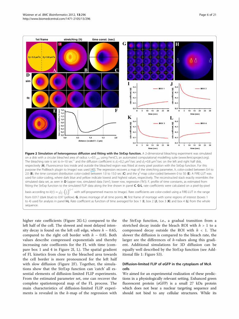
cytoplasmic and nuclear diffusion is very rapid (D ~ 25μm2/sec), eGFP shuttles between nucleus and cytoplasmby passive bidirectional exchange, which is well describedby exponential functions [14-16]. We repeatedly bleachedfluorescence of eGFP in a small cytoplasmic region (i.e., 5iterations per bleach at full laser power of 25 mW) andacquired the images at a rate of 1.6 frames/sec, includingthe bleach. This was fast enough to ensure that cytoplas-mic diffusion of eGFP cannot keep up with rapidnessof the bleaching process (Figure 3). In compliance withthe simulations of Figures 1 and 2 and with Table 1, fittingthe StrExp function to this data gives a gradient of stretch-ing parameters, h, ranging from h ~ 1.7 in the bleachedROI towards h ~ 0.7 at the distant side of the cell(Figure 3B and E). This is most obvious in the line profile
of the stretching parameter, h, drawn as function of dis-tance from the bleach spot (Figure 3E). Thus, FL kineticsof eGFP in this experiment shows a transition from astretched to a compressed exponential decay when mov-ing away from the bleach spot. The fitted time constants(Figure 3C) and rate constants (Figure 3D) reveal fasterdecay in the bleached ROI and its immediate surroundingthan more distant from it. Together, the parameter mapsobtained from fitting the StrExp function to the FLIP dataof eGFP reveal sensitively the diffusion-limited regime,even though a depletion zone is not readily apparent inthe data. Note, that we had a high detector gainfor the experiments shown in Figure 3 to visualize cyto-plasmic eGFP with high frame rate and SNR. Fluorescenceof nuclear eGFP was therefore saturated in these
Figure 3 Diffusion-limited FLIP of eGFP in the cytoplasm of McA cells. McArdle RH7777 cells expressing eGFP in the cytoplasm and nucleuswere placed on a temperature-controlled stage of a confocal microscope maintained at 35 ± 1°C. A 10 pixel diameter circular region in thecytoplasm (white circle) was repeatedly bleached with full laser power, while the whole field was scanned with 0.5% laser output between thebleach scans. This gave a total frame rate of 1.6 sec (see Methods for further details). A, montage of the time series with every 20th frame shown.B-D, fit of the StrExp function to the data giving a map of the stretching parameter (B), the time constant map (C) and its reciprocal, the rateconstant map (D). A FIRE-LUT was used for color-coding, where dark blue and yellow indicate lowest and highest values, respectively. The rangeof values is given below the images with a color bar without units in B, in sec in C and in sec-1 in D. Bar, 5 μm. E, profile of stretching parametersalong the line shown in B (grey line, data; black line moving average to smooth the profile).
Wüstner et al. BMC Bioinformatics 2012, 13:296 Page 7 of 21http://www.biomedcentral.com/1471-2105/13/296
experiments making exact recovery of rate constants andh-values impossible for the nuclear compartment. Tofurther confirm that our method can assess diffusion-limited FLIP data, we performed FLIP experiments usingthe fluorescent cholesterol probe, BODIPY-cholesterol(BChol) (Additional file 1: Figure S4). BChol diffuses by atleast a factor 30 slower than eGFP due to frequent parti-tion into organelle membranes in cells [37]. This allowedus to clearly discern a depletion-zone being accuratelydescribed by the StrExp function. Not only FL of BChol inthe cytoplasm but also in small moving vesicles could bedescribed by the StrExp suggesting sterol release into thecytoplasm (Additional file 1: Figure S4A-D). Together, theexperiments in Figure 3 and Additional file 1: Figure S4confirm the simulation results: pixel-wise regression of theStrExp function to FLIP image sets not only describes ac-curately diffusion-limited FLIP experiments but allowsalso for detecting even faint depletion zones based on thegradient of stretching parameters in the h-map.
Nucleo-cytoplasmic transport of eGFP in McA cells asexample of binding/barrier-limited FLIP experimentsIn many experimental situations, diffusion is much fasterthan the photobleaching process in the bleach ROI. Still,transport to the bleached area in FLIP experiments canbe hindered by transient binding events or barriers todiffusion, even though the diffusion of a molecule in theaqueous phase of the cytoplasm, i.e., the cytosol, is fast.For example, exchange of proteins between nucleus andcytoplasm has been shown to be limited by transportthrough the nuclear pore complex but not by diffusion[14-16,38]. Binding-/barrier dominated transport can bemodelled by a set of ordinary differential equations, asgiven in Eqs. 3–5 in Methods. For the given example,the constants k1 and k−1 reflect nuclear export and im-port rate constants, respectively, but the same equationswould also describe binding/dissociation-limited FLIPexperiments. We confirmed in additional simulations,that the StrExp function is able to describe binding/bar-rier-dominated FLIP experiments (see Additional file 1:Figure S5, S6, S7 and S8).For the purpose of an experimental realization of bind-
ing/barrier-limited FLIP, we studied nucleo-cytoplasmicshuttling of eGFP by our FLIP approach. A small region inthe cytoplasm of McArdle 7777A cells expressing eGFPwas repeatedly bleached with a total frame rate includingthe bleach of 2.6 sec (see white circle in Figure 4A forbleach ROI). Fluorescence loss takes place rather homoge-neously in the cytoplasm with no sign for a diffusion-limited depletion zone surrounding the bleach ROI(Figure 4A). Fitting the StrExp function on a pixel-by-pixel basis to that data did not reveal a gradient in thetime constant or h-map either, supporting that diffusion isnot the limiting factor in this FLIP experiment Figure 4E
and G). At the edges of the eGFP-filled cytoplasm, therecorded intensity as well as the amplitude, stretching andtime constant parameters are slightly larger than in theremaining cytoplasm (Figure 4A-B and E-F). That is likelya consequence of strong differences in refractive index atcell edges, which causes dispersion of the excitation lightand thereby an apparent image crisping [39]. It is notcaused by the fitting routine, since it was not found insynthetic image sets (compare Figures 1, 2 and Additionalfile 1: Figure S3, S5, S6, S7 and S8). The FL of eGFP in thenucleus was delayed compared to that in the cytoplasm,likely due to time-limiting transport of the proteinthrough the NPC [14-16]. Fitting the data to a StrExpfunction gives a very good reconstruction and regressionquality (Figure 4A, compare ‘data’ and ‘fit’). We found thatall eGFP can be bleached by our FLIP set-up. This isreflected by the absence of background in the non-linearregression of the StrExp model to the data (Figure 4B, C).Some ROI boxes were outlined manually and the experi-enced FL was fitted to a StrExp function (Figure 4B-D;boxes 1 and 2 in nucleus and box 3 in cytoplasm). Clearly,the StrExp function fits the data very well, with com-pressed exponential characteristics in the nucleus (box 1:h = 0.5427; k = 0.0195 frame-1; box 2: h = 0.6411; k =0.0169 frame-1) and an almost mono-exponential decay inthe cytoplasm (box 1: h = 0.9815; k = 0.0571 frame-1).Lower stretching with h < 1 and large time constants wereoften found in the nucleus (Figure 4E and G). This is a dir-ect consequence of the rate-limiting transport of eGFPthrough the nuclear pore complex. Crossing the nuclearmembrane causes some delay in the FL in the nucleuscompared to regions in the cytoplasm. Accordingly, therate coefficients calculated from the time constant and h-maps increase over time, most prominent in the nucleus(compare Table 1). The difference in time constants be-tween the two cellular compartments is so pronouncedthat one could even segment cell nuclei and cytoplasmbased on the FL time constant of eGFP (see two peaks intime constant histogram, Figure 4J). These results confirmall predictions for simulated barrier-limited FLIP experi-ments shown in Additional file 1: Figure S5. Notably, in-tensity in the neighbor cell, which is not affected by thebleaching protocol, remained constant throughout the ex-periment (Figure 4A). Since our plugin ‘PixBleach’ allowsfor excluding pixels from the fit which experience an in-tensity decay lower than some threshold value (‘minimaldecay’-option), fluorescence time traces in the neighborcells were not fitted by the software [40]. By closer inspec-tion, one finds sub-micron granularity in the time con-stant and h-maps for the fits of the StrExp function tothe FLIP experiments of eGFP, most pronounced in thenucleus (Figure 4E and G). Calculation of the time evolu-tion of the underlying rate coefficients on a pixel-by-pixel-basis allows for local comparison of decreasing or
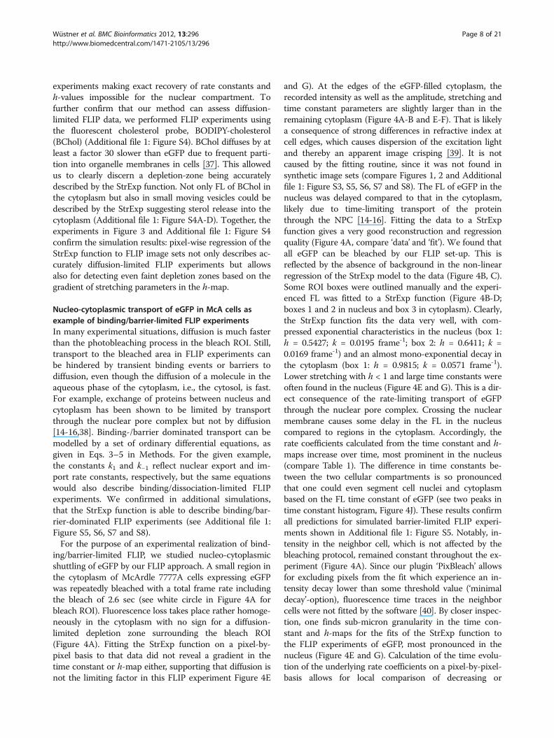
Wüstner et al. BMC Bioinformatics 2012, 13:296 Page 8 of 21http://www.biomedcentral.com/1471-2105/13/296
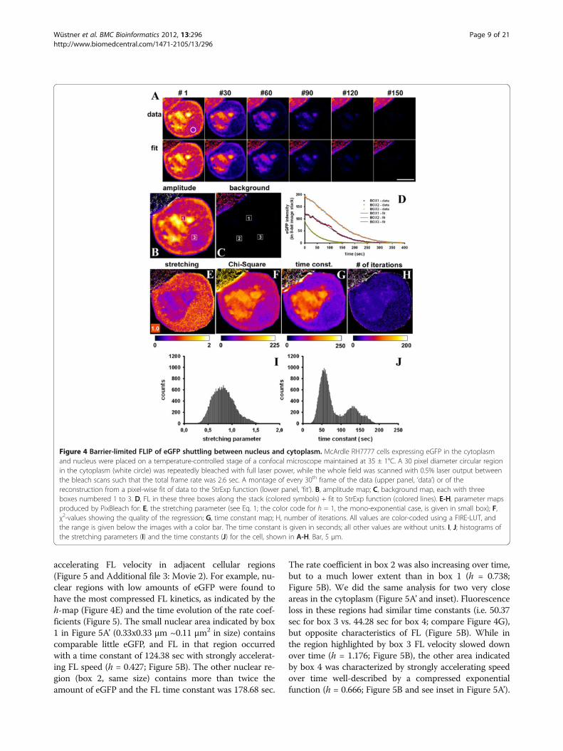
accelerating FL velocity in adjacent cellular regions(Figure 5 and Additional file 3: Movie 2). For example, nu-clear regions with low amounts of eGFP were found tohave the most compressed FL kinetics, as indicated by theh-map (Figure 4E) and the time evolution of the rate coef-ficients (Figure 5). The small nuclear area indicated by box1 in Figure 5A’ (0.33x0.33 μm ~0.11 μm2 in size) containscomparable little eGFP, and FL in that region occurredwith a time constant of 124.38 sec with strongly accelerat-ing FL speed (h = 0.427; Figure 5B). The other nuclear re-gion (box 2, same size) contains more than twice theamount of eGFP and the FL time constant was 178.68 sec.
The rate coefficient in box 2 was also increasing over time,but to a much lower extent than in box 1 (h = 0.738;Figure 5B). We did the same analysis for two very closeareas in the cytoplasm (Figure 5A’ and inset). Fluorescenceloss in these regions had similar time constants (i.e. 50.37sec for box 3 vs. 44.28 sec for box 4; compare Figure 4G),but opposite characteristics of FL (Figure 5B). While inthe region highlighted by box 3 FL velocity slowed downover time (h = 1.176; Figure 5B), the other area indicatedby box 4 was characterized by strongly accelerating speedover time well-described by a compressed exponentialfunction (h = 0.666; Figure 5B and see inset in Figure 5A’).
Figure 4 Barrier-limited FLIP of eGFP shuttling between nucleus and cytoplasm. McArdle RH7777 cells expressing eGFP in the cytoplasmand nucleus were placed on a temperature-controlled stage of a confocal microscope maintained at 35 ± 1°C. A 30 pixel diameter circular regionin the cytoplasm (white circle) was repeatedly bleached with full laser power, while the whole field was scanned with 0.5% laser output betweenthe bleach scans such that the total frame rate was 2.6 sec. A montage of every 30th frame of the data (upper panel, ‘data’) or of thereconstruction from a pixel-wise fit of data to the StrExp function (lower panel, ‘fit’). B, amplitude map; C, background map, each with threeboxes numbered 1 to 3. D, FL in these three boxes along the stack (colored symbols) + fit to StrExp function (colored lines). E-H, parameter mapsproduced by PixBleach for: E, the stretching parameter (see Eq. 1; the color code for h = 1, the mono-exponential case, is given in small box); F,χ2-values showing the quality of the regression; G, time constant map; H, number of iterations. All values are color-coded using a FIRE-LUT, andthe range is given below the images with a color bar. The time constant is given in seconds; all other values are without units. I, J; histograms ofthe stretching parameters (I) and the time constants (J) for the cell, shown in A-H. Bar, 5 μm.
Wüstner et al. BMC Bioinformatics 2012, 13:296 Page 9 of 21http://www.biomedcentral.com/1471-2105/13/296
Thus, local heterogeneity of eGFP transport exists in thecells, and, especially in the nucleus, regions with higheGFP concentration experience slower FL than regionswith low eGFP concentration. Additional simulations con-firmed that the local heterogeneity in FL, we detect by ourapproach is not due to detector noise or bad fitting per-formance (see Figures 4 and 5 and Additional file 1: FigureS6, S7 and S8). Note that we never observed stretched
exponential intensity loss in the non-bleached compart-ment (i.e. C1) nor compressed FL in the bleached ROI (i.e.C2) in simulations of binding/barrier-dominated FLIPsimulations. The fact, that we find this in FLIP experi-ments of eGFP in cells suggests that the simple compart-ment model is not adequate to describe local heterogeneityof intracellular eGFP motion. Further evidence for that isprovided in the Additional files, especially Additional
Figure 5 Pixel-wise rate coefficients of fluorescence loss kinetics of eGFP. Using the parameter maps of the FLIP analysis for the cell inFigure 4, rate coefficients were calculated for every pixel position, as outlined in Additional file. A, the resulting 32-bit image stack was color-coded using a FIRE-LUT, and selected frames of the rate coefficients were plotted (blue and yellow-white indicate low and high rate coefficients,respectively). Areas with accelerating speed of FL turn from blue to yellow-white over time. A’ few regions are highlighted with boxes (numbered‘1’ to ‘5’) for further analysis. Boxes 1 and 2 were placed in the nucleus, while boxes 3 to 5 were in the cytoplasm (inset shows a zoom for thearea containing boxes 3 and 4 close to the right edge of the cell). B, rate coefficients as function of time plotted for box 1 to 4. C, x,t-view of thetime evolution of the rate coefficients in box 5. See text for further explanations.
Wüstner et al. BMC Bioinformatics 2012, 13:296 Page 10 of 21http://www.biomedcentral.com/1471-2105/13/296
file 1: Figure S8. We also performed the opposite experi-ment and bleached repeatedly a small region in the nucleus(Additional file 1: Figure S9). As expected, fluorescencedecayed more rapidly in the nucleus than in the cytoplasmin this experiment, since influx of eGFP into the nucleus israte-limiting for FL in the cytoplasm. Moreover, we foundthat there is a positive correlation between amplitude andtime constant map, as obtained from fitting the StrExpfunction to FL data (Additional file 1: Figure S9). Together,fitting the StrExp function on a pixel-by-pixel basis to bind-ing/barrier-limited FLIP data allows for detecting regions ofdiffering eGFP mobility in living cells.
Heterogeneous transport of mutant huntingtin detectedby quantitative FLIP analysisA group of neurodegenerative disorders is characterizedby expansion of poly-glutamine (polyQ) repeats in ag-gregation prone proteins. For example, more than 36polyQ repeats in mutant huntingtin (mtHtt) causes ag-gregation of the protein in the cytoplasm of affectedcells causing Huntington disease [41]. There exist atleast three populations of mtHtt in affected cells; mono-mers, oligomers and large aggregates called inclusionbodies (IB’s). Probably oligomers are the most cytotoxicform, but the exact mechanism causing Huntingtondisease is not understood and a matter of debate [42].Formation of mtHtt aggregates has been studied in liv-ing cells by a variety of methods including FRAP andFLIP, photoactivation, Förster resonance energy transfer(FRET), fluorescence correlation spectroscopy (FCS) andNumber & Brightness (N&B) analysis [42-46]. We tran-siently expressed eGFP-tagged constructs of controlhuntingtin (containing 23 glutamine (Q) repeats; eGFP-Q23) or mtHtt containing 73 repeats (eGFP-Q73)in Chinese hamster ovarian (CHO) cells. While eGFP-Q23 had similar characteristics in FLIP experiments aspure eGFP (not shown), eGFP-Q73 differed significantly(Figures 6 and 7). That protein forms large IB’s in thecytoplasm, which were identified based on their delayedFL kinetics (Figure 6A, arrows). Fitting the StrExp func-tion on a pixel-by-pixel basis to the FL of that cellrevealed some interesting aspects: firstly, nuclear FL wasvery slow (Figure 6D), probably caused by small aggre-gates which cannot pass the nuclear pore complex. Sec-ondly, the map of the stretching parameters (Figure 6C),time constants (Figure 6D) and Chi-square values weremore structured than observed in a similar experimentin cells only expressing eGFP (Figure 4) or cells expres-sing eGFP-Q23 (not shown). This is probably due tosmall protein aggregates moving in the cytoplasm; andfinally the quality of the regression was significantlylower at the location of the IB, as inferred from the Chi-Square map (Figure 6E). The lower fit performance is aconsequence of movement of the IB causing abrupt
changes in intensity at the respective pixel positions dur-ing the FLIP experiment. This creates large artifacts inthe pixel-wise fitting performance (Figure 6F visualizesthe movement of the perinuclear IB). Accordingly, fittingthe StrExp function to FL on a pixel-by-pixel basis is notappropriate for analyzing moving particles. We thereforecombined quantitative FLIP with single particle tracking(SPT) performed with our SpotTracker plugin to ImageJ.This plugin not only provides particle coordinates butalso the associated particle intensity [47]. We fitted FLmeasured for the tracked IB to a StrExp function toquantify the decay (Figure 6G). The parameters wereA = 0.9455, h = 0.7876 and τ = 57.8 sec. In parallel,SpotTracker provides the IB trajectory (Figure 6H) fromwhich we calculated the mean square displacement(MSD) of the moving IB (Figure 6I, black line). By a lin-ear fit to the first 5 data points, we estimated an initialdiffusion constant of D = 1.4x10⁻³ μm2/s (red line inFigure 6I). The MSD-plot deviated significantly from alinear function for large time steps excluding furtheranalysis based on simple Brownian diffusion. We verifiedin simulated time-lapse sequences that the tracking algo-rithm could exactly determine particle positions, evenfor strongly decaying intensity and high noise levels(Figure 6J, K) [47]. FL was simulated with a mono-exponential process of rate constant k = 0.01 sec-1. Ourtracking program 'SpotTracker' could reliably extract theFL kinetics: a fit to the extracted FL profile recovers thebleach rate constant approximately, (fitted kb = 0.0156sec-1) and the residual term is a good measure of thebackground noise (~60 intensity units). For these tests,we implemented random walk simulations in ImageJusing self-programmed macros. Particle bleaching dur-ing movement was simulated using a single exponentialdecay of intensity in the presence of noise, and the in-tensity value was updated for every new position alongan image sequence. Since ImageJ by default allows onlyfor generating random numbers from a uniform distri-bution, the Box-Muller method was implemented totransform these random numbers to a normal (Gauss-ian) distribution [48].
Multi-compartment (MC) modeling of eGFP-mtHttexchange between cytoplasm and IB’sAlthough we were able to quantify FL of eGFP-mtHtt inmoving IB’s using the strategy outlined above, thismethod does not provide cell-independent measures ofeGFP-mtHtt dynamics. This is, since the FL depends notonly on the protein exchange dynamics but also on thetotal cellular pool size of eGFP-mtHtt. In other words,for a larger cell the same FLIP imaging settings wouldgive slower FL from a given IB than for a smaller cell,simply because it would take longer to deplete the wholeprotein pool in the larger cell. To directly compare
Wüstner et al. BMC Bioinformatics 2012, 13:296 Page 11 of 21http://www.biomedcentral.com/1471-2105/13/296
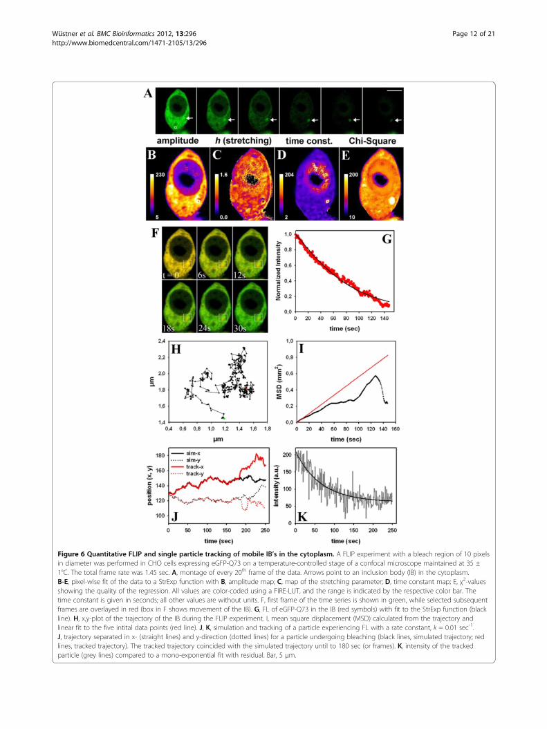
Figure 6 Quantitative FLIP and single particle tracking of mobile IB’s in the cytoplasm. A FLIP experiment with a bleach region of 10 pixelsin diameter was performed in CHO cells expressing eGFP-Q73 on a temperature-controlled stage of a confocal microscope maintained at 35 ±1°C. The total frame rate was 1.45 sec. A, montage of every 20th frame of the data. Arrows point to an inclusion body (IB) in the cytoplasm.B-E, pixel-wise fit of the data to a StrExp function with B, amplitude map; C, map of the stretching parameter; D, time constant map; E, χ2-valuesshowing the quality of the regression. All values are color-coded using a FIRE-LUT, and the range is indicated by the respective color bar. Thetime constant is given in seconds; all other values are without units. F, first frame of the time series is shown in green, while selected subsequentframes are overlayed in red (box in F shows movement of the IB). G, FL of eGFP-Q73 in the IB (red symbols) with fit to the StrExp function (blackline). H, x,y-plot of the trajectory of the IB during the FLIP experiment. I, mean square displacement (MSD) calculated from the trajectory andlinear fit to the five intital data points (red line). J, K, simulation and tracking of a particle experiencing FL with a rate constant, k = 0.01 sec-1.J, trajectory separated in x- (straight lines) and y-direction (dotted lines) for a particle undergoing bleaching (black lines, simulated trajectory; redlines, tracked trajectory). The tracked trajectory coincided with the simulated trajectory until to 180 sec (or frames). K, intensity of the trackedparticle (grey lines) compared to a mono-exponential fit with residual. Bar, 5 μm.
Wüstner et al. BMC Bioinformatics 2012, 13:296 Page 12 of 21http://www.biomedcentral.com/1471-2105/13/296
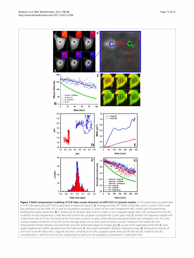
Figure 7 Multi-compartment modeling of FLIP data reveals dynamics of eGFP-Q73 in inclusion bodies. A FLIP experiment was performedin CHO cells expressing eGFP-Q73 as described in legend to Figure 6. A, montage of every 20th frame of the data. Inset is a zoom of the smallbox pointing to an IB; a FIRE LUT is used for visualization purposes. A’, sketch of the multi-compartment (MC) model used for determiningbinding/dissociation parameters. B, FL kinetics for the IB (blue dots) and for a region in the cytoplasm (green dots) with overlayed fit to the MCmodel for the IB (compartment 1; dark blue line) and for the cytoplasm (compartment 2; dark green line). C, another FLIP sequence imaged witha total frame rate of 2.4 sec. First frame of the time series is shown in green, while selected subsequent frames are overlayed in red. This colorcoding visualizes movement of two IB’s at the cell edge (large one up and small one below; arrows). Tracking of the mobile IB’s andmeasurement of their intensity was performed using the SpotTracker plugin for ImageJ [47]. D, x,y-plot of the trajectories of the IB’s; E, meansquare displacement (MSD) calculated from the trajectories; F, step length distribution between subsequent steps; G, fluorescence intensity ofeGFP-Q73 in the IB’s (blue dots = large IB; red dots = small IB) and in the cytoplasm (green dots) and fit with the MC model for the IB’s(compartments 1, dark blue and red line, respectively) as well as for the cytoplasm (compartment 2, dark green line).
Wüstner et al. BMC Bioinformatics 2012, 13:296 Page 13 of 21http://www.biomedcentral.com/1471-2105/13/296
exchange dynamics of eGFP-mtHtt between cytoplasmand IB’s from various cells, we developed an analyticalMC model providing association and dissociation rateconstants (see Figure 7 and Appendix 2). The basic ideaof that modeling strategy is that FL kinetics in the IB’sbecomes weighted by that in the cytoplasm for each cellthereby creating a cell-independent measure of proteinexchange dynamics in the aggregates. For that MCmodel, we assumed that eGFP-mtHtt is distributed intwo pools; several small IB’s as the first pool and thelarge cytoplasm as the second pool. Exchange betweenthe IB’s (compartment(s) 1, C1) and the cytoplasm (com-partment 2, C2) takes place with rate constants k1 and k−1 (Figure 7A’). The number of the IB’s can be large aslong as they together contain a much smaller amount ofeGFP-mtHtt than the cytoplasmic compartment. Fur-thermore, the experimental conditions should be chosensuch that binding-limited FLIP conditions are estab-lished (i.e., no depletion zone like in diffusion-limitedFLIP experiments; this can be verified by pixel-wise fit-ting the StrExp function to FLIP image sets). Finally, it ispresumed that the cytoplasmic pool of eGFP-mtHttchanges only because of the bleach but not due to re-lease of eGFP-mtHtt from IB’s. These conditions are jus-tified by the large size of the cytoplasmic compared tothe IB pool (see above), and they allow for decouplingthe differential equation system (see Appendix 2 for fur-ther details). We first fitted the FL in the cytoplasmcaused by the repeated bleaching with a mono-exponential decay function (Figure 7B; green symbols,data; dark green line, fit). Next, we used the estimatedrate constant as input for fitting FL in the IB outlined bythe blue box in Figure 7A (Figure 7B shows FL data asblue symbols and the fit as dark blue line).The model fit-ted the experimental data very well (R2 > 0.98) and gavea half-time for FL in the cytoplasm of t1/2= 51.7 sec.Note that the FL in the cytoplasm is solely a conse-quence of the bleach protocol. In contrast, the FL in theIB is a result of both, the dissociation of eGFP-Q73 fromthat protein aggregate and the subsequent photodestruc-tion of the released eGFP-Q73 in the bleach ROI. Usingthe FL kinetics in the cytoplasm (see above) and the MCmodel, eGFP-mtHtt dissociation could be calculated tooccur here with a half time of t1/2= ln2/k1 =73.8 sec. An-other example of a cell with 2 IB’s of varying mobility isshown in Figure 7C-G. The larger IB is more mobile butits associated eGFP-Q73 monomers are in slightly slowerexchange with the cytoplasm than for the smaller IB(i.e., half-time of dissociation was t1/2= 200.6 sec andt1/2= 142.3 sec for the large and small IB, respectively).We tracked additional IB’s in the cytoplasm of severalcells and found mean off- and on-rate constants foreGFP-Q73 of k1 = 0.0127 ± 0.004 sec-1 and k−1 = 0.016± 0.006 sec-1 (n=6; mean ± SE). Thus, the average half-
time of eGFP-Q73 dissociation from IB’s is 101.2 ± 30sec in our experiments. The observed heterogeneity inexchange dynamics of eGFP-mtHtt between cytoplasmand IB’s is in line with earlier qualitative FLIP and FRAPexperiments [43,44]. Together, combined fitting of FLkinetics to the MC model and SPT of moving IB’senabled us to determine in parallel aggregate mobilityand exchange dynamics of mtHtt between moving aggre-gates and the cytoplasm.
DiscussionFluorescence loss in photobleaching (FLIP) is a dynamicimaging technique with the potential to include spatialinformation into analysis of protein dynamics, but thispotential has not been explored. By treating images asdata arrays rather than pictures, we present two FLIPanalysis methods for assessing intracellular transport dy-namics of fluorescent proteins. Our first approach com-prises fitting a StrExp function to FL kinetics on a pixel-by-pixel basis. The rationale behind this idea is that thetime-dependent rate coefficient of the StrExp function issuitable to describe transport under conditions, wherethe “well-stirred compartment” assumption fails. Diffu-sion gradients or topological constraints cause deviationfrom the concept of compartment homogeneity, andthat can be modelled with differential equations havingtime-dependent rate coefficients [35]. The StrExp func-tion is based on such an equation, and our method ofpixel-wise fitting this function to FLIP image sets pro-vides kinetic maps of locally heterogeneous transportand allows for exact data reconstruction using thederived model parameters (for example, see Figure 4A).FLIP as an imaging method, has the advantage that theuser can determine the time resolution of the experi-ment by setting pauses of arbitrary length between indi-vidual bleaches. For short pauses, i.e., fast repeatedbleaching; FLIP-experiments might become diffusion-limited (see Figures 1, 2 and 3 and Additional file 1:Figure S1, S2 and S3). We demonstrate on simulated andexperimental FLIP image sets that a spatial gradient of thefitting parameters (i.e., stretching, h, and time or rate con-stant, respectively) of the StrExp function is characteristicfor a diffusion-limited FLIP experiment.For larger pauses between the bleaches, eventual hin-
drance to diffusion due to transient binding or barrierscan be detected on the sub-cellular level by pixel-wiseFLIP analysis without pre-selection of ROIs (Figures 4and 5). This result is not in line with isotropic normaldiffusion of eGFP in cells, though eGFP is known toshow minimal interaction with its surroundings [49]. Anoften (but not always, see [50]) overlooked aspect in theliterature of biophysical diffusion studies (for exampleusing FRAP) is that not only homogenous diffusion butalso locally heterogeneous diffusion (i.e., with a space-
Wüstner et al. BMC Bioinformatics 2012, 13:296 Page 14 of 21http://www.biomedcentral.com/1471-2105/13/296
dependent diffusion constant) with continuity betweenthe regions of varying D will cause an equilibration ofany concentration gradient. This is illustrated in thesimulation shown in Figure 2 (see above). Thus, alreadythe initial distribution of eGFP in the prebleach imagetells us that crowding effects exclude eGFP from someregions in the nucleus and the cytoplasm. We are aware ofone recent study, where the structured fluorescence inten-sity of an inert fluorescent protein (i.e., yellow fluorescentprotein, YFP) in a single image of the cell has been ascribedto the cytoplasm and nucleoplasm being an ‘effective por-ous medium’ [50]. In porous media, one distinguishes dif-fusion in the liquid phase (here the cytosol or nucleosol)from that in the medium with constrained motion. Wespeculate that the large variation in rate coefficients, wefind for FLIP data of eGFP in the cytoplasm is a conse-quence of this porosity. Since the optical resolution of theconfocal microscope (i.e., about 250 nm) is several timeslarger than the pore size of the cytoplasm (i.e., 30–100nm), one cannot segment this cytoplasmic ultrastructure,though it obstructs protein motion [51]. Furthermore, evenfor ‘inert’ biomolecules lacking specific interactions, likeeGFP, not only obstruction is found but also enhancementof diffusion could be triggered by active transport of othercomponents causing enforced motor activity and dynamicremodelling of the cytoskeleton network [52]. Thus, thecytosol might provide rapid decay channels for eGFP, whilethe high protein concentration, organelles and cytoskeletonconstrain eGFP mobility otherwise. Rapid decay channelscan be detected with the StrExp fit to FLIP data as individ-ual pixels with high rate coefficients, even far from thebleached ROI (see Figures 4 and 5 and Additional file 4:Movie S3). Additional simulations of binding-barrier-limited FLIP experiments using the compartment model ofEquations. 3–5 but with rate constants varying on a pixel-by-pixel basis were performed to account for spatial het-erogeneity (Additional file 1: Figures S7 and S8). By fittingthe StrExp function to these simulated time courses, wefound local variation of the recovered parameters (i.e. h-map and time constant map), but in a much narrowerrange than observed for the FLIP data for eGFP in cells(compare Figure 4 and 5 with Additional file 1: Figure S8).Thus, transport of eGFP as measured by FLIP follows acomplex dynamic relaxation pattern due to local hindranceto diffusion on one hand and local enhancement of diffu-sive transport on the other. Such complex behaviour can-not be understood from a simple binding-barrier ordiffusion model of intracellular transport but it can berevealed using pixel-wise fitting of the StrExp function toFLIP image sets.Using our quantitative FLIP analysis, we could also
visualize local diffusion barriers for eGFP in the nucleus(see Figures 4, 5 and Additional file 1: Figure S9). Thesekinetic domains in the nucleus are likely a consequence
of the fractal chromatin organization [53]. Recent resultspublished by Gratton and co-workers detect also barriersfor eGFP diffusion in the nucleus of CHO cells usingpair correlation functions (pCF) [54]. An advantage oftheir method is the ability to extract local diffusion coef-ficients directly from the cross-correlation of intensityfluctuations of the same fluorophore at two different butadjacent points in the sample [55]. Rare intensity burstsreport on eGFP molecules travelling across regions withstrongly varying DNA density [54]. The pCF method hasalso been applied to study transport of eGFP betweennucleus and cytoplasm, where the nuclear membrane in-cluding the NPC appeared as obstacle to eGFP inter-compartment diffusion [56]. This diffusion barrier isnicely detected in the time constant map of our quanti-tative pixel-based FLIP analysis (see Figure 4 G, J), sug-gesting that fluorescence fluctuation techniques like pCFand our approach provide complementary information.To the best of our knowledge, there is only one full
publication published this year, which also applies apixel-wise regression of a decay function to FL data ofFLIP image sets [23]. Lelieveldt and colleagues assumeda simple exponential decay of the FL allowing for linearregression in the logarithmic space. They also performeda noise reduction as pre-processing step and were ableto correct for sudden motion of the specimen or theimage field during acquisition of the FLIP data [23]. Ourapproach implemented in ‘PixBleach’ uses a Gaussian fil-tering in the space and time domain as pre-processingstep which corrects in our hands sufficiently for imagenoise and for small movements (in a range of one ortwo pixels) [40]. Since we apply a motion correction aspre-processing step in case of larger displacement, nofurther corrections were necessary for the image dataused here (see Methods section and [57]). Using theStrExp function is clearly superior to simple mono- orbi-exponential decay models, since it can model FL in-side and outside the bleached region for both, binding/barrier- and diffusion-limited FLIP experiments. Thedelayed decay described by a compressed exponentialfunction as a consequence of binding or barriers (inbinding/barrier -limited FLIP experiments) or due tolong distance to the bleached area (in diffusion-limitedFLIP experiments) cannot be adequately described withsimple exponential decay functions (see Table 1).Quantitative FLIP microscopy should also be a method
to compare results from different cells. This, however, isnot possible with the pixel-based FLIP analysis, becausethe kinetic parameters recovered from the StrExp fit todata are a function of the total amount of fluorophorein a given cell, the diameter of the bleach spot and thelength of the pauses between bleaches. The empiricalStrExp fitting function only provides information aboutthe nature of the observed transport process (e.g.
Wüstner et al. BMC Bioinformatics 2012, 13:296 Page 15 of 21http://www.biomedcentral.com/1471-2105/13/296
diffusion- or binding-limited, see Table 1) and aboutlocal variation in transport dynamics for a given experi-ment and cell. Experiment-independent binding anddissociation parameters of a fluorescent protein cantherefore not be measured by this approach. To over-come this limitation, we developed a complementarymethod based on compartment modelling of FLIPdata and applied it to determine local heterogeneity inintracellular protein aggregation. Such aggregation isobserved in cellular models of various neurodegenerativediseases [45,58,59]. Since protein aggregates tend tomove during FLIP experiments, we combine tracking ofindividual IB’s containing mtHtt with extended polyQrepeat (eGFP-Q73) with non-linear regression of an ana-lytical MC model to the FLIP-induced intensity decay inthese structures (Figures 6, 7). This enables us to meas-ure mobility parameters, like diffusion constants fromthe calculated MSD and to determine in parallel associ-ation and dissociation rate constants for eGFP-mtHttfrom the measured FL kinetics in IB’s. Thus, we demon-strate that mtHtt in IB’s can exchange with mtHtt in thecytoplasm in agreement with earlier observations forother polyQ diseases [43]. Using our MC modeling strat-egy, we can derive for the first time in-vivo binding para-meters of eGFP-mtHtt showing that the exchange takesplace with a half-time of 2–4 min. Since MC modelingof FLIP data provides physical parameters, this methodwill be of high value for quantitative comparison of pro-tein aggregates in Huntington disease with that in otherpolyQ diseases, like various forms of ataxia. In fact,compressed exponential FL, similar as we found foreGFP-mtHtt has been reported in FLIP-experiments ofataxin-3 aggregates, in the nucleoplasm of COS7 cells[58]. We are aware of the fact, that the presented two mod-elling approaches for FLIP data analysis are based on some-how contradictive assumptions: pixel-wise fitting revealslocal heterogeneity of protein transport in various cellularpools, while MC modelling of FLIP experiments requiresthe assumption of well-mixed compartments. Solving thiscontradiction satisfactorily would require modelling thewhole spatiotemporal protein dynamics including cellgeometry, space-dependent diffusion constants, diffusionbarriers, moving entities etc., but this is too ambitious atthe moment and outside the scope of this article. We foundthat deviation of FL kinetics from a mono-exponentialdecay becomes negligible when larger cytoplasmic regions(above ~5 pixels) are included in the analysis (not shown).We also implemented a StrExp decay model for the FL inthe bleached compartment (C2) in our MC model, but thatrecovered a mono-exponential decay with h = 1 (notshown, but see Figure 7C, green symbols and line). To-gether, with the observed time hierarchy, this makes the as-sumption of a well-mixed cytoplasmic compartmentcompared to measured FL of eGFP-Q73 in IB’s reasonable.
ConclusionsWe present two new approaches for quantitative analysisof FLIP experiments in living cells. Pixel-wise fitting of aStrExp function to FLIP image sets allows for detectingareas of different probe mobility, while physical model-ling of FLIP data in the second method provides for thefirst time dissociation parameters of fluorescent proteinsfrom moving entities. Our methods are easy to apply toother transport problems, where a fluorescent biomol-ecule is soluble in the nucleus or cytoplasm and binds toor partitions into static structures (for pixel-based FLIPquantification) or dynamic structures (for combined SPTand MC modeling). Typical other applications could betransient binding of ras-protein to the Golgi apparatusand plasma membrane [60], of lipases and other proteinsto lipid droplets [61], cohesin mobility in the nucleus ofyeast cells [62], recruitment of rab proteins to endo-somes [63] or the dynamic partitioning of fluorescentdrugs and lipids into subcellular membranes [64].
MethodsReagents and cell cultureFetal calf serum and DMEM were from GIBCO BRL(Life Technologies, Paisley, Scotland). 3,3,3’,3’-tetra-methylindocarbocyanine perchlorate (DiIC12) was pur-chased from Molecular Probes (Eugene, Oregon, USA).All other chemicals were from Sigma Chemical (St.Louis, MO). Medium 1 contained 150 mM NaCl, 5 mMKCl, 1 mM CaCl2, 1 mM MgCl2, 5 mM glucose and 20mM HEPES (pH 7.4). McArdle RH7777 (McA) cellsexpressing enhanced green fluorescent protein (EGFP)have been reported previously [65]. These cells weregrown in DMEM with 4.5 g/l glucose, supplementedwith 10% heat-inactivated FCS and antibiotics. Chinesehamster ovarian (CHO) cells were purchased fromATCC (www.atcc.org; LGC Standards Office Europe,AB, Boras, Sweden) and grown in bicarbonate bufferedHam’s F-12 medium supplemented with 5% FCS andantibiotics. Cells were routinely passaged in plastic tissueculture dishes. Two to three days prior to experiments,cells were seeded on microscope slide dishes coated withpoly-D-lysine.
Confocal laser scanning fluorescence microscopyConfocal microscopy was performed using a laser scan-ning inverted fluorescence microscope (Zeiss LSM 510META, Zeiss, Jena, Germany) equipped with a 63x 1.4NA plan Apochromat water immersion objective and a37°C temperature control (Zeiss, Jena, Germany). Fluor-escein and eGFP fluorescence was collected with a 505–530 bandpass filter after excitation with a 25-milliwattargon laser emitting at 488 nm. FLIP experiments wereperformed by first defining regions of interest (ROI),which were repeatedly bleached, while an image was
Wüstner et al. BMC Bioinformatics 2012, 13:296 Page 16 of 21http://www.biomedcentral.com/1471-2105/13/296
acquired with reduced laser power (0.5% output) at thestart of the experiment and after each bleach. Either oneiteration (for eGFP-Huntingtin constructs in CHO cells)or five iterations (for eGFP in McA cells) with 100%laser power were used for the bleaching pulses. An even-tual pause between the bleaches ensured some recoveryin the ROI. Images were acquired using the time-lapsefunction of the Zeiss LSM510 Meta confocal system.The microscope was located at a nitrogen-floated tableto prevent vibrations and focus drift and contained atemperature-controlled stage maintained at 35 ± 1°C.For spatial registration of image stacks, a plugin to Ima-geJ named “StackReg” developed by Dr. Thevenaz at theBiomedical Imaging Group, EPFL, Lausanne, Switzerlandwas used [57]. One pixel typically corresponded to0.055 × 0.055 μm (for example in Figures 4 and 5).
Non-linear regression of a stretched exponential functionto experimental and synthetic FLIP dataPre- and post-bleach images were imported into ImageJand combined into a single stack in 16-bit format. Imagetime series were smoothed with a Gaussian filter (standarddeviation = 0.5) in the spatial and temporal domain. Fluor-escence loss I( r→, t) at each pixel position r→ = (x, y) dueto repeated bleaching of fluorescence in the selected ROIwas fitted to a stretched exponential of the form:
I r→; t
� �¼ I0 r
→� �
: exp � t
τ r→ð Þ� � 1
h r→ð Þ" #
þ I1 r→� �
ð1Þ
using “PixBleach” our recently developed image fittingprogram implemented in ImageJ software (download at:http://bigwww.epfl.ch/algorithms/pixbleach/) [40]. Here,I0 is the pre-bleach intensity of the dynamic fluorescencepool, τ is the decay time constant and I∞ is the remainingintensity at infinite time, resembling either an immobilefraction or autofluorescence of the cells (most often, I∞approximates the background noise level). The decay timeconstant τ describes the rate of decrease in fluorescenceintensity of the probe, either directly in the bleached re-gion due to the intense laser beam, or outside thebleached region due to transport towards the repeatedlybleached ROI. Thus, the bleached regions act like a sinkfor fluorescent molecules being located outside the ROI.The stretched exponential differs from a normal mono-exponential function by an additional parameter, h,describing the stretching or compression of the decaycompared to a mono-exponential function. This param-eter is for h >1 a direct measure of the width of the decayconstant distribution (see Results section). The stretchedexponential model can therefore be considered as a linearsuperposition of simple mono-exponential decays [28]. All
parameter maps generated by ‘PixBleach’ are given in 32-bit format and were used in that format for furthercalculations.
Compartment modeling of fluorophore transport in FLIPexperimentsIn the classical FLIP experiment, a pause between theintended local photobleaching ensures that emittingflurophores are transported into the bleached area suchthat the recovery process is not diffusion-limited. In thiscase and if transient binding events take place, the FLIPexperiment can be modeled by a set of ordinary differ-ential equations (see below). The simplest model fortransport and bleaching contains two compartments; (1)the region outside the bleached area from which trans-port to the bleached ROI occurs with the amount offluorophores, N1(t) and (2) the repeatedly bleached re-gion acting as a “fluorescence sink” for the transport ofthe amount of labeled molecules, N2(t). For the model-ling, we assume that the concentration of fluorophores,n(x, y) =N/V, where V is the volume of the respectivecompartment, is proportional to the emitted fluores-cence intensity at a given wavelength, λ, according to[66]:
I x; y; λð Þ ¼ f θð Þ:g λð Þ:ΦF :L:ε:b:n x; yð Þ ð2Þ
Here, f(θ) is a geometric factor, g(λ) is the wavelength-dependent quantum efficiency of the detector, ΦF is thequantum yield of the fluorophore, L is the excitation in-tensity, ε is the molar extinction coefficient (in M-1 cm-
1) and b is the optical path length. Thus, we can inferrelative values for the concentration or density of probemolecules in certain cellular regions from the pixel-wisedetected fluorescence in the images.We consider bidirectional transport of fluorophores
between compartment 1 and 2 with the forward andbackward rate constants k1 and k−1, respectively. Thebleaching process within the repeatedly bleached area(compartment 2) is modeled by the rate constant k2.
N1 ↔k1;k�1
N2 →k2 ð3Þ
This gives the following system of differential equations:
dN1
dt¼ �k1:N1 þ k�1:N2
dN2
dt¼ k1:N1 � k�1 þ k2ð Þ:N2
ð4a; bÞ
The time-dependent solutions for both compartmentscalculated from the eigenvalues λ1 and λ2 and correspond-ing eigenvectors of the associated kinetic matrix are:
Wüstner et al. BMC Bioinformatics 2012, 13:296 Page 17 of 21http://www.biomedcentral.com/1471-2105/13/296
N1 tð Þ ¼ A þ Bð Þ: exp λ1:tð Þ þ �A þ Bð Þ: exp λ2:tð Þ½ �:N01
2:B
N2 tð Þ ¼ �A2þ Bð Þ: exp λ1:tð Þ þ A2 þ Bð Þ: exp λ2:tð Þ½ �:N02
2:B
with
A ¼ �k1: N01 þ N0
2
� �� k2:N02
A2 ¼ k1: N01 þ N0
2
� �� k2:N02
B ¼ffiffiffiffiffiffiffiffiffiffiffiffiffiffiffiffiffiffiffiffiffiffiffiffiffiffiffiffiffiffiffiffiffiffiffiffiffiffiffiffiffiffiffiffiffiffiffiffiffiffiffiffiffiffiffiffiffiffiffiffiffiffiffiffiffiffiffiffiffiffiffiffiffiffiffiffiffiffiffiffiffiffiffiffiffi�4:k1:k2: N0
2ð Þ2 þ k1: N01 þ N0
2ð Þ þ k2:N02ð Þ2
qλ1 ¼ 0:5: A� Bð Þ=N0
2λ2 ¼ 0:5: Aþ Bð Þ=N0
2
ð5a� fÞ
Here, the initial values N10 and N2
0 describe the initialamounts outside and inside the bleached region, respectively.We have eliminated k−1 by detailed balance, k1 ·N1
0 = k− 1 ·N20.
This kinetic compartment model was simulated with varyingparameter combinations as function of time using SigmaPlot9.0 (SPSS Inc, Chicago, IL, USA) or as function of time andpixel coordinates using self-programmed Macros in ImageJ.An extension of that model to an arbitrary number of non-bleached compartments 1 is given in Appendix 2.
Analytical model of FLIP experiments with spatiallyinvariant diffusion coefficientA FLIP experiment without pause between the individualbleaches results in diffusion-limited transport of fluoro-phores, n, towards the bleached area (see Figure 1A, B forgeometry). This can be modeled using the following par-tial differential equation:
∂n∂t ¼ D:∇2n � k:n:X with
X ¼ 1; . . . r < r10; . . . r > r1
ð6Þ
with the boundary conditions that n; ∂n∂r are continuous at
r = r1 and ∂n∂r ¼ 0 at r = r2. The solution of this model is
given in Appendix 1.
Numerical simulation of FLIP experiments with spatiallyheterogeneous diffusion.It has been reported that diffusion coefficients can dependon the position within the cells (i.e., D ¼ D r→ð Þ =d with r→
being the vector of particle positions [21]. For this situation,no analytical solution to Eq. 6 are available. We thereforeperformed a numerical simulation of a space dependent dif-fusion model for photobleaching rewriting Eq. 6 to:
∂n∂t
¼ div d gradnð Þ � k:n:X ð7Þ
Here, n models the number density of fluorophores, asabove, while the characteristic function X describes thecell area that is illuminated by the laser beam, as above.The bleaching rate is again k. The mathematical model,in variational form,
Z∂n∂t
Φdx þZ
d gradn: gradΦdx þZ
knΧΦdx ¼ 0 ∀Φ ð8Þ
is easily implemented in FEniCS (www.fenicsproject.org). The FEniCS project DOLFIN compiles the pseudo-code and assembles the discrete finite element system.The resulting linear system is solved using the PETSc(www.mcs.anl.gov/petsc) toolkit for scientific computing.We simulated a cylindrical bleaching experiment on adisk with r2 = 1μm. A circular bleached area of radiuswith r1 = 0.5 μm was placed in the disk center. Thebleaching rate is set to k = 10 sec-1 and the diffusion co-efficient is dL = 0.2 μm2/sec and dR = 0.8 μm2/sec onthe left and right half circle, respectively.
AppendixAppendix 1The diffusion problem in Equation 6 reads in Laplace space
(n r; uð Þ ¼Z10
n r; tð Þ:e�utdt):u:n � n0 ¼ D:∇2n � k:nu:n � n0 ¼ D:∇2n
forr < r1r > r1
where n0 is the initial density and
∇2 ¼ 1r:∂∂r
:r:∂∂r
It has the solution n ¼ B1:I0ffiffiffiffiffiffiffiuþkD
q:r
� �þ n0
uþk for r < r1
n ¼ A2:K0
ffiffiffiffiuD
r:r
� �þ B2:I0
ffiffiffiffiuD
r:r
� �þ n0
u
for r > r1 where In,Knrepresent modified Bessel functionsof the first and second kind, respectively of order n, andB1,A2, B2 are defined from the boundary conditions (seemain text and Eq. 6). We get
B1 ¼ �ffiffiffiffiffiffiffiffiffiffiffiu
u þ k
r:I01;1:K
01;2 � I01;2:K
01;1
Y:
k:n0u u þ kð Þ
B2 ¼ � Ik1;1:K01;2
Y:
k:n0u u þ kð Þ
Wüstner et al. BMC Bioinformatics 2012, 13:296 Page 18 of 21http://www.biomedcentral.com/1471-2105/13/296
Y ¼ I00;1:Ik1;1:K 0
1;2
þffiffiffiffiffiffiffiffiffiffiffiu
u þ k
r: Ik0;1:K
01;1:I01;2 � Ik0;1:I
01;1:K0
1;2
h iþ K0
0;1:I01;2:I
k1;1
A2 ¼ � Ik1;1:I01;2
Y:
k:n0u u þ kð Þ
where Xkn;m ¼ Xn
ffiffiffiffiffiffiffiuþkD
q:rm
� �with X = K, I
A numerical inverse Laplace transform has been usedto generate solutions in real space with MathematicaVersion 7.0 (Wolfram research Inc.).
Appendix 2A multi-compartment model for binding-limited FLIPexperiments can be described by a scheme like
Ni1 ↔ki1;k
i�1 N2 →
k2;
where i numbers individual compartments of type 1 em-bedded in a large compartment labeled 2. This gives thefollowing system of differential equations:
dNi1
dt¼ �ki1:N1 þ ki�1
:N2
dN2
dt¼Xi
ki1:Ni1 �
Xi
ki�1 þ k2
!:N2
We assume that compartment 2 is much larger thanthe individual compartments 1, such that FL in compart-ment 2 is solely governed by rate constant k2. This givesfor FL in compartment 2:
N2 tð Þ ¼ N02: exp �k2:tð Þ
The dynamics of the N1i (t) is assumed to satisfy
dNi1
dt¼ �ki1:N1 tð Þ þ ki�1
:N02: exp �k2:tð Þ
By taking into account that
ki�1 ¼ ki1:Ni;01 =N0
2
with the initial amount in the ith compartment of type 1denoted as N1
i,0, we obtain the following solution
Ni1 tð Þ ¼ Ni;0
1:
ki1ki1 � k2
: exp �k2:tð Þ � k2ki1 � k2
: exp �ki1:t� �� �
The solution N2(t) was fitted to FL in the cytoplasm.The solution for the N1
i (t) was fitted separately to each IBper cell using the value of k2 obtained from the fit of N2(t)to FL in the cytoplasm, such that the first IB in a cell givesk11 and N1
1(t), the second k12 and N1
2(t), and so on.
Additional files
Additional file 1: Figure S1. Simulation of the StrExp function anddistance-dependence of fitting to homogenous diffusion. Figure S2.Bleach profile of the Argon laser at 488 nm for various objectives. FigureS3. 3D simulation of a FLIP experiment with heterogeneous diffusion inthe unit sphere. Figure S4. FLIP experiment of BODIPY-cholesterol in CHOcells and fitting with the StrExp function. Figure S5. Simulation of thehomogeneous compartment model and fitting with the StrExp function.Figure S6. Effect of additive image noise on fitting performance. FigureS7. Effect of time noise on fitting performance. Figure S8. Correlationbetween amplitude and time constant maps in the StrExp fit to thespatially Figure S9. Pixel-wise FLIP analysis of eGFP in the nucleus.heterogeneous model.
Additional file 2: Movie 1. Numerical simulation of FLIP experimentwith space-dependent diffusion coefficient.
Additional file 3: Movie 2. Time course of barrier-limited FLIPexperiment of eGFP.
Additional file 4: Movie 3. Time evolution of rate coefficients forbarrier-limited FLIP experiment of eGFP.
Competing interestsThe authors declare that they have no competing interests.
Authors' contributionsConceived and designed experiments: DW. Performed experiments: DW,LMS, FWL. Analyzed experimental data: DW, FWL. Developed mathematicalmodels and performed simulations: DW, DS, HJS, MAL. Performed non-linearregression to simulated data sets: DW. Wrote the paper: DW (with commentsand criticism from all authors). All authors read and approved the finalmanuscript.
AcknowledgementsDW acknowledges funding by grants of the Lundbeck Foundation, the NovoNordisk Foundation and the Danish Research Foundations FNU and FSS. Weare grateful to Tanja Christensen for expert technical assistance. Dr. E. Snapp(Department of Anatomy and Structural Biology, Albert Einstein College ofMedicine, Bronx, New York, USA) is acknowledged for kindly providingplasmids for mtHtt-constructs used in this study.
Author details1Department of Biochemistry and Molecular Biology, University of SouthernDenmark, Campusvej 55, Odense M DK-5230, Denmark. 2Biomedical ImagingGroup, Ecole Polytechnique Fédérale de Lausanne (EPFL), Lausanne CH-1015,Switzerland. 3Institute for Mathematics and Computer Science (IMADA),University of Southern Denmark, Odense M DK-5230, Denmark. 4Departmentof Physics, Chemistry and Pharmacy, MEMPHYS Center for BiomembranePhysics, University of Southern Denmark, Odense M DK-5230, Denmark.
Received: 26 May 2012 Accepted: 31 October 2012Published: 13 November 2012
References1. Dunn KW, McGraw TE, Maxfield FR: Iterative fractionation of recycling
receptors from lysosomally destined ligands in an early sortingendosome. J Cell Biol 1989, 109:3303–3314.
2. Ghosh RN, Maxfield FR: Evidence for nonvectorial, retrograde transferrintrafficking in the early endosomes of HEp2 cells. J Cell Biol 1995,128:549–561.
3. Ghosh RN, Gelman DL, Maxfield FR: Quantification of low densitylipoprotein and transferrin endocytic sorting in HEp2 cells usingconfocal microscopy. J Cell Sci 1994, 107:2177–2189.
4. Wüstner D: Quantification of polarized trafficking of transferrin andcomparison with bulk membrane transport in hepatic cells.Biochem J 2006, 400:267–280.
5. Wüstner D: Mathematical analysis of hepatic high density lipoproteintransport based on quantitative imaging data. J Biol Chem 2005,280:6766–6779.
Wüstner et al. BMC Bioinformatics 2012, 13:296 Page 19 of 21http://www.biomedcentral.com/1471-2105/13/296
6. Wüstner D: Steady state analysis and experimental validation of a modelfor hepatic high density lipoprotein transport. Traffic 2006, 7:699–715.
7. Dunn KW, Maxfield FR: Delivery of ligands from sorting endosomes tolate endosomes occurs by maturation of sorting endosomes. J Cell Biol1992, 117:301–310.
8. Sheff DR, Daro EA, Hull M, Mellman I: The receptor recycling pathwaycontains two distinct populations of early endosomes with differentsorting functions. J Cell Biol 1999, 145:123–139.
9. Hirschberg K, Miller CM, Ellenberg J, Presley JF, Siggia ED, Phair RD,Lippincott-Schwartz J: Kinetic analysis of secretory protein traffic andcharacterization of golgi to plasma membrane transport intermediatesin living cells. J Cell Biol 1998, 143:1485–1503.
10. Dahm T, White J, Grill S, Füllekrug J, Stelzer EHK: Quantitative ER left-right-arrow golgi transport kinetics and protein separation upon golgi exitrevealed by vesicular integral membrane protein 36 dynamics in livecells. Mol Biol Cell 2001, 12:1481–1498.
11. White J, Keller P, Stelzer EH: Spatial partitioning of secretory cargo fromGolgi resident proteins in live cells. BMC Cell Biol 2001, 2:19.
12. Rhee SW, Starr T, Forsten-Williams K, Storrie B: The steady-state distributionof glycosyltransferases between the Golgi apparatus and theendoplasmic reticulum is approximately 90:10. Traffic 2005, 6:1–13.
13. Patterson GH, Hirschberg K, Polishchuk RS, Gerlich D, Phair RD, Lippincott-Schwartz J: Transport through the Golgi apparatus by rapid partitioningwithin a two-phase membrane system. Cell 2008, 133:1055–1067.
14. Peters R: Nuclear envelope permeability measured by fluorescencemicrophotolysis of single liver cell nuclei. J Biol Chem 1983,258:11427–11429.
15. Peters R: Nucleo-cytoplasmic flux and intracellular mobility in singlehepatocytes measured by fluorescence microphotolysis. EMBO J 1985,3:1831–1836.
16. Wei X, Henke VG, Strübing C, Brown EB, Clapham DE: Real-time imaging ofnuclear permeation by EGFP in single intact cells. Biophys J 2003,84:1317–1327.
17. Lippincott-Schwartz J, Altan-Bonnet N, Patterson GH: Photobleaching andphotoactivation: following protein dynamics in living cells. Nat Cell Biol2003, Suppl 7:S7–14.
18. Peters R, Brünger A, Schulten K: Continuous fluorescence microphotolysis:a sensitive method for studying diffusion processes in single cells. ProcNatl Acad Sci U S A 1981, 78:962–966.
19. Kubitscheck U, Wedekind P, Peters R: Three-dimensional diffusionmeasurements by scanning microphotolysis. J Microsc 1998, 192:126–138.
20. Wachsmuth M, Weidemann T, Müller G, Hoffmann-Rohrer UW, Knoch TA,Waldeck W, Langowski J: Analyzing intracellular binding and diffusionwith continuous fluorescence photobleaching. Biophys J 2003,84:3353–3363.
21. Müller F, Mazza D, Stasevich TJ, McNally JG: FRAP and kinetic modeling inthe analysis of nuclear protein dynamics: what do we really know? CurrOpin Cell Biol 2010, 22:403–411.
22. van Gemert AMC, Jost CR, van der Laan A, Dirks R, Reiber JH, Tanke HJ,Lelieveldt BP: Identification of cellular dynamic patterns resulting fromrepetitive photobleaching using independent component analysis.I S Biomed Imaging 2009, :1382–1385.
23. van de Giessen M, van der Laan A, Hendriks EA, Vidorreta M, Reiber JH, JostCR, Tanke HJ, Lelieveldt BP: Fully automated attenuation measurementand motion correction in FLIP image sequences. IEEE Trans Med Imaging2012, 31:461–473.
24. Klafter J, Shlesinger MF: On the relationship among three theories ofrelaxation in disordered systems. Proc Natl Acad Sci U S A 1986, 83:848–851.
25. Koppel DE: Analysis of macromolecular polydispersity in intensitycorrelation spectroscopy: the method of cumulants. J Chem Phys 1972,57:4814–4820.
26. Koppel DE, Carlson C, Smilowitz H: Analysis of heterogeneousfluorescence photobleaching by video kinetics imaging: the method ofcumulants. J Microsc 1989, 155:199–206.
27. Berberan-Santos MN, Bodunov EN, Valeur B: Mathematical functions forthe analysis of luminescence decays with underlying distributions 1.Kohlrausch decay function (stretched exponential). Chemical Physics 2005,315:171–182.
28. Lee KC, Siegel J, Webb SE, Lévêque-Fort S, Cole MJ, Jones R, Dowling K,Lever MJ, French PM: Application of the stretched exponential functionto fluorescence lifetime imaging. Biophys J 2001, 81:1265–1274.
29. Kohlrausch R: Theorie des elektrischen Rückstandes in der LeidnerFlasche. Pogg Ann Phys Chem 1854, 91:179–213.
30. Williams G, Watts DC: Non-symmetrical dielectric relaxation behaviourarising from a simple empirical decay function. Trans Farad Soc 1970,66:80–85.
31. Shlesinger MF, Montroll EW: On the Williams-Watts function of dielectricrelaxation. Proc Natl Acad Sci U S A 1984, 81:1280–1283.
32. Cumbrera FL, Sanchez-Bajo F, Guiberteau F, Solier JD, Muñoz A: TheWilliams-Watts dependence as a common phenomenological approachto relaxation processes in condensed matter. J Mat Sci 1993,28:5387–5396.
33. Braga J, Desterro JM, Carmo-Fonseca M: Intracellular macromolecularmobility measured by fluorescence recovery after photobleaching withconfocal laser scanning microscopes. Mol Biol Cell 2004, 15:4749–4760.
34. Berberan-Santos MN: Mathematical basis of the integral formalism ofchemical kinetics. Compact representation of the general solution of thefirst-order linear differential equation. J Math Chem 2010, 47:1184–1188.
35. In Modeling in Biopharmaceutics, Pharmacokinetics, and Pharmacodynamics.Edited by Macheras P, Iliadis A. New Yrok: Springer; 2006.
36. Berberan-Santos MN, Martinho JMG: A linear response approach tokinetics with time-dependent rate coefficients. Chem Phys 1992,164:259–269.
37. Lund FW, Lomholt MA, Solanko LM, Wüstner D: Two-photon time-lapsemicroscopy of BODIPY-cholesterol reveals anomalous sterol diffusion inchinese hamster ovary cells. 2012, In press.
38. Chen Y, Müller JD: Probing nucleocytoplasmic transport by two-photonactivation of PA-GFP. Microsc Res Tech 2006, 69:220–226.
39. Van Oostveldt P, Verhaegen F, Messens K: Heterogeneous photobleachingin confocal microscopy caused by differences in refractive index andexcitation mode. Cytometry 1998, 32:137–146.
40. Wüstner D, Landt Larsen A, Færgeman NJ, Brewer JR, Sage D: Selectivevisualization of fluorescent sterols in Caenorhabditis elegans by bleach-rate based image segmentation. Traffic 2010, 11:440–454.
41. Li SH, Li XJ: Huntingtin-protein interactions and the pathogenesis ofHuntington's disease. Trends Genet 2004, 20:146–154.
42. Hatters DM: Protein misfolding inside cells: the case of Huntingtin andHuntington's disease. IUBMB Life 2008, 60:724–728.
43. Kim S, Nollen EA, Kitagawa K, Bindokas VP, Morimoto RI: Polyglutamineprotein aggregates are dynamic. Nat Cell Biol 2002, 4:826–831.
44. Matsumoto G, Kim S, Morimoto RI: Huntingtin and mutant SOD1 formaggregate structures with distinct molecular properties in human cells.J Biol Chem 2006, 281:4477–4485.
45. Lajoie P, Snapp EL: Formation and toxicity of soluble polyglutamineoligomers in living cells. PloS One 2010, 5:e15245.
46. Ossato G, Digman MA, Aiken C, Lukacsovich T, Marsh JL, Gratton E: A two-step path to inclusion formation of huntingtin peptides revealed bynumber and brightness analysis. Biophys J 2010, 98:3078–3085.
47. Sage D, Neumann FR, Hediger F, Gasser SM, Unser M: Automatic trackingof individual fluorescence particles: application to the study ofchromosome dynamics. IEEE T Image Process 2005, 14:1372–1383.
48. Brandt S: Datenanalyse. Heidelberg: Spektrum Verlag; 1999.49. Brown CM, Dalal RB, Hebert B, Digman MA, Horwitz AR, Gratton E: Raster
image correlation spectroscopy (RICS) for measuring fast proteindynamics and concentrations with a commercial laser scanning confocalmicroscope. J Microsc 2008, 229:78–91.
50. Kühn T, Ihalainen TO, Hyväluoma J, Dross N, Willman SF, Langowski J,Vihinen-Ranta M, Timonen J: Protein diffusion in mammalian cellcytoplasm. PLoS One 2011, 6:e22962.
51. Kasza KE, Rowat AC, Liu J, Angelini TE, Brangwynne CP, Koenderink GH,Weitz DA: The cell as a material. Curr Opin Cell Biol 2007, 19:101–107.
52. Brangwynne CP, Koenderink GH, MacKintosh FC, Weitz DA: Intracellulartransport by active diffusion. Trends Cell Biol 2009, 19:423–427.
53. Bancaud A, Huet S, Daigle N, Mozziconacci J, Beaudouin J, Ellenberg J:Molecular crowding affects diffusion and binding of nuclear proteins inheterochromatin and reveals the fractal organization of chromatin. EMBOJ 2009, 28:3785–3798.
54. Hinde E, Cardarelli F, Digman MA, Gratton E: In vivo pair correlationanalysis of EGFP intranuclear diffusion reveals DNA-dependentmolecular flow. Proc Natl Acad Sci U S A 2010, 107:16560–16565.
55. Digman MA, Gratton E: Imaging barriers to diffusion by pair correlationfunctions. Biophys J 2009, 97:665–673.
Wüstner et al. BMC Bioinformatics 2012, 13:296 Page 20 of 21http://www.biomedcentral.com/1471-2105/13/296
56. Cardarelli F, Gratton E: In vivo imaging of single-molecule translocationthrough nuclear pore complexes by pair correlation functions. PLoS One2010, 5:e10475.
57. Thevenaz P, Ruttimann UE, Unser E: A pyramid approach to subpixelregistration based on intensity. IEEE T Image Process 1998, 7:27–41.
58. Chai Y, Shao J, Miller VM, Williams A, Paulson HL: Live-cell imaging revealsdivergent intracellular dynamics of polyglutamine disease proteins andsupports a sequestration model of pathogenesis. Proc Natl Acad Sci U S A2002, 99:9310–9315.
59. Nag S, Chen J, Irudayaraj J, Maiti S: Measurement of the attachment andassembly of small amyloid-beta oligomers on live cell membranes atphysiological concentrations using single-molecule tools. Biophys J 2010,99:1969–1975.
60. Goodwin JS, Drake KR, Remmert CL, Kenworthy AK: Ras diffusion issensitive to plasma membrane viscosity. Biophys J 2005, 89:1398–1410.
61. Wang H, Hu L, Dalen K, Dorward H, Marcinkiewicz A, Russell D, Gong D,Londos C, Yamaguchi T, Holm C, et al: Activation of hormone-sensitivelipase requires two steps, protein phosphorylation and binding tothe PAT-1 domain of lipid droplet coat proteins. J Biol Chem 2009,284:32116–32125.
62. McNairn AJ, Gerton JL: Intersection of ChIP and FLIP, genomic methodsto study the dynamics of the cohesin proteins. Chromosome Res 2009,17:155–163.
63. Rink J, Ghigo E, Kalaidzidis Y, Zerial M: Rab conversion as a mechanism ofprogression from early to late endosomes. Cell 2005, 122:735–749.
64. Cunningham CW, Mukhopadhyay A, Lushington GH, Blagg BS, PrisinzanoTE, Krise JP: Uptake, distribution and diffusivity of reactive fluorophoresin cells: implications toward target identification. Mol Pharm 2010,7:1301–1310.
65. Yu L, Bharadwaj S, Brown JM, Ma Y, Du W, Davis MA, Michaely P, Liu P,Willingham MC, Rudel LL: Cholesterol-regulated translocation of NPC1L1to the cell surface facilitates free cholesterol uptake. J Biol Chem 2006,281:6616–6624.
66. Bright GR, Fisher GW, Rogowska J, Taylor DL: Fluorescence ratio imagingmicroscopy. Methods in Cell Biology 1989, 30:157–192.
doi:10.1186/1471-2105-13-296Cite this article as: Wüstner et al.: Quantitative fluorescence loss inphotobleaching for analysis of protein transport and aggregation. BMCBioinformatics 2012 13:296.
Submit your next manuscript to BioMed Centraland take full advantage of:
• Convenient online submission
• Thorough peer review
• No space constraints or color figure charges
• Immediate publication on acceptance
• Inclusion in PubMed, CAS, Scopus and Google Scholar
• Research which is freely available for redistribution
Submit your manuscript at www.biomedcentral.com/submit
Wüstner et al. BMC Bioinformatics 2012, 13:296 Page 21 of 21http://www.biomedcentral.com/1471-2105/13/296
Related Documents