Brain Research, 346 (1985) 205- 230 205 Elsevier BRE 11091 Research Reports Quantitative Autoradiographic Mapping of Serotonin Receptors in the Rat Brain. I. Serotonin-1 Receptors A. PAZOS and J. M. PALACIOS Preclinical Research, Sandoz Ltd., CH-4002 Basel (Switzer/and) (Accepted January 15th, 1985) Key words: [3H]serotonin- - serotonin-1 receptor subtype - - quantitative autoradiography - - hippocampus - - basal ganglion - - choroid plexus - - substantia nigra - - septal nucleus The distribution of serotonin-1 (5-HT 0 receptors in the rat brain was studied by light microscopic quantitative autoradiography. Receptors were labeled with [3H]serotonin (5-[3H]HT), 8-hydroxy-2-[N-dipropylamino-3H]tetralin (8-OH-[aH]DPAT), [3H]LSD and [3H]mesulergine, and the densities quantified by microdensitometry with the aid of a computer-assisted image-analysis system. Com- petition experiments for 5-[3H]HT binding by several serotonin-1 agonists led to the identification of brain areas enriched in each one of the three subtypes of 5-HT1 recognition sites already described (5-HT1A,5-HTm, 5-HTlc ). The existence of these 'selective' areas allowed a detailed pharmacological characterization of these sites to be made in a more precise manner than has been attained in mem- brane-binding studies. While 5-[3HINT labeled with nanomolar affinity all the 5-HT1 subtypes, the other 3H-labeled ligands labeled selectively 5-HT1A(8-OH-[aH]DPAT), 5-HTIc ([3H]mesulergine) and both of them ([3H]LSD). Very high concentrations of 5-HT1 receptors were localized in the choroid plexus, lateroseptal nucleus, globus pallidus and ventral pallidum, dentate gyms, dorsal subi- culum, olivary pretectal nucleus, substantia nigra, reticular and external layer of the entorhinal cortex. The different fields of the hip- pocampus (CArCA4), some nuclei of the amygdaloid complex, the hypothalamic nuclei and the dorsal raph6, among others, also presented high concentrations of sites. Areas containing intermediate densities of 5-HT1 receptors included the claustrum, olfactory tubercle, accumbens, central grey and lateral cerebellar nucleus. The nucleus caudate-putamen and the cortex, at the different levels studied, presented receptor densities ranging from intermediate to low. Finally, in other brain areas - - pons, medulla, spinal cord - - only low or very low concentrations of 5-HT1receptors were found. From the areas strongly enriched in 5-HT1sites, dentate gyrus and septal nucleus contained 5-HTIAsites, while globus pallidus, dorsal subiculum, substantia nigra and olivary pretectal nucleus were en- riched in 5-HTm. The sites in the choroid plexus, which presented the highest density of receptors in the rat brain, were of the 5-HTlc subtype. The distribution of 5-HT1 receptors reported here is discussed in correlation with the distribution of serotoninergic neurons and fibers, the related anatomical pathways and the effects which appear to be mediated by these sites. INTRODUCTION Pharmacological, electrophysiological and bio- chemical findings have revealed the existence of mul- tiple brain serotonin (5-HT) recognition sitesl, 90. O n the basis of the results obtained in binding studies in rat brain, Peroutka and Snyderl09 classified these sites as 5-HT 1, labeled by 5-[3H]HT with high affinity and 5-HT2, which are recognized with high affinity by [3H]spiperone. [3H]LSD labels both receptor sites. The possibility of the existence of different sub- types of 5-HTl-binding sites has received considera- ble support in the recent years. Some serotoninergic agonists, such as the piperidinyl indole RU 24 969 (5- methoxy-3-[1,2,3,6-tetrahydro-4-pyridinil]lH-in- dole) 4° and the 8-OH-DPAT (8-hydroxy-2-[n-dipro- pylamino]tetralin) 55 present Hill coefficients lower than 1 when displacing 5-[3H]HT binding in the rat brain cortex 54,57,81. In a similar way, the neuroleptic spiperone and some fl-blockers also inhibit 5-[3H]HT binding in a biphasic manner (refs. 80, 86, 105, and Engel et al., in preparation). These results suggested the presence of two subtypes of 5-HT~ sites, 5-HTIA and 5-HTm, corresponding respectively to the high- Correspondence: J. M. Palacios, Preclinical Research, Sandoz Ltd., CH-4002 Basel, Switzerland. 0006-8993/85/$03.30 © 1985 Elsevier Science Publishers B.V. (Biomedical Division)

Welcome message from author
This document is posted to help you gain knowledge. Please leave a comment to let me know what you think about it! Share it to your friends and learn new things together.
Transcript
Brain Research, 346 (1985) 205 - 230 205 Elsevier
BRE 11091
Research Reports
Quantitative Autoradiographic Mapping of Serotonin Receptors in the Rat Brain. I. Serotonin-1 Receptors
A. PAZOS and J. M. PALACIOS
Preclinical Research, Sandoz Ltd., CH-4002 Basel (Switzer/and)
(Accepted January 15th, 1985)
Key words: [3H]serotonin - - serotonin-1 receptor subtype - - quantitative autoradiography - - hippocampus - - basal ganglion - - choroid plexus - - substantia nigra - - septal nucleus
The distribution of serotonin-1 (5-HT 0 receptors in the rat brain was studied by light microscopic quantitative autoradiography. Receptors were labeled with [3H]serotonin (5-[3H]HT), 8-hydroxy-2-[N-dipropylamino-3H]tetralin (8-OH-[aH]DPAT), [3H]LSD and [3H]mesulergine, and the densities quantified by microdensitometry with the aid of a computer-assisted image-analysis system. Com- petition experiments for 5-[3H]HT binding by several serotonin-1 agonists led to the identification of brain areas enriched in each one of the three subtypes of 5-HT 1 recognition sites already described (5-HT1A, 5-HT m, 5-HTlc ). The existence of these 'selective' areas allowed a detailed pharmacological characterization of these sites to be made in a more precise manner than has been attained in mem- brane-binding studies. While 5-[3HINT labeled with nanomolar affinity all the 5-HT 1 subtypes, the other 3H-labeled ligands labeled selectively 5-HT1A (8-OH-[aH]DPAT), 5-HTIc ([3H]mesulergine) and both of them ([3H]LSD). Very high concentrations of 5-HT 1 receptors were localized in the choroid plexus, lateroseptal nucleus, globus pallidus and ventral pallidum, dentate gyms, dorsal subi- culum, olivary pretectal nucleus, substantia nigra, reticular and external layer of the entorhinal cortex. The different fields of the hip- pocampus (CArCA4), some nuclei of the amygdaloid complex, the hypothalamic nuclei and the dorsal raph6, among others, also presented high concentrations of sites. Areas containing intermediate densities of 5-HT 1 receptors included the claustrum, olfactory tubercle, accumbens, central grey and lateral cerebellar nucleus. The nucleus caudate-putamen and the cortex, at the different levels studied, presented receptor densities ranging from intermediate to low. Finally, in other brain areas - - pons, medulla, spinal cord - - only low or very low concentrations of 5-HT 1 receptors were found. From the areas strongly enriched in 5-HT 1 sites, dentate gyrus and septal nucleus contained 5-HTIA sites, while globus pallidus, dorsal subiculum, substantia nigra and olivary pretectal nucleus were en- riched in 5-HT m. The sites in the choroid plexus, which presented the highest density of receptors in the rat brain, were of the 5-HTlc subtype. The distribution of 5-HT 1 receptors reported here is discussed in correlation with the distribution of serotoninergic neurons and fibers, the related anatomical pathways and the effects which appear to be mediated by these sites.
INTRODUCTION
Pharmacological, electrophysiological and bio-
chemical findings have revealed the existence of mul-
tiple brain serotonin (5-HT) recognition sitesl, 90. On
the basis of the results obtained in binding studies in
rat brain, Peroutka and Snyderl09 classified these
sites as 5-HT 1, labeled by 5-[3H]HT with high affinity
and 5-HT2, which are recognized with high affinity by
[3H]spiperone. [3H]LSD labels both receptor sites.
The possibility of the existence of different sub-
types of 5-HTl-binding sites has received considera-
ble support in the recent years. Some serotoninergic
agonists, such as the piperidinyl indole R U 24 969 (5-
methoxy-3-[1,2,3,6-tetrahydro-4-pyridinil]lH-in-
dole) 4° and the 8 - O H - D P A T (8-hydroxy-2-[n-dipro-
pylamino]tetralin) 55 present Hill coefficients lower
than 1 when displacing 5-[3H]HT binding in the rat
brain cortex 54,57,81. In a similar way, the neuroleptic
spiperone and some fl-blockers also inhibit 5-[3H]HT
binding in a biphasic manner (refs. 80, 86, 105, and
Engel et al., in preparation). These results suggested
the presence of two subtypes of 5-HT~ sites, 5-HTIA
and 5-HTm, corresponding respectively to the high-
Correspondence: J. M. Palacios, Preclinical Research, Sandoz Ltd., CH-4002 Basel, Switzerland.
0006-8993/85/$03.30 © 1985 Elsevier Science Publishers B.V. (Biomedical Division)
206
and the low-affinity sites of spiperone- and 8-OH- DPAT-competition curves 105. Recently, we have characterized 1°3 a new type of 5-[3H]HT-binding site, mainly localized in the mammalian choroid plexus and which is not recognized with high affinity by the 5-HTIA or 5-HT m 'selective' compounds described above, namely RU 24 969, 8-OH-DPAT, spiperone and fi-blockers, but it is labeled by nanomolar con- centrations of [3H]LSD as well as by the serotonin-2 ligand [3H]mesulergine22. We have named this site 5- HTIc 103.
Considerable efforts are being made in attempts to establish functional correlates for 5-HT 1 sites. A link of 5-HT 1 sites to a 5-HT-sensitive adenylate cyclase has been the subject of much discussion (refs. 17, 42, 88, 107; see ref. 106 for a review). In a similar way, contradictory results have been reported on the pro- posed mediation of the dog basilar artery contraction by 5-HT 1 sitesSSA0s.
A good correlation has been found between the 5- HT1 affinities of several drugs and their effects in models of presynaptic 5-HT receptors38,48, 73, al- though some discrepancies between the activities of the drugs in the 'presynaptic' test and in the binding assay have arisen (see ref. 44 for a discussion). It has been suggested that these discrepancies might reflect the existence of the different subtypes of 5-HT 1 rec- ognition sites. It has also been reported that the '5- HT syndrome', a behavioral syndrome consisting of several complex motor reflexes, is mediated by 5- HT~ sites 69. In addition to 5-HT itself and some 5-HT precursors, this syndrome is also produced by 8-OH- DPAT 127 and blocked by some fl-blockers53A 27. Fur- thermore, both 5-HT agonists RU 24 969 and 8-OH- DPAT appear to produce circling behavior in rats with striatal lesions15, 57. The participation of the dif- ferent subtypes of 5-HT 1 receptors in these effects is not clear at the present time (see Discussion).
The knowledge of the anatomical distribution of these 5-HT 1 sites in the brain constitutes an impor- tant requirement in order to better characterize these sites. It would allow (1) the detailed pharmacological characterization of the different subtypes of 5-HT 1 receptors, by analyzing the binding of 5-[3H]HT in areas enriched in one subtype, and (2) the correla- tion of receptor densities in different brain areas with the physiological activities proposed to be mediated by 5-HT 1 sites, clarifying their functional signifi-
cance. In this regard, quantitative autoradiographic techniques provide, besides a high level of anatom- ical resolution, the adequate estimation of receptor densities and affinities 26,95,1°1.
Autoradiographic visualization of 5-HTI sites in the central nervous system has been carried out using 5-[3H]HT14, ~38 and [3H]LSD75 as ligands. Attempts to
analyze separately the distribution of 5-HT~A and 5- HT m sites have been recently reported using 5- [3H]HT34 and 8-OH-[3H]DPATTL We have demon- strated the presence of 5-HT~c sites in the rat choroid plexus 25. We report here a detailed mapping of the serotonin-1 receptors in the rat brain, using 5- [3H]HT, [3H]LSD, 8-OH-[3H]DPAT and [3H]mesu- lergine as ligands, and analyzing the distribution of the three subtypes throughout the different brain
areas.
MATERIALS AND METHODS
Rats (male, Wistar, 180-250 g) were sacrificed by intracardial perfusion with phosphate-buffered sa- line containing 0.1% formalin, after pentobarbital anesthesia (50 mg/kg). The brains were removed and frozen in liquid nitrogen. 10 ~tm thick tissue sections were cut using a microtome-cryostat (Leitz 1720, from Leitz, Wetzlar, F.R.G.) mounted onto gelatin- coated microscope slides and stored at -20 °C until used.
Incubation conditions for each 3H-labeled ligand, including buffer solution, concentration of ligand, in- cubation time and exposure time are summarized in Table I. Blanks were generated by coincubation with 1 pM LSD in the case of 5-[3H]HT and with 100 nM 5- HT for the other 3H-labeled ligands. In competition studies, consecutive tissue sections were incubated in the presence of increasing concentrations of displac- ers.
The binding of the different 3H-labeled ligands used, 5-HT, LSD, 8-OH-DPAT and mesulergine, to slide mounted brain rat sections has been already ki- netically characterized, the saturation parameters and incubation procedures being defined 14,72, 74,75,102,138. The conditions defined in these studies were used in the present work, with slight modifica- tions.
After the washing period, tissues were dried with cold air and autoradiograms were generated by ap-
207
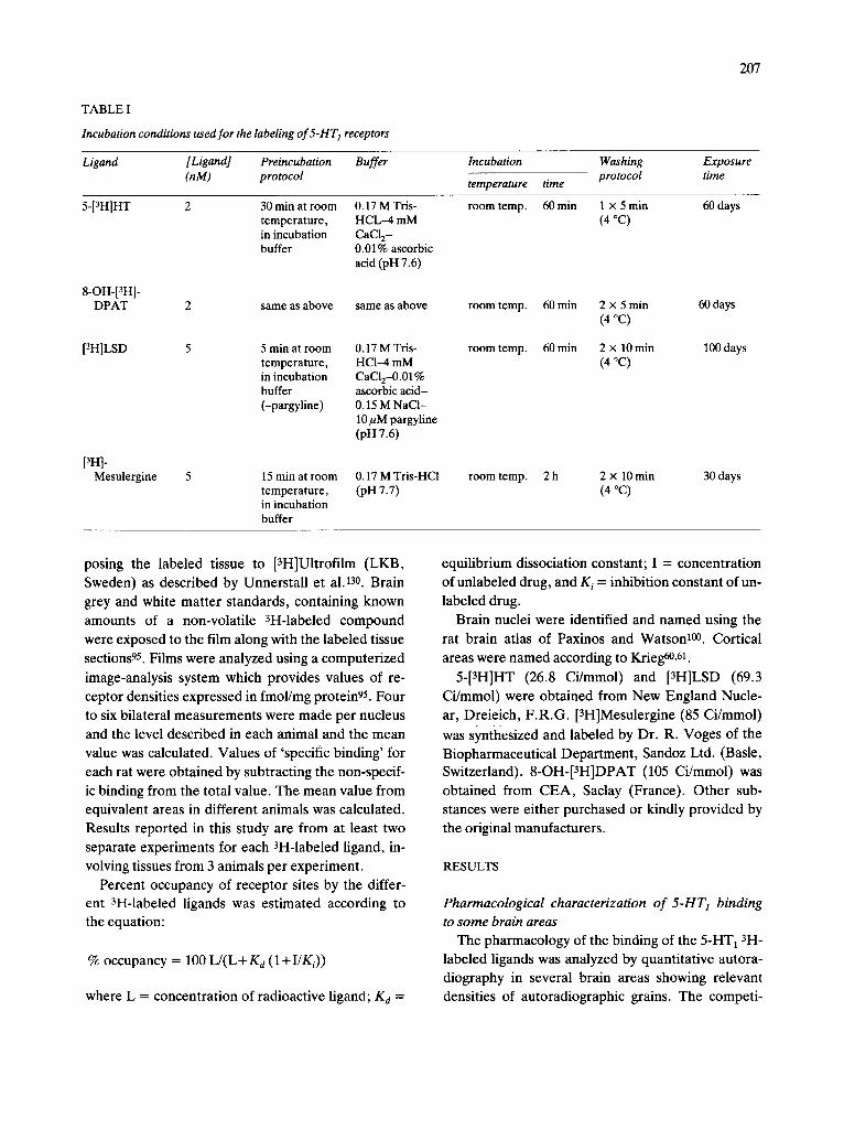
TABLE I
Incubation conditions used for the labeling of 5-HT 1 receptors
Ligand [Ligand] Preincubation Buffer (nM) protocol
Incubation Washing Exposure
temperature time protocol time
5-[3HIHT 2
8-OH-[3H]- DPAT 2
[3H]LSD 5
[3H]- Mesulergine 5
30 min at room 0.17 M Tris- temperature, HCL-4 mM in incubation CaCI 2- buffer 0.01% ascorbic
acid (pH 7.6)
same as above same as above
5 min at room 0.17 M Tris- temperature, HCI--4 mM in incubation CaCI2-0.01% buffer ascorbic acid- (-pargyline) 0.15 M NaCl-
10 ~M pargyline (pn 7.6)
15 rain at room 0.17 M Tris-HCl temperature, (pH 7.7) in incubation buffer
room temp. 60 min 1 x 5 min 60 days (4 °C)
room temp. 60 min 2 x 5 min 60 days (4 °C)
room temp. 60 min 2 x 10 min 100 days (4 °C)
room temp. 2 h 2 x 10 min 30 days (4 °C)
posing the labeled tissue to [3H]Ultrofilm (LKB, Sweden) as described by Unnerstall et al. 130. Brain
grey and white matter standards, containing known
amounts of a non-volatile 3H-labeled compound
were eXposed to the film along with the labeled tissue
sections 95. Films were analyzed using a computerized
image-analysis system which provides values of re- ceptor densities expressed in fmol/mg protein95. Four
to six bilateral measurements were made per nucleus and the level described in each animal and the mean
value was calculated. Values of 'specific binding' for
each rat were obtained by subtracting the non-specif-
ic binding from the total value. The mean value from equivalent areas in different animals was calculated.
Results reported in this study are from at least two separate experiments for each 3H-labeled ligand, in-
volving tissues from 3 animals per experiment.
Percent occupancy of receptor sites by the differ- ent 3H-labeled ligands was estimated according to the equation:
% occupancy = 100 L/ (L+Kd ( l+I /Ki ) )
where L = concentration of radioactive ligand; K d =
equilibrium dissociation constant; I = concentration of unlabeled drug, and K i = inhibition constant of un-
labeled drug.
Brain nuclei were identified and named using the rat brain atlas of Paxinos and Watson 100. Cortical areas were named according to Krieg60, 61.
5-[3H]HT (26.8 Ci/mmol) and [3H]LSD (69.3
Ci/mmol) were obtained from New England Nucle-
ar, Dreieich, F.R.G. [3H]Mesulergine (85 Ci/mmol)
was synthesized and labeled by Dr. R. Voges of the Biopharmaceutical Department, Sandoz Ltd. (Basle, Switzerland). 8-OH-[3H]DPAT (105 Ci/mmol) was
obtained from CEA, Saclay (France). Other sub- stances were either purchased or kindly provided by
the original manufacturers.
RESULTS
Pharmacological characterization o f 5-HT1 binding
to some brain areas
The pharmacology of the binding of the 5-HT1 3H- labeled ligands was analyzed by quantitative autora- diography in several brain areas showing relevant densities of autoradiographic grains. The competi-
208
A Q
10 9 8 7 6 5
- Log . FM] B
'°°V °i /
50~
! / , , , , , , , 11 10 9 8 7 6 5
-Log [M]
100 1 ~
°i l ~ 50
10 9 8 -Log
5
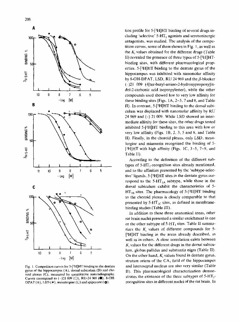
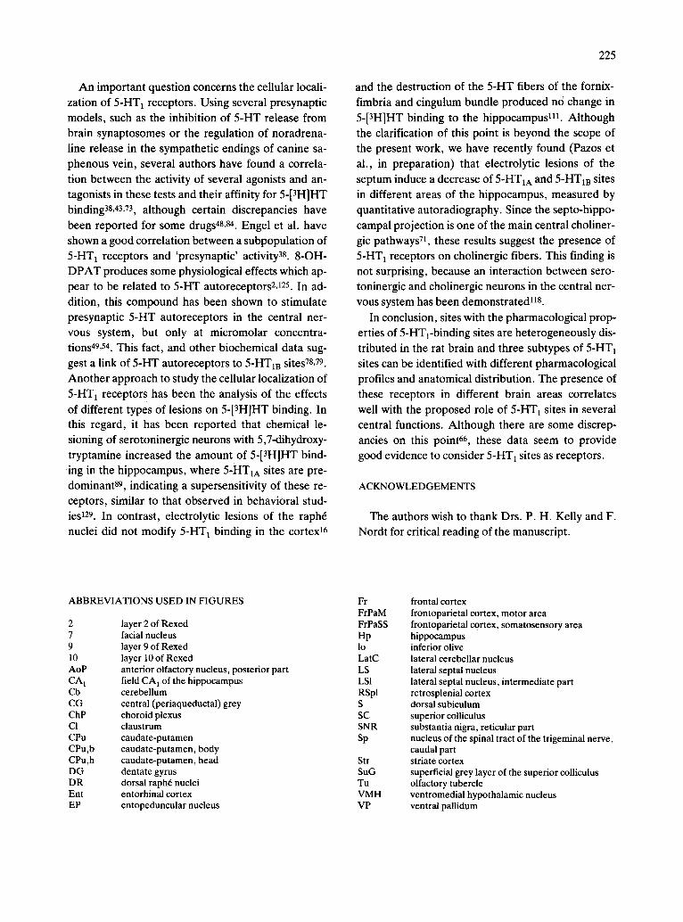
Fig. 1. Competition curves for 5-[3H]HT binding to the dentate gyrus of the hippocampus (A), dorsal subiculum (B) and cho- roid plexus (C), measured by quantitative autoradiography. Curves correspond to (-)21 009 (©), RU-24 969 (O), 8-OH- DPAT (Or), LSD ('#), mesulergine (13) and spiperone (O).
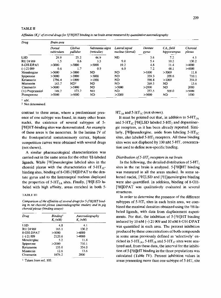
tion profile for 5-[3H]HT binding of several drugs in- cluding 'selective' 5-HT t agonists and serotoninergic antagonists, was studied. The analysis of the compe- tition curves, some of them shown in Fig. 1, as well as the K i values obtained for the different drugs (Table II) revealed the presence of three types of 5-[3H]HT -
binding sites, with different pharmacological prop- erties. 5-[3H]HT binding to the dentate gyrus of the hippocampus was inhibited with nanomolar affinity by 8-OH-DPAT, LSD, RU 24 969 and the fl-blocker (-)21 009 (4[ter-butyl-amino-2-hydroxypropoxy]in- dol-2-carbonic acid isopropylester), while the other compounds used showed low to very low affinity for these binding sites (Figs. 1A, 2-5, 7 and 8, and Table 1I). In contrast, 5-[3H]HT binding to the dorsal subi- culum was displaced with nanomolar affinity by RU 24 969 and (-) 21 009. While LSD showed an inter- mediate affinity for these sites, the other drugs tested inhibited 5-[3H]HT binding to this area with low or very low affinity (Figs. 1B, 2, 3, 5 and 8, and Table 1I). Finally, in the choroid plexus, only LSD, mesu-
lergine and mianserin recognized the binding of 5- [3H]HT with high affinity (Figs. 1C, 3-5, 7-9, and Table II).
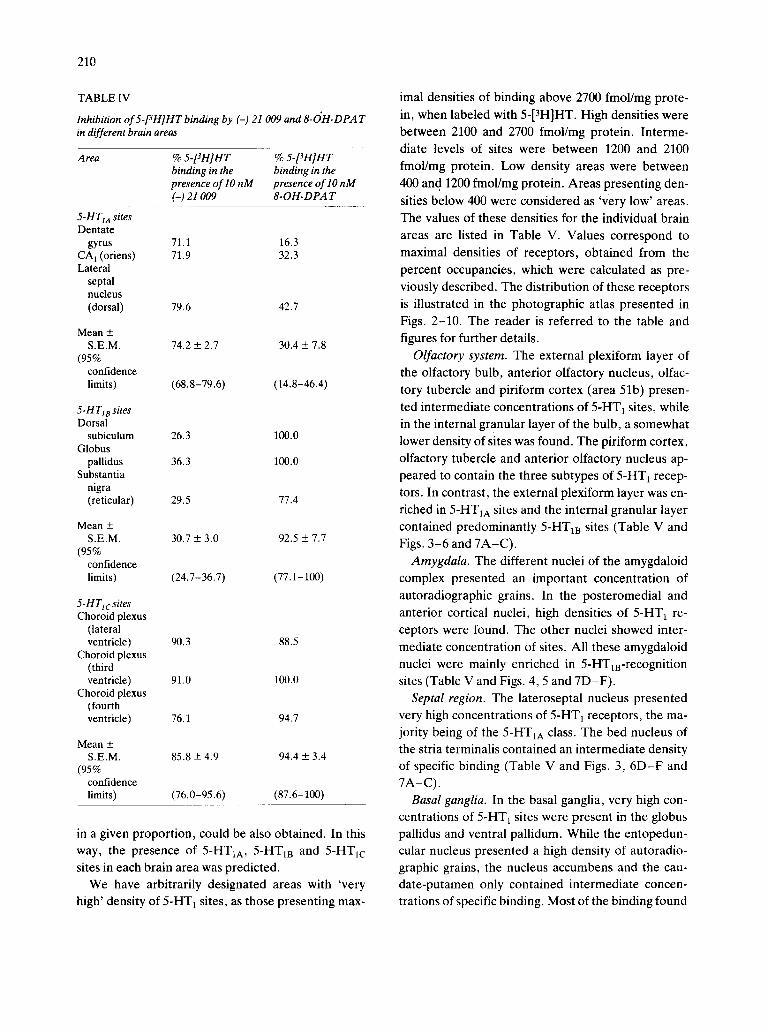
According to the definition of the different sub- types of 5-HTrrecognition sites already mentioned, and to the affinities presented by the 'subtype-selec- tive' ligands, 5-[3H]HT sites in the dentate gyrus cor- respond to the 5-HTtA subtype, while those in the dorsal subiculum exhibit the characteristics of 5- HT1B sites. The pharmacology of 5-[3H]HT binding to the choroid plexus is closely comparable to that presented by 5-HTIc sites, as defined in membrane- binding studies (Table III).
In addition to these three anatomical areas, other rat brain nuclei presented a similar enrichment in one or the other subtype of 5-HT~ sites. Table II summa- rizes the K, values of different compounds for 5- [3H]HT binding in the areas already described, as well as in others. A close correlation exists between Ki values for the different drugs in the dorsal subicu- lum, globus pallidus and substantia nigra (Table II). On the other hand, Ki values found in dentate gyrus, stratum oriens of the CA j field of the hippocampus and lateroseptal nucleus are also very similar (Table II). This pharmacological characterization demon- strates the existence of the three subtypes of 5-HTt- recognition sites in different nuclei of the rat brain. In
209
TABLE II
Affinities (K~ t of several drugs for 5[3H]HT binding to rat brain areas measured by quantitative autoradiography
Drug Brain area
Dorsal Globus Substantia nigra Lateral septal Dentate CA 1 field Choroid subiculum pallidus (reticular) nucleus (dorsal) gyrus hippocampus plexus
LSD 29.9 25.2 44.9 ND 3.6 7.2 4.1 RU 24 969 1.5 0.6 3.5 9.0 5.4 10.2 130.2 8-OH-DPAT >3000 >3000 >5000 19.2 4.8 11.4 >4000 (-) 21 009 0.6 1.7 0.9 6.0 7.2 48.1 >4000 Mesulergine >5000 >3000 ND ND > 1000 >3000 11.8 Spiperone >3000 >3000 >3000 ND 359.3 209.6 710.1 Ketanserin 1796.4 >1000 >1000 ND 598.8 >1000 354.0 Mianserin 143.7 ND 2 ND ND 269.5 ND 13.6 Cinanserin >3000 >5000 ND >5000 >2000 ND 2000 (+) Propranolol 149.7 173.7 ND ND 257.5 509.0 >5000 Pirenperone >5000 >5000 ND >2000 >5000 ND 1000
1 nM. 2 Not determined.
contrast to these areas, where a predominant pres-
ence of one subtype was found, in many other brain
nuclei, the existence of several subtypes of 5- [3H]HT-binding sites was demonstrated. An example
of these areas is the neocortex. In the lamina IV of
the frontoparietal somatosensory cortex, biphasic
competition curves were obtained with several drugs
(not shown). A similar pharmacological characterization was
carried out in the same areas for the other 3H-labeled
ligands. While [3H]mesulergine labeled sites in the
choroid plexus with the characteristics of 5-HTlc- binding sites, binding of 8 -OH-[aH]DPAT to the den-
tate gyrus and to the lateroseptal nucleus displayed
the properties of 5-HT1A sites. Finally, [3H]LSD la-
beled with high affinity, areas enriched in both 5-
TABLE III
Comparison of the affinities of several drugs for 5-[3H]HT bind- ing in rat choroid plexus (autoradiographic studies) and in pig choroid plexus (binding assays)
Drug Binding 1 A utoradiography K i (nM) g i (nM)
LSD 4.8 4.1 RU 24 969 165.1 130.2 8-OH-DPAT >5000 >4000 (-) 21 009 2120.6 >4000 Mesulergine 5.2 11.8 Spiperone >2000 710.1 Ketanserin 155.0 354.0 Mianserin 12.6 13.6 Cinanserin 1676.2 2000
1 Taken from ref. 103.
HTIA and 5-HT1c (not shown). It must be pointed out that, in addition to 5-HT1A
and 5-HTlc [3H]LSD labeled 5-HT2 and dopaminer- gic receptors, as it has been already reported. Simi-
larly, [3H]mesulergine, aside from labeling 5-HTIc
sites, also labeled 5-HT z receptors. All these binding
sites were not displaced by 100 nM 5-HT, concentra-
tion used to define non-specific binding.
Distribution o f 5 - H T 1 receptors in rat brain
In the following, the detailed distribution of 5-HT 1 sites in the rat brain is analyzed. 5-[3H]HT binding
was measured in all the areas studied. In some se-
lected nuclei, [3H]LSD and [3H]mesulergine binding
were also quantified. In addition, binding of 8-OH-
[3H]DPAT was qualitatively evaluated in several
structures.
In order to determine the presence of the different
subtypes of 5-HT 1 sites in each brain area, we com- bined the maximal densities obtained using the 3H-la-
beled ligands, with data from displacement experi-
ments. For that, the inhibition of 5-[3H]HT binding induced by 10 nM (-) 21 009 and 10 nM 8 - O H - D P A T
was quantified in each area. The percent inhibition produced by these concentrations of both compounds in some areas previously defined as 'selectively' en-
riched in 5-HTxA, 5-HT m and 5-HTlc sites were ana- lyzed and, from these data, the interval for the inhibi- tion of 5-[3H]HT binding in the three populations was calculated (Table IV). Percent inhibition values in
areas presenting more than one subtype of 5-HT 1 site
210
T A B L E IV
Inhibition of 5-[3H]HT binding by (-) 21 009 and 8-OH-DPA T in different brain areas
A rea % 5-[3H]HT % 5-[3H]HT binding in the binding in the presence of lO nM presence of lO nM (-) 21 009 8-OH-DPA T
5-HTIA sites Dentate
gyrus 71.1 16.3 CA 1 (oriens) 71.9 32.3 Lateral
septal nucleus (dorsal) 79.6 42.7
Mean + S.E.M. 74.2 + 2.7 30.4 + 7.8
(95% confidence limits) (68.8- 79.6 ) ( 14.8 - 46.4)
5-H T18 sites Dorsal
subiculum 26.3 100.0 Globus
pallidus 36.3 100.0 Substantia
nigra (reticular) 29.5 77.4
Mean + S.E.M. 30.7 ___ 3.0 92.5 + 7.7
(95% confidence limits) (24.7-36.7) (77.1-100)
5-HTlcsites Choroid plexus
(lateral ventricle) 90.3 88.5
Choroid plexus (third ventricle) 91.0 100.0
Choroid plexus (fourth ventricle) 76.1 94.7
Mean + S.E.M. 85.8 + 4.9 94.4 + 3.4
(95% confidence limits) (76.0- 95.6) (87.6-100)
m a given proportion, could be also obtained. In this way, the presence of 5-HT1A, 5-HTla and 5-HT1c sites in each brain area was predicted.
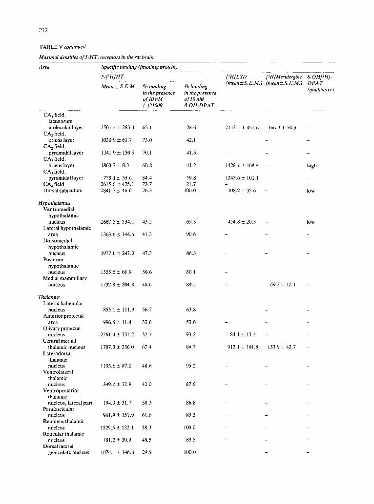
We have arbitrarily designated areas with 'very high' density of 5-HT 1 sites, as those presenting max-
imal densities of binding above 2700 fmol/mg prote-
in, when labeled with 5-[3H]HT. High densities were between 2100 and 2700 fmol/mg protein. Interme- diate levels of sites were between 1200 and 2100 fmol/mg protein. Low density areas were between 400 and 1200 fmol/mg protein. Areas presenting den-
sities below 400 were considered as 'very low' areas. The values of these densities for the individual brain areas are listed in Table V. Values correspond to maximal densities of receptors, obtained from the percent occupancies, which were calculated as pre- viously described. The distribution of these receptors is illustrated in the photographic atlas presented in Figs. 2-10. The reader is referred to the table and figures for further details.
Olfactory system. The external plexiform layer of the olfactory bulb, anterior olfactory nucleus, olfac- tory tubercle and piriform cortex (area 51b) presen-
ted intermediate concentrations of 5-HT 1 sites, while in the internal granular layer of the bulb, a somewhat lower density of sites was found. The piriform cortex, olfactory tubercle and anterior olfactory nucleus ap- peared to contain the three subtypes of 5-HT1 recep- tors. In contrast, the external plexiform layer was en- riched in 5-HT1A sites and the internal granular layer contained predominantly 5-HTIB sites (Table V and Figs. 3-6 and 7A-C) .
Amygdala. The different nuclei of the amygdaloid complex presented an important concentration of autoradiographic grains. In the posteromedial and anterior cortical nuclei, high densities of 5-HT 1 re- ceptors were found. The other nuclei showed inter- mediate concentration of sites. All these amygdaloid nuclei were mainly enriched in 5-HTm-recognition sites (Table V and Figs. 4, 5 and 7D-F) .
Septal region. The lateroseptal nucleus presented very high concentrations of 5-HT 1 receptors, the ma- jority being of the 5-HTIA class. The bed nucleus of the stria terminalis contained an intermediate density of specific binding (Table V and Figs. 3, 6 D - F and 7A-C).
Basal ganglia. In the basal ganglia, very high con- centrations of 5-HT1 sites were present in the globus pallidus and ventral pallidum. While the entopedun- cular nucleus presented a high density of autoradio- graphic grains, the nucleus accumbens and the cau- date-putamen only contained intermediate concen- trations of specific binding. Most of the binding found
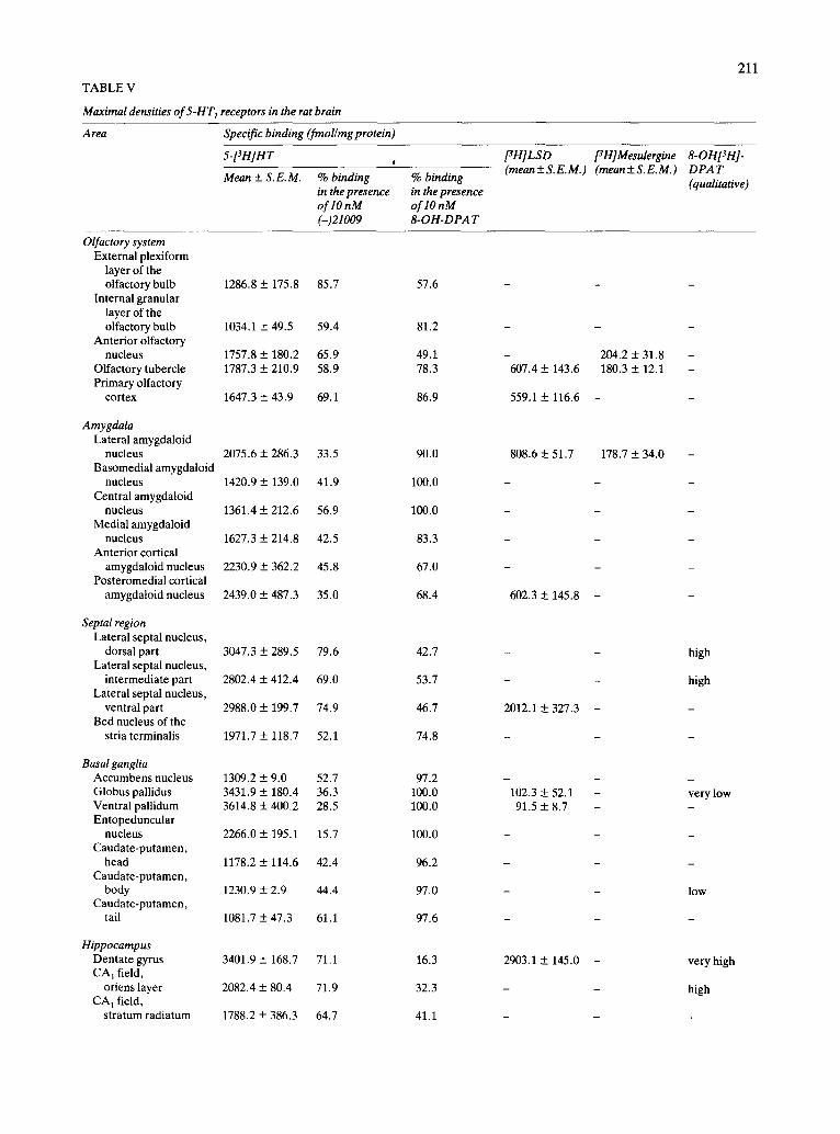
TABLE V
Maximal densities of 5-HT 1 receptors in the rat brain
211
Area Specific binding (fmol/mg protein)
5-[3H]HT
Mean + S.E.M. % binding in the presence of 10 nM (-)21009
% binding in the presence oflOnM 8-OH-DPA T
[3 H] L S D [3 H] Mesulergine (mean+ S.E.M.) (mean+_S.E.M.)
8-OI'tPH1- DPAT (qualitative)
Olfactory system External plexiform
layer of the olfactory bulb
Internal granular layer of the olfactory bulb
Anterior olfactory nucleus
Olfactory tubercle Primary olfactory
cortex
Amygdala Lateral amygdaloid
nucleus Basomedial amygdaioid
nucleus Central amygdaloid
nucleus Medial amygdaloid
nucleus Anterior cortical
amygdaloid nucleus Posteromedial cortical
amygdatoid nucleus
Sepml region Lateral septal nucleus,
dorsal part Lateral septal nucleus,
intermediate part Lateral septal nucleus,
ventral part Bed nucleus of the
stria terminalis
Basal ganglia Accumbens nucleus Globus pallidus Ventral pallidum Entopeduncular
nucleus Caudate-putamen,
head Caudate-putamen,
body Caudate-putamen,
tail
Hippocampus Dentate gyrus CA I field,
oriens layer CA 1 field,
stratum radiatum
1286.8 + 175.8
1034.1 + 49.5
1757.8 + 180.2 1787.3 + 210.9
1647.3 + 43.9
2075.6 + 286.3
1420.9 _+ 139.0
1361.4 + 212,6
1627.3 -+ 214,8
2230.9 + 362.2
2439.0 -+ 487.3
3047.3 + 289.5
2802.4 + 412.4
2988.0 + 199.7
1971.7 + 118.7
1309.2 + 9.0 3431.9 + 180.4 3614.8 + 400.2
2266.0 + 195.1
1178.2 + 114.6
1230.9 + 2.9
1081.7 + 47.3
3401.9 + 168.7
2082.4 + 80.4
1788.2 + 386.3
85.7
59.4
65.9 58.9
69.1
33.5
41.9
56.9
42.5
45.8
35.0
79.6
69.0
74.9
52.1
52.7 36.3 28.5
15.7
42.4
44.4
61.1
71.1
71.9
64.7
57.6
81.2
49.1 78.3
86.9
90.0
100.0
100.0
83.3
67.0
68.4
42.7
53.7
46.7
74.8
97.2 100.0 100.0
100.0
96.2
97.0
97.6
16.3
32.3
41.1
- 204.2 + 31.8 607.4 + 143.6 180.3 + 12.1
559.1+ 116.6 -
808.6 + 51.7 178.7 + 34.0
602.3 + 145.8
2012.1 + 327.3
102.3 _+ 52.1 91.5 + 8.7
2903.1 + 145.0
high
high
very low
low
very high
high
212
T A B L E V continued
Maximal densities of 5-HT l receptors in the rat brain
Area Specific binding (fmol/mg protein)
5-[3H]HT
Mean + S. E. M. % binding in the presence oflOnM (-)21009
% binding in the presence oflO nM 8-OH-DPA T
CA 1 field, lacunosum molecular layer
CA 2 field, oriens layer
CA 2 field, pyramidal layer
CA 3 field, oriens layer
CA 3 field, pyramidal layer
CA 4 field Dorsal subiculum
2501.2 + 283.4
1030.9 + 61.7
1341.9 + 130.9
1860.7 -+ 8.3
773.1 _-+ 55.6 2615.6 _-+ 475.1 2841.7 + 46.0
65.1
73.0
70.1
60.8
84.4 73.7 26.3
28.6
42.1
41.5
41.2
59.8 21.7
100.0
Hypothalamus Ventromedial
hypothalamic nucleus
Lateral hypothalamic area
Dorsomedial hypothalamic nucleus
Posterior hypothalamic nucleus
Medial mammillary nucleus
2667.5 _+ 234.1
1363.6 + 144.6
1977.0 _+ 247.3
1355.6 + 61.9
1792.9 _+ 204.8
43.2
41.3
47.3
36.6
48.6
69.3
90.6
86.3
80.1
89.2
Thalamus Lateral habenular
nucleus Anterior pretectal
area Olivary pretectal
nucleus Central medial
thalamic nucleus Laterodorsal
thalamic nucleus
Ventrolateral thalamic nucleus
Ventroposterior thalamic nucleus, lateral part
Parafascicular nucleus
Reuniens thalamic nucleus
Reticular thalamic nucleus
Dorsal lateral geniculate nucleus
855.1 + 111.9
996.5 _+ 11.4
2761.4 + 331.2
1707.3 _+ 236.0
1193.6 + 87.0
349.2 _+ 32.9
194.3 + 31.7
961.9 + 151.9
1529.5 + 132.1
181.2 + 30.9
1074.1 + 146.8
56.7
33.6
32.7
67.4
48.6
42.0
50.3
61.6
38.3
48.5
24.9
63.8
53.6
93.2
84.7
95.2
87.9
86.8
89.3
100.0
89.5
100.0
[3H]LSD [3H]Mesulergine (rnean+ S.E.M.) (mean+ S.E.M.)
2112.1 + 451.6 166.9 + 54.3
1428.1 _+ 168.4
1243.6_+ 101.1
108.2 + 35.6 - low
454.8 + 20.3 - low
69.3 + 12.1
84.1 + 13.2
912.1 _+ 101.8 133.9 ___ 42.7
8-OHI3H]- DPA T (qualitative)
high
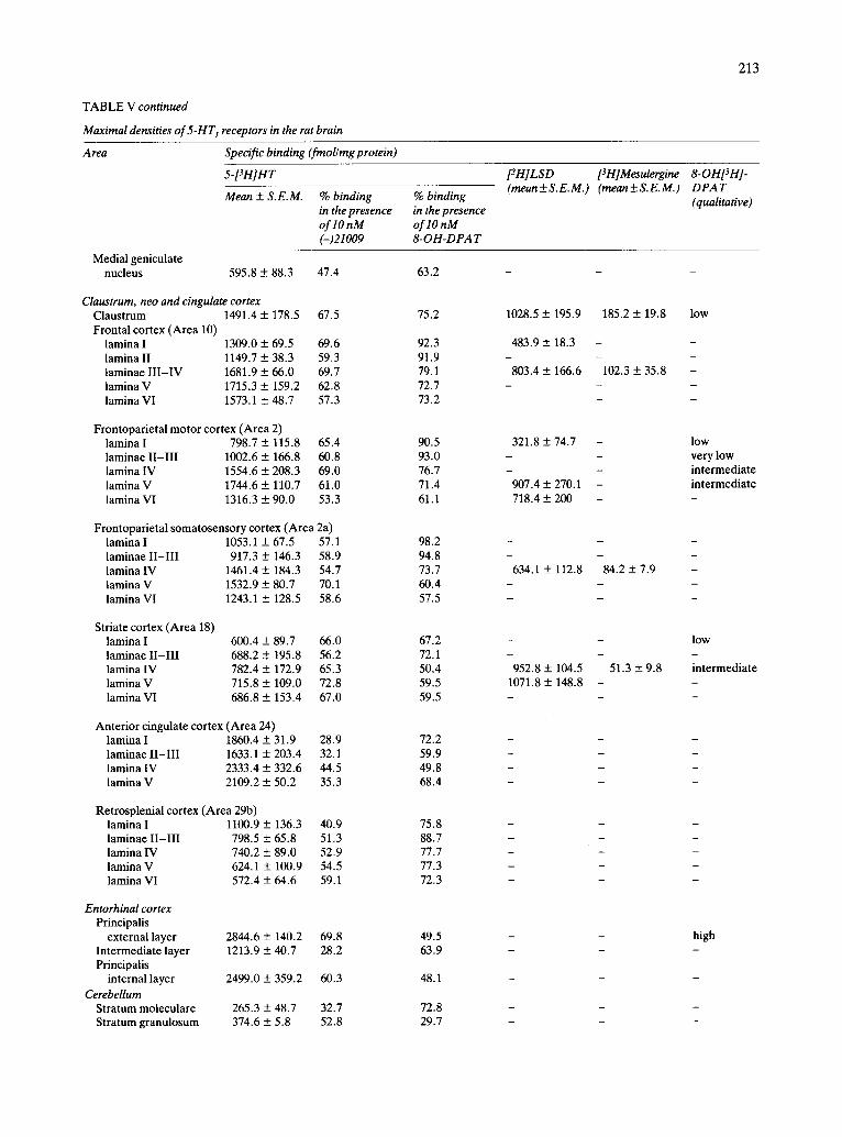
TABLE V continued
Maximal densities of 5-HT l receptors in the rat brain
213
Area Specific binding (finol/mg protein)
5-Pnln7
Mean ± S. E. M. % binding in the presence oflO nM (-)21009
% binding in the presence oflO nM 8-OH-DPAT
ffH]LSD (meun+ S.E.M.)
[Z H] Mesulergine (mean+ S.E.M.)
8-OH[3H]- DPAT (qualitative)
Medial geniculate nucleus 595.8 + 88.3 47.4
Claustrum, neo and cingulate cortex Claustrum 1491.4 + 178.5 67.5 Frontal cortex (Area 10)
lamina I 1309.0 + 69.5 69.6 lamina II 1149.7 + 38.3 59.3 laminae I I I - IV 1681.9 + 66.0 69.7 lamina V 1715.3 ± 159.2 62.8 lamina VI 1573.1 + 48.7 57.3
Frontoparietal motor cortex (Area 2) lamina I 798.7 ± 115.8 65.4 laminae II-III 1002.6 + 166.8 60.8 lamina IV 1554.6 ± 208.3 69.0 lamina V 1744.6 + 110.7 61.0 lamina VI 1316.3 ± 90.0 53.3
Frontoparietal somatosensory cortex (Area 2a) lamina I 1053.1 + 67.5 57.1 laminae II-III 917.3 + 146.3 58.9 lamina IV 1461.4 ± 184.3 54.7 lamina V 1532.9 + 80.7 70.1 lamina VI 1243.1 + 128.5 58.6
Striate cortex (Area 18) lamina I 600.4 + 89.7 66.0 laminae II-III 688.2 ± 195.8 56.2 lamina IV 782.4 ± 172.9 65.3 lamina V 715.8 + 109.0 72.8 lamina VI 686.8 + 153.4 67.0
Anterior cingulate cortex (Area 24) lamina I 1860.4 ± 31.9 28.9 laminae II-III 1633.1 ± 203.4 32.1 lamina IV 2333.4 + 332.6 44.5 lamina V 2109.2 + 50.2 35.3
Retrosplenial cortex (Area 29b) lamina I 1100.9 + 136.3 40.9 laminae II-III 798.5 _+ 65.8 51.3 lamina IV 740.2 ± 89.0 52.9 lamina V 624.1 ± 100.9 54.5 lamina VI 572.4 ± 64.6 59.1
Entorhinal cortex Principalis
external layer 2844.6 ± 140.2 69.8 Intermediate layer 1213.9 ± 40.7 28.2 Principalis
internal layer 2499.0 ± 359.2 60.3
Cerebellum Stratum moleculare 265.3 ± 48.7 32.7 Stratum granulosum 374.6 ± 5.8 52.8
63.2
75.2
92.3 91.9 79.1 72.7 73.2
90.5 93.0 76.7 71.4 61.1
98.2 94.8 73.7 60.4 57.5
67.2 72.1 50.4 59.5 59.5
72.2 59.9 49.8 68.4
75.8 88.7 77.7 77.3 72.3
49.5 63.9
48.1
72.8 29.7
1028.5 + 195.9
483.9 +__ 18.3
803.4 + 166.6
321.8 + 74.7
907.4 ± 270.1 718.4 + 200
634.1 + 112.8
w
952.8 ± 104.5 1071.8 ± 148.8
185.2 + 19.8
102.3 ± 35.8
84 .2±7 .9
51.3 + 9.8
low
l o w
very low intermediate intermediate
l o w
intermediate
high
214
T A B L E V continued
Maximal densities of 5-HT 1 receptors in the rat brain
Area Specific binding (fmol/mg protein)
Lateral nucleus Infracerebellar
nucleus Intermediate nucleus
Midbrain Superficial grey
layer of the superior colliculus
Optic nerve layer of the superior colliculus
Principal oculomotor nucleus
Central grey (periaqueductal)
Dorsal raph6 nucleus Median raph6 nucleus Substantia nigra,
reticular Substantia nigra,
compacta Ventral tegmental
area Interpeduncular
nucleus
Pons Nucleus of the spinal
tract of the trige- minal nerve, oral part
Facial nucleus Pontine nuclei Nucleus of the
solitary tract Raph6 obscurus
nucleus
Medulla oblongata Ambiguus nucleus Gracile nucleus Cuneate nucleus Inferior olive Hypoglossal nucleus Nucleus of the
spinal tract of the trigeminal nerve, caudal part
Nucleus of the spinal tract of the trigeminal nerve, gelatinosus subnucleus
Medial vestibular nucleus
5-[~H]HT
Mean ± S. E. M. % binding in the presence oflO nM (-)21009
% binding in the presence oflO nM 8-OH-DPA T
1283.9 ± 352.9 21.0
456.0 + 83.6 37.1 543.4 ± 36.0 18.3
89.1
94.1 89.6
1604.3 + 97.5 32.8
928.0 ± 67.5 32.9
1266.5 + 131.4 33.6
1456.0 _+ 21.4 39.6 2348,7 ± 317.3 60.1 1150.0 ___+ 266.0 49.8
2742.4 ± 135.8 29.5
819.7 ± 28.0 28.3
823.6 -+ 400.7 31.5
1140.4 ± 297.5 39.4
76.5
81.4
54.6
81.1 43.6 50.8
77.4
100.0
62.4
51.8
707.0 ± 90.4 48.3 773.9 ± 51.8 33.1 419.7 _+ 54.3 21.8
1184.6 ± 106.8 50.4
761.4 ± 155.8 48.2
53.4 53.1 71.0
59.3
89.6
745.8 + 77.8 21.8 860.9 + 79.5 18.4 616.0 + 85.8 23.0 793.7 ± 158.0 44.4 710.0 ± 3.6 26.4
836.3 + 123.8 39.4
1322.1 ± 193.9 59.3
791.7 ± 41.9 35.7
72.2 57.2 51.0 77.5 95.4
52.3
43.1
69,0
[3H]LSD [3H]Mesulergine 8-0141314]- (mean+ S.E.M.) (mean+ S.E.M.) DPA T
(qualitative)
45.6 + 1 1.8 very low
very low
142,1 + 23.4 - 2207,4 ± 121.5 - high
243.1 + 37.8 - low
7 1 . 6 ± 9 . 8
TABLE V continued
Maximal densities of 5-HT 1 receptors in the rat brain
215
A tea Specific binding (fmol/mg protein)
5-[3H]HT
Mean + S.E.M. % binding in the presence oflO nM (-)21009
% binding in the presence of lO nM 8- OH-DPA T
[3 H] LS D [3 H] Mesulergine (mean+ S.E.M.) (rnean+ S.E.M.)
8-OHpN]- DPA T (qualitative)
Spinal cord Layer 1 of Rexed 1011.2 + 169.8 89.6 Layer 4 of Rexed 1099.1 + 31.2 73.0 Layer 9 of Rexed 303.8 + 27.9 43.8 Layer 10 of Rexed 458.5 ± 63.2 54.3
42.4 62.8 68.9 78.4
91.4 + 21.6
Choroid plexus Lateral ventricle 3496.0 ± 530.2 90.3 Third ventricle 3381.7 ___ 402.5 91.0 Fourth ventricle 4265.3 _-_ 279.5 76.1
88.5 100.0 94.7
3787.9 + 391 1563.7 + 82.1 very low 2751.4 + 23.2 - 3381.0 + 293.7 1444.5 + 308.7 verylow
in the basal ganglia corresponded to the 5-HT m class
(Table V and Figs. 3-5 , 6 D - F and 7). Hippocampus. The hippocampal formation was
very enriched in 5-HT1 receptors. The dentate gyrus and the dorsal subiculum contained very high con- centrations of sites. In the different fields of the hip-
pocampus (CA1-CA4) , high to intermediate densi- ties of specific binding were found, depending on the layer analyzed. While binding to the dorsal subicu- lure showed the pharmacological characteristics of 5- HT1B sites, the other hippocampal areas mostly con- tained 5-HT1A sites (Table V and Figs. 2-5, 7 D - F and 8).
Hypothalamus. High concentrations of 5-HT 1 re- ceptors were found in the ventromedial hypothalam- ic nucleus. Other hypothalamic nuclei presented in- termediate densities of specific binding. Most of the binding to these structures exhibited characteristics of the 5-HTIB class (Table V and Figs. 3-5 and 7D-F) .
Thalamus. Marked differences were observed in the density of 5-HT 1 receptors throughout the thala- mus. Some nuclei, such as the ventrolateral and the reticular nuclei presented very low densities of spe- cific binding. Low concentrations were found in the laterodorsal nucleus and the dorsal lateral geniculate nucleus. The central medial and the olivary pretectal nuclei were rich in 5-HT 1 sites. 5-HT 1 receptors in the thalamus presented the pharmacological profile of 5- HTIB sites, although some 5-HTIc binding appeared to exist in the central medial nucleus (Table V and Figs. 3-5 and 7D-F) .
Claustrum, neo and cingulate cortex. The claus-
trum presented an intermediate density of 5-HT 1 re- ceptors. The three subtypes of 5-HT 1 sites appeared to be present in this structure (Table V and Figs. 3, 5
and 6). In the neocortex, intermediate concentrations of
specific binding were found in the different layers of the frontal (area 10) and frontoparietal cortex (areas 2 and 2a), while the striate cortex (area 18) presented only low concentrations of sites. It can be said that the internal laminae ( IV-VI ) were richer in 5-HT1 binding than the external ones ( I - I I I ) . In the frontal cortex, most of the binding to all the laminae was of
the 5-HT m class, although 5-HT1A and 5-HTIc sites were also present. In contrast, in the other regions studied, 5-HT m sites were predominant in the exter-
nal laminae, but both 5-HT1A and 5-HTIB sites ap- peared to coexist in laminae IV-VI . As indicated above, the presence of low densities of 5-HTIc sites at these levels, cannot be excluded.
Regarding the cingulate cortex, in the anterior cin- gular cortex (area 24) intermediate to high concen- trations of specific binding were found, the internal layers being the most enriched. A predominant but not exclusive presence of 5-HTIB sites was detected. Finally, the retrosplenial cortex (area 29b) presented low densities of 5-HT 1 receptors in all the layers (Table V and Figs. 2-8).
Entorhinal cortex. The external layer of the ento- rhinal cortex presented a very high concentration of 5-HT 1 receptors. In the internal layer, a high density of autoradiographic grains was found, while only in- termediate concentrations were present in the inter- mediate layer. Both 5-HTIA and 5-HTIB sites ap-
216
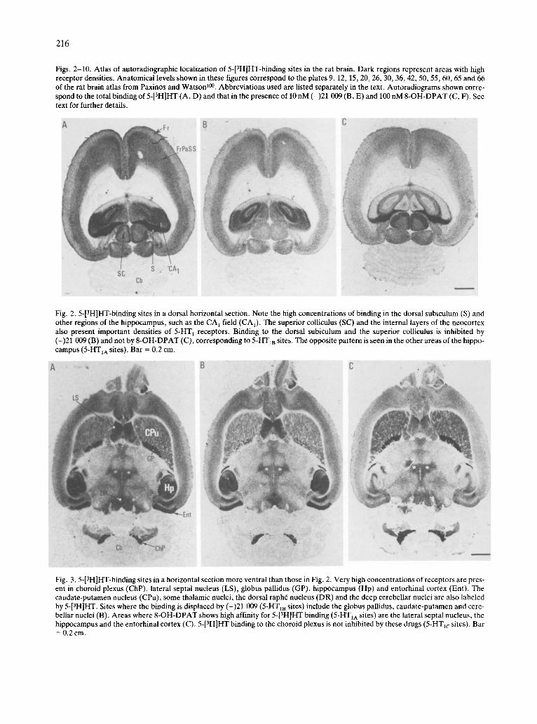
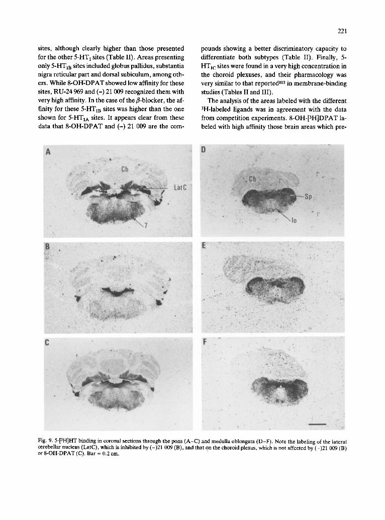
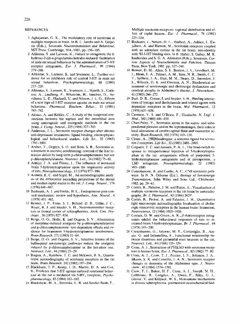
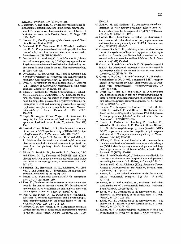
Figs. 2-10. Atlas of autoradiographic localization of 5-[3H]HT-binding sites in the rat brain. Dark regions represent areas with high receptor densities. Anatomical levels shown in these figures correspond to the plates 9, 12, 15, 20, 26, 30, 36, 42, 50, 55, 60, 65 and 66 of the rat brain atlas from Paxinos and Watson ~°°. Abbreviations used are listed separately in the text. Autoradiograms shown corre- spond to the total binding of 5-[3H]HT (A, D) and that in the presence of 10 nM (-)21 009 (B, E) and 100 nM 8-OH-DPAT (C, F). See text for further details.
!i!
iiii!!iiiii
Fig. 2.5-[3H]HT-binding sites in a dorsal horizontal section. Note the high concentrations of binding in the dorsal subiculum (S) and other regions of the hippocampus, such as the CA I field (CA1). The superior colliculus (SC) and the internal layers of the neocortex also present important densities of 5-HT l receptors. Binding to the dorsal subiculum and the superior colliculus is inhibited by (-)21 009 (B) and not by 8-OH-DPAT (C), corresponding to 5-HT1B sites. The opposite pattern is seen in the other areas of the hippo- campus (5-HTIA sites). Bar = 0.2 cm.
Fig. 3.5-[3H]HT-binding sites in a horizontal section more ventral than those in Fig. 2. Very high concentrations of receptors are pres- ent in choroid plexus (ChP), lateral septal nucleus (LS), globus pallidus (GP), hippocampus (Hp) and entorhinal cortex (Ent). The caudate-putamen nucleus (CPu), some thalamic nuclei, the dorsal raph6 nucleus (DR) and the deep cerebellar nuclei are also labeled by 5-[3H]HT. Sites where the binding is displaced by (-)21 009 (5-HT1B sites) include the globus pallidus, caudate-putamen and cere- bellar nuclei (B). Areas where 8-OH-DPAT shows high affinity for 5-[3H]HT binding (5-HTIA sites) are the lateral septal nucleus, the hippocampus and the entorhinal cortex (C). 5-[3H]HT binding to the choroid plexus is not inhibited by these drugs (5-HTIc sites). Bar = 0.2 cm.
217
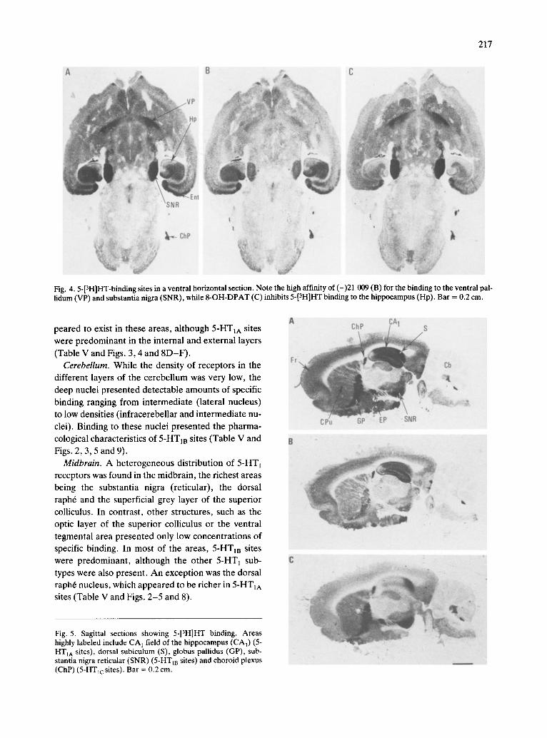
Fig. 4.5-[3H]HT-binding sites in a ventral horizontal section. Note the high affinity of (-)21 009 (B) for the binding to the ventral pal- lidum (VP) and substantia nigra (SNR), while 8-OH-DPAT (C) inhibits 5-[3H]HT binding to the hippocampus (Hp). Bar = 0.2 cm.
peared to exist in these areas, although 5-HTIA sites were predominant in the internal and external layers
(Table V and Figs. 3, 4 and 8D-F) . Cerebellum. While the density of receptors in the
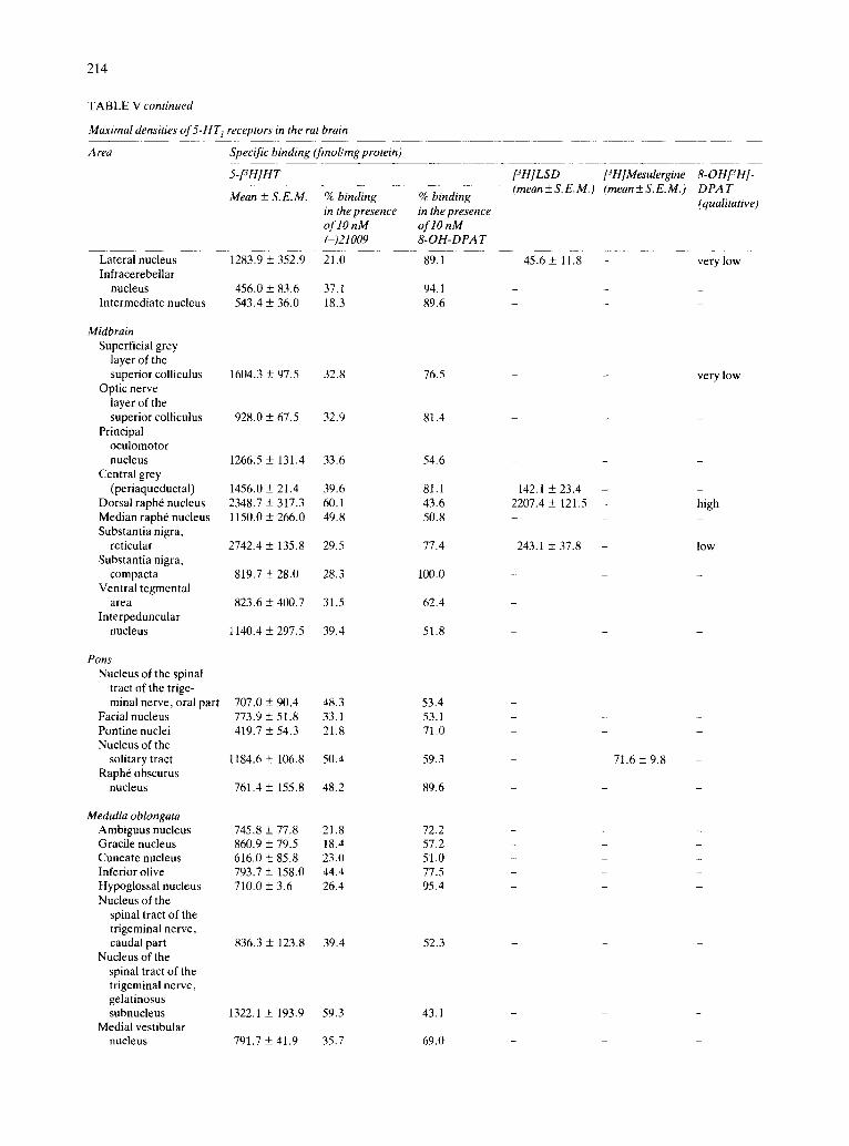
different layers of the cerebellum was very low, the deep nuclei presented detectable amounts of specific binding ranging from intermediate (lateral nucleus) to low densities (infracerebellar and intermediate nu- clei). Binding to these nuclei presented the pharma- cological characteristics of 5-HT m sites (Table V and Figs. 2, 3, 5 and 9).
Midbrain. A heterogeneous distribution of 5-HT1 receptors was found in the midbrain, the richest areas being the substantia nigra (reticular), the dorsal raph6 and the superficial grey layer of the superior colliculus. In contrast, other structures, such as the optic layer of the superior colliculus or the ventral tegmental area presented only low concentrations of specific binding. In most of the areas, 5-HT m sites were predominant, although the other 5-HT 1 sub- types were also present. An exception was the dorsal raph6 nucleus, which appeared to be richer in 5-HT1A sites (Table V and Figs. 2-5 and 8).
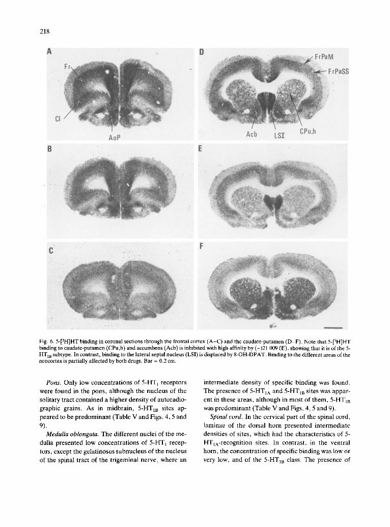
Fig. 5. Sagittal sections showing 5-[3H]HT binding. Areas highly labeled include CA 1 field of the hippocampus (CA1) (5- HTIA sites), dorsal subiculum (S), globus pallidus (GP), sub- stantia nigra reticular (SNR) (5-HT m sites) and choroid plexus (ChP) (5-HTlc sites). Bar = 0.2 cm.
218
A D
AoP B E
Acb LSZ CPu~
P ~
F
qf~
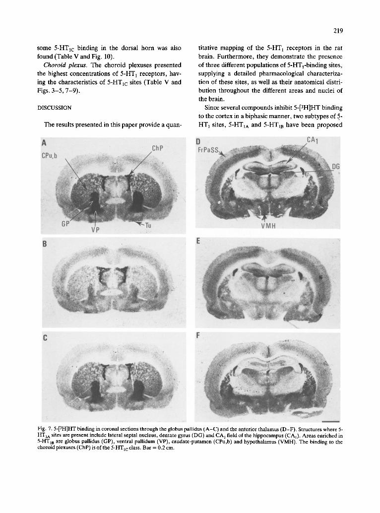
Fig. 6.5-[3H]HT binding in coronal sections through the frontal cortex (A-C) and the caudate-putamen (D-F). Note that 5-[3H]HT binding to caudate-putamen (CPu,h) and accumbens (Acb) is inhibited with high affinity by (-)21 009 (E), showing that it is of the 5- HT m subtype. In contrast, binding to the lateral septal nucleus (LSI) is displaced by 8-OH-DPAT. Binding to the different areas of the neocortex is partially affected by both drugs. Bar = 0.2 cm.
Pons. Only low concentrations of 5-HT 1 receptors were found in the pons, although the nucleus of the solitary tract contained a higher density of autoradio- graphic grains. As in midbrain, 5-HT m sites ap- peared to be predominant (Table V and Figs. 4, 5 and
9). Medulla oblongata. The different nuclei of the me-
dulla presented low concentrations of 5-HT1 recep- tors, except the gelatinosus subnucleus of the nucleus of the spinal tract of the trigeminal nerve, where an
intermediate density of specific binding was found. The presence of 5-HT1A and 5-HTIB sites was appar- ent in these areas, although in most of them, 5-HTIB was predominant (Table V and Figs. 4, 5 and 9).
Spinal cord. In the cervical part of the spinal cord, laminae of the dorsal horn presented intermediate densities of sites, which had the characteristics of 5- HTyA-recognition sites. In contrast, in the ventral horn, the concentration of specific binding was low or very low, and of the 5-HT m class. The presence of
some 5-HTlc binding in the dorsal horn was also
found (Table V and Fig. 10).
Choroid plexus. The choroid plexuses presented
the highest concentrations of 5-HT 1 receptors, hav-
ing the characteristics of 5-HTlc sites (Table V and Figs. 3-5, 7-9).
DISCUSSION
The results presented in this paper provide a quan-
219
titative mapping of the 5-HT 1 receptors in the rat
brain. Furthermore, they demonstrate the presence
of three different populations of 5-HTl-binding sites,
supplying a detailed pharmacological characteriza- tion of these sites, as well as their anatomical distri-
bution throughout the different areas and nuclei of
the brain.
Since several compounds inhibit 5-[3H]HT binding
to the cortex in a biphasic manner, two subtypes of 5.-
HT 1 sites, 5-HT1A and 5-HT m have been proposed
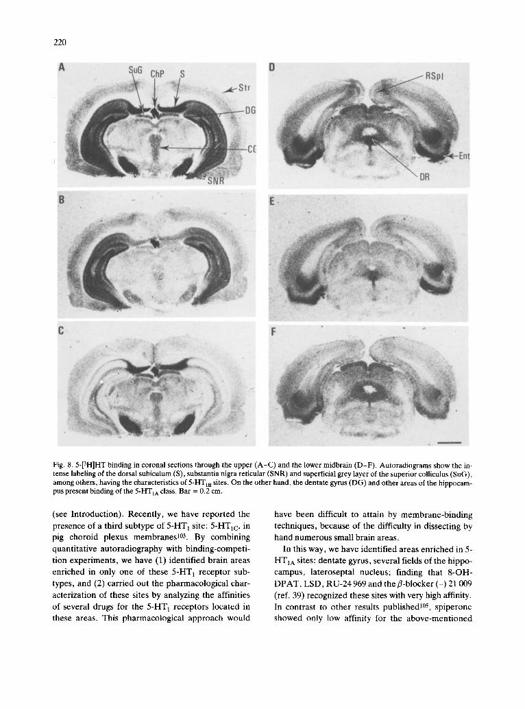
Fig. 7.5-[3H]HT binding in coronal sections through the globus pallidus (A-C) and the anterior thalamus (D-F). Structures where 5- HT1A sites are present include lateral septal nucleus, dentate gyrus (DG) and C A 1 field of the hippocampus (CA1). Areas enriched in 5-HT m are globus pallidus (GP), ventral pallidum (VP), caudate-putamen (CPu,b) and hypothalamus (VMH). The binding to the choroid plexuses (ChP) is of the 5-HTlc class. Bar = 0.2 cm.
220
~:ii !~ ;~! i~:~!ii~i!i! '̧ I̧ ~i!ii~ ~i~i/~ii~i ~i!i ̧ iii ̧ ~
Fig. 8.5-[3H]HT binding in coronal sections through the upper (A-C) and the lower midbrain (D-F). Autoradiograms show the m- tense labeling of the dorsal subiculum (S), substantia nigra reticular (SNR) and superficial grey layer of the superior colliculus (SuG), among others, having the characteristics of 5-HT m sites. On the other hand, the dentate gyrus (DG) and other areas of the hippocam- pus present binding of the 5-HTIA class. Bar = 0.2 cm.
(see Introduction). Recently, we have reported the
presence of a third subtype of 5-HT~ site: 5-HTlc, in pig choroid plexus membranes 103. By combining
quantitative autoradiography with binding-competi- tion experiments, we have (1) identified brain areas enriched in only one of these 5-HT l receptor sub- types, and (2) carried out the pharmacological char-
acterization of these sites by analyzing the affinities of several drugs for the 5-HT 1 receptors located in these areas. This pharmacological approach would
have been difficult to attain by membrane-binding techniques, because of the difficulty in dissecting by hand numerous small brain areas.
In this way, we have identified areas enriched in 5- HTIA sites: dentate gyrus, several fields of the hippo- campus, lateroseptal nucleus; finding that 8-OH-
DPAT, LSD, RU-24 969 and the fl-blocker (-) 21 009 (ref. 39) recognized these sites with very high affinity. In contrast to other results published105, spiperone showed only low affinity for the above-mentioned
sites, although clearly higher than those presented
for the other 5-HT 1 sites (Table II). Areas presenting only 5-HT m sites included globus pallidus, substantia nigra reticular part and dorsal subiculum, among oth- ers. While 8-OH2DPAT showed low affinity for these sites, RU-24 969 and (-) 21 009 recognized them with very high affinity. In the case of the fl-blocker, the af- finity for these 5-HT m sites was higher than the one
shown for 5-HTIA sites. It appears clear from these data that 8-OH-DPAT and (-) 21 009 are the com-
221
pounds showing a better discriminatory capacity to
differentiate both subtypes (Table II). Finally, 5- HTIc sites were found in a very high concentration in the choroid plexuses, and their pharmacology was very similar to that reported 103 in membrane-binding
studies (Tables II and III). The analysis of the areas labeled with the different
3H-labeled ligands was in agreement with the data from competition experiments. 8-OH-[aH]DPAT la- beled with high affinity those brain areas which pre-
Fig. 9.5-[3H]HT binding in coronal sections through the pons (A-C) and medulla oblongata (D-F). Note the labeling of the lateral cerebellar nucleus (LatC), which is inhibited by (-)21 009 (B), and that on the choroid plexus, which is not affected by (-)21 009 (B) or 8-OH-DPAT (C). Bar = 0.2 cm.
222
sented the characteristics of 5-HT1A sites, when 5- [3H]HT was used as a ligand. Bma x values of [3H]LSD binding were similar to those obtained with 5-[3H]HT
in the areas where no 5-HTIB sites were present, but clearly lower in those enriched in this subtype. Bind- ing of [3H]mesulergine only reached high concentra- tions in the choroid plexus, an area extremely en- riched in 5-HT~c receptor sites. The difference be- tween the maximal densities found in this structure with 5-[3H]HT and [3H]mesulergine could be due to the different labeling conditions used for both ligands (Table V). A multiplicity of serotoninergic recogni- tion sites has also been proposed on the basis of data obtained from electrophysiological studies l. Howev- er, further work is needed in order to clarify the pos- sible relationship between the different 'S' receptors
.C
d
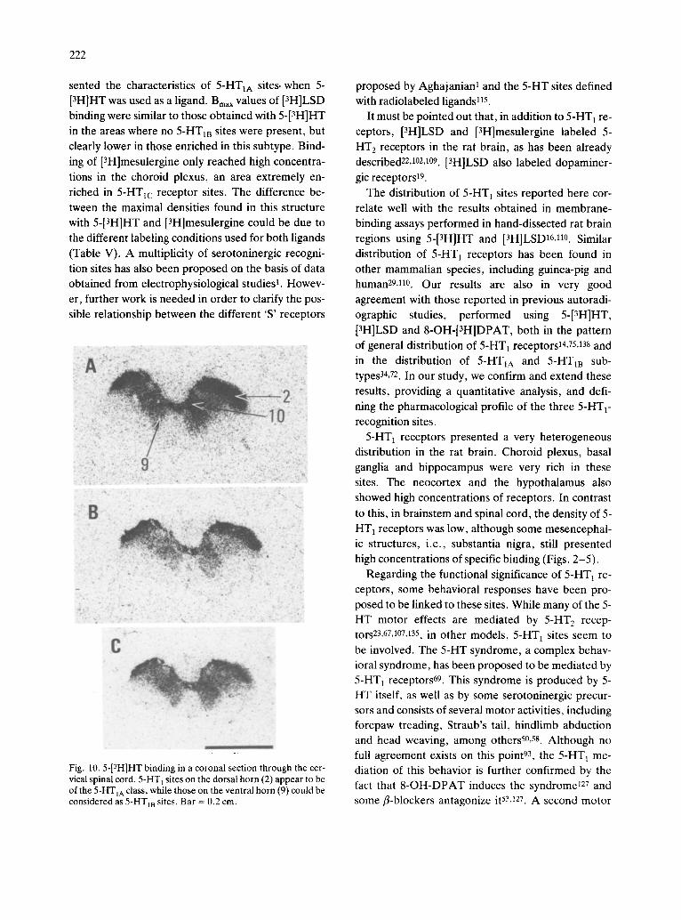
Fig. 10.5-[3H]HT binding in a coronal section through the cer- vical spinal cord. 5-HT 1 sites on the dorsal horn (2) appear to be of the 5-HTIA class, while those on the ventral horn (9) could be considered as 5-HT1B sites. Bar = 0.2 cm.
proposed by Aghajanian I and the 5-HT sites defined with radiolabeled ligands t15.
It must be pointed out that, in addition to 5-HT t re- ceptors, [3H]LSD and [3H]mesulergine labeled 5- HT 2 receptors in the rat brain, as has been already described22,102,1°9. [3H]LSD also labeled dopaminer- gic receptors 19.
The distribution of 5-HT 1 sites reported here cor- relate well with the results obtained in membrane- binding assays performed in hand-dissected rat brain regions using 5-[3H]HT and [3H]LSD16,11°. Similar
distribution of 5-HT 1 receptors has been found in other mammalian species, including guinea-pig and human29, tl0. Our results are also in very good
agreement with those reported in previous autoradi- ographic studies, performed using 5-[3H]HT, [3H]LSD and 8-OH-[3H]DPAT, both in the pattern of general distribution of 5-HT1 receptors 14,75,138 and
in the distribution of 5-HTIA and 5-HT1B sub- types34, 72. In our study, we confirm and extend these
results, providing a quantitative analysis, and defi- ning the pharmacological profile of the three 5-HT 1- recognition sites.
5-HT1 receptors presented a very heterogeneous distribution in the rat brain. Choroid plexus, basal ganglia and hippocampus were very rich in these sites. The neocortex and the hypothalamus also showed high concentrations of receptors. In contrast
to this, in brainstem and spinal cord, the density of 5- HT 1 receptors was low, although some mesencephai- ic structures, i.e., substantia nigra, still presented high concentrations of specific binding (Figs. 2-5).
Regarding the functional significance of 5-HT 1 re- ceptors, some behavioral responses have been pro- posed to be linked to these sites. While many of the 5- HT motor effects are mediated by 5-HT 2 recep- tors 23,67,107,135, in other models, 5-HT~ sites seem to
be involved. The 5-HT syndrome, a complex behav- ioral syndrome, has been proposed to be mediated by 5-HT 1 receptors 69. This syndrome is produced by 5- HT itself, as well as by some serotoninergic precur- sors and consists of several motor activities, including forepaw treading, Straub's tail, hindlimb abduction and head weaving, among othersS0, 5s. Although no full agreement exists on this point 93, the 5-HT 1 me- diation of this behavior is further confirmed by the fact that 8-OH-DPAT induces the syndrome ~27 and some r-blockers antagonize it 53,127. A second motor
223
effect which appears to be related to 5-HT 1 activation is the circling behavior in striatum-lesioned rats. Both agonists RU-24 969 and 8-OH-DPAT produce this behaviorlS, 57. Finally, it has been reported that
the indole 5-HT agonists, such as RU-24 969, origi- nate myoclonic jerking in guinea-pigs 70 and hyperlo- comotion in mice and ratsS2,128. A good correlation has been shown between the potencies of these com- pounds displacing 5-[3H]HT binding in the brainstem and those inducing myoclonus 70. However, caution is
required in analyzing the motor effects produced by 5-HT agonists, because of the interactions which have been demonstrated between 5-HT and dopa- minergic receptors in this regard36, 52. Although most
of these 5-HT1 behavioral effects have been claimed to be brainstem or spinally mediated59, 70, recent re- sults have demonstrated that at least some compo-
nents of the 5-HT syndrome are not dependent on these lower structures 35. Our autoradiographic data reveal the presence of 5-HT 1 receptors in many struc- tures known to be involved in motor activity. The spi- nal cord, substantia nigra, cerebellar nuclei, basal ganglia, entopeduncular nucleus and motor cortex present important concentrations of receptors, main- ly of the 5-HTIB subtype. The claustrum, a structure also connected to the motor cortex 2s is also rich in 5- HT 1 sites. These results support a predominant role of 5-HTm-recognition sites in these effects. Howev-
er, an involvement of 5-HT1A sites is possible, since they are also present in these structures•
There is also considerable evidence for an inhibito- ry role of central 5-HT systems on male rat sexual be- havior 3A17. In contrast, administration of 8-OH- DPAT causes marked stimulatory changes in this sexual behavior, reducing number of intromissions preceding ejaculation and shortening the ejaculatory latency 4. Furthermore, this sexual facilitatory effect of 8-OH-DPAT is not blocked by the administration of several serotoninergic antagonists2, 4. These results complicate the analysis of the relationship between 5- HT 1 receptors and sexual behavior, suggesting a pos- sible presynaptic mediation, as will be discussed lat- er. Nevertheless, the presence of high concentrations of 5-HTzA receptors in the different areas of the hip- pocampus, as well as in other structures of the limbic system, including the septum, which is strongly con- nected to hippocampal areasS, 71, could indicate a role of these sites in the mediation of this behavior•
A role of 5-HT 1 receptors in the control of visual activity can be suggested, according to the distribu- tion of these sites in some brain areas• The olivary pretectal nucleus and the superficial grey layer of the superior colliculus, both connected with the visual pathway120, TM present high concentrations of 5-HT1
receptors. Other structures related to the visual sys- tem, such as the claustrum 65, the striate cortex and lateral geniculate nucleus47,119,126 also contain impor-
tant receptor densities. Most of these areas are main- ly enriched in 5-HT1a-recognition sites•
5-HT agonists have been reported to induce stimu- lation of rat prolactin secretion (see ref. 77). Al- though this effect appears to be mediated by 5-HT 2 receptors 76, a role of 5-HT 1 sites cannot be ex- cluded 41. The important presence of 5-HTm-recogni- tion sites in different nuclei of the hypothalamus, an area which has been related to these effects 77, could suggest an involvement of 5-HT1B sites in this endo- crine response.
The central 5-HT systems are known to be in- volved in the regulation of nociceptive activity, main- ly through a raph6-spinal projection interconnected with the endogenous opioid pathways (see ref. 10 for a review), although brainstem serotoninergic con- nections are also relevant 12. It has been also shown that this serotoninergic role varies depending on the methodological approach used12,13,121,136. Further-
more, it has been recently suggested that spinal 5-HT receptors related to nociceptive control are of the 5- HT 1 class 121,139. Our autoradiographic data support
this hypothesis, showing the presence of 5-HT 1 re- ceptors in the raph6 nuclei and spinal cord, as well as in some other brainstem structures• Regarding the spinal cord, most of the receptors present in the dor- sal horn are of the 5-HTIA subtype, suggesting a role of these sites in this system•
Some vegetative functions, such as the control of blood pressure and respiration are known to be influ- enced by central 5-HT activity8,63. Although our data
reveal the presence of 5-HT 1 receptors in several brainstem nuclei which play an important role in these functions, such as the nucleus of the solitary tract, further work is needed to clarify the 5-HT in- volvement in these effects.
In the last few years, the role of 5-HT receptors in some neurological diseases has been the matter of in- creasing research• A decrease in the number of 5-HT
224
receptors in the cortex of patients suffering from se- nile dementia of the Alzheimer type has been report- edlS,30,113. Although the decrease in 5-HT 2 receptors
appears to be greater, 5-HT 1 receptors are also af- fected, mainly in those patients presenting a later stage of the disease18, 30. A role of diminished number
of 5-HT1 sites in the impaired cognitive activity char- acteristic of this disease is therefore possible. Re- lated to this, the involvement of serotoninergic trans- mission in learning and memory processes, as has been shown in animals 7.92 and in humans 132, must be
taken into consideration. A role of central 5-HT activity in the mechanisms
of schizophrenia has also been investigated. While an increase in the striatal concentration of 5-HT in brains from schizophrenic patients has been re- ported 31, a clear decrease of [3H]LSD binding to
frontal cortical membranes has also been found n. These data do not allow any statement on the possi- ble role of 5-HT 1 receptors in this disease. In a similar way, while considerable evidence has accumulated on the relationship between antidepressant treat- ments and number of cortical 5-HT 2 receptors, no clear effect has been shown in the case of 5-HT~ sites (refs. 82, 123; see ref. 6 for a review). Finally, the in- volvement of 5-HT mechanisms in the pathogenesis of migraine has been proposed20,112 and the prophy-
lactic effect of the 5-HT antagonist methysergide in some types of migraine is well-known (see ref. 64). Whether 5-HT~ sites are involved in this process is
unknown. One of the relevant findings of this work is the
characterization of some fl-blockers as drugs with high affinity and selectivity for the 5-HTiB-recogni- tion site. This fact confirms and extends previous re- ports showing the ability of fl-blockers to displace 5- [3H]HT binding in a stereospecific wayS0, 86. It has
also been established that some fl-blockers antago- nize several in vivo and in vitro effects induced by se- rotoninergic agonists (refs. 27, 51; see ref. 133 for a review). On the other hand, fl-adrenergic agonists have been reported to increase some 5-HT behavior- al effects 91,94. Taken together, these data suggest an
interaction of fl-adrenergic drugs with central 5-HT 1 receptors. This interaction could explain some of the neurological effects induced by fl-blockers in hu- mans, such as their therapeutic activity in schizo- phrenic states 124A37 and in migraine 43,131.
The localization of 5-HTI receptors described here correlates well with the distribution of serotoninergic terminals. Most of the areas presenting important concentrations of receptors have been found to con- tain nerve-endings from the serotoninergic neurons, mainly located in the raph6 nuclei 9,24,99,122. The hip-
pocampus, the septal nuclei, the neocortex, the basal ganglia, the different nuclei of the hypothalamus, ;he olfactory system, or the substantia nigra are struc- tures where the presence of 5-HT, 5-HT fibers and
biochemical markers have been reported z4, 32,37,45,46,56,68,99,116,122. The choroid plexuses have
also been shown to contain serotoninergic innerva- tion21.122. However, some areas very enriched in 5-
HT 1 sites do not present a high level of serotoninergic innervation. This is the case for the dorsal subiculum or the entopeduncular nucleus 24,122. The existence of
discrepancies between the distribution of fibers and receptors has been found with many neurotransmit- ters96. 98 and several explanations have been pro-
posed (see ref. 62). Furthermore, it should be kept in mind that the brain contains also 5-HT 2 receptors, whose distribution is different from that of 5-HT 1 sites 75,1°2 and this could explain some apparent dis-
crepancies. Our results on the distribution and pharmacology
of 5-HTlc sites confirm our previour reports on this subtype 25.103, showing that the pharmacological pro-
file obtained by quantitative autoradiography is iden- tical to that obtained in membrane-binding assays 103. The choroid plexuses are especially enriched in these
5-HTIc sites, presenting the highest density of 5-HTI receptors in the rat brain. Very high concentrations of 5-HT 1 binding in the choroid plexus have also been found in other mammalian species, such as pig 103 and human 97. The choroid plexuses present an important 5-HT innervation, the fibers arising from the raph6 nuclei and ending on the blood vessels that supply the plexuses 83,87. In addition, there are 5-HT fibers on the supraependymal surface of the ventricular sys- tem TM. Beside the choroid plexus, 5-HTlc sites are also present in other brain areas, as has already been reported in pig brain ~04. The possible physiological significance of 5-HTlc sites is not known at the pres- ent time. Since they are mainly localized in the cho- roid plexuses, they may play a role in the regulation of the production of cerebrospinal fluid, a function strongly linked to these plexuses 33.
225
An important question concerns the cellular locali-
zation of 5-HT 1 receptors. Using several presynaptic
models, such as the inhibition of 5-HT release from brain synaptosomes or the regulation of noradrena-
line release in the sympathetic endings of canine sa-
phenous vein, several authors have found a correla-
tion between the activity of several agonists and an-
tagonists in these tests and their affinity for 5-[3H]HT binding3S,43, 73, although certain discrepancies have
been reported for some drugs48, 84. Engel et al. have
shown a good correlation between a subpopulation of 5-HT 1 receptors and 'presynaptic' activity 38. 8-OH-
DPAT produces some physiological effects which ap- pear to be related to 5-HT autoreceptors2,125. In ad-
dition, this compound has been shown to stimulate
presynaptic 5-HT autoreceptors in the central ner- vous system, but only at micromolar concentra- tions49, 54. This fact, and other biochemical data sug-
gest a link of 5-HT autoreceptors to 5-HT1B sites78, 79.
Another approach to study the cellular localization of
5-HT 1 receptors has been the analysis of the effects
of different types of lesions on 5-[3H]HT binding. In this regard, it has been reported that chemical le- sioning of serotoninergic neurons with 5,7-dihydroxy-
tryptamine increased the amount of 5-[3H]HT bind-
ing in the hippocampus, where 5-HTtA sites are pre- dominant 89, indicating a supersensitivity of these re-
ceptors, similar to that observed in behavioral stud- ies 129. In contrast, electrolytic lesions of the raph6
nuclei did not modify 5-HT1 binding in the cortex 16
and the destruction of the 5-HT fibers of the fornix-
fimbria and cingulum bundle produced no change in
5-[3H]HT binding to the hippocampus 111. Although
the clarification of this point is beyond the scope of the present work, we have recently found (Pazos et
al., in preparation) that electrolytic lesions of the
septum induce a decrease of 5-HTIA and 5-HT m sites
in different areas of the hippocampus, measured by quantitative autoradiography. Since the septo-hippo-
campal projection is one of the main central choliner- gic pathways 71, these results suggest the presence of
5-HT 1 receptors on cholinergic fibers. This finding is
not surprising, because an interaction between sero- toninergic and cholinergic neurons in the central ner-
vous system has been demonstrated 118.
In conclusion, sites with the pharmacological prop- erties of 5-HTl-binding sites are heterogeneously dis-
tributed in the rat brain and three subtypes of 5-HT 1 sites can be identified with different pharmacological
profiles and anatomical distribution. The presence of
these receptors in different brain areas correlates
well with the proposed role of 5-HT 1 sites in several
central functions. Although there are some discrep- ancies on this point 66, these data seem to provide
good evidence to consider 5-HT 1 sites as receptors.
ACKNOWLEDGEMENTS
The authors wish to thank Drs. P. H. Kelly and F.
Nordt for critical reading of the manuscript.
ABBREVIATIONS USED IN FIGURES
2 layer 2 of Rexed 7 facial nucleus 9 layer 9 of Rexed 10 layer 10 of Rexed AoP anterior olfactory nucleus, posterior part CA l field CA 1 of the hippocampus Cb cerebellum CG central (periaqueductal) grey ChP choroid plexus CI claustrum CPu caudate-putamen CPu,b caudate-putamen, body CPu,h caudate-putamen, head DG dentate gyrus DR dorsal raph6 nuclei Ent entorhinal cortex EP entopeduncular nucleus
Fr FrPaM FrPaSS Hp 1o LatC LS LSI RSpl S SC SNR Sp
Str SuG Tu VMH VP
frontal cortex frontoparietal cortex, motor area frontoparietal cortex, somatosensory area hippocampus inferior olive lateral cerebellar nucleus lateral septai nucleus lateral septal nucleus, intermediate part retrosplenial cortex dorsal subiculum superior colliculus substantia nigra, reticular part nucleus of the spinal tract of the trigeminal nerve, caudal part striate cortex superficial grey layer of the superior colliculus olfactory tubercle ventromedial hypothalamic nucleus ventral pallidum
226
REFERENCES
1 Aghajanian, G. K., The modulatory role of serotonin at multiple receptors in brain. In B. L. Jacobs and A. Gelpe- rin (Eds.), Serotonin Neurotransmission and Behaviour, MIT Press, Cambridge, MA, 1981, pp. 156-185.
2 Ahlenius, S. and Larsson, K., Failure to antagonize the 8- hydroxy-2-(di-n-propylamino)tetralin-induced facilitation of male rat sexual behaviour by the administration of 5-HT receptor antagonists, Eur. J. Pharmacol., 99 (1984) 279-286.
3 Ahlenius, S., Larsson, K. and Svensson, L., Further evi- dence for an inhibitory role of central 5-HT in male rat sexual behaviour, Psychopharmacology, 68 (1980) 217-220.
4 Ahlenius, S., Larsson, K., Svensson, L., Hjorth, S., Carls- son, A., Lindberg, P., WikstrOm, H., Sanchez, D., Ar- vidsson, L.-E., Hacksell, U. and Nilsson, J. L. G., Effects of a new type of 5-HT receptor agonist on male rat sexual behaviour, Pharmacol. Biochem. Behav., 15 (1981) 785-792.
5 Alonso, A. and K6hler, C., A study of the reciprocal con- nections between the septum and the entorhinal area using anterograde and retrograde methods in the rat brain, J. Comp. Neurol., 225 (1984) 327-343.
6 Anderson, J. L., Serotonin receptor changes after chronic anti-depressant treatments: ligand binding, electrophysio- logical and behavioural studies, Life Sci., 32 (1983) 1791-1801.
7 Archer, T., Oegren, S. O. and Ross, S. B., Serotonin in- volvement in aversive conditioning: reversal of the fear re- tention deficit by long-term p-chtoroamphetamine but not p-chlorophenylalanine, Neurosci. Lett., 34 (1982) 75-82.
8 Armijo, J. A. and Fl6rez, J., The influence of increased brain 5-hydroxytryptamine upon the respiratory activity of cats, Neuropharmacology, 13 (1974) 977-986.
9 Azmitia, E. C. and Segal, M., An autoradiographic analy- sis of the differential ascending projections of the dorsal and median raph6 nuclei in the rat, J. Comp. Neurol., 179 (1978) 641-667.
10 Basbaum, A. I. and Fields, H. L., Endogeneous pain con- trol mechanism: review and hypothesis, Ann. Neurol., 4 (1978) 451-462.
11 Bennet, J. P., Enna, S. J., Bylund, D. B., Gillin, J. C., Wyatt, R. J. and Snyder, S. H., Neurotransmitter recep- tors in frontal cortex of schizophrenics, Arch. Gen. Psy- chiatr., 36 (1979) 927-934.
12 Berge, O.-G., Holle, K. and Oegren, S. V., Attenuation of morphine-induced analgesia by p-chlorophenylalanine and p-chloroamphetamine: test-dependent effects and ev- idence for brainstem 5-hydroxytryptamine involvement, Brain Research, 271 (1983) 51-64.
13 Berge, O.-G. and Oegren, S. V., Selective lesions of the bulbospinal serotonergic pathways reduce the analgesia induced by p-chloroamphetamine in the hot-plate test, Neurosci. Lett., 44 (1984) 25-29.
14 Biegon, A., Rainbow, T. C. and McEwen, B. S., Quanti- tative autoradiography of serotonin receptors in the rat brain, Brain Research, 242 (1982) 197-204.
15 Blackburn, T. P., Kemp, J. D., Martin, D. A. and Cox, B., Evidence that 5-HT agonist-induced rotational behav- iour in the rat is mediated via 5-HT 1 receptors, Psycho- pharmacology, 83 (1984) 163-165.
16 Blackshear, M. A., Steranka, L. R. and Sander-Bush, E.,
Multiple serotonin receptors: regional distribution and ef- fect of raph6 lesions, Eur. J. Pharmacol., 76 (1981) 325-334.
17 Bockaert, J., Nelson, D. L., Herbert, A., Adrien, J., En- jalbert, A. and Hamon, M., Serotonin receptors coupled with an adenylate cyclase in the rat brain: non-identity with 3H-5-HT binding sites. In B. Haber, S. Gabay, M. R. Issidorides and S. G. A. Alivisatos (Eds.), Serotonin, Cur- rent Aspects of Neurochemistry and Function, Plenum Press, New York, 1981, pp. 327-346.
18 Bowen, D. M., Allen, S. J., Benton, J. S., Goodhart, M. J., Haan, E. A., Palmer, A. M., Sims, N. R., Smith, C. C. T., Spillane, J. A., Esiri, M. M., Neary, D., Snowdon, J. S., Wilcock, G. K. and Davison, A. N., Biochemical as- sessment of serotonergic and cholinergic dysfunction and cerebral atrophy in Alzheimer's disease, J. Neurochem., 41 (1983) 266-272.
19 Butt, D. R., Creese, I. and Snyder, S. H., Binding interac- tions of lysergic acid diethylamide and related agents with dopamine receptors in the brain, Mol. Pharmacol., 12 (1976) 631-638.
20 Caviness, V. S. and O'Brien, P., Headache, N. Engl. J. Med., 302 (1980) 446-450.
21 Chan-Palay, V., Serotonin axons in the supra- and sube- pendymal plexuses and in the leptomeninges; their roles in local alterations of cerebro-spinal fluid and vasomotor ac- tivity, Brain Research, 102 (1976) 103-130.
22 Closse, A., [3H]Mesulergine, a selective ligand for seroto- nin-2 receptors, Life Sci., 32 (1983) 2485-2495.
23 Colpaert, F. C. and Janssen, P. A. J., The head-twitch re- sponse to intraperitoneal injection of 5-hydroxytrypto- phan in the rat: antagonistic effects of purported 5- hydroxytryptamine antagonists and of pirenperone, a LSD antagonist, Neuropharmacology, 22 (1983) 993-1000.
24 Consolazione, A. and Cuello, A. C., CNS serotonin path- ways. In N. N. Osborne (Ed.), Biology of Serotonergic Transmission, John Wiley and Sons Ltd., Chichester, 1982, pp. 29-61.
25 Cort6s, R., Palacios, J. M. and Pazos, A., Visualization of multiple serotonin receptors in the rat brain by autoradio- graphy, Br. J. Pharmacol., 82 (1984) 202P.
26 Cort6s, R., Probst, A. and Palacios, J. M., Quantitative light microscopic autoradiographic localization of cholin- ergic muscarinic receptors in the human brain: brainstem, Neuroscience, 12 (1984) 1003-1026.
27 Costain, D. W. and Green, A. R., fl-Adrenoceptor antag- onists inhibit the behavioural responses of rats to in- creased brain 5-hydroxytryptamine, Br. J. Pharmacol., 64 (1978) 193- 200.
28 Crescimanno, G., Salerno, M. T., Cortimiglia, R., Am- ato, G. and Infantellina, F., Functional relationship be- tween claustrum and pyramidal tract neurons in the cat, Neurosci. Lett., 44 (1984) 125-129.
29 Cross, A. J., Interaction of [JH]LSD with serotonin recep- tors in human brain, Eur. J. Pharmacol., 82 (1982) 77-80.
30 Cross, A. J., Crow, T. J., Ferrier, I. N., Johnson, J. A., Bloom, S. R. and Corsel!is, J. A. N., Serotonin receptor changes in dementia of the Alzheimer type, J. Neuro- chem., 43 (1984) 1574-1581.
31 Crow, T. J., Baker, H. F., Cross, A. J., Joseph, M. H., Lofthouse, R., Longden, A., Owen, F., Riley, G. J., Glover, V. and Killpack, W. S., Monoamine mechanisms in chronic schizophrenia: postmortem neurochemical find-
ings, Br. J. Psychiatr., 134 (1979) 249-256. 32 Dahlstrom, A. and Fuxe, K., Evidence for the existence of
monoamine-containing neurons in the central neurons sys- tem. I. Demonstration of monoamines in the cell bodies of brainstem neurons, Acta Physiol. Scand., 62, Suppl. 232 (1964) 1-55.
33 Dawson, H., The Physiology of the Cerebrospinal Fluid, Little Brown, Boston, 1967.
34 Deshmukh, P. P., Yamamura, H. I., Woods, L. and Nel- son, D. L., Computer-assisted autoradiographic localiza- tion of subtypes of serotonin-1 receptors in rat brain, Brain Research, 288 (1983) 338-343.
35 Dickinson, S. L., Andrews, C. D. and Curzon, G., The ef- fects of lesions produced by 5,7-dihydroxytryptamine on 5-hydroxytryptamine-mediated behaviour induced by am- phetamine in large doses in the rat, Neuropharmacology, 23 (1984) 423-429.
36 Dickinson, S. L. and Curzon, G., Roles of dopamine and 5-hydroxytryptamine in stereotyped and non-stereotyped behaviour, Neuropharmacology, 22 (1983) 805-812.
37 Dray, A., Serotonin in the basal ganglia. In N. N. Osborne (Ed.), Biology of Serotonergic Transmission, John Wiley and Sons, Chichester, 1982, pp. 335-361.
38 Engel, G., GSthert, M., Mfiller-Schweinitzer, E., Schlick- er, E., Sistonen, L. and Stadler, P. A., Evidence for com- mon pharmacological properties of [3H]5-hydroxytrypta- mine binding sites, presynaptic 5-hydroxytryptamine au- toreceptors in CNS and inhibitory presynaptic 5-hydroxy- tryptamine receptors on sympathetic nerves, Naunyn- Schmiedeberg's Arch. Pharmacol., 324 (1983) 116-124.
39 Engel, G., Wagner, O. and Wagner, H., Radioreceptor assay for the determination of fl-adrenoceptor blocking agents in blood and brain of rats, Experientia, 36 (1980) 706.
40 Euvrard, C. and Boissier, J. R., Biochemical assessment of the central 5-HT agonist activity of RU-24 969 (a pipe- ridinylindole), Eur. J. Pharmacol., 63 (1980) 65-72.
41 Fessler, R. G., Deyo, S. N., Meltzer, H. Y. and Miller, R. J., Evidence that the medial and dorsal raph6 nuclei me- diate serotonergically induced increases in prolactin re- lease from the pituitary, Brain Research, 299 (1984) 231-237.
42 Fillion, G., Beadoin, D., Rousselle, J. C., Deniau, J. M. and Fillion, M. P., Decrease of [3H]5-HT high affinity binding and 5-HT adenylate cyclase activation after kainic acid lesion in rat brain striatum, J. Neurochem., 33 (1979) 567-570.
43 Forssman, B., Henriksson, K.-G., Johansson, V., Lind- vail, L. and Lundin, H. C., Propranolol for migraine pro- phylaxis, Headache, 16 (1976) 238-245.
44 Fozard, J. R., Functional correlates of 5-HT 1 recognition sites, TIPS, 4 (1983) 288-289.
45 Fuxe, K., Evidence for the existence of monoamine neu- rons in the central nervous system. IV: Distribution of monoamine nerve terminals in the central nervous system, Acta Physiol. Scand., 64, Suppl. 247 (1965) 39-85.
46 Gall, C. and Moore, R. Y., Distribution of enkephalin, substance P, tyrosine hydroxylase and 5-hydroxytrypta- mine immunoreactivity in the septal region of the rat, J. Comp. Neurol., 225 (1984) 212-226.
47 Gilbert, C. D. and Wiesel, T. N., Morphology and intra- cortical projections of functionally characterized neurons in the cat visual cortex, Nature (London), 280 (1979)
227
120-125. 48 G6thert, M. and Schlicker, E., Autoreceptor-mediated
inhibition of 3H-5-hydroxytryptamine release from rat brain cortex slices by analogues of 5-hydroxytryptamine, Life Sci., 32 (1983) 1183-1191.
49 Gozlan, H., E1 Mestikawy, S., Pichat, L., Glowinski, J. and Hamon, M., Identification of presynaptic serotonin autoreceptors using a new ligand: 3H-PAT, Nature (Lon- don), 305 (1983) 140-142.
50 Grahame-Smith, D. G., Inhibitory effects of chlorproma- zine on the syndrome of hyperactivity produced by L-tryp- tophan or 5-methoxy-N,N-dimethyltryptamine in rats treated with a monoamine oxidase inhibitor, Br. J. Phar- macol., 43 (1971) 856-864.
51 Green, A. R. and Graham-Smith, D. G., (-)-Propranolol inhibits the behavioural responses of rats to increased 5- hydroxytryptamine in the central nervous system, Nature (London), 262 (1976) 594-596.
52 Green, A. R., Guy, A. P. and Gardner, C. R., The behav- ioural ,effects of RU-24 969, a suggested 5-HT l receptor agonist in rodents and the effect on the behaviour of treat- ment with antidepressants, Neuropharmacology, 23 (1984) 655-661.
53 Green, A. R., Hall, J. E. and Rees, A. R., A behavioural and biochemical study in rats of 5-hydroxytryptamine re- ceptor agonists and antagonists with observation on struc- ture-activity requirements for the agonists, Br. J. Pharma- col., 73 (1981) 703-719.
54 Hamon, M., Bourgoin, S., Gozlan, H., Hall, M. D., Goetz, C., Artaud, F. and Horn, S., Biochemical evi- dence for the 5-HT agonist properties of PAT (8-hydroxy- 2-[Di-n-propylamino]tetralin) in the rat brain, Eur. J. Pharmacol., 100 (1984) 263-276.
55 Hjorth, S., Carlsson, A., Lindberg, P., Sanchez, D., Wikstr6m, H., Arvidson, L. E., Hacksell, U. and Nilsson, J. L. G., 8-Hydroxy-2-(di-n-propylamino)tetralin, 8-OH- DPAT, a potent and selective simplified ergot congener with central 5-HT receptor stimulating activity, J. Neural. Transm., 55 (1982) 169-188.
56 H6kfelt, T., Fuxe, K. and Goldstein, M., Immunohisto- chemical localization of aromatic h-aminoacid decarboxyl- ase (DOPA-decarboxylase) in central dopamine and 5-hy- droxytryptamine nerve cell bodies of the rat brain, Brain Research, 53 (1973) 175-180.
57 Hunt, P. and Oberlander, C., The interaction of indole de- rivatives with the serotonin receptor and non-dopaminer- gic circling behaviour. In B. Haber, S. Gabay, M. R. Issi- dorides and S. G. A. Alivisatos (Eds.), Serotonin-Current Aspects of Neurochemistry and Function, Plenum Press, New York, 1981, pp. 547-562.
58 Jacobs, B. L., An animal behaviour model for studying central serotonergic synapses, Life Sci., 19 (1976) 777-786.
59 Jacobs, B. L. and Klemfuss, H., Brainstem and spinal cord mediation of a serotonergic behaviour syndrome, Brain Research, 100 (1975) 450-457.
60 Krieg, W. J. S., Connections of the cerebral cortex. I. The albino rat. A. Topography of the cortical areas, J. Cornp. Neurol., 84 (1947) 221-275.
61 Krieg, W. J. S., Connections of the cerebral cortex. I. The albino rat. B. Structure of the cortical areas, J. Comp. Neurol., 84 (1947) 277-323.
62 Kuhar, M. J., Autoradiographic localization of drug and neurotransmitter receptors in brain, Trends Neurosci., 4
228
(1981) 60-64. 63 Kuhn, D. M., Wolf, W. A. and Lovenberg, W., Review of
the role of the central serotonergic neuronal system in blood pressure regulation, Hypertension, 2 (1980) 243-245.
64 Lance, J. W., Mechanism and Management of Headache, 4th edn., Butterworths, Boston, 1982.
65 Le Vay, S. and Sherk, H., The visual claustrum of the cat. I. Structure and connections, J. Neurosci., 1 (1981) 956-980.
66 Leysen, J. L., Problems in in vitro receptor binding studies and identification and role of serotonin receptor sites, Neuropharmacology, 23 (1984) 247-254.
67 Leysen, J. E., Niemegeers, C. J. E., Van Nueten, J. M. and Laduron, P. M., [3H]Ketanserin (R-41 468), a selec- tive 3H-ligand for serotonin 2 receptor binding sites, Mol. Pharmacol., 21 (1982) 301-314.
68 Lidov, H. G. W., Grzanna, R. and Molliver, M. E., The serotonin innervation of the cerebral cortex in the rat. An immunohistochemical analysis, Neuroscience, 5 (1980) 207-227.
69 Lucki, I., Nobler, M. S. and Frazer, A., Differential ac- tions of serotonin antagonists on two behavioural models of serotonin receptor activation in the rat, J. Pharmacol. Exp. Ther., 228 (1984) 133-139.
70 Luscombe, G., Jenner, P. and Marsden, C. D., 5-Hydrox- ytryptamine (5-HT)-dependent myoclonus in guinea pig is induced through brainstem 5-HT 1 receptors, Neurosci. Lett., 44 (1984) 241-246.
71 Lynch, G., Rose, G. and Gall, C., Anatomical and func- tional aspects of the septo-hippocampal projections. In Functions of the Septo-Hippocampal System, Elsevier/ North Holland, 1978, pp. 5-24.
72 Marcinkiewicz, M., Verg6, D., Gozlan, H., Pichat, L. and Hamon, M., Autoradiographic evidence for the heteroge- neity of 5-HT 1 sites in the rat brain, Brain Research, 291 (1984) 159-163.
73 Martin, L. L. and Sanders-Bush, E., Comparison of the pharmacological characteristics of 5-HT l and 5-HT 2 bind- ing sites with those of serotonin autoreceptors which mod- ulate serotonin release, Naunyn-Schmiedeberg's Arch. Pharmacol., 321 (1982) 165-170.
74 Meibach, R. C., A detailed protocol for the in vitro radio- autographic visualization of serotonergic receptors, J. Histochem. Cytochem., 30 (1982) 831-836.
75 Meibach, R. C., Maayani, S. and Green, J. P., Character- ization and radioautography of [3H]LSD binding by rat brain slices in vitro: the effect of 5-hydroxytryptamine, Eur. J. Pharmacol., 67 (1980) 371-382.
76 Meltzer, H. Y., Simonovic, M. and Gudelsky, G. A., Ef- fects of pirenperone and ketanserin on rat prolactin secre- tion in vivo and in vitro, Eur. J. Pharmacol., 92 (1983) 83-89.
77 Meltzer, H. Y., Wiita, B., Tricon, B. J., Simonovic, M., Fang, V. S. and Manov, G., Effect of serotonin precursors and serotonin agonists on plasma hormone levels, Adv. Biol. Psychiatr., 34 (1982) 117.
78 Middlemiss, D. N., 8-Hydroxy-2-(di-n-propylamino)te- tralin is devoid of activity at the 5-hydroxytryptamine au- toreceptor in rat brain. Implication for the proposed link between the autoreceptor and the [3H]5-HT recognition site, Naunyn-Schmiedeberg's Arch. Pharrnacol., 327 (1984) 18-22.
79 Middlemiss, D. N., Stereoselective blockade at [3H]5-HT
binding sites and at the 5-HT autoreceptor by propranolol, Eur. J. Pharmaeol., 101 (1984) 289-293.
80 Middlemiss, D. N., Blakeborough, L. and Leather, S. R., Direct evidence for an interaction of fl-adrenergic block- ers with the 5-HT receptor, Nature (London), 267 (1977) 289-290.
81 Middlemiss, D. N. and Fozard, J., 8-Hydroxy-2-(di-n-pro- pylamino)tetralin discriminates between subtypes of the 5-HT 1 recognition site, Eur. J. Pharmacol., 90 (1983) 151-153.
82 Mikuni, M. and Meltzer, H. Y., Reduction of serotonin-2 receptors in rat cerebral cortex after subchronic adminis- tration of imipramine, chlorpromazine and the combina- tion thereof, Life Sci., 34 (1984) 87-92.
83 Moskowitz, M. A., Liebmann, J. E., Reinhard, J. F. and Schlosberg, A., Raph6 origin of serotonin-containing neu- rons within choroid plexus of the rat, Brain Research, 169 (1979) 590-594.
84 Mounsey, I., Brady, K. A., Carroll, J., Fisher, R. and Middlemiss, D. N., K÷-evoked [3H]5-HT release from rat frontal cortex slices: the effect of 5-HT agonists and antag- onists, Biochem. Pharmacol., 31 (1982) 49-53.
85 Miiller-Schweinitzer, E. and Engel, G., Evidence for me- diation by 5-HT 2 receptors of 5-hydroxytryptamine-in- duced contraction of canine basilar artery, Naunyn-Sch- miedeberg' s Arch. Pharmacol., 324 (1983)287-292.
86 Nahorski, S. R. and Willcocks, A. L., Interactions of fl- adrenoceptor antagonists with 5-hydroxytryptamine re- ceptor subtypes in rat cerebral cortex, Br. J. Pharmacol., 78 (Suppl.) (1983) 107P.
87 Napoleone, P., Sancesario, G. and Amenta, F., Indolea- minergic innervation of rat choroid plexus: a fluorescence histochemical study, Neurosci. Lett., 34 (1982) 143-147.
88 Nelson, D. L., Herbert, A., Adrien, J., Bockaert, J. and Hamon, M., Serotonin-sensitive adenylate cyclase and [3H]serotonin binding sites in the CNS of the rat. II. Re- spective regional and subcellular distributions and ontoge- netic developments, Biochem. Pharmacol., 29 (1980) 2455-2463.
89 Nelson, D. L., Herbert, A., Bourgoin, S., Glowinski, J. and Hamon, M., Characteristics of central 5-HT receptors and their adaptative changes following intracerebral 5,7- dihydroxytryptamine administration in the rat, Mol. Phar- macol., 14 (1978) 983-995.
90 Nelson, D. L., Pedigo, N. W. and Yamamura, H. I., Mul- tiple types of serotonin receptors. In H. I. Yamamura, R. W. Olsen and E. Usdin (Eds.), Psychopharmacology and Biochemistry of Neurotransmitter Receptors, Elsevier/ North-Holland, 1980, pp. 325-338.
91 Nimgaonkar, V. L., Green, A. R., Cowen, P. J., Heal, D. J., Grahame-Smith, D. G. and Deakin, J. F. W., Studies on the mechanisms by which clenbuterol, a fl-adrenocep- tor agonist, enhances 5-HT-mediated behaviour and in- creases metabolism of 5-HT in the brain of the rat, Neuro- pharmacology, 6 (1983) 739-749.
92 Oegren, S. O., Central serotonin neurons and learning in the rat. In N. N. Osborne (Ed.), Biology of Serotonergic Transmission, John Wiley and Sons, London, 1982, pp. 317-334.
93 Ortmann, R., Bischoff, S., Radeke, E., Buech, O. and Delini-Stula, A., Correlations between different meas- ures of antiserotonin activity of drugs. Study with neuro- leptics and serotonin receptor blockers, Naunyn- Schmiedeberg 's Arch. Pharmacol. , 321 (1982)
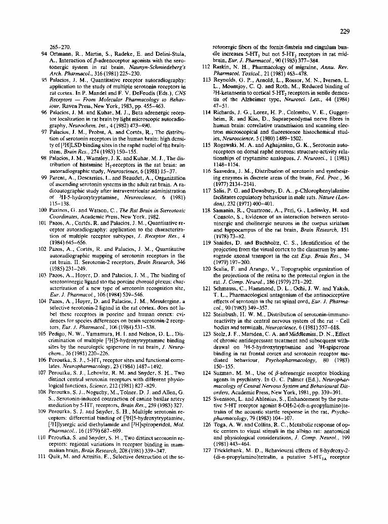
265-270. 94 Ortmann, R., Martin, S., Radeke, E. and Delini-Stula,
A., Interaction of fl-adrenoceptor agonists with the sero- tonergic system in rat brain, Naunyn-Schmiedeberg's Arch. Pharmacol., 316 (1981) 225-230.
95 Palacios, J. M., Quantitative receptor autoradiography: application to the study of multiple serotonin receptors in rat cortex. In P. Mandel and F. V. DeFeudis (Eds.), CNS Receptors -- From Molecular Pharmacology to Behav- iour, Raven Press, New York, 1983, pp. 455-463.
96 Palacios, J. M. and Kuhar, M. J., Beta adrenergic recep- tor localization in rat brain by light microscopic autoradio- graphy, Neurochem. Int., 4 (1982) 473-490.
97 Palacios, J. M., Probst, A. and Cort6s, R., The distribu- tion of serotonin receptors in the human brain: high densi- ty of [3H]LSD binding sites in the raph6 nuclei of the brain- stem, Brain Res., 274 (1983) 150-155.
98 Palacios, J. M., Wamsley, J. K. and Kuhar, M. J., The dis- tribution of histamine H~-receptors in the rat brain: an autoradiographic study, Neuroscience, 6 (1981) 15-37.
99 Parent, A., Descarries, L. and Beaudet, A., Organization of ascending serotonin systems in the adult rat brain. A ra- dioautographic study after intraventricular administration of 3H-5-hydroxytryptamine, Neuroscience, 6 (1981) 115-138.
100 Paxinos, G. and Watson, C., The Rat Brain in Stereotoxic Coordinates, Academic Press, New York, 1982.
101 Pazos, A., Cort6s, R. and Palacios, J. M., Quantitative re- ceptor autoradiography: application to the characteriza- tion of multiple receptor subtypes, J. Receptor Res., 4 (1984) 645-656.
102 Pazos, A., Cort6s, R. and Palacios, J. M., Quantitative autoradiographic mapping of serotonin receptors in the rat brain. II. Serotonin-2 receptors, Brain Research, 346 (1985) 231-249.
103 Pazos, A., Hoyer, D. and Palacios, J. M., The binding of serotoninergic ligand sto the porcine choroid plexus: char- acterization of a new type of serotonin recognition site, Eur. J. Pharmacol., 106 (1984) 539-546.
104 Pazos, A., Hoyer, D. and Palacios, J. M., Mesulergine, a selective serotonin-2 ligand in the rat cortex, does not la- bel these receptors in porcine and human cortex: evi- dences for species differences on brain serotonin-2 recep- tors, Eur. J. Pharmacol., 106 (1984) 531-538.
105 Pedigo, N, W., Yamamura, H. I. and Nelson, D. L., Dis- crimination of multiple [3H]5-hydroxytryptamine binding sites by the neuroleptic spiperone in rat brain, J. Neuro- chem., 36 (1981) 220-226.
106 Peroutka, S. J., 5-HT 1 receptor sites and functional corre- lates, Neuropharmacology, 23 (1984) 1487-1492.
107 Peroutka, S. J., Lebovitz, R. M. and Snyder, S. H., T w o distinct central serotonin receptors with different physio- logical functions, Science, 212 (1981) 827-829.
108 Peroutka, S. J., Noguchy, M., Tolner, D. J. and Allen, G. S., Serotonin-induced contraction of canine basilar artery mediation by 5-HT 1 receptors, Brain Res,, 259 (1983) 327.
109 Peroutka, S. J. and Snyder, S. H., Multiple serotonin re- ceptors: differential binding of [3H]5-hydroxytryptamine, [3H]lysergic acid diethylamide and [3H]spiroperidol, Mol. Pharmacol., 16 (1979) 687-699.
110 Peroutka, S. and Snyder, S. H., Two distinct serotonin re- ceptors: regional variations in receptor binding in mam- malian brain, Brain Research, 208 (1981) 339-347.
111 Quik, M. and Azmitia, E., Selective destruction ot the se-
229
rotonergic fibers of the fornix-fimbria and cingulum bun- dle increases 5-HT 1 but not 5-HT 2 receptors in rat mid- brain, Eur. J. Pharmacol., 90 (1983) 377-384.
112 Raskin, N. H., Pharmacology of migraine, Annu. Rev. Pharmacol. Toxicol., 21 (1981) 463-478.
113 Reynolds, G. P., Arnold, L., Rossor, M. N., Iversen, L. L., Mountjoy, C. Q. and Roth, M., Reduced binding of 3H-ketanserin to cortical 5-HT 2 receptors in senile demen- tia of the Alzheimer type, Neurosci. Lett., 44 (1984) 47-51.
114 Richards, J. G., Lorez, H. P., Colombo, V. E., Guggen- heim, R. and Kiss, D., Supraependymal nerve fibres in human brain: correlative transmission and scanning elec- tron microscopical and fluorescence histochemical stud- ies, Neuroscience, 5 (1980) 1489-1502.
115 Rogawski, M. A. and Aghajanian, G. K., Serotonin auto- receptors on dorsal raph6 neurons: structure-activity rela- tionships of tryptamine analogues, J. Neurosci., 1 (1981) 1148-1154.
116 Saavedra, J. M., Distribution of serotonin and synthesiz- ing enzymes in discrete areas of the brain, Fed. Proc., 36 (1977) 2134-2141.
117 Salis, P. G. and Dewsbury, D. A., p-Chlorophenylalanine facilitates copulatory behaviour in male rats, Nature (Lon- don), 232 (1971) 400-401.
118 Samanin, R., Quattrone, A., Peri, G., Ladinsky, H. and Consolo, S., Evidence of an interaction between seroto- ninergic and cholinergic neurons in the corpus striatum and hippocampus of the rat brain, Brain Research, 151 (1978) 73-82.
119 Sanides, D. and Buchholtz, C. S., Identification of the projection from the visual cortex to the claustrum by ante- rograde axonal transport in the cat Exp. Brain Res., 34 (1979) 197-200.
120 Scalia, F. and Arango, V., Topographic organization of the projections of the retina to the pretectal region in the rat, J. Comp. Neurol., 186 (1979) 271-292.
121 Schmauss, C., Hammond, D. L., Ochi, J. W. and Yaksh, T. L., Pharmacological antagonism of the antinociceptive effects of serotonin in the rat spinal cord, Eur. J. Pharma- col., 90 (1983) 349-357.
122 Steinbush, H. W. M., Distribution of serotonin-immuno- reactivity in the central nervous system of the rat - Cell bodies and terminals, Neuroscience, 6 (1981) 557-618.
123 Stolz, J. F., Marsden, C. A. and Middlemiss, D. N., Effect of chronic antidepressant treatment and subsequent with- drawal on 3H-5-hydroxytryptamine and 3H-spiperone binding in rat frontal cortex and serotonin receptor me- diated behaviour, Psychopharmacology, 80 (1983) 150-155.
124 Suzman, M. M., Use of fl-adrenergic receptor blocking agents in psychiatry. In G. C. Palmer (Ed.), Neurophar- macology of Central Nervous System and Behavioural Dis- orders, Academic Press, New York, 1981, pp. 339-391.
125 Svensson, L. and Ahlenius, S., Enhancement by the puta- tive 5-HT receptor agonist 8-OH-2-(di-n-propylamino)te- tralin of the acoustic startle response in the rat, Psycho- pharmacology, 79 (1983) 104-107.
126 Toga, A. W. and Collins, R. C., Metabolic response of op- tic centers to visual stimuli in the albino rat: anatomical and physiological considerations, J. Comp. Neurol., 199 (1981) 443-464.
127 Tricklebank, M. D., Behavioural effects of 8-hydroxy-2- (di-n-propylamino)tetralin, a putative 5-HTIA receptor
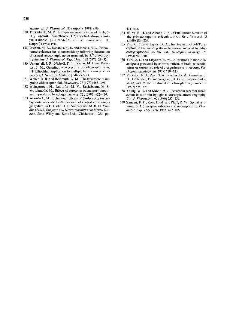
230
agonist, Br. J. PharmacoL, 81 (Suppl.) (i984J (746. 128 Tricklebank, M. D., Is hyperlocomotion induced by the 5-
HT 1 agonist, 5-methoxy-3(l,2,3,6-tetrahydropyridin-4- yl)lH-indole (RU-24969)?, Br. J. Pharmacol., 81 (Suppl.) (1984) P88.
129 Trulson, M. E., Eubanks, E. E. and Jacobs, B. L., Behav- ioural evidence for supersensitivity following destruction of central serotonergic nerve terminals by 5,7-dihydroxy- tryptamine, J. PharmacoL Exp. Ther., 198 (1976) 23-32.
130 Unnerstall, J. R., Niehoff, D. L., Kuhar, M. J. and Palac- ios, J. M., Quantitative receptor autoradiography using [3H]Ultrofilm: application to multiple benzodiazepine re- ceptors, J. Neurosci. Meth., 6 (1982) 59-73.
131 Weber, R. B. and Reinmuth, O. M., The treatment of mi- graine with propranolol, Neurology, 22 (1972) 366-369.
132 Weingartner, H., Rudorfer, M. V., Buchsbaum, M. S. and Linnoila, M., Effects of serotonin on memory impair- ments produced by ethanol, Science, 221 (1983) 472-474.
133 Weinstock, M., Behavioral effects of fl-adrenoceptor an- tagonists associated with blockade of central serotoniner- gic system. In E. Usdin, T. L. Sourkes and M. B. H. You- dim (Eds.), Enzymes and Neurotransmitters in Mental Dis- ease, John Wiley and Sons Ltd., Chichester, 1980, pp.
431-443. 134 Wurtz, R. H. and Albano, J. E., Visual-motor function of
the primate superior colliculus, Ann. Rev. Neurosci., 3 (1980) 189-226.
135 Yap, C. Y. and Taylor, D. A., Involvement of 5-HT 2 re- ceptors in the wet-dog shake behaviour induced by 5-hy- droxytryptophan in the rat, Neuropharmacology, 22 (1983) 801-804.
136 York, J. L. and Maynert, E. W., Alterations in morphine analgesia produced by chronic deficits of brain catechola- mines or serotonin: role of analgesimetric procedure, Psy- chopharmacology, 56 (1978) 119-125.
137 Yorkston, N. J., Zaki, S. A., Pitcher, D. R., Gruzelier, J. H., Hollander, D. and Sergeant, H. G. S., Propranolol as an adjunct to the treatment of schizophrenia, Lancet. ii (1977) 575-578.
138 Young, W. S. and Kuhar, M. J., Serotonin receptor locali- zation in rat brain by light microscopic autoradiography, Eur. J. Pharmacol., 62 (1980) 237-239.
139 Zemlan, F. P., Kow, L.-M. and Pfaff, D. W., Spinal sero- tonin (5-HT) receptor subtypes and nociception, J. Phar- macol. Exp. Ther., 226 (1983) 477-485.
Related Documents


![Characterization of Prejunctional Serotonin Receptorsjpet.aspetjournals.org/content/jpet/early/2005/09/... · Characterization of Prejunctional Serotonin Receptors Modulating [3H]-Acetylcholine](https://static.cupdf.com/doc/110x72/5f349070f7d6224d5a109173/characterization-of-prejunctional-serotonin-characterization-of-prejunctional-serotonin.jpg)