Subscriber access provided by University of Washington | Libraries Biomacromolecules is published by the American Chemical Society. 1155 Sixteenth Street N.W., Washington, DC 20036 Article Quantitative Affinity of Genetically Engineered Repeating Polypeptides to Inorganic Surfaces Urartu O. S. Seker, Brandon Wilson, Deniz Sahin, Candan Tamerler, and Mehmet Sarikaya Biomacromolecules, 2009, 10 (2), 250-257• DOI: 10.1021/bm8009895 • Publication Date (Web): 12 December 2008 Downloaded from http://pubs.acs.org on February 14, 2009 More About This Article Additional resources and features associated with this article are available within the HTML version: • Supporting Information • Access to high resolution figures • Links to articles and content related to this article • Copyright permission to reproduce figures and/or text from this article

Welcome message from author
This document is posted to help you gain knowledge. Please leave a comment to let me know what you think about it! Share it to your friends and learn new things together.
Transcript

Subscriber access provided by University of Washington | Libraries
Biomacromolecules is published by the American Chemical Society. 1155Sixteenth Street N.W., Washington, DC 20036
Article
Quantitative Affinity of Genetically EngineeredRepeating Polypeptides to Inorganic Surfaces
Urartu O. S. Seker, Brandon Wilson, Deniz Sahin, Candan Tamerler, and Mehmet SarikayaBiomacromolecules, 2009, 10 (2), 250-257• DOI: 10.1021/bm8009895 • Publication Date (Web): 12 December 2008
Downloaded from http://pubs.acs.org on February 14, 2009
More About This Article
Additional resources and features associated with this article are available within the HTML version:
• Supporting Information• Access to high resolution figures• Links to articles and content related to this article• Copyright permission to reproduce figures and/or text from this article
Quantitative Affinity of Genetically Engineered RepeatingPolypeptides to Inorganic Surfaces
Urartu O. S. Seker,†,‡ Brandon Wilson,† Deniz Sahin,†,‡ Candan Tamerler,†,‡ andMehmet Sarikaya*,†
Materials Science and Engineering, University of Washington, Seattle, Washington 98195, and MolecularBiology and Genetics, Istanbul Technical University, Maslak, Istanbul, Turkey
Received September 4, 2008; Revised Manuscript Received November 13, 2008
Binding kinetics of platinum-, silica-, and gold-binding peptides were investigated using a modified surface plasmonresonance spectroscopy (SPR). Platinum binding septa-peptides, quartz-binding dodecapeptides, and gold-binding14-aa peptides were originally selected using phage or cell surface display libraries using the mineral or pureforms of these materials. All of the peptides were synthesized singly to investigate their binding kinetics and toassess quantitatively the specific affinity of each to its material of selection. The peptides were also postselectionengineered to contain multiple copies of the same original sequences to quantify the effects of repeating units.SPR spectroscopy, normally using gold surfaces, was modified to contain a thin film (a few nm thick) of thematerial of interest (silica or platinum) on gold to allow the quantitative study of the adsorption kinetics of specificsolid-binding peptides. The SPR experiments, carried out at different concentrations, on all three materials substrates,resulted in Langmuir behavior that allowed the determination of the kinetic parameters, including adsorption,desorption, and equilibrium binding constants for each of the solids as well as free energy of adsorption.Furthermore, we also tested multiple repeats of the peptide sequences, specifically three repeats, to see if there isa general trend of increased binding with increased number of binding domains. There was no general trend inthe binding strength of the peptides with the increase of the repeat units from one to three, possibly because ofthe conformational changes between the single and multiple repeat polypeptides. In all cases, however, the bindingwas strong enough to suggest that these inorganic binding peptides could potentially be used as specific molecularlinkers to bind molecular entities to specific solid substrates due to their surface recognition characteristics.
Introduction
Peptides with inorganic surface binding ability have beenselected by using combinatorial biology techniques, namelyphage display1 and cell surface display2 methods that have beenadapted for materials science.3-5 Inorganic binding peptides withaffinity toward Au,3 Ag,5 Pt,6 ZnO,7 GaAs,4 TiO2,
8 and otherswere successfully selected by a number of research groups andus. Since their discovery, many of these inorganic bindingpeptides have been used in practical applications such asnanoparticle synthesis,3,9,10 molecular linkers,11 immobilizationplatforms,12-14 and assembly to create nanostructures.6 Despitethe ever increasing utility in both materials and medicine, anin-depth understanding of the quantitative affinity and bindingkinetics of many of these peptides has not been fully realized.Characterization of these peptides has been performed primarilyusing qualitative methods such as fluorescence microscopy ordirect colony/plaque counting, while the peptide was displayedon the host phages or cells. Only a limited investigation hasbeen accomplished to determine the solid binding of peptidesusing quartz crystal microbalance (QCM),14-16 atomic forcemicroscopy (AFM),6,17 surface plasmon resonance spectroscopy(SPR),18 and fluorometric peptide assays.19 In a few cases, weand others have probed possible molecular mechanism of thebinding especially due to secondary structure changes, obtainedexperimentally, for example, by NMR or by mutationalanalysis.19-21 Computational analysis of the peptide-surface
interactions was also carried out utilizing molecular dynamicsand molecular mechanics in simplified environments.22,23
Despite these preliminary studies, there has not been any globalunderstanding of the engineered peptide recognition of inorganicsolids, and the question of how biocombinatorially selectedpeptides bind to solids has so far been elusive. This investigationhas been undertaken to create a quantitative body of knowledgethat would provide a better insight in the mechanism for a morerobust design of these molecular building blocks as a utility toa wide range of practical technological molecular and nanoscalesystems.6
There are numerous methods to determine protein-protein,protein-small molecule, and protein-carbohydrate interactions;however, most of these methods are difficult to adapt formonitoring affinity of peptides to solid surfaces. For example,there has been considerable work carried out in the literatureon protein adsorption on solid particles; however, these areusually “bulk” experiments and mostly limited to high proteinconcentrations and, therefore, do not offer the capability forany real time monitoring and quantification.20,24,25 As analternative method, SPR spectroscopy has become a majortechnique to monitor adsorption kinetics, thermodynamics, andreal-time monitoring of molecular interactions at a goldsurface.26,27 The SPR signal is based on the effect of thecollective excitation of the electrons at the metal-dielectricinterface on the wavelength shift of the reflected light from aglass prism.28 Because of the changing surface resonance, theshift in the wavelength increases as the molecular binding onthe metal surface of the SPR chip progresses, thereby allowingthe monitoring of the molecular adsorption and coverage on
* To whom correspondence should be addressed. Tel.: (206) 543-0724.Fax: (206) 543-6381. E-mail: [email protected].
† University of Washington.‡ Istanbul Technical University.
Biomacromolecules 2009, 10, 250–257250
10.1021/bm8009895 CCC: $40.75 2009 American Chemical SocietyPublished on Web 12/12/2008
the surface. SPR spectroscopy is highly sensitive to the adsorbed(and desorbed) amount of molecules on the surface, makingthe method a useful tool to monitor adsorption of bio- andsynthetic molecular systems on the chip surface.29,30 Thetechnique is frequently utilized to investigate molecular interac-tions where one biomolecule is immobilized to the surface usinga metal-binding synthetic linker, for example, a thiol molecule,and the other end is free to interact with molecular entities withinthe SPR chamber. We have recently successfully employed aSPR approach using genetically engineered metal bindingpeptides, such as gold-binding and platinum binding, andobtained their Langmuir adsorption behavior.16,18 Quartz crystalmicrobalance is also another frequently used technique tomonitor the adsorption and desorption processes.31 This tech-nique relies on the change in the resonance frequency of quartzcrystal coated with a inorganic surface of interest (which isgenerally gold). The change in the frequency of the quartz crystalrepresents the amount of mass deposited on the quartz crystal.Besides gold coating on quartz crystal, platinum, silica, hy-droxyapatite, and different polymer coatings are also com-mercially available surfaces. The real time monitoring of theadsorption and desorption using QCM was carried out in manydifferent studies toward understanding the nature of molecularinteraction both qualitatively and quantitatively.31,32 Becauseboth techniques can be quantitative, SPR and QCM have alsobeen used together to monitor the validity of the molecularadsorption on solids in the same system.33 More qualitative othertechniques, such as fluorescence, have also been used inconjunction with SPR or QCM to monitor molecular adsorp-tion.34,35 In fact, in our research, as a trend, we first qualitativelycategorize the originally selected peptides rapidly using the FMtechnique in terms of their binding characteristics, and then applyeither SPR or QCM, or both,16 to obtain more quantitative data,such as the adsorption parameters of kinetics or thermodynamics.
Normally carrying out the SPR experiments on a givenmaterial surface is not possible because of the intrinsic limita-tions of the surface plasmon phenomena. Because of this, silverand, most frequently, gold surfaces are used to generate thesurface plasmon effect. In these studies, generally, SPR analysisis performed using a glass slide coated with a thin film (45-50nms) of gold.28-30 Other materials, even Pt or Pd, have poorplasmonic properties.36 Therefore, this problem limits theapplicability of the SPR technique to monitor molecularadsorption on other practical solid surfaces such as Ti, Pt, Cu2O,ZnO, Al2O3, silica, and others, all interesting for materials andmedical applications. To overcome this difficulty, we developeda protocol that provides the design principles for the coating(usually via vacuum deposition) of an additional ultrathin layerof target material, of any type, that is, metal or oxide, on to theAu chip still providing the SPR signal from the underlying goldlayer. The chip allows monitoring of molecular adsorption onthis new ultrathin-film material at the very top surface facingthe analyte solution. Protocols similar to ours have beendeveloped using a sol gel technique for silica.37 In a very recentstudy, using this approach, we monitored adsorption of Pt-binding peptides on to a 2 nm thick Pt-coated surface.18
Using the surface modified and properly prepared gold,platinum, and silica substrates, herein we carried out SPR toquantitatively characterize the binding kinetics of geneticallyengineered peptides for inorganics (GEPI),6 synthesized to havethe original molecular conformations displayed in the librarycoat protein either in the cell (for Au) or phage (for silica andPt). Two quartz-binding peptides (QBP1 and QBP2) and twoPt-binding peptides (PtBP1 and PtBP2) were used and binding
activities to their respective solids were compared to bindingof a gold-binding peptide, GBP13, that we have extensivelystudied in our previous work.6,16,18,20 The five peptides areknown (qualitatively) to bind to their respective materialsstrongly as have been already determined by fluorescencemicroscopy. In this work, we have also tested the hypothesisthat multiple repeats of the peptide sequences, which we call“repeating polypeptides”3, known to bind to a specific materialmay have improved binding. The resulting data, therefore, wereanalyzed from two different perspectives, namely, to assess theeffects of multiple repeats compared to the single-repeat peptidesand evaluating the specific affinities quantitatively on each ofthe corresponding materials.
Materials and Methods
Selection of the Binding Peptides. Phage Display. Target materials(quartz, platinum) were cleaned using ethanol (95%), and equilibratedusing phosphate/carbonate (PC) buffer overnight. After this procedure,the phage library housing 2 × 10⁹ different randomized peptidesequences were brought into contact with the target materials. Thelibrary was incubated with the target Pt or quartz powder (obtainedfrom pure quartz mineral) in potassium-carbonate (PC) buffer contain-ing 0.1% detergent. Unbound phage were removed by washing withphosphate carbonate buffer containing 0.1% detergent. Specifically,bound phage were eluted from the surface using elution buffers; theeluted phage pool was amplified with Eschericia coli ER2738. Theamplified phage were then purified. The amplified and purifiedphage were used for additional panning rounds; after each round, thephage were grown on solid media and single clones were selected bypicking single phage plaques. DNA of single phage clones were isolatedand sequenced. Because the insert position of the random sequencesare known, the peptide sequenced displayed on each clone can beobtained from the DNA sequences (see Supporting Information fordetails).
Cell Surface Display. Gold binding protein was selected using thecell surface display. In this approach,3 the Eschericia coli was used asthe host cell for carrying the peptide exposed to the environment ofthe cell. The randomized peptides were displayed on the membraneprotein of E. coli, multiporin, a fusion protein, which does not haveany affinity for gold in its native form. Multiporin was then cloned tomake a large number of copies (millions). The library has a diversityof 5 × 10⁶ peptides. Each copy carries a segment, which encodes therandom peptide sequence. For biopanning experiments, the cellsdisplaying the random peptide sequences were brought in contact withgold powder and, after selection of the weak and non specific binders,strongly bound cells were harvested and the gene expressing multiporinprotein was sequenced to obtain the information of DNA sequenceencoding the putative gold binding peptide (see Supporting Informationfor details).
Peptide, Buffer, and Solutions. The highest affinity peptidesobtained from the selection were synthesized using standard Fmoc solidphase peptide synthesis techniques and purified using C-18 reversephase liquid chromatography (RPLC) to a level >95% (UnitedBiochemical Research, U.S.A.). The peptides were synthesized withouta blocking group either at the -N or -C termini. Peptide solutions,containing the five different GEPIs, PtBP1, PtBP2, QBP1, and QBP2,for SPR measurements were prepared in PC buffer (55 mM KH2PO4,45 mM Na2CO3 and 200 mM NaCl). The fifth peptide, GBP1, wasprepared in phosphate buffer (1:3 mixture of 10 mM KH2PO4, 10 mMK2HPO4, and 100 mM KCl). The pH of both buffers were adjusted topH 7.5 using 0.1 M HCl and 0.1 M NaOH. None of the peptides werestudied in their zwitterions form, and the pH of the buffer solutionused was not at the isoelectric points of the peptides. Because histidinehas a -NH2 group on the side chain, it may contribute to the protonationof the GBP1. In fact, GBP1 can be protonated at pH 6.5 and the totalcharge becomes +1, while at pH 7.5, it is lower than +1.
Affinity of Genetically Engineered Peptides Biomacromolecules, Vol. 10, No. 2, 2009 251
SPR Experiments. The SPR measurements were made with a dualchannel instrument (Kretschmann configuration) developed by the RadioEngineering Institute, Czech Republic.28,30 The instrument can detectchanges at a level of 0.0001 refractive index unit. Unlike Au and Ag,noble metals such Pt and Pd, or dielectrics such as SiOx, do not havea measurable SPR signal. To overcome this difficulty, based on theresult of a numerical method we developed, a very thin film (a fewnms thick) of Pt or SiOx was prepared on the Au substrate, allowingthe sufficient generation of an SPR signal, where the thicknesses werepredetermined using the numerical model.18
Results and Discussion
Following the biopanning experiments using platinum andsilica powders two silica binding peptides (QBP1 and QBP2),and two platinum binding peptides (PtBP1 and PtBP2) wereisolated. Gold binding peptide was isolated previously from acell surface display library.3 The amino acid (aa) sequences ofthe peptides are given in Table 1.
To mimic the peptides in their original molecular architectureas they are displayed on pIII coat protein of the phage library,the QBPs were synthesized as 12-amino acid sequences in alinear form while the PtBPs were synthesized in 7-aa long cyclicform. The GBP1 was displayed on the Escherichia coli cellsurface as 14-aa linear form and, thus, synthesized as such. Toexamine whether tandem repeats of the peptide increases thebinding affinity, the peptides were also synthesized in three-repeat multimers for further testing. As a special case for Ptbinding peptides (that are originally displayed in constrainedform via cysteines at N and C termini, linear one repeat forms)were prepared to probe the effect of conformational constraintson binding affinity.
Two strong platinum binders (PtBP1 and PtBP2), identifiedfrom a set of 37 original binders,18 were tested for their bindingaffinity, both in the linear (l) and constrained (c) forms and in1- and 3-repeat tandem sequences. Based on the observedbinding behavior, the SPR data for platinum binding peptideswere fitted to two different adsorption models. The adsorptiondata from c-PtBP1, 3l-PtBP1 and l-PtBP2, c-PtBP2 were fittedto a simple Langmuir adsorption model, and those from l-PtBP1and 3l-PtBP2 were fitted to a biexponential Langmuir model.It is known from our earlier CD studies that these peptides havedifferent secondary structures in solutions.20 We attributedifferent binding behavior of constrained and linear forms ofthe same peptide sequences to possibly be a result of differencesof their conformations leading to different molecular recognitionof the solid and, hence, different adsorption behavior.
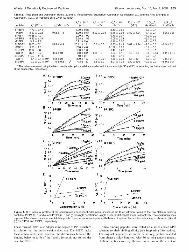
For the first Pt-binding peptide, that is, PtBP1, the equilibriumconstants (Keq) and, therefore, the free energy of adsorption ofthe constrained and singly linear forms of the PtBP1 are slightlydifferent, the constrained one being a stronger binder (Table2). According to our observations, this may be due to the factthat the constrained form of this peptide has a higher adsorptionrate (ka), 20 times, than that of the linear form, while both having
the same desorption rate (Figure 1A,C). To test whether the3-repeat form of the linear peptide has any effect on the bindingkinetics, we also studied the adsorption behavior. Again, asshown in Figure 1E and in Table 2, 3l-PtBP1 has equilibriumconstant and free energy of adsorption similar to those of thesingle repeat form of this Pt-binding peptide. It is interesting tonote that the 3-repeat polypeptide has about twice the adsorptionrate as well as twice the desorption rate of those of the single-repeat peptide, resulting in similar overall equilibrium behavior.These results suggest that the tandem repeat does not necessarilyimprove the adsorption behavior of a solid-binding peptide. Nochange in the adsorption behavior may be due to the confor-mational freedom (assuming various equilibrium molecularconformations) of the linear forms of these peptides as adsorbedon the solid surfaces. It should be noted also that, the constrainedforms of these peptides have two cystein residues at eitherterminals. These amino acids, in the free forms, are known toinfluence binding to metal surfaces. In the present peptides,cysteins are used to form the loop and, therefore, they do notnecessarily contribute to the adsorption behavior of the peptide(see below).
The SPR experiments that were carried out using the secondPt-binding peptide, that is, PtBP2, produce a new set ofinteresting results (Figure 1B,D,F and Table 2). First of all, theconstrained and the linear forms of this peptide have similarequilibrium constants (0.09 and 0.16 M-1, respectively) and,hence, similar free energy of adsorption (-6.7 and -7.1 kcal/mol, respectively). The adsorption and desorption kinetics ofthe linear peptide were several times faster than those of itsconstrained form (Figure 1B,D). This result is interesting andis just the opposite of what was found in the case of PtBP1,that is, solid-binding and -unbinding behaviors of this peptide(see above) are much faster than those of its linear counterpart.Interestingly, the 3-repeat linear tandem sequence of this peptidehas considerably higher equilibrium adsorption behavior thanthe single repeat form (Figure 1F). Correspondingly, the 3l-PtBP2 binds to Pt substrate about 30% more than its singlerepeat peptide (Figure 1D,F, Table 2). Again, this result isopposite what was found for the PtBP1 case where theseparameters corresponding to the single and 3-repeat forms hardlychanged.
From the above observation, we see that the differences inthe adsorption behavior of the platinum-binding peptides maybe a result of their molecular structural differences. The effectresulting from the constrained forms on the binding affinity ofthe platinum binding peptides cannot be concluded as a universalbehavior. On the one hand, we observed the binding affinity ofPtBP1 was increased by creating a constrained structure;however, this was not true in the case of PtBP2, where therewas no substantial change in the adsorption behavior. Similarly,the use of tandem linear repeats did not result in the sameadsorption behavior of these concatemers; that is, while the 3l-PtBP1 had no change in its equilibrium adsorption behavior,PtBP2 had substantial increase. One reason for these observa-tions is that the solid binding of a peptide is not only controlledby the amino acid sequences, but it may also depend on thesecondary structure for, at least, the platinum-binding peptides.A case study was carried out in this laboratory to attempt tocorrelate binding and structure of PtBP118 in the linear andconstrained forms. The CD experiments indicated the presenceof extended helical polyproline type II (PPII) secondarystructure, which has been noted in short peptides that containPro, Ala, and Gln. Thus, although both peptides feature the sameintegral sequence and possess some degree of random coil, the
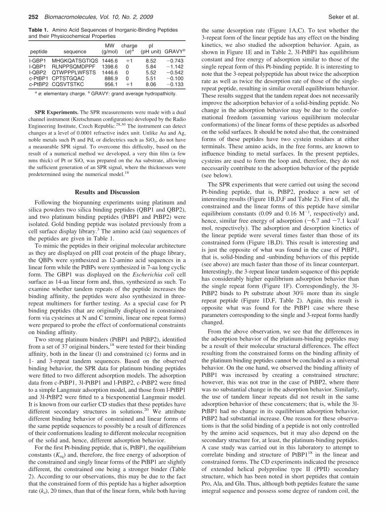
Table 1. Amino Acid Sequences of Inorganic-Binding Peptidesand their Physicochemical Properties
peptide sequenceMW
(g/mol)charge
(e)apI
(pH unit) GRAVYb
l-GBP1 MHGKQATSGTIQS 1446.6 +1 8.52 -0.743l-QBP1 RLNPPSQMDPPF 1398.6 0 5.84 -1.142l-QBP2 QTWPPPLWFSTS 1446.6 0 5.52 -0.542c-PtBP1 CPTSTGQAC 886.9 0 5.51 -0.100c-PtBP2 CQSVTSTKC 956.1 +1 8.06 -0.133
a e: elementary charge. b GRAVY: grand average hydropathicity.
252 Biomacromolecules, Vol. 10, No. 2, 2009 Seker et al.
linear form of PtBP1 also adopts some degree of PPII structurein solution but the cyclic version does not. The PtBP2 lacksthese amino acids, and therefore, the differences between thebinding behavior to Pt of the l and c-forms do not follow thecase for PtBP1.
Silica binding peptides were tested on a silica-coated SPRsubstrate for their binding affinity (see Supporting Information).The original sequences are linear 12 aa long peptide selectedfrom phage display libraries. Also 36 aa long tandem repeatsof these peptides were synthesized to determine the effect of
Table 2. Adsorption and Desorption Rates, ka and kd, Respectively, Equilibrium Adsorption Coefficients, Keq, and the Free Energies ofAdsorption, ∆Gads, of Peptides on a Given Surfacea
peptides ka1 (M-1 s-1) ka
2 (M-1 s-1)kd
1 × 10⁻⁵
(s-1)kd
2 × 10⁻⁴
(s-1)Keq
1 × 10⁶
(M-1)Keq
2 × 10⁴
(M-1)∆G ads
1
(kcal/mol)∆G ads
2
(kcal/mol)
c-PtBP1 170 ( 3.44 5.00 ( 0.88 3.40 ( 0.89 -8.9 ( 0.1l-PtBP1 8.27 ( 0.92 10.2 ( 1.3 5.00 ( 0.57 3.00 ( 0.33 0.16 ( 0.04 3.39 ( 1.24 -7.1 ( 0.1 -6.2 ( 0.23l-PtBP1 16.88 ( 4.07 9.00 ( 1.00 0.19 ( 0.07 -7.2 ( 0.2c-PtBP2 5.32 ( 1.15 6.28 ( 1.55 0.09 ( 0.04 -6.7 ( 0.3l-PtBP2 21.8 ( 2.2 15.22 ( 5.0 0.16 ( 0.07 -7.1 ( 0.33l-PtBP2 1603 ( 52 15.5 ( 4.9 26.11 ( 12 3.6 ( 0.5 6.73 ( 2.49 3.87 ( 1.24 -9.3 ( 0.3 -6.3 ( 0.2l-QBP1 238 ( 1.0 200 ( 4.0 0.122 ( 0.03 -6.9 ( 0.13l-QBP1 1912 ( 88 130 ( 1.0 1.48 ( 0.23 -8.5 ( 0.1l-QBP2 37.1 ( 2.7 484 ( 94 3.0 ( 0.2 200 ( 2 1.24 ( 0.1 3.2 ( 0.7 -8.4 ( 0.04 -6.2 ( 0.123l-QBP2 112.9 ( 11.49 10 ( 0.1 1.13 ( 0.12 -8.3 ( 0.1l-GBP1 1.4 ( 0.1 × 10⁴ 112 ( 21 890 ( 150 2 ( 0.01 1.56 ( 0.28 56 ( 10 -8.2 ( 0.1 -7.8 ( 0.13l-GBP1 2.9 ( 0.2 × 10⁴ 1.6 ( 0.2 × 10³ 773 ( 183 6.5 ( 3.7 3.91 ( 1.22 330 ( 190 -9.0 ( 0.2 -8.9 ( 0.3
a The values calculated using the biexponential adsorption model are labeled with the superscripts “1” and “2”, representing the first and second partof the exponential, respectively.
Figure 1. SPR spectral profiles of the concentration-dependent adsorption kinetics of the three different forms of the two platinum bindingpeptides; PtBP1 (a, b, and c) and PtBP2 (e, f, and g) for single constrained, single linear, and 3-repeat linear, respectively. The continuous linesrepresent the fit over the experimental data points. The concentration dependent behavior of apparent adsorption rates, kobs, is shown in (d) and(h) for PtPB1 and PtBP2, respectively.
Affinity of Genetically Engineered Peptides Biomacromolecules, Vol. 10, No. 2, 2009 253
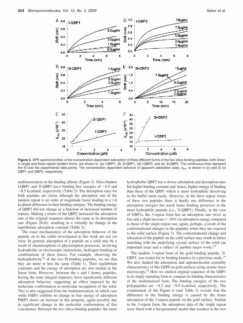
multimerization on the binding affinity (Figure 2). Silica bindersl-QBP1 and 3l-QBP1 have binding free energies of -6.9 and-8.5 kcal/mol, respectively (Table 2). The desorption rates forboth peptides are closer although the adsorption rate of thetandem repeat is an order of magnitude faster leading to a 1.6kcal/mol difference in their binding energies. The binding energyof QBP2 did not change as a function of increased number ofrepeats. Making a trimer of the QBP2 increased the adsorptionrate of the original sequence almost the same as its desorptionrate (Figure 2D,E), resulting in a virtually no change in theequilibrium adsorption constant (Table 2).
The exact mechanism(s) of the adsorption behavior of thepeptide on to the solids investigated in this work are not yetclear. In general, adsorption of a peptide on a solid may be aresult of chemisorptions or physisorption processes, involvinghydrophobic or electrostatic interactions, hydrogen bonding orcombinations of these forces. For example, observing thehydrophobicity38 of the two Pt-binding peptides, we see thatthey are more or less the same (Table 1). Their equilibriumconstants and the energy of adsorption are also similar in thelinear form. However, between the c and l forms, peptides,having the same integral sequence, have significantly differentadsorption behavior, suggesting an effect imposed by themolecular conformation in molecular recognition of the solid.This is also suggested from the multimerization, in which casewhile PtBP1 exhibits no change in free energy of adsorptionPtBP2 shows an increase in this property, again possibly dueto significant change in the molecular conformation of thisconcatamer. Between the two silica-binding peptides, the more
hydrophobic QBP2 has a slower adsorption and desorption ratesbut higher binding constant and, hence, higher energy of bindingthan those of the QBP1 which is more hydrophilic dissolvingin the buffer more easily. However, in the three repeat formsof these two peptides there is hardly any difference in theadsorption energies but much faster binding processes in themore hydrophilic peptide (i.e., 3l-QBP1). Finally, in the caseof GBP1s, the 3-repeat form has an adsorption rate twice asfast and a slight increase (∼10%) in adsorption energy comparedto those of the single repeat one, again, perhaps, a result of theconformational changes in the peptides when they are exposedto the solid surface (Figure 3). The conformational change andrelaxation of the peptide on the solid surface may result in latticematching with the underlying crystal surface of the solid (animportant issue and a subject of another major work).39
The tandem 3-repeat form of the gold binding peptide, 3l-GBP1, was tested for its binding kinetics in a previous study.16
We also studied the adsorption and supramolecular assemblycharacteristics of this GEPI on gold surfaces using atomic forcemicroscopy.39 Here we studied original sequence of the GBP1in its singly repeating form to compare its binding characteristicsof the multimerized form. The binding energies of the twopolypeptides are -8.2 and -9.0 kcal/mol, respectively. Theexamination of the Figure 4 (and Table 2) reveals that thedifference in the binding energy is caused by the fasteradsorption of the 3-repeat peptide on the gold surface. Similarto the 3-repeat form, the adsorption data of the single repeatwere fitted with a biexponential model that resulted in the two
Figure 2. SPR spectral profiles of the concentration-dependent adsorption of three different forms of the two silica binding peptides; both linear,in single and three-repeat tandem forms, are shown in: (a) l-QBP1, (b) 3l-QBP1, (d) l-QBP2, and (e) 3l-QBP2. The continuous lines representthe fit over the experimental data points. The concentration dependent behavior of apparent adsorption rates, kobs, is shown in (c) and (f) forQBP1 and QBP2, respectively.
254 Biomacromolecules, Vol. 10, No. 2, 2009 Seker et al.
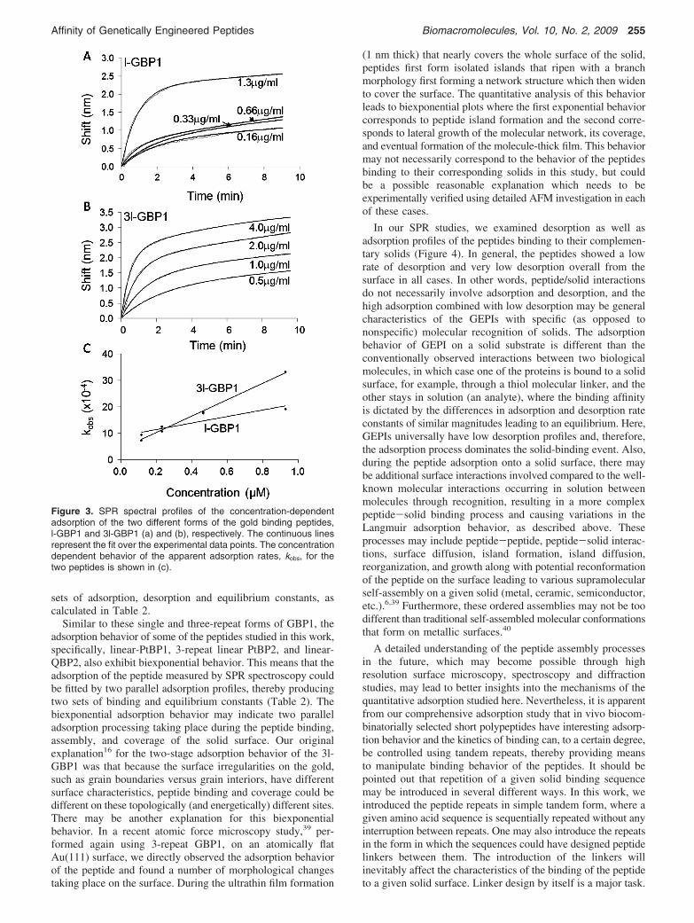
sets of adsorption, desorption and equilibrium constants, ascalculated in Table 2.
Similar to these single and three-repeat forms of GBP1, theadsorption behavior of some of the peptides studied in this work,specifically, linear-PtBP1, 3-repeat linear PtBP2, and linear-QBP2, also exhibit biexponential behavior. This means that theadsorption of the peptide measured by SPR spectroscopy couldbe fitted by two parallel adsorption profiles, thereby producingtwo sets of binding and equilibrium constants (Table 2). Thebiexponential adsorption behavior may indicate two paralleladsorption processing taking place during the peptide binding,assembly, and coverage of the solid surface. Our originalexplanation16 for the two-stage adsorption behavior of the 3l-GBP1 was that because the surface irregularities on the gold,such as grain boundaries versus grain interiors, have differentsurface characteristics, peptide binding and coverage could bedifferent on these topologically (and energetically) different sites.There may be another explanation for this biexponentialbehavior. In a recent atomic force microscopy study,39 per-formed again using 3-repeat GBP1, on an atomically flatAu(111) surface, we directly observed the adsorption behaviorof the peptide and found a number of morphological changestaking place on the surface. During the ultrathin film formation
(1 nm thick) that nearly covers the whole surface of the solid,peptides first form isolated islands that ripen with a branchmorphology first forming a network structure which then widento cover the surface. The quantitative analysis of this behaviorleads to biexponential plots where the first exponential behaviorcorresponds to peptide island formation and the second corre-sponds to lateral growth of the molecular network, its coverage,and eventual formation of the molecule-thick film. This behaviormay not necessarily correspond to the behavior of the peptidesbinding to their corresponding solids in this study, but couldbe a possible reasonable explanation which needs to beexperimentally verified using detailed AFM investigation in eachof these cases.
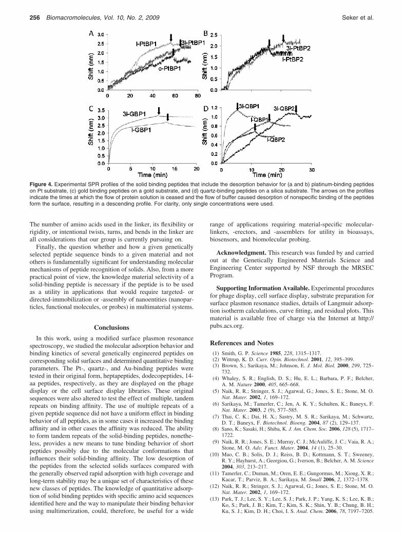
In our SPR studies, we examined desorption as well asadsorption profiles of the peptides binding to their complemen-tary solids (Figure 4). In general, the peptides showed a lowrate of desorption and very low desorption overall from thesurface in all cases. In other words, peptide/solid interactionsdo not necessarily involve adsorption and desorption, and thehigh adsorption combined with low desorption may be generalcharacteristics of the GEPIs with specific (as opposed tononspecific) molecular recognition of solids. The adsorptionbehavior of GEPI on a solid substrate is different than theconventionally observed interactions between two biologicalmolecules, in which case one of the proteins is bound to a solidsurface, for example, through a thiol molecular linker, and theother stays in solution (an analyte), where the binding affinityis dictated by the differences in adsorption and desorption rateconstants of similar magnitudes leading to an equilibrium. Here,GEPIs universally have low desorption profiles and, therefore,the adsorption process dominates the solid-binding event. Also,during the peptide adsorption onto a solid surface, there maybe additional surface interactions involved compared to the well-known molecular interactions occurring in solution betweenmolecules through recognition, resulting in a more complexpeptide-solid binding process and causing variations in theLangmuir adsorption behavior, as described above. Theseprocesses may include peptide-peptide, peptide-solid interac-tions, surface diffusion, island formation, island diffusion,reorganization, and growth along with potential reconformationof the peptide on the surface leading to various supramolecularself-assembly on a given solid (metal, ceramic, semiconductor,etc.).6,39 Furthermore, these ordered assemblies may not be toodifferent than traditional self-assembled molecular conformationsthat form on metallic surfaces.40
A detailed understanding of the peptide assembly processesin the future, which may become possible through highresolution surface microscopy, spectroscopy and diffractionstudies, may lead to better insights into the mechanisms of thequantitative adsorption studied here. Nevertheless, it is apparentfrom our comprehensive adsorption study that in vivo biocom-binatorially selected short polypeptides have interesting adsorp-tion behavior and the kinetics of binding can, to a certain degree,be controlled using tandem repeats, thereby providing meansto manipulate binding behavior of the peptides. It should bepointed out that repetition of a given solid binding sequencemay be introduced in several different ways. In this work, weintroduced the peptide repeats in simple tandem form, where agiven amino acid sequence is sequentially repeated without anyinterruption between repeats. One may also introduce the repeatsin the form in which the sequences could have designed peptidelinkers between them. The introduction of the linkers willinevitably affect the characteristics of the binding of the peptideto a given solid surface. Linker design by itself is a major task.
Figure 3. SPR spectral profiles of the concentration-dependentadsorption of the two different forms of the gold binding peptides,l-GBP1 and 3l-GBP1 (a) and (b), respectively. The continuous linesrepresent the fit over the experimental data points. The concentrationdependent behavior of the apparent adsorption rates, kobs, for thetwo peptides is shown in (c).
Affinity of Genetically Engineered Peptides Biomacromolecules, Vol. 10, No. 2, 2009 255
The number of amino acids used in the linker, its flexibility orrigidity, or intentional twists, turns, and bends in the linker areall considerations that our group is currently pursuing on.
Finally, the question whether and how a given geneticallyselected peptide sequence binds to a given material and notothers is fundamentally significant for understanding molecularmechanisms of peptide recognition of solids. Also, from a morepractical point of view, the knowledge material selectivity of asolid-binding peptide is necessary if the peptide is to be usedas a utility in applications that would require targeted- ordirected-immobilization or -assembly of nanoentities (nanopar-ticles, functional molecules, or probes) in multimaterial systems.
Conclusions
In this work, using a modified surface plasmon resonancespectroscopy, we studied the molecular adsorption behavior andbinding kinetics of several genetically engineered peptides oncorresponding solid surfaces and determined quantitative bindingparameters. The Pt-, quartz-, and Au-binding peptides weretested in their original form, heptapeptides, dodecopeptides, 14-aa peptides, respectively, as they are displayed on the phagedisplay or the cell surface display libraries. These originalsequences were also altered to test the effect of multiple, tandemrepeats on binding affinity. The use of multiple repeats of agiven peptide sequence did not have a uniform effect in bindingbehavior of all peptides, as in some cases it increased the bindingaffinity and in other cases the affinity was reduced. The abilityto form tandem repeats of the solid-binding peptides, nonethe-less, provides a new means to tune binding behavior of shortpeptides possibly due to the molecular conformations thatinfluences their solid-binding affinity. The low desorption ofthe peptides from the selected solids surfaces compared withthe generally observed rapid adsorption with high coverage andlong-term stability may be a unique set of characteristics of thesenew classes of peptides. The knowledge of quantitative adsorp-tion of solid binding peptides with specific amino acid sequencesidentified here and the way to manipulate their binding behaviorusing multimerization, could, therefore, be useful for a wide
range of applications requiring material-specific molecular-linkers, -erectors, and -assemblers for utility in bioassays,biosensors, and biomolecular probing.
Acknowledgment. This research was funded by and carriedout at the Genetically Engineered Materials Science andEngineering Center supported by NSF through the MRSECProgram.
Supporting Information Available. Experimental proceduresfor phage display, cell surface display, substrate preparation forsurface plasmon resonance studies, details of Langmuir adsorp-tion isotherm calculations, curve fitting, and residual plots. Thismaterial is available free of charge via the Internet at http://pubs.acs.org.
References and Notes(1) Smith, G. P. Science 1985, 228, 1315–1317.(2) Wittrup, K. D. Curr. Opin. Biotechnol. 2001, 12, 395–399.(3) Brown, S.; Sarikaya, M.; Johnson, E. J. Mol. Biol. 2000, 299, 725–
732.(4) Whaley, S. R.; English, D. S.; Hu, E. L.; Barbara, P. F.; Belcher,
A. M. Nature 2000, 405, 665–668.(5) Naik, R. R.; Stringer, S. J.; Agarwal, G.; Jones, S. E.; Stone, M. O.
Nat. Mater. 2002, 1, 169–172.(6) Sarikaya, M.; Tamerler, C.; Jen, A. K. Y.; Schulten, K.; Baneyx, F.
Nat. Mater. 2003, 2 (9), 577–585.(7) Thai, C. K.; Dai, H. X.; Sastry, M. S. R.; Sarikaya, M.; Schwartz,
D. T.; Baneyx, F. Biotechnol. Bioeng. 2004, 87 (2), 129–137.(8) Sano, K.; Sasaki, H.; Shiba, K. J. Am. Chem. Soc. 2006, 128 (5), 1717–
1722.(9) Naik, R. R.; Jones, S. E.; Murray, C. J.; McAuliffe, J. C.; Vaia, R. A.;
Stone, M. O. AdV. Funct. Mater. 2004, 14 (1), 25–30.(10) Mao, C. B.; Solis, D. J.; Reiss, B. D.; Kottmann, S. T.; Sweeney,
R. Y.; Hayhurst, A.; Georgiou, G.; Iverson, B.; Belcher, A. M. Science2004, 303, 213–217.
(11) Tamerler, C.; Duman, M.; Oren, E. E.; Gungormus, M.; Xiong, X. R.;Kacar, T.; Parviz, B. A.; Sarikaya, M. Small 2006, 2, 1372–1378.
(12) Naik, R. R.; Stringer, S. J.; Agarwal, G.; Jones, S. E.; Stone, M. O.Nat. Mater. 2002, 1, 169–172.
(13) Park, T. J.; Lee, S. Y.; Lee, S. J.; Park, J. P.; Yang, K. S.; Lee, K. B.;Ko, S.; Park, J. B.; Kim, T.; Kim, S. K.; Shin, Y. B.; Chung, B. H.;Ku, S. J.; Kim, D. H.; Choi, I. S. Anal. Chem. 2006, 78, 7197–7205.
Figure 4. Experimental SPR profiles of the solid binding peptides that include the desorption behavior for (a and b) platinum-binding peptideson Pt substrate, (c) gold binding peptides on a gold substrate, and (d) quartz-binding peptides on a silica substrate. The arrows on the profilesindicate the times at which the flow of protein solution is ceased and the flow of buffer caused desorption of nonspecific binding of the peptidesform the surface, resulting in a descending profile. For clarity, only single concentrations were used.
256 Biomacromolecules, Vol. 10, No. 2, 2009 Seker et al.
(14) Sano, K.; Ajima, K.; Iwahori, K.; Yudasaka, M.; Iijima, S.; Yamashita,I.; Shiba, K. Small 2005, 1, 826–832.
(15) Chen, H. B.; Su, X. D.; Neoh, K. G.; Choe, W. S. Anal. Chem. 2006,78, 4872–4879.
(16) Tamerler, C.; Oren, E. E.; Duman, M.; Venkatasubramanian, E.;Sarikaya, M. Langmuir 2006, 22 (18), 7712–7718.
(17) Yamashita, K.; Kirimura, H.; Okuda, M.; Nishio, K.; Sano, K. I.; Shiba,K.; Hayashi, T.; Hara, M.; Mishima, Y. Small 2006, 2 (10), 1148–1152.
(18) Seker, U. O. S.; Wilson, B.; Dincer, S.; Kim, I. W.; Oren, E. E.; Evans,J. S.; Tamerler, C.; Sarikaya, M. Langmuir 2007, 23 (15), 7895–7900.
(19) Sano, K. I.; Sasaki, H.; Shiba, K. Langmuir 2005, 21, 3090–3095.(20) Kulp, J. L.; Sarikaya, M.; Evans, J. S. J. Mater. Chem. 2004, 14, 2325–
2332.(21) Sano, K.; Shiba, K. J. Am. Chem. Soc. 2006, 125 (47), 14234–14235.(22) Oren, E. E.; Tamerler, C.; Sarikaya, M. Nano Lett. 2005, 5, 415–419.(23) Tomasio, S. M.; Walsh, T. R. Mol. Phys. 2007, 105, 221–229.(24) Rocha, S.; Thunemann, A. F.; Pereira, M. C.; Coelho, M. A. N.;
Mohwald, H.; Brezesinski, G. ChemBioChem 2005, 6 (2), 280–283.(25) Joshi, H.; Shirude, P. S.; Bansal, V.; Ganesh, K. N.; Sastry, M. J.
Phys. Chem. B 2004, 108 (31), 11535–11540.(26) Cocklin, S.; Jost, M.; Robertson, N. M.; D; Weeks, S.; Weber, H. W.;
Young, E; Seal, S.; Zhang, C.; Mosser, E.; Loll, P. J.; Saunders, A. J.;Rest, R. F.; Chaiken, I. M.; J. Mol. Recognit. 2006, 19 (4), 354–362.
(27) Day, Y. S. N; Baird, C. L.; Rich, R. L.; Myszka, D. G. Protein Sci.2002, 11, 1017–1025.
(28) Homola, J.; Lu, H. B. B.; Nenninger, G. G.; Dostalek, J.; Yee, S. S.Sens. Actuators, B 2001, 76, 403–410.
(29) Rich, R. L.; Myszka, D. G. J. Mol. Recognit. 2005, 18 (1), 1–39.(30) Jung, L. S.; Campbell, C. T.; Chinowsky, T. M.; Mar, M. N.; Yee,
S. S. Langmuir 1998, 14 (19), 5636–5648.(31) Bailey, L. E.; Kambhampat, D.; Kanazawa, K. K.; Knoll, W.; Frank,
C. W. Langmuir 2002, 18 (2), 479–489.(32) Choe, W. S.; Sastry, M. S. R.; Thai, C. K.; Dai, H.; Schwartz, D. T.;
Baneyx, F. Langmuir 2007, 23, 11347–11350.(33) Hook, F.; Kasemo, B. Anal. Chem. 2001, 73, 5796–5804.(34) Gestwicki, J. E.; Cairo, C. W.; Mann, D. A; Owen, R. M.; Kiessling,
L. L. Anal. Biochem. 2002, 305, 149–155.(35) Arima, Y.; Iwata, H. J. Mater. Chem. 2007, 17, 4079–4087.(36) Kovalenko, S. A.; Lisitsa, M. P. Semicond. Phys., Quantum Electron.
Optoelectron. 2001, 4, 352–357.(37) Szunerits, S.; Coffinier, Y.; Janel, S.; Boukherroub, R. Langmuir 2006,
22 (25), 10716–10722.(38) Kyte, J.; Doolittle, R. F. J. Mol. Biol. 1982, 157 (1), 105–132.(39) So, C. 2008, unpublished.(40) Brune, H. Surf. Sci. Rep. 1998, 31, 121–229.
BM8009895
Affinity of Genetically Engineered Peptides Biomacromolecules, Vol. 10, No. 2, 2009 257
Related Documents