Nuclear Medicine & Biology, Vol. 23, pp. 343-352, 1996 Copyright 0 1996 Elsevier Science Inc. ELSEVIER ISSN 0969-8051/96/$15.00 + 0.00 SSDI 0969-805 1(95)02097-7 Q uantitative 1231 IMP and 99mT~ HMPAO Imaging in the Dog Following Cocaine Administration Herbert Suss kind, ’ f David A. We ber , 1 * Marija Ivunovic , ’ * Christopher T. C. Wang,’ Constance E. DeHuan2 and Patrick R. Gavin’ ‘CLINICAL RESEARCH CENTER, BROOKHAVEN NATIONAL LABORATORY, BOX 5000, UPTON, NY 11973, USA, AND *DEPARTMENT OF CLINICAL VETERINARY MEDICINE AND SURGERY, WASHINGTON STATE UNIVERSITY, PULLMAN, WA 99163, USA. ABSTRACT. SPECT and associated imaging procedures were used in beagle dogs to (1) evaluate the uptake, distribution, and clearance properties of i.v.-injected 1231 IMP (IMP) and 99mTc HMPAO (HMPAO) in the brain, lungs, liver, and kidneys; (2) quantify the acute effects (after 15 set) of very low doses (0.5 or 1.0 mg/kg) cocaine on the kinetics and localization properties of IMP and HMPAO; and (3) evaluate comparative imaging properties of IMP and HMPAO for measuring regional cerebral blood flow (rCBF). Regional and global uptake and localization of IMP or HMPAO were evaluated in control studies using dynamic planar (O-30 min) and SPECT imaging (at 35 min). The regional distribution properties of IMP and HMPAO in the brain were estimated from regions of interest (ROIs) drawn around anatomic structures on MR slices and manually registered with corresponding SPECT slices. Cocaine significantly reduced the 30-min IMP uptake in the brain and lungs by -15%, but only slightly changed HMPAO uptake in the brain and other organs. In the control studies, the respective uptakes of IMP in the brain and lungs were 9 and 39% greater (p < 0.01) than those of HMPAO. In control SPECT studies, the highest uptake of IMP was observed in the thalamus and progressively less activity was observed in the parietal lobe, frontal lobe, cerebellum, occipital lobe, and entire brain; activity in the olfactory bulb was lower than in all other regions. Cocaine reduced IMP uptake in the cerebellum (p < O.Ol), occipital lobe (p < O.Ol), and entire brain (p < 0.05). IMP uptake (cpm/pixel- mCi) in the different brain regions was 1.3 to 2.1 times greater than that of HMPAO (p < 0.001). HMPAO uptake was more homogeneous throughout the gray matter of the brain; no significant uptake differences were observed among flagged regions. Results indicate that single, acute doses of cocaine, 0.5 and 1.0 mg/kg, significantly altered the uptake and localization properties of IMP in the dog’s brain, lungs, liver, and kidneys. Variations in regional uptake of IMP in the parietal, frontal, and occipital lobes, cerebellum, and thalamus were greater than with HMPAO. NUCL MED BIOL 23;3:343-352, 1996. KEY WORDS. Cocaine, Dog model, 1231 IMP, g9mTc HMPAO, rCBF INTRODUCTION SPECT imaging of iv.-administered lipophilic compounds such as iz31- labeled d,I-N-isopropy@iodoamphetamine hydrochloride (IMP) and 9gmTc-labeled d,l-hexamethylpropyleneamine oxime (HMPAO) pro- vides a useful procedure to evaluate regional cerebral blood flow (rCBF) (23). The pharmacokinetics of IMP are determined by meta- bolic processes related to turnover of neurohumoral amines (4). The initial IMP uptake occurs in the lungs at high-capacity, low-affinity binding sites at the lung endothelium (12). IMP is released back to the circulation, followed by high first-pass extraction by passive diffusion through the blood-brain barrier and retention by nonspecific, high- capacity, low-affinity amine binding sites within the brain (13). IMP then slowly washes out of the brain after conversion to nonamphet- amine metabolites. However, during the period of approximately 30 to 75 min after IMP administration, the distribution properties and slow brain clearance of IMP are adequate to allow SPECT imaging of the perfusion space for rCBF (9). There is significant IMP uptake in the *Present address: University of California Davis Medical Center, Sacramento, CA 95817. tAuthvr for correspondence. Accepted 26 November 1995. liver (12), peaking in humans at 3 to 4 h after administration, and excretion occurs through the kidneys (2). HMPAO localizes in the perfusion space of the brain by a different mechanism of uptake and localization. Whereas IMP is trapped in the brain by metabolic processes, HMPAO is trapped in the brain by ac- celerated decomposition to hydrophilic products that cannot diffuse back across the blood-brain barrier (6, 7, 17). HMPAO inflow occurs during the first pass through the brain, with passive diffusion of the lipophilic compound through the blood-brain barrier. HMPAO activ- ity in the human brain reaches a maximum at only -1 min, followed by an immediate loss of some activity. By 4 to 5 min, activity in the brain plateaus and remains constant for several hours. The uptake of HMPAO in the brain depends upon the relative speed with which the lipophilic molecule changes to secondary and/or hydrophilic com- pounds in the circulating blood and how quickly the HMPAO changes to hydrophilic compounds in the brain. HMPAO passes essentially unchanged through the lungs in humans. Its metabolites are removed in the liver and excreted by the kidneys. Few noninvasive imaging studies of cerebral blood flow have been reported in large laboratory animals. These include (1) distribution and kinetics of y9mTc-labeled HMPAO, NEP-DADT, and methyl NEP- DADT, and lZ31-labeled IMP for cerebral perfusion imaging with SPECT in dogs (5); (2) rCBF studies using PET carried out with baboons (8); (3) studies of the dog’s cholinergic system (19); and (4)

Welcome message from author
This document is posted to help you gain knowledge. Please leave a comment to let me know what you think about it! Share it to your friends and learn new things together.
Transcript
Nuclear Medicine & Biology, Vol. 23, pp. 343-352, 1996 Copyright 0 1996 Elsevier Science Inc.
ELSEVIER
ISSN 0969-8051/96/$15.00 + 0.00 SSDI 0969-805 1(95)02097-7
Q uantitative 1231 IMP and 99mT~ HMPAO Imaging in the Dog Following Cocaine Administration
Herbert Suss kind, ’ f David A. We ber , 1 * Marija Ivunovic , ’ * Christopher T. C. Wang,’ Constance E. DeHuan2 and Patrick R. Gavin’
‘CLINICAL RESEARCH CENTER, BROOKHAVEN NATIONAL LABORATORY, BOX 5000, UPTON, NY 11973, USA, AND *DEPARTMENT OF CLINICAL VETERINARY MEDICINE AND SURGERY, WASHINGTON STATE UNIVERSITY,
PULLMAN, WA 99163, USA.
ABSTRACT. SPECT and associated imaging procedures were used in beagle dogs to (1) evaluate the uptake, distribution, and clearance properties of i.v.-injected 1231 IMP (IMP) and 99mTc HMPAO (HMPAO) in the brain, lungs, liver, and kidneys; (2) quantify the acute effects (after 15 set) of very low doses (0.5 or 1.0 mg/kg) cocaine on the kinetics and localization properties of IMP and HMPAO; and (3) evaluate comparative imaging properties of IMP and HMPAO for measuring regional cerebral blood flow (rCBF). Regional and global uptake and localization of IMP or HMPAO were evaluated in control studies using dynamic planar (O-30 min) and SPECT imaging (at 35 min). The regional distribution properties of IMP and HMPAO in the brain were estimated from regions of interest (ROIs) drawn around anatomic structures on MR slices and manually registered with corresponding SPECT slices. Cocaine significantly reduced the 30-min IMP uptake in the brain and lungs by -15%, but only slightly changed HMPAO uptake in the brain and other organs. In the control studies, the respective uptakes of IMP in the brain and lungs were 9 and 39% greater (p < 0.01) than those of HMPAO. In control SPECT studies, the highest uptake of IMP was observed in the thalamus and progressively less activity was observed in the parietal lobe, frontal lobe, cerebellum, occipital lobe, and entire brain; activity in the olfactory bulb was lower than in all other regions. Cocaine reduced IMP uptake in the cerebellum (p < O.Ol), occipital lobe (p < O.Ol), and entire brain (p < 0.05). IMP uptake (cpm/pixel- mCi) in the different brain regions was 1.3 to 2.1 times greater than that of HMPAO (p < 0.001). HMPAO uptake was more homogeneous throughout the gray matter of the brain; no significant uptake differences were observed among flagged regions. Results indicate that single, acute doses of cocaine, 0.5 and 1.0 mg/kg, significantly altered the uptake and localization properties of IMP in the dog’s brain, lungs, liver, and kidneys. Variations in regional uptake of IMP in the parietal, frontal, and occipital lobes, cerebellum, and thalamus were greater than with HMPAO. NUCL MED BIOL 23;3:343-352, 1996.
KEY WORDS. Cocaine, Dog model, 1231 IMP, g9mTc HMPAO, rCBF
INTRODUCTION
SPECT imaging of iv.-administered lipophilic compounds such as iz31- labeled d,I-N-isopropy@iodoamphetamine hydrochloride (IMP) and 9gmTc-labeled d,l-hexamethylpropyleneamine oxime (HMPAO) pro- vides a useful procedure to evaluate regional cerebral blood flow (rCBF) (23). The pharmacokinetics of IMP are determined by meta- bolic processes related to turnover of neurohumoral amines (4). The initial IMP uptake occurs in the lungs at high-capacity, low-affinity binding sites at the lung endothelium (12). IMP is released back to the circulation, followed by high first-pass extraction by passive diffusion through the blood-brain barrier and retention by nonspecific, high- capacity, low-affinity amine binding sites within the brain (13). IMP then slowly washes out of the brain after conversion to nonamphet- amine metabolites. However, during the period of approximately 30 to 75 min after IMP administration, the distribution properties and slow brain clearance of IMP are adequate to allow SPECT imaging of the perfusion space for rCBF (9). There is significant IMP uptake in the
*Present address: University of California Davis Medical Center, Sacramento, CA 95817.
tAuthvr for correspondence. Accepted 26 November 1995.
liver (12), peaking in humans at 3 to 4 h after administration, and excretion occurs through the kidneys (2).
HMPAO localizes in the perfusion space of the brain by a different mechanism of uptake and localization. Whereas IMP is trapped in the brain by metabolic processes, HMPAO is trapped in the brain by ac- celerated decomposition to hydrophilic products that cannot diffuse back across the blood-brain barrier (6, 7, 17). HMPAO inflow occurs during the first pass through the brain, with passive diffusion of the lipophilic compound through the blood-brain barrier. HMPAO activ- ity in the human brain reaches a maximum at only -1 min, followed by an immediate loss of some activity. By 4 to 5 min, activity in the brain plateaus and remains constant for several hours. The uptake of HMPAO in the brain depends upon the relative speed with which the lipophilic molecule changes to secondary and/or hydrophilic com- pounds in the circulating blood and how quickly the HMPAO changes to hydrophilic compounds in the brain. HMPAO passes essentially unchanged through the lungs in humans. Its metabolites are removed in the liver and excreted by the kidneys.
Few noninvasive imaging studies of cerebral blood flow have been reported in large laboratory animals. These include (1) distribution and kinetics of y9mTc-labeled HMPAO, NEP-DADT, and methyl NEP- DADT, and lZ31-labeled IMP for cerebral perfusion imaging with SPECT in dogs (5); (2) rCBF studies using PET carried out with baboons (8); (3) studies of the dog’s cholinergic system (19); and (4)
344 H. Susskind et al.
PET imaging to evaluate the transfer of drugs across the blood-brain barrier in the dog (1).
Our study was designed to utilize planar and SPECT imaging to (1) evaluate the uptake, distribution, and clearance properties of IMP and HMPAO in the dog brain, lungs, liver, and kidneys; (2) quantify the acute effects of very low doses of cocaine on the kinetics and localiza- tion properties of IMP and HMPAO; and (3) evaluate comparative imaging properties of IMP and HMPAO for measuring regional cere- bral blood flow (rCBF).
MATERIALS AND METHODS Materials
The study was carried out in seven 8- to 11-kg beagle dogs. The dog allows systematic, repetitive in oivo testing in the same intact animal, where each animal serves as its own control. The size of the beagle facilitated SPECT and planar camera imaging between head and ab- dominal region simultaneously; the use of pure-bred dogs eliminated possible interbreed differences in the uptake and localization of the two radiopharmaceuticals. These studies were approved by the Brookhaven National Laboratory Institutional Review Committee.
Procedures
Unit doses of IMP (Iofetamine) were obtained from Medi-Physics, Inc. (South Plainfield, NJ). HMPAO (Ceretec) was labeled with 5 mL 99’“Tc-pertechnetate using the Amersham Corp. (Arlington Heights, IL) multidose kit (0.5 mg exametazine, 7.6 pg stannous chloride dihy-
drate, and 4.5 mg sodium chloride sealed under nitrogen) and the prescribed labeling procedure. Radiochemical purity was determined for each HMPAO preparation using the vendor-recommended, three- part paper chromatography procedure. Labeling efficiencies of >80% were obtained for all preparations, and the radiopharmaceutical was always injected within 30 min of preparation.
Dynamic planar and SPECT imaging studies were carried out with IMP and HMPAO in five dogs; an additional two dogs were imaged with only IMP. Dogs were positioned in dorsal recumbency for imaging. Six imaging studies were carried out in each animal, three with IMP and three with HMPAO. The three studies with each radiopharma- ceutical included one control study and two studies carried out after i.v. administration of 0.5 mg/kg and 1.0 mg/kg body weight of cocaine hydrochloride (cocaine). Successive studies in each dog were spaced at least 2 weeks apart. Single 3-mL boluses of -150 MBq (-4 mCi) IMP or -370 MBq (-10 mCi) HMPAO were injected into a dog’s femoral vein in all imaging studies. The acute effects of cocaine were evaluated using bolus injections of IMP or HMPAO 15 set after cocaine admin- istration. The dogs were lightly anesthetized with the same pentobar- bital dose (26 mg/kg) in each study. Each dog served as its own control in the IMP and HMPAO cocaine and control studies.
Studies were carried out with a rotating camera SPECT imaging system (Toshiba Model GCA 901A) using lz31 or LEHR collimators. Images (128 x 128 pixels) were recorded with a 20% window centered over the 159-keV energy peak of 1231 and the 140.5-keV peak of 99mTc. Dynamic planar data acquisition of the dog’s head and thorax in the posterior projection was started 15 set after the cocaine injections and continued for 30 min (Fig. 1). This was followed immediately by
FIG. 1. Typical composite planar images of a dog (1643) in the posterior projection acquired over first 30 min after i.v. injection showing uptake of lz31 IMP in brain, lungs, liver, and kidneys. Note that the respective ROIs are smaller than actual lung, liver, and kidney areas to exclude overlapping of adjacent organs.
Effect of Cocaine on Imaging in the Dog 345
SPECT acquisition, 60 projections recorded in 6” increments (30 set/ projection) (Fig. 2A,B).
Axial, frontal, and sagittal MR images of a dog’s head were obtained from Washington State University (Pullman, WA). The animal was imaged in sternal recumbency. A General Electric Signa MRI system with a 1.5-T superconducting magnet, 5-mm-thick slices, with 0 mm gap and a 12-cm field of view was used; images were reconstructed in a matrix size of 256 x 256 yielding a pixel volume of 1.10 mm3. Although the MR and SPECT studies were carried out with the dog in sternal and dorsal recumbency, respectively, the orientation of the sagittal slices was obtained in the same orthogonal plane for both procedures. Evaluation of a wide variety of MR brain images shows little variation in the brain size, shape, and anatomy among different dogs and dog species. Thus, a single set of orthogonal MRI slices was used as the template to evaluate the regional uptake of radiopharma- ceutical within the brain; care was taken to place the animals in the same orientation and position for all serial imaging studies.
Image Processing and Data Analysis
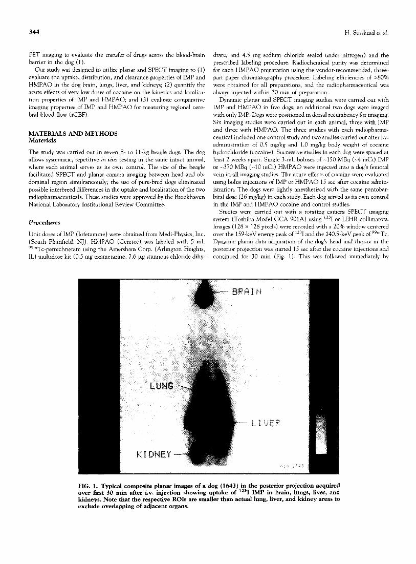
The kinetics and uptake of IMP and HMPAO were evaluated from counting data obtained using regions of interest (ROIs) placed over images of the dog’s brain, lungs, liver, and kidneys on the planar images collected in the posterior projection (Fig. 1). The ROIs were positioned to exclude overlapping organs. The planar distributions of IMP or HMP.40 activity in ROIs were calculated from the summed images of the 30-min acquisitions. The 30-min uptake data were normalized for ROI area and injected dose; the 99mTc data were also corrected for radioactive decay. A uniformity correction using high-count flood data (120 x lo6 counts of ‘231-IMP or 57Co flood fields) was applied to the SPECT projections prior to reconstruction. The tomographic slices were reconstructed using the conventional filtered back-projection method with a Shepp-Logan filter and no attenuation correction. We sized and aligned sagittal MR images centered at a dog’s midbrain (Fig. 3, upper left) with SPECT slices of the same dog’s brain (Fig. 3, upper right) using a 2-D registration algorithm based on manual registration of corresponding anatomical locations in each slice (26). Images were scaled to the same pixel size and then rotated and translated to align corresponding points (Fig. 3, bottom left). Seven ROIs drawn around anatomic structures on the MR slice were used to quantify regional activity on SPECT slices (Fig. 3, lower right) in the thalamus, parietal lobe, frontal lobe, cerebellum, occipital lobe, olfactory bulb, and the entire brain, comprising all the individual regions. Activity in the seven regions was determined from the average count density (cpm/ pixel-mCi) from four adjacent 4-mm/pixel sagittal slices centered at the midbrain. The respective sensitivities for the lz31 and 99mTc were 195 and 182 cpm/@Zi. The anatomic atlas of the dog brain developed at Washington State University (11) was used as a reference guide in the manual registration of MR and SPECT images.
Rates of IMP and HMPAO lung clearance and brain, liver, and kidney uptake for the first 30 min were calculated from the slopes of the time-activity curves obtained in the dynamic planar imaging study. The brain, liver, and kidney activity data were fit to a two-compart- ment uptake curve,
y(t) = c - ap?” - a,e+‘,
and the lung activity data to a two-compartment clearance curve,
y(t) = ap-‘” + asem“’ + c,
where A = In 2/t1,z, using a least-squares fitting routine. The fast and
slow biological half-times for IMP or HMPAO, tf and t,, of the two- compartment curves were calculated in each study. Time-activity curves for a typical four-organ IMP study are shown in Fig. 4. Time- activity curves for IMP and HMPAO in the brain and lungs are com- pared in Fig. SA,B, respectively.
Statistics
Statistical analyses of the data were performed using a randomized block design (ANOVA with single-factor repeated measure). The sig- nificance of IMP and of HMPAO uptake kinetics and distribution properties were determined for the control and two cocaine dose stud- ies in the brain, lungs, liver, and kidneys. The respective IMP and HMPAO 30.min uptakes and rates were compared for the dynamic planar images. Their regional distributions were also evaluated in sag- ittal SPECT brain slices. Statistical significance was selected as p < 0.05.
RESULTS Dynamic Planar Studies
The two dose levels of cocaine (0.5 mg/kg and 1 .O mg/kg) significantly reduced the 30-min IMP uptake in the brain and lungs by -15% (p < 0.05 and p < 0.001, respectively) (Table 1 and Fig. 6). However, no significant difference occurred in IMP uptake between the two cocaine doses. A reduction of -12% in liver and -10% in kidney uptake with cocaine (Table 1) was not statistically significant. Cocaine increased the 30-min HMPAO uptake in the lungs, liver, and kidneys by 12%, 22%, and 20%, respectively, and decreased uptake in the entire brain by 4% (Table 1); however, results were not statistically significant. The 30.min brain uptake of IMP was 9% greater than that of HMPAO in the absence of cocaine, whereas lung uptake of IMP was 39% greater than that of HMPAO (p < 0.01) (Table 1). Liver uptake of IMP was 22% greater than that of HMPAO, and kidney uptake of IMP was 10% less than that of HMPAO in the absence of cocaine.
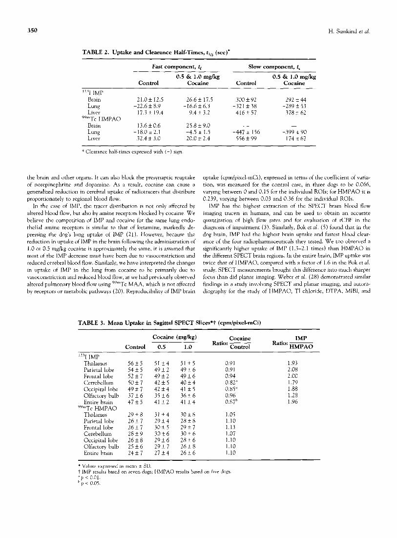
Administered cocaine increased the fast and slow rates of IMP clear- ance from the lung by 27% and 10% (p < O.Ol), respectively (Table 2). The fast brain uptake rate of IMP was reduced by 27%, while the slow rate remained essentially unchanged. Cocaine increased fast and slow IMP uptake rates in the liver by 46% and 9%, respectively. The fast rate of HMPAO uptake into the brain decreased by 90% following the administration of cocaine (Table 2); the fast and slow rates of HMPAO clearance from the lungs increased by 75% and 1 l%, respectively; and the fast and slow rates of HMPAO uptake in the liver increased by 38% and 69%, respectively. The fast IMP brain uptake rate in the control studies was 35% slower than that of HMPAO. The fast IMP lung clearance rate in the control studies was 20% slower, and the slow rate was 39% faster than those of HMPAO. The respective values of fast and slow IMP liver uptake rates for the control studies were 87% and 34% faster than those of HMPAO.
The slow IMP clearance component from the lung, t,, varied be- tween 263 and 368 set for the control studies, and between 164 and 357 set with cocaine. A weak positive correlation (r = 0.61) was obtained between the rate of IMP uptake in the brain and the rate of IMP clearance from the lungs. The slope of the IMP correlation with cocaine was -45% that of the control study, indicating a faster IMP lung clearance.
SPECT Studies
Both IMP and HMPAO activities in the brain reached plateaus after -25 min and -5 min, respectively, which permitted brain SPECT studies be-
346 H. Susskind et al.
::, _:
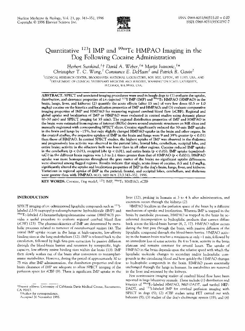
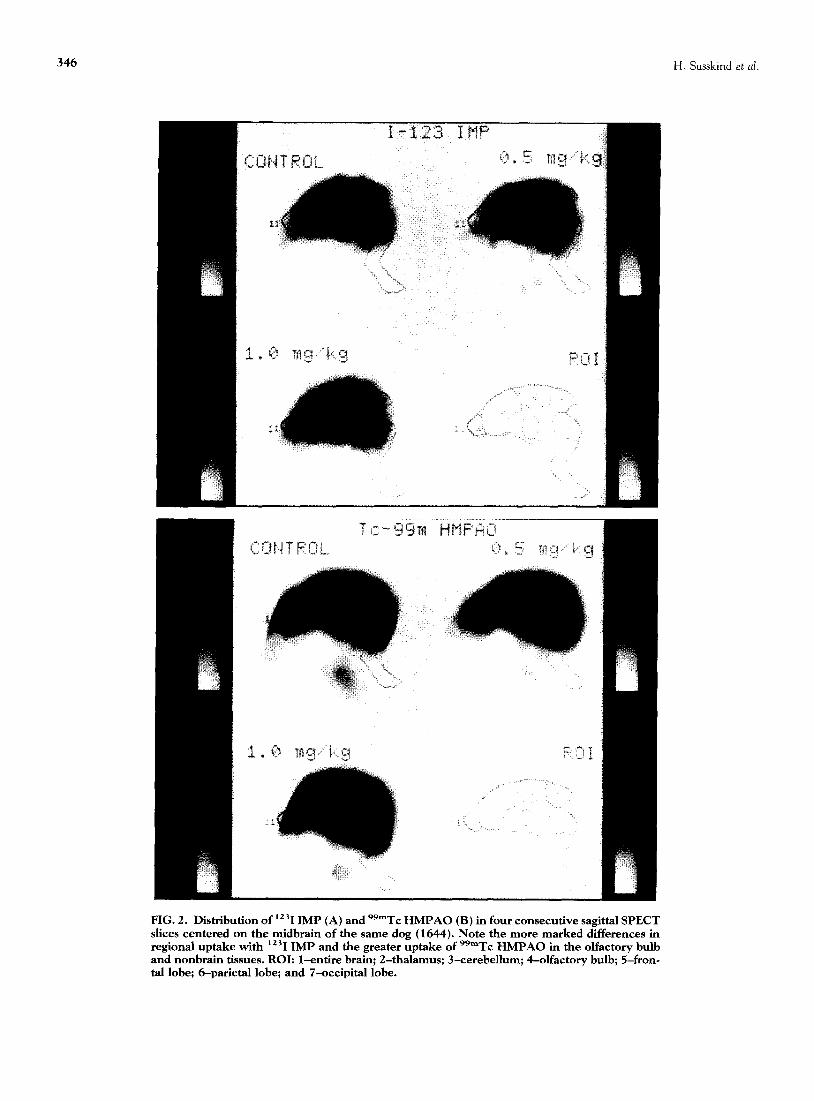
FIG. 2. Distribution of iz31 IMP (A) and 99mTc HMPAO (B) in four consecutive sagittal SPECT slices centered on the midbrain of the same dog (1644). Note the more marked differences in regional uptake with iz31 IMP and the greater uptake of 99mTc HMPAO in the olfactory bulb and nonbrain tissues. ROI: l-entire brain; 2-thalamus; 3-cerebellum; 4-olfactory bulb; 5-fron- tal lobe; 6-parletal lobe; and 7-occipital lobe.
Effect of Cocaine on Imaging in the Dog 347
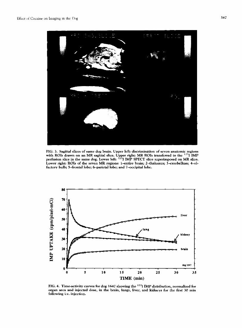
FIG. 3. Sag&al slices of same dog brain. Upper left: discrimination of seven anatomic regions with ROIs drawn on an MR sag&al slice. Upper right: MR ROIs transferred to the lz31 IMP perfusion slice in the same dog. Lower Ieft: lz31 IMP SPECT slice superimposed on MR slice. Lower right: ROIs of the seven MR regions: l-entire brain; 2-thalamus; 3-cerebellum; 4-01. factory bulb; 5-frontal lobe; 6-parietal lobe; and 7-occipital lobe.
0 5 10 15 20 25 30 35
TIME (min)
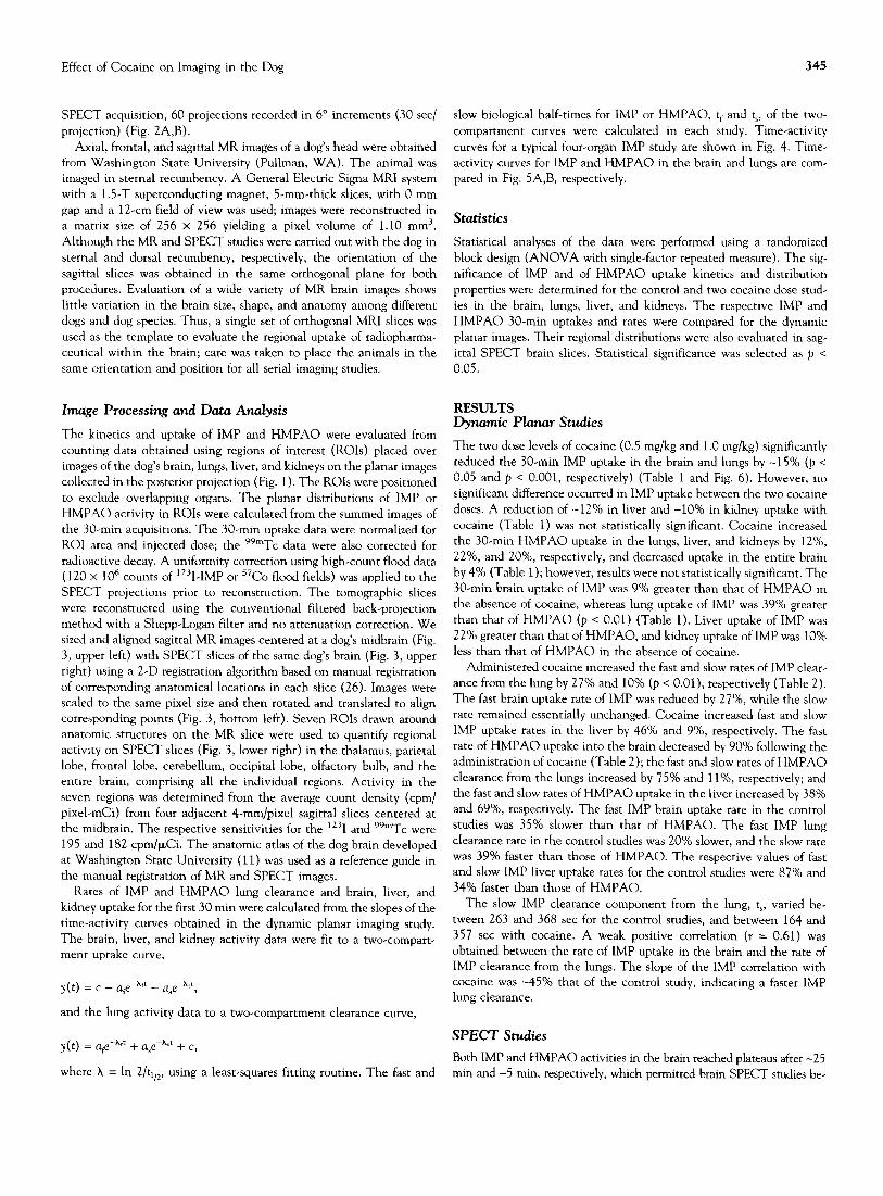
FIG. 4. Time-activity curves for dog 1647 showing the lz31 IMP distribution, normalized for organ area and injected dose, in the brain, lungs, liver, and kidneys for the fast 30 min following iv. injection.
348 H. Susskind et al.
IMP
HMPAO
TIME (min)
; lb 1; 2'0 2; 3-o ?
TIME (min)
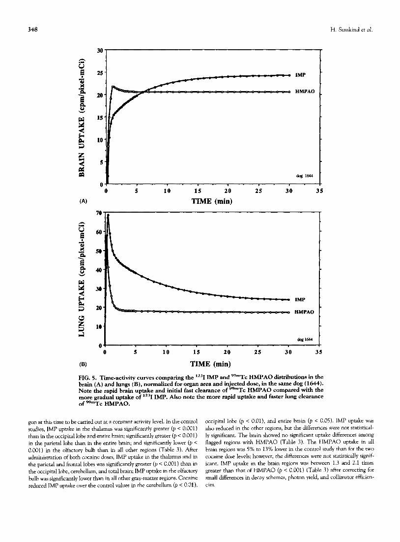
FIG. 5. Tie-activity curves comparing the lz31 IMP and 99mTc HMPAO distributions in the brain (A) and lungs (B), normalized for organ area and injected dose, in the same dog (1644). Note the rapid brain uptake and initial fast clearance of 99”Tc HMPAO compared with the more gradual uptake of lz31 IMP. Also note the more rapid uptake and faster lung clearance of 99mTc HMPAO.
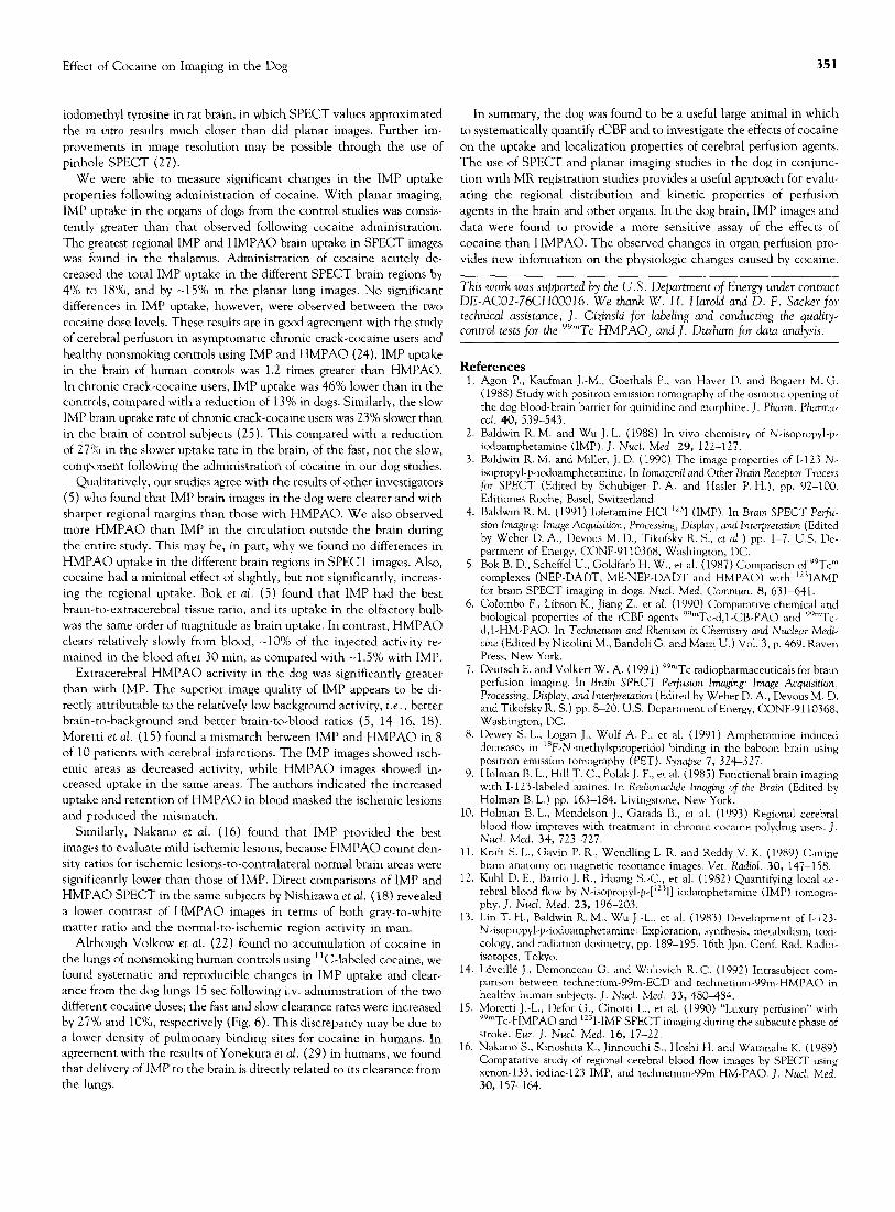
gun at this time to be carried out at a constant activity level. In the control occipital lobe (p < O.Ol), and entire brain (p c 0.05). IMP uptake was studies, IMP uptake in the thalamus was significantly greater (p < 0.001) also reduced in the other regions, but the differences were not statistical- than in the occipital lobe and entire brain; significantly greater (p < 0.001) ly significant. The brain showed no significant uptake differences among in the parietal lobe than in the entire brain; and significantly lower (p c flagged regions with HMPAO (Table 3). The HMPAO uptake in all 0.001) in the olfactory bulb than in all other regions (Table 3). After brain regions was 5% to 13% lower in the control study than for the two administration of both cocaine doses, IMP uptake in the thalamus and in cocaine dose levels; however, the differences were not statistically signif- the par&al and frontal lobes was significantly greater (p < 0.001) than in icant. IMP uptake in the brain regions was between 1.3 and 2.1 times the occipital lobe, cerebellum, and total brain; IMP uptake in the olfactory greater than that of HMPAO (e c 0.001) (Table 3) after correcting for bulb was significantly lower than in all other gray-matter regions. Cocaine small differences in decay schemes, photon yield, and collimator efficien- reduced IMP uptake over the control values in the cerebellum (p < O.Ol), cies.
Effect of Cocaine on Imaging in the Dog 349
TABLE 1. Total Uptake in Fit 30 Mm in Planar Images (cpm/pixeIdmCi)
Brain Lung Liver Kidney
Control Cocaine (mg/kg) Control Cocaine (mg/kg) Control Cocaine (mg/kg) Control Cocaine (mglkg)
Dog 0.5 1.0 0.5 1.0 0.5 1.0 0.5 1.0
123I IMP 1643 515 459 472 837 676 761 1,092 931 906 839 698 624 1644 658 530 623 922 796 788 1,219 1,148 1,183 991 754 761 1645 502 508 467 920 677 814 1,490 1,080 906 668 1646 702 562 442 688 533 537 811 850 885 536 571 612 1647 520 451 419 1,048 943 878 1,297 1,068 869 867 687 632 1660 478 457 436 708 647 530 1,053 1,020 907 734 704 704 1661 479 444 461 680 628 610 856 858 1,105 492 630 706
Mean 551 487 474 829 700 703 1,117 994 966 743 674 672 f SD 591 +_46 k 68 f 142 f 132 f 141 k241 k116 k 125 f 196 f 64 *54
99mTc HMPAO 1644 596 767 644 513 682 680 1,034 1,499 1,311 1,071 1,836 1,365 1645 495 526 547 493 540 566 898 1,102 1,255 835 1,023 1,072 1646 444 405 336 536 694 575 811 1,123 919 773 750 657 1660 495 349 323 539 516 498 878 718 735 567 445 1661 471 404 503 429 413 436 759 1,023 1,003 840 133 825
Mean 500 490 471 502 569 551 876 1,093 1,045 817 1,086 873 *SD * 58 3~ 168 + 139 f45 f119 +91 + 104 xk 279 f 239 + 180 f 518 41358
DISCUSSION uptake of these radiopharmaceuticals in the brain. In contrast to studies of altered rCBF in chronic crack cocaine users (10, 25), our study
Alterations in the uptake and distribution properties of IMP and focused on the effects of acute doses of cocaine on regional blood flow HMPAO resulting from the administration of very low single doses of in the brain and the localization properties of the perfusion agents in i.v.-injected cocaine provide sensitive measurements of the effects of other organs. We were able to measure significant changes in the IMP cocaine. The uptake and imaging properties of IMP and HMPAO in uptake properties 15 set after i.v. administration of cocaine doses as low the brain, lung, liver, and kidney provide new insight into the effects as 0.5 mg/kg. We believe these changes are a result of several factors. of cocaine on different organs and the influence of cocaine on the Cocaine is a potent vasoconstrictor and can alter regional blood flow in
70
60
50
40
30
20
10
dog 1647
0g 0 5 10 15 20 25 30 35
TIME (min)
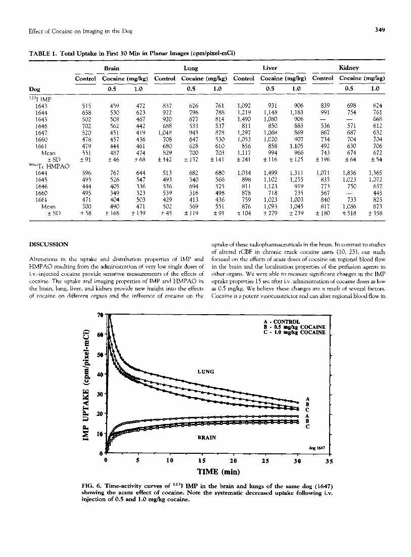
FIG. 6. Tiie*activity curves of “‘1 IMP in the brain and lungs of the same dog (1647) showing the acute effect of cocaine. Note the systematic decreased uptake following i.v. injection of 0.5 and 1.0 mglkg cocaine.
350 H. Susskind et al.
TABLE 2. Uptake and Clearance Half-Times, tllz (set)’
Fast component, 4 Slow component, f
0.5 & 1.0 mglkg 0.5 & 1.0 mglkg Control Cocaine Control Cocaine
Iz31 IMP Brain 21.0+ 12.5 26.6 * 17.5 300f92 292f44 Lung -22.6f8.9 -16.6 f 6.3 -321f38 -289+53 Liver 17.3 _+ 19.4 9.4 f 3.2 416_+57 378rt62
99mTc HMPAO Brain 13.6 + 0.6 25.8 rk 9.0 -
Lung -18.0 k 2.1 -4.5 k 1.5 -447; 156 -399k90 Liver 32.4 f 3.0 20.0 f 2.4 556+99 174k67
* Clearance half-times expressed with (-) sign
the brain and other organs. It can also block the presynaptic reuptake uptake (cpm/pixel-mCi), expressed in terms of the coefficient of varia- of norepinephrine and dopamine. As a result, cocaine can cause a tion, was measured for the control case, in three dogs to be 0.066, generalized reduction in cerebral uptake of radiotracers that distribute varying between 0 and 0.15 for the individual ROIs; for HMPAO it is proportionately to regional blood flow. 0.239, varying between 0.03 and 0.36 for the individual ROIs.
In the case of IMP, the tracer distribution is not only affected by altered blood flow, but also by amine receptors blocked by cocaine. We believe the competition of IMP and cocaine for the same lung endo- thelial amine receptors is similar to that of ketamine, markedly de- pressing the dog’s lung uptake of IMP (21). However, because the reduction in uptake of IMP in the brain following the administration of 1.0 or 0.5 mg/kg cocaine is approximately the same, it is assumed that most of the IMP decrease must have been due to vasoconstriction and reduced cerebral blood flow. Similarly, we have interpreted the changes in uptake of IMP in the lung from cocaine to be primarily due to vasoconstriction and reduced blood flow, as we had previously observed altered pulmonary blood flow using 99mTc MAA, which is not affected by receptors or metabolic pathways (20). Reproducibility of IMP brain
IMP has the highest extraction of the SPECT brain blood flow imaging tracers in humans, and can be used to obtain an accurate quantitation of high flow rates and for evaluation of rCBF in the diagnosis of impairment (3). Similarly, Bok et nl. (5) found that in the dog brain, IMP had the highest brain uptake and fastest blood clear- ance of the four radiopharmaceuticals they tested. We too observed a significantly higher uptake of IMP (1.3-2.1 times) than HMPAO in the different SPECT brain regions. In the entire brain, IMP uptake was twice that of HMPAO, compared with a factor of 1.6 in the Bok et al. study. SPECT measurements brought this difference into much sharper focus than did planar imaging. Weber et al. (28) demonstrated similar findings in a study involving SPECT and planar imaging, and autora- diography for the study of HMPAO, Tl chloride, DTPA, MIBI, and
TABLE 3. Mean Uptake in Sag&al SPECT Slices*t (cpm/pixelemCi)
Cocaine (mg/kg )
Control 0.5 1.0
Cocaine Ratio: ____
Control
IMP Ratio: HMPAO
“‘I IMP Thalamus Parietal lobe Frontal lobe Cerrebellum Occipital lobe Olfactory bulb Entire brain
99mTc HMPAO Thalamus Parietal lobe Frontal lobe Cerebellum Occipital lobe Olfactory bulb Entire brain
56f5 54k5 52+7 50+7 49k7 37f6 47+5
29f8 26f7 26f7 28f9 26f8 25+6 2417
51f4 49*2 49f2 42+5 42+4 35f6 41*2
31+4 29+4 30+5 30+6 29_+6 29k7 27f4
51*5 49f6 49+6 4Ok4 41f5 36f6 41+4
30+8 28f8 29f7 30f6 28f6 26+8 26f6
0.91 1.93 0.91 2.08 0.94 2.00 0.82" 1.79 0.85" 1.88 0.96 1.28 0.87' 1.96
1.05 1.10 1.13 1.07 1.10 1.10 1.10
* Values expressed as mean ? SD. t IMP results based on seven dogs; HMPAO results based on fwe dogs. “p < 0.01. “p < 0.05.
Effect of Cocaine on Imaging in the Dog
iodomethyl tyrosine in rat brain, in which SPECT values approximated the in vitro results much closer than did planar images. Further im- provements in image resolution may be possible through the use of pinhole SPECT (27).
We were able to measure significant changes in the IMP uptake properties following administration of cocaine. With planar imaging, IMP uptake in the organs of dogs from the control studies was consis- tently greater than that observed following cocaine administration. The greatest regional IMP and HMPAO brain uptake in SPECT images was found in the thalamus. Administration of cocaine acutely de- creased the total IMP uptake in the different SPECT brain regions by 4% to 18%, and by -15% in the planar lung images. No significant differences in IMP uptake, however, were observed between the two cocaine dose levels. These results are in good agreement with the study of cerebral perfusion in asymptomatic chronic crack-cocaine users and healthy nonsmoking controls using IMP and HMPAO (24). IMP uptake in the brain of human controls was 1.2 times greater than HMPAO. In chronic crack-cocaine users, IMP uptake was 46% lower than in the controls, compared with a reduction of 13% in dogs. Similarly, the slow IMP brain uptake rate of chronic crack-cocaine users was 23% slower than in the brain of control subjects (25). This compared with a reduction of 27% in the slower uptake rate in the brain, of the fast, not the slow, component following the administration of cocaine in our dog studies.
Qualitatively, our studies agree with the results of other investigators (5) who found that IMP brain images in the dog were clearer and with sharper regional margins than those with HMPAO. We also observed more HMPAO than IMP in the circulation outside the brain during the entire study. This may be, in part, why we found no differences in HMPAO uptake in the different brain regions in SPECT images. Also, cocaine had a minimal effect of slightly, but not significantly, increas- ing the regional uptake. Bok et al. (5) found that IMP had the best brain-to-extracerebral tissue ratio, and its uptake in the olfactory bulb was the same order of magnitude as brain uptake. In contrast, HMPAO clears relatively slowly from blood, -10% of the injected activity re- mained in the blood after 30 min, as compared with -1.5% with IMP.
Extracerebral HMPAO activity in the dog was significantly greater than with IMP. The superior image quality of IMP appears to be di- rectly attributable to the relatively low background activity, i.e., better brain-to-background and better brain-to-blood ratios (5, 14-16, 18). Moretti et al. (15) found a mismatch between IMP and HMPAO in 8 of 10 patients with cerebral infarctions. The IMP images showed isch- emit areas as decreased activity, while HMPAO images showed in- creased uptake in the same areas. The authors indicated the increased uptake and retention of HMPAO in blood masked the ischemic lesions and produced the mismatch.
Similarly, Nakano et al. (16) found that IMP provided the best images to evaluate mild ischemic lesions, because HMPAO count den- sity ratios for ischemic lesions-to-contralateral normal brain areas were significantly lower than those of IMP. Direct comparisons of IMP and HMPAO SPECT in the same subjects by Nishizawa et al. (18) revealed a lower contrast of HMPAO images in terms of both gray-to-white matter ratio and the normal-to-ischemic region activity in man.
Although Volkow et al. (22) found no accumulation of cocaine in the lungs of nonsmoking human controls using “C-labeled cocaine, we found systematic and reproducible changes in IMP uptake and clear- ance from the dog lungs 15 set following i.v. administration of the two different cocaine doses; the fast and slow clearance rates were increased by 27% and lo%, respectively (Fig. 6). This discrepancy may be due to a lower density of pulmonary binding sites for cocaine in humans. In agreement with the results of Yonekura et nl. (29) in humans, we found that delivery of IMP to the brain is directly related to its clearance from the lungs.
351
In summary, the dog was found to be a useful large animal in which to systematically quantify rCBF and to investigate the effects of cocaine on the uptake and localization properties of cerebral perfusion agents. The use of SPECT and planar imaging studies in the dog in conjunc- tion with MR registration studies provides a useful approach for evalu- ating the regional distribution and kinetic properties of perfusion agents in the brain and other organs. In the dog brain, IMP images and data were found to provide a more sensitive assay of the effects of cocaine than HMPAO. The observed changes in organ perfusion pro- vides new information on the physiologic changes caused by cocaine.
This work was supported by the U.S. Department of Energy under contract DE-AC02-76CH00016. We thank W. H. Harold and D. F. Sacker for technical assistance, J. Cizinski for labeling and conducting the quality- control tests for the 99mTc HMPAO, and J. Durham for data analysis.
References 1.
2.
3.
4.
Agon I’., Kaufman J.-M., Goethals I’., van Haver D. and Bogaert M. G. (1988) Study with positron emission tomography of the osmotic opening of the dog blood-brain barrier for quinidine and morphine. J. Pharm. Pharma- cd. 40, 539-543. Baldwin R. M. and Wu J. L. (1988) In viva chemistry of N-isopropyl-p- iodoamphetamine (IMP). J. &cl. Med. 29, 122-127. Baldwin R. M. and Miller, J. D. (1990) Th e image properties of 1-123 N- isopropyl-p-iodoamphetamine. In hazenil and Other Brain Receptor Tracers fw SPECT (Edlted by Schublger I’. A. and Ha&r P. H.), pp. 92-100. Editiones Roche, Basel, Switzerland. Baldwin R. M. (1991) Iofetamine HCI lriI (IMP). In Bram SPECT Per&
5.
6.
7.
8.
9.
10.
11.
12.
13.
14.
15.
sion Imaging: Image Acquisition, Processing, Display, and Interpretation (Ediied by Weher D. A., Devous M. D., Tikofskv R. S., et al.) on. 1-7. US. De- p&tment of Energy, CONF-9110368, Washington, D&.’ . Bok B. D., Scheffel U., Goldfarb H. W., et al. (1987) Comparison of ‘“Tc” complexes (NEP-DADT, ME-NEP-DADT and HMPAO) with “‘IAMP for brain SPECT imaging in dogs. Nucl. Med. Commun. 8, 631-641. Colomho F., Lihson K., Jiang Z., et al. (1990) Comparative chemical and biological properties of the rCBF agents ‘“‘“Tc-d-l-CB-PA0 and “““Tc- d,l-GM-PA01 In Technetium and Rienwn in Chemistry and Nuclear Medi- cine (Edited by Nicolini M., Bandoli G. and Mazzi U.) Vol. 3, p. 469. Raven Press, New York. Deutsch E. and Volkert W. A. (1991) 99mTc radiopharmaceuticals for hram perfusion imaging. In Brain SPECT Perfusion Imaging: Inuxge Acquisition, Processing, Display, and Interpretation (Edited by Weber D. A., Devous M. D. and Tikofsky R. S.) pp. 8-20. U.S. Department of Energy, CONF-9110368, Washington, DC. Dewey S. L., Logan J., Wolf A. I?., et al. (1991) Amphetamine Induced decreases m ‘sF-N-methylspiroperidol binding in the baboon brain using positron emission tomography (PET). Synapse 7, 324-327. Holman B. L., Hill T. C., Polak J. F., et al. (1985) Functional brain imaging with I-123-labeled amines. In Radionuclide Imaging of the Brain (Edited by Holman B. L.) pp. 163-184. Livingstone, New York. Holman B. L., Mend&on J., Garada B., et al. (1993) Regional cerebral blood flow improves with treatment in chronic cocaine polydrug users. J. Nucl. Med. 34, 723-727. Kraft S. L., Gavin P. R., Wendling L. R. and Reddy V. K. (1989) Canine brain anatomy on magnetic resonance images. Vet.‘Radiol. 30, 147-158. Kuhl D. E., Barrio 1. R., Huane: S.-C.. et al. (1982) Ouantifvine local ce- rehral blood flow by N-isopropyl-p-[‘LrI] iodamphetamine (IMP) tomogra- phy. J. Nucl. Med. 23, 196-203. Lin T. H., Baldwin R. M., Wu J.-L., et al. (1983) Development of I-123- N-isopropyl-p-iodoamphetamine: Exploration, synthesis, metabolism, toxi- cology, and radiation dosimetry, pp. 189-195. 16th Jpn. Conf. Rad. Radio- isotopes, Tokyo. Levedle J., Demonceau G. and Walovich R. C. (1992) Intrasublect com- parison between technetium-99m-ECD and technetium-99m-HMPAO in healthy human subjects. J. Nucl. Med. 33, 480484. Moretti J.-L., Deter G., Cinotti L., et al. (1990) “Luxury perfusion” with ““Tc-HMPAO and “‘I-IMP SPECT Imaging during the subacute phase of stroke. Eur. J. Nucl. Med. 16, 17-22.
16. Nakano S., Kinoshita K., Jinnouchi S., Hoshi H. and Watanabe K. (1989) Comparative study of regional cerebral blood flow Images by SPECT using xenon-133, iodine-123 IMP, and technetium-99m HM-PAO. J. Nucl. Med. 30, 157-164.
352 H. Susskind et al.
17. Neirinckx R. D., Canning L. A., Piper I. M., et al. (1987) Technetium,99m d,l-HM-PAO: A new radiopharmaceutical for SPECT imaging of regional cerebral blood perfusion. J. Nucl. Med. 28, 191-202.
18. Nishizawa S., Yonekura Y., Fujita T., et al. (1987) Brain perfusion SPECT with technetium-99m HM-PAO: Comparative study with I-123 IMP and CBF measured by PET. J. Nucl. Med. 28, 569.
19. Redies C., Diksic M., Collier B., et al. (1988) Influx of a choline analog to dog brain measured by positron emission tomography. Synapse 2, 406-411.
20. Susskind H., Weber D. A., Sacker D. F. and Wong C. T. C. (1991) Acute effects of cocaine on ventilation and perfusion in the dog’s lungs. Am. Rev. Respir. Dis. 143, A764.
21. Touya J. J., Akber S. F., Rahimian J. and Bennett L. R. (1982) Metabolic lung scanning with N-isopropyl-I-123~p-iodoamphetamine. In Proc. 3rd World Congress Nucl. Med. Biol. (Edited by Raynaud C.) pp. 2554-2557. Pergamon, III, Paris, France.
22. Volkow N. D., Fowler J. S., Wolf A. P., et al. (1992) Distribution and kinetics of carbon-l l-cocaine in the human body measured with PET. 1. Nucl. Med. 33, 521-525.
23. Weber D. A., Devous M. D., Tikofsky R. S., et al. (1991) Brain SPECT
perfusion imaging: Image acquisition, processing, display, and interpreta- tion. U.S. Department of Energy, CONF-9110368, Washington, DC.
24. Weber D.A., Cabahug C., Ivanovic M., Atkins H. L., Susskind H. and Wong C. T. C. (1992) 99mTc HMPAO and lz31 IMP rCBF imaging: Com- parative uptake. J. Nurl. Med. 33, 1015.
25. Weber D. A., Franceschi D., Ivanovic M., et al. (1993) SPECT and planar brain imaging in crack abuse: Iodine-123-iodoamphetamine uptake and localization. .I. Nucl. Med. 34, 899-907.
26. Weber D. A. and Ivanovic M. (1994a) C orrelative image registration. Sem. Nd. Med. 24, 311-323.
27. Weber D. A., Ivanovic M., Franceschi D., et al. (1994b) Pinhole SPECT: An approach to in viva high resolution SPECT imaging in small laboratory animals. J. Nucl. Med. 35, 342-348.
28. Weber D. A., Franceschi D., Ivanovic M., et al. (1994~) Tracer localization in intracerebral gliosarcoma. J. Nucl. Med. 35, 221P.
29. Yonekura Y., Fujita T., Nishizawa S., Iwasaki Y., Mukai T. and Konischi J. (1989) Temporal changes in accumulation of N-isopropyl-p-iodamphet- amine in human brain: Relation to lung clearance. J. Nucl. Med. 30, 1977-1981.
Related Documents