
Welcome message from author
This document is posted to help you gain knowledge. Please leave a comment to let me know what you think about it! Share it to your friends and learn new things together.
Transcript


Quantifying Lymphedema with
Noninvasive Methodology
• Physical Principles
• Practical Aspects
• Potential Limitations
Harvey N. Mayrovitz PhD
Professor of Physiology
College of Medical Sciences
Nova Southeastern University

Why Measure/Quantify?
• Track at-risk patients
• Early detection Early Tx
• Severity stratification
• Treatment outcomes
• Documentation aspects
• Research related

Pre-Surgical
Baseline
Threshold Change Detection
Therapy
Initiation
Periodic Follow-ups
Measures and Criteria
•Limb Volumes and Metrics
•Limb Bioimpedance
•Local Tissue Water
Goal: Earlier Detection and Intervention
A Rationale and Sensible Approach
Dr. HN Mayrovitz
Early Detection of Lymphedema

Methods Applicable to LIMBS
Limb Girth (Circumference)
• Girth Limb Volume or Sum of Girths
Limb Volume
• Water Displacement Limb Volume
Limb fluid content and its change
• Bioimpedance BIA & BIS Whole Limb
• Tissue Dielectric Constant (TDC) Local
Physical and Structural Properties
• Tonometry / Indentometry Various
• Imaging: Ultrasound - MRI - Other

Methods Applicable to MOST Sites
• Head
• Face
• Neck
• Breast
• Trunk
• Foot
• Toe
• etc
Forearm
Biceps
Fluid Content (TDC)Tissue Dielectric Constant

Methods Applicable to MOST Sites
Force
Indentation
(mm)
Physical Properties Tonometry/Identometry
0
40
80
120
160
200
240
280
320
360
400
1 2 3 4 5
F = 108 135 g
r = 0.996, p < 0.001
N = 24 legs
Fo
rce
(F
, g
)
Indentation Depth (, mm)
Healthy legs measured
10 cm proximal to the
medial malleolus
Tissue
“Hardness”
Mayrovitz HN
Lymphology
2009;42:88-98
Indent
(mm)
Measure
Force
200
220
240
260
280
300
320
340
Pre-Treat Post LLLT
Fo
rce (
g)
Affected Arm
Other Arm
200
250
300
350
400
450
Pre-Treat Post LLLT
Fo
rce (
g)
Treated Leg
Other Leg
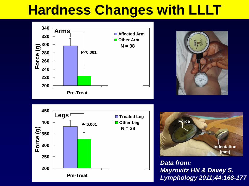
Hardness Changes with LLLT
P<0.001
P<0.001
N = 38
Data from:
Mayrovitz HN & Davey S.
Lymphology 2011;44:168-177
Force
Indentation
(mm)
Arms
Legs
N = 38

200
220
240
260
280
300
320
340
Pre-Treat Post LLLT
Fo
rce (
g)
Affected Arm
Other Arm
200
250
300
350
400
450
Pre-Treat Post LLLT
Fo
rce (
g)
Treated Leg
Other Leg
P<0.001
P<0.001
N = 38
P<0.001
P<0.001
Hardness Changes with LLLT
Data from:
Mayrovitz HN & Davey S.
Lymphology 2011;44:168-177
Force
Indentation
(mm)
Arms
Legs

Commercial Tonometers
Force
Applied
Displacement
Determined
Pallota O. J Lymphoedema 2011;6:34-41

Modified from: Mellor et al. The Breast J. 2004;10:496-503
Gel
Entry
Echo
Dermis
Sub
cutis
0.93 ± 0.13
Normal Lymphedematous
Ventral Forearm US-20 MHz
1.83 ± 1.28
Methods Applicable to MOST Sites
Imaging Ultrasound MRI Other
mm

Metric Measures for LIMBS

Metric Measures for LIMBS
Tape Measure Girth at multiple points
• Measure both limbs
Inter-limb differentials and sequential changes
Mark then Measure
Segment Length
• Measure one limb
Sequential data but miss systemic changes

Limb Girth Volume
Manual
Geometric Model
or Algorithm
Circumferences
@ 4 – 12 cm
intervals
0.95
1
1.05
1.1
1.15
0.4 0.5 0.6 0.7 0.8 0.9 1
= ratio of smaller to larger dimension
Cir
cu
lar
to e
llip
tic
al
vo
lum
e r
ati
o (
VC
/ V
E)
circular
VC/VE =(1/4) (1+ )3 /(1+)
A
B
a
b
L
C1
C2
General FrustumCalculation
Model
<5% difference for ratios > ≈ 0.6 So OK for most Arms & Legs
BUT Not OK for Hands or Feet
Dr HN Mayrovitz
Effect of Degree of Eccentricity
V =L(A2B-a2b)
3(A-a)
Circular/EllipticalVolume Ratio
Volume Tracking
Affected Limb
Contralateral Limb
Edema Volume
V = L/3 ( A1 + A2 + (A1A2)1/2
A1
A2

Foot-Plate
Rail
Frame
Perometer: Girth Volume
IR d
iod
es
Mayrovitz HN et al. Advances in Wound Care 2000;113:272-276

D1
IR
Diode
Array
D2
Area = KD1D2
Frame
Perometer: Basic Principle

Limb Girth & Volume LE Thresholds
Automated
Manual
GIRTH
If unilateral then lymphedema if
• inter-side differential > C1 cm or
if unilateral or bilateral then
• change from pre-surgery > C2 cm
VOLUME
If unilateral then lymphedema if
• inter-side differential > V1 ml or
• inter-side ratio > g
if unilateral or bilateral then
• change from pre-surgery > V2 ml

0
20
40
60
80
100
12 months 30 months
Lym
ph
ed
em
a R
ate
(%
)
2 cm
200 ml
10% volume
Arm Lymphedema Metric Criteria
Differences • Between sides
• or vs. baseline
Data from: Armer et al. J. Lymphoedema 2009;4:14-18
LE rate dependent on criteria used

Practical Aspects of Limb Girth

Mark in Relation To FLAT Surface
NOT along limb
Source of largeFollow-up error
Minimizing Method Error
Dr HN Mayrovitz
For Reproducibility: Mark along flat

What Segment Length to Use?

Leg Volume Measured (ml) Volume Reduction
Segment Length Pre-Treatment Post-Treatment (ml) (%)
4 cm 6649 ± 2482* 5465 ± 1969* 1183 ± 778 17.2 ± 7.1
8 cm 6676 ± 2497 5496 ± 1990 1180 ± 782 17.1 ± 7.2
12 cm 6756 ± 2510 5554 ± 2001 1202 ± 781 17.4 ± 7.0
Segment
Length
4 cm
8 cm
12 cm
4000
5000
6000
7000
8000
Leg
Vo
lum
e (
ml)
4 cm 8 cm 12 cm
Pre-treatment Volumes
4000
5000
6000
7000
8000
Leg
Vo
lum
e (
ml)
4 cm 8 cm 12 cm
Post-treatment Volumes
Bilateral lower
extremity lymphedema
> = 10 MLD Tx
What Segment Length to Use?
N = 70
Mayrovitz et al. Physical Therapy 2007; 87: 1362-1368

Limb Shape as a Factor

If you calculate
on the basis of
THIS
and its really more like
THIS
Then you obtain a volume
greater than the true value
Limb Shape as a Factor

0.0
0.2
0.4
0.6
0.8
1.0
1.2
0.0 0.1 0.2 0.3 0.4 0.5 0.6 0.7 0.8 0.9 1.0 1.1 1.2
Ratio (b/a)
Vo
lum
e R
ati
o (
Ve
/Vc
)Limb Shape as a Factor
0.95
1
1.05
1.1
1.15
0.4 0.5 0.6 0.7 0.8 0.9 1
= ratio of smaller to larger dimension
Cir
cu
lar
to e
llip
tic
al
vo
lum
e r
ati
o (
VC
/ V
E)
circular
VC/VE =(1/4) (1+ )3 /(1+)
A
B
a
b
L
C1
C2
General FrustumCalculation
Model
<5% difference for ratios > ≈ 0.6 So OK for most Arms & Legs
BUT Not OK for Hands or Feet
Dr HN Mayrovitz
Effect of Degree of Eccentricity
V =L(A2B-a2b)
3(A-a)
Circular/EllipticalVolume Ratio
C1 = 31.4 cm
C2 = 28.3 cm
L =10 cm
@ b/a 0.68
6.2% volume
deviation
Data from: Mayrovitz HN, Lymphology 2003;31:140-143
Ve
Vc
Ve = 0.938 Vc

Limb & Limb Segment Volumes

Photo from: K. Johansson & E Branje Acta Oncologica 2010;49:166-173
Arm lymphoedema in a cohort of breast cancer survivors 10 years after diagnosis
LE if change in edema volume >= 5% from pre-surgery
Volumes via H2O Displacement
Mostly used
as a so called
gold standard
when comparing
other methods and
in research studies

0
1000
2000
3000
4000
0 1000 2000 3000 4000
Given LAV predict RAV
Line of Identity
95% +Interval
Left Arm Volume (LAV, ml by H2O)
Rig
ht
Arm
Vo
lum
e (
RA
V,
ml)
Normal Arm Volume Differentials
Data from Gebruers N et al . Clin Physiol Funct Imaging 2007; 27:17-22
Line of
Identity
95% +CI
100 RH Females
RAV > LAV (2%)

0
50
100
150
200
250
300
ml
0-10 10 20 20-30 30-40 ARM
Arm Segment (cm)
Dom
NoDom
Normal Arm Volume Differentials
At-risk Arm Is:
Dominant
Non-Dominant
3S
D T
hre
sh
old
s (
ml) If dominant = at-risk
Then
Greater Threshold
Data from: Dylke ES et al Lymphatic Res Biology 2012;10:182-188
N =204
Healthy
Women
Age > 40
Girths via Perometer Volumes via frustum calculation

Hand Volume
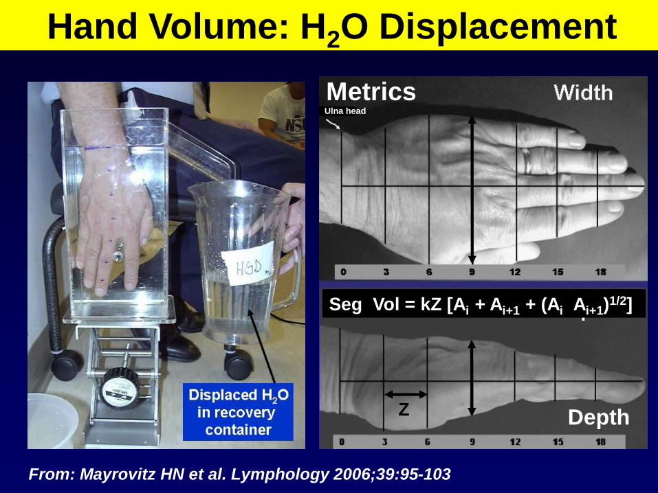
Hand Volume: H2O Displacement
Ulna head
Z
Seg Vol = kZ [Ai + Ai+1 + (Ai Ai+1)1/2]
Depth
From: Mayrovitz HN et al. Lymphology 2006;39:95-103
Metrics

200
250
300
350
400
450
500
550
600
200 250 300 350 400 450 500 550 600
VM = 1.02 Vw – 12.0 ml
r=0.985, p<0.001
N=60 Hands
Algorithm vs. Water Displacement
Volume by water displacement (VW, ml)
Vo
lum
e b
y A
lgo
rith
m
(VM
, m
l)
LOA
± 9.8%

200
250
300
350
400
450
500
550
with arm LE (n=20) no arm LE (n=20)
Han
d V
olu
me (
ml) Water
Perometer
Hand Volume: H2O vs. Perometer
Data from: Lee MJ et al. Lymphatic Research Biology 2011;9:13-18
Perometer values ~ 7.5%
greater than H2O values
r ~ 0.88

200
250
300
350
400
450
500
550
with arm LE (n=20) no arm LE (n=20)
Han
d V
olu
me (
ml) Water
Perometer
Hand Volume: H2O vs. Perometer
Data from: Lee MJ et al. Lymphatic Research Biology 2011;9:13-18
Perometer values ~ 7.5%
greater than H2O values
r ~ 0.88

Figure-of-Eight: Hand volume Surrogate
Pellecchia GL J Hand Therapy 2003;16:300-304
Maihafer GC J Hand Therapy 2003;16:305-310
cm (fig-8) vs. H2O displacement (ml)
R = 0.94-0.95 but only normal hands
Tracking ability unproven

Foot Volume

Water
Displacement
Foot Volume: H2O Displacement
4
8
12 cm
0
12
L1
Y
X
A
B
C
D E F
Foot in Water Filled Volumeter
Outflow Tube
L2
Mayrovitz HN et al. Lymphology 2005;38:20-27

Water
DisplacementCompared to
Metric
Measures
Foot Volume: H2O Displacement
4
8
12 cm
0
12
L1
Y
X
A
B
C
D E F
Foot in Water Filled Volumeter
Outflow Tube
L2
Mayrovitz HN et al. Lymphology 2005;38:20-27

Algorithm vs. Water Displacement
N = 60 feet
LOA = ± 9.3%
Mayrovitz HN et al. Lymphology 2005;38:20-27

Water
Displacement
PRO CON
• Direct – Accurate
Limb/Hand/Foot volumes
• Especially for irregularly
shaped limbs
• Impractical for whole limbs
• Bulky equipment
• sterilization procedures
• Patient mobility
• Patient flexibility
• Open wounds
Manual Girth • Low cost
• Portable
• Easy to use
• Whole legs measureable
• Hand & Foot algorithms
• Limited ROM no issue
• Wounds are not an issue
• Multiple measurements
• Time factor
• Volumes from calculations
• Site repeatability
Optoelectronic
(Perometer)• Quick –Easy
• Small segment lengths
• Stored Measurements
• Automatic processing
• Selective processing
• Accuracy depends on
proper positioning
• Patient mobility
• Patient flexibility
• Not portable
• Space requirements
• $$$

Bioimpedance Analysis
• Electrical Impedance of a limb depends
on the limb’s volume and constituents
• Lymphedema increase in low resistance
fluid content of the limb
•Bioimpedance (BIOZ)
•Bioimpedance Spectroscopy (BIS)
•Bioimpedance Analysis (BIA)
•Single Frequency BIA = SFBIA
•Multi-Frequency BIA = MFBIA

Muscle
Bone
Skin
Fat5-10%H2O 70-75% H2O
15-20% H2O
Limb Conducting Structures
1
20
2
Conductivity
@ 5KHz
Relative to Bone
Bone
Muscle
Fat

Basic Operating Principle
No cells
Just salineE
I
R = E/I

No cells
Just salineE
I
R = E/I
Cell
Membrane
•Polarized
•Charge Separation
•Electrical Capacitance
Basic Operating Principle

No cells
Just salineE
I
~
Z = E/I
Low
Frequency
High
Frequency
Sinusoidal
Voltage
ExcitationCurrent increases
with frequency
Basic Operating Principle

Cell
Membrane
Cell Interior
Cm
Ri
Re
Frequency Analysis Basis
Cell Exterior
~E
I
Z = E / I
ECW
ICW

0.000000
0.050000
0.100000
0.150000
0.200000
0.250000
0.300000
0.400000 0.500000 0.600000 0.700000 0.800000 0.900000 1.000000 1.100000
Cole-Cole Plot: estimate parameters
increasing
frequency
ReRiRe/(Ri + Re)
Cell
Membrane
Cell Interior
Cm
Ri
Re
Cell Exterior
~E
I
Z = E / IECW
ICW
Low fHigh f
ECW + ICW ECW
MFBIA = BIS
R∞ R0

Current Injecting Electrodes
Voltage Measuring
Electrodes
I
Basic Operating Principle
Mayrovitz HN Clinical Physiology 1998;18:234-242
Z = E/I
E

Leg Volumes: Supine StandL
eg
Vo
lum
e (
ml)
Time (minutes)
Blood volume shift to lower extremities
Supine Standing Supine
-20 -10 0 10 20 30 40 50
2100
2000
1900
1800
Girth-Volume
Measurements

Z Depends on Frequency & Volume
Time (minutes)
|Z|
(W)
Supine Standing Supine
-20 -10 0 10 20 30 40 50
130
120
110
100
090
080
070
Z ~ (1 / volume)
Z ~ (1 / f)
5 KHz
500 KHz

Assessing Arm Lymphedema

~
V
I
Z = V / I
Assessing Arm Lymphedema
Current
Source
Measured
Voltage
Current

Single Frequency BIA ECW

R∞
R0
Ri
Ri
R0
R0DOM
R0NONDOM
360.1± 45.8 354.8 ± 45.9
266.5 ± 39.2 257.8 ± 39.4
1052.3 ± 276.2 966.7 ± 264.9
2.988 ± 0.653 2.781 ± 0.595
0.986 ± 0.040
Multi-Frequency BIA
Nondomnant Dominant
Data from: Ward LC et al. Lymphatic Research Biology 2011;9:47-51
172 paired arms
3SD
lymphedema
thresholds
nondom/dom
dom = at-risk
1.134
nondom = at-risk
1.106

SFBIA = MFBIA for estimating ECW
York SL et al. Breast Cancer Res Treat 2009;117:177-182
Inter-Limb Z Ratios
Arm LEArm controlsLeg LE
Single Frequency Bio-impedance (BIA)
Mu
ltip
le F
req
ue
nc
y B
io-i
mp
ed
an
ce
(B
IS) 2.4
2.2
2.0
1.8
0.8 1.2 1.6 2.0 2.4
Both estimate
Re (Low f)

So …. Why use MFBIA (BIS)?
ECW
ICW
• If ICW relatively unchanged even with LE then
may not have to depend on inter-arm ratios
• May be approximately true if muscle mass
does not significantly change since the largest
fraction of ICW is associated with muscle
Proposed Concept

0
1
2
3
4
5
6
Normal Arm At-Risk Arm
EC
W /
IC
W
Pre-Surgery
Lymphedema Dx
ECW / ICW Ratios
Data from: Cornish BH et al. Angiology 2002;53:41-47
N = 20

Tissue Dielectric Constant (TDC)
Relative Permittivity (er)
PRINCIPLE
Local Tissue Water Assessment

H
H
O-
+ +
-
+
+Dipole
What is Dielectric Constant?
H2O Molecule Charge Separation

What is Dielectric Constant?
H
H
O-
+
+
+
H
H
O-
+
+
+
+
-
+
-
Hydrogen bonding
between water
molecules
2 molecules
“Hook-up”

What is Dielectric Constant?
+
-
Time varying
electric field
of force - E
Dipole movement
Displacement - D
of various types
D = e E
Dielectric
Constant
= ere0E
er = ratio e/e0 = TDC
H2O @ 32oC er = 76

• Multi-Probe
• Single Probe (compact)
Measurement Devices

0.5 1.5 2.5 5.0 mm
Effective Measurement Depth
Signal Generation
and Processing
300 MHz
Signal
Reflected Wave yields TDC
Control Unit
10 mm 15 mm 22 mm
Effective Measurement Depths0.5 mm 1.5 mm 2.5 mm
Outer Diameters
Tissue Water via Dielectric Constant
MoistureMeter-D
• Low power 300 MHz
incident wave
• Reflected wave depends
on the tissue’s
dielectric constant
• Dielectric constant
depends on total tissue
water (free + bound)
• Pure water has a
dielectric constant of
about 78
• Calibrated for each
probe from 1 - 80
Penetration Depth (0.5 – 5 mm)
0.5 1.5 2.5 5.0 mm
TDC
readout
Multi-Probe

20 mm
Display has pressure
bar indicator during
measurement
Effective
measurement
depth is
between
1.5 & 2.5 mm
Multi-Probe
Artery
Nerve
Capillary
Duct
Epidermis
Dermis
Arteriole
Hair
Pore
Fat
Hypo-Dermis
Single Probe (Compact)
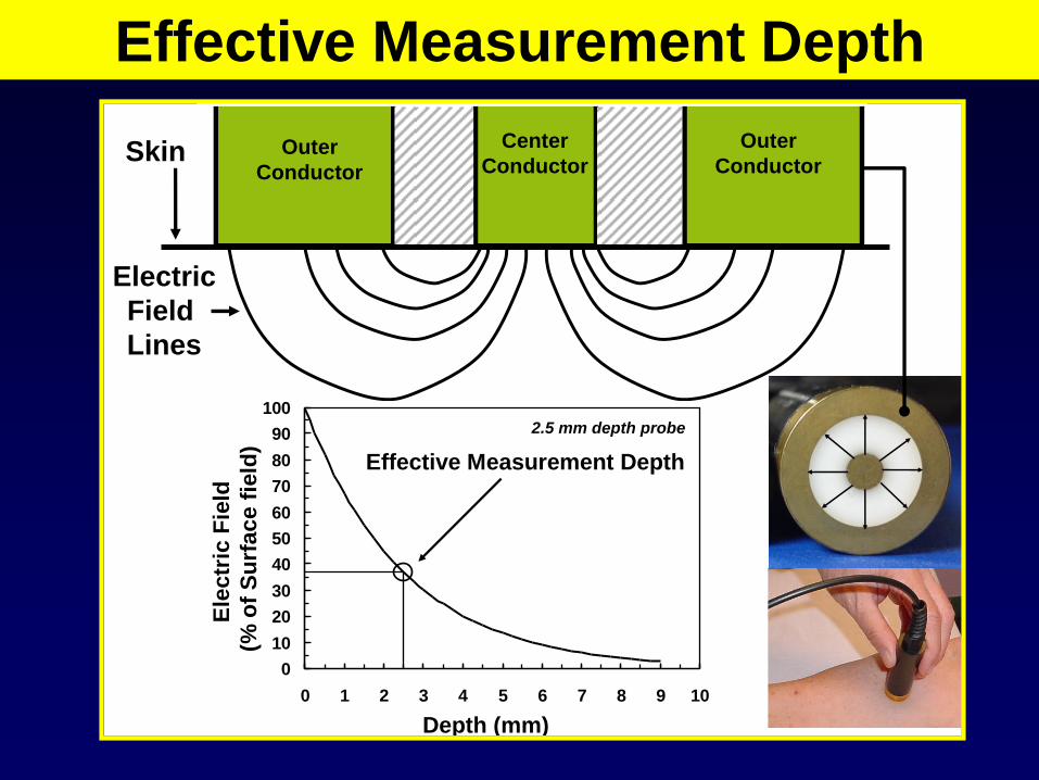
Center
ConductorOuter
Conductor
Outer
Conductor
Electric
Field
Lines
0
10
20
30
40
50
60
70
80
90
100
0 1 2 3 4 5 6 7 8 9 10
Ele
ctr
ic F
ield
(% o
f S
urf
ace f
ield
)
Depth (mm)
Effective Measurement Depth
2.5 mm depth probe
Tissue Water via Dielectric Constant
MoistureMeter-D
• Low power 300 MHz
incident wave
• Reflected wave depends
on the tissue’s
dielectric constant
• Dielectric constant
depends on total tissue
water (free + bound)
• Pure water has a
dielectric constant of
about 78
• Calibrated for each
probe from 1 - 80
Penetration Depth (0.5 – 5 mm)
0.5 1.5 2.5 5.0 mm
Skin
Effective Measurement Depth

Gel
Entry
Echo
Dermis
Sub
cutis
0.93 ± 0.13
mm
Low water
content
High water
content
Normal Lymphedematous
Ventral Forearm
Interrogation
Depth (2.5 mm)
1.83 ± 1.28
mm
5.0
mm1.5
mm
subcutis
skin
Modified from
Mellor et al.
The Breast J.
2004;10:496-503
Effective Measurement Depth

0
10
20
30
40
50
60
70
0 5 10 15 20 25 30 35 40 45 50 55 60 65 70 75
TD
C
Calibration Example
(2.5 mm probe)
Water (%)
Y = 0.632 X – 22.1
r2 = 0.998, p < 0.001Ethanol-Water
Mixture
Probe inserted
Into Mixture
Cable to
Control
Unit
TDC dependence on H2O
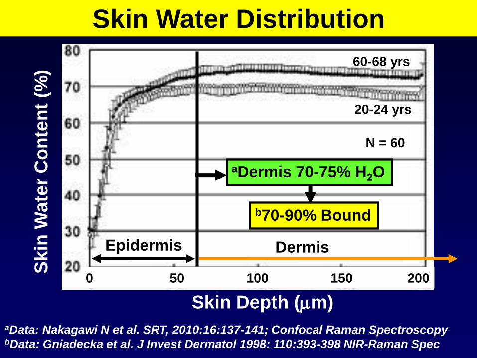
Skin Water Distribution
Skin Depth (mm)
Skin
Wate
r C
on
ten
t (%
)
aData: Nakagawi N et al. SRT, 2010:16:137-141; Confocal Raman Spectroscopy bData: Gniadecka et al. J Invest Dermatol 1998: 110:393-398 NIR-Raman Spec
20-24 yrs
60-68 yrs
aDermis 70-75% H2O
N = 60
b70-90% Bound
0 50 100 150 200
Epidermis Dermis

Free and Bound Water
Protein
(1 g)
Bound H2O
(0.2 – 0.5 g)
Limited
Mobility H2O
~ 20 g
Free Mobile
Free MobileFree Mobile

Fluid
Protein
Blood
Vessel
Lymphatic Dysfunction
Lymph
Vessel
XBound and immobile
water not readily
measureable with
Standard BIA
Interstitium

Data: Idy-Peretti et al. Int J Dermatol 1998;110:782-787 Hi-Res MRI N=21
Mobile water shows intense
Dermal Water in Lymphedema
1 mm 1 mm
Contralateral Leg Lymphedema calf
40% increase
in Calf
Dermal
Water in
Lymphedema
11 primary LE
10 secondary LE

TDC Features and Applications
Forearm
Biceps
Signal
Generation
and
Processing
TDC
Value
Display
Foot Dorsum
TDC measurement

20
25
30
35
40
Fo
reh
ead
Ch
eek
Th
um
b
A.
Fo
rea
rm
D. F
ore
arm
Pa
lm-T
he
nar
Pa
lm-C
en
ter
D. H
an
d C
ntr
1s
t T
oe
Pla
nta
r
A.
Gait
er
1stT
oe
Do
rsu
m
M.
Ga
iter
L.
Ga
iter
D. H
an
d W
eb
D.
Fo
ot
(1-2
)
D.
Fo
ot
(4=
5)
M. M
alleo
lus
TDC @ 1.5 mmTDC Site Variability
32 females
19 - 77 years
1.5 mm Depth
Data From: Mayrovitz HN et al. Skin Research and Technology 2013;19:47–54
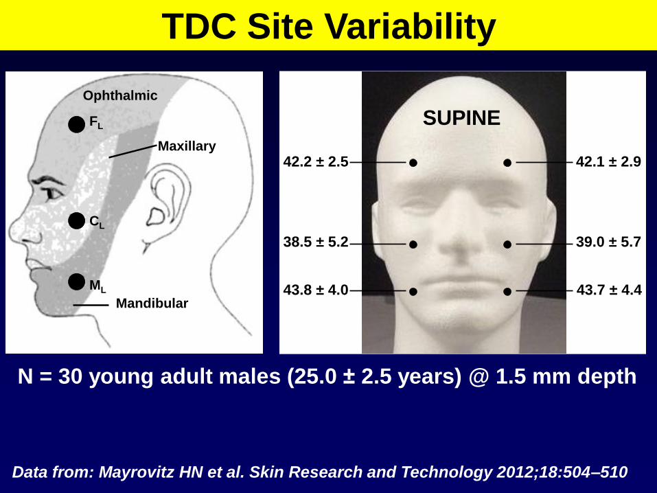
FL
CL
ML
Ophthalmic
Maxillary
Mandibular
42.1 ± 2.942.2 ± 2.5
SUPINE
39.0 ± 5.738.5 ± 5.2
43.7 ± 4.443.8 ± 4.0
N = 30 young adult males (25.0 ± 2.5 years) @ 1.5 mm depth
TDC Site Variability
Data from: Mayrovitz HN et al. Skin Research and Technology 2012;18:504–510

0
10
20
30
40
50
60
35 40 45 50 55 60 65 70 75
Total Body Water (%)
Fo
rea
rm T
DC
@ 5
.0 m
m D
ep
th
N = 130 (50 females)
Age 26.1 ± 3.0 (19-39)
BMI 24.5 ± 4.0 (16-40)
Y = 0.929 X – 22.3
r = 0.740, p < 0.001
Correlation with Total Body Water

20
22
24
26
28
30
32
34
36
38
40
0.0 0.5 1.0 1.5 2.0 2.5 3.0 3.5 4.0 4.5 5.0 5.5
Measurement Depth , mm)
TDC = 32.44 -0.185
r2 = 0.997, p<0.001
N = 80 females
TDC Depth Dependence: Forearm
TDCPattern of Depth Dependence
May Vary by Site0.5 mm
1.5 mm
2.5 mm
5.0 mm
Compact

TDC Vascular Component
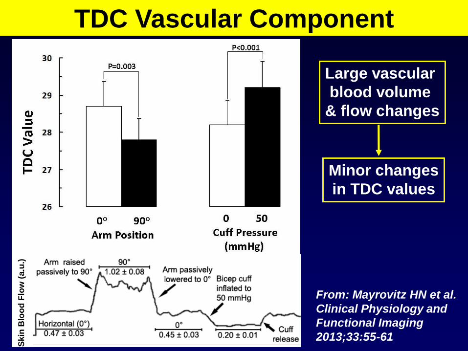
Skin
Blo
od
Flo
w (
a.u
.)TDC Vascular Component
From: Mayrovitz HN et al.
Clinical Physiology and
Functional Imaging
2013;33:55-61
Large vascular
blood volume
& flow changes
Minor changes
in TDC values

0.6
1.0
1.4
1.8
2.2T
DC
in
ter-
arm
rati
o
PatientsAffected/Control
1.64 ± 0.30
N=18
Premenopausal Postmenopausal1.04 ± 0.04 1.04 ± 0.04
N=15 N=15
Mayrovitz HN Lymphology 2007;40:87-94
TDC Lymphedema Discriminations
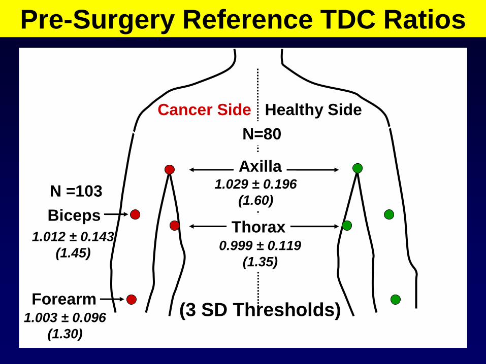
Pre-Surgery Reference TDC Ratios
Cancer Side Healthy Side
Axilla
Biceps
Forearm
1.029 ± 0.196
(1.60)
1.012 ± 0.143
(1.45)
1.003 ± 0.096
(1.30)
N =103
N=80
Thorax0.999 ± 0.119
(1.35)
(3 SD Thresholds)

0.900
0.950
1.000
1.050
1.100
1.150
1.200
pre-surgery 3 6 12 18 24
Th
ora
x T
DC
Ra
tio
(A
t-R
isk
/Co
ntr
ol
Sid
e)
0-3-6-12-18-24 month (N=35)
0-3-6-12-18 month (N=41)
0-3-6-9-12 month (N=47)
0-3-6 month (N=53)
0-3 month (N=60)
**
**** **
** *** * ****
Sequential TDC Ratio Changes
Lateral
Thorax
0.900
0.950
1.000
1.050
1.100
1.150
1.200
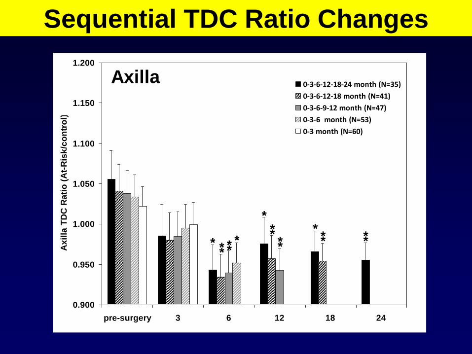
pre-surgery 3 6 12 18 24
Ax
illa
TD
C R
ati
o (
At-
Ris
k/c
on
tro
l)
0-3-6-12-18-24 month (N=35)
0-3-6-12-18 month (N=41)
0-3-6-9-12 month (N=47)
0-3-6 month (N=53)
0-3 month (N=60)
* *****
*
**
** ** ***
Sequential TDC Ratio Changes
Axilla

TDC BIA/BIS (Delfin Technologies Ltd) (Impedimed Ltd) Operating principle
Frequency applied EMF 300 MHz 4 - 1000 kHz
Current flowing in the body Very Localized Much of the body
Number of electrodes / probes 1 probe 4 electrodes
Total single measurement time ~ 8 sec ~ 60 sec
Measurement Depth 0.5 – 5 mm Undefined
Measurement quantity Tissue dielectric constant Resistance
Measurement parameter Skin-to-fat tissue fluid Parameter ~ to ECF
Applicability Practically all body sites Limbs
Patient preparation
Patient position Any body position Supine
Arm-leg skin contact No effect Limbs must be abducted
Arm and hand position No restriction Palms flat on surface
Shoe and socks removal Not needed to remove Must be removed
Bladder emptying necessary No Yes
Dominant side affects No Yes
Measurement sites
Hairy skin shaving Yes (very hairy) Yes
Precautions for measurement
Patient metal contact problem No Yes
Methods Features Comparison

Related Documents


