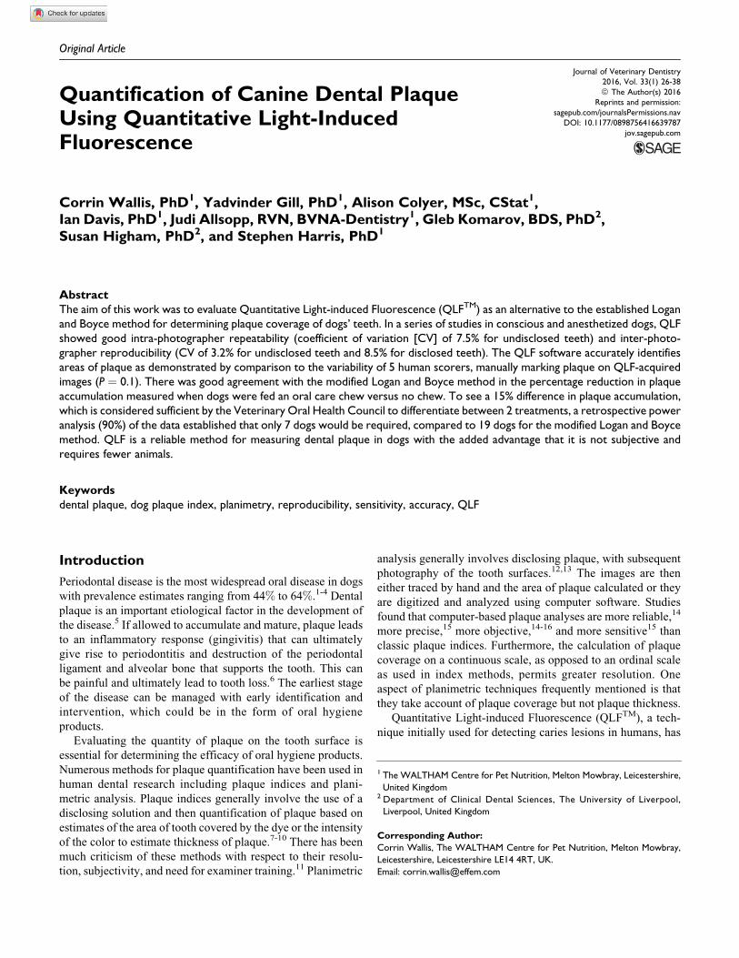
Original Article Quantification of Canine Dental Plaque Using Quantitative Light-Induced Fluorescence Corrin Wallis, PhD 1 , Yadvinder Gill, PhD 1 , Alison Colyer, MSc, CStat 1 , Ian Davis, PhD 1 , Judi Allsopp, RVN, BVNA-Dentistry 1 , Gleb Komarov, BDS, PhD 2 , Susan Higham, PhD 2 , and Stephen Harris, PhD 1 Abstract The aim of this work was to evaluate Quantitative Light-induced Fluorescence (QLF TM ) as an alternative to the established Logan and Boyce method for determining plaque coverage of dogs’ teeth. In a series of studies in conscious and anesthetized dogs, QLF showed good intra-photographer repeatability (coefficient of variation [CV] of 7.5% for undisclosed teeth) and inter-photo- grapher reproducibility (CV of 3.2% for undisclosed teeth and 8.5% for disclosed teeth). The QLF software accurately identifies areas of plaque as demonstrated by comparison to the variability of 5 human scorers, manually marking plaque on QLF-acquired images (P ¼ 0.1). There was good agreement with the modified Logan and Boyce method in the percentage reduction in plaque accumulation measured when dogs were fed an oral care chew versus no chew. To see a 15% difference in plaque accumulation, which is considered sufficient by the Veterinary Oral Health Council to differentiate between 2 treatments, a retrospective power analysis (90%) of the data established that only 7 dogs would be required, compared to 19 dogs for the modified Logan and Boyce method. QLF is a reliable method for measuring dental plaque in dogs with the added advantage that it is not subjective and requires fewer animals. Keywords dental plaque, dog plaque index, planimetry, reproducibility, sensitivity, accuracy, QLF Introduction Periodontal disease is the most widespread oral disease in dogs with prevalence estimates ranging from 44% to 64%. 1-4 Dental plaque is an important etiological factor in the development of the disease. 5 If allowed to accumulate and mature, plaque leads to an inflammatory response (gingivitis) that can ultimately give rise to periodontitis and destruction of the periodontal ligament and alveolar bone that supports the tooth. This can be painful and ultimately lead to tooth loss. 6 The earliest stage of the disease can be managed with early identification and intervention, which could be in the form of oral hygiene products. Evaluating the quantity of plaque on the tooth surface is essential for determining the efficacy of oral hygiene products. Numerous methods for plaque quantification have been used in human dental research including plaque indices and plani- metric analysis. Plaque indices generally involve the use of a disclosing solution and then quantification of plaque based on estimates of the area of tooth covered by the dye or the intensity of the color to estimate thickness of plaque. 7-10 There has been much criticism of these methods with respect to their resolu- tion, subjectivity, and need for examiner training. 11 Planimetric analysis generally involves disclosing plaque, with subsequent photography of the tooth surfaces. 12,13 The images are then either traced by hand and the area of plaque calculated or they are digitized and analyzed using computer software. Studies found that computer-based plaque analyses are more reliable, 14 more precise, 15 more objective, 14-16 and more sensitive 15 than classic plaque indices. Furthermore, the calculation of plaque coverage on a continuous scale, as opposed to an ordinal scale as used in index methods, permits greater resolution. One aspect of planimetric techniques frequently mentioned is that they take account of plaque coverage but not plaque thickness. Quantitative Light-induced Fluorescence (QLF TM ), a tech- nique initially used for detecting caries lesions in humans, has 1 The WALTHAM Centre for Pet Nutrition, Melton Mowbray, Leicestershire, United Kingdom 2 Department of Clinical Dental Sciences, The University of Liverpool, Liverpool, United Kingdom Corresponding Author: Corrin Wallis, The WALTHAM Centre for Pet Nutrition, Melton Mowbray, Leicestershire, Leicestershire LE14 4RT, UK. Email: [email protected] Journal of Veterinary Dentistry 2016, Vol. 33(1) 26-38 ª The Author(s) 2016 Reprints and permission: sagepub.com/journalsPermissions.nav DOI: 10.1177/0898756416639787 jov.sagepub.com

Welcome message from author
This document is posted to help you gain knowledge. Please leave a comment to let me know what you think about it! Share it to your friends and learn new things together.
Transcript
Original Article
Quantification of Canine Dental PlaqueUsing Quantitative Light-InducedFluorescence
Corrin Wallis, PhD1, Yadvinder Gill, PhD1, Alison Colyer, MSc, CStat1,Ian Davis, PhD1, Judi Allsopp, RVN, BVNA-Dentistry1, Gleb Komarov, BDS, PhD2,Susan Higham, PhD2, and Stephen Harris, PhD1
AbstractThe aim of this work was to evaluate Quantitative Light-induced Fluorescence (QLFTM) as an alternative to the established Loganand Boyce method for determining plaque coverage of dogs’ teeth. In a series of studies in conscious and anesthetized dogs, QLFshowed good intra-photographer repeatability (coefficient of variation [CV] of 7.5% for undisclosed teeth) and inter-photo-grapher reproducibility (CV of 3.2% for undisclosed teeth and 8.5% for disclosed teeth). The QLF software accurately identifiesareas of plaque as demonstrated by comparison to the variability of 5 human scorers, manually marking plaque on QLF-acquiredimages (P ¼ 0.1). There was good agreement with the modified Logan and Boyce method in the percentage reduction in plaqueaccumulation measured when dogs were fed an oral care chew versus no chew. To see a 15% difference in plaque accumulation,which is considered sufficient by the Veterinary Oral Health Council to differentiate between 2 treatments, a retrospective poweranalysis (90%) of the data established that only 7 dogs would be required, compared to 19 dogs for the modified Logan and Boycemethod. QLF is a reliable method for measuring dental plaque in dogs with the added advantage that it is not subjective andrequires fewer animals.
Keywordsdental plaque, dog plaque index, planimetry, reproducibility, sensitivity, accuracy, QLF
Introduction
Periodontal disease is the most widespread oral disease in dogs
with prevalence estimates ranging from 44% to 64%.1-4 Dental
plaque is an important etiological factor in the development of
the disease.5 If allowed to accumulate and mature, plaque leads
to an inflammatory response (gingivitis) that can ultimately
give rise to periodontitis and destruction of the periodontal
ligament and alveolar bone that supports the tooth. This can
be painful and ultimately lead to tooth loss.6 The earliest stage
of the disease can be managed with early identification and
intervention, which could be in the form of oral hygiene
products.
Evaluating the quantity of plaque on the tooth surface is
essential for determining the efficacy of oral hygiene products.
Numerous methods for plaque quantification have been used in
human dental research including plaque indices and plani-
metric analysis. Plaque indices generally involve the use of a
disclosing solution and then quantification of plaque based on
estimates of the area of tooth covered by the dye or the intensity
of the color to estimate thickness of plaque.7-10 There has been
much criticism of these methods with respect to their resolu-
tion, subjectivity, and need for examiner training.11 Planimetric
analysis generally involves disclosing plaque, with subsequent
photography of the tooth surfaces.12,13 The images are then
either traced by hand and the area of plaque calculated or they
are digitized and analyzed using computer software. Studies
found that computer-based plaque analyses are more reliable,14
more precise,15 more objective,14-16 and more sensitive15 than
classic plaque indices. Furthermore, the calculation of plaque
coverage on a continuous scale, as opposed to an ordinal scale
as used in index methods, permits greater resolution. One
aspect of planimetric techniques frequently mentioned is that
they take account of plaque coverage but not plaque thickness.
Quantitative Light-induced Fluorescence (QLFTM), a tech-
nique initially used for detecting caries lesions in humans, has
1 The WALTHAM Centre for Pet Nutrition, Melton Mowbray, Leicestershire,
United Kingdom2 Department of Clinical Dental Sciences, The University of Liverpool,
Liverpool, United Kingdom
Corresponding Author:
Corrin Wallis, The WALTHAM Centre for Pet Nutrition, Melton Mowbray,
Leicestershire, Leicestershire LE14 4RT, UK.
Email: [email protected]
Journal of Veterinary Dentistry2016, Vol. 33(1) 26-38ª The Author(s) 2016
Reprints and permission:sagepub.com/journalsPermissions.nav
DOI: 10.1177/0898756416639787jov.sagepub.com
also been employed to detect dental plaque.11,17-19 This method
either relies on the natural fluorescence of plaque under blue
light (405 nm) or uses a standard disclosing solution to enhance
bacterial fluorescence. The images are captured in real time
using a modified version of a standard SLR camera, and image
analysis software is then used to quantify the amount of plaque.
The advantages and disadvantages of this technique are similar
to other planimetric methods, but there is the additional major
advantage that the greater contrast between the gingiva and the
tooth, which is a feature of this technique, circumvents the need
to manually define the tooth area accurately. This difference
reduces the analysis time considerably and potentially
increases accuracy when determining the plaque coverage of
the tooth surface.
Several techniques for the quantification of plaque have been
developed for use in cats and dogs. Routine methods include the
modified Logan and Boyce plaque index which is used to quan-
tify plaque accumulation on the buccal surface of the whole
tooth20 and the gingival contour plaque index (GCPI), which
focuses on plaque that accumulates along the buccal gingival
margin.21,22 Both of these methods have been endorsed by the
Veterinary Oral Health Council (VOHC; www.vohc.org) for
supporting product claims relating to plaque control. In vivo
product efficacy trials require a clean mouth model, where cats
or dogs are anesthetized at the start of the study and at the end of
each test phase so the teeth may be scaled and polished. Alter-
native methods that reduce the number of anesthetic procedures
per animal, decrease the duration of anesthesia, reduce subjectiv-
ity, and improve accuracy (thereby reducing the number of ani-
mals required) are desirable to reduce the impact of the testing
procedure on the animals involved. To our knowledge, the more
reliable, objective, and sensitive planimetric methods such as
QLF have not been described for use in dogs. Therefore, the aims
of these studies were to evaluate the repeatability, reproducibility
and accuracy of QLF for quantification of canine dental plaque
and to compare this to an established clinical scoring system,
namely, the modified Logan and Boyce20 method.
Materials and Methods
The dogs included in the studies detailed subsequently were
pair housed at the WALTHAM Centre for Pet Nutrition in
environmentally enriched kennels and provided with a compre-
hensive dog–dog and dog–human socialization program
adjusted to the needs of individual dogs. All dogs received a
pre-study veterinary examination to ensure suitability for trial,
which included a physical examination and an assessment of
the dog’s veterinary history. The studies were approved by the
WALTHAM Animal Welfare and Ethical Review Body and
run under licensed authority in accordance with the UK Ani-
mals (Scientific Procedures) Act 1986.
Intra-photographer Repeatability—Undisclosed Teeth
Eleven miniature schnauzer dogs, aged between 2.5 and 6.9
years (6 females and 5 males, weight range 7.0-10.2 kg), which
had received a recent scaling and polishing and had little or no
visible calculus, were recruited to the study. Dogs’ teeth were
brushed daily for approximately 1 week prior to the start of the
trial using compact medium or soft brushes (TePe Oral
Hygiene Products Ltd, Bronsaldersgatan 5213 76 Malmo, Swe-
den) and water. Dogs received no subsequent tooth brushing for
21 days when images of undisclosed teeth were captured using
the commercially available QLF-D Biluminator 2 system
(Inspektor Research Systems, Amsterdam, Netherlands; see
section on QLF image acquisition and analysis for further
details). Three repeated sets of images, 2 in the morning and
1 in the afternoon, were taken of conscious dogs by a single
photographer. A set of images comprised 4 views around the
mouth, 2 images on both the left and right hand side of the
dog’s mouth, were taken to visualize the maxillary first pre-
molars (P1; 105, 205), second premolars (P2; 106, 206), third
premolars (P3; 107, 207), and fourth premolars (P4; 108, 208).
Inter-photographer Reproducibility: Undisclosed Teeth
Twelve miniature schnauzer dogs, aged 3.1 to 7.5 years (7
females and 5 males, weight range 7.5-10.6 kg), that had their
teeth brushed every other day as part of their normal oral care
regimen from 1 year of age were allocated to 1 of the 3 groups
based on time since their teeth were previously brushed. The
purpose of this was to ensure that the reproducibility of QLF
was assessed across the whole of the plaque coverage range:
Group A had their tooth brushing stopped 21 days prior to
examination, group B 10 days before the examination, and
group C was tooth brushed the day before their examination.
Three dogs, one from each group, were allocated to 1 of 4
consecutive assessment days on which 5 photographers cap-
tured images of undisclosed teeth using QLF.
The dogs were trained so that QLF images could be captured
without the need for anesthesia and with minimal restraint (see
section on QLF image acquisition and analysis for further
details). Each photographer took 4 images of each dog captur-
ing left and right maxillary third incisor (I3; 103, 203), canine
(C; 104, 204), P1, P2, P3, and P4.
Inter-photographer Reproducibility: Disclosed Teeth
Seven miniature schnauzer dogs, aged 3 to 5.3 years (2 females
and 5 males, weight range 7.2-10.8 kg), had their teeth brushed
every other day up to the day before the start of the trial. As the
dogs had not received a recent scale and polish, there was suf-
ficient natural variation in the amount of plaque present to allow
reproducibility across the plaque coverage range to be assessed
adequately. The dogs were trained so that the teeth could be
disclosed and QLF images captured without the need for
anesthesia. For imaging plaque, the teeth on the dogs’ right sides
were first washed with 3 mL water using a plastic Pasteur pip-
ette, and then 1 mL undiluted disclosing solution (GUM Red
Cote liquid, Sunstar, Butler) was applied on the buccal surface of
the teeth. The lip was dropped back to spread the disclosing
solution, and excess solution was washed off with a further 3
Wallis et al 27
mL of water. The QLF images were immediately taken of the
disclosed teeth by 3 photographers in close succession to reduce
the effect conferred by loss of stain over time on the observed
plaque coverage. This method was then repeated on the left side
of the dog. Each photographer took 4 images of the maxilla of
each dog capturing the I3, C, P1, P2, P3, and P4 on each side.
Accuracy
The ability of the QLF software to identify plaque correctly was
determined by comparing the software results with those from 5
human scorers who had manually marked plaque on QLF-
acquired images in an image-processing package as described
subsequently. The 5 human scorers (including 2 veterinary den-
tists) were trained to be able to assess plaque coverage using the
modified Logan and Boyce method. A test set of QLF images,
anesthetized dogs with disclosed teeth, were selected to contain
examples of teeth with a range of plaque coverages. This set
contained 54 teeth in 30 QLF images from 9 dogs. Raw images
were opened in Adobe Photoshop software (Version CC, Adobe
Systems Inc, San Jose, California), and 54 teeth were selected as
individual layers using the quick selection tool to outline each
tooth. Each scorer independently marked plaque areas using a
brush (hardness 100%), scorers were allowed to resize the brush
as appropriate. Plaque coverage for each tooth was determined
by the percentage of pixels within the tooth area marked as
plaque in relation to total tooth area. For visual comparison of
the agreement between the 5 scorers and the QLF software, an
image projection for each tooth was rendered using Image J
(image processing program developed at the National Institutes
of Health) by stacking each plaque image from the 5 scorers.
Comparison to Modified Logan and Boyce Method
A randomized cross-over trial, a study design endorsed by the
VOHC, was undertaken to determine the agreement between
QLF and modified Logan and Boyce in distinguishing the levels
of plaque on the teeth of dogs fed a commercially available oral
care chew (OC chew) compared to with no chew. Twenty-six
miniature schnauzer dogs aged between 1.4 and 8.2 years (11
females and 15 males, weight range 7.1-12.5 kg) were included
in the study. They were divided into 2 groups where 1 group was
fed a daily OC chew in phase 1 and no chew in phase 2 of the
study, and the other group received no chew in phase 1 and a
daily OC chew in phase 2. Each test phase lasted for 28 days. For
the duration of the study, all dogs received a single batch of a
commercially available dry diet (Royal Canin Medium Adult),
which conformed to the National Research Council Nutrient
Guidelines 200623; dogs were fed according to their individual
energy requirement to maintain bodyweight. On chew feeding
days, the amount of main meal was reduced to account for the
calorie content of the chew. Each day, 30 g of the diet was
removed from the main meal and used for the purpose of training
the dogs as part of their normal socialization routine.
At the start of the study, each dog received a full mouth scaling
and polishing followed by 7 days of tooth brushing to maintain
oral health. Dogs also received a full mouth scaling and polishing
at the end of each test phase. All examinations and full mouth
scale and polishes were performed under general anesthesia.
Dogs were fasted overnight and following a premedication of
acepromazine (0.05 mg/kg) and buprenorphine (0.02 mg/kg),
general anesthesia was induced by an injection of propofol (4
mg/kg) via an intravenous catheter. Gas anesthesia was main-
tained with oxygen and isoflurane via a cuffed endotracheal tube.
At the end of each test phase, plaque (coverage and thickness)
was scored using a modified Logan and Boyce technique.20 The
overall plaque score for each tooth half (gingival and coronal)
was calculated by multiplying the coverage and thickness
scores. Gingival and coronal scores were then added to give the
total tooth score. The mean of all tooth scores provided the
mouth score. The following teeth were included in the assess-
ments: Maxillary I3, C, P2, P3, P4 and first molar (M1; 109,
209), and mandibular C, P2, P3, P4, and M1 (309, 409). Five
examiners determined plaque coverage and thickness scores,
and all received training by a recognized European specialist
in veterinary dentistry and were calibrated 2 weeks prior to the
start of the trial to ensure consistency between examiners.
During anesthesia, QLF images of undisclosed and dis-
closed teeth were captured. In addition, undisclosed QLF
images were taken from 10 of the dogs consciously at the end
of each test phase prior to the dog being placed under general
anesthetic. Only images of the maxillary I3, C, P3, and P4 were
captured consciously due to difficulties accessing the caudal
maxillary and mandibular teeth.
Data were excluded from the analysis where the protocol
was not correctly followed. This included occasions where the
dog consumed the chew on fewer than 26 of the 28 days
offered, where the dog was inappropriately fed the chew or
where the dog’s teeth were brushed by mistake. This resulted
in 5.7% of the data being excluded. In addition, images where
all 18 teeth specified by the VOHC were not visible by QLF
were also excluded to allow direct comparison with the stan-
dard modified Logan and Boyce protocol. This accounted for a
further 8.7% of the data. The teeth defined by the VOHC are
the maxillary I3, C, P3, P4, M1, and mandibular C, P3, P4, M1
which must be scored for any trials that support VOHC product
claims relating to plaque coverage.
QLF Image Acquisition and Analysis
For conscious imaging, dogs were trained to sit on a low table
and to have their lips held open, either using fingers or a plastic
cheek retractor (Mirahold child’s cheek retractor, Henry
Schein, 135 Duryea Road, Melville, NY 11747), to allow
visualization of the upper jaw. In addition, dogs were trained
to accept the presence of the QLF camera.
On average, it took 6 weeks to train dogs for QLF image
capture when provided with 30-minute sessions each day (20-
25 hours). These dogs had also received mouth handling from
about 4 weeks of age and were confident with tooth brushing.
The QLF-D Biluminator 2 system was used for imaging of
both undisclosed and disclosed teeth. It is based on a full-sensor
28 Journal of Veterinary Dentistry 33(1)
SLR camera Canon 450D. The camera is equipped with an
illumination tube with white and blue LEDs placed in a ring
around the lens opening (the Biluminator). The lens also com-
prises differential filtering allowing both normal and fluores-
cence photography using the same camera. Photograph capture
is managed via image capture software on an attached personal
computer.
For undisclosed teeth, the QLF system works on the princi-
ple that if teeth are illuminated with a blue light (405 nm), the
plaque will naturally fluoresce with red light, which is then
captured via a band-pass filter and camera. Disclosed plaque
also fluoresces red against the white fluorescence of the teeth.
The examinations were conducted in a darkened room to max-
imize the quality of the QLF images captured.24 The individual
image was inspected at the time of taking for quality control
and if teeth were missing from the frame, obscured, or blurred,
another image was immediately taken.
The red fluorescence of plaque in the undisclosed QLF
images was analyzed using a modified version of the proprie-
tary software associated with the unit (Inspektor-Pro QA2 ver-
sion 1.23). The modifications were co-developed by Inspektor
Research Systems BV to enable the more rapid annotation and
analysis of imaged teeth. Modifications included a new tooth
masking tool and canine dentition-specific annotation of each
mask to reduce transcript error when the data were exported.
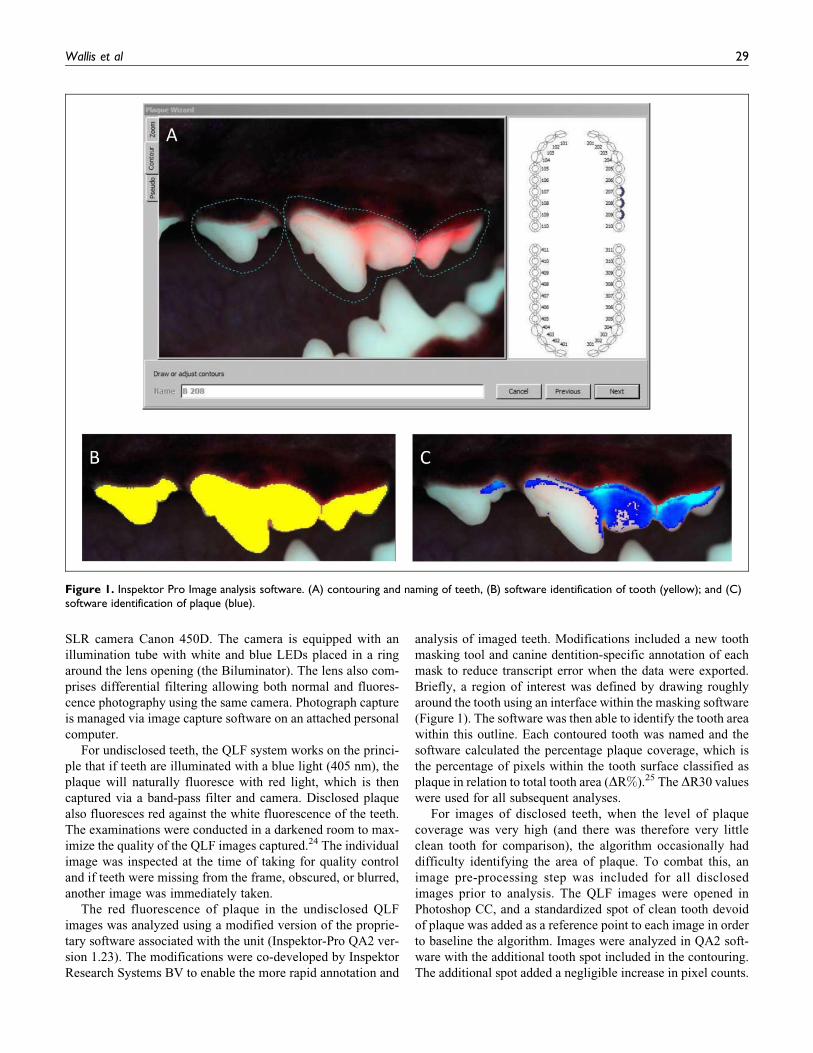
Briefly, a region of interest was defined by drawing roughly
around the tooth using an interface within the masking software
(Figure 1). The software was then able to identify the tooth area
within this outline. Each contoured tooth was named and the
software calculated the percentage plaque coverage, which is
the percentage of pixels within the tooth surface classified as
plaque in relation to total tooth area (DR%).25 The DR30 values
were used for all subsequent analyses.
For images of disclosed teeth, when the level of plaque
coverage was very high (and there was therefore very little
clean tooth for comparison), the algorithm occasionally had
difficulty identifying the area of plaque. To combat this, an
image pre-processing step was included for all disclosed
images prior to analysis. The QLF images were opened in
Photoshop CC, and a standardized spot of clean tooth devoid
of plaque was added as a reference point to each image in order
to baseline the algorithm. Images were analyzed in QA2 soft-
ware with the additional tooth spot included in the contouring.
The additional spot added a negligible increase in pixel counts.
Figure 1. Inspektor Pro Image analysis software. (A) contouring and naming of teeth, (B) software identification of tooth (yellow); and (C)software identification of plaque (blue).
Wallis et al 29
Images were scrutinized for quality in terms of focus, parts
of teeth obscured, illumination, or any other artifacts that could
have affected the analysis. During this process, it was observed
that for undisclosed images, in rare instances where there were
very high levels of plaque, the algorithm occasionally identi-
fied that the whole tooth was covered in plaque but reported
plaque coverage as 0%. In this instance, a value of 100% plaque
coverage was imputed.
Statistical Analysis
Intra-photographer repeatability. Linear mixed effects models
(Restricted maximum likelihood [REML]) were used to estimate
variance components of the percentage plaque coverage, using
repeat nested within dog as random effects. First, a model for an
average mouth (maxillary P1, P2, P3, and P4) was used, followed
by assessment of each tooth type. The percentage of variability
that was accountable to repeatability and the percentage of coef-
ficient of variability (%CV; repeatability standard deviation rela-
tive to the overall mean of the model) were then calculated.
Inter-photographer reproducibility. Linear mixed models (REML)
were used to estimate variance components of the percentage
plaque coverage, with photographer nested in dog as the ran-
dom effects. The percentage of variability accountable to the
photographer and the %CV (reproducibility standard deviation
relative to the overall mean of the model) were then calculated.
Accuracy. The accuracy of the software was determined by com-
paring its results with those of human scorers. Whole mouth
scores from 9 dogs, as assessed by 5 human scorers, were
analyzed by a linear mixed model with scorer nested in dog
fitted as the random effects. The variance estimates were then
used to inform a simulation of 1000 scorers (assuming each
scorer assessed 9 dogs) with an average of 46.8% plaque cov-
erage (as was found from the 5 human scorers). The probability
of the QLF software results falling within the distribution of the
human scorers’ results was calculated by the percentage of
simulated scorers with an average less than the average QLF
software score. A test level of 5% was used.
Comparison to modified Logan and Boyce. The percentage plaque
coverage measured by QLF and modified Logan and Boyce,
averaged for all teeth, were analyzed by linear mixed models
with dog as a random effect and chew type as a fixed effect.
This was used to assess the difference in mean plaque scores
between chew types, at the 5% significance level. The mean
and difference between mean plaque scores for each chew
type are reported with 95% confidence intervals. These data,
and their associated variances, were then used to inform retro-
spective sample size analyses for a 2-way crossover trial to
detect a 15% reduction (as defined as relevant by the VOHC)
in plaque accumulation compared to no chew with at least
90% power.
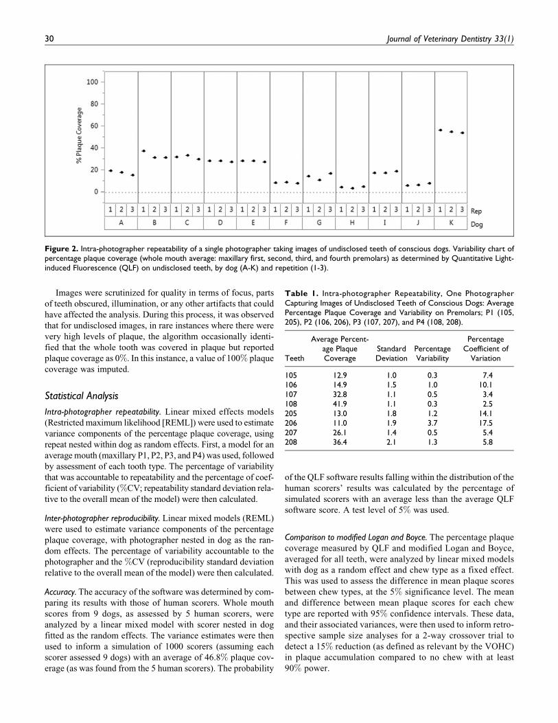
Figure 2. Intra-photographer repeatability of a single photographer taking images of undisclosed teeth of conscious dogs. Variability chart ofpercentage plaque coverage (whole mouth average: maxillary first, second, third, and fourth premolars) as determined by Quantitative Light-induced Fluorescence (QLF) on undisclosed teeth, by dog (A-K) and repetition (1-3).
Table 1. Intra-photographer Repeatability, One PhotographerCapturing Images of Undisclosed Teeth of Conscious Dogs: AveragePercentage Plaque Coverage and Variability on Premolars; P1 (105,205), P2 (106, 206), P3 (107, 207), and P4 (108, 208).
Teeth
Average Percent-age PlaqueCoverage
StandardDeviation
PercentageVariability
PercentageCoefficient of
Variation
105 12.9 1.0 0.3 7.4106 14.9 1.5 1.0 10.1107 32.8 1.1 0.5 3.4108 41.9 1.1 0.3 2.5205 13.0 1.8 1.2 14.1206 11.0 1.9 3.7 17.5207 26.1 1.4 0.5 5.4208 36.4 2.1 1.3 5.8
30 Journal of Veterinary Dentistry 33(1)
Comparison between conscious and unconscious imaging. The per-
centage plaque coverage as measured by QLF of undi-
sclosed teeth from conscious (average of upper jaw teeth)
and anesthetized (average of all teeth) dogs was analyzed
using linear mixed models. Dog was included as a random
effect and chew type, measure type, and their interactions
were included as fixed effects. Contrasts were performed
within and between measure types at a family wise con-
trolled error rate of 5% (R v3.02 using libraries nlme and
multcomp).
Results
Intra-photographer Repeatability: Undisclosed Images
Variance components analysis of data from 264 images of
undisclosed maxillary teeth (P1, P2, P3, and P4) from 11
conscious miniature schnauzers was used to quantify the intra-
photographer repeatability of a single photographer and
showed that the repeatability coefficient of variability (stan-
dard deviation relative to the mean plaque coverage) was 7.5%(Figure 2).
The intra-photographer repeatability component of variability
showed that the QLF method was highly repeatable and
accounted for <1.4% of the total variability for most teeth. The
exception was tooth 206 where it accounted for 3.7% of
the variability (Table 1). When the variance components were
made relative to the mean plaque coverage for each tooth, this
showed that the %CV ranged from 2.5% to 17.5% (Table 1). The
P1 and P2 had the highest %CV ranging from 7.4% to 17.5%, and
these teeth also had the lowest average percentage plaque cover-
age ranging from an average of 11.0% to 14.9%. The average
percentage plaque coverage for the P3 and P4 ranged from 26.1%to 41.9% with %CVs ranging from 2.5% to 5.8%.
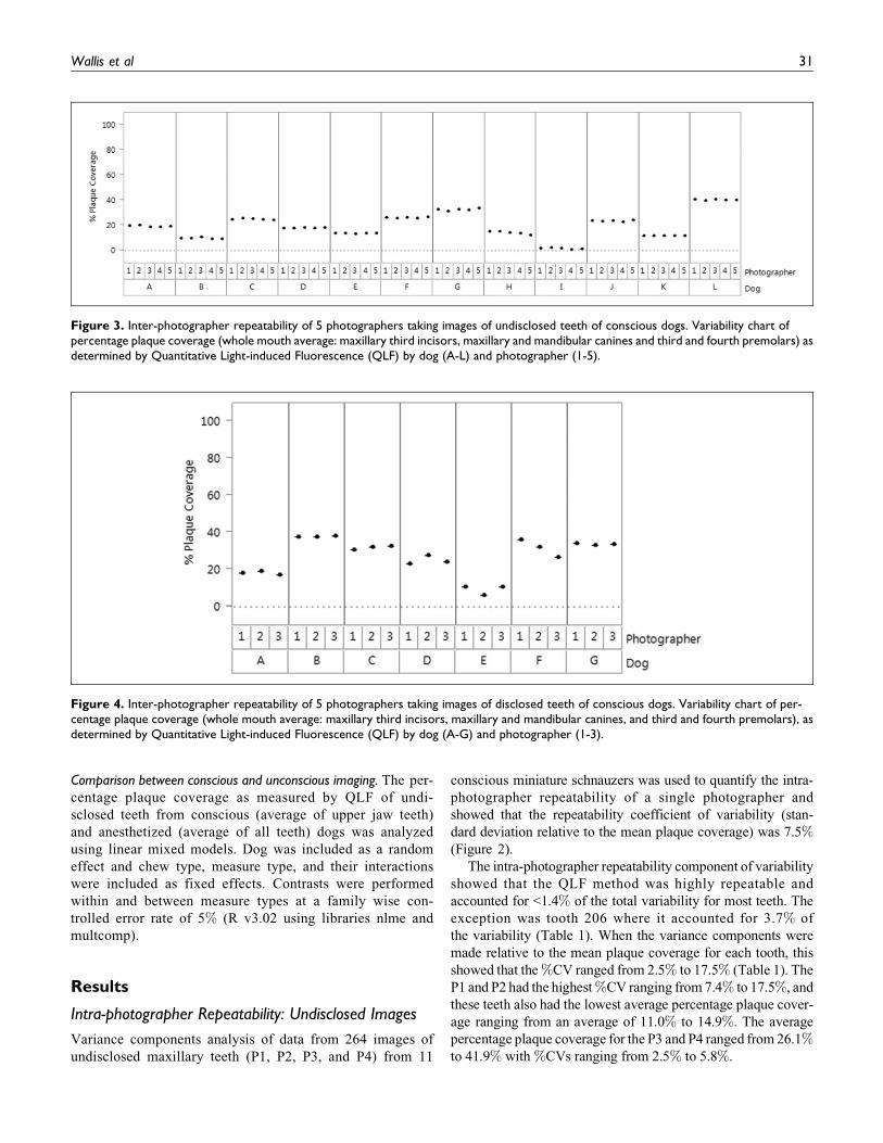
Figure 3. Inter-photographer repeatability of 5 photographers taking images of undisclosed teeth of conscious dogs. Variability chart ofpercentage plaque coverage (whole mouth average: maxillary third incisors, maxillary and mandibular canines and third and fourth premolars) asdetermined by Quantitative Light-induced Fluorescence (QLF) by dog (A-L) and photographer (1-5).
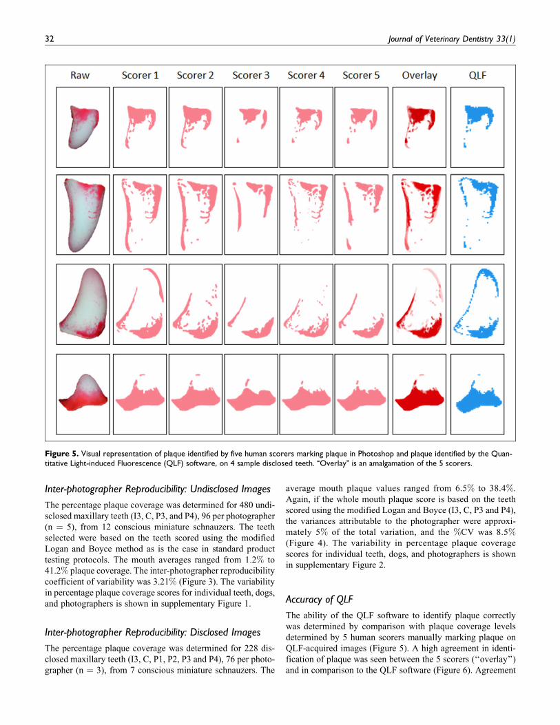
Figure 4. Inter-photographer repeatability of 5 photographers taking images of disclosed teeth of conscious dogs. Variability chart of per-centage plaque coverage (whole mouth average: maxillary third incisors, maxillary and mandibular canines, and third and fourth premolars), asdetermined by Quantitative Light-induced Fluorescence (QLF) by dog (A-G) and photographer (1-3).
Wallis et al 31
Inter-photographer Reproducibility: Undisclosed Images
The percentage plaque coverage was determined for 480 undi-
sclosed maxillary teeth (I3, C, P3, and P4), 96 per photographer
(n ¼ 5), from 12 conscious miniature schnauzers. The teeth
selected were based on the teeth scored using the modified
Logan and Boyce method as is the case in standard product
testing protocols. The mouth averages ranged from 1.2% to
41.2% plaque coverage. The inter-photographer reproducibility
coefficient of variability was 3.21% (Figure 3). The variability
in percentage plaque coverage scores for individual teeth, dogs,
and photographers is shown in supplementary Figure 1.
Inter-photographer Reproducibility: Disclosed Images
The percentage plaque coverage was determined for 228 dis-
closed maxillary teeth (I3, C, P1, P2, P3 and P4), 76 per photo-
grapher (n ¼ 3), from 7 conscious miniature schnauzers. The
average mouth plaque values ranged from 6.5% to 38.4%.
Again, if the whole mouth plaque score is based on the teeth
scored using the modified Logan and Boyce (I3, C, P3 and P4),
the variances attributable to the photographer were approxi-
mately 5% of the total variation, and the %CV was 8.5%(Figure 4). The variability in percentage plaque coverage
scores for individual teeth, dogs, and photographers is shown
in supplementary Figure 2.
Accuracy of QLF
The ability of the QLF software to identify plaque correctly
was determined by comparison with plaque coverage levels
determined by 5 human scorers manually marking plaque on
QLF-acquired images (Figure 5). A high agreement in identi-
fication of plaque was seen between the 5 scorers (‘‘overlay’’)
and in comparison to the QLF software (Figure 6). Agreement
Figure 5. Visual representation of plaque identified by five human scorers marking plaque in Photoshop and plaque identified by the Quan-titative Light-induced Fluorescence (QLF) software, on 4 sample disclosed teeth. ‘‘Overlay’’ is an amalgamation of the 5 scorers.
32 Journal of Veterinary Dentistry 33(1)
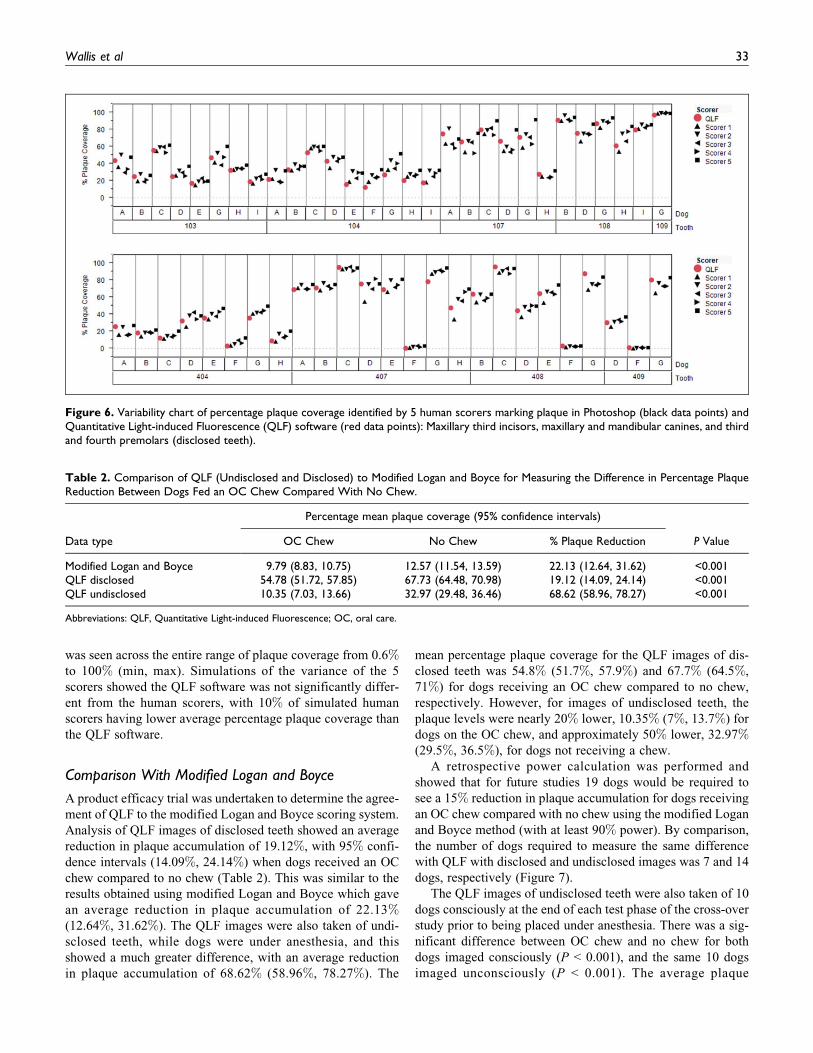
was seen across the entire range of plaque coverage from 0.6%to 100% (min, max). Simulations of the variance of the 5
scorers showed the QLF software was not significantly differ-
ent from the human scorers, with 10% of simulated human
scorers having lower average percentage plaque coverage than
the QLF software.
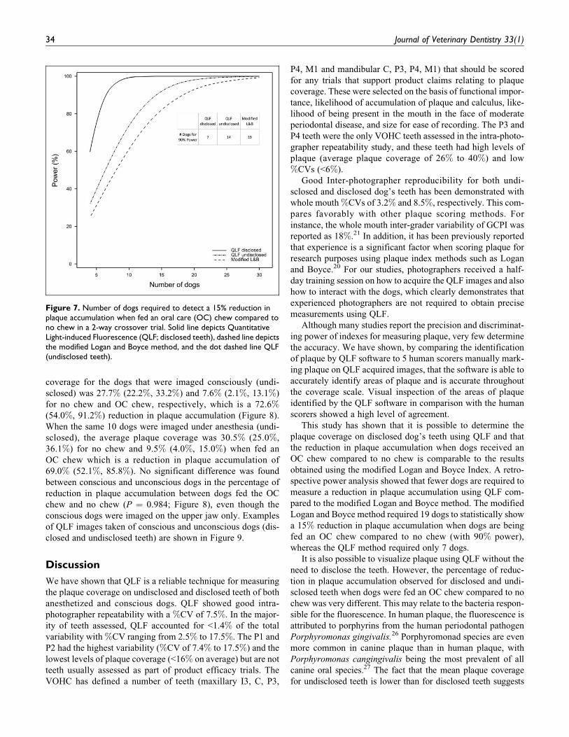
Comparison With Modified Logan and Boyce
A product efficacy trial was undertaken to determine the agree-
ment of QLF to the modified Logan and Boyce scoring system.
Analysis of QLF images of disclosed teeth showed an average
reduction in plaque accumulation of 19.12%, with 95% confi-
dence intervals (14.09%, 24.14%) when dogs received an OC
chew compared to no chew (Table 2). This was similar to the
results obtained using modified Logan and Boyce which gave
an average reduction in plaque accumulation of 22.13%(12.64%, 31.62%). The QLF images were also taken of undi-
sclosed teeth, while dogs were under anesthesia, and this
showed a much greater difference, with an average reduction
in plaque accumulation of 68.62% (58.96%, 78.27%). The
mean percentage plaque coverage for the QLF images of dis-
closed teeth was 54.8% (51.7%, 57.9%) and 67.7% (64.5%,
71%) for dogs receiving an OC chew compared to no chew,
respectively. However, for images of undisclosed teeth, the
plaque levels were nearly 20% lower, 10.35% (7%, 13.7%) for
dogs on the OC chew, and approximately 50% lower, 32.97%(29.5%, 36.5%), for dogs not receiving a chew.
A retrospective power calculation was performed and
showed that for future studies 19 dogs would be required to
see a 15% reduction in plaque accumulation for dogs receiving
an OC chew compared with no chew using the modified Logan
and Boyce method (with at least 90% power). By comparison,
the number of dogs required to measure the same difference
with QLF with disclosed and undisclosed images was 7 and 14
dogs, respectively (Figure 7).
The QLF images of undisclosed teeth were also taken of 10
dogs consciously at the end of each test phase of the cross-over
study prior to being placed under anesthesia. There was a sig-
nificant difference between OC chew and no chew for both
dogs imaged consciously (P < 0.001), and the same 10 dogs
imaged unconsciously (P < 0.001). The average plaque
Figure 6. Variability chart of percentage plaque coverage identified by 5 human scorers marking plaque in Photoshop (black data points) andQuantitative Light-induced Fluorescence (QLF) software (red data points): Maxillary third incisors, maxillary and mandibular canines, and thirdand fourth premolars (disclosed teeth).
Table 2. Comparison of QLF (Undisclosed and Disclosed) to Modified Logan and Boyce for Measuring the Difference in Percentage PlaqueReduction Between Dogs Fed an OC Chew Compared With No Chew.
Data type
Percentage mean plaque coverage (95% confidence intervals)
P ValueOC Chew No Chew % Plaque Reduction
Modified Logan and Boyce 9.79 (8.83, 10.75) 12.57 (11.54, 13.59) 22.13 (12.64, 31.62) <0.001QLF disclosed 54.78 (51.72, 57.85) 67.73 (64.48, 70.98) 19.12 (14.09, 24.14) <0.001QLF undisclosed 10.35 (7.03, 13.66) 32.97 (29.48, 36.46) 68.62 (58.96, 78.27) <0.001
Abbreviations: QLF, Quantitative Light-induced Fluorescence; OC, oral care.
Wallis et al 33
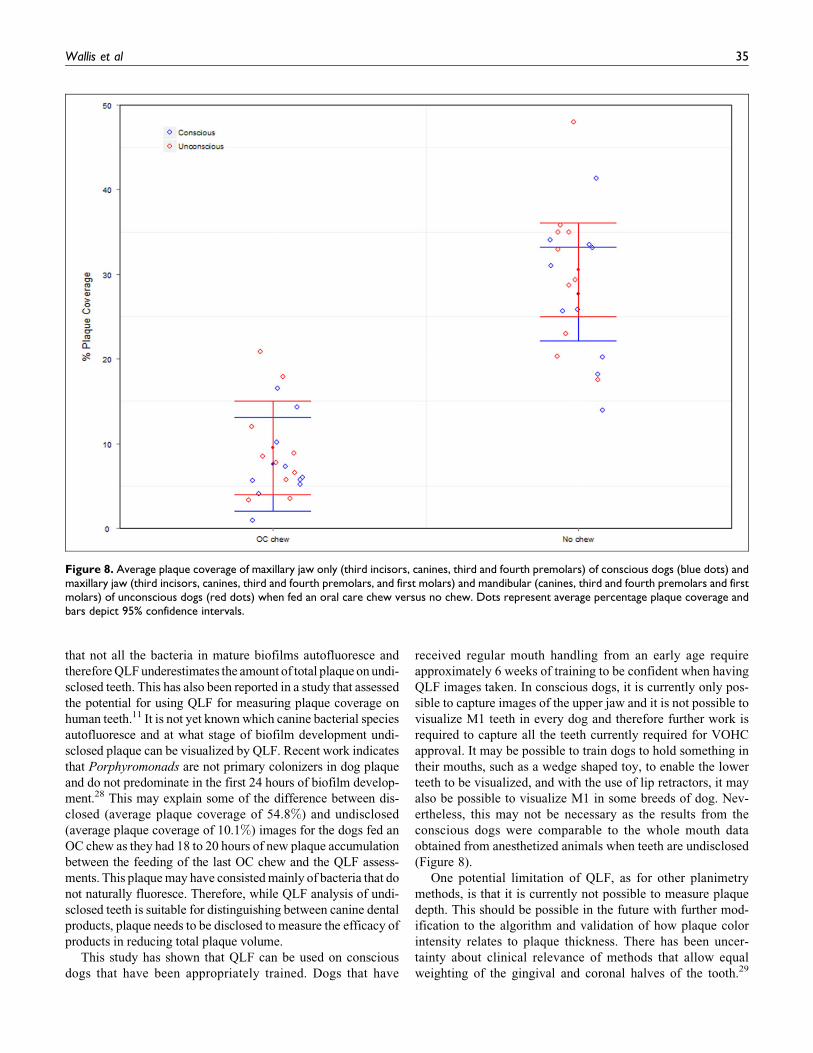
coverage for the dogs that were imaged consciously (undi-
sclosed) was 27.7% (22.2%, 33.2%) and 7.6% (2.1%, 13.1%)
for no chew and OC chew, respectively, which is a 72.6%(54.0%, 91.2%) reduction in plaque accumulation (Figure 8).
When the same 10 dogs were imaged under anesthesia (undi-
sclosed), the average plaque coverage was 30.5% (25.0%,
36.1%) for no chew and 9.5% (4.0%, 15.0%) when fed an
OC chew which is a reduction in plaque accumulation of
69.0% (52.1%, 85.8%). No significant difference was found
between conscious and unconscious dogs in the percentage of
reduction in plaque accumulation between dogs fed the OC
chew and no chew (P ¼ 0.984; Figure 8), even though the
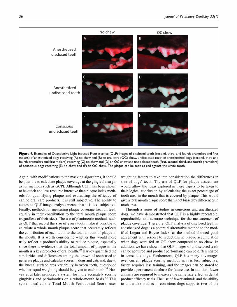
conscious dogs were imaged on the upper jaw only. Examples
of QLF images taken of conscious and unconscious dogs (dis-
closed and undisclosed teeth) are shown in Figure 9.
Discussion
We have shown that QLF is a reliable technique for measuring
the plaque coverage on undisclosed and disclosed teeth of both
anesthetized and conscious dogs. QLF showed good intra-
photographer repeatability with a %CV of 7.5%. In the major-
ity of teeth assessed, QLF accounted for <1.4% of the total
variability with %CV ranging from 2.5% to 17.5%. The P1 and
P2 had the highest variability (%CV of 7.4% to 17.5%) and the
lowest levels of plaque coverage (<16% on average) but are not
teeth usually assessed as part of product efficacy trials. The
VOHC has defined a number of teeth (maxillary I3, C, P3,
P4, M1 and mandibular C, P3, P4, M1) that should be scored
for any trials that support product claims relating to plaque
coverage. These were selected on the basis of functional impor-
tance, likelihood of accumulation of plaque and calculus, like-
lihood of being present in the mouth in the face of moderate
periodontal disease, and size for ease of recording. The P3 and
P4 teeth were the only VOHC teeth assessed in the intra-photo-
grapher repeatability study, and these teeth had high levels of
plaque (average plaque coverage of 26% to 40%) and low
%CVs (<6%).
Good Inter-photographer reproducibility for both undi-
sclosed and disclosed dog’s teeth has been demonstrated with
whole mouth %CVs of 3.2% and 8.5%, respectively. This com-
pares favorably with other plaque scoring methods. For
instance, the whole mouth inter-grader variability of GCPI was
reported as 18%.21 In addition, it has been previously reported
that experience is a significant factor when scoring plaque for
research purposes using plaque index methods such as Logan
and Boyce.20 For our studies, photographers received a half-
day training session on how to acquire the QLF images and also
how to interact with the dogs, which clearly demonstrates that
experienced photographers are not required to obtain precise
measurements using QLF.
Although many studies report the precision and discriminat-
ing power of indexes for measuring plaque, very few determine
the accuracy. We have shown, by comparing the identification
of plaque by QLF software to 5 human scorers manually mark-
ing plaque on QLF acquired images, that the software is able to
accurately identify areas of plaque and is accurate throughout
the coverage scale. Visual inspection of the areas of plaque
identified by the QLF software in comparison with the human
scorers showed a high level of agreement.
This study has shown that it is possible to determine the
plaque coverage on disclosed dog’s teeth using QLF and that
the reduction in plaque accumulation when dogs received an
OC chew compared to no chew is comparable to the results
obtained using the modified Logan and Boyce Index. A retro-
spective power analysis showed that fewer dogs are required to
measure a reduction in plaque accumulation using QLF com-
pared to the modified Logan and Boyce method. The modified
Logan and Boyce method required 19 dogs to statistically show
a 15% reduction in plaque accumulation when dogs are being
fed an OC chew compared to no chew (with 90% power),
whereas the QLF method required only 7 dogs.
It is also possible to visualize plaque using QLF without the
need to disclose the teeth. However, the percentage of reduc-
tion in plaque accumulation observed for disclosed and undi-
sclosed teeth when dogs were fed an OC chew compared to no
chew was very different. This may relate to the bacteria respon-
sible for the fluorescence. In human plaque, the fluorescence is
attributed to porphyrins from the human periodontal pathogen
Porphyromonas gingivalis.26 Porphyromonad species are even
more common in canine plaque than in human plaque, with
Porphyromonas cangingivalis being the most prevalent of all
canine oral species.27 The fact that the mean plaque coverage
for undisclosed teeth is lower than for disclosed teeth suggests
Figure 7. Number of dogs required to detect a 15% reduction inplaque accumulation when fed an oral care (OC) chew compared tono chew in a 2-way crossover trial. Solid line depicts QuantitativeLight-induced Fluorescence (QLF; disclosed teeth), dashed line depictsthe modified Logan and Boyce method, and the dot dashed line QLF(undisclosed teeth).
34 Journal of Veterinary Dentistry 33(1)
that not all the bacteria in mature biofilms autofluoresce and
therefore QLF underestimates the amount of total plaque on undi-
sclosed teeth. This has also been reported in a study that assessed
the potential for using QLF for measuring plaque coverage on
human teeth.11 It is not yet known which canine bacterial species
autofluoresce and at what stage of biofilm development undi-
sclosed plaque can be visualized by QLF. Recent work indicates
that Porphyromonads are not primary colonizers in dog plaque
and do not predominate in the first 24 hours of biofilm develop-
ment.28 This may explain some of the difference between dis-
closed (average plaque coverage of 54.8%) and undisclosed
(average plaque coverage of 10.1%) images for the dogs fed an
OC chew as they had 18 to 20 hours of new plaque accumulation
between the feeding of the last OC chew and the QLF assess-
ments. This plaque may have consisted mainly of bacteria that do
not naturally fluoresce. Therefore, while QLF analysis of undi-
sclosed teeth is suitable for distinguishing between canine dental
products, plaque needs to be disclosed to measure the efficacy of
products in reducing total plaque volume.
This study has shown that QLF can be used on conscious
dogs that have been appropriately trained. Dogs that have
received regular mouth handling from an early age require
approximately 6 weeks of training to be confident when having
QLF images taken. In conscious dogs, it is currently only pos-
sible to capture images of the upper jaw and it is not possible to
visualize M1 teeth in every dog and therefore further work is
required to capture all the teeth currently required for VOHC
approval. It may be possible to train dogs to hold something in
their mouths, such as a wedge shaped toy, to enable the lower
teeth to be visualized, and with the use of lip retractors, it may
also be possible to visualize M1 in some breeds of dog. Nev-
ertheless, this may not be necessary as the results from the
conscious dogs were comparable to the whole mouth data
obtained from anesthetized animals when teeth are undisclosed
(Figure 8).
One potential limitation of QLF, as for other planimetry
methods, is that it is currently not possible to measure plaque
depth. This should be possible in the future with further mod-
ification to the algorithm and validation of how plaque color
intensity relates to plaque thickness. There has been uncer-
tainty about clinical relevance of methods that allow equal
weighting of the gingival and coronal halves of the tooth.29
Figure 8. Average plaque coverage of maxillary jaw only (third incisors, canines, third and fourth premolars) of conscious dogs (blue dots) andmaxillary jaw (third incisors, canines, third and fourth premolars, and first molars) and mandibular (canines, third and fourth premolars and firstmolars) of unconscious dogs (red dots) when fed an oral care chew versus no chew. Dots represent average percentage plaque coverage andbars depict 95% confidence intervals.
Wallis et al 35
Again, with modifications to the masking algorithms, it should
be possible to calculate plaque coverage at the gingival margin
as for methods such as GCPI. Although GCPI has been shown
to be quick and less resource intensive than plaque index meth-
ods for quantifying plaque and evaluating the efficacy of
canine oral care products, it is still subjective. The ability to
automate QLF image analysis means that it is less subjective.
Finally, methods for measuring plaque coverage treat all teeth
equally in their contribution to the total mouth plaque score
(regardless of their size). The use of planimetric methods such
as QLF that record the size of every tooth make it possible to
calculate a whole mouth plaque score that accurately reflects
the contribution of each tooth to the total amount of plaque in
the mouth. It is worth considering whether this would more
truly reflect a product’s ability to reduce plaque, especially
since there is evidence that the total amount of plaque in the
mouth is a key predictor of oral health.30 Harvey quantified the
similarities and differences among the crown of teeth used to
generate plaque and calculus scores in dogs and cats and, due to
the buccal surface area variability between teeth, questioned
whether equal weighting should be given to each tooth.31 Har-
vey et al later proposed a system for more accurately scoring
gingivitis and periodontitis on a whole-mouth basis.32 This
system, called the Total Mouth Periodontal Score, uses
weighting factors to take into consideration the differences in
size of dogs’ teeth. The use of QLF for plaque assessment
would allow the ideas explored in these papers to be taken to
their logical conclusion by calculating the exact percentage of
tooth area in the mouth that is covered by plaque. This would
give a total mouth plaque score that is not biased by differences in
tooth area.
Through a series of studies in conscious and anesthetized
dogs, we have demonstrated that QLF is a highly repeatable,
reproducible, and accurate technique for the measurement of
plaque coverage. Therefore, QLF analysis of disclosed teeth in
anesthetized dogs is a potential alternative method to the mod-
ified Logan and Boyce Index, as the method showed good
agreement with respect to reductions in plaque accumulation
when dogs were fed an OC chew compared to no chew. In
addition, we have shown that QLF images of undisclosed teeth
can be acquired and product performance can be differentiated
in conscious dogs. Furthermore, QLF has many advantages
over current plaque scoring methods as it is less subjective,
faster, requires less training, and the images can be stored to
provide a permanent database for future use. In addition, fewer
animals are required to measure the same size effect in dental
product efficacy trials. The use of fewer animals and the ability
to undertake studies in conscious dogs supports two of the
OC chew
Anesthe�zeddisclosed teeth
No chew
Anesthe�zedundisclosed teeth
Consciousundisclosed teeth
A B
C D
E F
Figure 9. Examples of Quantitative Light-induced Fluorescence (QLF) images of disclosed teeth (second, third, and fourth premolars and firstmolars) of anesthetized dogs receiving (A) no chew and (B) an oral care (OC) chew, undisclosed teeth of anesthetized dogs (second, third andfourth premolars and first molars) receiving (C) no chew and (D) an OC chew and undisclosed teeth (first, second, third, and fourth premolars)of conscious dogs receiving (E) no chew and (F) an OC chew. The plaque can be seen as red against the white tooth.
36 Journal of Veterinary Dentistry 33(1)
guiding principles underpinning the humane use of animals in
scientific research: (1) reducing the number of animals used
and (2) refining experiments to improve animal welfare.
Acknowledgments
The authors would like to acknowledge the WALTHAM staff who
trained the dogs, volunteered to take part in the reproducibility study,
helped with the accuracy assessments, and specifically Mark Marshall
for managing the modified Logan and Boyce trial. The authors would
like to acknowledge Inspektor Research Systems BV, Amsterdam, the
Netherlands, for the algorithm modifications and for their technical
support. Finally, the authors would like to acknowledge Lisa Milella
and Florian Boutille for their help in scoring images for the accuracy
experiments.
Author Contribution
Corrin Wallis and Yadvinder Gill contributed equally to this work.
They both contributed to the design and co-ordination of the studies
and the preparation of the manuscript. Alison Colyer participated in
the design of the studies, performed all statistical analyses and
reviewed the manuscript. Ian Davis and Judi Allsopp were involved
in the acquisition, analysis and interpretation of the data. Gleb
Komarov and Sue Higham contributed to the conception and design
of the studies, data analysis and revision of the manuscript. Stephen
Harris conceived and participated in the design and co-ordination of
the study and reviewed the manuscript. All authors approved the final
article.
Declaration of Conflicting Interests
The author(s) declared no potential conflicts of interest with respect to
the research, authorship, and/or publication of this article.
Funding
The author(s) disclosed receipt of the following financial support for
the research, authorship, and/or publication of this article: This work
was funded by the WALTHAM Centre for Pet Nutrition the employer
of Corrin Wallis, Yadvinder Gill, Alison Colyer, Ian Davis, Judi All-
sopp and Stephen Harris. There are no products in development or
marketed products to declare.
Supplementary Material
The online [appendices/data supplements/etc] are available at http://
jov.sagepub.com/supplemental
References
1. Hamp S, Olsson S, Farso-Madsen K, Viklands P, Fornell J. A
macroscopic and radiologic investigation of dental diseases of the
dog. Veterinary Radiol. 1984;25(2):86-92.
2. Butkovic V, Simpraga M, Sehic M, et al. Dental diseases of dogs:
A retrospective study of radiological data. Acta Veterinaria Brno.
2001;70(2):203-208.
3. Kyllar M, Witter K. Prevalence of dental disorders in pet dogs.
Veterinarni Medicina-Czechoslovakia. 2005;50(11):496-505.
4. Kortegaard H, Eriksen T, Baelum V. Periodontal disease in
research beagle dogs - an epidemiological study. J Small Anim
Pract. 2008;49(12):610-616.
5. Van Dyke TE. The etiology and pathogenesis of periodontitis
revisited. J Appl Oral Sci. 2009;17(1):1678-7757.
6. Williams RC. Periodontal disease. N Engl J Med. 1990;322(6):
373-382.
7. Quigley G, Hein J. Comparative cleansing efficiency of manual
and power brushing. J Am Dent Assoc. 1962;65:26-29.
8. Silness J, Loe H. Periodontal disease in pregnancy. II. Correlation
between oral hygiene and periodontal condition. Acta Odolltol
Scalld. 1964;22:121-135.
9. Loe H. The gingival index, the plaque index and the retention
index systems. J Periodontol. 1967;38(6):610-616.
10. Turesky S, Gilmore ND, Glickman I. Reduced plaque formation by the
chloromethyl analogue of vitamin C. J Periodontol. 1970;41(1):41-43.
11. Pretty IA, Edgar WM, Smith PW, Higham SM. Quantification of
dental plaque in the research environment. J Dent. 2005;33(3):
193-207.
12. Soder PO, Jin LJ, Soder B. Computerized planimetric method for
clinical plaque measurement. Scand J Dent Res. 1993;101(1):
21-25.
13. Staudt CB, Kinzel S, Hassfeld S, Stein W, Staehle HJ, Dorfer CE.
Computer-based intraoral image analysis of the clinical plaque
removing capacity of 3 manual toothbrushes. J Clin Periodontol.
2001;28(8):746-752.
14. Verran J, Rocliffe MD. Feasibility of using automatic image anal-
ysis for measuring dental plaque in situ. J Dent. 1986;14(1):11-13.
15. Block RP, Bouwsma OJ, Howardnordan KS, Miller JM, Poore
CL, Sunberg RJ. Validation of computerized photoimage analysis
(PIA) measurement of plaque. J Dent Res. 1996;75:367.
16. Shaloub A, Addy M. Evaluation of accuracy and variability of
scoring-area-based plaque indices. A laboratory model. J Clin
Periodontol. 2000;27(1):16-21.
17. Pretty IA, Edgar WM, Higham SM. A study to assess the efficacy
of a new detergent free, whitening dentifrice in vivo using QLF
planimetric analysis. Br Dent J. 2004;197(9):561-566.
18. Mohan N, Mahesh MR, Varghese VI, Pretty IA, Taylor AM.
Evaluation of the sensitivity of a digital plaque imaging system
on different tooth surfaces. J Clin Dent. 2012;23(1):11-16.
19. Hope CK, Wang Q, Burnside G, et al. Assessing the association
between oral hygiene and preterm birth by quantitative light-
induced fluorescence. ScientificWorldJournal. 2014;2014:374694.
20. Hennet P, Servet E, Salesse H, Soulard Y. Evaluation of the
Logan and Boyce Plaque Index for the Study of Dental Plaque
Accumulation in Dogs. Res Veterinary Sci. 2006;80(2):175-180.
21. Scherl DS, Coffman L, van Cleave M, Lowry S. Validation of a
new dental plaque quantification method in dogs. J Vet Dent.
2007;24(1):14-19.
22. Scherl DS, Bork K, Coffman L, Lowry SR, VanCleave M. Appli-
cation of the Gingival Contour Plaque Index: six-month plaque
and gingivitis study. J Vet Dent. 2009;26(1):23-27.
23. National Research Council (US). Ad Hoc Committee on Dog and
Cat Nutrition. Nutrient Requirements of Dogs and Cats. Washing-
ton, D.C.: National Academies Press; 2006.
24. Pretty IA, Edgar WM, Higham SM. The effect of ambient light on
QLF analyses. J Oral Rehabil. 2002;29(4):369-373.
25. De Josselin de Jong E, Higham SM, Smith PW, van Daelen CJ,
van der Veen MH. Quantified light-induced fluorescence, review
of a diagnostic tool in prevention of oral disease. J Appl Phys.
2009;105(10):102031.
Wallis et al 37
26. Marsh PD, Martin MV. Oral Microbiology, 3rd ed. London:
Chapman & Hall; 1992.
27. Davis IJ, Wallis C, Deusch O, Colyer A, Milella L, Loman N,
Harris S. A cross-sectional survey of bacterial species in plaque
from client owned dogs with healthy gingiva, gingivitis or mild
periodontitis. PLoS One. 2013;8(12):e83158.
28. Holcombe LJ, Patel N, Colyer A, et al. Early Canine Plaque
Biofilms: characterization of key bacterial interactions involved
in initial colonization of enamel. PLoS One. 2014;9(12):e113744.
doi:10.1371/journal.pone.0113744.
29. Hennet P. Review of studies assessing plaque accumulation and
gingival inflammation in dogs. J Vet Dent. 1999;16(1):23-29.
30. Darveau R, Tanner A, Page R. The microbial challenge in period-
ontitis. Periodontology. 1997;14:12-32.
31. Harvey CE. Shape and size of teeth of dogs and cats-relevance to
studies of plaque and calculus accumulation. J Vet Dent. 2002;
19(4):186-195.
32. Harvey CE, Laster L, Schofer F, Miller B. Scoring the full extent of
periodontal disease in the dog: development of a total mouth per-
iodontal score (TMPS) system. J Vet Dent. 2008;25(3):176-180.
38 Journal of Veterinary Dentistry 33(1)
Related Documents