Quality Assurance/Quality Control Manual: Ohio Water Microbiology Laboratory By Donna S. Francy, Rebecca N. Bushon, Jessica R. Cicale, Amie M. G. Brady, Christopher M. Kephart, Erin A. Stelzer, and Christopher D. Ecker Updated January 2017 ABSTRACT INTRODUCTION Purpose and scope Organizational structure GENERAL LABORATORY QUALITY ASSURANCE/QUALITY-CONTROL PRACTICES Analytical methods Training Safety Laboratory materials and equipment General sterility and cleanliness Autoclaves Laboratory water Analytical balances Hoods Specific conductance, pH, and turbidity meters Micropipettors Vacuum pump Thermometers, incubators, water baths, refrigerators, and freezers Microscope Centrifuge Thermal cyclers Sample management and documentation METHODS OF ANALYSIS, MEDIA AND REAGENT PREPARATION, AND ANALYTICAL QUALITY-CONTROL PROCEDURES

Welcome message from author
This document is posted to help you gain knowledge. Please leave a comment to let me know what you think about it! Share it to your friends and learn new things together.
Transcript

Quality Assurance/Quality Control Manual: Ohio Water Microbiology Laboratory
By Donna S. Francy, Rebecca N. Bushon, Jessica R. Cicale, Amie M. G. Brady, Christopher M. Kephart, Erin A. Stelzer, and Christopher D. Ecker
Updated January 2017
ABSTRACT
INTRODUCTION
Purpose and scopeOrganizational structure
GENERAL LABORATORY QUALITY ASSURANCE/QUALITY-CONTROL PRACTICES
Analytical methodsTrainingSafetyLaboratory materials and equipment
General sterility and cleanlinessAutoclavesLaboratory waterAnalytical balancesHoodsSpecific conductance, pH, and turbidity metersMicropipettorsVacuum pumpThermometers, incubators, water baths, refrigerators, and freezersMicroscope CentrifugeThermal cyclers
Sample management and documentation
METHODS OF ANALYSIS, MEDIA AND REAGENT PREPARATION, AND ANALYTICAL QUALITY-CONTROL PROCEDURES
Culture methods for indicator bacteria ColiphageActinomycetesRapid methods for indicator bacteriaMolecular methods for microbial source tracking markers and cyanobacteriaMolecular methods for virusesMethods for cyanobacterial toxins

REFERENCES
APPENDICESGeneral InstructionsA1. Instructions for quality control of deionized waterA2. Buffer preparationA3. Storage and maintenance of bacteria control culturesA4. Laboratory map
FormsB1. Temperature log sheetB2. Sample log sheetB3. Analytical services request formB4. Media and buffer quality-control log sheetB5. Expendable supplies request formB6. QC log for Actinomycetes detection by double agar layer methodB7. QC log for aerobic endospores by membrane filtration
Culture methods for bacteria and coliphageC1. M-Endo method for total coliformsC2. MI agar method for total coliforms and Escherichia coliC3. Colilert Quanti-Tray and presence/absence methods for total coliforms and Escherichia coliC4. mFC method for fecal coliformsC5. Modified mTEC method for Escherichia coliC6. Colilert Quanti-Tray sediment method for Escherichia coliC7. mEI method for enterococciC8. Verification of enterococci colonies from mEIC9. Enterolert Quanti-Tray for enterococciC10. mCP agar method for Clostridium perfringensC11. Indicator bacteria quality-control proceduresC12. Coliphage detection by USEPA Method 1602, single-agar layer methodC13. Coliphage detection by USEPA Method 1601, Two-step enrichment methodC14. Detection of Actinomycetes in waterC15. Detection of aerobic endospores in water
Molecular methods for MST markers and Cyanobacteria (appendices are available upon request)D1. Collection and initial processing of water samples for MST markers and CyanobacteriaD1a. Bench sheet for filtration and extraction for MST and CyanobacteriaD2. Collection and initial processing of solid samples for MST markers and CyanobacteriaD2a. Field sheet for collection of known-source fecal samplesD3. DNA extraction and purification using the GeneRite DNA-EZ kitD3a. Extraction batch bench sheetD4. Detection of MST markers and Cyanobacteria by qPCRD4a. qPCR bench sheets for MST assaysD4b qPCR bench sheets for Cyanobacteria DNA
2

.D4c. RT-qPCR bench sheets for Cyanobacteria RNAD5. Development and quantification of plasmid positive controlsD5a. Plasmid-based positive control bench sheetD5b.
Alternative development and quantification of plasmid positive controls
D6. qPCR data interpretation and file managementD7. RNA extraction and purification using the MoBio PowerPlant RNA Isolatin Kit with DNaseD8. Limits of blank, detection, and quantification for qPCR and qRT-PCR analyses
Enteric virus methods (appendices are available upon request)E1. Virus elution and organic flocculationE1a. Virus processing formE1b.
Virus qPCR batch sheet
E2. Molecular concentration and extractionE2a. Foam elution and concentration from dead-end ultrafiltrationE3. Reverse transcription and qPCR protocol for virusesE3a. qPCR and RT-qPCR benchsheets for virusesE4. Total culturable virus assayE4a. Total culturable virus and viral titering benchsheetE5. Collection and initial processing of water samples for human polyomarvirusE5a. Human polyomavirus filtration benchsheetE6. Extraction for human polyomavirus samples
Cyanobacterial toxin methods F1. Total microcystins—ADDA by ELISAF1a. ELISA bench sheet and plate map template
TABLES1. Current laboratory personnel and qualifications2. Acceptance criteria for laboratory water quality-assurance checks3. Acceptance criteria for laboratory thermometers4. Acceptance criteria for laboratory refrigerators, freezers, incubators, and water baths5. Culture methods for indicator bacteria used by the Ohio Water Microbiology6. Information on media, buffered-dilution water, and reagents prepared and stored in the Ohio
Water Microbiology Laboratory7. Target pH values for media and reagent preparation8. Microbial source tracking marker qPCR assays 9. Cyanobacterial qPCR assays
3

Quality Assurance/Quality Control Manual: Ohio Water Microbiology Laboratory
ABSTRACT
The U.S. Geological Survey (USGS), Ohio Water Microbiology Laboratory (the OWML) provides water-quality data on microorganisms of public health significance for a variety of projects within the USGS. Currently, the OWML analyzes samples for and provides training on bacterial indicators, coliphage, Actinomycetes, enteric viruses, cyanobacteria, and microbial source-tracking markers.
Quality-assurance and quality-control (QA/QC) practices for the operation of the OWML are described in this manual. The Laboratory Manager, Laboratory Coordinator, Chemical Hygiene Officer, and laboratory and field staff are responsible for implementing QA/QC procedures. This includes correctly following methods of analysis, media and reagent preparation and storage, and analytical quality-control procedures. A sample management and documentation system involves the use of analytical services request forms and login ID’s for each sample. A laboratory information management system (LIMS) has been implemented to store sample login information and results. Laboratory equipment maintenance and calibration records are also stored in LIMS.
INTRODUCTION
The USGS Ohio Water Microbiology Laboratory (the OWML) provides analytical data for projects within the Michigan-Ohio Water Science Center (MI-OH WSC), for the USGS National Water Quality Assessment (NAWQA) Program, and for other USGS Center programs by request. Samples are collected to determine the presence of microbiological organisms of public health significance in ground waters, surface waters, and sediments for a variety of study objectives. For example, some local studies are done to judge compliance with standards for protection of public health in swimmable or drinkable waters. Other studies investigate the occurrence, distribution, and trends of pathogenic organisms and indicators in surface and ground waters and relate these to environmental and water-quality factors.
The OWML fulfills analytical requirements of various USGS programs by analyzing environmental samples for bacterial indicators, coliphage, Actinomycetes, enteric viruses, cyanobacteria, and microbial source-tracking (MST) markers. OWML personnel provide assistance for project planning and training, as well as sampling and analytical methods for these organisms. The OWML continuously updates and adds analytical methods and microorganisms to its analytical list. The OWML is involved in some method development at the present time; however, the OWML mostly tests new methods developed by others for applicability to ambient monitoring programs.
The OWML is committed to providing quality microbiological analytical services to the USGS. The quality assurance/quality control (QA/QC) program is designed to ensure the production of scientifically sound, legally defensible data of known and documented quality. The effectiveness
4

of this program relies on clearly defined objectives, well-documented procedures, and management support.
Purpose and ScopeThe purpose of this manual is to identify and document practices and standard operating procedures for those activities of the OWML that affect quality of data. The manual provides OWML personnel and customers with general descriptions of quality practices and goals to aid in the interpretation of data. This manual is intended to be an unpublished, dynamic document that will be frequently updated as laboratory activities expand or change.
Organizational Structure The Laboratory Manager (1) oversees the operation of the OWML, including planning and budgeting (2) directs technical personnel in the proper performance of laboratory procedures and the reporting of results, (3) ensures that appropriate methods are used, (4) plans activities leading to testing and modification of analytical procedures, and (5) designs and implements a comprehensive QA/QC program. The Laboratory Manager is responsible for initiating the QA/QC program, providing information and training to the staff, and reviewing QA/QC activities on a continual basis.
The Laboratory Coordinator oversees the daily operations of the OWML, including scheduling of daily samples and communication with customers. The Laboratory Coordinator implements the QA/QC program in the daily tasks of conducting analyses, performing quality-control checks, and calculating and reporting results. The Laboratory Coordinator oversees entry of all sample and quality-control results in the Laboratory Information Management System (LIMS). The Laboratory Coordinator is also responsible for (1) maintaining and updating the QA/QC database in LIMS, (2) ensuring that all QA/QC tasks are completed in a timely manner, (3) notifying the Laboratory Manager when results are not as expected, and (4) ensuring that the equipment is properly maintained and calibrated.
The Laboratory Administrator oversees communication with customers and billing and sending results for services and supplies. All sample and quality-control results are approved by the Laboratory Administrator in the Laboratory Information Management System (LIMS) before data is released. The Laboratory Administrator is also responsible for estimating costs for analytical services.
The Chemical Hygiene Officer (1) oversees safety operations in the laboratory with assistance from the Laboratory Manager and Laboratory Coordinator, (2) reviews the Chemical Hygiene Plan annually to ensure that the Plan is up to date, (3) assists employees in obtaining Material Safety Data Sheets, and (4) maintains the laboratory chemical inventory list and MSDS books in the laboratory and front lobby.
The laboratory and field staffs are responsible for correctly implementing collection and analysis procedures and for identifying and working with supervisors to correct and avoid potential problems.
5
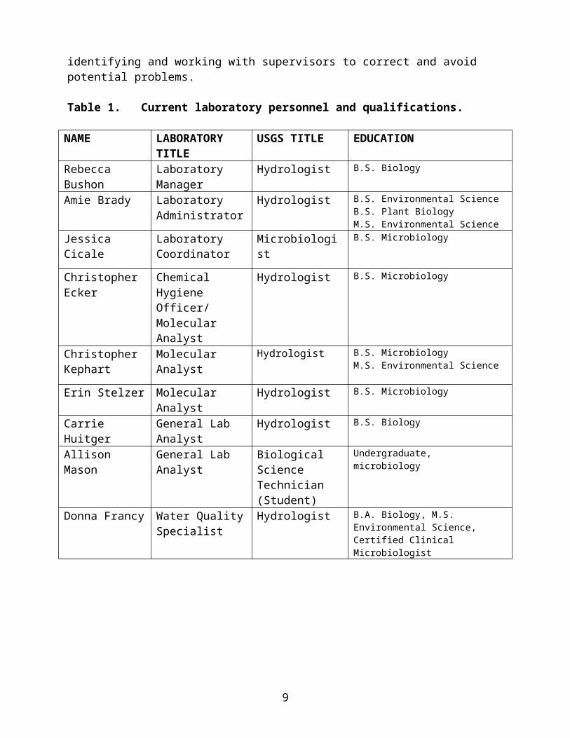
Table 1. Current laboratory personnel and qualifications.
NAME LABORATORY TITLE
USGS TITLE EDUCATION
Rebecca Bushon Laboratory Manager
Hydrologist B.S. Biology
Amie Brady Laboratory Administrator
Hydrologist B.S. Environmental ScienceB.S. Plant BiologyM.S. Environmental Science
Jessica Cicale Laboratory Coordinator
Microbiologist B.S. Microbiology
Christopher Ecker Chemical Hygiene Officer/ Molecular Analyst
Hydrologist B.S. Microbiology
Christopher Kephart
Molecular Analyst Hydrologist B.S. MicrobiologyM.S. Environmental Science
Erin Stelzer Molecular Analyst Hydrologist B.S. Microbiology
Carrie Huitger General Lab Analyst
Hydrologist B.S. Biology
Allison Mason General Lab Analyst
Biological Science Technician (Student)
Undergraduate, microbiology
Donna Francy Water Quality Specialist
Hydrologist B.A. Biology, M.S. Environmental Science,Certified Clinical Microbiologist
GENERAL LABORATORY QUALITY ASSURANCE/QUALITY- CONTROL PRACTICES
An overview of analytical methods, training policies, safety, laboratory maintenance, sample management, and data documentation is given in this section.
Analytical methodsThe methods used by the OWML can be categorized into three groups: compliance, official, and research. The United States Environmental Protection Agency (USEPA) and others in the research community are continuously developing new methods for detecting and quantifying microbiological pathogens and indicators in water; therefore, several types of methods for target organisms may be currently in use at the OWML.
Compliance methods are those published by USEPA in the Federal Register and are used to determine compliance with standards for protection of public health in swimmable or drinkable waters. Analytical methods for fecal-indicator bacteria are often in this group because they are straightforward, quantitative, and routinely used.
6

Official methods are those noncompliance methods published by water-analysis authorities such as American Public Health Association, the U.S. Environmental Protection Agency, or the USGS. Official methods should be well established, have known levels of bias and variability, and be relatively easy to apply in field operations or have holding times long enough to allow shipping to a central laboratory for analysis.
Research methods are published and unpublished methods. Published research methods have been tested and QA/QC programs have been established. Unpublished research methods are currently being testing to establish QA/QC practices and determine applicability to ambient monitoring programs.
TrainingThe Laboratory Manager is responsible for ensuring that laboratory employees receive proper training in analytical methods and laboratory procedures and for documenting any training received. In particular, laboratory employees will be trained in sterile technique before handling samples for microbiological analysis. A new employee will receive orientation and skills training. New or established employees may receive training on new methods given by the method developer. The Laboratory Coordinator will maintain training records for microbiological methods on file by employee; this includes on-the-job training as certification of proficiency in microbiology.
The Laboratory Manager, Chemical Hygiene Officer, and Water Center Safety Officer provide safety orientation to new employees and safety education to all employees. The employee orientation covers general safety issues, emergency procedures, standard-safety operations, the chemical-hygiene plan, hazardous-waste management, waste disposal, and location of safety equipment.
SafetyDetailed laboratory safety practices and responsibilities are described in the Chemical Hygiene Plan. Safety activities include safeguards to avoid electric shock; prevent fire; prevent accidental chemical spills; and minimize microbiological dangers, facility deficiencies, and equipment failures.
Laboratory personnel that are isolating microorganisms from natural sources must be made aware that pathogens may be present in environmental samples. Technicians are to wear disposable gloves and lab coats when handling samples that are likely to contain pathogens. Safety glasses are worn if there is a chance of projectiles, aerosols, or other foreign matter entering the eye. This includes when using positive-pressure air to blow out any remaining liquid during the filtration of water samples. Immunizations are offered to all OWML workers for Hepatitis A virus, Hepatitis B virus, and tetanus. Laboratory personnel will receive immunizations for these pathogens for all laboratory work and for less common pathogens on a project-specific basis. Projects sending samples to the OWML for less common pathogens are required to have a project safety plan.
Safety equipment is tested at regular intervals. Safety showers and eyewash stations are tested annually and recorded in LIMS. Locations of the shower and eye wash stations are indicated on
7
the laboratory map (Appendix A4). Single-use eyewash bottles are located in the side laboratory and warehouse. Fire extinguishers are inspected annually. The Chemical Hygiene Officer maintains a database of lab chemicals and gives the list of chemicals ready for hazardous waste disposal to the environmental compliance coordinator each year.
Laboratory materials and equipmentThe Laboratory Manager sets policies for preventive maintenance and calibration of laboratory materials and equipment. Three QA/QC logbooks are kept in the laboratory bookshelf with records of equipment calibrations, certifications, and repairs. The logbooks are for equipment – (1) autoclaves, balances, hoods, and thermometers; (2) pipettors; and (3) laboratory water. QA/QC data for equipment prior to September 30, 2003 have been filed and are kept by the Laboratory Manager. Results of quality-assurance checks of materials and equipment starting in FY 04 are stored in LIMS.
For some pieces of equipment, the use of daily logbooks to record operating times and other types of frequent entries are required. A daily logbook is kept with the incubators, refrigerators, and the water-quality meters (pH, specific conductance, and turbidity).
The locations of equipment, chemical storage cabinets, and temperature sensors can be found on the laboratory map (Appendix A4).
General sterility and cleanlinessThe sterility and cleanliness of the laboratory is necessary to ensure the integrity of samples and analytical procedures.
Traffic through the laboratory is restricted to those doing work in the laboratory, especially when analytical work is being done.
The countertops are wiped down with surface disinfectants, such as Conflikt (Decon Labs, Inc., King of Prussia, PA) or 70 percent ethanol, before and after use.
Antimicrobial soap is available at various laboratory sinks to facilitate hand washing before and after laboratory work.
Sticky mats are placed inside the laboratory doors to minimize dirt and debris from entering the laboratory.
Clean and sterile glassware that is free of detergent residue is crucial to ensure valid results in microbiology.
Dirty dishes are placed on a moveable laboratory cart after use and are not to be stored on countertops. Dishes are washed in a dishwasher or by hand with hot water and laboratory-grade phosphate-free detergent, such as Liqui-Nox (Alconox, Inc., White Plains, NY). Dishes are rinsed with tap water and then 3 rinses with deionized water.
Autoclaves Sterilization is the process that eliminates living organisms from substances or objects. The OWML is equipped with three autoclaves for sterilization of glassware, reagents, media, and
8
disposables—two medium-sized autoclaves (Market Forge) that are operated in the side laboratory and one large autoclave (Consolidated) that is operated in the warehouse.
Dishes that need to be sterilized are wrapped in aluminum foil or kraft paper and placed in the autoclave for moist heat sterilization. Clean and sterile dishes are stored in closed cupboards until use.
The autoclaves are operated at 15 lb/in2 steam pressure, producing an inside temperature of 121 to 124oC (American Public Health Association, 2005, Section 9020B). Do not overload the autoclave. Autoclave time depends on the type and amount of equipment as follows:
o Glassware and up to 250 mL of liquid—15 minutes
o 500 to 2,000 mL liquid—30 minutes
o Greater than 2,000 mL to 6,000 mL liquid—15 minutes per 1,000 mL
o Greater than 6,000 mL liquid—90 minutes
o Carbohydrate-containing media—15 minutes (no more than 250 mL volumes)
o Pathogenic organisms—30 minutes, allow autoclave chamber pressure to decrease, then run for a 60 minute cycle
o Contaminated materials and discarded cultures—45 minutes
Heat-sterilizing tape is used with each run to identify supplies that have been properly sterilized and checks the performance of the autoclave. The performance is also checked monthly by using spore indicators and recorded in LIMS.
If the autoclave does not reach the specified temperature or fails the spore indicator test, the autoclave is serviced and all glassware and reagents that were insufficiently sterilized are re-sterilized.
For the two medium-sized autoclaves, general maintenance is as follows:
o The autoclaves are operated using a mixture of deionized water and tap water.
o At the end of the week, autoclaves are drained. Twice a month, autoclaves are cleaned with Liqui-nox, rinsed with water, and drained. The condensate holding tank is drained daily or as needed. The cleaning date is recorded in LIMS.
o Twice a year, a contractor inspects and calibrates the autoclaves and performs preventive maintenance. Preventive maintenance dates are recorded in LIMS.
o Twice a year, the chambers are cleaned with 10% muriatic acid and flushed well with water. Cleaning dates are recorded in LIMS.
9

For the large autoclave, general maintenance is as follows:
o Once a month, the chamber is cleaned with water and liquinox. Cleaning dates are recorded in LIMS.
o Twice a year, a contractor performs preventive maintenance and inspection, cleans and services the generator, cleans the door gasket and head ring, applies graphite to the door gasket, oils the door hinge pins, and lubricates the door hub. Preventive maintenance dates are recorded in LIMS.
o Twice a year, the chamber is cleaned with 10% muriatic acid and flushed well with water. Cleaning dates are recorded in LIMS.
Laboratory waterThe OWML has two types of laboratory water:
1. Type III deionized water (“deionized water”) produced from City of Columbus tap water for general laboratory use. The deionized water unit and tap are stored in the warehouse. The system is described in Francy and Shaffer (2008). The vendor changes the cation and anion columns, moves forward the standby mixed-bed column, installs a new standby tank, and changes the carbon filter when the red service light illuminates. Maintenance checks are recorded in LIMS.
2. Reagent-grade water produced using a Millipore MilliQ system (“MilliQ water”). Deionized
water is used as source water for the MilliQ system. Reagent water is used for cultivation media and additives (modified mTEC, MI, mEI, antibiotic stocks, and others) as well as for preparation of reagents for sensitive procedures (elutions, PCR, and others). The MilliQ cartridges are changed by OWML laboratory personnel when the service light blinks and the display message reads “EXCH. CARTRIDGES.” The date of cartridge changes are recorded in LIMS.
A variety of quality-control checks are routinely done on the two types of water and may differ depending on the type of water. Acceptance criteria are listed in table 2. For deionized water, two levels of acceptance criteria are listed—(1) a warning level wherein the system is inspected and constituents are retested and (2) a shut-down level. For MilliQ water, only a shut-down level is listed in table 2.
Quarterly checks of specific conductance and turbidity are done on both types of water and recorded in LIMS. Instructions for performing this check are in Appendix A1 .
Quarterly checks of bacterial growth are done on the MilliQ water and recorded in LIMS. Instructions for performing this check are in Appendix A1.
A blank of deionized water is submitted to the National Water Quality Laboratory (NWQL) annually and analyzed for low level nutrients (Schedule 1217), and total-organic carbon (Labcode 3211), and the results are recorded in LIMS. We no longer analyze a blank for
10
trace elements and low-level major ions because the need for these low-level analyses is project specific.
Table 2. Acceptance criteria for laboratory water quality-assurance checks[Adopted from USEPA (1978), APHA (2005), and ASTM (1999); NA is not applicable]
DEIONIZED MILLIQ
ACTION warning shut down shut downSpecific conductance (s/cm) 3 5 2
Turbidity 1 5 1
Heterotrophic plate count (colonies/mL) NA NA <1
Total organic carbon (mg/L) 0.5 10 NA
Nutrients individual (mg/L) 0.1 1 NA
Analytical balances Analytical balances are used for accurate weighing of reagents and media. They are checked and calibrated annually by the manufacturer’s service technician, and the results are recorded in LIMS. Calibration records are stored in the equipment logbook. Balances must rest on a firm, level surface. Balance trays are wiped off after each use with water or a surface disinfectant, such as Conflikt. Do not use alcohol to clean balance surfaces.
HoodsThe OWML has four types of hoods—two biosafety cabinets (Hoods 1 and 5), a laminar-flow hood (Hood 2), a hazardous-waste fume hood (hood 3), and two PCR workstations (Hoods 4 and 6).
The operation of all hoods (except for the PCR workstations) are checked and certified by a qualified inspector annually and recorded in LIMS.
The biosafety and laminar flow hoods have magnehelic pressure gauges (MAG) that are used to monitor operation of the hoods. Effectively running hoods will have pressure readings at levels approximately equal to the annually recorded MAG level in the calibration report. A significant increase in pressure indicates that the filters are dirty whereas a significant decrease in pressure indicates an electrical problem.
The biosafety cabinets, laminar-flow hood, and PCR workstation (Hoods 1, 5, 2, and 4) must be free from contamination.
11
The working surfaces of the biosafety cabinets, the laminar-flow hood and the PCR workstations (Hoods 1, 5, 2, 4, and 6) are wiped down with a surface disinfectant. The gold standard for hood disinfection is to wipe down the working surface with 10% household bleach and then wipe down with ethanol, methanol, or Conflikt before and after use. Each of these hoods should be more thoroughly disinfected quarterly. Quarterly disinfection requires that all surfaces inside the working area are treated as described above, including underneath the removable working surface of the biosafety cabinets. Quarterly internal cleaning of the hoods are recorded in LIMS.
The biosafety cabinets and PCR workstations (Hoods 1, 5, 4, and 6) have ultraviolet (UV) bulbs for additional germicidal purposes. When possible, UV lights are to be turned on in the hoods for up to 15 minutes before and after hood use (longer exposure can begin to any degrade plastics contained in the hood). The UV lights in the biosafety cabinets and PCR workstations are cleaned quarterly by wiping the bulbs with a soft cloth and methanol or ethanol. Cleaning dates are recorded in LIMS. A bulb that is dull in the center needs to be replaced. Record the bulb change in LIMS.
Biannually, nonselective media plates are exposed to airflow in the laminar-flow hood, the biosafety cabinets, and PCR workstations for 1 hour (Hoods 1, 5, 2, 4, and 6). The plates are incubated at 35oC for 24 hours and examined for contamination. The results are recorded in LIMS. If contamination (growth on nonselective media) is observed, whole-hood disinfection will be done as described above for the quarterly treatment.
The hazardous-waste fume hood (Hood 3) must be checked to ensure that it is operating properly.
Check the operation of the hazardous-waste fume hood (Hood 3) quarterly by use of fume cartridges and record results in LIMS.
A light-weight lab wipe (single-ply) is taped to the outside of the hood sash as a quick check for proper airflow.
Specific conductance, pH, and turbidity metersWith each use of the specific conductance, pH, or turbidity meter, calibrate the instrument according to the manufacturer’s instructions (kept with the meter). Use a calibrated solution that is within the range of the water sample to be measured. Label specific conductance and pH buffer solutions with the date opened and discard working solution weekly. Each piece of equipment has a daily logbook; record all calibrations in the appropriate logbook.
MicropipettorsMicropipettors are used for the accurate delivery of small volumes.
Pipettors are sent to the manufacturer annually for cleaning, preventive maintenance, calibration, and adjustment, if necessary. Preventive maintenance dates are recorded in LIMS. Preventive maintenance includes a new seal and piston cleaning annually, and a new
12

shaft and reconditioned piston every 3 years. Calibration records are stored in the pipettor logbook.
Vacuum pumpThe vacuum pump is mainly used for membrane filtration. The oil is changed in the pump every 2 years. Record the oil change in LIMS.
Thermometers, incubators, water baths, refrigerators, and freezersThermometers are kept in three areas and are inventoried according to storage and use: (1) the National Institute of Standards and Technology (NIST) thermometer (2) daily-use water-bath, incubator, and refrigerator thermometers, and (3) digital thermometers.
The NIST thermometer is calibrated and certified annually by an outside service technician. Certification dates are recorded in LIMS. Calibration records are stored in the equipment logbook.
Daily-use thermometers are checked quarterly against the NIST thermometer. Results are recorded in LIMS and acceptance criteria are listed in table 3. Criteria are based on use.
Digital thermometers are checked quarterly against the NIST thermometer and are calibrated annually by the manufacturer. Results and calibration dates are recorded in LIMS.
13
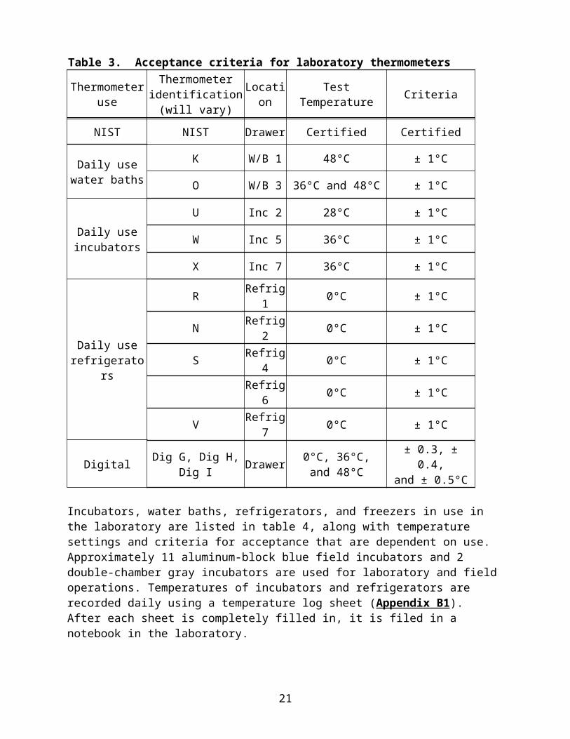
Table 3. Acceptance criteria for laboratory thermometers
Thermometer use
Thermometer identification
(will vary)Location Test Temperature Criteria
NIST NIST Drawer Certified Certified
Daily usewater baths
K W/B 1 48°C ± 1°C
O W/B 3 36°C and 48°C ± 1°C
Daily useincubators
U Inc 2 28°C ± 1°C
W Inc 5 36°C ± 1°C
X Inc 7 36°C ± 1°C
Daily userefrigerators
R Refrig 1 0°C ± 1°C
N Refrig 2 0°C ± 1°C
S Refrig 4 0°C ± 1°C
Refrig 6 0°C ± 1°C
V Refrig 7 0°C ± 1°C
Digital Dig G, Dig H, Dig I Drawer 0°C, 36°C,and 48°C
± 0.3, ± 0.4,and ± 0.5°C
Incubators, water baths, refrigerators, and freezers in use in the laboratory are listed in table 4, along with temperature settings and criteria for acceptance that are dependent on use. Approximately 11 aluminum-block blue field incubators and 2 double-chamber gray incubators are used for laboratory and field operations. Temperatures of incubators and refrigerators are recorded daily using a temperature log sheet (Appendix B1). After each sheet is completely filled in, it is filed in a notebook in the laboratory.
As of 11/19/2013, temperature data loggers were installed in select incubators, refrigerators, and freezers (see table 4). For this equipment, temperatures are recorded hourly and daily digital files are stored on the laptop computer in the lab and backed up to an external hard drive. Temperature data from these loggers are available online, and select staff will be notified by text message when the temperature has fallen outside the acceptance criteria (if the temperature has not returned to acceptable after a 30-minute recovery period). Further, once-daily, manual recording of temperatures for this equipment has been discontinued.
The temperatures of the laboratory incubators, water baths, and refrigerators are checked monthly with NIST-calibrated laboratory thermometers and recorded in LIMS. During use, the incubators, water baths, and refrigerators (without temperature loggers) are checked daily
14
and recorded on a designated daily log sheet posted on the front or side of each piece of equipment. Temperature daily log sheets are stored in the Temperature logbook.
The temperatures of the freezers are checked quarterly with NIST-calibrated laboratory thermometers and recorded in LIMS. The -70ºC freezers are equipped with automatic alarms that are set to respond when temperatures rise above -60ºC; the alarm system sounds during regular working hours and is attached to a telephone notification system after hours.
The operating temperatures of microbiological aluminum-block incubators are checked annually (or in preparation for a major study) and recorded in LIMS. During periods of heavy use in the laboratory or in the field, the temperatures are checked and recorded daily.
The two –70oC freezers (freezers 3 and 4) are used to store samples and microbiological cultures. A filter is cleaned and fans behind the filter are checked by laboratory personnel for operation quarterly and dates are recorded in LIMS. The condenser is dusted or vacuumed every 6 months and recorded in LIMS. A temperature chart is changed after a single pass around the chart (weekly).
Water baths are filled with distilled water and are cleaned with Liqui-Nox quarterly, or more often as needed. Record quarterly cleanings in LIMS.
15
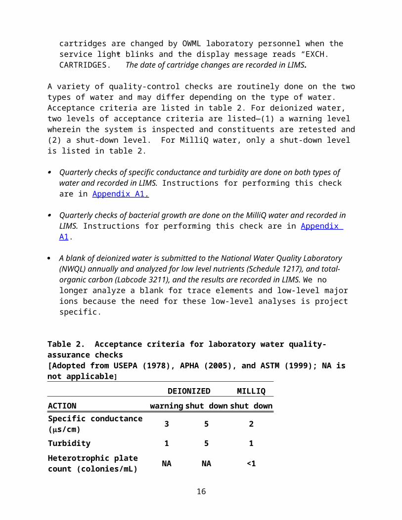
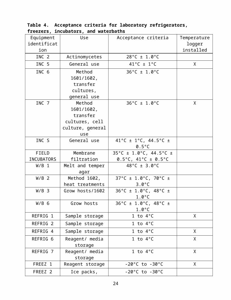
Table 4. Acceptance criteria for laboratory refrigerators, freezers, incubators, and waterbathsEquipment
identificationUse Acceptance criteria Temperature
logger installedINC 2 Actinomycetes 28°C ± 1.0°CINC 5 General use 41°C ± 1°C X
INC 6 Method 1601/1602, transfer cultures,
general use
36°C ± 1.0°C
INC 7 Method 1601/1602, transfer cultures, cell culture, general use
36°C ± 1.0°C X
INC S General use 41°C ± 1°C, 44.5°C ± 0.5°C
FIELD INCUBATORS
Membrane filtration 35°C ± 1.0°C, 44.5°C ± 0.5°C, 41°C ± 0.5°C
W/B 1 Melt and temper agar 48°C ± 3.0°C
W/B 2 Method 1602, heat treatments
37°C ± 1.0°C, 70°C ± 3.0°C
W/B 3 Grow hosts/1602 36°C ± 1.0°C, 48°C ± 1.0°C
W/B 6 Grow hosts 36°C ± 1.0°C, 48°C ± 1.0°C
REFRIG 1 Sample storage 1 to 4°C X
REFRIG 2 Sample storage 1 to 4°C
REFRIG 4 Sample storage 1 to 4°C X
REFRIG 6 Reagent/ media storage 1 to 4°C X
REFRIG 7 Reagent/ media storage 1 to 4°C X
FREEZ 1 Reagent storage -20°C to -30°C X
FREEZ 2 Ice packs, biological sample storage
-20°C to -30°C
FREEZ 3 Bacteria stocks, virus stocks, samples
Shelf 1 -70°C ± 10°CShelf 2 -70°C ± 10°CShelf 3 -60°C ± 10°CShelf 4 -60°C ± 10°CShelf 5 -60°C ± 10°C
X
FREEZ 5 Probes, hybridization reagents
-20°C to -30°C X
FREEZ 6 Cell culture, long term storage
Shelf 1 -70°C ± 10°CShelf 2 -70°C ± 10°CShelf 3 -60°C ± 10°CShelf 4 -60°C ± 10°CShelf 5 -60°C ± 10°C
X
FREEZ 7 Long term storage -20°C to -30°C
16

MicroscopeThere are two Zeiss microscopes used to perform microscopy. The Zeiss Axio Imager microscope has the capability to perform fluorescence microscopy and differential interference contrast (DIC). Additionally, it is equipped with a digital camera. The microscope has three objectives: 20X, 40X and 100X (oil), as well as an ocular micrometer. Furthermore, the microscope is equipped with excitation/band-pass filters for the immunofluorescence assay (FA) and 4',6-diamidino-2-pheylindole (DAPI) analysis. The microscope also has optics for DIC analysis under the 100X objective. It is kept in a room capable of being almost completely darkened.
The older Zeiss microscope is used for general laboratory work, Gram Stains, and Actinomycetes analysis. It has five objectives: 10X, 25X, 40X, 63X and 100X (oil), as well as an ocular micrometer.
The mercury bulb power supply for the Zeiss Axio Imager has an automated timer to monitor the number of hours the bulb has been on. After 150-200 hours, the mercury bulb should be changed. Note: the maximum life expectancy of the bulb is 300 hours. Call the manufacturer’s local representative or a professional company for assistance. The bulb must be disposed of in accordance with legal regulations, not in domestic waste. Contact the Carl Zeiss microscopy service for assistance. Record the mercury bulb number and date of installation in LIMS.
The ocular micrometer is calibrated for each objective at the time of purchase of the microscope by the manufacturer’s local representative. If a new objective is purchased for the microscope, the micrometer will need to be calibrated for this objective.
The cleaning procedures are the same for both microscopes.
The microscope is cleaned by a professional company yearly. Record this maintenance in LIMS.
After each use, clean the objectives and stage with lens paper and lens cleaner. A Q-tip and lens cleaner can be used to help remove oil from the end of the objective, as well as keep the oculars clean. Keep the dust protection cover on the microscopes while not in use and let the lamp housing cool before putting on the cover.
As needed, blow dust off of the microscope (especially the condenser, field aperature, and the oculars) with compressed air.
CentrifugesThere are three types of centrifuges that are used to perform separation of particles by centrifugal force. The refrigerated floor centrifuge is used to concentrate samples. The ultracentrifuge is used to isolate virus particles eluted from water samples and for concentrating bacterial spores. Two microcentrifuges are used for processing bacterial DNA/RNA extractions, purifications, concentrations, and phase separations. In addition, there are two larger refrigerated bench-top centrifuges that are used for concentrating viral particles and general separations.
Refrigerated floor centrifuge (1) and Refrigerated Bench-Top centrifuges (2)
17
The temperature is monitored quarterly with the digital thermometer (acceptance criteria is 4+ 3ºC).
The buckets are disinfected with dilute bleach and dechlorinated quarterly. Rotors and adapters are checked for deterioration, as needed. Lubrication is done annually, or as needed. All maintenance is recorded in LIMS.
Ultracentrifuge
Each run of the centrifuge is recorded in the centrifuge log book. The rotor and buckets are disinfected with dilute bleach and dechlorinated quarterly. Lubrication of the O-ring with vacuum grease, and lubrication of the buckets and cap
mating surfaces with Spinkote lubricant are done quarterly. The O-rings on the buckets are replaced twice a year. The vacuum pump oil is changed every 2 years. All maintenance is recorded in LIMS.
Microcentrifuge (2)
The chamber and the rotor are cleaned with soap and water quarterly. The air intake and exhaust vents are cleared from obstructions quarterly. Lubrication of the drive shaft and threads and O-ring with vacuum grease is done
quarterly. All maintenance is recorded in LIMS.
Thermal cyclersThe OWML uses two real-time thermal cyclers used to perform the quantitative polymerase chain reaction (qPCR), both are from Applied Biosystems: The AB 7500 Real Time PCR System (AB 7500) and the AB StepOnePlus Real Time PCR System. qPCR is done to amplify microbial DNA targets through a series of temperatures changes. Numerous projects use qPCR for a variety of applications including microbial source tracking, detection and/or quantification of enteric viruses, and detection and/or quantification of bacterial indicators.
Quality assurance measures for both thermal cyclers are as follows:
Background calibration is performed monthly. A pure-dye spectral assay is done annually or as needed (if replicate amplification curves
are continually wavy or non-uniform). A RNase P verification run is done as needed (as a troubleshooting measure). A regions of interest (ROI) plate is run as needed (following lamp replacement or if the
system is jostled). Annual preventative maintenance is done annually by AB technicians. All quality assurance checks are recorded in LIMS.
Sample management and documentationSamples for the bacterial indicators, E. coli, enterococci, fecal coliform, and total coliforms, are most commonly processed and analyzed in the field; however, they may be done in the OWML.
18

Holding times are 6 hours for compliance purposes and 24 hours for noncompliance purposes (American Public Health Association, 1998, Section 9060 B.) Adhering to a 6-hour holding time for all bacteriological samples, however, is highly recommended.
Samples for Clostridium perfringens and coliphage are processed and analyzed in the OWML; samples are kept on ice and processed within 48 hours of sample collection. Samples for the analysis of coliphage that arrive chilled within 48 to 96 hours from collection are acceptable, but the results are qualified. Samples for enteric viruses arrive at the OWML concentrated on filters; they must be kept on ice and processed within 72 hours of the start of filtration. Samples for Actinomycetes are processed in the OWML; samples are kept on ice and processed within 24 hours of sample collection. Samples for microbial source tracking markers generally arrive at the OWML as whole water samples; these samples are filtered and frozen within 24 hours of sample collection.
Laboratory personnel will then enter sample information on to a log sheet (Appendix B 2 ) and log the sample into LIMS, which will assign a login ID. There is one sample logbook that contains a log sheet, analytical services request forms (described below), and bench sheets with results. Laboratory personnel will write the login ID on the analytical services request form (front and back) and on the sample bottle (or filter cartridge).
All requests for laboratory analyses must be submitted using an analytical services request form (Appendix B3). This is a general service request form than can be altered for use with specific projects. For example, different analyses can be removed or added, or a list of for entering sample information can be included depending on project sampling strategies. The following categories must be filled out when requesting sample analysis: station name, site number, date/time of sample collection, medium code, sample type, Water Center user code, and project number. The field personnel must check off requested analyses and make prior arrangements with laboratory personnel. Upon receipt of the sample in the laboratory, personnel will fill in Received By and Date Received on the front of the analytical services request form. This form is filed in the sample logbook.
Samples are stored in the laboratory refrigerators until processing. Appropriate bench sheets are routed to the analyst, who will enter sample login ID, processing date/times, and analytical information.
Upon completion of the analysis, the analyst writes final results on the back of the analytical services request form. A second analyst routinely checks the calculations of the analyst performing the work. The results are entered into LIMS. All data supported by NWIS will be sent to the Water Quality Data Transfer System (QWDX) and available for download at https://qwdx.cr.usgs.gov/. An email will be sent to all clients when the data is available for download. Other data will be sent directly to the client.
After all of the sample result information is distributed to the client, the analytical services request form and appropriate bench sheets are then filed together in an archived folder sorted by login ID.
19

METHODS OF ANALYSIS, MEDIA AND REAGENT PREPARATION, AND ANALYTICAL QUALITY-CONTROL PROCEDURES
Methods of analysis, media and reagent preparation and storage, and analytical quality-control procedures are discussed in this section. Because microbiological analyses measure constantly changing living organisms, the methods are inherently variable. Some quality-control tools used by chemists, therefore, may not be available to the microbiologist (American Public Health Association, 1998, Section 9020 A).
References of published microbiological methods are kept in a notebook in the laboratory. Media-preparation instructions and method summaries written by the OWML are kept in the reference notebook and furnished as Appendices to this document.
The OWML has been working to optimize and field apply research methods that are being used in a variety of applications, including rapid assessment of recreational water quality, direct detection of pathogens, and microbial source tracking.
Culture methods for indicator bacteriaThe methods used for analysis of indicator bacteria are those of the USGS, USEPA, and APHA and others (table 5). All indicator bacteria methods used by the OWML are compliance or official methods.
Table 5. Culture methods for indicator bacteria used by the Ohio Water Microbiology Laboratory (OWML) [DW is drinking water, RW is recreational water]
BACTERIA METHOD TYPE OF METHOD REFERENCETotal coliforms
mENDO methodAppendix C1
Compliance—DWOfficial—other waters
Britton and Greeson (1989)APHA (2006, Section 9222B)
MI methodAppendix C2
Compliance—DWOfficial—other waters
USEPA (2002)
Colilert methodAppendix C3
Compliance—DW, RWOfficial—other waters
Idexx Corp., Westbrook, MEAPHA (2004, Section 9223)
Fecal coliforms
mFC methodAppendix C4
Compliance—DWOfficial—other waters
Britton and Greeson (1987)APHA (2006, Section 9222D)
Escherichia coli
Modified mTECAppendix C5
Compliance—RW, DW Official—other waters
USEPA (2006a)
MI methodAppendix C2
Compliance—DWOfficial—other waters
USEPA (2002)
Colilert methodAppendix C3 (water), C6 (sediment)
Compliance—DW, RWOfficial—other waters
Official—sediment
Idexx Corp., Westbrook, MEAPHA (2004, Section 9223)
Myers and others (2007)
20
Enterococci mEI methodAppendix C7, C8
Compliance—RW, DWOfficial—other waters
USEPA (2006b)APHA (2007, Section 9230C)
Enterococci EnterolertAppendix C9
Compliance—DW, RWOfficial—other waters
Idexx Corp., Westbrook, MEAPHA (2007, Section 9230D)
Clostridium perfringens
Modified mCP method
Appendix C10
Official—all waters USEPA (1996), modified by OWML
Reagents and media for indicator bacterial analyses are prepared according to the methods cited above. Bottle and plates are labeled with the name of the reagent or media, the preparation date, and the initials of the person who prepared it. Each lot of media is quality-control tested with a pure culture of the target bacterium or a sewage sample as a positive control; negative controls are also required (Appendix C11). Fresh sewage samples are obtained from the Olentangy Wastewater Treatment Plant as needed. Stock cultures of the positive and negative controls are kept on slants in the refrigerator and transferred once a month. Transfer dates are recorded in LIMS. When preparing positive and negative controls to be sent to other Water Centers, stock cultures must be transferred within a week before use.
QC results are recorded in the “Media and Buffer” logbook on quality-control sheets (Appendix B4); documentation of preparation procedures is also kept in this logbook. Media storage requirements and holding times are strictly followed (table 6). Target pH values for media and reagents are followed as described in table 7 and are recorded on the quality-control sheets in the “Media and Buffer” logbook. Requests for media, buffered-dilution water, and reagent preparation by project personnel are made using the “Expendable supplies request forms” (Appendix B5 ).
The type of buffered-dilution water used by the OWML is phosphate buffer with magnesium chloride dilution water (U.S. EPA, 2000a). Instructions for preparation are listed in Appendix A2.
Table 6. Information on media, buffered-dilution water, and reagents prepared and stored in the Ohio Water Microbiology Laboratory (OWML).
TYPE OF MEDIA/BUFFER
SOURCE STORAGE HOLDING TIME
mENDO agar Fisher Scientific or Hardy Diagnostics
Dry agar – CabinetPlates - refrigerator
As specified by manufacturer5 days as plates
MI agar OWML Refrigerator 6 months in dilution bottles2 weeks as plates
MI agar Govern. Scientific Source or Fisher Scientific
Dry agar – CabinetPlates - refrigerator
As specified by manufacturer2 weeks as plates
Colilert Idexx Corp., Westbrook, ME
Cabinet As specified by manufacturer
mFC agar Fisher Scientific or Hardy Diagnostics
Dry agar – CabinetPlates - refrigerator
As specified by manufacturer3 days as plates
21

Modified mTEC OWML Refrigerator 6 months in dilution bottles2 weeks as plates
Modified mTEC Govern. Scientific Source or Fisher Scientific
Dry agar – CabinetPlates - refrigerator
As specified by manufacturer2 weeks as plates
mEI OWML Refrigerator 6 months in dilution bottles3 days to 2 weeks as plates*
mEI Govern. Scientific Source or Fisher Scientific
Dry agar – CabinetPlates - refrigerator
As specified by manufacturer2 weeks as plates
Enterlolert Idexx Corp., Westbrook, ME
Cabinet As specified by manufacturer
mCP agar OWML Refrigerator 6 months in dilution bottles1 month as plates
mCP agar Oxoid Dry agar – Cabinet As specified by manufacturerPhosphate buffer with magnesium chloride (Appendix A2)
OWML Cabinet (unopened)Refrigerator (after opening)
1 year (unopened)1 week (after opening)
Phosphate buffer with magnesium chloride
Hardy Diagnostics, CA
Cabinet (unopened)Refrigerator (after opening)
As specified by manufacturer
* If reagents that are added after autoclaving are filter sterilized, the longer holding time is applied.
Table 7: Target pH values for media and reagent preparation
Media/Reagent Final pH +/- Reference
Actinomycete Isolation Agar 8.10 0.2 Difco #212168 Bottle
Bile Esculin Agar 6.80 0.2 USEPA; Method 1600; EPA-821-R-06-009; 2006
Brain Heart Infusion Agar 7.40 0.2 USEPA; Method 1600; EPA-821-R-06-009; 2006
Brain Heart Infusion Broth 7.40 0.2 USEPA; Method 1600; EPA-821-R-06-009; 2006
EC Broth 6.90 0.2 USEPA; Method 1603; EPA-821-R-06-011; 2006
ISP Media 1 7.00 0.2 Difco #276910 Bottle
ISP Media 2 7.20 0.2 Difco #277010 Bottle
mCP Agar* 7.60 not stated Bisson and Cabelli (1979)
mE Agar 7.10 0.2 Difco #233320 Bottle
mEI Agar** 7.10 0.2 USEPA; Method 1600; EPA-821-R-06-009; 2006
mEndo Agar 7.20 0.2 Difco #273620 Bottle
mEndo Broth MF 7.20 0.1 Difco #274930 Bottle
22

mFC Agar 7.40 0.2 Difco #267720 Bottle
MI Agar*** 6.95 0.2 USEPA; Method 1604; EPA-821-R-02-024; 2002
MI Broth 7.05 0.2 USEPA; Method 1604; EPA-821-R-02-024; 2002
mod mTEC Agar 7.30 0.2 USEPA; Method 1603; EPA-821-R-06-011; 2006
Nutrient Agar 6.80 0.2 USEPA; Method 1603; EPA-821-R-06-011; 2006
Phosphate Buffered Saline 7.40 0.2 USEPA; Method 1603; EPA-821-R-06-011; 2006
Simmons Citrate Agar 6.90 0.2 USEPA; Method 1603; EPA-821-R-06-011; 2006
Tryptic Soy Agar 7.30 0.2 USEPA; Method 1604; EPA-821-R-02-024; 2002
Tryptic Soy Broth 7.30 0.2 USEPA; Method 1603; EPA-821-R-06-011; 2006
Tryptone 7.30 0.2 USEPA; Method 1603; EPA-821-R-06-011; 2006
Working Phosphate Buffered Dilution Water 7.00 0.2 USEPA; Method 1603; EPA-821-R-06-011; 2006
*pH adjust before autoclaving
**Note: pH after nalidixic acid and TTC addition (aliquot a portion to check the pH)***Note: pH after cefsulodin addition (aliquot a portion to check the pH)
Updated 10/1/08
Analytical quality-control samples for fecal-indicator bacteria by membrane filtration (mENDO, MI, mFC, modified mTEC, mEI, and mCP agar methods) include the following:
Filter blank—a 50-100 mL aliquot of sterile buffered water is plated before the sample to confirm the sterility of equipment and supplies.
Procedure blank—a 50-100 mL aliquot of sterile buffered water is plated after every fifth sample to measure the effectiveness of the analyst’s rinsing technique or presence of incidental contamination of the buffered water.
A sewage sample is plated daily on mCP agar when C. perfringens analysis is done to evaluate the test procedure and to ensure anaerobic culture conditions.
For all media, positive and negative controls are plated for each batch of agar prepared or more often for some projects (Appendix C11).
For MI, positive and negative controls are plated every 10 samples to ensure proficiency with the method and evaluate the integrity of the medium. Positive and negative controls include the following:
o Positive controls of E. coli and Serratia marcescens
23
o Negative controls of Pseudmonas ATCC 10145 (unable to grow on MI and ensures the selectively of the agar) and Providencia alcalifaciens (grows on MI but will not fluoresce and ensures target colonies are correctly identified).
For some projects, a sewage sample is plated with each batch of MI plates at the time of sample analysis to evaluate the effectiveness of cefsulodin (an antibiotic added at the time of plate preparation).
Analytical quality-control samples for Colilert and Enterolert are purchased directly from IDEXX. For Colilert, E. coli and Klebsiella pneumoniae are the positive cultures and Pseudomonas aeruginosa is the negative culture. For Enterolert, Enterococcus faecalis is the positive culture and E. coli and Streptococcus bovis are the negative cultures. The acceptable ranges for each lot number of controls are provided in the manufacturer’s “Certificate of Analysis” available at http://www.idexx.com/view/xhtml/en_us/water/certificates.jsf. Control sets are run every week during busy periods or after every 20 samples to evaluate the test procedure and aid in interpretation of results.
Strain maintenance for positive and negative bacterial control cultures is described in Appendix A3. Strain information and culture maintenance are documented in the OWML LIMS.
ColiphageThe method currently in use for quantitative coliphage analysis by the OWML is the USEPA Method 1602, single-agar layer (SAL) procedure (USEPA, 2001b). This method is generally most suitable for quantification of coliphage in surface-water samples. Antibiotic-resistant E. coli CN-13 (resistant to nalidixic acid) and E. coli Famp (resistant to streptomycin and ampicillin) are used as bacterial hosts for somatic and F-specific coliphage, respectively. The protocol for Method 1602 and forms for regular and QC samples are included in Appendix C12.
The method currently in use for qualitative coliphage analysis by the OWML in larger sample volumes is the USEPA Method 1601, two-step enrichment method (USEPA, 2001a). Sample volumes of 1 L are recommended for detection of coliphage using this method. Because the SAL method is impractical for sample volumes above 100 mL, the two-step enrichment method is often used for groundwater sample analysis. The same bacterial hosts are used in the two-step enrichment method as are used in the SAL method. The protocol for Method 1601 and forms for regular and QC samples are included in Appendix C13.
Results from coliphage QC samples are recorded in LIMS. Coliphage QC log forms, results for other coliphage QC samples, and coliphage stock enumeration results are kept in the Sample Log Book. Information on media sterility is stored in the Media Log Book. Information on host culture strains is maintained in LIMS.
ActinomycetesThe Actinomycetes are a large group of filamentous gram-positive bacteria that resemble fungi because they produce mycelium and dry spores, called conidia (Madigan and others, 2000). They are considered nuisance organisms for those in the water industry, as they are one of two types of organisms that impart an earthy-musty odor to waters. The odors are caused by two
24

compounds formed during normal actinomycete development, geosmin and 2-methylisoborneol (American Public Health Association, 2005).
The method used for isolation of Actinomycetes from water in the OWML is based on a published method (American Public Health Association, 2005) and a method provided by a commercial supplier of Actinomycetes medium (BD-Difco, Sparks, Maryland, The Difco & BBL Manual, 2009). A double agar layer method is used (Appendix C14) and QC samples described in this method are entered into a QC log specifically for Actinomycetes analysis (Appendix B6). Stock cultures of Streptomycetes albus are used as positive controls. The stock culture is transferred every two months and transfers are recorded in LIMS.
Rapid methods for indicator bacteriaTraditional microbiological methods for detecting fecal-indicator bacteria and pathogenic organisms can take at least 18 hours to obtain results. Because water quality can change significantly during this timeframe, the safety of the water may not be accurately assessed. The need for rapid detection methods that provide reliable results of the current day’s water-quality conditions is widely recognized. The USGS OWML is currently using two rapid detection methods for the enumeration of E. coli and enterococci.
The immunomagnetic separation/adenosine triphosphate (IMS/ATP) rapid method requires approximately 2 hours from sample collection to availability of results (Bushon and others, 2007; Bushon and others, 2009; Lee and Deininger, 2004). Magnetic beads that are coated with antibodies for either Escherichia coli (E. coli) or enterococci are added to a water sample. This mixture is then subjected to IMS, in which the bacteria-antibody-bead complex is separated from extraneous materials in the sample by use of a strong magnet. Following several wash/concentration steps, the bacterial cells are ruptured by an enzymatic process, releasing ATP, which is the energy molecule found in living cells. The amount of ATP in the sample is measured with a microluminometer and results are reported in relative light units (RLUs).
The quantitative polymerase chain reaction (qPCR) method enumerates targeted genetic sequences within microorganisms in less than three hours. First, water samples are concentrated by passing through a 0.4 µm filter. The concentrated organisms are lysed by both physical and chemical disruption and the genetic material that was contained in these organisms is then isolated and purified. The qPCR is run and the genetic sequence unique to the organism of interest is quantified by detecting the accumulation of a fluorescent probe. The OWML is currently applying qPCR assays for enterococci (U.S. Environmental Protection Agency, 2012a) and E. coli (Noble and others, 2010).
Molecular methods for microbial source tracking markers and cyanobacteriaNote: Appendices for molecular methods are available upon request.
Molecular methods for microbial source tracking (MST) markers and cyanobacteria used by the OWML, target genetic sequences in bacteria using quantitative polymerase chain reaction (qPCR). Polymerase chain reaction is a technology in which DNA from a targeted gene is amplified, generating millions of copies, in order to determine if the targeted gene is present. For quantitative polymerase chain reaction (qPCR), a fluorescent signal is used to quantify the amount of targeted gene relative to a known quantity positive control.
25
Microbial source tracking (MST) is a term used for the process of identifying the source of fecal contamination in the environment. Microbial source tracking using molecular markers is carried out by detecting genetic sequences in the DNA of fecal-origin bacteria that are specific to the host species that produced the feces. Host- associated molecular markers have been identified based on the theory that the physiology in the gut of the host animal (e.g. diet, temperature, antibiotic treatment, etc.) is unique from one animal to another. These unique conditions select for unique subsets of microorganisms in the gut. Host-associated markers have been identified from different groups of fecal-origin bacteria, often from the genus Bacteroides, a bacterium abundant in the gut of warm-blooded animals. The OWML has adopted the capability to analyze for the MST marker assays listed in Table 8.
Table 8: Microbial source tracking (MST) marker qPCR assays
Marker Source Targeted bacterium ReferenceAllBac General Bacteroides Layton and others, 2006GenBac General Bacteroides Siefring and others, 2008HF183 Human Bacteroides Seurinck and others, 2005BacHum Human Bacteroides Kildare and others, 2007BoBac Ruminant Bacteroides Layton and others, 2006Gull2 Gull Catellicoccus
marimammaliumSinigalliano and others, 2010
BacCan Dog Bacteroides Kildare and others, 2007HoF597 Horse/Mule Bacteroides Dick and others, 2005GFD General waterfowl Helicobacter Green and others, 2012
Toxic freshwater cyanobacterial blooms are of concern in many parts of the world because of their effects on drinking water, water-based recreation, and watershed ecology. Microcystins are one of the most frequently detected hepatotoxins in freshwaters and are generally produced by strains of the genera Microcystis, Planktothrix and Anabaena (Rantala and others, 2006). For toxin production to occur, the microcystin synthetase genes (mcy) must be present in the genome of toxic strains. Known microcystin-producing genera include both toxic strains (with the mcy genes) and nontoxic strains (without the mcy genes), which can be differentiated only by molecular detection methods qPCR. The OWML has developed the capability to analyze samples by several cyanobacteria molecular assays using qPCR (Table 9). The DNA-based qPCR assays reveal the presence of toxin genes (irrespective of whether they are actively producing toxin). The RNA-based assays, using reverse-transcription quantitative polymerase chain reaction (RT-qPCR), can detect microcystin-producing cyanobacteria that are actively expressing the toxin genes (Sipari et al., 2010). Table 9: Cyanobacterial assays for freshwater studies (toxic strains refer to the ability to produce microcystin)
Assay Description ReferenceCyanobacteria, general
DNA assay, targets several cyanobacterial genera
Rinta-Kanto and others, 2005
Microcystis spp. DNA assay, targets toxic and non-toxic strains of Microcystis
Rinta-Kanto and others, 2005
26

Anabaena spp. DNA assay, targets toxic and non-toxic strains of Anabaena
Doblin and others, 2007
Planktothrix spp. DNA assay, targets toxic and non-toxic strains of Planktothrix
Ostermaier and Kurmayer, 2009
General mcyE DNA assay, targets toxic cyanobacteria
Rantala and others, 2004
Microcystis and Anabaena mcyE
DNA and RNA assays, targets toxin genes specific to Microcystis and Anabaena
Sipari and others, 2010
Planktothrix mcyE DNA and RNA assays, targets toxin genes specific to Planktothrix
Rantala and others, 2006
Because they are both bacterial targets, the steps for processing and analyzing samples by qPCR for MST markers are similar to those for cyanobacteria. Studies incorporating MST often include analysis of known-source fecal samples or sediment samples in addition to water samples; sample collection and filtration steps are split into separate protocols for these two different matrix types (solid material and liquid material).
1. Sample collection and initial processing. a. Collection of water samples for MST and cyanobacterial analyses is done following
the same USGS protocol as is used for indicator bacteria (Myers and others, 2007). One difference in sample collection is that the bottles used to collect the samples should be acid-treated to remove all DNA, RNA, or DNA/RNA degrading substances (Appendix D1). Appendix D1a is a benchsheet that is used for any sample type being processed for MST.
b. Collection and initial processing of known-source fecal samples for MST markers is done following the protocol in Appendix D2 and using the fecal sample collection field form (Appendix D2a).
c. Collection and initial processing of sediment samples for MST markers is done following the protocol in Appendix D2.
2. DNA and RNA extraction and purification.a. DNA extraction and purification of a sample for MST and cyanobacteria is done
using the GeneRite DNA-EZ kit as described in Appendix D3.b. RNA extraction and purification of a sample for cyanobacteria is done using the
MoBio PowerPlant RNA isolation kit with DNase as described in Appendix D7.c. Extraction information for each sample is recorded on the “filtration and extraction”
benchsheet (Appendix D1a).d. The extraction and purification process is done in sample batches which are recorded
on a benchsheet (Appendix D3a).
3. qPCR for MST markers and cyanobacteria.a. Detection of MST markers and cyanobacteria can be done by following Appendix
D4. b. Details and benchsheets for MST assays can be found in Appendix D4a.
27

c. Details and benchsheets for cyanobacterial DNA assays can be found in Appendix D4b.
d. Details and benchsheets for cyanobacterial RNA assays can be found in Appendix D4c.
4. Positive control development. Positive controls are used to establish known quantity standard curves which are used to quantify unknown samples and ensure the qPCR reaction was performed properly.
a. E. coli plasmids containing the target sequence for MST and cyanobacterial assays are generated and quantified as described in Appendix D5 for use as positive controls.
b. A plasmid-based positive control benchsheet can be found in Appendix D5a.
5. Standard curves. Successful establishment of positive controls will allow for subsequent development of a standard curve which will be used to quantify unknown samples.
a. Standard curve development of plasmid-based positive controls can be found in Appendix D5.
b. Standard curve information for each qPCR run is recorded on the assay-specific benchsheet (Appendices D4a-D4d).
6. Data interpretation.a. A protocol for the handling of qPCR data, assessment of matrix inhibition, and
recording of final results can be found in Appendix D6.b. Data templates for water, sediment, and fecal-source samples can be found in
Micro/Current Molecular Protocols.
Methods for enteric virusesNote: Appendices for enteric viruses are available upon request.
Samples for enteric viruses are analyzed by the OWML by use of USEPA Method 1615 (USEPA, 2012b). Method 1615 includes two methods for analyzing water samples—reverse-transcription quantitative polymerase chain reaction (RT-qPCR) and cell culture. The RT-qPCR method quantifies human enteroviruses and human noroviruses by targeting genetic sequences specific to these viruses. Molecular methods, such as RT-qPCR, do not determine infectivity of viruses, but they have an advantage in that they can detect many more types of enteric viruses. The cell culture method detects enterovirus and orthoreovirus species that are capable of infecting and producing cytopathic effects (CPE) in the Buffalo Green Monkey Kidney (BGM) cell line. Not all viruses can be detected by cell culture, including noroviruses. Cell culture is used to determine whether the enteroviruses are infectious.
Protocols for RT-qPCR can be found in Appendix E1 (elution and organic flocculation protocol), Appendix E2 (foam elution and concentration), and Appendix E3 (reverse transcription and qPCR protocol). The sample processing benchsheets can be found in Appendixes E1a and E2a, and the qPCR batch sheets can be found in Appendix E1b. Appendix E3a contains the assay specific qPCR benchsheets with detailed information about each assay. The cell culture protocol can be found in Appendix E4, along with bench sheets for cell culture and viral titer results (Appendix E4a). The method for collection and initial processing of water samples for human
28
polyomavirus and the corresponding bench sheet are found in Appendixes E5 and E5a. Extraction for human polyomavirus is described in Appendix E6.
Methods for cyanobacterial toxinsSamples for the cyanobacterial toxins are analyzed by the OWML for MI-OH WSC projects only. Currently the OWML analyzes samples for one toxin—microcystin—by an enzyme linked immunosorbent assay (ELISA) provided in a kit (Abraxis LLC, Warminster, Penn.). The OWML SOP for ELISA is based on Ohio Environmental Protection Agency (2015) (Appendix F1). The sample processing benchsheet can be found in Appendix F1a.
REFERENCES
American Public Health Association, American Water Works Association, and Water Environment Federation, 2004–2007, Standard methods for the analysis of water and wastewater: Washington, D.C., American Public Health Association [variously paginated].
American Society for Testing and Materials, 1999, Annual Book of ASTM Standards, Section 11, Water and Environmental Technology, Designation: D 1193-99, p. 107-109.
Bisson, J.W., and Cabelli, V.J., 1979, Membrane Filter Enumeration Method for Clostridium perfringens: Applied and Environmental Microbiology, v. 37, no. 1, p. 55-66.
Britton , L.J., and Greeson, P.E., eds., 1987, Methods for collection and analysis of aquatic biological and microbiological samples: U.S. Geological Survey Techniques of Water-Resources Investigations, book 5, chap. A4, 363 p.
Bushon, R.N., Brady, A.M., Likirdopulos, C.A., and Cireddu, J.V., 2009, Rapid detection of Escherichia coli and enterococci in recreational water using immunomagnetic separation/adenosine triphosphate technique: Journal of Applied Microbiology, v. 106, p. 432-441.
Bushon, R.N., Brady, A.M.G., and Plona, M.B., 2007, Using a rapid method to predict recreational water quality at the Cuyahoga Valley National Park, Ohio: Park Science, v. 24, no.2, p. 89–93.
Dick, L.K., Bernhard, A.E., Brodeur, T.J., Santo Domingo, J.W., Simpson, J. M., Walters, S.P., and Field, K.G., 2005, Host distributions of uncultivated fecal Bacteroidales reveal genetic markers for fecal source identification: Applied and Environmental Microbiology, v. 71, no. 6, p. 3184–3191.
Doblin, M.A., Coyne, K.J., Rinta-Kanto, J.M., Wilhelm, S.W., and Dobbs, F.C., 2007, Dynamics and short-term survival of toxic cyanobacteria species in ballast water from NOBOB vessels transiting the Great lakes—implications for HAB invasions: Harmful Algae, v. 6., p. 519-530.
29

Fout, G.S., Martinson, B.C., Moyer, M.W.N., and Dahling, D.R., 2003, A multiplex reverse transcription-PCR method for detection of human enteric viruses in groundwater: Applied and Environmental Microbiology, v. 69, no. 6, p. 3158-3164.
Francy, D.S., and Shaffer, K., 2008, Quality-assurance plan for water-quality activities in the Ohio Water Science Center: U.S. Geological Survey Open-File Report 2008-1250, 73 p.
Francy, D.S., Helsel, D.L., and Nally, R.A., 2000, Occurrence and distribution of micro-biological indicators in ground water and stream water: Water Environment Research, v. 72, no. 2, 152 p.
Green, H.C., Dick, L.K., Gilpin, B., Samadpour, M., and Field, K.G., 2012, Genetic markers for rapid PCR-based identification of gull, Canada goose, duck, and chicken fecal contamination in water: Applied and Environmental Microbiology, v. 78, no. 2, p. 503–510.
Gregory, J.B., Litaker, R.W., and Noble, R.T., 2006, Rapid one-step quantitative reverse transcriptase PCR assay with competitive internal positive control for detection of enteroviruses in environmental samples: Applied and Environmental Microbiology, v. 72, no. 6, p. 3960-3967.
Ijzerman, M.M., and Hagedorn, C., 1992, Improved method for coliphage detection based on -galactosidase induction: Journal of Virological Methods, v. 40, p. 31-36.
Jothikumar, N., Cromeans, T.L., Hill, V.R., Lu, X., Sobsey, M.D., and Erdman, D.D., 2005a, Quantitative real-time PCR assays for detection of human adenovirus and identification of serotypes 40 and 41: Applied and Environmental Microbiology, v. 71, no. 6, p. 3131-3136.
Jothikumar, N., Lowther, J.A., Henshilwood, K., Lees, D.N., Hill, V.R., Vinje, J., 2005b, Rapid and Sensitive Detection of Noroviruses by Using TaqMan-Based One-Step Reverse Transcription-PCR Assays and Application to Naturally Contaminated Shellfish Samples: Applied and Environmental Microbiology, v. 71, no. 4, p. 1870-1875.
Kildare, B.J., Leutenegger, C.M., McSwain, B.S., Bambic, D.G., Rajal, V.B., and Wuertz, S., 2007, 16S rRNA-based assays for quantitative detection of universal, human-, cow-, and dog-specific fecal Bacteroidales: A Bayesian approach: Water Research, v. 41, p. 3701–3715.
Lambertini, E., Spencer, S.K., Bertz, P.D., Loge, F.J., Kieke, B.A., and Borchardt, M.A., 2008, Concentration of enteroviruses, adenoviruses, and noroviruses from drinking water by use of glass wool filters: Applied and Environmental Microbiology: vol. 74, no. 10, p. 2990-2996.
Layton, A., McKay, L., Williams, D., Garrett, V., Gentry, R., and Sayler, G., 2006, Development of Bacteroides 16S rRNA gene TaqMan-based real-time PCR assays for estimation of total, human, and bovine fecal pollution in water: Applied and Environmental Microbiology, v. 72, no. 6, p. 4214-4224.
30

Lee, JiYoung, and Deininger, R.A., 2004, Detection of E. coli in beach water within 1 hour using immunomagnetic separation and ATP bioluminescence: Luminescence, v. 19, p. 31–36.
Madigan, M. T., Martinko, J.M., and Parker, J., Brock, Biology of Microoganisms—Ninth Edition: Prentice Hall, Upper Sadlle River, NJ, p. 519.
Morales-Morales, H.A., Vidal, G., Olszewski, J., Rock, C.M., Dasgupta, D., Oshima, K.H., and Smith, G.B., 2003, Optimization of a reusable hollow-fiber ultrafilter for simultaneous concentration of enteric bacteria, protozoa, and viruses from water: Applied and Environmental Microbiology, v. 69, p. 4098−4012.
Myers, D.N., Stoeckel, D.M., Bushon, R.N., Francy, D.S., and Brady, A.M.G., 2007, Fecal indicator bacteria (ver. 2.0): U.S. Geological Survey Techniques of Water-Resources Investigations, book 9, chap. A7, section 7.1, February, accessed April 2013 from http://pubs.water.usgs.gov/twri9A/.
Noble, R.T., Blackwood, A. Denene, Griffith, J.F., McGee, C.D., and Weisberg, S.B., 2010 , Comparison of Rapid Quantitative PCR-Based and Conventional Culture-Based Methods for Enumeration of Enterococcus spp. and Escherichia coli in Recreational Waters: Applied and Environmental Microbiology, v. 76, no. 22, p. 7437-7443.
Ohio Environmental Protection Agency, 2015, Ohio EPA Total (extracellular and intracellular) microcystins—ADDA by ELISA Analytical Methodology, Version 2.2, November 2015, 6 p.
Ostermaier and Kurmayer, 2009, Distribution and abundance of nontoxic mutants of cyanobacteria in lakes of the Alps: Microb. Ecol. v. 58, 323-333.
Rantala, A., Fewer, D.P., Hisbergues, M., Rouhiainen, L., Vaitomaa, J., Borner, T., Sivonen, K., 2004, Phylogenetic evidence for the early evolution of microcystin synthesis: Proceedings of the National Academy of Science, v. 101, no. 2, p. 568-573.
Rantala, A., Rajaniemi-Wacklin, P., Lyra, C., Lepisto, L., Rintala, J., Mankiewicz-Boczek, J., Sivonen, K. 2006. Detection of microcystin-producing cyanobacteria in Finnish lakes with genus-specific microcystin synthetase gene E (mcyE) PCR and associations with environmental factors. Appl. Environ. Microbiol. 72:6101-6110.
Rinta-Kanto, J.M., Ouellette, A.J.A., Boyer, G.L., Twiss, M.R., Bridgeman, T.B., Wilhelm, S.W. 2005. Quantification of toxic Microcystis spp. during the 2003 and 2004 blooms in western Lake Erie using quantitative real-time PCR. Environ. Sci. Technol. 39:4198-4205.
Seurinck, S., Defoirdt, T., Verstraete, W., Siciliano, S.D., 2005, Detection and quantification of the human-specific HF183 Bacteroides 16S rRNA genetic marker with real-time PCR for assessment of human faecal pollution in freshwater: Environmental Microbiology, v. 7, no. 2, p. 249–259.
31

Siefring S., Varma M., Atikovic E., Wymer L., and Haugland R., 2008, Improved real-time PCR assays for the detection of fecal indicator bacteria in surface waters with different instrument and reagent systems: Journal of Water and Health, v. 6, no. 2, p. 225–237.
Sinigalliano, C.D., Fleisher, J.M., Gidley, M.L., Solo-Gabriele, H.M., Shibata, T., Plano, L.R.W., Elmir, S.M., Wanless, D., Bartkowiak, J., Boiteau, R., Withum, K., Abdelzaher, A.M., He, G., Ortega, C., Zhu, X., Wright, M.E., Kish, J., Hollenbeck, J., Scott, T., Backer, L.C., Fleming, L.E. 2010. Traditional and molecular analyses for fecal indicator bacteria in non-point source subtropical recreational marine waters. Water Research, v. 44, p. 3763-3772.
Sipari, H., Rantala-Ylinen, A., Jokela, J., Oksanen, I., Sivonen, K. 2010. Development of a chip assay and quantitative PCR for detecting microcystin synthetase E gene expression. Appl. Environ. Microbiol. 76:3797-3805.
U.S. Environmental Protection Agency, 1978, Microbiological methods for monitoring the environment—water and wastes: Cincinnati, Ohio, U.S. Environmental Protection Agency, EPA-600/8-78-017, 338 p.
_________________1985, Test methods for Escherichia coli and enterococci in water by the membrane filtered procedure: Cincinnati, Ohio, Environmental Monitoring and Support Laboratory, EPA 600/4-85/076, 24 p.
_________________1996, EPA Information Collection Rule microbial laboratory manual: Washington, D.C., U.S. Environmental Protection Agency, EPA/600/R-95/178.
_________________2000a, Improved enumeration methods for the recreational water quality indicators: enterococci and Escherichia coli: U.S. Environmental Protection Agency, Office of Science and Technology, EPA/821/R-97/004.
_________________2001a, Method 1601: Male-specific (F+) and somatic coliphage in water by two-step enrichment procedure: Washington, D.C., U.S. Environmental Protection Agency, EPA-821-R-01-030.
_________________2001b, Method 1602: Male-specific (F+) and somatic coliphage in water by single agar layer (SAL) procedure: Washington, D.C., U.S. Environmental Protection Agency, EPA-821-R-01-029.
_________________ 2002, Method 1604—Total coliforms and Escherichia coli in water by membrane filtration using a simultaneous detection technique (MI medium): Washington D.C., EPA 821-R-02-024, 14 p.
_________________ 2005, Method 1623Cryptosporidium and Giardia in water by filtration/IMS/FA: Washington, D.C., EPA 815-R-05-002, 76 p.
______________ 2006a, Method 1603—Escherichia coli in water by membrane filtration using modified membrane-thermotolerant Escherichia coli agar (Modified mTEC): Washington, D.C., EPA 821-R-06-011, 24 p.
32
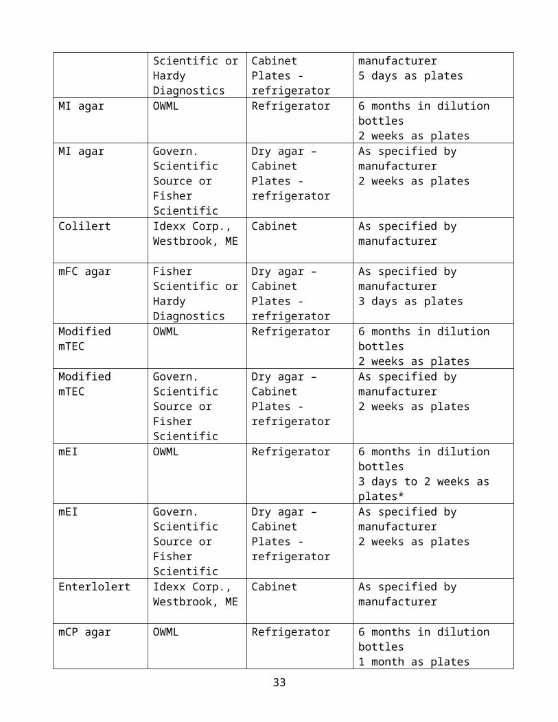
_________________2006b, Method 1600—Enterococci in water by membrane filtration using membrane-Enterococcus Indoxyl-b-D-Glucoside agar (mEI): Washington, D.C., EPA-821-R-06-009, 24 p.
______________ 2012a, Method 1611—Enterococci in water by TaqMan quantitative polymerase chain reaction (qPCR) assay: Washington, D.C., EPA 821-R-12-008, 48 p., accessed April 2013 at http://water.epa.gov/scitech/methods/cwa/bioindicators/biological_index.cfm.
______________ 2012b, Method 1615—measurement of enterovirus and norovirus occurrence in water by culture and RT-qPCR: Office of Research and Development, National Exposure Research Laboratory, Cincinnati, OH., EPA 600/R-10/181, accessed July 2013 at http://www.epa.gov/nerlcwww/research/waterbourneviruses.html
Weidhaas, J.L., Macbeth, T.W., Olsen, R.L., Sadowsky, M.J., Norat, D., and Harwood, V.J., 2010, Identification of a Brevibacterium marker gene specific to poultry litter and development of a quantitative PCR assay: Journal of Applied Microbiology, v. 109, p. 334–347.
Return to Microbiology || Ohio Home Page
U.S. Department of the Interior, U.S. Geological SurveyEmail Maintainer: webmaster with comments, suggestions, etc.Privacy Statement || Disclaimer ||Accessibility
Last update: July 2016
33
Related Documents