RESEARCH ARTICLE Q fever in Egypt: Epidemiological survey of Coxiella burnetii specific antibodies in cattle, buffaloes, sheep, goats and camels Jessica Klemmer 1 *, John Njeru 2 , Aya Emam 3 , Ahmed El-Sayed 4 , Amira A. Moawad 1 , Klaus Henning 1 , Mohamed A. Elbeskawy 5 , Carola Sauter-Louis 6 , Reinhard K. Straubinger 7 , Heinrich Neubauer 1 , Mohamed M. El-Diasty 3 1 Institute of Bacterial Infections and Zoonoses, Friedrich-Loeffler-Institut, Jena, Germany, 2 Centre for Microbiology Research, Kenya Medical Research Institute, Nairobi, Kenya, 3 Mansoura Provincial Laboratory, Institute of Animal Health Research, Mansoura, Egypt, 4 Alshalateen Provincial Laboratory, Institute of Animal Health Research, Alshalateen, Egypt, 5 Department of Internal Medicine, Infectious Diseases and Fish Diseases, Faculty of Veterinary Medicine, Mansoura University, Mansoura, Egypt, 6 Institute of Epidemiology, Friedrich-Loeffler-Institut, Greifswald, Germany, 7 Institute of Infectious Diseases and Zoonoses, Department of Veterinary Sciences, Faculty of Veterinary Medicine, Ludwig- Maximilian University, Munich, Germany * [email protected] Abstract Q fever is a zoonotic disease caused by the bacterium Coxiella burnetii. Clinical presenta- tion in humans varies from asymptomatic to flu-like illness and severe sequelae may be seen. Ruminants are often sub-clinically infected or show reproductive disorders such as abortions. In Egypt, only limited data on the epidemiology of Q fever in animals are available. Using a stratified two stage random sampling approach, we evaluated the prevalence of Coxiella burnetii specific antibodies among ruminants and camels in 299 herds. A total of 2,699 blood samples was investigated using enzyme-linked-immunosorbent assay (ELISA). Coxiella burnetii specific antibodies were detected in 40.7% of camels (215/528), 19.3% of cattle (162/840), 11.2% of buffaloes (34/304), 8.9% of sheep (64/716) and 6.8% of goats (21/311), respectively. Odds of seropositivity were significantly higher for cattle (aOR: 3.17; 95% CI: 1.96–5.13) and camels (aOR: 9.75; 95% CI: 6.02–15.78). Significant differ- ences in seropositivity were also found between domains (Western Desert, Eastern Desert and Nile Valley and Delta) and 25 governorates (p < 0.001), respectively. Animal rearing in the Eastern Desert domain was found to be a significant risk factor (aOR: 2.16; 95% CI: 1.62-2.88). Most seropositive animals were older than four years. No correlation between positive titers and husbandry practices or animal origin were found (p > 0.05). Only 8.7% of the interviewed people living on the farms consumed raw camel milk and none reported prior knowledge on Q fever. Findings from this nationwide study show that exposure to Cox- iella burnetii is common in ruminants and camels. Disease awareness among physicians, veterinarians and animal owners has to be raised. Future epidemiological investigations have to elucidate the impact of Q fever on human health and on the economy of Egypt. PLOS ONE | https://doi.org/10.1371/journal.pone.0192188 February 21, 2018 1 / 12 a1111111111 a1111111111 a1111111111 a1111111111 a1111111111 OPEN ACCESS Citation: Klemmer J, Njeru J, Emam A, El-Sayed A, Moawad AA, Henning K, et al. (2018) Q fever in Egypt: Epidemiological survey of Coxiella burnetii specific antibodies in cattle, buffaloes, sheep, goats and camels. PLoS ONE 13(2): e0192188. https:// doi.org/10.1371/journal.pone.0192188 Editor: Pierre Roques, CEA, FRANCE Received: August 30, 2017 Accepted: January 19, 2018 Published: February 21, 2018 Copyright: © 2018 Klemmer et al. This is an open access article distributed under the terms of the Creative Commons Attribution License, which permits unrestricted use, distribution, and reproduction in any medium, provided the original author and source are credited. Data Availability Statement: All relevant data are within the paper and its Supporting Information files. A dataset for reproducibility is available from the zenodo.org database (accession number 10. 5281/zenodo.1148508). URL: https://doi.org/10. 5281/zenodo.1148508. Funding: This project was funded by the German Federal Foreign Office (https://www.auswaertiges- amt.de/de/ ). The funding was received by the Friedrich-Loeffler-Institut. No grant number exists. The funders had no role in study design, data

Welcome message from author
This document is posted to help you gain knowledge. Please leave a comment to let me know what you think about it! Share it to your friends and learn new things together.
Transcript
RESEARCH ARTICLE
Q fever in Egypt: Epidemiological survey of
Coxiella burnetii specific antibodies in cattle,
buffaloes, sheep, goats and camels
Jessica Klemmer1*, John Njeru2, Aya Emam3, Ahmed El-Sayed4, Amira A. Moawad1,
Klaus Henning1, Mohamed A. Elbeskawy5, Carola Sauter-Louis6, Reinhard
K. Straubinger7, Heinrich Neubauer1, Mohamed M. El-Diasty3
1 Institute of Bacterial Infections and Zoonoses, Friedrich-Loeffler-Institut, Jena, Germany, 2 Centre for
Microbiology Research, Kenya Medical Research Institute, Nairobi, Kenya, 3 Mansoura Provincial
Laboratory, Institute of Animal Health Research, Mansoura, Egypt, 4 Alshalateen Provincial Laboratory,
Institute of Animal Health Research, Alshalateen, Egypt, 5 Department of Internal Medicine, Infectious
Diseases and Fish Diseases, Faculty of Veterinary Medicine, Mansoura University, Mansoura, Egypt,
6 Institute of Epidemiology, Friedrich-Loeffler-Institut, Greifswald, Germany, 7 Institute of Infectious
Diseases and Zoonoses, Department of Veterinary Sciences, Faculty of Veterinary Medicine, Ludwig-
Maximilian University, Munich, Germany
Abstract
Q fever is a zoonotic disease caused by the bacterium Coxiella burnetii. Clinical presenta-
tion in humans varies from asymptomatic to flu-like illness and severe sequelae may be
seen. Ruminants are often sub-clinically infected or show reproductive disorders such as
abortions. In Egypt, only limited data on the epidemiology of Q fever in animals are available.
Using a stratified two stage random sampling approach, we evaluated the prevalence of
Coxiella burnetii specific antibodies among ruminants and camels in 299 herds. A total of
2,699 blood samples was investigated using enzyme-linked-immunosorbent assay
(ELISA). Coxiella burnetii specific antibodies were detected in 40.7% of camels (215/528),
19.3% of cattle (162/840), 11.2% of buffaloes (34/304), 8.9% of sheep (64/716) and 6.8% of
goats (21/311), respectively. Odds of seropositivity were significantly higher for cattle (aOR:
3.17; 95% CI: 1.96–5.13) and camels (aOR: 9.75; 95% CI: 6.02–15.78). Significant differ-
ences in seropositivity were also found between domains (Western Desert, Eastern Desert
and Nile Valley and Delta) and 25 governorates (p < 0.001), respectively. Animal rearing in
the Eastern Desert domain was found to be a significant risk factor (aOR: 2.16; 95% CI:
1.62-2.88). Most seropositive animals were older than four years. No correlation between
positive titers and husbandry practices or animal origin were found (p > 0.05). Only 8.7% of
the interviewed people living on the farms consumed raw camel milk and none reported
prior knowledge on Q fever. Findings from this nationwide study show that exposure to Cox-
iella burnetii is common in ruminants and camels. Disease awareness among physicians,
veterinarians and animal owners has to be raised. Future epidemiological investigations
have to elucidate the impact of Q fever on human health and on the economy of Egypt.
PLOS ONE | https://doi.org/10.1371/journal.pone.0192188 February 21, 2018 1 / 12
a1111111111
a1111111111
a1111111111
a1111111111
a1111111111
OPENACCESS
Citation: Klemmer J, Njeru J, Emam A, El-Sayed A,
Moawad AA, Henning K, et al. (2018) Q fever in
Egypt: Epidemiological survey of Coxiella burnetii
specific antibodies in cattle, buffaloes, sheep, goats
and camels. PLoS ONE 13(2): e0192188. https://
doi.org/10.1371/journal.pone.0192188
Editor: Pierre Roques, CEA, FRANCE
Received: August 30, 2017
Accepted: January 19, 2018
Published: February 21, 2018
Copyright: © 2018 Klemmer et al. This is an open
access article distributed under the terms of the
Creative Commons Attribution License, which
permits unrestricted use, distribution, and
reproduction in any medium, provided the original
author and source are credited.
Data Availability Statement: All relevant data are
within the paper and its Supporting Information
files. A dataset for reproducibility is available from
the zenodo.org database (accession number 10.
5281/zenodo.1148508). URL: https://doi.org/10.
5281/zenodo.1148508.
Funding: This project was funded by the German
Federal Foreign Office (https://www.auswaertiges-
amt.de/de/). The funding was received by the
Friedrich-Loeffler-Institut. No grant number exists.
The funders had no role in study design, data
Introduction
Q fever is a zoonotic disease in humans and animals affecting a wide range of hosts. The causa-
tive agent, Coxiella (C.) burnetii, is a Gram-negative obligate intracellular bacterium and is
known for its high tenacity and infectivity [1, 2]. C. burnetii has a worldwide distribution with
the exception of New Zealand [3, 4]. Q fever in humans is most often a self-limiting, flu-like ill-
ness with symptoms such as headache, myalgia or atypical pneumonia. Hepatitis or endocardi-
tis may be long lasting sequelae in chronic cases [5–10]. Animals are often sub-clinically
infected but naïve small ruminants (infected in the last trimester of gestation) may present
reproductive disorders such as (late) abortion, premature delivery, stillbirth and weak off-
spring. Cattle often suffer from sub-clinical mastitis resulting in reduction of milk production
and final break down of the quarter [11]. Ruminants shed bacteria in high numbers in birth
products and to a lower extent with milk, vaginal mucus and feces or urine [12, 13]. Abortions
or lambing in small ruminants have been linked to subsequent human Q fever outbreaks
because birth products are heavily contaminated and can easily contaminate the environment
[14, 15]. Infection in humans usually occurs via inhalation of contaminated aerosols such as
dust or tick feces. In general, risk of infection is increased for people living in rural regions or
with occupational risk such as people employed in veterinarian clinics, abattoirs and wool
industry due to close proximity to ruminants [16, 17]. Infection risk is also elevated in areas
with a high population of ruminants or movement of reservoir animals. Egypt’s hot and dry
climate with little total precipitation as well as open landscapes with high wind speed may
favor spreading of C. burnetti via contaminated aerosols [18]. The role of camels in transmis-
sion of C. burnetii to humans remains poorly understood [12, 19].
In Egypt like in many other developing countries, Q fever is not a notifiable disease
although seroprevalences of up to 32% in adults, 22% in children and 16% in veterinarians and
farmers have been reported [20–22]. Hence, a high socioeconomic impact of this disease is
very likely [23].
Nevertheless, to date only limited data on the epidemiology of C. burnetii in animals are
available for a few Egyptian districts although first serological evidence in Egyptian animals
and humans was reported in the 1950’s [4, 17, 24–26]. Therefore, this study was carried out to
describe the seroepidemiological situation of C. burnetii specific antibodies in ruminants and
camels and its potential impact in Egypt (except the Sinai). This study will provide a baseline
for further research into the public health impact of Q fever and implementation of public
health interventions.
Materials and methods
Study area
The territory of the Republic of Egypt encloses over 1,001,449 km2 and is divided into 27 gov-
ernorates. Based on its physical surface characteristics Egypt was divided into three large
domains, the Western Desert, the Eastern Desert, and the Nile Valley and Delta region. The
majority of the Western Desert and Eastern Desert domain are dry desert and steppe with scat-
tered oasis. The Nile Valley and Delta region is green land with wet or muddy soil conditions.
As a result of these differences in surface characteristics there is a distinct non-proportional
spatial distribution of animal species and numbers within the different domains.
Study population and study design
Cattle, buffalo, sheep, goat and camel herds in Egypt except those of the Sinai (governorates in
the Eastern Desert domain) due to ongoing political and security instability were investigated.
Coxiella burnetii specific antibodies and Egyptian livestock
PLOS ONE | https://doi.org/10.1371/journal.pone.0192188 February 21, 2018 2 / 12
collection and analysis, decision to publish, or
preparation of the manuscript.
Competing interests: The authors have declared
that no competing interests exist.
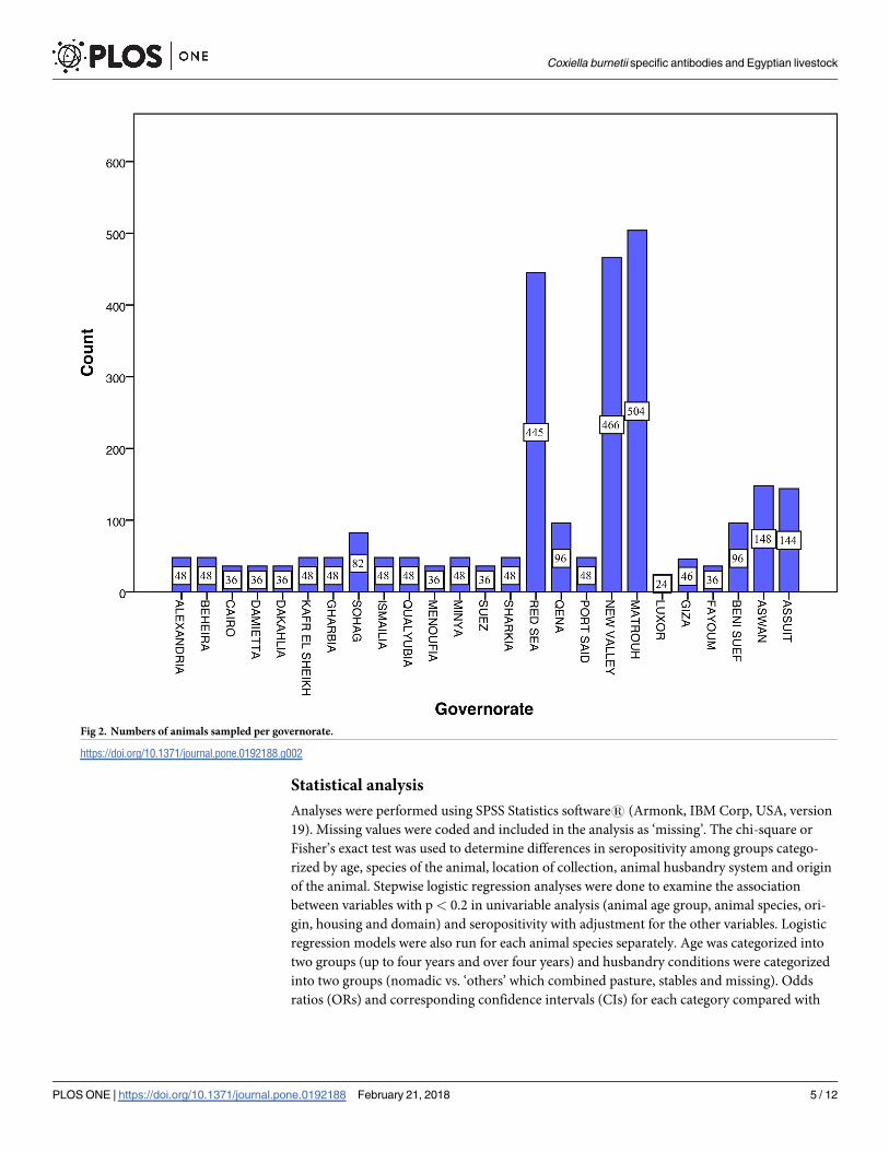
From October 2015 to March 2016 a cross-sectional study with a stratified (by governorates)
two stage random cluster sampling strategy was conducted. In the first stage 80 villages were
randomly selected from 25 governorates. The villages sampled are shown in Fig 1, whereas the
governorates are listed in Fig 2 and S1 Table. During the second sampling stage one or two
herds/farms were randomly selected without replacements from each sampling site. Thus, a
total of 299 herds/farms had to be tested. Due to a full census of the village livestock population
was not available sampling was distributed across all identified villages per domain. The num-
ber of animals to be tested was calculated using the two stage sampling formula. The calculated
number of animals was divided by the total number of villages of each domain to obtain the
final number of animals to be sampled per village. The animals sampled in the study were
older than 1.5 years to avoid false positive results due to maternal antibody cross reactions in
the ELISA test used. The estimated age of the animal was obtained from the farmer.
Sample collection
Blood (5 ml) was collected from the jugular veins of sheep, goats and camels and from the tail
veins (Vena caudalis mediana) of cattle and buffaloes. Blood samples were collected using dis-
posable needles (18 and 19 gauges) and 50/60 ml three part syringes (AMECO, Egypt). Blood
samples were then stored at room temperature for one hour to allow clotting. After centrifuga-
tion (1,449 x g, 10 minutes) serum was aliquoted into cryo-vials and stored at -20˚C before
being shipped to the Friedrich-Loeffler-Institut (FLI), Germany.
Questionnaire design and data collection
A questionnaire was used to obtain information covering a wide range of factors including
information about the animal (age, species, origin) and on the husbandry system practiced.
The animal husbandry systems were classified as follows: (a) stable/stationary: animals were
kept in an open stable with fences and a partial roof for sun protection, (b) pasture: animals
were kept on pasture/steppe in a fenced area and (c) nomadic: animals ranged free, might have
been guarded by a person and were occasionally moved from one area to the next. The animal
owners were interviewed about their general knowledge on Q fever including transmission,
clinical signs in animals and application of countermeasures such as removal of birth products
to reduce risk of infection with C. burnetii. Furthermore, they were asked if they consume raw
milk. The teams interviewed the respondents in Arabic language. Moreover, GPS data were
determined to identify the positions of the sampled villages.
Serological testing
The collected serum samples were screened for C. burnetii specific antibodies at the Q fever
reference laboratory of the FLI. An indirect ELISA (IDEXX CHEKIT Q fever Antibody ELISA
Test Kit, IDEXX Laboratories, Switzerland) was used and the results were evaluated according
to the manufacturer’s recommendations. Briefly, results with an optical density (OD) of�40%
or<30% of (PK�xNK�x) (PK = positive control, NK = negative control, �x = mean) were consid-
ered as positive or negative, respectively. Samples with a value between�30% and<40% were
considered equivocal and were re-tested. The manufacturer reported sensitivity and specificity
of the kit to be approximately 100% [27]. The test is certified for use in sheep, goats and cattle
(ruminants). Cattle and buffaloes share a closely related immune system allowing the use of
this ELISA for samples from buffaloes [28]. The IDEXX ELISA is commonly used in serum
samples of camels although a final validation of this test in camelids is still missing [25, 29].
Coxiella burnetii specific antibodies and Egyptian livestock
PLOS ONE | https://doi.org/10.1371/journal.pone.0192188 February 21, 2018 3 / 12
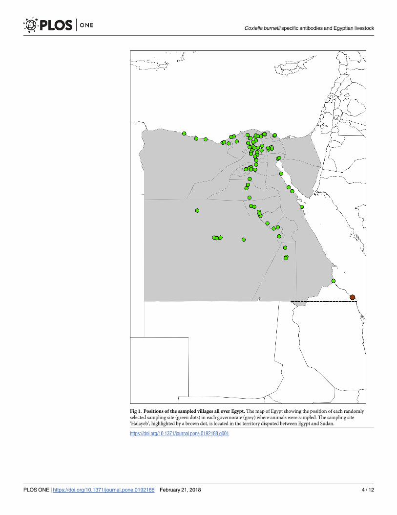
Fig 1. Positions of the sampled villages all over Egypt. The map of Egypt showing the position of each randomly
selected sampling site (green dots) in each governorate (grey) where animals were sampled. The sampling site
‘Halayeb’, highlighted by a brown dot, is located in the territory disputed between Egypt and Sudan.
https://doi.org/10.1371/journal.pone.0192188.g001
Coxiella burnetii specific antibodies and Egyptian livestock
PLOS ONE | https://doi.org/10.1371/journal.pone.0192188 February 21, 2018 4 / 12
Statistical analysis
Analyses were performed using SPSS Statistics software1 (Armonk, IBM Corp, USA, version
19). Missing values were coded and included in the analysis as ‘missing’. The chi-square or
Fisher’s exact test was used to determine differences in seropositivity among groups catego-
rized by age, species of the animal, location of collection, animal husbandry system and origin
of the animal. Stepwise logistic regression analyses were done to examine the association
between variables with p< 0.2 in univariable analysis (animal age group, animal species, ori-
gin, housing and domain) and seropositivity with adjustment for the other variables. Logistic
regression models were also run for each animal species separately. Age was categorized into
two groups (up to four years and over four years) and husbandry conditions were categorized
into two groups (nomadic vs. ‘others’ which combined pasture, stables and missing). Odds
ratios (ORs) and corresponding confidence intervals (CIs) for each category compared with
Fig 2. Numbers of animals sampled per governorate.
https://doi.org/10.1371/journal.pone.0192188.g002
Coxiella burnetii specific antibodies and Egyptian livestock
PLOS ONE | https://doi.org/10.1371/journal.pone.0192188 February 21, 2018 5 / 12
the reference group were calculated. P values < 0.05 were considered significant. The map dis-
playing the sampled villages was created using ArcGIS (ESRI, version 10).
Ethical considerations
This study was carried out in strict accordance with the recommendations of the Egyptian Net-
work of Research Ethics Committees (ENREC), which complies with the international laws
and regulations regarding ethical considerations in research. The ENREC approved this
research work. For purposes of this study all animal owners consented to sampling.
Results
Study population
A total of 2,699 livestock (31.1% cattle, 26.5% sheep, 19.6% camels 11.5% goats and 11.3% buf-
faloes) were sampled on 299 farms of 80 villages. The majority of the animals sampled was
from the Nile Valley and Delta (47.6%) and Western Desert (35.9%) regions. Animals from the
Eastern Desert domain accounted for 16.5% due to missing samples of the Sinai. Goats were
only sampled in 19 of 25 governorates. The number of goat samples collected differed from the
sample size calculated prior to the study, especially in the Western Desert and Eastern Desertregion. One thousand six hundred and thirty-nine (60.7%) animals were nomadic, 262 (9.7%)
on pasture and 685 (25.4%) stationary/stables. In the Western Desert region, most animals
were nomadic (936/2,699 [34.7%]) whereas stationary placement (18.1%) and pasture hus-
bandry (8.0%) were mainly found in the Nile Valley and Delta domain. More than eighty-eight
percent (88.5%) of all sampled livestock were bred in Egypt and only 311 animals (11.5%) were
imported. Camels were the only imported animals and all of them originated from Sudan
(58.9% [311/528]). Nine hundred and seventy (28.5%) animals were younger than 4 years and
1,729 (64.1%) were older than 4 years. Fig 2 and Table 1 summarize the characteristics of the
study population. None of the livestock owners interviewed reported prior knowledge on Q
fever or on any application of countermeasures. Twenty-six owners (8.7%) reported consump-
tion of raw camel milk. Transmission of C. burnetii to humans via consumption of raw milk is
still unknown.
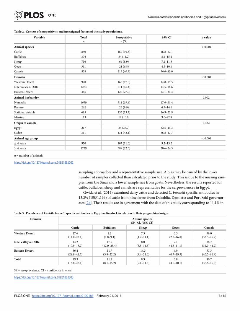
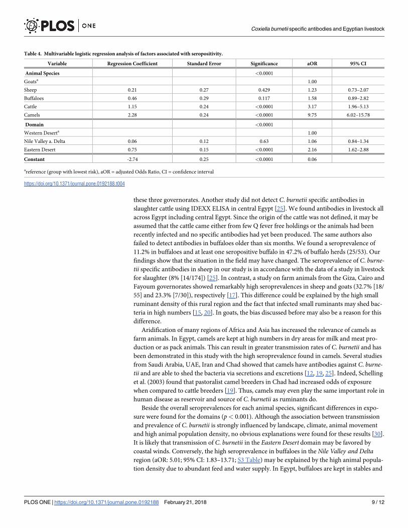
Seroprevalence
The seroprevalence in goats was 6.8%, in sheep 8.9%, in buffaloes 11.2%, in cattle 19.3% and in
camels 40.7% (Table 2). The differences in seroprevalence among the animal species were sig-
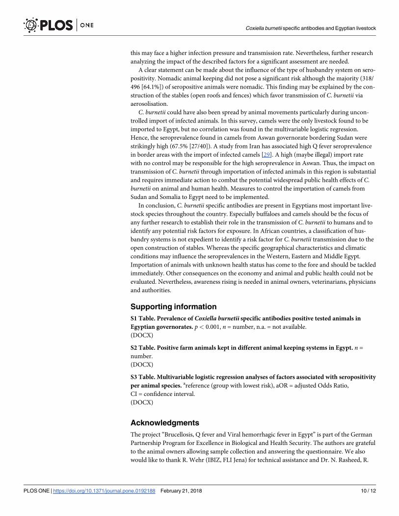
nificant (p< 0.001) (Table 3). Multivariable analysis showed significantly higher odds for sero-
positivity for cattle (aOR: 3.17; 95% CI: 1.96–5.13) and camels (aOR: 9.75; 95% CI: 6.02-15.78)
(Table 4). Cattle, sheep and camels of the Eastern Desert region had highest seroprevalences.
Seroprevalences in buffaloes and goats were highest in the Nile Valley and Delta domain
(p< 0.001) (Table 3). Seropositivity in the final logistic regression model was significantly
associated with animals from the Eastern Desert domain (aOR: 2.16; 95% CI: 1.62–2.88)
(Table 4). This was also evident in the analyses per animal species (S3 Table).
Seroprevalences at governorate level ranged from 4.2% to 36.4% in cattle, from 3.3% to
100% in buffaloes, from 5.3% to 25.0% in sheep, from 4.0% to 41.7% in goats and from 12.5%
to 75% in camels (p< 0.001) (S1 Table). Seroprevalences determined for the villages were in
the range of 4.8%-66.7% in cattle, 4.0%-100.0% in buffaloes, 3.3%-50.0% in sheep, 8.3%-50.0%
in goats and 16.7%-78.6% in camels (p< 0.001). Fifty-three percent (157/299) of all sampled
herds had at least one seropositive animal.
Coxiella burnetii specific antibodies and Egyptian livestock
PLOS ONE | https://doi.org/10.1371/journal.pone.0192188 February 21, 2018 6 / 12
Seroprevalence was found to be higher in animals from stationary/stable (135/685 [19.7%])
and nomadic (318/1639 [19.4%]) farming than from animals kept on pastures (26/262 [9.9%])
(p = 0.002) (Table 2). However, this variable was not significant in the multivariable analysis.
Higher seroprevalence 389/1729 (22.5%) was found in animals older than four years. Most
sheep and goats with a positive result were younger than four years. The difference in sero-
prevalence between these age groups was statistically significant (p< 0.001) in the univariable
analysis, but not in the multivariable analysis.
Of the imported camels 42.1% (131/311) were seropositive. Eighty-four (38.7%) camels
from Egyptian origin were tested positive but the difference was not statistically significant
(p = 0.432).
Discussion
This first nationwide cross-sectional study in ruminants and camels was conducted to provide
a deeper understanding of the epidemiology of Q fever in Egypt. An overall seroprevalence of
40.1% in camels, 19.3% in cattle, 11.2% in buffaloes, 8.9% in sheep and 6.8% in goats was
found. Seroprevalences were influenced by the geographical location, type of animal hus-
bandry and age of animal, however not by the origin of an animal. Potential risks associated
with seropositivity are animal species and the geographical location. Thus, Q fever is endemic
throughout Egypt in ruminants and camels.
Over the last 65 years, to the best of our knowledge, ten prevalence studies have been con-
ducted in Egypt. These studies had limitations in study design (missing or inadequate), study
area (locally restricted) or size of test specimens. Thus, all major farm animal species which
might serve as natural reservoirs were investigated using reliable study design, probabilistic
Table 1. Numbers of animals sampled per domain with age group, numbers of animals of a particular animal hus-
bandry system and origin of animals.
Variable Domain n (%)
Western Desert Nile Valley a. Delta Eastern Desert
Animal species
Cattle 340 (40.5) 360 (42.9) 140 (16.7)
Buffalo 120 (39.5) 124 (40.8) 60 (19.7)
Sheep 262 (36.6) 314 (43.9) 140 (19.6)
Goat 48 (15.4) 238 (76.5) 25 (8.0)
Camel 200 (37.9) 248 (47.0) 80 (15.2)
Total 970 (35.9) 1284 (47.6) 445 (16.5)
Animal husbandry
Nomadic 936 (57.1) 467 (28.5) 236 (14.4)
Pasture 34 (13.0) 215 (82.1) 13 (5.0)
Stationary/stable 0 (0) 489 (71.4) 196 (28.6)
Missing 0 (0) 113 (8.8) 0 (0)
Origin of animal
Egypt 970 (40.6) 1053 (44.1) 365 (15.3)
Sudan 0 (0) 231 (74.3) 80 (25.7)
Animal age group
� 4 years 326 (33.6) 484 (49.9) 160 (16.5)
> 4 years 644 (37.2) 800 (46.3) 285 (16.5)
n = number of animals
https://doi.org/10.1371/journal.pone.0192188.t001
Coxiella burnetii specific antibodies and Egyptian livestock
PLOS ONE | https://doi.org/10.1371/journal.pone.0192188 February 21, 2018 7 / 12
sampling approaches and a representative sample size. A bias may be caused by the lower
number of samples collected than calculated prior to the study. This is due to the missing sam-
ples from the Sinai and a lower sample size from goats. Nevertheless, the results reported for
cattle, buffaloes, sheep and camels are representative for the serprevalences in Egypt.
Gwida et al. (2014) examined dairy cattle and detected C. burnetii specific antibodies in
13.2% (158/1,194) of cattle from nine farms from Dakahlia, Damietta and Port Said governor-
ates [24]. Their results are in agreement with the data of this study corresponding to 11.1% in
Table 2. Context of seropositivity and investigated factors of the study populations.
Variable Total
nSeropositive
n (%)
95% CI p value
Animal species < 0.001
Cattle 840 162 (19.3) 16.8–22.1
Buffaloes 304 34 (11.2) 8.1–15.2
Sheep 716 64 (8.9) 7.1–11.3
Goats 311 21 (6.8) 4.5–10.1
Camels 528 215 (40.7) 36.6–45.0
Domain < 0.001
Western Desert 970 165 (17.0) 14.8–19.5
Nile Valley a. Delta 1284 211 (16.4) 14.5–18.6
Eastern Desert 445 120 (27.0) 23.1–31.3
Animal husbandry 0.002
Nomadic 1639 318 (19.4) 17.6–21.4
Pasture 262 26 (9.9) 6.9–14.1
Stationary/stable 685 135 (19.7) 16.9–22.9
Missing 113 17 (15.0) 9.6–22.8
Origin of camels 0.432
Egypt 217 84 (38.7) 32.5–45.3
Sudan 311 131 (42.1) 36.8–47.7
Animal age group < 0.001
� 4 years 970 107 (11.0) 9.2–13.2
> 4 years 1729 389 (22.5) 20.6–24.5
n = number of animals
https://doi.org/10.1371/journal.pone.0192188.t002
Table 3. Prevalence of Coxiella burnetii specific antibodies in Egyptian livestock in relation to their geographical origin.
Domain Animal species
SP [%], (95% CI)
Cattle Buffaloes Sheep Goats Camels
Western Desert 17.6
(14.0–22.1)
4.2
(1.8–9.4)
7.3
(4.7–11.1)
6.3
(2.2–16.8)
39.0
(32.5–45.9)
Nile Valley a. Delta 14.2
(10.9–18.2)
17.7
(12.0–25.4)
8.0
(5.5–11.5)
7.1
(4.5–11.1)
38.7
(32.9–44.9)
Eastern Desert 36.4
(28.9–44.7)
11.7
(5.8–22.2)
14.3
(9.4–21.0)
4.0
(0.7–19.5)
51.3
(40.5–61.9)
Total 19.3
(16.8–22.1)
11.2
(8.1–15.2)
8.9
(7.1–11.3)
6.8
(4.5–10.1)
40.7
(36.6–45.0)
SP = seroprevalence, CI = confidence interval
https://doi.org/10.1371/journal.pone.0192188.t003
Coxiella burnetii specific antibodies and Egyptian livestock
PLOS ONE | https://doi.org/10.1371/journal.pone.0192188 February 21, 2018 8 / 12
these three governorates. Another study did not detect C. burnetii specific antibodies in
slaughter cattle using IDEXX ELISA in central Egypt [25]. We found antibodies in livestock all
across Egypt including central Egypt. Since the origin of the cattle was not defined, it may be
assumed that the cattle came either from few Q fever free holdings or the animals had been
recently infected and no specific antibodies had yet been produced. The same authors also
failed to detect antibodies in buffaloes older than six months. We found a seroprevalence of
11.2% in buffaloes and at least one seropositive buffalo in 47.2% of buffalo herds (25/53). Our
findings show that the situation in the field may have changed. The seroprevalence of C. burne-tii specific antibodies in sheep in our study is in accordance with the data of a study in livestock
for slaughter (8% [14/174]) [25]. In contrast, a study on farm animals from the Giza, Cairo and
Fayoum governorates showed remarkably high seroprevalences in sheep and goats (32.7% [18/
55] and 23.3% [7/30]), respectively [17]. This difference could be explained by the high small
ruminant density of this rural region and the fact that infected small ruminants may shed bac-
teria in high numbers [15, 20]. In goats, the bias discussed before may also be a reason for this
difference.
Aridification of many regions of Africa and Asia has increased the relevance of camels as
farm animals. In Egypt, camels are kept at high numbers in dry areas for milk and meat pro-
duction or as pack animals. This can result in greater transmission rates of C. burnetii and has
been demonstrated in this study with the high seroprevalence found in camels. Several studies
from Saudi Arabia, UAE, Iran and Chad showed that camels have antibodies against C. burne-tii and are able to shed the bacteria via secretions and excretions [12, 19, 25]. Indeed, Schelling
et al. (2003) found that pastoralist camel breeders in Chad had increased odds of exposure
when compared to cattle breeders [19]. Thus, camels may even play the same important role in
human disease as reservoir and source of C. burnetii as ruminants do.
Beside the overall seroprevalences for each animal species, significant differences in expo-
sure were found for the domains (p< 0.001). Although the association between transmission
and prevalence of C. burnetii is strongly influenced by landscape, climate, animal movement
and high animal population density, no obvious explanations were found for these results [30].
It is likely that transmission of C. burnetii in the Eastern Desert domain may be favored by
coastal winds. Conversely, the high seroprevalence in buffaloes in the Nile Valley and Deltaregion (aOR: 5.01; 95% CI: 1.83–13.71; S3 Table) may be explained by the high animal popula-
tion density due to abundant feed and water supply. In Egypt, buffaloes are kept in stables and
Table 4. Multivariable logistic regression analysis of factors associated with seropositivity.
Variable Regression Coefficient Standard Error Significance aOR 95% CI
Animal Species <0.0001
Goatsa 1.00
Sheep 0.21 0.27 0.429 1.23 0.73–2.07
Buffaloes 0.46 0.29 0.117 1.58 0.89–2.82
Cattle 1.15 0.24 <0.0001 3.17 1.96–5.13
Camels 2.28 0.24 <0.0001 9.75 6.02–15.78
Domain <0.0001
Western Deserta 1.00
Nile Valley a. Delta 0.06 0.12 0.63 1.06 0.84–1.34
Eastern Desert 0.75 0.15 <0.0001 2.16 1.62–2.88
Constant -2.74 0.25 <0.0001 0.06
areference (group with lowest risk), aOR = adjusted Odds Ratio, CI = confidence interval
https://doi.org/10.1371/journal.pone.0192188.t004
Coxiella burnetii specific antibodies and Egyptian livestock
PLOS ONE | https://doi.org/10.1371/journal.pone.0192188 February 21, 2018 9 / 12
this may face a higher infection pressure and transmission rate. Nevertheless, further research
analyzing the impact of the described factors for a significant assessment are needed.
A clear statement can be made about the influence of the type of husbandry system on sero-
positivity. Nomadic animal keeping did not pose a significant risk although the majority (318/
496 [64.1%]) of seropositive animals were nomadic. This finding may be explained by the con-
struction of the stables (open roofs and fences) which favor transmission of C. burnetii via
aerosolisation.
C. burnetii could have also been spread by animal movements particularly during uncon-
trolled import of infected animals. In this survey, camels were the only livestock found to be
imported to Egypt, but no correlation was found in the multivariable logistic regression.
Hence, the seroprevalence found in camels from Aswan governorate bordering Sudan were
strikingly high (67.5% [27/40]). A study from Iran has associated high Q fever seroprevalence
in border areas with the import of infected camels [29]. A high (maybe illegal) import rate
with no control may be responsible for the high seroprevalence in Aswan. Thus, the impact on
transmission of C. burnetii through importation of infected animals in this region is substantial
and requires immediate action to combat the potential widespread public health effects of C.
burnetii on animal and human health. Measures to control the importation of camels from
Sudan and Somalia to Egypt need to be implemented.
In conclusion, C. burnetii specific antibodies are present in Egyptians most important live-
stock species throughout the country. Especially buffaloes and camels should be the focus of
any further research to establish their role in the transmission of C. burnetii to humans and to
identify any potential risk factors for exposure. In African countries, a classification of hus-
bandry systems is not expedient to identify a risk factor for C. burnetii transmission due to the
open construction of stables. Whereas the specific geographical characteristics and climatic
conditions may influence the seroprevalences in the Western, Eastern and Middle Egypt.
Importation of animals with unknown health status has come to the fore and should be tackled
immediately. Other consequences on the economy and animal and public health could not be
evaluated. Nevertheless, awareness rising is needed in animal owners, veterinarians, physicians
and authorities.
Supporting information
S1 Table. Prevalence of Coxiella burnetii specific antibodies positive tested animals in
Egyptian governorates. p< 0.001, n = number, n.a. = not available.
(DOCX)
S2 Table. Positive farm animals kept in different animal keeping systems in Egypt. n =
number.
(DOCX)
S3 Table. Multivariable logistic regression analyses of factors associated with seropositivity
per animal species. areference (group with lowest risk), aOR = adjusted Odds Ratio,
CI = confidence interval.
(DOCX)
Acknowledgments
The project “Brucellosis, Q fever and Viral hemorrhagic fever in Egypt” is part of the German
Partnership Program for Excellence in Biological and Health Security. The authors are grateful
to the animal owners allowing sample collection and answering the questionnaire. We also
would like to thank R. Wehr (IBIZ, FLI Jena) for technical assistance and Dr. N. Rasheed, R.
Coxiella burnetii specific antibodies and Egyptian livestock
PLOS ONE | https://doi.org/10.1371/journal.pone.0192188 February 21, 2018 10 / 12
Fouad (Mansoura Provincial Laboratory) and Dr. H. Eladawy (IBIZ, FLI Jena) for their efforts
and support. Dr. L. D. Sprague is thanked for revising the manuscript.
Author Contributions
Conceptualization: Jessica Klemmer, Carola Sauter-Louis.
Data curation: Jessica Klemmer.
Formal analysis: Jessica Klemmer, John Njeru, Carola Sauter-Louis.
Funding acquisition: Heinrich Neubauer.
Investigation: Jessica Klemmer.
Project administration: Jessica Klemmer.
Resources: Aya Emam, Ahmed El-Sayed, Amira A. Moawad, Mohamed A. Elbeskawy,
Mohamed M. El-Diasty.
Supervision: Reinhard K. Straubinger, Heinrich Neubauer.
Visualization: Jessica Klemmer, John Njeru.
Writing – original draft: Jessica Klemmer.
Writing – review & editing: Jessica Klemmer, John Njeru, Klaus Henning, Carola Sauter-
Louis, Reinhard K. Straubinger, Heinrich Neubauer.
References1. Heinzen RA, Hackstadt T, Samuel JE. Developmental biology of Coxiella burnettii. Trends in microbiol-
ogy. 1999; 7(4):149–54. Epub 1999/04/28. PMID: 10217829.
2. McCaul TF, Williams JC. Developmental cycle of Coxiella burnetii: structure and morphogenesis of veg-
etative and sporogenic differentiations. Journal of bacteriology. 1981; 147(3):1063–76. Epub 1981/09/
01. PMID: 7275931; PubMed Central PMCID: PMCPmc216147.
3. Hilbink F, Penrose M, Kovacova E, Kazar J. Q fever is absent from New Zealand. Int J Epidemiol. 1993;
22(5):945–9. PMID: 8282477.
4. Kaplan MM, Bertagna P. The geographical distribution of Q fever. Bull World Health Organ. 1955; 13
(5):829–60. PMID: 13284560; PubMed Central PMCID: PMCPMC2538086.
5. Honarmand H. Q Fever: an old but still a poorly understood disease. Interdisciplinary perspectives on
infectious diseases. 2012; 2012:131932. Epub 2012/12/06. https://doi.org/10.1155/2012/131932
PMID: 23213331; PubMed Central PMCID: PMCPmc3506884.
6. Derrick EH. "Q" fever, a new fever entity: clinical features, diagnosis and laboratory investigation.
Reviews of infectious diseases. 1983; 5(4):790–800. Epub 1983/07/01. PMID: 6622891.
7. Parker NR, Barralet JH, Bell AM. Q fever. Lancet (London, England). 2006; 367(9511):679–88. Epub
2006/03/01. https://doi.org/10.1016/s0140-6736(06)68266-4 PMID: 16503466.
8. van der Hoek W, Schneeberger PM, Oomen T, Wegdam-Blans MC, Dijkstra F, Notermans DW, et al.
Shifting priorities in the aftermath of a Q fever epidemic in 2007 to 2009 in The Netherlands: from acute
to chronic infection. Euro surveillance: bulletin Europeen sur les maladies transmissibles = European
communicable disease bulletin. 2012; 17(3):20059. Epub 2012/02/03. PMID: 22297101.
9. Raoult D, Marrie TJ, Mege JL. Natural history and pathophysiology of Q fever. The Lancet Infectious
Diseases. 2005; 5(4):219–26. http://dx.doi.org/10.1016/S1473-3099(05)70052-9. PMID: 15792739
10. Morroy G, van der Hoek W, Albers J, Coutinho RA, Bleeker-Rovers CP, Schneeberger PM. Population
Screening for Chronic Q-Fever Seven Years after a Major Outbreak. PLoS One. 2015; 10(7):e0131777.
https://doi.org/10.1371/journal.pone.0131777 PMID: 26132155; PubMed Central PMCID:
PMCPMC4489093.
11. Barlow J, Rauch B, Welcome F, Kim SG, Dubovi E, Schukken Y. Association between Coxiella burnetii
shedding in milk and subclinical mastitis in dairy cattle. Veterinary research. 2008; 39(3):23. Epub 2008/
02/07. https://doi.org/10.1051/vetres:2007060 PMID: 18252189.
Coxiella burnetii specific antibodies and Egyptian livestock
PLOS ONE | https://doi.org/10.1371/journal.pone.0192188 February 21, 2018 11 / 12
12. Mohammed OB, Jarelnabi AA, Aljumaah RS, Alshaikh MA, Bakhiet AO, Omer SA, et al. Coxiella burne-
tii, the causative agent of Q fever in Saudi Arabia: molecular detection from camel and other domestic
livestock. Asian Pacific Journal of Tropical Medicine. 2014; 7(9):715–9. http://dx.doi.org/10.1016/
S1995-7645(14)60122-X.
13. Rodolakis A, Berri M, Hechard C, Caudron C, Souriau A, Bodier CC, et al. Comparison of Coxiella bur-
netii shedding in milk of dairy bovine, caprine, and ovine herds. Journal of dairy science. 2007; 90
(12):5352–60. Epub 2007/11/21. https://doi.org/10.3168/jds.2006-815 PMID: 18024725.
14. Maurin M, Raoult D. Q fever. Clinical microbiology reviews. 1999; 12(4):518–53. Epub 1999/10/09.
PMID: 10515901; PubMed Central PMCID: PMCPmc88923.
15. Dijkstra F, van der Hoek W, Wijers N, Schimmer B, Rietveld A, Wijkmans CJ, et al. The 2007–2010 Q
fever epidemic in The Netherlands: characteristics of notified acute Q fever patients and the association
with dairy goat farming. FEMS immunology and medical microbiology. 2012; 64(1):3–12. Epub 2011/
11/10. https://doi.org/10.1111/j.1574-695X.2011.00876.x PMID: 22066649.
16. Lejeune JT, Rajala-Schultz PJ. Food safety: unpasteurized milk: a continued public health threat. Clini-
cal infectious diseases: an official publication of the Infectious Diseases Society of America. 2009; 48
(1):93–100. Epub 2008/12/05. https://doi.org/10.1086/595007 PMID: 19053805.
17. Nahed HGK, A.-M. A. Seroprevalence of Coxiella burnetii antibodies among farm animals and human
contacts in Egypt. Journal of American Science. 2012; 8(3):619–21.
18. Nusinovici S, Frossling J, Widgren S, Beaudeau F, Lindberg A. Q fever infection in dairy cattle herds:
increased risk with high wind speed and low precipitation. Epidemiol Infect. 2015:1–11. https://doi.org/
10.1017/S0950268814003926 PMID: 25783480.
19. Schelling E, Diguimbaye C, Daoud S, Nicolet J, Boerlin P, Tanner M, et al. Brucellosis and Q-fever sero-
prevalences of nomadic pastoralists and their livestock in Chad. Prev Vet Med. 2003; 61(4):279–93.
PMID: 14623412.
20. Abdel-Moein KA, Hamza DA. The burden of Coxiella burnetii among aborted dairy animals in Egypt and
its public health implications. Acta tropica. 2017; 166:92–5. Epub 2016/11/16. https://doi.org/10.1016/j.
actatropica.2016.11.011 PMID: 27845064.
21. Corwin A, Habib M, Olson J, Scott D, Ksiazek T, Watts DM. The prevalence of arboviral, rickettsial, and
Hantaan-like viral antibody among schoolchildren in the Nile river delta of Egypt. Trans R Soc Trop Med
Hyg. 1992; 86(6):677–9. PMID: 1363163.
22. Corwin A, Habib M, Watts D, Darwish M, Olson J, Botros B, et al. Community-based prevalence profile
of arboviral, rickettsial, and Hantaan-like viral antibody in the Nile River Delta of Egypt. The American
journal of tropical medicine and hygiene. 1993; 48(6):776–83. Epub 1993/06/01. PMID: 8101432.
23. van Asseldonk MA, Prins J, Bergevoet RH. Economic assessment of Q fever in the Netherlands. Prev
Vet Med. 2013; 112(1–2):27–34. Epub 2013/07/23. https://doi.org/10.1016/j.prevetmed.2013.06.002
PMID: 23866818.
24. Gwida M, El-Ashker M, El-Diasty M, Engelhardt C, Khan I, Neubauer H. Q fever in cattle in some Egyp-
tian Governorates: a preliminary study. BMC Res Notes. 2014; 7:881. https://doi.org/10.1186/1756-
0500-7-881 PMID: 25481509; PubMed Central PMCID: PMCPMC4295271.
25. Horton KC, Wasfy M, Samaha H, Abdel-Rahman B, Safwat S, Abdel Fadeel M, et al. Serosurvey for
zoonotic viral and bacterial pathogens among slaughtered livestock in Egypt. Vector Borne Zoonotic
Dis. 2014; 14(9):633–9. https://doi.org/10.1089/vbz.2013.1525 PMID: 25198525.
26. Mazyad SA, Hafez AO. Q fever (Coxiella burnetii) among man and farm animals in North Sinai, Egypt. J
Egypt Soc Parasitol. 2007; 37(1):135–42. PMID: 17580573.
27. IDEXX-Laboratories. Sensitivity and specificity ELISA assay 2015 [02 December 2015]. Available from:
http://www2.idexx.com/view/xhtml/en_us/livestock-poultry/newsletter/2007/200708.jsf%3Bjsessionid=
Lhcc8noo1efXWtKH-OKoTQ#fnq.
28. Ibeagha-Awemu EM, Lee JW, Ibeagha AE, Zhao X. Bovine CD14 gene characterization and relation-
ship between polymorphisms and surface expression on monocytes and polymorphonuclear neutro-
phils. BMC genetics. 2008; 9:50. Epub 2008/08/12. https://doi.org/10.1186/1471-2156-9-50 PMID:
18691417; PubMed Central PMCID: PMCPmc2536669.
29. Janati Pirouz H, Mohammadi G, Mehrzad J, Azizzadeh M, Nazem Shirazi MH. Seroepidemiology of Q
fever in one-humped camel population in northeast Iran. Tropical animal health and production. 2015;
47(7):1293–8. Epub 2015/06/14. https://doi.org/10.1007/s11250-015-0862-z PMID: 26070292.
30. Nusinovici S, Frossling J, Widgren S, Beaudeau F, Lindberg A. Q fever infection in dairy cattle herds:
increased risk with high wind speed and low precipitation. Epidemiol Infect. 2015; 143(15):3316–26.
Epub 2015/03/19. https://doi.org/10.1017/S0950268814003926 PMID: 25783480; PubMed Central
PMCID: PMCPMC4594051.
Coxiella burnetii specific antibodies and Egyptian livestock
PLOS ONE | https://doi.org/10.1371/journal.pone.0192188 February 21, 2018 12 / 12
Related Documents