Purified E255L Mutant SERCA1a and Purified PfATP6 Are Sensitive to SERCA-type Inhibitors but Insensitive to Artemisinins * □ S Received for publication, November 30, 2009, and in revised form, June 2, 2010 Published, JBC Papers in Press, June 8, 2010, DOI 10.1074/jbc.M109.090340 Delphine Cardi ‡§¶ , Alexandre Pozza ‡§¶ , Bertrand Arnou , Estelle Marchal ‡§¶ , Johannes D. Clausen , Jens Peter Andersen , Sanjeev Krishna** 1 , Jesper V. Møller , Marc le Maire ‡§¶2 , and Christine Jaxel ‡§¶3 From the ‡ Commissariat a ` l’Energie Atomique, Institut de Biologie et de Technologies de Saclay, SB2SM, the § Laboratoire de Recherche Associe ´ 17V, University of Paris-Sud, and ¶ URA CNRS 2096, Gif sur Yvette F-91191, France, the **Centre for Infection, Division of Cellular and Molecular Medicine, St George’s University of London, London SW17 0RE, United Kingdom, and the Department of Physiology and Biophysics, Centre for Membrane Pumps in Cells and Disease-PUMPKIN, Danish National Research Foundation, University of Aarhus, Ole Worms Alle 1185, Aarhus C DK-8000, Denmark The antimalarial drugs artemisinins have been described as inhibiting Ca 2 -ATPase activity of PfATP6 (Plasmodium falcipa- rum ATP6) after expression in Xenopus oocytes. Mutation of an amino acid residue in mammalian SERCA1 (Glu 255 ) to the equiva- lent one predicted in PfATP6 (Leu) was reported to induce sensi- tivity to artemisinin in the oocyte system. However, in the present experiments, we found that artemisinin did not inhibit mammalian SERCA1a E255L either when expressed in COS cells or after puri- fication of the mutant expressed in Saccharomyces cerevisiae. Moreover, we found that PfATP6 after expression and purification from S. cerevisiae was insensitive to artemisinin and significantly less sensitive to thapsigargin and 2,5-di(tert-butyl)-1,4-benzo- hydroquinone than rabbit SERCA1 but retained higher sensitivity to cyclopiazonic acid, another type of SERCA1 inhibitor. Although mammalian SERCA and purified PfATP6 appear to have different pharmacological profiles, their insensitivity to artemisinins sug- gests that the mechanism of action of this class of drugs on the calcium metabolism in the intact cell is complex and cannot be ascribed to direct inhibition of PfATP6. Furthermore, the success- ful purification of PfATP6 affords the opportunity to develop new antimalarials by screening for inhibitors against PfATP6. Malaria is a major infectious disease with about 500 million cases and one million deaths every year. Human malaria is caused by five species of protozoan parasites of the genus Plasmodium transmitted to humans by female Anopheles mosquitoes. Infec- tions with Plasmodium falciparum are responsible for the most severe disease and deaths. Without a vaccine, drug treatment is a critical part of malaria control strategies. However, drug-resistant parasites are severely compromising the effectiveness of many therapies (1, 2). The artemisinins have maintained their efficacy despite the emergence of drug resistance to other classes of anti- malarial drugs, although even this class now exhibits clinically rel- evant resistance in some areas (3). Artemisinins kill parasites faster (4) and with less toxicity (5) than other drugs, but their mechanism of action is not yet clearly known. Artemisinin is extracted from Artemisia annua, an herb long used to treat fevers in traditional Chinese medicine, and derivatives have been developed to improve their potency, solubility, stability, and pharmacokinetic properties (6 –9). Artemisinins are sesquiterpene trioxane lac- tones that contain an endoperoxide bridge essential for antimalar- ial activity (10). They are effective against the blood stages of Plasmodium and especially the early ring and sexual stages (game- tocytes) of the parasite life cycle (4, 11, 12). Artemisinins and deriv- atives accumulate preferentially in infected erythrocytes (13, 14) and are mainly located in parasite membranes and their neutral lipids, where the accumulation patterns are endoperoxide-depen- dent (15, 16). The generation of free radicals by artemisinins may be critical for killing parasites, an observation that is consistent with the importance of the endoperoxide bridge for drug efficacy (17). The nature of these radicals and how they are generated is debated, with hypotheses including roles for iron- or heme-cata- lyzing Fenton-like reactions (18, 19). Artemisinin inhibits the endocytosis of red blood cell cytoplas- mic macromolecules by the parasite (20), possibly via an increase in the cytosolic level of Ca 2 . Efficacy against Toxoplasma gondii has also been related to changes in calcium homeostasis in this member of the apicomplexan family of parasites that includes Plasmodium spp. (21, 22). Several P-type ATPases (PfATP6, a sar- co(endo)plasmic reticulum calcium ATPase (SERCA) 4 -type pro- tein; PfATP4, a PTM (plasma membrane ATPase-related)-like * This work was supported by the Danish Medical Research Council, the Dan- ish Natural Science Research Council (Center for Structural Biology, the Dansync program), the Aarhus University Research Foundation, the Novo- Nordic Foundation (Denmark), the Commissariat a ` l’Energie Atomique (Saclay, France), CNRS, and the DIM Maladies Infectieuses, Parasitaires et Nosocomiales Emergentes/Ile de France (France). Author’s Choice—Final version full access. □ S The on-line version of this article (available at http://www.jbc.org) contains supplemental Figs. S1 and S2. 1 Supported by Commission of the European Communities ANTIMAL Grant 018834 and Wellcome Trust Grant 074395/Z/04/Z. 2 To whom correspondence may be addressed: URA CNRS 2096, iBiTec-S/ SB2SM/LPM, CEA Saclay, 91191 Gif-sur-Yvette, France. Tel.: 33-169083379; Fax: 33-169088139; E-mail: [email protected]. 3 To whom correspondence may be addressed: URA CNRS 2096, iBiTec-S/ SB2SM/LPM, CEA Saclay, 91191 Gif-sur-Yvette, France. Tel.: 33-169083379; Fax: 33-169088139; E-mail: [email protected]. 4 The abbreviations used are: SERCA, sarcoplasmic reticulum Ca 2 -ATPase; SR, sarcoplasmic reticulum; BAD, biotin acceptor domain; DDM, dodecyl- maltoside; C 12 E 8 , dodecyl octaethylene glycol monoether; DOPC, dioleyl- phosphatidylcholine; Tg, thapsigargin; BHQ, 2,5-di(tert-butyl)-1,4-benzo- hydroquinone; CPA, cyclopiazonic acid; Tes, 2-{[2-hydroxy-1,1-bis(hydroxy- methyl)ethyl]amino}ethanesulfonic acid; LM, light membrane. THE JOURNAL OF BIOLOGICAL CHEMISTRY VOL. 285, NO. 34, pp. 26406 –26416, August 20, 2010 Author’s Choice © 2010 by The American Society for Biochemistry and Molecular Biology, Inc. Printed in the U.S.A. 26406 JOURNAL OF BIOLOGICAL CHEMISTRY VOLUME 285 • NUMBER 34 • AUGUST 20, 2010 at CEA SACLAY, on August 16, 2010 www.jbc.org Downloaded from http://www.jbc.org/content/suppl/2010/06/08/M109.090340.DC1.html Supplemental Material can be found at:

Welcome message from author
This document is posted to help you gain knowledge. Please leave a comment to let me know what you think about it! Share it to your friends and learn new things together.
Transcript
Purified E255L Mutant SERCA1a and Purified PfATP6 AreSensitive to SERCA-type Inhibitors but Insensitive toArtemisinins*□S
Received for publication, November 30, 2009, and in revised form, June 2, 2010 Published, JBC Papers in Press, June 8, 2010, DOI 10.1074/jbc.M109.090340
Delphine Cardi‡§¶, Alexandre Pozza‡§¶, Bertrand Arnou�, Estelle Marchal‡§¶, Johannes D. Clausen�,Jens Peter Andersen�, Sanjeev Krishna**1, Jesper V. Møller�, Marc le Maire‡§¶2, and Christine Jaxel‡§¶3
From the ‡Commissariat a l’Energie Atomique, Institut de Biologie et de Technologies de Saclay, SB2SM, the §Laboratoire deRecherche Associe 17V, University of Paris-Sud, and ¶URA CNRS 2096, Gif sur Yvette F-91191, France, the **Centre for Infection,Division of Cellular and Molecular Medicine, St George’s University of London, London SW17 0RE, United Kingdom, and the�Department of Physiology and Biophysics, Centre for Membrane Pumps in Cells and Disease-PUMPKIN, Danish National ResearchFoundation, University of Aarhus, Ole Worms Alle 1185, Aarhus C DK-8000, Denmark
The antimalarial drugs artemisinins have been described asinhibiting Ca2�-ATPase activity of PfATP6 (Plasmodium falcipa-rum ATP6) after expression in Xenopus oocytes. Mutation of anamino acid residue inmammalian SERCA1 (Glu255) to the equiva-lent one predicted in PfATP6 (Leu) was reported to induce sensi-tivity to artemisinin in the oocyte system. However, in the presentexperiments,wefoundthatartemisinindidnot inhibitmammalianSERCA1a E255L either when expressed in COS cells or after puri-fication of the mutant expressed in Saccharomyces cerevisiae.Moreover,we found thatPfATP6after expressionandpurificationfrom S. cerevisiae was insensitive to artemisinin and significantlyless sensitive to thapsigargin and 2,5-di(tert-butyl)-1,4-benzo-hydroquinone than rabbit SERCA1 but retained higher sensitivityto cyclopiazonic acid, another typeof SERCA1 inhibitor.Althoughmammalian SERCA and purified PfATP6 appear to have differentpharmacological profiles, their insensitivity to artemisinins sug-gests that the mechanism of action of this class of drugs on thecalcium metabolism in the intact cell is complex and cannot beascribed to direct inhibition of PfATP6. Furthermore, the success-ful purification of PfATP6 affords the opportunity to develop newantimalarials by screening for inhibitors against PfATP6.
Malaria is a major infectious disease with about 500 millioncases and onemillion deaths every year. Humanmalaria is causedby five species of protozoan parasites of the genus Plasmodiumtransmitted to humans by female Anopheles mosquitoes. Infec-
tions with Plasmodium falciparum are responsible for the mostsevere disease and deaths. Without a vaccine, drug treatment is acritical part of malaria control strategies. However, drug-resistantparasites are severely compromising the effectiveness of manytherapies (1, 2). The artemisinins have maintained their efficacydespite the emergence of drug resistance to other classes of anti-malarial drugs, although even this class now exhibits clinically rel-evant resistance in someareas (3).Artemisininskill parasites faster(4) andwith less toxicity (5) thanotherdrugs, but theirmechanismof action is not yet clearly known. Artemisinin is extracted fromArtemisia annua, an herb long used to treat fevers in traditionalChinese medicine, and derivatives have been developed toimprove their potency, solubility, stability, and pharmacokineticproperties (6–9). Artemisinins are sesquiterpene trioxane lac-tones that contain an endoperoxide bridge essential for antimalar-ial activity (10). They are effective against the blood stages ofPlasmodium and especially the early ring and sexual stages (game-tocytes) of theparasite life cycle (4, 11, 12).Artemisinins andderiv-atives accumulate preferentially in infected erythrocytes (13, 14)and are mainly located in parasite membranes and their neutrallipids, where the accumulation patterns are endoperoxide-depen-dent (15, 16). The generation of free radicals by artemisinins maybe critical for killing parasites, an observation that is consistentwith the importance of the endoperoxide bridge for drug efficacy(17). The nature of these radicals and how they are generated isdebated, with hypotheses including roles for iron- or heme-cata-lyzing Fenton-like reactions (18, 19).Artemisinin inhibits the endocytosis of red blood cell cytoplas-
mic macromolecules by the parasite (20), possibly via an increasein the cytosolic level of Ca2�. Efficacy against Toxoplasma gondiihas also been related to changes in calcium homeostasis in thismember of the apicomplexan family of parasites that includesPlasmodium spp. (21, 22). Several P-typeATPases (PfATP6, a sar-co(endo)plasmic reticulum calcium ATPase (SERCA)4-type pro-tein; PfATP4, a PTM (plasma membrane ATPase-related)-like
* This work was supported by the Danish Medical Research Council, the Dan-ish Natural Science Research Council (Center for Structural Biology, theDansync program), the Aarhus University Research Foundation, the Novo-Nordic Foundation (Denmark), the Commissariat a l’Energie Atomique(Saclay, France), CNRS, and the DIM Maladies Infectieuses, Parasitaires etNosocomiales Emergentes/Ile de France (France).Author’s Choice—Final version full access.
□S The on-line version of this article (available at http://www.jbc.org) containssupplemental Figs. S1 and S2.
1 Supported by Commission of the European Communities ANTIMAL Grant018834 and Wellcome Trust Grant 074395/Z/04/Z.
2 To whom correspondence may be addressed: URA CNRS 2096, iBiTec-S/SB2SM/LPM, CEA Saclay, 91191 Gif-sur-Yvette, France. Tel.: 33-169083379;Fax: 33-169088139; E-mail: [email protected].
3 To whom correspondence may be addressed: URA CNRS 2096, iBiTec-S/SB2SM/LPM, CEA Saclay, 91191 Gif-sur-Yvette, France. Tel.: 33-169083379;Fax: 33-169088139; E-mail: [email protected].
4 The abbreviations used are: SERCA, sarcoplasmic reticulum Ca2�-ATPase;SR, sarcoplasmic reticulum; BAD, biotin acceptor domain; DDM, dodecyl-maltoside; C12E8, dodecyl octaethylene glycol monoether; DOPC, dioleyl-phosphatidylcholine; Tg, thapsigargin; BHQ, 2,5-di(tert-butyl)-1,4-benzo-hydroquinone; CPA, cyclopiazonic acid; Tes, 2-{[2-hydroxy-1,1-bis(hydroxy-methyl)ethyl]amino}ethanesulfonic acid; LM, light membrane.
THE JOURNAL OF BIOLOGICAL CHEMISTRY VOL. 285, NO. 34, pp. 26406 –26416, August 20, 2010Author’s Choice © 2010 by The American Society for Biochemistry and Molecular Biology, Inc. Printed in the U.S.A.
26406 JOURNAL OF BIOLOGICAL CHEMISTRY VOLUME 285 • NUMBER 34 • AUGUST 20, 2010
at CE
A S
AC
LAY
, on August 16, 2010
ww
w.jbc.org
Dow
nloaded from
http://www.jbc.org/content/suppl/2010/06/08/M109.090340.DC1.htmlSupplemental Material can be found at:
protein; and three putative ATPases that seem to belong to theGolgi-endoplasmic reticulum-type family) and a single Ca2�/H�
exchanger were identified to be involved in the maintenance ofcalcium homeostasis in P. falciparum (23).Recently, Krishna and co-workers (24–26) observed that the
ATPase activity of the single SERCA of P. falciparum, PfATP6,expressed in Xenopus laevis oocyte was inhibited by artemisinin(Ki �150 nM). Isobologram analysis and competition studies withfluorophore derivatives localizing to parasites were consistentwith a common target for artemisinin and thapsigargin (Tg), aspecific inhibitor of SERCA-type proteins, because an antagonismwas observed in the action of these drugs. PfATP6 and SERCA1share an overall 40% identity with a well conserved transmem-brane region,whereas the cytosolic sequenceof theparasiteCa2�-ATPase contains about 200 additional residues. Mutation studieson PfATP6 expressed in oocytes suggested that, in particular,Leu263modulates the sensitivityof this enzymetoartemisinin (26).This experiment was in part based on the finding that rabbitSERCA1,whoseTgbinding site isnearPhe256 (27), is insensitive toartemisinin, and its amino acid sequence contains at the homolo-gous position of Leu263 a glutamate (Glu255). When the Leu263 ofPfATP6 was mutated to glutamate, sensitivity to artemisinin wasdecreased (26), and conversely when the glutamate residue ofSERCA1 was mutated to a leucine, SERCA1 became sensitive toartemisinin (26). These results suggest that PfATP6 is a target forartemisinins, with further support derived from correlationbetween certain pointmutations in PfATP6 in field isolates show-ing reduced in vitro sensitivity to artemether (28) and dihydroar-temisinin (29), although all of the cases of artemisinin resistanceare not related to these mutations but revealed some polymor-phism (3, 19, 30, 31).Up to now, only two of these transporters (PfATP6 and
PfATP4) and mutated SERCA1a E255L have been studied in theX. laevisoocyte system(24, 26, 32). Inorder to further examine theinteraction of artemisinins with PfATP6 and SERCA1a E255Lmutant, it is important to characterize inmore detail (functionallyand structurally) those Ca2�-ATPases. For that purpose, expres-sion in alternative systems (we investigatedCOS-1 and yeast cells)and purification of the proteins is required. Our group recentlydeveloped amethod to purify rabbit SERCA1a by affinity chroma-tography after its expression in yeast (33), and this method wassuccessfully used for studying and crystallizing wild type (34) andmutated SERCA1a (35). In the present study, yeast expressionwasapplied to purify and functionally characterize SERCA1a E255LandPfATP6andtostudy theeffectsof artemisininandotherdrugswhen combined with detergent or lipids.
EXPERIMENTAL PROCEDURES
Chemicals—All chemical products were purchased fromSigma unless specified otherwise. PfuTurbo� DNA polymerasewas from Stratagene, and Electroelution G-capsules were fromG-Biosciences (AGRO-BIO, La Ferte Saint Aubin, France).Phusion� high fidelity polymerase was from Finnzymes(Ozyme, Saint Quentin en Yvelines, France). Restriction andmodification enzymes were from New England Biolabs(Ozyme, Saint Quentin en Yvelines, France). High activitybovine thrombin was from Calbiochem, and the streptavidin-SepharoseTM High Performance was purchased from GE
Healthcare. All products for yeast and bacteria cultures werepurchased from Difco (BD Biosciences). 1,2-Dioleoyl-sn-glyc-ero-3-phosphocholine (DOPC), 1,2-dioleoyl-sn-glycero-3-phosphoserine, 1-palmitoyl-2-oleoyl-sn-glycero-3-phospho-ethanolamine, and egg yolk phosphatidylcholine were fromAvanti Polar Lipids (Alabaster, AL); L-�-phosphatidylinositolfrom soybean (catalog no. P0639) was from Sigma.N-Dodecyl-�-D-maltoside (DDM) was from Anatrace (Maumee, OH), andoctaethylene glycol mono-n-dodecyl ether (C12E8) was pur-chased fromNikkol Chemical (Tokyo, Japan). Precision proteinstandards were from Bio-Rad. Immobilon-P membranes werefrom Millipore (Bedford, MA). Avidin-peroxidase conjugatewas from Sigma. Phosphoenolpyruvate (catalog no. P3637),L-lactic dehydrogenase solution from bovine heart (catalog no.L1006), and pyruvate kinase preparation type VII from rabbitmuscle (catalog no. P7768) were from Sigma.COS-1 Cell Experiments—Site-directed mutagenesis of
cDNA encoding SERCA1a inserted into the pMT2 vector (36)was carried out using the QuikChange site-directed mutagen-esis kit (Stratagene), and the mutant cDNA was sequencedthroughout. To express wild type or E255L mutant cDNA,COS-1 cells were transfected using the calcium phosphate pre-cipitation method (37). Microsomal vesicles containing theexpressed proteins were isolated by differential centrifugation(38). The concentration of expressed Ca2�-ATPase was deter-mined by an enzyme-linked immunosorbent assay (39) and bydetermination of the maximum capacity for phosphorylationwith ATP (“active site concentration”; see Ref. 40). ATPaseactivity was determined by following the liberation of Pi (41) inthe presence of 4 �M calcium ionophore A23187 to preventinhibition caused by rebinding of Ca2� to the luminally facingCa2� sites (40). Inhibition assays were performed at 25 °C or37 °C by first preincubating microsomal vesicles together withthe drug over an 8-min period and then measuring the ATPaseactivity for 10 or 30 min, respectively.Yeast Transformation and Selection of Individual Clones—
The Saccharomyces cerevisiae yeast strain W303.1b/Gal4 (a,leu2, his3, trp1::TRP1-GAL10-GAL4, ura3, ade2-1, canr, cir�)was the same as previously described (42). Transformation wasperformed according to the lithium acetate/single-strandedcarrier DNA/polyethylene glycol method (43). Growth condi-tions and criteria for expression of the Ca2�-ATPase were car-ried out as described for the test of individual clones and for theexpression on minimal medium (42, 44). A colony streakedonto a minimum medium storage plate was toothpicked intominimum medium (0.1% bactocasamino acids, 0.7% yeastnitrogen base, 2% glucose (w/v), 20 �g/ml adenine) and grownat 28 °C for 24 h with shaking (200 rpm). For each assay, 500 �lof the minimum medium precultures were centrifuged for 5min at 4 °C and 1000 � gav (rotor AM2.19, Jouan MR22i) andresuspended in 5 ml of minimum medium with 2% galactoseinstead of glucose to induce expression. These cultures wereincubated at 28 °C for 18 h with shaking. For each culture, 4A600 nmwere centrifuged for 5min, at 4 °C and 8000� gav. Afterwashing with water, the pellets were resuspended in cooled 2%trichloroacetic acid. Glass beads were added, and the suspen-sions were mixed with a vortex at maximal speed for 8 min atroom temperature to break the cells. The tubes were then
SERCA Pumps and Artemisinin
AUGUST 20, 2010 • VOLUME 285 • NUMBER 34 JOURNAL OF BIOLOGICAL CHEMISTRY 26407
at CE
A S
AC
LAY
, on August 16, 2010
ww
w.jbc.org
Dow
nloaded from
http://www.jbc.org/content/suppl/2010/06/08/M109.090340.DC1.htmlSupplemental Material can be found at:
placed on ice, and glass beads were sedimented. The superna-tant was kept on ice. After three washes with 2% trichloroaceticacid, all of the collected supernatants were gathered. Theresulting solution was kept for 15min on ice for protein precip-itation. Then the samples were centrifuged for 15 min, at 4 °Cand 30,000 � gav. The pellets were resuspended in 100 �l of 50mM Tris-Cl, pH 7.5. These samples were analyzed by Westernblotting after SDS-PAGE in order to choose which clones werebest expressed.Expression of SERCA1a E255L in Fernbach Flasks—Growth
conditions of yeast and induction of the expression of themutant were the same as previously published for nativeSERCA1a expressed in yeast (33).Growth of Yeast Cells and Large Scale Expression of PfATP6
Using a Fermentor (Techfors-S Apparatus, INFORS HT, Massy,France)—This method is based on the one developed forSERCA1a in Fernbach flasks with the following modifications:20 liters of YPGE2X were inoculated with 1.2 liters of a cultureat exponential phase inminimummedium (�6� 106 cells/ml).Culture was performed at 28 °C under high aeration (1 volumeof air/volume/min; stirring rate 300 rpm) at the beginning ofthe culture and then regulated to maintain a dioxygen satura-tion of 20% until the cell density reached 3 � 108 cells/ml. Theculture was then cooled to 18 °C, the regulation of dioxygensaturation was stopped, and stirring rate wasmaintained at 300rpm, but aeration was lowered to 0.15 volume of air/volume/min. Thirty minutes later, a solution of sterile galactose (500g/liter) was added to a final concentration of 20 g/liter, and theculture was continued for 13 h (45).Preparation of Light Membrane Fractions—The light mem-
brane (LM) fractionwas obtained after breaking yeast cells withglass beads and differential centrifugation of the crude extractas described previously (42). They were finally resuspended inHepes-sucrose buffer (20 mM Hepes-Tris (pH 7.5), 0.3 M
sucrose, 0.1 mM CaCl2, 1 mM phenylmethylsulfonyl fluoride) ata final volume corresponding to 0.5 ml/g of the initial yeastpellet. The membranes can be stored at �80 °C until use. Theamount of the protein of interest is estimated by Western blotanalysis using the appropriate antibody.Solubilization and Batch Purification of PfATP6 by Strepta-
vidin-Sepharose Chromatography—These procedures aredescribed in the supplemental material.Protein Estimation and Ca2�-ATPase Quantification—Pro-
tein concentrations were measured by the bicinchoninic acidprocedure (46) in the presence of 2% SDS (w/v) with bovineserum albumin as a standard. SERCA1a from rabbit muscle(SR), used as a standard for protein estimation, was prepared aspreviously described (47). Ca2�-ATPase quantification wasperformed either by a Coomassie Blue staining gel after SDS-PAGE or Western blot using known amounts of SR asstandards.SDS-PAGE and Western Blotting—For SDS-PAGE, samples
were mixed with an equal volume of denaturing buffer, heatedat 90 °C for 2 min, and loaded onto Laemmli-type 8% (w/v)polyacrylamide gels (48). The amounts of proteins or volumesof initial samples loaded in each well are indicated in the figurelegends. After separation by SDS-PAGE, gels were stained withCoomassie Blue, or proteins were electroblotted onto polyvi-
nylidene difluoride Immobilon P membrane (49). For each gel,molecularmassmarkers (Precision Protein standards, Bio-Rad)were loaded.TheWestern blotting was followed by detection with avidin-
peroxidase for the recognition of biotinylated proteins or byimmunodetection with the polyclonal antibody anti-SERCA1a79B (a gift from A.-M. Lompre, INSERM, France) as describedpreviously (33).Immunodetection with Anti-PfATP6—For immunodetection
with anti-PfATP6 antibody, the polyclonal antibody anti-PfATP6 generated in goat from the peptide CQSSNKKDK-SPRGINK (the sequence from Q to K corresponds to the 574–588 region of PfATP6) was used. Anti-PfATP6 antibodies werepurchased from Bethyl Laboratories. After electroblotting,membranewas blocked for 10min in PBST (90mMK2HPO4, 10mM KH2PO4 (pH 7.7), 100 mM NaCl, 0.2% (v/v) Tween 20)containing 5% powdered skim milk. The primary antibody(1:10,000) was then added to the solution and incubated for 1 hat room temperature. The membrane was washed once for 10min in PBST and then incubated with horseradish peroxidase-conjugated secondary rabbit anti-goat antibody (1:10,000) inPBST containing 5% powdered skim milk. After three washeswith PBST for 10 min each, detection of proteins was per-formed with ECL (GE Healthcare). The chemiluminescencesignal was acquired with a GBoxHR 16 apparatus coupled withGeneSnap acquisition software and analyzed with GeneToolsanalysis software (Syngene, Ozyme, France).Preparation of Lipids—Phospholipids dissolved in chloro-
form were dried in a stream of nitrogen. Dried phospholipidswere then dissolved at 5 mg/ml in C12E8 (20 mg/ml).Detergent Removal and Relipidation—After purification and
before glycerol concentration adjustment, PfATP6was concen-trated with the aid of a 100 kDa cut-off concentrator unit (Cen-tricon YM100, Millipore). Egg yolk phosphatidylcholine wasthen added to concentrated PfATP6 at a final concentration of1mg/ml and a lipid/protein ratio of 3:1 (w/w). To remove deter-gent, Bio-beads SM2, prepared as described (50), were added tothe solution at a Bio-beads/detergent ratio of 200:1 (w/w), andthe whole solution was gently stirred at 18 °C for 3 h. Bio-beadswere then removed, and the solution was kept at 4 °C.ATPase ActivityMeasurement—ATPase activity was assayed
using a spectrophotometric method as described (51, 52). Ingeneral, from 1 to 10�g of proteins was used in 2ml of reactionbuffer (50mMTes/Tris, pH 7.5, 0.1 M KCl, 6mMMgCl2, 0.3mM
NADH, 1 mM phosphoenolpyruvate, 0.1 mg/ml lactate dehy-drogenase, 0.1 mg/ml pyruvate kinase containing 0.1 mM Ca2�
and 0.2:0.05 mg/ml C12E8/DOPC). Changes in reaction condi-tions, detergents, amount of proteins, and variations in reactiontemperature are indicated in the figure legends. The reactionwas started by the addition of 5 mM ATP to the medium andstopped by the addition of a final concentration of 750 �M
EGTA. The difference between the slopes obtained before andafter the addition of EGTA is considered to be due to the Ca2�-ATPase activity. To obtain the specific activity, the concentra-tion of Ca2�-ATPase (SERCA1a E255L or PfATP6) was deter-mined from Coomassie Blue-stained gels after SDS-PAGE.Inhibition Assays—The inhibition assays were performed by
enzymatic spectrophotometry as described above except for
SERCA Pumps and Artemisinin
26408 JOURNAL OF BIOLOGICAL CHEMISTRY VOLUME 285 • NUMBER 34 • AUGUST 20, 2010
at CE
A S
AC
LAY
, on August 16, 2010
ww
w.jbc.org
Dow
nloaded from
http://www.jbc.org/content/suppl/2010/06/08/M109.090340.DC1.htmlSupplemental Material can be found at:
vanadate because this inhibitor oxidizes NADH in a coupledenzyme system (see supplemental material).The drugs used (stock solutions at 15 mM Tg, 20 mM 2,5-
di(tert-butyl)-1,4-benzohydroquinone (BHQ), 3 mM cyclopia-zonic acid (CPA), 10mMartemisinin, 10mMartemisone, and 10mMdihydroartemisinin) were dissolved inDMSO.The effect ofDMSO alone was taken into account, and we corrected for itwhen calculating the specific effect of the inhibitors.All of the inhibition assays were performed by the addition of
2 �l of each drug solution. The effect of inhibitors was investi-gated by adding them during the ATPase turnover. In someexperiments, the protein was preincubated with these drugs fora few min (but longer incubations were not more efficient)before theATP addition, which triggers the start of the reaction(as explained in the figure legends).Because Fe2� ions were sometimes used in the assays, a 10
mMFeSO4 solutionwas freshly prepared and kept on ice (53). Insome cases, we also used Fe2� in the presence ofmetal chelatorsas suggested (54).
RESULTS AND DISCUSSION
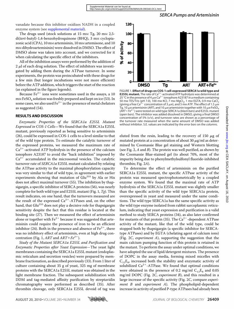
Enzymatic Properties of the SERCA1a E255L MutantExpressed in COS-1 Cells—We found that the SERCA1a E255Lmutant, previously reported as being sensitive to artemisinin(26), could be expressed in COS-1 cells to a level similar to thatof the wild type protein. To estimate the catalytic turnover ofthe expressed proteins, we measured the maximum rate ofCa2�-activated ATP hydrolysis in the presence of the calciumionophore A23187 to avoid the “back inhibition” imposed byCa2� accumulated in the microsomal vesicles. The catalyticturnover rate of SERCA1a E255Lmutant calculated by relatingthe ATPase activity to the maximal phosphorylation capacitywas very similar to that of wild type, in agreement with earlierexperiments showing that mutation of Glu255 by Ala or Hisdoes not affect maximal turnover (55). The inhibition by thap-sigargin, a specific inhibitor of SERCA proteins (56), was nearlycomplete for both wild type and E255Lmutant (Fig. 1,Tg). Thisresult indicates, on one hand, that the turnovers measured arethe result of the expressed Ca2�-ATPases and, on the otherhand, that Glu255 does not play a decisive role for thapsigarginsensitivity despite the fact that this residue is located at thebinding site (27). Then we measured the effect of artemisininalone or together with Fe2� because it was suggested that arte-misinin could require the presence of iron to be an efficientinhibitor (24). Both in the presence and absence of Fe2�, therewas no inhibitory effect of artemisinin, even at high drug con-centration (Fig. 1, ART and ART�Fe2�).Study of the Mutant SERCA1a E255L and Purification and
Enzymatic Properties after Yeast Expression—The yeast lightmembranes containing the SERCA1a E255Lmutant (endoplas-mic reticulum and secretion vesicles) were prepared by mem-brane fractionation, as described previously (33). From1 liter ofculture containing about 35 g of yeast, 325 mg of membraneproteins with the SERCA1a E255L mutant was obtained in thelight membrane fraction. The subsequent solubilization withDDM and tag-mediated affinity purification by streptavidinchromatography were performed as described (35). Afterthrombin cleavage, only SERCA1a E255L devoid of tag was
eluted from the resin, leading to the recovery of 150 �g ofmutated protein at a concentration of about 30 �g/ml as deter-mined by Coomassie Blue gel staining and Western blotting(see Fig. 2,A and B). The protein was well purified, as shown bythe Coomassie Blue-stained gel (to about 70%, most of theimpurity being due to phenylmethylsulfonyl fluoride-inhibitedthrombin; Fig. 2A).To determine the effect of artemisinin on the purified
SERCA1a E255L mutant, the specific ATPase activity of theprotein was measured spectrophotometrically by a coupledenzyme system. We found that the maximal rate of ATPhydrolysis of the SERCA1a E255L mutant was slightly smallerthan the specific activity of the wild type SERCA1a protein,overexpressed in yeast and measured under the same condi-tions. The wild type SERCA1a has the same specific activity asthe wild type enzyme isolated from rabbit sarcoplasmic reticu-lum, indicating that yeast expression and purification is a validmethod to study SERCA proteins (34), as also later confirmedfor mutants of that protein (35). The Ca2�-dependent ATPaseactivity of the mutant, like that of the wild type, could bestopped both by thapsigargin (a specific inhibitor for SERCA-type ATPases) and by EGTA (chelating agent of calcium ions)(Fig. 2C, experiment A), supporting the suggestion that themain calcium pumping function of this protein is retained inthemutant. To perform the assay under optimal conditions, wehave adopted the use of lipid/detergentmixtures. The presenceof DOPC in the assay media, forming mixed micelles withC12E8, increased both the stability and enzymatic activity ofsolubilized Ca2�-ATPase. We found that optimal conditionswere obtained in the presence of 0.2 mg/ml C12E8 and 0.05mg/ml DOPC (Fig. 2C, experiment B), and this resulted in alarge increase of the specific activity (Fig. 2C, compare experi-ment B and experiment A). The phospholipid-dependentincrease in activity of purified P-typeATPases had already been
FIGURE 1. Effect of drugs on COS-1 cell-expressed SERCA1a wild type andE255L mutant. The rate of Ca2�-activated ATP hydrolysis was determined at25 °C in the presence of 4 �M Ca2� ionophore A23187 in a medium containing50 mM TES/Tris (pH 7.0), 100 mM KCl, 7 mM MgCl2, 1 mM EGTA, 0.9 mM CaCl2(giving a free Ca2� concentration of 3 �M), and 5 mM ATP. The effect of 1.5 �M
Tg, 10 �M artemisinin (ART), and 10 �M artemisinin together with 10 �M FeSO4(ART�Fe2�) were tested on wild type SERCA1a (black bars) and E255L mutant(open bars). The inhibitor was added dissolved in DMSO, giving a final DMSOconcentration of 5% (v/v), and turnover rates are shown as a percentage ofthe turnover rate measured when the same amount of DMSO was addedwithout inhibitor. S.E. values are indicated by the error bars on the columns.
SERCA Pumps and Artemisinin
AUGUST 20, 2010 • VOLUME 285 • NUMBER 34 JOURNAL OF BIOLOGICAL CHEMISTRY 26409
at CE
A S
AC
LAY
, on August 16, 2010
ww
w.jbc.org
Dow
nloaded from
http://www.jbc.org/content/suppl/2010/06/08/M109.090340.DC1.htmlSupplemental Material can be found at:
observed (see Ref. 58, with the Na�/K� ATPase as a recentexample). The addition of artemisinin to the SERCA1a E255Lmutant solubilized in phospholipid/C12E8 medium did not
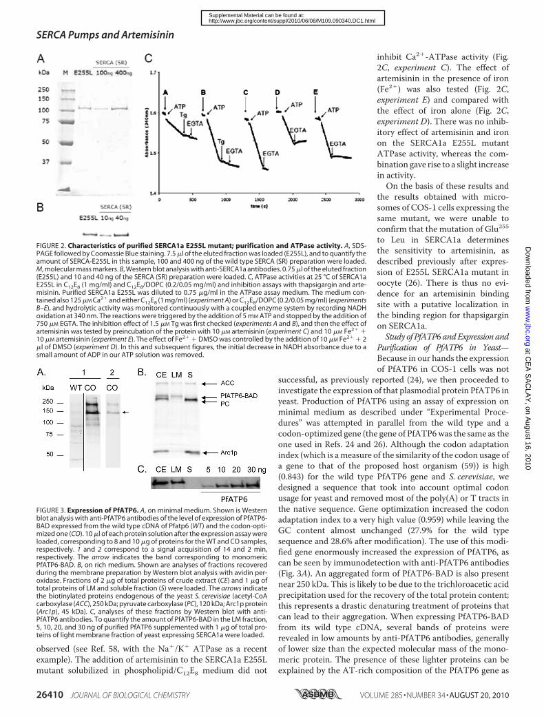
inhibit Ca2�-ATPase activity (Fig.2C, experiment C). The effect ofartemisinin in the presence of iron(Fe2�) was also tested (Fig. 2C,experiment E) and compared withthe effect of iron alone (Fig. 2C,experiment D). There was no inhib-itory effect of artemisinin and ironon the SERCA1a E255L mutantATPase activity, whereas the com-bination gave rise to a slight increasein activity.On the basis of these results and
the results obtained with micro-somes of COS-1 cells expressing thesame mutant, we were unable toconfirm that the mutation of Glu255to Leu in SERCA1a determinesthe sensitivity to artemisinin, asdescribed previously after expres-sion of E255L SERCA1a mutant inoocyte (26). There is thus no evi-dence for an artemisinin bindingsite with a putative localization inthe binding region for thapsigarginon SERCA1a.Study of PfATP6andExpression and
Purification of PfATP6 in Yeast—Because in our hands the expressionof PfATP6 in COS-1 cells was not
successful, as previously reported (24), we then proceeded toinvestigate the expression of that plasmodial protein PfATP6 inyeast. Production of PfATP6 using an assay of expression onminimal medium as described under “Experimental Proce-dures” was attempted in parallel from the wild type and acodon-optimized gene (the gene of PfATP6was the same as theone used in Refs. 24 and 26). Although the codon adaptationindex (which is ameasure of the similarity of the codon usage ofa gene to that of the proposed host organism (59)) is high(0.843) for the wild type PfATP6 gene and S. cerevisiae, wedesigned a sequence that took into account optimal codonusage for yeast and removed most of the poly(A) or T tracts inthe native sequence. Gene optimization increased the codonadaptation index to a very high value (0.959) while leaving theGC content almost unchanged (27.9% for the wild typesequence and 28.6% after modification). The use of this modi-fied gene enormously increased the expression of PfATP6, ascan be seen by immunodetection with anti-PfATP6 antibodies(Fig. 3A). An aggregated form of PfATP6-BAD is also presentnear 250 kDa. This is likely to be due to the trichloroacetic acidprecipitation used for the recovery of the total protein content;this represents a drastic denaturing treatment of proteins thatcan lead to their aggregation. When expressing PfATP6-BADfrom its wild type cDNA, several bands of proteins wererevealed in low amounts by anti-PfATP6 antibodies, generallyof lower size than the expected molecular mass of the mono-meric protein. The presence of these lighter proteins can beexplained by the AT-rich composition of the PfATP6 gene as
FIGURE 2. Characteristics of purified SERCA1a E255L mutant; purification and ATPase activity. A, SDS-PAGE followed by Coomassie Blue staining. 7.5 �l of the eluted fraction was loaded (E255L), and to quantify theamount of SERCA-E255L in this sample, 100 and 400 ng of the wild type SERCA (SR) preparation were loaded.M, molecular mass markers. B, Western blot analysis with anti-SERCA1a antibodies. 0.75 �l of the eluted fraction(E255L) and 10 and 40 ng of the SERCA (SR) preparation were loaded. C, ATPase activities at 25 °C of SERCA1aE255L in C12E8 (1 mg/ml) and C12E8/DOPC (0.2/0.05 mg/ml) and inhibition assays with thapsigargin and arte-misinin. Purified SERCA1a E255L was diluted to 0.75 �g/ml in the ATPase assay medium. The medium con-tained also 125 �M Ca2� and either C12E8 (1 mg/ml) (experiment A) or C12E8/DOPC (0.2/0.05 mg/ml) (experimentsB–E), and hydrolytic activity was monitored continuously with a coupled enzyme system by recording NADHoxidation at 340 nm. The reactions were triggered by the addition of 5 mM ATP and stopped by the addition of750 �M EGTA. The inhibition effect of 1.5 �M Tg was first checked (experiments A and B), and then the effect ofartemisinin was tested by preincubation of the protein with 10 �M artemisinin (experiment C) and 10 �M Fe2� �10 �M artemisinin (experiment E). The effect of Fe2� � DMSO was controlled by the addition of 10 �M Fe2� � 2�l of DMSO (experiment D). In this and subsequent figures, the initial decrease in NADH absorbance due to asmall amount of ADP in our ATP solution was removed.
FIGURE 3. Expression of PfATP6. A, on minimal medium. Shown is Westernblot analysis with anti-PfATP6 antibodies of the level of expression of PfATP6-BAD expressed from the wild type cDNA of Pfatp6 (WT) and the codon-opti-mized one (CO). 10 �l of each protein solution after the expression assay wereloaded, corresponding to 8 and 10 �g of proteins for the WT and CO samples,respectively. 1 and 2 correspond to a signal acquisition of 14 and 2 min,respectively. The arrow indicates the band corresponding to monomericPfATP6-BAD. B, on rich medium. Shown are analyses of fractions recoveredduring the membrane preparation by Western blot analysis with avidin per-oxidase. Fractions of 2 �g of total proteins of crude extract (CE) and 1 �g oftotal proteins of LM and soluble fraction (S) were loaded. The arrows indicatethe biotinylated proteins endogenous of the yeast S. cerevisiae (acetyl-CoAcarboxylase (ACC), 250 kDa; pyruvate carboxylase (PC), 120 kDa; Arc1p protein(Arc1p), 45 kDa). C, analyses of these fractions by Western blot with anti-PfATP6 antibodies. To quantify the amount of PfATP6-BAD in the LM fraction,5, 10, 20, and 30 ng of purified PfATP6 supplemented with 1 �g of total pro-teins of light membrane fraction of yeast expressing SERCA1a were loaded.
SERCA Pumps and Artemisinin
26410 JOURNAL OF BIOLOGICAL CHEMISTRY VOLUME 285 • NUMBER 34 • AUGUST 20, 2010
at CE
A S
AC
LAY
, on August 16, 2010
ww
w.jbc.org
Dow
nloaded from
http://www.jbc.org/content/suppl/2010/06/08/M109.090340.DC1.htmlSupplemental Material can be found at:
described for other plasmodial proteins expressed in S. cerevi-siae (60) and in Pichia pastoris (60–62). Indeed, AT-richregions in a gene may form hairpins and mimic a terminationsignal of transcription and therefore result in the synthesis oftruncated proteins. Consequently, we decided to use the opti-mized construct for a large scale expression of PfATP6. Like themutant SERCA1a E255L, we were able to express PfATP6 at ahigh level by the use of a fermentor for the yeast culture. Withthis equipment, 50 g of wet cells were recovered per liter ofculture.After preparation of the light yeast membrane fraction,
Western blot analyses with avidin peroxidase and anti-PfATP6antibodies confirmed that PfATP6-BAD had been expressedand had undergone in vivo biotinylation (see Fig. 3B). BecausePfATP6 is a SERCA-type protein with a location in the endo-plasmic reticulum, we have focused on the light membranefraction (endoplasmic reticulum) in the next steps. The amountof PfATP6-BAD contained in the LMs represents about 2% ofthe total protein content as determined by Western blotrevealed with anti-PfATP6 antibodies (see Fig. 3C) and 8mg/li-ter of culture. We also evaluated that about 30% of it was bio-tinylated (2.4mg; data not shown) and therefore subject to puri-fication by affinity chromatography. It can be noted thatnaturally biotinylated yeast proteins (acetyl-CoA carboxylase(ACC), 250 kDa; pyruvate carboxylase (PC), 120 kDa; Arc1pprotein (Arc1p), 45 kDa) aremainly eliminated with the solublefraction (see Fig. 3B). Nevertheless, because some of the solubleproteins remain bound to the LM fraction, we included one ortwo additional washing steps of themembranes with a highKClbuffer that helped to remove the major part of the remaining
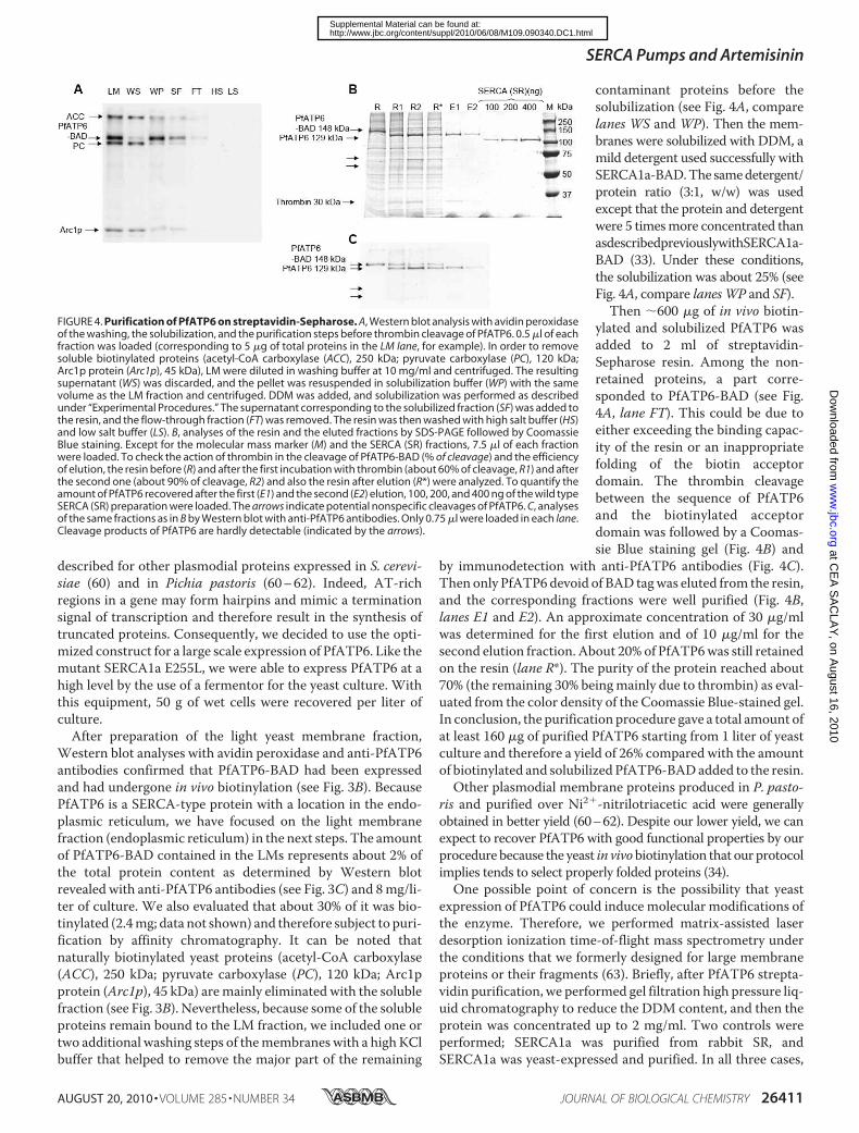
contaminant proteins before thesolubilization (see Fig. 4A, comparelanes WS and WP). Then the mem-branes were solubilized with DDM, amild detergent used successfully withSERCA1a-BAD.The samedetergent/protein ratio (3:1, w/w) was usedexcept that the protein and detergentwere 5 timesmore concentrated thanasdescribedpreviouslywithSERCA1a-BAD (33). Under these conditions,the solubilization was about 25% (seeFig. 4A, compare lanesWP and SF).Then �600 �g of in vivo biotin-
ylated and solubilized PfATP6 wasadded to 2 ml of streptavidin-Sepharose resin. Among the non-retained proteins, a part corre-sponded to PfATP6-BAD (see Fig.4A, lane FT). This could be due toeither exceeding the binding capac-ity of the resin or an inappropriatefolding of the biotin acceptordomain. The thrombin cleavagebetween the sequence of PfATP6and the biotinylated acceptordomain was followed by a Coomas-sie Blue staining gel (Fig. 4B) and
by immunodetection with anti-PfATP6 antibodies (Fig. 4C).Then only PfATP6devoid of BAD tagwas eluted from the resin,and the corresponding fractions were well purified (Fig. 4B,lanes E1 and E2). An approximate concentration of 30 �g/mlwas determined for the first elution and of 10 �g/ml for thesecond elution fraction. About 20% of PfATP6was still retainedon the resin (lane R*). The purity of the protein reached about70% (the remaining 30% beingmainly due to thrombin) as eval-uated from the color density of the Coomassie Blue-stained gel.In conclusion, the purification procedure gave a total amount ofat least 160 �g of purified PfATP6 starting from 1 liter of yeastculture and therefore a yield of 26% compared with the amountof biotinylated and solubilized PfATP6-BADadded to the resin.Other plasmodial membrane proteins produced in P. pasto-
ris and purified over Ni2�-nitrilotriacetic acid were generallyobtained in better yield (60–62). Despite our lower yield, we canexpect to recover PfATP6 with good functional properties by ourprocedure because the yeast in vivobiotinylation that our protocolimplies tends to select properly folded proteins (34).One possible point of concern is the possibility that yeast
expression of PfATP6 could induce molecular modifications ofthe enzyme. Therefore, we performed matrix-assisted laserdesorption ionization time-of-flight mass spectrometry underthe conditions that we formerly designed for large membraneproteins or their fragments (63). Briefly, after PfATP6 strepta-vidin purification, we performed gel filtration high pressure liq-uid chromatography to reduce the DDM content, and then theprotein was concentrated up to 2 mg/ml. Two controls wereperformed; SERCA1a was purified from rabbit SR, andSERCA1a was yeast-expressed and purified. In all three cases,
FIGURE 4. Purification of PfATP6 on streptavidin-Sepharose. A, Western blot analysis with avidin peroxidaseof the washing, the solubilization, and the purification steps before thrombin cleavage of PfATP6. 0.5 �l of eachfraction was loaded (corresponding to 5 �g of total proteins in the LM lane, for example). In order to removesoluble biotinylated proteins (acetyl-CoA carboxylase (ACC), 250 kDa; pyruvate carboxylase (PC), 120 kDa;Arc1p protein (Arc1p), 45 kDa), LM were diluted in washing buffer at 10 mg/ml and centrifuged. The resultingsupernatant (WS) was discarded, and the pellet was resuspended in solubilization buffer (WP) with the samevolume as the LM fraction and centrifuged. DDM was added, and solubilization was performed as describedunder “Experimental Procedures.” The supernatant corresponding to the solubilized fraction (SF) was added tothe resin, and the flow-through fraction (FT) was removed. The resin was then washed with high salt buffer (HS)and low salt buffer (LS). B, analyses of the resin and the eluted fractions by SDS-PAGE followed by CoomassieBlue staining. Except for the molecular mass marker (M) and the SERCA (SR) fractions, 7.5 �l of each fractionwere loaded. To check the action of thrombin in the cleavage of PfATP6-BAD (% of cleavage) and the efficiencyof elution, the resin before (R) and after the first incubation with thrombin (about 60% of cleavage, R1) and afterthe second one (about 90% of cleavage, R2) and also the resin after elution (R*) were analyzed. To quantify theamount of PfATP6 recovered after the first (E1) and the second (E2) elution, 100, 200, and 400 ng of the wild typeSERCA (SR) preparation were loaded. The arrows indicate potential nonspecific cleavages of PfATP6. C, analysesof the same fractions as in B by Western blot with anti-PfATP6 antibodies. Only 0.75 �l were loaded in each lane.Cleavage products of PfATP6 are hardly detectable (indicated by the arrows).
SERCA Pumps and Artemisinin
AUGUST 20, 2010 • VOLUME 285 • NUMBER 34 JOURNAL OF BIOLOGICAL CHEMISTRY 26411
at CE
A S
AC
LAY
, on August 16, 2010
ww
w.jbc.org
Dow
nloaded from
http://www.jbc.org/content/suppl/2010/06/08/M109.090340.DC1.htmlSupplemental Material can be found at:
the determined molecular mass was very close to the expectedmolecular mass. In the case of PfATP6, we found a mass of140,053 Da, whereas the expected mass is 139,994 Da. Undersimilar conditions, rabbit SR SERCA1a gave 109,497 Da for anexpected mass of 109,489 Da, and yeast expressed SERCA1a(which is slightly larger due to DNA construction (34)) gave110,090 Da for an expected mass of 110,069 Da.5 Under theseconditions, the error associated with this type of measurementis 0.05–0.1%, so that this excludes large modification, such aspartial proteolysis, glycosylations, etc.Study of the Enzymatic Properties of the Soluble Purified
PfATP6—We first measured the specific ATPase activity of thepurified PfATP6 with the aid of the coupled enzyme assay asdescribed for SERCA1a E255L and in the presence of 1 mg/mlC12E8 (data not shown) or 0.2mg/mlC12E8 (Fig. 5A, left). As canbe seen, the major part of ATP hydrolysis was stopped by theaddition of EGTA, indicative of calcium-dependent ATPaseactivity. However, even at higher C12E8 concentrations (datanot shown), we observed a decrease of the hydrolytic ratewith time, suggesting that under these conditions with puredetergent, the protein is inactivated during turnover. By theaddition of lipids (0.05 mg/ml of DOPC) together with 0.2mg/ml C12E8, we were able to maintain a stable hydrolysisrate of ATP that was still inhibited by EGTA (Fig. 5A, mid-dle). A similar stabilization was obtained in the presence ofother lipids (1,2-dioleoyl-sn-glycero-3-phosphoserine, eggyolk phosphatidylcholine, and 1-palmitoyl-2-oleoyl-sn-glycero-3-phosphoethanolamine (data not shown)) or a lipid mixtureconsisting of 48.4% DOPC, 43% 1-palmitoyl-2-oleoyl-sn-glyc-ero-3-phosphoethanolamine, and 8.6% L-�-phosphatidylinosi-tol) (Fig. 5A, right). The phospholipid composition of this mix-ture was based on the membrane lipidic composition ofP. falciparum as described (64). At a C12E8/DOPC ratio of 0.2:0.05mg/ml, the specific activity at 25 °C of the purified PfATP6
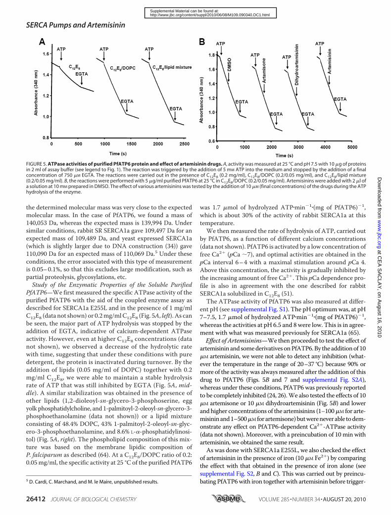
was 1.7 �mol of hydrolyzed ATP�min�1�(mg of PfATP6)�1,which is about 30% of the activity of rabbit SERCA1a at thistemperature.We then measured the rate of hydrolysis of ATP, carried out
by PfATP6, as a function of different calcium concentrations(data not shown). PfATP6 is activated by a low concentration offree Ca2� (pCa �7), and optimal activities are obtained in thepCa interval 6–4 with a maximal stimulation around pCa 4.Above this concentration, the activity is gradually inhibited bythe increasing amount of free Ca2�. This pCa dependence pro-file is also in agreement with the one described for rabbitSERCA1a solubilized in C12E8 (51).The ATPase activity of PfATP6 was also measured at differ-
ent pH (see supplemental Fig. S1). The pH optimumwas, at pH7–7.5, 1.7 �mol of hydrolyzed ATP�min�1�(mg of PfATP6)�1,whereas the activities at pH 6.5 and 8 were low. This is in agree-ment with what was measured previously for SERCA1a (65).Effect of Artemisinins—We then proceeded to test the effect of
artemisinin and somederivativesonPfATP6.By the additionof 10�M artemisinin, we were not able to detect any inhibition (what-ever the temperature in the range of 20–37 °C) because 90% ormore of the activity was alwaysmeasured after the addition of thisdrug to PfATP6 (Figs. 5B and 7 and supplemental Fig. S2A),whereas under these conditions, PfATP6 was previously reportedto be completely inhibited (24, 26).We also tested the effects of 10�M artemisone or 10 �M dihydroartemisinin (Fig. 5B) and lowerand higher concentrations of the artemisinins (1–100�M for arte-misininand1–500�Mforartemisone)butwereneverable todem-onstrate any effect on PfATP6-dependent Ca2�-ATPase activity(data not shown). Moreover, with a preincubation of 10 min withartemisinin, we obtained the same result.As was done with SERCA1a E255L, we also checked the effect
of artemisinin in the presence of iron (10 �M Fe2�) by comparingthe effect with that obtained in the presence of iron alone (seesupplemental Fig. S2, B and C). This was carried out by preincu-bating PfATP6with iron togetherwith artemisinin before trigger-5 D. Cardi, C. Marchand, and M. le Maire, unpublished results.
FIGURE 5. ATPase activities of purified PfATP6 protein and effect of artemisinin drugs. A, activity was measured at 25 °C and pH 7.5 with 10 �g of proteinsin 2 ml of assay buffer (see legend to Fig. 1). The reaction was triggered by the addition of 5 mM ATP into the medium and stopped by the addition of a finalconcentration of 750 �M EGTA. The reactions were carried out in the presence of C12E8 (0.2 mg/ml), C12E8/DOPC (0.2/0.05 mg/ml), and C12E8/lipid mixture(0.2/0.05 mg/ml). B, the reactions were performed with 5 �g/ml purified PfATP6 at 25 °C in C12E8/DOPC (0.2/0.05 mg/ml). Artemisinins were added with 2 �l ofa solution at 10 mM prepared in DMSO. The effect of various artemisinins was tested by the addition of 10 �M (final concentrations) of the drugs during the ATPhydrolysis of the enzyme.
SERCA Pumps and Artemisinin
26412 JOURNAL OF BIOLOGICAL CHEMISTRY VOLUME 285 • NUMBER 34 • AUGUST 20, 2010
at CE
A S
AC
LAY
, on August 16, 2010
ww
w.jbc.org
Dow
nloaded from
http://www.jbc.org/content/suppl/2010/06/08/M109.090340.DC1.htmlSupplemental Material can be found at:
ing the reaction. On the other hand, the Ca2�-ATPase activity aswell as the nonspecific activity was slightly decreased in the pres-ence of Fe2�. In addition, other artemisinin derivatives that werealso tested, including artemisone, which was also used in theoocyte tests (26), and artesunate (see supplemental Fig. S2,D andE), did not inhibit the ATPase activity of PfATP6 either.Effect of SERCA Inhibitors—We then tested the effect of spe-
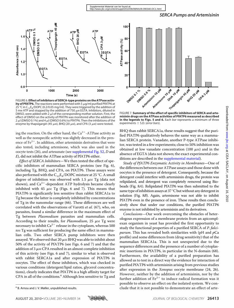
cific inhibitors of mammalian SERCA proteins (see Fig. 6),including Tg, BHQ, and CPA, on PfATP6. These assays werealso performedwith the C12E8/DOPCmixture at 25 °C. A smalldegree of inhibition was observed with 1.5 �M Tg (data notshown), and Ca2�-dependent ATP hydrolysis became clearlyinhibited with 45 �M Tg (Figs. 6 and 7). This means thatPfATP6 is significantly less sensitive than rabbit SERCA1a toTg because the latter is completely inhibited by concentrationsof Tg in the nanomolar range (66). These differences are wellcorrelated with the observations of Varotti et al. (67), who, onparasites, found a similar difference in the maximum effect ofTg between Plasmodium parasites and mammalian cells.According to their results, for Plasmodium, 25 �M Tg werenecessary to inhibit Ca2� release in the cytoplasm, whereas 500nM Tg was sufficient for producing the same effect in mamma-lian cells. Two other SERCA pump inhibitors were thenassayed.We observed that 20�MBHQwas able to inhibit about50% of the activity of PfATP6 (see Figs. 6 and 7) and that theaddition of 3�MCPA resulted in an almost complete inhibitionof this activity (see Figs. 6 and 7), similar to what is observedwith rabbit SERCA1a and after expression of PfATP6 inoocytes. The effect of these inhibitors, which was checked invarious conditions (detergent/lipid ratios, glycerol concentra-tions), clearly indicates that PfATP6 is a high affinity target forCPA in all of the conditions.6 Although less sensitive to Tg and
BHQ than rabbit SERCA1a, these results suggest that the puri-fied PfATP6 qualitatively behaves the same way as a mamma-lian SERCA protein. Vanadate, another P-type ATPase inhibi-tor, was tested in a few experiments; close to 50% inhibitionwasobtained at low vanadate concentration (100 �M) and in theabsence of EGTA (data not shown; the exact experimental con-ditions are described in the supplemental material).Study of PfATP6 Enzymatic Activity in Membranes—One of
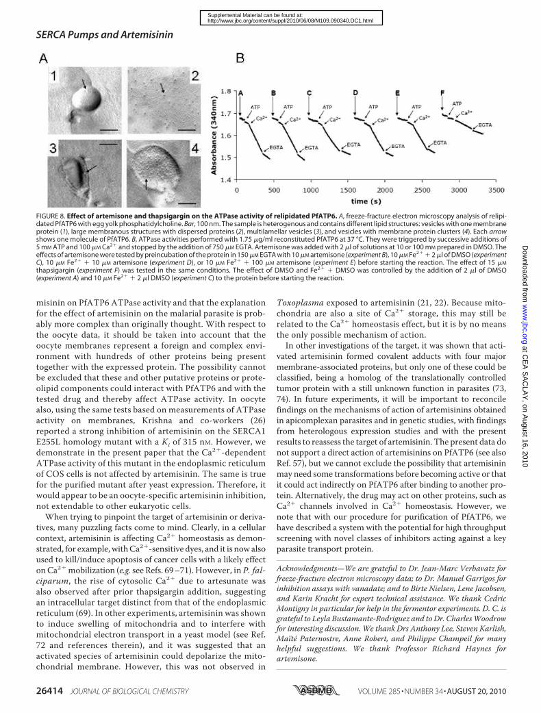
the differences between ourATPase assays and those donewithoocytes is the presence of detergent. Consequently, because thedetergent could interfere with artemisinin drugs, the protein wasrelipidated, and detergent was completely removed using Bio-beads (Fig. 8A). Relipidated PfATP6 was then submitted to thesametypeof inhibitionassaysat37 °Cbutwithoutanydetergent insolution (Fig. 8B). Again, artemisone was not able to inhibitPfATP6 even in the presence of iron. These results then conclu-sively show that under our conditions, the purified PfATP6enzyme is not inhibited by artemisinin and its derivatives.Conclusions—Our work overcoming the obstacles of heter-
ologous expression of a membrane protein from an apicompl-exan organism in yeast has provided the first opportunity tostudy the functional properties of a purified SERCA of P. falci-parum. This has revealed both similarities with (pH and pCaprofile) and some differences from (drug sensitivity) that of themammalian SERCA1a. This is not unexpected due to thesequence differences and the presence of a number of cytoplas-mic insertions in PfATP6, in particular in the N-domain (68).Furthermore, the availability of a purified preparation hasallowed us to test in a direct way the evidence for interaction ofpurified PfATP6with artemisinin arising from previous studiesafter expression in the Xenopus oocyte membrane (24, 26).However, neither by the addition of artemisinin, nor by thefurther addition of Fe2� to induce radical formation was itpossible to observe an effect on the isolated system. We con-clude that it is not possible to demonstrate an effect of arte-6 B. Arnou and J. V. Møller, unpublished results.
FIGURE 6. Effect of inhibitors of SERCA-type proteins on the ATPase activ-ity of PfATP6. The reactions were performed with 5 �g/ml purified PfATP6 at25 °C in C12E8/DOPC (0.2/0.05 mg/ml). They were triggered by the addition of5 mM ATP and stopped by the addition of 750 �M EGTA. Inhibitors, diluted inDMSO, were added with 2 �l of the corresponding mother solution. First, theeffect of DMSO on the activity of PfATP6 was monitored after the addition of2 �l (DMSO 0.1%) and 4 �l (DMSO 0.6%) to PfATP6. Then the inhibitions of theenzyme by thapsigargin (45 �M), BHQ (20 �M), and CPA (3 �M) were tested.
FIGURE 7. Summary of the effect of specific inhibitors of SERCA and arte-misinin drugs on the ATPase activities of PfATP6 measured as describedin the legends to Figs. 5 and 6. Each bar represents a minimum of threeexperiments � S.D. (error bars).
SERCA Pumps and Artemisinin
AUGUST 20, 2010 • VOLUME 285 • NUMBER 34 JOURNAL OF BIOLOGICAL CHEMISTRY 26413
at CE
A S
AC
LAY
, on August 16, 2010
ww
w.jbc.org
Dow
nloaded from
http://www.jbc.org/content/suppl/2010/06/08/M109.090340.DC1.htmlSupplemental Material can be found at:
misinin on PfATP6 ATPase activity and that the explanationfor the effect of artemisinin on the malarial parasite is prob-ably more complex than originally thought. With respect tothe oocyte data, it should be taken into account that theoocyte membranes represent a foreign and complex envi-ronment with hundreds of other proteins being presenttogether with the expressed protein. The possibility cannotbe excluded that these and other putative proteins or prote-olipid components could interact with PfATP6 and with thetested drug and thereby affect ATPase activity. In oocytealso, using the same tests based on measurements of ATPaseactivity on membranes, Krishna and co-workers (26)reported a strong inhibition of artemisinin on the SERCA1E255L homology mutant with a Ki of 315 nM. However, wedemonstrate in the present paper that the Ca2�-dependentATPase activity of this mutant in the endoplasmic reticulumof COS cells is not affected by artemisinin. The same is truefor the purified mutant after yeast expression. Therefore, itwould appear to be an oocyte-specific artemisinin inhibition,not extendable to other eukaryotic cells.When trying to pinpoint the target of artemisinin or deriva-
tives, many puzzling facts come to mind. Clearly, in a cellularcontext, artemisinin is affecting Ca2� homeostasis as demon-strated, for example,withCa2�-sensitive dyes, and it is nowalsoused to kill/induce apoptosis of cancer cells with a likely effectonCa2�mobilization (e.g. see Refs. 69–71). However, in P. fal-ciparum, the rise of cytosolic Ca2� due to artesunate wasalso observed after prior thapsigargin addition, suggestingan intracellular target distinct from that of the endoplasmicreticulum (69). In other experiments, artemisinin was shownto induce swelling of mitochondria and to interfere withmitochondrial electron transport in a yeast model (see Ref.72 and references therein), and it was suggested that anactivated species of artemisinin could depolarize the mito-chondrial membrane. However, this was not observed in
Toxoplasma exposed to artemisinin (21, 22). Because mito-chondria are also a site of Ca2� storage, this may still berelated to the Ca2� homeostasis effect, but it is by no meansthe only possible mechanism of action.In other investigations of the target, it was shown that acti-
vated artemisinin formed covalent adducts with four majormembrane-associated proteins, but only one of these could beclassified, being a homolog of the translationally controlledtumor protein with a still unknown function in parasites (73,74). In future experiments, it will be important to reconcilefindings on the mechanisms of action of artemisinins obtainedin apicomplexan parasites and in genetic studies, with findingsfrom heterologous expression studies and with the presentresults to reassess the target of artemisinin. The present data donot support a direct action of artemisinins on PfATP6 (see alsoRef. 57), but we cannot exclude the possibility that artemisininmay need some transformations before becoming active or thatit could act indirectly on PfATP6 after binding to another pro-tein. Alternatively, the drug may act on other proteins, such asCa2� channels involved in Ca2� homeostasis. However, wenote that with our procedure for purification of PfATP6, wehave described a system with the potential for high throughputscreening with novel classes of inhibitors acting against a keyparasite transport protein.
Acknowledgments—We are grateful to Dr. Jean-Marc Verbavatz forfreeze-fracture electron microscopy data; to Dr. Manuel Garrigos forinhibition assays with vanadate; and to Birte Nielsen, Lene Jacobsen,and Karin Kracht for expert technical assistance. We thank CedricMontigny in particular for help in the fermentor experiments. D. C. isgrateful to Leyla Bustamante-Rodriguez and toDr. CharlesWoodrowfor interesting discussion. We thank Drs Anthony Lee, Steven Karlish,Maïte Paternostre, Anne Robert, and Philippe Champeil for manyhelpful suggestions. We thank Professor Richard Haynes forartemisone.
FIGURE 8. Effect of artemisone and thapsigargin on the ATPase activity of relipidated PfATP6. A, freeze-fracture electron microscopy analysis of relipi-dated PfATP6 with egg yolk phosphatidylcholine. Bar, 100 nm. The sample is heterogenous and contains different lipid structures: vesicles with one membraneprotein (1), large membranous structures with dispersed proteins (2), multilamellar vesicles (3), and vesicles with membrane protein clusters (4). Each arrowshows one molecule of PfATP6. B, ATPase activities performed with 1.75 �g/ml reconstituted PfATP6 at 37 °C. They were triggered by successive additions of5 mM ATP and 100 �M Ca2� and stopped by the addition of 750 �M EGTA. Artemisone was added with 2 �l of solutions at 10 or 100 mM prepared in DMSO. Theeffects of artemisone were tested by preincubation of the protein in 150 �M EGTA with 10 �M artemisone (experiment B), 10 �M Fe2� � 2 �l of DMSO (experimentC), 10 �M Fe2� � 10 �M artemisone (experiment D), or 10 �M Fe2� � 100 �M artemisone (experiment E) before starting the reaction. The effect of 15 �M
thapsigargin (experiment F) was tested in the same conditions. The effect of DMSO and Fe2� � DMSO was controlled by the addition of 2 �l of DMSO(experiment A) and 10 �M Fe2� � 2 �l DMSO (experiment C) to the protein before starting the reaction.
SERCA Pumps and Artemisinin
26414 JOURNAL OF BIOLOGICAL CHEMISTRY VOLUME 285 • NUMBER 34 • AUGUST 20, 2010
at CE
A S
AC
LAY
, on August 16, 2010
ww
w.jbc.org
Dow
nloaded from
http://www.jbc.org/content/suppl/2010/06/08/M109.090340.DC1.htmlSupplemental Material can be found at:
REFERENCES1. White, N. J. (2008) Science 320, 330–3342. Fidock, D. A., Eastman, R. T., Ward, S. A., and Meshnick, S. R. (2008)
Trends Parasitol. 24, 537–5443. Dondorp, A. M., Nosten, F., Yi, P., Das, D., Phyo, A. P., Tarning, J., Lwin,
K. M., Ariey, F., Hanpithakpong, W., Lee, S. J., Ringwald, P., Silamut, K.,Imwong, M., Chotivanich, K., Lim, P., Herdman, T., An, S. S., Yeung, S.,Singhasivanon, P., Day, N. P., Lindegardh, N., Socheat, D., andWhite, N. J.(2009) N. Engl. J. Med. 361, 455–467
4. ter Kuile, F., White, N. J., Holloway, P., Pasvol, G., and Krishna, S. (1993)Exp. Parasitol. 76, 85–95
5. Woodrow, C. J., Haynes, R. K., and Krishna, S. (2005) Postgrad. Med. J. 81,71–78
6. Burk, O., Arnold, K. A., Nussler, A. K., Schaeffeler, E., Efimova, E., Avery,B. A., Avery, M. A., Fromm, M. F., and Eichelbaum, M. (2005)Mol. Phar-macol. 67, 1954–1965
7. Haynes, R. K., Fugmann, B., Stetter, J., Rieckmann, K., Heilmann, H. D.,Chan, H. W., Cheung, M. K., Lam, W. L., Wong, H. N., Croft, S. L., Vivas,L., Rattray, L., Stewart, L., Peters, W., Robinson, B. L., Edstein, M. D.,Kotecka, B., Kyle, D. E., Beckermann, B., Gerisch, M., Radtke, M.,Schmuck, G., Steinke, W., Wollborn, U., Schmeer, K., and Romer, A.(2006) Angew. Chem. Int. Ed. Engl. 45, 2082–2088
8. Meshnick, S. R., Taylor, T. E., and Kamchonwongpaisan, S. (1996)Micro-biol. Rev. 60, 301–315
9. O’Neill, P. M. (2005) Expert Opin. Investig. Drugs 14, 1117–112810. Avery, M. A., Gao, F., Chong, W. K., Mehrotra, S., and Milhous, W. K.
(1993) J. Med. Chem. 36, 4264–427511. Kumar, N., and Zheng, H. (1990) Parasitol. Res. 76, 214–21812. Kombila, M., Duong, T. H., Dufillot, D., Koko, J., Guiyedi, V., Guiguen, C.,
Ferrer, A., and Richard-Lenoble, D. (1997) Am. J. Trop. Med. Hyg. 57,643–645
13. Vyas, N., Avery, B. A., Avery,M.A., andWyandt, C.M. (2002)Antimicrob.Agents Chemother. 46, 105–109
14. Gu, H. M., Warhurst, D. C., and Peters, W. (1984) Trans. R. Soc. Trop.Med. Hyg. 78, 265–270
15. Olliaro, P. L., Haynes, R. K., Meunier, B., and Yuthavong, Y. (2001) TrendsParasitol. 17, 122–126
16. Hartwig, C. L., Rosenthal, A. S., D’Angelo, J., Griffin, C. E., Posner, G. H.,and Cooper, R. A. (2009) Biochem. Pharmacol. 77, 322–336
17. Ittarat, W., Sreepian, A., Srisarin, A., and Pathepchotivong, K. (2003)Southeast Asian J. Trop. Med. Public Health 34, 744–750
18. Golenser, J., Waknine, J. H., Krugliak, M., Hunt, N. H., and Grau, G. E.(2006) Int. J. Parasitol. 36, 1427–1441
19. Zhang, G., Guan, Y., Zheng, B.,Wu, S., and Tang, L. (2008)Malar. J. 7, 12220. Hoppe, H. C., van Schalkwyk, D. A., Wiehart, U. I., Meredith, S. A., Egan,
J., and Weber, B. W. (2004) Antimicrob. Agents Chemother. 48,2370–2378
21. Nagamune, K., Moreno, S. N., and Sibley, L. D. (2007) Antimicrob. AgentsChemother. 51, 3816–3823
22. Nagamune, K., Beatty, W. L., and Sibley, L. D. (2007) Eukaryot. Cell 6,2147–2156
23. Nagamune, K., and Sibley, L. D. (2006)Mol. Biol. Evol. 23, 1613–162724. Eckstein-Ludwig, U., Webb, R. J., Van Goethem, I. D., East, J. M., Lee,
A. G., Kimura, M., O’Neill, P. M., Bray, P. G., Ward, S. A., and Krishna, S.(2003) Nature 424, 957–961
25. Toovey, S., Bustamante, L. Y., Uhlemann, A. C., East, J. M., and Krishna, S.(2008) Basic Clin. Pharmacol. Toxicol. 103, 209–213
26. Uhlemann, A. C., Cameron, A., Eckstein-Ludwig, U., Fischbarg, J., Iserov-ich, P., Zuniga, F. A., East,M., Lee, A., Brady, L., Haynes, R. K., andKrishna,S. (2005) Nat. Struct. Mol. Biol. 12, 628–629
27. Toyoshima, C., and Nomura, H. (2002) Nature 418, 605–61128. Jambou, R., Legrand, E., Niang, M., Khim, N., Lim, P., Volney, B., Ekala,
M. T., Bouchier, C., Esterre, P., Fandeur, T., and Mercereau-Puijalon, O.(2005) Lancet 366, 1960–1963
29. Cojean, S., Hubert, V., Le Bras, J., and Durand, R. (2006) Emerg. Infect. Dis.12, 1798–1799
30. Ferreira, I. D., Martinelli, A., Rodrigues, L. A., do Carmo, E. L., do Rosario,
V. E., Povoa, M. M., and Cravo, P. (2008) Trop. Med. Int. Health 13,199–207
31. Krishna, S., Bustamante, L., Haynes, R. K., and Staines, H. M. (2008)Trends Pharmacol. Sci. 29, 520–527
32. Krishna, S.,Woodrow, C., Webb, R., Penny, J., Takeyasu, K., Kimura, M.,and East, J. M. (2001) J. Biol. Chem. 276, 10782–10787
33. Jidenko, M., Lenoir, G., Fuentes, J. M., le Maire, M., and Jaxel, C. (2006)Protein Expr. Purif. 48, 32–42
34. Jidenko, M., Nielsen, R. C., Sørensen, T. L., Møller, J. V., le Maire, M.,Nissen, P., and Jaxel, C. (2005) Proc. Natl. Acad. Sci. U.S.A. 102,11687–11691
35. Marchand, A., Winther, A. M., Holm, P. J., Olesen, C., Montigny, C.,Arnou, B., Champeil, P., Clausen, J. D., Vilsen, B., Andersen, J. P., Nissen,P., Jaxel, C., Møller, J. V., and le Maire, M. (2008) J. Biol. Chem. 283,14867–14882
36. Kaufman, R. J., Davies,M. V., Pathak, V. K., andHershey, J.W. (1989)Mol.Cell Biol. 9, 946–958
37. Chen, C., and Okayama, H. (1987)Mol. Cell Biol. 7, 2745–275238. Maruyama, K., and MacLennan, D. H. (1988) Proc. Natl. Acad. Sci. U.S.A.
85, 3314–331839. Vilsen, B., Andersen, J. P., andMacLennan, D.H. (1991) J. Biol. Chem. 266,
16157–1616440. Sorensen, T., Vilsen, B., and Andersen, J. P. (1997) J. Biol. Chem. 272,
30244–3025341. Baginski, E. S., Foa, P. P., and Zak, B. (1967) Clin. Chem. 13, 326–33242. Lenoir, G., Menguy, T., Corre, F., Montigny, C., Pedersen, P. A.,
Thines, D., le Maire, M., and Falson, P. (2002) Biochim. Biophys. Acta1560, 67–83
43. Gietz, R. D., Schiestl, R. H., Willems, A. R., andWoods, R. A. (1995) Yeast11, 355–360
44. Centeno, F., Deschamps, S., Lompre, A. M., Anger, M., Moutin, M. J.,Dupont, Y., Palmgren, M. G., Villalba, J. M., Møller, J. V., Falson, P., and leMaire, M. (1994) FEBS Lett. 354, 117–122
45. Pompon, D., Louerat, B., Bronine, A., and Urban, P. (1996) Methods En-zymol. 272, 51–64
46. Smith, P. K., Krohn, R. I., Hermanson, G. T., Mallia, A. K., Gartner, F. H.,Provenzano, M. D., Fujimoto, E. K., Goeke, N. M., Olson, B. J., and Klenk,D. C. (1985) Anal. Biochem. 150, 76–85
47. Champeil, P., Guillain, F., Venien, C., and Gingold, M. P. (1985) Biochem-istry 24, 69–81
48. Soulie, S., Denoroy, L., Le Caer, J. P., Hamasaki, N., Groves, J. D., and leMaire, M. (1998) J. Biochem. 124, 417–420
49. Juul, B., Turc, H., Durand, M. L., Gomez de Gracia, A., Denoroy, L.,Møller, J. V., Champeil, P., and le Maire, M. (1995) J. Biol. Chem. 270,20123–20134
50. Rigaud, J. L., Mosser, G., Lacapere, J. J., Olofsson, A., Levy, D., and Ranck,J. L. (1997) J. Struct. Biol. 118, 226–235
51. Møller, J. V., Lind, K. E., and Andersen, J. P. (1980) J. Biol. Chem. 255,1912–1920
52. Falson, P.,Menguy, T., Corre, F., Bouneau, L., Gomez deGracia, A., Soulie,S., Centeno, F., Moller, J. V., Champeil, P., and le Maire, M. (1997) J. Biol.Chem. 272, 17258–17262
53. Montigny, C., Jaxel, C., Shainskaya, A., Vinh, J., Labas, V., Møller, J. V.,Karlish, S. J., and le Maire, M. (2004) J. Biol. Chem. 279, 43971–43981
54. Witting, P. K., Douglas, D. J., and Mauk, A. G. (2001) J. Biol. Chem. 276,3991–3998
55. Clausen, J. D., and Andersen, J. P. (2003) Biochemistry 42, 2585–259456. Thastrup, O., Cullen, P. J., Drøbak, B. K., Hanley, M. R., and Dawson, A. P.
(1990) Proc. Natl. Acad. Sci. U.S.A. 87, 2466–247057. Bousejra-EIGarah, F., Stigliani, J. L., Cosledan, F.,Meunier, B., and Robert,
A. (2009) ChemMedChem. 4, 1469–147958. Haviv, H., Cohen, E., Lifshitz, Y., Tal, D. M., Goldshleger, R., and Karlish,
S. J. (2007) Biochemistry 46, 12855–1286759. Sharp, P. M., and Li, W. H. (1987) Nucleic Acids Res. 15, 1281–129560. Zhang, H., Howard, E. M., and Roepe, P. D. (2002) J. Biol. Chem. 277,
49767–4977561. Hedfalk, K., Pettersson, N., Oberg, F., Hohmann, S., andGordon, E. (2008)
Protein Expr. Purif. 59, 69–78
SERCA Pumps and Artemisinin
AUGUST 20, 2010 • VOLUME 285 • NUMBER 34 JOURNAL OF BIOLOGICAL CHEMISTRY 26415
at CE
A S
AC
LAY
, on August 16, 2010
ww
w.jbc.org
Dow
nloaded from
http://www.jbc.org/content/suppl/2010/06/08/M109.090340.DC1.htmlSupplemental Material can be found at:
62. Tan, W., Gou, D. M., Tai, E., Zhao, Y. Z., and Chow, L. M. (2006) Arch.Biochem. Biophys. 452, 119–128
63. Lenoir, G., Picard, M., Gauron, C., Montigny, C., Le Marechal, P., Falson,P., Le Maire, M., Møller, J. V., and Champeil, P. (2004) J. Biol. Chem. 279,9156–9166
64. Vial, H. J., Eldin, P., Tielens, A. G., and van Hellemond, J. J. (2003) Mol.Biochem. Parasitol. 126, 143–154
65. Lund, S., Orlowski, S., de Foresta, B., Champeil, P., le Maire, M., andMøller, J. V. (1989) J. Biol. Chem. 264, 4907–4915
66. Sagara, Y., and Inesi, G. (1991) J. Biol. Chem. 266, 13503–1350667. Varotti, F. P., Beraldo, F. H., Gazarini, M. L., and Garcia, C. R. (2003) Cell
Calcium 33, 137–14468. Kimura, M., Yamaguchi, Y., Takada, S., and Tanabe, K. (1993) J. Cell Sci.
104, 1129–113669. de Pilla Varotti, F., Botelho, A. C., Andrade, A. A., de Paula, R. C., Fagun-
des, E. M., Valverde, A., Mayer, L. M., Mendonca, J. S., de Souza, M. V.,Boechat, N., and Krettli, A. U. (2008) Antimicrob. Agents Chemother. 52,3868–3874
70. Riganti, C., Doublier, S., Viarisio, D., Miraglia, E., Pescarmona, G., Ghigo,D., and Bosia, A. (2009) Br. J. Pharmacol. 156, 1054–1066
71. Stockwin, L. H., Han, B., Yu, S. X., Hollingshead, M. G., ElSohly, M. A.,Gul, W., Slade, D., Galal, A. M., and Newton, D. L. (2009) Int. J. Cancer125, 1266–1275
72. Li, W., Mo, W., Shen, D., Sun, L., Wang, J., Lu, S., Gitschier, J. M., andZhou, B. (2005) PLoS Genet. 1, e36
73. Asawamahasakda, W., Ittarat, I., Pu, Y. M., Ziffer, H., and Meshnick, S. R.(1994) Antimicrob. Agents Chemother. 38, 1854–1858
74. Bhisutthibhan, J., Pan, X. Q., Hossler, P. A., Walker, D. J., Yowell, C. A.,Carlton, J., Dame, J. B., and Meshnick, S. R. (1998) J. Biol. Chem. 273,16192–16198
SERCA Pumps and Artemisinin
26416 JOURNAL OF BIOLOGICAL CHEMISTRY VOLUME 285 • NUMBER 34 • AUGUST 20, 2010
at CE
A S
AC
LAY
, on August 16, 2010
ww
w.jbc.org
Dow
nloaded from
http://www.jbc.org/content/suppl/2010/06/08/M109.090340.DC1.htmlSupplemental Material can be found at:
Related Documents