Elsevier Editorial System(tm) for International Journal of Biological Macromolecules Manuscript Draft Manuscript Number: Title: Purification of a secreted lectin from Andrias davidianus skin and its antibacterial activity Article Type: Research Report Keywords: Andrias davidianus; lectin; antibacterial activity Corresponding Author: Dr. Wei Li, Ph.D. Corresponding Author's Institution: First Author: Min Qu, Dr Order of Authors: Min Qu, Dr; Changqing Tong, Dr; Liang Kong, Dr; Chengyu Tan, Dr; Xin Yan; Oleg V Chenikov, Dr; Pavel A Lukyanov; Qiao Jin, Dr; Wei Li, Ph.D. Abstract: A lectin secreted from Andrias davidianus skin (ADL) was purified by affinity chromatography on porcine stomach mucin (type III) (PSM)-crosslinked albumin, followed by gel filtration on Sephadex G-100 and HPLC on TSK gel G3000PWXL. The purified lectin was found to be a dimeric protein, as revealed by SDS-PAGE and MALDI-TOF analysis. SDS-PAGE showed that the ADL protein had a molecular mass of 17 kDa. ADL produced a 9 kDa band when examined using SDS-PAGE under reducing conditions. ADL agglutinated native and trypsinized human B erythrocytes. The hemagglutination activity was inhibited by glycoproteins, such as PSM and asialo-PSM, but not by any of the monosaccharides tested. The activity was stable between 4°C and 50°C. Significant ADL activity was observed between pH 4-5. The lectin reaction did not depend on the presence of the divalent cations Ca2+ or Mg2+. The N-terminal ADL sequence was determined to be VGYTVGATPM. The lectin exhibited antibacterial activity, involving growth and respiration inhibition in Escherichia coli, Enterobacter aerogenes, Staphylococcus aureus, Bacillus subtilis and Shewanella sp. Furthermore, ADL showed inhibition activity against the yeast Saccharomyces cerevisiae. These findings suggest that ADL plays an important role in the innate immunity of A. davidianus on the body surface.

Welcome message from author
This document is posted to help you gain knowledge. Please leave a comment to let me know what you think about it! Share it to your friends and learn new things together.
Transcript

Elsevier Editorial System(tm) for International Journal of Biological Macromolecules Manuscript Draft Manuscript Number: Title: Purification of a secreted lectin from Andrias davidianus skin and its antibacterial activity Article Type: Research Report Keywords: Andrias davidianus; lectin; antibacterial activity Corresponding Author: Dr. Wei Li, Ph.D. Corresponding Author's Institution: First Author: Min Qu, Dr Order of Authors: Min Qu, Dr; Changqing Tong, Dr; Liang Kong, Dr; Chengyu Tan, Dr; Xin Yan; Oleg V Chenikov, Dr; Pavel A Lukyanov; Qiao Jin, Dr; Wei Li, Ph.D. Abstract: A lectin secreted from Andrias davidianus skin (ADL) was purified by affinity chromatography on porcine stomach mucin (type III) (PSM)-crosslinked albumin, followed by gel filtration on Sephadex G-100 and HPLC on TSK gel G3000PWXL. The purified lectin was found to be a dimeric protein, as revealed by SDS-PAGE and MALDI-TOF analysis. SDS-PAGE showed that the ADL protein had a molecular mass of 17 kDa. ADL produced a 9 kDa band when examined using SDS-PAGE under reducing conditions. ADL agglutinated native and trypsinized human B erythrocytes. The hemagglutination activity was inhibited by glycoproteins, such as PSM and asialo-PSM, but not by any of the monosaccharides tested. The activity was stable between 4°C and 50°C. Significant ADL activity was observed between pH 4-5. The lectin reaction did not depend on the presence of the divalent cations Ca2+ or Mg2+. The N-terminal ADL sequence was determined to be VGYTVGATPM. The lectin exhibited antibacterial activity, involving growth and respiration inhibition in Escherichia coli, Enterobacter aerogenes, Staphylococcus aureus, Bacillus subtilis and Shewanella sp. Furthermore, ADL showed inhibition activity against the yeast Saccharomyces cerevisiae. These findings suggest that ADL plays an important role in the innate immunity of A. davidianus on the body surface.
Editorial Office
Dr. Wei Li
College of Food Science and Engineering
Dalian Ocean University
Dalian 116023, P. R. China
Tel: (86)-411-84763553
Fax: (86)-411-84763500
E-mail: [email protected]
April, 17, 2014
Dear Editors,
Herewith we send novelty our regular paper “Purification of a secreted lectin from Andrias
davidianus skin and its antibacterial activity” Min Qu, Changqing Tong, Liang Kong, Chengyu
Tan, Xin Yan, Oleg V. Chernikov, Pavel A. Lukyanov, Qiao Jin, Wei Li.
The results of this work had not been published earlier, they are not under consideration
for publication elsewhere and all authors should greatly appreciate if you would find the
manuscript appropriate for publication in your journal.
The highlights of the paper as:
A lectin secreted from Andrias davidianus skin (ADL) was purified.
The carbohydrate-specificity of ADL showed that it is belong to new PSM binding
lectin.
SDS-PAGE showed that the ADL protein had a molecular mass of 17 kDa, and
consisted of 9 kDa subunits.
ADL exhibited antibacterial activity, involving growth and respiration inhibition in
Escherichia coli, Enterobacter aerogenes, Staphylococcus aureus, Bacillus subtilis
and Shewanella sp.
Sincerely yours,
______________
Dr. Wei Li
Covering LetterClick here to view linked References
Responses to Technical Check Results
Dear Editors,
Thank you very much for giving us these comments to revise our manuscript.
The revised texts were marked in the revision and the responds to comments are as
follows:
Comments:
Language: Pass
Technical:
* Cover letter should be submitted as separate file and the manuscript should be
arranged in the following order: Title, Author's names and affiliations, Abstract,
Keywords, Main text, Acknowledgments (if any), References, Appendices (if any),
Tables, Figure captions, and Figures.
* Figures' approximate locations should be indicated directly in the text.
Changes:
1)Cover letter was submitted as separate file.
2) Figures' approximate locations was indicated directly in the text.
Yours sincerely,
Wei Li
Responses to Technical Check Results

Abstract
A lectin secreted from Andrias davidianus skin (ADL) was purified by affinity
chromatography on porcine stomach mucin (type III) (PSM)-crosslinked
albumin, followed by gel filtration on Sephadex G-100 and HPLC on TSK gel
G3000PWXL. The purified lectin was found to be a dimeric protein, as revealed
by SDS-PAGE and MALDI-TOF analysis. SDS-PAGE showed that the ADL
protein had a molecular mass of 17 kDa. ADL produced a 9 kDa band when
examined using SDS-PAGE under reducing conditions. ADL agglutinated
native and trypsinized human B erythrocytes. The hemagglutination activity
was inhibited by glycoproteins, such as PSM and asialo-PSM, but not by any
of the monosaccharides tested. The activity was stable between 4°C and 50°C.
Significant ADL activity was observed between pH 4-5. The lectin reaction did
not depend on the presence of the divalent cations Ca2+ or Mg2+. The
N-terminal ADL sequence was determined to be VGYTVGATPM. The lectin
exhibited antibacterial activity, involving growth and respiration inhibition in
Escherichia coli, Enterobacter aerogenes, Staphylococcus aureus, Bacillus
subtilis and Shewanella sp. Furthermore, ADL showed inhibition activity
against the yeast Saccharomyces cerevisiae. These findings suggest that ADL
plays an important role in the innate immunity of A. davidianus on the body
surface.
Keywords: Andrias davidianus; skin secretion; lectin; antibacterial activity;
respiration
Abstract
1. Prof . Dr Thomas HAERTLE, FIRL, BIA, Institut National de la Recherche Agronomique, B.P.
71627, 44316 Nantes Cedex 03, France, Tel: 33-2-40675091, Fax: 33-2-40675085, E-mail:
2. Prof. Dr Yin XIN, Department of Biotechnology, Dalian Medical University, No.9 Western
Section, Lushun South Street, Dalian, 116044, China, Tel: 86-411-86110295, Fax:
86-411-86110295, E-mail: [email protected]
3. Dr. Wenjie Yuan
College of Life Science and Technology, Dalian University of Technology
No.2 Linggong Road, Dalian 116023, China
Tel: +86-411-84706308
E-mail: [email protected]
4. Prof. Dr. Yongge Wu
College of Life Science
The State Engineering Laboratory of AIDS Vaccine
Jilin University
2699 Qianjin Street, Changchun Jilin 130012, China
Tel: +86-431-85167829
Fax: +86-431-85167674
E-mail: [email protected]
5. Prof. Andrey B. Imbs
A.V. Zhirmunsky Institute of Marine Biology, Far Eastern Branch, Russian Academy of Sciences,
Palchevskogo str., 17, 690059 Vladivostok, Russian Federation
6. Prof. Wieslaw Kaca
Jan Kochanowski University, Department of Microbiology, 25-406 Kielce, Swietokrzyska 15
Poland
*Reviewer Suggestions
1
Title: Purification of a secreted lectin from Andrias davidianus skin and its
antibacterial activity
Authors: Min Qua1, Changqing Tonga1, Liang Kongb, Chengyu Tanb, Xin Yana, Oleg V.
Chernikovd, Pavel A. Lukyanovc,d*, Qiao Jina, Wei Lia,e*
Affiliations: aCollege of Food Science and Engineering, Dalian Ocean University,
Dalian 116023, P. R. China
bCollege of Marine Technology and Environment, Dalian Ocean
University, Dalian 116023, P. R. China
cCollege of Life Science, Heilongjiang University, Harbin 150080, P. R.
China
dG. B. Elyakov Pacific Institute of Bioorganic Chemistry, Far East
Branch of the Russian Academy of Science, Vladivostok 690022,
Russia
eJinchi Giant Salamander Biological Technology Co., Ltd, Zhangjiajie
(China), Zhangjiajie, 427400, P.R. China
*Corresponding author: Dr. Wei Li
College of Food Science and Engineering, Dalian Ocean University,
Dalian 116023, P. R. China
Tel: +86-411-84763553
Fax: +86-411-84763508
E-mail: [email protected]
Dr. Pavel Lukyanov
G. B. Elyakov Pacific Institute of Bioorganic Chemistry, Far East Branch
of the Russian Academy of Science, Vladivostok 690022, Russia
E-mail: [email protected]
1Both authors contributed equally
*ManuscriptClick here to view linked References
2
Abstract
A lectin secreted from Andrias davidianus skin (ADL) was purified by affinity
chromatography on porcine stomach mucin (type III) (PSM)-crosslinked albumin,
followed by gel filtration on Sephadex G-100 and HPLC on TSK gel G3000PWXL. The
purified lectin was found to be a dimeric protein, as revealed by SDS-PAGE and
MALDI-TOF analysis. SDS-PAGE showed that the ADL protein had a molecular
mass of 17 kDa. ADL produced a 9 kDa band when examined using SDS-PAGE
under reducing conditions. ADL agglutinated native and trypsinized human B
erythrocytes. The hemagglutination activity was inhibited by glycoproteins, such as
PSM and asialo-PSM, but not by any of the monosaccharides tested. The activity
was stable between 4°C and 50°C. Significant ADL activity was observed between
pH 4-5. The lectin reaction did not depend on the presence of the divalent cations
Ca2+ or Mg2+. The N-terminal ADL sequence was determined to be VGYTVGATPM.
The lectin exhibited antibacterial activity, involving growth and respiration inhibition in
Escherichia coli, Enterobacter aerogenes, Staphylococcus aureus, Bacillus subtilis
and Shewanella sp. Furthermore, ADL showed inhibition activity against the yeast
Saccharomyces cerevisiae. These findings suggest that ADL plays an important role
in the innate immunity of A. davidianus on the body surface.
Keywords: Andrias davidianus; skin secretion; lectin; antibacterial activity;
respiration
3
1. Introduction
The giant salamander, Andrias davidianus, is a rare amphibian in China. It lives
in clear water in mountain rivulets without environmental pollution [1]. Since the
1980s, artificial A. davidianus cultures have been developing in many provinces of
China [2]. Many A. davidianus farms have been established and developed in
artificial streams. In recent years, with the rapid growth of A. davidianus aquaculture,
A. davidianus diseases have been reported frequently, and these diseases do
serious damage to the cultures. To address this issue, numerous studies have
focused on A. davidianus skin.
Amphibian skin is a defense organ that fulfills the functions of antimicrobial
defense, anti-infection and anti-oxidation [3-5]. The skin gland secretion of A.
davidianus is a source of defense molecules with biological activities. In particular,
numerous antimicrobial peptides, phospholipase A2 and proteolytic enzymes are
secreted from A. davidianus skin [6, 7]. Recently, the skin secretions of A. davidianus
have been investigated in our laboratory, and glycopeptides have been prepared and
characterized [8, 9]. However, A. davidianus skin components have not been well
studied.
Lectins are widely distributed among plants, bacteria and animals, including
amphibians. Lectins are one of the important pattern-recognition proteins that have
been described as playing a role in processes as diverse as self-defense, parasitism
and symbiosis [10]. They can bind or immobilize microorganisms through
agglutination or limit pathogen infection [11]. Unfortunately, there are limited data and
literature concerning lectins from amphibian skin secretions compared to that
concerning their other organs. For instance, an egg lectin (13.5 kDa) from Rana
japonica was isolated by gel filtration and successive ion-exchange chromatography
on diethylaminoethyl cellulose and carboxymethylcellulose columns [12]. A
β-galactoside binding lectin (30 kDa) from Bufo arenarum oocytes was isolated by
salt extraction and affinity chromatography, and it was partially characterized with
data on its amino acid content and physico-chemical characteristics [13]. A
β-galactoside-binding lectin was isolated and characterized from Rana catesbeiana
frog eggs [14]. Two β-galactoside-binding lectins (50 and 56 kDa) from the Bufo
arenarum skin were isolated and characterized; they showed strong bacteriostatic
activity against Gram-negative bacteria (Escherichia coli K124100 and wild strains of
E. coli and Proteus morganii) and Gram-positive bacteria (Enterococcus faecalis) [15].
4
Odorranalectin, a small peptide lectin that recognizes L-fucose, was purified and
characterized from Odorrana grahami frog skin secretions. This lectin is composed of
only 17 amino acid residues (YASPKCFRYPNGVLACT) and contains a single
disulfide bridge [16]. Although lectins are found in various organs, the search for new
lectins from amphibian skin secretions remains a problem. Studying the properties
and functions of lectins from A. davidianus skin secretions is a promising endeavor
for understanding innate immune defense and for curing related diseases.
In this report, we describe the purification, characterization, carbohydrate
specificity and antibacterial activity of a new Ca2+-independent mucin-binding lectin
from the giant salamander A. davidianus.

5
2. Materials and Methods
2.1. Materials
Monosaccharides were obtained from Merck (Darmstadt, Germany). PSM,
porcine stomach mucin (type III), albumin (egg white), thyroglobulin and trypsin were
purchased from Sigma Chemical (USA). Human erythrocytes were obtained as
outdate red cell concentrates from the Center of Blood Utilization (Dalian). Sephadex
G-100 was obtained from Pharmacia Fine Chemicals (Uppsala, Sweden). The TSK
gel G3000PWXL column was purchased from TOSOH (Japan). The standard proteins
used for apparent molecular mass estimation by SDS-PAGE and HPLC were
purchased from Beijing Solarbio Science and Technology Company (Beijing, China).
Gram-positive bacterial strains (Bacillus subtilis ATCC 6633, Staphylococcus
aureus ATCC 25923) and Gram-negative bacterial strains (E. coli ATCC 35218,
Clostridium perfringens ATCC 13124 and Shewanella sp.) were used. All strains were
kindly provided by the Liaoning Entry-Exit Inspection and Quarantine Bureau. The
yeast S. cerevisiae was obtained from a commercial store (Angel Yeast Co., Ltd.,
China). The affinity sorbent for crosslinking albumin with PSM was prepared
according to the method described previously [17, 18].
2.2. A. davidianus skin secretion collection
Five 4-year-old, sexually mature giant salamanders (A. davidianus) were obtained
from Zhangjiajie Jinchi Giant Salamander Biological Technology Company Ltd.
(Zhangjiajie city, Hunan province of China). Their use in this research was approved
by the Aquatic Wild Animal Researching License of Hunan province (2011-021). Both
male and female (n = 5, 2 males, 3 females) animals (2-3 kg in weight) were selected
randomly for skin secretions. Animals were washed with distilled water to remove
contaminants from their skin. Then, the surface was stimulated by pulse durations of
1-2 min with a maximum stimulus strength of 20 V [19]. A milky skin secretion was
collected and centrifuged at 4000 rpm for 20 min. The supernatant was lyophilized to
yield a powder. The powder was stored at 4°C until use.
2.3. ADL isolation and purification
100 mg of the powdered sample was suspended in 2 mL of 0.01 M Tris-HCl buffer
(TB), pH 7.4, and then centrifuged at 4000 rpm for 20 min. The clear supernatant was
applied to a PSM-crosslinked albumin column (3 11 cm), which was previously

6
equilibrated and eluted with TB. After the elution of unbound proteins in TB, adsorbed
proteins were eluted with 1.5 M NaCl in TB. The protein concentration and
hemagglutination titer of each fraction were measured. Fractions containing
hemagglutination activity were pooled and further purified by gel filtration on
Sephadex G-100 (2.5 96 cm) equilibrated with 0.1 M TB, pH 7.4, containing 0.15 M
NaCl (TBS). The column was eluted with the same buffer at 10 mL/h, and fractions
showing hemagglutination activity were dialyzed against water and lyophilized to
yield a powder (6 mg). 50 μg of the purified sample was suspended in 20 μL of 0.01
M TB, pH 7.4, and then centrifuged at 8000 rpm for 20 min. The clear supernatant
was additionally purified by high performance liquid chromatography (HPLC) on a
G3000PWXL column (7.8 mm 30 cm). The column was washed with TB.
2.4. Molecular mass measurement of the purified ADL and its subunits
The molecular mass of the purified ADL subunit was measured by SDS-PAGE
using a 15% polyacrylamide separation gel and a 4% polyacrylamide stacking gel
[20]. The ADL molecular mass was determined by SDS-PAGE in the presence and
absence of dithiothreitol. ADL reduction was performed by heating at 100°C for 5 min
in sample buffer containing 2% SDS and 2.5% dithiothreitol. Gels were calibrated
using the following standard proteins: phosphorylase B (94,000), BSA (67,000),
ovalbumin (45,000), carbonic anhydrase (30,000), trypsin inhibitor (20,100) and
alpha-lactalbumin (14,400). Proteins were stained with Coomassie brilliant blue.
The molecular mass of the purified native lectin was measured by passing it
through a G4000PWXL column (7.8 mm 30 cm) in 0.01 M TBS, pH 7.4. The
standard proteins used were rabbit muscle phosphorylase B (97,000), albumin
bovine V (68,000), albumin egg (45,000) and trypsin (23,300).
The molecular masses were further investigated by MALDI-TOF mass
spectrometry. The measurements were performed on a 4800 Proteomics Analyzer
time-of-flight mass spectrometer (Applied Biosystems, USA) at the Institute of
Biochemistry and Cell Biology, Shanghai Institutes for Biological Sciences, Chinese
Academy of Sciences.
2.5. Hemagglutination assay
To assay the hemagglutination activity, ADL was serially diluted 2-fold with 0.01 M
7
TBS (25 L) into microtiter U-plates. To each well, an equal volume of 2% human B
type erythrocyte suspension was added, and the mixture was agitated. The
hemagglutination was visually evaluated after 30 min [21].
For the hemagglutination inhibition assay, aqueous solutions of various
substances were serially diluted 2-fold with TBS. ADL (25 L, 4 doses of agglutinated
material) and a 2% erythrocyte suspension (25 L) were added to each sample (25
L) successively. The obtained mixture was gently stirred by pipette and incubated
for 1 h. The minimal concentration of each substance required for complete inhibition
was determined.
2.6. Effect of divalent cations and pH
ADL was dialyzed for 24 h against 0.1 M TBS, pH 7.4, containing 50 mM
EDTANa2 or 50 mM CaCl2. Human B type erythrocytes were used as indicator cells.
The ADL pH dependence was determined by preincubating the samples with
different pH buffers for 1 h at 25°C as follows: 0.02 M sodium acetate/acetic acid, pH
3-5; 0.02 M sodium phosphate/HCl, pH 6-7; 0.02 M Tris/HCl, pH 8-8.5; and 0.02 M
glycine/NaOH, pH 9-10. The samples were subsequently dialyzed against 0.1 M PBS,
pH 7.8, and the agglutination activity was assessed using B type erythrocytes [21].
2.7. Amino acid analysis
Purified ADL (1.0 mg mL-1) was hydrolyzed under argon in a sealed tube with 6 M
HCl at 100°C for 24 h. The ADL amino acid composition was determined with a
Hitachi 835 amino acid analysis system.
2.8. Protein and carbohydrate conten, amino acid sequence analysis
The ADL protein content was determined according to the Lowry method [22]
using crystalline bovine serum albumin as the standard protein. The sugar content
was estimated by the phenol-sulfuric acid method using D-glucose as the standard
[23].
The N-terminal amino acid sequence was determined using a PPSQ-33A protein
8
sequencer-N (Shimadzu, Japan) at the Institute of Biochemistry and Cell Biology,
Shanghai Institutes for Biological Sciences, Chinese Academy of Sciences.
2.9. Antibacterial assay
The ADL antibacterial activity was determined using B. subtilis, S. aureus, E. coli,
C. perfringens and Shewanella sp. cultures by the agar disc diffusion method. The
test was performed in sterile Petri dishes (90 mm diameter) containing LB agar
medium. A suspension of the tested microorganism (0.1 mL of 107 CFU/mL) was
spread on the LB agar medium. ADL absorbed on sterile paper discs (10 µL per disc
with a 6 mm diameter) was placed on the surface of the previously inoculated
medium (10 µg per Petri dish). Every dish was incubated at 37°C for 24 h, followed by
measuring the inhibition zone diameter (expressed in mm). The scale of
measurement was as follows: >16 mm inhibition zone was strongly inhibitory; 11-16
mm inhibition zone was moderately inhibitory; 7-11 mm was weakly inhibitory; and <7
mm inhibition zone was not inhibitory.
2.10. ADL respiration inhibition test
E. coli, B. subtilis, C. perfringens, S. aureus and Shewanella sp. were inoculated
into LB medium and cultured overnight at 36°C with shaking at 120 rpm. The bacteria
were collected by centrifugation (4°C, 20 min, 4000 rpm). The pelleted bacterial cells
were washed three times in 0.1 M PBS, pH 7.4, containing 0.15 M NaCl and diluted in
the same PBS to a final OD600 of 1.0.
A solution of 15 mL of PBS, 1 mL of 1% glucose and 1 mL of bacterial suspension
was stirred vigorously for 5 min in a reactor that was sealed with Parafilm. During the
respiration inhibition test, each reactor was continuously stirred. Dissolved oxygen
(DO) was measured with a DO probe at 1 min intervals for 10 min (mg O2 L-1). The
controlled respiration rates (R0) in mg O2 L-1 min-1 were determined from the slope of
the linear portion of the DO vs. time curve [24].
The controlled solution was added either to 100 µL of sodium phosphate (50 mg
mL-1), malonic acid (50 mg mL-1), iodoacetic acid (50 mg mL-1) or ADL (12.5 mg mL-1)
or to a combination of 100 µL of sodium phosphate and 100 µL of ADL, 100 µL of
9
malonic acid and 100 µL of ADL or 100 µL of iodoacetic acid and 100 µL of ADL. The
inhibitor respiration rates (RI) were determined from the slope of the linear portion of
the DO vs. time curve. The percent inhibition was obtained according to Eq. (1)
(against the control average):
IR=(R0-RI)/R0×100 (1)
where RI is the respiration rate of the inhibitor in µmol O2 g-1 min-1; R0 is the
control respiration rate in µmol O2 g-1 min-1; and IR is the respiration rate of the
inhibitor against bacteria.
Superposition of the inhibitory rate was obtained according to Eq. (2) (against the
typical average):
RR=(RL-RLI)/RL×100 (2)
where RL is the respiration rate of ADL in µmol O2 g-1 min-1; RLI is the respiration
rate of the combination of typical inhibitors and ADL in µmol O2 g-1 min-1; and RR is
the superposition of the inhibitory rate [24].
2.11. Test for ADL anti-fungal activity
The cultivation medium consisted of D-glucose (150 g L-1), yeast extract (5 g L-1)
and peptone (10 g L-1). Cell growth was determined by plate counting in some cases.
Samples were withdrawn throughout fermentation and diluted appropriately in dilution
medium [25].
Yeast cells grew in 100 mL of cultivation medium to an exponential phase for 2 h
after the number of cells had reached approximately 6 106 cells mL-1. These cells
were used to initiate growth in the other media used in this study at a 1% v/v inoculum.
All ethanol fermentation studies were performed at 150 rpm in rotary-agitated 250 mL
Erlenmeyer flasks containing 100 mL of cultivation medium at 29°C for 24 h. ADL
inhibition was studied in the cultivation medium by adding different concentrations
(0.0684, 1.171 and 8.55 mg L-1 (w/v)) of ADL. Samples were withdrawn at 24 h. After
24 h, 60 L of cultured solution was removed to measure the OD at 600 nm.
2.12. Statistical analysis
10
Each experiment was performed in triplicate. Values are presented as the means
± standard deviation.
11
3. Results
3.1. ADL Purification
For ADL purification, affinity chromatography techniques are the most efficient
isolation methods. ADL was purified by affinity chromatography on a
PSM-crosslinked albumin column. The elution profile is presented in Fig. 1(Page 25).
The specifically bound fraction was eluted with TB containing 1.5 M NaCl. ADL was
further purified by gel filtration on Sephadex G-100 (Fig. 2, Page 26). The final step of
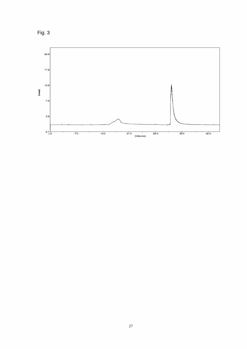
purification was by HPLC on a TSK gel G3000PWXL column (Fig. 3)(Page 27). ADL
migrated as a symmetrical peak by gel filtration, and the hemagglutination activity
was exactly coincident with the protein content (data not shown). The purified lectin
was eluted from the TSK gel G4000PWXL column at (Ve-Vo)/Vo = 4.76 (Fig. 4)(Page
28).
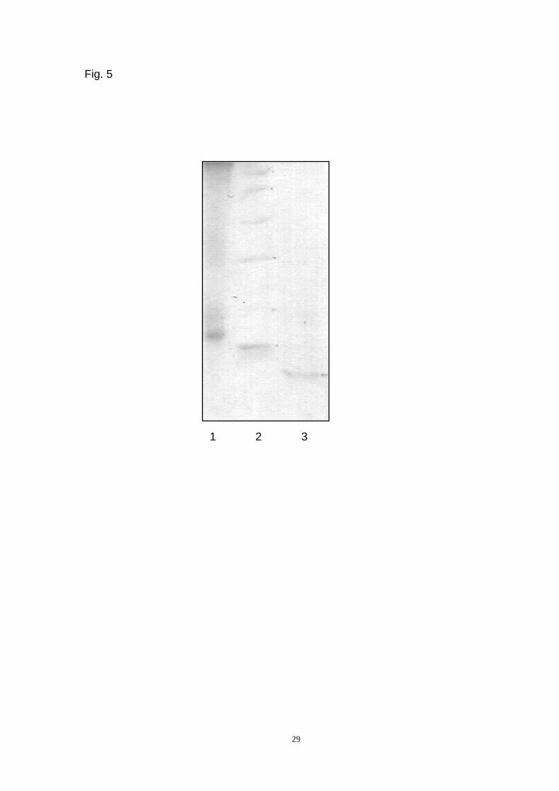
The ADL was analyzed by SDS-PAGE and found to migrate as a major intense
band with a relative molecular mass of approximately 17 kDa (Fig. 5, lane 1)(Page
29). Reduction with dithiothreitol led to the appearance of an 8.5 kDa band (Fig. 5,
lane 3) (Page 29). This finding indicates the presence of a disulfide bond and two
subunits with identical molecular masses.
The ADL molecular mass was further determined by MALDI-TOF mass
spectrometry. The ADL spectrum contained peaks corresponding to singly charged
(M+H)+ molecular ions from the subunit at m/z 8.5 kDa, while the peak at m/z 17 kDa
corresponded to the subunit dimer (Fig. 6) (Page 30).
In summary, the molecular mass determination by SDS-PAGE and MALDI-TOF
mass spectrometry showed that ADL has two subunits with a molecular mass of 17
kDa (2 × 8.5 kDa). The ADL N-terminal amino acid sequence was determined to be
VGYTVGATPM. According to the hemagglutination assay, ADL does not require the
divalent cations Ca2+ and Mg2+ for lectin activity (data not shown). The ADL lectin
activity was stable between 4°C and 50°C (Fig. 7 A) (Page 31). Significant ADL
hemagglutination activity was observed between pH 4-5 (Fig. 7 B) (Page 31).
3.2 Amino acid composition and carbohydrate content
As shown in Table 1, purified ADL contains relatively high amounts of the apolar
amino acids Gly, Pro, Val and Leu, and essential proportion of polar charged amino
acids His, Arg and apolar amino acid Met. The total carbohydrate content was 1%.
12
3.3 Carbohydrate-binding specificity
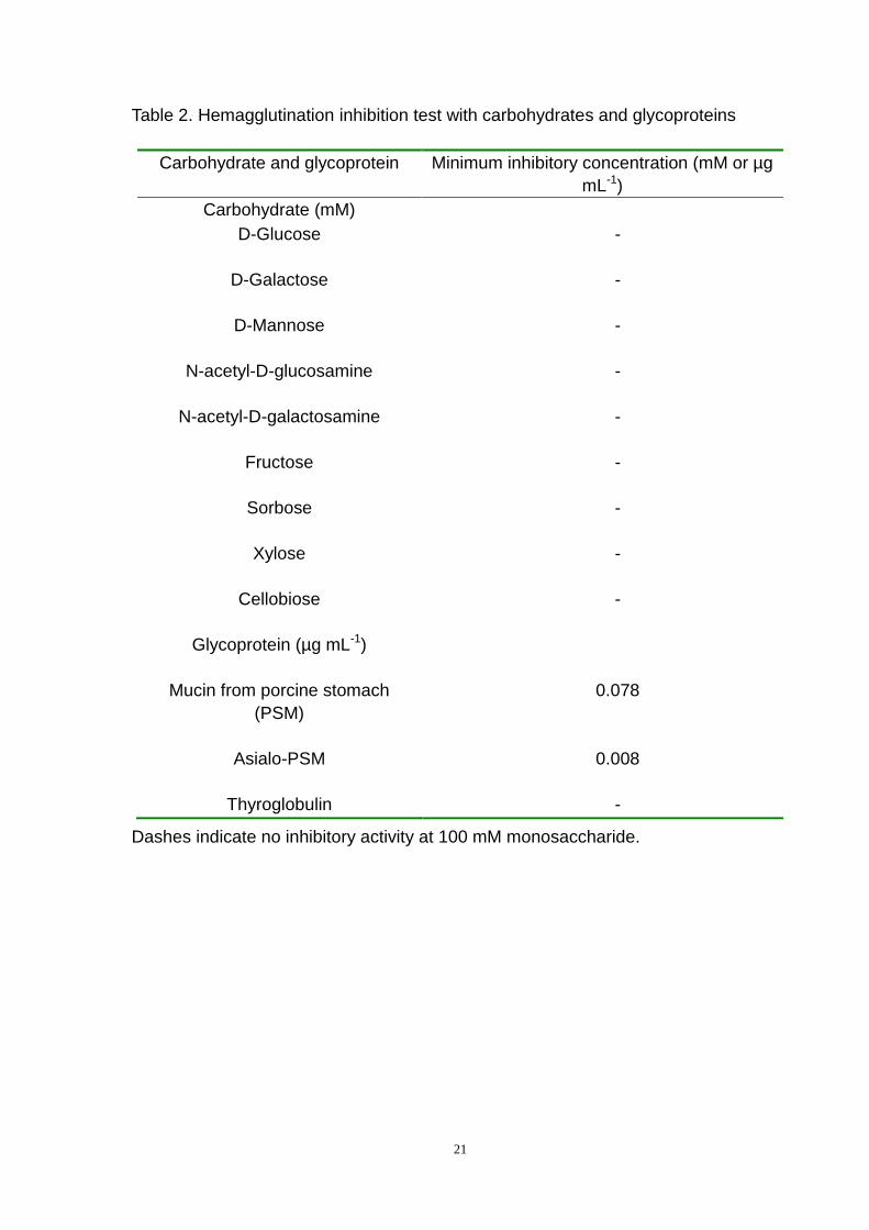
The ADL carbohydrate-binding specificity was examined by a hemagglutination
inhibition test. The ADL hemagglutination activity was not inhibited by any of the
monosaccharides or disaccharide examined, whereas it was inhibited by several
glycoproteins (Table 2). Among the glycoproteins, asialo-PSM was the most effective
inhibitor.
3.4 Antibacterial activity
The ADL antibacterial activity results are shown in Table 3. ADL exhibited strong
antibacterial activity against E. coli. ADL also exhibited antibacterial activity against E.
aerogenes, S. aureus, B. subtilis and Shewanella sp.
The respiration inhibition of E. coli, E. aerogenes, S. aureus, B. subtilis and
Shewanella sp. was determined by high-resolution respirometry (Dissolved oxygen
meter 8401, AZ Instrument Corp., Taiwan). Malonic acid, iodoacetic acid and sodium
phosphate are three typical inhibitors of respiratory metabolism and affect the
Embden-Meyerhof Pathway (EMP), tricarboxylic acid cycle (TCAC) and Hexose
Monophosphate Pathway (HMP), respectively [24]. When two inhibitors block
different pathways, the superposition of the inhibitory rates increases greatly;
however, if they inhibit the same pathway, then the rate increase is weak. ADL
inhibited the respiration of E. coli, E. aerogenes, S. aureus, B. subtilis and
Shewanella sp., and the inhibition rates are 23.7 ± 4.3%, 13.9 ± 5.5%, 59.2 ± 1.4%,
46.2 ± 0.7% and 31.8 ± 4.6%, respectively (Table 4). These rates indicated that ADL
inhibited the growth of S. aureus more than the other four bacteria. For E. coli, the
inhibitory superposition rates of malonic acid, iodoacetic acid and sodium phosphate
to ADL were 16.1 ± 1.6%, 69.9 ± 0.5% and 25.1 ± 1.5%, respectively, which indicted
that ADL inhibited the same pathway as malonic acid, i.e., TCAC. For E. aerogenes,
the inhibitory superposition rates of malonic acid, iodoacetic acid and sodium
phosphate to ADL were 53.2 ± 3.7%, 80.0 ± 0.6% and 6.8 ± 2.5%, respectively, which
indicted that ADL inhibited the same pathway as sodium phosphate, i.e., HMP. For S.
aureus, the inhibitory superposition rates of malonic acid, iodoacetic acid and sodium
phosphate to ADL were 10.0 ± 2.1%, 58.0 ± 1.7% and 44.0 ± 1.9%, respectively,
which indicted that ADL inhibited the same pathway as malonic acid, i.e., TCAC. For
B. subtilis, the inhibitory superposition rates of malonic acid, iodoacetic acid and
sodium phosphate to ADL were 64.3 ± 1.7%, 41.7 ± 3.3% and 12.5 ± 2.6%,
13
respectively, which indicted that ADL inhibited the same pathway as sodium
phosphate, i.e., HMP. For Shewanella sp., the inhibitory superposition rates of
malonic acid, iodoacetic acid and sodium phosphate to ADL were 31.5 ± 2.9%, 59.2 ±
1.3% and 14.3 ± 3.2%, respectively, which indicted that ADL inhibited the same
pathway as sodium phosphate, i.e., HMP.
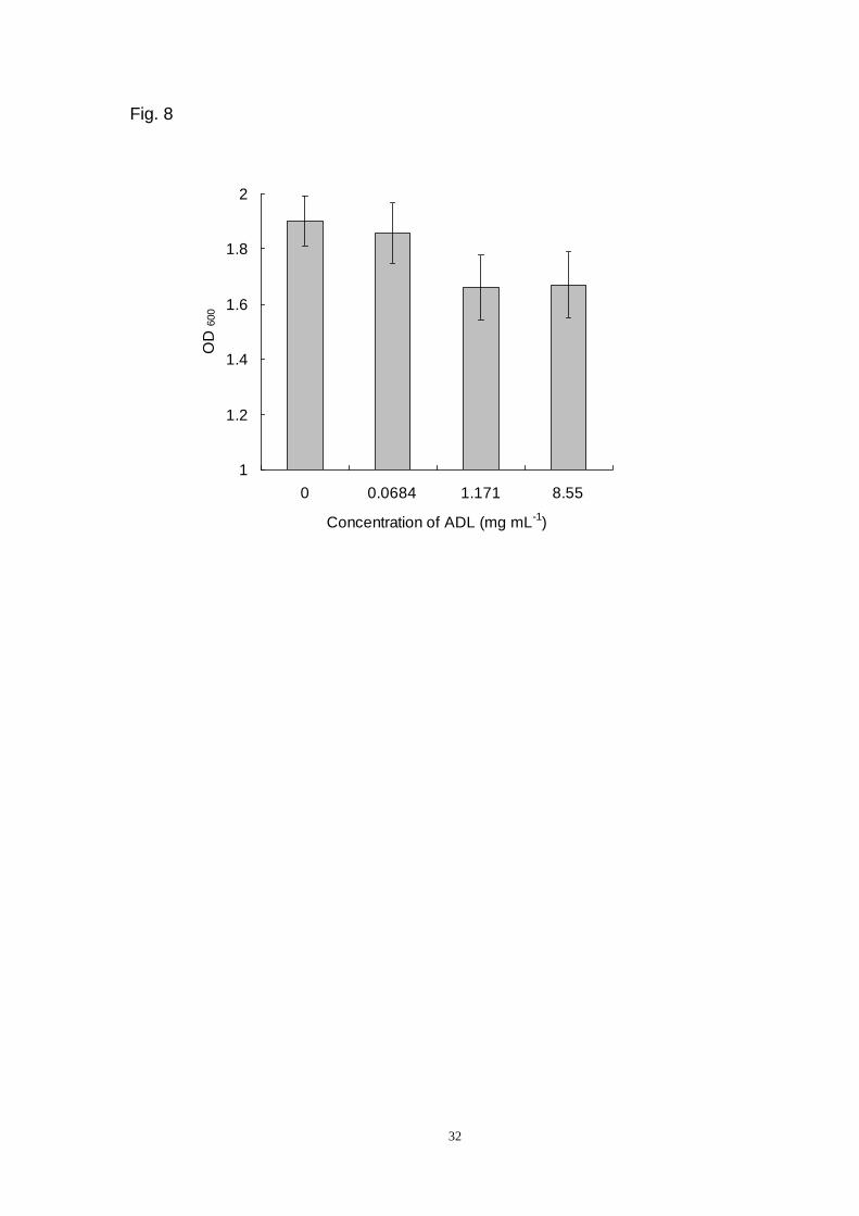
3.5 ADL suppresses yeast growth
To investigate the anti-fungal activity of ADL, its suppressive effect on yeast cell
growth was examined. The OD600 of yeast suspension with different ADL
concentrations (0.0684, 1.171 and 8.55 mg L-1 (w/v)) was obviously different from
that of the control (Fig. 8) (Page 32). The number of yeast cells declined with
increasing concentrations of ADL, indicating that this lectin suppresses S. cerevisiae
growth.

14
4. Discussion
As sources of lectins, amphibian skin secretions have not been widely
investigated in comparison with other organs (eggs and oocytes). Our interest in skin
secretion lectins arose from recent reports on the isolation and characterization of
two β-galactoside binding lectins (50 and 56 kDa) from the skin of Bufo arenarum that
have bacteriostatic activity against Gram-negative bacteria (E. coli K124100 and wild
strains of E. coli and Proteus morganii) and Gram-positive bacteria (Enterococcus
faecalis) [15]. Additionally, the L-fucose specific lectin from the skin of another
amphibian (O. grahami) has extremely low toxicity and immunogenicity in mice [16].
A new lectin secreted from A. davidianus skin was isolated by affinity
chromatography on PSM crosslinked albumin and purified by gel filtration on
Sephadex G-100 and HPLC on TSK gel G3000PWXL. The molecular weight of ADL
was estimated by SDS-PAGE, HPLC and MALDI-TOF mass spectrometry. The
results indicate that the native molecular weight of ADL is 68 kDa and that it is
composed of two identical 8.5 kDa subunits. The native ADL is a homotetrameric
glycoprotein that has two identical subunits that are connected with disulfide bonds
and are organized as a tetramer. Such structural organization is very rare, though
similar tetrameric structures have been described for lectins from the sea worm
Serpula vermicularis [26], the white shrimp Litopenaeus setiferus [27] and the tropical
sponges Aplysina archeri and A. lawnosa [28]. The ADL hemagglutination activity
was independent of divalent cations. Significant ADL hemagglutination activity was
observed between pH 4-5. The amino acid composition differs from that of lectins
isolated from the skin mucus of fish, where ADL is rich in apolar amino acids, such as
Gly, Pro, Val and Leu [29, 30].
The results of the hemagglutination inhibition study suggest that the topography
of the ADL-combining sites is significantly different from other known mucin-binding
lectins [21, 31, 32]. Among the native and asialo-glycoproteins tested in the
hemagglutination inhibition assays, asialo-PSM was the best inhibitor. The
carbohydrate side chains of PSM are O-glycosidically linked through GalNAc to Ser
or Thr of the protein core [33]. There are 12 carbohydrate side chains present, which
are composed of one to five sugar residues, with Galβ1-3 GalNAc α-O-Ser/Thr as the
carbohydrate core region. The high PSM potency may be attributed to the presence
15
of Galβ1-3 GalNAc α-O-Ser/Thr, where GalNAc is substituted at C 3 by a Gal residue.
The carbohydrate chain can be masked by N-glycolylneuraminic acid or sialic acid
(NeuNGl). The inhibitory ability of asialo-PSM was 10-fold greater than PSM. Most
likely, NeuGl residues, which have negative charges, interfere with the interaction of
PSM with ADL. The ADL hemagglutination activity was strongly inhibited by PSM
bearing mucin-type O-glycans, such as the lectin from Crenomytilus grayanus [21]
and the lectin from the sponge Craniella australiensis [34].
After the pathway of glucose oxidative metabolism was inhibited, the activities
necessary for life were interrupted. EMP, TCAC and HMP are glucose degradation
pathways. TCAC could provide massive energy and compounds for the synthesis of
other biomacromolecules, such as lipids and proteins. ADL inhibited TCAC in E. coli
and S. aureus as well as HMP in E. aerogenes, Shewanella sp. and B. subtilis. These
results are consistent with ADL antibacterial activity, i.e. ADL can inhibit E. coli and S.
aureus more efficiently than E. aerogenes and Shewanella sp.
Additionally, the inhibitory effect had no relationship with the type of bacteria, i.e.
Gram-positive or Gram-negative. ADL showed greater inhibitory effects on
Gram-negative bacteria, such as E. coli, than Gram-positive bacteria, such as S.
aureus and B. subtilis. However, ADL had a greater inhibitory effect on Gram-positive
bacteria, like S. aureus and B. subtilis, than Gram-negative bacteria, like E.
aerogenes and Shewanella sp. ADL could also suppress yeast cell growth, which
suggests that ADL is involved in self-defense against fungi and bacteria.
Microbial infection is the leading cause of disease in A. davidianus [2]. Through
evolution, antimicrobial components, including lectins, have given rise to counterparts
in microorganisms [16]. ADL participates in the A. davidianus host defense. The lectin
may inhibit the growth of microorganisms in the mucus, some of which may serve as
a nutrient to the microorganism [35]. ADL also revealed the A. davidianus immune
state.
16
Acknowledgements
The work was supported by grants from the Natural Science Foundation of
China (31071612) and the Marine Public Welfare Research Project (201205022-7).

17
References
[1] S.C. Lan, D.F. Li, J.C. Jiang, Call and skin glands secretion induced by
stimulation of midbrain in urodele (Andrias davidianus), Brain Research 528
(1990) 159-161.
[2] Y.L. Jiang, M. Zhang, H.L. Jing, L.Y. Gao, Isolation and characterization of an
Iridovirus from sick giant salamander (Andrias davidianus), Chinese Journal of
Virology 27 (2011) 274-282.
[3] B.T. Clarke, The natural history of amphibian skin secretions, their normal
functioning and potential medical applications, Biological Reviews of the
Cambridge Philosophical Society 72 (1997) 365-379.
[4] J. Li, X. Xu, C. Xu, W. Zhou, K. Zhang, H. Yu, Y. Zhang, Y. Zheng, H.H. Rees, R.
Lai, D. Yang, J. Wu, Anti-infection peptidomics of amphibian skin, Molecular &
Cellular Proteomics 6 (2007) 882-894.
[5] H. Yang, X. Wang, X. Liu, J. Wu, C. Liu, W. Gong, Z. Zhao, J. Hong, D. Lin, Y.
Wang, R. Lai, Antioxidant peptidomics reveals novel skin antioxidant system,
Molecular & Cellular Proteomics 8 (2009) 571-583.
[6] W. Guo, M. Ao, W. Li, J. Wang, L. Yu, Major biological activities of the skin
secretion of the Chinese giant salamander, Andrias davidianus, Zeitschrift Fur
Naturforschung C 67 (2012) 86-92.
[7] L. Wang, X. Li, D. Wang, Antibacterial effect of antimicrobial peptide from skin
secretions of Andrias davidianus on the wound of Pseudomonas aeruginosa
infection in mice, West China Journal of Pharmaceutical Sciences 26 (2011)
336-339.
[8] Q. Jin, F. Wei, C. Tong, W. Li, L. Kong, HPLC analysis and antioxidative activity of
glycopeptides from Andrias davidianus, Chinese Journal of Beijing University of
Agriculture, 26 (2011) 27-29.
[9] X. Feng, M. Qu, X. Yu, W. Li, W. Wang, C. Tong, L. Kong, Q. Jin, Study on
properties of glycopeptides from Andrias davidianus, Chinese Science and
Technology of Food Industry 33 (2012) 128-131.
[10] X. Jing, E.P. Espinosa, M. Perrigault, B. Allam, Identification, molecular
characterization and expression analysis of a mucosal C-type lectin in the
eastern oyster, Crassostrea virginica, Fish & Shellfish Immunology 30 (2011)
851-858.
[11] U. Holmskov, S. Thiel, J.C. Jensenius, Collectins and ficolins: humoral lectins of
the innate immune defense, Annual Review of Immunology 21 (2003) 547-578.
[12] F. Sakakibara, H. Kawauchi, G. Takayanagi, H. Ise, Egg lectin of Rana japonica
and its receptor glycoprotein of Ehrlich tumor cells, Cancer Research 39 (1979)
1347-1352.
[13] N.E. Fink de Cabutti, M. Caron, R. Joubert, M.T. Elola, D. Bladier, J. Herkovitz,
18
Purification and some characteristics of a β-galactoside binding soluble lectin
from amphibian ovary, FEBS Letters 223 (1987) 330-334.
[14] Y. Ozeki, T. Matsui, K. Nitta, H. Kawauchi, Y. Takayanagi, K. Titani, Purification
and characterization of β-galactoside binding lectin from frog (Rana catesbeiana)
eggs, Biochemical and Biophysical Research Communications 178 (1991)
407-413.
[15] A.S. Riera, A. Daud, A. Gallo, S. Genta, M. Aybar, S. Sanchez, Antibacterial
activity of lactose-binding lectins from Bufo arenarum skin, Biocell 27 (2003)
37-46.
[16] J. Li, H. Wu, J. Hong, X. Xu, H. Yang, B. Wu, Y. Wang, J. Zhu, R. Lai, X. Jiang, D.
Lin, M.C. Prescott, H.H. Rees, Odorranalectin is a small peptide lectin with
potential for drug delivery and targeting, Plos One 3 (2008) 1-10.
[17] R. Kowal, R.G. Parsons, Stabilization of proteins immobilized on Sepharose from
leakage by glutaraldehyde crosslinking, Analytical Biochemistry 102 (1980)
72-76.
[18] N.I. Belogortseva, R.G. Ovodova, S.V. Moroz, N.A. Odintsova, A.V. Yermak, Y.S.
Ovodov, Isolation and characterization of a structurally unusual lectin from
ascidian Didemnum ternatum (DTL). Bioorganicheskaya Khimiya 20 (1994)
975-983.
[19] M.J. Tyler, D.J. Stone, J.H. Bowie, A novel method for the release and collection
of dermal glandular secretions from the skin of frogs, Journal of Pharmacological
and Toxicological Methods 28 (1992) 199-200.
[20] U.K. Laemmli, Cleavage of structure proteins during assembly of the head of
bacteriophage T4, Nature 227 (1970) 680-685.
[21] N.I. Belogortseva, V.I. Molchanova, A.V. Kurika, A.S. Skobun, V.E. Glazkova,
Isolation and characterization of new GalNAc/Gal-specific lectin from the sea
mussel Crenomytilus grayanus, Comparative Biochemistry and Physiology Part
C: Pharmacology, Toxicology and Endocrinology 119 (1998) 45-50.
[22] O.H. Lowry, N.J. Rosenbrough, A.L. Farr, R.I. Randae, Protein measurement with
the Folin phenol reagent, the Journal of Biological Chemistry 193 (1951)
265-275.
[23] M. Dubois, K.A. Gilles, I.K. Hamilton, P.A. Rebers, F. Smith, Colorimetric method
for determination of sugars and related substances, Analytical Chemistry 28
(1956) 350-356.
[24] H.-T. Wang, S.-S. Shi, Y.-X. Li, H.-Q. Li, M.-J. Xie, Study on anti-microbial activity
of genistein and its mechanism, Acta Nutrimenta Sinica 30 (2008) 403-409.
[25] M. Bely, P. Storckle, I. Masneuf-Pomarede, D. Dubourdieu, Impact of mixed
Torulaspora delbrueckii-Saccharomyces cerevisiae culture on high-sugar
fermentation, International Journal of Food Microbiology 122 (2008) 312-320.
19
[26] V. Molchanova, I. Chikalovets, O. Chernikov, N. Belogortseva, W. Li, J. Wang,
D.Y. Ou Yang, Y.T. Zheng, P. Lukyanov, A new lectin from the sea worm Serpula
vermicularis: Isolation, characterization and anti-HIV activity. Comparative
Biochemistry and Physiology C Toxicology & Pharmacology 145 (2007) 184-193.
[27] J. Alpuche, A. Pereyra, C. Agundis, C. Rosas, C. Pascual, M.C. Slomianny, L.
Vazquez, E. Zenteno, Purification and characterization of a lectin from the white
shrimp Litopenaeus setiferus (Crustacea decapoda) hemolymph, Biochimca et
Biophysica Acta 1724 (2005) 86-93.
[28] P.B. Miarons, M. Fresno, Lectins from tropical sponges: purification and
characterization of lectins from genus Aplysina, The Journal of Biological
Chemistry 275 (2000) 29283-29289.
[29] S. Tsutsui, S. Tasumi, H. Suetake, Y. Suzuki, Lectins homologous to those of
monocotyledonous plants in the skin mucus and intestine of pufferfish, Fugu
rubripes, The Journal of Biological Chemistry 278 (2003) 20882-20889.
[30] S. Tsutsui, K. Iwamoto, O. Nakamura, T. Watanable, Yeast-binding C-type lectin
with opsonic activity from conger eel (Conger myriaster) skin mucus, Molecular
Immunology 44 (2007) 691-702.
[31] S. Banerjee, S. Chaki, J. Bhowal, B.P. Chatterjee, Mucin binding mitogenic from
freshwater Indian gastropod Belamyia bengalensis: purification and molecular
characterization, Archives of Biochemistry and Biophysics 421 (2004) 125-134.
[32] N. Belogortseva, N. Molchanova, V. Glazunov, E. Evtushenko, P. Luk’ynov,
N-Acetyl-D-glucosamine-specific lectin from the ascidian Didemnum ternatanum,
Biochimica et Biophysica Acta 1380 (1998) 249-256.
[33] T.A. Gerken, C.L. Owens, M. Pasumarthy, Site-specific core 1 O-glycosylation
pattern of the porcine submaxillary gland mucin tandem repeat: evidence for the
modulation of glycan length by peptide sequence, The Journal of Biological
Chemistry 273 (1998) 26580-26588.
[34] C. Xiong, W. Li, H. Liu, W. Zhang, J. Dou, X. Bai, Y. Du, X. Ma, A normal
mucin-binding lectin from the sponge Craniella australiensis. Comparative
Biochemistry and Physiology C Toxicology & Pharmacology 143 (2006) 9-16.
[35] S. Tasumi, T. Ohira, I. Kawazoe, H. Suetake, Y. Suzuki, K. Aida, Primary structure
and characteristics of a lectin from skin mucus of the Japanese eel Anguilla
japonica, The Journal of Biological Chemistry 277 (2002) 27305-27311.

20
Table 1. ADL amino acid composition
Amino acid Residues/100
Asx 4.76
Thr 7.05
Ser 5.62
Glx 5.25
Pro 9.02
Gly 17.88
Ala 4.83
Val 7.16
Ile 7.14
Leu 8.25
Tyr 5.26
Phe 5.27
His 1.79
Lys 7.22
Arg 2.07
Met 1.32
Cys, Trp – not determined.
21
Table 2. Hemagglutination inhibition test with carbohydrates and glycoproteins
Carbohydrate and glycoprotein Minimum inhibitory concentration (mM or µg
mL-1)
Carbohydrate (mM)
D-Glucose
-
D-Galactose
-
D-Mannose
-
N-acetyl-D-glucosamine
-
N-acetyl-D-galactosamine
-
Fructose
-
Sorbose
-
Xylose
-
Cellobiose
-
Glycoprotein (µg mL-1)
Mucin from porcine stomach
(PSM)
0.078
Asialo-PSM
0.008
Thyroglobulin -
Dashes indicate no inhibitory activity at 100 mM monosaccharide.

22
Table 3. ADL antibacterial activity
(-) No activity, (+) weak activity is 7-11 mm, (++) moderate activity is 11-16 mm, (+++) high activity
is >16 mm.
E. coli S. aureus B. subtilis E. aerogenes Shewanella
sp.
ADL +++ ++ ++ + +
Penicillin +++ +++ ++ ++ ++

23
Table 4. Effect of ADL on bacterial respiration inhibition. Values are means ± SD (n = 3).
Inhibitor E. coli E. aerogenes S. aureus B. subtilis Shewanella sp.
IR
(%)
RR
(%)
IR
(%)
RR
(%)
IR
(%)
RR
(%)
IR
(%)
RR
(%)
IR
(%)
RR
(%)
ADL 23.7 ± 4.3 13.9 ± 5.5 59.2 ± 1.4 46.2 ± 0.7 31.8 ± 4.6
Malonic acid 61.9 ± 1.1 16.1 ± 1.6 89.7 ± 1.2 53.2 ± 3.7 5.7 ± 2.0 10.0 ± 2.1 62.3 ± 2.4 64.3 ± 1.7 49.9 ± 3.6 31.5 ± 2.9
Iodoacetic acid 85.7 ± 0.2 69.9 ± 0.5 32.5 ± 6.1 80.0 ± 0.6 82.8 ± 1.0 58.0 ± 1.7 59.6 ± 1.9 41.7 ± 3.3 33.3 ± 2.4 59.2 ± 1.3
Sodium phosphate 20.0 ± 1.4 25.1 ± 1.5 31.3 ± 6.4 6.8 ± 2.5 8.3 ± 2.9 44.0 ± 1.9 22.6 ± 4.0 12.5 ± 2.6 22.3 ± 3.1 14.3 ± 3.2
24
Figure Legends
Fig. 1. Affinity chromatography of the A. davidianus skin secretion on a
PSM-crosslinked albumin column (3 11 cm) equilibrated and eluted with TB. The
bound fraction was eluted with 1.5 M NaCl in TB.
Fig. 2. Gel chromatography of ADL on Sephadex G-100 (2.5 96 cm).
Fig. 3. HPLC of purified ADL on a G3000PWXL column (7.8 mm 30 cm).
Fig. 4. Estimation of the ADL molecular mass by HPLC on the TSK gel G4000PWXL
column. 1, Trypsin (23,300); 2, Albumin egg (45,000); 3, Albumin bovine V (68,000); 4,
Rabbit muscle phosphorylase B (97,000).
Fig. 5. SDS-PAGE. Lane 1-ADL, Mr ~17,000. Lane 2-marker proteins: phosphorylase
B (94,000), BSA (67,000), ovalbumin (45,000), carbonic anhydrase (30,000), trypsin
inhibitor (20,100), alpha-lactalbumin (14,400). Lane 3-ADL treated with dithiothreitol.
Fig. 6. MALDI-TOF mass spectrum of ADL. The mass spectrum was obtained on a
4800 Proteomics Analyzer time-of-flight mass spectrometer (Applied Biosystems,
USA).
Fig. 7. General properties of the ADL. The effects of temperature (A) and pH (B) on
hemagglutination activity.
Fig. 8. ADL growth suppressive activity against yeast cells.

25
Fig. 1
0
0.05
0.1
0.15
0.2
1 11 21 31 41 51 61
Fraction number
OD
280
1.5 M NaCl

26
Fig. 2
0
0.2
0.4
0.6
0.8
1 11 21 31 41 51 61 71 81
Fraction number
OD
280
27
Fig. 3

28
Fig. 4
0
2
4
6
8
1 0
1 2
4. 3 4. 4 4. 5 4. 6 4. 7 4. 8 4. 9 5 5. 1
Log(Mr)
(Ve-V0)/V0
1
2
3
4 ADL
29
Fig. 5
1 2 3

30
Fig. 6 In
ten
s.[
a.u
.]
m/z
8530.4160

31
Fig. 7
0
2
4
6
8
10
4 20 30 40 50 60 70
Temperature(oC)
Log 2
of
tite
r
A
0
2
4
6
8
10
12
3 4 5 6 7 8 9 10
pH
Log 2
of
tite
r
B
32
Fig. 8
1
1.2
1.4
1.6
1.8
2
0 0.0684 1.171 8.55
Concentration of ADL (mg mL-1
)
OD
600
Related Documents


![A Basic Lectin from Bulbs of Arisaema ringens · 2018. 1. 4. · Arisaema basic lectin lectin were different from the Arum lectin [18]. From the result of hemmaglutination inhibition,](https://static.cupdf.com/doc/110x72/6142723dd9e4dc11f47f0e76/a-basic-lectin-from-bulbs-of-arisaema-ringens-2018-1-4-arisaema-basic-lectin.jpg)


