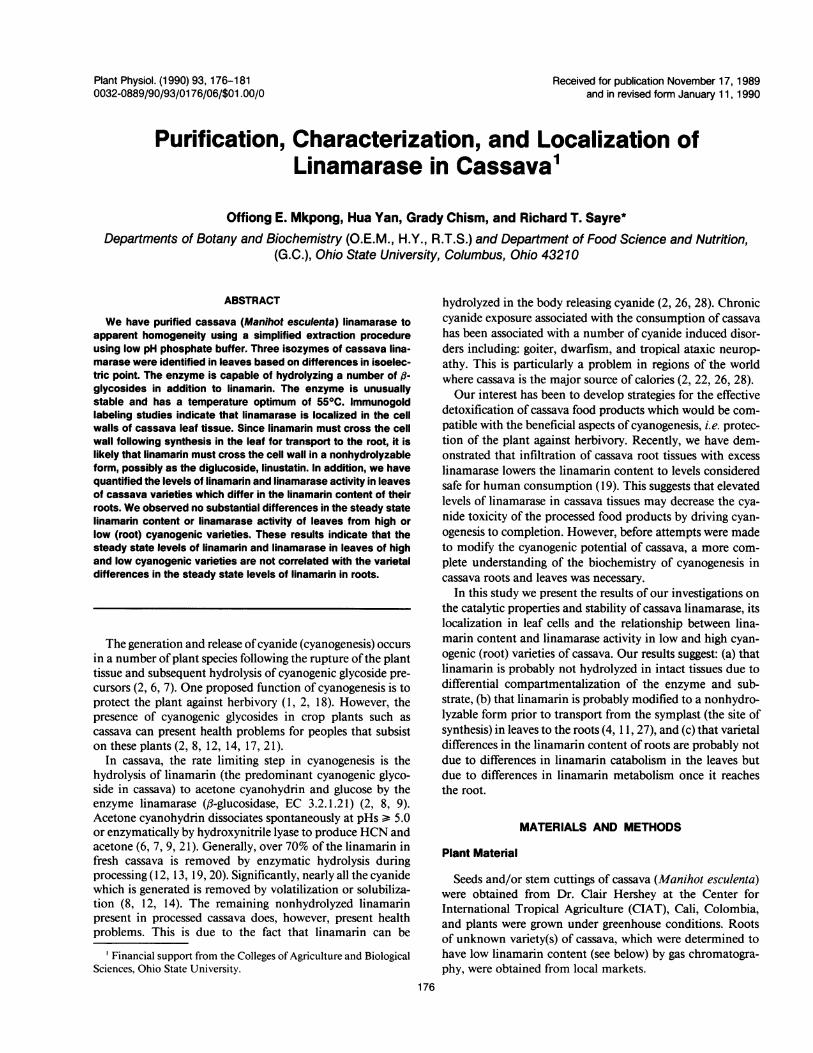
Plant Physiol. (1990) 93, 176-181 0032-0889/90/93/01 76/06/$01 .00/0 Received for publication November 17, 1989 and in revised form January 11, 1990 Purification, Characterization, and Localization of Linamarase in Cassava' Offiong E. Mkpong, Hua Yan, Grady Chism, and Richard T. Sayre* Departments of Botany and Biochemistry (O.E.M., H.Y., R.T.S.) and Department of Food Science and Nutrition, (G.C.), Ohio State University, Columbus, Ohio 43210 ABSTRACT We have purified cassava (Manihot esculenta) linamarase to apparent homogeneity using a simplified extraction procedure using low pH phosphate buffer. Three isozymes of cassava lina- marase were identified in leaves based on differences in isoelec- tric point. The enzyme is capable of hydrolyzing a number of ,B- glycosides in addition to linamarin. The enzyme is unusually stable and has a temperature optimum of 550C. Immunogold labeling studies indicate that linamarase is localized in the cell walls of cassava leaf tissue. Since linamarin must cross the cell wall following synthesis in the leaf for transport to the root, it is likely that linamarin must cross the cell wall in a nonhydrolyzable form, possibly as the diglucoside, linustatin. In addition, we have quantified the levels of linamarin and linamarase activity in leaves of cassava varieties which differ in the linamarin content of their roots. We observed no substantial differences in the steady state linamarin content or linamarase activity of leaves from high or low (root) cyanogenic varieties. These results indicate that the steady state levels of linamarin and linamarase in leaves of high and low cyanogenic varieties are not correlated with the varietal differences in the steady state levels of linamarin in roots. The generation and release of cyanide (cyanogenesis) occurs in a number of plant species following the rupture of the plant tissue and subsequent hydrolysis of cyanogenic glycoside pre- cursors (2, 6, 7). One proposed function of cyanogenesis is to protect the plant against herbivory (1, 2, 18). However, the presence of cyanogenic glycosides in crop plants such as cassava can present health problems for peoples that subsist on these plants (2, 8, 12, 14, 17, 21). In cassava, the rate limiting step in cyanogenesis is the hydrolysis of linamarin (the predominant cyanogenic glyco- side in cassava) to acetone cyanohydrin and glucose by the enzyme linamarase (f3-glucosidase, EC 3.2.1.21) (2, 8, 9). Acetone cyanohydrin dissociates spontaneously at pHs > 5.0 or enzymatically by hydroxynitrile lyase to produce HCN and acetone (6, 7, 9, 21). Generally, over 70% of the linamarin in fresh cassava is removed by enzymatic hydrolysis during processing (12, 13, 19, 20). Significantly, nearly all the cyanide which is generated is removed by volatilization or solubiliza- tion (8, 12, 14). The remaining nonhydrolyzed linamarin present in processed cassava does, however, present health problems. This is due to the fact that linamarin can be ' Financial support from the Colleges of Agriculture and Biological Sciences, Ohio State University. hydrolyzed in the body releasing cyanide (2, 26, 28). Chronic cyanide exposure associated with the consumption of cassava has been associated with a number of cyanide induced disor- ders including: goiter, dwarfism, and tropical ataxic neurop- athy. This is particularly a problem in regions of the world where cassava is the major source of calories (2, 22, 26, 28). Our interest has been to develop strategies for the effective detoxification of cassava food products which would be com- patible with the beneficial aspects of cyanogenesis, i.e. protec- tion of the plant against herbivory. Recently, we have dem- onstrated that infiltration of cassava root tissues with excess linamarase lowers the linamarin content to levels considered safe for human consumption ( 19). This suggests that elevated levels of linamarase in cassava tissues may decrease the cya- nide toxicity of the processed food products by driving cyan- ogenesis to completion. However, before attempts were made to modify the cyanogenic potential of cassava, a more com- plete understanding of the biochemistry of cyanogenesis in cassava roots and leaves was necessary. In this study we present the results of our investigations on the catalytic properties and stability of cassava linamarase, its localization in leaf cells and the relationship between lina- marin content and linamarase activity in low and high cyan- ogenic (root) varieties of cassava. Our results suggest: (a) that linamarin is probably not hydrolyzed in intact tissues due to differential compartmentalization of the enzyme and sub- strate, (b) that linamarin is probably modified to a nonhydro- lyzable form prior to transport from the symplast (the site of synthesis) in leaves to the roots (4, 11, 27), and (c) that varietal differences in the linamarin content of roots are probably not due to differences in linamarin catabolism in the leaves but due to differences in linamarin metabolism once it reaches the root. MATERIALS AND METHODS Plant Material Seeds and/or stem cuttings of cassava (Manihot esculenta) were obtained from Dr. Clair Hershey at the Center for International Tropical Agriculture (CIAT), Cali, Colombia, and plants were grown under greenhouse conditions. Roots of unknown variety(s) of cassava, which were determined to have low linamarin content (see below) by gas chromatogra- phy, were obtained from local markets. 176

Welcome message from author
This document is posted to help you gain knowledge. Please leave a comment to let me know what you think about it! Share it to your friends and learn new things together.
Transcript
Plant Physiol. (1990) 93, 176-1810032-0889/90/93/01 76/06/$01 .00/0
Received for publication November 17, 1989and in revised form January 11, 1990
Purification, Characterization, and Localization ofLinamarase in Cassava'
Offiong E. Mkpong, Hua Yan, Grady Chism, and Richard T. Sayre*Departments of Botany and Biochemistry (O.E.M., H.Y., R.T.S.) and Department of Food Science and Nutrition,
(G.C.), Ohio State University, Columbus, Ohio 43210
ABSTRACT
We have purified cassava (Manihot esculenta) linamarase toapparent homogeneity using a simplified extraction procedureusing low pH phosphate buffer. Three isozymes of cassava lina-marase were identified in leaves based on differences in isoelec-tric point. The enzyme is capable of hydrolyzing a number of ,B-glycosides in addition to linamarin. The enzyme is unusuallystable and has a temperature optimum of 550C. Immunogoldlabeling studies indicate that linamarase is localized in the cellwalls of cassava leaf tissue. Since linamarin must cross the cellwall following synthesis in the leaf for transport to the root, it islikely that linamarin must cross the cell wall in a nonhydrolyzableform, possibly as the diglucoside, linustatin. In addition, we havequantified the levels of linamarin and linamarase activity in leavesof cassava varieties which differ in the linamarin content of theirroots. We observed no substantial differences in the steady statelinamarin content or linamarase activity of leaves from high orlow (root) cyanogenic varieties. These results indicate that thesteady state levels of linamarin and linamarase in leaves of highand low cyanogenic varieties are not correlated with the varietaldifferences in the steady state levels of linamarin in roots.
The generation and release ofcyanide (cyanogenesis) occursin a number of plant species following the rupture ofthe planttissue and subsequent hydrolysis of cyanogenic glycoside pre-cursors (2, 6, 7). One proposed function of cyanogenesis is toprotect the plant against herbivory (1, 2, 18). However, thepresence of cyanogenic glycosides in crop plants such ascassava can present health problems for peoples that subsiston these plants (2, 8, 12, 14, 17, 21).
In cassava, the rate limiting step in cyanogenesis is thehydrolysis of linamarin (the predominant cyanogenic glyco-side in cassava) to acetone cyanohydrin and glucose by theenzyme linamarase (f3-glucosidase, EC 3.2.1.21) (2, 8, 9).Acetone cyanohydrin dissociates spontaneously at pHs > 5.0or enzymatically by hydroxynitrile lyase to produce HCN andacetone (6, 7, 9, 21). Generally, over 70% of the linamarin infresh cassava is removed by enzymatic hydrolysis duringprocessing (12, 13, 19, 20). Significantly, nearly all the cyanidewhich is generated is removed by volatilization or solubiliza-tion (8, 12, 14). The remaining nonhydrolyzed linamarinpresent in processed cassava does, however, present healthproblems. This is due to the fact that linamarin can be
' Financial support from the Colleges of Agriculture and BiologicalSciences, Ohio State University.
hydrolyzed in the body releasing cyanide (2, 26, 28). Chroniccyanide exposure associated with the consumption of cassavahas been associated with a number of cyanide induced disor-ders including: goiter, dwarfism, and tropical ataxic neurop-athy. This is particularly a problem in regions of the worldwhere cassava is the major source of calories (2, 22, 26, 28).Our interest has been to develop strategies for the effective
detoxification of cassava food products which would be com-patible with the beneficial aspects of cyanogenesis, i.e. protec-tion of the plant against herbivory. Recently, we have dem-onstrated that infiltration of cassava root tissues with excesslinamarase lowers the linamarin content to levels consideredsafe for human consumption ( 19). This suggests that elevatedlevels of linamarase in cassava tissues may decrease the cya-nide toxicity of the processed food products by driving cyan-ogenesis to completion. However, before attempts were madeto modify the cyanogenic potential of cassava, a more com-plete understanding of the biochemistry of cyanogenesis incassava roots and leaves was necessary.
In this study we present the results of our investigations onthe catalytic properties and stability of cassava linamarase, itslocalization in leaf cells and the relationship between lina-marin content and linamarase activity in low and high cyan-ogenic (root) varieties of cassava. Our results suggest: (a) thatlinamarin is probably not hydrolyzed in intact tissues due todifferential compartmentalization of the enzyme and sub-strate, (b) that linamarin is probably modified to a nonhydro-lyzable form prior to transport from the symplast (the site ofsynthesis) in leaves to the roots (4, 11, 27), and (c) that varietaldifferences in the linamarin content of roots are probably notdue to differences in linamarin catabolism in the leaves butdue to differences in linamarin metabolism once it reachesthe root.
MATERIALS AND METHODS
Plant Material
Seeds and/or stem cuttings of cassava (Manihot esculenta)were obtained from Dr. Clair Hershey at the Center forInternational Tropical Agriculture (CIAT), Cali, Colombia,and plants were grown under greenhouse conditions. Rootsof unknown variety(s) of cassava, which were determined tohave low linamarin content (see below) by gas chromatogra-phy, were obtained from local markets.
176
CYANOGENESIS IN CASSAVA
Isolation of Linamarase
Linamarase was isolated from leaves homogenized in a lowpH Na-phosphate buffer (0.1 M, pH 3.5). The homogenatewas filtered (Miracloth) and centrifuged (24,000g, 30 min) toremove cell debris and brought to 40% saturation with solidammonium sulfate to precipitate proteins. The precipitatedproteins were pelleted and dialyzed extensively against 0.05M Na-phosphate buffer (pH 4.5). The protein solution wasthen treated twice with 0.3 g activated charcoal/10 mL solu-tion, filtered, reprecipitated with 40% ammonium sulfate anddialyzed against 0.10 M Na-phosphate buffer (pH 8.0) priorto chromatography on a Sephadex G-200 column (2.5 x 50cm). Fractions having linamarase activity eluted near the voidvolume. For some preparations a second passage over thecolumn was necessary to purify the enzyme. All steps werecarried out at 4°C.
Determination of Linamarase Activity
Linamarase activity was routinely measured using the chro-mogenic substrate p-nitrophenol-3-glucopyranoside (20 mM)in 0.05 M Na-phosphate assay buffer at pH 5.5 for 1 h at25°C. The reaction was stopped by the addition of an equalvolume of 0.2 M sodium borate (pH 9.8) and p-nitrophenolwas quantified spectrophotometrically at 400 nm (13). Thefollowing substrates: linamarin, cellobiose, and prunasin wereassayed in a coupled reaction using glucose oxidase, horseradish peroxidase and the chromogenic substrate, o-diansidineto determine glucose production catalyzed by linamarase (13).Briefly, an aliquot of enzyme is incubated with the substrate(10-20 mM) in 50 mm Na-phosphate buffer (pH 7.5) for 1 hfollowed by boiling for 2 min to inactivate linamarase. Glu-cose production is then determined via the glucose oxidase/horseradish peroxidase coupled assay system according to themethod of Hosel and Barz (13). The linamarase activity ofvarious cassava varieties was determined from crude extractsof tissues homogenized in low pH phosphate buffer as de-scribed above. Dilutions ofthe crude extracts were used whichgave linear rates ofenzyme activity with linamarin. Linamar-ase activity was also detected in isoelectric focusing gels usingthe fluorescent substrate 4-methylumbelliferyl-f-glucopyran-oside (0.2 mM) according to the procedure of Selmar et al.(24). Amygdalin hydrolysis was determined by the productionof HCN using the picric acid HCN assay system of Cooke etal. (9).
Generation of Antibodies against Linamarase
Purified linamarase (Sephadex G-200 fraction) was sub-jected to SDS-PAGE electrophoresis and electroeluted fromthe gel prior to immunization. An aliquot (150 tg) of gelpurified linamarase was dissolved in 0.5 mL of 0.15 M Na-phosphate buffer (pH 8.0) and mixed with an equal volumeof Freund's complete adjuvant for subcutaneous injection inrabbits (23). Serum was collected after 3 weeks and useddirectly without further purification. Antibody specificity wasconfirmed by Western blots of total leaf extracts (23).
Immunohistochemical Localization of Linamarase inLeaves
Leaf tissue was cut into 1 x 2 mm pieces and fixed in 2%(v/v) glutaraldehyde and 1% (w/v) paraformaldehyde in 50mm K-phosphate buffer (pH 6.8) for 1 to 2 h at roomtemperature. Leaf pieces were then rinsed in buffer and de-hydrated in a graded methanol series at 4°C. The sampleswere infiltrated and embedded with LR Gold resin (EMS, Ft.Washington, PA) and polymerized using indirect UV light for24 to 28 h at -15°C. Ultrathin sections were cut with adiamond knife on a Reichert ultramicrotome and placed onformvar coated 150-mesh nickel grids. The grids were floated,section side down, on a drop of blocking buffer consisting of30 mm Tris-HCl buffer (pH 7.2); 1% bovine serum albumin,150 mm NaCl, 0.1% Tween 20 and 0.1% gelatin to preventnonspecific binding of the antibodies. The sections were thenincubated for 1 h in linamarase antibody diluted 1:200 in theaforementioned blocking buffer. The sections were thenrinsed in blocking buffer and incubated for 1 h with goat anti-rabbit IgG conjugated to 15 nm colloidal gold (Jansen LifeSciences, Olen, Belgium) diluted 1:50 with blocking buffer.Sections were rinsed in blocking buffer followed by water andfinally post-stained with 2% uranyl acetate and aqueous leadcitrate for 15 min each. Controls were done in parallel usingpreimmune rabbit IgG. Electron microscopy was carried outon a Zeiss-EM 10 electron microscope at 60 Kv.
Linamarin Determination
For the quantification of linamarin, cassava leaf, or rootmaterial was frozen and ground in liquid nitrogen to a finepowder using a mortar and pestle followed by lyophilization.To S mg of the freeze dried material, 10 ,uL of phenyl-fl-D-glucopyranoside (internal standard) was added, followed byaddition of 165 ,uL of acetonitrile and 25 ,uL of BSTFA.2 Thesamples were then heated in a capped serum vial at 90°C for30 min to derivatize linamarin. One ,L of sample was chro-matographed on 2% OV- 17 on 100 to 120 mesh ChromsorbAWHP in a 6' x 1/8" column using a nitrogen flow rate of62 mLs/min and detected by a flame ionization detector. Thetemperature program was 140 to 250 °C at 8°C/min with a 3min delay. Injector and detector temperatures were 280°C.Peak areas were determined using an HP3392A integrator.
Additional Analytical Procedures
Analytical SDS-PAGE and Western blots were carried outaccording to previously published procedures (23). Isoelectricfocusing was carried out using a Phast gel system (Pharmacia)on pH 3-5 isoelectric focussing gels according to the manu-facturers directions. Protein was quantified using the methodof Bradford (3).
2 Abbreviation: BSFTA; N,O-bis-(trimethylsilyl)-trifluoro-acetamide.
177
Plant Physiol. Vol. 93,1990
RESULTS
Purification and Catalytic Properties of Cassava LeafLinamarase
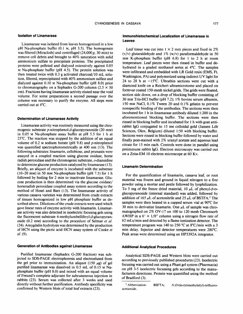
We have developed a simple purification procedure for theisolation of cassava linamarase based on extraction of leaftissues using a low pH buffer (Table I). The specific activityof linamarase increased 100-fold during purification with a
final yield of 4.0% of the total linamarase activity. The lowyield can be attributed to problems with effectively removingall contaminating proteins. Analysis of the polypeptide pro-files of various fractions by SDS-PAGE indicated that extrac-tion with low pH (3.5) Na-phosphate buffer essentially solu-bilized only two proteins. The final fraction in the purificationconsisted of a single polypeptide which had a molecular massof 65 kD (Fig. 1). The experimentally determined molecularmass was identical to that reported by Eksittikul and Chula-vatnatol (10) for purified cassava linamarase. In addition, wefound three different linamarase isozymes on isoelectric fo-
Table I. Cassava Linamarse PurificationTotal Total Specific
Fraction Activity Protein Activitym/h mg mmol/mg protein/h
Crude 1.44 2.3 x 10⁴ 0.0640% Ammonium sulfate 0.18 214 0.84Activated charcoal 0.09 43.9 2.1Sephadex G-200 0.058 9.74 6.0
5.S KL,
Figure 1. SDS-PAGE of purified cassava linamarase and Westernblot of crude leaf extracts screened with linamarase antibodies. Fivefractions in the purification of linamarase are shown in lanes 1 to 5.(1) 20 jug of purified cassava linamarase from the Sephadex G-200fraction; (2) 5 Ag of trypsin digested linamarase; (3) 50 jig of linamar-ase fraction treated with activated charcoal; (4)30 IAg of crude extractof linamarase precipitated with 40% ammonium sulfate; and (5) 50Ag of crude extract of cassava leaves. Proteins were stained withCoomassie blue. Lane A is a Western blot of crude cassava leafextract (25 jig) detected with antibodies generated against linamarase(1 :100 dilution).
Table II. Kinetic Properties of Linamarases from Various SpeciesProperty Cassava Butter Bean Rubber Tree
Vmax (linamarin)a (mmol/mg 29.4 11.5 14.6protein/h)
Km (mM)a 1.9 7.6 5.6Temperature optimum (0C) 55 62pH Optimumb 7.0 5.6 5.9
a Measured at 55, 30, and 370C, respectively. b PNBPG as asubstrate. References 15 and 24.
Table Ill. Linamarase Substrate SpecificityPNBPG = p-nitrophenol-l-glucopyranoside, PNAPG = p-nitro-
phenol-A-glucopyranoside, ONPGal = o-nitrophenol-fl-galactoside.Enzymes assays were carried out at 300C. with 10 mm substrate in50 mm Na-phosphate buffer (pH 6.5).
LinamaraseSubstrate Rate AcivityActivitymmol/mg protein/h %
Linamarin 16.5 100PNBPG 10.8 65PNAPG 0 0ONPGal 3.1 19Prunasin 10.7 65Amygdalin 0 0Cellobiose 0.19 1.1
cusing gels of the Sephadex G-200 fraction. The isozymeshave pIs of4.4, 3.4, and 3.0 (data not shown) (10). The kineticproperties of the leaf enzyme(s) (Sephadex G-200 fraction)are listed in Table II. The Km and Vmax for linamarin were 1.9mm and 29.4 mM/mg protein/h, respectively (measured at55°C, the optimum temperature). These values were signifi-cantly higher than those measured previously for the 4.3isoelectric point cassava isozyme (10) (Km = 0.57 mm andVmax = 3.5 mM/mg protein/h measured at 37°C). They aremore similar to values obtained for butter bean and rubbertree linamarase (15, 24). The differences in Vmax and Kmvalues between our work and that of Eksittikul and Chula-vatnatol (10) may be attributed either to differences in thekinetic properties of the isozymes or assay temperature (seebelow). As shown in Table III, cassava linamarase hydrolyzesa number of other glucosides in addition to linamarin includ-ing PNBPG and prunasin but does not efficiently hydrolyzea-glucopyranosides or diglucosides such as amygdalin or cel-lobiose (9, 10, 29). Interestingly, the galactoside, o-nitro-phenyl-,B-galactopyranoside was hydrolyzed by cassava lina-marase but at much lower rate than linamarin. Based on itsability to hydrolyze prunasin, cassava linamarase is moresimilar to that ofbutter bean linamarase than the more closelyrelated rubber tree ( 15, 24).
Last of all, several lines of evidence indicate that cassavalinamarase has unusually high stability. Similar to butter beanlinamarase, the enzyme has an unusually high temperatureoptimum of 55°C (Table I) (15). Second, we have determinedthat active enzyme can be isolated from desiccated (exfoliated)leaves. Finally, we found that linamarase (activity) is quite
178 MKPONG ET AL.

CYANOGENESIS IN CASSAVA
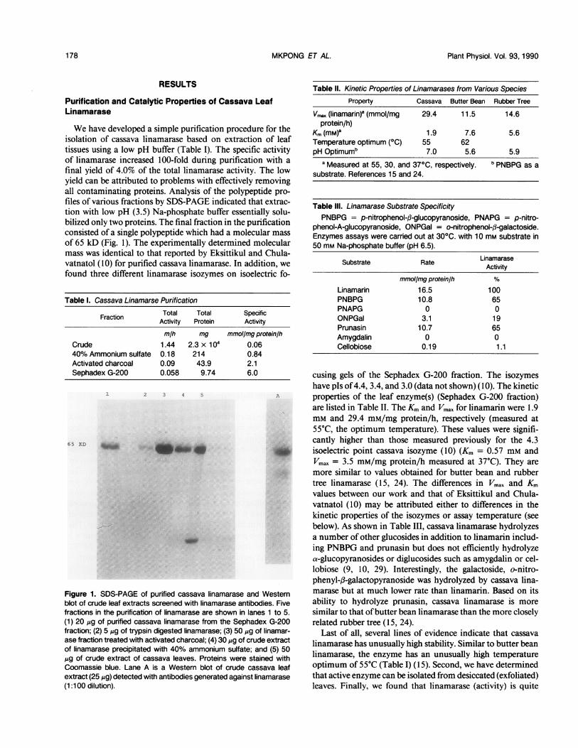
Figure 2. Localization of linamarase in the cell walls of cassavaleaves. The thin section of cassava leaf was treated with antibodiesagainst linamarase (1:100). Bound linamarase antibodies were local-ized with immunogold IgG. Magnification is x45,000.
insensitive to trypsin digestion (1.0 Ag trypsin/1.0 ,g lina-marase for 1 h, 37°C, Fig. 1).
Immunohistochemical Localization of Linamarase
Western blots of crude cassava leaf extracts are shown inFigure 1. Linamarase was the predominant protein detectedusing polyclonal antibodies generated against gel (SDS-PAGE) purified linamarase from the final Sephadex G-200fraction. The identity of a second lesser reactive protein (50kD) is not known.
Immunofluorescent labeling studies indicated that linamar-ase was localized either in the inner cell wall or the plasma-lemma of leaf tissue. In order to determine the localization oflinamarase at greater resolution linamarase was localized inthin sections by immunogold labeling. As shown in Figure 2,linamarase is localized in the cell walls of cassava leaf tissue.In addition, the golgi apparatus was also labeled by immu-nogold particles (Fig. 3). Since cell wall proteins are typicallyprocessed and packaged in the golgi apparatus prior to deliveryto the cell wall labeling of the golgi apparatus is consistentwith the localization of linamarase in the cell wall (5).
.?4M
Figure 3. Localization of linamarase in the golgi apparatus of cassavaleaves. The thin section was treated with antibodies against cassavalinamarase (1:200). Bound linamarase antibodies were localized withimmunogold IgG. Magnification is x64,000. Arrows indicate the po-sition of immunogold particles in the Golgi apparatus.
Relationship between Leaf Linamarin and LinamaraseActivities in Low Cyanide and High Cyanide CassavaVarieties
Cassava varieties can be classified into two groups based onthe linamarin content of their roots (2, 17). These groups arethe so-called low (-50 mg/Kg fresh weight) and high (2 100mg/Kg fresh weight) cyanide (linamarin) varieties (low cya-nide varieties have been derived from high cyanide varietiesby selective breeding.). Previously it has been demonstratedthat linamarin is synthesized in the leaf and petiole prior totransport to the root (2). To determine whether the steadystate pool sizes of linamarin in roots was regulated by eitherthe steady state level of linamarin or linamarase activity inleaves, we measured the linamarin content and linamaraseactivity of leaves and roots from low and high cyanide (cyan-ogenic) varieties of cassava. Significantly, the average steadystate linamarin content of leaves from low and high cyanidevarieties was nearly identical. Furthermore, the average lina-marase activities in leaves of low and high cyanide varietieswas also quite similar or only slightly higher in high cyanidevarieties (Table IV). However, most importantly, there wasno relationship between linamarin content and linamaraseactivity in leaves of low cyanide varieties. Compared withroots, both the linamarin content and linamarase activity ofleaves were approximately 18-fold higher (low cyanide vari-ety). These results suggest that varietal differences in root
179
P lo W"
iI
'i .1 1.

Plant Physiol. Vol. 93,1990
Table IV. Linamarin Content and Linamarase Activitya in Low andHigh Cyanide Varieties of Cassava
Linamarin LinamaraseVariety and Source Content Activity
.smol/g dry wt mmol/g dry wt/hLeaves, HighCyanideCM 849 49.0 1.46MCOL 1648 46.9 1.53SM 301-3 46.9 1.43MTAI-1 72.1 1.27MVEN-25 67.6 2.06
Average 56.5 1.55
Leaves, LowCyanideCM 3306-9 58.7 1.22CM 52.37 37.1 1.41MCOL 1522 48.9 0.19MCOL 1468 56.0 1.33MCOL 2215 72.1 1.00
Average 54.6 1.03
Roots, LowCyanideUnknown 2.96 (5.4%) 0.06 (5.8%)
aValues are the average of 2 to 3 measurements. Values inparentheses are the amounts present in roots expressed as a per-centage of amounts present in low cyanide leaves.
linamarin content cannot be attributed to the differences inthe catabolism of linamarin in leaves.
DISCUSSION
The pattern of substrate specificity observed for cassavalinamarase is similar to linamarases isolated from a numberof species but is most like that of butter bean based on itsability to hydrolyze prunasin (15). Interestingly, the rubbertree linamarase, which is evolutionarily more closely related,does not hydrolyze prunasin (24). However, rubber tree lina-marase may be unusual in other aspects. It appears to be theonly ,B-glucosidic enzyme present in that species and maycatalyze a number of reactions in addition to cyanogenesis(24). At present we do not known whether cassava linamaraseis likewise the sole f,-glucosidase present in the plant or
whether the enzyme has multiple functions.Based on this study as well as others it appears that there
are a number of different linamarase isozymes present incassava (15, 24). There are at least three difference isozymesof 65 kD enzyme in cassava leaves which can be distinguishedon the basis of their isoelectric points (10). Eksittikul andChulavatnatol (10) have determined the amino acid compo-sition for linamarase isozymes isolated from several tissuesincluding: leaves, petioles, and roots but found little variationin amino acid sequence between the different forms. Ourpreliminary analyses of the N-terminal amino acid sequenceof the leaf isozymes indicate that there is a two amino aciddisplacement in the position of the N-terminal amino acidbetween two of the isozymes, suggesting that some of thedifferences between the isozymes may be due to posttransla-
tional processing (data not shown). Further evidence for theheterogeneity ofcassava linamarases comes from comparisonsof the catalytic properties of leaf and root linamarase. Yeoh(29) demonstrated that cassava root linamarase (partiallypurified enzyme) has a Vmax (linamarin) which is 25-fold lowerthan that of the leaf enzyme. The significance of these differ-ences in terms of linamarin catabolism in roots and leavesremains to be determined but it is apparent that there areseveral distinctly different linamarases which are localized inan organ specific manner.The localization of linamarase in the cell walls of cassava
is consistent with the observation that cyanogenesis is inducedonly during cell rupture assuming that linamarin is localizedin the symplast (2, 1 1). Linamarase has also been shown tobe localized in the cell walls of white clover and lima bean(1 1, 16). However, the enzyme dhurrinase, which hydrolyzesthe cyanogenic glycoside, dhurrin, has been shown to belocalized in the chloroplasts of sorghum (27). Unfortunately,less is known about the cellular localization of linamarin.However, investigations by Fehrner and Conn (11) haveshown that linamarin is localized in protoplasts (symplastic)of Costa Rican wild lima beans and is not apoplastic. Ourresults indicating that linamarin steady state pool sizes arenot correlated with the level of linamarase activity in leavesversus roots supports the argument that linamarin is symplas-tic and linamarase is apoplastic.
For linamarin to be transported from leaves and petioles totarget tissues (roots) it must avoid hydrolysis by linamaraseeither by conversion to a nonhydrolyzable form or via trans-port by a nonapoplastic route. The latter possibility is unlikelysince linamarin has been shown to move through the vascularsystem of the petiole to the root (2). Alternatively, there isevidence indicating that linamarin may be transported in anonhydrolyzable form in some plants (1 1, 25). Selmar et al.(25) have proposed that in rubber tree linamarin is glycosy-lated to linustatin prior to crossing the apoplast for transportto expanding leaves in emerging rubber tree seedlings. Sincerubber tree linamarase is unable to hydrolyze cyanogenicdiglucosides, i.e. linustatin (as well as the diglycoside, amyg-dalin) it can cross the apoplast for transport to target tissues.Upon reaching the target tissue, linustatin is hydrolyzed togentiobiose and acetone cyanohydrin which in turn disso-ciates to cyanide. The cyanide is then fixed by ,B-cyanoalanine-synthase and the nitrogen incorporated into amino acids (25).By this mechanism linamarin/linustatin transport nitrogenfrom the seed to the expanding leaves (25). As shown in TableIII, cassava linamarase is also unable to hydrolyze the cyan-ogenic diglucoside, amygdalin as well as the diglucoside,cellobiose (1 1, 24, 29). Since the linamarase activity ofcassavaleaves is more than sufficient to effectively hydrolyze anylinamarin which is to be transported to roots we suggest thatthe linamarin must also be converted to a nonhydrolyzableform prior to transport to the roots in cassava. Furthermore,varietal differences in the cyanogenic glycoside content ofcassava roots must be attributed to differences in catabolismof cyanogenic glycosides in roots since leaf steady state levelsare similar in low and high cyanide varieties.
Further analyses on the metabolism of linamarin in rootsare necessary before a clearer understanding of the process of
180 MKPONG ET AL.

CYANOGENESIS IN CASSAVA
linamarin metabolism and cyanogenesis in roots can emerge.
However, it appears that in cassava leaves cyanogenesis isregulated by the hydrolytic enzyme linamarase which is com-partmentalized in the cell wall separate from its substrate.Therefore, we propose that manipulations which would ele-vate linamarase levels in leaf cell walls should not reduce thecyanogenic potential of cassava but perhaps enhance it. If theinduction of cyanogenesis in roots is analogous to that inleaves, then similarly elevated levels of linamarase in rootsshould not diminish cyanogenesis but more importantly,should facilitate detoxification of cassava food productsduring processing as a result of more complete hydrolysis oflinamarin (19).
LITERATURE CITED
1. Arias B, Bellotti A (1984) Aspectos ecologicos y de manejo deCytomenus bergi, Chinche de la viruela en el cultivo de la yucaManihot esculenta. CIAT publication, Cali, Colombia
2. Balagopalan C, Padmaja G, Nanda S, Morthy S (1988) Cassavain Food, Feed and Industry. CRC Press, Boca Raton, FL
3. Bradford M (1976) A rapid and sensitive method for quantificationof microgram quantities of protein utilizing the principle ofprotein-drug binding. Anal Biochem 72: 248-254
4. Butler G, Conn E (1964) Biosynthesis of cyanogenic glycosideslinamarin and lotaustralin. J Biol Chem 239: 1674-1679.
5. Cassab G, Varner J (1988) Cell wall proteins. Annu Rev PlantPhysiol Plant Mol Biol 39: 321-353
6. Conn E (1969) Cyanogenic glycosides. J Agric Food Chem 17:519-526
7. Conn E (1981) Cyanogenic glycosides. Biochem Plants 7: 479-5008. Cooke R (1978) An enzymatic assay for the total cyanide content
of cassava. J Sci Food Agric 29: 345-3529. Cooke R, Blake G, Battershill J (1978) Purification of cassava
linamarase. Phytochemistry 17: 381-38310. Eksittikul T, Chulavatnatol M (1988) Characterization of cyan-
ogenic B-glucosidase (linamarase) from cassava (Manihot es-culenta Crantz). Archiv Biochem Biophys 266: 263-269
1 1. Frehner M, Conn E (1987) The linamarin B-glucosidase in CostaRican wild lima beans (Phaseolus lunatus L.) is apoplastic.Plant Physiol 84: 1296-1300
12. Gomez G, Valdivieso M (1985) Cassava foliage: chemical com-
position, cyanide content and effect of drying on cyanideelimination. J Sci Food Agric 36: 433-441
13. Hosel W, Barz W (1975) B-glucosidases from Cicer arientum L.Eur J Biochem 57: 607-616
14. Ikediobi C, Onyia G, Eluwah C (1980) A rapid inexpensiveenzymatic assay for total cyanide in cassava and cassava prod-ucts. Agric Biol Chem 44: 2803-2809
15. Itoh-Nashida T, Hiraiwa M, Uda Y (1987) Purification andproperties of B-D-glucosidase (linamarase) from the butterbean Phaseolus lunatus. J Biochem 101: 847-854
16. Kakes P (1985) Linamarase and other B-glucosidases are presentin the cell walls of Trifolium repens L. leaves. Planta 166: 156-160
17. Laing D, Cock J, Roca W (1989) Report on the foundingworkshop for the advanced cassava research network. CIATdocument No. 52, Cali, Colombia.
18. Miller J, Conn E (1980) Metabolism of hydrogen cyanide byhigher plants. Plant Physiol 65: 1199-1202
19. Mkpong 0, Chism G, Sayre RT (1989) Isolation of cassavalinamarase: evidence that endogenous levels are insufficientfor effective hydrolysis of linamarin. Report on the foundingworkshop for the cassava biotechnology research network, page25, CIAT document No. 52. Cali, Colombia.
20. Nambisan B, Sundaresan S (1985) Effect of processing on thecyanoglucoside content of cassava. J Sci Food Agric 36: 1197-1203
21. Nartley F (1968) Studies on cassava, Cyanogenesis: the biosyn-thesis of linamarin and lotaustralin in etiolated seedlings. Phy-tochemistry 7: 1307-1312
22. Oke 0 (1980) Toxicity of cyanogenic glycosides. Food Chem 6:97-109
23. Sayre RT, Andersson B, Bogorad L (1986) The topology of amembrane protein: the orientation of the 32 Kd Qb-bindingchloroplast thylakoid membrane protein. Cell 47: 601-608
24. Selmar D, Lieberei R, Biehl B, Voight J (1987) Hevea linamar-ase-a nonspecific f,-glucosidase. Plant Physiol 83: 557-563
25. Selmar D, Liebrei R, Biehl B (1988) Mobilization and utilizationofcyanogenic glycosides: The linustatin pathway. Plant Physiol86:711-716
26. Tewe 0 (1984) Cyanogenic glycoside, protein interaction incassava peel based rations. Nutr Rep Int 30: 425-431
27. Thayer S, Conn E (1981) Subcellular localization of dhurrin ,B-glucosidase and hydroxynitrile lyase in the mesophyll cells ofSorghum leaf blades. Plant Physiol 67: 617-622
28. Umoh I, Ogunkoya F, Oke 0 (1985) Effect of thiamin status onthe metabolism of linamarin in rats. Ann Nutr Metab 29: 312-324
29. Yeoh H-H (1989) Kinetic properties of B-glucosidae from cas-sava. Phytochemistry 28: 721-724
181
Related Documents