Purification and Characterization of Sideroblasts from Patients with Acquired and Hereditary Sideroblastic Anemia Florent M. Martin 1 , Josef Prchal 2 , Jorge Nieva 3 , Alan Saven 4 , Jeffrey Andrey 4 , Kelly Bethel 5 , James C. Barton 6 , Gow Aripally 7 , Sylvia S. Bottomley 8 , and Jeffrey S. Friedman *,1 1 Department of Molecular and Experimental Medicine, The Scripps Research Institute, La Jolla, CA 2 Hematology Division, Department of Pathology, University of Utah, School of Medicine, Salt Lake City, UT, USA 3 Oncology and Hematology, Billings Clinic, Billings, MT 4 Division of Hematology/Oncology, Scripps Clinic, Ida M. and Cecil H. Green Cancer Center, La Jolla, CA, USA 5 Scripps Clinic Torrey Pines, La Jolla, CA 6 Southern Iron Disorders Center, Birmingham, AL 7 Duke University Medical Center, Durham, NC 8 Hematology-Oncology Section, Department of Medicine, University of Oklahoma Health Sciences Center, Oklahoma City, OK Keywords sideroblastic anemia; iron overload; mitochondria; oxidative stress; MDS The sideroblastic anemias (SAs) are a heterogeneous group of inherited (rare) and acquired (relatively common) disorders of erythroid development characterized by iron accumulation within the mitochondria of developing erythroid cells, i.e. ringed sideroblasts (RS). Mutations in a few genes cause types of hereditary SA—including the gene that encodes the haem biosynthetic enzyme 5-aminolevulinate synthase (Cotter, et al 1994), a putative mitochondrial transporter ATP binding cassette b7 (Allikmets, et al 1999) an RNA modifying enzyme, pseudouridine synthase (Bykhovskaya, et al 2004), and a mitochondrial localized protein involved in iron-sulfur cluster biogenesis, glutaredoxin 5 (Camaschella, et al 2007). A large deletion of mitochondrial DNA in Pearson marrow pancreas syndrome causes SA in the context of a multi-system mitochondrial disorder (Rotig, et al 1990). Acquired SA is most commonly seen in myelodysplastic syndromes (MDS), where standard karyotypic analyses have not defined cytogenetic abnormalities that predict the presence of ringed sideroblasts. The significance of mitochondrial DNA mutations in the pathogenesis of SA and MDS remains controversial, with conflicting data as to whether mutation frequency is increased in marrow cells from patients with this group of disorders (Gattermann 2000, Reddy, et al 2002, Shin, et al 2003), and little direct evidence to link specific mutations with disease (Wulfert, et al 2008). Herein, we describe purification of * Corresponding Author: Jeffrey S. Friedman, M.D., Ph.D., Department of Molecular & Experimental Medicine, The Scripps Research Institute, 10550 North Torrey Pines Road, MEM-151, La Jolla, CA 92037 USA. Phone (858) 784−2778, fax (858) 784−2131; E-mail: [email protected]. NIH Public Access Author Manuscript Br J Haematol. Author manuscript; available in PMC 2009 November 1. Published in final edited form as: Br J Haematol. 2008 November ; 143(3): 446–450. doi:10.1111/j.1365-2141.2008.07358.x. NIH-PA Author Manuscript NIH-PA Author Manuscript NIH-PA Author Manuscript

Welcome message from author
This document is posted to help you gain knowledge. Please leave a comment to let me know what you think about it! Share it to your friends and learn new things together.
Transcript
Purification and Characterization of Sideroblasts from Patientswith Acquired and Hereditary Sideroblastic Anemia
Florent M. Martin1, Josef Prchal2, Jorge Nieva3, Alan Saven4, Jeffrey Andrey4, KellyBethel5, James C. Barton6, Gow Aripally7, Sylvia S. Bottomley8, and Jeffrey S. Friedman*,1
1 Department of Molecular and Experimental Medicine, The Scripps Research Institute, La Jolla,CA2 Hematology Division, Department of Pathology, University of Utah, School of Medicine, SaltLake City, UT, USA3 Oncology and Hematology, Billings Clinic, Billings, MT4 Division of Hematology/Oncology, Scripps Clinic, Ida M. and Cecil H. Green Cancer Center, LaJolla, CA, USA5 Scripps Clinic Torrey Pines, La Jolla, CA6 Southern Iron Disorders Center, Birmingham, AL7 Duke University Medical Center, Durham, NC8 Hematology-Oncology Section, Department of Medicine, University of Oklahoma HealthSciences Center, Oklahoma City, OK
Keywordssideroblastic anemia; iron overload; mitochondria; oxidative stress; MDS
The sideroblastic anemias (SAs) are a heterogeneous group of inherited (rare) and acquired(relatively common) disorders of erythroid development characterized by iron accumulationwithin the mitochondria of developing erythroid cells, i.e. ringed sideroblasts (RS).Mutations in a few genes cause types of hereditary SA—including the gene that encodes thehaem biosynthetic enzyme 5-aminolevulinate synthase (Cotter, et al 1994), a putativemitochondrial transporter ATP binding cassette b7 (Allikmets, et al 1999) an RNAmodifying enzyme, pseudouridine synthase (Bykhovskaya, et al 2004), and a mitochondriallocalized protein involved in iron-sulfur cluster biogenesis, glutaredoxin 5 (Camaschella, etal 2007). A large deletion of mitochondrial DNA in Pearson marrow pancreas syndromecauses SA in the context of a multi-system mitochondrial disorder (Rotig, et al 1990).Acquired SA is most commonly seen in myelodysplastic syndromes (MDS), where standardkaryotypic analyses have not defined cytogenetic abnormalities that predict the presence ofringed sideroblasts. The significance of mitochondrial DNA mutations in the pathogenesisof SA and MDS remains controversial, with conflicting data as to whether mutationfrequency is increased in marrow cells from patients with this group of disorders(Gattermann 2000, Reddy, et al 2002, Shin, et al 2003), and little direct evidence to linkspecific mutations with disease (Wulfert, et al 2008). Herein, we describe purification of
*Corresponding Author: Jeffrey S. Friedman, M.D., Ph.D., Department of Molecular & Experimental Medicine, The Scripps ResearchInstitute, 10550 North Torrey Pines Road, MEM-151, La Jolla, CA 92037 USA. Phone (858) 784−2778, fax (858) 784−2131; E-mail:[email protected].
NIH Public AccessAuthor ManuscriptBr J Haematol. Author manuscript; available in PMC 2009 November 1.
Published in final edited form as:Br J Haematol. 2008 November ; 143(3): 446–450. doi:10.1111/j.1365-2141.2008.07358.x.
NIH
-PA Author Manuscript
NIH
-PA Author Manuscript
NIH
-PA Author Manuscript
sideroblasts from human marrow samples employing a simple, magnetic column-basedmethod that was initially developed using a mouse model system (Martin et al 2005), andpresent characterization of the purified human cells.
This study included four males and two females, aged 70−85 years, with MDS and RS (3refractory anemia with ringed sideroblasts [RARS], 3 multilineage dysplasia or excessblasts). One RARS patient provided two specimens for this study 10 months apart. One ofthe MDS patients progressed to acute myeloid leukemia ∼4 months after a sample wasobtained. The seventh patient in this study was a 26-year-old male with X-linkedsideroblastic anemia. All marrow samples were collected with the approval of the ScrippsResearch Institute human subjects committee. The control marrow specimens lacked RS orevidence of dysplasia. Sideroblasts were purified by adaptation of a simple magneticcolumn-based method as described previously for purification of murine siderocytes (Martinet al 2005). Reactive oxygen species (ROS) production and mitochondrial membranepotential (ΔΨm) flow cytometry (fluorescent-activated cell sorting) assays, and proteincarbonyl determination (oxyblot) were performed as described previously (Martin et al2005).
Using a murine model of SA, we have previously demonstrated that the increasedintracellular iron characteristic of sideroblasts or siderocytes (enucleated red cells containingiron-loaded mitochondria) can be exploited to obtain a highly purified population of thesecells (Martin et al 2005). Here, we utilized the same method (passage of a cell suspensionover a magnetic column in absence of any magnetic bead affinity reagent) for purification ofsideroblasts and siderocytes from 8 fresh human marrow specimens. 0.29 ± 0.07% vs. 0.08 ±0.01% of cells were recovered in the column-bound fraction (CBF) relative to startingmaterial (SM) in diseased (N = 8) vs. normal (N = 2) whole marrows, respectively. Thisrepresents a 3.62-fold greater proportion of magnet+ cells in diseased than in normalsamples (Fig. 1A). Cell counts included both nucleated cells and erythrocytes in the startingmarrow specimens and purified samples.
Magnetic purification of bone marrow suspensions showed a significant enrichment inglycophorin A (GPA)+ and transferrin receptor (CD71)+ iron-overloaded erythroblasts, i.e.sideroblasts (35.07 ± 6.06% of gated cells in CBF vs. 2.27 ± 0.67 and 2.40 ± 1.35% of gatedcells in SM and column flow-through (FT), respectively; N=8; ***, P<0.0001; not shown).Morphology by light microscopy showed that >90% of purified cells were erythroid(erythroblasts to siderocytes) (Fig. 1B). Perl's iron stain demonstrated significant ironaccumulation within cells in the magnet-purified fraction - including nucleated RS andsiderocytes.
Purified sideroblasts showed a significantly increased mitochondrial membrane potential,ΔΨm, with geometric mean fluorescence intensity (GeoMFI) of 57.05 ± 11.79 relative to SMand FT fractions (5.60 ± 0.87 and 5.54 ± 0.91 GeoMFI, respectively; N=8; ***, P<0.0001;Fig. 2B). These results are similar to those observed when purifying murine sideroblasts/siderocytes in a mouse model of SA secondary to loss of the intramitochondrial antioxidantprotein superoxide dismutase 2 (Martin et al 2005). Elevation of the mitochondrialmembrane potential implies an increase in the H+ gradient within the mitochondria, and mayreflect a defect in the distal portions of the electron transport chain or a defect inmitochondrial ATP synthesis.
ROS sensitive dyes dihydroethidium (DHE—sensitive to superoxide) and 5,6 chloromethyl2’,7’ dichlorodihydro-fluorescein diacetate (CM-H2DCFDA—a fluorescein derivativesensitive to peroxide and mixed ROS) were used to measure real-time peroxide andsuperoxide production in unfractionated marrow, magnet purified and flow through
Martin et al. Page 2
Br J Haematol. Author manuscript; available in PMC 2009 November 1.
NIH
-PA Author Manuscript
NIH
-PA Author Manuscript
NIH
-PA Author Manuscript
fractions. Most cells in the magnet-purified fraction (CBF) produced ROS, whereas fewcells from the SM and FT fractions showed significant dye oxidation (71.27 ± 5.05% ofgated cells vs. 1.26 ± 0.22 and 0.89 ± 0.16% of gated cells, respectively; N=8; ***,P<0.0001; Fig. 2 A). These results were consistent with comparisons of magnet purifiedsiderocytes from the murine model system (Martin et al 2005).
Sideroblasts purified from 3 patients (CBF fractions) showed a significant increase in theamount and complexity of protein oxidative modification, measured as carbonyls (Fig. 2 C),when compared with an equivalent amount of protein from starting material, or from cellsthat passed through the magnetic column. This is similar to the enrichment for oxidizedproteins observed when purifying sideroblasts/siderocytes from murine marrow orperipheral blood (Martin et al 2005). This association between excess iron and proteinoxidation raises the possibility that redox active iron is a source of protein-damaging ROSvia Fenton chemistry in developing erythrocytes.
We have demonstrated a simple and effective method for the enrichment of sideroblasts/siderocytes from marrow specimens of patients with SA or with myelodysplasia with ringedsideroblasts. These cells, purified on a magnet to take advantage of their high iron content,show evidence of increased oxidant production, altered mitochondrial function and oxidativedamage to protein. We anticipate that this purified cell population will be useful foradditional analyses to delineate other molecular and biochemical lesions characteristic ofacquired SA.
AcknowledgmentsWe thank Dr. Mathieu Marella and Pr. Takao Yagi, TSRI, for assistance and use of their microscope. This workwas supported by grant NIDDK RO1 DK62473 from the National Institutes of Health awarded to J.S.F. and TheStein endowment fund. This is TSRI MS #19210. F.M. Martin and J.S. Friedman designed the experiments. J.Prchal, J. Nieva, A. Saven, J. Andrei, G. Aripally, S. Bottomley, J. C. Barton and K. Bethel provided thebiospecimens and comments on the manuscript. F.M. Martin performed the experiments. F.M. Martin and J.S.Friedman analyzed and interpreted the data and wrote the manuscript.
ReferencesAllikmets R, Raskind WH, Hutchinson A, Schueck ND, Dean M, Koeller DM. Mutation of a putative
mitochondrial iron transporter gene (ABC7) in X-linked sideroblastic anemia and ataxia (XLSA/A).Human Molecular Genetics. 1999; 8:743–749. [PubMed: 10196363]
Bykhovskaya Y, Casas K, Mengesha E, Inbal A, Fischel-Ghodsian N. Missense mutation inpseudouridine synthase 1 (PUS1) causes mitochondrial myopathy and sideroblastic anemia(MLASA). American Journal of Human Genetics. 2004; 74:1303–1308. [PubMed: 15108122]
Camaschella C, Campanella A, De Falco L, Boschetto L, Merlini R, Silvestri L, Levi S, Iolascon A.The human counterpart of zebrafish shiraz shows sideroblastic-like microcytic anemia and ironoverload. Blood. 2007; 110:1353–1358. [PubMed: 17485548]
Cotter PD, Rucknagel DL, Bishop DF. X-linked sideroblastic anemia: identification of the mutation inthe erythroid-specific delta-aminolevulinate synthase gene (ALAS2) in the original family describedby Cooley. Blood. 1994; 84:3915–3924. [PubMed: 7949148]
Gattermann N. From sideroblastic anemia to the role of mitochondrial DNA mutations inmyelodysplastic syndromes. Leukemia Research. 2000; 24:141–151. [PubMed: 10654450]
Martin FM, Bydlon G, Welsh ML, Friedman JS. A Method for Rapid Mouse Siderocyte Enrichment.Experimental Hematology. 2005; 33:1493–1499. [PubMed: 16338492]
Reddy PL, Shetty VT, Dutt D, York A, Dar S, Mundle SD, Allampallam K, Alvi S, Galili N, SaberwalGS, Anthwal S, Shaikh M, Suleman S, Kamal SY, Raza A. Increased incidence of mitochondrialcytochrome c-oxidase gene mutations in patients with myelodysplastic syndromes. British Journalof Haematology. 2002; 116:564–575. [PubMed: 11849212]
Martin et al. Page 3
Br J Haematol. Author manuscript; available in PMC 2009 November 1.
NIH
-PA Author Manuscript
NIH
-PA Author Manuscript
NIH
-PA Author Manuscript
Rotig A, Cormier V, Blanche S, Bonnefont JP, Ledeist F, Romero N, Schmitz J, Rustin P, Fischer A,Saudubray JM, Munnich A. Pearson's marrow-pancreas syndrome. A multisystem mitochondrialdisorder in infancy. Journal of Clinical Investigation. 1990; 86:1601–1608. [PubMed: 2243133]
Shin MG, Kajigaya S, Levin BC, Young NS. Mitochondrial DNA mutations in patients withmyelodysplastic syndromes. Blood. 2003; 101:3118–3125. [PubMed: 12446454]
Wulfert M, Kupper AC, Tapprich C, Bottomley SS, Bowen D, Germing U, Haas R, Gattermann N.Analysis of mitochondrial DNA in 104 patients with myelodysplastic syndromes. ExperimentalHematology. 2008; 36:577–586. [PubMed: 18439489]
Martin et al. Page 4
Br J Haematol. Author manuscript; available in PMC 2009 November 1.
NIH
-PA Author Manuscript
NIH
-PA Author Manuscript
NIH
-PA Author Manuscript
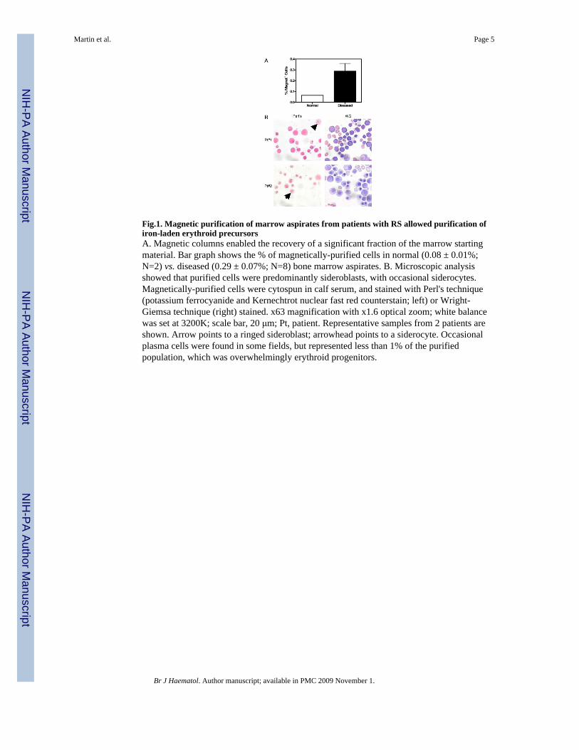
Fig.1. Magnetic purification of marrow aspirates from patients with RS allowed purification ofiron-laden erythroid precursorsA. Magnetic columns enabled the recovery of a significant fraction of the marrow startingmaterial. Bar graph shows the % of magnetically-purified cells in normal (0.08 ± 0.01%;N=2) vs. diseased (0.29 ± 0.07%; N=8) bone marrow aspirates. B. Microscopic analysisshowed that purified cells were predominantly sideroblasts, with occasional siderocytes.Magnetically-purified cells were cytospun in calf serum, and stained with Perl's technique(potassium ferrocyanide and Kernechtrot nuclear fast red counterstain; left) or Wright-Giemsa technique (right) stained. x63 magnification with x1.6 optical zoom; white balancewas set at 3200K; scale bar, 20 μm; Pt, patient. Representative samples from 2 patients areshown. Arrow points to a ringed sideroblast; arrowhead points to a siderocyte. Occasionalplasma cells were found in some fields, but represented less than 1% of the purifiedpopulation, which was overwhelmingly erythroid progenitors.
Martin et al. Page 5
Br J Haematol. Author manuscript; available in PMC 2009 November 1.
NIH
-PA Author Manuscript
NIH
-PA Author Manuscript
NIH
-PA Author Manuscript
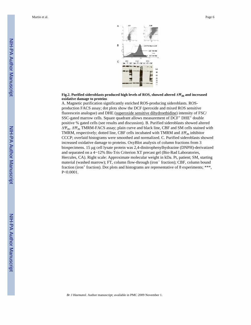
Fig.2. Purified sideroblasts produced high levels of ROS, showed altered ΔΨm and increasedoxidative damage to proteinsA. Magnetic purification significantly enriched ROS-producing sideroblasts. ROS-production FACS assay; dot plots show the DCF (peroxide and mixed ROS sensitivefluorescein analogue) and DHE (superoxide sensitive dihydroethidine) intensity of FSC/SSC-gated marrow cells. Square quadrant allows measurement of DCF+ DHE+ doublepositive % gated cells (see results and discussion). B. Purified sideroblasts showed alteredΔΨm. ΔΨm TMRM-FACS assay; plain curve and black line, CBF and SM cells stained withTMRM, respectively; dotted line, CBF cells incubated with TMRM and ΔΨm inhibitorCCCP; overlaid histograms were smoothed and normalized. C. Purified sideroblasts showedincreased oxidative damage to proteins. OxyBlot analysis of column fractions from 3biospecimens. 15 μg cell lysate protein was 2,4-dinitrophenylhydrazine (DNPH)-derivatizedand separated on a 4−12% Bis-Tris Criterion XT precast gel (Bio-Rad Laboratories,Hercules, CA). Right scale: Approximate molecular weight in kDa. Pt, patient; SM, startingmaterial (washed marrow); FT, column flow-through (iron− fraction); CBF, column boundfraction (iron+ fraction). Dot plots and histograms are representative of 8 experiments; ***,P<0.0001.
Martin et al. Page 6
Br J Haematol. Author manuscript; available in PMC 2009 November 1.
NIH
-PA Author Manuscript
NIH
-PA Author Manuscript
NIH
-PA Author Manuscript
Related Documents