Pupil dynamics during bistable motion perception Centre de Recherche Cerveau et Cognition, UPS, Université de Toulouse, Toulouse, France,& CerCo, CNRS, Toulouse, France Jean-Michel Hupé Centre de Recherche de l’Institut Cerveau et Moëlle Epinière, Université Pierre et Marie Curie, Paris, France, UMR 7225, INSERM-CNRS, Paris, France,& Service d’Ophtalmologie, Centre Hospitalier Universitaire d’Angers, Angers, France Cédric Lamirel Centre de Recherche de l’Institut Cerveau et Moëlle Epinière, Université Pierre et Marie Curie, Paris, France,& UMR 7225, INSERM-CNRS, Paris, France Jean Lorenceau Pupil size not only varies to changes in illumination but is also modulated by several cognitive factors, making it a potentially versatile physiological marker of cortical states. We recorded pupil dynamics while subjects continuously reported their bistable perception of ambiguous moving stimuli, plaids, and partially occluded rotating diamonds. We observed small (about 5% of surface change on average) but reliable pupil dilation around (j300 ms to 1.5 s) the button presses indicating the changes of percepts. We found that 70% of pupil dilation could be accounted for by the motor response. The remaining perceptual component was similar for spontaneously occurring transitions and transitions triggered by physical stimulus manipulations. Moreover, the amplitude of pupil modulation in the spontaneous condition was unrelated to the duration of each perceptual state. It is therefore unlikely that the mechanisms of endogenous perceptual bistability reflect in the pupil. In addition, we measured a clear constriction of the pupil after blinks (about 8% of surface change on average). As pupil changes have the potential to entail retino-cortical activity, their monitoring in studies of visual processing could prove worthwhile. Keywords: bistable percepts, ambiguous stimuli, pupil, blinks, plaid Citation: Hupé, J.-M., Lamirel, C., & Lorenceau, J. (2009). Pupil dynamics during bistable motion perception. Journal of Vision, 9(7):10, 1–19, http://journalofvision.org/9/7/10/, doi:10.1167/9.7.10. Introduction One function of the pupil is to regulate the flux of light entering the eye in response to changes in illumination. This pupil light reflex (PLR) involves a non-linear closed- loop feedback system. The predominant input to the PLR originates in melanopsin-containing, intrinsically photo- sensitive, retinal ganglion cells (ipRGC; Guler et al., 2008). The reflex loop then involves the pretectal olivary nucleus (PON; Clarke, Zhang, & Gamlin, 2003), the Edinger– Westphal nucleus (EW), and the ciliary ganglion, belong- ing to the parasympathetic pathway, which drives the sphincter muscle that constricts the iris (Loewenfeld, 1999). Pupil size depends also on the antagonistic activity of the dilator muscle, which is driven by a sympathetic circuit originating in the hypothalamus (Loewenfeld, 1999; for a synthetic diagram of main connections, see Szabadi & Bradshaw, 1996, reproduced in Merritt, Schnyders, Patel, Basner, & O’Neill, 2004). This pathway is not concerned with the PLR, and its function is far less specific. Regulation of pupil size is complicated by multiple modulations and interactions between these two pathways. The PLR parasympathetic pathway can be modulated at the level of the PON, which receives multiple cortical and subcortical inputs (Gamlin, 2006), possibly at the origin of a non-(directly) retinal component of the PLR (Barbur, 2004). The EW nucleus is thought to receive multiple inhibitory influences, in particular from the locus coeruleus (LC) and the cortex (Breen, Burde, & Loewy, 1983; Loewenfeld, 1999, Merritt et al., 2004). Pupil dilation in response to many situations may therefore be mainly explained by inhibition of the para- sympathetic sphincter pathway at the level of the EW and excitation of the dilator sympathetic pathway, through the hypothalamus, which receives projections from the cortex, the frontal cortex in particular. According to Loewenfeld (1999, p. 319), “Any sensory stimulation (with the excep- tion of light), can elicit pupillary dilation; and spontaneous thoughts and emotions have the same effect as sensory stimuli.” Well-studied examples of pupil dilations in response to non-sensory processing comprise cognitive load (Beatty & Wagoner, 1978; Kahneman & Beatty, 1966), attention (Beatty, 1977), stimulus probability Journal of Vision (2009) 9(7):10, 1–19 http://journalofvision.org/9/7/10/ 1 doi: 10.1167/9.7.10 Received February 3, 2009; published July 15, 2009 ISSN 1534-7362 * ARVO

Welcome message from author
This document is posted to help you gain knowledge. Please leave a comment to let me know what you think about it! Share it to your friends and learn new things together.
Transcript

Pupil dynamics during bistable motion perceptionCentre de Recherche Cerveau et Cognition, UPS,
Université de Toulouse, Toulouse, France, &CerCo, CNRS, Toulouse, FranceJean-Michel Hupé
Centre de Recherche de l’Institut Cerveau et MoëlleEpinière, Université Pierre et Marie Curie, Paris, France,
UMR 7225, INSERM-CNRS, Paris, France, &Service d’Ophtalmologie, Centre Hospitalier
Universitaire d’Angers, Angers, FranceCédric Lamirel
Centre de Recherche de l’Institut Cerveauet Moëlle Epinière, Université Pierre
et Marie Curie, Paris, France, &UMR 7225, INSERM-CNRS, Paris, FranceJean Lorenceau
Pupil size not only varies to changes in illumination but is also modulated by several cognitive factors, making it a potentiallyversatile physiological marker of cortical states. We recorded pupil dynamics while subjects continuously reported theirbistable perception of ambiguous moving stimuli, plaids, and partially occluded rotating diamonds. We observed small(about 5% of surface change on average) but reliable pupil dilation around (j300 ms to 1.5 s) the button presses indicatingthe changes of percepts. We found that 70% of pupil dilation could be accounted for by the motor response. The remainingperceptual component was similar for spontaneously occurring transitions and transitions triggered by physical stimulusmanipulations. Moreover, the amplitude of pupil modulation in the spontaneous condition was unrelated to the duration ofeach perceptual state. It is therefore unlikely that the mechanisms of endogenous perceptual bistability reflect in the pupil. Inaddition, we measured a clear constriction of the pupil after blinks (about 8% of surface change on average). As pupil changeshave the potential to entail retino-cortical activity, their monitoring in studies of visual processing could prove worthwhile.
Keywords: bistable percepts, ambiguous stimuli, pupil, blinks, plaid
Citation: Hupé, J.-M., Lamirel, C., & Lorenceau, J. (2009). Pupil dynamics during bistable motion perception. Journal ofVision, 9(7):10, 1–19, http://journalofvision.org/9/7/10/, doi:10.1167/9.7.10.
Introduction
One function of the pupil is to regulate the flux of lightentering the eye in response to changes in illumination.This pupil light reflex (PLR) involves a non-linear closed-loop feedback system. The predominant input to the PLRoriginates in melanopsin-containing, intrinsically photo-sensitive, retinal ganglion cells (ipRGC; Guler et al., 2008).The reflex loop then involves the pretectal olivary nucleus(PON; Clarke, Zhang, & Gamlin, 2003), the Edinger–Westphal nucleus (EW), and the ciliary ganglion, belong-ing to the parasympathetic pathway, which drives thesphincter muscle that constricts the iris (Loewenfeld,1999). Pupil size depends also on the antagonistic activityof the dilator muscle, which is driven by a sympatheticcircuit originating in the hypothalamus (Loewenfeld,1999; for a synthetic diagram of main connections, seeSzabadi & Bradshaw, 1996, reproduced in Merritt,Schnyders, Patel, Basner, & O’Neill, 2004). This pathwayis not concerned with the PLR, and its function is far lessspecific. Regulation of pupil size is complicated by
multiple modulations and interactions between these twopathways. The PLR parasympathetic pathway can bemodulated at the level of the PON, which receivesmultiple cortical and subcortical inputs (Gamlin, 2006),possibly at the origin of a non-(directly) retinal componentof the PLR (Barbur, 2004). The EW nucleus is thought toreceive multiple inhibitory influences, in particular fromthe locus coeruleus (LC) and the cortex (Breen, Burde, &Loewy, 1983; Loewenfeld, 1999, Merritt et al., 2004).Pupil dilation in response to many situations may
therefore be mainly explained by inhibition of the para-sympathetic sphincter pathway at the level of the EW andexcitation of the dilator sympathetic pathway, through thehypothalamus, which receives projections from the cortex,the frontal cortex in particular. According to Loewenfeld(1999, p. 319), “Any sensory stimulation (with the excep-tion of light), can elicit pupillary dilation; and spontaneousthoughts and emotions have the same effect as sensorystimuli.” Well-studied examples of pupil dilations inresponse to non-sensory processing comprise cognitiveload (Beatty & Wagoner, 1978; Kahneman & Beatty,1966), attention (Beatty, 1977), stimulus probability
Journal of Vision (2009) 9(7):10, 1–19 http://journalofvision.org/9/7/10/ 1
doi: 10 .1167 /9 .7 .10 Received February 3, 2009; published July 15, 2009 ISSN 1534-7362 * ARVO

(Reinhard & Lachnit, 2002), and emotional factors(Partala & Surakka, 2003). In addition, changes of pupilsize have been observed for specific visual computationssupposed to involve the visual cortex, like the transitionfrom incoherent to coherent motion (Sahraie & Barbur,1997), color perception (Barbur, Weiskrantz, & Harlow,1999; Wilhelm, Wilhelm, Moro, & Barbur, 2002), or theprocessing of faces (Conway, Jones, DeBruine, Little, &Sahraie, 2008), raising the possibility that pupil dynamicsreflects some aspects of cortical processing.We further investigate the relationship between cogni-
tive states and pupil dynamics by recording pupil sizeduring long-lasting observation of visual moving bistablestimuli. We were interested to determine whether changesin perception entail concomitant changes in pupil size.Perceptual bistability is an interesting tool in this regard asan unchanging stimulus yields different interpretationscorrelated to different brain states (reviews in Blake &Logothetis, 2002; Long & Toppino, 2004), such that anychange in pupil size cannot be accounted for by a changein luminance distribution. Comparisons with conditionswhere perceptual bistability is induced by smooth physicalstimulus changes that do not in themselves entail pupilmodulation further allow assessing whether changes in pupilsize are specific to endogenous or exogenous modificationin perception, so as to shed light on the mechanisms ofbistable perception.Recent studies indicate that pupil transiently dilates
when subjects change their perception of ambiguousstimuli (Einhauser, Stout, Koch, & Carter, 2008; Lamirel,Hupe, & Lorenceau, 2008). Whether these changes relateto the cause or the effect of perceptual transitions or aresimply an epiphenomenon remains an open issue that weaddress in the present study. Since pupil responds to manynon-specific factors, we ask whether this modulation ofpupil size is related to the cortical reorganization respon-sible for the percept change, to the change of the perceptitself, to attentional, predictive or decisional factors, orsimply to the motor command needed to report a percep-tual switch. In order to dissect the origin of the observedpupil dilation, additional experiments involved soundinduced and endogenously generated button presses, per-ceptual transitions induced by smooth and abrupt physicalstimulus changes without a motor response, and the pas-sive viewing of a static stimulus whose contrast distributionvaried over time. Our analyses reveal that pupil dilation isrelated to both motor response and perceptual transitions.Einhauser et al. (2008) observed that pupil dilation aroundthe time of perceptual switches (starting before a buttonpress) was correlated to the relative duration of the fol-lowing percept, suggesting that a structure involved incontrolling pupil dilation, the locus coeruleus, plays a rolein the perceptual reorganization itself, owing to its widelydistributed, ascending projections to the neocortex (Aston-Jones & Cohen, 2005). Here, we performed a similar study,but we do not observe such a correlation. An unexpectedresult, however, is the observation of a long-lasting pupil
constriction after blinks. Thus, we observe marked pupilchanges during the bistable perception of ambiguouspercepts, due to button presses, percept changes, and blinks.
Methods
Stimuli and procedure
Two experiments were run independently in Toulouseby JMH and in Paris by CL and JL. The Toulouseexperiments used moving plaids (Hupe & Rubin, 2003),the Paris experiments used rotating diamonds with varyingluminance distribution along their line segments (Lorenceau& Shiffrar, 1992).
Experiment 1: Plaids
Stimuli
See http://cerco.ups-tlse.fr/~hupe/plaid_demo/ forexamples of plaid stimuli. Plaids were displayed on a19-in. monitor (26.25-cm vertical viewable screen size) ata frame rate of 75 Hz. The screen resolution was 1024 �768 pixels. Two rectangular-wave gratings were presentedthrough a 6-degree radius circular aperture on a computerscreen 57 cm away. The gratings comprised thin darkstripes (j31.7% contrast over the mean luminance, dutycycle = 0.3, spatial frequency = 0.3 cycle/deg) on a lighterbackground (36% contrast) and appeared as figuresmoving over the background. The intersecting regionswere darker than the gratings (j47.9% contrast, in themiddle of the transparency range). Mean luminance was33 cd/m2. The gratings were drifting at 1.5 deg/s indirections 105 or 125 degrees apart. Plaids could move inthe four cardinal directions. As a secondary goal of thisstudy was to test observers’ capability to bias theirbistable percept at will, an “intention” instruction wasprinted on the screen (“neutral”, “coherent”, “transparent”)before each trial. A red fixation point over a 1-degree radiuscircular gray mask was added in the middle of the circularaperture and subjects were instructed to fixate this pointthroughout stimulus presentation. The stimulus can beperceived either as a single plaid moving in a givendirection (“coherent” percept) or as two gratings slidingin opposite directions on top of each other (“transparent”percept; Hupe & Rubin, 2003; Wallach, 1935; Wuerger,Shapley, & Rubin, 1996; we shall further name thispercept the “incoherent” percept, for semantic compati-bility with the diamond study, even though the transparentinterpretation requires as much global processing as thecoherent one: Rubin & Hupe, 2005).
Procedure
Fourteen observers (8 males and 6 females) gaveinformed consent to participate in the experiment (average
Journal of Vision (2009) 9(7):10, 1–19 Hupé, Lamirel, & Lorenceau 2

age: 27) with normal or corrected-to-normal eyesight. Theywere comfortably seated in front of the screen, with theirchin and forehead resting on a chin rest (University ofHouston College of Optometry). They were instructed toreport their conscious perception of each stimulus during1-min periods by pressing the right mouse button continu-ously for transparent percepts and the left button for coherentpercepts. They were asked to release both buttons if theywere unsure of their percept (this happened on average3% of the time). Twenty-four stimuli (8 different stimuliby 3 instructions) were presented in a randomized order.Observers were given a few practice trials. Seven subjectshad run previously other plaid experiments. On average,observers reported the coherent percept for 54% of the time(range [36% 68%]). The effect of intention will bedescribed in another paper. The analyses presented belowused all available data, but similar conclusions apply ifrestricting the analysis to the “neutral” trials (not shown;figures and analyses were, however, sent to the reviewers).Eye position (difference between the pupil and corneal
reflection centers) and pupil diameter were recordedbinocularly at 120 Hz (10 subjects) or 240 Hz by usingan ISCAN ETL-200 system (Burlington, MA, USA). Thecameras were attached to the chin rest just above the eyesand looked at the eyes through semi-transparent mirrors.
Experiment 2: Diamonds
Stimuli
The “hidden” moving diamond used in this studyconsisted in a set of four disconnected segments trans-lating sinusoidally along a vertical axis (Lorenceau &Alais, 2001). The phase relationship between segmentmotions was chosen such that their perceptual integrationresulted in seeing a diamond shape translating along acircular trajectory of 0.5- radius at 1 Hz. When segregated,the segments were seen as two independent pairs oscillatingup and down. The diamond subtended a visual angle of about6 degrees at a viewing distance of 50 cm. A small red fixationdisk was present at all times at the center of the screen.Background luminance was 15 cd/m2. The stimulus wasdisplayed on a 22-in. monitor (30.48-cm vertical viewablescreen size, screen resolution of 1024 � 768 pixels) at aframe rate of 85 Hz. Seven different conditions were testedin long-lasting run of 4 min each. These differentconditions, detailed below, were designed to disentanglethe possible contributions of perception, attention, predic-tion, decision, and motor commands to pupil changes.
Condition 1: Active report of spontaneous perceptualswitches of an ambiguous rotating diamond
In this condition, a physically unchanging version of the“broken” diamond was used. The stimulus was ambiguousand could either be perceived as a single diamond trans-lating clockwise along a circular trajectory (“coherent”percept) or as four independent segments oscillating up and
down (“incoherent” percept). As contrast gradients alongthe line segments can favor either the coherent or theincoherent percept (see below, Condition 2; Lorenceau &Shiffrar, 1992), we set the contrast gradient of the stimulusso as to yield well-balanced perceptual alternations(Figure 1).
Condition 2: Active report of induced perceptual switches
The stimulus was the same as in Condition 1, but thecontrast gradient along each line segment was smoothlyvaried to periodically induce a perception of either thecoherent or the incoherent percept (Lorenceau et al.,2008; Lorenceau & Shiffrar, 1992). Segments with lowcontrast line ends and a high contrast center favor theperception of a global shape rotation while segments withhigh contrast line ends and a low contrast center entailsthe perception of independent up and down translatingsegments, despite average luminance being the same inboth cases (Figure 1). Smooth periodic transitions fromone contrast gradient to the other induces alternationsbetween coherent and incoherent perceptual states with an8-s cycle, thus leading to a 4-s theoretical averageduration of perceptual episodes. Preliminary experimentswere performed prior to the main experiments to deter-mine the contrast gradient corresponding to well-balancedcoherent/incoherent episodes. This gradient was used inCondition 1 and as the midpoint gradient in Condition 2.
Condition 3: Active report of a sound stimulus
A brief sound (1000 Hz, 100 ms) to which observersmust respond as in the remaining conditions (by alternat-ing their finger presses) was produced every 4 s while astatic version of the diamond of Condition 1 wasdisplayed on the screen throughout the 4-min run tomaintain the same mean luminance.
Figure 1. Diamond display. (A) Induced bistability was controlledthrough smooth modulation of the contrast gradient along the linesegments (8-s period). Low contrast line ends yield a coherentpercept (black shaded area) while high contrast line ends yield anincoherent percept (gray shaded area). Mean contrast wasconstant over time. (B) A fixed contrast gradient derived frompreliminary testing was used in the spontaneous condition.
Journal of Vision (2009) 9(7):10, 1–19 Hupé, Lamirel, & Lorenceau 3
Condition 4: Constant stimulation
Only a static version of the diamond of Condition 1 wasdisplayed on the screen.
Condition 5: Induced perceptual switches with no report
The stimulus was the same as in Condition 2, butobservers were looking at the stimulus without reportingtheir perception.
Condition 6: Static diamond with dynamic contrastgradient
The stimulus was the same as in the previous conditionbut remained static. The only change was the distributionof the contrast along each line segment, with the samedynamics as before. This control condition aimed atverifying that no pupil changes correlated to the contrastchanges would occur.
Condition 7: Abrupt changes with no report
The contrast gradient along the line segments changedabruptly at random intervals of 1, 2, 3, or 4 s from adiamond strongly biased to promote the coherent percept toa diamond strongly biased toward the incoherent percept,so as to grossly mimic unpredictable perceptual switches asthose of Condition 1. Based on previous experiments(Lorenceau & Shiffrar, 1992), such abrupt changes reliablydrive perceptual alternations between coherent and inco-herent percepts. These abrupt time-stamped changes wereused to trigger changes in perception for analyzingpurposes.
Procedure
Ten observers with normal or corrected-to-normal eye-sight (8 males and 2 females; average age: 28) gave theirinformed consent to participate in the experiments. Theywere seated in front of the screen using a chin rest with abite bar. Each subject ran each condition once. The orderof appearance of the different conditions was counter-balanced across subjects using a Latin square. After aninitial fixation-only period lasting 4 s the stimulus waspresented for 240 s. A fixation-only period of 2 s endedthe trial. Subjects were instructed to fixate the centralfixation disk for the entire course of all trials.In Conditions 1 and 2, subjects were instructed to report
their visual perceptVcoherent or incoherentVby continu-ously pressing one of two mouse buttons. The assignmentof buttons to coherent and incoherent perceptual states wasbalanced across subjects. Observers were given a fewpractice trials. In Condition 1, they reported the coherentpercept on average for 38.5% of the time (range [12%65%]). In Condition 2, we verified that the gradientcorresponding to the average transition point between bothpercepts was the midpoint gradient found in the prelimi-nary experiments and that perceptual alternations followedluminance variations. However, we observed a hysteresiseffect, which is expected given the underlying cooperativeprocesses involved in motion binding (Lorenceau,
Gimenez-Sastre, & Lalanne, 2003). Intra- and inter-subject variability of response times to stimulus changeswas also observed (see Results section), as expected withstimuli supporting several interpretations.In Condition 3, subjects were instructed to continuously
press a mouse button and to change button every time theyheard the sound. In Condition 4, subjects had to continu-ously press a mouse button and to change it at will. In bothcases, there was no change at all of the visual display.In Conditions 5 to 7, subjects were instructed to pay
attention to their perceptual state but without reporting theirpercept (no button press), while still looking at the centerof the diamond as in all other conditions.Eye position (difference between the pupil and corneal
reflection centers) and pupil diameter were recordedmonocularly (right eye for all subjects) at 240 Hz usingan ISCAN ETL-400 system (Burlington, MA, USA). Thecamera looked at the right eye through a semi-transparentmirror.
Data analysisPretreatment
Oculomotor signal was pretreated using an interactiveinterface written in Matlab. Each trace was inspected andprocessed in three steps. First, blinks were removed byrelying on pupil size. By default, a blink was identifiedwhen pupil signal was below 60% of the median size forat least 50 ms (we systematically verified that this thresholdwas below actual pupil size). Blinks separated by less than100 ms were aggregated to a single blink. Each detectedblink was verified by inspecting the signal on both eyes(Experiment 1) as well as eye position around each blink(since partial occlusion of the pupil by the lid when theeyes are closing is recorded as eye position moving downfast just before signal loss). Blinks could thus be removedor added manually, which was necessary only in a fewcases. Next, eye position was scaled, using a 9-pointcalibration procedure recorded just before the experi-ments. Finally, artifacts were removed (loss of signals notdue to blinks, aberrant eye positions). The signal did notneed being filtered.
Exclusion criteria
After a first-pass analysis, we observed variations ofpupil diameter after blinks (see Figure 9) and aroundbutton presses (see Figure 2). In order to measure botheffects properly, we selected blinks and button presses thatwere isolated enough from other events (blink or buttonpress). Pupil response was significantly modulated forabout 1.5 s after a blink or a button press. For buttonpresses, modulation started about 500 ms before the press.An event was therefore kept for the analysis only if noother event happened within 1.5 before the beginning of
Journal of Vision (2009) 9(7):10, 1–19 Hupé, Lamirel, & Lorenceau 4

baseline and up to 1.5 after the event (2 s if the next eventwas a button press). Only about a third of the events couldbe included in the analyses. Refer to figure legends forexact numbers. Such strict exclusion criteria were neces-sary to ensure that the analyses and conclusions drawnfrom pupil dynamics would not be contaminated byirrelevant factors.
Statistical analysis
A baseline was computed before each event (start andend values of the baseline period are indicated in thefigure legends) and subtracted to the signal to get rid ofslow pupil variations. Time zero was the time of event.Since blinks last some time, time before each blink wasrelative to the beginning of the blink, time after each blinkrelative to the end of the blink. Median blink duration was100 ms (range of 21–567 ms) in Experiment 1 and 137 ms
(range of 21–1446 ms; “long” blinks correspond in fact toseveral blinks) in Experiment 2. Pupil size was convertedto z-scores in order to collapse data across subjects. Peri-event time histograms were computed at the originalsampling rate and then downsampled to 10 Hz for displayand statistics purposes (note that we verified that samplingat 60 Hz gave exactly the same result; 10 Hz correspondsbetter to pupil dynamics). Time “zero” in the graphscorresponds to the signal averaged from 50 ms before upto 50 ms after the event. ANOVAs were performed inStatistica 8 (Statsoft) independently at each time aroundthe event. Since these dependent variables are temporallycorrelated, no correction for multiple comparisons wasapplied. Some procedures do exist to balance temporalcorrelation and multiple comparisons (see, e.g., Hupe et al.,2001), at least for two-sample comparisons. Here, how-ever, we indicated p-values only to help the reader havean idea of the average effect robustness. We also indicateeffect sizes (partial Eta Squared, )p
2, varies between0 and 1). Data from individual subjects are presented whennecessary. Some of the effects were in any case verystrong (e.g., Figures 2 and 9), others borderline significant(e.g., Figures 6 and 7). Results are discussed accordingly.The variable “subject” was included in all statisticalanalyses and treated as a random factor. Residuals wereinspected for every analysis and dependent variable.Residuals were always normally distributed and variancesreasonably homogenous. Events were excluded if theabsolute standardized residual value was larger thanexpected for a normal distribution given the sample size.The Gaussian distribution predicts that 1% of values shouldbe either larger than 2.96 or smaller thanj2.96, and 1 over1000 either larger than 3.65 or smaller than j3.65. We setthe threshold value for outliers as the value for which the(two-sided) Gaussian probability multiplied by sample sizewas less than 0.5. This means that for a sample size of1000, our outlier threshold was 3.84. Here, residual valueswere therefore considered as outliers if their absolute valuewas larger than about 3.9 (Experiment 1, Experiment 2:analyses of Figures 4 and 6), about 3.6 (Experiment 2:analysis of Figure 7), or about 3.2 (Experiment 2,Condition 1) for at least one of the dependent variables.
Raw and relative modulations
We also performed analyses on raw data (no z-scoretransformation) to have an idea of the amplitude of pupilmodulations relative to pupil diameter (% change). Thisanalysis does not take into account the different noiselevels measured in different subjects, contrary to the z-scoreanalysis that we favored. In addition, the amplitude of thepupil light reflex was measured for all subjects inExperiment 2 and four subjects in Experiment 1, bymodulating the screen luminance from minimum tomaximum. For the 10 other subjects of Experiment 1,this amplitude was estimated by the difference between
Figure 2. Pupil response around perceptual switches of ambig-uous plaids and diamonds (mean T SEM). Plaids: Data from14 subjects, N = 1121 (10 outliers removed. The total number oftransitions was 3390). Diamonds: Data from 10 subjects, N = 134(9 outliers removed. The total number of transitions was 332).Subjects indicated percept changes by releasing and pressingmouse buttons. Since pupil response to blinks last on average 1.5 s(see Figure 9), button presses were excluded if a blink occurredwithin 2.5 s before (i.e., 1.5 s before baseline) or 1.5 s after thebutton press. In order to avoid between events contamination,button presses were excluded if following another press by lessthan 2.5 s or preceding another press by less than 2 s. Averagepupil diameter was computed for the period [j1 j0.6] s beforeeach button press and subtracted. For plaids, pupil diameterstarted to increase significantly above zero 400 ms before buttonpress (black curve, F(1, 1093) = 6.6, p = 0.021, )p
2 = 0.29). Theincrease was significant for 13 out of the 14 subjects (individualdata shown in Figure 8). For diamonds, it started 300 ms beforebutton press (red curve, F(1, 114) = 5.3, p = 0.026, )p
2 = 0.09). Theincrease was significant for 8 out of the 10 subjects (individualdata shown in Figure 5: red curves).
Journal of Vision (2009) 9(7):10, 1–19 Hupé, Lamirel, & Lorenceau 5

maximum and minimum pupil sizes (we verified that bothmeasures were tightly correlated and close to each other).This allowed us to compare the size of pupil modulationsin our experiments to the maximum range of pupil sizevariations.
Vergence
A possible confound in considering pupil size is its linkwith eye vergence. To determine the extent to whichvergence eye movements could account for changes inpupil size, we measured vergence eye movements inExperiment 1 using binocular recordings. For mostsubjects, vergence mirrored pupil dilation. Vergence mod-ulation was about half the size of pupil modulation whenboth measures were computed as z-scores, be the cause ofpupil modulation a button press, a blink, or a luminancechange. Average vergence was about 0.05 degrees afterbutton presses. We conclude that vergence modulations inour data were a measure artifact due to the fact that formost subjects pupil centers diverge when pupil dilates(anatomical constraint, Camellin, Gambino, & Casaro,2005; Wyatt, 1995; Yang, Thompson, & Burn, 2002). Noother vergence eye movements could be observed in ourdata. Eye position was also stable around button presses,stimulus changes, and blinks.
Correlation between pupil diameterand percept duration
We measured the correlation between pupil size andpercept duration in Experiment 1, as in Einhauser et al.(2008) by computing a “normalized measure of ‘post-switch’ duration relative to the corresponding ‘preswitch’duration”: Relative duration = (post j pre) / (post + pre).We selected only the switches that were not contaminatedby other events, as described previously. The first and lastswitches of each trial were also excluded, since relativeduration for these switches cannot be computed. Tracesaround switches were downsampled to 10 Hz (but theresults were identical at 60 Hz). For each subject, wecomputed the correlation coefficients between pupil size(z-score, no baseline removal) and relative duration foreach time point between 1 s before and 1.5 s after theswitch. The same number of data was used to computeeach correlation coefficient (mean number of values bysubject = 70, range = [21 121]). We also computed acorrelation coefficient over all values. We performed thesame analysis for the data of Experiment 2 (for the con-dition of spontaneous perceptual switches of ambiguousrotating diamonds), but much less data was available(10 subjects, mean number of values by subject = 13,range = [3 24]). We also performed the analyses usingabsolute postswitch duration (log values) instead of rel-ative duration. In order to compare directly our results tothose of Einhauser et al. (2008), we also performed the
correlation analyses without exclusion criteria. In addition,we considered several possible biases. We performed theanalyses without z-transformation (pupil size was thenmeasured in millimeters), or by selecting only trials forwhich competing percepts were well balanced (close to50%), or by selecting only trials with slow alternations (withno other exclusion criteria). All these analyses producedconverging results (they are not detailed here, but the figuresand statistical results were sent to the reviewers).
Results
Pupil response to perceptual switches
The pupil response to perceptual switches is presentedin Figure 2 for both experiments. Switches were indicatedby subjects by releasing the mouse button indicating theprevious percept and pressing the other mouse button toindicate the new percept. The time reference is the buttonpress corresponding to the new percept. Pupil dilationstarted before the time of button press (average increase isvisible as early as 500 ms and significant 400 ms beforethe button press for plaids). Pupil response was similar forboth types of perceptual transition and for both stimuli(Figure 3). This decorrelation between the “sign” of atransitionVfrom coherent to incoherent, or the reverse-and pupil changes indicates that pupil dilation is not
Figure 3. Percept type has no or little effect on the amplitude ortiming of pupil responses (mean T SEM). Same data set as inFigure 2. For spontaneous diamonds, we observed a tendency forthe pupil response to start earlier for switches to incoherentpercepts. However, amplitude difference between both traces didnot reach significance: The stronger difference occurred at thetime of button press (F(1, 114) = 3.02, p = 0.098, )p
2 = 0.14).Inspection of data of individual subjects confirmed that thislatency difference was not reliable in our data set.
Journal of Vision (2009) 9(7):10, 1–19 Hupé, Lamirel, & Lorenceau 6

related to a change inVperceivedVcontrast or luminance.Maximum effect size ()p
2) was 0.67 for both plaids anddiamonds, 100 to 200 ms after button press for diamonds,200 to 400 ms for plaids. Pupil response was, on average,2.2% of mean pupil diameter for plaids (range forindividual subjects: 0.1% to 6.6%) and 2.4% for diamonds(range of 1.1% to 4.8%). Average modulation correspondsto 5.2% (range of 0.2% to 15.6%) of pupil maximaldilation (see Methods section) for plaids and 4.9% fordiamonds (range of 1.8% to 10.9%).
Components of the pupil response
Since pupil dynamics is rather slow (the latency of thePLR is about 300 ms, with an absolute minimum of 180 msdue to the mechanical properties of the iris muscle:Loewenfeld, 1999), pupil dilation in Figures 2 and 3 wascaused by an event that happened as early as 700 msbefore the button press. This event might have beenrelated to the neural mechanism of bistability leading topercept switching (like the reorganization of the neuralnetworks corresponding to each interpretation), the per-ceptual change itself (and its awareness), with a possiblecomponent of surprise, the decision to press the button,the motor preparation and command to press the button, ora combination of these processes. Attentional componentsmay also modulate several stages of the processes. Forexample, a spontaneous switch of perception may tran-siently increase attention, or subjects may pay moreattention to their percepts when they have to report it.To try disentangling these different processes and
evaluate their respective contributions to pupil dilation,we ran six additional conditions, intermingled with themain “spontaneously changing diamond” condition in thesame observers (see Methods section, Experiment 2).First, we tested whether pupil dilation reflected specif-
ically the neural mechanism of perceptual bistability(independently of the percept switching itself) by compar-ing the pupil response to spontaneous and inducedtransitions. We designed a “diamond” condition whereperceptual transitions were induced by stimulus manipu-lations. Specifically, we smoothly varied the contrastgradient along each segment (period = 8 s; one switchevery 4 s), alternating between gradients that causeincoherent and coherent percepts, while keeping the samemean luminance. The left panel of Figure 4 shows thatpupil dilation is very similar for spontaneous and inducedtransitions, at least for the onset and the peak of dilation.Pupil dilation was on average more sustained for sponta-neous transitions, but the difference between the twocurves did not reach significance. Inspection of individualdata (Figure 5, blue and red curves) reveals that this lackof significance is unlikely to be due to a lack of power,since several subjects with clear pupil responses for bothconditions did not exhibit at all this effect (see, e.g.,subjects 2, 7, and 8). It is therefore unlikely that the neural
mechanisms of perceptual bistability have a specificinfluence on pupil size. These two conditions differedalso by both the timing and the possible prediction ofpercept switches. Spontaneous transitions happened atrandom times, with no warning, while induced perceptualtransitions followed the periodic smooth changes of thestimulus and were therefore highly predictable. The lackof significant difference between the pupil responses inboth conditions indicates that surprise and prediction havelittle effect on pupil dilation, at least for these exper-imental conditions.We then tested whether neural processes related to
decision and motor preparation were overall responsiblefor the changes in pupil size, with no specific influence of
Figure 4. Pupil response for different tasks. The same 9 subjectsran all four experiments (subject 5 had too many blinks in the“green” condition and was therefore excluded in this graph). Redlines: Condition 1, diamonds were ambiguous and subjectsexperienced spontaneous changes of percept (same data set asin Figures 2 and 3). Blue lines: Condition 2, perceptual transitionswere induced by stimulus manipulations. Purple lines: Condition 3,subjects were asked to switch mouse button each time there wasa sound. Green lines: Condition 4, subjects were asked to pressthe mouse buttons randomly. In those two conditions, subjectswere required to keep fixating a small disk at the center of a staticoccluded diamond (static version of the stimulus of Condition 1).Total number of events used for the computation of each PSTHwas, respectively, 138, 320, 164, and 303 (20 outliers wereremoved). Pupil response was stronger in the presence of rotatingdiamonds from 100 ms before until 500 ms after button press(comparison between the curves of the left and those of the rightpanel; maximum effect of stimulus 100 ms after button press,F(1,889) = 15.7, p = 0.003, )p
2 = 0.63). A trend for more sustainedpupil dilation is visible for both “spontaneous” conditions, com-pared to the “induced” conditions, but the difference was notsignificant (comparison of the curves across both panels;maximum effect 1.5 s after button press, F(1, 889) = 1.77,p = 0.22, )p
2 = 0.18).
Journal of Vision (2009) 9(7):10, 1–19 Hupé, Lamirel, & Lorenceau 7

perceptual transitions. To that aim, we added twoconditions with no change in visual perception. In thefirst one, we asked observers to switch button press after abrief sound produced every 4 s during 4 min. This timingwas similar to the perceptual transitions induced byphysical modulations of the contrast gradient in thediamond experiment. Since we were concerned that suchexogenous drive would not resemble the endogenousinspection of one’s perceptual state required with bistablestimuli, we also ran a second control condition whereobservers had to change button press at will, in theabsence of any driving sensory stimulus (i.e., wheneverthey felt ready for it; endogenous drive). The right panel
of Figure 4 shows that pupil dilated around button presses,with a time course similar to that observed with bistablestimuli. However, the overall amplitude of pupil dilationwas significantly less (p = 0.003, see legend of Figure 4)than that observed during the changing perception ofdiamonds, providing indirect evidence for a genuinecontribution of perceptual alternations to pupil dynamics.On average, the decision and motor components con-tributed to 70% of the maximal pupil response (measuredin both conditions 400 ms after button press). Inspectionof individual data (Figure 5) makes it reasonablyconvincing that, despite large individual variability, pupildilation was larger for the diamond conditions. The
Figure 5. Data of individual subjects for perceptual reports. Same color conventions as in Figure 4. Baseline was [j1 j0.6] s beforebutton press. SEM is plotted only for spontaneous perceptual switches of the diamond stimulus (pink lines around the red lines). Smallvertical bars on the left of each panel indicate the average and maximum standard errors of the mean over time, for each condition andsubject. The number of traces used to compute each PSTH is indicated within each panel (no outlier removal at this stage).
Journal of Vision (2009) 9(7):10, 1–19 Hupé, Lamirel, & Lorenceau 8

latency of pupil response was also shorter (by about200 ms) when button presses followed perceptualswitches, a further argument in favor of a perceptualcontribution to pupil dilation. However, the latencydifference could have been due to the amplitude difference(Figure 5, compare, e.g., subject 1 with subject 6 or 8).Comparing the pupil response to endogenously andexogenously driven button presses (the two curves of theright panel of Figure 4) revealed that pupil diameterdecreased, on average, more slowly after an “endogenous”button press than after an “exogenous” button press. Thiseffect was, however, not significant and probably due torandom inter-subject variations (Figure 5). Several sub-jects with clear pupil responses for both conditions did not
exhibit at all this effect (compare the green and magentacurves for subjects 8, 9, and 10).To further disentangle the specific contributions of
perceptual and motor components to pupil dilation, weran two additional conditions during which observerspassively looked at the diamond stimulus for 4 min (nobutton press). We used the same smooth variations ofcontrast gradient along segments as before. In onecondition (Condition 5), the segments were also movingin the same way as in Condition 2 (blue curve of Figure 4),in the other condition (Condition 6) the segments werestationary, in order to verify that variations of localluminance had no effect on pupil diameter by themselves.Pupil responses were now aligned to the reversals ofcontrast gradients (since there was no button press), that isthe times of “higher terminator luminance/lower centerluminance” and “lower luminance terminator/higher cen-ter luminance,” which cause, respectively, incoherent andcoherent percepts when the segments are moving. Figure 6shows the results, together with the data of Figure 4 (bluecurve) reprocessed in order to align the pupil response tothe stimulus changes, instead of the button presses.Changing the contrast gradient of the stationary segmentsdid not trigger any variation of pupil diameter (rightpanel). With a moving display inducing perceptualchanges, there was a small, almost significant, pupildilation (middle panel). This small variation is unlikely
Figure 6. Pupil response after smooth changes of the diamondstimulus. The “rotating/reports” condition (left panel) is the samecondition as the “diamond/induced” condition in Figure 4. ThePSTH is computed over the same data but is now taking as timereference the stimulus change instead of the button press. Timezero corresponds to the time when the stimulus should induce themaximum of either coherence or incoherence. Stimulus cycle was8 s, so a maximum was reached every 4 s. Traces were excludedif a blink occurred within 3.7 s before (i.e., 1.5 s before baseline)or 1.5 s after time zero. Average pupil diameter was computed forthe period [j2.2 j1.8] s and subtracted. (Middle panel) Samestimulus, but subjects did not report their percepts. (Right panel)Static stimulus, no button press. The dynamics of local luminancechanges were the same as in the other two conditions. The same9 subjects as before (subject 5 had too many blinks in the static/no report condition). Nine outlier values were removed. There wasno effect of percept type. Luminance changes per se did nottrigger any pupil response (right panel). There was a small pupilresponse due to the change of percept (middle panel, maximumresponse at time zero, average over both percepts, z-score =0.07, F(1,326) = 3.6, p = 0.08, )p
2 = 0.25). The effect of task wassignificant between j100 ms and 500 ms (maximum at 100 ms,F(2, 932) = 5.25, p = 0.015, )p
2 = 0.35).
Figure 7. Pupil response after abrupt changes of the diamondstimulus. Each stimulus change was supposed to trigger aperceptual flip. Subjects did not indicate their percepts. The same10 subjects as previously (N = 402, 8 outliers excluded). Traceswere excluded if a blink occurred within 1.7 s before (i.e., 1.5 sbefore baseline) or 1.5 after the stimulus change. Average pupildiameter was computed for the period [j0.2 0.2] s andsubtracted. There was no effect of the type of stimulus manipu-lation: Whether it should induce a switch to coherence orincoherence, there was a small increase of pupil diameter. Onaverage, this increase was just significant 600 ms after thestimulus change (black arrow) and after (maximum significance at700 ms, F(1,382) = 5.83, p = 0.038, )p
2 = 0.38).
Journal of Vision (2009) 9(7):10, 1–19 Hupé, Lamirel, & Lorenceau 9

to be due to stimulus changes per se (considering thepossibility that local luminance changes were morepowerful for moving stimuli than for static ones), becausepupil variations were not in opposite directions for bothtypes of stimulus variations. Moreover, the time course ofpupil dilation was similar to the one observed in theoriginal condition, when button presses followed perceptswitches (left panel). When measuring the latency of those
button presses relative to peak stimulus changes (timezero), we observed intra- and inter-subject variations.Induced perceptual switch was indicated by subjects 18%of the time at time j1 s, 50% at time zero, and 80% attime 1 s. This variation is larger than what we shouldexpect from the motor command itself. This means thatwhen smooth luminance variations are used, perceptualflips occur at slightly different times within and across
Figure 8. Pupil response and correlation analysis in the plaid experiment. Data for each subject and average (bottom right panel). Blackcurves: Pupil diameter as in Figure 2, but now measured in millimeters instead of z-scores, and with no outlier removed (mean T SEM).Only switches that were not contaminated by blinks or other switches were included, as usual. Red curves: For each subject and timepoint, we measured the correlation coefficient between percept relative duration and pupil size (measured in millimeters). The Y-axis hasthe same scale as for pupil size but then indicates r-values plotted between j0.35 and 0.35 (horizontal lines denote zero correlation). Inthe bottom right panel, the correlation was computed over the data from the 14 subjects all together. Y-axis indicates r-values for the redcurve and change of pupil diameter for the black curve (D indicates the average pupil diameter before button press). Correlations werealways computed over less data than indicated in the figure panels, because relative duration could not be computed for the first and lasttransitions of each trial (total N = 982). R-values were not significantly different from zero and not modulated over time (max r-value700 ms before button press, r = 0.02, p = 0.61, )p
2 = 0.0002).
Journal of Vision (2009) 9(7):10, 1–19 Hupé, Lamirel, & Lorenceau 10
observers, maybe because of a hysteresis effect. Whenconsidering the effect of the unflagged perceptual flip onpupil dilation, averaging in reference to the stimuluschanges may thus have “blurred” the effect.In a last condition (condition 7), we abruptly and
randomly changed the contrast distribution along thesegments every 1, 2, 3, or 4 s, in order to triggerperceptual transitions with less time jitter than withsmooth contrast variations. Subjects were still asked topassively look at the moving stimuli. Pupil dilation wasmodest but significant (Figure 7), with no differencebetween the “signs” of the contrast gradient.
Correlations between pupil diameter andpercept duration
Overall, the results reveal the presence of two maincomponents of the pupil response to perceptual switchesindicated by button presses. The major component (about70%) is correlated to the button press itself and mayinclude attention, decision, and motor preparation andexecution. The second, smaller, component is correlatedto perceptual alternations, also possibly including anattentional component. Both components are present inthe absence of any change in retinal illuminance and aretherefore independent of the PLR. Perceptual changes canbe induced either by exogenous stimulus manipulations orby endogenous perceptual reorganizations characteristicof bistable stimuli. The average pupil dilation is similar inboth cases, but a possible relationship between pupil sizeand the mechanisms of bistable perception may have beensubtler than a global effect on the amplitude of pupildilatation. Einhauser et al. (2008) suggested that pupil
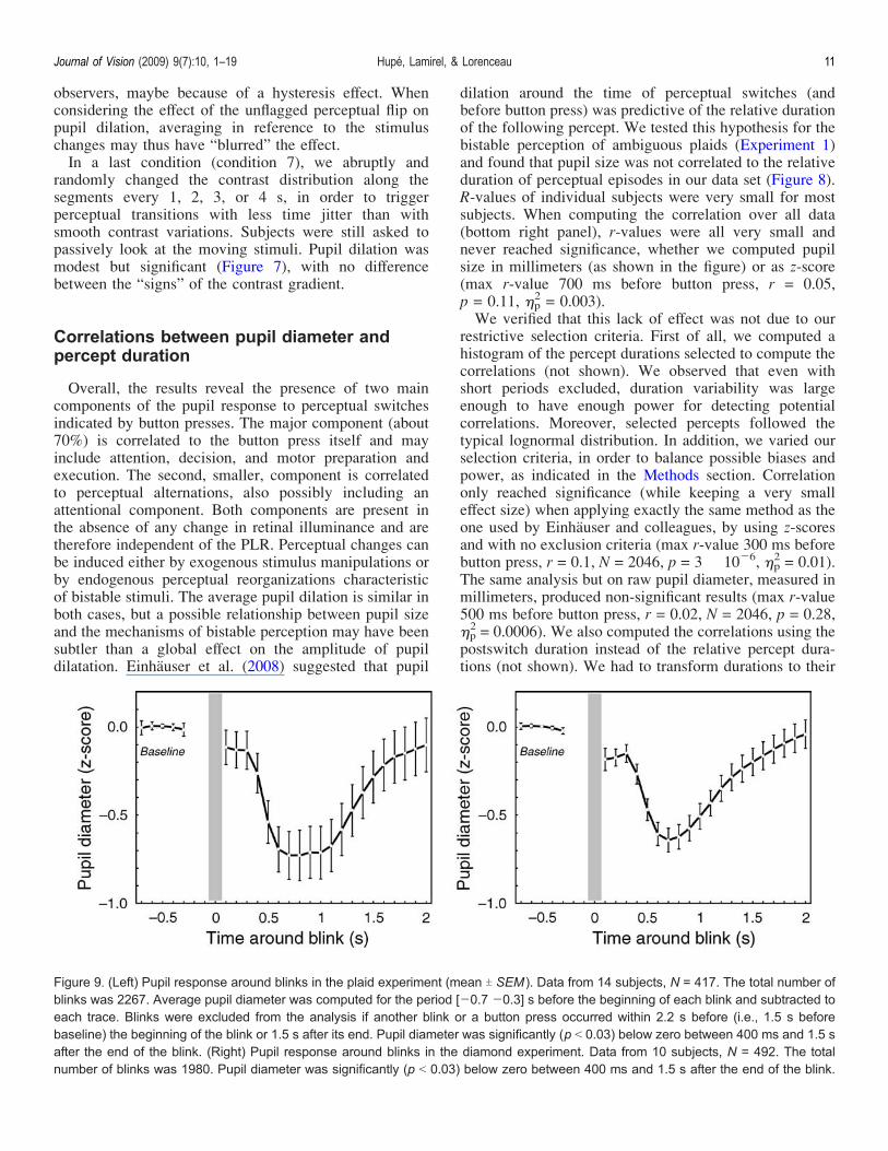
dilation around the time of perceptual switches (andbefore button press) was predictive of the relative durationof the following percept. We tested this hypothesis for thebistable perception of ambiguous plaids (Experiment 1)and found that pupil size was not correlated to the relativeduration of perceptual episodes in our data set (Figure 8).R-values of individual subjects were very small for mostsubjects. When computing the correlation over all data(bottom right panel), r-values were all very small andnever reached significance, whether we computed pupilsize in millimeters (as shown in the figure) or as z-score(max r-value 700 ms before button press, r = 0.05,p = 0.11, )p
2 = 0.003).We verified that this lack of effect was not due to our
restrictive selection criteria. First of all, we computed ahistogram of the percept durations selected to compute thecorrelations (not shown). We observed that even withshort periods excluded, duration variability was largeenough to have enough power for detecting potentialcorrelations. Moreover, selected percepts followed thetypical lognormal distribution. In addition, we varied ourselection criteria, in order to balance possible biases andpower, as indicated in the Methods section. Correlationonly reached significance (while keeping a very smalleffect size) when applying exactly the same method as theone used by Einhauser and colleagues, by using z-scoresand with no exclusion criteria (max r-value 300 ms beforebutton press, r = 0.1, N = 2046, p = 3 � 10j6, )p
2 = 0.01).The same analysis but on raw pupil diameter, measured inmillimeters, produced non-significant results (max r-value500 ms before button press, r = 0.02, N = 2046, p = 0.28,)p2 = 0.0006). We also computed the correlations using the
postswitch duration instead of the relative percept dura-tions (not shown). We had to transform durations to their
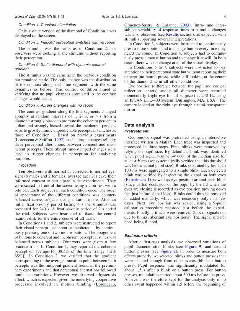
Figure 9. (Left) Pupil response around blinks in the plaid experiment (mean T SEM ). Data from 14 subjects, N = 417. The total number ofblinks was 2267. Average pupil diameter was computed for the period [j0.7 j0.3] s before the beginning of each blink and subtracted toeach trace. Blinks were excluded from the analysis if another blink or a button press occurred within 2.2 s before (i.e., 1.5 s beforebaseline) the beginning of the blink or 1.5 s after its end. Pupil diameter was significantly (p G 0.03) below zero between 400 ms and 1.5 safter the end of the blink. (Right) Pupil response around blinks in the diamond experiment. Data from 10 subjects, N = 492. The totalnumber of blinks was 1980. Pupil diameter was significantly (p G 0.03) below zero between 400 ms and 1.5 s after the end of the blink.
Journal of Vision (2009) 9(7):10, 1–19 Hupé, Lamirel, & Lorenceau 11

log in order to compute sensible statistics. The resultswere similar (r-value 700 ms before button press, r = 0.04,p = 0.20, )p
2 = 0.02; correlation computed over z-scores,with our exclusion criteria).
Pupil response to blinks
The time course of pupil size around blinks is displayedin Figure 9. There is a clear miosis after blinks in both theplaid (maximum effect 700 ms after blinking, )p
2 = 0.64)and diamond experiments (maximum effect 600 ms afterblinking, )p
2 = 0.68). This constriction was significant for12 out of the 14 subjects in the plaid experiment and 9 outof the 10 subjects in the diamond experiment. Miosisstarted about 350 ms after the end of a blink and lasted onaverage for more than 1 s. It was sometimes clearlyvisible on individual traces. Since blinks interrupt thevisual input, reopening the eye might be equivalent to aflash of light. The time course of miosis after a blink issimilar to the time course of the PLR, as measured in ourexperiments (see also Loewenfeld, 1999). Blink-inducedmiosis is, however, not exactly equivalent to the PLR,since there was no indication of pupil dilation during theblink, which could have been expected if a blink wasequivalent to switching off the light: Pupil size was, onaverage, even slightly smaller just after a blink, and therewas no positive correlation between blink duration andpupil size just after the blink (not shown). Second, therewas also no correlation between blink duration and miosispeak amplitude (not shown).
Discussion
We recorded pupil size while subjects continuouslyreported their bistable percept of ambiguous movingstimuli. We observed reliable pupil dilation around thebutton presses indicating the percepts. Pupil dilationstarted 300 to 400 ms before a button press and lastedafter it. In an attempt to dissect the origin of this pupildilation, we ran several control experiments whereobservers either responded toVexogenous or endoge-nousVperceptual transitions, passively viewed the samestimuli, performed motor responses to a sound or at will.In addition, we verified that changing the contrast gradientalong segments in the “diamond” experiment did notentail pupil changes by itself. From these control experi-ments, we could infer that both perceptual changes andmotor responses contributed to pupil dilation. However,the correlation found between changes in pupil diameterand perceptual transitions does not necessarily mean thatbistability is governed by non-visualVe.g., subcortical orfrontalVstructures or indicate that a non-visual generatorof bistability is involved, because the magnitude of pupil
dilation was similar for spontaneous perceptual alterna-tions and those triggered by physical manipulations of thestimulus. We also observed a clear constriction of thepupil after blinks in our data set, followed by a slow returnto baseline.
Components of the pupil response
Pupil response to motor report of perceptual changeswas, on average, 2.3% of pupil diameter, corresponding to5% of pupil maximal dilation (Figure 2). Pupil changesaround button presses were clearly visible on some but notall individual traces. Pupil dilation was also present whensubjects had only to press a mouse button, either after asound or at will. The amplitude of pupil dilation was,however, smaller than when button presses followed aperceptual change. By comparing both responses, wecould estimate that the motor response per se (preparationand motor command) accounted for about 70% of thepupil dilation due to the reports of perceptual switches.The difference was significant (Figure 4) and reasonablyconsistent between subjects (Figure 5). Could this differ-ence reflect a different attentional load in the test andcontrol conditions rather than a perceptual influence onpupil size? The task may indeed have been slightly moredemanding in the test condition with ambiguous stimuli,when subjects had to pay attention to unpredictablechanges of their perceptual states. Control conditionswere easy indeed. In one of them, subjects only had topress the mouse button at will. In another one, they had toswitch button after each brief sound, occurring every 4 s.However, the task was also easy when percept changeswere induced by smooth and periodic manipulations of thediamond stimulus. However, pupil dilation was notsignificantly smaller in that condition compared to thespontaneous condition (Figure 4), especially when con-sidering the onset and peak response. It is thereforeunlikely that difficulty or attentional load alone canaccount for the observed amplitude difference of pupildilation.More direct evidence of a perceptual component to
pupil responses was obtained in the conditions wheresubjects passively looked at the stimulus. Percept changesper se, induced by physical changes of the stimulus,caused a small dilation of the pupil. We observed thisresponse in two independent conditions, where perceptualchanges were induced either by smooth and periodic orabrupt and random changes of the stimulus. Although thecorresponding pupil responses were weak when consid-ered independently, combining both data sets of Figures 6(middle panel) and 7, aligning the peak responses (so timezero of the smoothed changes was aligned to time 1 s ofthe abrupt changes), maximum dilation was 0.1 (z-score)and p = 0.005 (F(1, 695) = 10.8, )p
2 = 0.43). In addition,pupil response was similar for switches to coherency andswitches to incoherency.
Journal of Vision (2009) 9(7):10, 1–19 Hupé, Lamirel, & Lorenceau 12
An important complementary result was the lack of pupildilation for the static condition (right panel of Figure 6),which ascertains that the local contrast change used toinduce perceptual switches did not trigger any pupilresponse by itself. Thus, the absence of pupil modulationindicates that pupil does not reflect any perceptual changebut rather shows some degree of specificity.Altogether, these results corroborate previous reports
that changes in pupil size are coupled to perceptualchanges and can signal visual cortical processes (seebelow). Pupil dilation is, however, small and cannot, atleast in our hands, be measured on a trial-by-trial basis toinfer a perceptual state without an objective report fromthe observer. Thus although pupil dynamics is a physio-logical signal related to perception, using it to read-outperceptual states appears limited.
Perceptual bistability and pupil response
Since pupil dilation follows specific and not anyperceptual changes, an exciting possibility was that pupildilation reflected the dynamics of neural events leading topercept changes in the absence of any stimulus modifica-tion. In other words, endogenous perceptual reorganiza-tion leading to bistable perception could reflect in thepupil. Unfortunately, this was not the case, since pupildilation was similar for spontaneous changes of a constantambiguous stimulus and for perceptual transitions inducedby physical modifications of the stimulus (Figure 4, left).Our observation corroborates the result of Einhauser et al.(2008), who had run a “replay” condition for twoambiguous displays (plaid and structure from motion),where induced transitions followed the time course ofspontaneous percept switches recorded in a previouscondition. They found that pupil modulation was verysimilar for the spontaneous and induced switches (seetheir Figure 2C).However, there could have been more information in
the pupil signal than its average amplitude. Einhauseret al. (2008) had the original idea to test whether pupil sizewas correlated to the duration of perceptual episodes.They found the relative amount of dilation before switch-ing being a weak but significant predictor of the durationof the subsequent percept. Noticing that pupil dilation iscorrelated to levels of norepinephrine (NE) released fromthe locus coeruleus (LC; Aston-Jones & Cohen, 2005),they suggested that NE could play a critical role inperceptual bistability. However, although we performedsimilar analyses using a larger data set, we were not ableto replicate this finding. We already indicated in acommentary one possible reason that may explain thediscrepant results between both studies (Hupe, Lamirel, &Lorenceau, 2008). In light of the data presented here (aswell as the many additional analyses not shown hereVseeMethods section), we emphasize now what we think arethe major controversial issues.
1. First of all, the correlation measured by Einhauseret al. (2008) was very weak (max r = 0.13, so r2 =0.017; value obtained by pooling the data of sixsubjects obtained in four different experiments).Such a weak result was therefore in demand ofconfirmation. With 14 subjects, our correlationvalues never reached above 0.1 (r2 = 0.01), evenwhen applying possibly biased methods (see below).
2. Einhauser et al. (2008) plotted p-values over timeand not r-values, without indicating the number ofvalues used to compute the correlation at each timepoint. They obtained a peak of significance beforethe button press, very suggestive of an interesting(even though weak) phenomenon. However, thispeak may reflect only the greater number of valuesused for these time points. Indeed, to avoid using thesame data point multiple times, they truncated eachtrace at the midpoints between switches. However,they used all their data points to compute thecorrelations. Since percept durations are variable,following a lognormal distribution (many shortperiods and a few very long periods), there weremore data points around the time of switchcompared to a few seconds before or after theswitch. Applying exactly the same procedure on ourdata set, we did observe a peak of p-value aroundswitch time, but this was only due to the greaternumber of values; r-values were positive and ratherconstant (around 0.1; not shown). In our analyses,we were careful to use the same number of valuesfor each time point.
3. Measures of duration and pupil size are in fact notindependent, mainly because of the slow dynamicsof pupil dilation that encompass short perceptualepisodes (Hupe, Lamirel et al., 2008). For perceptsshorter than 3 s, switch-related pupil dilation couldlast until the next switch. In our data set, given theoverall distribution of percept durations, the nextpercept lasted on average longer, which may be thecause of the positive correlation between pupil sizeand relative duration. Indeed, we observed that thecorrelation could be significant only when includingshort percepts (note however that the correlationwas weak and even not significant if using raw datainstead of z-scores). In their reply to our letter,Einhauser and colleagues indicated that they did notobserve that percepts tended to be longer after shortpercepts (G3 s). In fact, such a relation is expectedonly when one percept is more likely than the other,but the opposite relationship holds for variableswitch rates. On a whole set of data, both relation-ships may exist. Einhauser and colleagues did nottry to exclude short percepts.
In their reply to our letter, Einhauser et al. argue thatif their correlation was an artifact due to short perceptdurations, they should have observed it also in their
Journal of Vision (2009) 9(7):10, 1–19 Hupé, Lamirel, & Lorenceau 13

“replay” condition. However, this is true only in a purelydeterministic model, that is if each percept transitiontriggered a pupil response, or if the correlation wasstrong. In fact, pupil dilation is highly variable and theeffect is robust only when averaging many trials. Theirobservation that pupil dilation is on average similar inthe rivalry and replay condition of their Experiment 2(see their Figure 2) does not mean that each transitioncontributed equally to the mean response in bothconditions.
Pupil response to motor and to perceptualprocesses
One major but intriguing finding of the present study isthe observation of a reliable pupil dilation correlated to amotor response, when, for example, observers had to pressa key at will (Figure 4, green curve). This resultcorroborates a result by Simpson (1969) who observedenhanced pupil dilation for subjects who had to press akey to fulfill a pitch discrimination task, as well as pupildilation for sound-induced keypresses irrelevant to thetask. Since many experiments in cognitive neuroscienceinclude a motor response, pupil dilation that may in turnentail a visual response (see below) needs to be stressed,at least for methodological if not theoretical purposes. Ithas long been known that pupil dilation is related toarousal. “In alert individuals, excitatory impulses arisefrom the cerebral cortex and travel through the reticularactivating system and hypothalamus to cause pupildilation” (Merritt et al., 2004). Loewenfeld (1999) notesthat the reticular system in the brainstem receivesconnections from afferent sensory fibers as well as efferentmotor fibers, accounting for pupillary dilation observed toabout any sensory stimulation. The locus coeruleus (LC)is probably a key structure in this network, since it isinvolved in the regulation of sleep and arousal, exhibits anactivity well correlated with decision processes and motorresponses (Aston-Jones & Cohen, 2005), and projects tothe region of the Edinger–Westphal nucleus (EW) drivingthe pupil (at least in cat: Breen et al., 1983). The LC couldtherefore be involved in pupil dilation related to manualmotor responses.The enhanced pupil response during reports of bistable
perceptual changes as compared to a simple motorresponse further raises the possibility that cortical pro-cessing also influences pupil size (see above, “Compo-nents of the pupil response”), if one considers that bistableperception specifically involves cortical mechanisms.Hupe, Joffo, and Pressnitzer (2008) have defended, withothers, the idea that perceptual bistability is generated atthe level where the competing percepts are encoded, thatis, the visual cortex for plaids or ambiguous diamonds(Fang, Kersten, & Murray, 2008; Lorenceau et al., 2008).In our experiments, the stimulus remained identical overtime, providing a constant input to retinal ganglion cells
and subcortical structures. The observation that the PON,for example, receives significant cortical (e.g., V1, V4, IT,LIP, FEF) input, admittedly in addition to ventral thalamic(e.g., LGN), and midbrain (e.g., superior colliculus) input(Gamlin, 2006) provides evidence that an influence onpupil size originating in the visual cortex is possible. Oneissue is to uncover the nature of this potential influence,namely whether it induces a dilation, as observed here forbistable stimuli, or a constriction of the pupil as observedin other studies detailed below.Pupil constriction is characteristically observed for the
pupil light reflex (PLR), which is due to an increase oflight. However, many stimulus changes made at constantillumination also trigger pupil constrictions and couldinvolve cortical processing (see Barbur, 2004, for areview). The PLR originates in melanopsin-containing,intrinsically photosensitive, retinal ganglion cells(ipRGC), a special class of RGC that do not project tothe LGN but to the PON (Guler et al., 2008). Melanopsin-based signals are slow and code the average illuminationof the retina, so fast pupil responses to changes of visualstimuli are triggered by rods and cones whose signalsconverge to ipRGCs before being sent to the PON.Therefore, any stimulus change that trigger a retinalresponse, and not only a luminance change, may triggerthe PLR. The strongest evidence in favor of a corticalcomponent to the PLR is in fact based on lesion studies.Sahraie and Barbur (1997) observed pupil constrictionwhen incoherent moving random dots were physicallyswitched to a coherent motion flow, and they observed areduction of this constriction for patient GY when thestimuli were presented in his blind hemifield. Barbur et al.(1999) observed that the PLR to isoluminant green (butcuriously, not red) stimuli was abolished in the blindhemifield of two patients with cortical lesions. Wilhelmet al. (2002) observed that patients with Parinaud’ssyndrome, who had no pupil response to light changes,still displayed pupil constriction to gratings and isolu-minant red stimuli. Since the PON of these patients wasprobably destroyed, this result argues strongly in favor ofa pupil pathway independent of the ipRGC–PON path-way. They proposed that pupil constriction for thesestimuli was due to cortical modulation of central inhibitionat the level of the EW nucleus. Conway et al. (2008)reached a similar conclusion after observing a pupilconstriction stronger for upright than for inverted imagesof faces. This effect was present only for human faces,not monkey faces, so they took it as the likely signatureof a cortical process within the infero-temporal “face”area.It is possible that neuronal activity within the visual
cortex entails either dilation or constriction, depending onthe mechanisms specifically involved. However, it is alsopossible that either the constrictions described above orthe dilation observed in our study does not originatewithin the visual cortex. In favor of the first hypothesis,Loewenfeld suggested that reduced pupillary light reflex
Journal of Vision (2009) 9(7):10, 1–19 Hupé, Lamirel, & Lorenceau 14

in patients with cortical lesions are accounted for byretrograde trans-synaptic degeneration following a supra-geniculate lesion, eventually causing infrageniculate fiberloss (Loewenfeld, 1999, p. 220). Accordingly, she notedthat deficits of the PLR always needed some time after thelesion in order to develop. Similarly, the effects of GYcortical lesion on pupil responses may indeed be due toneuronal degeneration within subcortical structures.Weiskrantz, Cowey, and Le Mare (1998) observed similar,specific, changes like decrease of spatial frequencysensitivity of the PLR for GY and after V1 lesions inmonkeys. In the latter case, “the left dorsal lateralgeniculate nucleus was uniformly and totally degeneratedthroughout”. As noted above, the PON receives inputsfrom many structures, including the LGN. The PON may,however, not be invoked to explain the pupil responsesobserved in Parinaud patients (Wilhelm et al., 2002), butthis does not exclude the possible involvement of othersubcortical mechanisms (rather than cortical ones), possi-bly related to those of the near reflex, which is preserved.Concerning the study by Conway et al. (2008), they didnot consider that faces undoubtedly have an emotionalcomponent, possibly involving early subcortical activity(Morris, Ohman, & Dolan, 1998). In addition, the strongerconstriction for upright faces could be equally understoodas a dilation component added to the constriction forinverted faces. This dilation component could be due to agreater (involuntary) effort to try to recognize the invertedface. The use of a constant visual input, as in our study,allowed us to better (if not totally) control for subcorticalmodifications. On the other hand, the fact that weobserved dilation and not constriction leaves open theissue of whether the perceptual processes within the visualcortex are the origin of the observed pupil dilation orwhether it is mediated by modulation of (possibly supra-modal) attentional processes or mental workload. More-over, several fMRI studies report a frontal activity duringbistable perception (e.g., Sterzer, Russ, Preibisch, &Kleinschmidt, 2002). The existing projections from thefrontal cortex to the hypothalamus driving the dilator ofthe pupil further suggests that frontal cortex activity mayreflect in the pupil. In light of all these considerations, thequestions of whether neuronal activity within the visualcortex reflects or not in the pupil and whether it may resultin dilation or constriction of the pupil (or both) are still atstake.
Pupil response to blinks
We were surprised to observe a large and systematicmiosis after blinks. We had to take these modulations intoaccount in order to exclude postblink periods for ouranalyses of pupil size (see Methods section). We are notaware that such precautions were taken in other studies, orthat such changes were even systematically observed.Fukuda, Stern, Brown, and Russo (2005) even observed
pupil dilation after blinks. However, Loewenfeld (1999,p. 402) observed that blinks were “followed by smallpupillary contractions and redilations”, whose time coursewas similar to the one we observed. Such modulationswere not observed when blinks happened in darkness,suggesting that this response is caused by the dark periodsof lid closure and is therefore similar to the “darknessreflex”, where retina gains sensitivity during short inter-ruptions of the light. With dim light conditions or shortdark pauses (less than 500 ms), dilation of the pupil duringthe short dark period could be absent while the contrac-tion–redilation response was still present (Lowenstein &Loewenfeld, 1969, cited by Loewenfeld, 1999, p. 292),which may explain why we did not observe any dilation ofthe pupil just at the end of the blink. For the darknessreflex, however, the amplitude of the response depends onthe duration of the dark period (Stark, 1962, cited byLoewenfeld, 1999, p. 293). We did not observe such arelationship with blinks, probably because blink durationsdid not vary enough. Most blinks lasted less than 500 ms,a duration where retinal sensitivity measured for theamplitude of the darkness reflex is poorly modulated bydark duration (ibid.). It is surprising that Fukuda et al.(2005) did not observe any pupil constriction after blinks.Maybe the “darkness reflex” could be inhibited undercertain experimental conditions. It should be remindedthat the pupil is controlled through an antagonisticinfluence of the sympathetic and parasympathetic path-ways and that several modes of pupil control have beenconsidered as inhibition and excitation may interplay in acomplex non-linear way (Yamaji, Hirata, & Usui, 2000).Note that these antagonistic activities may stronglydepend upon the background and stimulus luminance,contrast or color, stimulus duration and characteristics,such that the outcome response may largely vary withthese parameters.
Consequences for imaging studies
The observation of long-lasting changes in pupildiameter (around 1.5 s in the present study), be they dueto perceptual changes, button presses, or blinks, suggestthat neural activity (see below) and visual processing(e.g., contrast sensitivity) may be affected by thesechanges. Although the changes in pupil diameter reportedherein are small (about 2% of pupil diameter), theycorrespond to non-negligible changes in pupil surface,thus changing the flux of light entering the eyes, as moreclearly demonstrated when considering pupil size mea-sured in millimeters. The average increase of pupildiameter was 0.14 mm (range of 0.01 to 0.33 mm),corresponding to an average increased surface of 1.2 mm2
(range of 0.06 to 4.6 mm2), or 5.4% (0.5% to 13.5%). Theaverage decrease of pupil diameter after blinks was0.19 mm (range of 0 to 0.43 mm), corresponding to an
Journal of Vision (2009) 9(7):10, 1–19 Hupé, Lamirel, & Lorenceau 15
average decreased surface of 1.3 mm2 (range of 0 to3.5 mm2), or 8.2% (0% to 15.1%).All these values are somehow larger than the 0.02-mm
average pupil dilation measured by Fukuda et al. (2005),which they relate to higher order processes, namely the“readiness to acquire new visual information”, or theabout 0.03 mm observed by Conway et al. (2008) forinverted vs. upright faces (see their Figure 2).Accordingly, a change in pupil diameter unrelated to a
change in a visual parameter, as those reported herein,entails a change in retinal illumination. Given the presentfindings, an intriguing possibility is that pupil dilationrelated to a cognitive or motor response elicits changingneuronal responses in the retina. This could be the case fora number of reasons: changing pupil size inducesvariations in the photon flux that could modulate thephotoreceptors activity, and possibly the activity of thecorresponding ganglion cells; opening or closing the pupilchanges the optical characteristics of the eye, such as thedepth field, which could also change the spatial frequencyrelated activity, by recruiting ganglion cells tuned todifferent spatial frequencies (Campbell & Gubish, 1966;Strang, Atchison, & Woods, 1999). The neuronalresponses elicited by these changes could in turn prop-agate through the thalamo-cortical pathways and modulatethe neuronal responses in striate and extrastriate areas. Iftrue, these would have two main consequences. One isthat such a pupil-related cortical activity might modulatevisual sensitivity in psychophysical detection tasks. Thesecond is that changes of pupil size provide a possiblesource of visual noise that could be taken into accountwhen analyzing fMRI, EEG, or MEG data, thus possiblyimproving the signal-to-noise ratio. Although this mayappear speculative at first sight, Siegle, Steinhauer,Stenger, Konecky, and Carter (2003) used pupil variationsas a regressor in their analysis of fMRI data, which didimprove the sensitivity of their analyses.We also speculate that the change of pupil diameter has
a stronger effect on the ganglion cells situated at theperiphery of the retina, since they would be rapidlycovered or uncovered by the iris. For these neurons,changes of pupil diameter correspond to ON/OFFresponses. These responses are not inhibited in any wayat the level of the retina, since they can trigger thepowerful “edge-light cycles” of pupil oscillations whenthe retina is illuminated only at the edge of the iris(Loewenfeld, 1999). Modulations of pupil diameter maytherefore trigger neuronal responses in the parts of thevisual cortex specifically coding the periphery of thevisual field. Since blinks are followed by a biphasic pupilresponse, measured neural responses to blinks might bedue in part to the change of pupil size, and not only to thebrief shutting off of the visual input (which is usually notconsciously detected, a phenomenon described as “blinksuppression”; see, e.g., Bristow, Frith, & Rees, 2005. Thefollowing references dealt with the mechanisms of eitherblink suppression or blinking control. We cite them here
within a different context). Blinks occurring in lightconditions trigger activity in the occipital cortex, includ-ing V1, as measured with fMRI (Bodis-Wollner, Bucher,& Seelos, 1999; Bristow et al., 2005; Kato & Miyauchi,2003; Yoon, Chung, Song, & Park, 2005), EEG (Berg &Davies, 1988), and MEG (Bardouille, Picton, & Ross,2006). Interestingly, activity is stronger in (or even limitedto) the anterior calcarine sulcus, where V1 codes theperiphery of the visual field (Stenbacka & Vanni, 2007a,2007b), even though subjects only had to maintain theirgaze over a fixation point (e.g., Bardouille et al., 2006;Bristow et al., 2005; Yoon et al., 2005). We observedsimilar blink-related BOLD activity in the retinotopicallydefined periphery of V1, although stimuli were presentedonly centrally (J.-M. Hupe & M. Dojat, unpublishedobservations). We are not aware that this specific activityin the periphery of V1 was either stressed out or explained(except by Simo Vanni, several years ago already:personal communication; see also Stenbacka & Vanni,2007b). The observed 200- to 300-ms latency of theseresponses (Bardouille et al., 2006; Berg & Davies, 1988)is compatible (though a bit too early) with the time courseof blink-induced pupil responses. A question at stake istherefore whether peripheral V1 activity related to blinks,when measured in light conditions, is only due to themechanisms of blink suppression, or whether it is alsorelated to blink-induced changes of pupil size.
Conclusions
The present work shows that pupil dilation accompanieschanges in perception with unchanging bistable stimuli.The several experiments ran to identify the origin of pupildilation suggests that it reflects different componentsrelated to both changes in perception and motor commands.The presence of a “true” perceptual component supportsthe hypothesis of an influence of activity within the visualcortex on pupil size. Although pupil dilation may not allowthe decoding of perceptual states on a trial-by-trial basis, itcould influence activity in the retino-geniculo-corticalpathways. Taking pupil activity into account may thusimprove the signal-to-noise ratio in psychophysical orimaging research. Is there a functional interpretation ofthe pupil response to perceptual transitions? Einhauseret al. (2008) suggest that pupil dilation reflects a consol-idation of a perceptual state. Additional studies addressingmore directly the individual long-term perceptual dynam-ics and its correlation to the pupillary responses seemnecessary before such a statement can be made.
Acknowledgments
The authors of this article do not wish to thank theFrench government for its current policy regarding public
Journal of Vision (2009) 9(7):10, 1–19 Hupé, Lamirel, & Lorenceau 16

research. CL was supported by “Fondation pour laRecherche Medicale,” “Fondation Berthe Fouassier,” and“Fondation de France.”
Commercial relationships: none.Corresponding authors: Jean-Michel Hupe and JeanLorenceau.Email: [email protected] and [email protected]: JMH: CerCo, Faculte de Medecine de Rangueil,31062 Toulouse Cedex 9, France. JL: Laboratoire deNeurosciences Cognitives and Imagerie Cerebrale, CNRSUPR640-LENA, 47 boulevard de l’hopital, 75651 ParisCedex 13, France.
References
Aston-Jones, G., & Cohen, J. D. (2005). An integrativetheory of locus coeruleus-norepinephrine function:Adaptive gain and optimal performance. AnnualReview of Neuroscience, 28, 403–450. [PubMed]
Barbur, J. L. (2004). Learning from the pupil-studies ofbasic mechanisms and clinical applications. In L. M.Chalupa & J. S. Werner (Eds.), The visual neuro-sciences (vol. 1, pp. 641–656). Cambridge, MA: MITPress.
Barbur, J. L., Weiskrantz, L., & Harlow, J. A. (1999). Theunseen color aftereffect of an unseen stimulus: Insightfrom blindsight into mechanisms of color afterim-ages. Proceedings of the National Academy ofSciences of the United States of America, 96,11637–11641. [PubMed] [Article]
Bardouille, T., Picton, T. W., & Ross, B. (2006).Correlates of eye blinking as determined by syntheticaperture magnetometry. Clinical Neurophysiology,117, 952–958. [PubMed]
Beatty, J. (1977). Activation and attention. In M. C.Wittrock, J. Beatty, J. E. Bogen, M. S. Gazzaniga,H. J. Jerrsion, S. D. Krashen, R. D. Nebes, & T. Teyler(Eds.), The human brain (pp. 63–85). EnglewoodCliffs, NJ: Prentice-Hall.
Beatty, J., & Wagoner, B. L. (1978). Pupillometric signsof brain activation vary with level of cognitiveprocessing. Science, 199, 1216–1218. [PubMed]
Berg, P., & Davies, M. B. (1988). Eyeblink-relatedpotentials. Electroencephalography and Clinical Neu-rophysiology, 69, 1–5. [PubMed]
Blake, R., & Logothetis, N. K. (2002). Visual competi-tion. Nature Reviews, Neuroscience, 3, 13–21.[PubMed]
Bodis-Wollner, I., Bucher, S. F., & Seelos, K. C. (1999).Cortical activation patterns during voluntary blinks
and voluntary saccades. Neurology, 53, 1800–1805.[PubMed]
Breen, L. A., Burde, R. M., & Loewy, A. D. (1983).Brainstem connections to the Edinger–Westphalnucleus of the cat: A retrograde tracer study. BrainResearch, 261, 303–306. [PubMed]
Bristow, D., Frith, C., & Rees, G. (2005). Two distinctneural effects of blinking on human visual processing.Neuroimage, 27, 136–145. [PubMed]
Camellin, M., Gambino, F., & Casaro, S. (2005).Measurement of the spatial shift of the pupil center.Journal of Cataract and Refractive Surgery, 31,1719–1721. [PubMed]
Campbell, F. W., & Gubisch, R. W. (1966). Opticalquality of the human eye. The Journal of Physiology,186, 558–578. [PubMed] [Article]
Clarke, R. J., Zhang, H., & Gamlin, P. D. (2003). Primatepupillary light reflex: Receptive field characteristicsof pretectal luminance neurons. Journal of Neuro-physiology, 89, 3168–3178. [PubMed] [Article]
Conway, C. A., Jones, B. C., DeBruine, L. M., Little, A. C.,& Sahraie, A. (2008). Transient pupil constrictions tofaces are sensitive to orientation and species. Journalof Vision, 8(3):17, 1–11, http://journalofvision.org/8/3/17/, doi:10.1167/8.3.17. [PubMed] [Article]
Einhauser, W., Stout, J., Koch, C., & Carter, O. (2008).Pupil dilation reflects perceptual selection and pre-dicts subsequent stability in perceptual rivalry. Pro-ceedings of the National Academy of Sciences of theUnited States of America, 105, 1704–1709. [PubMed][Article]
Fang, F., Kersten, D., & Murray, S. O. (2008). Perceptualgrouping and inverse fMRI activity patterns in humanvisual cortex. Journal of Vision, 8(7):2, 1–9, http://journalofvision.org/8/7/2/, doi:10.1167/8.7.2.[PubMed] [Article]
Fukuda, K., Stern, J. A., Brown, T. B., & Russo, M. B.(2005). Cognition, blinks, eye-movements, and pupil-lary movements during performance of a runningmemory task. Aviation, Space, and EnvironmentalMedicine, 76, C75–C85. [PubMed]
Gamlin, P. D. (2006). The pretectum: Connections andoculomotor-related roles. Progress in BrainResearch, 151, 379–405. [PubMed]
Guler, A. D., Ecker, J. L., Lall, G. S., Haq, S., Altimus,C. M., Liao, H. W., et al. (2008). Melanopsin cellsare the principal conduits for rod-cone input to non-image-forming vision. Nature, 453, 102–105.[PubMed]
Hupe, J.-M., James, A. C., Girard, P., Lomber, S. G.,Payne, B. R., & Bullier, J. (2001). Feedbackconnections act on the early part of the responses in
Journal of Vision (2009) 9(7):10, 1–19 Hupé, Lamirel, & Lorenceau 17

monkey visual cortex. Journal of Neurophysiology,85, 134–145. [PubMed] [Article]
Hupe, J.-M., Joffo, L. M., & Pressnitzer, D. (2008).Bistability for audiovisual stimuli: Perceptual deci-sion is modality specific. Journal of Vision, 8(7):1,1–15, http://journalofvision.org/8/7/1/, doi:10.1167/8.7.1. [PubMed] [Article]
Hupe, J.-M., Lamirel, C., & Lorenceau, J. (2008). Pupildilation does not predict subsequent stability inperceptual rivalry. Proceedings of the NationalAcademy of Sciences of the United States of America,105, E43. [PubMed] [Article]
Hupe, J.-M., & Rubin, N. (2003). The dynamics of bi-stable alternation in ambiguous motion displays: Afresh look at plaids. Vision Research, 43, 531–548.[PubMed]
Kahneman, D., & Beatty, J. (1966). Pupil diameter andload on memory. Science, 154, 1583–1585. [PubMed]
Kato, M., & Miyauchi, S. (2003). Functional MRI of brainactivation evoked by intentional eye blinking. Neuro-image, 18, 749–759. [PubMed]
Lamirel, C., Hupe, J.-M., & Lorenceau, J. (2008). Pupildynamics during bistable form/motion binding[Abstract]. Journal of Vision, 8(6):253, 253a, http://journalofvision.org/8/6/253/, doi:10.1167/8.6.253.
Loewenfeld, I. E. (1999). The pupil: Anatomy, physiol-ogy, and clinical applications. Boston: Butterworth-Heinemann.
Long, G. M., & Toppino, T. C. (2004). Enduring interest inperceptual ambiguity: Alternating views of reversiblefigures. Psychological Bulletin, 130, 748–768.[PubMed]
Lorenceau, J., & Alais, D. (2001). Form constraints inmotion binding. Nature Neuroscience, 4, 745–751.[PubMed]
Lorenceau, J., Gimenez-Sastre, B., & Lalanne, C. (2003).Hysteresis in perceptual binding. Perception, 32,ECVP Abstract Supplement.
Lorenceau, J., Paradis, A.-L., Lamirel, C., Poline, J.-B.,Artiges, E., Thirion, B., et al. (2008). Corticaldynamics of bistable form/motion binding: fMRIand eye movements [Abstract]. Journal of Vision,8(6):1105, 1105a, http://journalofvision.org/8/6/1105/,doi:10.1167/8.6.1105.
Lorenceau, J., & Shiffrar, M. (1992). The influence ofterminators on motion integration across space.Vision Research, 32, 263–273. [PubMed]
Lowenstein, O., & Loewenfeld, I. E. (1969). Physiology ofthe eye (2nd ed., vol. 3). New York: Academic Press.
Merritt, S. L., Schnyders, H. C., Patel, M., Basner, R. C.,& O’Neill, W. (2004). Pupil staging and EEGmeasurement of sleepiness. International Journal ofPsychophysiology, 52, 97–112. [PubMed]
Morris, J. S., Ohman, A., & Dolan, R. J. (1998). Consciousand unconscious emotional learning in the humanamygdala. Nature, 393, 467–470. [PubMed]
Partala, T., & Surakka, V. (2003). Pupil size variation asan indication of affective processing. InternationalJournal of Human Computer Studies, 59, 185–198.
Reinhard, G., & Lachnit, H. (2002). The effect of stimulusprobability on pupillary response as an indicator ofcognitive processing in human learning and catego-rization. Biological Psychology, 60, 199–215.[PubMed]
Rubin, N., & Hupe, J.-M. (2005). Dynamics of perceptualbi-stability: Plaids and binocular rivalry compared. InA. Alais & R. Blake (Eds.), Binocular rivalry.Cambridge, MA: MIT Press.
Sahraie, A., & Barbur, J. L. (1997). Pupil responsetriggered by the onset of coherent motion. Graefe’sArchive for Clinical and Experimental Ophthalmol-ogy, 235, 494–500. [PubMed]
Siegle, G. J., Steinhauer, S. R., Stenger, V. A., Konecky, R.,& Carter, C. S. (2003). Use of concurrent pupildilation assessment to inform interpretation andanalysis of fMRI data. Neuroimage, 20, 114–124.[PubMed]
Simpson, H. M. (1969). Effects of a task-relevant responseon pupil size. Psychophysiology, 6, 115–121.[PubMed]
Stark, L. (1962). Biological rhythms, noise, and asymme-try in the pupil-retinal control system. Annals of theNew York Academy of Sciences, 98, 1096–1108.[PubMed]
Stenbacka, L., & Vanni, S. (2007a). Central luminanceflicker can activate peripheral retinotopic representa-tion. Neuroimage, 34, 342–348. [PubMed]
Stenbacka, L., & Vanni, S. (2007b). fMRI of peripheralvisual field representation. Clinical Neurophysiology,118, 1303–1314. [PubMed]
Sterzer, P., Russ, M. O., Preibisch, C., & Kleinschmidt, A.(2002). Neural correlates of spontaneous directionreversals in ambiguous apparent visual motion.Neuroimage, 15, 908–916. [PubMed]
Strang, N. C., Atchison, D. A., & Woods, R. L. (1999).Effects of defocus and pupil size on human contrastsensitivity. Ophthalmic & Physiological Optics, 19,415–426. [PubMed]
Szabadi, E., & Bradshaw, C. M. (1996). Autonomicpharmacology of !2-adrenoceptors. Journal of Psy-chopharmacology, 10, 6–18.
Wallach, H. (1935). Uber visuell wahrgenommeneBewegungsrichtung. Psychologische Forschung, 20,325–380.
Journal of Vision (2009) 9(7):10, 1–19 Hupé, Lamirel, & Lorenceau 18
Weiskrantz, L., Cowey, A., & Le Mare, C. (1998).Learning from the pupil: A spatial visual channel inthe absence of V1 in monkey and human. Brain, 121,1065–1072. [PubMed]
Wilhelm, B. J., Wilhelm, H., Moro, S., & Barbur, J. L.(2002). Pupil response components: Studies in patientswith Parinaud’s syndrome. Brain, 125, 2296–2307.[PubMed]
Wuerger, S., Shapley, R., & Rubin, N. (1996). “On thevisually perceived direction of motion” by HansWallach: 60 years later. Perception, 25, 1317–1318.
Wyatt, H. J. (1995). The form of the human pupil. VisionResearch, 35, 2021–2136. [PubMed]
Yamaji, K., Hirata, Y., & Usui, S. (2000). A method formonitoring autonomic nervous activity by pupillaryflash response. Systems and Computers in Japan, 31,22–31.
Yang, Y., Thompson, K., & Burn, S. A. (2002). Pupillocation under mesopic, photopic, and pharmacolog-ically dilated conditions. Investigative Ophthalmology& Visual Science, 43, 2508–2512. [PubMed] [Article]
Yoon, H. W., Chung, J. Y., Song, M. S., & Park, H.(2005). Neural correlates of eye blinking; improvedby simultaneous fMRI and EOG measurement.Neuroscience Letters, 381, 26–30. [PubMed]
Journal of Vision (2009) 9(7):10, 1–19 Hupé, Lamirel, & Lorenceau 19
Related Documents