Pulpal irritants LINDA G. LEVIN The dental pulp is characterized as a connective tissue and as such it is not considered an external tissue, yet its exposure to external stimuli is constant. This is due to a number of factors including the permeability of attrited or disrupted enamel as well as that of physiologic dentin and cementum. The pulp is extraordinarily sensitive to its external environment. Once thought to be a vestigial organ, it is now understood that the dental pulp is an important tissue whose role in the defense of the dentition may be as significant as its role in odontogenesis. A variety of stimuli have been demonstrated to have an effect on the pulp. The reactions of the dental pulp to respective irritants are largely dictated by the character and duration of a stimulus. The resultant reaction is manifested by a continuum of disease bracketed by the normal pulp and the necrotic pulp and centrally composed of a gradient of inflammation that pro- gresses clinically from reversible to irreversible (1). Pulpal irritants have been classified as mechanical, thermal, chemical and infective. While the latter has historically been discounted, modern science has proven it to be the most significant cause of pulpal morbidity and mortality. That said, iatrogenic irritants deserve attention particularly in view of the fact that they are most under the control of the clinician. Mechanical irritants to the dental pulp Mechanical irritants to the dental pulp can be broken down into two categories, mechanical and biomecha- nical. Mechanical irritants include orthodontic move- ment and tooth preparation while biomechanical irritation results from functional and parafunctional forces placed on the dentition during mastication, clenching or bruxing. Mechanical irritants: operative procedures Numerous classic studies have confirmed the effects of cavity preparation on the dental pulp (2–6). A consistent finding of these early studies was that uncontrolled and extreme temperature changes pro- duced during operative procedures were detrimental to the pulp (7) (Fig. 1). One animal study documented that a temperature rise of 5.51C resulted in necrosis in 15% of monkey pulps (8). Several factors seem to contribute to excessive intraoperative heat generation, all of which can be controlled by the operator. Light pressure, sharp burs, high rotational speeds with proper water coolant and short intermittent cutting strokes prove to be the least irritating to the dental pulp (2). The inherent insulating capacity of dentin is sufficient to protect the subjacent pulp tissue during restorative procedures unless the remaining dentin thickness (RDT) is small and ineffective water coolant is employed (8). Under these conditions the most consistent histological manifestation of pulpal trauma is the displacement of odontoblasts into the tubules (7) (Fig. 2). There are several theories as to the mechanism of this response. The normal tissue pressure of the dental pulp is between 5 and 20 mmHg, however, subsequent to cutting of dentin, it can exceed 60 mmHg in localized inflamed areas (9, 10). High tissue pressures beneath newly exposed dentinal tubules promote an outward fluid flow that may carry the odontoblast cell body with it into the tubule. While some have argued that the increased tissue pressure is due to inflammation, a more likely cause is a concomitant rise in pulpal blood flow in response to thermal stimulation (11). An alternate theory is that during dry tooth preparation the accompanying desiccation of dentin produces an outward fluid flow 2 Endodontic Topics 2003, 5, 2–11 Printed in Denmark. All rights reserved Copyright r Blackwell Munksgaard ENDODONTIC TOPICS 2003

Welcome message from author
This document is posted to help you gain knowledge. Please leave a comment to let me know what you think about it! Share it to your friends and learn new things together.
Transcript

Pulpal irritantsLINDA G. LEVIN
The dental pulp is characterized as a connective tissue and as such it is not considered an external tissue, yet its
exposure to external stimuli is constant. This is due to a number of factors including the permeability of attrited or
disrupted enamel as well as that of physiologic dentin and cementum. The pulp is extraordinarily sensitive to its
external environment. Once thought to be a vestigial organ, it is now understood that the dental pulp is an
important tissue whose role in the defense of the dentition may be as significant as its role in odontogenesis.
A variety of stimuli have been demonstrated to have an
effect on the pulp. The reactions of the dental pulp to
respective irritants are largely dictated by the character
and duration of a stimulus. The resultant reaction is
manifested by a continuum of disease bracketed by the
normal pulp and the necrotic pulp and centrally
composed of a gradient of inflammation that pro-
gresses clinically from reversible to irreversible (1).
Pulpal irritants have been classified as mechanical,
thermal, chemical and infective. While the latter has
historically been discounted, modern science has
proven it to be the most significant cause of pulpal
morbidity and mortality. That said, iatrogenic irritants
deserve attention particularly in view of the fact that
they are most under the control of the clinician.
Mechanical irritants to the dentalpulp
Mechanical irritants to the dental pulp can be broken
down into two categories, mechanical and biomecha-
nical. Mechanical irritants include orthodontic move-
ment and tooth preparation while biomechanical
irritation results from functional and parafunctional
forces placed on the dentition during mastication,
clenching or bruxing.
Mechanical irritants: operative procedures
Numerous classic studies have confirmed the effects of
cavity preparation on the dental pulp (2–6). A
consistent finding of these early studies was that
uncontrolled and extreme temperature changes pro-
duced during operative procedures were detrimental to
the pulp (7) (Fig. 1). One animal study documented
that a temperature rise of 5.51C resulted in necrosis in
15% of monkey pulps (8). Several factors seem to
contribute to excessive intraoperative heat generation,
all of which can be controlled by the operator. Light
pressure, sharp burs, high rotational speeds with proper
water coolant and short intermittent cutting strokes
prove to be the least irritating to the dental pulp (2).
The inherent insulating capacity of dentin is sufficient
to protect the subjacent pulp tissue during restorative
procedures unless the remaining dentin thickness
(RDT) is small and ineffective water coolant is
employed (8). Under these conditions the most
consistent histological manifestation of pulpal trauma
is the displacement of odontoblasts into the tubules (7)
(Fig. 2). There are several theories as to the mechanism
of this response. The normal tissue pressure of the
dental pulp is between 5 and 20mmHg, however,
subsequent to cutting of dentin, it can exceed
60mmHg in localized inflamed areas (9, 10). High
tissue pressures beneath newly exposed dentinal
tubules promote an outward fluid flow that may carry
the odontoblast cell body with it into the tubule. While
some have argued that the increased tissue pressure is
due to inflammation, a more likely cause is a
concomitant rise in pulpal blood flow in response to
thermal stimulation (11). An alternate theory is that
during dry tooth preparation the accompanying
desiccation of dentin produces an outward fluid flow
2
Endodontic Topics 2003, 5, 2–11Printed in Denmark. All rights reserved
Copyrightr Blackwell Munksgaard
ENDODONTIC TOPICS 2003

of a magnitude that will force the cellular components
into the tubules. While the mechanism is debated, the
net result of odontoblast displacement is a disruption of
the cell layer that resolves after 20 days (6). During this
interval tertiary dentin must be produced by progeni-
tor cells that are initially more fibroblastic in nature and
produce less organized fibrodentin (12). Alternately
some tubules are not repopulated and become dead
tracts (13). Both outcomes produce a more permeable
dentin that can facilitate continued or future insult to
the subjacent pulp.
Although odontoblast displacement is rarely seen
with atraumatic technique, deep preparations using
appropriate safeguards can still induce cell damage by
transecting odontoblast processes. The extent of the
cytoplasmic process of odontoblasts is still under
debate but there is general agreement that at least the
inner one-third of dentin is occupied by these
cytoplasmic extensions (14). Once transected during
the cutting of dentin, the fate of the odontoblast is
variable depending on the proximity to the cell body. If
the odontoblast is destroyed the potential sequellae are
the same as for displacement.
Recent studies have reported that deep cavity
preparation not only affects the underlying odonto-
blasts but also induces an accumulation of HLA-DR-
positive cells and protein gene product 9.5-positive
nerve fibers (15). This effect appears to be inversely
proportional to RDT. A 50% reduction in RDT
changed the distribution of HLA-DR-positive dendri-
tic cells in human teeth. A two-thirds decrease in RDT
in non-carious teeth stimulated an influx of increased
numbers of HLA-DR-positive cells that displaced
odontoblasts and extended into the dentinal matrix
and associated dentinal tubules beneath the cavities.
The odontoblast displacement resolved 2 months
postoperatively and dentin sialoprotein (DSP)-positive
cells lined the dentin indicating that newly differen-
tiated odontoblasts had repopulated the region. Inter-
estingly this sequence was not observed under
preparations in carious teeth. Despite the presence of
increased numbers of HLA-positive dendritic cells
under carious dentin, odontoblast displacement was
not observed. Furthermore, subsequent to caries
excavation and restoration, small aggregates of HLA-
DR-positive cells, neuronal components and CD45-
positive T-lymphocytes persisted implying continued
irritation of the subjacent pulp.
It is important to consider that animal studies on the
effects of restorative procedures on the dental pulp
report the effects on healthy pulps. The clinical reality
in dentistry is that extensive restorative procedures
are frequently performed on teeth with histories of
Fig. 1. Excessive heat generated during crown preparation is responsible for the phenomenon referred to as ‘blushing’.The color change in the dentin (a) is due to vascular stasis and hemorrhage in the subodontoblastic vascular plexus (b).
Fig. 2. The arrows depict aspirated odontoblasts in thedentinal tubules. Aspiration results in cell death andnecessitates the differentiation of new replacementodontoblasts.
Levin
3

repeated cycles of disease and intervention that leave them
compromised.Hence the need tominimize themorbidity
of restorative procedures is even greater and the applica-
tion of our understanding of the pulpal reaction to dentin
manipulation must be even more judicious.
Mechanical irritants: orthodonticmovement
The most conspicuous pulpal change observed in
response to orthodontic forces is hemodynamic. Both
human and animal studies have confirmed that both
lateral and intrusive forces result in an increase in pulpal
blood flow (16–18). Furthermore, blood flow altera-
tions are not confined to the tooth in active movement.
Observed increases in blood flow are seen in teeth
adjacent to the focus of movement forces implying that
directed forces on one tooth can shunt blood to
proximal vessels supplying other oral structures includ-
ing teeth. If orthodontic forces are extreme, circulatory
interruptions can occur resulting in pulpal necrosis (19).
Biomechanical irritation: parafunctionalhabits
Occlusal loading of teeth effects deformation to
varying degrees (20). While enamel is largely resistant
to flexure the underlying dentin demonstrates con-
siderable elastic characteristics. As a result, defects in
enamel secondary to cracks, decay and/or restorative
preparation allow cuspal flexure with subsequent pulpal
responses. These responses are mediated by induced
dentinal fluid flow.
Multiple factors influence the degree of tooth
deformation during occlusal loading. Investigators
have noted that preparation geometry has a direct
impact on cuspal flexure. The width of the occlusal
isthmus relative to the faciolingual dimension of the
tooth as well as the ablation of marginal ridges directly
impact on the degree of cuspal flexure (21–23). MOD
preparations have been shown to effect a 50% reduction
in cuspal stiffness and resistance to fracture. Physical
properties of the restorative material can also play a part
in cuspal flexure. Studies have shown that polymeriza-
tion shrinkage of certain resin composites can induce an
inward deflection of cusps with resultant stresses on
tooth structure (24).
Symptomology from cuspal flexure can result from
two primary sources. It has been theorized that cuspal
flexure results in dentin deformation thus promoting
dentinal fluid flow that activates nerve endings in the
odontoblast layer of the tooth. This is supported in part
by an in vitro study that found that dentinal fluid flow
could be induced by occlusal loading of restored teeth
(25). A second source of pulpal pain is bacterial
microleakage created by a gap at the restoration/
dentin interface that is repeatedly opened during cycles
of occlusal loading. If repeated cuspal flexure gives rise
to a crack, dentinal exposure to bacteria and their by-
products is even greater.
Dentinal cracks expose tubules unoccluded by smear
layer and, therefore, offer a direct portal to the
subjacent pulp. When dentinal tubules are freely
exposed there is an outward flow of dentinal fluid
driven by relatively high pulpal tissue pressures.
Dentinal fluid is composed of proteins such as
fibrinogen and serum albumin that can coagulate and
effectively block the tubule lumen thereby limiting
fluid egress and resultant dentin hypersensitivity. This
phenomenon can occur within 2 days. As this serves as a
short-term protective mechanism for the pulp, dentinal
sclerosis and tertiary dentin formation ultimately can
provide greater protection for the pulp and ablation of
symptoms.
Chemical irritants to the dental pulp
The effects of restorative materials on the dental pulp
have been investigated and seem to relate directly to the
permeability of the associated dentin. The degree of
dentin permeability, however, is often variable and is
governed by several factors including age and caries
status (26). Perhaps the most important variable in
dentin permeability is the thickness of dentin between
the floor of the cavity preparation and the pulp (27).
Unbound components of resin materials and pre-
parative agents such as acid etchants can affect the
subjacent pulp by inducing an inflammatory response
(28–30). (Fig. 3) This is mediated by the indirect effects
of desiccation and/or demineralization of dentin as well
as direct effects of thematerial itself when in contact with
pulpal tissue. Studies have shown that the certain
cytotoxic components of resin monomers (triethylene
glycol dimethacrylate and 2-hydroxyethyl methacrylate)
readily penetrate dentin (31). Similarly, eugenol and
components (triamcinolone and demeclocycline) of
Ledermixs (Wyeth-PharmaGmbH,Munster, Germany)
have been shown to pass through dentin into the
Pulpal irritants
4

subjacent pulp (32, 33). In vivo data show that these
chemicals have an effect on the pulp, however, the
effect seems to be short lived and in the absence of
bacteria, reversible (34).
The mechanisms whereby restorative materials exert
an injurious effect on the dental pulp are varied.
Evidence exists that supports direct and in some
instances, prolonged cytotoxicity, stimulation of hy-
persensitivity reactions or impairment of the host
immune response to bacteria. Some of the components
of resin restorations are released at cytotoxic levels after
polymerization is completed leading to chronic stimu-
lation and a resultant prolonged inflammatory response
(35). Furthermore, even subtoxic concentrations of
certain agents are capable of eliciting allergic reactions
in humans (36). Primates hyperimmunized with BSA
showed significant pulpal damage with repeated anti-
genic challenge in class V cavity preparations suggest-
ing a role for antigen–antibody complex mediated
hypersensitivity in tissue destruction (37). In a separate
study, exposure to dentin primers elicited a delayed-
type hypersensitivity reaction in guinea-pigs (38).
These studies taken together present a compelling
argument for immune-mediated pulpal tissue damage
subsequent to exposure to restorative materials. For-
eign body reactions have also been described in pulps
containing extruded globules of resin material (39).
Histological examination of such pulps shows macro-
phages and giant cells surrounding the resin particles.
Lastly, resin monomers have been shown to decrease
the activity of immunocompetent cells in a dose-
dependent manner in in vitro functional assays (40).
While all of these effects are documented, their extent
and, therefore, morbidity on the dental pulp is
speculative and doubtless does not act solely to effect
pulpal demise. As previously noted, most restorative
materials are placed adjacent to pulps that are
previously compromised by bacterial insult and that
disease, debridement and restoration of the tooth have
cumulative effects on the dental pulp.
Pulpal irritation is largely considered to be a negative
sequellae, however, the irritant potential of certain
restorative materials is central to their usefulness in
restorative dentistry. Calcium hydroxide is one of the
oldest and most widely used medicaments for stimula-
tion of dentinal bridge formation subsequent to
microscopic or gross pulpal exposure. The low-grade
pulpal irritation that it induces is important for dentinal
bridge formation (41, 42). The degree of inflammation
is dependent on the preparation of calcium hydroxide
used. Aqueous suspensions of calcium hydroxide
applied to exposed pulps effect a superficial necrosis
of pulpal tissue covering pulpal parenchyme displaying
low-grade inflammation. Within 30 days the tissue
subjacent to the necrotic zone has reorganized and
resumed normal architecture. Hard setting calcium
hydroxide preparations are effective in eliciting dentinal
bridge formation with a much smaller to non-existent
necrotic zone (43). This is preferable in vital pulp
therapies such as the Cvek pulpotomy where main-
tenance of the maximum amount of vital pulp tissue is
desirable and the extent of pulpal inflammation is
minimal (44). The irritation potential of calcium
hydroxide across intact dentin is dependent on factors
such as the RDT and permeability. Application of
calcium hydroxide to intact dentin appears to induce
sclerosis by promoting crystal precipitation within the
tubules accompanied by reductions in permeability (45).
In addition to the direct chemical effects of restorative
materials, there are indirect factors that contribute to
pulpal irritation. The technique sensitivity of certain
materials predispose them to faulty bonds to tooth
structure that can translate to dentin hypersensitivity,
recurrent disease and pulpal inflammation or necrosis.
Much attention has been given to the interface created
between resin bonded materials and the dentin. During
the etching process, the more highly mineralized
peritubular dentin is preferentially dissolved leaving free
collagen fibrils and opening lateral tubular branches (46,
47). Applied resin infiltrates the exposed collagen mesh
creating a layer 5–10mm thick referred to as the hybrid
layer (48). This layer along with the resin permeating
exposed tubules forms the bond between the resin and
Fig. 3. Chemical irritants applied to dentin can result indamage and disorganization in the subjacent pulp.
Levin
5
dentin. If the preparation is too dry, the collagen fibrils
collapse and the resin cannot effectively permeate the
mesh, which results in a defective bond. As the optimal
degree of hydration of the preparation surface can vary
from material to material, resin restoration placement is
technique sensitive. This same principle is applicable to
the practice of bonding fractured tooth fragments where
the segment has become dehydrated while outside of
the mouth. Current protocols recommend rehydration
of the segment prior to bonding thus increasing the
mechanical and presumably themicrobial seal (49). This
is particularly important with a complicated crown
fracture where the pulpal protection by intact dentin is
absent.
Microbial irritants
Classic studies in the 1960 s underscored the centrality
of bacteria in the pathogenesis of the dental pulp (50).
Subsequent studies have only strengthened the finding
that by far themost noxious irritant to the dental pulp is
persistent microbial exposure. Furthermore, the sub-
jacent pulp is exquisitely sensitive to infection. Early
studies of Brannstrom and Lind (51) illustrated that
caries can exert its effects on the dental pulp even before
in infection breaches the dentin enamel junction.
Thereafter, the progression of infection exerts an
increasing effect on the underlying pulp by eliciting
defense and repair mechanisms chiefly aimed at
decreasing dentin permeability and eradicating patho-
gens. During this process pulpal injury can occur both
as the result of the direct effects of microbial products
(Exogenous irritants) and as indirect consequences of
microbial activation of non-specific host immune
responses (endogenous irritants) (Tables 1 and 2).
‘Exogenous’ irritants
Characterization of the pulpal response to microbial
infection has been based on studies of carious human
teeth and experimentally induced caries in animal
models (for a review see (52, 53)). These studies
have confirmed that there are specific ‘fronts’ of attack
during the progression of caries and that these
fronts are comprised first of bacteria, then bacterial
Table 1. A variety of bacterial virulence factors andtheir effects are listed
Virulence Factors & Their Effects
Factor Effect
Adherence Promotes affinity for host
tissues & invasion
Antiphagocytic factors Host Immune Avoidance
Extracellular Enzymes Promotes Host Invasion
Hemolysins Decrease host resistance
Toxins Host tissue damage
Exotoxins Host cell death
Table 2. Products of the host innate and adaptive immune responses can act non-specifically to effect bothpathogen and host cell damage and death
Endogenous Irritants & Their Effects
Factor Effect
Complement factors C3a & C5a Degranulation of mast cells and basophils
C5b-8 ‘‘Membrane Attack Complex’’ Cell Lysis
Lysosomal Enzymes Protein degradation
Initiation of Kinin, Prostaglandin and Leukotriene pathways
Cell Death
Cytokines Chemotaxis of neutrophils
Activation of effector cells
Oxygen-derived free radicals TNF-a Cytotoxicity
Pulpal irritants
6
metabolites and by-products of the proteolytic degra-
dation of the dentin matrix that include previously
sequestered growth factors (54). Bacterial metabolites
and growth factors from digested matrix are the
principle stimulatory molecules for the underlying vital
pulp in initial to moderate lesions. Diffusion of soluble
plaque factors placed on freshly cut dentin has been
shown to elicit an influx of polymorphonuclear
leukocytes and monocytes in the subjacent pulp (37).
Identified microbial metabolites from the carious
process include organic acids, such as proprionic,
butyric and isobutyric acids, polyamines, lipopolysac-
charide (LPS), collagenase, extracellular vesicles as well
as a variety of bacterially derived proteins that are
antigenic to the host. Perhaps the most thoroughly
studied virulence factor in the context of endodontic
infections is the lipid Amoiety of the gram negative cell
wall constituent LPS, referred to as endotoxin.
Endotoxin is a potent stimulator of the non-specific
immune response and has been correlated with a variety
of clinical symptoms. A particularly insidious character-
istic of endotoxin is that it remains active after the
organism is non-viable and is particularly tenacious.
Other bacterial virulence factors have been character-
ized and contribute to the pathology of infectious
diseases in the body. Their direct relevance
to pulpal pathosis is unclear except as it pertains to
cariogenic microorganisms and the pathogenesis of
dental caries. Nevertheless, microbial virulence factors
represent a category of exogenous irritants to the pulp.
Bacterial access to the vital pulp through sound
dentin has been reported and appears to be a common
feature of deep caries. Histological examination of deep
dentinal lesions in human teeth has revealed variable
invasion of a mixed flora, at low titers (o103CFUs)
into the subjacent pulp (55). The degree of invasion is
directly related to dentin permeability, which is
attenuated by dentinal sclerosis and reparative dentin
formation and influenced by anatomic location and
mechanism of exposure. It has been shown, for
example, that dentinogenesis is more rapid following
traumatic cavity preparations and exposed cervical
dentin (56). Furthermore, axial dentin is more perme-
able than occlusal dentin and coronal dentin is more
permeable than root dentin (57). The presence or
absence of a smear layer adds another dimension to
dentin permeability as the smear layer can decrease
dentin permeability, yet its removal is necessitated by
certain restorative material placement protocols.
Physiologic obstacles that inhibit the ingress of
bacteria into exposed tubules exist. In their healthy state
dentinal tubules are occupied by plasma proteins,
odontoblastic cellular contents, collagen fibrils and
mineral crystals. These structures allow the transport of
immunoglobulins to the infection front, the dilution and
removal of toxins, crystal formation and coagulation of
proteins to occlude the tubules. The opposite situation is
seen in rapidly progressing caries where the underlying
cells may be destroyed leaving an open tubule or ‘dead
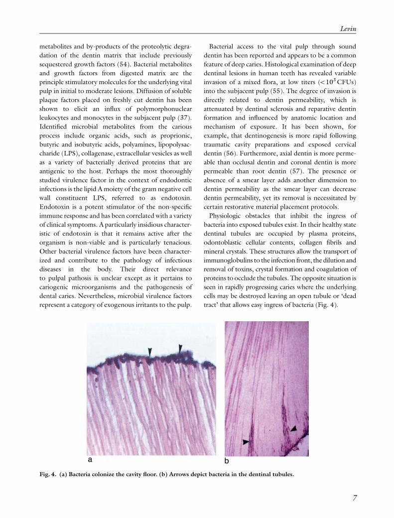
tract’ that allows easy ingress of bacteria (Fig. 4).
Fig. 4. (a) Bacteria colonize the cavity floor. (b) Arrows depict bacteria in the dentinal tubules.
Levin
7

The clinical significance of limited exposure of the
pulp to bacteria through sound dentin is not known. It
has been suggested that low titers of pathogens can be
dealt with by the host immune system provided there is
a thin layer of intervening sound dentin (58). By
contrast, pulpal responses to long-term provocation by
oral microbes across intact dentin have been docu-
mented. Freshly cut dentin left unrestored in human
teeth for up to 240 days revealed an initial intense acute
inflammatory response that slowly subsided within the
first 2 weeks. This response was accompanied by an
initial report of pain from experimental subjects that
gradually subsided. This cessation of symptomology
correlated with a regression of inflammation and
resumption of normal tissue architecture and immune
status within the pulp. Reparative dentin was also
evident. Subsequent studies confirmed the recovery
capacity of the pulp subsequent to bacterial challenge
but they also indicated that recovery depends on the
extent of the challenge (59). Full-coverage crown
preparations represent the most extensive operative
exposure of dentin. Teeth prepared for full coverage
and left in provisional restorations for prolonged
periods show an increased rate of pulpal necrosis (60).
‘Endogenous’ irritants
While certain bacterial virulence factors are directly
damaging to the host tissue, others stimulate a
prolonged non-specific host immune response that
results in tissue damage. In the progressing carious
lesion, the host immune response increases in intensity
as the infection advances. Titers of T helper cells, B-
lineage cells, neutrophils and macrophages are directly
proportional to lesion depth in human teeth (61). In
the most advanced phase of carious destruction, the
humoral immunoresponse is accompanied by immuno-
pathologic destruction of pulpal tissue. In animal
studies where monkeys were hyperimmunized to BSA
there was an observed increase in pulpal tissue destruc-
tion subsequent to antigenic challenge across freshly cut
dentin (62). These findings support the contention that
antigen–antibody complex formation, in addition to
various products of the inflammatory cascade, give rise
to a non-specific response that, while designed to rid the
body of pathogens, effects destruction of parenchymal
tissues as well (Fig. 5).
Neurogenic mediators are involved in the pulpal
response to irritants and like immune components,
Fig. 5. Bacteria stimulate the innate and adaptive immune systems. The result is the elaboration of inflammatorymediators that effect chemotaxis and degranulation of neutrophils and activation of complement. Many of thesemediators are non-specific in that they act on any cell with which they come in contact. This can be host or pathogen.
Pulpal irritants
8

they can mediate pathology. External stimulation of
dentin causes the release of pro-inflammatory neuro-
peptides from pulpal afferent nerves (63). Substance P
(SP) and calcitonin gene-related peptide (CGRP) are
released and effect vascular events such as vasodilata-
tion and increased vascular permeability. This results in
a net increase in tissue pressure that can progress to
necrosis in extreme and persistent circumstances. Other
pulpal elements such as fibroblasts, odontoblasts and
Schwann cells react to irritants by elaborating growth
factors and chemokines that are designed to counteract
pathogens but secondarily can contribute to pulpal
destruction. Odontoblasts exposed to bacteria and
their by-products express IL-8 mRNA and protein (64,
65). IL-8 is a potent chemotactic factor for neutrophils,
a predominant inflammatory effector cell observed in
inflamed pulps. As previously mentioned, neutrophilic
degranulation liberates lysosomal enzymes that digest
host as well as microbial cells.
As caries progresses towards the pulp, the acid
environment acts to dissolve mineral, liberate pre-
viously sequestered host growth factors and create a pH
gradient from the lesion into dentin due to the
buffering capacity of dentinal fluid. In essence soft
carious dentin represents a ‘poultice’ of growth factors,
enzymes, toxins and microbial metabolites whose
stimulatory effect on the subjacent pulp is inversely
proportional to the RDT. The documented pulpal
response to these factors is varied and dependent on the
mediator studied. Numerous investigators have shown
that dentin matrix components can stimulate dentino-
genesis (66, 67). Demineralized dentinmatrix as well as
dentin chips implanted at the site of pulpal exposure
induce reparative dentin formation (68). Matrix
component TGF-b1 has been shown to be an inducer
of tertiary dentin as well as a potent pulpal immuno-
suppressor (69–71). It has been theorized that during
caries progression, TGF-b previously trapped during
dentinogenesis, is released and is stimulatory to the
pulp (72). This theory is supported by studies that
demonstrated that transdentinal diffusion of TGF-b1induced an accumulation of dendritic cells in the
odontoblast and subodontoblast layers (73). It is
feasible that tertiary dentin formation is also stimulated
transdentinally by this and other by-products of the
carious degradation of dentin. In vitro studies employ-
ing the focal application of TGF-b1 to dentin demon-
strated that subjacent odontoblasts responded by
TGFb-1 receptor expression as well as alpha 1(I)
collagen gene transcription (74). Insulin-like growth
factors I and II and angiogenic growth factors are also
components of the dentin matrix during tooth forma-
tion (72). Angiogenic factors are liberated during
carious dissolution of dentin and it is likely that they
resume bioactivity (75).
Summary
A variety of stimuli exert effects on the dental pulp.
These effects are governed by the magnitude, duration
and frequency of the stimulus, as well as intervening
dentin permeability and thickness. Therapeutic inter-
ventions can diminish or ameliorate irritants provided
they are administered in a timely manner.
References
1. Bergenholtz G. Pathogenic mechanisms in pulpal dis-ease. J Endod 1990: 16: 98–101.
2. Stanley HR. Traumatic capacity of high-speed andultrasonic dental instrumentation. J Am Dent Assoc 1961:63: 749–766.
3. Stanley HR, Swerdlow H. Reaction of human pulp tocavity preparation: results produced by eight differentoperative techniques. J Am Dent Assoc 1959: 58: 49.
4. Kramer IRH. Pulp changes of non-bacterial origin. IntDent J 1959: 9: 435.
5. Brannstrom M. Cavity preparation and the pulp. DentProg 1961: 2: 4.
6. Langeland K, Langeland L. Pulp reactions to crownpreparations, impression, temporary crown fixation, andpermanent cementation. J Prosthet Dent 1965: 15: 129.
7. Langeland K. Histologic evaluation of pulp reactions tooperative procedures. Oral Surg 1959: 12: 1235.
8. Zach I, Cohen G. Thermogenesis in operative techni-ques. Comparisons of four methods. J Prosthet Dent1962: 12: 977.
9. Stenvik A, Mjor IA. Tissue pressure and histology ofnormal and inflamed tooth pulps in Macaque monkeys.Arch Oral Biol 1972: 17: 1501–1511.
10. Heyeras KJ, Kvinnsland I. Micropuncture measurementsof interstitial fluid pressure in normal and inflamed dentalpulps in cats. J Endod 1983: 9: 105–109.
11. Olgart LM. Involvement of sensory nerves in hemody-namic reactions. Proc Finn Dent Soc 1992: 88(Suppl):403–410.
12. Baume IJ. The biology of pulp and dentine. A historic,terminologic–taxonomic, histologic–biochemical, embryo-nic, and clinical survey. Monog Oral Sci 1980: 8: 1–220.
13. Fish EW. An Experimental Investigation of Enamel,Dentine and the Dental Pulp. London: John Bale Sonsand Danielsson, 1932.
14. Yoshiba K, Yoshiba N, Ejiri S, Iwaku M, Ozawa H.Odontoblast processes in human dentin revealed by
Levin
9

fluorescence labeling and transmission electron micro-
scopy. Histochem Cell Biol 2002: 118: 205–212.15. Yoshiba K, Yoshiba N, Iwaku M. Class II antigen-
presenting dendritic cell and nerve fiber responses to
cavities, caries, or caries treatment in human teeth. J DentRes 2003: 82: 422–427.
16. Kvinnsland S, Heyeraas K, Øfjord ES. Effect of experi-mental tooth movement on periodontal and pulpal
blood flow. Eur J Orthodont 1989: 11: 200–205.17. Nixon CE, Saviano JA, King GJ, Keeling SD. Histomir-
phometric study of dental pulp during orthodontic
movement. J Endod 1993: 19: 13–16.18. Olgart L, Gazelius B, Sundstrom F. Intradental nerve
activity and jaw opening reflex in response to mechanical
deformation of cat teeth. Acta Physiol Scand 1988: 133:399–406.
19. Butcher EO, Taylor AC. The effects of denervation andischemia upon the teeth of the monkey. J Dent Res 1951:30: 265–275.
20. MesserHM. Permanent restorations and the dental pulp.
In: Hargreaves KM, Goodis HE, eds. Seltzer and Bender’sDental Pulp. Carol Stream, IL:Quintessence International,2002.
21. Vale WA. Cavity preparation. Irish Dent Rev 1956: 2:33–41.
22. Reeh ES, Messer HH, Douglas WH. Reduction in toothstiffness as a result of endodontic and restorative
procedures. J Endod 1989: 15: 512–516.23. Panitvisai P, Messer HH. Cuspal deflection in relation to
restorative and endodontic procedures. J Endod 1995:
21: 57–61.24. Versluis A, Douglas WH, Cross M, Sakaguchi RI. Does
an incremental technique reduce polymerization shrink-
age stresses? J Dent Res 1996: 75: 871–878.25. Hirata K, Nakashima M, Sekine I, Mukouyama Y,
Kimura K. Dentinal fluid movement associated withloading of restorations. J Dent Res 1991: 70: 975–978.
26. Tagami J, HosodaH, BurrowMF,NakajimaM. Effect ofaging and caries on dentin permeability. Proc Finn DentSoc 1992: 88(Suppl): 149–154.
27. About I, Murray PE, Franquin JC, Remusat M, Smith
AJ. Pulpal inflammatory responses following non-carious
class V restorations. Oper Dentstry 2001: 26: 336–342.28. Qvist V, Stoltze K, Qvist J. Human pulp reactions to
resin restorations performed with different acid etchrestorative procedures. Acta Odont Scand 1989: 47:253–263.
29. Fujitani M, Inokoshi S, Hosoda H. Effect of acid etching
on the dental pulp in adhesive composite restorations.
Int Dent J 1992: 42: 3–11.30. Gwinnett AJ, Tay FR. Early and intermediate time
response of the dental pulp to an acid etch technique invivo. Am J Dent 1998: 11(Special issue): S35–S44.
31. Fitzgerald M, Hanks CT. In vivo study of resin diffusionthrough intact vital human dentin (abstract). J Dent Res1997: 76(Special issue): 305.
32. Hume R. An analysis of the release and the diffusion
through dentin of eugenol from zinc-oxide eugenol
mixtures. J Dent Res 1984: 63: 881–884.
33. Hume WR, Testa AE. Release of 3H-triamcinolone
from a steroid antibiotic mixture. J Endod 1980: 7:509–514.
34. Bergenholtz G, Cox CF, Loesche WI, Syed SA. Bacterial
leakage around dental restorations: its effect on thedental pulp. A microbial and histopathological study.
J Oral Pathol 1982: 11: 439–450.35. Ferracane JL, Condon JR. Rate of elution of leachable
components from composite.Dent Mater 1990: 6: 282–287.
36. Hume WR, Gerzina TM. Bioavailability of components
of resin-based materials which are applied to teeth. CritRev Oral Biol Med 1996: 7: 172–179.
37. Bergenholtz G, Warfvinge J. Migration of leukocytes in
the dental pulp in response to plaque bacteria. Scand JDent Res 1982: 90: 354–362.
38. KatsunoKA et al. A delayed type hypersensitivity reactionto dentine primer in the guinea pig. J Dent 1995: 23:295–299.
39. InoueT,Miyakoshi S, ShimonoMDentin/pulp adhesive
resin interface. Biological view from basic science to
clinic. In: Shimono M, Maeda T, Suda H, Takahashi K,eds. Proceedings of the International Conference onDentin/Pulp Complex 1995, Chiba, Japan. Chicago:
Quintessence Publishing Co., pp. 217–220, 1996.40. Jontell M, Hanks CT, Bratel J, Bergenholtz G. Effect of
unpolymerised resin components on the function ofaccessory cells derived from the rat incisor pulp. J DentRes 1995: 74: 1162–1167.
41. Schroder U. Effects of calcium hydroxide-containing
pulp-capping agents on pulp cell migration, prolifera-
tion, and differentiation. J Dent Res 1985: 64(Specialissue): 541–548.
42. Cvek M, Granath L, Cleaton-Jones P, Austin J.Hard tissue barrier formation in pulpotomized
monkey teeth capped with cyanoacrylate or calcium
hydroxide for 10 and 60min. J Dent Res 1987: 66:1166–1174.
43. Heys DR, Cox CF, Heys RJ, Avery JK. Histological
considerations of direct pulp capping agents. J Dent Res1981: 60: 1371–1379.
44. Cvek M. A clinical report on partial pulpotomy andcappingwith calciumhydroxide in permanent incisors with
complicated crown fracture. J Endod 1978: 4: 232–237.45. Mjor IA, Finn SB, Quigley MB. The effect of calcium
hydroxide and amalgam on non-carious dentine. ArchOral Biol 1961: 3: 283–291.
46. Gwinnett AJ, Tay FR, Pang KM, Wei SHY. Quantitative
contribution of the collagen network in dentin hybridi-zation. Am J Dent 1996: 9: 140–144.
47. Mjor IA. Initial Reactions to Tooth Preparations, in Pulp-Dentin biology in restorative dentistry. Chicago: Quintes-
sence Publishing, 2002.48. Nakabayashi N. Resin reinforced dentine due to infiltra-
tion of monomers into dentine at the adhesive interface.
J Jpn Dent Mater 1982: 1: 78–181.49. Farik B, Munksgaard EC, Andreasen JO, Kreiborg S.
Drying and rewetting anterior crown fragments prior tobonding. Endod Dent Traumatol 1999: 15: 113–116.
Pulpal irritants
10

50. Kakehashi S, Stanley HR, Fitzgerald RJ. The effectsof surgical exposure of dental pulps in germ-freeand conventional laboratory rats. Oral Surg 1965: 20:340–349.
51. Brannstrom M, Lind PO. Pulpal response to earlydentinal caries. J Dent Res 1965: 44: 1045–1050.
52. About I, Mitsiadis TA. Molecular aspects of toothpathogenesis and repair: in vivo and in vitro models.Adv Dent Res 2001: 15: 59–62.
53. Bergenholtz G. Factors in pulpal repair after oralexposure. Adv Dent Res 2001: 15: 84.
54. Magloire H, Bouvier M, Joffe A. Odontoblast responseunder carious lesions. Proc Finn Dent Soc 1992:88(Suppl 1): 257–274.
55. Hoshino E, Ando N, Sato M, Kota K. Bacterial invasionof non-exposed dental pulp. Int Endod J 1992: 25: 2–5.
56. Cox CE,White KC, Ramus DL, Farmer JB, SnuggsHM.Reparative dentin: factors affecting its deposition.Quintessence Int 1992: 23: 257–270.
57. Pashley DH. Considerations of dentin permeability incytotoxicity testing. Int Endod J 1988: 21: 143–154.
58. Lundy T, Stanley HR. Correlation of pulp histo-pathology and clinical symptoms in human teethsubjected to experimental irritation. Oral Surg 1969:27: 187–201.
59. Lervik T, Mjor IA. Evaluation of techniques for theinduction of pulpitis. J Biol Buccale 1977: 5: 137–148.
60. Felton D, Madison S, Kanoy E, Kantor M, Maryniuk G.Long-term effects of crown preparation on pulp vitality(abstract). J Dent Res 1989: 68(Special issue): 1004.
61. Izumi T, Kobayashi I, Okamura K, Sakai H. Immuno-histochemical study on the immunocompetent cells ofthe pulp in human non-carious and carious teeth. ArchOral Biol 1995: 40: 609–614.
62. Bergenholtz G, Ahlstedt S, Lindhe J. Experimentalpulpitis in immunized monkeys. Scand J Dent Res1977: 85: 396–406.
63. Byers MR, Narhi MV. Dental injury models: experi-mental tools for understanding neuroinflammatory
interactions and polymodal nociceptor functions. CritRev Oral Biol Med 1999: 10: 4–39.
64. Levin LG, Rudd A, Bletsa A, Reisner H. Expression ofIL-8 by cells of the odontoblast layer in vitro. Eur J OralSci 1999: 107: 131–137.
65. Huang GT, Potente AP, Kim JW, Chugal N, Zhang X.Increased interleukin-8 expression in inflamed humandental pulps. Oral Surg Oral Med Oral Pathol OralRadiol Endod 1999: 88: 214–220.
66. Tziafas D et al. Induction of odontoblast-like celldifferentiation in dog dental pulps after in vivo implanta-tion of dentinematrix components.Arch Oral Biol 1995:40: 883–893.
67. Smith AJ et al. Odontoblast stimulation in ferrets bydentine matrix components. Arch Oral Biol 1994: 39:13–22.
68. Smith AJ. Pulpal responses to caries and dental repair[Review] [93 refs]. Caries Res 2002: 36: 223–232.
69. Tziafas D, Papadimitriou S. Role of exogenous TGF-beta in induction of reparative dentinogenesis in vivo.Eur J Oral Sci 1998: 106(Suppl 1): 192–196.
70. Kulkarni AB et al. Transforming growth factor beta 1null mutation in mice causes excessive inflammatoryresponse and early death. Proc Natl Acad Sci USA 1993:90: 770–774.
71. D’Souza RN, Cavender A, Dickinson D, Roberts A,Letterio J. TGF-beta1 is essential for the homeostasis ofthe dentin–pulp complex. Eur J Oral Sci 1998:106(Suppl 1): 185–191.
72. Finkelman RD, Mohan S, Jennings JC, Taylor AK,Jepsen S, Baylink DJ. Quantitation of growth factorsIGF-I, SGF/IGF-II, and TGF-beta in human dentin.J Bone Miner Res 1990: 5: 717–723.
73. Farges JC et al. TGF-beta1 induces accumulation ofdendritic cells in the odontoblast layer. J Dent Res 2003:82: 652–656.
74. Magloire H, Romeas A, Melin M, Couble ML, BleicherF, Farges JC. Molecular regulation of odontoblastactivity under dentin injury. Ad Dent Res 2001: 15:46–50.
75. Roberts-Clark DJ, Smith AJ. Angiogenic growth factorsin human dentine matrix. Arch Oral Biol 2000: 45:1013–1016.
Levin
11
Related Documents