PULMONARY THROMBO EMBOLISM Dr . Tushar Goya DNB MEDICINE

Pulmonaryembolism
Aug 17, 2015
Welcome message from author
This document is posted to help you gain knowledge. Please leave a comment to let me know what you think about it! Share it to your friends and learn new things together.
Transcript

PULMONARY THROMBO EMBOLISM
Dr. Tushar GoyalDNB MEDICINE

PTE
It is obstruction of pulmonary vessels . OBSTRUCTION can be-
• Thrombotic –blood clot.• Non-thrombotic : Fat, Air, Tumor , Amniotic fluid.Clot may be –• Primary -formed in the pulmonary vessels itself.• secondary-Thrombosis of peripheral veins ,
embolisation to pulmonary vessels

• Venous thromboembolism[VTE]
DVT PE
Post phlebitic syndrm chronic thrombo embolic pulmonary hypertension

RISK FACTORS
Hypercoagubility- Malignancy
Nonmalignant thrombophiliaPregnancy
Postpartum status (<4wk)Estrogen/ OCP’s
Genetic mutations (Factor V Leiden ,prothrombin gene,} Protein C & S, anti-thrombin deficiency)
Venous Stasis
Bed rest > 24 hr Recent cast or external fixatorLong-distance travel or prolong automobile travel
Venous InjuryRecent surgery requiring endotracheal intubationRecent trauma (especially the lower extremities and pelvis)
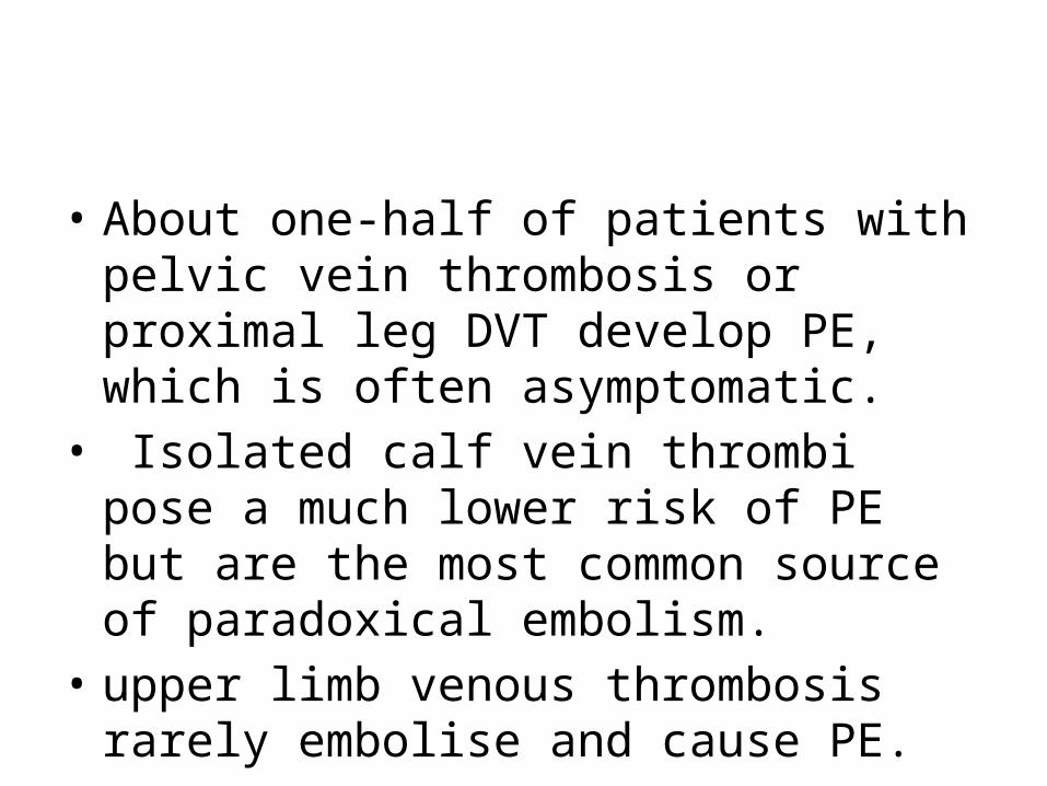
• About one-half of patients with pelvic vein thrombosis or proximal leg DVT develop PE, which is often asymptomatic.
• Isolated calf vein thrombi pose a much lower risk of PE but are the most common source of paradoxical embolism.
• upper limb venous thrombosis rarely embolise and cause PE.

Clinical Features of PTE
Acute Massive Pulmonary Embolism
Symptoms : faintness or collapse Crushing central chest pain severe dyspnoea apprehension
Signs : tachycardia hypotension to circulatory collapse raised JVP RV Gallop rhythm loud P2 Severe cyanosis decreased urinary output

Acute Moderate / Small Pulmonary embolism
Symptoms : dyspnoea restricted breathing cough , hemoptysis pleuritic chest pain
Signs : Tachycardia pleural rub crepitations fever ( low grade ) effusion ( blood stained ) raised hemipdiaphragm

Principal markers used for risk stratification
1) RV Dysfunction : hypotension RV dilation ,hypokinesis or overload RV dilation on spiral CT BNP or NT –Pro BNP elevated right heart pressure at RHC 2) Myocardial injury : Cardiac troponin T or I positive
Patients with massive PE : Rv dysfunction + hypotension Moderate to large PE : RV dysfunction + normotension Small to moderate PE : Normal RV function + normotension

Differential Diagnosis
1) Pneumonia ,Asthma , COPD , Pleurisy 2) Congestive heart failure , Pericarditis , ACS 3) Costochondritis , Musculoskeletal discomfort Rib fracture 4) Pneumothorax 5) Anxiety

evaluating patients with possible VTE
• The initial task is to decide on the clinical likelihood of the disorder.
• For this we have various scores like wells,geneva etc
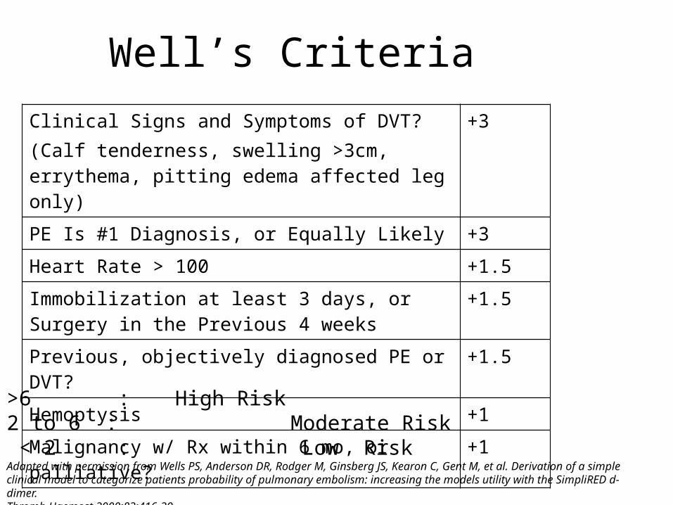
Well’s CriteriaClinical Signs and Symptoms of DVT?(Calf tenderness, swelling >3cm, errythema, pitting edema affected leg only)
+3
PE Is #1 Diagnosis, or Equally Likely +3
Heart Rate > 100 +1.5
Immobilization at least 3 days, or Surgery in the Previous 4 weeks
+1.5
Previous, objectively diagnosed PE or DVT? +1.5
Hemoptysis +1
Malignancy w/ Rx within 6 mo, or palliative? +1
>6 : High Risk2 to 6 : Moderate Risk < 2 : Low Risk Adapted with permission from Wells PS, Anderson DR, Rodger M, Ginsberg JS, Kearon C, Gent M, et al. Derivation of a simple clinical model to categorize patients probability of pulmonary embolism: increasing the models utility with the SimpliRED d-dimer.Thromb Haemost 2000;83:416-20.
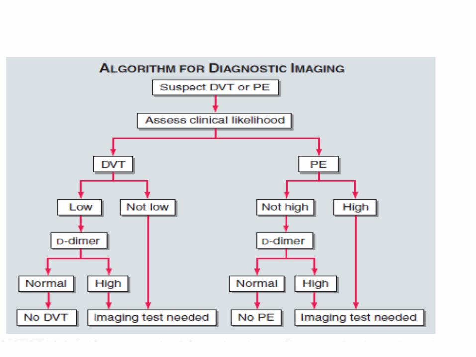

D - dimers
• The sensitivity is >80% for DVT and >95% for PE.
• a useful "rule out" test. • More than 95% of patients with a normal (<500
ng/mL) d-dimer do not have PE. • It is not specific. Levels increase in patients with
MI, pneumonia, sepsis, cancer, and the postoperative state and those in the2nd or 3rd trimester of pregnancy. .


Chest x ray
A normal or nearly normal chest x-ray often occurs in PE.
Others -1. focal oligemia (Westermark's sign),2. a peripheral wedged-shaped density above the diaphragm
(Hampton's hump)3. an enlarged right descending pulmonary artery (Palla's sign4. Pleuropulmonary opacities 5. Pleural effusion 6. Linear shadows 7. prominent RV

Westermarks sign

Hamptons hump

Palla”s sign

• ECG– 2 Most Common finding on EKG:
• Nonspecific ST-segment and T-wave changes• Sinus Tachycardia
– Historical abnormality suggestive of PE• S1Q3T3• Right ventricular strain• New incomplete RBBB

Chest CT• CT chest ( multidetector –row spiral CT) with IV contrast is the principal
imaging test for the diagnosis of PE
• The CT scan also obtains excellent images of the RV and LV and can be used for risk stratification along with its use as a diagnostic tool. In patients with PE, RV enlargement on chest CT indicates an increased likelihood of death within the next 30 days compared with PE patients who have normal RV size on chest CT.
• When imaging is continued below the chest to the knee, pelvic and proximal leg DVT also can be diagnosed by CT scanning.
• In patients without PE, the lung parenchymal images may establish alternative diagnoses not apparent on chest x-ray such as pneumonia, emphysema, pulmonary fibrosis, pulmonary mass, and aortic pathology.

Lung Scanning( V/Q scan)• Lung scanning has become a second-line diagnostic test for PE,
used mostly for patients who cannot tolerate intravenous contrast.
• A high-probability scan for PE is defined as one that indicates two or more segmental perfusion defects in the presence of normal
ventilation.The Combination of A High-Probability Ventilation-Perfusion
Scan Plus A High Clinical Suspicion is Diagnostic for Pulmonary Embolism.

VENOUS USG
Findings• loss of vein compressibility.• Vein does not wink when gently compressed• Failure to appose walls of vein• homogeneous and has low echogenicity thrombus can be
found . • Loss of normal respiratory variation .• In color doppler flow blunted rather than augmented with
compression• yields a positive result in around 20 %

2D - ECHO
• Main role is to rule out PE mimics.
• The best-known indirect sign of PE on TTE is McConnell's sign: hypokinesis of the RV free wall with normal motion of the RV apex.

INVASIVE DIAGNOSTIC METHODS
• Pulmonary Angiography : Indications : 1) unsatisfactory CT 2) Interventional procedure
such as catheter directed thrombolysis or embolectomy is planned
• Contrast Phlebography

TREATMENT

• Resustiation is important mainly in pts with massive embolism.
1. Hemodynamic support-
For patients with massive PE and hypotension, one should administer 500 mL of normal saline.
Additional fluid should be infused with extreme caution. Dopamine and dobutamine are first-line inotropic agents for
treatment of PE-related shock.
2) Respiratory support- intubation and oxygen

Treatment of embolism per se
PRIMARY THERAPY –1.Clot dissolution with thrombolysis2.Removal of PE by embolectomy
SECONDARY PREVENTION-1.Anticoagulation with heparin and warfarin2.Placement of IVC filters

RISK STRATIFICATION

ANTICOAGULATION

What anticoagulants to give?
• Parenteral - 1. IV or SC UFH 2.LMWH(enoxaparin ) 3. fondaparinux
Oral - 1.warfarin 2. rivaroxaban 3. dabigatran

If patient is having proven or suspected HIT use a direct thrombin inhibitor like argatroban,lepirudin,or bivalirudin

Prefered anticoagulant ?
• ACCP suggests LMWH or fondaparinux instead of UFH
EXCEPTION –
• Pt in whom SC absorption is inadequate • Pts who are being considered for thrombolytic
therapy

Why LMWH is prefered?
• Better subcutaneous bioavailability.
• Longer and more consistent monoexponential t1/2 once daily dose.
• Since aPTT is not prolonged no need of lab monitoring .
• Incidence of HIT is less

How to start the pt on anticoagulation ?
• Once the diagnosis is confirmed ,begin t/t with parenteral form and also start oral form on the same day or next day.
• Continue the parenteral form for atleast 5 days ( even if INR reaches 2 earlier) or until the INR is atleast 2 for 24 hrs or more

Both the parenteral and oral forms are started simultaneously because
Warfarin takes 5-7 days to achieve a therapeutic effect .
If warfarin is initiated as a montherapy during an acute thrombotic illness, paradoxical exacerbation of hypercoagulability can increase the likelihood of thrombosis rather than prevent it.

For UFH • Initial bolus 80units/kg followed by 18/kg/hr ie
bolus of 5000-10000 units followed by infusion of 1000-1500 units /hr.
For LMWH Enoxaparin : 1 mg /kg twice daily with normal
renal functionDalteparin: 200 u/kg once daily or 100 u/kg twice
dailyTinzaparin : 175 u/kg once a dayFondaparinux : weight based once daily


Dose of warfarin
• strating dose of warfarin is 10 mg daily for 2 days then dose by INR
• Target INR is 2.5 range is 2-3

Duration of anticoagulation

FIBRINOLYSIS
• INDICATIONS:• The only FDA-approved indication is massive PE. • Considered for patients with preserved systolic
BP and submassive PE with moderate or severe RV dysfunction if there is new hemodynamic instability , worsening respiratory insufficiency, severe RV dysfunction or major myocardial necrosis and low risk of bleeding

CONTRAINDICATIONS• Absolute Contraindications to Thrombolysis
– Active or recent internal bleeding ( last month)– Hemorrhagic Stroke or stroke of unknown origin at any
time – Intracranial Neoplasm – Recent cranial surgery or head trauma (within 3
weeks)– Ischemic stroke in preceding 6 months

DOSING• rtPA : 100 mg administered as a continuous peripheral intravenous
infusion over 2 hours or 0.6 mg/kg over 15 mins ( max dose 50 mg)
• Streptokinase : 2.5 lakh IU as a loading dose over 30 mins followed by 1 lakh IU for 12 to 24 hours or 1.5 million units IU over 2 hrs
• Urokinase : 4400IU/Kg as a loading dose over 10 mins followed by 4400 IU /Kg /hr for 12 to 24 hrs
• Patients appear to respond to fibrinolysis for up to 14 days after the PE has occurred

BENEFITS OF FIBRINOLYSIS
Rapidly reverses right heart failure and may result in a lower rate of death and recurrent PE by
• (1) dissolving much of the anatomically obstructing pulmonary arterial thrombus,
• (2) preventing the continued release of serotonin and other neurohumoral factors that exacerbate PAH
• (3) lysing much of the source of the thrombus in the pelvic or deep leg veins, thereby decreasing the likelihood of recurrent PE.

IVC FILTERS
• Indications –(1) active bleeding that precludes anticoagulation (2) recurrent venous thrombosis despite intensive
anticoagulation.(3) Patients with massive PE who survived but in
whom recurrent embolism will be fatal (4) In patients with a time limited indication for
IVC Filter placement

EMBOLECTOMY
• Open surgical
• Catheter embolectomy : 1) Aspiration thrombectomy 2) thrombus fragmentation 3) rheolytic thrombectomy

PREVENTION
• ACCP guide linesFor acutely ill hospitalised medical pts at low risk
of thrombosis ACCP recommends against the use of prophylaxsis.
Pts at moderate to high risk but who are not bleeding or at high risk of bleeding should be given either LMWH or UFH or fondaparinux.

PAUDA SCORE • RISK FACTOR 1) Active cancer2) Previous VTE 3) Reduced mobility4) Thrombophilic condition5) Recent <1 month trauma or surgery 6) Age > 70 yrs 7) Heart or respiratory failure 8) Acute MI or Ischemic stroke9) Active infection and /or rheumatological disease 10) BMI > 30 11) Ongoing hormonal treatment
• SCORE 3 3 3 3 2 1 1 1 1 1 1

• Patients with score more than 4 requires prophylactic therapy .
• For pts who are bleeding or at risk of bleeding use leg compression devices only.
• Pts are considered to be at high risk of bleeding if they meet any of the following criteria
active gastroduodenal ulcerBleeding in 3 months prior to admissionPlatelet count <50,000

• Or if they had multiple risk factors for bleeding of lesser predictive strengthlike age >84 yrs,severe renal failure , hepatic failure with INR > 1.5 , male ,current cancer, ICU admission.

THANK YOU