12 Pulmonary Infection in Adults DR MUHAMMAD BIN ZULFIQAR PGR III FCPS Services institute of Medical Sciences/ Services Hospital Lahore GRAINGER & ALLISON’S DIAGNOSTIC RADIOLOGY

Welcome message from author
This document is posted to help you gain knowledge. Please leave a comment to let me know what you think about it! Share it to your friends and learn new things together.
Transcript
12 Pulmonary Infection in Adults
DR MUHAMMAD BIN ZULFIQARPGR III FCPS Services institute of Medical
Sciences/ Services Hospital LahoreGRAINGER & ALLISON’S DIAGNOSTIC RADIOLOGY

• FIGURE 12-1 Cellular bronchiolitis. A 71-year-old man with ■fever of 48 h duration. (A) Posteroanterior chest radiograph is normal. (B) Complementary CT shows centrilobular branching nodular and linear opacities resulting in a ‘tree-in-bud’ appearance (arrows). Mycoplasma bronchiolitis was diagnosed.

• FIGURE 12-2 Round ■pneumonia. A previously healthy 64-yearold man with fever and productive cough. (A) Chest radiograph shows a mass-like area of consolidation in the left upper lobe (arrow). (B) CT shows a discrete fairly well-marginated opacity in the left upper lobe containing small areas of low attenuation. The abnormality resolved following appropriate antibiotic therapy and Gram stains of sputum demonstrated S. pneumoniae

• FIGURE 12-3 Lobar pneumonia. A 36-year-old-■man with S. pneumoniae pneumonia. Coronal reformatted CT image shows a homogeneous focal area of consolidation in the right upper lobe. Patent bronchi (air bronchograms) are seen within the area of consolidation.

• FIGURE 12-4 Bronchopneumonia caused by ■ H. influenzae. A 48-year-old man with productive cough and fever. Coronal reformatted CT shows a focal area of consolidation in the right lower lobe with visible air bronchogram and poorly defined margins (arrows). Also evident are small nodular opacities and a few ‘tree-in-bud’ opacities (arrowhead).

• FIGURE 12-5 Lung abscess. A 35-year-old man with high ■fever and large purulent sputum production with positive culture for P. aeruginosa. Coronal reformatted CT shows a large cavity in the left upper lobe. Note intracavitary thick septa.

• FIGURE 12-6 Pneumatocele. A 32-year-old ■woman with previous S. aureus pneumonia. CT shows thin-walled cystic lesion (pneumatocele) in the right lower lobe.
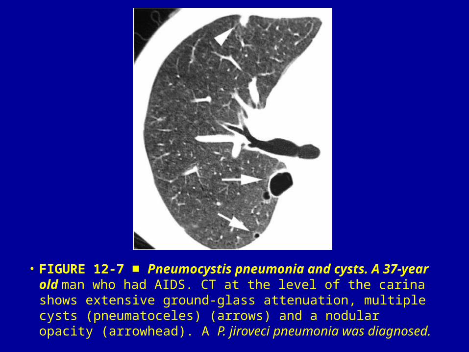
• FIGURE 12-7 ■ Pneumocystis pneumonia and cysts. A 37-year old man who had AIDS. CT at the level of the carina shows extensive ground-glass attenuation, multiple cysts (pneumatoceles) (arrows) and a nodular opacity (arrowhead). A P. jiroveci pneumonia was diagnosed.

• FIGURE 12-8 Septic embolism. A 40-year-old male, ■intravenous drug user with fever. CT shows multiple cavitated nodules in the left upper lobe. Different vessels (arrows) course into the nodules. Blood cultures were positive for S. aureus.
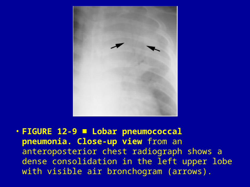
• FIGURE 12-9 Lobar pneumococcal pneumonia. ■Close-up view from an anteroposterior chest radiograph shows a dense consolidation in the left upper lobe with visible air bronchogram (arrows).
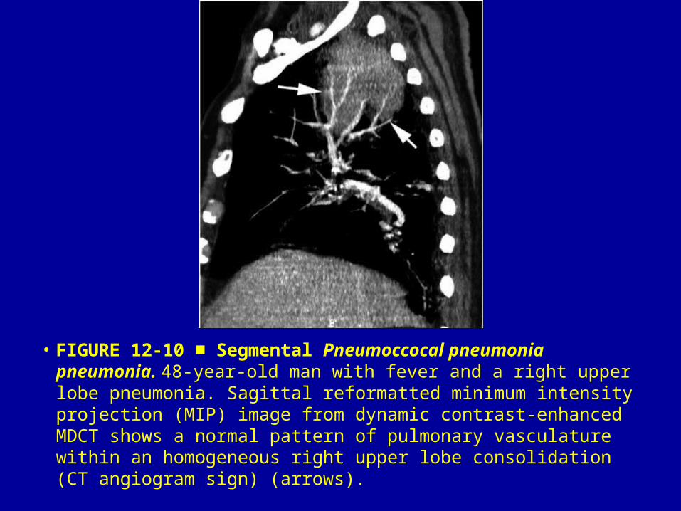
• FIGURE 12-10 Segmental ■ Pneumoccocal pneumonia pneumonia. 48-year-old man with fever and a right upper lobe pneumonia. Sagittal reformatted minimum intensity projection (MIP) image from dynamic contrast-enhanced MDCT shows a normal pattern of pulmonary vasculature within an homogeneous right upper lobe consolidation (CT angiogram sign) (arrows).

• FIGURE 12-11 Klebsiella pneumonia. A 50-year-old man ■with fever and a severe right pneumonia. Posteroanterior chest radiograph shows dense consolidation of the right upper lobe with visible areas of abscessification (arrowhead). Note an inferior convexity of the major fissure (‘bulging fissure’ sign) (arrows) characteristic of lobar expansion.
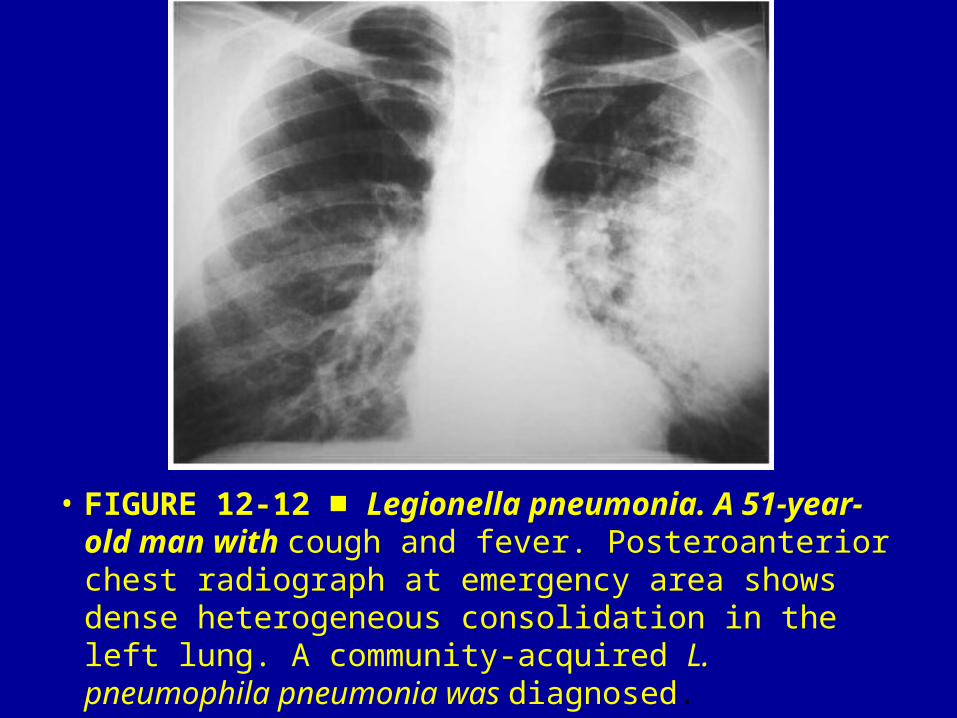
• FIGURE 12-12 ■ Legionella pneumonia. A 51-year-old man with cough and fever. Posteroanterior chest radiograph at emergency area shows dense heterogeneous consolidation in the left lung. A community-acquired L. pneumophila pneumonia was diagnosed.

• FIGURE 12-13 ■ Chlamydia pneumoniae pneumonia. A 67-yearold woman with chest pain, fever and non-productive cough. Coronal reformatted CT shows multiple ill-defined, rounded areas of consolidation in the left upper lobe with visible air bronchogram and poorly defined margins (arrows).

• FIGURE 12-14 ■ Moraxella catarrhalis pneumonia. A 70-year-old man with fever. CT shows a complete consolidation in the middle lobe (white arrow). Note bronchial wall thickening (thin white arrow) and focal areas of ground-glass opacities in the superior segment of the right lower lobe. Also evident is a significant dilatation of the main pulmonary artery (black arrow).
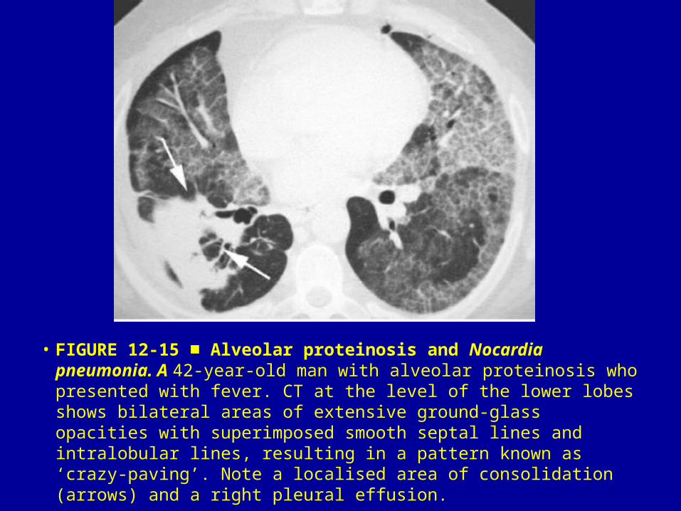
• FIGURE 12-15 Alveolar proteinosis and ■ Nocardia pneumonia. A 42-year-old man with alveolar proteinosis who presented with fever. CT at the level of the lower lobes shows bilateral areas of extensive ground-glass opacities with superimposed smooth septal lines and intralobular lines, resulting in a pattern known as ‘crazy-paving’. Note a localised area of consolidation (arrows) and a right pleural effusion.

• FIGURE 12-16 Pleuropulmonary actinomycosis. A 52-year-■old alcoholic man with fever, cough and left chest pain. Contrast enhanced CT shows consolidation in the left lower lobe containing multiple areas of decreased attenuation with small air bubbles. Also evident are pleural thickening and a small pleural effusion.
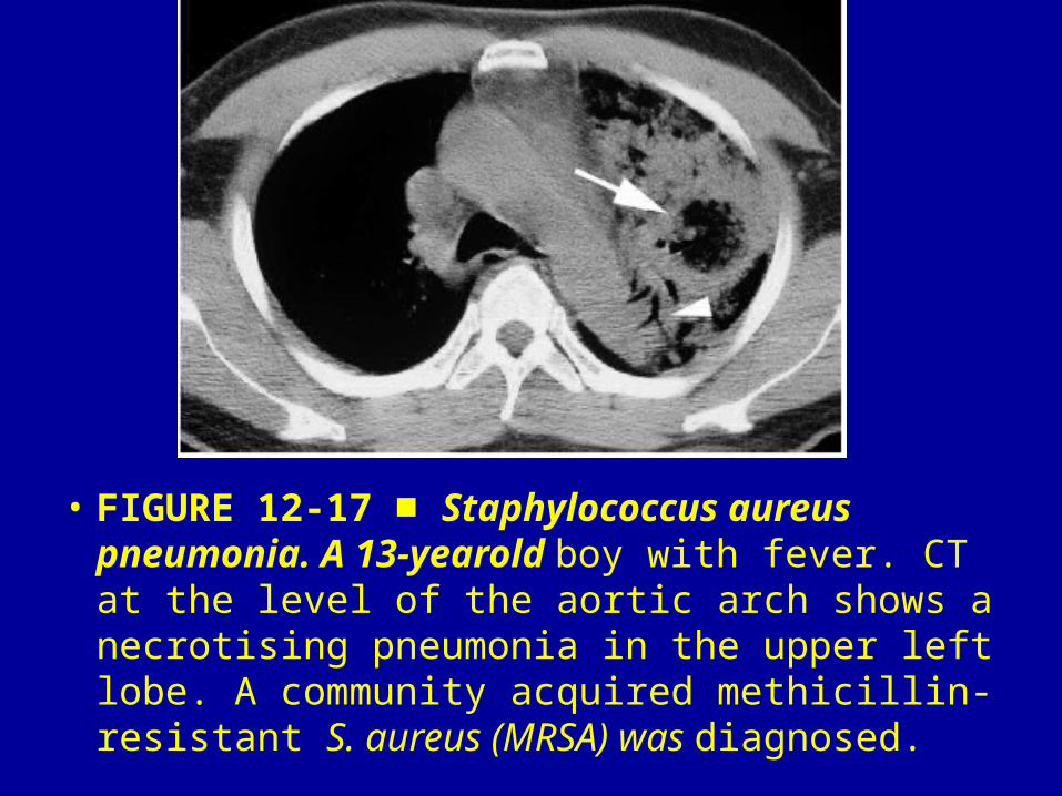
• FIGURE 12-17 ■ Staphylococcus aureus pneumonia. A 13-yearold boy with fever. CT at the level of the aortic arch shows a necrotising pneumonia in the upper left lobe. A community acquired methicillin-resistant S. aureus (MRSA) was diagnosed.

• FIGURE 12-18 ■ Escherichia coli pneumonia. A 54-year-old man with fever. Minimum intensity projection CT demonstrates multiple bilateral peripheral areas of consolidation (arrows)

• FIGURE 12-19 ■Pseudomonas aeruginosa pneumonia with abscess formation. A 45-year-old woman with chest pain and fever. Contrast-enhanced CT shows extensive dense consolidation in the right upper lobe with associated areas of necrosis and abscessification (arrows). Note vascular structures visible within the consolidated lung (arrowheads).

• FIGURE 12-20 ■ Haemophilus influenzae pneumonia. A 49-yearold man with fever. Posteroanterior chest radiograph shows bilateral areas of consolidation with ill-defined margins. A community-acquired H. influenzae pneumonia was diagnosed

• FIGURE 12-21 ■ Mycoplasma pneumonia. A 35-year-old man presents with non-productive cough and fever. CT shows airspace nodules, focal areas of lobular consolidation (arrows) and patchy ground-glass opacities (arrowhead).

• FIGURE 12-22 Swyer–James–MacLeod syndrome. A 61-yearold ■woman with chronic dyspnoea. Expiratory CT shows significant air trapping in the right lung with associated bronchiectasis (arrowheads). A focal area of air trapping is also visible in the superior segment of the left lower lobe. Note the contralateral shift of the mediastinum and anterior junction line (arrow).

• FIGURE 12-23 Adenovirus pneumonia. A 46-■year-old man with fever. Coronal reformatted CT shows bilateral multiple small branching centrilobular opacities, representing dilated peripheral bronchioles (arrows), associated with bilateral focal areas of consolidation (arrowheads).

• FIGURE 12-24 Respiratory syncytial virus ■pneumonia. A 43-year-old man after receiving allogeneic hematopoietic stem cell transplant. CT through the upper lobes shows bilateral areas of consolidation (arrows) and multiple ill-defined nodules (arrowheads). Note bilateral pleural effusion.

• FIGURE 12-25 Varicella pneumonia. A 30-year-old ■man with lymphoma and new development of fever and skin rash. CT of the lower lobes shows multiple, bilateral and randomly distributed well-defined small pulmonary nodules (arrows).

• FIGURE 12-26 Herpesvirus pneumonia. A 34-year-old ■severely immunocompromised patient with fever. CT at the level of the bronchus intermedius in a patient with herpesvirus infection shows multiple, bilateral and randomly distributed pulmonary nodules surrounded by a ‘halo’ of ground-glass opacity (arrows).
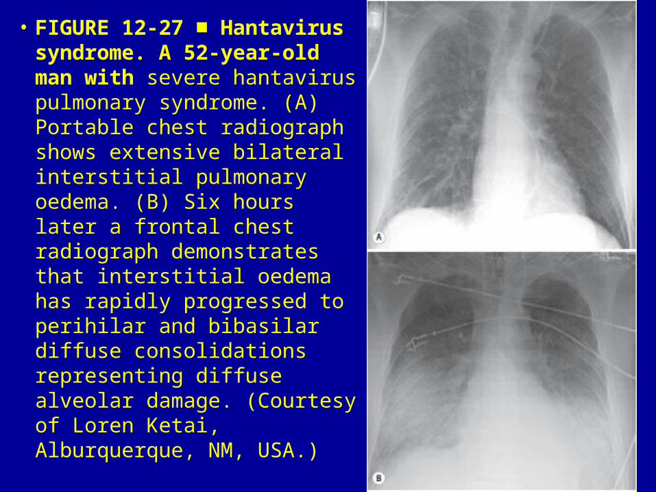
• FIGURE 12-27 Hantavirus ■syndrome. A 52-year-old man with severe hantavirus pulmonary syndrome. (A) Portable chest radiograph shows extensive bilateral interstitial pulmonary oedema. (B) Six hours later a frontal chest radiograph demonstrates that interstitial oedema has rapidly progressed to perihilar and bibasilar diffuse consolidations representing diffuse alveolar damage. (Courtesy of Loren Ketai, Alburquerque, NM, USA.)

• FIGURE 12-28 CMV pneumonia. A 25-year-■old man with acute myeloid leukaemia and hematopoietic stem cell transplantation. CT shows multiple scattered poorly defined nodules (arrows).

• FIGURE 12-29 Metapneumovirus pneumonia. A 34-year-■old man after receiving allogeneic hematopoietic stem cell transplant. CT obtained at level of right lower lobe shows multiple centrilobular branching opacities (tree-in-bud pattern) (arrows), and focal areas of consolidation.

• FIGURE 12-30 Severe acute respiratory syndrome (SARS). A ■previously healthy 43-year-old woman with dyspnoea and fever. Coronal reformatted CT shows bilateral ground-glass opacities involving both lower lungs.

• FIGURE 12-31 H1N1 pneumonia. A previously healthy 38-■yearold man with fever. CT shows patchy bilateral ground-glass opacities. Ring-like area of consolidation outline one of the ground-glass opacities (arrow) (‘reverse halo’ sign). The diagnosis of H1N1 was serologically confirmed.

• FIGURE 12-32 H1N1 obliterative bronchiolitis. A previously ■healthy 53-year-old woman with dyspnoea and fever. Inspiratory CT shows extensive bilateral areas of decreased attenuation and vascularity (mosaic perfusion/attenuation pattern). Expiratory CT confirmed air trapping (not shown). (Courtesy of Dr. Amador Prieto, Oviedo, Spain.)

• FIGURE 12-33 Immune reconstitution inflammatory ■syndrome (IRIS) in a patient with tuberculosis. Posteroanterior normal chest radiograph after initiation of HAART and before onset of IRIS (A). Follow-up chest radiograph (B) obtained 14 days later shows a significant enlargement of paratracheal lymph nodes (arrows).

• FIGURE 12-34 Primary TB. A 2-year-old boy ■with stridor and fever. Anteroposterior (A) and lateral (B) chest radiographs show bilateral enlargement of mediastinal lymph nodes (arrows).

• FIGURE 12-35 Reinfection tuberculosis. A 40-■year-old man presents with night sweats, non-productive cough and fever. CT shows extensive parenchymal consolidation of the left upper lobe with multiple cavities.

• FIGURE 12-36 Rasmussen aneurysm. A 65-year-old man with ■ chronic destructive pulmonary tuberculosis. Contrast-enhanced CT at the level of the right pulmonary artery shows a contrast filling aneurysm (arrow) within parenchymal consolidation in a superior segment of the right lower lobe. Note an associated parenchymal cavity (arrowhead).

• FIGURE 12-37 Endobronchial spread of tuberculosis. A 56-■yearold man with post-primary tuberculosis. CT shows a non cavitating consolidation in the left lung. Note typical images of ‘tree-in-bud’ opacities (arrows) and variable-sized bilateral nodular lesions (arrowheads).

• FIGURE 12-38 Miliary tuberculosis. A 26-■year-old man with fever and shortness of breath. CT shows a random distribution of multiple, discrete 1–2 mm in diameter nodules.
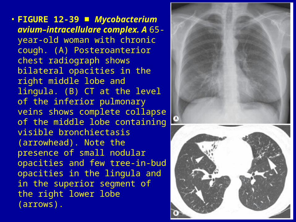
• FIGURE 12-39 ■ Mycobacterium avium–intracellulare complex. A 65-year-old woman with chronic cough. (A) Posteroanterior chest radiograph shows bilateral opacities in the right middle lobe and lingula. (B) CT at the level of the inferior pulmonary veins shows complete collapse of the middle lobe containing visible bronchiectasis (arrowhead). Note the presence of small nodular opacities and few tree-in-bud opacities in the lingula and in the superior segment of the right lower lobe (arrows).

• FIGURE 12-40 Intracavitary mycetoma (‘fungus ■ball’). A 68-yearold man with chronic lung disease and mild haemoptysis. CT shows a left upper lobe cavity containing a rounded soft-tissue opacity representing a fungus ball (arrows).

• FIGURE 12-41 Allergic bronchopulmonary ■aspergillosis. A 43-year-old asthmatic man with cough. Non-enhanced CT section shows a tubular opacity in the lingula containing a hyperdense mucoid impaction.

• FIGURE 12-42 Angioinvasive aspergillosis. A 65-■year-old man with immunosuppression and severe neutropenia presents with fever. CT shows a nodule with surrounding ground-glass attenuation (CT halo sign) (arrow). Note also bilateral poorly marginated peripheral areas of consolidation.

• FIGURE 12-43 ■ Candida pneumonia. A 52-year-old man who underwent bone marrow transplantation. CT shows multiple ill-defined bilateral nodules (arrows).

• FIGURE 12-44 ■ Pneumocystis pneumonia. A 45-year-old homosexual man with severe dyspnoea. CT shows extensive bilateral ground-glass opacities. Bronchoalveolar lavage showed P. jiroveci.

• FIGURE 12-45 Mucormycosis. A 62-year-old diabetic man ■with fever. A contrast-enhanced CT image shows lobar consolidation in the left upper lobe. An area of low attenuation is visible within the consolidation, consistent with abscess formation (arrow). Also note a small left pleural effusion (arrowhead).

• FIGURE 12-46 Cryptococcosis. A 24-year-old ■HIV-positive man with dyspnoea and fever. Close-up view of a posteroanterior chest radiograph shows diffuse small ill-defined nodules.
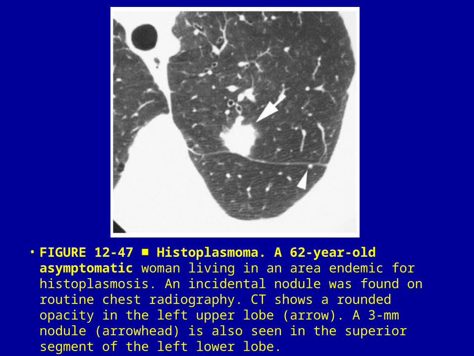
• FIGURE 12-47 Histoplasmoma. A 62-year-old asymptomatic ■woman living in an area endemic for histoplasmosis. An incidental nodule was found on routine chest radiography. CT shows a rounded opacity in the left upper lobe (arrow). A 3-mm nodule (arrowhead) is also seen in the superior segment of the left lower lobe.

• FIGURE 12-48 Coccidioidomycosis. A 38-year-old ■man with vague chest pain and fever. CT shows a parenchymal consolidation in the superior segment of the right lower lobe (arrow). Note multiple miliary nodules randomly distributed through both lungs.

• FIGURE 12-49 Paracoccidioidomycosis. A 61-year-■ol patient with cough and fever. CT at level of the lung bases shows combination of subsegmental areas of consolidation and randomly distributed bilateral ill-defined nodules of variable size. (Courtesy of Dr. Edson Marchiori, Rio de Janeiro, Brazil.)

• FIGURE 12-50 Ruptured hydatid cyst. A 65-year-old male ■shepherd with abrupt onset of expectoration and pruritus. Close-up view of the right upper lung shows a cystic lesion surrounded by a parenchymal consolidation due to a massive aspiration of intracystic content. Note a rounded opacity immediately above the fluid level (‘water lily’ sign) (arrows).

Related Documents