PULMONARY EDEMA Prepared by: South West Education Committee

PULMONARY EDEMA Prepared by: South West Education Committee.
Dec 14, 2015
Welcome message from author
This document is posted to help you gain knowledge. Please leave a comment to let me know what you think about it! Share it to your friends and learn new things together.
Transcript
PULMONARY EDEMA
Prepared by:
South West Education Committee

Congestive Heart Failureor
Acute Pulmonary Edema
SWEC Base Hospitals
Credit: W.A. (Bill) Penhallurick Southeastern Regional BHCredit: W.A. (Bill) Penhallurick Southeastern Regional BH

OUTLINE Review the pathophysiology and
etiology of Congestive Heart Failure Review the pathophysiology, etiology
and emergency treatment of Acute Pulmonary Edema
Review cardio-respiratory assessments Review the Acute Pulmonary Edema
protocol and the use of Nitroglycerin
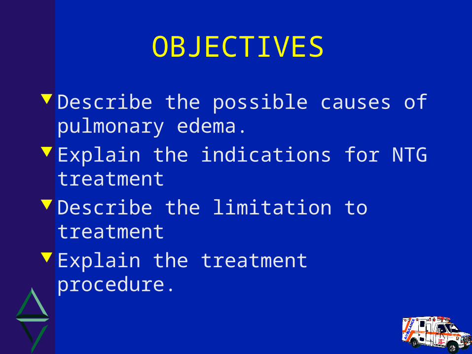
OBJECTIVES
Describe the possible causes of pulmonary edema.
Explain the indications for NTG treatment
Describe the limitation to treatment Explain the treatment procedure.

INTRODUCTION
Congestive Heart Failure (CHF)??? : A syndrome resulting from an
imbalance in pump function in which the heart fails to maintain an adequate circulation of blood.
Results in retention of fluid “congestion”.
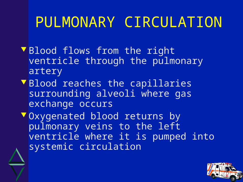
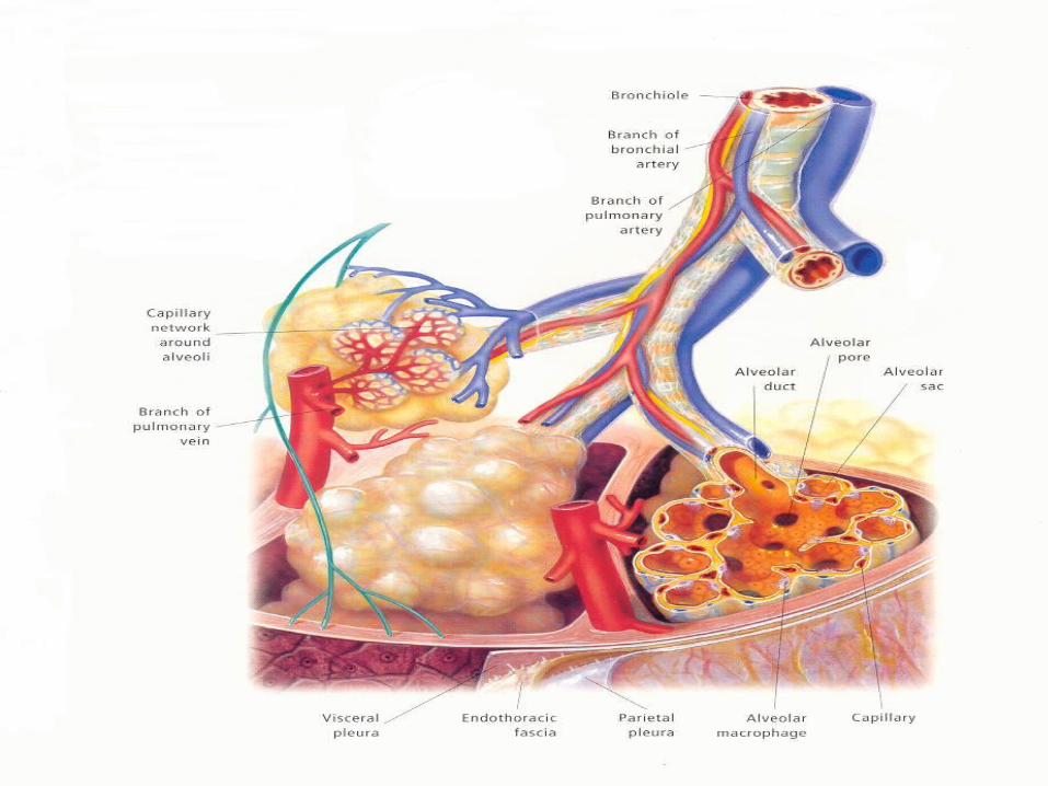
PULMONARY CIRCULATION
Blood flows from the right ventricle through the pulmonary artery
Blood reaches the capillaries surrounding alveoli where gas exchange occurs
Oxygenated blood returns by pulmonary veins to the left ventricle where it is pumped into systemic circulation

ETIOLOGY AND PATHOPHYSIOLOGY
Syndrome usually results from LV dysfunction and compensatory mechanisms
Cardiac performance is a function of 4 primary factors. What are they?

4 FACTORS DETERMINING CARDIAC PERFORMANCE
1. Preload (define)
2. Afterload (define)
3. Contractility
4. Heart Rate

Compensatory Mechanisms to Maintain Cardiac Output:
The Frank-Starling mechanism– Myocardial hypertrophy
Increased sympathetic tone– All result in increased myocardial O2 demand!
Kidneys
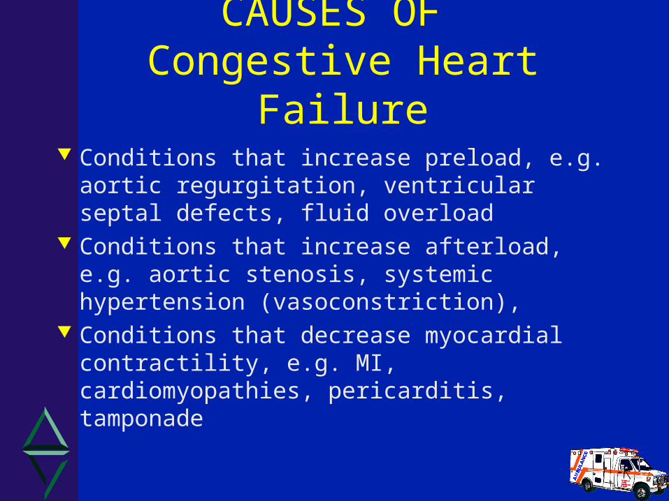
CAUSES OF Congestive Heart Failure
Conditions that increase preload, e.g. aortic regurgitation, ventricular septal defects, fluid overload
Conditions that increase afterload, e.g. aortic stenosis, systemic hypertension (vasoconstriction),
Conditions that decrease myocardial contractility, e.g. MI, cardiomyopathies, pericarditis, tamponade

SIGNS &SYMPTOMS OF Congestive Heart Failure
Exertional dyspnea usually with Crackles - fatigue may be the first sign Increased respiratory rate and effort Orthopnea and/or PND Cyanosis and pallor Tachycardia JVD Dependant edema
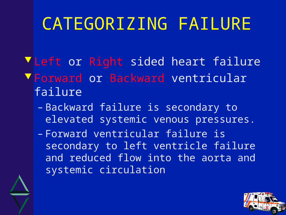
CATEGORIZING FAILURE
Left or Right sided heart failure Forward or Backward ventricular failure
– Backward failure is secondary to elevated systemic venous pressures.
– Forward ventricular failure is secondary to left ventricle failure and reduced flow into the aorta and systemic circulation
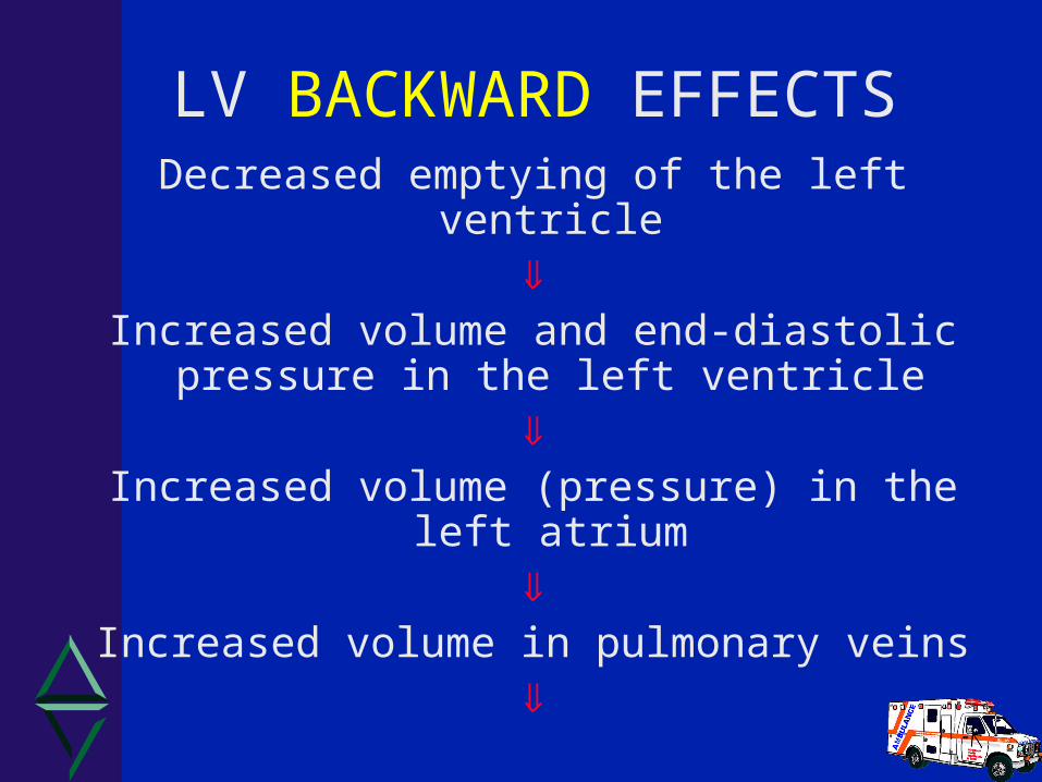
LV BACKWARD EFFECTSDecreased emptying of the left ventricle
Increased volume and end-diastolic
pressure in the left ventricle
Increased volume (pressure) in the left atrium
Increased volume in pulmonary veins

Increased volume in pulmonary capillary bed = increased hydrostatic pressure
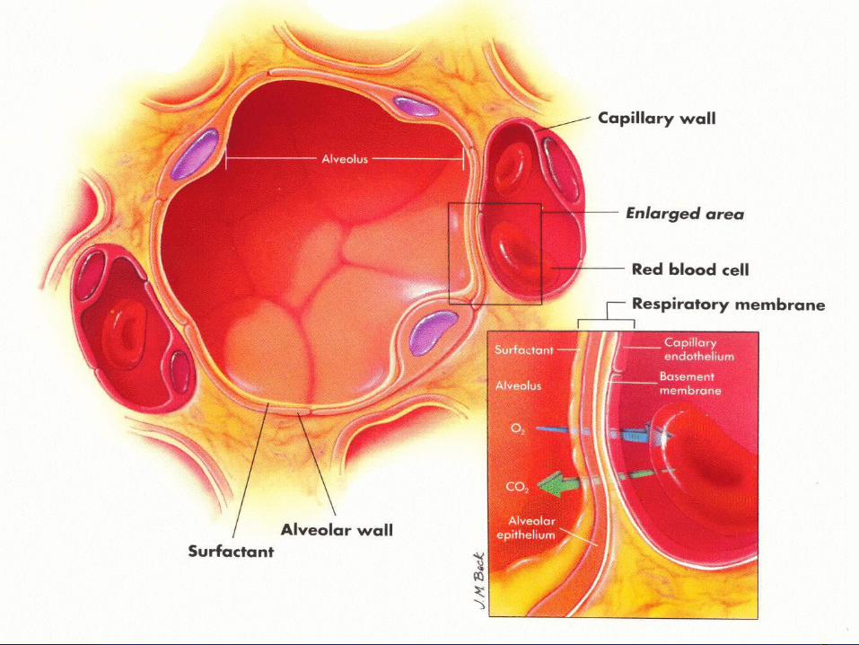
Transudation of fluid from capillaries to alveoli
Rapid filling of alveolar spaces
Pulmonary edema
LV BACKWARD EFFECTS con’t

LV FORWARD EFFECTSDecreased cardiac output
Decreased perfusion of tissues of body
Decreased blood flow to kidneys and glands
Increased reabsorption of sodium and water and
vasoconstriction
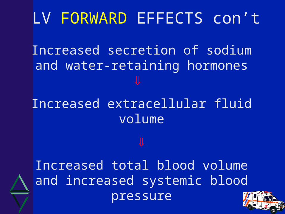
Increased secretion of sodium and water-retaining hormones
Increased extracellular fluid volume
Increased total blood volume and increased systemic blood pressure
LV FORWARD EFFECTS con’t

RV BACKWARD EFFECTSDecreased emptying of the right ventricle
Increased volume and end-diastolic pressure in
the right ventricle
Increased volume (pressure) in right atrium
Increased volume and pressure in the great veins

Increased volume in the systemic venous Increased volume in the systemic venous circulationcirculation
Increased volume in distensible organs Increased volume in distensible organs (hepatomegaly, splenomegaly)(hepatomegaly, splenomegaly)
Increased pressures at capillary lineIncreased pressures at capillary line
Peripheral, dependant edema and serous Peripheral, dependant edema and serous infusioninfusion
RV BACKWARD EFFECTS con’t
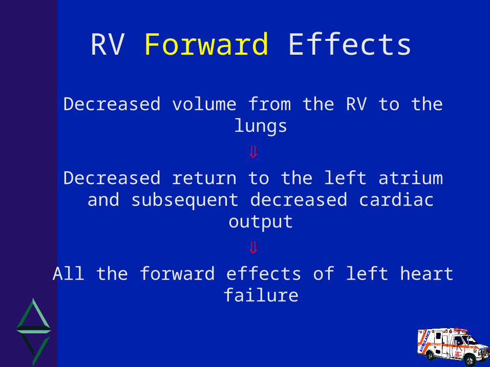
RV Forward Effects
Decreased volume from the RV to the lungs
Decreased return to the left atrium and
subsequent decreased cardiac output
All the forward effects of left heart failure
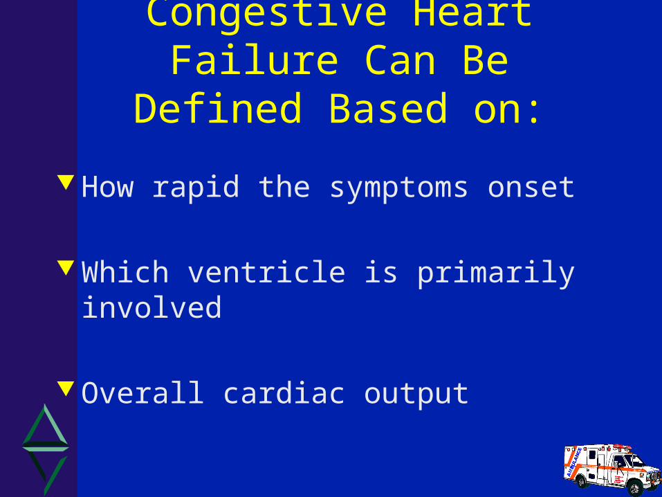
Congestive Heart Failure Can Be Defined Based on:
How rapid the symptoms onset
Which ventricle is primarily involved
Overall cardiac output
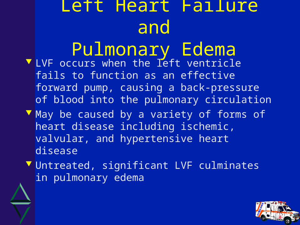
Left Heart Failure andPulmonary Edema
LVF occurs when the left ventricle fails to function as an effective forward pump, causing a back-pressure of blood into the pulmonary circulation
May be caused by a variety of forms of heart disease including ischemic, valvular, and hypertensive heart disease
Untreated, significant LVF culminates in pulmonary edema
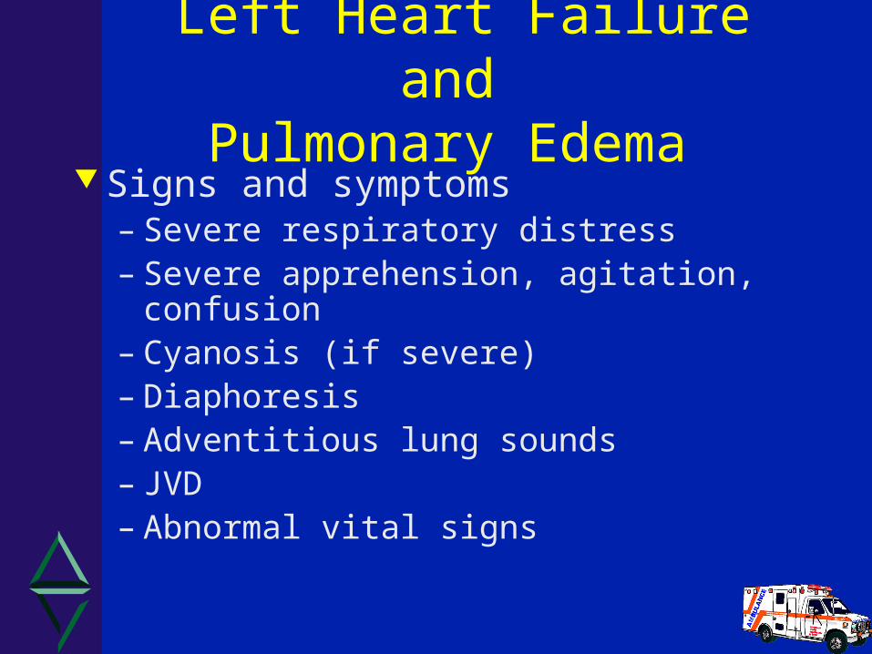
Left Heart Failure andPulmonary Edema
Signs and symptoms– Severe respiratory distress– Severe apprehension, agitation, confusion– Cyanosis (if severe)– Diaphoresis– Adventitious lung sounds– JVD– Abnormal vital signs

Right Heart Failure Occurs when the right ventricle fails as an
effective forward pump, causing back-pressure of blood into the systemic venous circulation
Can result from: – Chronic hypertension (in which LVF usually
precedes RVF)– COPD– Pulmonary embolism– Valvular heart disease– Right ventricular infarction
RVF most commonly results from LVF

Right Heart FailureSigns and symptoms
– Tachycardia– Venous congestion
• Engorged liver, spleen, or both• Venous distention; distention and pulsations
of the neck veins– Peripheral edema– Fluid accumulation in serous cavities– History-common signs and symptoms of acute
right-sided heart failure include chest pain, hypotension, and distended neck veins

CARDIOGENIC SHOCK
The most extreme form of pump failure Occurs when left ventricular function is
so compromised that the heart cannot meet the metabolic needs of the body
Usually caused by extensive myocardial infarction, often involving more than 40% of the left ventricle, or by diffuse ischemia
MAP drops below 70mmHg
New York Heart Association’s functional classification of
CHF

CLASS I
A patient who is not limited with normal physical activity by symptoms but has symptoms with exercise.
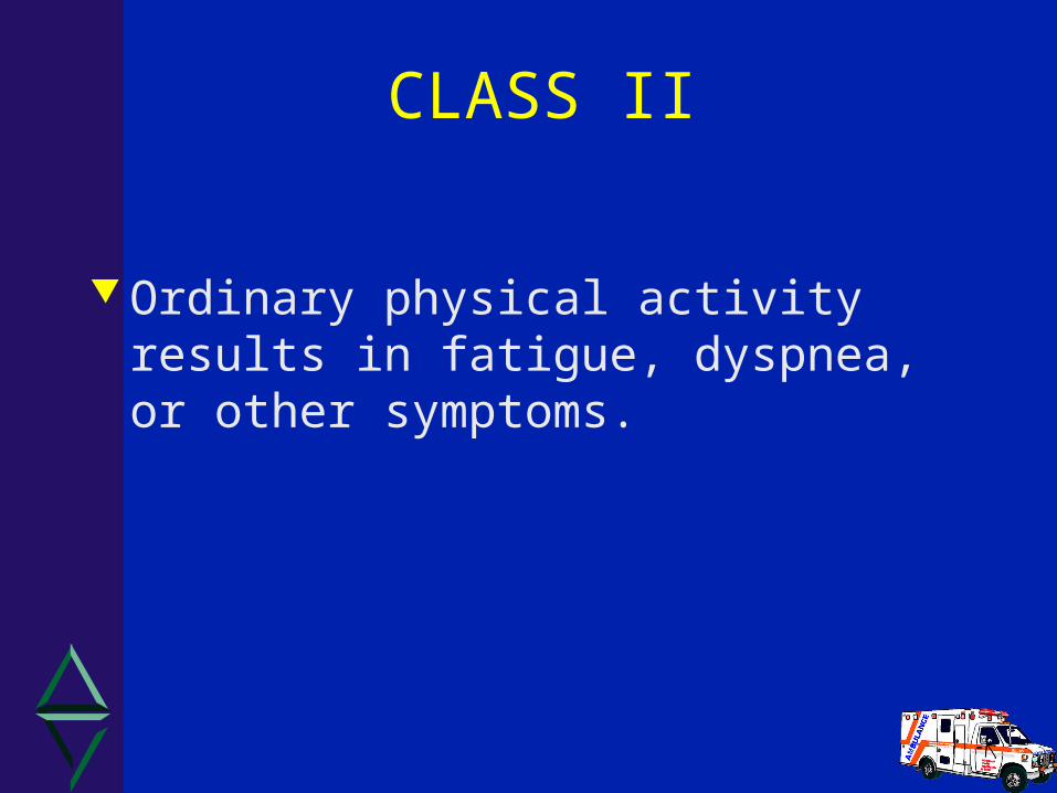
CLASS II
Ordinary physical activity results in fatigue, dyspnea, or other symptoms.
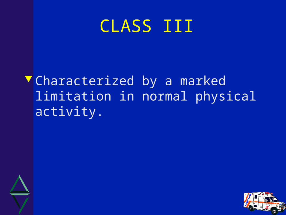
CLASS III
Characterized by a marked limitation in normal physical activity.

CLASS IV
Defined by symptoms at rest or with any physical activity.
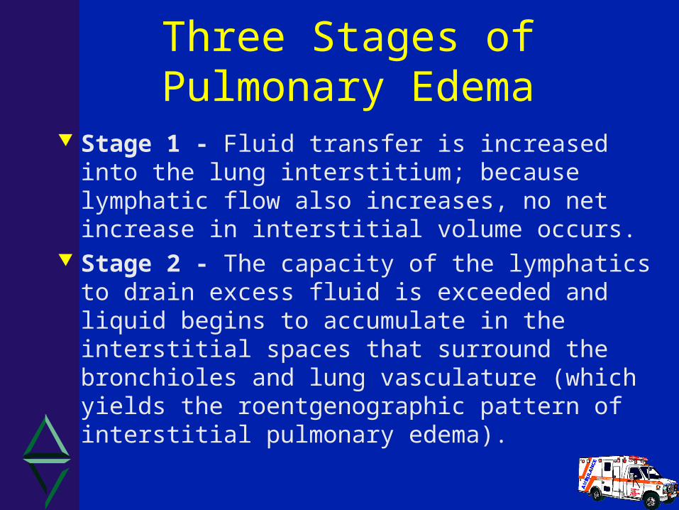
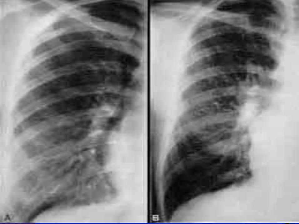
Three Stages of Pulmonary Edema
Stage 1 - Fluid transfer is increased into the lung interstitium; because lymphatic flow also increases, no net increase in interstitial volume occurs.
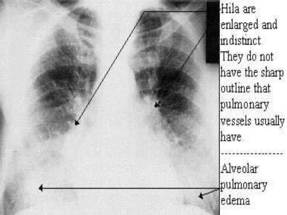
Stage 2 - The capacity of the lymphatics to drain excess fluid is exceeded and liquid begins to accumulate in the interstitial spaces that surround the bronchioles and lung vasculature (which yields the roentgenographic pattern of interstitial pulmonary edema).

Three Stages of Pulmonary Edema
Stage 3 - As fluid continues to build up, increased pressure causes it to track into the interstitial space around the alveoli.
Fluid first builds up in the periphery of the alveolar capillary membranes and finally floods the alveoli .
During stage 3 the x-ray picture of alveolar pulmonary edema is generated and gas exchange becomes impaired.
Three Stages of Pulmonary Edema
Stage 3 cont. Additionally gravity exerts an important influence on the fluid mechanics of the lung.
Blood is much denser than air and air-containing tissue
Under normal circumstances more perfusion occurs at the lung bases than at the apices; however, when pulmonary venous pressures rise and when fluid begins to accumulate at the lung bases the blood flow begins to be redistributed toward the apices.
Acute Pulmonary Congestion/ Pulmonary Edema

Mechanisms to Keep Interstitium and Alveoli Dry
Plasma oncotic pressure Connective tissue and cellular barriers
relatively impermeable to plasma proteins
Extensive lymphatic system

Acute Pulmonary Edema
May be CARDIAC or NON-CARDIAC in origin. Results from conditions such as:
– Increased pulmonary capillary pressure– Increased pulmonary capillary permeability– Decreased oncotic pressure– Lymphatic insufficiency– mixed or unknown mechanisms
Differential Diagnosis for APE: Cardiac causes of acute CHF COPD exacerbation Non-cardiac pulmonary edema: Tansudate vs. Exudate
– fluid overload– infection– ARDS– High altitude– Pulmonary Embolism– Pneumonia
CLINICAL PRESENTATION:
History Physical Exam EKG
• This should provide enough information to establish a cardiac etiology, if one exists!
HISTORICAL INFORMATION
Maintain a high clinical suspicion for ischemia or infarction – [# 1 cause of CHF (think ASA)]
Search for cardiac etiology A study of circadian patterns for
Cardiogenic acute pulmonary edema shows a significant peak for progressive symptoms and AMI between 06:00 - 11:59 (D.D. Buff, M.D. et all)
HISTORY Why did you call? What has changed? How long has the dyspnea been present? Was the onset gradual or abrupt? Is the dyspnea better or worse with position?
Is there associated orthopnea? Has the patient been coughing?
- If so, was the cough productive?
- What was the character and colour?
- Is there any hemoptysis?
- recent fever?

HISTORY Is there pain associated with the dyspnea?
- OPQRST for the pain Pt’s past history? Allergies Current Medications (pay close attention to
O2 therapy, oral bronchodilators, corticosteriods,Beta Blockers, Digitalis, ACE Inhibitors, Diuretics)

HISTORY
What is the patients normal level of activity?
How has the patient changed his/her environment to adjust to the disease?
- Pillow props
- Strategically placed chairs
- Meds within easy reach

Symptoms Suspicious of Pulmonary Congestion
Any complaint of dyspnea/ decreased exercise tolerance
PND/ Orthopnea Feeling of “suffocation” or air-hunger Restlessness and anxiety Cyanosis/Diaphoresis Pallor

Symptoms Suspicious of Pulmonary Congestion
Crackles Wheezing (Cardiac Asthma) Tachypnea Coughing (Dry cough may be med related) Retractions, accessory muscle use Frothy pink-tinged sputum

Physical Findings
Varying degrees of pulmonary and systemic vascular congestion and hypoperfusion
Classic patient with APE presents sitting “bolt” upright
Physical Findings ( cont. )
JVD Edema - ankle/pretibial vs sacral Ascites
- Positive Hepato-jugular reflex test BP and P are often markedly elevated Cardiac exam
– S3 or intermittent S4 may be present?– PMI may be shifted left
EKG Analysis:
Search for evidence of infarction or ischemia
Non-specific findings may include:– hypertrophy– chamber enlargement– conduction disturbances
CHEST XRAY:
Usually demonstrates increased heart size
Progression of pulmonary congestion:– first: Cephalization– second : Interstitial edema– third: Pulmonary (alveolar) edema

Treatment of APE:
First and foremost is to increase oxygen saturation
a reasonable approach is to base therapy on the Systolic Blood Pressure
Decrease the preload on the heart Shift and then eliminate excess fluids

Prehospital Management:
Patient sitting with legs dependent Supplemental O2 provided Cardiac monitoring/ Pulse oximetry Initiate necessary supportive therapy Nitroglycerin for APE if patient matches
protocol Be prepared to assist ventilations PPV is an effective treatment

Acute Pulmonary Edema Protocol - Indications
Patient in moderate to severe respiratory distress
Patient is assessed by the paramedic as being in Acute Pulmonary Edema

Acute Pulmonary Edema Protocol - Conditions
Weight > 40 Kg Patient has NOT taken
any erectile dysfunction medication within 48 hours
Heart rate greater then 60 & < 160 bpm
Initial and subsequent BP > 140 mmHg systolic
Acute Pulmonary Edema Protocol - Procedure
If the systolic blood pressure remains >140 mmHg - administer Nitroglycerin 0.4 mg spray SL every 5 minutes to a maximum of 6 doses.
Check the vital signs before administering EACH dose
NOTE: Do not administer further NTG if the systolic BP drops below 140 mmHg
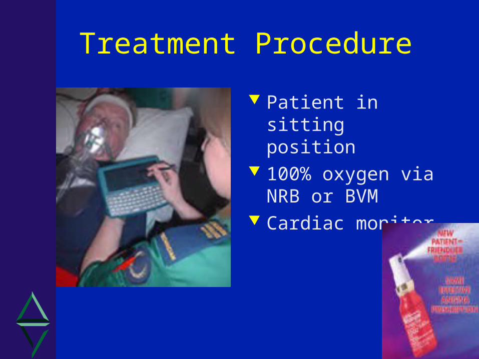
Treatment Procedure
Patient in sitting position
100% oxygen via NRB or BVM
Cardiac monitor

Limitations
Max of 6 doses of Nitro by Paramedic
Stop if– Systolic BP <140 mmhg– Drop in SBP by 1/3– Heart rate <60 or >160

Frequently Asked Questions
Q: If the patient is in Pulmonary Edema with crackles, can I give Salbutamol?

Answer
A. Continue with oxygen administration and NTG. Salbutamol is not the drug of choice.

Frequently Asked Questions
Q: What if I can only hear wheezing but suspect the patient is in Pulmonary Edema. Should I give Salbutamol?
Answer
A. Continue with oxygen administration. Consider the Acute Pulmonary Edema protocol and consult a BHP before administering Salbutamol if still uncertain.

QUESTIONS?
Related Documents