Pulmonary disorders, including vocal cord dysfunction Paul A. Greenberger, MD, and Leslie C. Grammer, MD Chicago, Ill The lung is a very complex immunologic organ and responds in a variety of ways to inhaled antigens, organic or inorganic materials, infectious or saprophytic agents, fumes, and irritants. There might be airways obstruction, restriction, neither, or both accompanied by inflammatory destruction of the pulmonary interstitium, alveoli, or bronchioles. This review focuses on diseases organized by their predominant immunologic responses, either innate or acquired. Pulmonary innate immune conditions include transfusion-related acute lung injury, World Trade Center cough, and acute respiratory distress syndrome. Adaptive immunity responses involve the systemic and mucosal immune systems, activated lymphocytes, cytokines, and antibodies that produce CD4 1 T H 1 phenotypes, such as for tuberculosis or acute forms of hypersensitivity pneumonitis, and CD4 1 T H 2 phenotypes, such as for asthma, Churg-Strauss syndrome, and allergic bronchopulmonary aspergillosis. (J Allergy Clin Immunol 2010;125:S248-54.) Key words: Innate, acquired, hypersensitivity, eosinophilia, lympho- cyte, tuberculosis, aspergillosis, bronchopulmonary, bronchiectasis, immunologic Pulmonary disorders can be organized according to whether the primary immune responses are characterized by innate or adap- tive immune responses. The innate responses use complement activation or activation of polymorphonuclear leukocytes (PMNs) and occur without a period for sensitization. The adaptive responses include T H 1 or T H 2 lymphocytes, eosinophils, antibody mediated, and granuloma formation. 1 This chapter will review the various pulmonary disorders with a predominant immunologic pattern and also discuss vocal cord dysfunction (VCD), which can coexist with asthma or occur independently and results in cough, shortness of breath, and dyspnea. INNATE IMMUNE RESPONSES Transfusion-related acute lung injury Transfusion-related acute lung injury (TRALI) is a nonhemo- lytic transfusion reaction that occurs within 10 minutes to as long as 6 hours after infusion of a blood product and causes very severe noncardiogenic pulmonary edema, cyanosis, arterial hypoxemia, and respiratory failure. 2,3 The donor plasma typically contains antibodies to human neutrophil antigens or HLA class I or II an- tigens. 2,3 Neutrophil alloantibodies are found in 10% to 20% of female donors and 1% to 4% of male donors, yet the incidence of TRALI is about 1:5000 transfusions. 3 Alloantibodies are gen- erated during pregnancy, but of course that would not explain the presence of such antibodies in men. Some recipients have anti- neutrophil antibodies. The immediate reaction, which might re- semble anaphylaxis, involves sequestration of PMNs in the pulmonary vasculature, complement activation, and generation of TGF-b, IL-8, and IL-13. 2 Immune complexes activate PMNs and cause disruption of the endothelium barrier to plasma. TRALI is extremely rare after intravenous immunoglobulin infusions but occurs with infusions of platelets (suspended in plasma), whole blood, cryoprecipitates, and fresh frozen plasma. The immediate management includes stopping the infusion, oxygen, mechanical ventilation if indicated, and treatment of hypotension with vasopressors. Donors should be deferred from future donations. Indeed, some transfusion experts have recom- mended that the donor pool should not include women who have been pregnant and that donor plasma be tested for alloanti- bodies. 2,3 Neither of these suggestions are standard practice. Acute respiratory distress syndrome and acute lung injury Acute respiratory distress syndrome (ARDS) and acute lung injury represent diffuse pulmonary disease that can be fatal. 4 ARDS is a more severe form of acute lung injury. Causes include sepsis, pneumonia, trauma, or aspiration pneumonia. 4 Patients experience severe dyspnea, tachypnea, and hypoxemia. The chest roentgeno- gram and computed tomographic (CT) examination demonstrate bilateral infiltrates, alveolar consolidation, and ‘‘white out’’ of the lung. The alveoli collapse as they become filled with protein and fi- brin-rich exudates (hyaline membranes), which inactivate surfac- tant. 4,5 Neutrophils release oxidant proteases, which damage the capillary endothelium. Bronchoalveolar lavage (BAL) reveals the presence of PMNs, procoagulant activity, IL-8 (chemotactic for PMNs), IL-2, IL-6, and TGF-b. There is reduced apoptosis of From the Division of Allergy-Immunology, Department of Medicine, Northwestern University Feinberg School of Medicine. Supported by the Ernest S. Bazley Grant to Northwestern Memorial Hospital and Northwestern University Disclosure of potential conflict of interest: P. A. Greenberger has served as an expert witness on the topics of immunotheraphy, remote practice, ABPA misdiagnosis, and anaphylaxis. L. Grammer has received research support from S&C Electric Company. Received for publication May 18, 2009; revised August 28, 2009; accepted for publica- tion September 2, 2009. Address for reprints: Paul A. Greenberger, MD, Division of Allergy-Immunology, 676 N St Clair Street, #14018, Chicago, IL 60611. E-mail: [email protected]. 0091-6749/$36.00 Ó 2010 American Academy of Allergy, Asthma & Immunology doi:10.1016/j.jaci.2009.09.020 Abbreviations used ABPA: Allergic bronchopulmonary aspergillosis ANCA: Antineutrophil cytoplasmic antibody ARDS: Acute respiratory distress syndrome BAL: Bronchoalveolar lavage COPD: Chronic obstructive pulmonary disease CSS: Churg-Strauss syndrome CT: Computed tomography FVC: Forced vital capacity HDAC: Histone deacetylase LT: Leukotriene PMN: Polymorphonuclear leukocyte RADS: Reactive airways dysfunction syndrome TLR: Toll-like receptor TRALI: Transfusion-related acute lung injury VCD: Vocal cord dysfunction S248

Welcome message from author
This document is posted to help you gain knowledge. Please leave a comment to let me know what you think about it! Share it to your friends and learn new things together.
Transcript
Pulmonary disorders, including vocal cord dysfunction
Paul A. Greenberger, MD, and Leslie C. Grammer, MD Chicago, Ill
The lung is a very complex immunologic organ and responds ina variety of ways to inhaled antigens, organic or inorganicmaterials, infectious or saprophytic agents, fumes, and irritants.There might be airways obstruction, restriction, neither, or bothaccompanied by inflammatory destruction of the pulmonaryinterstitium, alveoli, or bronchioles. This review focuses ondiseases organized by their predominant immunologicresponses, either innate or acquired. Pulmonary innate immuneconditions include transfusion-related acute lung injury, WorldTrade Center cough, and acute respiratory distress syndrome.Adaptive immunity responses involve the systemic and mucosalimmune systems, activated lymphocytes, cytokines, andantibodies that produce CD41 TH1 phenotypes, such as fortuberculosis or acute forms of hypersensitivity pneumonitis, andCD41 TH2 phenotypes, such as for asthma, Churg-Strausssyndrome, and allergic bronchopulmonary aspergillosis.(J Allergy Clin Immunol 2010;125:S248-54.)
Key words: Innate, acquired, hypersensitivity, eosinophilia, lympho-cyte, tuberculosis, aspergillosis, bronchopulmonary, bronchiectasis,immunologic
Pulmonary disorders can be organized according towhether theprimary immune responses are characterized by innate or adap-tive immune responses. The innate responses use complementactivation or activation of polymorphonuclear leukocytes (PMNs)and occur without a period for sensitization. The adaptiveresponses include TH1 or TH2 lymphocytes, eosinophils, antibodymediated, and granuloma formation.1 This chapter will review thevarious pulmonary disorders with a predominant immunologicpattern and also discuss vocal cord dysfunction (VCD), whichcan coexist with asthma or occur independently and results incough, shortness of breath, and dyspnea.
INNATE IMMUNE RESPONSESTransfusion-related acute lung injury
Transfusion-related acute lung injury (TRALI) is a nonhemo-lytic transfusion reaction that occurs within 10 minutes to as longas 6 hours after infusion of a blood product and causes very severenoncardiogenic pulmonary edema, cyanosis, arterial hypoxemia,and respiratory failure.2,3 The donor plasma typically contains
antibodies to human neutrophil antigens or HLA class I or II an-tigens.2,3 Neutrophil alloantibodies are found in 10% to 20% offemale donors and 1% to 4% of male donors, yet the incidenceof TRALI is about 1:5000 transfusions.3 Alloantibodies are gen-erated during pregnancy, but of course that would not explain thepresence of such antibodies in men. Some recipients have anti-neutrophil antibodies. The immediate reaction, which might re-semble anaphylaxis, involves sequestration of PMNs in thepulmonary vasculature, complement activation, and generationof TGF-b, IL-8, and IL-13.2 Immune complexes activate PMNsand cause disruption of the endothelium barrier to plasma. TRALIis extremely rare after intravenous immunoglobulin infusions butoccurs with infusions of platelets (suspended in plasma), wholeblood, cryoprecipitates, and fresh frozen plasma.
The immediate management includes stopping the infusion,oxygen, mechanical ventilation if indicated, and treatment ofhypotension with vasopressors. Donors should be deferred fromfuture donations. Indeed, some transfusion experts have recom-mended that the donor pool should not include women who havebeen pregnant and that donor plasma be tested for alloanti-bodies.2,3 Neither of these suggestions are standard practice.
Acute respiratory distress syndrome and acute lunginjury
Acute respiratory distress syndrome (ARDS) and acute lunginjury represent diffusepulmonary disease that can be fatal.4ARDSis a more severe form of acute lung injury. Causes include sepsis,pneumonia, trauma, or aspiration pneumonia.4 Patients experiencesevere dyspnea, tachypnea, and hypoxemia. The chest roentgeno-gram and computed tomographic (CT) examination demonstratebilateral infiltrates, alveolar consolidation, and ‘‘white out’’ of thelung. The alveoli collapse as they become filledwith protein and fi-brin-rich exudates (hyaline membranes), which inactivate surfac-tant.4,5 Neutrophils release oxidant proteases, which damage thecapillary endothelium. Bronchoalveolar lavage (BAL) reveals thepresence of PMNs, procoagulant activity, IL-8 (chemotactic forPMNs), IL-2, IL-6, and TGF-b. There is reduced apoptosis of
From the Division of Allergy-Immunology, Department of Medicine, NorthwesternUniversity Feinberg School of Medicine.
Supported by the Ernest S. Bazley Grant to Northwestern Memorial Hospital andNorthwestern University
Disclosure of potential conflict of interest: P. A. Greenberger has served as an expertwitness on the topics of immunotheraphy, remote practice, ABPA misdiagnosis, andanaphylaxis. L. Grammer has received research support from S&C Electric Company.
Received for publication May 18, 2009; revised August 28, 2009; accepted for publica-tion September 2, 2009.
Address for reprints: Paul A. Greenberger, MD, Division of Allergy-Immunology, 676 NSt Clair Street, #14018, Chicago, IL 60611. E-mail: [email protected].
0091-6749/$36.00! 2010 American Academy of Allergy, Asthma & Immunologydoi:10.1016/j.jaci.2009.09.020
Abbreviations usedABPA: Allergic bronchopulmonary aspergillosisANCA: Antineutrophil cytoplasmic antibodyARDS: Acute respiratory distress syndromeBAL: Bronchoalveolar lavage
COPD: Chronic obstructive pulmonary diseaseCSS: Churg-Strauss syndromeCT: Computed tomography
FVC: Forced vital capacityHDAC: Histone deacetylase
LT: LeukotrienePMN: Polymorphonuclear leukocyteRADS: Reactive airways dysfunction syndromeTLR: Toll-like receptor
TRALI: Transfusion-related acute lung injuryVCD: Vocal cord dysfunction
S248
PMNs, which is attributable to increased concentrations of BALfluid IL-2, IL-8, granulocyte colony-stimulating factor, GM-CSF,and growth-related oncogene a.6 Alternatively, there is enhancedapoptosis of epithelial cells, resulting in the lack of a sufficient bar-rier between the alveoli and capillaries. TNF-related apoptosis-in-duced ligand levels are increased in BAL fluid in patients withARDS and are recognized as proapoptotic for epithelial cells.6
Patients requiring mechanical ventilation benefit from smallervolumes, such as a tidal volume of 6 mL/kg, with positive end-expiratory pressures of 5 to 10 cmH2O. Fluid replacement shouldbe conservative. Corticosteroids and other interventions, such asnitric oxide and surfactants, are not effective.7
Community-acquired pneumoniaCommunity-acquired pneumonia presents with a productive
cough, fever, pleuritic chest pain, and abnormal chest roentgen-ographic results.8 On auscultation, there can be crackles and bron-chial breath sounds. Most pathogens include viruses,Streptococcus pneumoniae, Haemophilus influenzae, Myco-plasma pneumoniae, Staphylococcus aureus, and Legionellapneumophila.8 There might be no recovered organisms in somepatients. Comorbidities influence survival.8
Levels of proinflammatory cytokines, such as TNF-a and IL-6,and the anti-inflammatory cytokine IL-10 are increased in thosepatients who succumb compared with survivors.9 Impaired recog-nition ofmolecular patterns of bacteria is associatedwith decreasedactivation of innate immunity and worse clinical outcomes.10 Toll-like receptors (TLRs) recognize pathogen-associated molecularpatterns, and genetic polymorphisms have been identified inpatients who had invasive S pneumoniae infections.10 For example,polymorphisms of TLR4 impair its function in recognition of Spneumoniae pneumolysin, whereas polymorphisms of CD14, acoreceptor on monocytes for both TLR2 and TLR4, are associatedwith invasive S pneumoniae infections.10 Polymorphisms inFCgRIIA increase the susceptibility to invasive disease. Currenttherapy includes early administration of antibiotics and supportivecare. Future diagnosis might identify at-risk subjects proactively,and therapies will be able to strengthen the innate immune system.
NONINFECTIOUS PULMONARY CONDITIONSByssinosis occurs from the inhalation of dusts from flax,
cotton, sisal, and hemp. The dusts produce bronchoconstriction,typically on the first day of the workweek, but then tachyphylaxisdevelops with continued exposure. Byssinosis is not asthma orhypersensitivity pneumonitis.1 At-risk workers include thosewhoare exposed to endotoxin during the processing of raw cotton. Incontrast, workers who spin cotton are not exposed to the high con-centrations of endotoxin and are considered at low risk. Long-term exposure can result in symptoms of chronic bronchitis andcough. Modest reductions in FEV1 and forced vital capacity(FVC) have been found, but concurrent smoking appears to bethe major contributor as opposed toworkplace exposures. Preven-tion includes methods to reduce the generation of endotoxinsfrom gram-negative bacteria by reducing exposure to wastefrom cotton.
In contrast to byssinosis, the organic toxic dust syndrome is atoxic alveolitis that produces influenza-type symptoms of sudden-onset headache, chills, nonproductive cough, myalgias, arthral-gias, and dyspnea. Crackles can be present on lung auscultation.
The onset of symptoms is within 12 hours of inhalation of organicdusts. Although the clinical presentation might mimic that ofacute hypersensitivity pneumonitis, there is no requirement forprior exposure or immunologic sensitization (see the later sectionon hypersensitivity pneumonitis). Various circumstances of ex-posure have been described, such as from organic mulch,endotoxin-rich vegetables and grass seeds, and contaminatedseaweed. Massive inhalation of microbial products can cause anARDS-like presentation, and this is designated as organic dusttoxic syndrome or pulmonary mycotoxicosis.11
In patients with silo-unloader’s disease, there is inhalation ofnonorganic gases, such as NO, NO2, or N2O4. These nitrogen ox-ides thengeneratenitric andnitrous acids that causenoncardiacpul-monary edema and, in some patients, methemoglobinemia. Deathscan occur, whereas survivors might have bronchiolitis obliterans.
Grain-handler’s disease occurs in agricultural workers with achronic cough, symptoms of chronic bronchitis, or wheeze afterexposure to grain dusts. Concurrent cigarette smoking appears tobe more injurious to the lung and associated with reductions inspirometric values. Measures to reduce exposure to dust arebeneficial. Because of less implementation of safety standards,there is a major concern that workers will experience grain-handler’s disease and other respiratory disorders in the world’semerging economies.
Reactive airways dysfunction syndromeThe reactive airways dysfunction syndrome (RADS) describes
a single unexpected inhalation of high concentrations of irritantfumes, vapors, fog, or smoke that results in acute cough, dyspnea,andwheezingwithin 24 hours.12 An asthma-like syndrome beginsthat can last for months or years. Bronchial hyperreactivity can bedemonstrated by means of methacholine challenge testing, andspirometry reveals normal or decreased FEV1, FVC, and FEV1/FVC ratio. There might be little to no bronchodilator responseto albuterol. Bronchial biopsy specimens demonstrate loss of ep-ithelium, subepithelial fibrosis, and infiltrates with lymphocytesbut not eosinophils (as would be characteristic of asthma).
RADS might be confused with occupational asthma, wherethere is a sensitization period of months or years before symptomsbegin, and with aggravation of underlying asthma. But RADSrefers to the acute irritant-induced asthma.
World Trade Center coughThe first responders to the 2001 collapse of the World Trade
Center in New York City experienced a very troublesome coughwithin 24 hours of beginning rescue operations.13,14 The exposuresincluded acrid smoke, fires that burned for 3months, asbestos, glassfibers, lead, and aromatic compounds.Many responders did not useprotective masks. Subsequent evaluations identified methacholinehyperreactivity in 24% and a reduced FEV1/FVC ratio of lessthan 0.75 in 16% of affected subjects.13 The high exposures wouldbe consistent with a diagnosis of RADS in some subjects.14
VCDVCD is a form of ‘‘functional’’ or nonanatomic upper airway
obstruction.15 The inspiratory tracing on a flow-volume loop istruncated (Fig 1) or incompletely performed. Other causes of non-anatomic inspiratory obstruction include vocal cord paralysis,
J ALLERGY CLIN IMMUNOL
VOLUME 125, NUMBER 2
GREENBERGER AND GRAMMER S249
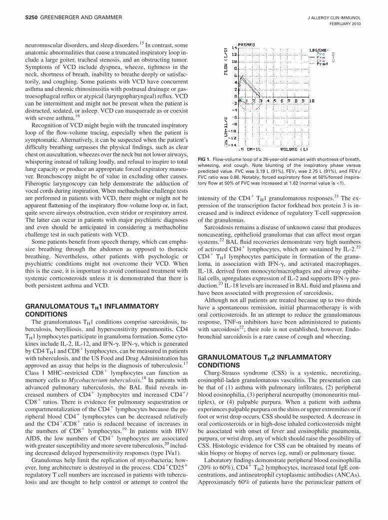
neuromuscular disorders, and sleep disorders.15 In contrast, someanatomic abnormalities that cause a truncated inspiratory loop in-clude a large goiter, tracheal stenosis, and an obstructing tumor.Symptoms of VCD include dyspnea, wheeze, tightness in theneck, shortness of breath, inability to breathe deeply or satisfac-torily, and coughing. Some patients with VCD have concurrentasthma and chronic rhinosinusitis with postnasal drainage or gas-troesophageal reflux or atypical (laryngopharyngeal) reflux. VCDcan be intermittent and might not be present when the patient isdistracted, sedated, or asleep. VCD can masquerade as or coexistwith severe asthma.16
Recognition of VCD might begin with the truncated inspiratoryloop of the flow-volume tracing, especially when the patient issymptomatic. Alternatively, it can be suspected when the patient’sdifficulty breathing surpasses the physical findings, such as clearchest on auscultation, wheezes over the neck but not lower airways,whispering instead of talking loudly, and refusal to inspire to totallung capacity or produce an appropriate forced expiratory maneu-ver. Bronchoscopy might be of value in excluding other causes.Fiberoptic laryngoscopy can help demonstrate the adduction ofvocal cords during inspiration. When methacholine challenge testsare performed in patients with VCD, there might or might not beapparent flattening of the inspiratory flow-volume loop or, in fact,quite severe airways obstruction, even stridor or respiratory arrest.The latter can occur in patients with major psychiatric diagnosesand even should be anticipated in considering a methacholinechallenge test in such patients with VCD.
Some patients benefit from speech therapy, which can empha-size breathing through the abdomen as opposed to thoracicbreathing. Nevertheless, other patients with psychologic orpsychiatric conditions might not overcome their VCD. Whenthis is the case, it is important to avoid continued treatment withsystemic corticosteroids unless it is demonstrated that there isboth persistent asthma and VCD.
GRANULOMATOUS TH1 INFLAMMATORYCONDITIONS
The granulomatous TH1 conditions comprise sarcoidosis, tu-berculosis, berylliosis, and hypersensitivity pneumonitis. CD4TH1 lymphocytes participate in granuloma formation. Some cyto-kines include IL-2, IL-12, and IFN-g. IFN-g, which is generatedby CD4 TH1 and CD8
1 lymphocytes, can be measured in patientswith tuberculosis, and the US Food and Drug Administration hasapproved an assay that helps in the diagnosis of tuberculosis.17
Class I MHC–restricted CD81 lymphocytes can function asmemory cells to Mycobacterium tuberculosis.18 In patients withadvanced pulmonary tuberculosis, the BAL fluid reveals in-creased numbers of CD41 lymphocytes and increased CD41/CD81 ratios. There is evidence for pulmonary sequestration orcompartmentalization of the CD41 lymphocytes because the pe-ripheral blood CD41 lymphocytes can be decreased relativelyand the CD41/CD81 ratio is reduced because of increases inthe numbers of CD81 lymphocytes.19 In patients with HIV/AIDS, the low numbers of CD41 lymphocytes are associatedwith greater susceptibility andmore severe tuberculosis,20 includ-ing decreased delayed hypersensitivity responses (type IVa1).
Granulomas help limit the replication of mycobacteria; how-ever, lung architecture is destroyed in the process. CD41CD251
regulatory T cell numbers are increased in patients with tubercu-losis and are thought to help control or attempt to control the
intensity of the CD41 TH1 granulomatous responses.21 The ex-pression of the transcription factor forkhead box protein 3 is in-creased and is indirect evidence of regulatory T-cell suppressionof the granulomas.
Sarcoidosis remains a disease of unknown cause that producesnoncaseating, epithelioid granulomas that can affect most organsystems.22 BAL fluid recoveries demonstrate very high numbersof activated CD41 lymphocytes, which are sustained by IL-2.22
CD41 TH1 lymphocytes participate in formation of the granu-loma, in association with IFN-g, and activated macrophages.IL-18, derived from monocyte/macrophages and airway epithe-lial cells, upregulates expression of IL-2 and supports IFN-g pro-duction.23 IL-18 levels are increased in BAL fluid and plasma andhave been associated with progression of sarcoidosis.
Although not all patients are treated because up to two thirdshave a spontaneous remission, initial pharmacotherapy is withoral corticosteroids. In an attempt to reduce the granulomatousresponse, TNF-a inhibitors have been administered to patientswith sarcoidosis22; their role is not established, however. Endo-bronchial sarcoidosis is a rare cause of cough and wheezing.
GRANULOMATOUS TH2 INFLAMMATORYCONDITIONS
Churg-Strauss syndrome (CSS) is a systemic, necrotizing,eosinophil-laden granulomatous vasculitis. The presentation canbe that of (1) asthma with pulmonary infiltrates, (2) peripheralblood eosinophilia, (3) peripheral neuropathy (mononeuritis mul-tiplex), or (4) palpable purpura. When a patient with asthmaexperiences palpable purpura on the shins or upper extremities or iffoot or wrist drop occurs, CSS should be suspected. A decrease inoral corticosteroids or in high-dose inhaled corticosteroids mightbe associated with onset of fever and eosinophilic pneumonia,purpura, or wrist drop, any of which should raise the possibility ofCSS. Histologic evidence for CSS can be obtained by means ofskin biopsy or biopsy of nerves (eg, sural) or pulmonary tissue.
Laboratory findings demonstrate peripheral blood eosinophilia(20% to 60%), CD41 TH2 lymphocytes, increased total IgE con-centrations, and antineutrophil cytoplasmic antibodies (ANCAs).Approximately 60% of patients have the perinuclear pattern of
FIG 1. Flow-volume loop of a 26-year-old woman with shortness of breath,wheezing, and cough. Note blunting of the inspiratory phase versuspredicted value. FVC was 3.19 L (91%), FEV1 was 2.75 L (91%), and FEV1/FVC ratio was 0.86. Notably, forced expiratory flow at 50%/forced inspira-tory flow at 50% of FVC was increased at 1.62 (normal value is <1).
J ALLERGY CLIN IMMUNOL
FEBRUARY 2010
S250 GREENBERGER AND GRAMMER
ANCAs, which on ELISA is positive for antibodies to myeloper-oxidase, whereas 10% of patients have positive results for cyto-plasmic staining, with antibodies directed against proteinase-3.1
Although the presence of a perinuclear pattern of ANCAs is help-ful in supporting a diagnosis, the ANCA titers do not provideprognostic information for disease management.24,25 Similarly,eosinophil-derived major basic protein and cationic proteinhave not been demonstrated to have utility in guiding treatment.24
Urinary concentrations of leukotriene (LT) E4, the major metab-olite of LTC4 and LTD4, and eosinophil-derived neurotoxin and3-bromotyrosine, a marker for oxidation of eosinophils, are in-creased in patients with CSS.26
The 6-year survival has been reported to be 70%.24 Long-termsurvival, up to 26 years, has also been reported.27 The most effec-tive therapy has been with oral corticosteroids.24,27 Additionalcorticosteroid-sparing and immunosuppressive therapies includecyclophosphamide, azathioprine, IFN-a, mepolizumab (anti–IL-5), omalizumab (anti-IgE), and rituximab (anti-CD20 Blymphocytes). There are potential untoward effects from cyclo-phosphamide (cytopenias, hemorrhagic cystitis, and malignancypotential), azathioprine (cytopenia, nausea, and vomiting), andIFN-a (depression and progressive multifocal leukoencephalopa-thy). Often patients can be managed long-term with prednisoneadministered on an alternate-day schedule with or without immu-nosuppressive therapy, such as with azathioprine. Abrupt discon-tinuation of prednisone is not advisable because it can result infever, eosinophilia, and pulmonary infiltrates within a few days,demonstrating that the CSS might be controlled but is not inremission.
TH1-RELATED INFLAMMATORY CONDITIONSHypersensitivity pneumonitis
Hypersensitivity pneumonitis is a CD41 TH1 and CD81 lym-phocyte–predominant alveolitis that results in noncaseastinggranulomas and pulmonary fibrosis. Clinical stages include acute,subacute (clinically similar to acute), and chronic. In the acuteand subacute stages inhalation of organic antigens causes cough,shortness of breath, myalgias, and fever within 4 to 6 hours. Thephysical examination would reveal pulmonary crackles. A patientmight self-treat for ‘‘flu’’ or be given an improper diagnosis ofcommunity-acquired pneumonia. When there is continued or re-peated exposure to antigens, such as bird excreta, patients mighthave subacute episodes or evolve into chronic hypersensitivitypneumonitis where typical flu-like illness does not occur. The lat-ter patients experience a nonproductive cough and progressivedyspnea and, in advanced cases, oxygen requirements. Pulmo-nary function tests in the acute and subacute stages typically aredescribed as restrictive; however, especially with bird fanciers,obstructive findings can occur and mimic asthma. The restrictivefindings are associated with a decreased diffusing capacity forcarbon monoxide. In contrast, the diffusing capacity for carbonmonoxide in patients with asthma is normal or even increased.
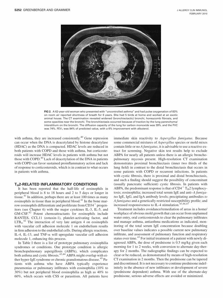
High-resolution CT scans demonstrate small nodules (<5 mm)that indicate alveolitis or areas of pulmonary fibrosis. Mosaicfindings of fibrosis are present in patients with chronic hypersen-sitivity pneumonitis. An example of pulmonary fibrosis andtraction bronchiectasis from avian hypersensitivity pneumonitisis shown in Fig 2.
There is striking BAL lymphocytosis of 60% to 80% fromacutely ill patients.28,29 The classic finding is a CD4/CD8 ratio of
less than 1, whereas in patients with sarcoidosis, the CD4/CD8 ra-tio is as high as 8 because of the CD41 alveolitis.30 In patientswith hypersensitivity pneumonitis, levels of TH1 cytokines are in-creased, including IL-12, IL-18, and TNF-a. CD81 lymphocytesserve as effector cells but are not sufficiently functional.28,31,32 Incontrast, in patients with chronic hypersensitivity pneumonitis,there can be an increase in the CD4/CD8 ratio as the CD4 (andTH2) lymphocytes increase and CD81 lymphocytes decrease.32
It has been suggested that the effector CD81 lymphocytes be-come ‘‘exhausted.’’ These data suggest that chronic hypersensitiv-ity pneumonitis is associated with ‘‘skewing’’ toward TH2lymphocytes, IL-4 production, and pulmonary fibrosis.32 IL-17,which is proinflammatory, increases activation and numbers ofneutrophils, and upregulates IL-6, IL-8, and TNF-a, might partic-ipate in hypersensitivity pneumonitis.33,34
Treatment includes early identification of patients with hyper-sensitivity pneumonitis, avoidance/remediation of the antigensinvolved, oral corticosteroids for short-term use, and monitoringof overall respiratory status depending on the stage that is present.
Chronic obstructive pulmonary diseaseChronic obstructive pulmonary disease (COPD) is character-
ized by fixed dyspnea, lack of fully reversible airways obstruction,and progressive loss of FEV1 over time. Cessation of cigarettesmoking and use of oxygen have proved of value. Pharmacother-apy includes short- and long-acting bronchodilators and anticho-linergic medications. For patients with moderate–to–very severeCOPD, when the FEV1 is less than 50% and the FEV1/FVC ratiois less than 70%, combination inhaled corticosteroid/long-actingb-agonist therapy is recommended. Treatment with combinationfluticasone propionate and salmeterol has resulted in fewer exac-erbations but not fewer deaths.35 In a study of patients with COPDin whom fluticasone/salmeterol or salmeterol was added to tio-tropium, therewas no additional benefit over tiotropium in the pri-mary outcome of exacerbations of COPD.36 Secondary outcomesdid demonstrate increases in FEV1, fewer hospitalizations, andimproved quality-of-life measures in those patients receiving flu-ticasone/salmeterol.36 An unexpected finding has been increasednumbers of cases of pneumonia in patients with COPD receivinghigh-dose fluticasone propionate.35,37
The pathogenesis of COPD includes cigarette smoking (mostcases), viral or bacterial infections (or a combination), geneticsusceptibility, oxidative stress, and little to no response to high-dose corticosteroids. Sputum often harbors PMNs, but eosino-phils can be present with either viral or combined viral andbacterial infections.38 In patients with COPD, not only is therepresence of PMNs and macrophages, there are also increases inCD4 TH1 and CD8 lymphocyte numbers.39
The impaired response to corticosteroids helps differentiatesCOPD from asthma in most cases. After absorption, the cortico-steroid binds to its receptor and traverses the cytoplasm and entersthe nucleus, where it interacts with glucocorticoid responseelements of DNA.40 Then corticosteroids can reduce levels ofthe proinflammatory transcription factors nuclear factor kB andactivator protein 1. It is thought that these transcription factorswould have been upregulated by viral upper respiratory tract in-fections. Transcription factors can be generated as the DNA-his-tone complex ‘‘unwinds’’ during a process of acetylation byhistone acetyltransferase.40 Histone acetyltransferase levels areincreased in some but not all cases of COPD, whereas in patients
J ALLERGY CLIN IMMUNOL
VOLUME 125, NUMBER 2
GREENBERGER AND GRAMMER S251
with asthma, they are increased consistently.40 Gene repressioncan occur when the DNA is deacetylated by histone deacetylase(HDAC) as the DNA is compacted. HDAC levels are reduced inboth patients with COPD and those with asthma, but corticoste-roids will increase HDAC levels in patients with asthma but notthose with COPD.40 Lack of deacetylation of the DNA in patientswith COPD can favor sustained proinflammatory action and lackof response to corticosteroids, which is in contrast to what occursin patients with asthma.
TH2-RELATED INFLAMMATORY CONDITIONSIt has been reported that the half-life of eosinophils in
peripheral blood is 8 to 18 hours and 2 to 5 days or longer intissue.41 In addition, perhaps there are at least 100 times as manyeosinophils in tissue than in peripheral blood.41 In the bone mar-row eosinophils differentiate and proliferate fromCD341 progen-itors (see Chapter 6) with the major cytokines IL-3, IL-5, andGM-CSF.42 Potent chemoattractants for eosinophils includeRANTES, CCL11 (eotaxin-1), platelet-activating factor, andLTB4.
42 The interaction of very late antigen 4 on eosinophilswith vascular cell adhesion molecule 1 on endothelium resultsin firm adhesion to the endothelial cells. During allergic reactions,IL-4, IL-13, and TNF-a will upregulate vascular cell adhesionmolecule 1, enhancing this process.
In Table I there is a list of prototype pulmonary eosinophiliasyndromes or conditions. One prototype condition is allergicbronchopulmonary aspergillosis (ABPA), which complicatesboth asthma and cystic fibrosis.43,44 ABPAmight overlap with ei-ther hyper-IgE syndrome or chronic granulomatous disease.45 Pa-tients with asthma who have ABPA typically experiencepneumonias or pulmonary infiltrates with eosinophilia (10% to30%) but not peripheral blood eosinophilia as high as 40% to60%, which occurs with CSS or parasitism. All patients have
immediate skin reactivity to Aspergillus fumigatus. Becausesome commercial mixtures of Aspergillus species or mold mixescontain little or no A fumigatus, it is advisable to use a reactive ex-tract for screening. Negative skin test results help to excludeABPA for nearly all patients unless there is an allergic broncho-pulmonary mycosis present. High-resolution CT examinationdemonstrates proximal bronchiectasis (inner two thirds of thelung field) in contrast to the distal bronchiectasis that occurs insome patients with COPD or recurrent infections. In patientswith cystic fibrosis, there is proximal and distal bronchiectasis,and such a finding should suggest the possibility of concomitant(usually pancreatic sufficient) cystic fibrosis. In patients withABPA, the predominant response is that of CD41 TH2 lymphocy-tosis; eosinophilia; increased total serum IgE and anti–A fumiga-tus IgE, IgG, and IgA antibody levels; precipitating antibodies toA fumigatus and a genetically restricted susceptibility profile; andincreased responsiveness to IL-4 stimulation.43,46,47
Treatment includes avoidance/remediation of areas in a home/workplace of obviousmold growth that can occur from unplannedwater entry, oral corticosteroids to clear the pulmonary infiltratesand manage asthma, antiasthma medications as indicated, mon-itoring of the total serum IgE concentration because doublingover baseline values indicates a possible current new pulmonaryinfiltrate, and assessment of pulmonary function and respiratorystatus over time.43 For initial treatment of a patient with newly di-agnosed ABPA, the dose of prednisone is 0.5 mg/kg given eachmorning for 1 to 2 weeks, with conversion to alternate day-ther-apy for 2 months. The radiographic findings can be expected toclear or be reduced, as demonstrated by means of high-resolutionCT examination in 2 months. Then the prednisone can be taperedand discontinued. It is not necessary to continue prednisone indef-initely in the absence of new infiltrates or development of severe(prednisone dependent) asthma. With use of the alternate-dayprednisone, serious adverse effects are avoided or minimized.
FIG 2. A 62-year-old woman who presented with ‘‘uncontrolled asthma’’ and had pulse oxygenation of 83%on room air reported shortness of breath for 6 years. She had 5 birds at home and worked at an exoticanimal house. The CT examination revealed widened (bronchiectactic) bronchi, honeycomb fibrosis, andsome opacities near the bronchi. The bronchiectasis occurred because of traction by the lung parenchyma/interstitium on the bronchi. The diffusion capacity of the lung for carbon monoxide was 39%, and the FVCwas 74%. FEV1 was 84% of predicted value, with a 6% improvement with albuterol.
J ALLERGY CLIN IMMUNOL
FEBRUARY 2010
S252 GREENBERGER AND GRAMMER
Antifungal therapies have been used for the treatment of ABPAand are considered adjunctive.48,49 A potentially good candidatefor antifungal therapy is a patient with sputum plugs harboringA fumigatus despite prednisone therapy. There are reports of theuse of omalizumab50 for patients with ABPA, but it remains tobe established whether this treatment will help to prevent newinfiltrates or improve asthma symptoms.
Eosinophilic pneumonias are divided into 4 types: acute,chronic, simple, and tropical (Table I).Acute eosinophilic pneumo-nia canmasquerade as severe community-acquired pneumonia andpresentwith respiratory failure.When there is noor little peripheralblood eosinophilia, the diagnosis can be made with bronchoscopyandBAL showing eosinophilia of 25% to 60%.Alternatively, theremight be peripheral blood eosinophilia as high as 42%.51 Drugs,nonprescription products, parasitism, and other causes of wide-spread pulmonary infiltrates should be considered.
Chronic eosinophilic pneumonia is characterized by respira-tory symptoms for at least 2 weeks, peripheral blood eosinophiliaof at least 1000/mm3 or BAL eosinophilia of greater than 25%,and bilateral pulmonary infiltrates.52 In classic presentations theinfiltrates are in the periphery, suggesting the photographic nega-tive of pulmonary edema. Most patients require years of oral cor-ticosteroid treatment. The radiographic infiltrates and surges ofperipheral blood eosinophilia can be controlled with modestdoses of prednisone.
Simple pneumonia (Loffler syndrome) is a mild conditionlasting less than 4 weeks and has transient pulmonary infiltrates.
Tropical pulmonary eosinophilia is characterized by wide-spread pulmonary infiltrates and high levels of peripheral bloodeosinophilia. Mediastinal lymph nodes might be enlarged and canharbor activated eosinophils. Patients typically have lived inendemic areas of parasites before tropical pulmonary eosinophiliaoccurs.
SUMMARYThe immunologic features of pulmonary disorders can be used
to categorize various conditions and provide focus for potentialinnovative therapies. Although usually there is not a singletreatment that antagonizes a critical component of either the
innate or acquired immune system and results in clinical im-provement, complex conditions might be amenable to immuno-logically based treatments in the future. A more ambitious goal isprimary prevention of many of the pulmonary conditionsdiscussed in this chapter. The ability to diagnose pulmonaryconditions and the masquerader of asthma, VCD, continues toimprove, which should result in earlier diagnoses and improvedoutcomes.
REFERENCES1. Greenberger PA. 7. Immunologic lung disease. J Allergy Clin Immunol 2008;
121(suppl):S393-7.2. Eder AF, Herron R, Strupp A, Dy B, Notari EP, Chambers LA, et al. Transfusion-
related acute lung injury surveillance (2003-2005) and the potential impact of theselective use of plasma of male donors in the American Red Cross. Transfusion2007;47:599-607.
3. Eder AF, Benjamin RJ. TRALI risk reduction: donor and component managementstrategies. J Clin Apher 2009;24:122-9.
4. Tsushima K, King LS, Aggarwal NR, De Gorordo A, D’Alessio FR, Kubo K.Acute lung injury review. Intern Med 2009;48:621-30.
5. Bastarache JA, Wang L, Geiser T, Wang Z, Albertine KH, Matthay MA, et al. Thealveolar epithelium can initiate the extrinsic coagulation cascade through expres-sion of tissue factor. Thorax 2007;62:608-16.
6. Lee KS, Choi YH, Kim YS, Baik SH, Oh YJ, Sheen SS, et al. Evaluation of bron-choalveolar lavage fluid from ARDS patients with regard to apoptosis. Respir Med2008;102:464-9.
7. Calfee CS, Matthay MA. Nonventilatory treatments for acute lung injury andARDS. Chest 2007;131:913-20.
8. Mandell LA, Wunderink RG, Anzueto A, Bartlett JG, Campbell GD, Dean NC,et al. Infectious Diseases Society of America/American Thoracic Society consen-sus guidelines on the management of community-acquired pneumonia in adults.Clin Infect Dis 2007;44(suppl):S27-72.
9. Reade MC, Yende S, D’Angelo G, Kong L, Kellum JA, Barnato AE, et al. Differ-ences in immune response may explain lower survival among older men with pneu-monia. Crit Care Med 2009;37:1655-62.
10. Yuan FF, Marks K, Wong M, Watson S, de Leon E, McIntyre PB, et al. Clinicalrelevance of TLR2, TLR4, CD14 and FCgRIIA gene polymorphisms in Strepto-coccus pneumoniae infection. Immunol Cell Biol 2008;86:268-70.
11. Perry LP, Iwata M, Tazelaar HD, Colby TV, Yousem SA. Pulmonary toxicosis: aclinicopathologic study of three cases. Mod Pathol 1998;11:432-6.
12. Brooks SM, Weiss MA, Bernstein IL. Reactive airways dysfunction syndrome(RADS). Persistent asthma syndrome after high level irritant exposures. Chest1985;88:376-84.
13. Prezant DJ, Weiden M, Banauch GI, McGuinness G, Rom WN, Aldrich TK, et al.Cough and bronchial responsiveness in firefighters at the world trade center site.N Engl J Med 2002;347:806-15.
14. Banauch GI, Alleyne D, Sanchez R, Olender K, Cohen HW, Weiden M, et al. Per-sistent hyperreactivity and reactive airways dysfunction in firefighters at the worldtrade center. Am J Respir Crit Care Med 2003;168:54-62.
15. Sterner JB, Morris MJ, Sill JM, Hayes JA. Inspiratory flow-volume curve evalua-tion for detecting upper airway disease. Respir Care 2009;54:461-6.
16. Moore WC, Peters SP. Severe asthma: an overview. J Allergy Clin Immunol 2006;117:487-94.
17. Menzies D, Pai M, Comstock G. Meta-analysis: new tests for the diagnosis of la-tent tuberculosis infection: areas of uncertainty and recommendations for research.Ann Intern Med 2007;146:340-54.
18. Guyot-Revol V, Innes JA, Hackforth S, Hinks T, Lalvani A. Regulatory T cells areexpanded in blood and disease sites in patients with tuberculosis. Am J Respir CritCare Med 2006;173:803-10.
19. Tsao TCY, Chen H-C, Jong J-H, Hsieh M-J, Tsao K-C, Lee C- H. Shifts of T4/T8T lymphocytes from BAL fluid and peripheral blood by clinical grade in patientswith pulmonary tuberculosis. Chest 2002;122:1285-91.
20. Glynn JR, Murray J, Bester A, Nelson G, Shearer S, Sonnenberg P. Effects of du-ration of HIV infection and secondary tuberculosis transmission on tuberculosis in-cidence in the South African gold mines. AIDS 2008;22:1859-67.
21. Richeldi L. An update on the diagnosis of Tuberculosis infection. Am J Respir CritCare Med 2006;174:736-42.
22. Callejas-Rubio JL, Lopez-Perez L, Ortego-Centeno N. Tumor necrosis factor-alphainhibitor treatment for sarcoidosis. Ther Clin Risk Manag 2008;4:1305-13.
23. Kieszko R, Krawczyk P, Jankowska O, Chocholska S, Krol A, Milanowski J. Theclinical significance of interleukin 18 assessment in sarcoidosis patients. RespirMed 2007;101:722-8.
TABLE I. Pulmonary eosinophilia syndromes or conditions
Asthma (allergic and nonallergic)Asthma with atelectasis from mucus pluggingABPAAllergic bronchopulmonary mycosisCSSCollagen vascular disease (rare)Drug allergy with pulmonary eosinophiliaEosinophilic pneumonia
Acute (BAL fluid eosinophilia 25% to 60% with little or no peripheralblood eosinophilia)
Chronic (high peripheral blood eosinophilia)Simple eosinophilia (Loffler syndrome)Tropical pulmonary eosinophilia
Hypereosinophilic syndromes (interstitial infiltrates and pleural effusions,thromboembolism)
NeoplasmsParasitism (helminthic)Sarcoidosis (very rare)
Adapted with permission from Greenberger.1
J ALLERGY CLIN IMMUNOL
VOLUME 125, NUMBER 2
GREENBERGER AND GRAMMER S253
24. Klion AD, Bochner BS, Gleich GJ, Nutman TB, Rothenberg ME, Simon H-U,et al. Approaches to the treatment of hypereosinophilic syndrome: a workshopsummary report. J Allergy Clin Immunol 2006;117:1292-302.
25. Guillevin L, Cohen P, Gayraud M, Lhote F, Jarrousse B, Cassassus P. Churg-Strauss syndrome. Clinical study and long-term follow-up of 96 patients. Medicine(Baltimore) 1999;78:26-37.
26. Highashi N, Mita H, Taniguchi M, Turikisawa N, Higashi A, Ozawa Y, et al. Uri-nary eicosanoid and tyrosine derivative concentrations in patients with vasculitides.J Allergy Clin Immunol 2004;114:1353-8.
27. Vemuri P, Greenberger PA, Patterson R. Churg-Strauss syndrome: survival for 26years. Ann Allergy Asthma Immunol 2002;88:640-3.
28. Fink JN, Ortega HG, Reynolds HY, Cormier YF, Fan LL, Franks TJ, et al. Needsand opportunities for research in hypersensitivity pneumonitis. Am J Respir CritCare Med 2005;171:792-8.
29. Agostini C, Calabrese F, Poletti V, Marcer G, Facco M, Miorin M. CXCR3/CXCL10 interactions in the development of hypersensitivity pneumonitis. RespirRes 2005;6:20.
30. Hamagami S, Miyagawa T, Ochi T, Tsuyuguchi I, Kishimoto S. A raised level ofsoluble CD8 in bronchoalveolar lavage in summer-type hypersensitivity pneumo-nitis. Am J Respir Crit Care Med 1999;159:1830-4.
31. Chen B, Tong Z, Nakamura S, Costabel V, Guzman J. Production of IL-12, IL-18and TNF-alpha by alveolar macrophages in hypersensitivity pneumonitis. Sarcoid-osis Vasc Diffuse Lung Dis 2004;21:199-203.
32. Barrera L, Mendoza F, Zuniga J, Estrada A, Zamora AC, Melendro EI, et al. Func-tional diversity of T-cell subpopulations in subacute and chronic hypersensitivitypneumonitis. Am J Respir Crit Care Med 2008;177:44-55.
33. Joshi AD, Fong DJ, Oak SR, Trujillo G, Flaherty KR, Martinez FJ, et al. Interleu-kin-17-mediated immunopathogenesis in experimental hypersensitivity pneumoni-tis. Am J Respir Crit Care Med 2009;179:705-16.
34. Selman M, Pardo A, Barrera L, Estrada A, Watson SR, Wilson K, et al. Gene ex-pression profiles distinguish idiopathic pulmonary fibrosis from hypersensitivitypneumonitis. Am J Respir Crit Care Med 2006;173:188-98.
35. Calverly PM, Anderson JA, Celli B, Ferguson GT, Jenkins C, Jones PW. Salmeteroland fluticasone propionate and survival in chronic obstructive pulmonary disease.N Engl J Med 2007;356:775-89.
36. Aaron SD, Vandemheen KL, Fergusson D, Maltais F, Bourdeau J, Goldstein R,et al. Tiotropium in combination with placebo, salmeterol, or fluticasone-salme-terol for treatment of chronic obstructive pulmonary disease: a randomized trial.Ann Intern Med 2007;146:545-55.
37. Weddzicha JA, Calverly PM, Seemungal TA, Hagan G, Ansari Z, Stockley RA,et al. The prevention of chronic obstructive pulmonary disease exacerbations bysalmeterol/fluticasone propionate or tiotropium bromide. Am J Respir Crit CareMed 2008;177:19-26.
38. Papi A, Bellettato CM, Braccioni F, Romagnoli M, Casolari P, Caramori G, et al.Infections and airway inflammation in chronic obstructive pulmonary disease se-vere exacerbations. Am J Respir Crit Care Med 2006;173:1114-21.
39. Fabbri LM, Luppi F, Beghe B, Rabe KF. Update in chronic obstructive pulmonarydisease 2005. Am J Respir Crit Care Med 2006;173:1056-65.
40. Ito K, Ito M, Elliott WM, Cosio B, Caramori G, Kon OM, et al. Decreased histonedeacetylase activity in chronic obstructive pulmonary disease. N Engl J Med 2005;352:1967-76.
41. Kita H, Adolphson CR, Gleich GJ. Biology of eosinophils. In: Adkinson NF, Yun-ginger JW Jr, Busse WW, Bochner BS, Holgate ST, Simons FER, editors. 6th ed.Philadelphia: Mosby; 2003. p. 305-32 Middleton’s allergy: principles andpractice.
42. Prussin C, Metcalfe DD. IgE, mast cells, basophils, and eosinophils. J Allergy ClinImmunol 2006;117(suppl):S450-6.
43. Greenberger PA. Allergic bronchopulmonary aspergillosis. J Allergy Clin Immunol2002;110:685-92.
44. Stevens DA, Moss RB, Kurup VP, Knutsen AP, Greenberger PA, Judson MA, et al.Allergic bronchopulmonary aspergillosis in cystic fibrosis—state of the art: Cystic Fi-brosis Foundation Consensus Conference. Clin Infect Dis 2003;37(suppl 3):S225-64.
45. Eppinger TM, Greenberger PA, White DA, Brown AE, Cunningham-Rundles C.Sensitization to Aspergillus species in the congenital neutrophil disorders chronicgranulomatous disease and hyper-IgE syndrome. J Allergy Clin Immunol 1999;104:1265-72.
46. Knutsen AP, Kariuki B, Consolino JD, Warrier MR. IL-4 alpha chain receptor (IL-4Ralpha) polymorphisms in allergic bronchopulmonary aspergillosis. Clin Mol Al-lergy 2006;4:3.
47. Kurup VP, Knutsen AP, Moss RB, Bansal NK. Specific antibodies to recombinantallergens of Aspergillus fumigatus in cystic fibrosis patients with ABPA. Clin MolAllergy 2006;4:11.
48. Wark PA, Hensley MJ, Saltos N, Boyle MJ, Toneguzzi RC, Epid GD, et al.Anti-inflammatory effect of itraconazole in stable allergic bronchopulmonaryaspergillosis: a randomized controlled trial. J Allergy Clin Immunol 2003;111:952-7.
49. Moss RB. Critique of trials in allergic bronchopulmonary aspergillosis and fungalallergy. Med Mycol 2006;44:269-72.
50. van der Ent CK, Hoekstra H, Rijkers GT. Successful treatment of allergic broncho-pulmonary aspergillosis with recombinant anti-IgE antibody. Thorax 2007;62:276-7.
51. Shorr AF, Scoville SL, Cersovsky SB, Shanks GD, Ochenhouse CF, Smoak BL,et al. Acute eosinophilic pneumonia in US Military personnel deployed in ornear Iraq. JAMA 2004;292:2997-3005.
52. Tsuirkisawa N, Saito H, Tsuburai T, Oshikata C, Ono E, Mitomi H, et al. Differ-ences in regulatory T cells between Churg-Strauss syndrome and chronic eosino-philic pneumonia with asthma. J Allergy Clin Immunol 2008;122:610-6.
J ALLERGY CLIN IMMUNOL
FEBRUARY 2010
S254 GREENBERGER AND GRAMMER
Mucosal immunology, eosinophilic esophagitis, and otherintestinal inflammatory diseases
Dan Atkins, MD,a,b,d and Glenn T. Furuta, MDa,b,c,d Denver and Aurora, Colo
The gastrointestinal mucosa constitutes the largest host-environment interface of the body. It uses both innate andadaptive immune mechanisms to provide protection from thediverse onslaught of foods, microbes, and other ingestedproducts. The innate immune system is genetically encoded andevolutionarily ancient, possesses no memory, and lacks diversity.In contrast, the adaptive immune system is quite diverse,develops memory, and undergoes expansion after stimulation.The gastrointestinal mucosa is charged with the difficult task ofmounting protective responses against invading microorganismswhile simultaneously maintaining an overall state ofnonresponsiveness or tolerance to innocuous substances, such ascommensal bacteria and food antigens. Perturbation ormalfunction of these complex protective mechanisms results indiseases, such as inflammatory bowel diseases, celiac disease, oreosinophilic gastrointestinal diseases. (J Allergy Clin Immunol2010;125:S255-61.)
Key words: Mucosal immunity, eosinophilic esophagitiseosinophilic gastrointestinal diseases
OVERVIEW OF GUT-ASSOCIATED LYMPHOIDTISSUE
Mucosa-associated lymphoid tissues comprise the largestimmune organ of the body and are active at multiple host-environment interfaces, such as the gastrointestinal tract and thegenitourinary and bronchopulmonary systems. A discussion ofthe site-specific aspects of each component of the mucosa-associated lymphoid tissue is beyond the scope of this Primer,but the reader is referred to a number of outstanding reviews onthese topics.1-6 Here we will briefly review the gastrointestinalmucosal immune system and its gut-associated lymphoid tissue(GALT).
The human gastrointestinal tract is presented daily with aseemingly overwhelming load of diverse substances, includingcommensal bacteria and dietary antigens. Typically, the GALT isable to discriminate pathogens that require an immediate immuneresponse from normal microbial flora or nutritive products. This
process of maintaining a state of nonresponsiveness is known asoral tolerance. Themechanisms that govern tolerance are not onlyinteresting and important aspects of this homeostatic process butare being potentially harnessed as therapeutic approaches for thetreatment of certain autoimmune and inflammatory diseases.
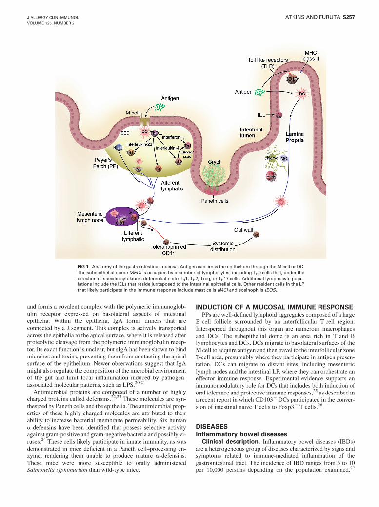
The mucosal system is characterized as a site where antigen isselectively sampled and tolerance develops to maintain a state ofcontrolled and protective inflammation. To accomplish thesegoals, the mucosa is composed of luminal protective molecules,the epithelial barrier, and the immunologically rich lamina propria(LP; Table I). The overall anatomy of the GALT is presented inFig 1. This general overview shows important elements of the sys-tem, including the sampling of luminal antigens by microfold (M)cells, dendritic cells (DCs), and epithelia and the antigen-drivenpriming and maturation of naive T and B lymphocytes.
ANATOMY OF GALTAlthough the primary responsibility of the intestinal epithelial
cell is nutrient absorption, its role in mucosal immunity haspreviously been relegated to barrier function and the transport ofsecretory IgA. However, it is now clear that epithelia possess theability to actively participate in mucosal immune responses.7 In-testinal epithelial cells act as nonprofessional antigen-presentingcells, recognize and respond to bacterial and viral motifs by virtueof the expression of nucleotide oligomerization domain (NOD)and Toll-like receptors, and produce cytokines/chemokines thatinfluence immune responses.7 In addition, intestinal epithelialcells likely influence expansion of intestinal regulatory T (Treg)cells and cytokine expression.8,9
The epithelial surface overlying the Peyer patches (PPs) andlymphoid follicles is composed of a single layer of columnar cellstermed the follicle-associated epithelium (FAE). Within the FAEreside specialized M cells derived from enterocytes under theinfluence of lymphotoxin. Human M cells differ from absorptiveepithelium in that they do not harbor microvilli or membrane-
From athe Department of Pediatrics, National Jewish Health, bthe Department of Pediat-rics, University of Colorado Medical School, and cthe Section of Pediatric Gastroen-terology, Hepatology and Nutrition, dthe Gastrointestinal Eosinophilic DiseasesProgram, National Jewish Health, the Children’s Hospital, Denver, Aurora, Colo.
Disclosure of potential conflict of interest: D. Atkins has received research support fromthe National Institutes of Health Consortium of Food Allergy Research. G. T. Furuta isa consultant for Meritage Pharma, has received research support from the AmericanGastroenerological Association and the National Institutes of Health, and has providedexpert witness testimony on the subject of eosinophilic esophagitis.
Received for publication June 24, 2009; revised November 16, 2009; accepted forpublication November 23, 2009.
Reprint requests: Glenn T. Furuta, MD, Pediatric Gastroenterology, the Children’sHospital, Denver, 13123 East 16th Ave B290, Aurora, CO 80045. E-mail: [email protected].
0091-6749/$36.00! 2010 American Academy of Allergy, Asthma & Immunologydoi:10.1016/j.jaci.2009.11.037
Abbreviations usedDC: Dendritic cell
EGID: Eosinophilic gastrointestinal diseaseEoE: Eosinophilic esophagitisFAE: Follicle-associated epithelium
Foxp3: Forkhead box protein 3GALT: Gut-associated lymphoid tissueIBD: Inflammatory bowel diseaseIEL: Intraepithelial lymphocyteLP: Lamina propriaM: Microfold
NOD: Nucleotide oligomerization domainPP: Peyer patch
TCR: T-cell receptorTreg: Regulatory T
S255
associated hydrolytic enzymes and contain less glycocalyx but doexpress cathepsin E andToll-like receptors. Regional differences inM cells (ie, differences inM cells in the colon comparedwith thoseof the small intestine) are thought to exist, suggesting accommo-dations to changing microflora; however, the functional signifi-cance of this is unknown. A distinctive characteristic of the M cellis the presence of an invaginated subdomain at the basolateralmembrane forming an intraepithelial ‘‘pocket.’’10 At this site, pre-dominantly CD41 CD45RO memory T cells and both naive(sIgD1) and memory (sIgD2) B cells interact with the M cell.
The major function of M cells is the transepithelial vesiculartransport of antigens from the lumen directly to the subepitheliallymphoid tissues. M cells have been shown to transport partic-ulate proteins, bacteria, viruses, and noninfectious particles.11
This sampling of luminal antigens and microorganisms is thoughtto be important in the development of immune responses and tol-erance. Although various pathogenic organisms can exploit thepropensity for vectorial transport of M cells as a mechanism togain entry for infection, M cells also transport commensalflora as a potential mechanism to regulate immune responses toendogenous flora.
Microenvironmental anatomic differences within the differentparts of the gastrointestinal tract are well described. Although theesophagus is lined by a stratified squamous epithelium, M cellshave not yet been identified, and no resident eosinophils are pre-sent in the mucosal surface (see the eosinophilic esophagitis[EoE] section below). In contrast, the small intestine and colonare lined by a columnar epithelium, and the cellular componentsof the GALT are localized within microenvironments, such asPPs or interstitial lymphoid follicles. Formation of PPs is depen-dent on several factors, such as the IL-7 receptor and TNF, alongwith TNF receptor familymembers. Theseminiorgans are coveredby M cells and FAE that participate in antigen trafficking, asdescribed above. Within the barrier are also unique cell types, in-cluding intraepithelial lymphocytes (IELs) and the antimicrobial-filled Paneth cells that reside at the crypt base. The LP is populatedby T and B cells, along with unique populations of DCs. Mesen-teric lymph nodes are a robust site of antigen processing andform a filter that separates the mucosa from other mucosal organs.
T lymphocytes localize in the small intestine as a result ofselective expression of a4b7 and CCR9. CD41 and CD81 T cellsoccupy the LP, whereas CD81 T cells preferentially reside in the
intraepithelial space. IELs are a heterogeneous population of lym-phocytes that are predominantly effector/effector memory cellsmade up of gd T-cell receptor (TCR) CD81 T cells and 2 distinctsubsets of ab TCR cells: ab TCR CD41 or CD81 cells and thosethat lack coreceptor expression, the so-called double-negativecells.
One subset of T cells receiving recent recognition is the Tregcell.12 Treg cells generally have suppressive capacities that partic-ipate in the maintenance of self-tolerance. Surface marker studieshave identified several subtypes, including the forkhead box pro-tein 3 (Foxp3)–positive T cell. Mutations in the gene encodingFoxp3, a Treg-specific transcription factor, have been associatedwith autoimmunity in murine models and a clinical syndrometermed immunodysregulation, polyendocrinopathy, enteropathy,X-linked syndrome. Patients with this disease have severediarrhea and small and large intestinal inflammation.
B cells secreting IgA1 originate in the PPs, ultimately takingup residence in the intestinal LP. This journey is regulated bythe interaction of site-specific adhesion molecules, an a4b7 onthe lymphocytes, and mucosal addressin cell-adhesion molecule1 on the high endothelial venules in the LP. A smaller percentageof IgA-producing cells in the gut (about 25%) are derived fromperitoneal B1 lymphocytes driven by commensal bacteria in aT cell–dependent manner and are thought to be important inmodulating the mucosal immune response to bacterial flora.
Mast cells are abundant throughout the gastrointestinal tract,and although important in the host response to parasitic infection,they might participate also in innate immune responses tobacteria.13 LP mast cells and lymphocytes interface with theenteric nervous system, providing another pathway that caninfluence mucosal immune responses.14
Eosinophils are absent in the normal esophagus but are residentcells of the stomach and small and large bowel. Chemotacticfactors contributing to this population include the constitutiveexpression of eotaxin-1.15,16 The exact numbers that define nor-malcy are open to debate but likely depend on a number of differ-ent factors. Like mast cells, eosinophils can perform importanteffector functions during parasitic infection and allergicresponses but can also contribute to normal gut homeostasis.17
INNATE MECHANISMS OF DEFENSEOften ignored are a host of mediators that participate in the
innate defense mechanisms.18 These molecules participate innonspecific actions that limit antigens and microbes from com-municating with the epithelium and LP. Mucus is composed ofa number of glycoproteins that form a viscoelastic blanket thatcovers the epithelial surface. The inner mucus layer ranges be-tween 50 and 100 mm, whereas the outer layer measures up to500 mm. This mucus blanket is primarily composed of mucin-2but also harbors a number of different mediators, including trefoilfactors, secretory IgA, and antimicrobial peptides.
Trefoil factors are shamrock-shaped proteins held together by 3disulfide bonds.19 Several types of trefoil factors have beendescribed that localize to different mucosal surfaces to assist inbarrier repair and wound healing. Numerous stimuli induce theproduction of trefoil factors, including hypoxia and epithelialdisruption.
IgA antibodies are divided into 2 subclasses, IgA1 and IgA2,with IgA2 representing the predominant form on intestinalsurfaces. Secretory IgA, which is secreted by B cells, binds to
TABLE I. Elements of the mucosal immune system
Innate mechanisms of defenseMucusTrefoil factorsIgAPeristalsisTight junctional proteinsAntimicrobial peptides
Adaptive elements of defenseB cellsCD41 T cellsCD81 T cellsTreg cellsDCs
Cellular componentsEosinophilsMast cellsNeuronsEpithelial cells
J ALLERGY CLIN IMMUNOL
FEBRUARY 2010
S256 ATKINS AND FURUTA
and forms a covalent complex with the polymeric immunoglob-ulin receptor expressed on basolateral aspects of intestinalepithelia. Within the epithelia, IgA forms dimers that areconnected by a J segment. This complex is actively transportedacross the epithelia to the apical surface, where it is released afterproteolytic cleavage from the polymeric immunoglobulin recep-tor. Its exact function is unclear, but sIgA has been shown to bindmicrobes and toxins, preventing them from contacting the apicalsurface of the epithelium. Newer observations suggest that IgAmight also regulate the composition of the microbial environmentof the gut and limit local inflammation induced by pathogen-associated molecular patterns, such as LPS.20,21
Antimicrobial proteins are composed of a number of highlycharged proteins called defensins.22,23 These molecules are syn-thesized by Paneth cells and the epithelia. The antimicrobial prop-erties of these highly charged molecules are attributed to theirability to increase bacterial membrane permeability. Six humana-defensins have been identified that possess selective activityagainst gram-positive and gram-negative bacteria and possibly vi-ruses.24 These cells likely participate in innate immunity, as wasdemonstrated in mice deficient in a Paneth cell–processing en-zyme, rendering them unable to produce mature a-defensins.These mice were more susceptible to orally administeredSalmonella typhimurium than wild-type mice.
INDUCTION OF A MUCOSAL IMMUNE RESPONSEPPs are well-defined lymphoid aggregates composed of a large
B-cell follicle surrounded by an interfollicular T-cell region.Interspersed throughout this organ are numerous macrophagesand DCs. The subepithelial dome is an area rich in T and Blymphocytes and DCs. DCs migrate to basolateral surfaces of theM cell to acquire antigen and then travel to the interfollicular zoneT-cell area, presumably where they participate in antigen presen-tation. DCs can migrate to distant sites, including mesentericlymph nodes and the intestinal LP, where they can orchestrate aneffector immune response. Experimental evidence supports animmunomodulatory role for DCs that includes both induction oforal tolerance and protective immune responses,25 as described ina recent report in which CD1031 DCs participated in the conver-sion of intestinal naive T cells to Foxp31 T cells.26
DISEASESInflammatory bowel diseases
Clinical description. Inflammatory bowel diseases (IBDs)are a heterogeneous group of diseases characterized by signs andsymptoms related to immune-mediated inflammation of thegastrointestinal tract. The incidence of IBD ranges from 5 to 10per 10,000 persons depending on the population examined.27
FIG 1. Anatomy of the gastrointestinal mucosa. Antigen can cross the epithelium through the M cell or DC.The subepithelial dome (SED) is occupied by a number of lymphocytes, including TH0 cells that, under thedirection of specific cytokines, differentiate into TH1, TH2, Treg, or TH17 cells. Additional lymphocyte popu-lations include the IELs that reside juxtaposed to the intestinal epithelial cells. Other resident cells in the LPthat likely participate in the immune response include mast cells (MC) and eosinophils (EOS).
J ALLERGY CLIN IMMUNOL
VOLUME 125, NUMBER 2
ATKINS AND FURUTA S257
Typical symptoms include abdominal pain and bloody diarrhea inaddition to other extraintestinal symptoms, such as fever, fatigue,arthralgias, and uveitis. In children growth failure can be an earlysign. Physical examination reveals abdominal tenderness, partic-ularly in the right lower quadrant.
Mucosal inflammation associated with Crohn disease canoccur anywhere along the length of the gastrointestinal tract,with preponderance in the terminal ileum. Histologic hallmarksof tissues affected by Crohn disease include transmural inflam-mation and often noncaseating granulomas. Endoscopic featuresinclude skip lesions consisting of aphthous ulcerations, andradiographic studies show terminal ileal narrowing.
Suggestive laboratory abnormalities include anemia, increasedsedimentation rate or C-reactive protein level, hypoalbuminemia,and increased liver enzyme levels. Ulcerative colitis has manyclinical features in common with Crohn disease, but intestinalinvolvement is limited to the colon. In addition, the intestinalinflammation is limited to the superficial mucosa without gran-ulomas, involves the rectum, and extends proximally. Other formsof IBD include microscopic colitis, lymphocytic colitis, anddiversion colitis. Long-term complications include colorectaldysplasia and cancer.
Neutrophilic crypt abscesses are one of the most characteristichistologic features of both forms of IBD. In addition, eosinophilsmight be present, although to a seemingly lesser degree.
Pathophysiology. Acomplete reviewof the pathophysiologyof IBD is beyond the scope of this Primer; the reader is referred toexcellent reviews for more detailed descriptions.5,18,28-32Althoughthe exact pathophysiology of IBD has not been determined, it isthought to develop when a genetically predisposed host is exposedto a luminal/environmental trigger. Over the course of the last fewyears, a number of genes have been identified in patients withCrohn disease, in particular genes linked to epithelial responsesto luminal bacteria, autophagy, IL-10, and IL-23/IL-17 pathways.For instance, pathogen recognition receptors are present on the ep-ithelial surface. One group of pathogen recognition receptors, theNOD molecules, recognize pathogen-associated molecular pat-terns that are present on bacterial membranes. A specific mutationof theNOD2 gene allows for inappropriate sensing of bacteriawithsubsequent epithelial activation, leading to increased proinflamma-tory cytokine production within the mucosa. One gene associatedwith autophagy, ATG16L1, has been associated with Crohn dis-ease. IL-10 suppresses deleterious intestinal inflammation, and re-cent studies have linked IL10mutations to IBD.33 IL-23 and IL-17mediate innate microbial defense and are linked to IBD.34 In addi-tion, loss of tolerancemight also play a role because themucosa af-fected by IBD contains fewer Treg cells than the healthy mucosa.IgE and food allergies are not thought to play a role in the underly-ing pathogenesis of these diseases.
Treatment. Goals of treatment of IBD include reducinginflammation, maintaining remission, enhancing quality of life,and avoiding the potential toxicity associatedwith treatments.35-37
With this inmind, acute exacerbations are typicallymanagedwithsystemic corticosteroids, whereas remission is addressed with theuse of either aminosalicylates or immunomodulators, such asmercaptopurine or azathioprine. Recently, biological treatments,including anti–TNF-a antibodies (ie, infliximab andadalimumab), have significantly affected the clinicopathologicalfeatures of IBD. A number of other agents, such as antidiarrhealagents, bile binders, and antispasmodics, might enhance qualityof life.
Celiac diseaseClinical description. Celiac disease is an immune-mediated
enteropathy that occurs as a result of gluten sensitivity ingenetically predisposed individuals (DQ2/DQ8-positiveHLA).38 The incidence is 1 in 133 persons in the United States.Manifestations include those related to the gastrointestinal tract,such as chronic/recurrent diarrhea, abdominal pain, constipation,and slow growth, and nongastrointestinal symptoms, includingdermatitis herpetiformis, seizures with occipital calcifications,dental hypoplasia, osteopenia, short stature, iron deficiencyanemia, hepatitis, infertility, and arthritis.39,40
Other conditions associated with celiac disease include type1 diabetes; Williams, Down, and Turner syndromes; IgA defi-ciency; autoimmune diseases; and a family history of first-degreerelatives with celiac disease. Without treatment, there is anincreased risk of intestinal lymphoma. Although serologic testresults, such as increased IgA anti-endomysial antibody or tissuetransglutaminase levels, provide strong evidence for celiacdisease, the diagnosis rests on the finding of villous bluntingand increased IEL numbers in mucosal biopsy specimens of theduodenal mucosa.
Pathophysiology. Recent studies have shown that a33-amino-acid peptide in gliadin that is resistant to digestioncontains the epitopes critical to the development of abnormalsmall intestinal mucosa in patients with celiac disease.41,42 Afteruptake by the epithelium, processing of this 33-mer leads to acti-vation of CD41 LP T cells, upregulation of the IL-2 receptor,increased production of IFN-g and IL-15, and infiltration of theepithelia with gd T cells. The resultant inflammatory processleads to villous blunting, crypt elongation, and loss of absorptivesurfaces. Celiac disease is a cell-mediated and not IgE-mediatedfood allergic disease.
Treatment. Complete elimination of gliadin from the diet isthe primary treatment of celiac disease.43 Of paramount impor-tance is attention to education and support of patients withrespect to dietary elimination of gluten-containing products,review of alternative diets, adequacy of caloric and nutrientintake, and psychological support. For instance, although anumber of foods should obviously be avoided, a number ofproducts, including candies, gravies, food colorings, soy sauce,medications, play dough, and cosmetics, contain gluten inquantities sufficient to cause inflammation resulting in symp-toms. In addition, many products that have been deemed wheatfree, such as oats, are frequently contaminated with gliadin andshould not be ingested by patients with celiac disease. Patientsmight also have iron, zinc, folic acid, and B complex vitamindeficiencies.
Eosinophilic gastrointestinal diseasesClinical description. Eosinophilic gastrointestinal diseases
(EGIDs) are heterogeneous diseases characterized by a diverse setof symptoms that occur in association with intestinal eosino-philia.44 These diseases have been termed EoE, eosinophilicgastritis, eosinophilic gastroenteritis, and eosinophilic colitisdepending on the anatomic location in which eosinophil numbersare increased. Over the last decade, EoE has been recognized asthe most common EGID. The remainder of this section will focuson EoE. For further information, the reader is referred to recentreviews on other EGIDs.44-46 Recent reports have expanded theassociation of esophageal eosinophilia with other diseases,
J ALLERGY CLIN IMMUNOL
FEBRUARY 2010
S258 ATKINS AND FURUTA
including celiac disease; the exact pathogenetic mechanisms andtherapeutic implications of this are uncertain.47-50
EoE is a clinicopathological disease characterized by upperintestinal symptoms that occur in association with dense esoph-ageal eosinophilia; other potential causes must have been ruledout as causes of symptoms and eosinophilia.51 Children with EoEpresent with a wide range of symptoms, including vomiting, ab-dominal pain, feeding dysfunction, and dysphagia.46,52 Feedingdysfunction is often overlooked and requires specific questioningregarding how patients eat foods (eg, dysphagia, food sticking, re-quiring water to wash food down, and prolonged chewing).53
Adults present with stereotypical features of food impaction ordysphagia.54 Patients presenting with food impaction, especiallywhen recurrent, should be evaluated for a diagnosis of EoE. EoEoccurs in all age groups and has been reported in all continentsexcept Africa, with a reported prevalence of EoE ranging between1 and 4 per 10,000 persons. Although the natural history isunknown, the one identified complication is esophageal strictureor narrowing.55
The physical examination should be directed toward excludingother causes of esophageal eosinophilia, such as IBDs, celiacdisease, and connective tissue diseases. No single marker,including peripheral eosinophilia, provides diagnostic supportfor or against the diagnosis of EoE, although one study suggeststhat the combination of peripheral eosinophilia and increasedserum eotaxin-3 and eosinophil-derived neurotoxin levels corre-lates with esophageal eosinophil density.56 Upper gastrointestinalseries can screen for other causes of vomiting and for evidenceof esophageal stricture or long-segment narrowing, featuresassociated with EoE.
Pathophysiology. Esophageal eosinophilia is a nonspecificfinding that reflects a state of injury. Although a variety of diseaseshave been associated with this type of inflammation, includinggastroesophageal reflux disease, EoE, celiac disease, infections,and IBDs among others, the exact mechanism driving thisresponse is not certain.44 For instance, recent evidence suggeststhat specific cytokines, including IL-6 and IL-1, might participatein acid-induced injury.57 In contrast, as discussed below, IL-5 iscritical to this response in murine models, and eotaxin-3 contrib-utes to human disease.58,59 The acute inflammatory infiltrate inpatients with EoE is exclusively composed of eosinophils, withthe virtual complete absence of neutrophils.
A series of recent studies have identified potential mechanismsfor the pathogenesis of EoE. Mishra et al59 provided the first mu-rine model of aeroallergen-induced esophageal eosinophilia bysensitizing and challenging with Aspergillus fumigatus. By apply-ing this system to IL-5 null mice, the investigators were able todemonstrate that esophageal eosinophilia was dependent onIL-5, as well as T cells.59 The inflamedmucosa contains increasedCD41 effector T cells and decreased Treg cells.60,61 In addition,the same investigators have determined the effect of IL-5 on tissueremodeling associated with this eosinophilic inflammation.62-Together, these findings provided support for the developmentof therapeutics targeting IL-5 in the treatment of this disease.Addressing this need are 2 ongoing studies of anti–IL-5 in thetreatment of pediatric EoE.
Translational studies have also brought increased understand-ing of EoE. For instance, a number of studies have begun to definethe immunomicroenvironment of the esophageal mucosa.Although diagnostic criteria have solely focused on eosinophilnumbers, other studies are examining associated inflammatory
features, including eosinophil degranulation, that appear to beincreased compared with those seen in gastroesophageal refluxdisease.63 In addition, the mucosa from patients with EoE con-tains increased TH1 and TH2 proinflammatory cytokines (IL-5and TNF-a), CD8 lymphocytes (CD8 and CD1a), B cells, andmast cell and basophil infiltration.64 The exact role of Treg cellsin EoE is uncertain because one study demonstrated immunohis-tochemical evidence of Foxp31 cells in both patients with EoEand those with gastroesophageal reflux disease.60 One genome-wide microarray analysis revealed that the most upregulatedgene in the esophageal epithelia was eotaxin-3, a chemokinecritical for eosinophil migration.65 Another study identifiedincreased eotaxin expression in the affected mucosa, providingfurther support for eotaxin’s role in EoE’s pathogenesis.66
Other studies have focused on remodeling in EoE, showing anincreased level of esophageal fibrosis in children with EoE.67,68
Although the exact mechanisms of this response are not certain,Aceves et al68 showed increased TGF-b expression with activa-tion of the SMAD pathway. Therapeutic studies suggest thatfibrosis might be reversible.69
IgE and non-IgE immune mechanisms might participate in thepathogenesis of EoE, an important point to be kept in mind whenevaluating these patients for allergen sensitization. Although skinprick testing and the measurement of food allergen–specific IgElevels are often useful in identifying potential culprit foods, theyare not helpful in the detection of causative foods in non–IgE-mediated reactions. However, atopy patch testing to foodshas been proposed as a useful method to potentially identifyfoods causing symptoms through a non–IgE-mediated immunemechanism.70,71
In summary, evidence to date supports a role for both IgE-mediated and non–IgE-mediated mechanisms in the pathogenesisof EoE, with eotaxin-3 and IL-5 being central mediators andfibrosis being one of the potential outcomes.
Treatment. Treatment goals have been directed towardsymptom elimination and reduction/normalization of esophagealinflammation. The rationale for the later end point has been thatcomplete histologic remission might reduce the incidence ofcomplications. To date, the incidence of esophageal complica-tions is unknown, and potential emotional and developmentaleffects of chronic treatments and repeated endoscopic analysesare beginning to be recognized.72 Prospective studies will providedata to illuminate this area of controversy.
Despite this issue, at least 2 effective treatments, corticoste-roids and dietary elimination of suspected culprit foods, havebeen identified. The reader is referred to a number of recentreviews on this topic for further details regarding the specifics ofeach treatment.46,73 Regardless, evidence to date suggests thatEoE is a chronic disease, and without continuous treatment,symptoms and inflammation will persist or return. To date, nomedical maintenance treatment has been identified.51
SUMMARYThe mucosal immune system has a unique anatomy and
physiology aimed at providing a mechanism that will allowtolerance to food antigens and commensal bacteria along with thecapacity to respond to pathogenic microbes, other injuriousagents, or both. The monolayered epithelium forms the initialinterface between the environment and host that forms not only abarrier but also a sensor providing bidirectional communication
J ALLERGY CLIN IMMUNOL
VOLUME 125, NUMBER 2
ATKINS AND FURUTA S259
with other resident mucosal lymphoid cells. The lymphocytes,DCs, mast cells, and eosinophils in the LP interact to form apluripotent network that orchestrates an innate and adaptiveimmune response to potential pathogens. Further delineation ofthe mechanisms governing the normal responses of the mucosalimmune system will provide insight into disease states, such asfood allergies, IBDs, and EGIDs.
REFERENCES1. Cario E. Innate immune signalling at intestinal mucosal surfaces: a fine line be-
tween host protection and destruction. Curr Opin Gastroenterol 2008;24:725-32.2. Boirivant M, Amendola A, Butera A. Intestinal microflora and immunoregulation.
Mucosal Immunol 2008;1(suppl 1):S47-9.3. Artis D, Grencis RK. The intestinal epithelium: sensors to effectors in nematode
infection. Mucosal Immunol 2008;1:252-64.4. Macpherson AJ, McCoy KD, Johansen FE, Brandtzaeg P. The immune geography
of IgA induction and function. Mucosal Immunol 2008;1:11-22.5. Neish AS. Microbes in gastrointestinal health and disease. Gastroenterology 2009;
136:65-80.6. Mason KL, Huffnagle GB, Noverr MC, Kao JY. Overview of gut immunology.
Adv Exp Med Biol 2008;635:1-14.7. Dahan S, Roth-Walter F, Arnaboldi P, Agarwal S, Mayer L. Epithelia: lymphocyte
interactions in the gut. Immunol Rev 2007;215:243-53.8. Allez M, Brimnes J, Dotan I, Mayer L. Expansion of CD81 T cells with regulatory
function after interaction with intestinal epithelial cells. Gastroenterology 2002;123:1516-26.
9. Dotan I, Allez M, Nakazawa A, Brimnes J, Schulder-Katz M, Mayer L. Intestinalepithelial cells from inflammatory bowel disease patients preferentially stimulateCD41 T cells to proliferate and secrete interferon-gamma. Am J Physiol Gastro-intest Liver Physiol 2007;292:G1630-40.
10. Neutra MR, Mantis NJ, Kraehenbuhl JP. Collaboration of epithelial cells withorganized mucosal lymphoid tissues. Nat Immunol 2001;2:1004-9.
11. Jang MH, Kweon MN, Iwatani K, Yamamoto M, Terahara K, Sasakawa C, et al.Intestinal villous M cells: an antigen entry site in the mucosal epithelium. ProcNatl Acad Sci U S A 2004;101:6110-5.
12. Himmel ME, Hardenberg G, Piccirillo CA, Steiner TS, Levings MK. The role ofT-regulatory cells and Toll-like receptors in the pathogenesis of human inflamma-tory bowel disease. Immunology 2008;125:145-53.
13. Galli SJ, Tsai M. Mast cells: versatile regulators of inflammation, tissue remodel-ing, host defense and homeostasis. J Dermatol Sci 2008;49:7-19.
14. Koon HW, Pothoulakis C. Immunomodulatory properties of substance P: thegastrointestinal system as a model. Ann N Y Acad Sci 2006;1088:23-40.
15. Blanchard C, Rothenberg ME. Biology of the eosinophil. Adv Immunol 2009;101:81-121.
16. Blanchard C, Rothenberg ME. Chemotactic factors associated with eosinophilicgastrointestinal diseases. Immunol Allergy Clin North Am 2009;29:141-8, xi.
17. Hogan SP, Rothenberg ME. Eosinophil function in eosinophil-associated gastroin-testinal disorders. Curr Allergy Asthma Rep 2006;6:65-71.
18. McGuckin MA, Eri R, Simms LA, Florin TH, Radford-Smith G. Intestinal barrierdysfunction in inflammatory bowel diseases. Inflamm Bowel Dis 2009;15:100-13.
19. Kjellev S. The trefoil factor family—small peptides with multiple functionalities.Cell Mol Life Sci 2009;66:1350-69.
20. Suzuki K, Fagarasan S. How host-bacterial interactions lead to IgA synthesis in thegut. Trends Immunol 2008;29:523-31.
21. Evolution Fagarasan S. development, mechanism and function of IgA in the gut.Curr Opin Immunol 2008;20:170-7.
22. Braff MH, Hawkins MA, Di Nardo A, Lopez-Garcia B, Howell MD, Wong C, et al.Structure-function relationships among human cathelicidin peptides: dissociationof antimicrobial properties from host immunostimulatory activities. J Immunol2005;174:4271-8.
23. Ong PY, Ohtake T, Brandt C, Strickland I, Boguniewicz M, Ganz T, et al. Endog-enous antimicrobial peptides and skin infections in atopic dermatitis. N Engl J Med2002;347:1151-60.
24. Ericksen B, Wu Z, Lu W, Lehrer RI. Antibacterial activity and specificity of the sixhuman a-defensins. Antimicrob Agents Chemother 2005;49:269-75.
25. Bilsborough J, Viney JL. Gastrointestinal dendritic cells play a role in immunity,tolerance, and disease. Gastroenterology 2004;127:300-9.
26. Coombes JL, Siddiqui KR, Arancibia-Carcamo CV, Hall J, Sun CM, Belkaid Y,et al. A functionally specialized population of mucosal CD1031 DCs inducesFoxp31 regulatory T cells via a TGF-beta and retinoic acid-dependent mechanism.J Exp Med 2007;204:1757-64.
27. Shanahan F, Bernstein CN. The evolving epidemiology of inflammatory boweldisease. Curr Opin Gastroenterol 2009;25:301-5.
28. van Lierop PP, Samsom JN, Escher JC, Nieuwenhuis EE. Role of the innateimmune system in the pathogenesis of inflammatory bowel disease. J PediatrGastroenterol Nutr 2009;48:142-51.
29. Round JL, Mazmanian SK. The gut microbiota shapes intestinal immune responsesduring health and disease. Nat Rev Immunol 2009;9:313-23.
30. Weinstock JV, Elliott DE. Helminths and the IBD hygiene hypothesis. InflammBowel Dis 2009;15:128-33.
31. Yoshida H, Granger DN. Inflammatory bowel disease: a paradigm for the linkbetween coagulation and inflammation. Inflamm Bowel Dis 2009;15:1245-55.
32. Abraham C, Cho J. Inflammatory bowel disease. N Engl J Med 2009;361:2066-78.33. Glocker EO, Kotlarz D, Boztug K, Gertz EM, Schaffer AA, Noyan F, et al. Inflam-
matory bowel disease and mutations affecting the interleukin-10 receptor. N Engl JMed 2009;361:2033-45.
34. Abraham C, Cho JH. IL-23 and autoimmunity: new insights into the pathogenesisof inflammatory bowel disease. Annu Rev Med 2009;60:97-110.
35. Yamamoto T, Nakahigashi M, Saniabadi AR. Review article: diet and inflamma-tory bowel disease—epidemiology and treatment. Aliment Pharmacol Ther2009;30:99-112.
36. Regueiro M. Management and prevention of postoperative Crohn’s disease.Inflamm Bowel Dis 2009;15:1583-90.
37. Rutgeerts P, Vermeire S, Van Assche G. Biological therapies for inflammatorybowel diseases. Gastroenterology 2009;136:1182-97.
38. Hunt KA, van Heel DA. Recent advances in coeliac disease genetics. Gut 2009;58:473-6.
39. Barker JM, Liu E. Celiac disease: pathophysiology, clinical manifestations, andassociated autoimmune conditions. Adv Pediatr 2008;55:349-65.
40. Freeman HJ. Pearls and pitfalls in the diagnosis of adult celiac disease. Can JGastroenterol 2008;22:273-80.
41. Molberg O, Uhlen AK, Jensen T, Flaete NS, Fleckenstein B, Arentz-Hansen H,et al. Mapping of gluten T-cell epitopes in the bread wheat ancestors: implicationsfor celiac disease. Gastroenterology 2005;128:393-401.
42. Schumann M, Richter JF, Wedell I, Moos V, Zimmermann-Kordmann M,Schneider T, et al. Mechanisms of epithelial translocation of the alpha(2)-glia-din-33mer in coeliac sprue. Gut 2008;57:747-54.
43. HainesML,Anderson RP,Gibson PR. Systematic review: the evidence base for long-term management of coeliac disease. Aliment Pharmacol Ther 2008;28:1042-66.
44. Fleischer DM, Atkins D. Evaluation of the patient with suspected eosinophilicgastrointestinal disease. Immunol Allergy Clin North Am 2009;29:53-63, ix.
45. Straumann A. Idiopathic eosinophilic gastrointestinal diseases in adults. Best PractRes Clin Gastroenterol 2008;22:481-96.
46. Atkins D, Kramer R, Capocelli K, Lovell M, Furuta GT. Eosinophilic esophagitis:the newest esophageal inflammatory disease. Nat Rev Gastroenterol Hepatol 2009;6:267-78.
47. Leslie C,MewsC, Charles A, RavikumaraM. Celiac disease and eosinophilic esoph-agitis: a true association. J Pediatr Gastroenterol Nutr 2009 [Epub ahead of print].
48. Verzegnassi F, Bua J, De Angelis P, Dall’oglio L, Di Leo G, Ventura A. Eosino-philic oesophagitis and coeliac disease: is it just a casual association? Gut 2007;56:1029-30.
49. Ooi CY, Day AS, Jackson R, Bohane TD, Tobias V, Lemberg DA. Eosinophilicesophagitis in children with celiac disease. J Gastroenterol Hepatol 2008;23:1144-8.
50. Kagalwalla AF, Shah A, Ritz S, Melin-Aldana H, Li BU. Cow’s milk protein-induced eosinophilic esophagitis in a child with gluten-sensitive enteropathy.J Pediatr Gastroenterol Nutr 2007;44:386-8.
51. Furuta GT, Liacouras CA, Collins MH, Gupta SK, Justinich C, Putnam PE, et al.Eosinophilic esophagitis in children and adults: a systematic review and consensusrecommendations for diagnosis and treatment. Gastroenterology 2007;133:1342-63.
52. Spergel JM, Brown-Whitehorn TF, Beausoleil JL, Franciosi J, Shuker M, Verma R,et al. 14 years of eosinophilic esophagitis: clinical features and prognosis. J PediatrGastroenterol Nutr 2009;48:30-6.
53. Haas A, Creskoff-Naune N. Feeding dysfunction in children with eosinophilicesophagitis. Immunol Allergy Clin North Am 2009. In press.
54. Desai TK, Stecevic V, Chang CH, Goldstein NS, Badizadegan K, Furuta GT. As-sociation of eosinophilic inflammation with esophageal food impaction in adults.Gastrointest Endosc 2005;61:795-801.
55. Straumann A. The natural history and complications of eosinophilic esophagitis.Gastrointest Endosc Clin North Am 2008;18:99-118, ix.
56. Konikoff MR, Blanchard C, Kirby C, Buckmeier BK, Cohen MB, Heubi JE, et al.Potential of blood eosinophils, eosinophil-derived neurotoxin, and eotaxin-3 as bio-markers of eosinophilic esophagitis. Clin Gastroenterol Hepatol 2006;4:1328-36.
57. Souza RF, Huo X, Mittal V, Schuler CM, Carmack SW, Zhang HY, et al. Gastro-esophageal reflux may cause esophagitis through a cytokine-mediated mechanism,not by caustic (acid) injury. Gastroenterology 2009;137:1776-84.
J ALLERGY CLIN IMMUNOL
FEBRUARY 2010
S260 ATKINS AND FURUTA
58. Blanchard C, Wang N, Stringer KF, Mishra A, Fulkerson PC, Abonia JP, et al.Eotaxin-3 and a uniquely conserved gene-expression profile in eosinophilic esoph-agitis. J Clin Invest 2006;116:536-47.
59. Mishra A, Hogan SP, Brandt EB, Rothenberg ME. IL-5 promotes eosinophiltrafficking to the esophagus. J Immunol 2002;168:2464-9.
60. Tantibhaedhyangkul U, Tatevian N, Gilger MA, Major AM, Davis CM. Increasedesophageal regulatory T cells and eosinophil characteristics in children with eosin-ophilic esophagitis and gastroesophageal reflux disease. Ann Clin Lab Sci 2009;39:99-107.
61. Zhu X, Wang M, Crump CH, Mishra A. An imbalance of esophageal effector andregulatory T cell subsets in experimental eosinophilic esophagitis in mice. Am JPhysiol Gastrointest Liver Physiol 2009;297:G550-8.
62. Mishra A, Wang M, Pemmaraju VR, Collins MH, Fulkerson PC, Abonia JP, et al.Esophageal remodeling develops as a consequence of tissue specific IL-5-inducedeosinophilia. Gastroenterology 2008;134:204-14.
63. Protheroe C, Woodruff SA, de Petris G, Mukkada V, Ochkur SI, JanarthananS, et al. A novel histologic scoring system to evaluate mucosal biopsies frompatients with eosinophilic esophagitis. Clin Gastroenterol Hepatol 2009;7:749-55, e11.
64. Vicario M, Blanchard C, Stringer KF, Collins MH, Mingler MK, Ahrens A, et al.Local B cells and IgE production in the esophageal mucosa in eosinophilic esoph-agitis. Gut 2009 [Epub ahead of print].
65. Blanchard C, Durual S, Estienne M, Emami S, Vasseur S, Cuber JC. Eotaxin-3/CCL26 gene expression in intestinal epithelial cells is up-regulated by
interleukin-4 and interleukin-13 via the signal transducer and activator of transcrip-tion 6. Int J Biochem Cell Biol 2005;37:2559-73.
66. Bhattacharya B, Carlsten J, Sabo E, Kethu S, Meitner P, Tavares R, et al. Increasedexpression of eotaxin-3 distinguishes between eosinophilic esophagitis and gastro-esophageal reflux disease. Hum Pathol 2007;38:1744-53.
67. Chehade M, Sampson HA, Morotti RA, Magid MS. Esophageal subepithelial fibro-sis in children with eosinophilic esophagitis. J Pediatr Gastroenterol Nutr 2007;45:319-28.
68. Aceves SS, Newbury RO, Dohil R, Bastian JF, Broide DH. Esophageal remodelingin pediatric eosinophilic esophagitis. J Allergy Clin Immunol 2007;119:206-12.
69. Aceves SS, Newbury RO, Chen D, Mueller J, Dohil R, Hoffman H, et al. Resolu-tion of remodeling in eosinophilic esophagitis correlates with epithelial response totopical corticosteroids. Allergy 2009 [Epub ahead of print].
70. Spergel JM, Andrews T, Brown-Whitehorn TF, Beausoleil JL, Liacouras CA.Treatment of eosinophilic esophagitis with specific food elimination diet directedby a combination of skin prick and patch tests. Ann Allergy Asthma Immunol2005;95:336-43.
71. Spergel JM, Beausoleil JL, Mascarenhas M, Liacouras CA. The use of skin pricktests and patch tests to identify causative foods in eosinophilic esophagitis.J Allergy Clin Immunol 2002;109:363-8.
72. Klinnert MD. Psychological impact of eosinophilic esophagitis on children andfamilies. Immunol Allergy Clin North Am 2009;29:99-107, x.
73. RothenbergME. Biology and treatment of eosinophilic esophagitis. Gastroenterology2009;137:1238-49.
J ALLERGY CLIN IMMUNOL
VOLUME 125, NUMBER 2
ATKINS AND FURUTA S261
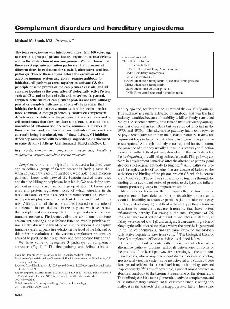
Complement disorders and hereditary angioedema
Michael M. Frank, MD Durham, NC
The term complement was introduced more than 100 years agoto refer to a group of plasma factors important in host defenseand in the destruction of microorganisms. We now know thatthere are 3 separate activation pathways that appeared atdifferent times in evolution: the classical, alternative, and lectinpathways. Two of these appear before the evolution of theadaptive immune system and do not require antibody forinitiation. All pathways come together to activate C3, theprinciple opsonic protein of the complement cascade, and allcontinue together to the generation of biologically active factors,such as C5a, and to lysis of cells and microbes. In general,complete deficiencies of complement proteins are rare, althoughpartial or complete deficiencies of one of the proteins thatinitiates the lectin pathway, mannose-binding lectin, are farmore common. Although genetically controlled complementdefects are rare, defects in the proteins in the circulation and oncell membranes that downregulate complement so as to limituncontrolled inflammation are more common. A number ofthese are discussed, and because new methods of treatment arecurrently being introduced, one of these defects, CI inhibitordeficiency associated with hereditary angioedema, is discussedin some detail. (J Allergy Clin Immunol 2010;125:S262-71.)
Key words: Complement, complement deficiencies, hereditaryangioedema, atypical hemolytic uremic syndrome
Complement is a term originally introduced a hundred yearsago to define a group of factors present in fresh plasma that,when activated by a specific antibody, were able to kill microor-ganisms.1 Later work showed the bacteria studied were lysedand that the killing principle was heat labile. We now define com-plement as a collective term for a group of about 30 known pro-teins and protein regulators, some of which circulate in theblood and some of which are cell membrane bound. The comple-ment proteins play a major role in host defense and innate immu-nity. Although all of the early studies focused on the role ofcomplement in host defense, in recent years, we have learnedthat complement is also important in the generation of a normalimmune response. Phylogenetically, the complement proteinsare ancient, serving a host defense function even in primitive an-imals in the absence of any adaptive immune system. The adaptiveimmune system appears in evolution at the level of the fish, and bythis point in evolution, all the various complement proteins arearrayed to produce their regulatory and host defense functions.2
We have come to recognize 3 pathways of complementactivation (Fig 1).3,4 The first pathway was defined almost a
century ago and, for this reason, is termed the classical pathway.This pathway is usually activated by antibody and was the firstpathway identified because of its ability to kill antibody-sensitizedbacteria. A second pathway, now termed the alternative pathway,was first observed in the 1950s but was studied in detail in the1970s and 1980s.5 The alternative pathway has been shown tobe phylogenetically older than the classical pathway. It does notrequire antibody to function and is found in organisms as primitiveas sea squirts.2 Although antibody is not required for its function,the presence of antibody usually allows this pathway to functionmore efficiently. A third pathway described in the past 2 decades,the lectin pathway, is still being defined in detail. This pathway ap-pears in development sometime after the alternative pathway andalso does not require antibody to function.6 All 3 pathways pro-ceed through a series of proteins that are discussed below to theactivation and binding of the plasma protein C3, which is centralto all 3 pathways. The pathways then proceed together through thebinding of an additional series of proteins to the lytic and inflam-mation-promoting steps in complement action.
Most reviews focus on the 3 major effector functions ofcomplement in host defense. First is its ability to lyse cells,second is its ability to opsonize particles (ie, to render them easyfor phagocytes to engulf), and third is the ability of the proteins onactivation to generate cleavage fragments that have potentinflammatory activity. For example, the small fragment of C5,C5a, can cause mast cells to degranulate and release histamine, asif theywere coated with IgE and antigen. It can causemigration ofphagocytic cells toward the place where the peptide is generated(ie, to induce chemotaxis) and can cause cytokine and biologi-cally active peptide release from cells.7,8 The biological basis ofthese 3 complement effector activities is defined below.
It is rare to find patients with deficiencies of classical oralternative pathway proteins, although deficiencies of some ofthe proteins of the lectin pathway are surprisingly more common.In most cases, when complement contributes to disease it is actingappropriately (ie, the system is being activated and causing tissuedamage and cell death in a normal fashion), but it is being activatedinappropriately.9,10 Thus, for example, a patient might produce anabnormal antibody to the basement membrane of the glomerulus.The antibody canbind to theglomerulus, activate complement, andcause inflammatory damage. In this case complement is acting nor-mally; it is the antibody that is inappropriate. Table I lists some
Abbreviations usedC1-INH: C1 inhibitor
C’: complementFDA: US Food and Drug AdministrationHAE: Hereditary angioedemaiC3b: Inactivated C3b
MASP: Mannose-binding lectin–associated serine proteaseMBL: Mannose-binding lectinMCP: Membrane cofactor proteinPNH: Paroxysmal nocturnal hemoglobinuria
From the Department of Pediatrics, Duke University Medical Center.Disclosure of potential conflict of interest: M. Frank is a consultant for Viropharma, CSLBehring, and Dyax.
Received for publication July 13, 2009; revised October 1, 2009; accepted for publicationOctober 7, 2009.
Reprint requests: Michael Frank, MD, Box 2611 Room 131 MSRB, Duke UniversityMedical Center, Durham NC, 27710. E-mail: [email protected].
0091-6749/$36.00! 2010 American Academy of Allergy, Asthma & Immunologydoi:10.1016/j.jaci.2009.10.063
S262
diseases in which complement deficiency is associated with clini-cal illness. Because of the many untoward effects of inappropriatecomplement activation, there are many control proteins that act todownregulate activated complement proteins at each step in thevarious reaction cascades. The importance of these proteins isthat they prevent unwanted damage of one’s own tissues and cells.Although the absence of complement proteins is unusual, the ab-sence of control proteins ismore common, andmanyof the patientswhohave absent control proteins have poorly controlled inflamma-tory disease.Moreover, asmentioned in our discussion of the lectinpathway below, there is a sizable group of patients with allotypicvariations of mannose-binding lectin (MBL) that lead to verylow levels of this circulating protein.
Although effector functions of complement in host defensehave received the most attention over the years, more recently, ithas been found that complement also functions in the induction ofadaptive immunity, but here there has been far less study, and lessinformation is available.11 One reason that this information iscoming to light slowly is that there are so few complement-defi-cient subjects to study. With the advent of knockout gene technol-ogy, it has been possible to develop murine strains missing 1 ormore complement proteins or complement receptors, and it hasbeen found that these animals have defects in the developmentof many aspects of the normal murine immune response.12 Thisis discussed further toward the end of this chapter.
THE CLASSICAL PATHWAYThe classical pathway is usually activated by antibody. IgM
and the IgG subclasses IgG1, IgG2, and IgG3 bind the firstcomponent of complement, C1, to initiate activation of theclassical pathway.3 C1 exists in serum as a 3-part molecule(C1q, C1r, and C1s) held together in the presence of ionic cal-cium. C1q has a central protein core and 6 radiating arms, eachending in a pod-like protein domain that can bind to the Fc frag-ment of IgG or IgM. Each of the 6 arms is made up of 3 inter-twined chains, C1q A, B, and C, and has a triple-helix structurelike collagen, providing flexibility. In the case of IgG, the bindingof multiple IgG molecules to an antigenic surface allows bindingof multiple arms of C1q, each to an Fc fragment, with sufficientaffinity of the C1q to allow C1 activation. In the case of IgM, asinglemolecule bound to an antigenic surface bymultiple bindingsites with the availability of multiple Fc fragments within this onepolymeric molecule is sufficient to bind C1q and activate thispathway. On binding of C1q to antibody, a distortion of the C1qmolecule takes place that in turn causes autoactivation of C1r,which then activates C1s. C1s, like C1r, acquires enzymatic activ-ity and continues the complement cascade sequence. C1 requirescalcium for self-association and therefore the classical pathwayrequires calcium for initiation. The function of activated C1 isto bind and cleave C4, the next protein in the classical pathwayactivation sequence. C4 is cleaved into a large fragment (C4b)and a small fragment (C4a). The large fragment continues thecomplement cascade and the small fragment, like the small frag-ments of C3 (C3a) and the small fragment of the next protein (C5)in the sequence (C5a), has anaphylatoxic activity. All of thesefragments are able to cause mast cell degranulation with resultinghistamine release.7
On activation of C4, a thioester-containing binding site isexposed on C4b that allows covalent attachment of C4b to thetarget. The nature of the binding site on C4 and C3 is similar and is
discussed in further detail in the section on the alternative path-way. The site now containing C1 and C4 bound to a target allowsthe next protein, C2, to bind to C4b. On C2 binding to the C14bsite, C1s cleaves C2 also into a large and small fragment. Again,the large fragment remains bound to the assembling protein com-plex. C2 binding to C4 requires the presence of ionic magnesium.The new site consisting of C4b and C2 (C4b2a) no longer requiresC1 for activity. Enzymatic activity resides in the C2 fragment.This site is termed the C3 convertase of the classical pathway be-cause it can bind the next complement protein in the sequence(C3) and, as in the earlier steps, cleave it into a large fragment(C3b) and a small fragment (C3a), which again has inflammatoryactivity. Just as in the case of C4b, C3b can bind covalently to thetarget of attack. In many cases it binds directly to C4b on thetarget. As mentioned earlier, C3 is the central component of all3 complement pathways and is present at high concentration inserum, about 1.2 mg/mL.
An important function of complement is the ability to opsonizeparticles, which means to coat them with complement-derivedprotein fragments that allow them to be phagocytosed easily.Phagocytic cells have on their surface specific receptors forcomplement-derived peptides, often cleavage fragments of C3.When these fragments are deposited on microbes, they can linkthe microbe to the phagocyte receptors; the adherence facilitatesthe phagocytic process.
On the addition of C3b to the C4bC2 site, a new binding site iscreated that can bind C5, the next protein in the sequence. Again,C5 is cleaved into a large fragment and a small fragment. Thelarge fragment, C5b, continues the complement cascade, al-though it does not form a covalent bond with the target andremains associated with C3b. The small fragment released, C5a,is one of the most potent inflammatory peptides released bycomplement activation and has strong neutrophil-aggregatingactivity, strong neutrophil chemotactic activity and is an excellentanaphylatoxin.7 Injection of sufficient purified C5a into an animal
FIG 1. The 3 complement activation pathways. The classical pathway isusually activated by antibody. The lectin pathway is activated by therecognition molecule MBL binding to structures with the appropriaterepetitive sugars. The ficolins are MBL-like molecules that can also activatethis pathway. The alternative pathway does not have a recognition mole-cule as such. It is initiated by the binding of factor B to C3, which can then becleaved by factor D. Because C3 always undergoes slow hydrolysis, thepathway is always undergoing some degree of activation. Properdinstabilizes the complex and can also initiate alternative pathway activation.C3b itself is an efficient activator of the alternative pathway, and classicalpathway activation leading to C3 deposition on a target rapidly activates thealternative pathway.
J ALLERGY CLIN IMMUNOL
VOLUME 125, NUMBER 2
FRANK S263
may cause anaphylaxis and death from neutrophil aggregation inthe circulation and massive histamine release.7,13
The complement cascade continues after C5b binding with thebinding of C6, C7, C8, and C9. One molecule of C6 and C7 eachbind to C5b on the target surface. If this binding takes place at thesurface of a cell or microbe, the introduction of C7 to the bindingsite leads to an increase in hydrophobicity of the C5-7 complexand insertion of the complex into the lipid cytoplasmic membraneof the cell. Under these circumstances, the cell is targeted forlysis. With the binding of one molecule of C8 to the C5-7complex, a slow leak in cells such as erythrocytes appears, andwith the binding of up to 16 molecules of C9, a cylinder or donut-like structure is formed, containing all the proteins C5b throughC9, that penetrates the cell membrane. The pore-like interior ofthe donut allows free fluid transfer and destroys the ability tomaintain its osmotic equilibrium, and it lyses.
Cells protect themselves from complement attack in a varietyof ways. Themany complement control proteins will be discussedin greater detail below, but also the lytic C5b–9 complex can beshed from the surface of some cells or internalized and destroyedas the cell acts to protect itself from damage. Cells such aserythrocytes with little intracellular protein synthetic machineryto help repair their membranes rely on the control proteins forprotection. Cells such as macrophages and endothelial cells havethese extra mechanisms for clearing their membranes of depos-ited complement proteins.
THE LECTIN PATHWAYThe lectin pathway, unlike the classical pathway, does not
require antibody to function and is developmentally more prim-itive than the classical pathway. It is quite similar in function tothe classical pathway.6 In the more evolved classical pathway, therecognition molecule that that sees foreign antigen with greatspecificity and induces complement activation is antibody. Thelectin pathway does not use antibody but has its own more prim-itive recognitionmolecule. The pathway is initiated by the plasmaprotein MBL or by the related proteins, the ficolins. MBL has astructure remarkably similar to C1q, with a central core and a se-ries of radiating arms composed of a flexible triple helix, eachending in a binding structure. Unlike C1q, in MBL the helix con-tains 3 copies of a single chain. In the case of C1q, the binding
structure at the end of the arms recognizes the Fc fragment of im-munoglobulin, and antibody is the recognition protein that trig-gers the activation sequence. In the case of MBL, there are 3lectin-binding sites at the termination of each of the arms of theMBL. Each lectin-binding site has low affinity for sugars likemannose, but with the binding of multiple arms of the MBL,each with 3 binding sites to, for example, the repeating polysac-charides on the surface of a bacterium, the association is stabi-lized and the complement pathway is activated. Therefore theprotein that recognizes the foreign structure is not a specific anti-body but MBL itself. MBL circulates as a series of multimers andcan have 2, 4, or 6 arms. In general, it is thought that the 4-armstructure predominates.
Associated with MBL in the circulation are proteins termedmannose-binding lectin–associated serine protease (MASPs).The functional structure again resembles that of C1 becauseC1q, the subunit with collagen-like arms that binds to antibody,also associates with serine proteases, C1r, and C1s. In the case ofMBL, the associated serine proteases are MASP1, MASP2, andMASP3, as well as some other related molecules. Recent workfurther demonstrates the similarity of the classical and lectinpathways. C1r and C1s are reported to have some affinity forMBL, and the MASPs have an affinity for C1q. It is believed thatMASP2 is the principle serine protease involved in continuationof the complement cascade, with MASP1 also active. MASP3’sfunction is still being explored, but it might have a role inactivating the alternative pathway. Currently, it is thought that themain path of activation afterMBL binding is through activation ofC4 by MASP2. Thus lectin pathway activation is very much likeclassical pathway activation. In the classical pathway antibody isthe recognition molecule. It binds C1, which is then activated andcleaves C4. In the lectin pathway the recognition molecule isMBL, and MASP2 is the C1q like molecule that cleaves C4 intoC4a and C4b. C4b then binds C2, the C2 is cleaved by MASP2,and the pathway continues to C9, just as in the classical pathway.
THE ALTERNATIVE PATHWAYThe alternative pathway is probably the oldest of the comple-
ment pathways in phylogenetic terms and is more difficult tounderstand because it operates by means of a mechanism that isfundamentally different and more primitive than that of the
TABLE I. Functional and clinical consequences of complement deficiency
Defect Classical pathway Lectin pathway Alternative pathwayC3 and factors thatcontrol C3 levels
Late-acting proteins:C5-C9
FunctionalConsequence
Delayed C9 activation,decreased immuneresponse, poor antibodyactivation of C9
Decreased activation inthe absence of antibody
Decreased C9 activationin the absence ofantibody
Decreased opsonization:if control factors areabnormal, increased C9mediated pathology
Inability to form lyticLesions; C5 importantin PMN chemotaxis
Clinicalconsequences
Increased incidence ofautoimmune disease:infection with high-grade pathogens(eg, pneumococcus)
Infection in the newborn:question of increasedrheumatic disease
Question of risk ofincreased infectionwith unusual pathogenslike cryptosporidiumand aspergillis
Increase in infection withhigh-grade pathogens:some increase inNeisseria speciesinfections
Marked increase ininfection with high-grade pathogens:failure to downregulateC3 associated withhemolytic uremicsyndrome and adult-onset maculardegeneration
Marked increase inneisserial infection
J ALLERGY CLIN IMMUNOL
FEBRUARY 2010
S264 FRANK
classical and lectin pathways. In these latter 2 cases the pathway isspecifically activated by a recognition molecule that binds to thetarget of attack and activates a serine protease that activates therest of the complement sequence. In the alternative pathway C3 isitself the recognition molecule, and activation of the pathway isinefficient. C3 is a 2-chain molecule, a and b, with an internalthioester joining a cysteine at position 988 with a glutamine atposition 991 in the a chain backbone. The tertiary configurationof the molecule protects the internal thioester from cleavagecaused by nucleophilic attack bywater; even so, it undergoes slowhydrolysis in the circulation. When water penetrates to thethioester bond, the bond is hydrolyzed, leaving a free sulfhydrylat position 988 and a hydrated carboxyl ion at position 991. This isassociated with a marked change in tertiary structure, and themolecule comes to resemble C3b. Hydrolyzed C3, like C3b itself,is capable of binding factor B, a protein of the alternative pathwayvery much like C2 of the classical pathway. On binding tohydrated C3 or C3b, factor B can be cleaved by a serine protease,very much like C1s of the classical pathway, termed factor D.Thus a protein complex is formed consisting of hydrated C3 orC3b and the large fragment of cleaved factor B, termed Bb, withthe release of the small fragment Ba. This complex is the C3convertase of the alternative pathway. It can bind a new moleculeof C3 and cleave it into C3a and C3b. The major differencebetween the C3 convertase (C3 cleaving enzyme) of the alterna-tive pathway and the C3 convertase of the classical and lectinpathways is that there is no C4b in the convertase of the alternativepathway. C3b itself takes the place of C4b, with factor B actinglike C2 and factor D acting like C1. A second difference is thatfactor D, themolecule that resembles C1s of the classical pathwayand MASP2 of the lectin pathway, is not physically bound to theactive site but acts as a fluid-phase enzyme.
In summary, the initial alternative pathway C3 convertase(C3[H2O],Bb), which can form slowly and spontaneously in thecirculation, can bind and cleave another molecule of C3. WhenC3a is cleaved from the C3 to form C3b, the thioester becomesimmediately available. If this cleavage occurs close to the surfaceof a cell or microbe, the carboxyl on the C3b generated can forman ester or amide bond with the surface of a cell or microbe. Thistarget-bound C3b can accept another factor Bmolecule and, in thepresence of factor D, can cleave more C3 into C3a and C3b, withmore C3b becoming target bound. In the case of the classical andlectin pathways, the C42 complex is unstable and slowly decays.In the case of the alternative pathway, the C3bBb complex is alsounstable. It rapidly decays and is stabilized in the circulation byyet another protein termed properdin. Properdin binds C3b, andit has recently been suggested that properdin bound to a substratecan also bind C3b and initiate alternative pathway attack. Like theclassical pathway convertase, the alternative pathway convertaserequires magnesium ion to function. Presumably the first pathwayto develop in the complement system, in terms of phylogeneticdevelopment, was the alternative pathway. Because pathway ini-tiation is not directed and requires the chance hydrolysis of a C3close to the target of destruction, its binding, and then binding ofadditional C3 to the target, it is very inefficient. It is believed thatthe lectin pathway evolved to recognize the target more directlyby binding to sugar groups on its surface. With the appearanceof antibody, the target could be even more specifically identified.C3b deposited on a target by the lectin or classical pathway canalso engage proteins of the alternative pathway to further amplifyC3 deposition.
C3b undergoes a complex sequence of degradation steps, witheach degradation product having different biological activity.Because these steps are regulated by control molecules, they areconsidered in the sections below.
COMPLEMENT RECEPTORS AND COMPLEMENTCONTROL MOLECULES
By definition, complement receptors recognize and bind var-ious complement proteins and fragments. As with other receptors,this can cause cellular activation. However, unlike most cellularreceptors, some of the complement receptors also act as controlmolecules and interact with the molecule they bind to allow forfurther degradation of the bound fragment. In performing thisfunction, the receptors act like the complement control moleculesthat regulate the degradation of complement proteins to controltheir biological function. These many receptors and controlmolecules are discussed below. At virtually each step of thecomplement cascade, control points are established to down-regulate the possibility of untoward complement activation.A few of the control molecules linked to disease are listed inTable II.
Control of activity of C1 and MASPs. In the classicalpathway the activation of C1 with cleavage of C4 is down-regulated by C1 inhibitor (C1-INH).14 This single-chainmoleculeis a serpin (serine protease inhibitor). Enzyme inhibitors of thisclass present a bait sequence to the enzyme to be inhibited thatlooks like the enzyme’s substrate. When an enzyme cleaves theinhibitor at the site of the bait sequence (amino acid 444 of theC1-INH), the inhibitor springs apart, uncovering a highly reactivesite that forms a covalent bond with the active site on the enzyme.C1-INH inhibits C1r andC1s of the classical pathway andMASPs1 and 2 of the lectin pathway. C1-INH has been termed a suicideinhibitor because it is used up during the inhibition process. Dur-ing the process of C1 inhibition, the C1 molecule is taken apart,C1r and C1s are removed, and C1q is left bound to the antibodysite. As discussed in a later section, C1-INH inhibits enzymesin a number of other mediator pathways in plasma, includingthe kinin-generating pathway, and patients with abnormalitiesin even one of the genes for normal C1-INH have hereditaryangioedema (HAE), a swelling disorder.
Control of the activity of C4 and C2. The next steps in theclassical and lectin complement pathway, the interaction of C4and C2, are also under the control of a circulating protein, C4-binding protein.15 This protein binds to C4b, preventing its inter-action with C2 and accelerating the decay of the C4b, C2 site onceformed. It also is capable of binding to C3b when these reactantsare present at high concentration. As discussed, the C4b, C2 site isfurther controlled because it is subject to spontaneous degrada-tion over time, losing its activity. Loss of activity is accompaniedby the release of C2 from the C4b site. The C4b site can acceptanother C2 and, in the presence of C1, can regenerate the C4b/2 site.
Control molecules and cellular receptors that inter-act with C3. As an essential component in the lytic pathway, C3functions in the classical, lectin, and alternative pathways. C3bbound to a target can not only continue the complement cascadebut also acts as a potent opsonin by binding its receptor CD35, onphagocytes, aiding the phagocytic process. Because C3b, ifdeposited on tissue cells, can become a focus of tissue damage,its formation and degradation are under tight regulation. It is
J ALLERGY CLIN IMMUNOL
VOLUME 125, NUMBER 2
FRANK S265
simplest to describe the steps in degradation in plasma and thenthe effect of the receptors (Fig 2).
There are a number circulating proteins and cell-surfacereceptors that can interact with C3b, and the results of theinteraction might differ depending on the set of control proteinswith which it interacts.16,17 Virtually all normal cells have thesecontrol molecules. Two plasma proteins, factors H and I, are crit-ical regulators of C3b in plasma and to some extent on certaincells, such as erythrocytes. When C3b is generated, it will bindfactor H, and the complex of C3b and H can be attacked by thecirculating complement enzyme factor I, which can then cleavethe C3b a chain, leading to the formation of inactivated C3b(iC3b). iC3b no longer functions as a C3 or C5 convertase, butit remains cell bound and remains a potent opsonin. The rarepatients missing factor I have low C3 levels in the circulationbecause the alternative pathway stays active and cleaves C3,and these patients also have an increased incidence of infection.
It is interesting that the complement system attempts todiscriminate self from nonself in an attempt to minimize un-wanted tissue damage. C3b deposited on one’s own tissues orcells is often close to a sialic acid which is present in relativelylarge amounts in normal tissues and cellular membrane carbohy-drates. Factor H binding and activity is facilitated by sialic acid.Any C3b deposited on one’s own cells therefore tends to becleaved by factor I, preventing further complement activation.Most microorganism surfaces are not rich in sialic acid. Factor Hfunction is not facilitated. C3b remains on the organism surface,and the C3 convertase of the alternative pathway continues todeposit additional C3b on the microbe to promote phagocytosis.Many pathogens have evolved mechanisms to incorporate sialicacid into surface structures to protect themselves in part fromcomplement attack. For example, Escherichia coli K1 has devel-oped sialic acid–containing capsules to mimic the surface of thenormal cell and thus protect the bacterium from destruction.18
Five different cellular receptors are important in the bindingand phagocytosis of C3-coated particles. CD35 (also termedCR1) recognizes C3b, as does a recently described receptor,which is present on Kupffer cells and some monocytes, termedCRIg.19 The b2-integrins (CD11b/CD18, which is also termedCR3, and CD11c/CD18, which is also termed CR4) recognize tar-get-bound iC3b, the product formed by the action of factors H andI acting on C3b, andmediate phagocytosis. Receptors for iC3b are
present on all phagocytes and dendritic cells, although they arenot present on lymphocytes. The b2-integrins are 2-chain mole-cules (a and b chain).20 The a chain (CD11b or CD11c) providesthe ligand recognition, and the b chain (CD18) is required fortransport of the 2-chain complex to the cell surface. Patientswith leukocyte adhesion deficiency have a defect leading to theirinability to express these molecules on the cell surface and arehighly susceptible to infection. CD11c/CD18 is the signature re-ceptor used in identification of monocytic dendritic cells. Presum-ably this receptor, acting through complement bound to antigen,is of critical importance in processing of antigen for presentationto the immune system.
The C3b receptor CD35 (CR1) is present on erythrocytes,phagocytes, dendritic cells, and all B cells.21 As mentioned, bind-ing of a particle to a phagocyte surface by CD35 aids in the phag-ocytic process. However, if an immune complex forms in thecirculation and binds C3b, most often it will bind not to the sur-face of a phagocyte but to the surface of an erythrocyte througherythrocyte CD35 because of the large number of erythrocytesin the circulation. The immune complex, bound to the surfaceof the red cell, is effectively out of the circulation and cannot eas-ily leave the intravascular space to be deposited in tissues, such asthe kidneys. As the erythrocyte circulates through the liver andspleen, the immune complex comes in contact with the fixedphagocytes in the sinusoids of these organs and is removedfrom the red cell surface and phagocytosed. The red cell exitsthe liver or spleen free of the complex and continues to have nor-mal survival. During this process, some of the CD35 is removedfrom the red cell as the immune complex is removed. The infusionof normal erythrocytes into patients with active systemic lupus er-ythematosus with circulating immune complexes is followed bythose erythrocytes gradually losing their CD35 as the CD35 onthe infused erythrocytes binds the circulating immune complexesand transports them to the liver and spleen.
CD35 itself acts as a cofactor protein for degradation of C3, butits function is different from that of the proteins listed above. LikeC3b that has bound factorH,C3bbound toCD35canbe cleaved byfactor I, but the cleavage leads to a different fragmentation pattern.Cleavage of thea chain leads first to the formation of iC3b, but theprocess does not stop at this step. Further cleavage of the a chainleads to release from the target-boundC3b of the largest part of theiC3b, C3c, with retention of a 40-kd fragment of the a chain ofiC3b, C3dg, which is bound to the target. This fragment can befurther trimmed by proteases to C3d. C3dg and C3d do not bind toCD35 or to the b2-integrins, but do bind to CD21 (CR2), which ispresent on all B cells, a T-cell subset, and follicular dendritic cells.Because b2-integrins are not on B cells and CD21 is not presenton most phagocytes, the fragmentation pattern of C3 mediatedby the various cofactor proteins can direct targets of attack orantigens to phagocytes, antigen-presenting cells, or B cells.
A group of other complement control molecules on themembrane of normal cells also act to dampen the activity of C3if it is accidently deposited.21 Thus membrane cofactor protein(MCP; CD46) acts as a cofactor for the cleavage of C3b by factorI, just as factor H does. Another molecule present on most cells,which is bound to the cells by a phosphatidylinositol linkage, de-cay-accelerating factor (CD55), interacts with both the classicaland alternative pathway C3 convertase to increase the rate of deg-radation of the convertase, destroying its activity. It is interestingthat these 2molecules, which arewidely distributed on cells of thebody, together havemuch of the activity of CD35 on immune cells
TABLE II. Some regulators of complement activation and their
role in disease
C1-INH downregulates the complement, kinin-generating, clotting, andfibrinolytic pathways. Heterozygous deficient individuals have HAE.
MCP (CD46) is a cofactor for the cleavage of C3. Homozygous orheterozygous defects can lead to aHUS.
Factor H is a cofactor for the cleavage of C3. Complete deficiency isassociated with glomerulonephritis. Partial and complete deficiencies areassociated with aHUS. Polymorphism is associated with age-relatedmacular degeneration and HELLP syndrome.
Factor I is a cofactor for the cleavage of C4 and C3. Complete deficiency isassociated with low levels of C3 and infection. Deficiencies are associatedwith aHUS.
CD59 downregulates formation of the membrane attack complex. Acquireddeficiency by hematopoietic progenitors leads to paroxysmal nocturnalhemoglobinuria.
aHUS, Atypical hemolytic uremic syndrome; HELLP, hemolytic anemia, elevatedliver enzymes, and low platelets, occurring during pregnancy.
J ALLERGY CLIN IMMUNOL
FEBRUARY 2010
S266 FRANK
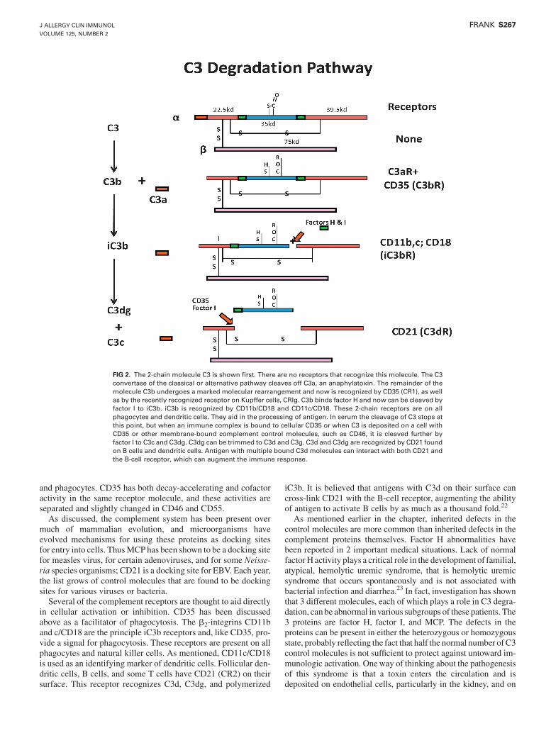
and phagocytes. CD35 has both decay-accelerating and cofactoractivity in the same receptor molecule, and these activities areseparated and slightly changed in CD46 and CD55.
As discussed, the complement system has been present overmuch of mammalian evolution, and microorganisms haveevolved mechanisms for using these proteins as docking sitesfor entry into cells. ThusMCP has been shown to be a docking sitefor measles virus, for certain adenoviruses, and for some Neisse-ria species organisms; CD21 is a docking site for EBV. Each year,the list grows of control molecules that are found to be dockingsites for various viruses or bacteria.
Several of the complement receptors are thought to aid directlyin cellular activation or inhibition. CD35 has been discussedabove as a facilitator of phagocytosis. The b2-integrins CD11band c/CD18 are the principle iC3b receptors and, like CD35, pro-vide a signal for phagocytosis. These receptors are present on allphagocytes and natural killer cells. As mentioned, CD11c/CD18is used as an identifying marker of dendritic cells. Follicular den-dritic cells, B cells, and some T cells have CD21 (CR2) on theirsurface. This receptor recognizes C3d, C3dg, and polymerized
iC3b. It is believed that antigens with C3d on their surface cancross-link CD21 with the B-cell receptor, augmenting the abilityof antigen to activate B cells by as much as a thousand fold.22
As mentioned earlier in the chapter, inherited defects in thecontrol molecules are more common than inherited defects in thecomplement proteins themselves. Factor H abnormalities havebeen reported in 2 important medical situations. Lack of normalfactor H activity plays a critical role in the development of familial,atypical, hemolytic uremic syndrome, that is hemolytic uremicsyndrome that occurs spontaneously and is not associated withbacterial infection and diarrhea.23 In fact, investigation has shownthat 3 different molecules, each of which plays a role in C3 degra-dation, can be abnormal invarious subgroups of these patients. The3 proteins are factor H, factor I, and MCP. The defects in theproteins can be present in either the heterozygous or homozygousstate, probably reflecting the fact that half the normal number ofC3control molecules is not sufficient to protect against untoward im-munologic activation. Oneway of thinking about the pathogenesisof this syndrome is that a toxin enters the circulation and isdeposited on endothelial cells, particularly in the kidney, and on
FIG 2. The 2-chain molecule C3 is shown first. There are no receptors that recognize this molecule. The C3convertase of the classical or alternative pathway cleaves off C3a, an anaphylatoxin. The remainder of themolecule C3b undergoes a marked molecular rearrangement and now is recognized by CD35 (CR1), as wellas by the recently recognized receptor on Kupffer cells, CRIg. C3b binds factor H and now can be cleaved byfactor I to iC3b. iC3b is recognized by CD11b/CD18 and CD11c/CD18. These 2-chain receptors are on allphagocytes and dendritic cells. They aid in the processing of antigen. In serum the cleavage of C3 stops atthis point, but when an immune complex is bound to cellular CD35 or when C3 is deposited on a cell withCD35 or other membrane-bound complement control molecules, such as CD46, it is cleaved further byfactor I to C3c and C3dg. C3dg can be trimmed to C3d and C3g. C3d and C3dg are recognized by CD21 foundon B cells and dendritic cells. Antigen with multiple bound C3d molecules can interact with both CD21 andthe B-cell receptor, which can augment the immune response.
J ALLERGY CLIN IMMUNOL
VOLUME 125, NUMBER 2
FRANK S267
erythrocytes. As the subject makes an immune response to thetoxin, in the absence of sufficient controlmolecules, antibodybindsto the toxin, and cells with toxin and antibody are destroyed bypoorly regulated complement activation. In truth, no onehas shownthat this is the mechanism of disease, but it places the disease in aframework that allows the pathophysiology to make sense.
It has also recently been reported that the largest risk factor inthe development of macular degeneration in the elderly is analteration of the amino acid at position 402 in factor H from atyrosine to a histadine.24 It is believed from statistical studies ofDNA sequences from pedigrees of families with inherited macu-lar degeneration that approximately 50% of cases are associatedwith this alteration in one amino acid, although the factor H allelewith histadine in position 402 is fairly common in the population,and other factors must be involved.
Receptors for the anaphylatoxins C3a, C4a, and C5aOf the anaphylatoxins, C5a has been studied in the greatest
detail.7 It is a potent chemotactic factor causing the directedmigra-tion of phagocytes. It contracts smooth muscle cells and causesmast cells to degranulate in the absence of IgE antibody. It causesneutrophils to adhere to one another and to endothelium in vessels.It clearly plays a part in the damage observed during the course ofimmunologic lung disease. Mice with a defect in the C5a receptordo not experience all of themanifestations of immunologic or aller-gic lung disease. It is likely that far more information will becomeavailable about this important receptor in the development ofasthma. There is less information available on C4a and C3a bind-ing. Themembrane receptor for C3a is clearly different from that ofC5a and can be triggered to cause mucus secretion in the airways,but its role in immunologic airways disease is still speculative.
Control of the late steps in the complement cascadeThe later steps in the complement cascade are also under tight
control. The site composed of the C3 convertase with bound C5will decay if it does not bind C6 rapidly, and there are a series ofmolecules that downregulate the late-acting proteins both inserum and on cells. S-protein, a plasma protein, interacts with C7as theC5, C6, andC7 complex forms and becomes hydrophobic.21
On binding S-protein, this complex is neutralized and can nolonger bind to cell surfaces. Similarly, clusterin, another plasmaprotein, binds to the forming C5-9 complex and prevents its acti-vation and completion. Most cells in the body have membrane-bound CD59, which interacts with the C5b-8 site, decreasingthe binding of C9 and preventing polymerization of C9. It protectsthe cell by preventing effective pore formation. All of these con-trol molecules are important in maintaining homoeostasis, andloss of the control molecules often leads to disease. CD59, likeCD55, is linked to cell membranes by a phosphatidylinositol link-age. By not having a transmembrane domain, the protein is free tomove rapidly in the fatty hydrophobic plane of the cell membraneto intercept forming C5b-9 and prevent cell lysis. Almost all pa-tients with the disease paroxysmal nocturnal hemoglobinuria(PNH) have an acquired bone marrow defect in which they havea mutation in bone marrow stem cells of the gene PIGA (phospha-tidylinositol glycan class A), the first enzyme in the developmentof phosphatidylinositol linkages.25 A single patient with a geneticdeficiency of CD59 has been reported, and this patient also hadPNH. This gene is present on the X-chromosome, and a single
gene defect in a bonemarrow stem cell leads to an inability to syn-thesize the first intermediate in this linkage pathway and thereforethe failure to have phosphatidylinositol-linked proteins on the cellmembrane. A failure to generate hematopoietic cells with CD59causes all hematopoietic cells of bone marrow origin derivedfrom the abnormal clone to be easily lysed by complement.As mentioned, alternative pathway proteins in the circulation un-dergo slow activation; CD59 is critical for neutralizing membraneattack proteins when they bind to our own cells. In patients withPNH, this mechanism is defective, and patients have a hemolyticanemia, often thrombocytopenia, and often a low neutrophilcount. Recently eculizumab, a humanized monoclonal anti-C5protein was approved for the treatment of PNH.26 This antibodybinds C5 and prevents complement-mediated lysis while allowingopsonization that occurs at the earlier C3 step to proceed. This isthe firstmedication that improves cell survival in this patient groupwith a disease that has a generally grim prognosis.
The role of complement in the generation of immunologic lungdisease is of particular interest. For many years, it was taken asgospel that complement plays no role in IgE-mediated lungdisease or asthma. Recent work has suggested that this might notbe the case. Complement can play a number of interestingfunctions in the generation of lung pathology.
First, it has been suggested that complement functions impor-tantly in directing immune responses towardTH1- or TH2-type im-munity. TH1 immunity is generally considered most important inprevention of infection, and TH2 immunity is associated withasthma and other allergic diseases. It is believed that the activationof C5 and the generation of C5a are important in directing the im-mune response toward a TH1 phenotype, and lack of C5 thereforeskews the system toward the generation of TH2 immunity.27,28 Onthe other hand, once immunity or allergy is established, it is be-lieved that C5amight be generated during immunologic responsesin the lung and, acting as an anaphylatoxin, might cause mast celldegranulation, smoothmuscle contraction, and so on, thereby con-tributing to the asthmatic response.
COMPLEMENT IN THE AFFERENT LIMB OF THEADAPTIVE IMMUNE RESPONSE
In recent years, attention has turned to the role of complementin the development of immunity.29,30 This discussion has focusedso far on the efferent limb of the response and how tissue damageis caused or controlled by complement. As mentioned early in thechapter, the complement system is phylogenetically older than theadaptive immune system, and many of the complement proteinsexisted as the adaptive immune system evolved.31 Therefore itis not surprising that elements of the complement system were in-corporated into the adaptive immune system, and these elementsare only now being slowly identified. As mentioned in an earliersection, the binding of complement to an antigen allowing cross-linking of CD21 and the B-cell receptor increases antigenicity byup to 1000-fold. In this case complement augments the immuneresponse. It is also known that subjects deficient in complement,although rare, often havemajor defects in adaptive immunity. An-imals deficient in C1q, C4, C3, and CR1/2 make a poor immuneresponse, particularly to T-dependent antigens; have poor germi-nal center formation; and have poor immunologic memory. Com-plement aids in the localization and retention of antigens withinthe germinal center, and it is believed that this localization of an-tigen to the germinal center facilitates an ongoing immune
J ALLERGY CLIN IMMUNOL
FEBRUARY 2010
S268 FRANK
response. Perhaps surprisingly, patients deficient in C1, C4, and,to a lesser extent, C2, have a high propensity toward systemic lu-pus erythematosus.32,33 In fact, of the relatively fewC1q-deficientsubjects who have been described, 96% have had systemic lupus.Of the relatively few C4-deficient subjects who have been de-scribed, 75% have had lupus. Even heterozygosity of the genesfor C4 predispose subjects to the development of lupus. This pro-pensity to cause systemic lupus erythematosus seems to be inde-pendent of the genetic localization of C4, C3, and factor B in themajor histocompatibility locus as class III genes and thereforetheir linkage to the MHC. In addition to the above, animals, par-ticularly those deficient in C1q and C4, do not develop normal tol-erance as well, although animals deficient in C3 and CR1/2 do notappear to have this defect. Although these are intriguing findingsand have been repeated in many laboratories, it is still not com-pletely clear how complement functions in the afferent limb ofthe adaptive immune response. It is quite likely that this questionwill be clarified over the next few years.
COMPLEMENT DEFICIENCIES AND CLINICALILLNESS
In the preceding paragraphs we have mentioned many diseasesassociated with defects in complement activation or control.Although recent research has demonstrated that HAE is not adisease whose clinical manifestations are due to defects in com-plement activation, it has typically been considered in this group.Because enormous progress has been made in defining its patho-genesis and treatment, it is given more detailed consideration.34,35
HAE is an inherited disease caused by low functional levels ofthe complement control plasma protein C1-INH. Patients havespontaneous episodic attacks of angioedema or deep localizedswelling, most commonly of a hand or foot, that begin duringchildhood and becomemuchmore severe during adolescence. Theedema is nonpitting and nonpruritic and is not associated withurticaria. Patients usually have a prodrome, a tightness or tinglingin the area that will swell, lastingmost frequently for several hours,followed by the development of angioedema. The swelling typi-cally becomes more severe over about 1! days and then resolvesover about the same time period. In some patients attacks arepreceded by the development of an erythematous rash that is notraised and not pruritic: erythema marginatum. The second majorsymptom complex noted by these patients is attacks of severeabdominal pain caused by edema of the mucosa of any portion ofthe gastrointestinal tract. The intensity of the pain can approximatethat of an acute abdomen, often resulting in unnecessary surgicalintervention. The gastrointestinal edema generally follows thesame time course to resolution as the cutaneous attacks.
Laryngeal edema is the most feared complication of HAE andcan cause complete respiratory obstruction. Although life-threat-ening attacks are infrequent, more than half the patients with HAEhave laryngeal involvement at some time during their lives.Dental work with the injection of a topical anesthetic into thegums is a common precipitant, but laryngeal edema is often spon-taneous. The clinical condition can deteriorate rapidly, progress-ing through mild discomfort to complete airway obstruction overa period of hours. Soft tissue edema can be difficult to see when itinvolves the larynx. If the swelling progresses to difficulty swal-lowing secretions or a change in the tone of the voice, this shouldbe considered an emergency andmight require emergency intuba-tion or even tracheostomy to ensure an adequate airway. Other
presentations are less common, but genital swelling is sometimesnoted in male and female patients.
In most cases the cause of the attack is unknown, but somepatients note that trauma or emotional stress precipitates attacks.In some female patients menstruation also regularly inducesattacks and estrogens increase attack frequency. The frequency ofattacks varies greatly among affected subjects and at differenttimes in the same subject, with some experiencing weeklyepisodes, whereas others might go years between attacks, andattacks can start at any age.
As noted above, C1-INH is a serpin that inactivates its target byforming a stable one-to-one complex with the enzyme to beinhibited. Although hepatocytes are the primary source of C1-INH, the protein is also synthesized bymonocytes. The regulationof the protein production is not completely understood, butbecause patients respond clinically to attenuated androgens withincreased serum levels of C1-INH, it is believed that theseandrogensmay stimulate C1-INH synthesis. HAE is an autosomaldominant disease, with as many as 25% of patients providing nofamily history. Presumably, most of these cases are caused by newgene mutations. Because all C1-INH–deficient patients are het-erozygous for this gene defect, it is believed that half the normallevel of C1-INH is not sufficient to prevent attacks.
Although named for its action on the first component ofcomplement (C1 esterase), C1-INH also inhibits proteins of thefibrinolytic, clotting, and kinin pathways (Fig 3). Specifically, C1-INH inactivates plasmin-activated Hageman factor (factor XII)and its fragments, activated factor XI, tissue plasminogen activa-tor, and kallikrein. Within the complement system, C1-INHblocks the activation and activity of C1 of the classical pathwayand MASPs 1 and 2 of the lectin pathway. Without C1-INH, un-checked activation of complement causes cleavage of the C4 andC2 proteins in the complement sequence, and patients often havelow levels of these proteins. Levels of the next protein in the com-plement cascade, C3, are normal. Themajor factor responsible forthe edema formation is now known to be bradykinin, an importantnonapeptide mediator that can induce leakage of post capillaryvenules. Bradykinin is derived from cleavage of the circulatingprotein high-molecular-weight kininogen by the plasma enzymekallikrein, the activity of which is controlled by C1-INH.
There are 2 genetic types of C1-INH deficiency that result inessentially the same phenotypic expression. The C1-INH geneserping 1 is located on chromosome 11 in the p11-q13 region. Theinheritance is autosomal dominant with incomplete penetrance.Type 1 is the most common form and accounts for approximately85% of cases. Synthesis of or secretion C1-INH is blocked at thesite of a faulty allele but occurs at the normal allele. The result istranscription of the normal protein, yielding quantitative serumconcentrations of C1-INH that are approximately 10% to 40% ofnormal values. Type 2 HAE accounts for approximately 15% ofcases. Mutations near the active site of the inhibitor lead tosynthesis and secretion of nonfunctional C1-INH protein. Thesepatients also have a normal functioning allele. Patients with type IIHAE have either normal or increased concentrations of the protein.
A clinical syndrome resembling HAE and termed type 3 HAEhas been described that affects mostly woman. In this conditionno abnormalities of complement or of C1-INH have beendescribed, but one third of patients have been found to have again-of-function abnormality of clotting factor XII, and it appearsthat many of the other patients with type III disease have defectsin the proteins that cause normal bradykinin degradation.
J ALLERGY CLIN IMMUNOL
VOLUME 125, NUMBER 2
FRANK S269
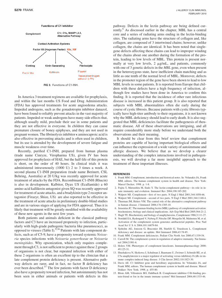
In America 3 treatment regimens are available for prophylaxis,and within the last months US Food and Drug Administration(FDA) has approved treatments for acute angioedema attacks.Impeded androgens, such as the gonadotropin inhibitor danazol,have been found to reliably prevent attacks in the vast majority ofpatients. Impeded or weak androgens have many side effects that,although usually mild, preclude their use in some patients andthey are not effective in everyone. In children they can causepremature closure of boney epiphyses, and they are not used inpregnant women. The fibrinolysis inhibitor e aminocaproic acid isalso effective in preventing attacks and is often used in children,but its use is attended by the development of severe fatigue andmuscle weakness over time.
Recently, purified C1-INH, prepared from human plasma(trade name Cinryze, Viropharma US), given IV has beenapproved for prophylaxis of HAE, but the half-life of this proteinis short, on the order of 40 hours. In clinical trials it wasadministered intravenously (1000 U) 2 to 3 times a week. Asecond plasma C1-INH preparation (trade name Berinert, CSLBehring, Australia) at 20 U/kg was recently approved for acutetreatment of attacks by the FDA. Recombinant C1-INH (Rhucin)is also in development. Kalbitor, Dyax US (Ecallantide) a 60amino acid kallikrein antagonist given SQ was recently approvedfor treatment of acute attacks, and a bradykinin type 2 receptor an-tagonist (Firazyr, Shize, US) are also reported to be effective inthe treatment of acute attacks in preliminary double-blind studiesand are in various stages of applying for FDA approval. Thus it islikely that treatment will be greatly modified with the availabilityof these new agents in the next few years.
Both patients and animals deficient in the classical pathwayfactors and C3 have an increased propensity to infection, partic-ularly with high-grade pathogenic bacteria like pneumococci, asopposed to viruses (Table I).9,36 Patients with late component de-fects, such as of C5-9, have a propensity toward systemic Neisse-ria species infections with Neisseria gonorrhoeae or Neisseriameningitides. Why opsonization, which only requires comple-ment through C3, is not sufficient to protect against these 2 groupsof organisms is not clear, but repeated infection with either ofthese 2 organisms is often an excellent tip to the clinician that alate complement protein deficiency is present. Alternative path-way defects are rarer, and in fact, no factor B deficiency hasever been described.37 The few patients with factor D deficiencyalso have a propensity toward infection, but autoimmunity has notbeen seen in either animals or patients with defects in this
pathway. Defects in the lectin pathway are being defined cur-rently.6 As discussed earlier in the chapter, MBL has a centralcore and a series of radiating arms ending in the lectin-bindingsites. The radiating arms have the structure of collagen and, likecollagen, are composed of 3 intertwined chains; however, unlikecollagen, the chains are identical. It has been noted that single-gene defects affecting these chains can lead to improper windingof the chains about one another during the formation of the pro-tein, leading to low levels of MBL. This protein is present nor-mally at very low levels, 2 mg/mL, and patients, commonlywith one of 3 genetic defects in theMBL gene, even when presentin the heterozygous state, have inefficient chain matching and aslittle as one tenth of the normal level of MBL. Moreover, defectsin the promoter region of the gene have been shown to lead to lowMBL levels in some patients. It is reported from Europe that chil-dren with these defects have a high frequency of infection, al-though few studies have been done in America to confirm thisfinding. It is reported that the incidence of other rare infectiousdisease is increased in this patient group. It is also reported thatsubjects with MBL abnormalities often die early during thecourse of cystic fibrosis. Because patients with cystic fibrosis typ-ically have high-titer antibody to their organisms, it is not knownwhy the MBL deficiency should lead to early death. It is also sug-gested that MBL deficiencies facilitate the pathogenesis of rheu-matic disease. All of these observations are intriguing, and allrequire considerably more study before we understand both theobservations and their meaning.
It should be clear from this brief review that complementproteins are capable of having important biological effects andcan influence the expression of a wide variety of autoimmune andallergic diseases. We believe that as we develop a clearerunderstanding of the complex interactions involved in pathogen-esis, we will develop a far more insightful approach to thetreatment of these important illnesses.
REFERENCES1. Frank MM. Complement, introduction and historical notes. In: Volanakis JA, Frank
MM, editors. The human complement system in health and disease. New York:Marcel Decker; 1998. p. 1-8.
2. Fujita T, Matsushita M, Endo Y. The lectin-complement pathway—its role in in-nate immunity and evolution. Immunol Rev 2004;198:185-202.
3. Walport MJ. Complement—first of two parts. N Engl J Med 2001;344:1058-66.4. Walport MJ. Complement—second of two parts. N Engl J Med 2001;344:1140-4.5. Thurman JM, Holers VM. The central role of the alternative complement pathway
in human disease. J Immunol 2006;176:1305-10.6. Jensenius JC. Themannan-binding lectin (MBL) pathway of complement activation:
biochemistry, biology and clinical implications. Adv Exp Med Biol 2005;564:21-2.7. Hugli TE. Biochemistry and biology of anaphylatoxins. Complement 1986;3:111-27.8. Nordahl EA, Rydengard V, Nyberg P, Nitsche DP, Morgelin M, Malmsten M, et al.
Activation of the complement system generates antibacterial peptides. Proc NatlAcad Sci U S A 2004;101:16879-84.
9. Sjoholm AG, Jonsson G, Braconier JH, Sturfelt G, Truedsson L. Complementdeficiency and disease: an update. Mol Immunol 2006;43:78-85.
10. Frank MM. Complement deficiencies. Pediatr Clin North Am 2000;47:1339-54.11. Carroll M. The complement system in regulation of adaptive immunity. Nat Immu-
nol 2004;5:981-6.12. Holers VM. Phenotypes of complement knockouts. Immunopharmacology 2000;
49:125-31.13. Shushakova N, Skokowa J, Schulman J, Baumann U, Zwirner J, Schmidt RE, et al.
C5a anaphylatoxin is a major regulator of activating versus inhibitory FcgRs in im-mune complex-induced lung disease. J Clin Invest 2002;110:1823-30.
14. Davis AE. C1 Inhibitor gene and hereditary angioedema. In: Volanakis JA, andFrank MM, editors. The human complement system in health and disease. MarcelDekker: New York; 1998. p. 455-80.
15. Blom AM, Villoutreix BO, Dahlback B. Complement inhibitor C4b-binding pro-tein-friend or foe in the innate immune system? Mol Immunol 2004;40:1333-46.
FIG 3. Functions of C1-INH.
J ALLERGY CLIN IMMUNOL
FEBRUARY 2010
S270 FRANK
16. Kim DD, Song WC. Membrane complement regulatory proteins. Clin Immunol2006;118:127-36.
17. Barilla-LaBarca ML, Liszewski MK, Lambris JD, Hourcade D, Atkinson JP. Roleof membrane cofactor protein (CD46) in regulation of C4b and C3b deposited oncells. J Immunol 2002;168:6298-304.
18. Joiner KA, Brown EJ, Frank MM. Complement and bacteria: chemistry and biol-ogy in host defense. Ann Rev Immunol 1984;2:461-84.
19. He JQ, Wiesmann C, van Lookeren Campagne M. A role of macrophage comple-ment receptor CRIg in immune clearance and inflammation. Mol Immunol 2008;45:4041-7.
20. Staunton DE, Lupher ML, Liddington R, Gallatin WM. Targeting integrin structureand function in disease. Adv Immunol 2006;91:111-57.
21. Liszewski MK, Atkinson JP. Regulatory proteins of complement. In: Volanakis JA,Frank MM, editors. The human complement system in health and disease. NewYork: Marcel Dekker; 1998. p. 149-66.
22. Dempsey PW, Allison ME, Akkaraju S, Goodnow CC, Fearon DT. C3d of comple-ment as a molecular adjuvant: bridging innate and acquired immunity. Science1996;271:348-50.
23. Fang CJ, Richards A, Liszewski MK, Kavanagh D, Atkinson JP. Advances inunderstanding of pathogenesis of aHUS and HELLP. Br J Haematol 2008;143:336-48.
24. Haines JL, Hauser MA, Schmidt S, Scott WK, Olson LM, Gallins P, et al. Comple-ment factor H variant increases the risk of age-related macular degeneration.Science 2005;308:419-21.
25. Luzzatto L. Paroxysmal nocturnal hemoglobinuria: an acquired X-linked geneticdisease with somatic-cell mosaicism. Curr Opin Genet Dev 2006;16:317-22.
26. Hillmen P, Young NS, Schubert J, Brodsky RA, Socie G, Muus P, et al. The com-plement inhibitor eculizumab in paroxysmal nocturnal hemoglobinuria. N Engl JMed 2006;355:1233-43.
27. Kohl J, Baelder R, Lewkowich IP, Pandey MK, Hawlisch H, Wang L, et al. A reg-ulatory role for the C5a anaphylatoxin in type 2 immunity in asthma. J Clin Invest2006;116:783-96.
28. Peng T, Hao L, Madri JA, Su X, Elias JA, Stahl GL, et al. Role of C5 in the devel-opment of airway inflammation, airway hyperresponsiveness, and ongoing airwayresponse. J Clin Invest 2005;115:1590-600.
29. Carroll M. The role of complement and complement receptors in induction andregulation of immunity. Ann Rev Immunol 1998;16:545-68.
30. Carroll MC, Holers VM. Innate autoimmunity. Adv Immunol 2005;86:137-57.31. Nonaka M. Phylogeny of the complement system. In: Volanakis JA, Frank MM,
editors. The human complement system in health and disease. New York: MarcelDekker; 1998. p. 203-16.
32. Manderson AP, Botto M, Walport MJ. The role of complement in the developmentof systemic lupus erythematosus. Ann Rev Immunol 2004;22:431-56.
33. Walport M, Davies KA, Botto M. C1q and systemic lupus erythematosus. Immun-obiology 1998;199:256-85.
34. Frank MM. Hereditary angioedema: the clinical syndrome and its management inthe United States. Immunol Allergy Clin North Am 2006;26:653-68.
35. ZurawBL.Clinical practice.Hereditary angioedema.NEngl JMed2008;359:1027-36.36. Figueroa J, Densen P. Infectious diseases associated with complement deficiencies.
Clin Microbiol Rev 1991;4:369-95.37. Thurman JM, Holers VM. The central role of the alternative complement pathway
in human disease. J Immunol 2006;176:1305-10.
J ALLERGY CLIN IMMUNOL
VOLUME 125, NUMBER 2
FRANK S271
Immune responses to malignancies
Theresa L. Whiteside, PhD, ABMLI Pittsburgh, Pa
Immune responses to tumor-associated antigens (TAs) are oftendetectable in tumor-bearing hosts, but they fail to eliminatemalignant cells or prevent the development of metastases.Patients with cancer generate robust immune responses toinfectious agents (bacteria and viruses) perceived as a ‘‘dangersignal’’ but only ineffective weak responses to TAs, which areconsidered as ‘‘self.’’ This fundamental difference in responsesto self versus nonself is further magnified by the ability oftumors to subvert the host immune system. Tumors inducedysfunction and apoptosis in CD81 antitumor effector cells andpromote expansion of regulatory T cells, myeloid-derivedsuppressor cells, or both, which downregulate antitumorimmunity, allowing tumors to escape from the host immunesystem. The tumor escape is mediated by several distinctmolecular mechanisms. Recent insights into these mechanismsencourage expectations that a more effective control of tumor-induced immune dysfunction will be developed in the nearfuture. Novel strategies for immunotherapy of cancer are aimedat the protection and survival of antitumor effector cells andalso of central memory T cells in the tumor microenvironment.(J Allergy Clin Immunol 2010;125:S272-83.)
Key words: Cancer, immunity, tumor escape, immune suppression,effector T cells
Evidence accumulated over the last few years convincinglyshows that the host immune system is involved in cancerdevelopment and progression, as well as control of metastasis.The presence of antitumor cellular responses, humoral responses,or both to tumor-associated antigens (TAs) has been observed inmany, but not all, patients with cancer.1,2 The evidence for suchpre-existing antitumor immunity in patients with cancer confirmsthat the tumor-bearing host is capable of mounting an immune re-sponse to TAs. Tumor progression from a single transformed cellto a mass of malignant cells is a multistep process involving a se-ries of genetic changes occurring in human subjects over a periodof months or years and culminating in the established tumor.3
During this period, neither the host immune system nor the devel-oping tumor are idle: those newly emerging tumor cells that arerecognized by the immune system are eliminated only to be re-placed by genetic tumor variants resistant to immune interventionand giving rise to a heterogenous population of malignant cells
found in any tumor. Tumors are genetically unstable, and theemergence of new genetic variants, which is responsible for thetumor heterogeneity, ensures that the tumor survives in the faceof the host immune system. Only the tumor cells that manageto avoid recognition escape and survive, whereas those that arerecognized by the immune system are eliminated as soon asthey arise. The tumor development involves a prolonged seriesof checks and balances between the host attempting to curtail tu-mor growth and the tumor benefiting from genetic changes, alter-ing its microenvironment and avoiding immune elimination. Thusthe tumor becomes resistant to immune effector cells.
The interactions between the host and the tumor have beenreferred to as ‘‘immune surveillance,’’ a concept that originatedmany years ago with F. M. Burnett and that introduced his visionof a vigilant host immune system able to spot, recognize, andeliminate tumor cells. A modern version of the immune surveil-lance theory not only emphasizes the ability of the host immunesystem to recognize and destroy tumor cells but also its contri-bution to ‘‘immune selection’’ of resistant tumor variants. Thusthe ‘‘immune editing’’ hypothesis2,4 has been advanced to suggestthat by means of elimination of tumor cells sensitive to immuneintervention, the host immune system edits for survival of tumorsthat become resistant to immune cells. An alternative hypothesisallows for the progressing tumor to develop immunosuppressivemechanisms that will thwart any attempt of immune tumor elim-ination and in effect will induce a state of tumor-specific toler-ance.5 In the first instance the immune system initiates theselection of resistant tumor variants, and in the second the tumorbecomes a perpetrator of immune unresponsiveness. Central tothe paradigms of immune selection or immune editing and
Abbreviations usedAnx: Annexin VAPC: Antigen-presenting cellAPM: Antigen-processing machineryb2 m: b2-microglobulinCTL: Cytolytic T lymphocyteDC: Dendritic cell
FasL: Fas ligandFOXP3: Forkhead box protein 3iNOS: Inducible nitric oxide synthase
MDSC: Myeloid-derived suppressor cellNK: Natural killer
PD-1: Programmed death 1PD-L1: Programmed death ligand 1PGE2: Prostaglandin E2
ROS: Reactive oxygen speciesSTAT3: Signal transducer and activator of transcription 3
TA: Tumor-associated antigenTAM: Tumor-associated macrophageTCR: T-cell receptorTIL: Tumor-infiltrating lymphocyteTreg: Regulatory T
VEGF: Vascular endothelial growth factor
From the University of Pittsburgh Cancer Institute and the Departments of Pathology,Immunology, and Otolaryngology, University of Pittsburgh School of Medicine.
Supported in part by National Institutes of Health grant PO1-CA109688 to Theresa L.Whiteside.
Disclosure of potential conflict of interest. T. L. Whiteside has declared that she has noconflict of interest.
Received for publication July 10, 2009; revised September 11, 2009; accepted for pub-lication September 17, 2009.
Reprint requests: Theresa L. Whiteside, PhD, University of Pittsburgh Cancer Institute,5171 Centre Ave, Pittsburgh, PA 15213. E-mail: [email protected].
0091-6749/$36.00! 2010 American Academy of Allergy, Asthma & Immunologydoi:10.1016/j.jaci.2009.09.045
S272
immune suppression is the premise that the tumors acquiring newmutations are able to avoid immune intervention and are capableof both escaping and disabling the host immune system. Neitherof the 2 hypotheses has been completely accepted today, and thereare those who believe that tumors progress because of the geneticinstability and others who favor tumor-specific tolerance of theimmune system, which enables the tumor to take advantage ofthe tissue microenvironment regardless of the immune systemand benefit from it. This controversy regarding the significanceof the immune system in tumor development and progression un-derscores the complexity of interactions between the tumor andthe immune cells. It surmises that these interactions might be bi-directional, are influenced by the local microenvironment, and notinfrequently might result in demise not of the tumor but of tumor-reactive immune cells.
In this chapter the nature and components of the host immuneresponse against tumors will be discussed, including the reasonsfor the failure of the immune system to contain tumor growth andmetastasis. It is this latter aspect of the immunobiology of humanmalignancies that will be emphasized, largely because it directlyaffects cancer immunotherapy. A relatively recent realization thattumors have devised multiple and remarkably effective mecha-nisms for disarming the host immune system has opened away forthe introduction of novel therapeutic strategies aimed at elimi-nating tumor escape. If the tricks tumors use for protection fromimmune intervention by the host are responsible for theirprogression, then it could be surmised that a limited success ofcurrent immune therapies for cancer can be reversed by therapiesthat target the escape mechanisms, and because these escapemechanisms might be unique for each tumor rather than gener-alized, the future challenge will be to identify the ‘‘immunologicsignature’’ of each tumor and then use selective therapies toeliminate the tricks and restore vigorous antitumor immunity.
TUMOR PROGRESSION AND THE HOST IMMUNERESPONSE
There are several lines of evidence that point to an early, as wellas late, involvement of the immune system in tumor development.Early tumor lesions, and even premalignant foci, such asmelanocytic nevi, are frequently infiltrated with hematopoieticcells, including lymphocytes, macrophages, and occasionallygranulocytes.6,7 The presence of immune cells in the tumor atlater stages of development (ie, the abundance of tumor-infiltrat-ing lymphocytes [TILs]) has been associated with improved pa-tient survival in several early studies (reviewed in Whiteside8).More recently, studies by Fridman’s group performed a compre-hensive multivariate analysis of cellular interactions in the tumormicroenvironment based on the type, density, localization, andfunction of immune cells present within human colorectal cancerand demonstrated that immune reactivity at the tumor site influ-ences clinical outcome.9-11 Thus increased densities of T-cell in-filtrates with a high proportion of CD81 T cells within primarycolorectal carcinomas were associated with a significant protec-tion against tumor recurrence.11 Furthermore, the same groupalso showed that coexpression of genes mediating cytotoxicityand TH1 adaptive immune responses accurately predicted sur-vival in patients with colorectal carcinoma independently of themetastatic status.12 In aggregate these multiparameter analysesof tumor-infiltrating cells in situ suggest that immune cells canand indeed often do play a role in tumor control but that both
intrinsic and extrinsic factors in the tumormicroenvironment alterthe balance required for optimal control.12
In many patients with cancer, it is possible to expand in cultureand in vitro test functions of tumor-specific cytolytic T lympho-cytes (CTLs) from the peripheral blood or TILs.8 This finding,which has been reproduced in many laboratories, suggests thatprecursors of such CTLs exist in the circulation or at the tumorsite in patients with cancer and can be induced to proliferatewhen autologous dendritic cells (DCs) pulsed with relevant tumorepitopes and used as antigen-presenting cells (APCs). More re-cent experiments, using tetramers and flow cytometry, have di-rectly demonstrated the presence of tumor peptide–specific Tcells in the circulation of patients with cancer.1,13,14 Furthermore,the frequency of such peptide-specific T cells appears to be higherin the circulation of patients with cancer than in healthy sub-jects.15 Finally, the SEREX technology, based on the presenceof tumor-specific antibodies in sera of patients with cancer, hasbeen successfully used for tumor-antigen discovery in many lab-oratories.16 These findings, as well as recent identification of nu-merous TAs that appear to be immunogenic in that they inducehumoral immune responses, cellular immune responses, or bothin vitro by using human immune cells and in vivo in animalmodels of tumor growth, strongly support the notion that thehost immune system recognizes the presence of the tumor andresponds to it by generating both local and systemic immuneresponses.
If the tumors are not ignored by the immune system, why dothey progress? Several answers to this question can be considered.First, there is the old argument for the lack of a ‘‘danger signal’’17
in tumors akin to those presented by pathogens invading tissuesduring an infection. Recognition by DCs of pathogen-associatedmolecular patterns through the ubiquitous Toll-like receptorsleads to efficient DC activation and maturation. It promotes gen-eration of vigorous cellular and antibody responses to bacterial orviral antigens, presumably because the immune system perceivesan infection as a danger signal17 benefiting the host. However,functional Toll-like receptors are known to be expressed bymany human solid tumors,18 and recent data indicate that tumorsuse them to promote their own growth; for protection fromspontaneous, immune-mediated, or drug-induced apoptosis; orboth.18,19
Second, TAs are perceived by the immune system as ‘‘self’’ or‘‘altered self’’ antigens, which evoke weak immune responsesbecause tolerance prevents generation of immune responses toself. The only ‘‘unique’’ TAs are mutated antigens, and these arestrongly immunogenic and elicit robust immune responses.20
However, only a handful of such mutated TAs are known, andthe vast majority of TAs are poorly immunogenic or simply toler-ogenic. In this context cancer can be viewed as an autoimmunephenomenon in which tolerance to self prevents effective immuneresponses to TAs Patients with cancer who have not been treatedwith chemotherapy or radiotherapy generally have normal im-mune responses to viral or bacterial antigens, yet they are unableto respond to their own TAs. Except for late-stage disease, theygenerally have normal delayed-type hypersensitivity responsesto recall antigens but are anergic to autologous TAs. Althoughtolerance to self is a detriment to the generation of antitumorresponses in patients with cancer, another factor that exerts anoverwhelming effect on these responses is the tumor microenvi-ronment. Each tumor creates its own milieu characterized bythe presence of immunosuppressive factors and by the excess of
J ALLERGY CLIN IMMUNOL
VOLUME 125, NUMBER 2
WHITESIDE S273
TAs produced and released by the growing tumor. Evidence sug-gests that tumors produce a broad array of immunoinhibitory fac-tors, which exert either local or systemic effects on the hostantitumor immune responses.5 Therefore it is not surprising thatantitumor immunity might be weak, inefficient, or even absentin patients with cancer, depending on the nature of tumor-host in-teractions, as well as the robustness of regulatory mechanisms incontrol of immune tolerance.
Immune antitumor responses could be influenced by thegradual deterioration of the immune system with age.21 The in-creased incidence of cancer present in the elderly might be dueto immunosenescence (ie, progressive remodeling of the immunesystem with a reduced ability of immune cells to respond to acti-vating stimuli and increased responsiveness to tolerogenic sig-nals).21 Immunosenescence can significantly interfere with theeffectiveness of cancer immunotherapies, and it has beensuggested that clinical trials testing immunopotentiating agentsin patients with cancer should be conducted in elderly subjects.21
Recent multiparameter analyses of primary and metastatichuman tumors (eg, colorectal carcinoma) recognize several majorimmune ‘‘coordination profiles,’’ the presence of which is influ-enced by the balance between tumor escape and immune antitu-mor responses and that are subject to host-tumor cross-talk.12 Inthis context it is important to consider differences between pri-mary and metastatic tumors. Not only are metastatic tumorsmore immunosuppressive, but also they appear to be less readilyrecognized by TA-specific immune effector cells. The latter couldbe due to defects in the expression levels of antigen-processingmachinery (APM) components, MHC molecules, or both in thetumor and its metastases.22 Because different copy numbers ofdistinct trimolecular peptide–b2-microglobulin (b2 m)–MHCcomplexes presented on the tumor surface might lead to differen-tial T-cell recognition, this aspect of tumor–immune cell interac-tions is critical.22,23 A recent comparison of primary renal cellcarcinoma, renal cell carcinoma metastases, and normal renal tis-sue with respect to HLA ligand presentation and gene expressiondemonstrated a greater similarity between primary tumor andmetastasis than between the tumor and normal tissue.24 Thisobservation provides a good rationale for peptide-based immuno-therapy because it is likely to preferentially target the tumor andits metastases and not the normal tissue.
NATURAL VERSUS ADAPTIVE IMMUNERESPONSES TO MALIGNANCIES
Antitumor immune responses can be innate (natural) oracquired (adaptive). Innate immunity is mediated by cells orsoluble factors that naturally exist in tissues or body fluids and caninterfere with tumor growth or survival. Among hematopoieticcells, macrophages, granulocytes, natural killer (NK) cells(CD32CD561), non–MHC-restricted T cells (CD31CD562),and gd T cells have the natural capability to eliminate tumorcell targets.21 In addition, natural antibodies with specificities di-rected at surface components of tumor cells might be present inthe sera of patients with cancer.16 Other serum factors, includingcomplement components, C-reactive protein, mannose-bindingprotein, and serum amyloid protein, also play a role in innate im-munity.25 Adaptive immune responses to tumors are mediated byCD31T-cell receptor (TCR1) T cells when they recognize tumor-derived peptides bound to self-MHC molecules expressed onAPCs. Little is currently known about the molecular signals and
cellular steps involved in directing APCs, such as DCs, to executea tolerogenic versus immunogenic program in response to anti-gens. As indicated above, tumors can also serve as APCs, al-though low levels of MHC class I molecule expression, MHCclass II molecule expression, or both on the surface of tumor cellsmakes this an inefficient process.22 More likely, TAs are taken upby DCs present at the tumor site, processed, and cross-presentedto T cells in the tumor-draining lymph nodes in the form of thetrimolecular peptide–b2m–MHC complexes.23 For adaptive im-mune response to occur, T cells expressing correct (cognate)TCRs have to be present. Recognition of the peptide and its bind-ing to the variable domains of the TCR initiates signaling (signal1) that leads to T-cell activation.26 This requirement implies priorsensitization and a clonal expansion of memory T cells inresponse to a cognate tumor epitope (anamnestic or recallresponses). Alternatively, precursor T cells expressing the TCRcan be primed by the cognate peptide–MHC ligands presentedon APCs, and the subsequent development of antitumor effectorcells is viewed as a primary immune response. In either case cos-timulatory molecules (signal 2) are necessary for an immuneresponse to proceed,27 and once T-cell proliferation is initiated,appropriate cytokines (signal 3) become essential for sustainingthe response.28 Recent findings stress the key importance of signal3 for the development of immune responses and for their contrac-tion.28 Like all immune responses, those that are TA specific donot go on forever but peak and then contract, restoring the preac-tivation balance. The precise mechanisms responsible forimmune contraction are not yet defined, and regulatory T (Treg)cells, as well as other mechanisms, have been proposed toregulate immune reactivity, but it is clear that events in theenvironment play a dominant role in this respect.
Immune responses to malignant cells can be categorized aslocoregional or systemic. In situ or local responses refer mainly toTILs, which accumulate in most human solid tumors and the roleof which in tumor progression remains highly controversial. Longconsidered by some an effector arm of antitumor responses, TILsare viewed by others as victims of the tumor microenvironmentbecause their effector functions are often impaired, presumablyby tumor-derived factors.29 A failure of local antitumor responsesmediated by TILs is thought to contribute to tumor progression.Systemic immunity to tumors, as measured by delayed-typehypersensitivity responses or by various ex vivo assays of T-cellresponses in the peripheral circulation of patients with cancer,are difficult to demonstrate, and TA-specific responses havebeen particularly elusive. Nevertheless, by using highly sensitivemulticolor flow cytometry, it has been possible to detect and mea-sure the frequency of TA-specific CD81 and CD41 T cells in theperipheral circulation of patients with cancer.1 Although theresponse levels vary, TA-specific and nonspecific proliferativeor cytotoxic responses of peripheral lymphocytes in patientswith cancer appear to be at least partially impaired.29-31 Data in-dicate that the same functional impairments seen in TILs arefound in both circulating and lymph node lymphocytes of patientswith cancer.29,32 Thus it has been concluded that, in general, hu-man tumors exert profound suppressive effects on both local andsystemic antitumor immunity in these patients.
In contrast to the failure of antitumor immune responses tocontrol tumor progression in human subjects, a large body ofexperimental evidence derived from preclinical animal models ofcancer suggests that the immune system can prevent tumorgrowth or cause its rejection.33 In the prevention setting
J ALLERGY CLIN IMMUNOL
FEBRUARY 2010
S274 WHITESIDE
vaccination of animals with TAs plus adjuvant protects them fromrechallenge with tumor,34 whereas immunotherapy of establishedtumors with vaccines, cytokines, adoptively transferred immunecells, or immunomodulatory agents results in tumor rejection,provided the tumor is not in an advanced stage. Remarkably,this has been a consistent pattern seen with carcinogen-induced,virally induced, and spontaneously arising tumors in mice, sug-gesting a fundamental difference in immune responses to tumorantigens between mice and human subjects. Indeed, it appearsthat the difference might be due to appreciably greater immuno-genicity of murine TAs, which in most cases are virus- or carcin-ogen-related epitopes and thus foreign rather than self-epitopes.Alternatively, the answer might be that experimental murinetumors are established, grow, progress, and are eliminated bytherapy in the very short time required for the completion of theexperiment, leaving no time for the development of tumor escapemechanisms. In contrast, human tumors are diagnosed and treatedafter many years of coexistence with the host. An introduction orestablishment of the tumor in mice is a dramatic event that mobi-lizes host defenses in contrast to a silent coexistence of tumorcells with the immune system for many years in human subjects.To minimize this difference, transgenic murine models have beendeveloped, allowing for ensured, genetically driven tumor devel-opment in a ‘‘spontaneous’’ environment.35 Transgenic mice havebeen especially useful in the design of preventive cancervaccines,34 and information they provide is encouraging for thedevelopment of immunoprophylaxis of cancer in human subjects.Nevertheless, to date, it has been difficult to translate the positiveresults seen in mice to immunotherapy of established humantumors. It is plausible that numerous and varied mechanisms ofescape developed by the latter during the prolonged residenceand interactions with the host provide human tumors with advan-tages not afforded to murine tumors established in an experimen-tal setting.
TUMOR ASSOCIATED ANTIGENSRecent progress in the development of cancer vaccines has
been greatly facilitated by the availability of well-defined TAs,many of which have been characterized in the last decade.36 Mostof these TAs are derived from self-proteins that are either mutatedor otherwise differentially expressed in normal and tumor cells, asexemplified by oncogenes or oncofetal or cancer testis antigens.The major categories of TAs that have been often used as candi-dates for immune therapies are listed in Table I.36,37 A recent re-port provides a much longer prioritized list of well-characterizedcancer antigens best suited for use in cancer vaccines.38 The list isbased on criteria generated by a panel of experts convened by theNational Cancer Institute38 and is designed to assist investigatorsin the field of immunotherapy in the selection of the most prom-ising TAs for further testing in clinical trials.
As already indicated, immune responses to TAs, even to thoserepresenting altered self-antigens, are detectable in tumor-bear-ing hosts, although in most cases no correlations between thepresence of in vitro responses to TAs and prognosis have beendocumented. This is in contrast to numerous animal tumormodels, which have provided strong evidence that in the presenceof effective antitumor immunity, tumors fail to progress and es-tablished tumors regress.39 Nevertheless, human cancer vaccinetrials in patients with cancer have made use of many well-charac-terized TAs in the hope that their presentation on appropriately
polarized DCs will overcome difficulties with the generation ofa strong immune response in the therapeutic setting. The most re-cent reports of such clinical trials in patients with cancer indicatethat multiple subcutaneous injections of an immunogenic tumorpeptide, such as NY-ESO-1, plus a mix of 2 potent adjuvants,such as Montanide ISA-51 and CpG7909, can be effective in in-ducing sustained peptide-specific immune responses and signifi-cantly prolong survival, even in patients with advanced disease,including solid tumors other than melanoma.40 These reports,demonstrating that antitumor, antivaccine, or both immuneresponses correspond to clinical outcome, suggest that the optimi-zation of vaccination strategies is likely to overcome tumor-induced suppression and to restore the immune balance alteredby cancer development.
IMMUNE CELLS IN THE TUMORMICROENVIRONMENT
Immune cells that are most frequently found in the humanmicroenvironment are lymphocytes, which are capable of medi-ating both innate and adaptive immunity, although monocytes,tumor-associated macrophages (TAMs), and DCs are also com-monly seen.41 Inflammatory cells present in the tumor are in inti-mate contact with tumor cells, stromal fibroblasts, extracellularmatrix components, and blood vessels. Proinflammatorycytokines secreted by inflammatory cells can contribute to tumorprogression, and soluble factors produced by the tumor inresponse to nonspecific or tumor-specific signals, such as prosta-glandin E2 (PGE2), adenosine, or TGF-b, downregulate functionsof immune cells. The tumor microenvironment is created by thetumor, and it is continuously shaped and dominated by the tumor,which directs all cellular and molecular events taking place in thesurrounding tissue.
Immune cells recruited to the tumor include T cells(CD31TCR1), which are by far the largest component of mono-nuclear tumor infiltrates41 and have received the most attention.Although their accumulation in the tumor might be consideredevidence of immune surveillance by the host, they are largely in-effective in arresting tumor growth, although they can proliferateand mediate antitumor cytotoxicity on their removal from thetumor bed and ex vivo IL-2 activation.42
Phenotypic and functional characteristics of human TILs arelisted in Table II. More current data on the status of T cells foundin human tumors suggest that their phenotypic and functionalprofile varies depending on the microenvironment created bythe tumor and that this profile or ‘‘immune signature’’ can influ-ence prognosis and disease outcome.9,12 It appears that TILs ob-tained from advanced or metastatic lesions are more functionallyimpaired than those from early lesions, suggesting that tumor bur-den or the potential of a tumor to suppress immune cells might de-termine the functional status of infiltrating T cells. Among CD41
T cells present in the tumor, a subset of CD41CD25high forkheadbox protein 3 (FOXP3)–positive Treg cells is expanded to consti-tute from 5% to 15% of CD4 T cells in the infiltrate. Their fre-quency is higher in the tumor than in the peripheralcirculation.43,44 These cells suppress functions of other immunecells in the microenvironment by mechanisms that might be cellcontact dependent or might involve the production of inhibitorycytokines or adenosine.43-46 Recently, a potent proinflammatoryT-cell subset, IL-17–producing TH17 cells, were observed amongCD41 cells in patients with ovarian carcinoma. The presence of
J ALLERGY CLIN IMMUNOL
VOLUME 125, NUMBER 2
WHITESIDE S275
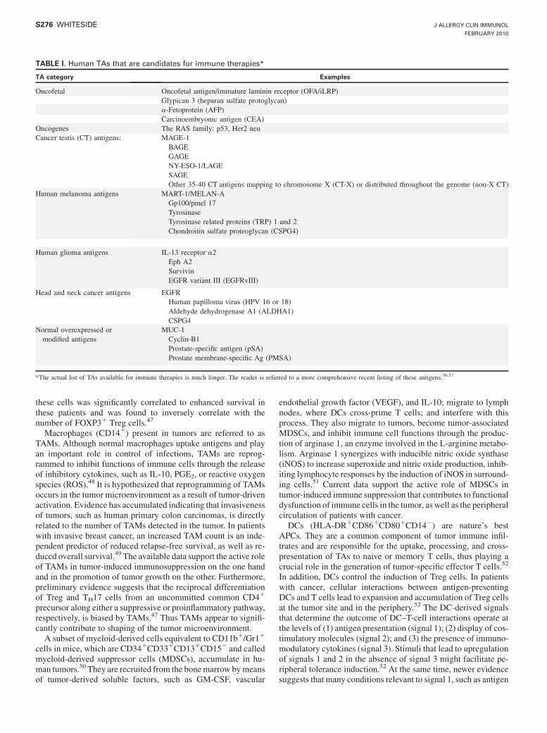
these cells was significantly correlated to enhanced survival inthese patients and was found to inversely correlate with thenumber of FOXP31 Treg cells.47
Macrophages (CD141) present in tumors are referred to asTAMs. Although normal macrophages uptake antigens and playan important role in control of infections, TAMs are reprog-rammed to inhibit functions of immune cells through the releaseof inhibitory cytokines, such as IL-10, PGE2, or reactive oxygenspecies (ROS).48 It is hypothesized that reprogramming of TAMsoccurs in the tumor microenvironment as a result of tumor-drivenactivation. Evidence has accumulated indicating that invasivenessof tumors, such as human primary colon carcinomas, is directlyrelated to the number of TAMs detected in the tumor. In patientswith invasive breast cancer, an increased TAM count is an inde-pendent predictor of reduced relapse-free survival, as well as re-duced overall survival.49 The available data support the active roleof TAMs in tumor-induced immunosuppression on the one handand in the promotion of tumor growth on the other. Furthermore,preliminary evidence suggests that the reciprocal differentiationof Treg and TH17 cells from an uncommitted common CD41
precursor along either a suppressive or proinflammatory pathway,respectively, is biased by TAMs.47 Thus TAMs appear to signifi-cantly contribute to shaping of the tumor microenvironment.
A subset of myeloid-derived cells equivalent to CD11b1/Gr11
cells in mice, which are CD341CD331CD131CD152 and calledmyeloid-derived suppressor cells (MDSCs), accumulate in hu-man tumors.50 They are recruited from the bonemarrow bymeansof tumor-derived soluble factors, such as GM-CSF, vascular
endothelial growth factor (VEGF), and IL-10; migrate to lymphnodes, where DCs cross-prime T cells; and interfere with thisprocess. They also migrate to tumors, become tumor-associatedMDSCs, and inhibit immune cell functions through the produc-tion of arginase 1, an enzyme involved in the L-arginine metabo-lism. Arginase 1 synergizes with inducible nitric oxide synthase(iNOS) to increase superoxide and nitric oxide production, inhib-iting lymphocyte responses by the induction of iNOS in surround-ing cells.51 Current data support the active role of MDSCs intumor-induced immune suppression that contributes to functionaldysfunction of immune cells in the tumor, as well as the peripheralcirculation of patients with cancer.
DCs (HLA-DR1CD861CD801CD142) are nature’s bestAPCs. They are a common component of tumor immune infil-trates and are responsible for the uptake, processing, and cross-presentation of TAs to naive or memory T cells, thus playing acrucial role in the generation of tumor-specific effector T cells.52
In addition, DCs control the induction of Treg cells. In patientswith cancer, cellular interactions between antigen-presentingDCs and T cells lead to expansion and accumulation of Treg cellsat the tumor site and in the periphery.52 The DC-derived signalsthat determine the outcome of DC–T-cell interactions operate atthe levels of (1) antigen presentation (signal 1); (2) display of cos-timulatory molecules (signal 2); and (3) the presence of immuno-modulatory cytokines (signal 3). Stimuli that lead to upregulationof signals 1 and 2 in the absence of signal 3 might facilitate pe-ripheral tolerance induction.52 At the same time, newer evidencesuggests that many conditions relevant to signal 1, such as antigen
TABLE I. Human TAs that are candidates for immune therapies*
TA category Examples
Oncofetal Oncofetal antigen/immature laminin receptor (OFA/iLRP)Glypican 3 (heparan sulfate protoglycan)a-Fetoprotein (AFP)Carcinoembryonic antigen (CEA)
Oncogenes The RAS family: p53, Her2 neuCancer testis (CT) antigens: MAGE-1
BAGEGAGENY-ESO-1/LAGESAGEOther 35-40 CT antigens mapping to chromosome X (CT-X) or distributed throughout the genome (non-X CT)
Human melanoma antigens MART-1/MELAN-AGp100/pmel 17TyrosinaseTyrosinase related proteins (TRP) 1 and 2Chondroitin sulfate proteoglycan (CSPG4)
Human glioma antigens IL-13 receptor a2Eph A2SurvivinEGFR variant III (EGFRvIII)
Head and neck cancer antigens EGFRHuman papilloma virus (HPV 16 or 18)Aldehyde dehydrogenase A1 (ALDHA1)CSPG4
Normal overexpressed ormodified antigens
MUC-1Cyclin-B1Prostate-specific antigen (pSA)Prostate membrane-specific Ag (PMSA)
*The actual list of TAs available for immune therapies is much longer. The reader is referred to a more comprehensive recent listing of these antigens.36,37
J ALLERGY CLIN IMMUNOL
FEBRUARY 2010
S276 WHITESIDE
dose, determine whether Treg or TH2 effector (Teff) cells are in-duced, irrespective of the maturation state of DCs.52 In addition,insights into the APM in DCs and evidence that some of the com-ponents of APM, including MHC class II molecules, might bedownregulated or altered in patients with cancer,23 suggest thatTreg cell induction might be influenced not only by the natureand dose of the antigen but also by its processing and its presen-tation to T cells.
Tumor-associated DCs directly exposed to tumor cells, tumor-derived factors, or both have been shown to readily undergoapoptosis and to have impaired maturation.53 Specifically, tumor-derived factors, such as gangliosides, were shown to inhibit DCgeneration and their function in vitro.54 This suppressive effectof gangliosides on DCs was found to be mediated by tumor-de-rived VEGF, a known antidendropoietic factor.53 The data onfunctional impairments of tumor-associated DCs have to be bal-anced by numerous reports in the literature, which suggest thatthe presence of DCs in tumors is associated with improved prog-nosis and prolonged patient survival, as well as a reduced inci-dence of recurrent or metastatic disease.55 In contrast, patientswith lesions reported to be scarcely infiltrated with DCs have arelatively poor prognosis.56 Fewer DCs were observed in meta-static than in primary lesions. In one study it was shown thatthe number of DCs present in the tumor was by far the strongestindependent predictor of overall survival, as well as disease-freesurvival and time to recurrence, in a large cohort (n5 132) of pa-tients with oral carcinoma compared with such well-establishedprognostic factors as disease stage or lymph node involvement.55
It appears that not only the number of DCs but also the presence offunctionally unimpaired, normally signaling T cells in the tumormicroenvironment are important for overall survival of patientswith cancer.55
NK cells (CD32CD561CD161), which mediate innate immu-nity and contain both perforin-rich and granzyme-rich granules,are well equipped to mediate lysis of tumor cells. Although NKcells represent ‘‘the first line of defense’’ against pathogens,57
most human tumor cells are resistant to perforin-mediated NKcell lysis, and NK cells are rarely found among TILs.41 This is
despite the fact that tumor cells often downregulate MHC antigenexpression and are enriched in MICA and MICB molecules.58
There might be several reasons for the paucity of NK cells in tu-mors, including the possibility that NK cells are present in prema-lignant or early lesions and absent from advanced tumors, whichis consistent with their role in immune surveillance rather thankilling of cancer cells at the tumor site.41More recent data suggestthat the primary biologic role of NK cells in tumor-bearing hostsmight not be the elimination of tumor targets but rather the facil-itation of DC–T-cell interactions and driving the immune re-sponses to TAs.59 Because the tumor site is not likely to be theoptimal milieu for this type of immune interaction, the paucityof NK cells in tumors might fit with their physiologic functions.The in vivo role of NK cells in antitumor immune defense is notyet clear, and work continues to define it further.
Polymorphonuclear leukocytes are infrequently seen in infil-trates of human solid tumors, with the exception of nests ofeosinophils that might be present in association with tumor cellsin some cases. In human tumors granulocytes, which are a majorcellular component of many murine tumors, are rare, beinglargely replaced by TAMs or MDSCs. This could be explained bythe fact that most inflammatory infiltrates into human tumors arechronic rather than acute, with granulocytes long gone by the timehuman tumors are diagnosed, biopsied, and examined.
B cells (CD191, CD201) are also rare in most human tumors,with the exception of breast cancer and melanoma.6,60 Theprimary function of B cells is differentiation into antibody-producing plasma cells. Although TA-specific antibodies are fre-quently detected in the circulation of patients with cancer, theseantibodies are made and secreted in the tumor-draining lymphnodes, spleen, or other lymphoid tissues. From these sites, IgGmolecules can readily be transported through plasma or lymphto tissue sites. Therefore the presence of B cells or plasma cellsin tumors is not expected a priori, although it might be that theability to make antibodies in situ could be an important aspectof host defense.
Inflammatory infiltrates present in human tumors change incomposition and intensity during tumor progression. The initialacute inflammation involving the recruitment and influx ofantitumor effector cells is replaced by chronic inflammation inlater stages of tumor progression. Tissue hypoxia plays a majorrole in shaping the nature of immune infiltrates in tumors. It iscreated by activation of hypoxia-responsive genes in tumorcells61 and favors the influx of granulocytes and phagocyticmacrophages, which depend on the glycolytic pathway for sur-vival.62 These cells take up and process dying tumor cells,producing an abundance of ROS. The subsequent reoxygenationof the microenvironment is accompanied by activation of thenuclear factor kB pathway in both tumor cells and infiltratingimmune cells, leading to the excessive secretion of proinflamma-tory cytokines.5 Responding to this nuclear factor kB–drivencascade of proinflammatory cytokines, the tumor and stromalcells produce a variety of soluble factors with wide-rangingbiologic effects, including the promotion of tumor cell prolifer-ation.5 In the tumor microenvironment cellular expansion, differ-entiation, or activation, as well as cell migration, matrixremodeling, and blood vessel growth, are reprogrammed tobenefit the tumor. Thus the nature of chronic inflammatoryinfiltrates and functions of the tumor-infiltrating immune cellsdepend on how aggressively a given tumor remodels itsmicroenvironment.
TABLE II. Morphologic, phenotypic, and functional characteris-
tics of TILs found in human solid tumors
Morphology: small to large lymphocytesPhenotype: CD31TCR-a/b1 T cells; few (<5%) CD32CD561 NK cells
Mix of CD41 and CD81 cells; variable CD4/CD8 ratioLargely CD45RO1CCR72 memory T cellsExpress activation markers (CD25, HLA-DR)Nearly all are CD951
Accumulations of Treg cells (CD41CD39 1 TGF-b1) and CD41IL-171
TH17 cellsClonality: oligoclonal, as determined based on TcR Vb gene expressionSpecificity: autotumor-specific T cells detectable in some tumors at a lowfrequency
Functions: Low or absent z chain expression: inefficient TCR signalingSuppressed nuclear factor kB activationDecreased locomotion, proliferation, cytotoxicityCytokine profile: TH2 type with IL-4, IL-5, and IL-13 production andno/little IL-2 or IFN-g production; excess of IL-10 or TGF-b
In vitro response to IL-2 variable but more decreased in TILs recoveredfrom metastatic rather than primary lesions
Increased levels of caspase-3 activityApoptosis of CD81 T cells (TUNEL1; Anx1)
TUNEL, Terminal deoxynucleotidyl transferase–mediated dUTP nick end labeling.
J ALLERGY CLIN IMMUNOL
VOLUME 125, NUMBER 2
WHITESIDE S277
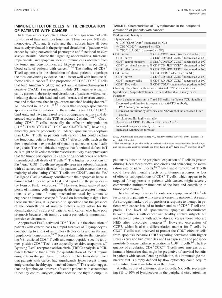
IMMUNE EFFECTOR CELLS IN THE CIRCULATIONOF PATIENTS WITH CANCER
In human subjects peripheral blood is the major source of cellsfor studies of their antitumor functions. T lymphocytes, NK cells,monocytes, DCs, and B cells and their subsets have all beenextensively evaluated in the peripheral circulation of patients withcancer by using conventional phenotypic and functional in vitroassays. Results indicate that signaling abnormalities, functionalimpairments, and apoptosis seen in immune cells obtained fromthe tumor microenvironment are likewise present in peripheralblood cells of patients with cancer.63,64 The finding of CD81
T-cell apoptosis in the circulation of these patients is perhapsthe most convincing evidence that all is not well with immune ef-fector cells in cancer.65 The proportion of CD81CD951 T cellsthat bind Annexin V (Anx) and yet are 7-amino-actinomycin Dnegative (7AAD2) or propidium iodide (PI) negative is signifi-cantly greater in the peripheral circulation of patients with cancer,including those with head and neck, breast, and ovarian carcino-mas andmelanoma, than in age- or sex-matched healthy donors.65
As indicated in Table III,66-68 T cells that undergo spontaneousapoptosis in the circulation of these patients are CD31CD951,bind Anx, and have increased levels of caspase-3 activity and de-creased expression of the TCR-associated z chain.63,69,70 Circu-lating CD81 T cells, especially the effector subpopulations(CD81CD45RO1CCR72CD272 and CD81CD282), have a sig-nificantly greater propensity to undergo spontaneous apoptosisthan CD41 T cells in patients with cancer. This could explainthe functional deficits found in CD81 effector cells, such as thedownregulation in expression of signaling molecules, specificallythe z chain. The available data suggest that functional defects in Tcells might be linked to their increased sensitivity to apoptosis andthat the tumor participates in engineering spontaneous or activa-tion-induced cell death of T cells.65 The highest proportions ofFas1Anx1CD81 T cells are generally seen in a subset of patientswith advanced active disease.70 In patients with cancer, the vastmajority of circulating CD81 T cells are CD951, and the Fas/Fas ligand (FasL) pathway contributes to their apoptosis becausehuman solid tumors express FasL and export it to the periphery inthe form of FasL1 exosomes.71,72 However, tumor-induced apo-ptosis of immune cells engaging death ligand/receptor interac-tions is only one of many mechanisms used by tumors toengineer an immune escape.65 Based on increasing insights intothese mechanisms, it is possible to speculate that the presenceof the constellation of immune defects might allow for theidentification of a subset of patients with cancer who have poorprognosis because their tumors create a particularly immunosup-pressive environment.
Apoptosis of Fas1, activated CD81 T cells in the circulation ofpatients with cancer leads to a rapid turnover of T lymphocytes,contributing to a loss of antitumor effector cells and an aberrantlymphocyte homeostasis.66,73 Recent data indicate that circulat-ing Vb-restricted CD81 T cells and tumor peptide–specific tetra-mer–positive CD81T cells are especially sensitive to apoptosis.74
By using T-cell receptor excision circle (TREC) analysis, a PCR-based technique that allows for quantification of recent thymicemigrants in the peripheral circulation, it has been determinedthat patients with cancer had significantly fewer recent thymicemigrants than healthy age-matched donors.67 The results suggestthat the lymphocyte turnover is faster in patients with cancer thanin healthy control subjects, either because the thymic output in
patients is lower or the peripheral expansion of T cells is greater,diluting T-cell receptor excision circles and enhancing the matu-ration rate of naive T cells.66,73 Such rapid turnover of T cellscould have detrimental effects on antitumor responses. A lossof effector subpopulations of CD81 T cells, which appear to betargeted for apoptosis in patients with cancer, might severelycompromise antitumor functions of the host and contribute totumor progression.73
The clinical significance of spontaneous apoptosis of CD81 ef-fector cells in patients with cancer is currently unknown. A searchfor surrogate markers of prognosis or a response to therapy in pa-tients with cancer has led to further studies of CD81 T-cell apo-ptosis. The level of spontaneous apoptosis discriminatesbetween patients with cancer and healthy control subjects butnot between patients with active disease versus those who areNED after oncologic therapies.67 However, expression ofCCR7, which is also a differentiation marker for T cells, byCD81 T cells was observed to protect the CD81 effector cellsfrom apoptosis because CCR7 signaling correlated with higherBcl-2 expression but lower Bax and Fas expression and phospho-inositide 3-kinase pathway activation in CD81 T cells.68 The fre-quency of circulating CD81CCR71 T cells now emerges as animmune biomarker that might be predictive of survival benefitsin patients with cancer. Pending validation, this immunologic bio-marker that is simply defined by flow cytometry could acquiresubstantial clinical usefulness in the future.
Another subset of antitumor effector cells, NK cells, represent-ing 8% to 10% of lymphocytes in the peripheral circulation, has
TABLE III. Characteristics of T lymphocytes in the peripheral
circulation of patients with cancer*
Predominant phenotypeT lymphocytes:% CD31CD951Anx1 (increased vs NC)% CD31CD251 (increased vs NC)% CD31HLA-DR1 (increased vs NC)
CD81 subset: % CD81CD951Anx1 (increased vs NC)CD81 naive: % CD81CD45RO2CCR71 (decreased vs NC)CD81 central memory: % CD81CD45RO1CCR71 (decreased vs NC)CD81 peripheral memory: % CD81CD45RO1CCR72 (increased vs NC)CD81 effector cells: % CD81CD45RO2CCR72 (increased vs NC)
CD41 subset: % CD41CCR71 (decreased vs NC)CD41 naive: % CD41CD45RO2CCR71 (decreased vs NC)CD41 memory cells: % CD41RO45RO1CCR71 (decreased vs NC)CD41 Treg cells: % CD41CD251 (increased proportions vs NC)
Clonality: Polyclonal with various restricted TCR Vb specificitiesSpecificity: TA-specific/tetramer1 T cells detectable in many casesFunctions
Low z chain expression in T and NK cells: inefficient TCR signalingDecreased proliferation in response to anti-CD3 antibody,PMA/ionomycin, mitogens
Decreased antitumor cytotoxicity and NK/lymphokine-activated killeractivity
Cytokine profile: highly variableApoptosis of CD81 T cells and NK cells (Anx1)Increased caspase-3 activity in T cellsIncreased lymphocyte turnover
LAK, Lymphokine-activated killer; NC, healthy control subjects; PMA, phorbol 12-myristate 13-acetate.*The percentage of positive cells in patients with cancer compared with healthy age-and sex-matched control subjects are from Kuss et al,66 Kim et al,67 and Kim et al.68
J ALLERGY CLIN IMMUNOL
FEBRUARY 2010
S278 WHITESIDE
been credited with the ability to eliminate tumor cells in thecirculation and thus prevent establishment of distant metasta-ses.75 Recent data suggest that in addition to mediating perforin-mediated lysis, NK cells constitutively express several ligands ofthe TNF family and can therefore induce apoptosis in a broad va-riety of tumor cell targets.76 This mechanism of tumor cell elim-ination might be of greater biologic importance than secretory,granule-mediated killing, largely because most tumor cellsexpress receptors for the TNF family ligands and are sensitiveto death by apoptosis.76 NK cells, which are able to discriminatebetween normal and abnormal cells based on the presence andexpression levels of MHC class I molecules, are considered toplay a major role in early stages of tumor development. Theyexpress receptors that enable them to survey the target for therespective ligands. These receptors are of 2 types: killer inhibitoryreceptors, killer activating receptors, or both.57 NK cell functionsand their interactions with other cells or extracellular matrix mol-ecules are regulated through these receptors and Fcg receptors.57
In the peripheral circulation of patients with cancer, NK cells, likeCD81T cells, can also be dysfunctional. On a per-cell basis, theseNK cells mediate lower levels of cytotoxicity.77 Furthermore,some studies suggest that NK cells are also sensitive to apopto-sis.78 Among circulating NK cells in patients with breast cancer,a subset of CD56brightCD16dim NK cells, which represents about95% of all NK cells and is responsible for effector functions, pref-erentially binds Anx and thus is primed for apoptosis.79 Thesepatients also had significantly lower NK activity than the age-and sex-matched healthy control subjects tested in parallel. Theseand other data suggest that endogenous circulating NK cells havethe potential to play a role in tumor surveillance, but in the pres-ence of the tumor, their antitumor functions are subverted, and nolonger control metastasis dissemination. Once the tumor is estab-lished, it especially subverts the subsets of NK cells found at thesites of metastasis and those responsible for cytotoxic functions.
In addition to NK cells, another category of nonspecificeffector cells, CD31CD561NK/T cells, can potentially eliminatetumor targets. They represent a very minor subset of circulatinglymphocytes in healthy subjects but have been reported to beexpanded in patients with cancer, as well as tumor-bearingrodents.80 NK/T cells are also a minor component of TILs. Inthe presence of IL-2, NK/T cells, like CD32CD561 NK cells,readily differentiate into lymphokine-activated killer cells con-taining numerous granzyme- and perforin-containing granulesand are able to mediate tumor cell lysis.77 Both NK and NK/Tcells express receptors for IL-18 and thus are activated in thepresence of this cytokine as well.
REGULATORY IMMUNE CELLS IN PATIENTS WITHCANCER
The presence in the circulation of patients with cancer ofsuppressor lymphocytes capable of downregulating functions ofother immune cells was described many years ago.81 Today suchcells are phenotypically identified as CD41CD25highFOXP31 Tcells and referred to as Treg cells.82 They can be isolated fromPBMCs or tumor sites by means of immunoselection on magneticbeads coated with antibodies to surface antigens expressed onTreg cells, such as CD25 or CD39. In mice depletion ofCD41CD251T cells results in the development of autoimmunity,and in tumor-bearing animals it promotes immune responses toautologous tumor. In patients with cancer, tumor-associated
lymphocytes are enriched in CD31CD41CD25high T cells.83 Onsorting by flow, these T cells have been shown to secrete TGF-b or IL-10 and to enzymatically cleave ATP to adenosine.45,46
The mechanisms through which these T cells regulate antitumorimmune responses are being intensively investigated, and becauseTreg cells come in different flavors (eg, natural Treg cells, induc-ible TR1 cells, CD391 Treg cells, or cytotoxic T lymphocyte–associated antigen–positive Treg cells), these mechanisms vary,likely depending on the microenvironmental context. Similarly,the microenvironment influences the induction of Treg cells; forexample, TR1 cells are preferentially induced at the tumor site,which is rich in IL-10, TGF-b, and PGE2, all of which havebeen shown to promote TR1 cell generation.43,44 The prognosticsignificance of Treg cells in patients with cancer has been contro-versial, with many reports linking their accumulations to poorprognosis, presumably as a result of suppressed antitumor immu-nity,84 and others reporting better survival in the presence of in-creased Treg cell frequencies,85 possibly because of their abilityto suppress tumor-promoting mechanisms or induce tumor celldeath. The controversy arises because in human subjects no def-inite identity marker for Treg cells exists, and their functionalrepertoire is broad and varied. Nevertheless, their responsibilityfor the contraction of immune responses is critical for health.
Another subset of CD41T cells with an origin sharedwith Tregcells has recently been identified. Like Treg cells, CD41 TH17-producing T cells originate from uncommitted CD41 T-cell pre-cursors, and the participation of TGF-b in their differentiationlinks them to Treg cells.86 However, TH17 cells produce IL-17,IL-21, and IL-IL-22, promoting tissue inflammation, and requirethe presence of IL-6, as well as the transcription factors signaltransducer and activator of transcription 3 (STAT3), RORg, andRORa, for differentiation.87 Although the presence of TH17 cellshas been documented in several human carcinomas,86 their func-tion in tumors remains controversial. Recent reports show thatCD41FOXP31CCR61 Treg cells can produce IL-17 on activa-tion and can inhibit proliferation of CD41 responder T cells,87
confirming a relationship between Treg and TH17 cells that canbe modulated by cytokines in the tumor microenvironment. Italso emphasizes the plasticity of T-suppressor and T-effectorsubsets of CD41 lymphocytes.
The second major subset of regulatory cells in cancer areMDSCs (CD341CD331CD131CD11b1CD152).50 Tumors re-cruit MDSCs from the bone marrow through tumor-derived solu-ble factors, such as GM-CSF, TGF-b, IL-10, and VEGF.5
Immature myeloid cells migrate to lymph nodes, where DCscross-prime T cells and interfere with this process, thus suppress-ing CTL generation. They also migrate to the tumor site and be-come MDSCs able to produce arginase I and promote iNOSactivation.5,51 MDSCs also produce high levels of ROS and indo-leamine-2,3-dioxygenase, an enzyme involved in the catabolismof tryptophan, an essential amino acid for T-cell proliferationand differentiation.88 In tumor-bearing mice MDSCs accumulatein the spleen, reaching a very high frequency and exerting potentimmune suppression, thereby favoring tumor growth. GM-CSF,often used as an immune adjuvant,89 is also a product of tumorcells, which recruitsMDSCs from the bonemarrow and is respon-sible for their accumulation in patients with cancer.90 In patientswith cancer, normal physiologic functions of GM-CSF andMDSCs are subverted by the tumor to promote its development.
The tumor uses a variety of mechanisms and produces variousfactors and enzymes that enable it to suppress the host antitumor
J ALLERGY CLIN IMMUNOL
VOLUME 125, NUMBER 2
WHITESIDE S279
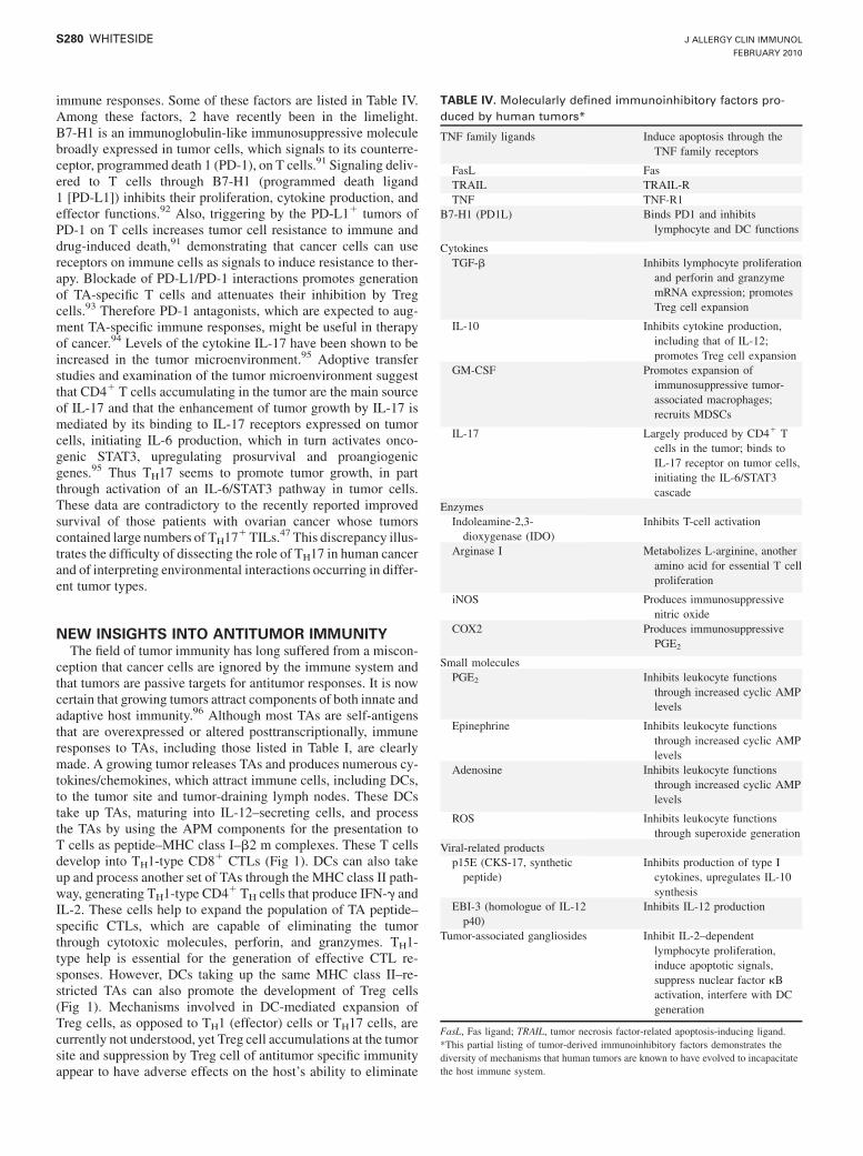
immune responses. Some of these factors are listed in Table IV.Among these factors, 2 have recently been in the limelight.B7-H1 is an immunoglobulin-like immunosuppressive moleculebroadly expressed in tumor cells, which signals to its counterre-ceptor, programmed death 1 (PD-1), on T cells.91 Signaling deliv-ered to T cells through B7-H1 (programmed death ligand1 [PD-L1]) inhibits their proliferation, cytokine production, andeffector functions.92 Also, triggering by the PD-L11 tumors ofPD-1 on T cells increases tumor cell resistance to immune anddrug-induced death,91 demonstrating that cancer cells can usereceptors on immune cells as signals to induce resistance to ther-apy. Blockade of PD-L1/PD-1 interactions promotes generationof TA-specific T cells and attenuates their inhibition by Tregcells.93 Therefore PD-1 antagonists, which are expected to aug-ment TA-specific immune responses, might be useful in therapyof cancer.94 Levels of the cytokine IL-17 have been shown to beincreased in the tumor microenvironment.95 Adoptive transferstudies and examination of the tumor microenvironment suggestthat CD41 T cells accumulating in the tumor are the main sourceof IL-17 and that the enhancement of tumor growth by IL-17 ismediated by its binding to IL-17 receptors expressed on tumorcells, initiating IL-6 production, which in turn activates onco-genic STAT3, upregulating prosurvival and proangiogenicgenes.95 Thus TH17 seems to promote tumor growth, in partthrough activation of an IL-6/STAT3 pathway in tumor cells.These data are contradictory to the recently reported improvedsurvival of those patients with ovarian cancer whose tumorscontained large numbers of TH17
1TILs.47 This discrepancy illus-trates the difficulty of dissecting the role of TH17 in human cancerand of interpreting environmental interactions occurring in differ-ent tumor types.
NEW INSIGHTS INTO ANTITUMOR IMMUNITYThe field of tumor immunity has long suffered from a miscon-
ception that cancer cells are ignored by the immune system andthat tumors are passive targets for antitumor responses. It is nowcertain that growing tumors attract components of both innate andadaptive host immunity.96 Although most TAs are self-antigensthat are overexpressed or altered posttranscriptionally, immuneresponses to TAs, including those listed in Table I, are clearlymade. A growing tumor releases TAs and produces numerous cy-tokines/chemokines, which attract immune cells, including DCs,to the tumor site and tumor-draining lymph nodes. These DCstake up TAs, maturing into IL-12–secreting cells, and processthe TAs by using the APM components for the presentation toT cells as peptide–MHC class I–b2 m complexes. These T cellsdevelop into TH1-type CD81 CTLs (Fig 1). DCs can also takeup and process another set of TAs through the MHC class II path-way, generating TH1-type CD4
1 TH cells that produce IFN-g andIL-2. These cells help to expand the population of TA peptide–specific CTLs, which are capable of eliminating the tumorthrough cytotoxic molecules, perforin, and granzymes. TH1-type help is essential for the generation of effective CTL re-sponses. However, DCs taking up the same MHC class II–re-stricted TAs can also promote the development of Treg cells(Fig 1). Mechanisms involved in DC-mediated expansion ofTreg cells, as opposed to TH1 (effector) cells or TH17 cells, arecurrently not understood, yet Treg cell accumulations at the tumorsite and suppression by Treg cell of antitumor specific immunityappear to have adverse effects on the host’s ability to eliminate
TABLE IV. Molecularly defined immunoinhibitory factors pro-
duced by human tumors*
TNF family ligands Induce apoptosis through theTNF family receptors
FasL FasTRAIL TRAIL-RTNF TNF-R1
B7-H1 (PD1L) Binds PD1 and inhibitslymphocyte and DC functions
CytokinesTGF-b Inhibits lymphocyte proliferation
and perforin and granzymemRNA expression; promotesTreg cell expansion
IL-10 Inhibits cytokine production,including that of IL-12;promotes Treg cell expansion
GM-CSF Promotes expansion ofimmunosuppressive tumor-associated macrophages;recruits MDSCs
IL-17 Largely produced by CD41 Tcells in the tumor; binds toIL-17 receptor on tumor cells,initiating the IL-6/STAT3cascade
EnzymesIndoleamine-2,3-dioxygenase (IDO)
Inhibits T-cell activation
Arginase I Metabolizes L-arginine, anotheramino acid for essential T cellproliferation
iNOS Produces immunosuppressivenitric oxide
COX2 Produces immunosuppressivePGE2
Small moleculesPGE2 Inhibits leukocyte functions
through increased cyclic AMPlevels
Epinephrine Inhibits leukocyte functionsthrough increased cyclic AMPlevels
Adenosine Inhibits leukocyte functionsthrough increased cyclic AMPlevels
ROS Inhibits leukocyte functionsthrough superoxide generation
Viral-related productsp15E (CKS-17, syntheticpeptide)
Inhibits production of type Icytokines, upregulates IL-10synthesis
EBI-3 (homologue of IL-12p40)
Inhibits IL-12 production
Tumor-associated gangliosides Inhibit IL-2–dependentlymphocyte proliferation,induce apoptotic signals,suppress nuclear factor kBactivation, interfere with DCgeneration
FasL, Fas ligand; TRAIL, tumor necrosis factor-related apoptosis-inducing ligand.*This partial listing of tumor-derived immunoinhibitory factors demonstrates thediversity of mechanisms that human tumors are known to have evolved to incapacitatethe host immune system.
J ALLERGY CLIN IMMUNOL
FEBRUARY 2010
S280 WHITESIDE
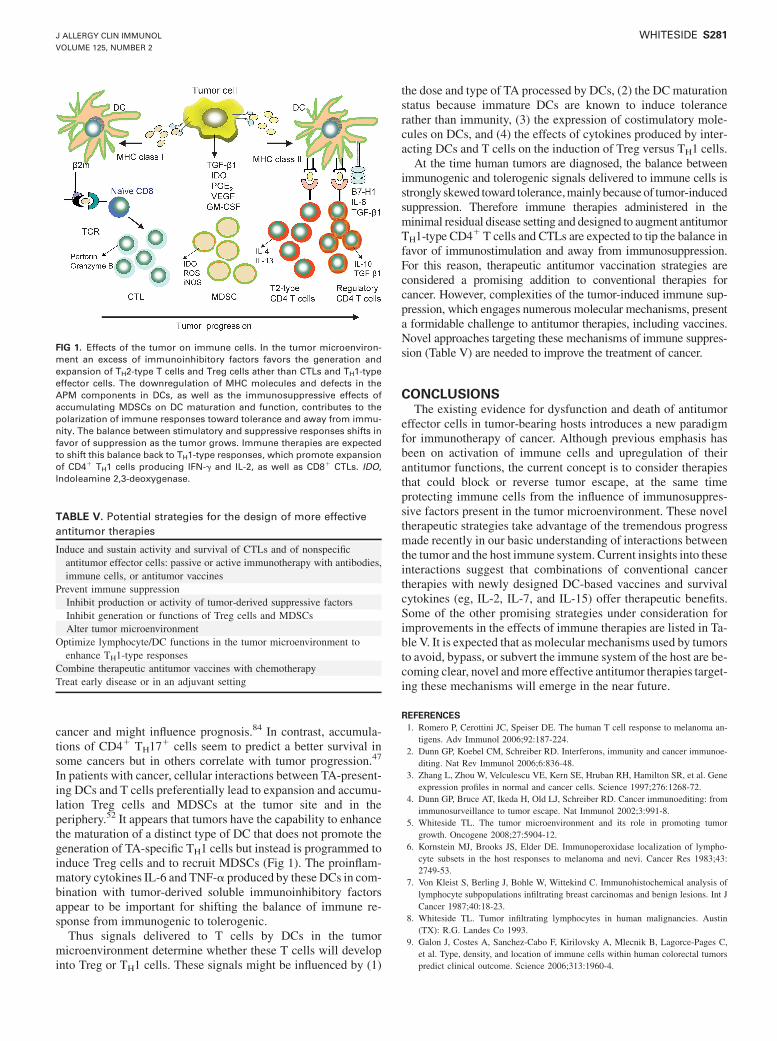
cancer and might influence prognosis.84 In contrast, accumula-tions of CD41 TH17
1 cells seem to predict a better survival insome cancers but in others correlate with tumor progression.47
In patients with cancer, cellular interactions between TA-present-ing DCs and T cells preferentially lead to expansion and accumu-lation Treg cells and MDSCs at the tumor site and in theperiphery.52 It appears that tumors have the capability to enhancethe maturation of a distinct type of DC that does not promote thegeneration of TA-specific TH1 cells but instead is programmed toinduce Treg cells and to recruit MDSCs (Fig 1). The proinflam-matory cytokines IL-6 and TNF-a produced by these DCs in com-bination with tumor-derived soluble immunoinhibitory factorsappear to be important for shifting the balance of immune re-sponse from immunogenic to tolerogenic.
Thus signals delivered to T cells by DCs in the tumormicroenvironment determine whether these T cells will developinto Treg or TH1 cells. These signals might be influenced by (1)
the dose and type of TA processed by DCs, (2) the DCmaturationstatus because immature DCs are known to induce tolerancerather than immunity, (3) the expression of costimulatory mole-cules on DCs, and (4) the effects of cytokines produced by inter-acting DCs and T cells on the induction of Treg versus TH1 cells.
At the time human tumors are diagnosed, the balance betweenimmunogenic and tolerogenic signals delivered to immune cells isstrongly skewed toward tolerance,mainlybecauseof tumor-inducedsuppression. Therefore immune therapies administered in theminimal residual disease setting and designed to augment antitumorTH1-type CD4
1 T cells and CTLs are expected to tip the balance infavor of immunostimulation and away from immunosuppression.For this reason, therapeutic antitumor vaccination strategies areconsidered a promising addition to conventional therapies forcancer. However, complexities of the tumor-induced immune sup-pression, which engages numerous molecular mechanisms, presenta formidable challenge to antitumor therapies, including vaccines.Novel approaches targeting these mechanisms of immune suppres-sion (Table V) are needed to improve the treatment of cancer.
CONCLUSIONSThe existing evidence for dysfunction and death of antitumor
effector cells in tumor-bearing hosts introduces a new paradigmfor immunotherapy of cancer. Although previous emphasis hasbeen on activation of immune cells and upregulation of theirantitumor functions, the current concept is to consider therapiesthat could block or reverse tumor escape, at the same timeprotecting immune cells from the influence of immunosuppres-sive factors present in the tumor microenvironment. These noveltherapeutic strategies take advantage of the tremendous progressmade recently in our basic understanding of interactions betweenthe tumor and the host immune system. Current insights into theseinteractions suggest that combinations of conventional cancertherapies with newly designed DC-based vaccines and survivalcytokines (eg, IL-2, IL-7, and IL-15) offer therapeutic benefits.Some of the other promising strategies under consideration forimprovements in the effects of immune therapies are listed in Ta-ble V. It is expected that as molecular mechanisms used by tumorsto avoid, bypass, or subvert the immune system of the host are be-coming clear, novel andmore effective antitumor therapies target-ing these mechanisms will emerge in the near future.
REFERENCES1. Romero P, Cerottini JC, Speiser DE. The human T cell response to melanoma an-
tigens. Adv Immunol 2006;92:187-224.2. Dunn GP, Koebel CM, Schreiber RD. Interferons, immunity and cancer immunoe-
diting. Nat Rev Immunol 2006;6:836-48.3. Zhang L, Zhou W, Velculescu VE, Kern SE, Hruban RH, Hamilton SR, et al. Gene
expression profiles in normal and cancer cells. Science 1997;276:1268-72.4. Dunn GP, Bruce AT, Ikeda H, Old LJ, Schreiber RD. Cancer immunoediting: from
immunosurveillance to tumor escape. Nat Immunol 2002;3:991-8.5. Whiteside TL. The tumor microenvironment and its role in promoting tumor
growth. Oncogene 2008;27:5904-12.6. Kornstein MJ, Brooks JS, Elder DE. Immunoperoxidase localization of lympho-
cyte subsets in the host responses to melanoma and nevi. Cancer Res 1983;43:2749-53.
7. Von Kleist S, Berling J, Bohle W, Wittekind C. Immunohistochemical analysis oflymphocyte subpopulations infiltrating breast carcinomas and benign lesions. Int JCancer 1987;40:18-23.
8. Whiteside TL. Tumor infiltrating lymphocytes in human malignancies. Austin(TX): R.G. Landes Co 1993.
9. Galon J, Costes A, Sanchez-Cabo F, Kirilovsky A, Mlecnik B, Lagorce-Pages C,et al. Type, density, and location of immune cells within human colorectal tumorspredict clinical outcome. Science 2006;313:1960-4.
FIG 1. Effects of the tumor on immune cells. In the tumor microenviron-ment an excess of immunoinhibitory factors favors the generation andexpansion of TH2-type T cells and Treg cells ather than CTLs and TH1-typeeffector cells. The downregulation of MHC molecules and defects in theAPM components in DCs, as well as the immunosuppressive effects ofaccumulating MDSCs on DC maturation and function, contributes to thepolarization of immune responses toward tolerance and away from immu-nity. The balance between stimulatory and suppressive responses shifts infavor of suppression as the tumor grows. Immune therapies are expectedto shift this balance back to TH1-type responses, which promote expansionof CD41 TH1 cells producing IFN-g and IL-2, as well as CD81 CTLs. IDO,Indoleamine 2,3-deoxygenase.
TABLE V. Potential strategies for the design of more effective
antitumor therapies
Induce and sustain activity and survival of CTLs and of nonspecificantitumor effector cells: passive or active immunotherapy with antibodies,immune cells, or antitumor vaccines
Prevent immune suppressionInhibit production or activity of tumor-derived suppressive factorsInhibit generation or functions of Treg cells and MDSCsAlter tumor microenvironment
Optimize lymphocyte/DC functions in the tumor microenvironment toenhance TH1-type responses
Combine therapeutic antitumor vaccines with chemotherapyTreat early disease or in an adjuvant setting
J ALLERGY CLIN IMMUNOL
VOLUME 125, NUMBER 2
WHITESIDE S281
10. Galon J, Fridman W-H, Pages F. The adaptive immunologic microenvironment incolorectal cancer: a novel perspective. Cancer Res 2007;67:1883-6.
11. Pages F, Berger A, Camus M, Sanchez-Cabo F, Costes A, Molidor R, et al. Effectormemory T cells, early metastasis and survival in colorectal cancer. N Engl J Med2005;353:2654-66.
12. Camus M, Tosolini M, Mlecnik B, Pages F, Kirilovsky A, Berger A, et al. Coordi-nation of intratumoral immune reaction and human colorectal cancer recurrence.Cancer Res 2009;69:2685-93.
13. Lee PP, Yee C, Savage PA, Fong L, Brockstedt D, Weber JS, et al. Characterizationof circulating T cells specific for tumor-associated antigens in melanoma patients.Nat Med 1999;5:677-85.
14. Pittet MJ, Speiser DE, Lienard D, Valmore D, Guillaume P, Dutoit V, et al.Expansion and functional maturation of human tumor antigen-specific CD81 T cells after vaccination with antigenic peptide. Clin Cancer Res 2001;7:796s-803.
15. Hoffmann TK, Donnenberg AD, Finkelstein SD, Donnenberg VS, Friebe-Hoff-mann F, Myers EN, et al. Frequencies of tetramer1 T cells specific for the wild-type sequence p53264-272 peptide in the circulations of patients with head andneck cancer. Cancer Res 2002;62:3521-9.
16. Sahin U, Tureci O, Pfreundschuh M. Serologic identification of human tumor an-tigens. Curr Opin Immunol 1997;9:709-16.
17. Matzinger P. An innate sense of danger. Semin Immunol 1998;10:399-415.18. Szczepanski MJ, Czystowska M, Szajnik M, Harasymczuk M, Boyiadzis M, Kruk-
Zagajewska A, et al. Triggering of toll-like receptor 4 expressed on human headand neck squamous cell carcinoma promotes tumor development and protectsthe tumor from immune attack. Cancer Res 2009;69:3105-13.
19. Uematsu S, Akira S. Toll-like receptors and innate immunity. J Mol Med 2006;84:712-25.
20. Hollstein M, Sidransky D, Vogelstein B, Harris C. p53 mutations in human cancers.Science 1991;253:49-53.
21. Provinciali M. Immunosenescence and cancer vaccines. Cancer Immunol Immun-other 2009;58:1959-67.
22. Ferrone S, Whiteside TL. Tumor microenvironment and immune escape. Surg On-col Clin North Am 2007;16:755-74.
23. Ferris RL, Whiteside TL, Ferrone S. Immune escape associated with functional de-fects in antigen processing machinery in head and neck cancer. Clin Cancer Res2006;12:3890-5.
24. Stickel JS, Weinzieri AO, Hillen N, Drews O, Schuler MM, Hennenlotter J, et al.HLA ligand profile of primary renal cell carcinoma maintained in metastases. Can-cer Immunol Immunother 2009;58:1407-17.
25. Fearon DT, Locksley RM. The instructive role of innate immunity in the acquiredimmune response. Science 1996;272:50-4.
26. Irving BA, Weiss A. The cytoplasmic domain of the T cell receptor zeta chain issufficient to couple to receptor-associated signal transduction pathways. Cell1991;64:891-901.
27. Greenwald RJ, Freeman GJ, Sharpe AH. The B7 family revisited. Annu Rev Im-munol 2005;23:515-48.
28. Kalinski P, Hilkens CM, Wierenga EA, Kapsenberg ML. T-cell priming by type-1 and type-2 polarized dendritic cells: the concept of a third signal. Immunol Today1999;20:561-7.
29. Whiteside TL. Immune suppression in cancer: effects on immune cells, mecha-nisms and future therapeutic interventions. Semin Cancer Biol 2006;16:3-15.
30. Reichert TE, Strauss L, Wagner EM, Gooding W, Whiteside TL. Signaling abnor-malities and reduced proliferation of circulating and tumor-infiltrating lymphocytesin patients with oral carcinoma. Clin Cancer Res 2002;8:3137-45.
31. Uzzo RG, Clark PE, Rayman P, Bloom T, Rybicki L, Novick AC, et al. Alterationsin NFkB activation in T lymphocytes of patients with renal cell carcinoma. J NatlCancer Inst 1999;91:718-21.
32. Reichert TE, Rabinowich H, Johnson JT, Whiteside TL. Human immune cells inthe tumor microenvironment: mechanisms responsible for signaling and functionaldefects. J Immunother 1998;21:295-306.
33. Cavallo F, Offringa R, van der Burg SH, Forni G, Melief CJ. Vaccination for treat-ment and prevention of cancer in animal models. Adv Immunol 2006;90:175-213.
34. Lollini PL, De Giovanni C, Pannellini T, Cavallo F, Forni G, Nanni P. Cancer im-munoprevention. Future Oncol 2005;1:57-66.
35. Pannellini T, Forni G, Musiani P. Immunobiology of her-2/neu transgenic mice.Breast Dis 2004;20:33-42.
36. Parmiani G, De Filippo A, Novellino L, Castelli C. Unique human tumor antigens:immunobiology and use in clinical trials. J Immunol 2007;178:1975-9.
37. Finn OJ, Binder RJ, Brickner AG, Butterfield LH, Ferris RL, Kalinski P, et al. Hu-man tumor antigens as targets of immunosurveillance and candidates for cancervaccines. In: Gires O, Seliger B, editors. Tumor-associated antigens: identification,characterization, and clinical applications. Weinheim (Germany): Wiley-VCH;2009. p. 23-43.
38. Cheever M, Allison JP, Ferris AS, Finn OJ, Hastings BM, Hecht TT, et al. The pri-oritization of cancer antigens: a National Cancer Institute pilot project for the ac-celeration of translational research. Clin Cancer Res 2009;15:5323-37.
39. Smyth MJ, Dunn GP, Schreiber RD. Cancer immunosurveillance and immunoedit-ing: the roles of immunity in suppressing tumor development and shaping tumorimmunogenicity. Adv Immunol 2006;90:1-50.
40. Jager E, Karbach J, Gnjatic S, Neumann A, Bender A, Valmori D, et al. Recombi-nant vaccinia/fowlpox NY-ESO-1 vaccines induce both humoral and cellular NY-ESO-1-specific immune responses in cancer patients. Proc Natl Acad Sci U S A2006;103:14453-8.
41. Whiteside TL. The local tumor microenvironment. In: Kaufmann H, Wolchok JD,editors. General principles of tumor immunotherapy: basic and clinical applica-tions of tumor immunology. New York: Springer; 2007. p. 145-67.
42. June CH. Adoptive T cell therapy for cancer in the clinic. J Clin Invest 2007;117:1466-76.
43. Bergmann C, Strauss L, Zeidler R, Lang S, Whiteside TL. Expansion of human Tregulatory type 1 cells in the microenvironment of COX-2-overexpressing head andneck squamous cell carcinoma. Cancer Res 2007;67:8865-73.
44. Woo EY, Chu CS, Goletz TJ, Schlienger K, Yeh H, Coukos G, et al. RegulatoryCD4 1 CD25 1 T cells in tumors from patients with early-stage non-small celllung cancer and late-stage ovarian cancer. Cancer Res 2001;61:4766-72.
45. Strauss L, Bergmann C, Szczepanski M, Gooding W, Johnson TJ, Whiteside TL. Aunique subset of CD41 CD25highFOXP31 T cells secreting IL-10 and TGF-b1 me-diates suppression in the tumor microenvironment. Clin Cancer Res 2007;13:4345-54.
46. Mandapathil M, Szczepanski MJ, Szajnik M, Ren J, Lenzner DE, Jackson EK,et al. Increased ectonucleotidase expression and activity in Treg of patients withhead and neck cancer. Clin Cancer Res 2009;15:6348-57.
47. Kryczek I, Banerjee M, Cheng P, Vatan L, Szeliga W, Wei S, et al. Phenotype, dis-tribution, generation, and functional and clinical relevance of Th17 cells in the hu-man tumor microenvironment. Blood 2009;114:1141-9.
48. Martinez O, Sica A, Mantovani A, Locati M. Macrophage activation and polariza-tion. Front Biosci 2008;13:453-61.
49. Leek RD, Lewis CE, Whitehouse R, Geenall M, Clarke J, Harris AL. Associationof macrophage infiltration with angiogenesis and prognosis in invasive breast car-cinoma. Cancer Res 1996;56:4625-9.
50. Serafini P, Borello I, Bronte V. Myeloid suppressor cells in cancer: recruitment,phenotype, properties, and mechanisms of immune suppression. Semin CancerBiol 2006;16:53-65.
51. Ochoa AC, Zea AH, Hernandez C, Rodrıguez PC. Arginase, prostaglandins, andmyeloid supressor cells in renal cell carcinoma. Clin Cancer Res 2007;13:721s-6s.
52. Diebold SS. Determination of T-cell fate by dendritic cells. Immunol Cell Biol2008;86:389-97.
53. Gabrilovich DI, Chen HL, Girgis KR, Cunningham HT, Meny GM, Nadaf S, et al.Production of vascular endothelial growth factor by human tumors inhibits thefunctional maturation of dendritic cells. Nat Med 1996;2:1096-103.
54. Shurin GV, Shurin MR, Bykovskaja S, Shogan J, Lotze MT, Barksdale EM. Neu-roblastoma-derived gangliosides inhibit dendritic cell generation and function.Cancer Res 2001;61:363-9.
55. Reichert TE, Scheuer C, Day R, Wagner W, Whiteside TL. The number of intra-tumoral dendritic cells and z-chain expression in T cells as prognostic and survivalbiomarkers in patients with oral carcinoma. Cancer 2001;91:2136-47.
56. Murphy GF, Radu A, Kaminer M, Berg D. Autologous melanoma vaccine inducesinflammatory responses in melanoma metastases: relevance to immunologic re-gression and immunotherapy. J Invest Dermatol 1993;100(suppl):335S-41.
57. Lanier LL. Natural killer cell receptor signaling. Curr Opin Immunol 2003;15:308-14.
58. Chang CC, Campoli M, Ferrone S. Classical and nonclassical HLA class I antigenand NK cell-activating ligand changes in malignant cells: current challenges andfuture direction. Adv Cancer Res 2005;93:189-234.
59. Kelly JM, Darcy PK, Markby JL, Godfrey DI, Takeda K, Yagitab H, et al. Induc-tion of tumor-specific T cell memory by NK cell-mediated tumor rejection. Nat Im-munol 2002;3:83-90.
60. Coronella JA, Spier C, Welch M, Trevor KT, Stopeck AT, Villar H, et al. Antigen-driven oligoclonal expansion of tumor-infiltrating B cells in infiltrating ductal car-cinoma of the breast. J Immunol 2002;169:1829-36.
61. Denko NC, Fontana LA, Hudson KM, Sutphin PD, Raychaudhuri S, Altman R,et al. Investigating hypoxic tumor physiology through gene expression patterns.Oncogene 2003;22:5907-14.
62. Aller MA, Arias JL, Nava MP, Arias J. Posttraumatic inflammation is a complexresponse based on the pathological expression of the nervous, immune and endo-crine function systems. Exp Biol Med 2004;229:170-81.
63. Dworacki G, Meidenbauer N, Kuss I, Hoffmann TK, Gooding MS, Lotze M, et al.Decrease z chain expression and apoptosis in CD31 peripheral blood T lympho-cytes of patients with melanoma. Clin Cancer Res 2001;7(suppl):947s-57.
J ALLERGY CLIN IMMUNOL
FEBRUARY 2010
S282 WHITESIDE
64. Finke JH, Rayman P, George R, Tannenbaum CS, Kolenko V, Uzzo R, et al. Tumor-induced sensitivity to apoptosis in T cells from patients with renal cell carcinoma:role of nuclear factor kappa B suppression. Clin Cancer Res 2001;7(suppl):940s-6.
65. Whiteside TL. Tumor-induced death of immune cells: its mechanisms and conse-quences. Semin Cancer Biol 2001;12:43-50.
66. Kuss I, Schaefer C, Godfrey TE, Ferris RL, Harris J, Gooding W, et al. Recent thy-mic emigrants and subsets of naıve and memory T cells in the circulation of pa-tients with head and neck cancer. Clin Immunol 2005;116:27-36.
67. Kim W-J, Tsukishiro T, Johnson JT, Whiteside TL. Expression of pro- and anti-ap-optotic proteins in circulating CD8 1 T cells of patients with squamous cell car-cinoma of the head and neck (SCCHN). Clin Cancer Res 2004;10:5101-10.
68. Kim J-W, Ferris RL, Whiteside TL. Chemokine receptor 7 (CCR7) expression andprotection of circulating CD8 1 T lymphocytes from apoptosis. Clin Cancer Res2005;11:7901-10.
69. Kuss I, Saito T, Johnson JT, Whiteside TL. Clinical significance of decreased zchain expression in peripheral blood lymphocytes of patients with head and neckcancer. Clin Cancer Res 1999;5:329-34.
70. Hoffmann TK, Dworacki G, Meidenbauer N, Gooding W, Johnson JT, WhitesideTL. Spontaneous apoptosis of circulating T lymphocytes in patients with headand neck cancer and its clinical importance. Clin Cancer Res 2002;8:2553-62.
71. Whiteside TL. The role of death receptor ligands in shaping tumor microenviron-ment. Immunol Invest 2007;36:25-46.
72. O’Connell J, O’Sullivan GD, Collins JK, Shanahan F. The Fas counterattack: Fas-mediated T cell killing by colon cancer cells expressing Fas ligand. J Exp Med1996;184:1075-82.
73. Whiteside TL. Lymphocyte homeostasis and the antitumor immune response. ExpRev Clin Immunol 2006;1:369-78.
74. Albers AE, Visus C, Tsukishiro T, Ferris RL, Gooding W, Whiteside TL, et al. Al-terations in the T-cell receptor variable b gene-restricted profile of CD8 1 T lym-phocytes in the peripheral circulation of patients with squamous cell carcinoma ofthe head and neck. Clin Cancer Res 2006;12:2394-403.
75. Whiteside TL. Immune effector cells in the tumor microenvironment: their role inregulation of tumor progression. In: Yefenof E, editor. Innate and adaptive re-sponses in the tumor microenvironment. New York: Springer; 2008. p. 1-34.
76. Vujanovic NL, Nagashima S, Herberman RB, Whiteside TL. Non-secretory apo-ptotic killing by human natural killer cells. J Immunol 1996;157:1117-26.
77. Whiteside TL, Vujanovic NL, Herberman RB. Natural killer cells and tumor ther-apy. Curr. Topics Microbiol Immunol 1998;230:221-44.
78. Hansson M, Asea A, Ericsson U, Hermodsson S, Hellstrand K. Induction of apo-ptosis in NK cells by monocyte-derived reactive oxygen metabolites. J Immunol1996;156:42-7.
79. Whiteside TL. Apoptosis of immune cells in the tumor microenvironment and pe-ripheral circulation of patients with cancer: implications for immunotherapy. Vac-cine 2002;20:A46-51.
80. Smyth MF, Godfrey DI. NKT cells and tumor immunity—a double-edged sword.Nat Immunol 2000;1:459-60.
81. Gershon RK. A disquisition on suppressor T cells. Transpl Rev 1975;26:170-85.82. Bluestone JA, Abbas AK. Natural versus adaptive regulatory T cells. Nat Rev
2003;3:253-7.83. Bergmann C, Strauss L, Wang Y, Szczepanski MJ, Lang S, Johnson JT, et al. T reg-
ulatory type 1 cells (Tr1) in squamous cell carcinoma of the head and neck:mechanisms of suppression and expansion in advanced disease. Clin Cancer Res2008;14:3706-15.
84. Curiel TJ, Coukos G, Zou L, Alvarez X, Cheng P, Mottram P, et al. Specific recruit-ment of regulatory T cells in ovarian carcinoma foster immune privilege and pre-dicts reduced survival. Nat Med 2004;10:942-9.
85. Salama P, Phillips M, Grieu P, Morris M, Zeps N, Joseph D, et al. Tumor-infiltrat-ing FOXP3 1 T regulatory cells show strong prognostic significance in colorectalcancer. J Clin Oncol 2008;27:186-92.
86. Martin-Orozco N, Dong C. The IL-17/IL23 axis of inflammation in cancer: friendor foe? Curr Opin Investig Drugs 2009;10:543-9.
87. Voo KS, Wang Y-H, Santori FR, Boggiano C, Yi-H Wang, Arima K, et al. Identi-fication of IL-17-producing FOXP1 regulatory T cells in humans. Proc Natl AcadSci U S A 2009;106:4793-8.
88. Munn DH, Mellor AL. Indoleamine 2, 3-dioxygenase and tumor-induced toler-ance. J Clin Invest 2007;117:1147-54.
89. Dranoff G, Jaffee E, Lazenby A, Golumbek P, Levitsky H, Brose K, et al. Vacci-nation with irradiated tumor cells engineered to secrete murine granulocyte-macro-phage colony-stimulating factor stimulates potent, specific, and long-lasting anti-tumor immunity. Proc Natl Acad Sci U S A 1993;90:3539-43.
90. Filipazzi F, Valenti R, Huber V, Pilla L, Canese P, Iero M, et al. Identification of anew subset of myeloid suppressor cells in peripheral blood of melanoma patientswith modulation by a granulocyte-macrophage colony-stimulation factor-based an-titumor vaccine. J Clin Oncol 2007;25:2546-53.
91. Azuma T, Yao S, Zhu G, Flies SJ, Chen L. B7-H1 is a ubiquitous antiapoptotic re-ceptor on cancer cells. Blood 2008;111:3635-43.
92. Blank C, Mackensen A. Contribution of the PD-L1/PD-1 pathway to T-cell exhaus-tion: an update on implications for chronic infections and tumor evasion. CancerImmunol Immunother 2007;56:739-45.
93. Wang W, Lau R, Yu D, Zhu W, Korman A, Weber J. PD1 blockade reverses thesuppression on melanoma antigen-specific CTL by CD4 1 CD25hi regulatory Tcells. Int Immunol 2009;21:1065-77.
94. Hirano F, Kaneko K, Tamura H, Dong H, Wang S, Ichikawa M, et al. Blockade ofB7-H1 and PD-1 by monoclonal antibodies potentiates cancer therapeutic immu-nity. Cancer Res 2005;65:1089-96.
95. Wang L, Yi T, Kortylewski M, Pardoll DM, Zeng D, Yu H. IL-17 can promote tumorgrowth through an IL-6-Stat3 signalling pathway. J Exp Med 2009;206:1457-64.
96. Finn OJ. Cancer immunology. N Engl J Med 2008;358:2704-15.
J ALLERGY CLIN IMMUNOL
VOLUME 125, NUMBER 2
WHITESIDE S283
Clinical laboratory assessment of immediate-typehypersensitivity
Robert G. Hamilton, PhD Baltimore, Md
Clinical laboratory analyses aid in the diagnosis andmanagement of human allergic (IgE-dependent) diseases.Diagnosis of immediate-type hypersensitivity begins with athorough clinical history and physical examination. Oncesymptoms compatible with an allergic disorder have beenidentified, a skin test, blood test, or both for allergen-specific IgEantibodies provide confirmation of sensitization, whichstrengthens the diagnosis. Skin testing provides a biologicallyrelevant immediate-type hypersensitivity response withresultant wheal-and-flare reactions within 15 minutes ofallergen application. Allergen-specific IgE antibody in serum isquantified by using 3 laboratory-based autoanalyzers(ImmunoCAP, Immulite, and HYTEC-288) and novelmicroarray and lateral-flow immunoassays. Technologicadvances in serologic allergen-specific IgE measurements haveinvolved increased automation, with enhanced reproducibility,greater quantification, lower analytic sensitivity, andcomponent-supplemented extract-based allergen use. In vivoprovocation tests involving inhalation, ingestion, or injection ofallergens serve to clarify discordant history and skin- or blood-based measures of sensitization. Other diagnostic allergylaboratory analyses include total and free serum IgEmeasurement, precipitating IgG antibodies specific for organicdusts, mast cell tryptase, and indicator allergen analyses toassess indoor environments to promote patient-targeted allergenavoidance programs. A critique is provided on the predictiveutility of serologic measures of specific IgE for food allergy andasthma. Reasons for the lack of clinical utility for food-specificIgG/IgG4 measurements in allergy diagnosis are examined.When the specific IgE measures are inconsistent with theclinical history, they should be confirmed by means of repeatand alternative method analysis. Ultimately, the patient’sclinical history remains the principal arbiter that determinesthe final diagnosis of allergic disease. (J Allergy Clin Immunol2010;125:S284-96.)
Key words: Diagnosis, skin testing, RAST, IgE antibody, provoca-tion testing
The clinical laboratory plays an increasing role in the diagnosisand management of allergic disorders (immediate or type
1 hypersensitivity). The clinician begins the diagnostic processwith a thorough clinical history and physical examination.Symptoms that suggest a diagnosis of asthma, allergic rhinitisand sinusitis, occupational asthma and allergy, food allergy, drugallergy, or an allergic disease of the skin are matched withsuspected relevant allergen exposures. Once the clinician has ahigh degree of suspicion that the patient has a particular allergicdisorder, in vivo (skin and provocation tests) and laboratory-basedserologic analyses for IgE antibody are performed to strengthenthe likelihood that the chosen allergy diagnosis is correct. Adefinitive diagnosis of allergic disease then permits a number oftherapeutic interventions involving avoidance, pharmacotherapy,immunotherapy, or anti-IgE therapy to be instituted.Managementof a patient with allergic disorders can also be facilitated withdifferent laboratory analyses. This chapter examines clinicallaboratory tests that aid in the diagnosis and management ofpatients with a disease associated with type 1 hypersensitivity.
IGE PROPERTIESThe reagin in serum that mediates the immediate-type wheal-
and-flare reactionwas identified as IgE in 1967.1,2 The properties ofhuman IgE are described in Table I. IgE (approximately 190,000 d)circulates as a monomer at a serum concentration that is highly agedependent. It constitutes approximately 0.0005% of the total serumimmunoglobulins in adults.3 Cord blood levels of IgE remain low(<2 kU/L [<4.8 mg/L]) because IgE does not cross the placentalbarrier in significant amounts.Mean serum IgE levels progressivelyincrease in healthy children up to 10 to 15 years of age and then de-crease from the second through eighth decades of life. By 14 yearsof age, total serum IgE levels of greater than 333 kU/L (800 mg/L)are considered abnormally increased and strongly associated withand suggestive of atopic disorders, such as allergic rhinitis, extrinsicasthma, and atopic dermatitis.4,5
Environmental antigen exposure can occur by means ofinhalation, ingestion, or skin and parenteral contact. Once takenup by antigen-presenting cells, processed antigen is presented tohelper T cells that secrete a number of cytokines that cause B-celllymphocytes to proliferate and in some cases produce allergen-specific IgE antibody. IgE binds onto high-affinity Fce receptorson the surface of a number of cells, particularly mast cells andbasophils, creating a state of ‘‘sensitization’’ within the patient.
From the Allergy and Clinical Immunology Division, Department of Medicine, JohnsHopkins University School of Medicine.
Disclosure of potential conflict of interest: R. G. Hamilton has declared that he has noconflict of interest.
Received for publication August 3, 2009; revised September 24, 2009; accepted forpublication September 30, 2009.
Reprint requests: Robert G. Hamilton, PhD, Room 1A20, Johns Hopkins Asthma andAllergy Center, 5501 Hopkins Bayview Circle, Baltimore, MD 21224. E-mail:[email protected].
0091-6749/$36.00! 2010 American Academy of Allergy, Asthma & Immunologydoi:10.1016/j.jaci.2009.09.055
Abbreviations usedBHR: Basophil histamine release
CI: Chronic urticariaCLIA-88: Federal Clinical Laboratory Improvement Act of 1988DBPCFC: Double-blind, placebo-controlled food challenge
ISAC: Immunosorbent Allergen ChipPWV: Polistes species wasp venomWHO: World Health OrganizationYJV: Yellow jacket venom
S284
Subsequent allergen exposure causes mast cell surface–boundIgE antibody to be cross-linked, leading to an increase inintracellular calcium levels and the release of both preformedmediators (eg, histamine and proteases) and newly synthesizedlipid-derived mediators (eg, leukotrienes and prostaglandins).Allergic symptoms can subsequently occur as a result of medi-ator-induced physiologic and anatomic changes. The measure-ment of allergen-specific IgE antibody will thus be a principalfocus of this chapter.
ALLERGENSSeveral hundred allergenic proteins, (glycoproteins and lipo-
proteins), are extracted from well-defined (usually biological)sources, including weed, grass, and tree pollens and animaldander, molds, house dust mites, parasites, insect venoms,occupational allergens (eg, natural rubber latex), drugs, andfoods.6 These allergens elicit IgE antibody production when in-troduced into an immunocompetent and genetically predisposedhost. Individual allergenic proteins can be identified by usingIgE antibody–containing human serum in combination with anumber of immunochemical assays that separate proteins basedon their net charge (isoelectric focusing), size (Western blot anal-ysis), and ability to bind antibody (competitive inhibition immu-noassay). A compendium of the known clinically importantallergens (together with their scientific names), purified major al-lergen components, and diagnostic codes is presented elsewhere.6
Many important allergenic proteins from dust mites, pollens,animal dander, insects (eg, cockroach and Hymenoptera venoms),molds, and foods have been cloned and sequenced, and recombi-nant proteins have been expressed during the past decade.7,8 Thishas sparked a debate as to whether native allergens possess anyunique advantages over their recombinant counterparts as diagnos-tic reagents andwhether crude allergenmixtures should giveway tothe use of purifiedallergens in cocktails todetect IgE antibody in theskin and blood. Allergens extracted fromnatural sources are knownto be heterogeneous, often containingmany nonallergenic proteins.Moreover, different natural extracts vary in their allergen composi-tion and potency, and they can be contaminatedwith allergens fromother sources. Purified recombinant allergens are attractive becausetheir availability in pure form simplifies reagent preparation andpromotes reproducibility and standardization. However, allergicpatients are known to respond differently to combinations of isoal-lergens that are essentially identical except for minor differences intheir primary amino acid composition or substituted side chains.Thus a single recombinant allergen that does not represent all theisoallergen forms of that allergen might not be sufficiently ‘‘glob-ally diagnostic’’ to be able to detect all clinically relevant IgE anti-bodies of that allergen specificity.
Extract-based reagents for both skin test and IgE antibodyserology are here to stay for the foreseeable future because of theirmore comprehensive coverage of the allergenic repertoire of anyparticular specificity. However, the future use of certain purifiedrecombinant allergens as diagnostic reagents in both in vivo and invitro IgE antibody testing holds promise. A purified recombinantor native principal allergen of a particular specificity, such as Bet v1 from birch pollen, can be a good indicator allergen for detectingsensitization to that specificity.However, it is not sufficiently com-prehensive to replace the birch extract–based diagnostic reagent,especially for evaluation of subjects who produce IgE antibodyto less predominant allergens that are present in the birch pollen
extract. Additionally, the allergenic profile of any given specificityof an allergen-containing reagent, as produced by different manu-facturers, is expected to vary in its protein composition, allergenicpotency, and immunoreactivity, regardless of extensive cross-val-idation. One generic rule of allergy diagnostics has evolved fromthis, namely that despite clearance by regulatory agencies, such asthe US Food and Drug Administration, each in vivo or in vitro-allergen extract–containing reagent should be expected to detectslightly different populations of IgE antibodies. Thus IgE anti-body measurements generated with different skin test– or se-rum-based IgE antibody assay reagents are expected to producereasonably clinically equivalent but not identical results.6
DIAGNOSTIC ALGORITHM FOR ALLERGIC DISEASEThe diagnosis of allergic disease begins with a thorough
clinical history and physical examination.9,10 The signs andsymptoms associated with the various allergic disorders are ex-tensively discussed in chapters 8-14. Once the history has beencollected, one of several primary confirmatory tests for sensitiza-tion can be performed to detect allergen-specific IgE in the skin orblood. Because the history is viewed by many as the arbiter of thediagnostic test’s performance, a subject with a positive clinicalhistory for allergic disease and a positive skin or blood test resultfor IgE is considered to have a true-positive result (Table II). Ide-ally, all patients with a positive allergy history would have a pos-itive allergen-specific IgE test result, and those with a negativehistory would have a negative allergen-specific IgE antibodytest result. However, more realistically, some patients with aller-gic disease are classified as having false-negative IgE antibodytest results, and others with no evident allergic disease are identi-fied as having positive IgE antibody test results that would be con-sidered false-positive results.
In vivo provocation tests are considered secondary-levelconfirmatory tests that are available when one needs to adjudicatethe correctness of discordant clinical history and results fromallergen-specific IgE antibody skin or serologic tests.9-11 However,provocation tests are more difficult to perform in a reproduciblemanner than skin or blood tests for IgE antibodies, and they placethe patient at some risk for a reaction because they involve a directallergen challenge. Interpretation of their results can also be diffi-cult because they often involve subjective end points that can bealtered by observer and patient bias. In certain cases, such as foodallergy, the in vivo provocation test (double-blind, placebo-controlled food challenge [DBPCFC]) has become the referencebenchmark for identifying type 1 hypersensitivity to foods. Theactual in vivoprovocation test that is useful in thediagnosticworkupof a patient ultimately depends on the nature of the disease processthat is being investigated (eg, sting challenge for Hymenopteravenom allergy).
The presence of IgE antibodies is necessary but not sufficient forallergic disease expression. Allergen-specific IgE antibody mightbe detectable in the patient’s skin or blood, and the patientmight nothave had any evident allergic symptoms after allergen exposure.Some health careworkerswith a positive immediate-type latex skintest result, IgE anti-natural rubber latex blood test result, or bothexperience no allergic symptoms when they are exposed to highlyallergenic powdered latex examination gloves.12 The relativestrengths and limitations of in vivo and in vitro diagnostic testsand the principal technical reasons for false-negative and false-positive test results are discussed subsequently.
J ALLERGY CLIN IMMUNOL
VOLUME 125, NUMBER 2
HAMILTON S285
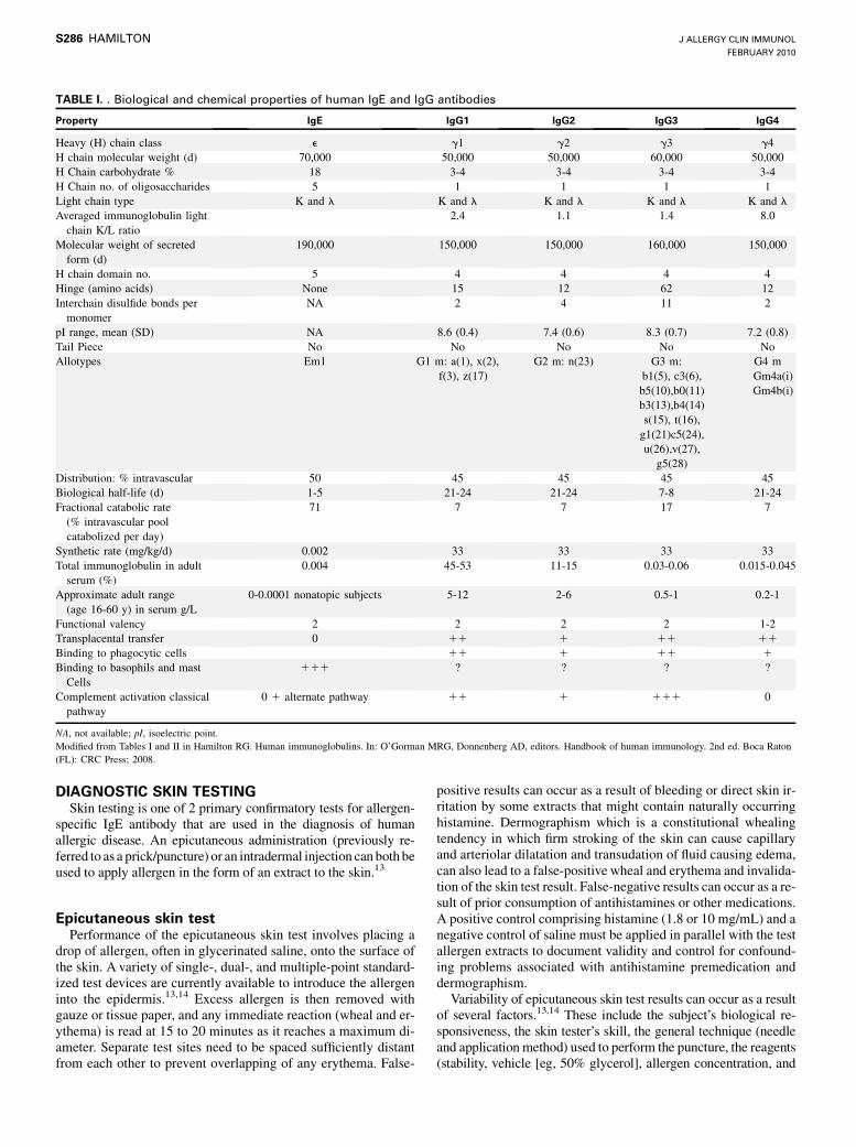
DIAGNOSTIC SKIN TESTINGSkin testing is one of 2 primary confirmatory tests for allergen-
specific IgE antibody that are used in the diagnosis of humanallergic disease. An epicutaneous administration (previously re-ferred to as a prick/puncture) or an intradermal injection can bothbeused to apply allergen in the form of an extract to the skin.13
Epicutaneous skin testPerformance of the epicutaneous skin test involves placing a
drop of allergen, often in glycerinated saline, onto the surface ofthe skin. A variety of single-, dual-, and multiple-point standard-ized test devices are currently available to introduce the allergeninto the epidermis.13,14 Excess allergen is then removed withgauze or tissue paper, and any immediate reaction (wheal and er-ythema) is read at 15 to 20 minutes as it reaches a maximum di-ameter. Separate test sites need to be spaced sufficiently distantfrom each other to prevent overlapping of any erythema. False-
positive results can occur as a result of bleeding or direct skin ir-ritation by some extracts that might contain naturally occurringhistamine. Dermographism which is a constitutional whealingtendency in which firm stroking of the skin can cause capillaryand arteriolar dilatation and transudation of fluid causing edema,can also lead to a false-positive wheal and erythema and invalida-tion of the skin test result. False-negative results can occur as a re-sult of prior consumption of antihistamines or other medications.A positive control comprising histamine (1.8 or 10 mg/mL) and anegative control of saline must be applied in parallel with the testallergen extracts to document validity and control for confound-ing problems associated with antihistamine premedication anddermographism.
Variability of epicutaneous skin test results can occur as a resultof several factors.13,14 These include the subject’s biological re-sponsiveness, the skin tester’s skill, the general technique (needleand application method) used to perform the puncture, the reagents(stability, vehicle [eg, 50% glycerol], allergen concentration, and
TABLE I. . Biological and chemical properties of human IgE and IgG antibodies
Property IgE IgG1 IgG2 IgG3 IgG4
Heavy (H) chain class e g1 g2 g3 g4H chain molecular weight (d) 70,000 50,000 50,000 60,000 50,000H Chain carbohydrate % 18 3-4 3-4 3-4 3-4H Chain no. of oligosaccharides 5 1 1 1 1Light chain type K and l K and l K and l K and l K and lAveraged immunoglobulin lightchain K/L ratio
2.4 1.1 1.4 8.0
Molecular weight of secretedform (d)
190,000 150,000 150,000 160,000 150,000
H chain domain no. 5 4 4 4 4Hinge (amino acids) None 15 12 62 12Interchain disulfide bonds permonomer
NA 2 4 11 2
pI range, mean (SD) NA 8.6 (0.4) 7.4 (0.6) 8.3 (0.7) 7.2 (0.8)Tail Piece No No No No NoAllotypes Em1 G1 m: a(1), x(2),
f(3), z(17)G2 m: n(23) G3 m:
b1(5), c3(6),b5(10),b0(11)b3(13),b4(14)s(15), t(16),g1(21)c5(24),u(26),v(27),
g5(28)
G4 mGm4a(i)Gm4b(i)
Distribution: % intravascular 50 45 45 45 45Biological half-life (d) 1-5 21-24 21-24 7-8 21-24Fractional catabolic rate(% intravascular poolcatabolized per day)
71 7 7 17 7
Synthetic rate (mg/kg/d) 0.002 33 33 33 33Total immunoglobulin in adultserum (%)
0.004 45-53 11-15 0.03-0.06 0.015-0.045
Approximate adult range(age 16-60 y) in serum g/L
0-0.0001 nonatopic subjects 5-12 2-6 0.5-1 0.2-1
Functional valency 2 2 2 2 1-2Transplacental transfer 0 11 1 11 11Binding to phagocytic cells 11 1 11 1Binding to basophils and mastCells
111 ? ? ? ?
Complement activation classicalpathway
0 1 alternate pathway 11 1 111 0
NA, not available; pI, isoelectric point.Modified from Tables I and II in Hamilton RG. Human immunoglobulins. In: O’Gorman MRG, Donnenberg AD, editors. Handbook of human immunology. 2nd ed. Boca Raton(FL): CRC Press; 2008.
J ALLERGY CLIN IMMUNOL
FEBRUARY 2010
S286 HAMILTON
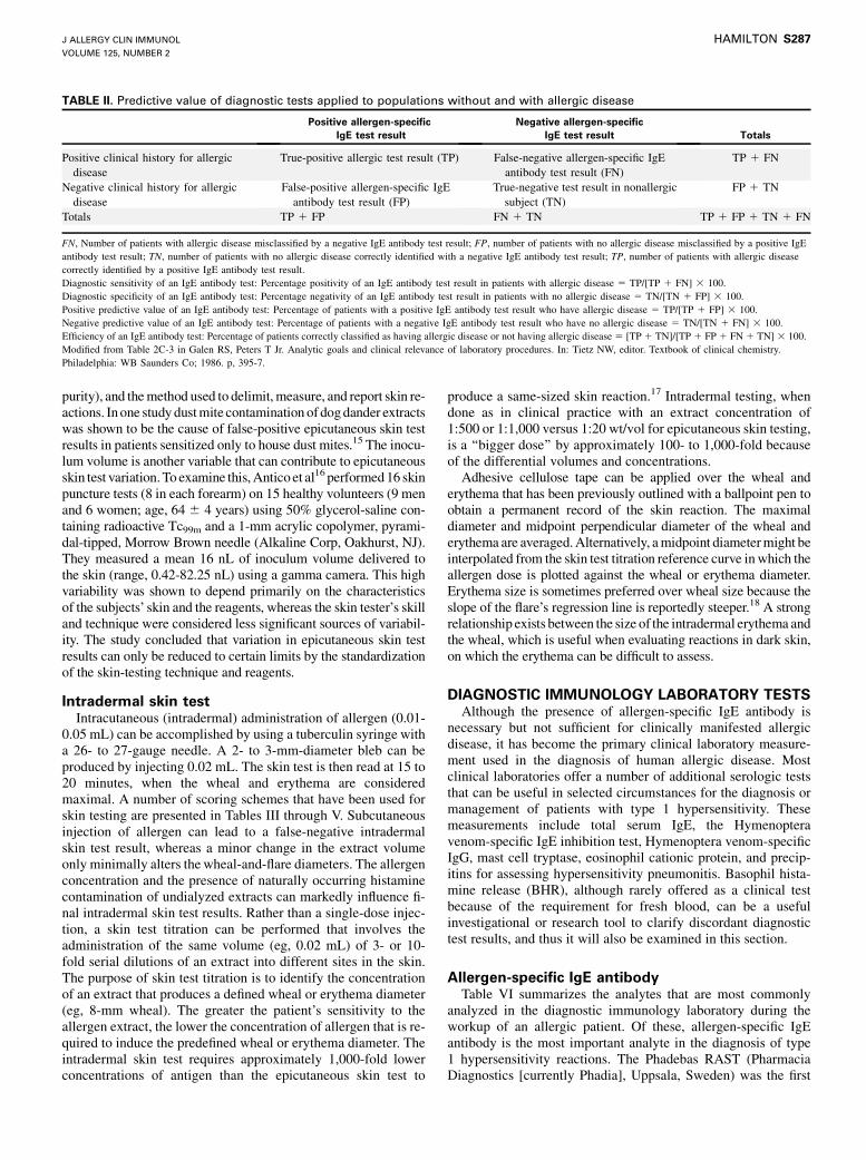
purity), and themethod used to delimit, measure, and report skin re-actions. Inone studydustmite contamination of dogdander extractswas shown to be the cause of false-positive epicutaneous skin testresults in patients sensitized only to house dust mites.15 The inocu-lum volume is another variable that can contribute to epicutaneousskin test variation.To examine this,Anticoet al16 performed16 skinpuncture tests (8 in each forearm) on 15 healthy volunteers (9 menand 6 women; age, 64 6 4 years) using 50% glycerol-saline con-taining radioactive Tc99m and a 1-mm acrylic copolymer, pyrami-dal-tipped, Morrow Brown needle (Alkaline Corp, Oakhurst, NJ).They measured a mean 16 nL of inoculum volume delivered tothe skin (range, 0.42-82.25 nL) using a gamma camera. This highvariability was shown to depend primarily on the characteristicsof the subjects’ skin and the reagents, whereas the skin tester’s skilland technique were considered less significant sources of variabil-ity. The study concluded that variation in epicutaneous skin testresults can only be reduced to certain limits by the standardizationof the skin-testing technique and reagents.
Intradermal skin testIntracutaneous (intradermal) administration of allergen (0.01-
0.05 mL) can be accomplished by using a tuberculin syringe witha 26- to 27-gauge needle. A 2- to 3-mm-diameter bleb can beproduced by injecting 0.02 mL. The skin test is then read at 15 to20 minutes, when the wheal and erythema are consideredmaximal. A number of scoring schemes that have been used forskin testing are presented in Tables III through V. Subcutaneousinjection of allergen can lead to a false-negative intradermalskin test result, whereas a minor change in the extract volumeonly minimally alters the wheal-and-flare diameters. The allergenconcentration and the presence of naturally occurring histaminecontamination of undialyzed extracts can markedly influence fi-nal intradermal skin test results. Rather than a single-dose injec-tion, a skin test titration can be performed that involves theadministration of the same volume (eg, 0.02 mL) of 3- or 10-fold serial dilutions of an extract into different sites in the skin.The purpose of skin test titration is to identify the concentrationof an extract that produces a defined wheal or erythema diameter(eg, 8-mm wheal). The greater the patient’s sensitivity to theallergen extract, the lower the concentration of allergen that is re-quired to induce the predefined wheal or erythema diameter. Theintradermal skin test requires approximately 1,000-fold lowerconcentrations of antigen than the epicutaneous skin test to
produce a same-sized skin reaction.17 Intradermal testing, whendone as in clinical practice with an extract concentration of1:500 or 1:1,000 versus 1:20 wt/vol for epicutaneous skin testing,is a ‘‘bigger dose’’ by approximately 100- to 1,000-fold becauseof the differential volumes and concentrations.
Adhesive cellulose tape can be applied over the wheal anderythema that has been previously outlined with a ballpoint pen toobtain a permanent record of the skin reaction. The maximaldiameter and midpoint perpendicular diameter of the wheal anderythema are averaged.Alternatively, amidpoint diametermight beinterpolated from the skin test titration reference curve inwhich theallergen dose is plotted against the wheal or erythema diameter.Erythema size is sometimes preferred over wheal size because theslope of the flare’s regression line is reportedly steeper.18 A strongrelationship exists between the size of the intradermal erythema andthe wheal, which is useful when evaluating reactions in dark skin,on which the erythema can be difficult to assess.
DIAGNOSTIC IMMUNOLOGY LABORATORY TESTSAlthough the presence of allergen-specific IgE antibody is
necessary but not sufficient for clinically manifested allergicdisease, it has become the primary clinical laboratory measure-ment used in the diagnosis of human allergic disease. Mostclinical laboratories offer a number of additional serologic teststhat can be useful in selected circumstances for the diagnosis ormanagement of patients with type 1 hypersensitivity. Thesemeasurements include total serum IgE, the Hymenopteravenom-specific IgE inhibition test, Hymenoptera venom-specificIgG, mast cell tryptase, eosinophil cationic protein, and precip-itins for assessing hypersensitivity pneumonitis. Basophil hista-mine release (BHR), although rarely offered as a clinical testbecause of the requirement for fresh blood, can be a usefulinvestigational or research tool to clarify discordant diagnostictest results, and thus it will also be examined in this section.
Allergen-specific IgE antibodyTable VI summarizes the analytes that are most commonly
analyzed in the diagnostic immunology laboratory during theworkup of an allergic patient. Of these, allergen-specific IgEantibody is the most important analyte in the diagnosis of type1 hypersensitivity reactions. The Phadebas RAST (PharmaciaDiagnostics [currently Phadia], Uppsala, Sweden) was the first
TABLE II. Predictive value of diagnostic tests applied to populations without and with allergic disease
Positive allergen-specificIgE test result
Negative allergen-specificIgE test result Totals
Positive clinical history for allergicdisease
True-positive allergic test result (TP) False-negative allergen-specific IgEantibody test result (FN)
TP 1 FN
Negative clinical history for allergicdisease
False-positive allergen-specific IgEantibody test result (FP)
True-negative test result in nonallergicsubject (TN)
FP 1 TN
Totals TP 1 FP FN 1 TN TP 1 FP 1 TN 1 FN
FN, Number of patients with allergic disease misclassified by a negative IgE antibody test result; FP, number of patients with no allergic disease misclassified by a positive IgEantibody test result; TN, number of patients with no allergic disease correctly identified with a negative IgE antibody test result; TP, number of patients with allergic diseasecorrectly identified by a positive IgE antibody test result.Diagnostic sensitivity of an IgE antibody test: Percentage positivity of an IgE antibody test result in patients with allergic disease 5 TP/[TP 1 FN] 3 100.Diagnostic specificity of an IgE antibody test: Percentage negativity of an IgE antibody test result in patients with no allergic disease 5 TN/[TN 1 FP] 3 100.Positive predictive value of an IgE antibody test: Percentage of patients with a positive IgE antibody test result who have allergic disease 5 TP/[TP 1 FP] 3 100.Negative predictive value of an IgE antibody test: Percentage of patients with a negative IgE antibody test result who have no allergic disease 5 TN/[TN 1 FN] 3 100.Efficiency of an IgE antibody test: Percentage of patients correctly classified as having allergic disease or not having allergic disease 5 [TP1 TN]/[TP 1 FP 1 FN 1 TN] 3 100.Modified from Table 2C-3 in Galen RS, Peters T Jr. Analytic goals and clinical relevance of laboratory procedures. In: Tietz NW, editor. Textbook of clinical chemistry.Philadelphia: WB Saunders Co; 1986. p, 395-7.
J ALLERGY CLIN IMMUNOL
VOLUME 125, NUMBER 2
HAMILTON S287
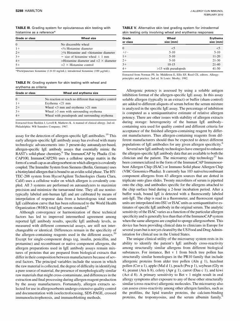
assay for the detection of allergen-specific IgE antibodies.19 Thisearly allergen-specific IgE antibody assay has evolved with manytechnologic advancements into 3 present-day autoanalyzer-based,allergen-specific IgE antibody assays that essentially mimic theRAST’s solid-phase chemistry.6 The ImmunoCAP by Phadia (Uni-CAP100, ImmunoCAP250) uses a cellulose sponge matrix in theformofa small capasanallergosorbentonwhichallergen is covalentlycoupled. The Immulite System from Siemens (Berlin, Germany) usesa biotinylated allergen that is bound to an avidin solid phase. TheHY-TEC-288 system from Hycor/Agilent Technologies (Santa Clara,Calif) uses a cellulose wafer on which allergen is covalently cou-pled. All 3 systems are performed on autoanalyzers to maximizeprecision and minimize the turnaround time. They all use noniso-typically labeled anti-human IgE and are calibrated by means ofinterpolation of response data from a heterologous total serumIgE calibration curve that has been referenced to the World HealthOrganization (WHO) total IgE serum standard.6
Although convergence or harmonization of these technicalfactors has led to improved intermethod agreement amongreported IgE antibody results, specific IgE antibody levels, asmeasured with different commercial assays, are still not inter-changeable or identical. Differences remain in the specificity ofthe allergen-containing reagents used in the different assays.20
Except for single-component drugs (eg, insulin, penicillin, andprotamine) and recombinant or native component allergens, theallergen preparations used in IgE antibody assays remain mix-tures of proteins that are prepared from biological extracts thatdiffer in their composition betweenmanufacturers because of sev-eral factors. The principal variables include the season in whichthe rawmaterial is collected, the degree of difficulty in identifyinga pure source of material, the presence of morphologically similarrawmaterials that might cross-contaminate, and differences in theextraction and final processing during allergen reagent productionby the assay manufacturers. Fortunately, allergen extracts se-lected for use in allergosorbents undergo extensive quality controland documentation with isoelectrofocusing, SDS-PAGE, crossedimmunoelectrophoresis, and immunoblotting methods.
Allergenic potency is assessed by using a soluble antigeninhibition format of the allergen-specific IgE assay. In this assaysoluble allergen (typically in an extract) or buffer (sham control)are added to different aliquots of serum before the serum mixtureis analyzed in the specific IgE assay. The percentage of inhibitionis computed as a semiquantitative estimate of relative allergenpotency. There are other issues with stability of allergen extractsduring storage: heterogeneity of the human IgE antibody–containing sera used for quality control and different criteria foracceptance of the finished allergen-containing reagent by differ-ent manufacturers. Thus allergen-containing reagents from dif-ferent manufacturers should thus be expected to detect differentpopulations of IgE antibodies for any given allergen specificity.6
Several new IgE antibody technologies have emerged to enhancethe allergen-specific IgE antibody data that are available to both theclinician and the patient. The microarray chip technology21 hasbeen commercialized in the form of the ImmunoCAP Immunosor-bent Allergen Chip (ISAC) or Immuno Solid phase Allergen Chip(VBC Genomics-Phadia). It currently has 103 native/recombinantcomponent allergens from 43 allergen sources that are dotted intriplicate onto glass slides. Twenty microliters of serum is pipettedonto the chip, and antibodies specific for the allergens attached tothe chip surface bind during a 2-hour incubation period. After abuffer wash, bound IgE is detected with a fluorescently labeledanti-IgE. The chip is read in a fluorometer, and fluorescent signalunits are interpolated into ISUor ISACunits as semiquantitative es-timates of specific IgE antibody in the original serum. The analyticsensitivity of the ISACvaries as a function of the particular allergenspecificity and is generally less than that of the ImmunoCAPsystemwhen the same allergens are coupled to sponge allergosorbent. Thisdevice has been providing clinical data to clinicians in Europe forseveral years but is not yet cleared by theUSFood andDrugAdmin-istration for clinical use in the United States.
The unique clinical utility of the microarray system rests in itsability to identify the patient’s IgE antibody cross-reactivityamong structurally similar allergens from different biologicalsubstances. For instance, Bet v 1 from birch tree pollen hasstructurally similar homologues in the PR10 family that includeallergenic proteins from alder tree pollen (Aln g 1), hazelnutpollen (Cor a 1), apple (Mal d 1), peach (Pru p 1), soybean (Gly m4), peanut (Ara h 8), celery (Apr g 1), carrot (Dau c 1), and kiwi(Act d 8). A primary sensitivity to Bet v 1 might result in oralallergy symptoms after exposure to any of these other structurallysimilar (cross-reactive) allergenicmolecules. Themicroarray alsocan assess cross-reactivity among other allergen families, such asthe profilins, the lipid transfer proteins, the calcium-bindingproteins, the tropomyosins, and the serum albumin family.6
TABLE III. Grading system for epicutaneous skin testing with
histamine as a reference*
Grade or class Wheal size
0 No discernible wheal11 <! Histamine diameter21 !! Histamine and <histamine diameter31 5 size of histamine wheal 6 1 mm41 >Histamine diameter and <2 3 diameter51 >2 3 Histamine control
*Prick/puncture histamine (1.8-10 mg/mL); intradermal histamine (100 mg/mL).
TABLE IV. Grading system for skin testing with wheal and
erythema as criteria
Grade or class Wheal and erythema size
0 No reaction or reach no different than negative control11 Erythema <21 mm21 Wheal <3 mm and erythema >21 mm31 Wheal >3 mm with surrounding erythema41 Wheal with pseudopods and surrounding erythema
Extracted from Sheldon J, Lovell R, Mathews K. A manual of clinical allergy. 2nd ed.Philadelphia: WB Saunders Company; 1967.
TABLE V. Alternative skin test grading system for intradermal
skin testing only involving wheal and erythema responses
Gradeor class
Whealsize (mm)
Erythemasize (mm)
0 <5 <51/2 5-10 5-1011 5-10 11-2021 5-10 21-3031 10-15 21-4041 >15 with pseudopods 41-50
Extracted from Norman PS. In: Middleton E, Ellis EF, Reed CE, editors. Allergy:principles and practice. 2nd ed. St Louis: Mosby; 1982.
J ALLERGY CLIN IMMUNOL
FEBRUARY 2010
S288 HAMILTON
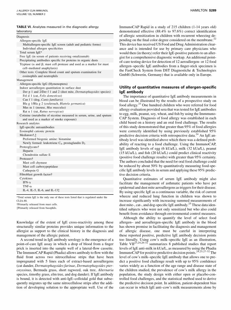
Knowledge of the extent of IgE cross-reactivity among thesestructurally similar proteins provides unique information to theallergist as support to the clinical history in the diagnosis andmanagement of the allergic patient.
A second trend in IgE antibody serology is the emergence of apoint-of-care IgE assay in which a drop of blood from a fingerprick is inserted into the sample well of a lateral-flow cassette.The ImmunoCAPRapid (Phadia) allows antibody to flowwith thefluid front across two nitrocellulose strips that have beenimpregnated with 5 lines each of extract-based aeroallergens(cat dander, Dermatophagoides farinae, Dermatophagoides pter-onyssinus, Bermuda grass, short ragweed, oak tree, Alternariaspecies, timothy grass, elm tree, and dog dander). If IgE antibodyis bound, it is detected with anti-IgE–colloidal gold that subse-quently migrates up the same nitrocellulose strips after the addi-tion of developing solution to the appropriate well. Use of the
ImmunoCAP Rapid in a study of 215 children (1-14 years old)demonstrated effective (88.4% to 97.6%) correct identificationof allergic sensitization in children with recurrent wheezing de-pending on the final color degree considered on the membrane.22
This device has received US Food and Drug Administration clear-ance and is intended for use by primary care physicians whowould then (in theory) refer their IgE-positive patients to an aller-gist for a comprehensive diagnostic workup. An additional point-of-care testing device for detection of 12 aeroallergen- or 12 foodallergen–specific IgE antibodies from a finger-stick specimen isthe FastCheck System from DST Diagnostiche & TechnologiesGmbH (Schwerin, Germany) that is available only in Europe.
Utility of quantitative measures of allergen-specificIgE antibody
The importance of quantitative IgE antibody measurements inblood can be illustrated by the results of a prospective study onfood allergy.23 One hundred children who were referred for foodallergy evaluation provided sera that was tested for IgE antibodiesto egg, milk, peanut, soy, wheat, and fish by using the Immmuno-CAP System. Diagnosis of food allergy was established in eachchild based on a history and an oral food challenge. The resultsof this study demonstrated that greater than 95% of food allergieswere correctly identified by using previously established 95%predictive decision criteria with retrospective data.24 An IgE an-tibody level was identified above which there was a defined prob-ability of reacting to a food challenge. Using the ImmunoCAP,IgE antibody levels of egg (6 kUa/L), milk (32 kUa/L), peanut(15 kUa/L), and fish (20 kUa/L) could predict clinical reactivity(positive food challenge results) with greater than 95% certainty.The authors concluded that the need for oral food challenge couldbe reduced by about 50% by quantitatively measuring food-spe-cific IgE antibody levels in serum and applying these 95% predic-tive decision criteria.
Quantitative estimates of serum IgE antibody might alsofacilitate the management of asthmatic patients who have petepidermal and dust mite aeroallergens as triggers for their disease.By using specific IgE as a continuous variable, the risk of currentwheeze and reduced lung function in children was shown toincrease significantly with increasing summed measurements ofdust mite-, cat-, and dog-specific IgE antibody.25 These data iden-tified subjects who were not only sensitized but who also couldbenefit from avoidance through environmental control measures.
Although the ability to quantify the level of select foodallergen– and aeroallergen-specific IgE antibody in the bloodhas shown promise in facilitating the diagnosis and managementof allergic disease, one must be careful in interpretingthese reported positive, predictive IgE antibody decision pointstoo literally. Using cow’s milk–specific IgE as an illustration,Table VII23,24,26-32 summarizes 8 published studies that reportlevels of IgE anti-milk in kUa/L, as measured by using the PhadiaImmunoCAP for positive predictive decision points.24,25,27-32 Thelevel of cow’s milk–specific IgE antibody that allows one to pre-dict a positive food challenge result with up to 95% confidencevaries widely as a function of the age range and disease state ofthe children studied, the prevalence of cow’s milk allergy in thepopulation, the study design with either open or placebo-con-trolled food challenges, and the statistical method used to derivethe predictive decision point. In addition, patient-dependent biascan occur in which IgE anti–cow’s milk measurements alone by
TABLE VI. Analytes measured in the diagnostic allergy
laboratory
DiagnosisAllergen-specific IgEMultiallergen-specific IgE screen (adult and pediatric forms)Individual allergen specificities
Total serum IgE*Free IgE (in serum of patients receiving omalizumab)Precipitating antibodies specific for proteins in organic dustsTryptase (a and b; mast cell protease and used as a marker for mastcell–mediated anaphylaxis)
Other tests: Complete blood count and sputum examination foreosinophils and neutrophils
ManagementAllergen-specific IgG (Hymenoptera)Indoor aeroallergen quantitation in surface dustDer p 1 and 2/Der f 1 and 2 (dust mite, Dermatophagoides species)Fel d 1 (cat, Felis domesticus)Can f 1 (dog, Canis familiaris)Bla g 1/Bla g 2 (cockroach, Blattela germanica)Mus m 1 (mouse, Mus musculus)Rat n 1 (rat, Rattus norvegicus)
Cotinine (metabolite of nicotine measured in serum, urine, and sputumand used as a marker of smoke exposure)
Research analytesIgE-specific autoantibodiesEosinophil cationic proteinMediators!,"Preformed biogenic amine: histamineNewly formed: leukotriene C4, prostaglandin D2
Proteoglycans!HeparinChondroitin sulfate E
Proteases!Mast cell chymaseMast cell carboxypeptidaseCathepsin G
Fibroblast growth factor!CytokinesIFN-gTNF-aIL-4, IL-5, IL-6, and IL-13"
*Total serum IgE is the only one of these tests listed that is regulated under theCLIA-88.!Primarily released from mast cells."Primarily released from basophils.
J ALLERGY CLIN IMMUNOL
VOLUME 125, NUMBER 2
HAMILTON S289
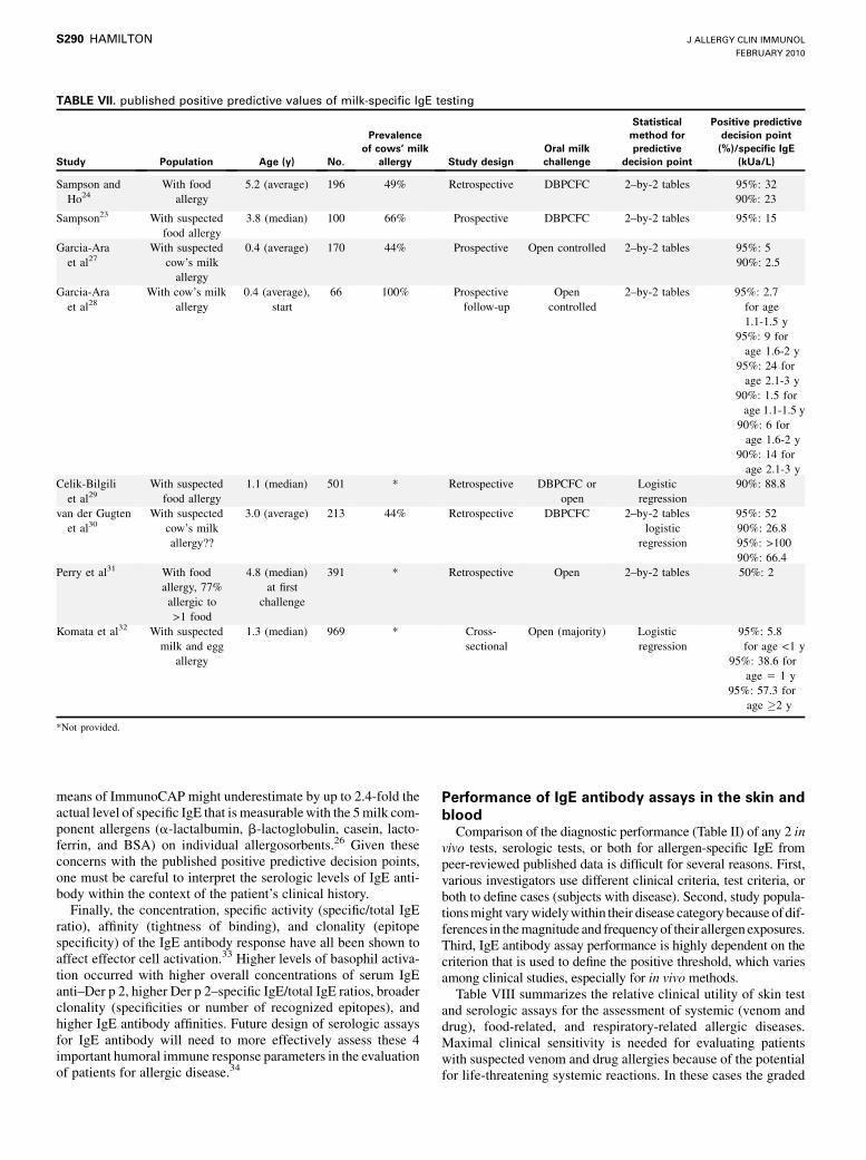
means of ImmunoCAP might underestimate by up to 2.4-fold theactual level of specific IgE that is measurablewith the 5milk com-ponent allergens (a-lactalbumin, b-lactoglobulin, casein, lacto-ferrin, and BSA) on individual allergosorbents.26 Given theseconcerns with the published positive predictive decision points,one must be careful to interpret the serologic levels of IgE anti-body within the context of the patient’s clinical history.
Finally, the concentration, specific activity (specific/total IgEratio), affinity (tightness of binding), and clonality (epitopespecificity) of the IgE antibody response have all been shown toaffect effector cell activation.33 Higher levels of basophil activa-tion occurred with higher overall concentrations of serum IgEanti–Der p 2, higher Der p 2–specific IgE/total IgE ratios, broaderclonality (specificities or number of recognized epitopes), andhigher IgE antibody affinities. Future design of serologic assaysfor IgE antibody will need to more effectively assess these 4important humoral immune response parameters in the evaluationof patients for allergic disease.34
Performance of IgE antibody assays in the skin andblood
Comparison of the diagnostic performance (Table II) of any 2 invivo tests, serologic tests, or both for allergen-specific IgE frompeer-reviewed published data is difficult for several reasons. First,various investigators use different clinical criteria, test criteria, orboth to define cases (subjects with disease). Second, study popula-tionsmight varywidelywithin their disease categorybecauseof dif-ferences in themagnitude and frequencyof their allergenexposures.Third, IgE antibody assay performance is highly dependent on thecriterion that is used to define the positive threshold, which variesamong clinical studies, especially for in vivo methods.
Table VIII summarizes the relative clinical utility of skin testand serologic assays for the assessment of systemic (venom anddrug), food-related, and respiratory-related allergic diseases.Maximal clinical sensitivity is needed for evaluating patientswith suspected venom and drug allergies because of the potentialfor life-threatening systemic reactions. In these cases the graded
TABLE VII. published positive predictive values of milk-specific IgE testing
Study Population Age (y) No.
Prevalenceof cows’ milk
allergy Study designOral milkchallenge
Statisticalmethod forpredictive
decision point
Positive predictivedecision point(%)/specific IgE
(kUa/L)
Sampson andHo24
With foodallergy
5.2 (average) 196 49% Retrospective DBPCFC 2–by-2 tables 95%: 3290%: 23
Sampson23 With suspectedfood allergy
3.8 (median) 100 66% Prospective DBPCFC 2–by-2 tables 95%: 15
Garcia-Araet al27
With suspectedcow’s milkallergy
0.4 (average) 170 44% Prospective Open controlled 2–by-2 tables 95%: 590%: 2.5
Garcia-Araet al28
With cow’s milkallergy
0.4 (average),start
66 100% Prospectivefollow-up
Opencontrolled
2–by-2 tables 95%: 2.7for age1.1-1.5 y
95%: 9 forage 1.6-2 y
95%: 24 forage 2.1-3 y
90%: 1.5 forage 1.1-1.5 y
90%: 6 forage 1.6-2 y
90%: 14 forage 2.1-3 y
Celik-Bilgiliet al29
With suspectedfood allergy
1.1 (median) 501 * Retrospective DBPCFC oropen
Logisticregression
90%: 88.8
van der Gugtenet al30
With suspectedcow’s milkallergy??
3.0 (average) 213 44% Retrospective DBPCFC 2–by-2 tableslogistic
regression
95%: 5290%: 26.895%: >10090%: 66.4
Perry et al31 With foodallergy, 77%allergic to>1 food
4.8 (median)at first
challenge
391 * Retrospective Open 2–by-2 tables 50%: 2
Komata et al32 With suspectedmilk and egg
allergy
1.3 (median) 969 * Cross-sectional
Open (majority) Logisticregression
95%: 5.8for age <1 y
95%: 38.6 forage 5 1 y
95%: 57.3 forage !2 y
*Not provided.
J ALLERGY CLIN IMMUNOL
FEBRUARY 2010
S290 HAMILTON
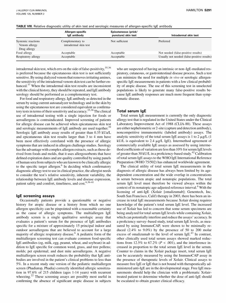
intradermal skin test, which errs on the side of false-positivity,35,36
is preferred because the epicutaneous skin test is not sufficientlysensitive.By using dialyzed venom that removes irritating amines,the sensitivity of the intradermal venom skin test can be further en-hanced.37 When the intradermal skin test results are inconsistentwith the clinical history, they should be repeated, and IgE antibodyserology should be performed as a complementary test.
For food and respiratory allergy, IgE antibody as detected in theserum by using current autoanalyzer technology and in the skin byusing the epicutaneous test are considered equivalent as confirma-tory tests in terms of their sensitivity and accuracy.35,38 The clinicaluse of intradermal testing with a single injection for foods oraeroallergens is contraindicated. Improved screening of patientsfor allergic disease can be achieved when epicutaneous skin testand serologic measurements of IgE antibody are used together.39
Serologic IgE antibody assay results of greater than 0.35 kUa/Land epicutaneous skin test results larger than 3 to 4 mm havebeen most effectively correlated with the presence of allergicsymptoms that are induced in allergen challenge studies. Serologyhas the advantagewith complex allergen extracts, such as those de-rived from foods and molds, that it uses allergosorbents that havedefined expiration dates and are quality controlled by using panelsof human sera fromsubjectswhoareknown tobe clinically allergicto the specific target allergen.6 In deciding which confirmatorydiagnostic allergy test to use in clinical practice, the allergist needsto consider the test’s relative sensitivity, inherent variability, therelationship between IgE antibody levels and disease expression,patient safety and comfort, timeliness, and cost.6,36-41
IgE screening assaysOccasionally patients provide a questionable or negative
history for atopic disease or a history from which no oneallergen specificity can be pinpointed with a reasonable certaintyas the cause of allergic symptoms. The multiallergen IgEantibody screen is a single qualitative serologic assay thatevaluates a patient’s serum for the presence of IgE antibodiesspecific for a mixture of approximately 15 principal indoor andoutdoor aeroallergens that are believed to account for a largemajority of allergic respiratory disease.6 A pediatric form of themultiallergen screening test can evaluate common food-specificIgE antibodies (eg, milk, egg, peanut, wheat, and soybean) in ad-dition to IgE specific for common weed, grass, and tree pollens;molds; pet epidermal; and dust mite aeroallergens. A negativemultiallergen screen result reduces the probability that IgE anti-bodies are involved in the patient’s clinical problems to less than5%. In a recent study one version of the pediatric multiallergenscreen (Phadiatop, Phadia) correctly identified allergic sensitiza-tion in 97.6% of 215 children (ages 1-14 years) with recurrentwheezing.22 These screening assays are possibly most useful inconfirming the absence of significant atopic disease in subjects
who are suspected of having an intrinsic or non–IgE-mediated res-piratory, cutaneous, or gastrointestinal disease process. Such a testcan minimize the need for multiple in vivo or serologic allergen-specific IgE measurements in patients with a low clinical probabil-ity of atopic disease. The use of this screening test in unselectedpopulations is likely to generate many false-positive results be-cause IgE antibody responses are much more frequent than symp-tomatic disease.
Total serum IgETotal serum IgE measurement is currently the only diagnostic
allergy test that is regulated in the United States under the ClinicalLaboratory Improvement Act of 1988 (CLIA-88). These assaysare either nephelometric or 2-site (capture anddetection antibody),noncompetitive immunometric (labeled antibody) assays. Theanalytic sensitivity of the total serum IgE assays is 1 to 2 mg/L (1kU/L is equivalent to 2.4 mg/L IgE). Intermethod agreement ofcommercially available IgE assays as assessed by using interme-thodcoefficients of variation are less than 10%for serum IgE levelsof greater than 30 kU/L in a proficiency-based study.42 Calibrationof total serum IgE assays to theWHO IgE International ReferencePreparation (WHO 75/502) has enhanced worldwide agreement.
The clinical utility of total serum IgE measurements in thediagnosis of allergic disease has always been limited by its age-dependent concentration and the wide overlap in concentrationsin serum between atopic and nonatopic populations. The totalserum IgE level must therefore be viewed always within thecontext of its nonatopic age-adjusted reference interval.6With thelicensing of anti-IgE (Xolair [omalizumab]; Genentech, Inc,South San Francisco, Calif) therapy in 2003, there has been an in-crease in total IgE measurements because Xolair dosing requiresknowledge of the patient’s total serum IgE level. The increaseduse of Xolair has led to concern that some serum specimens arebeing analyzed for total serum IgE levels while containing Xolair,which can potentially interfere and reduce the assays’ accuracy. Ina proficiency survey–based study, total serum IgE levels, as mea-sured by using ImmunoCAP, were shown to be minimally re-duced (2.4% to 9.0%) by the presence of 50 to 200 molarexcess of omalizumab to the level of serum IgE.43 In contrast,other clinically used total serum assays showed marked reduc-tions from 12.5% to 67.2% (P < .001), and the interference in-creased in proportion to the total serum IgE level in the serum.Counter to claims in the Xolair package insert, total serum IgEcan be accurately measured by using the ImmunoCAP assay inthe presence of therapeutic levels of Xolair. Clinical assays tomeasure free IgE or IgE that is not bound with therapeutically ad-ministered anti-IgE are in the developmental stage. Free IgEmea-surements should help the clinician with a problematic Xolair-treated patient to determine whether the dose of anti-IgE shouldbe escalated to obtain greater clinical efficacy.
TABLE VIII. Relative diagnostic utility of skin test and serologic measures of allergen-specific IgE antibody
Allergen-specificIgE antibody
Epicutaneous (prick/puncture) skin test Intradermal skin test
Systemic reactionsVenom allergyDrug allergy
Complementary tointradermal skin test
Not sufficient Preferred
Food allergy Acceptable Acceptable Not needed (false-positive results)Respiratory allergy Acceptable Acceptable Usually not needed (false-positive results)
J ALLERGY CLIN IMMUNOL
VOLUME 125, NUMBER 2
HAMILTON S291
Proficiency testing for total and allergen-specific IgEantibody assays
The CLIA-88 requires that all federally licensed clinical labo-ratories participate in an external proficiency survey. One suchdiagnostic allergy survey is conducted by the College of AmericanPathologists.42 The survey involves analysis of 5 or 6 challenge seraevery 17 weeks (3 cycles per year) for total serum IgE and IgE an-tibody levels to 5 allergen specificities and a multiallergen screen.Results are impartially collated, and interlaboratory (intramethod)coefficients of variation are computed, critiqued, and then sent toboth participating laboratories and credentialing agencies. Exceptfor the occasional nonatopic serum, interlaboratory/intramethodand intermethod coefficients of variation for total serum IgE areroutinely excellent at less than 15%.42
Allergen-specific IgE levels historically have been reported bydifferent assays in nonequivalent arbitrary units or classes. Today,the 3 principal assays report allergen-specific IgE levels in morequantitative kUa/L units. Although differences continue to existin the levels of reported allergen-specific IgE among the variousIgE antibody assays, in general, the 3 principal assays correctlyidentify the IgE-negative (nonsensitized) subjects’ sera from serathat are IgE antibody positive (sensitized) for most of the allergenspecificities. It is the responsibility of the laboratory to indicatethe method they use on their final report to the clinician. It is,however, the responsibility of the referring physician to ensurethat the laboratory that performs IgE antibody testing is CLIA-88certified and that they use a validated assay method and performsuccessfully on a diagnostic allergy proficiency survey.6,44
Venom competitive inhibition IgE antibody assayOne unique competitive inhibition form of the IgE antibody
assay has a specific application to patients with Hymenopteravenom sensitivity. Of the medically important Hymenoptera,structural similarity exists between the vespid and Polistes specieswasp allergens phospholipase A1/B (Ves g I and Pol a I) and hyal-uronidase (Ves g II and Pol a II), which leads to IgE antibody cross-reactivity. A serologic venom inhibition assay is used to determinethemost appropriate therapeutic composition of venoms for immu-notherapy.45 Patients with venom allergy who have a strong skintest response or high level of serum IgE antibody to yellow jacketvenom (YJV) and a weak skin reactivity or low level of serum IgEantibody specific for Polistes species wasp venom (PWV) are can-didates for this analysis. In the assay a patient’s serum that containsYJV- and PWV-specific IgE antibodies is preincubated with solu-ble YJV (heterologous venom), PWV (homologous venom con-trol), or buffer (no inhibition control). The mixtures are thenincubated separately with PWV allergosorbent, and the assay iscompleted with the final addition of labeled anti-human IgE anti-body. The amount of IgE anti-PWVbound to the PWVallergosorb-ent is measured, and greater than 95% inhibition of IgE anti-PWVbinding with the addition of soluble YJV is considered completecross-inhibition. Sera from 305 patients with Hymenoptera venomallergywith greater than 2 ng/mL of IgE antibody toYJVand PWVwere evaluated to determine whether PWV should be included inthe venom immunotherapy regimen together with yellow jacketor mixed vespid venom. The venom competitive inhibition assayidentified one third (36.4%) of these subjects as having an exclusiveYJV sensitivity. These subjects were candidates for exclusion ofPWV from their immunotherapy regimen because their IgE anti-PWV was greater than 95% cross-inhibitable with soluble YJV.45
Hymenoptera venom–specific IgGAllergen injections during immunotherapy are known to en-
hance the production of specific IgG ‘‘blocking’’ antibodies (TableI).46 As a general rule, quantitative measurements of allergen-spe-cific IgG (or IgG subclass) antibodies in studies of allergic rhinitishave not correlatedwell with improvement in clinical symptoms ofindividual patients receiving immunotherapy. However, clinicallysuccessful immunotherapy is almost always accompanied byhigh serum levels of allergen-specific IgG, particularly of theIgG4 subclass. One proposed application of allergen-specific IgGantibody measurements has been as an aid in documenting effec-tive immunotherapy in patients with Hymenoptera venom sensitiv-ity. In a prospective study Hymenoptera venom–specific IgGantibodiesweremonitored in the serum of 109 patients with venomallergy to examinewhether their levels could provide an indicationfor the relative risk of a systemic reaction after a sting challenge inpatients receiving venom immunotherapy.47 Over a 4-year period,systemic symptomsoccurred in16%of211venom sting challengesin the group with less than 3 mg/mL venom-specific IgG antibody.This contrasted with a reaction rate of 1.6% in patients with venomIgG levels of greater than 3 mg/mL. The highest rate of allergicreactions (26%) occurred among patients who had both a venom-specific IgG antibody level of less than 3 mg/mL and less than 4years of venom immunotherapy. The study concluded that quanti-tative venom-specific IgG antibody levels can be useful for individ-ualizing the dose and frequency of injections to maximize itsprotective effects. The clinical utility of venom-specific IgGantibody measurements, however, appears to be restricted to thefirst 4 years of venom immunotherapy.
Food-specific IgG and IgG4 antibodiesHistorically, IgG4 reaginic antibodies were believed to be
diagnostic because monoclonal anti-human IgG4 could induceBHR from allergic donors cells.48 In 1992, this issue was chal-lenged by Lichtenstein et al,49 who showed no histamine release(<10%) was detected from nonatopic donor cells after incubationwith a panel of highly specific International Union of Immunolog-ical Societies–documented human IgG subclass–specific mAbs.50
Moreover, 85%of these same cells released to anti-IgE. In contrast,the study confirmed that 75% of atopic donor basophils releasedgreater than 10% of their histamine to 1 or more of the humananti-IgG subclass–specific mAbs and not only of the IgG4 subclassspecificity. After a series of elaborate basophil-based lactic acidstripping and add-back experiments, it was shown that atopic sub-jects can possess basophil IgE receptor–bound IgG anti-IgE–IgEcomplexes, and cells from these subjects can be triggered by theaddition of anti-IgGmAbs that cross-link the IgE receptors throughthis complex. This provided a rationale for why the presence orlevels of IgG or IgG4 antibodies specific for food antigens havenever shown a correlation with the diagnostic results of positiveDBPCFCs. This also supports the European Academy of Allergyand Clinical Immunology Task Force recommendation51 thatfood-specific IgG and IgG4 antibody responses are not useful diag-nostic tools for assessing allergic disease or planning food-elimina-tion diets. Further work on this issue is needed with modern IgGand IgG4 antibody autoanalyzers with sera from non–IgE-medi-ated food-sensitive subjects to confirm this recommendation andverify that allergen-specific IgG antibody levels are simply a reflec-tion of the extent of a subject’s environmental antigen exposure andnot a marker for allergen sensitization.52
J ALLERGY CLIN IMMUNOL
FEBRUARY 2010
S292 HAMILTON
Precipitating IgG antibodies (precipitins)Extrinsic allergic alveolitis, also referred to as hypersensitivity
pneumonitis, is an inflammatory reaction in the lung interstitiumand terminal bronchioles induced by chronic exposure to antigenicorganic dusts (eg, molds and bird droppings). Although the lung-lesion histology indicates a cell-mediated pathology, most patientswith hypersensitivity pneumonitis have high levels of precipitatingIgG antibody to the offending antigens in their blood.53,54 Someclinical laboratories still perform the double-diffusion (Ouchterl-ony) assay to detect precipitating antibodies to extracts of organicdusts. This involves inserting a crude antigen extract of the organicmaterial (bird fecal material and mold) into one well and a controland the patient’s serum (containing antibody) in 2 other closelyspaced wells in a porous agarose gel. If diffusion of the antigenand antibody over 2 to 3 days in a moist chamber produces precip-itating antibodieswith lines of identity to the control antiserum, thiscan support the diagnosis of hypersensitivity pneumonitis. Precip-itating antibodies, or precipitins, can be detected in the sera ofnearly all patients who have active hypersensitivity pneumonitis,but they are alsopresent in the seraof asmany as50%of asymptom-atic subjects who have been exposed to the relevant organic dusts.54
Immunoassays for IgG antibody to the appropriate organic dust an-tigens appear to be too sensitive and are viewed as less diagnosti-cally useful. Precipitin assays are performed to organic dustscontaining the thermophilic actinomyces (Micropolyspora faeni,Thermoactinomyces vulgaris, and Thermoactinomyces candidus),multiple antigens from Aspergillus species (Aspergillus fumigatus,Aspergillus niger, and Aspergillus flavus), pigeon serum, Aureoba-sidum pullulans, and fecal particles from parakeets and a variety ofexotic household birds (eg, cockatiel and blue Amazon).
Mast cell tryptaseMediators, which include prestored histamine and newly
generated vasoactive mediators, are released from activatedmast cells into surrounding soft tissue (Table VI). Mast cell tryp-tase (molecular weight 134,000 d), which is a serine esterase with4 subunits, each with an enzymatically active site, is also releasedfrom an activated cell. When dissociated from heparin, tryptaserapidly degrades into its monomers and loses enzymatic activity.Human basophils contain 300- to 700-fold less tryptase than lungor skin mast cells, and therefore tryptase in serum is considered amarker of systemic mast cell activation.55 The a-tryptase concen-tration in blood is a measure of the mast cell number, and it is es-timated by subtracting b-tryptase levels from the total serumtryptase concentration. In contrast, b-tryptase levels in bloodare considered a measure of mast cell activation.
Healthy nondiseased subjects have serum total tryptase levelsthat range from 1 to 10 ng/mL (average, 5 ng/mL). If baseline totalserum tryptase levels exceed 20 ng/mL, systemic mastocytosisshould be suspected. Serum b-tryptase levels of less than 1 ng/mLare observed in nondiseased subjects. b-Tryptase levels of greaterthan 1 ng/mL indicate mast cell activation. Blood samples shouldbe collected from 0.5 to 4 hours after the initiation of a suspectedmast cell–mediated systemic reaction for optimal results.55,56
Peak b-tryptase levels of greater than 10 ng/mL in a postmortemserum suggest systemic anaphylaxis as one probable cause ofdeath. An insect sting–induced b-tryptase level can peak atgreater than 5 ng/mL by 30 to 60 minutes after the sting andthen decrease with a biological half-life of approximately 2hours.57 Increased postmortem tryptase levels, however, have
been observed in the absence of anaphylaxis,58 thus reducingthe utility of postmortem tryptase levels in placing the cause ofdeath for some deceased Hymenoptera-sensitive subjects. Tryp-tase has also beenmeasured in bronchoalveolar lavage fluid, nasallavage fluid, tears, and skin chamber fluid; however, there are cur-rently no clinical indications for such measurements.
BHR testThe BHR assay has been used to detect the presence of
allergen-specific IgE on surface basophils by means of directchallenge or passive sensitization. When used as an alternativeconfirmatory diagnostic test for allergen-specific IgE antibody,BHR test results have highly correlated with results from skintesting and bronchoprovocation.59 In the direct challenge BHRassay, peripheral blood leukocytes are isolated from whole bloodby means of dextran sedimentation, washed, and incubated withallergen or anti-human IgE at varying concentrations (eg, 3- to10-fold dilutions). In the passive sensitization BHR assay, baso-phils are stripped of their IgE by means of lactic acid elutionand then incubated with serum containing IgE antibody and chal-lenged with antigen. In either the direct or passively sensitizedBHR assay, mediator release is complete within 30 minutes,and then histamine or leukotriene released into the supernatantis measured. The BHR dose-response curve typically consistsof a characteristic bell-shaped curve with a linear ascending por-tion, which is maximal or peaks at the optimal cross-linking aller-gen dose, and a descending portion at higher than optimal allergenconcentrations. The allergen concentration required to induce50% histamine release can be used to define the relative sensitiv-ity of the patient’s basophils for a given allergen extract. At pre-sent, the BHR test is used primarily in research laboratoriesbecause of its need for fresh blood. It has been especially usefulas an alternative assay for clarifying discrepancies between skintest and serologic IgE antibody test results.
The basophil has been examined as a possible indicator cell forassessing the autoimmune status of patients experiencing a formof chronic urticaria (CU).60-63 Autoantibodies specific for IgE,FceeR1, or FceRII can be present in 30% to 50% of patientswith CU.60 One clinical laboratory offers a CU index test in whichhighly selected donor basophils are incubated with serum fromthe patient with CU, and released histamine is quantified.62 Otherinvestigators dispute the validity of this assay and suggest that aprimary basophil abnormality, unknown serologic factors, orboth affecting basophils in patients with CU might be more clin-ically relevant to disease pathogenesis than the presence or levelof FceRI/II, IgE-reactive autoantibodies, or both.63
As a measure of basophil activation, flow cytometry has beenused to quantify the level of basophil surface markers afterexposure to allergen (CD63, CD203c), allergen exposure in thepresence of IL-3 (CD63), and exposure to other degranulatingstimuli, such as N-formyl-methionyl-leucyl-phenylalanine andionophores. Although whole blood can be used for these analyses,conditions used to lyse contaminating red blood cells can interfereby directly stimulating basophil activation. Controversy remainsas to whether an individual surface marker or the panel ofactivation markers should be analyzed to reflect basophil medi-ator release and how well the kinetics of change of each markeractually reflect basophil activation. Details of the BHR assay andflow cytometric detection of basophil activation surface markersare presented elsewhere.64
J ALLERGY CLIN IMMUNOL
VOLUME 125, NUMBER 2
HAMILTON S293
IN VIVO DIAGNOSTIC PROVOCATION TESTINGWhen discordance occurs between the clinical history and
primary diagnostic confirmatory test results, one of severalprovocation tests might be performed.13 Bronchial and nasalprovocation challenges are techniques used to identify a relation-ship between an inhaled substance and a change in the patient’sbronchial or nasal physiology. A DBPCFC is used to evaluatepatients who have experienced food-induced gastrointestinal re-actions (eg, nausea, colic, vomiting, and diarrhea) that can occurwithin minutes to hours after the consumption of selected foods.Each of these tests will be briefly discussed.
Bronchoprovocation studies involving the use of methacholineare particularly useful in the diagnosis of difficult cases ofasthma.65 In general, bronchoprovocation involves the adminis-tration of either methacholine or histamine by means of a cali-brated nebulizer starting at doses of 0.05 to 0.1 mg/mL anddoubling the concentration up to 10 to 25 mg/mL. Methacholineis an analog of acetylcholine that directly stimulates bronchialsmooth muscle rather than inducing mast cell enzyme and medi-ator release. Alternatively, allergen extracts can be administeredin increasing doses. Allergen, in contrast to methacholine, in-duces changes in pulmonary function as a direct result of mastcell activation in the lung. The clinical effect of the analyte expo-sure is monitoredwith pulmonary function tests after each dose. Apositive response is usually defined as the concentration of ago-nist that results in a decrease in FEV1 of 20% or more from thepreprovocation baseline value, which must be greater than 70%of predicted value for valid interpretation. More extensive detailsregarding the methods and interpretation of bronchial challengesare presented elsewhere.13,65
Nasal provocation involves the controlled administration ofbuffer (human serum albumin–saline) or increasing concentrationsof allergen into a washed nasal passage. A symptom score iscollected (eg, number of sneezes induced) and/or the concentrationof mast cell mediators or albumin released into nasal lavage fluidsafter each concentration of allergen indicates the relative sensitivityof a subject to the allergen in question. N-tosyl-L-arginine methylester [TAME] esterase and histamine are commonly monitored innasal lavage fluid. Nasal airway resistance is a less satisfactory endpoint because of high intrinsic variation. Details of the procedureand applications can be found elsewhere.66
The DBPCFC involves the controlled ingestion of frequentlyeaten foods that are known to contain potent allergens. Thesefoods typically include cow’s milk (caseins, b-lactoglobulin, anda-lactalbumin), chicken egg white (ovalbumin, ovomucoid, andovotransferrin), cereal grains (wheat, rye, barley, and oats),legumes (peanut, soybean, and white bean), fish, and seafood(shrimp, crabs, lobsters, and oysters). The DBPCFC begins with astrict elimination diet for the suspect foods for 7 to 14 days beforethe challenge. An equal number of randomly alternating foodallergen and placebo challenges, starting with 125 to 500 mg oflyophilized food, are then administered to the patient in a fastingstate, doubling the dose every 15 to 60minutes. Clinical reactivitycan be ruled out once 10 g of lyophilized food blinded in maskingfoods (eg, pudding and chili) or capsules is tolerated. NegativeDBPCFC results must then be confirmed with an open feedingchallenge under observation to rule out possible false-negativechallenge results. Serum levels of food-specific IgE antibody cansometimes be used to exclude the need for a food challenge if thelevels are sufficiently high to exceed reported 95% confidence
limits for a positive food challenge result (Table VII).23,24 An ex-tensive discussion of the DBPCFC and variables influencing itsoutcome are presented elsewhere.67
INDOOR AEROALLERGEN TESTINGAvoidance by separating the allergic patient from the allergen
source is possibly the least expensive and most effective mode oftreatment for allergic disease, when it is achievable. Knowledgeabout the levels of allergen in an environment can support thedecision to initiate expensive alterations of their home, school, orworkplace to facilitate avoidance of indoor aeroallergens. Someclinical laboratories perform environmental allergen quantifica-tion in which an air sample or a surface dust specimen is collectedwith a vacuum from either the general indoor environment orindividual rooms. An inexpensive air-sampling cassette or surfacedust collector is attached to a vacuum, and a bulk dust specimen iscollected. It is sent to a specially equipped laboratory, where it isprocessed through a 50-mesh metal sieve to exclude particleslarger than 300 mm. Fine dust is then quantitatively extracted (eg,100 mg per 2 mL of physiologic saline-albumin buffer). Solubleallergens, once extracted, are quantified with mAb-based immu-noenzymetric assays or bead-based multiplex assays for dustmite–, pet epidermal–, rodent-, cockroach-, and mold-relatedindicator allergens. Currently, Der f 1 and 2 and Der p 1 and 2 areallergens that are excreted in fecal particles by dust mites (D far-inae and D pteronyssinus). Fel d 1 and Can f 1 are allergens ex-creted by the sweat glands of the domestic cat (Felis domesticus)and dog (Canis familiaris). Bla g 1/Bla g 2 allergens are releasedby the German cockroach (Blatella germanica). Mus m 1 and Ratn 1 are allergens excreted into urine by the mouse (Mus musculus)and rat (Rattus norvegicus). The level of these indoor allergensserve as ‘‘indicators’’ for environments that are contaminatedwith higher than desirable levels of allergens for sensitized sub-jects (especially children with atopic asthma). Indoor evaluationallows allergen-laden environments to be identified and cleanedin an attempt to facilitate avoidance of allergen exposure. Risklevels have been assigned for some of the allergens. Levels ofDer p 1 allergen, Der f 1 allergen, or both of greater than 2,000ng/g fine dust have been associated with an increased risk of al-lergic symptoms in sensitized subjects, whereas levels of greaterthan 10,000 ng/g of fine dust have been associated with an in-creased risk of sensitization. For other allergens, such as cock-roach, mouse, and rat allergens, just the presence of detectableallergen can be an indicator of clinically relevant environmentalcontamination. Further details can be obtained elsewhere.68
The kingdom Fungi encompasses yeasts, molds, smuts, andmushrooms, which are plants without leaves, flowers, or roots thatreproduce from spores (2-20 mm in diameter and 1-100 mm inlength). Molds lack chlorophyll and vascular tissue and range inform from a single cell to a body mass of branched filamentoushyphae that spread into and feed off of dead organic matter orliving organisms. Some molds produce allergen-laden spores thatare generally invisible to the naked eye and are used in speciationof the mold by means of microscopic, immunologic, and molec-ular biological techniques.
Sampling for mold is unnecessary in cases in which visiblemold growth or musty odors identify mold infestation. Alterna-tively, a bulk dust can be distributed on a microbiological cultureplate containing media and antibiotics or inoculated with a swabor by being placed in a gravity sampler or a suction impactor.
J ALLERGY CLIN IMMUNOL
FEBRUARY 2010
S294 HAMILTON
Viable spores are enumerated 24, 48, and 72 hours later by meansof macroscopic and microscopic assessment.69Alternaria and As-pergillus species allergens (Alt a 1 and Asp f 1) can be quantifiedin extracts of surface dust by using mAb-based immunoenzymet-ric assays; however, their utility is limited to environmentalconditions in which molds secrete these allergens. Alternatively,an Environmental Relative Moldiness Index test involves PCR-based DNA analysis for the relative levels of 26 molds associatedwith water damage and 10 molds not associated with water dam-age.When levels of these 36 molds were measured by using DNAtechniques in dust from 271 homes of asthmatic children, the En-vironmental Relative Moldiness Index level was more effectivethan a binary classification of homes as either moldy or nonmoldybased on onsite inspection in predicting the development ofrespiratory illness (wheeze, rhinitis, or both).70
OUTDOOR AEROALLERGEN TESTINGMost major cities across the United States have an aerobiology
monitoring station with a collection device on a platform or rooftop, typically 1 story off the ground (eg, 13 feet). Ideally it is in anopen space distant from trees, which can bias the aeroallergenresults. The Rotorod Sampler (Sampling Technologies, St LouisPark,Minn) is onewidely used rotating-arm impactor that recoversairborne particles on 2 rapidly moving plastic collector rods.71,72 Itcontains a pair of 1.59-mm-wide plastic rods that extend during ro-tation on a central arm at defined time intervals (eg, 10-60 secondsevery 10 minutes). A thin layer of silicon grease that is coated onthe leading edge of the rod (edge in the direction of rotation) im-pacts particles in the air, and they imbed in the grease. Every 24hours, the rod is removed from the device, stained, and microscop-ically evaluated by a qualified technician for the number and typesof pollens and mold spores (grains or spores per cubic meter of airsampled for the previous 24-hour period). The efficiency of particlecollection on the Rotorod decreases with particle size (eg, 7-mmparticle: 10% efficiency to 25-mm particle: 100% efficiency).72
Fungal spores are smaller (diameter5 1 to >100 mm) than pollengrains (diameters5 20-70 mm). Newer devices, such as the Burk-ard Hirst Trap and Burkard SporeWatch (Burkard Mfg Co. Rick-mansworth, Herts, England), are suction impactors that are moreeffective in detectingmold spores than the rotarod.73 These devicesalso have the capacity to collect longitudinal samples over a 7-dayperiod. Although pollen and spore counts are commonly transmit-ted to local weather stations and newspapers for public use, they aresomewhat limited in their use because they describe the levels inthe air over the previous 24 hours.
CONCLUSIONA number of analytic measurements are used to promote more
accurate diagnosis and better management of allergic subjects.The clinician should remember that all in vivo and serologic anal-yses are subject to inherent variation and potential interference.Thus it is prudent to question the validity of any in vivo or labora-tory test that is inconsistent with a carefully collected clinical his-tory.One should repeat in vivo testing on a different day or performserologic testing with a new blood specimen, a different labora-tory, or both. Alternative tests might seem redundant, but theyare useful in confirming observations because different methods(eg, skin test and serologic assays) measure different subsets ofthe IgE antibody response. Most importantly, let the clinical
history drive the diagnosis. Maintain a healthy skepticism aboutdiagnostic test results, and verify the quality control and validityof in vivo diagnostic reagents used and the performance standardsof serologic assays and the laboratories that perform them.
I thank Dr Jelena Bogdanovic and Dr Romi Saini from the Johns HopkinsUniversity School of Medicine, Baltimore, Maryland, for preparing Table VIIand helpful comments after review of the manuscript.
REFERENCES1. Ishizaka K, Ishizaka T. Physiochemical properties of reaginic antibody. I. Associ-
ation of reaginic activity with an immunoglobulin other than gamma A or gammaG globulin. J Allergy 1967;37:169-72.
2. Johansson SGO, Bennich H. Immunological studies of an atypical (myeloma) im-munoglobulin. Immunology 1967;13:381-94.
3. Hamilton RG. Human immunoglobulins. In: O’Gorman MRG, Donnenberg AD,editors. Handbook of human immunology. 2nd ed. Boca Raton (FL): CRC Press;2008. p. 63-106.
4. Barbee RA, Halomen M, Lebowitz M, Burrows B. Distribution of IgE in a com-munity population sample: correlation with age, sex and allergen skin test reactiv-ity. J Allergy Clin Immunol 1981;68:106-12.
5. Wittig HJ, Belloit J, DeFillippi I, Royal G. Age-related serum IgE levels in healthysubjects and in patients with allergic disease. J Allergy Clin Immunol 1980;66:305-9.
6. Matsson P, Hamilton RG, Esch RE, Kleine-Tebbe J, Reeves JP, Renz H, et-al. Ana-lytical performance characteristics and clinical utility of immunological assays forhuman immunoglobulin E antibodies of defined allergen specificities. Wayne, Pa:Clinical and Laboratory Standards Institute (CLSI); 2009. Approved guideline I/LA20
7. Chapman MD, Smith AM, Vailes LD, Arruda LK, Dhanaraj V, Pomes A. recom-binant allergens for diagnosis and therapy of allergic disease. J Allergy Clin Immu-nol 2000;106:409-18.
8. Ballmer-Weber BK, Scheurer S, Fritsche P, Enrique E, Cistero-Bahima A, Haase T,et al. Component resolved diagnosis with recombinant allergens in patients withcherry allergy. J Allergy Clin Immunol 2002;110:167-73.
9. Hamilton RG. Assessment of human allergic diseases. In: Rich RR, Fleisher TA,Shearer WT, Schroeder HW, Frew AJ, Weyand CM, editors. Clinical immunology:principles and practice. 3rd ed. London: Elsevier; 2008. p. 1471-84.
10. Hamilton RG. Laboratory diagnosis and management of human allergic disease.In: Leung DYM, Sampson HA, Geha RS, Szefler SJ, editors. Pediatric allergy:principles and practice. 2nd ed London: Elsevier; 2009.
11. Kurtz MK, Hamilton RG, Adkinson NF Jr. Role and application of provocation inthe diagnosis of occupational latex allergy. Ann Allergy Asthma Immunol 1999;83:634-9.
12. Brown RH, Schauble JA, Hamilton RG. Prevalence of latex allergy in occupation-ally exposed healthcare workers: identification of sensitized but non-symptomaticlatex allergic anesthesiologists. Anesthesiology 1998;89:292-9.
13. DaminDA, PeeblesRS. Invivo diagnostic allergy testing. In: DetrickB,HamiltonRG,Folds JD, editors. Manual of molecular and clinical laboratory immunology. 7th ed.Washington (DC): American Society for Microbiology Press; 2006. p. 947-54.
14. Nelson HS, Lahr J, Buchmeier A, McCormick D. Evaluation of devices for skinprick testing. J Allergy Clin Immunol 1998;101:153-6.
15. van der Veen MJ, Mulder M, Witteman AM, van Ree R, Aalberse RC, Jansen HM,et al. False positive skin prick test responses to commercially available dog danderextracts caused by contamination with house dust mite (Dermatophagoides ptero-nyssinus) allergens. J Allergy Clin Immunol 1996;98:1028-34.
16. Antico A, Lima G, Arisi M, Ostan A, Morrica B. Assay of prick test inoculum vol-ume. II. Average values and individual variability. Ann Allergy Asthma Immunol2000;85:145-9.
17. Belin LG, Norman PS. Diagnostic tests in the skin and serum of workers sensitizedto Bacillus subtilis enzymes. Clin Allergy 1977;7:55-68.
18. Turkeltaub PC, Rastogi SC, Baer H, Anderson MC, Norman PS. A standardizedquantitative skin test assay for allergen potency and stability: Studies on theallergen dose-response curve and effect of wheal, erythema and patient selectionon assay results. J Allergy Clin Immunol 1982;70:343-52.
19. Wide L, Bennich H, Johansson SGO. Diagnosis by an in vitro test for allergenspecific IgE antibodies. Lancet 1967;2:1105-9.
20. Wood RA, Segall N, Ahlstedt S, Williams PB. Accuracy of IgE antibody laboratoryresults. Ann Allergy Asthma Immunol 2007;99:34-41.
21. Hiller R, Laffer S, Harwanegg C, Huber M. Microarrayed allergen molecules:diagnostic gatekeepers for allergy treatment. FASEB J 2002;16:414-6.
J ALLERGY CLIN IMMUNOL
VOLUME 125, NUMBER 2
HAMILTON S295
22. Diaz-Vazquez C, Torregrosa-Bertet MJ, Carvajal-Uruena I, Cano-Garcinuno A,Fos-Escriva E, Garcıa-Gallego A, et al. Accuracy of ImmunoCAP(R) rapid inthe diagnosis of allergic sensitization in children between 1 and 14 years with re-current wheezing: the IReNE study. Pediatr Allergy Immunol 2009;20:601-9.
23. Sampson HA. Utility of food specific IgE concentrations in predicting symptomaticfood allergy. J Allergy Clin Immunol 2001;107:891-6.
24. Sampson HA, Ho DG. Relationship between food specific IgE concentrations andthe risk of positive food challenges in children and adolescents. J Allergy Clin Im-munol 1997;100:444-51.
25. Simpson A, Soderstrom L, Ahlstedt S, Murray CS, Woodcock A, Custovic A. IgEantibody quantification and the probability of wheeze in preschool children. J Al-lergy Clin Immunol 2005;116:744-9.
26. Bogdanovic J, Skripak JM, Matsui EC, Burkes AW, Wood RA, Hamilton RG.Agreement of total versus component milk specific IgE ImmunoCAP measure-ments [abstract]. J Allergy Clin Immunol 2009;123(suppl):S33.
27. Garcia-Ara C, Boyano-Martinez T, Diaz-Pena JM, Martin-Munoz F, Reche-FrutosM, Martin-Esteban M. Specific IgE levels in the diagnosis of immediate hypersensi-tivity to cows’ milk protein in the infant. J Allergy Clin Immunol 2001;107:185-90.
28. Garcia-Ara C, Boyano-Martinez T, Diaz-Pena JM, Martin-Munoz F, Martin-Este-ban M. Cow’s milk-specific immunoglobulin E levels as predictors of clinical re-activity in the follow-up of the cow’s milk allergy infants. Clin Exp Allergy2004;34:866-70.
29. Celik-Bilgili S, Mehl A, Verstege A, Staden U, Nocon M, Beyer K, et al. The pre-dictive value of specific immunoglobulin E levels in serum for the outcome of oralfood challenges. Clin Exp Allergy 2005;35:268-73.
30. van der Gugten AC, den Otter M, Meijer Y, Pasmans SG, Knulst AC, HoekstraMO. Usefulness of specific IgE levels in predicting cow’s milk allergy. J AllergyClin Immunol 2008;121:531-3.
31. Perry TT, Matsui EC, Conover-Walker MK, Wood RA. The relationship of aller-gen-specific IgE levels and oral food challenge outcome. J Allergy Clin Immunol2004;114:144-9.
32. Komata T, S!oderstr!om L, Borres MP, Tachimoto H, Ebisawa M. The predictive re-lationship of food-specific IgE concentrations to challenge outcomes for egg andmilk varies by patient age. J Allergy Clin Immunol 2007;119:1272-4.
33. Christensen LH, Holm J, Lund G, Riise E, Lund K. Several distinct properties ofthe IgE repertoire determine effector cell degranulation in response to allergenchallenge. J Allergy Clin Immunol 2008;122:298-304.
34. Hamilton RG, Saito H. IgE antibody concentration, specific activity, clonality, andaffinity measures from future diagnostic confirmatory tests. J Allergy Clin Immu-nol 2008;122:305-6.
35. Wood RA, Phipatanakul W, Hamilton RG, Eggleston PA. A comparison of prickskin tests, intradermal skin tests and the radioallergosorbent test in the diagnosisof cat allergy. J Allergy Clin Immunol 1999;103:773-9.
36. Golden DBK, Kagey-Sobotka A, Norman PS, Hamilton RG, Lichtenstein LM. In-sect sting allergy with negative venom skin tests. J Allergy Clin Immunol 2001;107:897-901.
37. Golden DB, Kelly D, Hamilton RG, Wang NY, Kagey-Sobotka A. Dialyzed venomskin tests for identifying yellow jacket-allergic patients not detected using standardvenom. Ann Allergy Asthma Immunol 2009;102:47-50.
38. Bernstein IL, Li JT, Bernstein DI, Hamilton RG, Spector SL, Tan R, Sicherer S,et al. Allergy diagnostic testing: an updated practice parameter. Ann AllergyAsthma Immunol 2008;100(suppl):S1–S148.
39. Rance F, Abbal M, Lauwers-Cances V. Improved screening for peanut allergy bythe combined use of skin prick tests and specific IgE assays. J Allergy Clin Immu-nol 2002;109:1027-33.
40. Yunginger JW, Ahlstedt S, Eggleston PA, Homburger HA, Nelson HS, Ownby DR,et al. Quantitative IgE antibody assays in allergic diseases. J Allergy Clin Immunol2000;105:1077-84.
41. McCann WA, Ownby DR. The reproducibility of the allergy skin test scoring andinterpretation by board-certified/board eligible allergists. Ann Allergy Asthma Im-munol 2002;89:368-71.
42. Hamilton RG. Proficiency survey based evaluation of clinical total and allergen-specific IgE assay performance. Arch Pathol Lab Med. In press. 2009.
43. Hamilton RG. Accuracy of Food and Drug Administration-cleared IgE antibodyassays in the presence of anti-IgE (omalizumab). J Allergy Clin Immunol 2006;117:759-66.
44. Hamilton RG. Responsibility for quality IgE antibody results rests ultimately withthe referring physician. Ann Allergy Asthma Immunol 2001;86:353-4.
45. Hamilton RG, Wisenauer JA, Golden DB, Valentine MD, Adkinson NF Jr. Selec-tion of Hymenoptera venoms for immunotherapy based on patient’s IgE antibodycross-reactivity. J Allergy Clin Immunol 1993;92:651-9.
46. Lichtenstein LM, Holtzman NA, Burnett LS. A quantitative in vitro study of thechromatographic distribution and immunoglobulin characteristics of human block-ing antibody. J Immunol 1968;101:317-21.
47. Golden DBK, Lawrence ID, Hamilton RG, Kagey-Sobotka A, Valentine MD, Lich-tenstein LM. Clinical correlation of the venom specific IgG antibody level duringmaintenance venom immunotherapy. J Allergy Clin Immunol 1992;90:386-93.
48. Fagan DL, Slaughter CA, Capra JD, Sullivan TJ. Monoclonal antibodies to immu-noglobulin G4 induced histamine release from human basophils in vitro. J AllergyClin Immunol 1982;70:399-404.
49. Lichtenstein LM, Kagey-Sobotka A, White JM, Hamilton RG. Anti-human IgGcauses basophil histamine release by acting on IgG-IgE complexes bound to IgEreceptors. J Immunol 1992;148:3929-36.
50. Jefferis R, Reimer CB, Skvaril F, de Lange G, Ling NR, Lowe J, et al. Evaluationof monoclonal antibodies having specificity for human IgG subclasses: results ofan IUIS/WHO collaborative study. Immunol Lett 1985;10:223-52.
51. Stapel SO, Asero R, Ballmer-Weber BK, Knol EF, Strobel S, Vieths S, et al. Test-ing for IgG4 against foods is not recommended as a diagnostic tool: EAACI TaskForce Report. Allergy 2008;63:793-6.
52. Hamilton RG. Relevance of (IgG anti-IgE)-IgE complexes, IgG subclass andmodernIgG antibody autoanalyzers in the dying IgG reagin story. Allergy 2009;64:317-8.
53. Zacharisen MC, Schuleter DR, Kurup VP, Fink JN. The long-term outcome inacute, subacute and chronic forms of pigeon breeder’s disease hypersensitivitypneumonitis. Ann Allergy Asthma Immunol 2002;88:175-82.
54. Fan LL. Hypersensitivity pneumonitis in children. Curr Opin Pediatr 2002;14:323-6.
55. Duff Hogan A, Schwartz LB. Markers of mast cell degranulation. Methods 1997;13:43-52.
56. Schwartz LB, Sakai K, Bradford TR, Ren S, Zweiman B, Worobec S, et al. Thealpha form of human tryptase is the predominant type present in blood at baselinein normal subjects and is elevated in those with systemic mastocytosis. J Clin In-vest 1995;96:2702-10.
57. Van der Linden PW, Hack CE, Poortman J, Vivie-Kipp YC, Struyvenberg A, van derZwan JK. Insect sting challenge in 138 patients: relation between clinical severity ofanaphylaxis and mast cell activation. J Allergy Clin Immunol 1992;90:110-8.
58. Randall B, Butts J, Halsey JF. Elevated postmortem tryptase in the absence of an-aphylaxis. J Forensic Sci 1995;40:208-11.
59. Levy D, Osler A. Studies on the mechanisms of hypersensitivity phenomena. XIV.Passive sensitization in vitro of human leukocytes to ragweed pollen antigen. J Im-munol 1966;97:203-12.
60. Kaplan AP, Greaves M. Pathogenesis of chronic urticaria. Clin Exp Allergy 2009;39:777-87.
61. Saini SS. Basophil responsiveness in chronic urticaria. Curr Allergy Asthma Rep2009;9:286-90.
62. Altrich ML, Halsey JF, Altman LC. Comparison of the in vivo autologous skin testwith in vitro diagnostic tests for diagnosis of chronic autoimmune urticaria. AllergyAsthma Proc 2009;30:28-34.
63. Eckman JA, Hamilton RG, Saini SS. Independent evaluation of a commercial testfor ‘‘autoimmune’’ urticaria in normal and chronic urticaria subjects. J Invest Der-matol 2009;129:1584-6.
64. Schroeder JT, Saini S. Assay method for measurement of mediator and markersof allergic inflammation. In: Detrick B, Hamilton RG, Folds JD, editors.Manual of molecular and clinical laboratory immunology. 7th ed. Washington(DC): American Society for Microbiology Press; 2006. p. 964-74.
65. Cockcroft DW. Bronchial challenge testing. In: Adkinson NF, Bochner BS, BusseWW, Holgate ST, Lemanske RF, Simons FER, editors. Middleton’s allergy:principles and practice. 7th ed. London: Elsevier; 2009. p. 1295-308.
66. Togias A, Corren J, Wagenmann M. Nasal provocation testing. In: Adkinson NF,Bochner BS, Busse WW, Holgate ST, Lemanske RF, Simons FER, editors. Middle-ton’s allergy: principles and practice. 7th ed. London: Elsevier; 2009. p. 1281-94.
67. Wood RA. Oral food challenge testing. In: Adkinson NF, Bochner BS, Busse WW,Holgate ST, Lemanske RF, Simons FER, editors. Middleton’s allergy: principlesand practice. 7th ed. London: Elsevier; 2009. p. 1309-18.
68. Hamilton RG, Eggleston PA. Environmental allergen analyses. Methods 1997;13:53-60.
69. Vesper S, McKinstry C, Ashley P, Haugland R, Yeatts K, Bradham K, et al. Quan-titative PCR analysis of molds in the dust from homes of asthmatic children inNorth Carolina. J Environ Monit 2007;9:826-30.
70. Vesper SJ, McKinstry C, Haugland RA, Iossifova Y, Lemasters G, Levin L, et al.Relative moldiness index as predictor of childhood respiratory illness. J Expo SciEnviron Epidemiol 2007;17:88-94.
71. Solomon WR. Techniques of air sampling. In: Sheldon JM, Lovell RM, MathewsKP, editors. Manual of clinical allergy. 2nd ed. Philadelphia: WB Saunders; 1967.
72. Edmonds RL. Collection efficiency of Rotorod samplers for sampling fungalspores in the atmosphere. Plant Dis Reporter 1972;56:704-9.
73. Burge HP, Boise JR, Rutherford JA, Solomon WR. Comparative recoveries ofairborne fungus spores by viable and non-viable modes of volumetric collection.Mycopathologia 1977;61:27-33.
J ALLERGY CLIN IMMUNOL
FEBRUARY 2010
S296 HAMILTON
Laboratory evaluation of primary immunodeficiencies
Joao B. Oliveira, MD, PhD, and Thomas A. Fleisher, MD Bethesda, Md
Primary immunodeficiencies are congenital disorders caused bydefects in different elements of the immune system. Affectedpatients usually present clinically with recurrent infections,severe infections, or both, as well as autoimmune phenomenathat are associated with many of these disorders. Earlydiagnosis is essential for referral to specialized care centers andthe prompt initiation of appropriate therapy. In this article theauthors describe a general approach for the investigation of themost common primary immunodeficiencies, outlining thetypical clinical symptoms and most appropriate laboratoryinvestigations. (J Allergy Clin Immunol 2010;125:S297-305.)
Key words: Primary immunodeficiency, laboratory assessment, im-munologic diagnosis, immunity
The clinical spectrum of characterized primary immunodefi-ciencies (PID) has expanded significantly over the past 2 decades,and the underlying genetic basis of the majority of primaryimmunodeficiencies (PIDs) also has been identified. The accuratediagnosis of patients with PIDs is critical for appropriate therapyand also affords the opportunity to provide appropriate geneticcounseling to the patient and his or her family. In virtually allcases the clinical symptoms involve increased susceptibility toinfection, and early diagnosis and therapy provides the greatestopportunity to prevent significant disease-associated morbidity.In this setting the laboratory serves as the primary source ofdiagnostic information used to define the immunologic defect.The optimal use of the laboratory for the diagnosis and charac-terization of PIDs is the focus of this chapter.
EVALUATING SUSPECTED ANTIBODY DEFICIENCYDISORDERSWhen to suspect
The majority of patients with primary antibody deficienciespresent with recurrent bacterial infections of the sinopulmonarytract, including recurrent otitis media, sinusitis, and pneumonia(Table I).1,2 The most commonly isolated organism is Streptococ-cus pneumoniae, but Haemophilus influenzae (often untypeable),Staphylococcus and Pseudomonas species are also seen. Diarrheaaffects up to 25% of these patients, often associated withGiardia lamblia infection. However, infections with rotavirus,
enterovirus, Campylobacter, Salmonella, and Shigella speciesmight also be found.1 In addition, autoimmune manifestationsare seen in up to 25% of these patients, with autoimmune hemo-lytic anemia and autoimmune thrombocytopenia beingmost com-monly observed. Finally, granulomatous disease involvingvarious organs with particular predilection for the lung mightalso be present, and in some patients this process can result in sig-nificant morbidity.1
PIDs that commonly manifest some degree of hypogamma-globulinemia include selective IgA deficiency, commonvariable immunodeficiency, and congenital agammaglobuline-mias (both X-linked and autosomal recessive inheritance,Table II). Less common causes include agammaglobulinemiawith thymoma (Good syndrome) and X-linked lymphoproli-ferative syndrome (XLP).1 X-linked agammaglobulinemiashould be suspected in all male patients with recurrent otitisand even a single episode of pneumonia, even if the familyhistory is negative. This condition also might present withneutropenia and sepsis by Pseudomonas or Staphylococcus.3
Occasionally, the ataxia-telangiectasia syndrome manifestswith recurrent infections and upper respiratory tract symptomsassociated with IgA deficiency before the onset of overtneurologic signs.4 Concomitant bacterial sinopulmonary andopportunistic infections, including low pathogenic mycobacte-ria, should raise suspicion of a cellular defect that also affectsantibody production, such as nuclear factor kB essential mod-ulator (NEMO; also called IKK-g) or CD40 ligand (CD154)deficiencies.5,6 Selected complement deficiency and phago-cytic defects might also have a clinical presentation similarto that of antibody deficiency and could be considered forinvestigation (Table II).
From the Department of Laboratory Medicine, Clinical Center, National Institutes ofHealth.
Disclosure of potential conflict of interest: The authors have declared that they have noconflict of interest.
Received for publication July 6, 2009; revised August 17, 2009; accepted for publicationAugust 18, 2009.
Reprint requests: Thomas A. Fleisher, MD, Bldg 10 Room 2C306, 10 Center Dr, MSC1508, Bethesda, MD 20892-1508. E-mail: [email protected].
0091-6749/$36.00Published by Elsevier, Inc on behalf of the American Academy of Allergy, Asthma &Immunology
doi:10.1016/j.jaci.2009.08.043
Abbreviations usedALPS: Autoimmune lymphoproliferative syndrome
APECED: Autoimmune polyendocrinopathy, candidiasis,ectodermal dystrophy syndrome
DHR: Dihydrorhodamine 123FOXP3: Forkhead box protein 3HLH: Hemophagocytic lymphohistiocytosis
IFNGR1: IFN-g receptor 1 geneIFNGR2: IFN-g receptor 2 geneIL12RB1: IL-12 receptor b1 gene
IPEX: Immunodysregulation, polyendocrinopathy, enteropathy,X-linked syndrome
IRAK4: IL-1 receptor–associated kinase 4LAD: Leukocyte adhesion deficiency
NEMO: Nuclear factor kB essential modulator, also called IKK-gNK: Natural killerPID: Primary immunodeficiency
SCID: Severe combined immunodeficiencySTAT: Signal transducer and activator of transcriptionTCR: T-cell receptorTLR: Toll-like receptor
TREC: T-cell receptor excision circleXLP: X-linked lymphoproliferative syndrome
S297
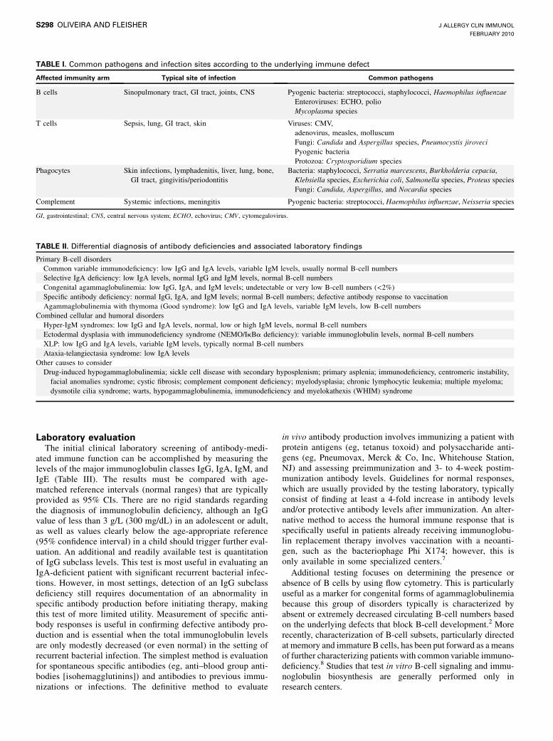
Laboratory evaluationThe initial clinical laboratory screening of antibody-medi-
ated immune function can be accomplished by measuring thelevels of the major immunoglobulin classes IgG, IgA, IgM, andIgE (Table III). The results must be compared with age-matched reference intervals (normal ranges) that are typicallyprovided as 95% CIs. There are no rigid standards regardingthe diagnosis of immunoglobulin deficiency, although an IgGvalue of less than 3 g/L (300 mg/dL) in an adolescent or adult,as well as values clearly below the age-appropriate reference(95% confidence interval) in a child should trigger further eval-uation. An additional and readily available test is quantitationof IgG subclass levels. This test is most useful in evaluating anIgA-deficient patient with significant recurrent bacterial infec-tions. However, in most settings, detection of an IgG subclassdeficiency still requires documentation of an abnormality inspecific antibody production before initiating therapy, makingthis test of more limited utility. Measurement of specific anti-body responses is useful in confirming defective antibody pro-duction and is essential when the total immunoglobulin levelsare only modestly decreased (or even normal) in the setting ofrecurrent bacterial infection. The simplest method is evaluationfor spontaneous specific antibodies (eg, anti–blood group anti-bodies [isohemagglutinins]) and antibodies to previous immu-nizations or infections. The definitive method to evaluate
in vivo antibody production involves immunizing a patient withprotein antigens (eg, tetanus toxoid) and polysaccharide anti-gens (eg, Pneumovax, Merck & Co, Inc, Whitehouse Station,NJ) and assessing preimmunization and 3- to 4-week postim-munization antibody levels. Guidelines for normal responses,which are usually provided by the testing laboratory, typicallyconsist of finding at least a 4-fold increase in antibody levelsand/or protective antibody levels after immunization. An alter-native method to access the humoral immune response that isspecifically useful in patients already receiving immunoglobu-lin replacement therapy involves vaccination with a neoanti-gen, such as the bacteriophage Phi X174; however, this isonly available in some specialized centers.7
Additional testing focuses on determining the presence orabsence of B cells by using flow cytometry. This is particularlyuseful as a marker for congenital forms of agammaglobulinemiabecause this group of disorders typically is characterized byabsent or extremely decreased circulating B-cell numbers basedon the underlying defects that block B-cell development.2 Morerecently, characterization of B-cell subsets, particularly directedat memory and immature B cells, has been put forward as a meansof further characterizing patients with common variable immuno-deficiency.8 Studies that test in vitro B-cell signaling and immu-noglobulin biosynthesis are generally performed only inresearch centers.
TABLE I. Common pathogens and infection sites according to the underlying immune defect
Affected immunity arm Typical site of infection Common pathogens
B cells Sinopulmonary tract, GI tract, joints, CNS Pyogenic bacteria: streptococci, staphylococci, Haemophilus influenzaeEnteroviruses: ECHO, polioMycoplasma species
T cells Sepsis, lung, GI tract, skin Viruses: CMV,adenovirus, measles, molluscumFungi: Candida and Aspergillus species, Pneumocystis jiroveciPyogenic bacteriaProtozoa: Cryptosporidium species
Phagocytes Skin infections, lymphadenitis, liver, lung, bone,GI tract, gingivitis/periodontitis
Bacteria: staphylococci, Serratia marcescens, Burkholderia cepacia,Klebsiella species, Escherichia coli, Salmonella species, Proteus speciesFungi: Candida, Aspergillus, and Nocardia species
Complement Systemic infections, meningitis Pyogenic bacteria: streptococci, Haemophilus influenzae, Neisseria species
GI, gastrointestinal; CNS, central nervous system; ECHO, echovirus; CMV, cytomegalovirus.
TABLE II. Differential diagnosis of antibody deficiencies and associated laboratory findings
Primary B-cell disordersCommon variable immunodeficiency: low IgG and IgA levels, variable IgM levels, usually normal B-cell numbersSelective IgA deficiency: low IgA levels, normal IgG and IgM levels, normal B-cell numbersCongenital agammaglobulinemia: low IgG, IgA, and IgM levels; undetectable or very low B-cell numbers (<2%)Specific antibody deficiency: normal IgG, IgA, and IgM levels; normal B-cell numbers; defective antibody response to vaccinationAgammaglobulinemia with thymoma (Good syndrome): low IgG and IgA levels, variable IgM levels, low B-cell numbers
Combined cellular and humoral disordersHyper-IgM syndromes: low IgG and IgA levels, normal, low or high IgM levels, normal B-cell numbersEctodermal dysplasia with immunodeficiency syndrome (NEMO/IkBa deficiency): variable immunoglobulin levels, normal B-cell numbersXLP: low IgG and IgA levels, variable IgM levels, typically normal B-cell numbersAtaxia-telangiectasia syndrome: low IgA levels
Other causes to considerDrug-induced hypogammaglobulinemia; sickle cell disease with secondary hyposplenism; primary asplenia; immunodeficiency, centromeric instability,facial anomalies syndrome; cystic fibrosis; complement component deficiency; myelodysplasia; chronic lymphocytic leukemia; multiple myeloma;dysmotile cilia syndrome; warts, hypogammaglobulinemia, immunodeficiency and myelokathexis (WHIM) syndrome
J ALLERGY CLIN IMMUNOL
FEBRUARY 2010
S298 OLIVEIRA AND FLEISHER
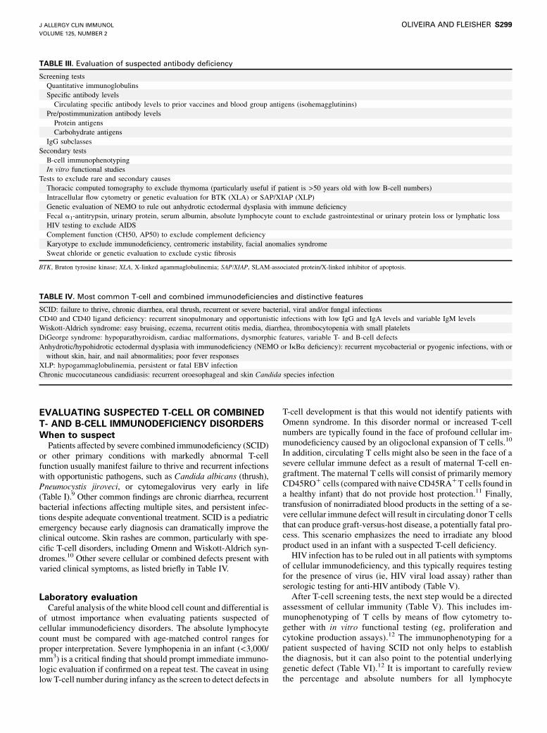
EVALUATING SUSPECTED T-CELL OR COMBINEDT- AND B-CELL IMMUNODEFICIENCY DISORDERSWhen to suspect
Patients affected by severe combined immunodeficiency (SCID)or other primary conditions with markedly abnormal T-cellfunction usually manifest failure to thrive and recurrent infectionswith opportunistic pathogens, such as Candida albicans (thrush),Pneumocystis jiroveci, or cytomegalovirus very early in life(Table I).9 Other common findings are chronic diarrhea, recurrentbacterial infections affecting multiple sites, and persistent infec-tions despite adequate conventional treatment. SCID is a pediatricemergency because early diagnosis can dramatically improve theclinical outcome. Skin rashes are common, particularly with spe-cific T-cell disorders, including Omenn and Wiskott-Aldrich syn-dromes.10 Other severe cellular or combined defects present withvaried clinical symptoms, as listed briefly in Table IV.
Laboratory evaluationCareful analysis of thewhite blood cell count and differential is
of utmost importance when evaluating patients suspected ofcellular immunodeficiency disorders. The absolute lymphocytecount must be compared with age-matched control ranges forproper interpretation. Severe lymphopenia in an infant (<3,000/mm3) is a critical finding that should prompt immediate immuno-logic evaluation if confirmed on a repeat test. The caveat in usinglow T-cell number during infancy as the screen to detect defects in
T-cell development is that this would not identify patients withOmenn syndrome. In this disorder normal or increased T-cellnumbers are typically found in the face of profound cellular im-munodeficiency caused by an oligoclonal expansion of T cells.10
In addition, circulating T cells might also be seen in the face of asevere cellular immune defect as a result of maternal T-cell en-graftment. The maternal T cells will consist of primarily memoryCD45RO1 cells (compared with naive CD45RA1T cells found ina healthy infant) that do not provide host protection.11 Finally,transfusion of nonirradiated blood products in the setting of a se-vere cellular immune defect will result in circulating donor T cellsthat can produce graft-versus-host disease, a potentially fatal pro-cess. This scenario emphasizes the need to irradiate any bloodproduct used in an infant with a suspected T-cell deficiency.
HIV infection has to be ruled out in all patients with symptomsof cellular immunodeficiency, and this typically requires testingfor the presence of virus (ie, HIV viral load assay) rather thanserologic testing for anti-HIV antibody (Table V).
After T-cell screening tests, the next step would be a directedassessment of cellular immunity (Table V). This includes im-munophenotyping of T cells by means of flow cytometry to-gether with in vitro functional testing (eg, proliferation andcytokine production assays).12 The immunophenotyping for apatient suspected of having SCID not only helps to establishthe diagnosis, but it can also point to the potential underlyinggenetic defect (Table VI).12 It is important to carefully reviewthe percentage and absolute numbers for all lymphocyte
TABLE III. Evaluation of suspected antibody deficiency
Screening testsQuantitative immunoglobulinsSpecific antibody levelsCirculating specific antibody levels to prior vaccines and blood group antigens (isohemagglutinins)
Pre/postimmunization antibody levelsProtein antigensCarbohydrate antigens
IgG subclassesSecondary tests
B-cell immunophenotypingIn vitro functional studies
Tests to exclude rare and secondary causesThoracic computed tomography to exclude thymoma (particularly useful if patient is >50 years old with low B-cell numbers)Intracellular flow cytometry or genetic evaluation for BTK (XLA) or SAP/XIAP (XLP)Genetic evaluation of NEMO to rule out anhydrotic ectodermal dysplasia with immune deficiencyFecal a1-antitrypsin, urinary protein, serum albumin, absolute lymphocyte count to exclude gastrointestinal or urinary protein loss or lymphatic lossHIV testing to exclude AIDSComplement function (CH50, AP50) to exclude complement deficiencyKaryotype to exclude immunodeficiency, centromeric instability, facial anomalies syndromeSweat chloride or genetic evaluation to exclude cystic fibrosis
BTK, Bruton tyrosine kinase; XLA, X-linked agammaglobulinemia; SAP/XIAP, SLAM-associated protein/X-linked inhibitor of apoptosis.
TABLE IV. Most common T-cell and combined immunodeficiencies and distinctive features
SCID: failure to thrive, chronic diarrhea, oral thrush, recurrent or severe bacterial, viral and/or fungal infectionsCD40 and CD40 ligand deficiency: recurrent sinopulmonary and opportunistic infections with low IgG and IgA levels and variable IgM levelsWiskott-Aldrich syndrome: easy bruising, eczema, recurrent otitis media, diarrhea, thrombocytopenia with small plateletsDiGeorge syndrome: hypoparathyroidism, cardiac malformations, dysmorphic features, variable T- and B-cell defectsAnhydrotic/hypohidrotic ectodermal dysplasia with immunodeficiency (NEMO or IkBa deficiency): recurrent mycobacterial or pyogenic infections, with orwithout skin, hair, and nail abnormalities; poor fever responses
XLP: hypogammaglobulinemia, persistent or fatal EBV infectionChronic mucocutaneous candidiasis: recurrent oroesophageal and skin Candida species infection
J ALLERGY CLIN IMMUNOL
VOLUME 125, NUMBER 2
OLIVEIRA AND FLEISHER S299
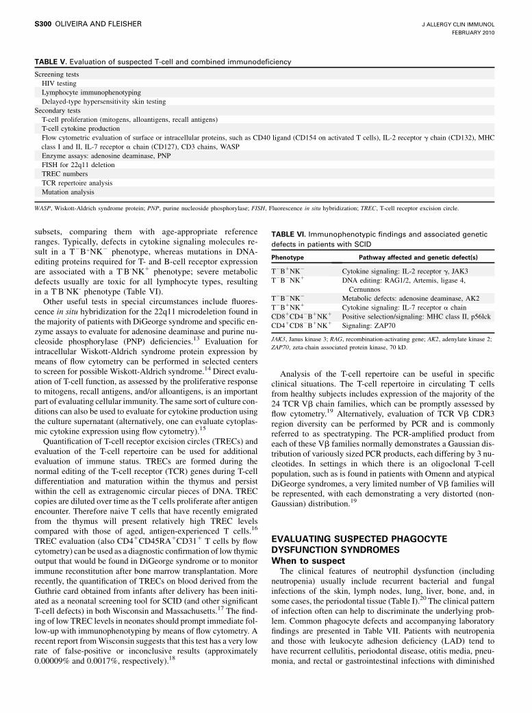
subsets, comparing them with age-appropriate referenceranges. Typically, defects in cytokine signaling molecules re-sult in a T2B1NK2 phenotype, whereas mutations in DNA-editing proteins required for T- and B-cell receptor expressionare associated with a T-B-NK1 phenotype; severe metabolicdefects usually are toxic for all lymphocyte types, resultingin a T-B-NK- phenotype (Table VI).
Other useful tests in special circumstances include fluores-cence in situ hybridization for the 22q11 microdeletion found inthe majority of patients with DiGeorge syndrome and specific en-zyme assays to evaluate for adenosine deaminase and purine nu-cleoside phosphorylase (PNP) deficiencies.13 Evaluation forintracellular Wiskott-Aldrich syndrome protein expression bymeans of flow cytometry can be performed in selected centersto screen for possible Wiskott-Aldrich syndrome.14 Direct evalu-ation of T-cell function, as assessed by the proliferative responseto mitogens, recall antigens, and/or alloantigens, is an importantpart of evaluating cellular immunity. The same sort of culture con-ditions can also be used to evaluate for cytokine production usingthe culture supernatant (alternatively, one can evaluate cytoplas-mic cytokine expression using flow cytometry).15
Quantification of T-cell receptor excision circles (TRECs) andevaluation of the T-cell repertoire can be used for additionalevaluation of immune status. TRECs are formed during thenormal editing of the T-cell receptor (TCR) genes during T-celldifferentiation and maturation within the thymus and persistwithin the cell as extragenomic circular pieces of DNA. TRECcopies are diluted over time as the T cells proliferate after antigenencounter. Therefore naive T cells that have recently emigratedfrom the thymus will present relatively high TREC levelscompared with those of aged, antigen-experienced T cells.16
TREC evaluation (also CD41CD45RA1CD311 T cells by flowcytometry) can be used as a diagnostic confirmation of low thymicoutput that would be found in DiGeorge syndrome or to monitorimmune reconstitution after bone marrow transplantation. Morerecently, the quantification of TRECs on blood derived from theGuthrie card obtained from infants after delivery has been initi-ated as a neonatal screening tool for SCID (and other significantT-cell defects) in both Wisconsin and Massachusetts.17 The find-ing of low TREC levels in neonates should prompt immediate fol-low-up with immunophenotyping by means of flow cytometry. Arecent report fromWisconsin suggests that this test has a very lowrate of false-positive or inconclusive results (approximately0.00009% and 0.0017%, respectively).18
Analysis of the T-cell repertoire can be useful in specificclinical situations. The T-cell repertoire in circulating T cellsfrom healthy subjects includes expression of the majority of the24 TCR Vb chain families, which can be promptly assessed byflow cytometry.19 Alternatively, evaluation of TCR Vb CDR3region diversity can be performed by PCR and is commonlyreferred to as spectratyping. The PCR-amplified product fromeach of these Vb families normally demonstrates a Gaussian dis-tribution of variously sized PCR products, each differing by 3 nu-cleotides. In settings in which there is an oligoclonal T-cellpopulation, such as is found in patients with Omenn and atypicalDiGeorge syndromes, a very limited number of Vb families willbe represented, with each demonstrating a very distorted (non-Gaussian) distribution.19
EVALUATING SUSPECTED PHAGOCYTEDYSFUNCTION SYNDROMESWhen to suspect
The clinical features of neutrophil dysfunction (includingneutropenia) usually include recurrent bacterial and fungalinfections of the skin, lymph nodes, lung, liver, bone, and, insome cases, the periodontal tissue (Table I).20 The clinical patternof infection often can help to discriminate the underlying prob-lem. Common phagocyte defects and accompanying laboratoryfindings are presented in Table VII. Patients with neutropeniaand those with leukocyte adhesion deficiency (LAD) tend tohave recurrent cellulitis, periodontal disease, otitis media, pneu-monia, and rectal or gastrointestinal infections with diminished
TABLE V. Evaluation of suspected T-cell and combined immunodeficiency
Screening testsHIV testingLymphocyte immunophenotypingDelayed-type hypersensitivity skin testing
Secondary testsT-cell proliferation (mitogens, alloantigens, recall antigens)T-cell cytokine productionFlow cytometric evaluation of surface or intracellular proteins, such as CD40 ligand (CD154 on activated T cells), IL-2 receptor g chain (CD132), MHCclass I and II, IL-7 receptor a chain (CD127), CD3 chains, WASPEnzyme assays: adenosine deaminase, PNPFISH for 22q11 deletionTREC numbersTCR repertoire analysisMutation analysis
WASP, Wiskott-Aldrich syndrome protein; PNP, purine nucleoside phosphorylase; FISH, Fluorescence in situ hybridization; TREC, T-cell receptor excision circle.
TABLE VI. Immunophenotypic findings and associated genetic
defects in patients with SCID
Phenotype Pathway affected and genetic defect(s)
T2B1NK2 Cytokine signaling: IL-2 receptor g, JAK3T2B2NK1 DNA editing: RAG1/2, Artemis, ligase 4,
CernunnosT2B2NK2 Metabolic defects: adenosine deaminase, AK2T2B1NK1 Cytokine signaling: IL-7 receptor a chainCD81CD42B1NK1 Positive selection/signaling: MHC class II, p56lckCD41CD82B1NK1 Signaling: ZAP70
JAK3, Janus kinase 3; RAG, recombination-activating gene; AK2, adenylate kinase 2;ZAP70, zeta-chain associated protein kinase, 70 kD.
J ALLERGY CLIN IMMUNOL
FEBRUARY 2010
S300 OLIVEIRA AND FLEISHER
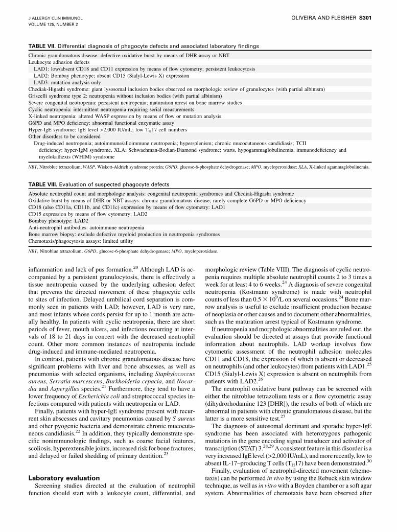
inflammation and lack of pus formation.20 Although LAD is ac-companied by a persistent granulocytosis, there is effectively atissue neutropenia caused by the underlying adhesion defectthat prevents the directed movement of these phagocytic cellsto sites of infection. Delayed umbilical cord separation is com-monly seen in patients with LAD; however, LAD is very rare,and most infants whose cords persist for up to 1 month are actu-ally healthy. In patients with cyclic neutropenia, there are shortperiods of fever, mouth ulcers, and infections recurring at inter-vals of 18 to 21 days in concert with the decreased neutrophilcount. Other more common instances of neutropenia includedrug-induced and immune-mediated neutropenia.
In contrast, patients with chronic granulomatous disease havesignificant problems with liver and bone abscesses, as well aspneumonias with selected organisms, including Staphylococcusaureus, Serratia marcescens, Burkholderia cepacia, and Nocar-dia and Aspergillus species.21 Furthermore, they tend to have alower frequency of Escherichia coli and streptococcal species in-fections compared with patients with neutropenia or LAD.
Finally, patients with hyper-IgE syndrome present with recur-rent skin abscesses and cavitary pneumonias caused by S aureusand other pyogenic bacteria and demonstrate chronic mucocuta-neous candidiasis.22 In addition, they typically demonstrate spe-cific nonimmunologic findings, such as coarse facial features,scoliosis, hyperextensible joints, increased risk for bone fractures,and delayed or failed shedding of primary dentition.23
Laboratory evaluationScreening studies directed at the evaluation of neutrophil
function should start with a leukocyte count, differential, and
morphologic review (Table VIII). The diagnosis of cyclic neutro-penia requires multiple absolute neutrophil counts 2 to 3 times aweek for at least 4 to 6 weeks.24 A diagnosis of severe congenitalneutropenia (Kostmann syndrome) is made with neutrophilcounts of less than 0.53 109/L on several occasions.24 Bone mar-row analysis is useful to exclude insufficient production becauseof neoplasia or other causes and to document other abnormalities,such as the maturation arrest typical of Kostmann syndrome.
If neutropenia andmorphologic abnormalities are ruled out, theevaluation should be directed at assays that provide functionalinformation about neutrophils. LAD workup involves flowcytometric assessment of the neutrophil adhesion moleculesCD11 and CD18, the expression of which is absent or decreasedon neutrophils (and other leukocytes) from patients with LAD1.25
CD15 (Sialyl-Lewis X) expression is absent on neutrophils frompatients with LAD2.26
The neutrophil oxidative burst pathway can be screened witheither the nitroblue tetrazolium tests or a flow cytometric assay(dihydrorhodamine 123 [DHR]), the results of both of which areabnormal in patients with chronic granulomatous disease, but thelatter is a more sensitive test.27
The diagnosis of autosomal dominant and sporadic hyper-IgEsyndrome has been associated with heterozygous pathogenicmutations in the gene encoding signal transducer and activator oftranscription (STAT) 3.28,29A consistent feature in this disorder is avery increased IgE level (>2,000 IU/mL), andmore recently, low toabsent IL-17–producing T cells (TH17) have been demonstrated.30
Finally, evaluation of neutrophil-directed movement (chemo-taxis) can be performed in vivo by using the Rebuck skin windowtechnique, as well as in vitrowith a Boyden chamber or a soft agarsystem. Abnormalities of chemotaxis have been observed after
TABLE VII. Differential diagnosis of phagocyte defects and associated laboratory findings
Chronic granulomatous disease: defective oxidative burst by means of DHR assay or NBTLeukocyte adhesion defects
LAD1: low/absent CD18 and CD11 expression by means of flow cytometry; persistent leukocytosisLAD2: Bombay phenotype; absent CD15 (Sialyl-Lewis X) expressionLAD3: mutation analysis only
Chediak-Higashi syndrome: giant lysosomal inclusion bodies observed on morphologic review of granulocytes (with partial albinism)Griscelli syndrome type 2: neutropenia without inclusion bodies (with partial albinism)Severe congenital neutropenia: persistent neutropenia; maturation arrest on bone marrow studiesCyclic neutropenia: intermittent neutropenia requiring serial measurementsX-linked neutropenia: altered WASP expression by means of flow or mutation analysisG6PD and MPO deficiency: abnormal functional enzymatic assayHyper-IgE syndrome: IgE level >2,000 IU/mL; low TH17 cell numbersOther disorders to be considered
Drug-induced neutropenia; autoimmune/alloimmune neutropenia; hypersplenism; chronic mucocutaneous candidiasis; TCIIdeficiency; hyper-IgM syndrome, XLA; Schwachman-Bodian-Diamond syndrome; warts, hypogammaglobulinemia, immunodeficiency andmyelokathexis (WHIM) syndrome
NBT, Nitroblue tetrazolium;WASP, Wiskott-Aldrich syndrome protein; G6PD, glucose-6-phosphate dehydrogenase;MPO, myeloperoxidase; XLA, X-linked agammaglobulinemia.
TABLE VIII. Evaluation of suspected phagocyte defects
Absolute neutrophil count and morphologic analysis: congenital neutropenia syndromes and Chediak-Higashi syndromeOxidative burst by means of DHR or NBT assays: chronic granulomatous disease; rarely complete G6PD or MPO deficiencyCD18 (also CD11a, CD11b, and CD11c) expression by means of flow cytometry: LAD1CD15 expression by means of flow cytometry: LAD2Bombay phenotype: LAD2Anti-neutrophil antibodies: autoimmune neutropeniaBone marrow biopsy: exclude defective myeloid production in neutropenia syndromesChemotaxis/phagocytosis assays: limited utility
NBT, Nitroblue tetrazolium; G6PD, glucose-6-phosphate dehydrogenase; MPO, myeloperoxidase.
J ALLERGY CLIN IMMUNOL
VOLUME 125, NUMBER 2
OLIVEIRA AND FLEISHER S301
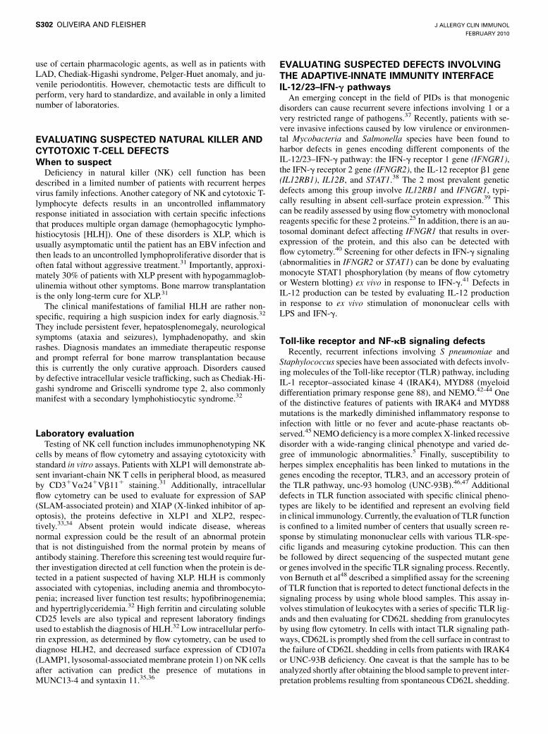
use of certain pharmacologic agents, as well as in patients withLAD, Chediak-Higashi syndrome, Pelger-Huet anomaly, and ju-venile periodontitis. However, chemotactic tests are difficult toperform, very hard to standardize, and available in only a limitednumber of laboratories.
EVALUATING SUSPECTED NATURAL KILLER ANDCYTOTOXIC T-CELL DEFECTSWhen to suspect
Deficiency in natural killer (NK) cell function has beendescribed in a limited number of patients with recurrent herpesvirus family infections. Another category of NK and cytotoxic T-lymphocyte defects results in an uncontrolled inflammatoryresponse initiated in association with certain specific infectionsthat produces multiple organ damage (hemophagocytic lympho-histiocytosis [HLH]). One of these disorders is XLP, which isusually asymptomatic until the patient has an EBV infection andthen leads to an uncontrolled lymphoproliferative disorder that isoften fatal without aggressive treatment.31 Importantly, approxi-mately 30% of patients with XLP present with hypogammaglob-ulinemia without other symptoms. Bone marrow transplantationis the only long-term cure for XLP.31
The clinical manifestations of familial HLH are rather non-specific, requiring a high suspicion index for early diagnosis.32
They include persistent fever, hepatosplenomegaly, neurologicalsymptoms (ataxia and seizures), lymphadenopathy, and skinrashes. Diagnosis mandates an immediate therapeutic responseand prompt referral for bone marrow transplantation becausethis is currently the only curative approach. Disorders causedby defective intracellular vesicle trafficking, such as Chediak-Hi-gashi syndrome and Griscelli syndrome type 2, also commonlymanifest with a secondary lymphohistiocytic syndrome.32
Laboratory evaluationTesting of NK cell function includes immunophenotyping NK
cells by means of flow cytometry and assaying cytotoxicity withstandard in vitro assays. Patients with XLP1 will demonstrate ab-sent invariant-chain NK T cells in peripheral blood, as measuredby CD31Va241Vb111 staining.31 Additionally, intracellularflow cytometry can be used to evaluate for expression of SAP(SLAM-associated protein) and XIAP (X-linked inhibitor of ap-optosis), the proteins defective in XLP1 and XLP2, respec-tively.33,34 Absent protein would indicate disease, whereasnormal expression could be the result of an abnormal proteinthat is not distinguished from the normal protein by means ofantibody staining. Therefore this screening test would require fur-ther investigation directed at cell function when the protein is de-tected in a patient suspected of having XLP. HLH is commonlyassociated with cytopenias, including anemia and thrombocyto-penia; increased liver function test results; hypofibrinogenemia;and hypertriglyceridemia.32 High ferritin and circulating solubleCD25 levels are also typical and represent laboratory findingsused to establish the diagnosis of HLH.32 Low intracellular perfo-rin expression, as determined by flow cytometry, can be used todiagnose HLH2, and decreased surface expression of CD107a(LAMP1, lysosomal-associated membrane protein 1) on NK cellsafter activation can predict the presence of mutations inMUNC13-4 and syntaxin 11.35,36
EVALUATING SUSPECTED DEFECTS INVOLVINGTHE ADAPTIVE-INNATE IMMUNITY INTERFACEIL-12/23–IFN-g pathways
An emerging concept in the field of PIDs is that monogenicdisorders can cause recurrent severe infections involving 1 or avery restricted range of pathogens.37 Recently, patients with se-vere invasive infections caused by low virulence or environmen-tal Mycobacteria and Salmonella species have been found toharbor defects in genes encoding different components of theIL-12/23–IFN-g pathway: the IFN-g receptor 1 gene (IFNGR1),the IFN-g receptor 2 gene (IFNGR2), the IL-12 receptor b1 gene(IL12RB1), IL12B, and STAT1.38 The 2 most prevalent geneticdefects among this group involve IL12RB1 and IFNGR1, typi-cally resulting in absent cell-surface protein expression.39 Thiscan be readily assessed by using flow cytometry with monoclonalreagents specific for these 2 proteins.25 In addition, there is an au-tosomal dominant defect affecting IFNGR1 that results in over-expression of the protein, and this also can be detected withflow cytometry.40 Screening for other defects in IFN-g signaling(abnormalities in IFNGR2 or STAT1) can be done by evaluatingmonocyte STAT1 phosphorylation (by means of flow cytometryor Western blotting) ex vivo in response to IFN-g.41 Defects inIL-12 production can be tested by evaluating IL-12 productionin response to ex vivo stimulation of mononuclear cells withLPS and IFN-g.
Toll-like receptor and NF-kB signaling defectsRecently, recurrent infections involving S pneumoniae and
Staphylococcus species have been associated with defects involv-ing molecules of the Toll-like receptor (TLR) pathway, includingIL-1 receptor–associated kinase 4 (IRAK4), MYD88 (myeloiddifferentiation primary response gene 88), and NEMO.42-44 Oneof the distinctive features of patients with IRAK4 and MYD88mutations is the markedly diminished inflammatory response toinfection with little or no fever and acute-phase reactants ob-served.45 NEMOdeficiency is amore complexX-linked recessivedisorder with a wide-ranging clinical phenotype and varied de-gree of immunologic abnormalities.5 Finally, susceptibility toherpes simplex encephalitis has been linked to mutations in thegenes encoding the receptor, TLR3, and an accessory protein ofthe TLR pathway, unc-93 homolog (UNC-93B).46,47 Additionaldefects in TLR function associated with specific clinical pheno-types are likely to be identified and represent an evolving fieldin clinical immunology. Currently, the evaluation of TLR functionis confined to a limited number of centers that usually screen re-sponse by stimulating mononuclear cells with various TLR-spe-cific ligands and measuring cytokine production. This can thenbe followed by direct sequencing of the suspected mutant geneor genes involved in the specific TLR signaling process. Recently,von Bernuth et al48 described a simplified assay for the screeningof TLR function that is reported to detect functional defects in thesignaling process by using whole blood samples. This assay in-volves stimulation of leukocytes with a series of specific TLR lig-ands and then evaluating for CD62L shedding from granulocytesby using flow cytometry. In cells with intact TLR signaling path-ways, CD62L is promptly shed from the cell surface in contrast tothe failure of CD62L shedding in cells from patients with IRAK4or UNC-93B deficiency. One caveat is that the sample has to beanalyzed shortly after obtaining the blood sample to prevent inter-pretation problems resulting from spontaneous CD62L shedding.
J ALLERGY CLIN IMMUNOL
FEBRUARY 2010
S302 OLIVEIRA AND FLEISHER
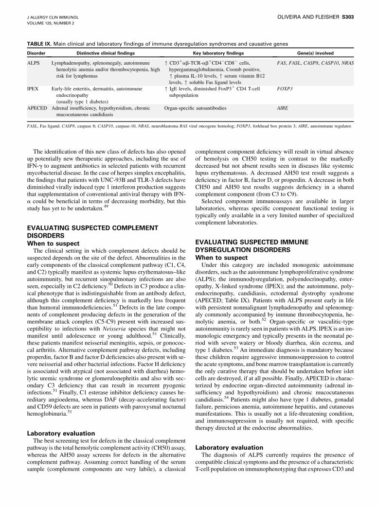
The identification of this new class of defects has also openedup potentially new therapeutic approaches, including the use ofIFN-g to augment antibiotics in selected patients with recurrentmycobacterial disease. In the case of herpes simplex encephalitis,the findings that patients with UNC-93B and TLR-3 defects havediminished virally induced type 1 interferon production suggeststhat supplementation of conventional antiviral therapy with IFN-a could be beneficial in terms of decreasing morbidity, but thisstudy has yet to be undertaken.49
EVALUATING SUSPECTED COMPLEMENTDISORDERSWhen to suspect
The clinical setting in which complement defects should besuspected depends on the site of the defect. Abnormalities in theearly components of the classical complement pathway (C1, C4,and C2) typically manifest as systemic lupus erythematosus–likeautoimmunity, but recurrent sinopulmonary infections are alsoseen, especially in C2 deficiency.50 Defects in C3 produce a clin-ical phenotype that is indistinguishable from an antibody defect,although this complement deficiency is markedly less frequentthan humoral immunodeficiencies.51 Defects in the late compo-nents of complement producing defects in the generation of themembrane attack complex (C5-C9) present with increased sus-ceptibility to infections with Neisseria species that might notmanifest until adolescence or young adulthood.51 Clinically,these patients manifest neisserial meningitis, sepsis, or gonococ-cal arthritis. Alternative complement pathway defects, includingproperdin, factor B and factor D deficiencies also present with se-vere neisserial and other bacterial infections. Factor H deficiencyis associated with atypical (not associated with diarrhea) hemo-lytic uremic syndrome or glomerulonephritis and also with sec-ondary C3 deficiency that can result in recurrent pyogenicinfections.51 Finally, C1 esterase inhibitor deficiency causes he-reditary angioedema, whereas DAF (decay-accelerating factor)and CD59 defects are seen in patients with paroxysmal nocturnalhemoglobinuria.51
Laboratory evaluationThe best screening test for defects in the classical complement
pathway is the total hemolytic complement activity (CH50) assay,whereas the AH50 assay screens for defects in the alternativecomplement pathway. Assuming correct handling of the serumsample (complement components are very labile), a classical
complement component deficiency will result in virtual absenceof hemolysis on CH50 testing in contrast to the markedlydecreased but not absent results seen in diseases like systemiclupus erythematosus. A decreased AH50 test result suggests adeficiency in factor B, factor D, or properdin. A decrease in bothCH50 and AH50 test results suggests deficiency in a sharedcomplement component (from C3 to C9).
Selected component immunoassays are available in largerlaboratories, whereas specific component functional testing istypically only available in a very limited number of specializedcomplement laboratories.
EVALUATING SUSPECTED IMMUNEDYSREGULATION DISORDERSWhen to suspect
Under this category are included monogenic autoimmunedisorders, such as the autoimmune lymphoproliferative syndrome(ALPS); the immunodysregulation, polyendocrinopathy, enter-opathy, X-linked syndrome (IPEX); and the autoimmune, poly-endocrinopathy, candidiasis, ectodermal dystrophy syndrome(APECED; Table IX). Patients with ALPS present early in lifewith persistent nonmalignant lymphadenopathy and splenomeg-aly commonly accompanied by immune thrombocytopenia, he-molytic anemia, or both.52 Organ-specific or vasculitic-typeautoimmunity is rarely seen in patients with ALPS. IPEX is an im-munologic emergency and typically presents in the neonatal pe-riod with severe watery or bloody diarrhea, skin eczema, andtype 1 diabetes.53 An immediate diagnosis is mandatory becausethese children require aggressive immunosuppression to controlthe acute symptoms, and bonemarrow transplantation is currentlythe only curative therapy that should be undertaken before isletcells are destroyed, if at all possible. Finally, APECED is charac-terized by endocrine organ–directed autoimmunity (adrenal in-sufficiency and hypothyroidism) and chronic mucocutaneouscandidiasis.54 Patients might also have type 1 diabetes, gonadalfailure, pernicious anemia, autoimmune hepatitis, and cutaneousmanifestations. This is usually not a life-threatening condition,and immunosuppression is usually not required, with specifictherapy directed at the endocrine abnormalities.
Laboratory evaluationThe diagnosis of ALPS currently requires the presence of
compatible clinical symptoms and the presence of a characteristicT-cell population on immunophenotyping that expresses CD3 and
TABLE IX. Main clinical and laboratory findings of immune dysregulation syndromes and causative genes
Disorder Distinctive clinical findings Key laboratory findings Gene(s) involved
ALPS Lymphadenopathy, splenomegaly, autoimmunehemolytic anemia and/or thrombocytopenia, highrisk for lymphomas
[ CD31ab-TCR-ab1CD42CD82 cells,hypergammaglobulinemia, Coomb positive,[ plasma IL-10 levels, [ serum vitamin B12levels, [ soluble Fas ligand levels
FAS, FASL, CASP8, CASP10, NRAS
IPEX Early-life enteritis, dermatitis, autoimmuneendocrinopathy(usually type 1 diabetes)
[ IgE levels, diminished FoxP31 CD4 T-cellsubpopulation
FOXP3
APECED Adrenal insufficiency, hypothyroidism, chronicmucocutaneous candidiasis
Organ-specific autoantibodies AIRE
FASL, Fas ligand; CASP8, caspase 8; CASP10, caspase-10, NRAS, neuroblastoma RAS viral oncogene homolog; FOXP3, forkhead box protein 3; AIRE, autoimmune regulator.
J ALLERGY CLIN IMMUNOL
VOLUME 125, NUMBER 2
OLIVEIRA AND FLEISHER S303
ab-TCR but does not express CD4 or CD8 markers, which arereferred to as double-negative T cells (Table IX). Determinationof this T-cell subpopulation requires the use of antibodies toab-TCR because most double-negative T cells in normal samplesare gd-TCR1 and are not relevant for establishing a diagnosis ofALPS. Normal ranges for ab double-negative T cells should beestablished for each laboratory. At the National Institutes ofHealth, more than 1% of the total lymphocyte population is con-sidered abnormal in adults. Other supporting features include hy-pergammaglobulinemia and increased plasma IL-10, vitaminB12, and soluble Fas ligand levels (J.B.O. and T.A.F., unpub-lished observations).55 In addition, for a diagnosis of certainty,one must demonstrate defective lymphocyte apoptosis in vitroor the presence of a mutation on FAS, FASL (FAS ligand),CASP8 (caspase-8), CASP10 (caspase-10), or NRAS (neuroblas-toma RAS viral oncogene homolog).56-61
Screening for IPEX is based on demonstrating absent ordiminished population of forkhead box protein 3 (Foxp3)–expressing CD4 T cells in the peripheral blood, as assessed byintracellular flow cytometry. Another common laboratory findingis a marked increase in IgE levels. The gold standard for diagnosisis the demonstration of mutations on the FOXP3 gene. However,in approximately 50% of patients with clinical findings compati-ble with IPEX, nomutation is demonstrated (Troy Torgerson, per-sonal communication). Diagnosis of APECED in the setting of aclinically consistent phenotype currently requires sequencing ofthe AIRE (autoimmune regulator) gene.
CONCLUSIONLaboratory testing serves as the critical approach necessary for
evaluating immune function in the setting of a patient with ahistory of recurrent infections, unusual infections, or both. Theappropriate and directed use of immune function testing providesnot only critical diagnostic information but also directs decisionsregarding the most appropriate therapy. The latter is crucial tolimit disease-associated morbidity. The use of the laboratory inevaluating the immune system should not follow a shotgunapproach but rather should be a focused evaluation using specifictesting in an orderly process based on the clinical history.
REFERENCES1. Yong PF, Chee R, Grimbacher B. Hypogammaglobulinaemia. Immunol Allergy
Clin North Am 2008;28:691-713, vii.2. Conley ME, Dobbs AK, Farmer DM, Kilic S, Paris K, Grigoriadou S, et al. Pri-
mary B cell immunodeficiencies: comparisons and contrasts. Annu Rev Immunol2009;27:199-227.
3. Conley ME, Howard V. Clinical findings leading to the diagnosis of X-linkedagammaglobulinemia. J Pediatr 2002;141:566-71.
4. Ersoy F, Berkel AI, Sanal O, Oktay H. Twenty-year follow-up of 160 patients withataxia-telangiectasia. Turk J Pediatr 1991;33:205-15.
5. Hanson EP, Monaco-Shawver L, Solt LA, Madge LA, Banerjee PP, May MJ, et al.Hypomorphic nuclear factor-kappaB essential modulator mutation database and re-constitution system identifies phenotypic and immunologic diversity. J AllergyClin Immunol 2008;122:1169-77, e1116.
6. Allen RC, Armitage RJ, Conley ME, Rosenblatt H, Jenkins NA, Copeland NG,et al. CD40 ligand gene defects responsible for X-linked hyper-IgM syndrome.Science 1993;259:990-3.
7. Ochs HD, Buckley RH, Kobayashi RH, Kobayashi AL, Sorensen RU, Douglas SD,et al. Antibody responses to bacteriophage phi X174 in patients with adenosine de-aminase deficiency. Blood 1992;80:1163-71.
8. Wehr C, Kivioja T, Schmitt C, Ferry B, Witte T, Eren E, et al. The EUROclass trial:defining subgroups in common variable immunodeficiency. Blood 2008;111:77-85.
9. Notarangelo LD, Giliani S, Mazza C, Mella P, Savoldi G, Rodriguez-Perez C, et al.Of genes and phenotypes: the immunological and molecular spectrum of combined
immune deficiency. Defects of the gamma(c)-JAK3 signaling pathway as a model.Immunol Rev 2000;178:39-48.
10. Villa A, Notarangelo LD, Roifman CM. Omenn syndrome: inflammation in leakysevere combined immunodeficiency. J Allergy Clin Immunol 2008;122:1082-6.
11. Muller SM, Ege M, Pottharst A, Schulz AS, Schwarz K, Friedrich W. Transplacen-tally acquired maternal T lymphocytes in severe combined immunodeficiency: astudy of 121 patients. Blood 2001;98:1847-51.
12. Fleisher TA, Oliveira JB. Functional and molecular evaluation of lymphocytes.J Allergy Clin Immunol 2004;114:227-35.
13. Sullivan KE. Chromosome 22q11.2 deletion syndrome: DiGeorge syndrome/veloc-ardiofacial syndrome. Immunol Allergy Clin North Am 2008;28:353-66.
14. Kawai S, Minegishi M, Ohashi Y, Sasahara Y, Kumaki S, Konno T, et al. Flow cyto-metric determination of intracytoplasmic Wiskott-Aldrich syndrome protein in pe-ripheral blood lymphocyte subpopulations. J Immunol Methods 2002;260:195-205.
15. Prussin C, Metcalfe DD. Detection of intracytoplasmic cytokine using flow cytometryand directly conjugated anti-cytokine antibodies. J ImmunolMethods 1995;188:117-28.
16. Douek, et al. Changes in thymic function with age and during the treament of HIVinfection. Nature 1998;396:690-5.
17. Chan K, Puck JM. Development of population-based newborn screening for severecombined immunodeficiency. J Allergy Clin Immunol 2005;115:391-8.
18. Routes JM, Grossman WJ, Kumar A, Verbsky J, Brokopp C, Laessig RH, et al. Up-date of statewide newborn screening program for severe combined immunodefi-ciency (SCID) by T-cell receptor excision circles (TRECs). J Allergy ClinImmunol 2009;123(suppl):S68.
19. Pilch H, Hohn H, Freitag K, Neukirch C, Necker A, Haddad P, et al. Improved as-sessment of T-cell receptor (TCR) VB repertoire in clinical specimens: combina-tion of TCR-CDR3 spectratyping with flow cytometry-based TCR VB frequencyanalysis. Clin Diagn Lab Immunol 2002;9:257-66.
20. Rosenzweig SD, Holland SM. Phagocyte immunodeficiencies and their infections.J Allergy Clin Immunol 2004;113:620-6.
21. MartireB,RondelliR, SoresinaA, PignataC,Broccoletti T, FinocchiA, et al. Clinicalfeatures, long-term follow-up and outcome of a large cohort of patients with chronicgranulomatous disease: an Italianmulticenter study. Clin Immunol 2008;126:155-64.
22. Ozcan E, Notarangelo LD, Geha RS. Primary immune deficiencies with aberrantIgE production. J Allergy Clin Immunol 2008;122:1054-63.
23. Grimbacher B, Holland SM, Puck JM. Hyper-IgE syndromes. Immunol Rev 2005;203:244-50.
24. Boztug K, Welte K, Zeidler C, Klein C. Congenital neutropenia syndromes. Immu-nol Allergy Clin North Am 2008;28:259-75, vii-viii.
25. Oliveira JB,NotarangeloLD, Fleisher TA.Applications offlowcytometry for the studyof primary immune deficiencies. Curr Opin Allergy Clin Immunol 2008;8:499-509.
26. Etzioni A. Leukocyte adhesion deficiencies: molecular basis, clinical findings, andtherapeutic options. Adv Exp Med Biol 2007;601:51-60.
27. Jirapongsananuruk O, Malech HL, Kuhns DB, Niemela JE, Brown MR, Anderson-Cohen M, et al. Diagnostic paradigm for evaluation of male patients with chronicgranulomatous disease, based on the dihydrorhodamine 123 assay. J Allergy ClinImmunol 2003;111:374-9.
28. Holland SM, DeLeo FR, Elloumi HZ, Hsu AP, Uzel G, Brodsky N, et al. STAT3mutations in the hyper-IgE syndrome. N Engl J Med 2007;357:1608-19.
29. Minegishi Y, Saito M, Tsuchiya S, Tsuge I, Takada H, Hara T, et al. Dominant-neg-ative mutations in the DNA-binding domain of STAT3 cause hyper-IgE syndrome.Nature 2007;448:1058-62.
30. Milner JD, Brenchley JM, Laurence A, Freeman AF, Hill BJ, Elias KM, et al. Im-paired T(H)17 cell differentiation in subjects with autosomal dominant hyper-IgEsyndrome. Nature 2008;452:773-6.
31. Nichols KE, Ma CS, Cannons JL, Schwartzberg PL, Tangye SG. Molecular andcellular pathogenesis of X-linked lymphoproliferative disease. Immunol Rev2005;203:180-99.
32. Filipovich AH. Hemophagocytic lymphohistiocytosis and other hemophagocyticdisorders. Immunol Allergy Clin North Am 2008;28:293-313, viii.
33. Marsh RA, Villanueva J, Zhang K, Snow AL, Su HC, Madden L, et al. A rapid flowcytometric screening test for X-linked lymphoproliferative disease due to XIAP de-ficiency. Cytometry B Clin Cytom 2009;76:334-44.
34. Shinozaki K, Kanegane H, Matsukura H, Sumazaki R, Tsuchida M, Makita M,et al. Activation-dependent T cell expression of the X-linked lymphoproliferativedisease gene product SLAM-associated protein and its assessment for patient de-tection. Int Immunol 2002;14:1215-23.
35. Marcenaro S, Gallo F, Martini S, Santoro A, Griffiths GM, Arico M, et al. Analysisof natural killer-cell function in familial hemophagocytic lymphohistiocytosis(FHL): defective CD107a surface expression heralds Munc13-4 defect and dis-criminates between genetic subtypes of the disease. Blood 2006;108:2316-23.
36. Kogawa K, Lee SM, Villanueva J, Marmer D, Sumegi J, Filipovich AH. Perforinexpression in cytotoxic lymphocytes from patients with hemophagocytic lympho-histiocytosis and their family members. Blood 2002;99:61-6.
J ALLERGY CLIN IMMUNOL
FEBRUARY 2010
S304 OLIVEIRA AND FLEISHER
37. Casanova JL, Fieschi C, Zhang SY, Abel L. Revisiting human primary immunode-ficiencies. J Intern Med 2008;264:115-27.
38. Al-Muhsen S, Casanova JL. The genetic heterogeneity of mendelian susceptibilityto mycobacterial diseases. J Allergy Clin Immunol 2008;122:1043-52.
39. Filipe-Santos O, Bustamante J, Chapgier A, Vogt G, de Beaucoudrey L, Feinberg J,et al. Inborn errors of IL-12/23- and IFN-gamma-mediated immunity: molecular,cellular, and clinical features. Semin Immunol 2006;18:347-61.
40. Jouanguy E, Lamhamedi-Cherradi S, Lammas D, Dorman SE, Fondaneche MC,Dupuis S, et al. A human IFNGR1 small deletion hotspot associated with dominantsusceptibility to mycobacterial infection. Nat Genet 1999;21:370-8.
41. Fleisher TA, Dorman SE, Anderson JA, Vail M, BrownMR, Holland SM. Detection ofintracellular phosphorylated STAT-1 by flow cytometry. Clin Immunol 1999;90:425-30.
42. von Bernuth H, Picard C, Jin Z, Pankla R, Xiao H, Ku CL, et al. Pyogenic bacterialinfections in humans with MyD88 deficiency. Science 2008;321:691-6.
43. Picard C, von Bernuth H, Ku CL, Yang K, Puel A, Casanova JL. Inherited humanIRAK-4 deficiency: an update. Immunol Res 2007;38:347-52.
44. Doffinger R, Smahi A, Bessia C, Geissmann F, Feinberg J, Durandy A, et al. X-linked anhidrotic ectodermal dysplasia with immunodeficiency is caused by im-paired NF-kappaB signaling. Nat Genet 2001;27:277-85.
45. von Bernuth H, Puel A, Ku CL, Yang K, Bustamante J, Chang HH, et al. Septice-mia without sepsis: inherited disorders of nuclear factor-kappa B-mediated inflam-mation. Clin Infect Dis 2005;41(suppl 7):S436-9.
46. Zhang SY, Jouanguy E, Ugolini S, Smahi A, Elain G, Romero P, et al. TLR3 de-ficiency in patients with herpes simplex encephalitis. Science 2007;317:1522-7.
47. CasrougeA,ZhangSY,EidenschenkC, JouanguyE, PuelA,YangK, et al.Herpes sim-plex virus encephalitis in human UNC-93B deficiency. Science 2006;314:308-12.
48. von Bernuth H, Ku CL, Rodriguez-Gallego C, Zhang S, Garty BZ, Marodi L, et al.A fast procedure for the detection of defects in Toll-like receptor signaling. Pedi-atrics 2006;118:2498-503.
49. Sancho-Shimizu V, Zhang SY, Abel L, Tardieu M, Rozenberg F, Jouanguy E, et al.Genetic susceptibility to herpes simplex virus 1 encephalitis in mice and humans.Curr Opin Allergy Clin Immunol 2007;7:495-505.
50. Truedsson L, Bengtsson AA, Sturfelt G. Complement deficiencies and systemiclupus erythematosus. Autoimmunity 2007;40:560-6.
51. Botto M, Kirschfink M, Macor P, Pickering MC, Wurzner R, Tedesco F. Comple-ment in human diseases: Lessons from complement deficiencies. Mol Immunol2009;46:2774-83.
52. Oliveira JB, Fleisher T. Autoimmune lymphoproliferative syndrome. Curr Opin Al-lergy Clin Immunol 2004;4:497-503.
53. Torgerson TR, Ochs HD. Immune dysregulation, polyendocrinopathy, enteropathy,X-linked: forkhead box protein 3 mutations and lack of regulatory T cells. J Al-lergy Clin Immunol 2007;120:744-51.
54. Moraes-Vasconcelos D, Costa-Carvalho BT, Torgerson TR, Ochs HD. Primary im-mune deficiency disorders presenting as autoimmune diseases: IPEX andAPECED. J Clin Immunol 2008;28(suppl 1):S11-9.
55. Magerus-Chatinet A, et al. FAS-L, IL-10, and double-negative CD4-CD8-TCRalpha/beta+ T cells are reliable markers of autoimmune lymphoproliferativesyndrome (ALPS) associated with FAS loss of function. Blood 2009;113:3027-30.
56. Fisher GH, Rosenberg FJ, Straus SE, Dale JK, Middleton LA, Lin AY, et al. Dom-inant interfering Fas gene mutations impair apoptosis in a human autoimmune lym-phoproliferative syndrome. Cell 1995;81:935-46.
57. Bi LL, Pan G, Atkinson TP, Zheng L, Dale JK, Makris C, et al. Dominant inhibi-tion of Fas ligand-mediated apoptosis due to a heterozygous mutation associatedwith autoimmune lymphoproliferative syndrome (ALPS) Type Ib. BMC MedGenet 2007;8:41.
58. Chun HJ, Zheng L, Ahmad M, Wang J, Speirs CK, Siegel RM, et al. Pleiotropicdefects in lymphocyte activation caused by caspase-8 mutations lead to human im-munodeficiency. Nature 2002;419:395-9.
59. Rieux-Laucat F, Le Deist F, Hivroz C, Roberts IA, Debatin KM, Fischer A, et al.Mutations in Fas associated with human lymphoproliferative syndrome and auto-immunity. Science 1995;268:1347-9.
60. Holzelova E, Vonarbourg C, Stolzenberg MC, Arkwright PD, Selz F, Prieur AM,et al. Autoimmune lymphoproliferative syndrome with somatic Fas mutations. NEngl J Med 2004;351:1409-18.
61. Wang J, Zheng L, Lobito A, Chan FK, Dale J, Sneller M, et al. Inherited humancaspase 10 mutations underlie defective lymphocyte and dendritic cell apoptosisin autoimmune lymphoproliferative syndrome type II. Cell 1999;98:47-58.
J ALLERGY CLIN IMMUNOL
VOLUME 125, NUMBER 2
OLIVEIRA AND FLEISHER S305
Allergen immunotherapy
Anthony J. Frew, MD, FRCP Brighton, United Kingdom
Specific immunotherapy (SIT) involves the administration ofallergen extracts to achieve clinical tolerance of those allergensthat cause symptoms in patients with allergic conditions.Immunotherapy is effective in patients with mild forms ofallergic disease and also in those who do not respond well tostandard drug therapy. Most SIT is given by means of injection,but there is increasing interest in performing SIT through thesublingual route. SIT remains the treatment of choice forpatients with systemic allergic reactions to wasp and bee stingsand should be considered as an option in patients with allergicrhinitis, asthma, or both. SIT can modify the course of allergicdisease by reducing the risk of new allergic sensitizations andinhibiting the development of clinical asthma in children treatedfor allergic rhinitis. The precise mechanisms responsible for thebeneficial effects of SIT remain a matter of research and debate.An effect on regulatory T cells seems most probable and isassociated with switching of allergen-specific B cells towardIgG4 production. Few direct comparisons of SIT and drugtherapy have been made. Existing data suggest that the effects ofSIT take longer to develop, but once established, SIT achieveslong-lasting relief of allergic symptoms, whereas the benefits ofdrugs only last as long as they are continued. (J Allergy ClinImmunol 2010;125:S306-13.)
Key words: Immunotherapy, immunomodulation, rhinitis, asthma,T cell, B cell, IgE, IgG, sublingual
In allergen specific immunotherapy (SIT) allergen extracts aregiven to patients with allergic conditions to modify or abolish theirsymptoms.The process is specific in that SIT targets those allergensidentifiedby the patient and physician as responsible for symptoms.Although the precise mechanisms involved remain uncertain, thereis a substantial body of clinical evidence and practice to support theuse of SIT. Before deciding to use SIT, the patient’s condition needsto be carefully assessed, with particular regard to allergic triggers.In addition, because the course of treatment is lengthy and rela-tively expensive, there must also be an assessment of the risksand costs compared with those of symptomatic treatment with an-tihistamines and topical corticosteroids.
Immunotherapy was first developed at St Mary’s HospitalLondon at the end of the 19th century,1 andmany of the basic prin-ciples described by Noon and Freeman remain valid today. How-ever, over the years, SIT has evolved in different ways in differentcenters and in different countries, leading to varied treatment reg-imens and distinct philosophic approaches to the therapy. Indeed,much of the early literature on SIT is striking for its clinical em-piricism and the lack of the type of objective evidence that wouldbe required if this technique were to be introduced nowadays.Unfortunately, this has allowed critics to level charges of unscien-tific practice against allergists, even though the same point couldbe made about a whole range of medical practice. In recent years,clinical trials conducted according tomodern principles have con-firmed the effectiveness of SIT and have validated several of thealternative regimens that have been tried over the years. However,there is still a range of clinical practice and a variety of stronglyheld opinion about the best way to perform SIT. In particular,American allergists tend to treat for all sensitivities identified asclinically relevant on skin testing using mixtures of extracts pre-pared from bulk vials, whereas in Europe patients are normallyonly treated with a single allergen, which is supplied directfrom the manufacturer. Mixed allergen extracts are availableand used in some parts of Europe but only as custom mixesfrom manufacturers. Another difference in clinical practice isthat allergen extracts used in the United States are prepared inthe allergist’s office, whereas those used in Europe are usuallysupplied by the manufacturer in their final form. European ex-tracts are dialyzed to remove low-molecular-weight componentsand standardized according to their ability to elicit a wheal. In theUnited States extracts might not be dialyzed; although ragweedand cat extracts are standardized in terms of major allergen con-tent, most extracts are standardized by their ability to elicit ery-thema rather than wheal. However, at the end of the day, thebasic aims and principles of SITare similar worldwide: the differ-ences are in the details.
Typically, patients receive a course of injections, starting with avery low dose of allergen and building up gradually until a plateauor maintenance dose is achieved. Maintenance injections are thengiven at 4- to 6-week intervals for 3 to 5 years. The updosingphase is generally given as a series of weekly injections, butseveral alternative induction regimens have been tried, somegiving several doses on each day and then waiting a week beforegiving a further series of injections (cluster protocol), whereasothers give the whole series of incremental injections in a singleday (rush protocol). The main drawback to the rush protocol is therisk of adverse reactions, which are much more common than inconventional or cluster protocols. On the other hand, full
Abbreviations usedEPD: Enzyme-potentiated desensitizationSIT: Specific immunotherapy
SLIT: Sublingual immunotherapyVIT: Venom immunotherapy
From the Department of Respiratory Medicine, Brighton & Sussex Medical School,Brighton, United Kingdom.
Disclosure of potential conflict of interest: A. J. Frew is on the Advisory Board forAllergopharma and Stallergenes; gives lectures for MSD, Schering-Plough, andNovartis; gives lectures and is on the Advisory Board for ALK-Abello, UK; hasreceived research support from Allergy Therapeutics, Ltd, and ARTU biological NV;has provided expert witness testimony on the topic of antihistamine patents; is on theExecutive Committee for the European Academy of Allergy and Clinical Immunol-ogy; and is a Council Member for the British Society for Allergy and ClinicalImmunology.
Received for publication August 13, 2009; revised October 11, 2009; accepted for pub-lication October 14, 2009.
Reprint requests: Anthony J. Frew, MD, FRCP, Department of Respiratory Medicine,Brighton General Hospital, Brighton, United Kingdom BN2 3EW. E-mail: [email protected].
0091-6749/$36.00! 2010 American Academy of Allergy, Asthma & Immunologydoi:10.1016/j.jaci.2009.10.064
S306
protection against anaphylaxis induced by Hymenoptera stingscan be attained in a few days comparedwith the 3months requiredwith the conventional regimen.
MECHANISMS OF IMMUNOTHERAPYThe primary reason for studying the mechanisms of SIT is to
seek out the element or elements that are biologically importantand hence devise new forms of immunotherapy that mightimprove efficacy, increase safety margins, shorten treatmentcourses, or achieve more durable results. Several mechanismshave been proposed to explain the beneficial effects of immuno-therapy (Table I). Whether administered by means of injection orsublingually, SIT induces changes in T-cell and antibody re-sponses. The challenge for clinical scientists has been to workout which of the observed changes drive the clinical benefit andwhich are just epiphenomena. Allergen-specific IgE levels in-crease temporarily during the initial phase of SIT but fall backto pretreatment levels during maintenance therapy.2 The immedi-ate wheal-and-flare response to skin testing usually reduces dur-ing the initial phases of SIT, but this effect is relatively smallcompared with the degree of clinical benefit. In contrast, thelate-phase response to skin testing is virtually abolished after suc-cessful SIT. Similar patterns are observed for late-phaseresponses in the nose and airways.3 SITalso induces allergen-spe-cific IgG antibodies, particularly antibodies of the IgG4 subclass.At one time, it was believed that these antibodies might interceptthe allergenic particles at the mucosal surface and ‘‘block’’ the al-lergic response. Current opinion is against this, partly because theincrease in IgG levels follows rather than precedes the onset ofclinical benefit and partly because manymast cells are on the mu-cosal surfaces and therefore meet allergen before antibodies caninterpose themselves. Moreover, there is a poor correlation be-tween the amount of allergen-specific IgG and clinical protection.In most studies the IgG level correlates better with the dose of al-lergen that has been given rather than with the degree of protec-tion achieved. That said, there has been a recent resurgence ofinterest in a possible inhibitory role of specific IgG antibodiesin grass pollen immunotherapy.4 In particular, the time courseof this effect raises the possibility of specific IgG antibodies inter-fering with IgE-dependent cytokine secretion from mast cells orfacilitated antigen presentation to T cells.
SIT also induces changes in allergen-specific T-cell responses.In nasal and skin allergen challenge models, successful SIT isaccompanied by a reduction in T-cell and eosinophil recruitmentin response to allergen. In parallel, there is a shift in the balance ofTH1 and TH2 cytokine expression in the allergen-challenged site.TH2 cytokine expression is not affected, but there is an increasedproportion of T cells expressing the TH1 cytokines IL-2, IFN-g,and IL-12.5-7 After venom SIT, there is induction of allergen-spe-cific CD41 regulatory T cells that express CD25, forkhead boxprotein 3, and IL-10, as well as a shift in TH1/TH2 balance.
8,9 Sim-ilar findings have also been reported after SITwith inhalant aller-gens.10 IL-10 has a complex series of actions on the immuneresponse, including downregulation of T cells and induction of al-lergen-specific IgG4 antibodies, which probably explains theIgG4 response to SIT. If the IL-10 effect on T cells is what mat-ters, then the IgG4 response should perhaps be viewed as a surro-gate marker of IL-10 induction rather than the beneficialmechanism of SIT.11 Overall, it is clear that SIT has a modulatoryeffect on allergen-specific T cells, and it seems that this is why
clinical and late-phase responses are attenuated without suppres-sion of allergen-specific antibody levels or immediate allergicresponses.
CLINICAL INDICATIONSSIT for venom hypersensitivity
Anaphylaxis to Hymenoptera venom is relatively rare but canbe fatal. Venom-specific IgE antibodies are found in 30% to 40%of all adults for a few months after a sting, but these usuallydisappear in a few months. This response is related to the totalserum IgE level and the patient’s IgE response to inhalantallergens. Some unlucky subjects react more vigorously withhigh concentrations of venom-specific antibodies, which canpersist for many years without further exposure to stings. Thisgroup of patients are at risk of anaphylaxis to subsequent stings,and a small number die from anaphylaxis each year. Precisefigures are hard to come by, but a figure of at least 40 deaths peryear in the United States has been cited. Additional sting-relateddeaths may have occured in persons reported to have died ofunknown cause.12
The purpose of venom immunotherapy (VIT) is 2-fold: toreduce the risk of fatality and to improve the patient’s quality oflife by allowing him or her to go out and work or play withoutworrying about the possibility of a serious allergic reaction. Giventhe relatively small number of fatalities, the main effect of VIT ison a person’s quality of life. The decision to proceed with VIT isbased on a careful assessment of the patient, as well as anunderstanding of the natural history of venom allergy.13 Patientswho have experienced systemic symptoms after a sting are atmuch greater risk of anaphylaxis on subsequent stings comparedwith patients who have only had large local reactions. The fre-quency of systemic reactions to stings in children and adultswith a history of large local reactions is about 5% to 10%,whereasthe risk in patients with a previous systemic reaction is between30% and 70%. In general, children are less at risk of repeated sys-temic reactions, as are those with a history of milder reactions.With time, the risk of a systemic reaction decreases: by 10 yearsafter a previous systemic response, the risk is about 15% com-pared with the general population’s risk of 2% to 3%. Occupa-tional and geographic factors that might affect the likelihood offuture stings should also be considered. Bee stings are muchmore common in beekeepers, their families, and their neighbors.For most persons, wasp stings are sporadic, but they are an occu-pational hazard for bakers, greengrocers, gardeners, tree sur-geons, for example. Other factors to consider are the potentialrisks of emergency treatment with epinephrine and the variousmedical contraindications to SIT (see below).
Desensitization with venom accelerates the rate at which therisk decreases and rapidly provides protection against field andlaboratory stings. After completing VIT, there is a residual risk ofsystemic reactions of approximately 10%, but when reactions dooccur to stings after VIT, they are typically mild. Patients who
TABLE I. Possible mechanisms of immunotherapy
Reduction in specific IgE levels (long-term)Induction of IgG (blocking) antibodiesReduced recruitment of effector cellsAltered T-cell cytokine balance (shift to TH1 from TH2)T-cell anergyInduction of regulatory T cells
J ALLERGY CLIN IMMUNOL
VOLUME 125, NUMBER 2
FREW S307
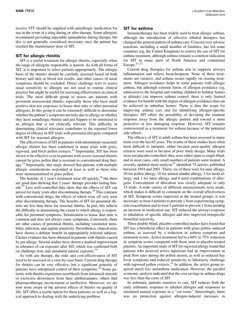
receive VIT should be supplied with antiallergic medication foruse in the event of a sting during or after therapy. Some allergistsrecommend providing injectable epinephrine during therapy, butthis is not generally considered necessary once the patient hasreached the maintenance dose of SIT.
SIT for allergic rhinitisSIT is a useful treatment for allergic rhinitis, especially when
the range of allergens responsible is narrow. As with all forms ofSIT, it is important to select patients appropriately. The allergicbasis of the rhinitis should be carefully assessed based on bothhistory and skin or blood test results, and other causes of nasalsymptoms should be excluded. Direct challenge tests to assessnasal sensitivity to allergen are not used in routine clinicalpractice but might be useful for assessing effectiveness in clinicaltrials. The most difficult group to assess are patients withpersistent nonseasonal rhinitis, especially those who have smallpositive skin test responses to house dust mite or other perennialallergens. In this group it can be extremely difficult to determinewhether the patient’s symptoms are truly due to allergy or whetherthey have nonallergic rhinitis and just happen to be sensitized toan allergen that is not clinically relevant. This difficulty indetermining clinical relevance contributes to the reported lowerdegree of efficacy in SIT trials with perennial allergens comparedwith SIT for seasonal allergies.
The effectiveness of SIT in patients with intermittent (seasonal)allergic rhinitis has been confirmed in many trials with grass,ragweed, and birch pollen extracts.14 Importantly, SIT has beenshown to be effective even in patients with severe seasonal rhinitiscaused by grass pollen that is resistant to conventional drug ther-apy.15 Importantly, this study showed that patients with multipleallergic sensitizations responded at least as well as those whowere monosensitized to grass pollen.
The benefits of 1 year’s treatment wear off quickly,16 but thereare good data showing that 3 years’ therapy provides lasting ben-efit.17 Less well-controlled data show that the effects of SIT canpersist for many years after discontinuing therapy.18 This contrastswith conventional drugs, the effects of which wear off very soonafter discontinuing therapy. The benefits of SIT for perennial rhi-nitis are less than those for seasonal rhinitis. In part, this reflectsthe difficulty in determining the extent to which allergy is respon-sible for perennial symptoms. Sensitization to house dust mite iscommon and does not always cause symptoms. Conversely, thereare other causes of perennial rhinitis, including vasomotor insta-bility, infection, and aspirin sensitivity. Nevertheless, clinical trialshave shown a definite benefit in appropriately selected subjects.Clearer evidence has been obtained in patients with rhinitis causedby pet allergy. Several studies have shown a marked improvementin tolerance of cat exposure after SIT, which was confirmed bothon challenge tests and simulated natural exposure.19
As with any therapy, the risks and cost-effectiveness of SITneed to be assessed on a case-by-case basis. Current drug therapyfor rhinitis can be very effective, but a significant minority ofpatients have suboptimal control of their symptoms.20 Some pa-tients with rhinitis experience nosebleeds from intranasal steroidsor excessive drowsiness from their antihistamines; others findpharmacotherapy inconvenient or ineffective. Moreover, we arenow more aware of the adverse effects of rhinitis on quality oflife. SIT offers a useful option for these patients, as well as a log-ical approach to dealing with the underlying problem.
SIT for asthmaImmunotherapy has been widely used to treat allergic asthma,
although the introduction of effective inhaled therapies haschanged the general pattern of asthma care. Concern over adversereactions, including a small number of fatalities, has led somecountries (eg, the United Kingdom) to restrict the use of SIT forasthma treatment, although asthma remains a common indicationfor SIT in many parts of North America and continentalEurope.21,22
Current drug therapies for asthma aim to suppress airwaysinflammation and relieve bronchospasm. None of these treat-ments are curative, and asthma recurs rapidly on ceasing treat-ment. Allergen avoidance helps in some patients with allergicasthma, but although extreme forms of allergen avoidance (eg,admission to the hospital and sending children to holiday homesat altitude) can improve asthma control, there is only limitedevidence for benefit with the degree of allergen avoidance that canbe achieved in suburban homes. There is thus the scope forimproving asthma care and for identifying allergen-specifictherapies. SIT offers the possibility of deviating the immuneresponse away from the allergic pattern and toward a moreprotective or less damaging response. However, SIT remainscontroversial as a treatment for asthma because of the potentialside effects.
The efficacy of SIT in adult asthma has been assessed in manytrials over the last 65 years. The results of these studies have oftenbeen difficult to interpret, either because poor-quality allergenextracts were used or because of poor study design. Many trialswere not placebo controlled; theywere either open or single blind,and in most cases, only small numbers of patients were treated. Arecently updated meta-analysis22 identified 75 articles publishedbetween 1954 and 2001. Thirty-six of these were for mite allergy,20 for pollen allergy, 10 for animal dander allergy, 2 for mold al-lergy, and 1 for latex allergy, and 6 used combinations of aller-gens. Concealment of allocation was clearly adequate in only15 trials. A wide variety of different measurements were made,which makes it difficult to comment on the overall effectivenessof SIT. Symptom scores improved in the treated groups; it wasnecessary to treat 4 patients to prevent 1 from experiencing symp-tom exacerbation and to treat 5 patients to prevent 1 from needingan increase in medication use. SIT reduced the airways responseto inhalation of specific allergen and also improved nonspecificbronchial reactivity.
Three double-blind, placebo-controlled studies have found thatSIT has a beneficial effect in patients with grass pollen–inducedasthma, as assessed by a reduction in asthma symptom andtreatment scores. Active treatment led to a 60% to 75% reductionin symptom scores compared with those seen in placebo-treatedpatients. An important study of SIT for ragweed allergy found thatpatients who received active injections had an improvement inpeak flow rates during the pollen season, as well as reduced hayfever symptoms and reduced sensitivity to laboratory challengewith ragweed pollen extracts.23 In addition, the active group re-quired much less antiasthma medication. However, the paralleleconomic analysis indicated that the cost savings in asthma drugswas less than the costs of SIT.
In asthmatic patients sensitive to cats, SIT reduces both theearly asthmatic response to inhaled allergen and responses tosimulated natural exposure in a ‘‘cat room.’’ Interestingly, therewas no protection against allergen-induced increases in
J ALLERGY CLIN IMMUNOL
FEBRUARY 2010
S308 FREW
nonspecific bronchial hyperresponsiveness, despite the cleardelay in onset of symptoms and an overall reduction in symptomsand peak flow recordings after exposure to cats. Others havefound reductions in both specific and nonspecific bronchialreactivity after SIT for cat allergy (measured by using inhalationchallenges with cat extract and histamine, respectively).24
The main drawback in using SIT to treat asthma is the risk ofserious adverse reactions. The vast majority of fatal reactions toSIT have occurred in patients with asthma, and although asthma isnot an absolute contraindication, it is clear that patients withunstable asthma should not be offered SIT, and caution should beexercised in anyone with an increased level of asthma symptomsor transiently reduced peak flow rates.
Comparison of SIT with other types of treatment forasthma
The majority of clinical trials of SIT for patients with asthmahave compared SIT either with untreated historical controlsubjects or with a matched placebo-treated group. To date, theeffectiveness of SIT in patients with asthma has rarely beencompared with conventional management (avoidance measuresand inhaled or oral antiasthma drugs). One recent study assessedSIT in asthmatic children receiving conventional drug therapyand found no additional benefit in patients who were alreadyreceiving optimal drug therapy.25 There were some significantflaws in the design of this study, and further work of this type isurgently needed.
Effects on natural history of allergic diseaseChildren often start with a limited range of allergic sensitivities
and progress over time to have IgE against a wider range of inhaledallergens. Treatment with SIT might limit this tendency to acquirenew sensitizations,26 although the clinical benefit of this preventiveeffect is not clear. A proportion of patients with allergic rhinitisdevelop asthma each year. This annual rate of progression hasbeen estimated at 5% in college students,27 but this is perhaps sur-prisingly an area of considerable ignorance. A number of long-term epidemiologic studies are now in progress under the auspicesof the International Study of Asthma and Allergies in Childhood,and these should eventually shed light on the rate of progressionat different ages and the extent of regional and international varia-tion. It has been suggested that SIT might modify the natural his-tory of asthma in children who are known to be atopic but have notyet developed asthma. Only limited data are available to supportthis proposition. In the key study a group of 205 children aged 6to 14 years without previously diagnosed asthma were treatedwith SIT for birch or grass pollen allergy in an open randomizeddesign. Three years after completing treatment, 45% of the un-treated group had asthma, whereas only 26% of the treated grouphad asthma. These results have been sustained out to 7 years aftercompleting therapy. Thus 4 children had to be treated to prevent1 case of asthma, which makes this an extremely effective ther-apy.28 SIT might also modify the progression of establishedasthma. An early open study with uncharacterized mixed allergenextracts supported this view, with about 70% of treated childrenlosing their asthma after 4 years’ therapy compared with about19% of untreated control subjects, a result that was sustained upto the age of 16 years. The proportion of children whose asthmawas severe at age 16 years was also much lower in the treated
group.29 By modern standards, this study was not well designed,and it needs repeating with modern SIT extracts in an up-to-datetrial design.
In contrast, there is no current evidence that SIT influences theevolution of established asthma in adults. Studies that haveinvestigated withdrawal of therapy have found rapid recurrence ofasthma symptoms, although rhinitis symptoms seem to showmuch more sustained relief after SIT.30
Thus SIT is a valid but controversial treatment for asthma.Although it seems entirely logical to try to treat allergic disordersby specifically suppressing the immune response to the triggeringagents, the critical issue is whether SIT in its present form is thebest option for managing patients with asthma. To assess thisproperly would require comparisons of best current SIT versusbest current drug therapy, with robust end points includingsymptoms, objective measures of lung function, evaluation ofcost/benefit ratios, safety, and quality of life. In vitro and in vivomeasures, such as skin test responses or allergen-specific IgG4measurements, are not sufficiently specific or sensitive to serveas surrogates for clinical efficacy. To date, there have been rela-tively few well-controlled studies of SIT in asthmatic subjects,but there is increasing evidence that SIT is beneficial in patientswith mite-induced and pollen-induced asthma. The clinical effi-cacy of SIT in adult asthmatic patients sensitive to cats or moldsis less certain, and no comparative studies with conventionaltreatment have been performed. Further clinical trials are indi-cated, particularly in patients with mild-to-moderate childhoodasthma and also in patients with atopic disease who have notyet had asthma but are at high risk of progression to asthma.
Safety of SITThe most obvious risk of SIT is that of provoking a systemic
allergic reaction. In the United Kingdom between 1957 and 1986,26 fatal reactions caused by SITwere reported to the Committeeon Safety ofMedicines.31 The indication for SITwas documentedin 17 of the fatal cases, 16 of whomwere in patients receiving SITto treat their asthma. Similarly, in the American Academy of Al-lergy, Asthma & Immunology inquiry into SIT-associated deaths,asthma appeared to be the cause of death in most of the fatalcases.32,33 In those cases in which asthma was not cited as a con-tributory factor, asthma statuswas not documented, whereas bron-chospasm was a feature of the clinical course of the fatalanaphylactic reactions. The incidence of systemic reactions in pa-tients receiving SIT for asthma varies between series and has beenreported to range from5% to35%.The central issue in using safetyas an end point is that we have to accept that all treatments carryrisks. Where differential risks exist between therapies, a morerisky therapy can only be justified if that therapy offers substantialadditional benefit over the safer therapy. The science of assessingrisk/benefit ratios is still in its infancy, and we have to recognizethat even when faced with the same facts, different patients andagencies can come to widely varying risk assessments. However,where possible, we should take steps to minimize the risks.
Separately, there is some concern about the use of immuno-modulatory treatments in patients with autoimmune disorders,immunodeficiency syndromes, or malignant disease. Althoughthere is no hard evidence that SIT is actually harmful to thesepatients, some clinicians feel uncomfortable about manipulatingthe immune system in such patients, not least because of the riskthat spontaneous and unrelated variations in the autoimmune
J ALLERGY CLIN IMMUNOL
VOLUME 125, NUMBER 2
FREW S309
disorder or cancer might be blamed on SIT. However, providedthe risks and benefits are weighed and discussed with the patient,SIT can be administered where the risk/benefit ratio is consideredto be in favor of treatment. Other medical contraindications to SITinclude the coexistence of significant cardiac disease that mightbe exacerbated by any adverse reactions to SIT. b-Blockers arealso contraindicated in patients receiving SIT. Although they donot increase the risk of adverse reactions, they will prevent thepatient from responding to the epinephrine that might be neededto treat adverse reactions to SIT. Where the indication for SIT isstrong, alternatives to b-blockers should be used so that the SITcan be given safely. Some clinics advise avoiding angiotensin-converting enzyme inhibitors because they can accentuate angi-oedema (angiotensin receptor antagonists [sartans] do not sharethis property).
Alternative forms of immunotherapyAlternative allergy practice covers 3 principal themes: the use
of unconventional diagnostic tests to seek causative agents fordiseases that everyone agrees are allergic in origin; the use ofunconventional therapies to treat allergic disease; and the diag-nosis and therapy of diseases that are not conventionally consid-ered to involve allergic mechanisms. Alternative immunotherapyregimens fall into the second of these categories, but the other 2areas fall outside the scope of this review.
Unconventional forms of immunotherapy include the use oftopical immunotherapy, enzyme-potentiated desensitization (EPD),and homeopathic desensitization.
Topical immunotherapy. High-dose topical immunother-apy regimens were used in the first half of the 20th century butsubsequently fell into disrepute. The last 20 years have seen arevival of interest in sublingual immunotherapy (SLIT). Theprecise mechanisms by which sublingual SIT works remainunclear. In mice locally administered allergen is taken up bymucosal dendritic cells and then presented to T cells together withIL-12, biasing the response toward a TH1 profile and away fromthe pro-IgE TH2 profile. It is less clear whether this mechanismcan suppress established allergic responses. In contrast, the im-munologic response to SLIT in human studies has been relativelymodest. Some changes have been found in skin sensitivity, butmost studies have not found any change in systemic parameters,such as specific IgE, specific IgG, or T-cell cytokine balance.
A body of evidence has accumulated from well-conductedclinical trials indicating that SLIT can be effective, with up to30% to 40% reductions in symptom scores and rescue medicationuse in patients with seasonal allergic rhinitis.34 Treatment regi-mens typically involve a rapid build-up phase followed by treat-ment given either daily or 3 times per week with rapidlydissolving tablets containing allergen extracts. Some preparationsare supplied in liquid form, with a calibrated dropper. A recentmeta-analysis of SLIT found 22 studies in which 979 patients re-ceived active therapy.34 Although many of these studies weresmall and inconclusive, the combined results indicate that SLITis indeed effective, with an estimated power of about two thirdsthat seen in comparable studies of injected SIT. Local side effectswere common but well tolerated.
In the grass pollen tablet trials about half the patients experi-enced some local irritation with the first dose. This was minor andgenerally did not require a reduction in subsequent doses. Abouthalf of those with initial side effects had lost these by the eighth
day of treatment; only 1 in 25 of all patients had continuing localside effects after 3 months treatment.35 Systemic side effects wererelatively rare, and none of the side effects were judged to be life-threatening. For perennial allergens, less trials data are avail-able,36 and only limited data are available in children, althoughthe most recent studies have been encouraging.37,38 Other formsof topical immunotherapy (oral and nasal) have limited efficacybut are associated with high levels of side effects.
SLIT is now being used routinely in some parts of Europe(especially Italy and France), but often the doses and regimensbeing prescribed are different from those used in the clinical trials.As performed in the published trials, SLIT involves giving 20 to400 times the total dose that would be given in a course of injectedSIT. There is no evidence that giving smaller doses sublinguallyhas any clinical effect.Overall, SLIT is likely towiden the scope ofSIT and bring in additional prescribers. As with all forms ofimmunotherapy, patient selection will be the key to ensuring thattherapy is targeted to those who are likely to benefit from it.
Some areas of uncertainty remain. For example, the optimumduration and durability of therapy have not been defined. Recentclinical trials have confirmed that the benefits of SLIT persist forthe first year after discontinuing treatment, but if they do persist,for how long do they persist? Based on experience with injectedSIT, manufacturers recommend that SLIT should be continued for3 years, although most clinical trials were short-term (6-12months). For seasonal allergens, most open-label use in clinicalpractice has been intermittent, starting 2 to 3 months before theseason and stopping at the end of the season. However, themanufacturer of the only licensed product recommends starting 4months before the first grass pollen season and continuingthroughout the year for 3 years. This has major implications fordirect costs and cost-effectiveness,39 and some supporting datawould be welcome.
The relative efficacy of SLIT and injected SIT has not beendetermined. The only published comparative studies were far toosmall to produce meaningful results.40,41 Based on the effect sizeseen in the meta-analyses,14,34 it seems likely that SLIT has be-tween 60% and 100% of the efficacy of injected SIT, althoughit is difficult to make a true comparison.
EPD. In EPD very small doses of allergens are given togetherwith the enzyme b-glucuronidase. The allergen doses are ap-proximately 0.1% of the doses used in conventional SIT, and sideeffects are apparently not encountered. The theory behind EPD isthat the b-glucuronidase enables the allergen to gain access to theimmune system more efficiently than is possible with conven-tional SIT. No convincing evidence has been published to supportthe efficacy of EPD.
Homeopathic desensitization. A detailed discussion ofthe principles underlying homeopathy lies outside the scope ofthis chapter. However, homeopathy espouses the concept thatdiseases can be treated with very small doses of substances thatcause similar symptoms. Some homeopathic remedies aremimicsof the disorder, whereas others use the actual material that triggersthe disorder. Thus homeopathic remedies for hay fever bear somesuperficial similarity to SIT. A systematic review of homeopathyhas concluded that homeopathy did appear to offer some benefitin patients with hay fever and cited trials of homeopathy in hayfever as an example of good practice in homeopathic research.42
However, a more recent, carefully controlled study of homoeopa-thy for house dust mite allergy found no evidence of any benefit inpatients with asthma.43
J ALLERGY CLIN IMMUNOL
FEBRUARY 2010
S310 FREW
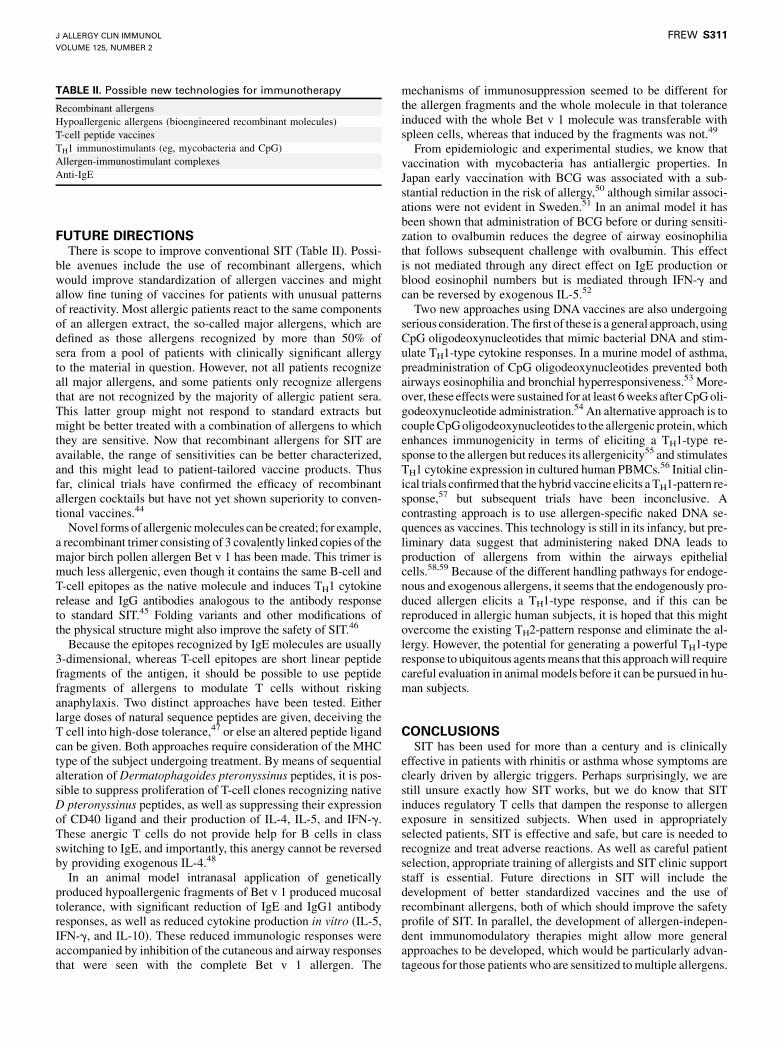
FUTURE DIRECTIONSThere is scope to improve conventional SIT (Table II). Possi-
ble avenues include the use of recombinant allergens, whichwould improve standardization of allergen vaccines and mightallow fine tuning of vaccines for patients with unusual patternsof reactivity. Most allergic patients react to the same componentsof an allergen extract, the so-called major allergens, which aredefined as those allergens recognized by more than 50% ofsera from a pool of patients with clinically significant allergyto the material in question. However, not all patients recognizeall major allergens, and some patients only recognize allergensthat are not recognized by the majority of allergic patient sera.This latter group might not respond to standard extracts butmight be better treated with a combination of allergens to whichthey are sensitive. Now that recombinant allergens for SIT areavailable, the range of sensitivities can be better characterized,and this might lead to patient-tailored vaccine products. Thusfar, clinical trials have confirmed the efficacy of recombinantallergen cocktails but have not yet shown superiority to conven-tional vaccines.44
Novel forms of allergenicmolecules can be created; for example,a recombinant trimer consisting of 3 covalently linked copies of themajor birch pollen allergen Bet v 1 has been made. This trimer ismuch less allergenic, even though it contains the same B-cell andT-cell epitopes as the native molecule and induces TH1 cytokinerelease and IgG antibodies analogous to the antibody responseto standard SIT.45 Folding variants and other modifications ofthe physical structure might also improve the safety of SIT.46
Because the epitopes recognized by IgE molecules are usually3-dimensional, whereas T-cell epitopes are short linear peptidefragments of the antigen, it should be possible to use peptidefragments of allergens to modulate T cells without riskinganaphylaxis. Two distinct approaches have been tested. Eitherlarge doses of natural sequence peptides are given, deceiving theT cell into high-dose tolerance,47 or else an altered peptide ligandcan be given. Both approaches require consideration of the MHCtype of the subject undergoing treatment. By means of sequentialalteration of Dermatophagoides pteronyssinus peptides, it is pos-sible to suppress proliferation of T-cell clones recognizing nativeD pteronyssinus peptides, as well as suppressing their expressionof CD40 ligand and their production of IL-4, IL-5, and IFN-g.These anergic T cells do not provide help for B cells in classswitching to IgE, and importantly, this anergy cannot be reversedby providing exogenous IL-4.48
In an animal model intranasal application of geneticallyproduced hypoallergenic fragments of Bet v 1 produced mucosaltolerance, with significant reduction of IgE and IgG1 antibodyresponses, as well as reduced cytokine production in vitro (IL-5,IFN-g, and IL-10). These reduced immunologic responses wereaccompanied by inhibition of the cutaneous and airway responsesthat were seen with the complete Bet v 1 allergen. The
mechanisms of immunosuppression seemed to be different forthe allergen fragments and the whole molecule in that toleranceinduced with the whole Bet v 1 molecule was transferable withspleen cells, whereas that induced by the fragments was not.49
From epidemiologic and experimental studies, we know thatvaccination with mycobacteria has antiallergic properties. InJapan early vaccination with BCG was associated with a sub-stantial reduction in the risk of allergy,50 although similar associ-ations were not evident in Sweden.51 In an animal model it hasbeen shown that administration of BCG before or during sensiti-zation to ovalbumin reduces the degree of airway eosinophiliathat follows subsequent challenge with ovalbumin. This effectis not mediated through any direct effect on IgE production orblood eosinophil numbers but is mediated through IFN-g andcan be reversed by exogenous IL-5.52
Two new approaches using DNA vaccines are also undergoingserious consideration. Thefirst of these is a general approach, usingCpG oligodeoxynucleotides that mimic bacterial DNA and stim-ulate TH1-type cytokine responses. In a murine model of asthma,preadministration of CpG oligodeoxynucleotides prevented bothairways eosinophilia and bronchial hyperresponsiveness.53 More-over, these effectswere sustained for at least 6weeks after CpGoli-godeoxynucleotide administration.54 An alternative approach is tocoupleCpGoligodeoxynucleotides to the allergenic protein,whichenhances immunogenicity in terms of eliciting a TH1-type re-sponse to the allergen but reduces its allergenicity55 and stimulatesTH1 cytokine expression in cultured human PBMCs.56 Initial clin-ical trials confirmed that the hybridvaccine elicits a TH1-pattern re-sponse,57 but subsequent trials have been inconclusive. Acontrasting approach is to use allergen-specific naked DNA se-quences as vaccines. This technology is still in its infancy, but pre-liminary data suggest that administering naked DNA leads toproduction of allergens from within the airways epithelialcells.58,59 Because of the different handling pathways for endoge-nous and exogenous allergens, it seems that the endogenously pro-duced allergen elicits a TH1-type response, and if this can bereproduced in allergic human subjects, it is hoped that this mightovercome the existing TH2-pattern response and eliminate the al-lergy. However, the potential for generating a powerful TH1-typeresponse to ubiquitous agentsmeans that this approachwill requirecareful evaluation in animal models before it can be pursued in hu-man subjects.
CONCLUSIONSSIT has been used for more than a century and is clinically
effective in patients with rhinitis or asthma whose symptoms areclearly driven by allergic triggers. Perhaps surprisingly, we arestill unsure exactly how SIT works, but we do know that SITinduces regulatory T cells that dampen the response to allergenexposure in sensitized subjects. When used in appropriatelyselected patients, SIT is effective and safe, but care is needed torecognize and treat adverse reactions. As well as careful patientselection, appropriate training of allergists and SIT clinic supportstaff is essential. Future directions in SIT will include thedevelopment of better standardized vaccines and the use ofrecombinant allergens, both of which should improve the safetyprofile of SIT. In parallel, the development of allergen-indepen-dent immunomodulatory therapies might allow more generalapproaches to be developed, which would be particularly advan-tageous for those patients who are sensitized tomultiple allergens.
TABLE II. Possible new technologies for immunotherapy
Recombinant allergensHypoallergenic allergens (bioengineered recombinant molecules)T-cell peptide vaccinesTH1 immunostimulants (eg, mycobacteria and CpG)Allergen-immunostimulant complexesAnti-IgE
J ALLERGY CLIN IMMUNOL
VOLUME 125, NUMBER 2
FREW S311
REFERENCES1. Freeman J. Vaccination against hay fever: report of results during the first three
years. Lancet 1914;1:1178.2. Creticos PS, Van Metre TE, Mardiney MR, Rosenberg GL, Norman PS, Adkinson
NF. Dose-response of IgE and IgG antibodies during ragweed immunotherapy.J Allergy Clin Immunol 1984;73:94-104.
3. Iliopoulos O, Proud D, Adkinson NF, Creticos PS, Norman PS, Kagey-Sobotka A,et al. Effects of immunotherapy on the early, late and rechallenge nasal reaction toprovocation with allergen: changes in inflammatory mediators and cells. J AllergyClin Immunol 1991;87:855-66.
4. Francis J, James L, Paraskevopoulos G, Wong C, Calderon MA, Durham SR, et al.Grass pollen immunotherapy: IL-10 induction and suppression of late responsesprecedes IgG4 inhibitory antibody activity. J Allergy Clin Immunol 2008;121:1120-5, e2.
5. Durham SR, Ying S, Varney VA, Jacobson MR, Sudderick RM, Mackay IS, KayAB, et al. Grass pollen immunotherapy inhibits allergen-induced infiltration ofCD41 T-lymphocytes and eosinophils in the nasal mucosa and increases the num-ber of cells expressing mRNA for interferon-gamma. J Allergy Clin Immunol1996;97:1356-65.
6. McHugh SM, Deighton J, Stewart AG, Lachmann PJ, Ewan PW. Bee venom im-munotherapy induces a shift in cytokine responses from a Th2 to a Th1 dominantpattern: comparison of rush and conventional immunotherapy. Clin Exp Allergy1995;25:828-38.
7. Ebner C, Siemann U, Bohle B, Willheim M, Wiedermann U, Schenk S, et al.Immunological changes during specific immunotherapy of grass pollen allergy:reduced lymphoproliferative responses to allergen and shift from Th2 to Th1 inT-cell clones specific for Phl p1, a major grass pollen allergen. Clin Exp Allergy1997;27:1007-15.
8. Jutel M, Akdis M, Blaser K, Akdis CA. Mechanisms of allergen specific immuno-therapy—T-cell tolerance and more. Allergy 2006;61:796-807.
9. Nasser SM, Ying S, Meng Q, Kay AB, Ewan PW. IL-10 levels increase in cutane-ous biopsies of patients undergoing wasp venom immunotherapy. Eur J Immunol2001;31:3704-13.
10. Akdis CA, Blesken T, Akdis M, Wuthrich B, Blaser K. Role of IL-10 in specificimmunotherapy. J Clin Invest 1998;102:98-106.
11. Bellinghausen I, Knop J, Saloga J. The role of IL-10 in the regulation of allergicimmune responses. Int Arch Allergy Appl Immunol 2001;126:97-101.
12. Graft DF. Insect sting allergy. Med Clin North Am 2006;90:211-32.13. Golden DB, Marsh DG, Freidhoff LR, Kwiterovich KA, Addison B, Kagey-
Sobotka A, et al. Natural history of hymenoptera venom sensitivity in adults.J Allergy Clin Immunol 1997;100:760-6.
14. Calderon MA, Alves B, Jacobson M, Hurwitz B, Sheikh A, Durham S. Allergeninjection immunotherapy for seasonal allergic rhinitis. Cochrane Database SystRev 2007; (1):CD001936.
15. Frew AJ, Powell RM, Corrigan CJ, Durham SR. Efficacy and safety of specific im-munotherapy with SQ allergen extract in treatment-resistant seasonal allergic rhi-noconjunctivitis. J Allergy Clin Immunol 2006;117:319-25.
16. Naclerio RM, Proud D, Moylan B, Balcer S, Freidhoff L, Kagey-Sobotka A, et al.A double blind study of the discontinuation of ragweed immunotherapy. J AllergyClin Immunol 1997;100:293-300.
17. Durham SR, Walker SM, Varga EM, Jacobson MR, O’Brien F, Nobel W, et al.Long-term clinical efficacy of grass pollen immunotherapy. N Engl J Med 1999;341:468-75.
18. Eng PA, Borer-Reinhold M, Heijnen IA, Gnehm HP. Twelve-year follow-up afterdiscontinuation of preseasonal grass pollen immunotherapy in childhood. Allergy2006;61:198-201.
19. Varney VA, Edwards J, Tabbah K, Brewster H, Mavroleon G, Frew AJ. Clinicalefficacy of specific immunotherapy to cat dander: a double blind placebo controlledtrial. Clin Exp Allergy 1997;27:860-7.
20. White PJ, Smith HE, Baker N, Davis W, Frew AJ. Symptom control in patientswith hay fever in UK general practice: how well are we doing and is there aneed for allergen immunotherapy? Clin Exp Allergy 1998;28:266-70.
21. Bousquet J, Lockey RF, Malling HJ. WHO position paper. Allergen immunother-apy: therapeutic vaccines for allergic disease. Allergy 1998;53(suppl):1-42.
22. Abramson M, Puy R, Weiner J. Immunotherapy in asthma: an updated systematicreview. Allergy 1999;54:1022-41.
23. Creticos PS, Reed CE, Norman PS, Khoury J, Adkinson NF, Buncher CR, et al.Ragweed immunotherapy in adult asthma. N Engl J Med 1996;334:501-6.
24. Lilja G, Sundin B, Graff-Lonnevig V, Hedlin G, Heilborn H, Norrlind K, et al. Im-munotherapy with partially purified and standardised animal dander extracts. IV.Effects of 2 years of treatment. J Allergy Clin Immunol 1989;83:37-44.
25. Adkinson NF, Eggleston PA, Eney D, Goldstein EO, Schuberth KC, Bacon JR,et al. A controlled trial of immunotherapy for asthma in allergic children. NEngl J Med 1997;336:324-31.
26. Des Roches A, Paradis L, Menardo JL, Bouges S, Daures JP, Bousquet J. Immu-notherapy with a standardised Dermatophagoides pteronyssinus extract. VI. Spe-cific immunotherapy prevents the onset of new sensitisations in children.J Allergy Clin Immunol 1997;99:450-3.
27. Horak F. Manifestation of allergic rhinitis in latent sensitised patients. A prospec-tive study. Arch Otorhinolaryngol 1985;242:242-9.
28. Niggemann B, Jacobsen L, Dreborg S, Ferdousi HA, Halken S, Host A, et al. Five-year follow-up on the PAT study: specific immunotherapy and long-term preven-tion of asthma in children. Allergy 2006;61:855-9.
29. Johnstone DE, Dutton A. The value of hyposensitization therapy for bronchialasthma in children. A 14 year study. Pediatrics 1968;42:793-802.
30. Shaikh WA. Immunotherapy vs inhaled budesonide in bronchial asthma: an openparallel comparative trial. Clin Exp Allergy 1997;27:1279-84.
31. Committee on the Safety of Medicines. CSM update: immunotherapy. BMJ 1986;293:948.
32. Stewart GE, Lockey RF. Systemic reactions from allergen immunotherapy. J Al-lergy Clin Immunol 1992;90:567-78.
33. Bernstein DI, Wanner M, Borish L, Liss GM. Twelve-year survey of fatal reactionsto allergen injections and skin testing: 1990-2001. J Allergy Clin Immunol 2004;113:1129-36.
34. Wilson DR, Lima MT, Durham SR. Sublingual immunotherapy for allergic rhini-tis: systematic review and meta-analysis. Allergy 2005;60:4-12.
35. Dahl R, Kapp A, Colombo G, de Monchy JG, Rak S, Emminger W, et al. Efficacyand safety of sublingual immunotherapy with once daily grass allergen tablets forseasonal allergic rhinoconjunctivitis. J Allergy Clin Immunol 2006;118:434-40.
36. Alvarez-Cuesta E, Berges-Gimeno P, Mancebo EG, Fernandez-Caldas E, Cuesta-Herranz J, Casanovas M. Sublingual immunotherapy with a standardised cat danderextract: evaluation of efficacy in a double-blind placebo-controlled study [publishederratum in Allergy 2007;62:1100]. Allergy 2007;62:810-7.
37. Di Rienzo V, Marcucci F, Puccinelli P, Parmiani S, Frati F, Sensi L, et al. Long-last-ing effect of sublingual immunotherapy in children with asthma due to house dustmite: a 10 year prospective study. Clin Exp Allergy 2003;33:206-10.
38. Bufe A, Ziegler-Kirbach E, Stoeckmann E, Heidemann P, Gehlhar K, Holland-LetzT, et al. Efficacy of sublingual swallow immunotherapy in children with severegrass pollen allergic symptoms: a double-blind placebo-controlled study. Allergy2004;59:498-504.
39. Bachert C, Vestenbaek U, Christensen J, Griffiths UK, Poulsen PB. Cost-effective-ness of grass allergen tablet (GRAZAX) for the prevention of seasonal grass-pol-len-induced rhinoconjunctivitis—a Northern European perspective. Clin ExpAllergy 2007;37:772-9.
40. Khinchi MS, Poulsen LK, Carat F, Andre C, Hansen AB, Malling HJ. Clinical ef-ficacy of sublingual and subcutaneous birch pollen allergen-specific immunother-apy: a randomized, placebo-controlled, double-blind, double-dummy study.Allergy 2004;59:45-53.
41. Mauroa M, Russelloa M, Incorvaiab C, Gazzolaa GB, Di Carac G, Frati F. Com-parison of efficacy, safety and immunologic effects of subcutaneous and sublingualimmunotherapy in birch pollinosis: a randomized study. Allerg Immunol (Paris)2007;39:119-22.
42. Reilly DT, Taylor MA, McSharry C, Aitchison T. Is homeopathy a placebo re-sponse? Controlled trial of homeopathic potency with pollen in hay fever as amodel. Lancet 1986;2:881-6.
43. Lewith GT, Watkins AD, Hyland ME, Shaw S, Broomfield JA, Dolan G, et al. Useof ultramolecular potencies of allergen to treat asthmatic people allergic to housedust mite: double blind randomised controlled clinical trial. BMJ 2002;324:520.
44. Pauli G, Larsen TH, Rak S, Horak F, Pastorello E, Valenta R, et al. Efficacy of re-combinant birch pollen vaccine for the treatment of birch-allergic rhinoconjuncti-vitis. J Allergy Clin Immunol 2008;122:951-60.
45. Valenta R, Niederberger V. Recombinant allergens for immunotherapy. J AllergyClin Immunol 2007;119:826-30.
46. Purohit A, Niederberger V, Kronqvist M, Horak F, Gronneberg R, Suck R, et al.Clinical effects of immunotherapy with genetically modified recombinant birchpollen Bet v1 derivatives. Clin Exp Allergy 2008;38:1514-25.
47. O’Hehir RE, Yssel H, Verma S, de Vries JE, Spits H, Lamb JR. Clonal analysis ofdifferential lymphokine production in peptide and superantigen-induced T-cell an-ergy. Int Immunol 1991;3:819-26.
48. Fasler S, Aversa G, de Vries JE, Yssel H. Antagonistic peptides specifically inhibitproliferation, cytokine production, CD40L expression and help for IgE synthesis byDer p 1-specific human T-cell clones. J Allergy Clin Immunol 1998;101:521-30.
49. Wiedermann U, Herz U, Baier K, Vrtala S, Neuhaus-Steinmetz U, Bohle B, et al.Intranasal treatment with a recombinant hypoallergenic derivative of the majorbirch pollen allergen Bet v 1 prevents allergic sensitization and airway inflamma-tion in mice. Int Arch Allergy Immunol 2001;126:68-77.
50. Shirakawa T, Enomoto T, Shimazu SI, Hopkin JM. The inverse associationbetween tuberculin responses and atopic disorder. Science 1997;275:77-9.
J ALLERGY CLIN IMMUNOL
FEBRUARY 2010
S312 FREW

51. Strannegaard IL, Larsson LO, Wennergren G, Strannegaard O. Prevalence ofallergy in children in relation to prior BCG vaccination and infection with atypicalmycobacteria. Allergy 1998;53:249-54.
52. Erb KJ, Holloway JW, Sobeck A, Moll H, Le Gros G. Infection of mice with My-cobacterium bovis-BCG suppresses allergen-induced airways eosinophilia. J ExpMed 1998;187:561-9.
53. Kline JN, Waldschmidt TJ, Businga TR, Lemish JE, Weinstock JV, Thorne PS,et al. Modulation of airway inflammation by CpG oligodeoxynucleotides in a mu-rine model of asthma. J Immunol 1998;160:2555-9.
54. Sur S, Wild JS, Choudury BK, Sur N, Alam R, Klinman DM. Long-term preven-tion of allergic lung inflammation in a mouse model of asthma by CpG oligodeox-ynucleotides. J Immunol 1999;162:6284-93.
55. Tighe H, Takabayashi K, Schwartz D, van Nest G, Tuck S, Eiden JJ, et al. Conjugationof immunostimulatoryDNA to the short ragweed allergenAmb a 1 enhances its immu-nogenicity and reduces its allergenicity. J Allergy Clin Immunol 2000;106:124-34.
56. Marshall JD, Abtahi S, Eiden JJ, Tuck S, Milley R, Haycock F, et al. Immunosti-mulatory sequence DNA linked to the Amb a 1 allergen promotes Th1 cytokineexpression while downregulating Th2 cytokine expression in PBMCs from humanpatients with ragweed allergy. J Allergy Clin Immunol 2001;108:191-7.
57. Creticos PS, Eiden JJ, Broide D, Balcer-Whaley SL, Schroeder JT, KhattignavongA, et al. Immunotherapy with immunostimulatory oligonucleotides linked to puri-fied ragweed Amb a 1 allergen: effects on antibody production, nasal allergenprovocation and ragweed seasonal rhinitis. J Allergy Clin Immunol 2002;109:743-4.
58. Hsu CH, Chua KY, Tao MH, Lai YL, Wu HD, Huang SK, et al. Immunoprophy-laxis of allergen-induced IgE synthesis and airway hyperresponsiveness in vivo bygenetic immunisation. Nat Med 1996;2:540-4.
59. Hartl A, Kiesslich J, Weiss R, Bernhaupt A, Mostbock S, Scheiblhofer S, et al. Im-mune responses after immunisation with plasmid DNA encoding Bet v 1, the majorallergen of birch pollen. J Allergy Clin Immunol 1999;103:107-13.
J ALLERGY CLIN IMMUNOL
VOLUME 125, NUMBER 2
FREW S313
Immunomodulator therapy: Monoclonal antibodies, fusionproteins, cytokines, and immunoglobulins
Susan J. Lee, MD,a,b Javier Chinen, MD, PhD,c and Arthur Kavanaugh, MDa San Diego, Calif, and Houston, Tex
The immune system consists of a diverse array ofimmunocompetent cells and inflammatory mediators that existin complex networks. These components interact throughcascades and feedback circuits, maintaining physiologicinflammation (eg, tissue repair) and immunosurveillance. Invarious autoimmune and allergic diseases, a foreign antigen orautoantigen might upset this fine balance, leading todysregulated immunity, persistent inflammation, and ultimatelypathologic sequelae. In recent years, there has been tremendousprogress delineating the specific components of the immunesystem that contribute to various aspects of normal immunityand specific disease states. With this greater understanding ofpathogenesis coupled with advances in biotechnology, manyimmunomodulatory agents commonly called ‘‘biologic agents’’have been introduced into the clinic for the treatment of variousconditions, including immune globulins and cytokines. The 2most common classes of approved biologic agents are mAbs andfusion proteins with exquisite specificity. These agents have thepotential both to optimize outcomes through more thoroughmodulation of specific parts of the dysregulated immuneresponse and to minimize toxicity compared with less specificmethods of immunosuppression. (J Allergy Clin Immunol2010;125:S314-23.)
Key words: Monoclonal antibodies, fusion proteins, immunoglobu-lins, cytokines, autoimmunity
Biologic agents can work through several mechanisms. Thesimplest would be inhibition of the function of a target moleculeby binding to it, thereby preventing ligation with its counter-receptor and downstream effects. Potential targets include (1)lineage- or activation status–specificmolecules on B cells, T cells,and other immunocompetent cells; (2) soluble inflammatorymediators, such as cytokines, chemokines, complement proteins,enzymes, and immunoglobulin molecules; and (3) surface recep-tors for thesemediators. Biologic agents can alter cell populationsby engaging effector functions, including the complement cas-cade and antibody-dependent cellular cytotoxicity; of note, manymAbs and fusion proteins possess functional IgG Fc pieces. Cell
depletion can also be induced by apoptosis subsequent to ligationof appropriate targets. Small-molecular-weight immunomodula-tors, such as glucocorticoids, are reviewed in Chapter 16.
MONOCLONAL ANTIBODIESMonoclonal antibodies to human targets can be generated
either in other species, such as mice, or through recombinantengineering (Fig 1). With chimeric mAbs, the variable region of amurine mAb is fused to the Fc piece of a human IgG molecule.The resulting construct is approximately one quarter murine.For humanized mAbs, only the complementarity determining re-gions from the original murine mAb are retained, resulting in aconstruct that is approximately 95% human. There are a numberof approaches to create human mAbs to human targets, includingimmunizing human/severe combined immunodeficient murinechimeras, using EBV-transformed human B cells, and repertoirecloning, in which target antigen is used to capture human comple-mentarity determining regions generated from vast human cDNAlibraries, with the mAb then generated from there. Proteins suchas mAbs can have residues of polyethylene glycol added. Thisprocess, called pegylation, enhances the half-life of the nativeprotein by reducing its renal and cellular clearance after adminis-tration. Although even fully human proteins can be immunogenic,in general, the more human a construct, the less immunogenic.Pegylation might further reduce antigenicity and immunogenicityof the native protein. Immunogenicity can develop to moleculeswith amino acid sequences identical to human sequences relatedto factors such as differences in patterns of glycosylation. In ad-dition, immunogenicity to mAbs can be anti-idiotypic. Other fac-tors affecting immunogenicity include route of administration(intravenous vs subcutaneous), treatment paradigm (continuousvs intermittent), and concurrent use of immunosuppressivetherapy.
Standard nomenclature for mAbs identifies their source withthe last 4 or 5 letters: -omab, murine; -ximab, chimeric; -zumab,
From athe Division of Rheumatology, Allergy, and Immunology, the University of Cal-ifornia, San Diego; bthe Veterans Affairs San Diego Healthcare System, San Diego;and cthe Allergy and Immunology Section, Department of Pediatrics, Baylor Collegeof Medicine, Houston.
Disclosure of potential conflict of interest: A. Kavanaugh has received research supportfromAmgen, UCB, Abbott, Centocor, Roche, BMS, Genentech, and Biogen Idec. S. J.Lee and J. Chinen have declared that they have no conflict of interest.
Received for publication June 3, 2009; revised July 31, 2009; accepted for publicationAugust 3, 2009.
Reprint requests: Arthur Kavanaugh, MD, the University of California, San Diego, 9500Gilman Dr, Mail Code 0943, La Jolla, CA 92093-0943. E-mail: [email protected].
0091-6749/$36.00! 2010 American Academy of Allergy, Asthma & Immunologydoi:10.1016/j.jaci.2009.08.018
Abbreviations usedAS: Ankylosing spondylitis
CHF: Congestive heart failureCTLA-4: Cytotoxic T lymphocyte–associated antigen 4DMARD: Disease-modifying antirheumatic drug
FDA: US Food and Drug AdministrationICAM: Intercellular adhesion moleculeIL-1Ra: IL-1 receptor antagonistIVIG: Intravenous immunoglobulinLFA: Lymphocyte function–associated antigenMS: Multiple sclerosis
PML: Progressive multifocal leukoencephalopathyPsA: Psoriatic arthritisRA: Rheumatoid arthritis
SCIG: Subcutaneous immunoglobulinSLE: Systemic lupus erythematosus
S314
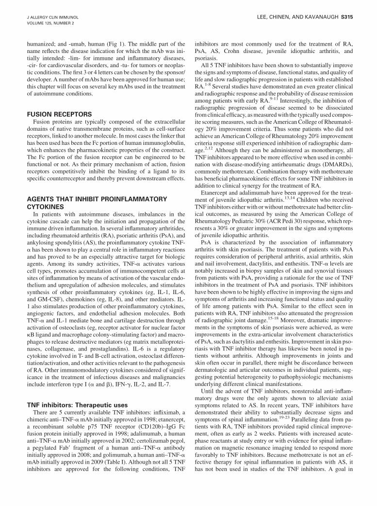
humanized; and -umab, human (Fig 1). The middle part of thename reflects the disease indication for which the mAb was ini-tially intended: -lim- for immune and inflammatory diseases,-cir- for cardiovascular disorders, and -tu- for tumors or neoplas-tic conditions. The first 3 or 4 letters can be chosen by the sponsor/developer. A number of mAbs have been approved for human use;this chapter will focus on several key mAbs used in the treatmentof autoimmune conditions.
FUSION RECEPTORSFusion proteins are typically composed of the extracellular
domains of native transmembrane proteins, such as cell-surfacereceptors, linked to another molecule. Inmost cases the linker thathas been used has been the Fc portion of human immunoglobulin,which enhances the pharmacokinetic properties of the construct.The Fc portion of the fusion receptor can be engineered to befunctional or not. As their primary mechanism of action, fusionreceptors competitively inhibit the binding of a ligand to itsspecific counterreceptor and thereby prevent downstream effects.
AGENTS THAT INHIBIT PROINFLAMMATORYCYTOKINES
In patients with autoimmune diseases, imbalances in thecytokine cascade can help the initiation and propagation of theimmune driven inflammation. In several inflammatory arthritides,including rheumatoid arthritis (RA), psoriatic arthritis (PsA), andankylosing spondylitis (AS), the proinflammatory cytokine TNF-a has been shown to play a central role in inflammatory reactionsand has proved to be an especially attractive target for biologicagents. Among its sundry activities, TNF-a activates variouscell types, promotes accumulation of immunocompetent cells atsites of inflammation by means of activation of the vascular endo-thelium and upregulation of adhesion molecules, and stimulatessynthesis of other proinflammatory cytokines (eg, IL-1, IL-6,and GM-CSF), chemokines (eg, IL-8), and other mediators. IL-1 also stimulates production of other proinflammatory cytokines,angiogenic factors, and endothelial adhesion molecules. BothTNF-a and IL-1 mediate bone and cartilage destruction throughactivation of osteoclasts (eg, receptor activator for nuclear factorkB ligand andmacrophage colony-stimulating factor) andmacro-phages to release destructive mediators (eg matrix metalloprotei-nases, collagenase, and prostaglandins). IL-6 is a regulatorycytokine involved in T- and B-cell activation, osteoclast differen-tiation/activation, and other activities relevant to the pathogenesisof RA. Other immunomodulatory cytokines considered of signif-icance in the treatment of infectious diseases and malignanciesinclude interferon type I (a and b), IFN-g, IL-2, and IL-7.
TNF inhibitors: Therapeutic usesThere are 5 currently available TNF inhibitors: infliximab, a
chimeric anti–TNF-amAb initially approved in 1998; etanercept,a recombinant soluble p75 TNF receptor (CD120b)–IgG Fcfusion protein initially approved in 1998; adalimumab, a humananti–TNF-amAb initially approved in 2002; certolizumab pegol,a pegylated Fab9 fragment of a human anti–TNF-a antibodyinitially approved in 2008; and golimumab, a human anti–TNF-amAb initially approved in 2009 (Table I). Although not all 5 TNFinhibitors are approved for the following conditions, TNF
inhibitors are most commonly used for the treatment of RA,PsA, AS, Crohn disease, juvenile idiopathic arthritis, andpsoriasis.
All 5 TNF inhibitors have been shown to substantially improvethe signs and symptoms of disease, functional status, and quality oflife and slow radiographic progression in patients with establishedRA.1-8 Several studies have demonstrated an even greater clinicaland radiographic response and the probability of disease remissionamong patients with early RA.9-11 Interestingly, the inhibition ofradiographic progression of disease seemed to be dissociatedfromclinical efficacy, asmeasuredwith the typically used compos-ite scoringmeasures, such as the American College of Rheumatol-ogy 20% improvement criteria. Thus some patients who did notachieve anAmericanCollege ofRheumatology 20% improvementcriteria response still experienced inhibition of radiographic dam-age.2,12 Although they can be administered as monotherapy, allTNF inhibitors appeared to bemore effectivewhen used in combi-nation with disease-modifying antirheumatic drugs (DMARDs),commonly methotrexate. Combination therapy with methotrexatehas beneficial pharmacokinetic effects for some TNF inhibitors inaddition to clinical synergy for the treatment of RA.
Etanercept and adalimumab have been approved for the treat-ment of juvenile idiopathic arthritis.13,14 Children who receivedTNF inhibitors eitherwith orwithoutmethotrexate had better clin-ical outcomes, as measured by using the American College ofRheumatology Pediatric 30% (ACRPedi 30) response, which rep-resents a 30% or greater improvement in the signs and symptomsof juvenile idiopathic arthritis.
PsA is characterized by the association of inflammatoryarthritis with skin psoriasis. The treatment of patients with PsArequires consideration of peripheral arthritis, axial arthritis, skinand nail involvement, dactylitis, and enthesitis. TNF-a levels arenotably increased in biopsy samples of skin and synovial tissuesfrom patients with PsA, providing a rationale for the use of TNFinhibitors in the treatment of PsA and psoriasis. TNF inhibitorshave been shown to be highly effective in improving the signs andsymptoms of arthritis and increasing functional status and qualityof life among patients with PsA. Similar to the effect seen inpatients with RA, TNF inhibitors also attenuated the progressionof radiographic joint damage.15-18 Moreover, dramatic improve-ments in the symptoms of skin psoriasis were achieved, as wereimprovements in the extra-articular involvement characteristicsof PsA, such as dactylitis and enthesitis. Improvement in skin pso-riasis with TNF inhibitor therapy has likewise been noted in pa-tients without arthritis. Although improvements in joints andskin often occur in parallel, there might be discordance betweendermatologic and articular outcomes in individual patients, sug-gesting potential heterogeneity to pathophysiologic mechanismsunderlying different clinical manifestations.
Until the advent of TNF inhibitors, nonsteroidal anti-inflam-matory drugs were the only agents shown to alleviate axialsymptoms related to AS. In recent years, TNF inhibitors havedemonstrated their ability to substantially decrease signs andsymptoms of spinal inflammation.19-23 Paralleling data from pa-tients with RA, TNF inhibitors provided rapid clinical improve-ment, often as early as 2 weeks. Patients with increased acute-phase reactants at study entry or with evidence for spinal inflam-mation on magnetic resonance imaging tended to respond morefavorably to TNF inhibitors. Because methotrexate is not an ef-fective therapy for spinal inflammation in patients with AS, ithas not been used in studies of the TNF inhibitors. A goal in
J ALLERGY CLIN IMMUNOL
VOLUME 125, NUMBER 2
LEE, CHINEN, AND KAVANAUGH S315
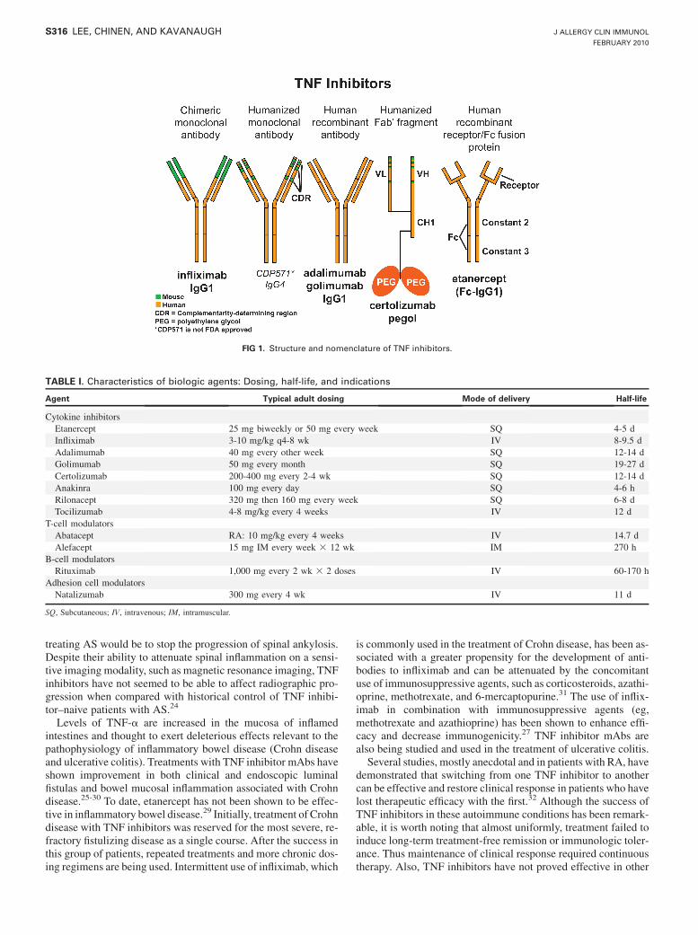
treating AS would be to stop the progression of spinal ankylosis.Despite their ability to attenuate spinal inflammation on a sensi-tive imaging modality, such as magnetic resonance imaging, TNFinhibitors have not seemed to be able to affect radiographic pro-gression when compared with historical control of TNF inhibi-tor–naive patients with AS.24
Levels of TNF-a are increased in the mucosa of inflamedintestines and thought to exert deleterious effects relevant to thepathophysiology of inflammatory bowel disease (Crohn diseaseand ulcerative colitis). Treatments with TNF inhibitor mAbs haveshown improvement in both clinical and endoscopic luminalfistulas and bowel mucosal inflammation associated with Crohndisease.25-30 To date, etanercept has not been shown to be effec-tive in inflammatory bowel disease.29 Initially, treatment of Crohndisease with TNF inhibitors was reserved for the most severe, re-fractory fistulizing disease as a single course. After the success inthis group of patients, repeated treatments and more chronic dos-ing regimens are being used. Intermittent use of infliximab, which
is commonly used in the treatment of Crohn disease, has been as-sociated with a greater propensity for the development of anti-bodies to infliximab and can be attenuated by the concomitantuse of immunosuppressive agents, such as corticosteroids, azathi-oprine, methotrexate, and 6-mercaptopurine.31 The use of inflix-imab in combination with immunosuppressive agents (eg,methotrexate and azathioprine) has been shown to enhance effi-cacy and decrease immunogenicity.27 TNF inhibitor mAbs arealso being studied and used in the treatment of ulcerative colitis.
Several studies, mostly anecdotal and in patients with RA, havedemonstrated that switching from one TNF inhibitor to anothercan be effective and restore clinical response in patients who havelost therapeutic efficacy with the first.32 Although the success ofTNF inhibitors in these autoimmune conditions has been remark-able, it is worth noting that almost uniformly, treatment failed toinduce long-term treatment-free remission or immunologic toler-ance. Thus maintenance of clinical response required continuoustherapy. Also, TNF inhibitors have not proved effective in other
TABLE I. Characteristics of biologic agents: Dosing, half-life, and indications
Agent Typical adult dosing Mode of delivery Half-life
Cytokine inhibitorsEtanercept 25 mg biweekly or 50 mg every week SQ 4-5 dInfliximab 3-10 mg/kg q4-8 wk IV 8-9.5 dAdalimumab 40 mg every other week SQ 12-14 dGolimumab 50 mg every month SQ 19-27 dCertolizumab 200-400 mg every 2-4 wk SQ 12-14 dAnakinra 100 mg every day SQ 4-6 hRilonacept 320 mg then 160 mg every week SQ 6-8 dTocilizumab 4-8 mg/kg every 4 weeks IV 12 d
T-cell modulatorsAbatacept RA: 10 mg/kg every 4 weeks IV 14.7 dAlefacept 15 mg IM every week 3 12 wk IM 270 h
B-cell modulatorsRituximab 1,000 mg every 2 wk 3 2 doses IV 60-170 h
Adhesion cell modulatorsNatalizumab 300 mg every 4 wk IV 11 d
SQ, Subcutaneous; IV, intravenous; IM, intramuscular.
FIG 1. Structure and nomenclature of TNF inhibitors.
J ALLERGY CLIN IMMUNOL
FEBRUARY 2010
S316 LEE, CHINEN, AND KAVANAUGH
conditions, including several wherein there was pathophysiologicevidence for a role for this cytokine in the disease process. Amongautoimmune conditions, TNF inhibitor therapy has been notablyineffective to date in patients with Sj!ogren syndrome and severalforms of vasculitis, including Wegener granulomatosis and poly-myalgia rheumatica/temporal arteritis. With regard to congestiveheart failure (CHF), data from animal models of ischemic cardi-omyopathy implicated TNF as a key mediator of deterioratingcardiac function and hence an attractive target. However, TNF in-hibitors have failed to improve symptoms in patients with CHF inclinical trials and sometimes resulted in worsened clinical out-come. Although limited, there were studies on TNF inhibitorsthat showed negative results in patients with multiple sclerosis(MS), along with anecdotal reports of the development or worsen-ing of demyelinating symptoms among patients with RA treatedwith these agents. TNF inhibitors are still being actively investi-gated in a variety of other diseases.
TNF inhibitors: Safety considerationsIn general, TNF inhibitors have been well tolerated in clinical
trials. In vitro studies suggested that TNF inhibitors selectivelydecrease proinflammatory cytokine levels while preserving boththe humoral and cell-mediated arms of the immune response.However, a number of relevant safety issues regarding the useof TNF inhibitors have emerged in postmarketing pharmacovigi-lance assessments.33,34 Adverse events associated with TNF in-hibitors can be broadly classified as target/class related or agentrelated. Target-related adverse events include those potentially at-tributable to the immunosuppression inherent in blocking a keycomponent of the immune system, such as an inflammatory cyto-kine; this would include increased susceptibility to infections andmalignancies. In addition, specific inhibition of TNF might pre-dispose patients to increased susceptibility to tuberculosis, auto-antibody production, hepatotoxicity, demyelinating disease, andclinical worsening of CHF. Agent-related adverse events, suchas allergic reactions and antigenicity, are idiosyncratic reactionsthat relate to the particular agent used.
Safety data from clinical trials and registries have shown asmall but consistent increase in infections among TNF inhibitor–treated patients comparedwith those treatedwith DMARDs,mostcommonly methotrexate. Generally, the risk of serious infectionswas not substantially greater, with relative risks ranging from 0 to2. The risk of infection with TNF inhibitors increased signifi-cantly when combined with other biologic agents. For example,combination therapy with the TNF inhibitor etanercept and theIL-1 receptor antagonist (IL-1Ra) anakinra resulted in a higherrate of serious infections in patients with RA, despite the failureto achieve any additive clinical benefit. Data, particularly frompharmacovigilance, have noted a number of opportunistic infec-tions (eg, listeriosis, histoplasmosis, and coccidioidomycosis)among those patients treated with TNF inhibitors. Because ofthe increased baseline risk of infection among patients withRA, without a control group, it is difficult to ascertain the excessinfection risk specifically attributable to TNF inhibitors in thesepatients. Another potential sequela of immunosuppression is ma-lignancy. With a few notable exceptions, the bulk of the data todate do not support an increased risk of solid tumors related toTNF inhibitor therapy. However, greater numbers of hematologicmalignancies, particularly non-Hodgkin lymphoma, have beenobserved in some registries. Complicating the assessment of the
risk attributable to therapy is the increased baseline risk of lym-phoma among patients with RA, especially among those withhigher disease activity. This introduces bias toward observingcases among patients treated with TNF inhibitors as the most se-vere, and patients with active RA are often the most common typeof patients treated. The relative effect of dose and duration of ther-apy and host factors, such as comorbidities, relevant genetic pol-ymorphisms, and concomitant medications, on the risk ofinfections and malignancy remains incompletely defined. Be-cause of potential immunosuppression, vaccination with live vac-cines is not recommended while patients are receiving TNFinhibitors.
In addition, inhibition of TNF might predispose patients to avariety of untoward effects that seem to be specific to inhibition ofthe TNF molecule. There are a fair amount of animal and ex vivodata supporting the important role played by TNF in controllingtuberculosis. In contrast to typical presentation of acute tubercu-losis as pneumonia, about half of the cases of tuberculosis relatedto TNF inhibitors presented as extrapulmonary or disseminatedtuberculosis. The majority of these tuberculosis cases appear tobe reactivation of latent tuberculosis, with infection occurringwithin the first few months of therapy; however, newly acquiredcases have been well described. The incidence of cases mightbe greater with the mAb TNF inhibitors than with the fusion pro-tein inhibitor. Fortunately, screening for latent tuberculosis beforeinitiating TNF inhibitor therapy has been an effective strategy,with a reduction in incidence of new tuberculosis cases by ap-proximately 85%. Latent tuberculosis can be screened by usingeither a tuberculin skin test with purified protein derivative orex vivo tests that quantify IFN-g release from sensitized lympho-cytes in blood incubated with tuberculosis antigens. Treatmentwith TNF inhibitors has also been associated with developmentof autoantibodies. Although the mechanism of this is unknown,it does not appear to result from inhibition of TNF itself, perhapsthrough induction of apoptosis. The autoantibodies typically gen-erated include the antinuclear antibody (which develops in abouthalf of patients with RA treated with TNF inhibitors), antibodiesto double-strandedDNA (which develop in approximately 10% to15% of patients treated with TNF inhibitors), and anticardiolipinantibodies. Although rare, progression to a lupus-like illness canoccur in patients treated with TNF inhibitors. Also, mild-to-mod-erate increases in liver function test results (generally <3 timesthe upper limit of normal) have been observed with TNF inhibi-tors. Many of these cases were confounded by concomitant useof potentially hepatotoxic drugs and underlying medical condi-tions. However, in light of the occurrence of liver failure of un-identifiable cause in several cases, clinicians should be aware ofthese rare events and consider monitoring liver function tests.Lastly, several cases of MS and other demyelinating conditionshave been identified among patients treated with TNF inhibitors,although the true effect of TNF inhibitors on the development ofMS remains undefined.
Despite their shared ability to inhibit TNF, there are somenotable differences between the 5 approved TNF inhibitors.Infliximab, adalimumab, golimumab, and certolizumab areIgG1 mAbs that are specific for TNF-a; etanercept is a fusionprotein of the type II TNF receptor and binds both TNF-a andlymphotoxin a (also known as TNF-b). The clinical relevance ofthis distinction is unknown. In addition, the binding characteris-tics of themAbs and the fusion protein differ slightly. Although allagents bind soluble TNFwith high affinity, themAbs have slightly
J ALLERGY CLIN IMMUNOL
VOLUME 125, NUMBER 2
LEE, CHINEN, AND KAVANAUGH S317
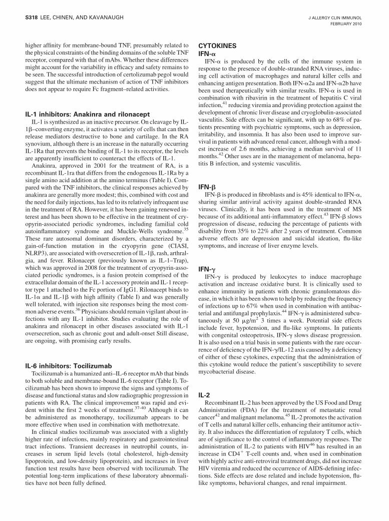
higher affinity for membrane-bound TNF, presumably related tothe physical constraints of the binding domains of the soluble TNFreceptor, compared with that of mAbs. Whether these differencesmight account for the variability in efficacy and safety remains tobe seen. The successful introduction of certolizumab pegol wouldsuggest that the ultimate mechanism of action of TNF inhibitorsdoes not appear to require Fc fragment–related activities.
IL-1 inhibitors: Anakinra and rilonaceptIL-1 is synthesized as an inactive precursor. On cleavage by IL-
1b–converting enzyme, it activates a variety of cells that can thenrelease mediators destructive to bone and cartilage. In the RAsynovium, although there is an increase in the naturally occurringIL-1Ra that prevents the binding of IL-1 to its receptor, the levelsare apparently insufficient to counteract the effects of IL-1.
Anakinra, approved in 2001 for the treatment of RA, is arecombinant IL-1ra that differs from the endogenous IL-1Ra by asingle amino acid addition at the amino terminus (Table I). Com-pared with the TNF inhibitors, the clinical responses achieved byanakinra are generally more modest; this, combined with cost andthe need for daily injections, has led to its relatively infrequent usein the treatment of RA. However, it has been gaining renewed in-terest and has been shown to be effective in the treatment of cry-opyrin-associated periodic syndromes, including familial coldautoinflammatory syndrome and Muckle-Wells syndrome.35
These rare autosomal dominant disorders, characterized by again-of-function mutation in the cryopyrin gene (CIASI,NLRP3), are associated with oversecretion of IL-1b, rash, arthral-gia, and fever. Rilonacept (previously known as IL-1–Trap),which was approved in 2008 for the treatment of cryopyrin-asso-ciated periodic syndromes, is a fusion protein comprised of theextracellular domain of the IL-1 accessory protein and IL-1 recep-tor type 1 attached to the Fc portion of IgG1. Rilonacept binds toIL-1a and IL-1b with high affinity (Table I) and was generallywell tolerated, with injection site responses being the most com-mon adverse events.36 Physicians should remain vigilant about in-fections with any IL-1 inhibitor. Studies evaluating the role ofanakinra and rilonacept in other diseases associated with IL-1oversecretion, such as chronic gout and adult-onset Still disease,are ongoing, with promising early results.
IL-6 inhibitors: TocilizumabTocilizumab is a humanized anti–IL-6 receptor mAb that binds
to both soluble and membrane-bound IL-6 receptor (Table I). To-cilizumab has been shown to improve the signs and symptoms ofdisease and functional status and slow radiographic progression inpatients with RA. The clinical improvement was rapid and evi-dent within the first 2 weeks of treatment.37-40 Although it canbe administered as monotherapy, tocilizumab appears to bemore effective when used in combination with methotrexate.
In clinical studies tocilizumab was associated with a slightlyhigher rate of infections, mainly respiratory and gastrointestinaltract infections. Transient decreases in neutrophil counts, in-creases in serum lipid levels (total cholesterol, high-densitylipoprotein, and low-density lipoprotein), and increases in liverfunction test results have been observed with tocilizumab. Thepotential long-term implications of these laboratory abnormali-ties have not been fully defined.
CYTOKINESIFN-a
IFN-a is produced by the cells of the immune system inresponse to the presence of double-stranded RNAviruses, induc-ing cell activation of macrophages and natural killer cells andenhancing antigen presentation. Both IFN-a2a and IFN-a2b havebeen used therapeutically with similar results. IFN-a is used incombination with ribavirin in the treatment of hepatitis C viralinfection,41 reducing viremia and providing protection against thedevelopment of chronic liver disease and cryoglobulin-associatedvasculitis. Side effects can be significant, with up to 68% of pa-tients presenting with psychiatric symptoms, such as depression,irritability, and insomnia. It has also been used to improve sur-vival in patients with advanced renal cancer, althoughwith amod-est increase of 2.6 months, achieving a median survival of 11months.42 Other uses are in the management of melanoma, hepa-titis B infection, and systemic vasculitis.
IFN-bIFN-b is produced in fibroblasts and is 45% identical to IFN-a,
sharing similar antiviral activity against double-stranded RNAviruses. Clinically, it has been used in the treatment of MSbecause of its additional anti-inflammatory effect.43 IFN-b slowsprogression of disease, reducing the percentage of patients withdisability from 35% to 22% after 2 years of treatment. Commonadverse effects are depression and suicidal ideation, flu-likesymptoms, and increase of liver enzyme levels.
IFN-gIFN-g is produced by leukocytes to induce macrophage
activation and increase oxidative burst. It is clinically used toenhance immunity in patients with chronic granulomatous dis-ease, in which it has been shown to help by reducing the frequencyof infections up to 67% when used in combination with antibac-terial and antifungal prophylaxis.44 IFN-g is administered subcu-taneously at 50 mg/m2 3 times a week. Potential side effectsinclude fever, hypotension, and flu-like symptoms. In patientswith congenital osteopetrosis, IFN-g slows disease progression.It is also used on a trial basis in some patients with the rare occur-rence of deficiency of the IFN-g/IL-12 axis caused by a deficiencyof either of these cytokines, expecting that the administration ofthis cytokine would reduce the patient’s susceptibility to severemycobacterial disease.
IL-2Recombinant IL-2 has been approved by the US Food and Drug
Administration (FDA) for the treatment of metastatic renalcancer42 andmalignant melanoma.45 IL-2 promotes the activationof T cells and natural killer cells, enhancing their antitumor activ-ity. It also induces the differentiation of regulatory T cells, whichare of significance to the control of inflammatory responses. Theadministration of IL-2 to patients with HIV46 has resulted in anincrease in CD41 T-cell counts and, when used in combinationwith highly active anti-retroviral treatment drugs, did not increaseHIV viremia and reduced the occurrence of AIDS-defining infec-tions. Side effects are dose related and include hypotension, flu-like symptoms, behavioral changes, and renal impairment.
J ALLERGY CLIN IMMUNOL
FEBRUARY 2010
S318 LEE, CHINEN, AND KAVANAUGH
IL-7Because of its biologic activity in the homeostasis of T cells,
which includes the expansion of naive and memory T cells in thesetting of lymphopenia, IL-7 has been suggested as an adjuvant inthe treatment of HIV infection and in lymphopenia after chemo-therapy. Reports of its administration in HIV-infected patientsshowed less significant side effects than IL-2 treatment, withsustained dose-dependent expansion of T cells.47
AGENTS THAT INHIBIT T CELLSThere is a large body of evidence suggesting autoreactive T
cells, especially CD41 TH1 T cells, serve a key role in orchestrat-ing the immune-driven inflammatory responses in patients withautoimmune diseases, such as RA, Crohn disease, PsA, and pso-riasis. Productive CD41 T-cell responses require 2 signals: bind-ing of specific antigen-associated MHC class II molecule to theT-cell receptor complex and a second signal from costimulatorymolecules. If T cells do not receive the second signal, then toler-ance or ignorance of the antigen ensues, and a productive immuneresponse is not generated. Among the most important costimula-tory molecules is CD28, which binds CD80 and CD86. CD28 andits natural inhibitor, cytotoxic T lymphocyte–associated antigen 4(CTLA-4; CD152), are present on T cells and bind to CD80 andCD86 on antigen-presenting cells. CD28 ligation results in stim-ulation of T cells, whereas CTLA-4 serves an inhibitory role.CTLA-4, which binds CD80 and CD86 with substantially higheraffinity than CD28, inhibits the stimulatory effects of CD28 bycompetitively binding to CD80 and CD86.
Daclizumab and basiliximabThese 2 therapeutic antibodies are directed against CD25, the
protein a component of the IL-2 receptor.48 Their therapeutic ef-fect is the block of IL-2 binding in T and B cells, inhibiting theiractivation and the development of an immune response and induc-ing anergy. They are indicated for the prevention of organ trans-plant rejection, particularly kidney grafts, and have beensuggested for the management of autoimmune disorders. Forthis purpose, the humanized antibody daclizumab is in phase IItrials for the treatment of MS and has been shown to decreasethe frequency of relapses. These agents induce a state of immuno-suppression, which results in an increased frequency of urinarytract infections and respiratory tract infections; however, oppor-tunistic infections have not been observed. Other side effectsare paresthesias, transient increased in liver enzyme and bilirubinlevels, and skin rash.
AbataceptAbatacept, approved in 2005 for the treatment of RA, is a
soluble protein consisting of the extracellular domain of CTLA-4linked to the Fc portion of IgG1 (Table I). Abatacept has beenshown to improve the signs and symptoms of disease, functionalstatus, and quality of life and slow radiographic progression in pa-tients with RA.49,50 Abatacept was well tolerated in clinical trials,with a slight increase in the incidence of infections, especiallyamong those with underlying chronic obstructive pulmonary dis-ease. In one study abatacept appeared to have efficacy comparablewith that of a TNF inhibitor in patients with RA receiving meth-otrexate.51 As with other biologic agents, live vaccines should be
avoided when receiving abatacept. A safety study assessing thecombination of abatacept and TNF inhibitor therapy observed ahigher incidence of serious adverse effects, including infections,at 1-year follow-up compared with that seen in those receivingmonotherapy.52 Given similar findings of increased infectionswith TNF inhibitors and IL-1ra combination therapy, combina-tion therapy with abatacept and other biologic agents is alsodiscouraged.
AlefaceptAlefacept, approved in 2003 for the treatment of chronic plaque
psoriasis, is a fusion protein of a soluble form of the extracellulardomain of lymphocyte function–associated antigen (LFA) 3attached to the Fc portion of an IgG1 molecule. It binds CD21
T cells and is thought to improve symptoms of psoriasis by induc-ing memory T-cell apoptosis, inhibiting inflammatory gene ex-pression, and preventing T-cell migration into psoriatic plaques.The interaction of LFA-3 on antigen-presenting cells and CD2on T cells is thought to be important in T-cell activation and inthe development of cells into memory T cells. Alefacept, eitheras monotherapy or in combination with other psoriasis therapy(eg, methotrexate), has been shown to be effective for skin psori-asis.53,54 T-cell depletion related to therapy did not correlate orpredict the response rate during treatment or follow-up. Despiteits effectiveness in the treatment of psoriasis, alefacept appearsto be only modestly effective for PsA.55 With the availabilityand effectiveness of TNF-I in the treatment of PsA, alefacept istherefore rarely used for the treatment of PsA.
Anti-p40 agentsAnother approach to modulating the function of T cells in
autoimmune and inflammatory diseases targets cytokines relevantto the development of certain T-cell subsets. IL-12, a cytokinecentral to the development of TH1 T cells, and IL-23, a cytokinethat helps sustain TH17 T cells, share a common p40 subunit.56
Agents that target the p40 subunit, including the human mAb us-tekinumab and ABT874, might be expected to attenuate inflam-matory processes driven by TH1 and TH17 T cells. Thesetherapies are under investigation in a variety of autoimmune dis-eases, and ustekinumab has received regulatory approval in sev-eral countries for the treatment of psoriasis. In patients withpsoriasis, ustekinumab therapy induced a substantial improve-ment, as measured by the psoriasis area and severity index.57
The extent of improvement appeared to perhaps even have beenlarger than that achieved with TNF inhibitors, which are them-selves highly effective in patients with psoriasis. The same agenthas also been studied in patients with PsA and been found to havesome efficacy in that condition.58 Interestingly, the duration ofclinical benefit after a few injections is prolonged and appearsto far exceed the pharmacokinetic profile of the drug.
AGENTS THAT INHIBIT B CELLSRecent data suggest that B cellsmight contribute significantly to
the initiation and perpetuation of the immune response in variousautoimmune diseases, including RA and systemic lupus eryth-ematosus (SLE). Not only can B cells produce potentially patho-logic autoantibodies (eg, rheumatoid factor and antinuclearantibody) andproinflammatory cytokines, but they can also present
J ALLERGY CLIN IMMUNOL
VOLUME 125, NUMBER 2
LEE, CHINEN, AND KAVANAUGH S319
antigens to T cells and provide costimulatory signals essential forT-cell activation, clonal expansion, and effector function.
CD20 inhibitor: RituximabRituximab is a chimeric IgG1 mAb directed against the B-
lymphocyte surface antigen CD20. It was initially approved in1997 for the treatment of CD201 B-cell non-Hodgkin lymphomaand later for the treatment of RA in 2006. CD20 is a cell-surfacemolecule restricted to the surface of pre-B through activated ma-ture B cells. Rituximab is thought to induce lysis of CD201 Bcells through several mechanisms, including complement activa-tion, antibody-dependent cell-mediated cytotoxicity, and induc-tion of apoptosis. Depletion of B cells can last up to 9 monthsor longer after a single course of therapy. Rituximab has beenshown to improve the signs and symptoms of disease, functionalstatus, and quality of life and slow radiographic progression ofdisease in patients with RA.59,60 Although rituximab can beused alone or in combination with DMARDs, the combinationtherapy yielded better clinical outcomes. Also, patients who areseropositive for rheumatoid factor had greater clinical responsecompared with rheumatoid factor–seronegative patients. Insmaller studies rituximab has shown promising results in thetreatment of other autoimmune diseases, such as SLE, primarySj!ogren syndrome, idiopathic thrombocytopenic purpura, chronicinflammatory demyelinating polyneuropathy, and vasculitis. Ad-ditional trials are underway that should answer questions regard-ing dosing, treatment intervals, safety, and tolerability in theseconditions.
Despite the potential for immunodeficiency related to depletionof mature B cells, no significant increases in infections, eitherserious or opportunistic, were reported in patients with RA andnon-Hodgkin lymphoma treated with rituximab. The overalllevels of serum immunoglobulin generally remain stable duringtreatment. This could be related to preserved function of plasmacells, which lack CD20 and are therefore not depleted byrituximab. However, if rituximab is used as a recurrent ormaintenance therapy for autoimmune conditions, this mightbecome more of a safety concern because plasma cells are notreplenished by memory B cells. Thus far, some patients haveundergone more than 4 cycles of rituximab without increased riskof adverse events.61 Other notable adverse effects include rareneutropenia, reactivation of hepatitis B, and progressive multifo-cal leukoencephalopathy (PML). Three cases of PML in patientsreceiving rituximab for non–FDA-approved conditions, mainlySLE, have been reported.62 The exact role of rituximab in the de-velopment of PML remains unknown given its rare occurrence,but it highlights the importance of pharmacovigilance and poten-tial unforeseen long-term adverse effects related to biologicagents. Although treatment has overall been well tolerated, infu-sions have been associated with hypersensitivity reactions, Ste-vens-Johnson syndrome, and type III serum sickness–likeillness and cytokine release syndrome. The infusion reactionsare more common during the first infusion and might occurmore in patients with lymphoma than in those with RA.33,34
Lastly, given the potential for suboptimal response, vaccinationsshould be administered before rituximab, if possible.
Anti-IgE antibody: OmalizumabThis antibody was developed to aid in the management of
severe asthma with an allergic component. Omalizumab binds
IgE with high affinity, considerably reducing levels of free IgEand inhibiting its interaction with the IgE receptor. The clinicalimprovement correlated well with the measurement of biologicmarkers.63 Its administration to patients with severe asthma withlow to moderately increased serum IgE levels results in a 26% de-crease in the asthma exacerbation rate and a 50% decrease in se-vere exacerbations and emergency department visits, as well as areduction in systemic corticosteroid use.64 It has also been shownto be useful to reduce symptoms in patients with corticosteroid-resistant chronic urticaria.
AGENTS THAT INHIBIT CELL ADHESION,MIGRATION, OR BOTH
Activated T lymphocytes must migrate to sites of inflammationand lymph tissue to exert their diverse effects. The entry oflymphocytes into specific sites occurs through several specificinteractions between the adhesion molecules on lymphocytes,including the integrins and their ligands on endothelial cells.Particularly important for lymphocyte migration and homing areLFA-1 and its counterreceptors, intercellular adhesion molecule(ICAM) 1 and ICAM-2, and very late antigen-4 and its counter-receptor, vascular cell adhesion molecule 1.
Integrin inhibitors: NatalizumabNatalizumab, approved in 2004 for the treatment of MS, is a
recombinant humanized IgG4 mAb directed against the a4 subu-nit of a4b1; it also binds to and inhibits the function of the a4b7
integrins, the ligand of which is mucosal addressin cell adhesionmolecule 1.a4b1 integrin, an adhesionmolecule present on leuko-cytes, has been implicated in the pathogenesis of MS by facilitat-ing migration of lymphocytes into the site of disease. In additionto blocking the migration of lymphocytes into the central nervoussystem and intestinal parenchyma, natalizumab induces T-cell ap-optosis and anergy and prevents T-cell binding to osteopontin andfibronectin, thereby attenuating T cell–mediated inflammation. In2 large clinical trials, natalizumab, either alone or in combinationwith IFN-b-1a was associated with significantly lower relapserates and disability and fewer new MS lesions on magnetic reso-nance imaging.65 However, shortly after FDA approval, natalizu-mab was temporarily withdrawn from the market after 3 cases ofPML were reported. Similar to rituximab, the exact role of natali-zumab in the development of PML remains unknown.
CD11a inhibitor: EfalizumabEfalizumab, approved in 2003 for the treatment of psoriasis, is
a humanized IgG1 mAb directed against the cell adhesionmolecule CD11a. CD11a is an a subunit of the LFA-1 moleculeon T cells that binds to ICAM-1 on antigen-presenting cells andendothelial cells. In addition to inhibiting activation of T cells,efalizumab also blocks trafficking of lymphocytes into the skin byblocking LFA-1/ICAM-1 interaction. Efalizumab has been shownto provide greater improvement in symptoms of skin psoriasisafter 3 months of therapy, with a continued increase in response iftherapy was continued for another 3-month cycle.66 However, thedevelopment of PML among several patients treated with efalizu-mab led to its withdrawal in 2009.
J ALLERGY CLIN IMMUNOL
FEBRUARY 2010
S320 LEE, CHINEN, AND KAVANAUGH
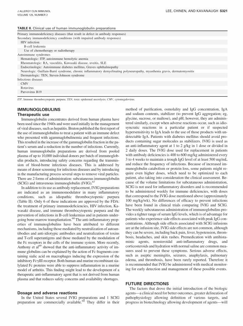
IMMUNOGLOBULINSTherapeutic use
Immunoglobulin concentrates derived from human plasma havebeen used since the 1940s andwere used initially in themanagementof viral diseases, such as hepatitis. Bruton published the first report ofthe use of immunoglobulins to treat a patient with an immune defectwho presented with agammaglobulinemia and frequent infections.This resulted in the increase of the gammaglobulin fraction in the pa-tient’s serum and a reduction in the number of infections. Currently,human immunoglobulin preparations are derived from pooledplasma of up to 10,000 individual donors per batch of immunoglob-ulin products, introducing safety concerns regarding the transmis-sion of blood-borne infectious diseases. This is addressed bymeans of donor screening for infectious diseases and by introducingin the manufacturing process several steps to remove viral particles.There are 2 forms of administration: subcutaneous immunoglobulin(SCIG) and intravenous immunoglobulin (IVIG).67
In addition to its use as antibody replacement, IVIG preparationsare indicated as an immunomodulator in many inflammatoryconditions, such as idiopathic thrombocytopenic purpura(Table II). Only 6 of these indications are approved by the FDA:the treatment of primary immunodeficiencies, HIV infection, Ka-wasaki disease, and immune thrombocytopenic purpura and theprevention of infections in B-cell leukemias and in patients under-going bone marrow transplantation.68 The anti-inflammatory prop-erties of immunoglobulins have been attributed to differentmechanisms, including thosemediated by neutralization of autoan-tibodies and anti-idiotypic antibodies and neutralization of toxinsand T-cell superantigens and those mediated by the modulation ofthe Fc receptors in the cells of the immune system. More recently,Anthony et al69 showed that the anti-inflammatory activity of im-mune globulins can be explained by the action of Fc fragments con-taining sialic acid on macrophages inducing the expression of theinhibitory FcgIII receptor. Both human andmurine recombinant sia-lylated Fc proteins were able to suppress inflammation in a murinemodel of arthritis. This finding might lead to the development of atherapeutic anti-inflammatory agent that is not derived from humanplasma and that reduces safety concerns and availability shortages.
Dosage and adverse reactionsIn the United States several IVIG preparations and 1 SCIG
preparation are commercially available.68 They differ in their
method of purification, osmolality and IgG concentration, IgAand sodium contents, stabilizer (to prevent IgG aggregation; eg,glycine, sucrose, or maltose), and pH; however, they are adminis-tered similarly, except when adverse reactions occur, such as idio-syncratic reactions in a particular patient or if suspectedhypersensitivity to IgA leads to the use of those products with un-detectable IgA. Patients with diabetes mellitus should avoid pro-ducts containing sugar molecules as stabilizers. IVIG is used asan anti-inflammatory agent at 1 to 2 g/kg in 1 dose or divided in2 daily doses. The IVIG dose used for replacement in patientswith antibody deficiencies is 400 to 600 mg/kg administered every3 to 4 weeks to maintain a trough IgG level of at least 500 mg/mLand reduce the frequency of infections. Because of increased im-munoglobulin catabolism or protein loss, some patients might re-quire even higher doses, which need to be optimized to eachpatient, also taking into consideration the clinical assessment. Be-cause of the volume limitations for subcutaneous administration,SCIG is not used for inflammatory disorders and is recommendedto be administered weekly for immune deficiencies, with dosesthat correspond to the IVIG dose mentioned above (approximately100 mg/kg/wk). No differences of efficacy to prevent infectionshave been found in clinical trials comparing IVIG and SCIG.Theweekly subcutaneous administration of immunoglobulins pro-vides a tighter range of serum IgG levels, which is of advantage forpatients who experience side effects associated with peak IgG con-centrations. Although side effects associated with SCIG infusionsare at the infusion site, IVIG side effects are not common, althoughthey can be severe, including back pain, fever, hypotension, throm-bosis, headaches, and skin rashes. Premedication with antihista-minic agents, nonsteroidal anti-inflammatory drugs, andcorticosteroids and hydrationwith normal saline are commonmea-sures used to prevent these symptoms. Serious adverse effects,such as aseptic meningitis, seizures, anaphylaxis, pulmonaryedema, and thrombosis, have been rarely reported. Therefore itis recommended that IVIG be administered with medical monitor-ing for early detection and management of these possible events.
FUTURE DIRECTIONSThe factors that drove the initial introduction of the biologic
agents—a clinical need for better outcomes, greater delineation ofpathophysiology allowing definition of various targets, andprogress in biotechnology allowing development of agents—will
TABLE II. Clinical use of human immunoglobulin preparations
Primary immunodeficiency diseases (that result in defect in antibody responses)Secondary immunodeficiency conditions (with impaired antibody responses)
HIV infectionB-cell leukemiaUse of chemotherapy or radiotherapy
Autoimmune syndromesHematologic: ITP, autoimmune hemolytic anemiaRheumatologic: RA, vasculitis, Kawasaki disease, uveitis, SLEEndocrinologic: Autoimmune diabetes mellitus, Graves ophthalmopathyNeurologic: Guillain-Barre syndrome, chronic inflammatory demyelinating polyneuropathy, myasthenia gravis, dermatomyositisDermatologic: TEN, Steven-Johnson syndrome
Infectious diseasesCMVRotavirusParvovirus B19
ITP, Immune thrombocytopenic purpura; TEN, toxic epidermal necrolysis; CMV, cytomegalovirus.
J ALLERGY CLIN IMMUNOL
VOLUME 125, NUMBER 2
LEE, CHINEN, AND KAVANAUGH S321
no doubt continue to fuel progress in this area. It can be expectedthat additional mAbs and fusion receptors, both directed atexisting targets and against novel targets, will continue to bedeveloped and brought to the clinic. Along with the number ofagents, it is anticipated that the conditions for which these agentsare used will also expand. For existing biologic agents, a numberof questions remain as to the optimum treatment paradigms (eg,sequence of biologic agents) and most appropriate patient pop-ulations for their use; this will be germane for newer agents aswell. As always, the balance between achieving higher levels ofefficacy, with disease remission being the ultimate goal, need tobe balanced against safety considerations. For macromolecules,such as mAbs and soluble receptors, there is the potential foroptimizing their characteristics, including ease of use, immuno-genicity, and cost. For certain targets, it is possible that small-molecule inhibitors might be developed that can address some ofthese issues. However, because these molecules can be antici-pated to have pharmacokinetic, mechanistic, and other importantdifferences from their macromolecular counterparts, this mighttranslate into variable safety and efficacy. Therefore newer agentsof a different class, even thosewhose putative target is the same asexisting therapies, need to be assessed with the same rigor as thecurrently available agents.
REFERENCES1. Maini RN, Breedveld FC, Kalden JR, Smolen JS, Furst D, Weisman MH, et al.
Sustained improvement over two years in physical function, structural damage,and signs and symptoms among patients with rheumatoid arthritis treated with in-fliximab and methotrexate. Arthritis Rheum 2004;50:1051-65.
2. Lipsky PE, van der Heijde DM, St Claire EW, Furst DE, Breedveld FC, Kalden JR,et al. Infliximab and methotrexate in the treatment of rheumatoid arthritis. N Engl JMed 2000;343:1594-602.
3. Moreland LW, Weinblatt ME, Keystone EC, Kremer JM, Martin RW, Schiff MH,et al. Etanercept treatment in adults with established rheumatoid arthritis: 7 yearsof clinical experience. J Rheumatol 2005;33:854-61.
4. van der Heijde D, Klareskog L, Rodriguez-Valverde V, Codreanu C, Bolosiu H,Melo-Gomes J, et al. Comparison of etanercept and methotrexate, alone and com-bined, in the treatment of rheumatoid arthritis: two-year clinical and radiographicresults from the TEMPO study, a double-blind, randomized trial. Arthritis Rheum2006;54:76-81.
5. Weinblatt ME, Keystone EC, Furst DE, Moreland LW, Weisman MH, Birbara CA,et al. Adalimumab, a fully human anti-tumor necrosis factor-a monoclonal anti-body, for the treatment of rheumatoid arthritis in patients taking concomitant meth-otrexate: the ARMADA trial [published erratum appears in Arthritis Rheum 2003;48:855]. Arthritis Rheum 2003;48:35-45.
6. Furst DE, Schiff MH, Fleischmann RM, Strand V, Birbara CA, Compagnone D,et al. Adalimumab, a fully human anti-tumor necrosis factor-a monoclonal anti-body, and concomitant standard antirheumatic therapy for the treatment of rheuma-toid arthritis: results of STAR (Safety Trial of Adalimumab in Rheumatoidarthritis). J Rheumatol 2003;30:2563-71.
7. Kay J, Matteson EL, Dasgupta B, Nash P, Durez P, Hall S, et al. Golimumab inpatients with active rheumatoid arthritis despite treatment with methotrexate.Arthritis Rheum 2008;58:964-75.
8. Keystone E, van der Heijde D, Mason D Jr, Landewe R, van Vollenhoven R,Combe B, et al. Certolizumab pegol plus methotrexate is significantly more effec-tive than placebo plus methotrexate in active rheumatoid arthritis. Arthritis Rheum2008;58:3319-29.
9. St Clair EW, van der Heijde DMFM, Smolen JS, Maini RN, Bathon JM, Emery P,et al. Combination of infliximab and methotrexate therapy for early rheumatoidarthritis. Arthritis Rheum 2004;50:3432-43.
10. Genovese MC, Bathon JM, Martin RW, Fleischmann RM, Tesser JR, Schiff MH,et al. Etanercept versus methotrexate in patients with early rheumatoid arthritis:two year radiographic and clinical outcomes. Arthritis Rheum 2002;46:1443-50.
11. Breedveld FC, Weisman MH, Kavanaugh AF, Cohen SB, Pavelka K, van Vollen-hoven R, et al. The PREMIER study: a multicenter, randomized, double-blind clin-ical trial of combination therapy with adalimumab plus methotrexate versusmethotrexate alone or adalimumab alone in patients with early, aggressive rheuma-toid arthritis who had not had previous methotrexate treatment. Arthritis Rheum2006;54:26-37.
12. Landewe R, van der Heijde D, Klareskog L, van Vollenhoven R, Fatenejad S. Dis-connect between inflammation and joint destruction after treatment with etanerceptplus methotrexate: results from the trial of etanercept and methotrexate with radio-graphic and patient outcomes. Arthritis Rheum 2006;54:3119-25.
13. Lovell DJ, Ruperto N, Goodman S, Reiff A, Jung L, Jarosova K, et al. Adalimumabwith or without methotrexate in juvenile rheumatoid arthritis. N Engl J Med 2008;359:810-20.
14. Ruperto N, Lovell DJ, Cuttica R, Wilkinson N, Woo P, Espada G, et al. A random-ized, placebo-controlled trial of infliximab plus methotrexate for the treatment ofpolyarticular-course juvenile rheumatoid arthritis. Arthritis Rheum 2007;59:3096-106.
15. Antoni CE, Kavanaugh A, van der Heijde D, Beutler A, Keenan G, Zhou B, et al.Two-year efficacy and safety of infliximab treatment in patients with active psori-atic arthritis: findings of the Infliximab Multinational Psoriatic Arthritis ControlledTrial (IMPACT). J Rheumatol 2008;35:869-76.
16. Mease PJ, Kivitz AJ, Burch FX, Siegel EL, Cohen SB, Ory P, et al. Etanercepttreatment of psoriatic arthritis: safety, efficacy, and effect on disease progression.Arthritis Rheum 2004;50:2264-72.
17. Mease PJ, Gladman DD, Ritchlin CT, Ruderman EM, Steinfeld SD, Choy EH,et al. Adalimumab for the treatment of patients with moderately to severely activepsoriatic arthritis: result of a double-blind, randomized, placebo-controlled trial.Arthritis Rheum 2005;52:3279-89.
18. Rozenblit M, Lebwohl M. New biologics for psoriasis and psoriatic arthritis. Der-matol Ther 2009;22:56-60.
19. van der Heijde D, Dijkmans B, Geusens P, Sieper J, DeWoody K, Williamson P,et al. Efficacy and safety of infliximab in patients with ankylosing spondylitis.Arthritis Rheum 2005;52:582-91.
20. Baraliakos X, Brandt J, Listing J, Haibel H, S!orensen H, Rudwaleit M, et al. Out-come of patients with active ankylosing spondylitis after two years of therapy withetanercept: clinical and magnetic resonance imaging data. Arthritis Rheum 2005;53:856-63.
21. Sieper J, Baraliakos X, Listing J, Brandt J, Haibel H, Rudwaleit M, et al. Persistentreduction of spinal inflammation as assessed by magnetic resonance imaging in pa-tients with ankylosing spondylitis after 2 years of treatment with the anti-tumor ne-crosis factor agent infliximab. Rheumatology 2005;44:1525-30.
22. Van der Heijde D, Landewe R, Baraliakos X, Houben H, van Tubergen A, William-son P, et al. Radiographic findings following two years of infliximab therapy in pa-tients with ankylosing spondylitis. Arthritis Rheum 2008;58:3063-70.
23. van der Heijde D, Pangan AL, Schiff MH, Braun J, Borofsky M, Torre J, et al. Ada-limumab effectively reduces the signs and symptoms of active ankylosing spondy-litis in patients with total spinal ankylosis. Ann Rheum Dis 2008;67:1218-21.
24. Inman RD, Davis JC Jr, Heijde D, Diekman L, Sieper J, Kim SI, et al. Efficacy andsafety of golimumab in patients with ankylosing spondylitis: results of a random-ized, double-blind, placebo-controlled, phase III trial. Arthritis Rheum 2008;58:3402-12.
25. Present DH, Rutgeerts P, Targan S, Hanauer SB, Mayer L, van Hogezand RA, et al.Infliximab for the treatment of fistulas in patients with Crohn’s disease. N Engl JMed 1999;340:1398-405.
26. Rutgeerts P, Sandborn WJ, Feagan BG, Reinisch W, Olson A, Johanns J, et al. In-fliximab for induction and maintenance therapy for ulcerative colitis. N Engl J Med2005;353:2462-76.
27. Hanauer SB, Sandborn WJ, Rutgeerts P, Fedorak RN, Lukas M, MacIntosh D, et al.Human anti-tumor necrosis factor monoclonal antibody (adalimumab) in Crohn’sdisease: the CLASSIC-I trial. Gastroenterology 2006;130:323-33.
28. Sandborn WJ, Hanauer SB, Katz S, Safdi M, Wolf DG, Baerg RD, et al. Etanerceptfor active Crohn’s disease: a randomized, double-blind, placebo-controlled trial.Gastroenterology 2001;121:1088-94.
29. Sandborn WJ, Feagan BG, Stoinov S, Honiball PJ, Rutgeerts P, Mason D, et al.Certolizumab pegol for the treatment of Crohn’s disease. N Eng J Med 2007;357:228-38.
30. Schreiber S, Khaliq-Kareemi M, Lawrence IC, Thomsen OO, Hanauer SB,McColm J, et al. Maintenance therapy with certolizumab pegol for Crohn’s dis-ease. N Eng J Med 2007;357:239-50.
31. Baert F, Noman M, Verneire S, Van Assche G, D’Haens G, Carbonez A, et al. In-fluence of immunogenicity on the long-term efficacy of infliximab in Crohn’s dis-ease. N Engl J Med 2003;348:601-8.
32. Hyrich KL, Lunt M, Watson KD, Symmons DPM, Silman AJ. for the British So-ciety for Rheumatology Biologics Register. Outcomes after switching from oneanti-tumor necrosis factor a agent to a second anti-tumor necrosis factor a agentin patients with rheumatoid arthritis. Arthritis Rheum 2007;56:13-20.
33. Kapoor D, Ruderman EM. Safety of newer therapies in rheumatoid arthritis.Rheum Dis Clin Updates 2007;2:1-9.
34. Lee SJ, Kavanaugh A. Adverse events related to biologic agents. J Allergy Clin Im-munol 2005;116:900-5.
J ALLERGY CLIN IMMUNOL
FEBRUARY 2010
S322 LEE, CHINEN, AND KAVANAUGH
35. Ross JB, Finlayson LA, Klotz PJ, Langley RG, Gaudet R, Thompson K, et al. Useof anakinra (Kineret) in the treatment of familial cold autoinflammatory syndromewith a 16-month follow-up. J Cutan Med Surg 2008;12:8-16.
36. Hoffman HM, Throne ML, Amar NJ, Sebai M, Kivitz AJ, Kavanaugh A, et al. Ef-ficacy and safety of rilonacept (interleukin-1 trap) in patients with cryopyrin-asso-ciated periodic syndrome. Arthritis Rheum 2008;58:2443-52.
37. Smolen JF, Beaulieu A, Rubbert-Roth A, Ramos-Remus C, Rovensky J, AlecockE, et al. Effect of interleukin-6 receptor inhibition with tocilizumab in patientswith rheumatoid arthritis (OPTION study): a double-blind, placebo-controlled, ran-domized trial. Lancet 2008;371:987-97.
38. Genovese MC, McKay JD, Nasonov EL, Mysler EF, da Silva NA, Alecock E, et al.Interleukin-6 receptor inhibition with tocilizumab reduces diseae activity in rheu-matoid arthritis with inadequate response to disease-modifying antirheumaticdrugs. Arthritis Rheum 2008;58:2968-80.
39. Emery P, Keystone E, Tony HP, Cantagrel A, van Vollenhoven R, Sanchez A, et al.IL-6 receptor inhibition with tocilizumab improves treatment outcomes in patientswith rheumatoid arthritis refractory to anti-tumour necrosis factor biologicals: re-sults from a 24-week multicentre randomized placebo-controlled trial. Ann RheumDis 20008;67:1516-1523.
40. Nishimoto N, Hashimoto J, Miyasaka N, Yamamoto K, Kawai S, Takeuchi T, et al.Study of active controlled monotherapy used for rheumatoid arthritis, an IL-6 in-hibitor (SAMURAI): evidence of clinical and radiographic benefit from an x rayreader-blinded randomized controlled trial of tocilizumab. Ann Rheum Dis 2007;66:1162-7.
41. Welzel TM, Morgan TR, Bonkovsky HL, Naishadham D, Pfeifer RM, Wright EC,et al. Variants in interferon-alpha pathway genes and response to pegylated inter-feron-alpha 2a plus reibavirin in the treatment of hepatitis C virus infection in thehepatitic C antiviral long-term treatment against cirrhosis trial. Hepatology 2009;49:1847-58.
42. Coppin C, Le L, Porzsolt F, Wilt T. Targeted therapy for advanced renal cell car-cinoma. Cochrane Database Syst Rev 2008 (2):CD006017.
43. Durelli L, Conti L, Clerico M, Boselli D, Contessa G, Ripellino P, et al. T-helper 17cells expand in multiple sclerosis and are inhibited by interferon-beta. Ann Neurol2009;65:499-509.
44. Holland SM. Chronic Granulomatous Disease. Clin Rev Allergy Immunol 2009[Epub ahead of print].
45. Tarhini AA, Kirkwood JM, Gooding WE, Moschos S, Agarwala SS. A phase 2 trialof sequential temozolomide chemotherapy followed by high-dose interleukin 2 im-munotherapy for metastatic melanoma. Cancer 2008;113:1632-40.
46. Mitsuyasu R, Gelman R, Cherng DW, Landay A, Fahey J, Reichman R, et al. Thevirologic, immunologic, and clinical effects of interleukin 2 with potent antiretro-viral therapy in patients with moderately advanced human immunodeficiency virusinfection: a randomized controlled clinical trial—AIDS Clinical Trials Group 328.Arch Intern Med 2007;167:597-605.
47. Levy Y, Lacabaratz C, Weiss L, Viard JP, Goujard C, Lelievre JD, et al. EnhancedT cell recovery in HIV-1-infected adults through IL-7 treatment. J Clin Invest 2009;119:997-1007.
48. Waldmann TA. Anti-Tac (daclizumab, Zenapax) in the treatment of leukemia, au-toimmune diseases, and in the prevention of allograft rejection: a 25-year personalodyssey. J Clin Immunol 2007;27:1-18.
49. Genovese MC, Schiff M, Luggen M, Becker JC, Aranda R, Teng J, et al. Efficacyand safety of the selective co-stimulation modulator abatacept following 2 years oftreatment in patients with rheumatoid arthritis and an inadequate response to anti-tumour necrosis factor therapy. Ann Rheum Dis 2008;67:547-54.
50. Kremer JM, Geneant HK, Moreland LW, Russell AS, Emery P, Abud-Mendoza C,et al. Results of a two-year followup study of patients with rheumatoid arthritiswho received a combination of abatacept and methotrexate. Arthritis Rheum2008;58:953-63.
51. Schiff M, Keiserman M, Codding C, Songcharoen S, Berman A, Nayiager S, et al.Efficacy and safety of abatacept or infliximab vs placebo in ATTEST: a phase III,multi-centre, randomised, double-blind, placebo-controlled study in patients withrheumatoid arthritis and an inadequate response to methotrexate. Ann RheumDis 2008;67:1096-103.
52. Weinblatt M, Combe B, Covucci A, Aranda R, Becker JC, Keystone E. Safety ofthe selective costimulation modulator abatacept in rheumatoid arthritis patients re-ceiving background biologic and nonbiologic disease-modifying antirheumaticdrugs: a one-year randomized, placebo-controlled study. Arthritis Rheum 2006;54:2807-16.
53. Sugiyama H, McCormick TS, Cooper KD, Korman N. Alefacept in the treatmentof psoriasis. Clin Dermatol 2008;26:503-8.
54. Krueger GG, Gottlieb AB, Sterry W, Korman N, van de Kerkhof P. A multiceter,open-label study of repeat courses of intramuscular alefacept in combination withother psoriasis therapies in patients with chronic plaque psoriasis. J Dermatol Treat2008;19:146-55.
55. Mease PJ, Gladman DD, Keystone EC. Alefacept in Psoriatic Arthritis StudyGroup. Alefacept in combination with methotrexate for the treatment of psoriaticarthritis. Arthritis Rheum 2006;54:1638-45.
56. Murphy CA, Langrish CL, Chen Y, Blumenschein W, McClanahan T, KasteleinRA, et al. Divergent pro-and antiinflammatory roles for IL-23 and IL-12 in jointautoimmune inflammation. J Exp Med 2003;98:1951-7.
57. Krueger GG, Langley RG, Leonardi C, Yeilding N, Guzzo C, Wang Y, et al. A hu-man interleukin-12/23 monoclonal antibody for the treatment of psoriasis. N Engl JMed 2007;356:580-92.
58. Gottlieb A,Menter A,Mendelsohn A, ShenYK, Li S, Guzzo C, et al. Ustekinumab, ahuman interleukin 12/23 monoclonal antibody, for psoriatic arthritis: randomized,double-blind, placebo-controlled, crossover trial. Lancet 2009;373:633-40.
59. Emery P, Fleischmann R, Filipowicz-Sosnowska A, Schechtman J, Szczepanski L,Kavanaugh A, et al. The efficacy and safety of Rituximab in patients with activerheumatoid arthritis despite methotrexate treatment: results of a phase IIB random-ized, double-blind, placebo-controlled, dose-ranging trial. Arthritis Rheum 2006;54:1390-400.
60. Cohen SB, Emery P, Greenwald MW, Dougados M, Furie RA, Genovese MC, et al.Rituximab for rheumatoid arthritis refractory to anti-tumor necrosis factor therapy:results of a multicenter, randomized, double-blind, placebo-controlled, phase IIItrial evaluating primary efficacy and safety at twenty-four weeks. Arthritis Rheum2006;54:2793-806.
61. Keystone E, Fleischmann R, Emery P, Furst DE, van Vollenhoven R, Bathon J,et al. Safety and efficacy of additional courses of rituximab in patients with activerheumatoid arthritis: an open-label extension analysis. Arthritis Rheum 2007;56:3896-908.
62. Calabrese LH, Molloy ES. Progressive multifocal leucoencephalopathy in therheumatic disease: assessing the risks of biological immunosuppressive therapies.Ann Rheum Dis 2008;67(suppl III):iii64-5.
63. Slavin RG, Ferioli C, Tannenbaum SJ, Martin C, Blogg M, Lowe PJ. Asthmasymptom re-emergence after omalizumab withdrawal correlates well with increas-ing IgE and decreasing pharmacokinetic concentrations. J Allergy Clin Immunol2009;123:107-13.
64. Humbert M, Beasley R, Ayres J, Slavin R, Hebert J, Bousquet J, et al. Benefits ofomalizumab as add-on therapy in patients with severe persistent asthma who areinadequately controlled despite best available therapy (GINA 2002 step 4 treat-ment): INNOVATE. Allergy 2005;60:309-16.
65. Hutchinson M, Kappos L, Calabresi PA, Confavreux C, Giovannoi G, Galetta SL,et al. The efficacy of natalizumab in patients with relapsing multiple sclerosis: sub-group analyses of AFFIRM and SENTINEL. J Neurol 2009;256:405-45.
66. Toth DP, Papp K, Gratton D. Long-term efficacy of up to 15 months’ efalizumabtherapy in patients with moderate-to-severe chronic plaque psoriasis. DermatolTher 2008;21(suppl 3):S6-14.
67. Ballow M. Immunoglobulin therapy: methods of delivery. J Allergy Clin Immunol2008;122:1038-9.
68. Orange JS, Hossny EM, Weiler CR, Ballow M, Berger M, Bonilla FA, et al. Use ofintravenous immunoglobulin in human disease: a review of evidence by membersof the Primary Immunodeficiency Committee of the American Academy of Al-lergy, Asthma and Immunology. J Allergy Clin Immunol 2006;117(suppl):S525-53.
69. Anthony RM, Nimmerjahn F, Ashline DJ, Reinhold VN, Paulson JC, Ravetch JV.Recapitulation of IVIG anti-inflammatory activity with a recombinant IgG Fc. Sci-ence 2008;320:373-6.
J ALLERGY CLIN IMMUNOL
VOLUME 125, NUMBER 2
LEE, CHINEN, AND KAVANAUGH S323
Transplantation immunology: Solid organ and bone marrow
Javier Chinen, MD, PhD,a and Rebecca H. Buckley, MDb Houston, Tex, and Durham, NC
Development of the field of organ and tissue transplantation hasaccelerated remarkably since the human MHC was discoveredin 1967. Matching of donor and recipient for MHC antigens hasbeen shown to have a significant positive effect on graftacceptance. The roles of the different components of theimmune system involved in the tolerance or rejection of graftsand in graft-versus-host disease have been clarified. Thesecomponents include antibodies, antigen-presenting cells, helperand cytotoxic T-cell subsets, immune cell-surface molecules,signaling mechanisms, and cytokines. The development ofpharmacologic and biological agents that interfere with thealloimmune response has had a crucial role in the success oforgan transplantation. Combinations of these agents worksynergistically, leading to lower doses of immunosuppressivedrugs and reduced toxicity. Reports of significant numbers ofsuccessful solid-organ transplantations include those of thekidneys, liver, heart, and lung. The use of bone marrowtransplantation for hematologic diseases, particularlyhematologic malignancies and primary immunodeficiencies, hasbecome the treatment of choice in many of these conditions.Other sources of hematopoietic stem cells are also being used,and diverse immunosuppressive drug regimens of reducedintensity are being proposed to circumvent the mortalityassociated with the toxicity of these drugs. Gene therapy tocorrect inherited diseases by means of infusion of gene-modifiedautologous hematopoietic stem cells has shown efficacy in 2forms of severe combined immunodeficiency, providing analternative to allogeneic tissue transplantation. (J Allergy ClinImmunol 2010;125:S324-35.)
Key words: Bone marrow transplantation, solid-organ transplanta-tion, graft rejection, graft-versus-host disease
Efforts to transplant organs or tissues from one human subjectto another had been unsuccessful for many decades until thediscovery of the human MHC in 1967.1 Identification of thisgenetic region launched the field of clinical organ and tissuetransplantation. In 1968, the World Health Organization Nomen-clature Committee designated that the leukocyte antigens
controlled by the closely linked genes of the human MHC benamed HLA (for human leukocyte antigen). This chapter re-views general immunologic concepts that have supported thesuccess of human organ and tissue transplantation and summa-rizes current medical progress in the field of transplantationmedicine.
TRANSPLANTATION ANTIGENSMHC
Histocompatibility antigens are tissue cell-surface antigenscapable of inducing an immune response in a geneticallydissimilar (allogeneic) recipient, resulting in the rejection ofthe tissues or cells bearing those antigens. The genes that encodethese antigens reside in the MHC region on the short arm ofhuman chromosome 6 (Fig 1). The HLA complex contains morethan 200 genes, more than 40 of which encode leukocyte anti-gens.2,3 These genes and their encoded cell-surface and solubleprotein products are divided into 3 classes (I, II, and III) on thebasis of their tissue distribution, structure, and function.3-5
MHC class I and II genes encode codominantly expressedHLA cell-surface antigens, and class III genes encode severalcomponents of the complement system, all of which share impor-tant roles in immune function.
Class I MHC antigens are present on all nucleated cells and areeach composed of a 45-kd a heavy chain encoded by genes of theHLA-A, HLA-B, or HLA-C loci on chromosome 6 and associatednoncovalently with a 12-kd protein, b2-microglobulin, encodedby a gene on chromosome 15 (Fig 2).3 MHC class II antigenshave a more limited tissue distribution and are expressed onlyon B lymphocytes, activated T lymphocytes, monocytes, macro-phages, Langerhans cells, dendritic cells, endothelium, and epi-thelial cells.5 Each is a heterodimer composed of noncovalentlyassociated a and b chains of approximately 230 amino acids en-coded by genes of the HLA-D region (Fig 2). On cells expressingboth class I and class II HLA antigens, there are 3 class I antigensand 3 or more (usually 4) class II heterodimers.
Class III genes are located between the HLA-B and HLA-Dloci and determine the structure of 3 components of the comple-ment system: C2, C4, and factor B.3,4 HLA antigens are inheritedin a Mendelian dominant manner. Because of the closeness of thedifferent loci of the MHC and the resultant low crossoverfrequency, however, HLA genes are almost always inherited
From athe Department of Pediatrics, Allergy/Immunology, Baylor College of Medicine,Houston, and bthe Department of Pediatrics, Allergy/Immunology, Duke UniversityMedical Center, Durham.
Disclosure of potential conflict of interest: J. Chinen has declared that he has no conflictof interest. R. H. Buckley has received research support from the National Institute ofAllergy and Infectious Diseases and is the Chair of the Medical Advisory Committeefor the Immune Deficiency Foundation.
Received for publication October 14, 2009; revised November 9, 2009; accepted for pub-lication November 11, 2009.
Reprint requests: Rebecca H. Buckley, MD, Duke University Medical Center, Depart-ment of Pediatrics, Allergy/Immunology, 362 Jones Building (Campus Box 2898),Durham, NC 27710-0001. E-mail: [email protected].
0091-6749/$36.00! 2010 American Academy of Allergy, Asthma & Immunologydoi:10.1016/j.jaci.2009.11.014
Abbreviations usedADA: Adenosine deaminaseALG: Antilymphocyte globulinAPC: Antigen-presenting cellATG: Antithymocyte globulinCGD: Chronic granulomatous disease
GVHD: Graft-versus-host diseaseIL-2R: IL-2 receptorSCID: Severe combined immunodeficiency
S324
together. To date, 3756 different class I and II HLA gene alleleshave been identified.2 The fixed combination of these genetic de-terminants present in 1 chromosome of a subject is referred to as ahaplotype. Chromosome 6 is an autosome, and therefore all sub-jects have 2 HLA haplotypes (1 for each chromosome), and thereare only 4 possible combinations of haplotypes among theoffspring of any 2 parents. Thus there is a 25% probability thatbiological siblings will have identical HLA alleles.
The ABO systemABO incompatibility does not cause stimulation in mixed
leukocyte cultures, indicating that ABO compatibility is of muchless importance than HLA compatibility in graft survival. How-ever, ABO incompatibility can result in hyperacute rejection ofprimarily vascularized grafts, such as those of the kidney andheart.6 This is thought to occur because (1) ABO blood group an-tigens are highly expressed on kidney and cardiac grafts, particu-larly those from patients who are blood group A or B antigensecretors, and (2) preformed naturally occurring antibodies toblood group substances are present in mismatched recipients.Advances in immunosuppressive therapies to prevent immunerejection of the graft have more recently allowed performanceof organ transplantations across the ABO barrier.7
Donor-recipient HLA matchingTwo laboratory methods are used to pair donors and recipients
for transplantation. The first matching method involves thedetermination of HLA antigens on donor and recipient leukocytesby using either serologic or DNA-typing methods. The secondmethod is functional and involves the measurement of theresponse of immunocompetent cells from the recipient to antigenspresent on donor cells (and vice versa for bone marrow transplan-tation). Results of bothmethods are generally consistent with eachother. Disparities that are serologically detected are referred to asantigen mismatches, whereas differences that can be identifiedonly by DNA-based typing are called allele mismatches. Becausethese methods take considerable time to perform, results are notknown in time for some solid-organ transplantations, such aslung transplantations, which are performed based on immediateorgan availability. Since 2000, the National Donor Matching Pro-gram performs HLA typing of donor volunteers exclusively usinga DNA-based method, the PCR single-strand oligonucleotideprobe. Currently, approximately 60% of volunteer donors on theNational Donor Matching Program Registry had their HLA typesdetermined by using this method. Efforts continue to improve theefficiency of HLA typing and to reduce the costs of the assays.8
Donor-recipient serologic cross-matchingSerologic cross-matching is of particular importance to the
success of primarily vascularized grafts, such as those of thekidney and heart. Serum from the prospective recipient is testedagainst cells from the potential donor for the presence ofantibodies to red blood cell or HLA antigens. The presence ofsuch antibodies correlates with hyperacute renal graft rejection.6
For this reason, a positive serologic cross-match result has beenconsidered a contraindication to renal transplantation, althoughtherapeutic strategies, such as the use of plasmapheresis, areproposed when the mismatch cannot be avoided.7
Usefulness of HLA typing in clinical organ andtissue transplantation
Although typing for intrafamilial transplants of all types isclearly of great value, the usefulness of HLA typing in cadaverickidney grafting has been a point of controversy since cyclosporinebecame available.9 Although short-term survival rates did not ap-pear to be that different for closely or poorly matched cadaverickidneys, the degree of HLA matching does correlate with long-term survival.10 Until 1980, only HLA-identical siblings couldbe used as bone marrow donors because both graft rejection andlethal graft-versus-host disease (GVHD) were common compli-cations if this was not the case.11 Fortunately, the developmentduring the past 3 decades of techniques to rigorously deplete post-thymic T cells from donor marrow has permitted numeroussuccessful half-HLA-matched marrow transplantations with noor minimal GVHD.12,13
MECHANISMS OF GRAFT REJECTIONRole of alloimmune antibodies
The strongest evidence for a role for antibodies in graftrejection is the hyperacute rejection of primarily vascularizedorgans, such as the kidney and heart. High titers of antidonorantibodies can be demonstrated in recipients presenting withthese reactions.6 These antibodies combinewith HLA antigens onendothelial cells, with subsequent complement fixation and accu-mulation of polymorphonuclear cells. Endothelial damage thenoccurs, probably as a result of enzymes released from polymor-phonuclear leukocytes; platelets then accumulate, thrombidevelop, and the result is renal cortical necrosis or myocardialinfarction.14
Leukocytes and cytokines in graft rejectionAllograft rejection results from the coordinated activation
of alloreactive T cells and antigen-presenting cells (APCs).Although acute rejection is a T cell–dependent process, thedestruction of the allograft results from a broad array of effectorimmune mechanisms. Cell-cell interactions and the release byprimed TH cells of multiple types of cytokines (IL-2, IL-4, IL-5,IL-7, IL-10, IL-15, TNF-a, and IFN-g) recruit not only immuno-competent donor-specific CD41 T cells, CD81 cytotoxic T cells,and antibody-forming B cells but also nonspecific inflammatorycells, which constitute the majority of cells infiltrating an allo-graft.15 Other cells specific to the transplanted organ might playa role in the balance of tolerance and rejection, such as theKupffer cells and the sinusoidal epithelial cells in the liver.16
Stimulation of CD41 T cells through their antigen receptors isnot sufficient to initiate T-cell activation unless costimulation isprovided by interaction of other ligand-receptor pairs present onthe surfaces of T cells and APCs during the encounter. Some ofthese interactive pairs include the T-cell surface molecule CD2and its ligand CD58 on APCs, CD11a/CD18-CD54, CD5-CD72, CD40 ligand–CD40, and CD28–CD80 or CD86.CD41 T-cell anergy or tolerance induction occurs when the T-cell receptor interacts with the APC unless signals are providedthrough 1 or more of these receptor-ligand interactions (particu-larly through CD40 ligand–CD40 and CD28–CD80 or CD86)or by cytokines (eg, IL-1 and IL-6 from the APC). Thus T-cell ac-cessory proteins and their ligands on APCs are target moleculesfor antirejection therapy.17,18 If costimulation does occur, theCD41 T cell becomes activated, which leads to stable
J ALLERGY CLIN IMMUNOL
VOLUME 125, NUMBER 2
CHINEN AND BUCKLEY S325
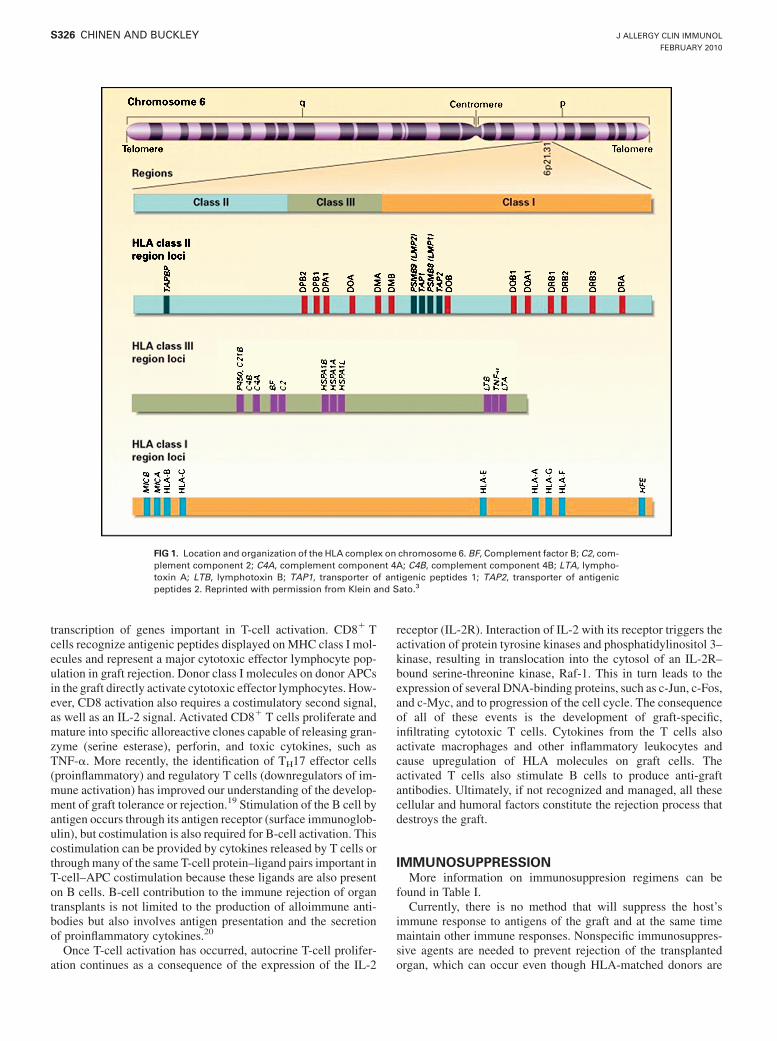
transcription of genes important in T-cell activation. CD81 Tcells recognize antigenic peptides displayed onMHC class I mol-ecules and represent a major cytotoxic effector lymphocyte pop-ulation in graft rejection. Donor class I molecules on donor APCsin the graft directly activate cytotoxic effector lymphocytes. How-ever, CD8 activation also requires a costimulatory second signal,as well as an IL-2 signal. Activated CD81 T cells proliferate andmature into specific alloreactive clones capable of releasing gran-zyme (serine esterase), perforin, and toxic cytokines, such asTNF-a. More recently, the identification of TH17 effector cells(proinflammatory) and regulatory T cells (downregulators of im-mune activation) has improved our understanding of the develop-ment of graft tolerance or rejection.19 Stimulation of the B cell byantigen occurs through its antigen receptor (surface immunoglob-ulin), but costimulation is also required for B-cell activation. Thiscostimulation can be provided by cytokines released by T cells orthroughmany of the same T-cell protein–ligand pairs important inT-cell–APC costimulation because these ligands are also presenton B cells. B-cell contribution to the immune rejection of organtransplants is not limited to the production of alloimmune anti-bodies but also involves antigen presentation and the secretionof proinflammatory cytokines.20
Once T-cell activation has occurred, autocrine T-cell prolifer-ation continues as a consequence of the expression of the IL-2
receptor (IL-2R). Interaction of IL-2 with its receptor triggers theactivation of protein tyrosine kinases and phosphatidylinositol 3–kinase, resulting in translocation into the cytosol of an IL-2R–bound serine-threonine kinase, Raf-1. This in turn leads to theexpression of several DNA-binding proteins, such as c-Jun, c-Fos,and c-Myc, and to progression of the cell cycle. The consequenceof all of these events is the development of graft-specific,infiltrating cytotoxic T cells. Cytokines from the T cells alsoactivate macrophages and other inflammatory leukocytes andcause upregulation of HLA molecules on graft cells. Theactivated T cells also stimulate B cells to produce anti-graftantibodies. Ultimately, if not recognized and managed, all thesecellular and humoral factors constitute the rejection process thatdestroys the graft.
IMMUNOSUPPRESSIONMore information on immunosuppresion regimens can be
found in Table I.Currently, there is no method that will suppress the host’s
immune response to antigens of the graft and at the same timemaintain other immune responses. Nonspecific immunosuppres-sive agents are needed to prevent rejection of the transplantedorgan, which can occur even though HLA-matched donors are
FIG 1. Location and organization of the HLA complex on chromosome 6. BF, Complement factor B; C2, com-plement component 2; C4A, complement component 4A; C4B, complement component 4B; LTA, lympho-toxin A; LTB, lymphotoxin B; TAP1, transporter of antigenic peptides 1; TAP2, transporter of antigenicpeptides 2. Reprinted with permission from Klein and Sato.3
J ALLERGY CLIN IMMUNOL
FEBRUARY 2010
S326 CHINEN AND BUCKLEY
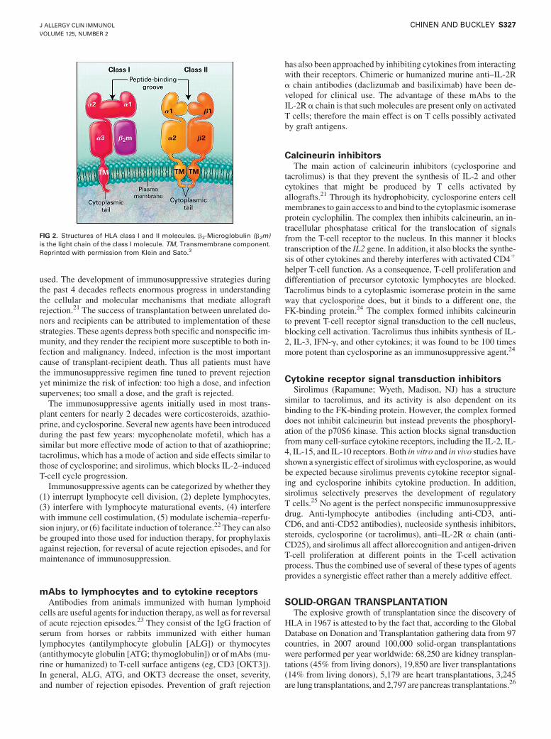
used. The development of immunosuppressive strategies duringthe past 4 decades reflects enormous progress in understandingthe cellular and molecular mechanisms that mediate allograftrejection.21 The success of transplantation between unrelated do-nors and recipients can be attributed to implementation of thesestrategies. These agents depress both specific and nonspecific im-munity, and they render the recipient more susceptible to both in-fection and malignancy. Indeed, infection is the most importantcause of transplant-recipient death. Thus all patients must havethe immunosuppressive regimen fine tuned to prevent rejectionyet minimize the risk of infection: too high a dose, and infectionsupervenes; too small a dose, and the graft is rejected.
The immunosuppressive agents initially used in most trans-plant centers for nearly 2 decades were corticosteroids, azathio-prine, and cyclosporine. Several new agents have been introducedduring the past few years: mycophenolate mofetil, which has asimilar but more effective mode of action to that of azathioprine;tacrolimus, which has a mode of action and side effects similar tothose of cyclosporine; and sirolimus, which blocks IL-2–inducedT-cell cycle progression.
Immunosuppressive agents can be categorized by whether they(1) interrupt lymphocyte cell division, (2) deplete lymphocytes,(3) interfere with lymphocyte maturational events, (4) interferewith immune cell costimulation, (5) modulate ischemia–reperfu-sion injury, or (6) facilitate induction of tolerance.22 They can alsobe grouped into those used for induction therapy, for prophylaxisagainst rejection, for reversal of acute rejection episodes, and formaintenance of immunosuppression.
mAbs to lymphocytes and to cytokine receptorsAntibodies from animals immunized with human lymphoid
cells are useful agents for induction therapy, as well as for reversalof acute rejection episodes.23 They consist of the IgG fraction ofserum from horses or rabbits immunized with either humanlymphocytes (antilymphocyte globulin [ALG]) or thymocytes(antithymocyte globulin [ATG; thymoglobulin]) or of mAbs (mu-rine or humanized) to T-cell surface antigens (eg, CD3 [OKT3]).In general, ALG, ATG, and OKT3 decrease the onset, severity,and number of rejection episodes. Prevention of graft rejection
has also been approached by inhibiting cytokines from interactingwith their receptors. Chimeric or humanized murine anti–IL-2Ra chain antibodies (daclizumab and basiliximab) have been de-veloped for clinical use. The advantage of these mAbs to theIL-2R a chain is that such molecules are present only on activatedT cells; therefore the main effect is on T cells possibly activatedby graft antigens.
Calcineurin inhibitorsThe main action of calcineurin inhibitors (cyclosporine and
tacrolimus) is that they prevent the synthesis of IL-2 and othercytokines that might be produced by T cells activated byallografts.21 Through its hydrophobicity, cyclosporine enters cellmembranes to gain access to and bind to the cytoplasmic isomeraseprotein cyclophilin. The complex then inhibits calcineurin, an in-tracellular phosphatase critical for the translocation of signalsfrom the T-cell receptor to the nucleus. In this manner it blockstranscription of the IL2 gene. In addition, it also blocks the synthe-sis of other cytokines and thereby interferes with activated CD41
helper T-cell function. As a consequence, T-cell proliferation anddifferentiation of precursor cytotoxic lymphocytes are blocked.Tacrolimus binds to a cytoplasmic isomerase protein in the sameway that cyclosporine does, but it binds to a different one, theFK-binding protein.24 The complex formed inhibits calcineurinto prevent T-cell receptor signal transduction to the cell nucleus,blocking cell activation. Tacrolimus thus inhibits synthesis of IL-2, IL-3, IFN-g, and other cytokines; it was found to be 100 timesmore potent than cyclosporine as an immunosuppressive agent.24
Cytokine receptor signal transduction inhibitorsSirolimus (Rapamune; Wyeth, Madison, NJ) has a structure
similar to tacrolimus, and its activity is also dependent on itsbinding to the FK-binding protein. However, the complex formeddoes not inhibit calcineurin but instead prevents the phosphoryl-ation of the p70S6 kinase. This action blocks signal transductionfrommany cell-surface cytokine receptors, including the IL-2, IL-4, IL-15, and IL-10 receptors. Both in vitro and in vivo studies haveshown a synergistic effect of sirolimuswith cyclosporine, aswouldbe expected because sirolimus prevents cytokine receptor signal-ing and cyclosporine inhibits cytokine production. In addition,sirolimus selectively preserves the development of regulatoryT cells.25 No agent is the perfect nonspecific immunosuppressivedrug. Anti-lymphocyte antibodies (including anti-CD3, anti-CD6, and anti-CD52 antibodies), nucleoside synthesis inhibitors,steroids, cyclosporine (or tacrolimus), anti–IL-2R a chain (anti-CD25), and sirolimus all affect allorecognition and antigen-drivenT-cell proliferation at different points in the T-cell activationprocess. Thus the combined use of several of these types of agentsprovides a synergistic effect rather than a merely additive effect.
SOLID-ORGAN TRANSPLANTATIONThe explosive growth of transplantation since the discovery of
HLA in 1967 is attested to by the fact that, according to the GlobalDatabase on Donation and Transplantation gathering data from 97countries, in 2007 around 100,000 solid-organ transplantationswere performed per year worldwide: 68,250 are kidney transplan-tations (45% from living donors), 19,850 are liver transplantations(14% from living donors), 5,179 are heart transplantations, 3,245are lung transplantations, and 2,797 are pancreas transplantations.26
FIG 2. Structures of HLA class I and II molecules. b2-Microglobulin (b2m)is the light chain of the class I molecule. TM, Transmembrane component.Reprinted with permission from Klein and Sato.3
J ALLERGY CLIN IMMUNOL
VOLUME 125, NUMBER 2
CHINEN AND BUCKLEY S327
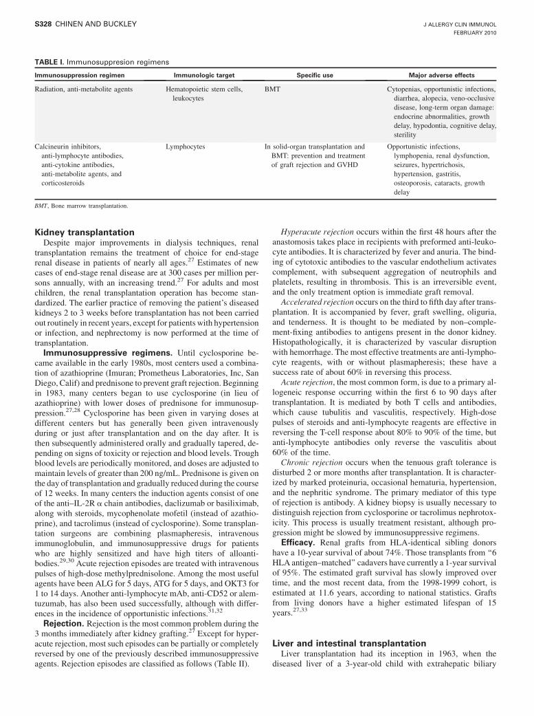
Kidney transplantationDespite major improvements in dialysis techniques, renal
transplantation remains the treatment of choice for end-stagerenal disease in patients of nearly all ages.27 Estimates of newcases of end-stage renal disease are at 300 cases per million per-sons annually, with an increasing trend.27 For adults and mostchildren, the renal transplantation operation has become stan-dardized. The earlier practice of removing the patient’s diseasedkidneys 2 to 3 weeks before transplantation has not been carriedout routinely in recent years, except for patients with hypertensionor infection, and nephrectomy is now performed at the time oftransplantation.
Immunosuppressive regimens. Until cyclosporine be-came available in the early 1980s, most centers used a combina-tion of azathioprine (Imuran; Prometheus Laboratories, Inc, SanDiego, Calif) and prednisone to prevent graft rejection. Beginningin 1983, many centers began to use cyclosporine (in lieu ofazathioprine) with lower doses of prednisone for immunosup-pression.27,28 Cyclosporine has been given in varying doses atdifferent centers but has generally been given intravenouslyduring or just after transplantation and on the day after. It isthen subsequently administered orally and gradually tapered, de-pending on signs of toxicity or rejection and blood levels. Troughblood levels are periodically monitored, and doses are adjusted tomaintain levels of greater than 200 ng/mL. Prednisone is given onthe day of transplantation and gradually reduced during the courseof 12 weeks. In many centers the induction agents consist of oneof the anti–IL-2R a chain antibodies, daclizumab or basiliximab,along with steroids, mycophenolate mofetil (instead of azathio-prine), and tacrolimus (instead of cyclosporine). Some transplan-tation surgeons are combining plasmapheresis, intravenousimmunoglobulin, and immunosuppressive drugs for patientswho are highly sensitized and have high titers of alloanti-bodies.29,30 Acute rejection episodes are treated with intravenouspulses of high-dose methylprednisolone. Among the most usefulagents have been ALG for 5 days, ATG for 5 days, and OKT3 for1 to 14 days. Another anti-lymphocyte mAb, anti-CD52 or alem-tuzumab, has also been used successfully, although with differ-ences in the incidence of opportunistic infections.31,32
Rejection. Rejection is the most common problem during the3 months immediately after kidney grafting.27 Except for hyper-acute rejection, most such episodes can be partially or completelyreversed by one of the previously described immunosuppressiveagents. Rejection episodes are classified as follows (Table II).
Hyperacute rejection occurs within the first 48 hours after theanastomosis takes place in recipients with preformed anti-leuko-cyte antibodies. It is characterized by fever and anuria. The bind-ing of cytotoxic antibodies to the vascular endothelium activatescomplement, with subsequent aggregation of neutrophils andplatelets, resulting in thrombosis. This is an irreversible event,and the only treatment option is immediate graft removal.
Accelerated rejection occurs on the third to fifth day after trans-plantation. It is accompanied by fever, graft swelling, oliguria,and tenderness. It is thought to be mediated by non–comple-ment-fixing antibodies to antigens present in the donor kidney.Histopathologically, it is characterized by vascular disruptionwith hemorrhage. The most effective treatments are anti-lympho-cyte reagents, with or without plasmapheresis; these have asuccess rate of about 60% in reversing this process.
Acute rejection, the most common form, is due to a primary al-logeneic response occurring within the first 6 to 90 days aftertransplantation. It is mediated by both T cells and antibodies,which cause tubulitis and vasculitis, respectively. High-dosepulses of steroids and anti-lymphocyte reagents are effective inreversing the T-cell response about 80% to 90% of the time, butanti-lymphocyte antibodies only reverse the vasculitis about60% of the time.
Chronic rejection occurs when the tenuous graft tolerance isdisturbed 2 or more months after transplantation. It is character-ized by marked proteinuria, occasional hematuria, hypertension,and the nephritic syndrome. The primary mediator of this typeof rejection is antibody. A kidney biopsy is usually necessary todistinguish rejection from cyclosporine or tacrolimus nephrotox-icity. This process is usually treatment resistant, although pro-gression might be slowed by immunosuppressive regimens.
Efficacy. Renal grafts from HLA-identical sibling donorshave a 10-year survival of about 74%. Those transplants from ‘‘6HLA antigen–matched’’ cadavers have currently a 1-year survivalof 95%. The estimated graft survival has slowly improved overtime, and the most recent data, from the 1998-1999 cohort, isestimated at 11.6 years, according to national statistics. Graftsfrom living donors have a higher estimated lifespan of 15years.27,33
Liver and intestinal transplantationLiver transplantation had its inception in 1963, when the
diseased liver of a 3-year-old child with extrahepatic biliary
TABLE I. Immunosuppresion regimens
Immunosuppression regimen Immunologic target Specific use Major adverse effects
Radiation, anti-metabolite agents Hematopoietic stem cells,leukocytes
BMT Cytopenias, opportunistic infections,diarrhea, alopecia, veno-occlusivedisease, long-term organ damage:endocrine abnormalities, growthdelay, hypodontia, cognitive delay,sterility
Calcineurin inhibitors,anti-lymphocyte antibodies,anti-cytokine antibodies,anti-metabolite agents, andcorticosteroids
Lymphocytes In solid-organ transplantation andBMT: prevention and treatmentof graft rejection and GVHD
Opportunistic infections,lymphopenia, renal dysfunction,seizures, hypertrichosis,hypertension, gastritis,osteoporosis, cataracts, growthdelay
BMT, Bone marrow transplantation.
J ALLERGY CLIN IMMUNOL
FEBRUARY 2010
S328 CHINEN AND BUCKLEY
atresia was replaced.34 Although that patient died, subsequentsuccesses have established liver transplantation as the standardtherapy for advanced chronic liver disease.35 Since 1983, the1-year survival rates have increased from 25% to 78%, dependingon the age and health of the recipient, the underlying condition,and various clinical considerations.
Liver transplantation is indicated for chronic end-stage liverdisease, fulminant acute liver failure, and cancer limited to theliver.36 As with renal transplantation, combined therapy targetingseveral facets of the potential rejection process is used for livertransplantation.
Anti–IL-2R a chain antibodies are given intravenously on theday of transplantation, followed by tacrolimus, which is givenintravenously initially and orally thereafter and then by myco-phenolate mofetil and steroids tapered slowly over a year.Survival has increased by 20% in the last 2 decades withtacrolimus-based immunosuppression.37
Although this improvementmight be the result of several factors,retransplantation as a result of acute or chronic rejection has notoccurred in patients treated with tacrolimus. Similar to all solid-organ transplantation, lackof suitable donors is amajor problem forliver transplantation. Since 1988, this organ shortage has beenapproached by partial hepatectomies of living related donors.Donor safety is much greater with use of the left lateral segment.38
Intestinal transplantation is offered to patients who haveintestinal failure (caused by short-bowel syndrome, mucosaldisease, motility disorders, and tumors) and who present withsevere complications of parenteral nutrition, such as cholestaticliver disease and recurrent loss of central venous access.39 Ad-vances in surgical techniques, control of immune rejection, andtreatment of infections have improved the outcomes over time.In 2008, 185 intestinal transplantations were performed in theUnited States. The 1-year rate of patient survival has increasedfrom 57% in 1997 to 80% in 2005 and to 90% if the data are lim-ited to centers that perform the largest number of transplantations.
Heart, heart-lung, and lung transplantationThe various forms of cardiomyopathy are the most common
indications for heart transplantation, followed by congenital heartdisease. Approximately 25% of heart transplant recipients areinfants.22 Immunosuppressive regimens for heart transplantationare similar in many respects to those already described for renal
and hepatic grafts. Usually an anti–IL-2R a chain mAb is givenfor induction therapy on the day of transplantation, along withhigh-dose intravenous methylprednisolone. Prednisone is givenafter the operation and maintained orally until it is discontinuedafter the first normal findings from an endomyocardial biopsy.Tacrolimus is then begun as the primary immunosuppressiveagent with or without mycophenolate.22
Since the introduction of cyclosporine 26 years ago, the resultsof cardiac transplantation have improved greatly. The Interna-tional Heart Transplantation Registry has shown a 4-year survivalof 71% for patients receiving cyclosporine- or tacrolimus-basedtriple immunosuppression therapy. Survival is influenced by theage of the recipient; patients younger than 40 years have a bettersurvival.40 Lung transplantation has been performed for the fol-lowing major diagnostic categories: cystic fibrosis, pulmonaryvascular disease, bronchiolitis obliterans, pulmonary alveolarproteinosis, and pulmonary fibrosis, with 4-year survival atapproximately 50%.22,41
BONE MARROW TRANSPLANTATIONSince 1955, more than 240,000 bone marrow transplantations
have been performed worldwide at 450 centers in 47 countries forthe treatment of more than 50 different fatal diseases (Table III).42
Most of these transplantations have been done by reinfusingstored autologous marrow cells collected before the patient re-ceives intensive chemotherapy or irradiation. Annually, 25,000to 35,000 autologous transplantations are performed comparedwith approximately 15,000 allogeneic transplantations. Certainunique problems distinguish bone marrow transplantation fromtransplantation of solid organs, such as the kidney, liver, and heart.The first problem is that immunocompetent cells, both in therecipient and in the donor marrow or blood, have the potentialto reject each other, resulting in graft rejection on the one handand GVHD on the other.43 The second concern is that successfulunfractionated marrow grafting usually requires strict donor andrecipientMHC class II antigen compatibility to minimize such re-actions. Finally, except for patients with severe combined immu-nodeficiency (SCID), complete DiGeorge anomaly, or identicaltwin donors, even HLA-identical recipients must be pretreatedwith cytotoxic and myeloablative agents to prevent graftrejection.43 Diseases treated successfully by allogeneic bonemarrow transplantation include radiation injury, primary
TABLE II. Solid-organ rejection patterns: Renal rejection as an example
TypeTime after
transplantationSigns andsymptoms
Rapidity ofonset
Immunecomponent
Pathologicfindings Treatment
Successrate (%)
Hyperacute <24 h Fever, anuria Hours Antibody andcomplement
Polymorphonuclearneutrophil depositionand thrombosis
None 0
Accelerated 3-5 d Fever, graft swelling,oliguria, tenderness
1 d Non–complement-fixing antibody
Vascular disruptionhemorrhage
ALG, ATG,anti-CD3
60
Acute 6-90 d Oliguria, salt retention,graft swelling,tenderness,sometimes fever
Days to weeks T cells and antibody Tubulitis,endovasculitis
Steroids, ALG,ATG, anti-CD3
60-90
Chronic >60 d Edema, hypertension,proteinuria, occasionalhematuria
Months to years Antibody Vascular onionskinning
None 0
Reprinted with permission from Buckley R. Transplantation. In: Stiehm ER, editor. Immunologic disorders in infants, children, and adults. 5th ed. Philadelphia: WB Saunders;2006.
J ALLERGY CLIN IMMUNOL
VOLUME 125, NUMBER 2
CHINEN AND BUCKLEY S329
immunodeficiencies, hemoglobinopathies, aplastic anemia, mul-tiple myeloma, leukemia, neuroblastoma, non-Hodgkin lym-phoma, inborn errors of metabolism, and certain autoimmunediseases.44 In addition, autologous marrow transplantation hasbeen used after lethal irradiation or chemotherapy in the treatmentof patients with some hematologic malignancies, solid tumors, orbreast cancer, as well as for the treatment of several autoimmunediseases.45
Other sources of hematopoietic stem cellsfor transplantation
Bone marrow is not the only source of hematopoietic stemcells. These cells are capable of reconstituting all blood celllineages and can also be obtained from peripheral blood or cordblood. Peripheral blood–derived hematopoietic stem cells areretrieved after the donor receives granulocyte colony-stimulatingfactor, usually at 5 to 10 mg/kg/d for 5 days, to allowmobilizationof the hematopoietic stem cells. These are then collected bymeans of leukapheresis, and the stem cells are positively selectedby using affinity columns containing antibodies to the cell-surfacemarkers CD34 or CD133, both of which are suggested to have thehighest specificity for pluripotential hematopoiesis.46 Cord bloodis increasingly being used because of its availability and simplic-ity of procurement and the potential of a lower severity of GVHDwithout full HLA matching.47 The number of cells in cord bloodunits is a limiting factor that is currently being addressed by usingmore than 1 donor’s cord blood.
Clinical features of GVHDAcuteGVHDbegins6ormoredays after transplantation (or after
transfusion in the case of nonirradiated blood products).48 Signs ofGVHD include fever, amorbilliform erythematous rash, and severediarrhea.49 The rash becomes progressively confluent and might
involve the entire body surface; it is both pruritic and painful andeventually leads to marked exfoliation. Eosinophilia and lympho-cytosis develop, followed shortly by hepatosplenomegaly, exfolia-tive dermatitis, protein-losing enteropathy, bone marrow aplasia,generalized edema, increased susceptibility to infection, anddeath.50 Skin biopsy specimens reveal basal vacuolar degenerationor necrosis, spongiosis, single-cell dyskeratosis, eosinophilicnecrosis of epidermal cells, and a dermal perivascular round cellinfiltration. Similar necrotic changes can occur in the liver, intesti-nal tract, and eventually most other tissues.
Treatment of GVHDMany regimens have been used to mitigate GVHD in both
HLA-incompatible and HLA-compatible bone marrow trans-plants. InMHC-compatible bonemarrow transplants into patientswith SCID or complete DiGeorge anomaly, it is not usuallynecessary to give immunosuppressive agents to prevent ormitigate the mild GVHD that might occur, although occasionallysteroids are used to treat more severe forms of this condition. Forunfractionated, HLA-identical marrow transplants into all pa-tients for whom pretransplantation chemotherapy is given toprevent rejection, however, it is necessary to use prophylaxisagainst GVHD. Patients are usually given a combination ofmethotrexate, corticosteroids, and a calcineurin inhibitor daily for6 months.51-53 When GVHD becomes established, it is extremelydifficult to treat. Antithymocyte serum, steroids, cyclosporine, ta-crolimus, anti–IL-2R a chain antibodies, anti–TNF-a inhibitors,mycophenolate mofetil, and murine mAbs to human T-cell sur-face antigens have ameliorated some cases, but the course hasbeen inexorably fatal in many patients similarly treated.54-56
The best approach to GVHD reactions is prevention, and by farthe best preventive approach is the removal of all postthymicT cells from the donor marrow or blood.
HLA-identical bone marrow transplantationfor patients with SCID
The only adequate therapy for patients with severe forms ofcellular immunodeficiency is immunologic reconstitution bymeans of transplantation of immunocompetent hematopoieticstem cells. Until 1980, only HLA-identical unfractionated bonemarrow could be used for this purpose because of the lethalGVHD that ensued if mismatched donors were used.57 In mostcases, both T-cell and B-cell immunity have been reconstitutedby such fully matched transplants, with evidence of function de-tected very soon after unfractionated marrow transplantation.58
Analysis of the genetic origins of the immune cells in the en-grafted patients has revealed that although the T cells are all of do-nor origin, the B cells are often those of the recipient.12 Initially, itwas considered that bone marrow was effective in conferring im-munity in patients with SCID because it provided normal stemcells, but it is apparent from later experience with T cell–depletedmarrow59 that the early restoration of immune function after un-fractionated HLA-identical marrow transplantation is caused byadoptive transfer ofmature Tand B cells in the donormarrow. Un-fortunately, because of the lack of HLA-identical related donors,unfractionated bonemarrow transplantation has not been possiblefor more than 85% of the immunodeficient patients who couldhave benefited. As a consequence, before the year 1982, mostsuch patients died with severe infections.
TABLE III. Conditions treated with hematopoietic stem cell
transplantation
Leukemias Acute lymphoblastic leukemiaAcute myelogenous leukemiaChronic lymphocytic leukemiaChronic myelogenous leukemia
Lymphomas Non-Hodgkin lymphomaHodgkin disease
Plasma cell disorders Multiple myeloma and related disordersSolid-organ neoplasias Breast cancer, ovarian cancer, melanoma
neuroblastoma, lung cancer, sarcomaMyelodysplastic syndromesSevere aplastic anemiaAutoimmune diseases Multiple sclerosis, systemic sclerosis,
systemic lupus erythematosus
Inherited erythrocyteabnormalities
Sickle cell disease, thalassemia
Inherited metabolicdiseases
Mucopolysaccharidosis type I,adrenoleukodystrophy, osteopetrosis
Primary immunodeficiencies SCIDWiskott-Aldrich syndromeCGDLeukocyte adhesion deficiencyCD40 ligand deficiencyX-linked lymphoproliferative diseaseHemophagocytic lymphohistiocytosis
J ALLERGY CLIN IMMUNOL
FEBRUARY 2010
S330 CHINEN AND BUCKLEY
HLA-haploidentical bone marrow transplantationfor patients with SCID
The fact that totally HLA-disparate fetal liver cells couldcorrect the immune defect in a few such patients without causingGVHD gave hope that HLA-disparate marrow stem cells could dothe same if all donor postthymic T cells could be removed. Earlysuccess in T-cell depletion was achieved in experimental animalsby treating donor marrow or spleen cells with anti–T-cell antise-rum or agglutinating the unwanted cells with plant lectins.60 Theremaining immature marrow or splenic non-T cells restored lym-phohematopoietic function to lethally irradiated MHC-disparaterecipients without lethal GVHD. This approach was applied tohuman subjects in the early 1980s and has been highly successfulin infants with SCID.12,59-63
The time to development of immune function after haploident-ical stem cell grafting is quite different from that after unfractio-nated HLA-identical marrow grafting. Lymphocytes with matureT-cell phenotypes and functions fail to increase significantly until3 to 4 months after transplantation; normal T-cell function isreached between 4 and 7 months.59 B-cell function developsmuch more slowly, averaging 2 to 2.5 years for normalization;many do not have B-cell function developed, despite normalT-cell function.12,13 Genetic analyses of the lymphocytes fromsuch chimeric patients have revealed all T cells to be geneticallyfrom donor origin, whereas the B cells and APCs almost alwaysremain those of the recipient.61,62 These observations indicatethat the thymic microenvironment of most infants with SCID iscapable of differentiating half-matched normal stem cells to ma-ture and functioning T lymphocytes that can cooperate effectivelywith host B cells for antibody production. Thus the genetic defectin SCID does not compromise the function of the thymus.
Efficacy of bone marrow transplantation in patientswith immunodeficiency diseases
Although precise figures are not available, during the past40 years, more than 1,200 patients worldwidewith different formsof genetically determined immunodeficiency have been givenbone marrow transplants in attempts to correct their underlyingimmune defects. Possibly because of earlier diagnosis beforeuntreatable opportunistic infections develop, the results haveimproved considerably during the last 2 decades.62-67 As wouldbe expected, survival outcomes of HLA-matched related trans-plants have been superior to those of HLA-haploidentical orHLA-identical unrelated transplants in several series of patientstreated in specialized centers worldwide.
SCID. Bone marrow transplantation has been more widelyapplied and more successful in infants with SCID than any otherprimary immunodeficiency. The use of pretransplantation mye-losuppressive or myeloablative conditioning is advocated bysome investigators to prevent graft rejection, but because infantswith SCID lack T cells, there should be no need to usepretransplantation chemotherapy. The largest multicenter reportof patients with SCID who received bone marrow transplantationwas a European collaborative study from 1968 to 1999, including153 patients receiving an HLA-matched related (from parent orsibling) transplant, with a survival rate of 77%, and 294 patientsreceiving a haploidentical HLA-matched transplant, with asurvival of 54%.63 Twenty-eight patients received an HLA-matched unrelated donor transplant, with a survival rate of63%. These outcomes have improved in the last decade, likely
because of progress in early diagnosis and medical care, specifi-cally in the availability of newer antibacterial and antiviral agents,as well as immunosuppressive drugs for the control and prophy-laxis of GVHD. In addition, difference in the use of myeloablativeand rejection prophylaxis regimens with their inherent toxicity isa variable that affects the survival rate. The largest series of pa-tients with SCID receiving bone marrow transplantation in theUnited States reported 161 patients who did not receive pretrans-plantation conditioning.62,68 Sixteen of them received an HLA-matched related donor transplant, with 100% survival. The othersreceived a haploidentical HLA-matched related donor transplant,with a long-term (up to 26 years) survival rate of 77%. Neverthe-less, this is a major accomplishment because SCID is 100% fatalwithout marrow transplantation or, in the case of adenosine deam-inase (ADA)–deficient SCIDs, enzyme replacement therapy. Ofnote, thosewho underwent transplantation earlier than 3.5monthsof age had a survival of 94%, possibly reflecting the influence ofopportunistic infections as determinants of transplantation suc-cess. These studies and others have shown that such transplantscan provide normal numbers of T cells and normalize T-cell func-tion in all known molecular types of SCID. Thus there appearsto be no survival advantage in performing such transplantationsin utero69,70 as opposed to performing them soon after birth. Inutero transplantations carry the risks associated with the invasiveprocedure that involves accessing the fetus and the difficulty ofmonitoring the possible development of GVHD during gestation.
Other primary immunodeficiencies. The second largestgroup of patients with immunodeficiency given bone marrowtransplants since 1968 are those with the Wiskott-Aldrich syn-drome.71,72 In a report from the International Bone MarrowTransplant Registry, 170 patients withWiskott-Aldrich syndromehad undergone transplantation, and the 5-year probability of sur-vival for all subjects was 70% (95% CI, 63% to 77%). Probabil-ities differed according to donor type: 87% (95%CI, 74% to 93%)with HLA-identical sibling donors, 52% (95% CI, 37% to 65%)with other related donors, and 71% (95% CI, 58% to 80%) withmatched unrelated donors (P5 .0006). Boys who had receiveda matched unrelated donor transplant before 5 years of age hadsurvivals similar to those receiving HLA-identical sibling trans-plants. Of note, the incidence of autoimmunity in these patientsafter bone marrow transplantation is up to 20%.72
Patients with combined immunodeficiencies characterized byless severe T-cell defects than those seen in patients with SCID,such as ZAP70 deficiency, constitute the third largest group ofpatients given bone marrow transplants. Forty-five patients withOmenn syndrome were reported as having received marrowtransplants, and 23 (51%) were alive at the time of the report.61
Fourteen (54%) of 26 patients with the bare lymphocyte syn-dromewere alive after having been givenmarrow transplants.73,74
Other disorders treated successfully with bone marrow transplan-tation include X-linked hyper-IgM,75 reticular dysgenesis,76 pu-rine nucleoside phosphorylase deficiency,77 cartilage hairhypoplasia, and X-linked lymphoproliferative syndrome.
Patients with complete DiGeorge syndrome have undergoneboth marrow and thymic transplantations. Six of 9 such patientswere reported to have survived 2 to 24 years after having receivedunfractionated HLA-identical sibling marrow78; however, possi-ble publication bias was suggested, proposing that a number ofpatients who might not have survived had not been taken intoaccount.79 Because the underlying defect in this condition isabsence of the thymus, a more direct approach is to perform
J ALLERGY CLIN IMMUNOL
VOLUME 125, NUMBER 2
CHINEN AND BUCKLEY S331
thymus transplantation. To this end, 54 infants with complete Di-George syndrome have undergone thymic transplantation withcultured HLA-unmatched unrelated thymic tissue, with a survivalrate of 69%.80 An important immunologic difference is that thetransplanted thymus allows the development of naive T cellseven with a disparate HLA haplotype between donor and recipi-ent. In contrast, patients with complete DiGeorge syndrome whoreceive bone marrow transplants survive with a reduced T-cellnumber and absent naive T-cell population.
Patients with primarily phagocytic disorders also have beenshown to benefit from bone marrow transplantation. Recently, areport from Europe included data from 24 patients with chronicgranulomatous disease (CGD) who had received bone marrowtransplants, with 19 patients surviving.81 At Texas Children’sHospital (Houston, Texas), 11 patients with CGD (9 withX-linked CGD and 2 with autosomal recessive CGD) have under-gone transplantation, with 10 patients surviving and immunore-constituted and a median follow-up of 25 months (unpublisheddata). Four of these received HLA-matched related transplants,and 6 received HLA-matched unrelated grafts. One patient whoreceived a mismatched related (HLA 5/6 matched) transplantdid not survive. Other leukocyte disorders that have been success-fully treated with bone marrow transplantation include pigmen-tary dilution (Griscelli) syndrome, Chediak-Higashi syndrome,familial hemophagocytic histiocytosis, severe congenital neutro-penia, and leukocyte adhesion deficiency.61,82
Efficacy of bone marrow transplantationin malignancy
Bone marrow transplantation is the therapy of choice forleukemia, lymphoma, and myelodysplastic proliferative disor-ders.83 The success of marrow transplantation in curing malig-nancy depends on a number of factors, the most important ofwhich are the type of malignant disease, the stage of the disease,and the age of the recipient. Most patients with acute myeloge-nous leukemia achieve remission after chemotherapy; however,approximately 65% of patients will relapse within 2 years.84
During the first complete remission, consolidation chemother-apy or bone marrow transplantation are possible alternatives.In patients with intermediate-risk disease, the projected dis-ease-free survivals at 5 years are 52% for allogeneic transplan-tation and 45% for autologous transplantation.85 For patientswith chronic myelogenous leukemia, allogenic bone marrowtransplantation is considered primarily for pediatric patients,with a success rate of more than 80%, and for those adultswho have had unsuccessful medical treatment with tyrosine ki-nase inhibitors.83,86 Three-year overall survival is variableamong different series, reaching up to 80%. The best survivalrates with the lowest probability of relapse occurred in patientsyounger than 20 years who had acute nonlymphocytic leukemiaand underwent transplantation in first remission and in patientswith chronic myelogenous leukemia who underwent transplan-tation in the chronic phase.87
The rationale for allogeneic bone marrow transplantation inpatients with leukemia is the hope that the leukemic cells can bereduced or eliminated bymeans of irradiation or chemotherapy andthat the grafted allogeneic normal T cells can then reject anyremaining leukemic cells.88 Supporting a need for T cells in thegraft is the fact that T cell–depleted bone marrow transplantshave beenassociatedwith a higher degreeof leukemia recurrence.89
Efficacy of bone marrow transplantation inhemoglobinopathies, osteopetrosis, metabolicstorage diseases, and severe autoimmunity
Bone marrow transplantation has been highly effective for thetreatment of homozygous b-thalassemia, with survivals reaching70% to 80% for marrow transplants from HLA-identical sib-lings.90 Likewise, HLA-identical bone marrow transplantationhas also been successful for patients with sickle cell disease,with 59 patients known to have been treated, 55 of whom weresurviving, with 50 free of sickle cell disease.91 The EuropeanBone Marrow Transplantation Group reported on 69 patientswith autosomal recessive osteopetrosis who received HLA-iden-tical or haploidentical bonemarrow transplants between 1976 and1994.92,93 Recipients of genotypically HLA-identical marrow hadan actuarial probability for 5-year survival of up to 60%, with os-teoclast function of 79% of the survivors. Mucopolysaccharidosistype I (Hurler disease) and adrenoleukodystrophy, but not otherlysosomal storage diseases, have been successfully treated withbone marrow transplantation when performed before significantorgan damage occurs, as an alternative to enzyme replacement.94
Autologous and allogeneic bone marrow transplantation proto-cols have been used with relative success in patients with severeautoimmunity. In a large collaborative study of more than 500patients with autoimmune conditions, survival was 80%, withsustained improvement in 70% of the survivors.95
Nonmyeloablative bone marrow transplantationFor patients with pre-existing organ damage, there is signifi-
cant morbidity and mortality from traditional conditioning reg-imens with busulfan and cyclophosphamide or irradiation.Because of this, there has been increasing interest in developingconditioning regimens that are less toxic.96
This has been accomplished by using either total lymphoidirradiation or a combination of nucleoside analogs and anti–lymphocyte antibody preparations. Although these regimensare significantly less cytotoxic than high-dose alkylating agentsand total-body irradiation, they are profoundly immunosup-pressive. Opportunistic infections, such as the reactivation ofcytomegalovirus, remain clinical obstacles when nonmyeloa-blative stem cell transplantations are performed with theseagents, especially in elderly and previously immunosuppressedpatients. GVHD prophylaxis with cyclosporine and methotrex-ate, with added mycophenolate mofetil in some cases, has beennecessary because GVHD is common after nonmyeloablativetransplantation.
Gene therapy for primary immunodeficienciesGene therapy trials in the last decade have shown ‘‘proof of
concept’’ that genetic disorders can be modified and even cured.Significant progress was made in patients with X-linked SCID,ADA-deficient SCID, and X-linked CGD. The reports byCavazzana-Calvo et al97 and Hacein-Bey-Abina et al98,99 of suc-cessful gene therapy in infants with X-linked SCID represented amajor step forward because repeated efforts to achieve gene cor-rection of ADA-deficient SCID had failed during the decade be-fore 2000. Subsequently, Gaspar et al100 reported a similar genetherapy protocol for X-linked SCID conducted in London, con-firming the efficacy of this novel approach. The group at the Ho-pital Necker in Paris treated 11 patients with X-linked SCID with
J ALLERGY CLIN IMMUNOL
FEBRUARY 2010
S332 CHINEN AND BUCKLEY
gene-corrected autologous bone marrow cells. Nine infants hadnormal T- and B-cell functions after the treatments. Two did notimprove and were given allogeneic bone marrow transplants.The 9 patients who did acquire normal immune function did notrequire intravenous immunoglobulin infusions and were athome without any medication. Four of the 10 patients treated inLondon have poor B-cell reconstitution and are dependent on im-munoglobulin supplementation. Natural killer cell reconstitutionin this molecular type of SCID is also poor, which is similar to thatseen in patients who receive bone marrow transplantation.
However, serious adverse events with this therapy occurred in 4patients treated at the Hopital Necker and 1 patient treated inLondon.99 Shortly before varicella developed, the first patient wasdiscovered to have a high white blood cell count as a result of anexpanded clonal population of circulating gd-positive T cells. Thewhite blood cell count became much higher and became a leuke-mic-like process that was treated with chemotherapy. The T-cellclone was shown to carry the inserted retroviral gene vectorwithin an intron in a gene on chromosome 11 called LMO2.LMO2 is an oncogene that is aberrantly expressed in acute lym-phoblastic leukemia of childhood.101 Similarly, the other 3 pa-tients in that protocol and 1 of the 10 patients treated in Londonhad T-cell proliferation with upregulation of the expression ofnot only LMO2 but also of other oncogenes. Fortunately, 4 ofthese patients responded to conventional chemotherapy regimensand are presently in remission, with a relatively normal quality oflife. Insertional oncogenesis has long been known to be a potentialcomplication of retroviral vector gene transfer because retrovirusintegration might occur within oncogenes in the genome. Thiscomplication has been thought to be unlikely with such vectorsbecause the vectors cannot reproduce themselves and cannot re-peatedly insert into the cell’s chromosomes to increase the likeli-hood of malignant change. Before these cases, malignant changeshad not been seen in any human subjects given retroviral vectorsfor gene transfer. Considering the success of bone marrow trans-plantation for recipients of HLA-matched related donor grafts andfor those who are treated in early infancy, new gene therapy trialsfor X-linked SCID are now being developed with the objective ofreducing their oncogenesis potential, such as with the use oflentivirus-based gene vectors.102
Gene therapy trials for ADA deficiency were initiated in theearly 1990s, with targeting of peripheral lymphocytes and laterCD34-enriched bonemarrow cells. The success of these trials wasmodest, resulting in detection of a small proportion of gene-modified cells in peripheral blood but no evidence of immuno-logic benefits.103 The required concomitant use of polyethyleneglycol–modified bovine ADA is considered to have been acontributing cause to the failures in the US trials. Recently, 2European research groups reported gene therapy trials for ADAdeficiency using low-dose busulfan pretherapy without polyethyl-ene glycol–modified bovine ADA or (in those patients who werereceiving it) withdrawing the enzyme for a few weeks before in-fusion of the gene-modified cells.104,105 Eleven of the 15 patientstreated with this approach (10 in Italy and 5 in London) showedgood immunoreconstitution. Of note, there have not been casesof leukemia or lymphoma in the cases of ADA-deficient SCIDthat have been corrected by gene therapy, although insertions ofgene vectors near oncogenes similar to the X-linked SCID trialshave been observed.
A small number of patients with X-linked CGD have beentreated with gene therapy approaches.106 In the United States
initial efforts in 1997 by Malech and collaborators resulted inthe detection of genetically corrected cells, although in minimalproportion (<1% of granulocytes). A more recent European trialadding a myeloablative regimen before infusion of the gene-cor-rected cells showed a larger proportion of gene-modified cells, al-though with only transient expression of the gene. The treatmentprovided initial clinical benefit, including resolution of severe andchronic fungal and bacterial infections. Patients in one of the trialsdemonstrated cell expansion as a result of insertional mutagenesisand required bone marrow transplantation, which was curative inone of 2 patients.107 Efforts aimed to improve the expression ofthe gene and to reduce oncogenesis are underway.
CONCLUSIONSAdvances in transplantation immunology have allowed the
exponential growth of organ and tissue transplantation in med-icine over the last 3 decades. Newer immunosuppressive agentshave allowed the control of solid-organ and tissue rejection andGVHD, even when HLA incompatibility is present. For thetreatment of hematologic disorders, including primary immuno-deficiencies, hematopoietic stem cell transplantation is not onlyfeasible but is also the treatment of choice in many cases. Futuredevelopments in the field of transplantation immunology willhopefully include novel immunosuppressors with less toxicityand more specificity to control graft rejection while sparingoverall immunity and thereby enabling better infection control.Gene therapy has shown promise in curing severe primaryimmunodeficiencies; however, problems with this approachurgently need to be addressed, the most important of which isinsertional mutagenesis seen with the gene vectors used to date.
REFERENCES1. Bach FH, Amos DB. Hu-1: major histocompatibility locus in man. Science 1967;
156:1506-8.2. Holdsworth R, Hurley CK, Marsh SG, Lau M, Noreen HJ, Kempenich JH, et al.
The HLA dictionary 2008: a summary of HLA-A, -B, -C, -DRB1/3/4/5, and-DQB1 alleles and their association with serologically defined HLA-A, -B, -C,-DR, and -DQ antigens. Tissue Antigens 2009;73:95-170.
3. Klein J, Sato A. The HLA system: first of two parts. N Engl J Med 2000;343:702-9.
4. Marsh SG. WHO Nomenclature Committee for Factors of the HLA System. No-menclature for factors of the HLA system, update. Tissue Antigens June 2009;2009(74):364-6.
5. Klein J, Sato A. The HLA system: second of two parts. N Engl J Med 2000;343:782-6.
6. Kissmeyer-Nielsen F, Olsen S, Petersen VP, Fjeldborg O. Hyperacute rejection ofkidney allografts associated with pre-existing humoral antibodies against donorcells. Lancet 1966;2:662-5.
7. Thielke J, Kaplan B, Benedetti E. The role of ABO-incompatible living donors inkidney transplantation: state of the art. Semin Nephrol 2007;27:408-13.
8. Spellman S, Setterholm M, Maiers M, Noreen H, Oudshoorn M, Fernandez-VinaM, et al. Advances in the selection of HLA-compatible donors: refinements inHLA typing and matching over the first 20 years of the National Marrow DonorProgram Registry. Biol Blood Marrow Transplant 2008;14(suppl):37-44.
9. Kahan BD. Cyclosporine: a revolution in transplantation. Transplant Proc 1999;31(suppl):14S-5S.
10. Zou Y, Stastny P. The role of major histocompatibility complex class I chain-re-lated gene A antibodies in organ transplantation. Curr Opin Organ Transplant2009;14:414-8.
11. Mickelson EM, Fefer A, Storb R, Thomas ED. Correlation of the relative re-sponse index with marrow graft rejection in patients with aplastic anemia. Trans-plantation 1976;22:294-330.
12. Buckley RH, Schiff SE, Schiff RI, Markert L, Williams LW, Roberts JL, et al.Hematopoietic stem cell transplantation for the treatment of severe combined im-munodeficiency. N Engl J Med 1999;340:508-16.
13. Buckley RH. Molecular defects in human severe combined immunodeficiencyand approaches to immune reconstitution. Annu Rev Immunol 2004;22:625-55.
J ALLERGY CLIN IMMUNOL
VOLUME 125, NUMBER 2
CHINEN AND BUCKLEY S333
14. Al-Lamki RS, Bradley JR, Pober JS. Endothelial cells in allograft rejection.Transplantation 2008;86:1340-8.
15. Karczewski J, Karczewski M, Glyda M, Wiktorowicz K. Role of TH1/TH2 cyto-kines in kidney allograft rejection. Transplant Proc 2008;40:3390-2.
16. Knechtle SJ, Kwun J. Unique aspects of rejection and tolerance in liver transplan-tation. Semin Liver Dis 2009;29:91-101.
17. Snanoudj R, de Preneuf H, Creput C, Arzouk N, Deroure B, Beaudreuil S, et al.Costimulation blockade and its possible future use in clinical transplantation.Transpl Int 2006;19:693-704.
18. Li XC, Rothstein DM, Sayegh MH. Costimulatory pathways in transplantation:challenges and new developments. Immunol Rev 2009;229:271-93.
19. Atalar K, Afzali B, Lord G, Lombardi G. Relative roles of Th1 and Th17 effectorcells in allograft rejection. Curr Opin Organ Transplant 2009;14:23-9.
20. Balin SJ, Platt JL, Cascalho M. Noncognate function of B cells in transplantation.Transpl Int 2009;22:593-8.
21. Kahan BD. Forty years of publication of transplantation proceedings—the seconddecade: the cyclosporine revolution. Transplant Proc 2009;41:1423-37.
22. Webber SA, McCurry K, Zeevi A. Heart and lung transplantation in children.Lancet 2006;368:53-69.
23. Brennan DC, Daller JA, Lake KD, Cibrik D, Del Castillo D. Thymoglobulin In-duction Study Group. Rabbit antithymocyte globulin versus basiliximab in renaltransplantation. N Engl J Med 2006;355:1967-77.
24. Webster AC,Woodroffe RC, Taylor RS, Chapman JR, Craig JC. Tacrolimus versusciclosporin as primary immunosuppression for kidney transplant recipients: meta-analysis and meta-regression of randomised trial data. BMJ 2005;331:810.
25. Coenen JJ, Koenen HJ, van Rijssen E, Hilbrands LB, Joosten I. Rapamycin, andnot cyclosporin A, preserves the highly suppressive CD271 subset of humanCD4 1 CD251 regulatory T cells. Blood 2006;107:1018-23.
26. Matesanz R, Mahillo B, Alvarez M, Carmona M. Global observatory and data-base on donation and transplantation: world overview on transplantation activi-ties. Transplant Proc 2009;41:2297-301.
27. Magee CC, Pascual M. Update in renal transplantation. Arch Intern Med 2004;164:1373-88.
28. Knoll G. Trends in kidney transplantation over the past decade. Drugs 2008;68(suppl 1):3-10.
29. Lefaucheur C, Nochy D, Andrade J, Verine J, Gautreau C, Charron D, et al.Comparison of combination Plasmapheresis/IVIg/anti-CD20 versus high-doseIVIg in the treatment of antibody-mediated rejection. Am J Transplant 2009;9:1099-107.
30. Uchida J, Iwai T, Kato M, Machida Y, Naganuma T, Kumada N, et al. A novelapproach to successful ABO-incompatible high-titer renal transplantation. Trans-plant Proc 2008;40:2285-8.
31. Kauffman HP, Stormstad AS, Sampson D, Stawicki AT. Randomized steroid ther-apy of human kidney transplant rejection. Transplant Proc 1979;11:36-8.
32. Clatworthy MR, Friend PJ, Calne RY, Rebello PR, Hale G, Waldmann H, et al.Alemtuzumab (CAMPATH-1H) for the treatment of acute rejection in kidneytransplant recipients: long-term follow-up. Transplantation 2009;87:1092-5.
33. Paramesh A, Zhang R, Yau CL, Balamuthusamy S, Shenava R, Killackey M, et al.Long-term outcome of single pediatric donor kidney transplants between African-American and non-African-American adults. Clin Nephrol 2009;72:55-61.
34. Starzl TE, Esquivel C, Gordon R, Todo S. Pediatric liver transplantation. Trans-plant Proc 1987;19:3230-5.
35. Jain A, Mazariegos G, Kashyap R, Kosmach-Park B, Starzl TE, Fung J, et al. Pe-diatric liver transplantation. A single center experience spanning 20 years. Trans-plantation 2002;73:941-7.
36. Mowat AP. Liver disorders in children: the indications for liver replacement inparenchymal and metabolic diseases. Transplant Proc 1987;19:3236-41.
37. Pillai AA, Levitsky J. Overview of immunosuppression in liver transplantation.World J Gastroenterol 2009;15:4225-33.
38. Hong JC, Yersiz H, Farmer DG, Duffy JP, Ghobrial RM, Nonthasoot B, et al.Long term outcomes for whole and segmental liver grafts in adult and pediatricliver transplant recipients: a 10- year comparative analysis of 2,988 cases.J Am Coll Surg 2009;208:682-9.
39. Fishbein TM. Intestinal transplantation. N Engl J Med 2009;361:998-1008.40. Aurora P, Edwards LB, Christie J, Dobbels F, Kirk R, Kucheryavaya AY, et al.
Registry of the International Society for Heart and Lung Transplantation: elev-enth official pediatric lung and heart/lung transplantation report—2008. J HeartLung Transplant 2008;27:978-83.
41. Hertz MI, Aurora P, Christie JD, Dobbels F, Edwards LB, Kirk R, et al. Registryof the International Society for Heart and Lung Transplantation: a quarter centuryof thoracic transplantation. J Heart Lung Transplant 2008;27:937-42.
42. Horowitz M. The role of registries in facilitating clinical research in BMT: exam-ples from the Center for International Blood and Marrow Transplant Research.Bone Marrow Transplant 2008;42(suppl 1):S1-2.
43. Martin PJ, Hansen JA, Storb R, Thomas ED. Human marrow transplantation: animmunological perspective. Adv Immunol 1987;40:379-438.
44. Appelbaum FR. Hematopoietic-cell transplantation at 50. N Engl J Med 2007;357:1472-5.
45. Ljungman P, Urbano-Ispizua A, Cavazzana-Calvo M, Demirer T, Dini G, EinseleH, et al. European Group for Blood and Marrow. Allogeneic and autologous trans-plantation for haematological diseases, solid tumours and immune disorders: def-initions and current practice in Europe. Bone Marrow Transplant 2006;37:439-49.
46. Padmanabhan A, Reich-Slotky R, Jhang JS, Dael S, Crowder T, Colovai AI, et al.Use of the haematopoietic progenitor cell parameter in optimizing timing ofperipheral blood stem cell harvest. Vox Sang 2009;97:153-9.
47. Kurtzberg J. Update on umbilical cord blood transplantation. Curr Opin Pediatr2009;21:22-9.
48. Dwyre DM, Holland PV. Transfusion-associated graft-versus-host disease. VoxSang 2008;95:85-93.
49. Glucksberg H, Storb R, Fefer A, Buckner CD, Neiman PE, Clift RA, et al. Clin-ical manifestations of graft-versus-host disease in human recipients of marrowfrom HLA-matched sibling donors. Transplantation 1974;18:295-304.
50. Deeg HJ, Antin JH. The clinical spectrum of acute graft-versus-host disease.Semin Hematol 2006;43:24-31.
51. Carnevale-Schianca F, Leisenring W, Martin PJ, Furlong T, Schoch G, Anasetti C,et al. Longitudinal assessment of morbidity and acute graft-versus-host diseaseafter allogeneic hematopoietic cell transplantation: retrospective analysis of amulticenter phase III study. Biol Blood Marrow Transplant 2009;15:749-56.
52. Finke J, Bethge WA, Schmoor C, Ottinger HD, Stelljes M, Zander AR, et al. Stan-dard graft-versus-host disease prophylaxis with or without anti-T-cell globulin inhaematopoietic cell transplantation from matched unrelated donors: a rando-mised, open-label, multicentre phase 3 trial. Lancet Oncol 2009;10:855-64.
53. Ogawa H, Soma T, Hosen N, Tatekawa T, Tsuboi A, Oji Y, et al. Combination oftacrolimus, methotrexate, and methylprednisolone prevents acute but not chronicgraft-versus-host disease in unrelated bone marrow transplantation. Transplanta-tion 2002;74:236-43.
54. Adkins D, Ratanatharathorn V, Yang H, White B. Safety profile and clinical out-comes in a phase I, placebo-controlled study of siplizumab in acute graft-versus-host disease. Transplantation 2009;88:198-202.
55. Pidala J, Kim J, Anasetti C. Sirolimus as primary treatment of acute graft-versus-host disease following allogeneic hematopoietic cell transplantation. Biol BloodMarrow Transplant 2009;15:881-5.
56. Ferrara JL, Levine JE, Reddy P, Holler E. Graft-vs-host disease. Lancet 2009;373:1550-61.
57. Bortin MM, Rimm AA. Severe combined immunodeficiency disease. Character-ization of the disease and results of transplantation. JAMA 1977;238:591-600.
58. Sindel LJ, Buckley RH, Schiff SE, Ward FE, Mickey GH, Huang AT, et al. Severecombined immunodeficiency with natural killer cell predominance: abrogation ofgraft-versus-host disease and immunologic reconstitution with HLA-identicalbone marrow cells. J Allergy Clin Immunol 1984;73:829-36.
59. Buckley RH, Schiff SE, Sampson HA, Schiff RI, Markert ML, Knutsen AP, et al.Development of immunity in human severe primary T cell deficiency followinghaploidentical bone marrow stem cell transplantation. J Immunol 1986;136:2398-407.
60. Reisner Y, Itzicovitch L, Meshorer A, Sharon N. Hematopoietic stem cell trans-plantation using mouse bone marrow and spleen cells fractionated by lectins. ProcNatl Acad Sci U S A 1978;75:2933-6.
61. Buckley RH, Fischer A. Bone marrow transplantation for primary immunodefi-ciency diseases. In: Ochs HD, Smith CIE, Puck JM, editors. Primary immunode-ficiency diseases: a molecular and genetic approach. 2nd ed New York: OxfordUniversity Press; 2006. p. 669-83.
62. Sarzotti-Kelsoe M, Win CM, Parrott RE, Cooney M, Moser BK, Roberts JL, et al.Thymic output, T-cell diversity, and T-cell function in long-term human SCIDchimeras. Blood 2009;114:1445-53.
63. Antoine C, Muller S, Cant A, Cavazzana-Calvo M, Veys P, Vossen J, et al.Long-term survival and transplantation of haemopoietic stem cells for immuno-deficiencies: report of the European experience 1968-99. Lancet 2003;361:553-60.
64. Grunebaum E, Mazzolari E, Porta F, Dallera D, Atkinson A, Reid B, et al. Bonemarrow transplantation for severe combined immune deficiency. JAMA 2006;295:508-18.
65. Mazzolari E, Forino C, Guerci S, Imberti L, Lanfranchi A, Porta F, et al. Long-term immune reconstitution and clinical outcome after stem cell transplantationfor severe T-cell immunodeficiency. J Allergy Clin Immunol 2007;120:892-9.
66. Neven B, Leroy S, Decaluwe H, Le Deist F, Picard C, Moshous D, et al. Long-term outcome after haematopoietic stem cell transplantation of a single-centre co-hort of 90 patients with severe combined immunodeficiency: long-term outcomeof HSCT in SCID. Blood 2009;113:4114-24.
J ALLERGY CLIN IMMUNOL
FEBRUARY 2010
S334 CHINEN AND BUCKLEY
67. Patel NC, Chinen J, Rosenblatt HM, et al. Outcomes of patients with severecombined immunodeficiency treated with hematopoietic stem cell transplanta-tion with and without preconditioning. J Allergy Clin Immunol 2009;124:1062-9.
68. Railey MD, Lokhnygina Y, Buckley RH. Long-term clinical outcome of patientswith severe combined immunodeficiency who received related donor bone mar-row transplants without pre-transplant chemotherapy or post-transplant GVHDprophylaxis. J Pediatr 2009;155:834-40.
69. Wengler GS, Lanfranchi A, Frusca T, Verardi R, Neva A, Brugnoni D, et al.In-utero transplantation of parental CD34 haematopoietic progenitor cells ina patient with X-linked severe combined immunodeficiency (SCIDX1). Lancet1996;348:1484-7.
70. Flake AW, Roncarolo MG, Puck JM, Almeida-Porada G, Evans MI, Johnson MP,et al. Treatment of X-linked severe combined immunodeficiency by in uterotransplantation of paternal bone marrow. N Engl J Med 1996;335:1806-10.
71. Ozsahin H, Cavazzana-Calvo M, Notarangelo LD, Schulz A, Thrasher AJ, Maz-zolari E, et al. Long-term outcome following hematopoietic stem-cell transplan-tation in Wiskott-Aldrich syndrome: collaborative study of the European Societyfor Immunodeficiencies and European Group for Blood and Marrow Transplanta-tion. Blood 2008;111:439-45.
72. Filipovich AH, Stone JV, Tomany SC, Ireland M, Kollman C, Pelz CJ, et al.Impact of donor type on outcome of bone marrow transplantation for Wiskott-Aldrich syndrome: collaborative study of the International Bone Marrow Trans-plant Registry and the National Marrow Donor Program. Blood 2001;97:1598-603.
73. Klein C, Lisowska-Grospierre B, LeDeist F, Fischer A, Griscelli C. Major histo-compatibility complex class II deficiency: clinical manifestations, immunologicfeatures, and outcome. J Pediatr 1993;123:921-8.
74. Klein C, Cavazzana-Calvo M, Le Deist F, Jabado N, Benkerrou M, Blanche S,et al. Bone marrow transplantation in major histocompatibility complex class IIdeficiency: a single center study of 19 patients. Blood 1995;85:580-7.
75. Duplantier JE, Seyama K, Day NK, Hitchcock R, Nelson RP Jr, Ochs HD, et al.Immunologic reconstitution following bone marrow transplantation for X-linkedhyper IgM syndrome. Clin Immunol 2001;98:313-8.
76. Bertrand Y, M!uller SM, Casanova JL, Morgan G, Fischer A, Friedrich W. Retic-ular dysgenesis: HLA non-identical bone marrow transplants in a series of 10 pa-tients. Bone Marrow Transplant 2002;29:759-62.
77. Broome CB, Graham ML, Saulsbury FT, Hershfield MS, Buckley RH. Correctionof purine nucleoside phosphorylase deficiency by transplantation of allogeneicbone marrow from a sibling. J Pediatr 1996;128:373-6.
78. McGhee SA, Lloret MG, Stiehm ER. Immunologic reconstitution in 22q deletion(DiGeorge) syndrome. Immunol Res 2009;45:37-45.
79. Markert ML. Treatment of infants with complete DiGeorge anomaly. J AllergyClin Immunol 2008;121:1063.
80. Markert ML, Devlin BH, Alexieff MJ, Li J, McCarthy EA, Gupton SE, et al. Re-view of 54 patients with complete DiGeorge anomaly enrolled in protocols forthymus transplantation: outcome of 44 consecutive transplants. Blood 2007;109:4539-47.
81. van den Berg JM, van Koppen E, Ahlin A, Belohradsky BH, Bernatowska E, Cor-beel L, et al. Chronic granulomatous disease: the European experience. PLoS One2009;4:e5234.
82. Qasim W, Cavazzana-Calvo M, Davies EG, Davis J, Duval M, Eames G, et al.Allogeneic hematopoietic stem-cell transplantation for leukocyte adhesion defi-ciency. Pediatrics 2009;123:836-40.
83. Rowley S, Friedman TM, Korngold R. Hematopoietic stem cells transplantationfor malignant diseases. In: Rich RR, editor. Clinical immunology. Principles andpractice. Philadelphia: Elsevier; 2008. p. 1223-36.
84. Shipley JL, Butera JN. Acute myelogenous leukemia. Exp Hematol 2009;37:649-58.
85. Koreth J, Schlenk R, Kopecky KJ, Honda S, Sierra J, Djulbegovic BJ, et al. Al-logeneic stem cell transplantation for acute myeloid leukemia in first completeremission: systematic review and meta-analysis of prospective clinical trials.JAMA 2009;301:2349-61.
86. Fernandez HF, Kharfan-Dabaja MA. Tyrosine kinase inhibitors and allogeneic he-matopoietic cell transplantation for chronic myeloid leukemia: targeting boththerapeutic modalities. Cancer Control 2009;16:153-7.
87. Sharathkumar A, Thornley I, Saunders EF, Calderwood S, Freedman MH, DoyleJ. Allogeneic bone marrow transplantation in children with chronic myelogenousleukemia. J Pediatr Hematol Oncol 2002;24:215-9.
88. Barrett AJ. Understanding and harnessing the graft-versus-leukaemia effect. BrJ Haematol 2008;142:877-88.
89. Goerner M, Weber-Nordt R, Hoepfner S, Benner A, Luft T, Ho AD. Addition of alow fixed number of CD31 cells to CD34-enriched allografts: effects on engraft-ment, graft-versus-host disease, and survival after related and unrelated peripheralstem cell transplantation. J Hematother Stem Cell Res 2003;12:309-20.
90. Lucarelli G, Andreani M, Angelucci E. The cure of thalassemia by bone marrowtransplantation. Blood Rev 2002;16:81-5.
91. Walters MC, Patience M, Leisenring W, Rogers ZR, Aquino VM, Buchanan GR,et al. Stable mixed hematopoietic chimerism after bone marrow transplantationfor sickle cell anemia. Biol Blood Marrow Transplant 2001;7:665-73.
92. Gerritsen EJ, Vossen JM, Fasth A, Friedrich W, Morgan G, Padmos A, et al. Bonemarrow transplantation for autosomal recessive osteopetrosis. J Pediatr 1994;125:896-902.
93. Mazzolari E, Forino C, Razza A, Porta F, Villa A, Notarangelo LD. A single-cen-ter experience in 20 patients with infantile malignant osteopetrosis. Am J Hem-atol 2009;84:473-9.
94. Orchard PJ, Blazar BR, Wagner J, Charnas L, Krivit W, Tolar J. Hematopoieticcell therapy for metabolic disease. J Pediatr 2007;151:340-6.
95. Gratwohl A, Passweg J, Bocelli-Tyndall C, Fassas A, van Laar JM, Farge D, et al.Autologous hematopoietic stem cell transplantation for autoimmune diseases.Bone Marrow Transplant 2005;35:869-79.
96. Sandmaier BM, Mackinnon S, Childs RW. Reduced intensity conditioning for al-logeneic hematopoietic cell transplantation: current perspectives. Biol BloodMarrow Transplant 2007;13(suppl 1):87-97.
97. Cavazzana-Calvo M, Hacein-Bey S, deSaint Basile G, Gross F, Yvon E, NusbaumP, et al. Gene therapy of human severe combined immunodeficiency (SCID)-X1disease. Science 2000;288:669-72.
98. Hacein-Bey-Abina S, Le Deist F, Carlier F, Bouneaud C, Hue C, De Villartay JP,et al. Sustained correction of X-linked severe combined immunodeficiency by exvivo gene therapy. N Engl J Med 2002;346:1185-93.
99. Hacein-Bey-Abina S, Garrigue A, Wang GP, Soulier J, Lim A, Morillon E, et al.Insertional oncogenesis in 4 patients after retrovirus-mediated gene therapy forSCID-X1. J Clin Invest 2008;118:3132-42.
100. Gaspar HB, Parsley KL, Howe S, King D, Gilmour KC, Sinclair J, et al. Genetherapy of X-linked severe combined immunodeficiency by use of a pseudotypedgammaretroviral vector. Lancet 2004;364:2181-7.
101. Rabbitts TH. LMO T-cell translocation oncogenes typify genes activated by chro-mosomal translocations that alter transcription and developmental processes.Genes Dev 1998;12:2651-7.
102. Throm RE, Ouma AA, Zhou S, Chandrasekaran A, Lockey T, Greene M, et al. Ef-ficient construction of producer cell lines for a SIN lentiviral vector for SCID-X1gene therapy by concatemeric array transfection. Blood 2009;113:5104-10.
103. Onodera M, Nelson DM, Sakiyama Y, Candotti F, Blaese RM. Gene therapyfor severe combined immunodeficiency caused by adenosine deaminase defi-ciency: improved retroviral vectors for clinical trials. Acta Haematol 1999;101:89-96.
104. Aiuti A, Cattaneo F, Galimberti S, Benninghoff U, Cassani B, Callegaro L, et al.Gene therapy for immunodeficiency due to adenosine deaminase deficiency.N Engl J Med 2009;360:447-58.
105. Santilli G, Thornhill SI, Kinnon C, Thrasher AJ. Gene therapy of inherited immu-nodeficiencies. Expert Opin Biol Ther 2008;8:397-407.
106. Kang EM, Malech HL. Advances in treatment for chronic granulomatous disease.Immunol Res 2009;43:77-84.
107. OttMG, SchmidtM, Schwarzwaelder K, Stein S, Siler U, Koehl U, et al. Correctionof X-linked chronic granulomatous disease by gene therapy, augmented by inser-tional activation of MDS1-EVI1, PRDM16 or SETBP1. Nat Med 2006;12:401-9.
J ALLERGY CLIN IMMUNOL
VOLUME 125, NUMBER 2
CHINEN AND BUCKLEY S335
Embryonic and adult stem cell therapy
Anne C. Brignier, MD, and Alan M. Gewirtz, MD Philadelphia, Pa
There are many types of stem cells. All share the characteristicsof being able to self-renew and to give rise to differentiatedprogeny. Over the last decades, great excitement has beengenerated by the prospect of being able to exploit theseproperties for the repair, improvement, and/or replacement ofdamaged organs. However, many hurdles, both scientific andethical, remain in the path of using human embryonic stem cellsfor tissue-engineering purposes. In this report we reviewcurrent strategies for isolating, enriching, and, most recently,inducing the development of human pluripotent stem cells. In sodoing, we discuss the scientific and ethical issues associated withthis endeavor. Finally, progress in the use of stem cells astherapies for type 1 diabetes mellitus, congestive heart failure,and various neurologic and immunohematologic disorders, andas vehicles for the delivery of gene therapy, is briefly discussed.(J Allergy Clin Immunol 2010;125:S336-44.)
Key words: Stem cells, human embryonic stem cells, induced plurip-otent stem cells, regenerative medicine, gene therapy, cell therapy
Stem cells are not homogeneous but exist instead as part of adevelopmental continuum. The most primitive of the cells is thetotipotent stem cell. This cell has the potential to develop into acomplete embryo (ie, to form any type of cell, includingextraembryonic tissues [embryonic membranes, umbilical cord,and placenta]). This unique property is evanescent. It appears withfertilization of the egg and disappears by the time the embryoreaches the 4- to 8-cell stage. With subsequent divisions, embry-onic stem cells lose the ability to generate an entire organism.However, they are capable of differentiating into cells present inall 3 embryonic germ layers, namely ectoderm, mesoderm, andendoderm, and on this basis are called pluripotent. With subse-quent divisions, cells become more and more restricted in theirability to differentiate into multiple lineages. They are then calledmultipotent; that is, they are capable of forming a limited numberof cell types. This is the property of adult stem cells, also referredto as somatic stem cells or nonembryonic stem cells, which areable to self-renew during the lifetime of the organism and togenerate differentiated daughter cells. In the adult, tissues are in aperpetual state of flux under homeostatic conditions. Even in theabsence of injury, they are continuously producing new cells toreplace those that have worn out. For this reason, adult stem cells
can be found in a metabolically quiescent state in most special-ized tissues of the body, including the brain, bone marrow, liver,skin, and gastrointestinal tract. These cells are scarce, however,and with the relative exception of hematopoietic stem cells(HSCs), they are difficult to isolate. Typically, preparations ofthese cells are often contaminated with more differentiatedprogenitor cells, which decreases the long-term efficiency of theproduct because progenitor cells are fixed with respect to cell fateand do not self-renew.
One could argue that 3 major technologic achievements havedriven the field of stem cell therapeutics. The first occurred in1961, when the pioneering studies of Till andMcCulloch,1 using arevolutionary in vivo bioassay, unequivocally demonstrated theexistence of HSCs. The second major enabling technologic leapoccurred in 1998, when Thomson et al2 reported the isolation ofhuman embryonic stem cells (hESCs) from blastocysts and thecreation of hESC lines for study. The most recent was reportedby Yamanaka’s group3 in 2006, which induced the formation ofpluripotent stem cells from murine fibroblasts.
Each of these advances has furthered the ability of researchersto use stem cells for basic research on cell-lineage fate anddevelopment, as well as for drug testing, modeling, and treatingdisease. It is in the latter area, in particular, that exciting progresshas been made over the last few years.
SOURCES OF STEM CELLSHaving defined the different types of stem cells, we will now
describe currently available sources of stem cells.
hESCshESCs are characterized by self-renewal, immortality, and
pluripotency. Ongoing attempts to use hESCs in the laboratoryfinally came to fruition in 1998with the creation of several human
Abbreviations usedAHSCT: Autologous hematopoietic stem cell transplantationG-CSF: Granulocyte-colony stimulating factorGVHD: Graft-versus-host diseaseGVL: Graft-versus-leukemiahESC: Human embryonic stem cell
hESC-CM: Human embryonic stem cell–derived cardiomyocyteHSC: Hematopoietic stem cell
HSCT: Hematopoietic stem cell transplantationiPSC: Induced pluripotent stem cellLVEF: Left ventricular ejection fractionMSC: Mesenchymal stem cellNK: Natural killerNSC: Neural stem cellSCID: Severe combined immunodeficiencyT1DM: Type 1 diabetes mellitusUCB: Umbilical cord blood
From the Division of Hematology/Oncology, Department of Medicine, and theAbramson Family Cancer Research Institute, University of Pennsylvania School ofMedicine.
A.C.B. is supported by a Fondation pour la Recherche Medicale (FRM) fellowship.Disclosure of potential conflict of interest: The authors have declared that they have noconflict of interest.
Received for publication July 27, 2009; revised September 18, 2009; accepted for pub-lication September 21, 2009.
Address for reprints: Anne C. Brignier, MD, Division of Hematology/Oncology, Depart-ment of Medicine, Rm 727, BRB II/III, University of Pennsylvania School of Medi-cine, 421 Curie Blvd, Philadelphia, PA 19104. E-mail: [email protected].
0091-6749/$36.00! 2010 American Academy of Allergy, Asthma & Immunologydoi:10.1016/j.jaci.2009.09.032
S336
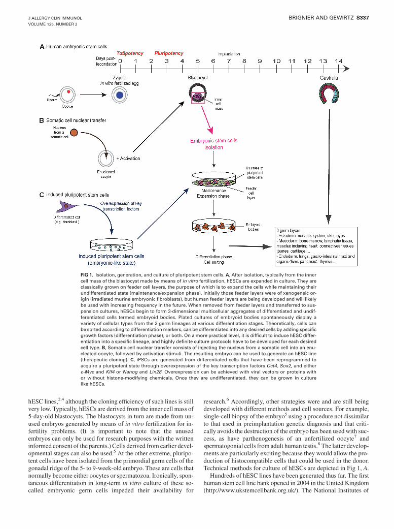
hESC lines,2,4 although the cloning efficiency of such lines is stillvery low. Typically, hESCs are derived from the inner cell mass of5-day-old blastocysts. The blastocysts in turn are made from un-used embryos generated by means of in vitro fertilization for in-fertility problems. (It is important to note that the unusedembryos can only be used for research purposes with the writteninformed consent of the parents.) Cells derived from earlier devel-opmental stages can also be used.5 At the other extreme, pluripo-tent cells have been isolated from the primordial germ cells of thegonadal ridge of the 5- to 9-week-old embryo. These are cells thatnormally become either oocytes or spermatozoa. Ironically, spon-taneous differentiation in long-term in vitro culture of these so-called embryonic germ cells impeded their availability for
research.6 Accordingly, other strategies were and are still beingdeveloped with different methods and cell sources. For example,single-cell biopsy of the embryo5 using a procedure not dissimilarto that used in preimplantation genetic diagnosis and that criti-cally avoids the destruction of the embryo has been used with suc-cess, as have parthenogenesis of an unfertilized oocyte7 andspermatogonial cells from adult human testis.8 The latter develop-ments are particularly exciting because they would allow the pro-duction of histocompatible cells that could be used in the donor.Technical methods for culture of hESCs are depicted in Fig 1, A.
Hundreds of hESC lines have been generated thus far. The firsthuman stem cell line bank opened in 2004 in the United Kingdom(http://www.ukstemcellbank.org.uk/). The National Institutes of
FIG 1. Isolation, generation, and culture of pluripotent stem cells. A, After isolation, typically from the innercell mass of the blastocyst made by means of in vitro fertilization, hESCs are expanded in culture. They areclassically grown on feeder cell layers, the purpose of which is to expand the cells while maintaining theirundifferentiated state (maintenance/expansion phase). Initially those feeder layers were of xenogeneic or-igin (irradiated murine embryonic fibroblasts), but human feeder layers are being developed and will likelybe used with increasing frequency in the future. When removed from feeder layers and transferred to sus-pension cultures, hESCs begin to form 3-dimensional multicellular aggregates of differentiated and undif-ferentiated cells termed embryoid bodies. Plated cultures of embryoid bodies spontaneously display avariety of cellular types from the 3 germ lineages at various differentiation stages. Theoretically, cells canbe sorted according to differentiationmarkers, can be differentiated into any desired cells by adding specificgrowth factors (differentiation phase), or both. On a more practical level, it is difficult to induce hESC differ-entiation into a specific lineage, and highly definite culture protocols have to be developed for each desiredcell type. B, Somatic cell nuclear transfer consists of injecting the nucleus from a somatic cell into an enu-cleated oocyte, followed by activation stimuli. The resulting embryo can be used to generate an hESC line(therapeutic cloning). C, iPSCs are generated from differentiated cells that have been reprogrammed toacquire a pluripotent state through overexpression of the key transcription factors Oct4, Sox2, and eitherc-Myc and Klf4 or Nanog and Lin28. Overexpression can be achieved with viral vectors or proteins withor without histone-modifying chemicals. Once they are undifferentiated, they can be grown in culturelike hESCs.
J ALLERGY CLIN IMMUNOL
VOLUME 125, NUMBER 2
BRIGNIER AND GEWIRTZ S337
Health registry (http://stemcells.nih.gov/research/registry/) hasalso archived a number of hESC lines and established criteriafor demonstration of the pluripotency of these lines. Specifically,cells should be able to give rise to any cell lineage of the body andthus to form a teratoma (a tumor containing tissues from the 3 pri-mary germ layers) in vivo after injection in an immune-compro-mised animal and should be capable of unlimited self-renewal.
Nuclear reprogramming and induced pluripotencyNuclear reprogramming is a procedure that causes changes in
gene expression that allow a cell of one type to develop into a cellof another type.9 Recent strategies for generating stem cells arefocused on nuclear reprogramming of differentiated cells to forcethem to become pluripotent. An example is somatic cell nucleartransfer (Fig 1, B). This consists of injection of the nucleus of asomatic cell into an enucleated oocyte.10 The resulting pluripotentcells are genetically matched with the cell donor (this technique isthereby often called ‘‘therapeutic cloning’’), except for the mito-chondrial DNA, which comes from the egg. Another method isaccomplished by means of cell fusion with an hESC, which canproduce cells with some stem cell characteristics.11
Avery recent and very exciting advance in reprogramming hasbeen the generation of induced pluripotent stem cells (iPSCs).First reported in 2006 using murine fibroblasts,3 iPSCs can bemade from multiple murine and human somatic cell types,12,13
and it is now possible to create patient-specific iPSCs.14 iPSCscan be generated from differentiated cells by using retroviral-me-diated expression of core transcription factors known to be re-quired for maintenance of pluripotency and proliferation ofembryonic stem cells.3 These genes are Oct4, Sox2, and eitherc-Myc and Klf4 orNanog and Lin28 (Fig 1,C). iPSCs exhibit sim-ilar features to embryonic stem cells, including cell morphology,cell-surface markers, growth properties, telomerase activity, ex-pression, and epigenetic marks (ie, methylation or acetylationof histones, which result in changes in gene expression) of plurip-otent cell–specific genes12,13 but not global gene expressionsignatures.15 They can give rise to cells derived from all 3 germlayers in vitro and in vivo, and murine iPSCs injected into murineblastocysts have been shown to contribute to embryonicdevelopment.3
Using pluripotent stem cells in the clinic: Scientific/medical issues
A number of scientific/medical issues need to be addressedbefore stem cells can be considered safe for clinical applications.The first hurdle is the tumorigenic potential of pluripotent cells(hESCs and iPSCs). Because pluripotency is evidenced by theability to form teratomas when transplanted in immunodeficientmice, the concern exists that these cells could form malignanttumors in their new host. One strategy for dealing with thisproblem is to select pure populations of more committed cells fortransfer. Demonstrating genetic and epigenetic stability willtherefore be important before these cells are used clinically. Infact, karyotypic abnormalities have been described in severalhESC lines, although changes might be at least partially depen-dent on culture techniques.16
In additional to biologic issues directly affecting the stem cellproduct, it is imperative that controlled, standardized practices andprocedures be followed to maintain the integrity, uniformity, and
reliability of the human stem cell preparations. Because stem cellsare both maintained and expanded in vitro before transplantation,culture conditions compatible with human administration must beused. Feeder cells and sera of animal origin have to be reducedand ideally avoided to reduce the potential risk of contaminationby xenogeneic protein and pathogens. Finally, transplantation ofhESCs into patients is also limited by potential HLA incompatibil-ity. Consequently, life-long immunosuppressive therapy, which canlead to infections and organ-based toxic side effects, such as ne-phropathy, might be required to prevent graft rejection. In this re-gard iPSCs hold great promise because they are histocompatiblewith the patient and because their use avoids one of the major eth-ical concerns (see below) associated with hESCs.
Although iPSCs solve the tissue-barrier problem, they too havetechnical drawbacks that are presently limiting their use. First isthe issue of the risk of insertional mutagenesis caused by viralintegration into the genome. This is of particular concern becausepatients who have received gene-modified lymphoid cells havehad aggressive leukemias as a result of this phenomenon (seebelow).17 The possibility that iPSCsmight be generated with non-integrating expression plasmids or adenoviral vectors is being ex-plored in the murine system and appears possible.18,19
Another risk is reactivation of a viral oncogene, such as c-Myc,used to engineer the cells. Here there are data to suggest that theuse of histone-modifying chemicals, such as the histone deacety-lase inhibitor valproic acid, improves reprogramming efficiencyand avoids the need to use c-Myc and that Oct4 and Sox2 aloneare then sufficient in the generation of human iPSCs.20 Recently,iPSCs were successfully obtained from murine fibroblasts cul-tured without any genetic material at all by using only valproicacid and recombinant proteins for the necessary transcription fac-tors.21 However, even as technical hurdles are overcome, genera-tion of iPSCs still suffers from low efficiency and high cost,although no doubt these problems will be solved in time as well.In particular, the reprogramming efficiency is typically less than1% but could depend on the differentiation stage of the cells. In-deed, the efficiency of iPSCgenerationhas been recently increasedto 28% with the use of hematopoietic stem and progenitor cells.22
Adult stem cellsThe best-known example of the adult stem cell is the HSC,
which is located in the bone marrow niche. HSCs and progenitorscan be readily harvested from bone marrow and umbilical cordblood (UCB). They can even be collected from peripheral bloodafter mobilization from the marrow with granulocyte colony-stimulating factor (G-CSF) with or without CXCR4 antagonist.23
HSCs are characterized by the expression of cell-surface markers,which allows for their isolation. In human subjects the HSCsurface phenotype is typically lineage-specific antigen negative(lin2), CD341CD382CD1331c-Kit/CD1171CD591Thy1/CD901CXCR41. Apart from differentiating into all myeloidand lymphoid lineages, HSCs have been shown to be able to dif-ferentiate in vitro into cells of nonhematopoietic lineages. How-ever, such plasticity was probably an experimental artifact thatis currently explained by the presence of heterogeneous popula-tions of non-HSCs in hematopoietic organs or by the phenomenonof cell fusion.
Mesenchymal stem cells (MSCs) are another type of adultmultipotent cells that are capable of differentiating into variousmesodermal cell lineages, including myocytes, osteoblasts,
J ALLERGY CLIN IMMUNOL
FEBRUARY 2010
S338 BRIGNIER AND GEWIRTZ
chondroblasts, fibroblasts, adipocytes, and other stromal ele-ments. MSCs are present in almost all organs, but for therapeuticpurposes, they are most conveniently isolated from bone marrowandUCB.MSCs can be organ specific. Consequently, populationsisolated from various sources, although morphologically similar,might be functionally different. For example, MSCs isolated fromthe umbilical cord do not have the same abilities to give rise toosteoblasts, chondrocytes, and cardiomyocytes as bone marrow–derived MSCs.24 MSCs can be readily expanded ex vivo andmanipulated, if needed, to acquire specific properties. The Inter-national Society for Cellular Therapy recommended changingtheir name to ‘‘multipotent mesenchymal stromal cells’’ becausethe majority of MSCs lack complete ‘‘stemness’’ property andproposed minimal criteria for standardization of preparations.25
Human MSCs must be plastic adherent; express CD105, CD73and CD90; lack hematopoietic markers (CD45, CD34, CD14 orCD11b, CD79a or CD19 m and HLA-DR); and be able to differ-entiate into osteoblasts, adipocytes, and chondroblasts in vitro.MSCs display trophic, anti-inflammatory, and immunomodula-tory capacities, both through secretion of soluble factors (indole-amine 2,3-dioxygenase, IL-6, TGF-b1, hepatocyte growth factor,inducible nitric oxide synthase, and prostaglandin) and directcell-to-cell interaction with immune cells. In vitroMSCs suppresseffector and cytotoxic T-cell, B-cell, natural killer (NK) cell, anddendritic cell activity and can induce regulatory T cells.26 How-ever, how MSCs assist in repairing a damaged organ is still un-clear. Mounting evidence suggests that direct substitution ofinjured cells by in situ differentiated MSCs is unlikely (althoughstill argued). Establishment of a favorable environment or nichefor reconstruction of the tissue by intrinsic stem cells per se seemsmore likely. Regardless, because of their low immunogenicity andclaimed beneficial effects on organ regeneration (whatever themechanism), MSCs are being examined in an increasing numberof regenerative medicine applications, as well as in inflammatoryand immunologic diseases.
Finally, amnioticfluid,UCB, and theplacenta areother sourcesofnonembryonic stem cells. However, it is not clear yet whether theyare pluripotent ormultipotent and how clinically useful theywill be.
POTENTIAL CLINICAL USES OF STEM CELLSStem cells are postulated to have a tremendous number of
applications, but tissue engineering seems to generate the greatestexcitement. Stem cells can be used in regenerative medicine,immunotherapy, and gene therapy. Animal models and clinicalstudies have shown that transplantation of stem cells from diverseorigins can successfully treat many acute and chronic diseases,such as immunohematologic disorders, type 1 diabetes mellitus(T1DM), Parkinson disease, neuronal destruction, and congestiveheart failure.
Hematology-immunologyDuring the last 50 years, allogeneic hemopoietic stem cell
transplantation (HSCT) has progressively become a commonprocedure for the treatment of a variety of inherited or acquiredimmunohematologic diseases, including thalassemias, sickle celldisease, Fanconi anemia, inborn errors of metabolism, severeaplastic anemia, severe combined immunodeficiency (SCID), andother primary immune deficiencies. HSCT is also widely used forthe treatment of hematologic malignancies, such as acute myeloid
and lymphoid leukemias, chronic myeloid leukemia and othermyeloproliferative syndromes, myelodysplastic disorders, lym-phoma, myeloma, and even solid tumors, such as renal cellcancer, breast cancer, ovarian carcinoma, and neuroblastoma.27
The aim of allogeneic HSCT in malignancies is not only asubstitution of the malignant bone marrow but also a form ofadoptive immunotherapy. In the context of HLA compatibility,donor allogeneic T lymphocytes detect differences in minorhistocompatibility antigens in both the host and the tumor and candestroy the residual malignant cells, thereby contributing to thecure of the patient. This is the graft-versus-tumor or graft-versus-leukemia (GVL) effect. Particularly in the case of disease relapseafter transplantation, donor lymphocyte infusionsmight induce orenhance a GVL effect and reinduce the patient into remission.
Major complications of HSCT include organ toxicity from theconditioning regimen used and graft-versus-host disease(GVHD), in which the donor’s immune system destroys therecipient’s normal tissues, particularly the skin, gastrointestinaltract, and liver. Other important complications of HSCT are graftfailure, infertility, growth retardation in children, and secondarycancers thought to arise as a result of chronic immunosuppressionand DNA-damaging preparative regimens.
In an effort to decrease these complications, several strategieshave been developed. First among these are the reduced-intensityconditioning regimens, or so-called minitransplantations, whichhave made HSCTavailable to older and less fit patients.28 Signif-icant immunosuppression in these patients and GVHD as a resultof the conditioning regimen remain serious problems and havesuggested to many that the use of the term ‘‘mini’’ is misleadingwith respect to potential complications.
Second, the cell source has also been examined with respect tocomplications. Several studies convincingly show that CD341
cells harvested from peripheral blood engraft faster but are asso-ciated with a higher incidence of GVHD.29
Finding a histocompatible donor remains a problem for manypatients in need of HSCT. Ideally, one would like to use an HLA-matched sibling as a donor. If such a donor is not available, then amatched unrelated donor is an acceptable alternative. This can bea major problem for minority groups underrepresented in regis-tries of volunteer donors, information on which is centralized inthe BoneMarrowDonorsWorldwide database. In the absence of acompatible donor, HLA-haploidentical mismatched HSCT froma relative can be performed,30 generally T depleted to avoidGVHD. In that context NK cells could facilitate engraftmentand display an antileukemic activity without GVHD.31
Finally, UCB transplantation has the potential to significantlyenlarge the number of potential HSCT recipients. UCBs arerapidly and easily available from cord blood banks and can be usedwhen only partially HLA matched because they are much lesslikely to induce acute and chronic GVHD.32 Remarkably, the GVLeffect seems to be preserved, likely as a result of NK cells presentin the cord blood preparation.33 Issues that remain to be solved aredelayed engraftment, prolonged T lymphopenia, and defectivethymopoiesis.34 In addition, it is not currently possible to performdonor lymphocyte infusions in the case of relapse, but an ongoingclinical trial is testing ex vivo expansion of UCB T cells. Becausethe number of HSCs per unit of UCB is small, thereby limitingtheir ability to be used for adult patients, strategies using combinedunits or ex vivo expanded cells are being developed.35,36
Autologous hematopoietic stem cell transplantation (AHSCT)is commonly performed in certain settings as well. Its main role is
J ALLERGY CLIN IMMUNOL
VOLUME 125, NUMBER 2
BRIGNIER AND GEWIRTZ S339
to lessen the period of aplasia (rescue therapy) after high-dosechemotherapy and thereby lessen the risk of infection andbleeding. When used for the treatment of hematologic malignan-cies and solid tumors, HSCs to be used for AHSCTare commonlycollected after few cycles of chemotherapy to lessen contamina-tion of the graft by tumor cells. Moreover, autoimmune diseases,such as multiple sclerosis, rheumatoid arthritis, and systemiclupus erythematosus, might benefit from AHSCT. It is thoughtthat immunoablative therapy resets the immune system byeliminating autoreactive Tand B lymphocytes, lessening memoryT cells, increasing thymus-derived naive T cells, generating adiverse but distinct T-cell receptor repertoire,37 and promotingregulatory T cells.38 Several recent studies have shown, as mightbe expected, that nonmyeloablative and low-intensity myeloabla-tive regimens have fewer treatment-related complications andmortality than high-intensity myeloablative regimens39 and thatbest results might be obtained during the inflammatory phase ofautoimmune disease. Allogeneic HSCT has also been used forthe treatment of autoimmune disease on the theory of both im-mune reset and correction of the genetic predisposition, but therisks of GVHD and infection are substantial, and therefore thisform of therapy should be reserved only for treatment of very se-rious and refractory disease.39
Finally, the immunomodulatory capability of MSCs is beingtested in the treatment of patients with systemic lupus erythema-tosus, multiple sclerosis, Crohn disease, amyotrophic lateralsclerosis, and T1DM. In solid organ transplantation and HSCT,results from preclinical animal studies, although controversial,suggest that donor-derived MSCs could have a tolerogenic effectand might therefore be used for prevention and/or treatment ofgraft rejection and GVHD.40-42
T1DMT1DM is an autoimmune disease resulting from the destruction
of pancreatic insulin-producing b cells in the islets of Langer-hans. Insulin replacement therapy, even when rigorously con-trolled, is often not efficient enough to prevent long-termcomplications of the disease. Transplantation of a whole pancreasor isolated mature islets can restore proper glucose regulation, butthe former is amorbid high-complication procedure, and the latterappears to be only a transient solution.43 For these reasons, othersources of b cells suitable for transplantation are being sought.Pancreatic stem cells have been identified in ductal epitheliumof injured pancreas on the basis of expression of the transcriptionfactor neurogenin 3.44 Efforts are now being directed towardmethods to expand these cells ex vivo or to stimulate their prolif-eration in vivo.
Generation of insulin-secreting cells from hESCs and iPSCsholds great promise for the cure of T1DM. hESC-derivedpancreatic endoderm can be differentiated in vivo into glucose-re-sponsive insulin-secreting cells in immunodeficient mice withstreptozotocin-induced diabetes (ab cell–selective destruction).45
Unfortunately, teratoma formation was found in approximately15% of the recipient mice and is a safety concern. The immuno-logic incompatibility of hESCs could be resolved by using bioen-gineered porous capsules, which are designed to protect the graftfrom immune cells but remain permissive to the passage of smallmolecules. In regard to iPSCs, a particularly exciting study fromZhou et al46 reported the reprogramming of differentiated murinepancreatic exocrine cells into b-like cells in vivo. These
investigators found that transient expression of 3 key developmen-tal transcription factors, neurogenin 3, MafA, and Pancreatic du-odenal homeobox-1 (Pdx1), by adenoviral vectors injecteddirectly into the pancreas was sufficient to reprogram exocrinecells into insulin-secreting cells responding to hyperglycemia.
The immunomodulatory effects of MSCs are also beingexplored in the setting of T1DM. At the time of diagnosis,b-cell destruction is often not yet complete. In theory, ameliora-tion of the immune attack might allow the survival of the residualislet cells. In diabetic immunodeficient mice human MSCsdecreased hyperglycemia and increased endogen insulin levelsand b-cell numbers.47 Clinical studies testing allogeneic MSCs inpatients with recently diagnosed diabetes are ongoing.
Finally, nonmyeloablative AHSCT was performed in 23 pa-tients with early-onset T1DM.48 Twenty enjoyed a variable insu-lin-free period, and 12 of these patients remained insulin free aftera mean follow-up of 31 months. Interestingly, benefit was demon-strated in those patients with transient responses as well. In thissmall group (8 patients) daily insulin doses were significantly di-minished, and C-peptide levels (reflective of endogenous insulinsynthesis) were increased.
Diseases of the nervous systemIt was thought for a long time that nerve cells do not divide in
the adult mammalian brain, but this has now been shown to beincorrect. Neurogenesis not only occurs during prenatal andpostnatal development but also in adults. Neurogenic niches havebeen identified in the subventricular zone of the lateral ventriclesand in the subgranular zone of the dentate gyrus of the hippo-campus. In brain-injury models neural stem cells (NSCs) prolif-erate in those neurogenic regions and are even able to migratetoward the site of damage.49 NSCs are multipotent and capable ofself-renewing. In vitro they cluster in ‘‘neurospheres,’’ which areable to differentiate into the 3 major neuroectodermal lineages(neurons, astrocytes, and oligodendrocytes).
Neural stem/progenitor cells can be isolated from embryonic,fetal, or adult brain tissue by sorting cells on the basis of nestinexpression primarily, as well as other markers, and can then beplaced into culture for expansion. Clearly, for autologous stemcell therapy,more accessible NSCs are required. Curiously, dentalpulp and peridontium have been shown to be sources of NSC, aswell as olfactory mucosa, which is readily harvested by means ofnasal biopsy.50 Investigations for NSC-based therapy are ongoingfor various neurologic diseases. The neural repair probably resultsfrom a replacement of defective cells but also from neuroprotec-tive, trophic, and immunomodulatory effects. To date, the idealNSC source, schedule, and route of transplantation have notbeen established and are likely to be disease specific. The useof NSCs to treat Parkinson disease might be instructive in thisregard.
Parkinson disease is an incurable, progressive, neurodegener-ative disease that affects dopaminergic neurons. Levodopa, whichis converted to dopamine in the brain, is the mainstay oftreatment, but most patients acquire tachyphylaxis to its effectsover time. In contrast to patients with diabetes, in whomtransplantation of islet cells is a therapeutic option, implantationof fully differentiated dopamine-releasing neurons into the brainis not presently feasible because such cells do not survive.Transplantation of embryonic/fetal nigral dopaminergic neuronswas tested in 2 double-blind, placebo-controlled trials, but results
J ALLERGY CLIN IMMUNOL
FEBRUARY 2010
S340 BRIGNIER AND GEWIRTZ
were not as encouraging as those from previous open-trialreports.51,52 However, modest clinical improvement was notedin some patients, and striatal fluorodopa uptake was significantlyenhanced. Unfortunately, several patients subsequently had dys-kinesias. Postmortem examination of the brains of some patientsprovided evidence that transplanted dopamine neurons can differ-entiate and survive for many years without immunosuppression.However, it appeared that at least some of the grafted tissue wasinvolved by disease over a period lasting from 9 to 16 years.53
Widespread application of this therapy will likely be limited aslong as access to fetal donor tissue is required. Moreover, thesafety of those cells is not completely assessed because theyhave not been tested in a large number of patients and because de-velopment of cerebral mass lesions of donor origin was reportedin a patient less than 6 years after fetal transplantation forHuntington disease.54 Accordingly, finding alternative sourcesof NSCs for therapeutic purposes is the object of intenseinvestigation.
In one case autologous NSCs were harvested by means ofcerebral biopsy, expanded, and differentiated into dopaminergicneurons ex vivo and then injected back to the patient’s putamen 9months later.55 Clinical evaluation and fluorodopa uptake wereimproved after transplantation, but all benefits had disappearedby 5 years. In animal studies other stem cell types partially alle-viated Parkinson symptoms, including MSCs,56 olfactoryNSCs,57 hESC-derived neurons,58 and iPSCs.59 Human clinicaltrials with cells of these types can therefore be anticipated.
Unlike Parkinson disease, which theoretically requires replace-ment of only 1 cell type, therapies for other neurologic diseases,such as stroke and spinal cord injury, in which large numbers ofcells of many types (neurons, glia, and endothelial cells) aredestroyed, face much larger hurdles. In recent years, human NSCsfrom diverse origins, including hESCs, HSCs, and MSCs, havebeen tested in preclinical models of ischemic stroke. Theseexperiments have enabled the development of treatment strategiesand have demonstrated the critical importance of transplantationtiming for clinical success. Only a few clinical studies have beenperformed for the treatment of stroke. Stereotactic injection ofneuronal cells derived from embryonal carcinoma cell line (NT2/D1) did not display significant benefits compared with controlresults, but some patients experienced improvement.60 In otherstudies an investigation into the utility of fetal porcine cells wasstopped because of adverse events (temporaryworsening of deficitsand seizures) in 2 of 5 patients,61 whereas intravenous infusion ofMSCs was shown to be safe without significant benefit. HSCs arecurrently being evaluated in phase I/II protocols. The injection ofspecific growth factors to stimulate proliferation of intrinsic neuro-progenitors in the brain is a novel approach to this problem.62
Spinal cord injury often results in permanent motor deficiency,sensory deficiency, or both, thereby rendering treatment particu-larly challenging. In a small number of paraplegic and quadriple-gic patients, olfactory NSCs have been injected into intralesionaland perilesional areas.50 Feasibility and safety were acceptable,but unfortunately, clinical improvement remained slight after 3years of follow-up. Logically, early-phase treatment might yieldthe best results. In fact, a phase I/II clinical trial tested infusionof HSCs into the spinal cord associated with G-CSF injections:the Association Impairment Scale grade improved in 30.4% of pa-tients treated quite early (<8 weeks) after the initial lesion; how-ever, no enhancement was noted when the treatment wasperformed later.63
Cardiac repairCongestive heart failure afflicts millions of persons around the
world, with 400,000 new cases being reported each year in theUnited States alone. The most common cause is coronary arterydisease. After myocardial necrosis has occurred, the cell loss isirreversible, and although many medical and surgical treatmentsare available for the subsequent congestive heart failure, the long-term prognosis of these patients remains guarded, with a 5-yearmortality of 50%. Transplanting stem cells would have clearadvantages to transplanting a heart because it would obviate theconstraint of a donor and, in case of autologous cells, for therequisite immunosuppression.
hESC-derived cardiomyocytes (hESC-CMs) have been suc-cessfully generated. Intramyocardial injection of hESC-CMs afew days after infarction in immunodeficient rodents seemed toenhance left ventricular ejection fraction (LVEF) compared withthat seen in a control group when evaluated at 4 weeks.64 Unfor-tunately, this enhancement was not sustained after 12 weeks offollow-up. Another study suggested that a coinfusion of hESC-CMs and MSCs in mice was of benefit because, according tothe authors, a ‘‘synergistic trophic effect that enhanced repair ofinjured host tissue’’ was brought about.65 Importantly, no tera-toma was found in animals receiving hESC-CMs.64,65
Despite a controversial plasticity in vitro, a considerableamount of data from actual preclinical studies suggest that it is un-likely that transdifferentiation of HSCs and MSCs into functionalcardiomyocytes happens to any significant degree in vivo. In veryspecific culture conditions, MSCs might be driven toward differ-entiating into cardiomyocyte-like cells at a very low frequency(approximately 0.07%) that would not be enough for cardiac re-pair.24 It is now generally agreed that the transplanted cells exerttheir beneficial role through paracrine effects and by creating a fa-vorable trophic environment for intrinsic cell recovery, enhancingangiogenesis, and limiting ventricular remodeling.66 A studycomparing the efficacy of transplanted bonemarrowmononuclearcells, MSCs, skeletal myoblasts, and fibroblasts in mice with ex-perimental myocardial infarcts was carried out and showed thatHSCs had the most beneficial effect on left ventricular functionin that model.67 Recently, infusion of endogenous cardiac stemcells isolated from endomyocardial biopsy specimens and ex-panded ex vivo appeared to enhance myocardial viability andLVEF in a murine infarction model.68
Intracoronary infusions of HSCs69 or MSCs70 have been per-formed a few days after percutaneous coronary intervention foracute myocardial infarction. Despite contradictory results of clin-ical trials, meta-analyses reported moderate but significant bene-fits of such therapy compared with the condition of controlpatients, with improvement in LVEF, infarct size, and end-sys-tolic volume.71 In patients with chronic ischemic disease, intra-coronary and intramyocardial injections of HSCs are associatedwith modest enhancements as well.71 Other cell types are also be-ing tested, such as skeletal myoblasts (although lack of electricalsynchronization with cardiomyocytes could potentially be ar-rhythmogenic) or endothelial progenitor cells.
Stem cells and gene therapyThe goal of gene therapy is to cure diseases caused by
malfunctioning genes. It does so by substituting the function ofa normal gene for the one that causes disease. Until now, the mostcommonly used procedure in human gene therapy clinical trials is
J ALLERGY CLIN IMMUNOL
VOLUME 125, NUMBER 2
BRIGNIER AND GEWIRTZ S341
the insertion of a normal copy of the target gene in a nonspecificlocation into the host genomic DNA. The therapeutic transgene ispackaged into a delivery vehicle, which is typically a replication-deficient virus. Nonintegrating virus (adenovirus or adeno-asso-ciated virus) can be used in nondividing cells, such as neurons andcardiomyocytes. In dividing cells, such as stem cells, vectors thatintegrate into host DNA, such as g-retrovirus or lentivirus, arerequired to have a transmission to daughter cells.
Stem cells are of great benefit to cell-based gene therapybecause they are self-renewing and thusmight reduce or eliminatethe necessity for repeated administrations of the therapeutic cells.Single-gene inherited diseases are particularly good candidatesfor gene therapy. In theory the host’s own stem cells can berepaired through genetic engineering and then used in an autol-ogous transplantation. This avoids all the risks of transplantedallogeneic cells, including the risks associated with long-termimmunosuppression, as well as GVHD, in patients receivingHSCT. The first clinical trials with engineered HSCs involvedpatients with genetic immunodeficiency diseases, such as aden-osine deaminase–deficient SCID.72 Trials have also been carriedout in patients with X-linked SCID (g-common [g-c] cytokine re-ceptor deficient or SCID-X1)17 and chronic granulomatous dis-ease.73 The clinical results have been quite promising but havebeen marred by the development of leukemia, which has beenshown to be caused by insertional mutagenesis in a number ofthese patients (see below). Gene therapy for hemoglobinopathies,such as b-thalassemia and sickle cell disease, are ongoing. Easilyaccessible mucosal and skin stem cells are also being used, for ex-ample in treatment of diseases such as junctional epidermolysisbullosa.74
These early studies revealed problems that need to beaddressed, such as difficulties controlling protein levels withoutendogenous gene regulatory regions, maintenance of gene ex-pression through long periods, low protein production, andinsertional mutagenesis of the retroviral transgene vector. Indeed,the major side effect was thus far the occurrence of T cell–acutelymphoblastic leukemia in 5 of 19 patients successfully treated forSCID-X1 in 2 distinct French and British trials.17,75 In all casesthe retroviral vector was found in the leukemic clone, integratednear a proto-oncogene, and particularly before the LIM do-main–only 2 in 4 cases and was associated with acquired somaticmutations. g-Retroviral vectors were subsequently shown to inte-grate preferentially in the 59 ends of genes76 near transcriptionstart sites in a nonrandom manner near genes that provide selec-tive advantage to the clone. Interestingly, when the same retroviralvector, the murine leukemia virus, was used to deliver other trans-genes, no case of leukemia was observed, suggesting that g-c re-ceptor overexpression might be involved in the oncogenesis.
New techniques are being developed to enhance efficiency andto avoid the risk of insertional oncogenesis. First, safer deliverysystems are being developed. For example, HIV-derived lentivirusis able to transduce nondividing cells, is easier to use, and caninduce less mutagenesis than g-retrovirus, as assessed in murinemodels.77 Other modifications under development include the useof inducible and tissue-specific promoters, a weaker viral pro-moter/enhancer, and self-inactivating retroviral vectors and intro-duction of suicide genes. At last, the improvement of direct genecorrection with homologous recombination (a normal copy of thegene is switched with the defective allele) is promising in murinemodels.78 That last technology could be particularly significant inthe treatment of dominant genetic diseases.
The potential utility of hESCs and iPSCs was discussed earlier,but the use of such cells is under active investigation for humangene therapy.
Finally, new attempts are focusing on cell-based deliveryvehicles for tumor-specific therapy. MSCs appear to be goodagents for this purpose because, in addition to their propertiesdescribed above, they have been shown to migrate toward tumors.For example, MSCs engineered to express the TNF-relatedapoptosis-inducing ligand induced apoptosis in tumor cells,reduced tumor growth in vivo, and prolonged survival in murinemodels of human glioma.79
ETHICAL ISSUESThe use of hESCs in medical research has drawn much
attention from many sectors of the public. Religious, historical,cultural, medical, and other points of view have contributed to avery vigorous and wide-ranging discourse over the use of thesematerials.80 Some consider research with hESCs to be inherentlyimmoral because these individual’s believe that life begins withfertilization of the ovum, and the destruction of an embryo withthe potential to develop into a viable human being is thought tan-tamount to infanticide. For this reason, the American federal gov-ernment severely restricted access and use of hESCs in 2001.These restrictions have now been largely overturned by the Ob-ama administration. In contrast, proponents of this line of re-search insist that the potential benefits to humankind from thisresearch mitigate such concerns. They also argue that hESCsare made from unwanted fertilized ovum that would likely be de-stroyed in any event.
Stem cells created by means of nuclear transfer share the sameethical concerns. Furthermore, because these cells have thepotential to generate a complete embryo, they also raise theeven more highly charged possibility of cloning human beings,so-called reproductive cloning. Many organizations and countrieshave already banned reproductive cloning of human beings.Because this procedure can be used to generate stem cells fortherapeutic purposes, in countries where this type of cloning islegal, such as Australia and the United Kingdom, the createdembryos must be destroyed within 14 days. Federal laws in theUnited States are not clear on the legality of therapeutic cloning,but the Obama administration has pledged establishment of strictguidelines to ensure that cloning research will not be used forhuman reproduction.
Because of the shortage of human oocytes, generation ofhuman-animal chimeras was legalized in 2008 in the UnitedKingdom for research purposes only. A human nucleus istransferred into an animal’s oocyte, creating a hybrid embryothat must be destroyed within 2 weeks and cannot be implanted.Clearly, creation of such tissues raises even more complex issues.
Finally, the issue of financial compensation for embryo andgamete donors is also controversial, with guidelines for thisproblem being proposed by the International Society of Stem CellResearch (http://www.isscr.org/guidelines/index.htm). All partiesinvolved in the debate want very much to avoid the developmentof an underground black market in spare embryos.
CONCLUSIONS AND PERSPECTIVESThe promise of stem cell therapeutics powers the field of
regenerative medicine and has generated a huge amount of
J ALLERGY CLIN IMMUNOL
FEBRUARY 2010
S342 BRIGNIER AND GEWIRTZ
excitement, anticipation, and hope. Accordingly, research withhESCs is increasing exponentially worldwide, particularly in theUnited States, where important limitations on research with suchcells were overturned in 2009. Furthermore, the US Food and DrugAdministration recently approved the world’s first phase I clinicaltrial using hESC-based therapy in patients with spinal cord injury.
Nonetheless, a number of substantive scientific and ethicalissues remain to be resolved before hESCs can enter the thera-peutic mainstream. In the meantime, recent breakthroughs ingenerating iPSCs would obviate the need to solve the most vexingof these problems. In fact, it seems reasonable to hope that in thenext few years many of the enabling issues relevant to iPSCs willbe solved, allowing the field of regenerative medicine to deliveron its vast potential promise.
Although it is difficult to predict the ultimate utility of stemcell–based therapy at this time, it is not difficult to conclude thatthis is an extremely important area of scientific research.Surrounded by controversy and a good many ethical concerns,thoughtful legislative action could both foster the field and ensurecontinued progress. This would clearly be more desirable thanhaving the whole endeavor driven underground and potentiallyinto the hands of less ethical and less regulated scientists. Opendiscussions between political bodies and the various interestgroups in the scientific, medical, and religious communities needto take place to address the concerns of each and to provide anultimate solution that is clearly in the interest of humanity.
We thank M. C. Tamby, A. Savigner, Y. Auger, and the reviewers for theirhelpful advice on this report.
REFERENCES1. Till JE, McCulloch EA. A direct measurement of the radiation sensitivity of nor-
mal mouse bone marrow cells. Radiat Res 1961;14:213-22.2. Thomson JA, Itskovitz-Eldor J, Shapiro SS, Waknitz MA, Swiergiel JJ, Marshall
VS, et al. Embryonic stem cell lines derived from human blastocysts. Science1998;282:1145-7.
3. Takahashi K, Yamanaka S. Induction of pluripotent stem cells from mouse embry-onic and adult fibroblast cultures by defined factors. Cell 2006;126:663-76.
4. Battey JF Jr, Cole LK, Goldthwaite CA Jr. Regenerative Medicine. Department ofHealth and Human Services. August 2006. http://stemcells.nih.gov/info/scireport/2006report.htm. Accessed December 15, 2009.
5. Klimanskaya I, Chung Y, Becker S, Lu SJ, Lanza R. Human embryonic stem celllines derived from single blastomeres. Nature 2006;444:481-5.
6. Turnpenny L, Brickwood S, Spalluto CM, Piper K, Cameron IT, Wilson DI, et al.Derivation of human embryonic germ cells: an alternative source of pluripotentstem cells. Stem Cells 2003;21:598-609.
7. Revazova ES, Turovets NA, Kochetkova OD, Kindarova LB, Kuzmichev LN, Ja-nus JD, et al. Patient-specific stem cell lines derived from human parthenogeneticblastocysts. Cloning Stem Cells 2007;9:432-49.
8. Conrad S, Renninger M, Hennenlotter J, Wiesner T, Just L, Bonin M, et al. Gen-eration of pluripotent stem cells from adult human testis. Nature 2008;456:344-9.
9. Gurdon JB, Melton DA. Nuclear reprogramming in cells. Science 2008;322:1811-5.
10. French AJ, Adams CA, Anderson LS, Kitchen JR, Hughes MR, Wood SH. Devel-opment of human cloned blastocysts following somatic cell nuclear transfer withadult fibroblasts. Stem Cells 2008;26:485-93.
11. Cowan CA, Atienza J, Melton DA, Eggan K. Nuclear reprogramming of somaticcells after fusion with human embryonic stem cells. Science 2005;309:1369-73.
12. Takahashi K, Tanabe K, Ohnuki M, Narita M, Ichisaka T, Tomoda K, et al. Induc-tion of pluripotent stem cells from adult human fibroblasts by defined factors. Cell2007;131:861-72.
13. Yu J, Vodyanik MA, Smuga-Otto K, Antosiewicz-Bourget J, Frane JL, Tian S, et al.Induced pluripotent stem cell lines derived from human somatic cells. Science2007;318:1917-20.
14. Raya A, Rodriguez-Piza I, Guenechea G, Vassena R, Navarro S, Barrero MJ, et al.Disease-corrected haematopoietic progenitors from Fanconi anaemia induced plu-ripotent stem cells. Nature 2009;460:53-9.
15. Chin MH, Mason MJ, Xie W, Volinia S, Singer M, Peterson C, et al. Induced plu-ripotent stem cells and embryonic stem cells are distinguished by gene expressionsignatures. Cell Stem Cell 2009;5:111-23.
16. Mitalipova MM, Rao RR, Hoyer DM, Johnson JA, Meisner LF, Jones KL, et al.Preserving the genetic integrity of human embryonic stem cells. Nat Biotechnol2005;23:19-20.
17. Hacein-Bey-Abina S, Garrigue A, Wang GP, Soulier J, Lim A, Morillon E, et al.Insertional oncogenesis in 4 patients after retrovirus-mediated gene therapy ofSCID-X1. J Clin Invest 2008;118:3132-42.
18. Okita K, Nakagawa M, Hyenjong H, Ichisaka T, Yamanaka S. Generation of mouseinduced pluripotent stem cells without viral vectors. Science 2008;322:949-53.
19. Stadtfeld M, Nagaya M, Utikal J, Weir G, Hochedlinger K. Induced pluripotentstem cells generated without viral integration. Science 2008;322:945-9.
20. Huangfu D, Osafune K, Maehr R, Guo W, Eijkelenboom A, Chen S, et al. Induc-tion of pluripotent stem cells from primary human fibroblasts with only Oct4 andSox2. Nat Biotechnol 2008;26:1269-75.
21. Zhou H, Wu S, Joo JY, Zhu S, Han DW, Lin T, et al. Generation of induced plu-ripotent stem cells using recombinant proteins. Cell Stem Cell 2009;4:381-4.
22. Eminli S, Foudi A, Stadtfeld M, Maherali N, Ahfeldt T, Mostoslavsky G, et al. Dif-ferentiation stage determines potential of hematopoietic cells for reprogramminginto induced pluripotent stem cells. Nat Genet 2009;41:968-76.
23. Flomenberg N, Devine SM, Dipersio JF, Liesveld JL, McCarty JM, Rowley SD,et al. The use of AMD3100 plus G-CSF for autologous hematopoietic progenitorcell mobilization is superior to G-CSF alone. Blood 2005;106:1867-74.
24. Martin-Rendon E, Sweeney D, Lu F, Girdlestone J, Navarrete C, Watt SM. 5-Aza-cytidine-treated human mesenchymal stem/progenitor cells derived from umbilicalcord, cord blood and bone marrow do not generate cardiomyocytes in vitro at highfrequencies. Vox Sang 2008;95:137-48.
25. Dominici M, Le Blanc K, Mueller I, Slaper-Cortenbach I, Marini F, Krause D, et al.Minimal criteria for defining multipotent mesenchymal stromal cells. The Interna-tional Society for Cellular Therapy position statement. Cytotherapy 2006;8:315-7.
26. Selmani Z, Naji A, Zidi I, Favier B, Gaiffe E, Obert L, et al. Human leukocyte an-tigen-G5 secretion by human mesenchymal stem cells is required to suppress Tlymphocyte and natural killer function and to induce CD4 1 CD25highFOXP31regulatory T cells. Stem Cells 2008;26:212-22.
27. Demirer T, Barkholt L, Blaise D, Pedrazzoli P, Aglietta M, Carella AM, et al.Transplantation of allogeneic hematopoietic stem cells: an emerging treatment mo-dality for solid tumors. Nat Clin Pract Oncol 2008;5:256-67.
28. Storb R. Can reduced-intensity allogeneic transplantation cure older adults withAML? Best Pract Res Clin Haematol 2007;20:85-90.
29. Cutler C, Giri S, Jeyapalan S, Paniagua D, Viswanathan A, Antin JH. Acute andchronic graft-versus-host disease after allogeneic peripheral-blood stem-cell andbone marrow transplantation: a meta-analysis. J Clin Oncol 2001;19:3685-91.
30. Aversa F, Terenzi A, Tabilio A, Falzetti F, Carotti A, Ballanti S, et al. Full haplotype-mismatched hematopoietic stem-cell transplantation: a phase II study in patientswith acute leukemia at high risk of relapse. J Clin Oncol 2005;23:3447-54.
31. Ruggeri L, Capanni M, Urbani E, Perruccio K, Shlomchik WD, Tosti A, et al. Ef-fectiveness of donor natural killer cell alloreactivity in mismatched hematopoietictransplants. Science 2002;295:2097-100.
32. Takahashi S, Ooi J, Tomonari A, Konuma T, Tsukada N, Oiwa-Monna M, et al.Comparative single-institute analysis of cord blood transplantation from unrelateddonors with bone marrow or peripheral blood stem-cell transplants from related do-nors in adult patients with hematologic malignancies after myeloablative condi-tioning regimen. Blood 2007;109:1322-30.
33. Lu X, Kondo Y, Takamatsu H, Ohata K, Yamazaki H, Takami A, et al. CD16 1CD56- NK cells in the peripheral blood of cord blood transplant recipients: aunique subset of NK cells possibly associated with graft-versus-leukemia effect.Eur J Haematol 2008;81:18-25.
34. Komanduri KV, St John LS, de Lima M, McMannis J, Rosinski S, McNiece I,et al. Delayed immune reconstitution after cord blood transplantation is character-ized by impaired thymopoiesis and late memory T-cell skewing. Blood 2007;110:4543-51.
35. Ruggeri A, de Latour RP, Rocha V, Larghero J, Robin M, Rodrigues CA, et al.Double cord blood transplantation in patients with high risk bone marrow failuresyndromes. Br J Haematol 2008;143:404-8.
36. Robinson SN, Ng J, Niu T, Yang H, McMannis JD, Karandish S, et al. Superior exvivo cord blood expansion following co-culture with bone marrow-derived mesen-chymal stem cells. Bone Marrow Transplant 2006;37:359-66.
37. Muraro PA, Douek DC, Packer A, Chung K, Guenaga FJ, Cassiani-Ingoni R, et al.Thymic output generates a new and diverse TCR repertoire after autologous stemcell transplantation in multiple sclerosis patients. J Exp Med 2005;201:805-16.
38. Bayer AL, Jones M, Chirinos J, de Armas L, Schreiber TH, Malek TR, et al. HostCD4 1 CD25 1 T cells can expand and comprise a major component of the Tregcompartment after experimental HCT. Blood 2009;113:733-43.
J ALLERGY CLIN IMMUNOL
VOLUME 125, NUMBER 2
BRIGNIER AND GEWIRTZ S343
39. Burt RK, Loh Y, Pearce W, Beohar N, Barr WG, Craig R, et al. Clinical applica-tions of blood-derived and marrow-derived stem cells for nonmalignant diseases.JAMA 2008;299:925-36.
40. Wan CD, Cheng R, Wang HB, Liu T. Immunomodulatory effects of mesenchymalstem cells derived from adipose tissues in a rat orthotopic liver transplantationmodel. Hepatobiliary Pancreat Dis Int 2008;7:29-33.
41. Ball LM, Bernardo ME, Roelofs H, Lankester A, Cometa A, Egeler RM, et al. Co-transplantation of ex vivo expanded mesenchymal stem cells accelerates lympho-cyte recovery and may reduce the risk of graft failure in haploidenticalhematopoietic stem-cell transplantation. Blood 2007;110:2764-7.
42. Le Blanc K, Frassoni F, Ball L, Locatelli F, Roelofs H, Lewis I, et al. Mesenchymalstem cells for treatment of steroid-resistant, severe, acute graft-versus-host disease:a phase II study. Lancet 2008;371:1579-86.
43. Shapiro AM, Ricordi C, Hering BJ, Auchincloss H, Lindblad R, Robertson RP,et al. International trial of the Edmonton protocol for islet transplantation. NEngl J Med 2006;355:1318-30.
44. Xu X, D’Hoker J, Stange G, Bonne S, De Leu N, Xiao X, et al. Beta cells can begenerated from endogenous progenitors in injured adult mouse pancreas. Cell2008;132:197-207.
45. Kroon E, Martinson LA, Kadoya K, Bang AG, Kelly OG, Eliazer S, et al. Pancre-atic endoderm derived from human embryonic stem cells generates glucose-re-sponsive insulin-secreting cells in vivo. Nat Biotechnol 2008;26:443-52.
46. Zhou Q, Brown J, Kanarek A, Rajagopal J, Melton DA. In vivo reprogramming ofadult pancreatic exocrine cells to beta-cells. Nature 2008;455:627-32.
47. Lee RH, SeoMJ, RegerRL, Spees JL, Pulin AA,Olson SD, et al.Multipotent stromalcells from human marrow home to and promote repair of pancreatic islets and renalglomeruli in diabetic NOD/scidmice. Proc Natl Acad Sci U SA 2006;103:17438-43.
48. Couri CE, Oliveira MC, Stracieri AB, Moraes DA, Pieroni F, Barros GM, et al. C-peptide levels and insulin independence following autologous nonmyeloablativehematopoietic stem cell transplantation in newly diagnosed type 1 diabetes melli-tus. JAMA 2009;301:1573-9.
49. Arvidsson A, Collin T, Kirik D, Kokaia Z, Lindvall O. Neuronal replacement fromendogenous precursors in the adult brain after stroke. Nat Med 2002;8:963-70.
50. Mackay-Sim A, Feron F, Cochrane J, Bassingthwaighte L, Bayliss C, Davies W,et al. Autologous olfactory ensheathing cell transplantation in human paraplegia:a 3-year clinical trial. Brain 2008;131:2376-86.
51. Freed CR, Greene PE, Breeze RE, Tsai WY, DuMouchel W, Kao R, et al. Trans-plantation of embryonic dopamine neurons for severe Parkinson’s disease. N Engl JMed 2001;344:710-9.
52. Olanow CW, Goetz CG, Kordower JH, Stoessl AJ, Sossi V, Brin MF, et al. A dou-ble-blind controlled trial of bilateral fetal nigral transplantation in Parkinson’s dis-ease. Ann Neurol 2003;54:403-14.
53. Braak H, Del Tredici K. Assessing fetal nerve cell grafts in Parkinson’s disease.Nat Med 2008;14:483-5.
54. Keene CD, Chang RC, Leverenz JB, Kopyov O, Perlman S, Hevner RF, et al. Apatient with Huntington’s disease and long-surviving fetal neural transplants thatdeveloped mass lesions. Acta Neuropathol 2009;117:329-38.
55. Levesque MF, Neuman T, Rezak M. Therapeutic microinjection of autologousadult human neural stem cells and differentiated neurons for Parkinson’s disease:five-year post-operative outcome. Open Stem Cell J 2009;1:20-9.
56. Levy YS, Bahat-Stroomza M, Barzilay R, Burshtein A, Bulvik S, Barhum Y, et al.Regenerative effect of neural-induced human mesenchymal stromal cells in ratmodels of Parkinson’s disease. Cytotherapy 2008;10:340-52.
57. Murrell W, Wetzig A, Donnellan M, Feron F, Burne T, Meedeniya A, et al. Olfac-tory mucosa is a potential source for autologous stem cell therapy for Parkinson’sdisease. Stem Cells 2008;26:2183-92.
58. Cho MS, Lee YE, Kim JY, Chung S, Cho YH, Kim DS, et al. Highly efficient andlarge-scale generation of functional dopamine neurons from human embryonicstem cells. Proc Natl Acad Sci U S A 2008;105:3392-7.
59. Wernig M, Zhao JP, Pruszak J, Hedlund E, Fu D, Soldner F, et al. Neurons derivedfrom reprogrammed fibroblasts functionally integrate into the fetal brain and im-prove symptoms of rats with Parkinson’s disease. Proc Natl Acad Sci U S A2008;105:5856-61.
60. Kondziolka D, Steinberg GK, Wechsler L, Meltzer CC, Elder E, Gebel J, et al.Neurotransplantation for patients with subcortical motor stroke: a phase 2 random-ized trial. J Neurosurg 2005;103:38-45.
61. Savitz SI, Dinsmore J, Wu J, Henderson GV, Stieg P, Caplan LR. Neurotransplan-tation of fetal porcine cells in patients with basal ganglia infarcts: a preliminarysafety and feasibility study. Cerebrovasc Dis 2005;20:101-7.
62. Nakatomi H, Kuriu T, Okabe S, Yamamoto S, Hatano O, Kawahara N, et al. Re-generation of hippocampal pyramidal neurons after ischemic brain injury by re-cruitment of endogenous neural progenitors. Cell 2002;110:429-41.
63. Yoon SH, Shim YS, Park YH, Chung JK, Nam JH, Kim MO, et al. Complete spinalcord injury treatment using autologous bone marrow cell transplantation and bonemarrow stimulation with granulocyte macrophage-colony stimulating factor: phaseI/II clinical trial. Stem Cells 2007;25:2066-73.
64. van Laake LW, Passier R, Monshouwer-Kloots J, Verkleij AJ, Lips DJ, Freund C,et al. Human embryonic stem cell-derived cardiomyocytes survive and mature inthe mouse heart and transiently improve function after myocardial infarction.Stem Cell Res 2007;1:9-24.
65. Puymirat E, Geha R, Tomescot A, Bellamy V, Larghero J, Trinquart L, et al. Canmesenchymal stem cells induce tolerance to cotransplanted human embryonic stemcells? Mol Ther 2009;17:176-82.
66. Hamamoto H, Gorman JH 3rd, Ryan LP, Hinmon R, Martens TP, Schuster MD,et al. Allogeneic mesenchymal precursor cell therapy to limit remodeling after my-ocardial infarction: the effect of cell dosage. Ann Thorac Surg 2009;87:794-801.
67. van der Bogt KE, Sheikh AY, Schrepfer S, Hoyt G, Cao F, Ransohoff KJ, et al.Comparison of different adult stem cell types for treatment of myocardial ischemia.Circulation 2008;118(suppl):S121-9.
68. Smith RR, Barile L, Cho HC, Leppo MK, Hare JM, Messina E, et al. Regenerativepotential of cardiosphere-derived cells expanded from percutaneous endomyocar-dial biopsy specimens. Circulation 2007;115:896-908.
69. Dill T, Schachinger V, Rolf A, Mollmann S, Thiele H, Tillmanns H, et al. Intra-coronary administration of bone marrow-derived progenitor cells improves leftventricular function in patients at risk for adverse remodeling after acute ST-seg-ment elevation myocardial infarction: results of the Reinfusion of Enriched Pro-genitor cells And Infarct Remodeling in Acute Myocardial Infarction study(REPAIR-AMI) cardiac magnetic resonance imaging substudy. Am Heart J2009;157:541-7.
70. Chen SL, Fang WW, Ye F, Liu YH, Qian J, Shan SJ, et al. Effect on left ventricularfunction of intracoronary transplantation of autologous bone marrow mesenchymalstem cell in patients with acute myocardial infarction. Am J Cardiol 2004;94:92-5.
71. Abdel-Latif A, Bolli R, Tleyjeh IM, Montori VM, Perin EC, Hornung CA, et al.Adult bone marrow-derived cells for cardiac repair: a systematic review andmeta-analysis. Arch Intern Med 2007;167:989-97.
72. Aiuti A, Cattaneo F, Galimberti S, Benninghoff U, Cassani B, Callegaro L, et al.Gene therapy for immunodeficiency due to adenosine deaminase deficiency. NEngl J Med 2009;360:447-58.
73. Ott MG, Schmidt M, Schwarzwaelder K, Stein S, Siler U, Koehl U, et al. Correctionof X-linked chronic granulomatous disease by gene therapy, augmented by inser-tional activation of MDS1-EVI1, PRDM16 or SETBP1. Nat Med 2006;12:401-9.
74. Mavilio F, Pellegrini G, Ferrari S, Di Nunzio F, Di Iorio E, Recchia A, et al. Cor-rection of junctional epidermolysis bullosa by transplantation of genetically mod-ified epidermal stem cells. Nat Med 2006;12:1397-402.
75. Howe SJ, Mansour MR, Schwarzwaelder K, Bartholomae C, Hubank M, KempskiH, et al. Insertional mutagenesis combined with acquired somatic mutations causesleukemogenesis following gene therapy of SCID-X1 patients. J Clin Invest 2008;118:3143-50.
76. Wu X, Li Y, Crise B, Burgess SM. Transcription start regions in the human genomeare favored targets for MLV integration. Science 2003;300:1749-51.
77. Montini E, Cesana D, Schmidt M, Sanvito F, Ponzoni M, Bartholomae C, et al. He-matopoietic stem cell gene transfer in a tumor-prone mouse model uncovers lowgenotoxicity of lentiviral vector integration. Nat Biotechnol 2006;24:687-96.
78. Wu LC, Sun CW, Ryan TM, Pawlik KM, Ren J, Townes TM. Correction of sicklecell disease by homologous recombination in embryonic stem cells. Blood 2006;108:1183-8.
79. Kim SM, Lim JY, Park SI, Jeong CH, Oh JH, Jeong M, et al. Gene therapy usingTRAIL-secreting human umbilical cord blood-derived mesenchymal stem cellsagainst intracranial glioma. Cancer Res 2008;68:9614-23.
80. Leist M, Bremer S, Brundin P, Hescheler J, Kirkeby A, Krause KH, et al. The bi-ological and ethical basis of the use of human embryonic stem cells for in vitro testsystems or cell therapy. ALTEX 2008;25:163-90.
J ALLERGY CLIN IMMUNOL
FEBRUARY 2010
S344 BRIGNIER AND GEWIRTZ
Study questions
Overview of the immune response
Learning objectives: ‘‘Overview of the immune response’’
1. To understand the fundamental ways in which the innate and the adaptive arms of the immune system work together to help thehost recognize, inactivate, and clear pathogenic microbes, neoplastic cells, toxins, and other exogenous threats.
2. To understand the mechanisms used by the innate and the adaptive arms of the immune response to distinguish self from non-selfso that the immune effector mechanisms can be focused on appropriate targets and avoid damage to the host’s normal tissues.
Question 1. For CD81 T lymphocytes to recognize virallyinfected cells, the infected cells must —
A. express functional HLA-DM molecules.B. express functional transporter associated with antigen
presentation (TAP) 1 and 2 proteins.C. have trafficked through a germinal center.D. extinguish expression of its class I HLA molecules.
Question 2. The HLA invariant chain —
A. controls loading of viral peptide fragments into class IHLA molecules.
B. is delivered to HLA molecules in endosomes.C. prevents loading of antigenic peptides into class II HLA
molecules until it is proteolytically degraded.D. differs in primary amino acid sequence among different
subjects in the population.
Question 3. For TH cells, a functional T-cell receptor requires allof the following except
A. the CD3 complex.B. coexpression of CD4.C. rearranged a and b chains.D. b2-microglobulin.
Question 4. Which of the following statements is true?
A. The extracellular domains of Toll-like receptors (TLRs)are homologous to the corresponding domains of the IL-1receptor.
B. All TLRs are cell-surface proteins.C. TLRs are found on macrophages, dendritic cells,
neutrophils, and endothelial cells.D. TLR4 is activated by CpG DNA.
J ALLERGY CLIN IMMUNOL February 2010 S345
Study questions
Innate immunity
Learning objectives: ‘‘Innate immunity’’
1. To appreciate the contribution of the innate immune system to host defense.2. To understand the cellular and humoral elements involved in innate immune responses.3. To be aware of the molecular strategies used by the innate immune system to sense infection or tissue damage.4. To recognize how innate immune defects contribute to human disease and how the innate immune system can be modulated to
prevent or treat illness.
Question 1. Which of the following statements regarding activa-tion of Toll-like receptor (TLR) 9 in allergic responses is true?
A. Activation of TLR9 expressed by eosinophils inhibitsgeneration of prostaglandin D2.
B. Activation of TLR9 expressed by CD41 cells inhibitsgeneration of TH2 cytokines.
C. Activation of TLR9 expressed by dendritic cells inhibitsTH2 cell generation of cytokines.
D. Activation of TLR9 expressed by endothelial cellsinhibits recruitment of TH2 cells to sites of allergicinflammation.
Question 2. Which of the following human diseases is primarilycaused by a defect in the innate immune system?
A. IL-1 receptor–associated kinase 4 (IRAK4) deficiencyB. severe combined immunodeficiency (SCID)C. X-linked agammaglobulinemia (XLA)D. DiGeorge syndrome
Question 3. One recognition strategy used by the innate immunesystem is to detect conserved microbial components. Which ofthe following is an example of a well-characterized innate im-mune system receptor-ligand pair?
A. TLR4 and peptidoglycanB. T-cell receptor and H1N1 influenza A peptideC. caspase 1 and the muramyl dipeptide component of
peptidoglycanD. TLR5 and flagellin
Question 4. Aluminum-containing vaccine adjuvants (alum)appear to mediate their beneficial immunostimulatory effectsthrough which of the following molecules of the innate immunesystem?
A. TLR1/6 heterodimersB. nucleotide oligomerization domain–like receptor family,
pyrin domain–containing 3 (NLRP3, also called NALP3or cryopyrin)
C. myeloid differentiation primary response gene 88(MyD88)
D. killer cell immunoglobulin-like receptor (KIR)
S346 February 2010 J ALLERGY CLIN IMMUNOL
Study questions
Adaptive immunity
Learning objectives: ‘‘Adaptive immunity’’
1. To understand the process of T-cell development, including the mechanisms of somatic genetic rearrangements that generateT-cell receptor diversity.
2. To recognize the different functional subsets of effector T cells.3. To know the subunits that comprise the immunoglobulin pre-B cell receptor, which is critical for B-cell development in the bone
marrow.4. To know the critical processes of antigen-dependent B-cell development, which takes place in germinal centers.
Question 1. Which of the following statements concerning T cellsis true?
A. On full maturation, T cells exiting the thymus expressboth CD4 and CD8.
B. CD8 serves as a coreceptor by binding tononpolymorphic domains on MHC class II molecules.
C. T-cell activation leads both to release of intracellularcalcium stores and to influx of extracellular calcium.
D. Newborn screening for severe combinedimmunodeficiency is performed by counting T cells on ablood spot.
Question 2. Which of the following statements concerning ef-fector T cells is true?
A. CD251 regulatory T cells express the transcription factorretinoic acid receptor related orphan receptor gt (RORgt).
B. TH17 cells arise from TH0 precursors under the influenceof IL-4 and IFN-g.
C. Killing of virally infected target cells by cytolytic Tlymphocytes is mediated by complement.
D. Natural killer T cells expressabT-cell receptors andCD56.
Question 3. The pre-B cell receptor expressed on developing Bcells in the bone marrow consists of —
A. IgM heavy chain, k or l light chain, Ig-a and Ig-b.B. IgM heavy chain, surrogate light chain, CD20.C. IgD heavy chain, surrogate light chain, Ig-a and Ig-b.D. IgM heavy chain, surrogate light chain, Ig-a and Ig-b.
Question 4. Which of the following takes place predominantly ingerminal centers?
A. immunoglobulin gene rearrangementB. expression of IgD on the B-cell surfaceC. immunoglobulin class-switching and somatic
hypermutationD. large-scale antibody secretion
J ALLERGY CLIN IMMUNOL February 2010 S347
Study questions
Structure and function of immunoglobulins
Learning objectives: ‘‘Structure and function of immunoglobulins’’
1. To understand the molecular basis of immunoglobulin gene rearrangement.2. To gain insight into the structural features of immunoglobulin that allow an individual antibody to distinguish between antigens.3. To understand the contribution of immunoglobulin heavy chain structure to effector functions, such as complement activation
and antibody-dependent cellular cytotoxicity.4. To recognize that classes of immunoglobulin heavy chains differentially contribute to innate and adaptive immune responses.
Question 1. To generate their antigen receptors, developing Bcells must undergo a complex process of DNA gene rearrange-ments that begins with precise cutting of the DNA strands andends with the imprecise, in-frame joining of the ends of thenonhomologous sequences that encode the various portions ofthe future variable domain. Which of the following proteins ismost critical for immunoglobulin rearrangement?
A. activation-induced cytidine deaminaseB. k light chainC. recombination-activating gene (RAG) 1 and 2D. surrogate light chain (l14.1 [l5] and VpreB)E. terminal deoxynucleotidyl transferase (TdT)
Question 2. Activation of complement is one mechanism bywhich antibodies can kill cells. However, not all antibodies canbind complement, and some bind it better than others. Of thefollowing isotypes, which one activates complement best?
A. IgAB. IgDC. IgED. IgG3E. IgG4
Question 3. Which of the following functions cannot be per-formed by IgA?
A. binding Fce receptors on mast cellsB. blocking pathogen adhesionC. facilitation of antibody-dependent cellular cytotoxicityD. mucosal transportE. neutralizing toxins
Question 4. As a glycoprotein, there are potential N- and O-linkedsites on the protein backbone of an immunoglobulin. Which ofthe following statements regarding immunoglobulin glycosyla-tion is true?
A. All immunoglobulins are glycosylated the same.B. Aglycosylated immunoglobulins function the same as
glycosylated immunoglobulins.C. Aberrantly glycosylated immunoglobulins play a role in
some disease manifestations.D. Fucose is the only sugar moiety on an immunoglobulin.
S348 February 2010 J ALLERGY CLIN IMMUNOL
Study questions
Immunologic messenger molecules: Cytokines, interferons,and chemokines
Learning objectives: ‘‘Immunologic messenger molecules: Cytokines, interferons, and chemokines’’
1. To recognize how different cytokines modulate cellular immune function.2. To describe how the different T-cell populations develop and the role that cytokines play in modulating this response.3. To understand how chemokines are grouped into separate families based on structure and function to modulate cell recruitment
under inflammatory and homeostatic conditions.
Question 1. IL-6 and the IL-6 family of cytokines trigger signaltransducer and activator of transcription 3 phosphorylationthrough which of the following receptors?
A. glycoprotein 130B. shared g chainC. shared b chain (CD131)D. nuclear factor IL-6E. oncostatin M receptor a chain
Question 2. Which of the following is the master regulator forTH17-like lymphocytes?
A. T-bet transcription factorB. GATA-3C. retinoic acid receptor–related orphan receptor gtD. signal transducer and activator of transcription 3E. Forkhead box protein 3
Question 3. Which of the following cytokines does not use theshared g chain as part of its receptor?
A. IL-4B. thymic stromal lymphopoietinC. IL-7D. IL-15E. IL-21
Question 4. Which of the following chemokines is not involved inTH1-like recruitment?
A. CCL3 (macrophage inflammatory protein 1a)B. CCL4 (macrophage inflammatory protein 1b)C. CCL5 (RANTES)D. CCL11 (eotaxin)E. CCL17 (thymus and activation-regulated chemokine).
J ALLERGY CLIN IMMUNOL February 2010 S349
Study questions
IgE, mast cells, basophils, and eosinophils
Learning objectives: ‘‘IgE, mast cells, basophils, and eosinophils’’
1. To understand the biology of IgE, mast cells, basophils, and eosinophils.2. To understand the role of IgE, mast cells, basophils, and eosinophils in disease.
Question 1. Which of the following regarding the high-affinityIgE receptor FceRI is true?
A. The g chain amplifies signaling through the receptor.B. The b chain is absent on basophils.C. The a chain binds to the C2 domain of the Fc region of
IgE.D. The b chain associates with Lyn kinase.
Question 2. All of the following are produced by basophils afteractivation except —
A. GM-CSF.B. granzyme B.C. IL-4.D. prostaglandin D2.
Question 3. Which of the following is associated witheosinopenia?
A. Addison diseaseB. sepsisC. Kimura diseaseD. Omenn syndrome
Question 4. Which of the following statements is true regardingtryptase?
A. Anaphylaxis to food allergens is always associated withan increase in total serum tryptase levels.
B. Baseline serum tryptase is composed of predominantlythe mature b-tryptase.
C. Tryptase is stabilized in secretory granules by heparin.D. Protryptase is the predominant form of tryptase stored in
the secretory granules of mast cells.
S350 February 2010 J ALLERGY CLIN IMMUNOL
Study questions
Genetics of allergic disease
Learning objectives: ‘‘Genetics of allergic disease’’
1. To comprehend the principles of study design for genetic and genomic approaches to studying allergic disease.2. To identify single nucleotide polymorphisms (SNPs) that have been identified as potential markers for allergic disease in the
latest genome-wide association studies.3. To apply knowledge of genetic studies to the pharmacogenetics of allergic disease.4. To analyze mechanisms of genetic susceptibility to allergic disease and their associated candidate genes.
Question 1. Which of the following approaches to studying thegenetics of allergic disease would be most appropriate to iden-tifying the role of variation in a candidate gene in susceptibilityto allergic disease?
A. positional cloning/linkage studies examiningtransmission of genetic markers with clinical phenotypein families
B. examination of ‘‘tagging’’ SNPs that capture the commonvariation in a defined region of the genome in a case-control cohort
C. using a genome-wide association study approach toassess variation across the whole genome to findpolymorphisms associated with allergic disease
D. examining the effect of an amino acid variant on proteinfunction in in vitro studies
Question 2. SNPs in or near which of the following genes havebeen found to be associated with asthma or allergic phenotypesin genome-wide association studies?
A. ORMDLB. CHRNA3 (nicotinic acetylcholine receptor subunit)C. IL13 (IL-13)D. SH2B3 (SH2B adaptor protein 3)
Question 3. Which of the following genes have SNPs that havebeen associated with pharmacogenetic responses in asthmatreatment?
A. CYP1A1 (cytochrome P450 1A1)B. ADRB2 (b2-adrenergic receptor)C. IL5 (IL-5)D. CD14
Question 4. Which of the following pairs of mechanisms andgenes correctly matches a proposed disease susceptibilitymechanism for allergic disease with a relevant candidate gene?
A. modulation of the effect of environmental risk factors forallergic disease–IL13
B. loss of epithelial barrier function–FLG (filaggrin)C. regulation of atopic inflammation–ORMDL3D. tissue response genes–PHF11
J ALLERGY CLIN IMMUNOL February 2010 S351
Study questions
Asthma: Clinical expression and molecular mechanisms
Learning objectives: ‘‘Asthma: Clinical expression and molecular mechanisms’’
1. To understand the importance of viral respiratory tract infections in asthma inception and exacerbations.2. To recognize the potential contribution of various comorbidities to asthma control.3. To understand the contribution of allergic sensitization to asthma expression and management.
Question 1. When asthma severity and control are being evalu-ated, which of the following factors is part of the assessment ofthe risk domain?
A. pulmonary functionB. symptomsC. exacerbationsD. rescue albuterol use
Question 2. Which of the following viruses is the most frequentrespiratory tract infection associated with asthmaexacerbations?
A. metapneumovirusB. rhinovirusC. respiratory syncytial virusD. parainfluenza
Question 3. Which of the following pain medications canbe safely given to an asthmatic subject sensitive toaspirin?
A. ibuprofenB. naproxenC. acetaminophenD. indomethacin
Question 4. Which of the following medications has been asso-ciated with an increased risk for severe asthma exacerbationswhen used as the only treatment?
A. inhaled corticosteroidsB. theophyllineC. leukotriene receptor antagonistsD. long-acting b-agonists
S352 February 2010 J ALLERGY CLIN IMMUNOL
Study questions
Rhinitis and sinusitis
Learning objectives: ‘‘Rhinitis and sinusitis’’
1. To understand the mechanism of dust mite allergen sensitization in the nasal mucosa.2. To understand the association between nonallergic rhinitis and eosinophilia.3. To understand the pathologic processes involved in chronic rhinosinusitis (CRS) with or without nasal polyps.4. To learn the clinical significance of hyperdensities on sinus computed tomographic (CT) scanning in a patient with CRS.
Question 1. Which of the following processes involved duringnatural allergen sensitization through the nasal mucosa inpatients with allergic rhinitis is most specific for dust miteantigen?
A. elaboration of thymic stromal lymphopoietin by nasalepithelial cells
B. local and systemic production of allergen-specific IgEC. enhancement through induction of Toll-like receptor
4 (TLR4) signalingD. interaction of dust mite antigen with interepithelial and
subepithelial dendritic cells
Question 2. Which of the following subtypes of nonallergicrhinitis is most likely to be associated with eosinophilia?
A. irritant-induced rhinitisB. cold-induced rhinitisC. vasomotor rhinitisD. rhinitis associated with aspirin sensitivity (aspirin-
exacerbated respiratory disease)
Question 3. Which of the following pathologic processes impli-cated in the pathogenesis of CRS is most specific for CRS withnasal polyposis?
A. TH2-type immune hyperresponsiveness (production ofIL-5 and IL-13) directed toward colonizing fungi in sinussecretions
B. glandular hyperplasiaC. formation of bacterial biofilm on sinus mucosal tissueD. local production of IgE directed against staphylococcal
enterotoxins (ie, superantigens) from Staphylococcusaureus
Question 4. In patients with CRS, the sinus CT scan might revealhyperdensities within an opacified sinus cavity. Which of thefollowing statements best describes the significance ofhyperdensities?
A. They are pathognomonic of allergic fungal rhinosinusitis.B. They are suggestive of the presence of necrotizing
infection (abscess formation).C. They are often associated with mucocele formation.D. They are suggestive of the presence of thick inspissated
secretions containing large numbers of degranulatedeosinophils (allergic mucin) and possibly colonizingfungi.
Dr. Mark Dykewicz, as a member of the Board of Directors ofthe American Board of Allergy and Immunology, did not partici-pate in the development or review of these questions.
J ALLERGY CLIN IMMUNOL February 2010 S353
Study questions
Food allergy
Learning objectives: ‘‘Food allergy’’
1. To understand the epidemiologic aspects of food hypersensitivity disorders.2. To understand the pathogenesis of food allergy.3. To understand the clinical manifestations of food allergy.4. To understand current and future diagnosis and management.
Question 1. Which of the following most accurately describes anepidemiologic feature of food allergy?
A. Allergy to fish/shellfish is more prevalent among childrenthan among adults.
B. On the basis of studies from a referral center in the UnitedStates, allergy to milk and egg might be more persistentthan noted in past decades, with fewer than 20%resolving these allergies by age 4 years.
C. Food allergy has approximately doubled in children overthe past decade.
D. Peanut allergy resolves by school age for 35% of childrengiven diagnoses at less than 2 years of age.
Question 2. A 27-year-old atopic man experienced mild oralpruritus when eating raw apples but tolerates apple juice andbaked apple. Which of the following is most likely to be true?
A. He has an increased IgE level that binds lipid transferprotein in apple.
B. He has positive skin test results to commercial extract ofapple.
C. He has an increased IgE level to Bet v 1.D. The Maillard reaction during heating apple results in a
change in conformation that abrogates IgE binding forthis subject.
Question 3. Which of the following clinical descriptions is mostlikely to represent a food allergy?
A. A 3-year-old experiences acute, transient, nonpruriticerythema over the left cheek minutes after she ingests, onseparate occasions, lemonade, spicy potato chips, andsour candy.
B. A breast-fed 5-month-old infant experiences severevomiting, lethargy, and an increased white blood cellcount with bandemia 2 hours after she is fed rice cereal.Skin test results to rice are negative.
C. An 18-year-old experiences cramps and diarrhea afteringesting a large milk shake.
D. A 47-year-old experiences facial flushing and a tinglingsensation in the mouth after ingesting tuna in a restaurant.He previously tolerated all fish.
Question 4. An infant experienced urticaria and angioedemawhen introduced to egg, and the egg-specific IgE concentrationwas 4.7 kIU/L. At age 2 years, she accidentally ingested a bite ofegg and experienced wheezing and generalized urticaria andaround that time had an egg IgE level of 1.7 kIU/L. At age 3years, she accidentally ingested a small amount of egg and ex-perienced generalized urticaria. At age 3! years, she presentsfor evaluation, and the serum egg IgE level was less than0.35 kIU/L. Which of the following would be the most reason-able next step toward diagnosis?
A. Perform an open oral food challenge to egg.B. Perform a double-blind, placebo-controlled oral food
challenge to egg.C. Perform a skin prick test to egg.D. Allow the child to add egg to the diet.
S354 February 2010 J ALLERGY CLIN IMMUNOL
Study questions
Drug allergy
Learning objectives: ‘‘Drug allergy’’
1. To recognize the limitations of diagnostic testing in most patients with drug allergy.2. To gain an understanding of the negative predictive value of penicillin skin testing.3. To gain an understanding of duration, indications, and contraindications of procedures to induce drug tolerance.4. To be able to differentiate the various drug-induced allergic reactions to aspirin and nonsteroidal anti-inflammatory drugs.
Question 1. In evaluation of a patient with drug allergies, which ofthe following is generally the best tool to guide management?
A. skin testingB. in vitro testsC. detailed historyD. Gell and Coombs classification
Question 2. Which of the following is true regarding penicillinallergy?
A. History is adequate for diagnosis.B. Skin testing has high negative predictive value.C. Cross-reactivity with cephalosporins is high.D. Resensitization is common.
Question 3. Procedures to induce drug tolerance —
A. involve only IgE-mediated allergy.B. cause permanent drug tolerance.C. can take days to weeks to complete.D. are indicated for Stevens-Johnson syndrome reactions.
Question 4. A 30-year-old man has a history of shortness ofbreath, urticaria, and lightheadedness 30 minutes after ingestingibuprofen. He most likely —
A. has asthma.B. has nasal polyposis.C. will react to celecoxib.D. will tolerate aspirin.
J ALLERGY CLIN IMMUNOL February 2010 S355
Study questions
Allergic skin diseases
Learning objectives: ‘‘Allergic skin diseases’’
1. To identify common clinical patterns and sensitizing allergens in patients with contact dermatitis.2. To understand the newest concepts regarding the immunology and treatment of chronic urticaria.
Question 1. Which of the following statements concerning auto-antibodies in patients with chronic urticaria is true?
A. The autologous serum skin test is the gold standard.B. Commercially available tests for autoantibody activity are
well established.C. The presence, titer, or both of autoantibodies to the high-
affinity receptor for IgE, FceRIa, predict clinicaloutcome.
D. Approximately 40% of patients with chronic urticariahave evidence of autoantibodies with the ability toactivate mast cells.
Question 2. For patients with chronic urticaria unresponsive tohigh doses of antihistamines, the immunomodulatory drug withthe best efficacy data is —
A. hydroxychloroquine.B. cyclosporin A.C. sulfasalazine.D. mycophenolate.
Question 3. Which of the following statements would be true for apatient with contact dermatitis?
A. Irritant contact dermatitis commonly presents as ageneralized dermatitis with vesicles extending beyond thearea of contact and involving the whole hand, includingthe webs of fingers and the dorsal and ventral surfaces ofthe hands.
B. Allergic contact dermatitis often has vesicles that favor thedorsum of the hands and, less commonly, involve the palms.
C. Atopic dermatitis is not an important factor insusceptibility to persistent postoccupational dermatitis.
D. A patient with allergic contact dermatitis can use‘‘unscented’’ products and botanical extracts because theseproducts are typically free of classic fragrance ingredients.
Question 4. Which of the following is true for patch testing?
A. Patch test results are affected by oral corticosteroids,cancer chemotherapy, immunosuppressive drugs, andantihistamines but not by topical corticosteroids.
B. Allergens not found on commercially available screeningseries in the United States are generally irrelevantreactions, and personal products have no use assupplements in patch testing.
C. Metals (gold, potassium dichromate, nickel, and cobalt),topical antibiotics (neomycin and bacitracin), topicalcorticosteroids, and paraphenylenediamine (PPD) mightproduce positive results after 7 days.
D. Hairdressers allergic to glycerol thioglycolate inpermanent wave solution and PPD in hair dye might beable to cut hair after it has been rinsed out.
E. Lanolin in medicaments is less sensitizing than lanolin incosmetics and is a weak sensitizer in normal skin but astronger sensitizer in damaged skin.
S356 February 2010 J ALLERGY CLIN IMMUNOL
Study questions
Environmental and occupational allergies
Learning objectives: ‘‘Environmental and occupational allergies’’
1. To know what allergens can occur in the air both indoors and outdoors and how to identify which ones are important to an in-dividual patient.
2. To understand the methods of reducing exposure to these allergens.3. To understand the effects of indoor and outdoor air pollution.4. To recognize and diagnose occupational asthma.
Question 1. Concentrations of which of the following allergensare most closely related to indoor humidity?
A. Dermatophagoides farinaeB. Blatella germanicaC. Alternaria alternataD. Felis domesticus
Question 2. Which of the following methods of controllingexposure to cat allergen is most useful?
A. keep the cat out of the bedroomB. dispose of the catC. wash the cat once a weekD. use high-efficiency particle filtration
Question 3. Which of the following air pollutants increases pro-duction of IgE antibodies?
A. sulfur dioxideB. nitric oxideC. diesel exhaust particlesD. ozone.
Question 4. Which of the following statements about occupa-tional asthma is true?
A. Symptoms occur only on days when the patient is atwork.
B. Bronchial provocation tests are required to confirm thediagnosis.
C. Approximately 5% of asthma beginning in adulthood isdue to occupational exposure.
D. All the exposures that cause occupational asthma elicit anIgE response.
J ALLERGY CLIN IMMUNOL February 2010 S357
Study questions
Anaphylaxis
Learning objectives: ‘‘Anaphylaxis’’
1. To describe the triggers, mechanisms, and patient-specific risk factors in anaphylaxis.2. To state the principles of risk assessment in anaphylaxis.3. To discuss long-term risk reduction in anaphylaxis: preventive measures and emergency preparedness.
Question 1. The lifelong prevalence of anaphylaxis from alltriggers in the general population is estimated at —
A. 0.001%.B. 0.01%.C. 0.1%.D. 0.05% to 2%.
Question 2. You diagnose idiopathic anaphylaxis in a 50 year-oldwoman who has had 2 episodes during the past year. Whatshould you do next?
A. Refer her for a bone marrow biopsy.B. Measure her serum total tryptase level.C. Advise her to avoid peanut, tree nuts, shellfish, and fish.D. Prescribe prednisone, 60 mg daily, for a week and then
taper the dose.
Question 3. For which of the following is a 3- to 5-year course ofsubcutaneous immunotherapy recommended based onrandomized, double-blind, placebo-controlled trials?
A. food-induced anaphylaxisB. medication-induced anaphylaxisC. stinging insect venom–induced anaphylaxisD. natural rubber latex–induced anaphylaxis
Question 4. Epinephrine is the drug of first choice in anaphylaxisbecause —
A. its a1-adrenergic vasoconstrictor effects decreasemucosal edema and increase peripheral vascularresistance.
B. its a2-adrenergic receptor effects decrease release ofinsulin and norepinephrine.
C. its b1-adrenergic effects increase the rate and force ofcardiac contractions.
D. its b2-adrenergic effects increase bronchodilation anddecrease mediator release.
S358 February 2010 J ALLERGY CLIN IMMUNOL
Study questions
Primary immunodeficiencies
Learning objectives: ‘‘Primary immunodeficiencies’’
1. To recognize the key diagnostic features of congenital defects of lymphocyte development and neutrophil function.2. To learn the mainstay of treatment for patients with antibody deficiency.
Question 1. Which of the following statements concerning severecombined immunodeficiency (SCID) is true?
A. SCID is characterized by severe deficiency of T cells.B. SCID is characterized by severe deficiency of T and B
cells.C. SCID is characterized by severe deficiency of T, B, and
natural killer cells.D. SCID is characterized by severe deficiency of both
lymphocytes and neutrophils.
Question 2. Which of the following statements concerning X-linked agammaglobulinemia (XLA) is true?
A. XLA is characterized by lack of immunoglobulins inspite of a normal number of circulating B cells.
B. XLA is characterized by a virtual lack of circulating Bcells.
C. In patients with XLA, the profound deficiency ofimmunoglobulins reflects defects of TH lymphocytes.
D. The mainstay of treatment of patients with XLA isantibiotic prophylaxis.
Question 3. Which of the following presentations is common inpatients with chronic granulomatous disease?
A. autoimmune manifestations resembling systemic lupuserythematosus
B. interstitial pneumonia caused by Pneumocystis jiroveciC. purulent lymphadenitisD. recurrent otitis media
Question 4. Which of the following statements concerning treat-ment with immunoglobulins is true?
A. Initial treatment for patients with agammaglobulinemiashould be with intravenous immunoglobulin, 100 mg/kgevery 3 weeks.
B. Subcutaneous immunoglobulins should be used at thedose of 100 mg/kg/wk in children less than 14 years ofage. Beyond that age, the dose for adults is 4 g/wk.
C. The usual dose for subcutaneous immunoglobulins is 100mg/kg/wk.
D. Patients with IgA deficiency should receive preparationsenriched in IgA.
J ALLERGY CLIN IMMUNOL February 2010 S359
Study questions
Secondary immunodeficiencies, including HIV infection
Learning objectives: ‘‘Secondary immunodeficiencies, including HIV infection’’
1. To define the concept of secondary immunodeficiency as a clinical condition in which the immune response is adversely affectedby extrinsic factors.
2. To realize that the frequency of patients with secondary immunodeficiencies far exceeds the frequency of those with primary(genetic) immunodeficiencies.
3. To appreciate the many diverse factors and conditions that produce secondary immunodeficiency.4. To understand that HIV infection is one of the best understood yet most challenging examples of a secondary immunodeficiency.
Question 1. An 18-year-old woman presents with a history ofrecurrent respiratory tract infections in the past 3 months. Shehas been previously healthy. Which of the following is the mostlikely cause of immunodeficiency in this patient?
A. severe combined immunodeficiencyB. HIV infectionC. X-linked agammaglobulinemiaD. hyper-IgM syndrome
Question 2. Which of the following is a characteristic of a sec-ondary immunodeficiency?
A. The clinical presentation is variable.B. A defect in T-cell function can always be identified.C. Management should prioritize immunoglobulin
supplementation in all cases.D. Phagocyte chemotaxis is normal.
Question 3. Calcineurin inhibitors primarily inhibit—
A. oxidative burst.B. complement activation.C. T-cell activation.D. calcium membrane receptor.
Question 4. In patients with HIV infection, which of the followingis true?
A. AIDS, the advanced stage of HIV infection, developswhen B cells are depleted.
B. The presence of the chemokine receptor CCR5 in cellmembrane blocks HIV infection.
C. The presence of the chemokine receptor CCR5 in cellmembrane is necessary for HIV infection.
D. An adenovirus-based anti-HIV vaccine has beendemonstrated to reduce HIV infection in a large placebo-controlled trial.
S360 February 2010 J ALLERGY CLIN IMMUNOL
Study questions
Immunologic rheumatic disorders
Learning objectives: ‘‘Immunologic rheumatic disorders’’
1. To recognize the diagnostic utility and the prognostic significance of the rheumatoid factor and anti–cyclic citrullinated peptide(anti-CCP) antibody tests in patients with rheumatoid arthritis (RA).
2. To understand the basic mechanisms behind the biologic disease-modifying medications currently available to treat RA.3. To identify the antibody tests that are useful in making the diagnosis of systemic lupus erythematosus (SLE) while recognizing
that the diagnosis is based on the whole clinical picture and not simply the laboratory findings.
Question 1. Which of the following statements is true regardinganti-CCP antibodies in patients with RA?
A. The appearance of anti-CCP antibodies in thebloodstream coincides with the onset of clinical RA.
B. Anti-CCP antibodies are highly specific for RA.C. Patients with RA who have anti-CCP antibodies tend to
have milder disease than those who do not have theantibodies.
D. Rheumatoid factor and anti-CCP antibodies are aboutequally specific for RA.
Question 2. Comparing the traditional disease-modifying anti-rheumatic drugs (DMARDs) used to treat RAwith the newbiologic DMARDS, which of the following statements is true?
A. The biologic DMARDs target specific factors in theimmune system, such as proinflammatory cytokines.
B. Current American College of Rheumatologyrecommendations include initiation of biologic DMARDswithin 3 months of diagnosis of RA.
C. Traditional DMARDs, such as methotrexate, increasecardiovascular risk in patients with RA.
D. Combining biologic DMARDs with traditional DMARDsdoes not increase efficacy in patients with RA.
Question 3. Which of the following diseases is more common inmen than in women?
A. RAB. seronegative spondyloarthropathiesC. SLED. Sjogren syndrome
Question 4. Which of the following statements is true regardingantibodies in SLE?
A. A patient with arthralgias and a low titer of antinuclearantibody (ANA) is likely to have SLE.
B. Patients with SLE frequently have negative ANA testresults.
C. The presence of anti-Ro (SSA) rules out SLE in favor ofSjogren syndrome.
D. Anti-Smith antibodies are highly specific for SLE.
J ALLERGY CLIN IMMUNOL February 2010 S361
Study questions
Vasculitis
Learning objectives: ‘‘Vasculitis’’
1. To describe the diagnostic yield from biopsy specimens of different organs in patients with Wegener granulomatosis.2. To identify the sites of organ involvement in patients with Churg-Strauss syndrome.3. To recognize the antigen associations of antineutrophil cytoplasmic antibodies (ANCAs).4. To distinguish the prominent clinical features of giant cell arteritis (GCA).
Question 1. Which of the following biopsies of a clinically in-volved site has the highest likelihood of yielding a diagnosis ofWegener granulomatosis?
A. sinusB. kidneyC. lungD. gastrointestinal mucosa
Question 2. Which of the following is the most common organsystem affected by vasculitis in patients with Churg-Strausssyndrome?
A. peripheral nerveB. heartC. kidneyD. gastrointestinal tract
Question 3. Which of the following antigens do ANCAs mostcommonly target in patients with Wegener granulomatosis?
A. myeloperoxidaseB. proteinase 3C. human neutrophil elastaseD. bactericidal permeability–increasing protein
Question 4. Which of the following statements is true regardingGCA?
A. Visual loss occurs in 50% to 60% of patients.B. Isolated polymyalgia rheumatica requires
treatment with 40 to 60 mg/d prednisone.C. An increased sedimentation rate occurs in less than 50%
of patients with GCA.D. Large-vessel involvement of the aorta or its primary
branches occurs in 27% of cases.
S362 February 2010 J ALLERGY CLIN IMMUNOL
Study questions
Immunologic endocrine disorders
Learning objectives: ‘‘Immunologic endocrine disorders’’
1. To understand general HLA and autoantibody testing for type 1 diabetes and the associated celiac disease.2. To understand the genetics of 2 major monogenic forms of autoimmune type 1 diabetes.3. To become familiar with major autoantibodies measured in patients with polyendocrine syndromes.4. To recognize the similarities and differences between the major autoimmune polyendocrine syndromes.
Question 1. Which of the following statements concerning type1 diabetes is true?
A. The highest-risk genotype for type 1 diabetes is DR4/4.B. Islet autoantibodies typically appear years before the
development of diabetes.C. Insulin autoantibodies are most common in adults rather
than children with diabetes.D. Celiac disease and anti-transglutaminase autoantibodies
are not increased in patients with type 1 diabetes.
Question 2. Immune dysfunction, polyendocrinopathy, enterop-athy, X-linked (IPEX) syndrome and autoimmune polyendo-crine syndrome type 1 (APS-1) are —
A. monogenic disorders influencing the development ofregulatory T cells or expression of peripheral antigens inthe thymus.
B. inherited in an autosomal recessive manner.C. polygenic disorders.D. common.
Question 3. Which of the following statements concerning auto-antibodies is true?
A. Anti–21-hydroxylase autoantibodies are diagnostic ofAddison disease.
B. Anti-insulin autoantibodies develop in most individualstreated with subcutaneous insulin, including patients withtype 2 diabetes.
C. Transglutaminase autoantibodies are the best marker ofceliac disease.
D. All of the above
Question 4. APS-1 differs from autoimmune polyendocrine syn-drome type 2 (APS-2) in that —
A. patients with APS-1 have a mutation of AIRE.B. Addison disease and type 1 diabetes occur in both
syndromes.C. mucocutaneous candidiasis is present only in patients
with APS-1.D. All of the above
J ALLERGY CLIN IMMUNOL February 2010 S363
Study questions
Diagnostic testing and interpretation of testsfor autoimmunity
Learning objectives: ‘‘Diagnostic testing and interpretation of tests for autoimmunity’’
1. To understand the usefulness of autoantibodies and immunologic studies.2. To understand the limitations of these studies.3. To understand the major clinical presentations of autoimmune diseases.
Question 1. A 36-year-old woman is seen in the emergencydepartment for new-onset shortness of breath with wheezing.After bronchodilation therapy, the patient no longer wheezes.Other than chronic sinusitis, she has been well. Examinationshows her to be comfortable, afebrile, and normotensive, with arespiratory rate of 14 breaths/min. No wheezes are auscultated.Tender subcutaneous nodules are discovered on her rightanterior leg. Screening laboratory tests reveal a mild anemiaand slightly increased white blood cell count with increasedeosinophil numbers. The Westergren erythrocyte sedimentationrate (ESR) is 88 mm, and the high-sensitivity C-reactive proteinlevel is 46 mg/dL. Chest radiography shows patchy opacitieswithout lobar or segmental distribution. What laboratory testwould be highly suggestive that this is Churg-Strausssyndrome?
A. antinuclear antibody (ANA)B. anti–proteinase 3C. anti-myeloperoxidase (anti-MPO)D. anti–extractible nuclear antigen (anti-ENA)E. anti–double-stranded DNA (anti-dsDNA)
Question 2. What is the most sensitive serologic test for systemiclupus erythematosus (SLE)?
A. anti-dsDNAB. ANAC. anti-SmithD. anti-ENAE. ESR
Question 3. What is the most specific serologic test for SLE?
A. anti-dsDNAB. ANAC. anti-SmithD. anti-ENAE. ESR
Question 4. In immune complex deposition diseases, such asvasculitis, which is associated with rheumatoid arthritis, serumC3 levels will most commonly —
A. increase.B. decrease.C. remain unchanged.D. decrease and then increase.
S364 February 2010 J ALLERGY CLIN IMMUNOL
Study questions
Pulmonary disorders, including vocal cord dysfunction
Learning objectives: ‘‘Pulmonary disorders, including vocal cord dysfunction’’
1. To explore the classification of pulmonary disorders with various immunologic processes.2. To consider the causes of pulmonary eosinophilia syndromes or conditions.3. To differentiate granulomatous TH1 and TH2 inflammatory conditions.4. To appreciate the variable aspects of diagnosis of vocal cord dysfunction.
Question 1. In patients with pulmonary tuberculosis, the numberof CD41CD251 regulatory T cells is —
A. decreased.B. increased.C. very low or absent.D. very high.
Question 2. Antibodies in classic cases of Churg-Strauss syn-drome are directed against —
A. proteinase-3.B. myeloperoxidase.C. single-stranded DNA.D. Churg-Strauss syndrome protein.
Question 3. The expected finding in bronchoalveolar lavagedifferential count in a patient with acute hypersensitivitypneumonitis is —
A. macrophages of 95%.B. lymphocytes of 60%.C. eosinophils of 40%.D. polymorphonuclear leukocytes of 60%.
Question 4. A characteristic feature of the reactive airwaysdysfunction syndrome (RADS) is that —
A. theperiod for sensitization isusually1 to5years before onset.B. respiratory symptoms resolve by 3 months after exposure.C. bronchial hyperresponsiveness is present.D. bronchial biopsy demonstrating eosinophilia is consistent
with the diagnosis.
J ALLERGY CLIN IMMUNOL February 2010 S365
Study questions
Mucosal immunology, eosinophilic esophagitis, and otherintestinal inflammatory diseases
Learning objectives: ‘‘Mucosal immunology, eosinophilic esophagitis, and other intestinal inflammatorydiseases’’
1. To identify anatomic features of the gastrointestinal mucosal immune system.2. To recognize clinicopathologic features of common gastrointestinal diseases that are linked by perturbations in the mucosal im-
mune system.
Question 1. Defects in which of the following cell types lead to asyndrome termed immunodysregulation, polyendocrinopathy,enteropathy, X-linked syndrome?
A. B cellsB. mast cellsC. regulatory T cellsD. eosinophils
Question 2. Which of the following are the correct diagnosticfeatures of eosinophilic esophagitis in a child or adult?
A. esophageal inflammation consisting of more than 15eosinophils per high-power field
B. dysphagia and food impaction that persists despite acidblockade
C. symptoms consistent with esophageal dysfunction andmore than 15 eosinophils per high-power field in whichother causes have been ruled out
D. feeding dysfunction and esophageal inflammationconsisting of more than 15 eosinophils per high-powerfield
Question 3. Which of the following diseases can be treatedprimarily with dietary exclusion?
A. Crohn diseaseB. ulcerative colitisC. celiac diseaseD. gastroesophageal reflux disease
Question 4. Which of the following is a feature of defensins?
A. biochemical characterized as charge neutral moleculesB. They are primarily produced by Paneth cells.C. They participate as an element of the acquired immune
system.D. Three different types have been identified.
S366 February 2010 J ALLERGY CLIN IMMUNOL
Study questions
Complement disorders and hereditary angioedema
Learning objectives: ‘‘Complement disorders and hereditary angioedema’’
1. To understand the role of complement in host defense.2. To be able to identify and understand the difference between the various complement pathways.3. To understand hereditary angioedema, its relation to complement and kinins, and the new therapeutic approaches.4. To understand the consequences of genetic deficiency of complement proteins and regulatory proteins.
Question 1. The complement pathway initiated by the binding of aplasma protein to sugars on the surface of a microbe is —
A. the lectin pathway.B. the alternative pathway.C. the classical pathway.D. the kinin pathway.
Question 2. Therapy for hereditary angioedema is directed atwhich of the following outcomes?
A. control of C1 esterase inhibitor levels to greater than 75%activity
B. decrease in the number of angioedema episodes tomaximize quality of life with minimal adverse effects
C. control of C4 levels to normalityD. control of all episodes of angioedema
Question 3. The pathway of complement activation activated byantibody usually is —
A. the lectin pathway.B. the alternative pathway.C. the classical pathway.D. the lectin pathway.
Question 4.What is the genetic defect that is frequently associatedwith high-grade pathogen infection, such as with pneumococcior staphylococci?
A. C8 deficiencyB. factor B deficiencyC. C3 deficiencyD. mannose-binding lectin deficiency
J ALLERGY CLIN IMMUNOL February 2010 S367
Study questions
Immune responses to malignancies
Learning objectives: ‘‘Immune responses to malignancies’’
1. To understand the role of the immune system in control of tumor progression.2. To recognize the effect of the tumor microenvironment on functions of immune cells.3. To understand the molecular and cellular mechanisms used by the tumor for its escape from the host immune system.
Question 1. What type of evidence do we have that a patient withcancer makes an immune response specifically directed at his orher own tumor?
A. Tumors induce apoptosis of CD81 T cells.B. Tumor cells release tumor-associated antigens (TAs) into
the circulation.C. Antibodies to MHC molecules are detectable in patients’
sera.D. A measurable or increased (relative to that seen in healthy
control subjects) frequency in the blood of CD81
cytotoxic T lymphocytes that stain with TA-specifictetramers is present.
Question 2. Dendritic cells (DCs) are professional antigen-pre-senting cells that play a key role in the induction of adaptiveimmune responses to TAs. DCs in patients with cancer do notefficiently cross-present TAs to T cells because —
A. they do not home to the tumor or tumor-draining lymphnodes.
B. they are enriched in class I and class II MHC moleculesrelative to DCs in healthy control subjects.
C. they are immature because of the presence of solubletumor-derived factors.
D. they produce excessive levels of IL-12.
Question 3. Regulatory T cells are in part responsible for down-regulation of antitumor activity in patients with cancer. These Tcells —
A. mediate suppression of other immune cells through cell-to-cell contact.
B. secrete IFN-g and IL-2.C. are decreased in number in the peripheral blood of
patients with cancer.D. do not kill other T or B cells.
Question 4. Inflammatory infiltrates into human solid tumors havebeen carefully examined because of their potential prognosticsignificance. These infiltrates have the following characteristic.
A. They resemble acute inflammatory infiltrates into healingwounds.
B. They are enriched in natural killer cells.C. The ratio of CD81/CD41 T cells is always high because
of the excess of CD81 T cells.D. They are a major source of proinflammatory cytokines
that support tumor growth.
S368 February 2010 J ALLERGY CLIN IMMUNOL
Study questions
Clinical laboratory assessment of immediate-typehypersensitivity
Learning objectives: ‘‘Clinical laboratory assessment of immediate-type hypersensitivity’’
1. To understand the principal properties of human IgE antibodies.2. To describe the components of the diagnostic algorithm that are used in the evaluation of a patient with a suspected allergic
disease.3. To define laboratory methods for studying IgE antibody cross-reactivity with structurally similar allergens (eg, Hymenoptera
venoms).4. To define the humoral immune response parameters that determine the most effective translation of an IgE antibody response into
mediator release from mast cells and basophils.5. To understand when detection of IgG antibody responses can be diagnostically useful in the evaluation of lung-related hyper-
sensitivity states.
Question 1. IgE is the immunoglobulin that has been called the‘‘gatekeeper’’ of the allergic response. Once bound to the surfaceof basophils or mast cells, it serves to mediate vasoactivemediator release after cross-linking by allergenic molecules.Which of the following is a property of human IgE antibodies?
A. Its molecular weight is approximately 150,000 d.B. It freely passes the placenta to contribute to neonatal total
serum IgE levels that allow identification of a neonate’satopic predisposition.
C. Its concentration in serum is highly age-dependent.D. It constitutes 2% of the total serum immunoglobulin.
Question 2. The diagnostic algorithm for allergic disease beginswith a carefully collected clinical history. If the history indicatesa highly probable association between the patient’s reportedupper airway allergic symptoms and a probable aeroallergenexposure, what is the next recommended step in the diagnosticprocess based on the practice parameters?
A. Immediately quantify mast cell a-tryptase within30 minutes to 4 hours after symptom initiation.
B. Perform serology or skin test measurements (dependingon the suspected allergen specificity) to verify that thepatient is sensitized (IgE antibody positive).
C. Perform a direct allergen challenge of the indicated targetorgan.
D. Perform an allergen-specific IgG measurement to verifyexposure.
Question 3. IgE antibody can be present in the absence of anyevident clinical symptoms. Which one of the following humoralimmune response–related conditions enhances the effectivenessof IgE antibody responses to induce mediator release from mastcells and basophils?
A. lower allergen-specific IgE antibody concentration incirculation
B. less mature specificity directed at the allergen’s orallergens’ specific epitopes
C. lower proportion of specific IgE to total IgE in a patient’sserum
D. higher affinity of the IgE antibody for specific allergen
Question 4. In which one of the following situations are specificIgG antibody responses considered diagnostically useful in theworkup of a patient?
A. evaluation of food allergy symptoms for the planning offood-elimination diets
B. assessment of patients with rhinitis associated withseasonal aeroallergen exposure
C. testing of a child with spina bifida who experiencedanaphylaxis after the insertion of a Hevea brasiliensislatex catheter
D. evaluation of a patient suspected of hypersensitivitypneumonitis as a result of exposure to organic dusts
J ALLERGY CLIN IMMUNOL February 2010 S369
Study questions
Laboratory evaluation of primary immunodeficiencies
Learning objectives: ‘‘Laboratory evaluation of primary immunodeficiencies’’
1. To recognize the clinical symptoms of the most common primary immunodeficiencies.2. To describe the appropriate laboratory approach for investigating general categories of primary immunodeficiencies.
Question 1. Choose the alternative that best describes the mostappropriate initial laboratory test to evaluate a patient with re-current bacterial sinopulmonary infections and chronic diarrheacaused by Giardia lamblia.
A. lymphocyte immunophenotypingB. mitogen-induced lymphocyte proliferationC. oxidative burst by dihydrorhodamine 123 (DHR)D. serum immunoglobulin levels
Question 2. Recurrent infections by a narrow range of pathogens,such as Streptococcus pneumoniae and Staphylococcus aureus,together with a poor fever response to infection is most consis-tent with a defect in signaling by which of the following?
A. IFN-g receptorB. IL-12 receptorC. Toll-like receptorsD. GM-CSF receptor
Question 3. The results of which of the following laboratory testswould most likely be abnormal in a 2 month-old infant withfailure to thrive, persistent diarrhea, and oral thrush?
A. DHRB. CH50C. lymphocyte countD. serum immunoglobulin measurement
Question 4. Which of the following assays correlates most closelywith CD45RA expression on CD4 T cells?
A. T-cell receptor excision circlesB. T-cell receptor diversityC. T cell–mediated cytotoxicityD. T-cell response to mitogens
S370 February 2010 J ALLERGY CLIN IMMUNOL
Study questions
Allergen immunotherapy
Learning objectives: ‘‘Allergen immunotherapy’’
1. To have a clear understanding of the mechanisms believed to be responsible for the beneficial effects of allergen immunotherapy.2. To be able to explain the principles of patient selection and safe administration of immunotherapy.3. To be aware of the scope for improving immunotherapy in the future.
Question 1. Which of these conditions is not an indication forspecific immunotherapy (SIT)?
A. bee venom–induced anaphylaxisB. aspirin-induced asthmaC. allergic rhinitisD. cat dander–induced asthma
Question 2. Sublingual immunotherapy —
A. uses similar doses of allergen to conventional (injected) SIT.B. has a large evidence base for use in children.C. works by induction of local (mucosal) tolerance.D. has been show to induce antigen-specific regulatoryT cells.
Question 3. Venom immunotherapy (VIT) —
A. abolishes the risk of anaphylaxis to subsequent stings.B. provides protection against large local reactions.C. offers protection once the maintenance dose is reached.D. needs to be continued indefinitely in most patients.
Question 4. Recombinant allergens —
A. are more effective than conventional extracts in clinicaltrials in patients with allergic rhinitis.
B. are inherently less allergenic than natural allergens.C. could allow the development of patient-tailored therapy.D. work better if coupled to CpG oligodeoxynucleotides.
J ALLERGY CLIN IMMUNOL February 2010 S371

Study questions
Immunomodulator therapy: Monoclonal antibodies, fusionproteins, cytokines, and immunoglobulins
Learning objectives: ‘‘Immunomodulator therapy: Monoclonal antibodies, fusion proteins, cytokines, andimmunoglobulins’’
1. To review potential therapeutic roles of mAbs and fusion proteins in the treatment of autoimmune conditions.2. To review the mechanism of action for biologic agents commonly used in the treatment of inflammatory arthritis.3. To recognize potential safety concerns related to mAbs and fusion proteins used in the treatment of autoimmune diseases.
Question 1. TNF inhibitors have been shown to be effective inthe treatment of several autoimmune diseases. Which offollowing statements is true regarding the efficacy of TNFinhibitors?
A. All approved TNF inhibitors are effective in the treatmentof rheumatoid arthritis (RA).
B. Etanercept is effective in the treatment of inflammatorybowel disease (eg, Crohn disease).
C. TNF inhibitors are effective in the treatment of congestiveheart failure.
D. TNF inhibitors are effective in the treatment ofdemyelinating diseases (eg, multiple sclerosis).
Question 2. Biologic agents have significantly improved theclinical outcomes of many autoimmune diseases. However, theyare also associated with potentially serious adverse events.Which of the following statements on safety considerations ofbiologic agents is true?
A. The risk of infection does not increase when TNFinhibitors are combined with other biologic agents.
B. TNF inhibitors have been associated with increased riskof tuberculosis.
C. No cases of progressive multifocal leukoencephalopathyhave been reported among patients treated withrituximab.
D. Rituximab is safe to use in patients with hepatitis B.
Question 3. Autoreactive T cells, especially CD41 TH1 T cells,serve a key role in orchestrating the immune-driven inflamma-tory responses in autoimmune diseases. Which of the followingstatements on T-cell activation is true?
A. The binding of specific antigen-associated MHC class IImolecules to the T-cell receptor is sufficient to activateCD41 T cells.
B. The binding of CD28 to CD80/CD86 results in T-cellinhibition and anergy.
C. Cytotoxic T lymphocyte–associated antigen 4 (CTLA-4)binds to CD80/CD86 with higher affinity than CD28 andinhibits T-cell costimulation.
D. The binding of lymphocyte function–associated antigen 3to CD21 T cells activates memory T cells.
Question 4. Rituximab therapy has been approved for the treat-ment of non-Hodgkin lymphoma and RA. Which of the fol-lowing statements on the potential mechanism of action is true?
A. Rituximab binds to CD20, which is present in bothmature B cells and plasma cells.
B. Rituximab can be used alone or in combination with otherdisease-modifying antirheumatic drugs in the treatment ofRA.
C. Seropositivity for rheumatoid factor does not appear toaffect the efficacy of rituximab among patients with RA.
D. B-cell depletion after a course of rituximab rarely lastslonger than 3 months.
S372 February 2010 J ALLERGY CLIN IMMUNOL
Study questions
Transplantation immunology: Solid organ and bone marrow
Learning objectives: ‘‘Transplantation immunology: Solid organ and bone marrow’’
1. To recognize the central role of donor-recipient HLA matching in transplant outcomes.2. To know the most common diseases that benefit from hematopoietic stem cell transplantation (HSCT).
Question 1. Disparity of the HLA proteins between a transplantdonor and the recipient results in —
A. immune tolerance.B. immune activation.C. transplant engraftment.D. no immune effect.
Question 2. In solid-organ transplantation a characteristic of hy-peracute rejection is —
A. that it usually occurs within 48 hours of transplantation.B. that it involves the new development of anti-HLA
antibodies.C. that it is mostly mediated by T cells.D. that treatment based on steroids is usually successful.
Question 3. Low risk of graft-versus-host disease (GVHD) inHSCT can be predicted when the graft is —
A. a cord blood unit with only 1 of 6 HLA antigens matchedwith the recipient.
B. bone marrow from an HLA-haploidentical related donorthat has not been T-cell depleted.
C. non–T cell–depleted bone marrow from an unrelateddonor with a match of 4 of 6 HLA antigens.
D. non–T cell–depleted bone marrow from an HLA-matchedrelated donor.
Question 4. HSCT is indicated in which one of the followingprimary immunodeficiencies?
A. X-linked agammaglobulinemiaB. C2 complement deficiencyC. severe combined immunodeficiencyD. partial DiGeorge syndrome
J ALLERGY CLIN IMMUNOL February 2010 S373
Study questions
Embryonic and adult stem cell therapy
Learning objectives: ‘‘Embryonic and adult stem cell therapy’’
1. To develop a basic understanding of the processes involved in stem cell development.2. To understand the complexity of and recent advances in stem cell programming.3. To appreciate the therapeutic possibilities and problems associated with the transplantation of manipulated stem cells.4. To appreciate the ethical and political debate surrounding the use of human stem cells.
Question 1. Allogeneic hematopoietic stem cell transplantation isoften used for the treatment of acute leukemia. The most de-sirable source of donor cells is from —
A. a parent.B. an HLA-matched sibling.C. HLA-matched umbilical cord blood.D. an HLA-matched unrelated donor.
Question 2. When and why would one use donor lymphocyteinfusion during the course of allogeneic transplantation formalignant hematopoietic disorders?
A. 1 week before to facilitate engraftmentB. 1 month after to consolidate engraftmentC. At the time of relapse to obtain remissionD. Never, because it has too many side effects
Question 3. Haploidentical hematopoietic stem cell transplanta-tion is made possible by what manipulation of the donor cells?
A. T-cell depletionB. B-cell depletionC. natural killer cell depletionD. That procedure is currently not possible in human
subjects.
Question 4. Human embryonic stem cells can be derived fromwhich of the following sources?
A. aborted fetusB. hematopoietic stem cellsC. living embryos in uteroD. unused embryos made by means of in vitro fertilization
for infertility problems
S374 February 2010 J ALLERGY CLIN IMMUNOL
Author index
AAtkins D, S255Atkinson J, S204
BBonilla FA, S33Borish L, S53Brasington R, S204Brignier AC, S336Broide DH, S24Buckley RH, S324Busse WW, S95
CCastro C, S238Cavacini L , S41Chaplin DD, S3Cheng TP, S204Chinen J, S195, S314, S324Commins SP, S53
DDreskin SC, S138Dykewicz MS, S103
EEisenbarth GS, S226
FFleisher TA, S297Fonacier LS, S138Frank MM, S262Frew AJ, S306Furuta GT, S255
GGewirtz AM, S336Gourley M, S238Grammer LC, S248Greenberger PA, S248
HHamilos DL, S103Hamilton RG, S284Holgate ST, S81Holloway JW, S81
JJoseph A, S204
KKahl L, S204Kavanaugh A, S314Khan DA, S126
LLangford CA, S216Lee SJ, S314Lemanske RF Jr, S95Leung DYM, S1, S138
MMetcalfe DD, S73Michels AW, S226
NNotarangelo LD, S182
OOettgen HC, S33Oliveira JB, S297
PPeden D, S150Prussin C, S73
RRanganathan P, S204Reed CE, S150
SSampson HA, S116Schroeder HW Jr, S41Shearer WT, S1, S195Sicherer SH, S116Simons FER, S161Solensky R, S126Steinke JW, S53Stone KD, S73
TTurvey SE, S24
WWhiteside TL, S272
YYang IA, S81
J ALLERGY CLIN IMMUNOL February 2010 S375
Subject index
AAddison disease
pathogenesis, diagnosis, and treatment [Michels] S226Air pollution
respiratory allergic disease and [Peden] S150Allergens
asthma and [Lemanske] S95environmental and occupational exposure [Peden] S150specific immunotherapy [Frew] S306
Allergyadverse drug reactions [Khan] S126allergic skin diseases [Fonacier] S138anaphylaxis: pathogenesis, assessment, and management
[Simons] S161cytokines and allergic immunity [Commins] S53environmental and occupational exposure [Peden] S150food allergy overview [Sicherer] S116genetics of allergic disease [Holloway] S81IgE, mast cells, basophils, and eosinophils [Stone] S73laboratory assessment of immediate-type hypersensitivity
[Hamilton] S284rhinitis and sinusitis [Dykewicz] S103
Anaphylaxisfood allergy overview [Sicherer] S116pathogenesis, assessment, and management [Simons] S161
Anesthetics, localadverse reactions to [Khan] S126
Angioedema, hereditypathophysiology and treatment [Frank] S262
Angiotensin-converting exzyme inhibitoradverse reactions to [Khan] S126
Antibioticsadverse reactions to [Khan] S126
Antibody deficiencies. see immunodeficiency diseasesAntigen-presenting cells
cytokine production by [Commins] S53Antigen recognition
innate immunity and [Turvey] S24by T lymphocytes [Chaplin] S3
Antigens. see also superantigenstumor-associated, immune responses to [Whiteside] S272
Antineutrophil cytoplasmic antibodyoverview of vasculitic diseases [Langford] S216
Antiphospholipid syndromepathogenesis, diagnosis, and treatment [Joseph] S204
Arteritisvasculitic disease overview [Langford] S216
Arthritispathogenesis, diagnosis, and treatment [Joseph] S204
Aspirinadverse reactions to [Khan] S126
Asthmaclinical expression andmolecular mechanisms [Lemanske]
S95environmental and occupational exposure [Peden] S150genetics of allergic disease [Holloway] S81
progression, prevention, and treatment [Lemanske] S95specific immunotherapy [Frew] S306
Atopic dermatitisadvances in understanding of [Fonancier] S138genetics of allergic disease [Holloway] S81
Atopyimmunopathology and [Chaplin] S3
Autoimmune diseaseallergic skin diseases [Fonacier] S138diagnostic testing and test interpretation [Castro] S238immunopathology and [Chaplin] S3
Autoimmune polyendocrine syndromepathogenesis, diagnosis, and treatment [Michels] S226
BB lymphocytes
adaptive immunity and [Bonilla] S33agents inhibiting [Lee] S314development, activation and subpopulations [Bonilla] S33evaluation of primary immunodeficiency diseases
[Oliveira] S297immune response overview [Chaplin] S3immunoglobulin diversity and [Schroeder] S41specific immunotherapy [Frew] S306
Basophilsallergic inflammation and [Stone] S73in anaphylaxis [Simons] S161
Biologic modifiersadverse reactions to [Khan] S126
Bone marrow transplantationtransplantation immunology and [Chinen] S182
CCancer
adverse reactions to chemotherapeutic agents [Khan] S126immune responses to malignancies [Whiteside] S272
Carbapenemadverse reactions to [Khan] S126
Cardiac repairstem cell therapy and [Brignier] S336
Celiac diseasedescription, pathophysiology, and treatment [Atkins] S255
Cephalosporinsadverse reactions to [Khan] S126
Chemokinesimmune response overview [Commins] S53
Chitinasesimmune response overview [Chaplin] S3
Churg-Strauss syndromepathogenesis, diagnosis, and treatment [Langford] S216
Class switchingimmunoglobulin structure and function overview
[Schroeder] S41Climate change
respiratory allergic disease and [Peden] S150
S376 February 2010 J ALLERGY CLIN IMMUNOL
Collectinsimmune response overview [Chaplin] S3
Complementactivation pathways and complement disorders [Frank]
S262evaluation of primary immunodeficiency diseases
[Oliveira] S297immune response overview [Chaplin] S3
Contact dermatitisallergic skin diseases [Fonacier] S138
Costimulationimmune response overview [Chaplin] S3
Cyclophosphamideoverview of vasculitic diseases [Langford] S216
Cytokinesimmunomodulation and [Commins] S53immunomodulator therapy and [Lee] S314laboratory testing for autoimmune disease [Castro] S238signaling by, and immune response overview [Chaplin] S3
DDectin-1
immune response overview [Chaplin] S3Dermatomyositis
pathogenesis, diagnosis, and treatment [Joseph] S204Diabetes
genetics, pathogenesis, diagnosis, and treatment [Michels]S226
secondary immunodeficiencies and [Chinen] S195stem cell therapy and [Brignier] S336
Drug allergyallergic skin diseases [Fonacier] S138anaphylaxis and [Simons] S161classification, diagnosis and management [Khan] S126
Drug therapymonoclonal antibodies, fusion proteins, cytokines, and
immunoglobulins [Lee] S314pharmacogenetics and allergic disease [Holloway] S81therapeutic modulation of innate immunity [Turvey] S24
EEndocrine disorders, immunologic
overview of [Michels] S226Environmental exposure
gene-environment interaction in allergic disease[Holloway] S81
pathogenesis, diagnosis, and management of respiratoryallergic disease [Peden] S150
secondary immunodeficiencies and [Chinen] S195Eosinophilia
interleukins and [Commins] S53pulmonary disorders and [Greenberger] S248
Eosinophilic gastrointestinal diseasesdescription, pathophysiology, and treatment [Atkins]
S255Eosinophils
allergic inflammation and [Stone] S73Epigenetics
genetics of allergic disease [Holloway] S81
Epinephrinefor anaphylaxis [Simons] S161
Ethical issuesembryonic and adult stem cell therapy [Brignier] S336
Exerciseasthma and [Lemanske] S95
FFicolins
immune response overview [Chaplin] S3Filaggrin gene
genetics of allergic disease [Holloway] S81Food allergy
anaphylaxis and [Simons] S161overview [Sicherer] S116
Fungal sinusitispathophysiology, diagnosis, and treatment [Dykewicz]
S103Fusion proteins
immunomodulator therapy and [Lee] S314
GGastroesophageal reflux
asthma and [Lemanske] S95Gastrointestinal mucosal immunity
anatomy, mechanisms and diseases of [Atkins] S255Gene therapy
embryonic and adult stem cell therapy [Brignier] S336for primary immunodeficiencies [Chinen] S324for primary immunodeficiencies [Notarangelo] S182
Genetic diseasescomplement disorders and heredity angioedema [Frank]
S262primary immunodeficiencies [Notarangelo] S182
Geneticsof allergic disease [Holloway] S81immunoglobulin structure and function overview
[Schroeder] S41secondary immunodeficiencies and [Chinen] S195
Glucocorticoidsoverview of vasculitic diseases [Langford] S216
Graft rejectionmechanisms of [Chinen] S324
Graft-versus-host diseasefeatures and treatment of [Chinen] S324
Graves diseasepathogenesis, diagnosis, and treatment [Michels] S226
Gut-associated lymphoid tissues (GALT)anatomy, functions, and diseases of [Atkins] S255
HHashimoto thyroiditis
pathogenesis, diagnosis, and treatment [Michels] S226Hematologic disease
laboratory testing for autoimmune disease [Castro] S238Hematopoietic cell transplantation
other sources of stem cells [Chinen] S182primary immunodeficiencies and [Notarangelo] S182
J ALLERGY CLIN IMMUNOL
VOLUME 125, NUMBER 2
SUBJECT INDEX S377
stem cell therapy and [Brignier] S336Hemolytic uremic syndrome atypical
characterized [Atkins] S262Henoch-Schonlein purpurea
laboratory testing for autoimmune disease [Castro] S238pathogenesis, diagnosis, and treatment [Langford] S216
Heredity angioedemapathophysiology and treatment [Frank] S262
Histamineanaphylaxis and [Simons] S161
HIV infectionvirology, diagnosis, and treatment [Chinen] S195
Hymenoptera stingsanaphylaxis and [Simons] S161venom-specific IgG [Hamilton] S284
Hypoparathyroidism, idiopathicpathogenesis, diagnosis, and treatment [Michels] S226
IIatrogenic autoimmunity
pathogenesis, diagnosis, and treatment [Michels] S226IgE
allergic inflammation and [Stone] S73in anaphylaxis [Simons] S161regulation of, by cytokines [Commins] S53specific immunotherapy [Frew] S306
IgE antibodyassessment of immediate-type hypersensitivity [Hamilton]
S284Immune dysfunction, polyendocrinopathy, enteropathy,X-linked
pathogenesis, diagnosis, and treatment [Michels] S226Immune response
to food proteins [Sicherer] S116in gastrointestinal mucosa [Atkins] S255to malignancies [Whiteside] S272organization of the human immune system [Turvey] S24overview of immunity [Chaplin] S3
Immune suppressionimmune response to malignancies [Whiteside] S272
Immunity, adaptivegeneral features of [Chaplin] S3general features of [Turvey] S24mast cells, basophils, eosinophils and [Stone] S73overview of [Bonilla] S33pulmonary diseases and [Greenberger] S248
Immunity, allergiccytokines: regulation of IgE, eosinophilia, and mast cell
proliferation [Commins] S53Immunity, cellular
cytokines and [Commins] S53T lymphocytes and [Bonilla] S33
Immunity, cytotoxiccytokines and [Commins] S53
Immunity, humoralcytokines and [Commins] S53T lymphocytes and [Bonilla] S33
Immunity, innatedefects of [Notarangelo] S182general features [Chaplin] S3
mast cells, basophils, eosinophils and [Stone] S73overview of [Turvey] S24pulmonary diseases and [Greenberger] S248therapeutic modulation of [Turvey] S24
Immunodeficiency diseasesprimary: bone marrow transplants for [Chinen] S324primary: gene therapy for [Chinen] S324primary: laboratory evaluation of [Oliveira] S297primary: pathogenesis, diagnosis, and treatment
[Notarangelo] S182secondary: causes and management of [Chinen] S195toll-like receptor defects and [Turvey] S24
Immunoglobulin replacement therapyprimary immunodeficiencies and [Notarangelo] S182
Immunoglobulinsimmunomodulator therapy and [Lee] S314structure and function overview [Schroeder] S41transport of [Schroeder] S41
ImmunologyClinical Immunology Tree of Life [Shearer] S1
Immunomodulator therapymonoclonal antibodies, fusion proteins, cytokines, and
immunoglobulins [Lee] S314Immunosuppression
secondary immunodeficiencies and [Chinen] S195transplantation immunology and [Chinen] S324
Immunotherapyspecific immunotherapy [Frew] S306
Inclusion-body myositispathogenesis, diagnosis, and treatment [Joseph] S204
Infectionsrhinitis and sinusitis [Dykewicz] S103secondary immunodeficiencies and [Chinen] S195
Inflammationagents inhibiting cytokines [Lee] S314cytokines and [Commins] S53granulomatous TH1 and TH2 inflammatory conditions
[Greenberger] S248immune response overview [Chaplin] S3laboratory testing for autoimmune disease [Castro] S238TH1- and TH2-related inflammatory conditions
[Greenberger] S248Inflammatory bowel diseases
description, pathophysiology, and treatment [Atkins] S255Interleukins
immune response overview [Commins] S53Intestinal epithelial cells
immunology and diseases of [Atkins] S255
KKawasaki’s syndrome
pathogenesis, diagnosis, and treatment [Langford] S216
LLaboratory testing
assessment of immediate-type hypersensitivity [Hamilton]S284
for autoimmune disease [Castro] S238
J ALLERGY CLIN IMMUNOL
FEBRUARY 2010
S378 SUBJECT INDEX
evaluation of primary immunodeficiency diseases[Oliveira] S297
Leukocyte adhesionimmune response overview [Chaplin] S3
Lymphocytic hypophysitispathogenesis, diagnosis, and treatment [Michels] S226
Lymphoid tissuesanatomy and functions of GALT [Atkins] S255immune response overview [Chaplin] S3
Lymphopeniasecondary immunodeficiencies and [Chinen] S195
MMalnutrition
secondary immunodeficiencies and [Chinen] S195Mast cells
allergic inflammation and [Stone] S73in anaphylaxis [Simons] S161
MHC moleculesstructure and function [Chaplin] S3
Molecular targetingmonoclonal antibodies, fusion proteins, cytokines, and
immunoglobulins [Lee] S314Monoclonal antibodies
immunomodulator therapy and [Lee] S314Mucosal immunity, gastrointestinal
anatomy, mechanisms and diseases of [Atkins] S255
NNasal polyposis
rhinitis and sinusitis [Dykewicz] S103Natural killer cells
evaluation of primary immunodeficiency diseases[Oliveira] S297
immune response overview [Chaplin] S3Nonsteroidal anti-inflammatory drugs
adverse reactions to [Khan] S126asthma and [Lemanske] S95
Nucleotide oligomerization domain–like receptorsstructure and function overview [Turvey] S24
OOccupational exposure
allergic skin diseases [Fonacier] S138pathogenesis, diagnosis, and management of respiratory
allergic disease [Peden] S150Oral tolerance induction
food allergy overview [Sicherer] S116
PPenicillins
adverse reactions to [Khan] S126Pentraxins
immune response overview [Chaplin] S3Phagocytes
immune response overview [Chaplin] S3Pharmacogenetics
genetics of allergic disease [Holloway] S81
Polyangiitis, microscopiclaboratory testing for autoimmune disease [Castro] S238
Polymyositispathogenesis, diagnosis, and treatment [Joseph] S204
Psychosocial factors/stressasthma and [Lemanske] S95
Pulmonary diseasesimmune response and [Greenberger] S248
RRadiocontrast media
adverse reactions to [Khan] S126RAST
for assessment of immediate-type hypersensitivity[Hamilton] S284
Receptors, cytokinesignal transduction by [Commins] S53
Receptors, Fcimmunoglobulin structure and function overview
[Schroeder] S41Rheumatic disorders, immunologic
pathogenesis, diagnosis, and treatment [Joseph] S204Rhinitis
pathophysiology, diagnosis, and treatment [Dykewicz]S103
specific immunotherapy [Frew] S306
SSelf-tolerance
discrimination of self from nonself [Chaplin] S3Signal transduction
by cytokine receptors [Commins] S53Sinusitis
pathophysiology, diagnosis, and treatment [Dykewicz]S103
Sjogren syndromepathogenesis, diagnosis, and treatment [Joseph] S204
Skinallergic skin diseases [Fonacier] S138
Skin testingassessment of immediate-type hypersensitivity [Hamilton]
S284Spondyloarthritis, seronegative
pathogenesis, diagnosis, and treatment [Joseph] S204Stem cells
embryonic and adult stem cell therapy [Brignier] S336Sulfonamides
adverse reactions to [Khan] S126Superantigens
immune response overview [Chaplin] S3Systemic lupus erythematosus
laboratory testing for autoimmune disease [Castro] S238pathogenesis, diagnosis, and treatment [Joseph] S204
TT lymphocytes
adaptive immunity and [Bonilla] S33agents inhibiting [Lee] S314
J ALLERGY CLIN IMMUNOL
VOLUME 125, NUMBER 2
SUBJECT INDEX S379
cytokines and TH lymphocyte families [Commins] S53development, activation and subpopulations [Bonilla] S33development, activation and subpopulations [Chaplin] S3effector: immune response to malignancies [Whiteside]
S272evaluation of primary immunodeficiency diseases
[Oliveira] S297primary immunodeficiencies and [Notarangelo] S182pulmonary disorders and [Greenberger] S248specific immunotherapy [Frew] S306
Tolerancedrug allergy overview [Khan] S126immune response overview [Chaplin] S3
Toll-like receptorsimmune response overview [Chaplin] S3structure and function overview [Turvey] S24
Transplantation immunologysolid organ and bone marrow [Chinen] S324
Treg lymphocytesin cancer patients [Whiteside] S272subclasses and differentiation of [Commins] S53
Tryptaseanaphylaxis and [Simons] S161
UUremia
secondary immunodeficiencies and [Chinen] S195Urticaria
allergic skin diseases [Fonacier] S138
VVasculitis
overview of vasculitic diseases [Langford] S216Venom allergy
anaphylaxis and [Simons] S161venom-specific IgG [Hamilton] S284
Vocal cord dysfunctioncharacteristics of [Greenberger] S248
WWegener’s granulomatosis
laboratory testing for autoimmune disease [Castro] S238pathogenesis, diagnosis, and treatment [Langford] S216
World Trade Center coughcharacteristics of [Greenberger] S248
J ALLERGY CLIN IMMUNOL
FEBRUARY 2010
S380 SUBJECT INDEX
Answers to Study questions
Chapter 1: Overview of the immune response
1. Answer: BExplanation: In virus-infected cells, some viral proteins are degraded in the cellular proteasome, the major cellular protein degrading
organelle. TAP-1 and TAP-2 are proteins that participate in the transport of peptide fragments from the proteasome into the endoplasmicreticulumwhere they are loaded into newly synthesized class IMHCmolecules.Without TAP-1 or TAP-2, viral peptides are not loaded intoclass I molecules, and recognition by CD81 T cells does not occur. HLA-DM is required for peptide loading into class IIMHCmolecules,permitting recognition by CD41 T cells. B-lymphocytes must traffic through the germinal center in order to undergo somaticmutation anddifferentiation to high affinity antibody producing cells. Trafficking through the germinal center is not required for CD81 T cells devel-opment or differentiation. Someviruses down-regulate (extinguish) expression of the class I proteins of the cell they have infected. This is astrategy for avoiding the CD8 host immune response. Sustained class I molecule expression is necessary for recognition by CD81 T cells.
2. Answer: CExplanation: Loading of peptide fragment from exogenous antigens into class II HLAmolecules occurs when the acidified endosome
fuses with the class II loading compartment. This fusion results in the proteolytic degradation of the invariant chain that then allowspeptides to associate with the class II molecules. The invariant chain is not associated with peptide loading into class I HLAmolecules.The invariant chain associates with the newly synthesized class II HLA molecules in the endoplasmic reticulum. The invariant chain isdesignated as !invariant" because is shows very low sequence variability in different individuals in the population.
3. Answer: DExplanation: b2-Microglobulin associates with class I MHC molecules and several other molecules with class I–like structures. The
MHCmolecules that are the targets of TH cells (CD41T cells) are class IIMHCmolecules. These do not containb2-microglobulin. TheCD3 complex, rearranged a and b chains of the T-cell receptor, and the CD4 molecule are all important components for recognition ofpeptide antigens associated with class II MHC molecules.
4. Answer: CExplanation: TLRs are found onmany somatic cells, particularly ones that are involved in early contact with microbes that invade the
body through skin and mucosal tissues. The intracellular domains of TLRs are homologous to the corresponding domains of the IL-1 receptor. Most TLRs are cell-surface proteins, but TLR3 and TLR9 are intracellular proteins that interact with their ligands duringthe intracellular part of their lifecycle. TLR4 is activated by LPS. TLR9, in contrast, is activated by bacterial CpG DNA.
Chapter 2: Innate immunity
1. Answer: CExplanation: Because TLRs are highly expressed on dendritic cells but not on T cells, the goal of TLR-based therapies in patients with
allergy and asthma is to activate dendritic cells to produce a cytokine milieu (eg, IL-12 and interferons) that favors inhibition of the TH2immune response.
2. Answer: AExplanation: IRAK4 deficiency is a novel primary immunodeficiency specifically affecting TLR function, which is a component of
the innate immune system. IRAK4 is involved in downstream signaling from most TLRs.
3. Answer: DExplanation: Flagellin is the ligand for TLR5.
4. Answer: BExplanation: The NLRP3 (NALP3) inflammasome is involved in mediating the adjuvant effects of alum. This adjuvancy can occur
directly through the triggering of the NALP3 inflammasome by alum crystals or indirectly through release of the endogenous dangersignal uric acid, which subsequently activates NLRP3.
Chapter 3: Adaptive immunity
1. Answer: CExplanation: T-cell receptor activation leads to release of intracellular calcium stores, as well as influx of extracellular calcium.
Mature T cells express either CD4 or CD8 but not both. CD8 serves as a coreceptor forMHC class I and notMHC class II molecules. Thenewborn screening for severe combined immunodeficiency is performed by means of PCR quantitation of T-cell receptor excisioncircles on blood spots.
S381
2. Answer: DExplanation: Natural killer T cells express markers of both T (ab) and natural killer (CD56) cells. CD251 regulatory T cells express
forkhead box protein 3. RORgt is present in TH17 cells, which arise under the influence of IL-6 and TGF-b. Target cell killing by cy-tolytic T lymphocytes is complement independent and involves perforin, granzymes, and Fas-mediated apoptotic mechanisms.
3. Answer: DExplanation: The complete pre-B cell receptor is made up of the IgM heavy chain, the surrogate light chain, and the Ig-a and Ig-b
signal-transducing molecules. The k and l light chains and IgD heavy chains are contained in mature B-cell immunoglobulin receptors.CD20 is a B-cell marker that is not a component of the immunoglobulin receptor.
4. Answer: CExplanation: Immunoglobulin class-switching and somatic hypermutation are the critical processes of antigen-dependent B-cell
development that take place in germinal centers. Immunoglobulin gene rearrangement occurs mainly during B-cell development in thebone marrow, IgD is expressed on the surfaces of mature IgM-expressing B cells in the spleen and in the circulation, and large-scaleantibody production by plasma cells occurs mainly in the spleen, bone marrow, and mucosal sites.
Chapter 4: Structure and function of immunoglobulins
1. Answer: CExplanation: Activation-induced cytidine deaminase deaminates cytosine to produce uracil, which in turn can be removed fromDNA
through the action of uracil DNA glycosylase to permit either double-stranded DNA breaks that permit class-switch recombination orsubstitution of nucleotide sequence to advance somatic hypermutation. k Light chains can be replaced by l light chains; hence althoughthe repertoire is restricted in their absence, rearrangement can still occur in the heavy chain and l locus to permit immunoglobulinformation. RAG1 and RAG2 directly catalyze V(D)J recombination. In their absence VDJ recombination does not occur. The result is acomplete deficiency of B and T cells. Surrogate light chain plays a key role in checking the function of new heavy chains after they haverearranged. In the absence of surrogate light chain, B-cell development is blocked at the pre–B-cell stage, creating agammaglobu-linemia. However, this occurs after heavy chain rearrangement. TdTadds nucleotides at random to rearranging gene segments, but it isnot necessary for the rearrangement process itself. Indeed, fetal mice lack TdT expression entirely.
2. Answer: DExplanation: Of the isotypes, IgM is the most potent at activating complement, followed by IgG. Within IgG subclasses, IgG2 and
IgG4 are very ineffective at activating complement, with considerable activity by IgG1 and IgG3.
3. Answer: AExplanation: Effector cells differ in their expression of Fce receptors, and IgA receptors are not present on mast cells.
4. Answer: CExplanation: Proper glycosylation of immunoglobulin is very important to immunoglobulin function, metabolism, and half-life.
Aberrant glycosylation can result in altered clearance and/or be recognized as foreign by the immune system and lead to autoimmunemanifestations.
Chapter 5: Immunologic messenger molecules: Cytokines, interferons, and chemokines
1. Answer: AExplanation: IL-6 signals through a ligand-binding IL-6 receptor a chain (CD126) and the signal-transducing component
glycoprotein 130 (CD130). CD130 is the common signal transducer for several cytokines in the IL-6 family and is ubiquitouslyexpressed.
2. Answer: CExplanation: As a unique regulator of TH17 development, retinoic acid receptor–related orphan receptor gt through stimulation with
IL-6 acting in the additional presence of TGF-b is responsible for differentiation of TH17 cells.
3. Answer: BExplanation: Unlike many cytokines that use the shared g chain, TSLP receptor is a heterodimer composed of a unique TSLP-specific
receptor and the IL-7 receptor a chain (CD127).
4. Answer: DExplanation: Expression of CCL17, which can be induced by IL-4 and IL-13, promotes TH2 cell development.
J ALLERGY CLIN IMMUNOL
FEBRUARY 2010
S382 ANSWERS
Chapter 6: IgE, mast cells, basophils, and eosinophils
1. Answer: DExplanation: The tetrameric form of the FceRI receptor (abg2) is present on mast cells and basophils, whereas the trimeric form
(ag2), lacking the b chain, is present on antigen-presenting cells. The b chain stabilizes the receptor and amplifies signaling. The asubunit of FceRI binds the c3 domain of the Fc region of IgE, the same domain recognized by omalizumab.
2. Answer: DExplanation: Unlike mast cells, prostaglandin D2 is not produced in significant quantities by basophils. Production of GM-CSF, IL-4,
and granzyme B by basophils has been reported.
3. Answer: BExplanation: Peripheral eosinophilia is present in hypoadrenalism (Addison disease); some primary immunodeficiency diseases,
including Omenn syndrome; and Kimura disease. Eosinopenia is common in the setting of acute bacterial or viral infections, such assepsis.
4. Answer: CExplanation: Anaphylaxis to parenteral agents is usually associated with increased serum tryptase levels, whereas anaphylaxis to oral
agents frequently is not associated with increased serum tryptase levels. Baseline serum tryptase levels are composed primarily ofprotryptases, whereas mature b-tryptase is the form stored in secretory granules and secreted after mast cell activation. Tryptase isstabilized in the secretory granules by heparin.
Chapter 7: Genetics of allergic disease
1. Answer: BExplanation: The most efficient approach to studying whether genetic variation affecting the expression level of a candidate gene or
function of the encoded protein alters susceptibility to allergic disease would be to use a panel of genetic variations across the geneselected on the basis of linkage disequilibrium patterns to tag all commonvariations in the gene region to genotype a case-control cohort.Genome-wide association study approaches, or genome-wide positional cloning in families, are hypothesis-independent approaches inwhich the entire genome is assessed for gene regions/genes that are associated/linked to the phenotype being assessed. Hence these arebest suited to finding novel genes whose encoded proteins by definitionmust play important roles in disease pathophysiology, or geneticvariation affecting their expression, function, or both would not be associated with disease.
2. Answer: AExplanation:CHRNA3 is a candidategene identified for lung cancer, chronic obstructive pulmonarydisease, and smokingbehavior.Genetic
variation in the promoter and coding region of thegene encoding IL-13has been shown to be associatedwith atopy and asthmaphenotypes in anumber of case-control candidate gene studies. SH2B3was identified as a gene associated with blood eosinophil levels in a genome-wide as-sociation approach butwas not associatedwith asthma; rather, it was strongly associatedwith risk ofmyocardial infarction. The genetic regionaround the ORMDL3 gene was the first locus identified for asthma susceptibility by using a genome-wide association approach.
3. Answer: BExplanation: ADRB2 polymorphisms have been associated with bronchodilator responses in asthma. Polymorphisms in the genes
encoding IL-5 and CD14 have been associated with asthma and atopy phenotypes in case-control studies. The cytochrome P450gene CYP1A1might potentially modify responses to therapeutics metabolized by this isoenzyme but has not been identified as playingan important role in modulating responses to asthma therapy.
4. Answer: BExplanation: IL13 can regulate both atopic inflammation through its effect on B-cell IgE production and tissue responses through
effects on structural cells, such as promoting mucus hypersecretion by airway epithelial cells and collagen production by airway fibro-blasts. FLG polymorphisms do modulate epidermal barrier function and are the strongest genetic risk factor for atopic dermatitis, al-though they are not expressed in the lung, and association with asthma can occur through increased allergen sensitization as a result of apoor epidermal barrier. ORMDL3, although of unknown function, is expressed in epithelial cells and might be important in epithelialbarrier function.PHF11 is a candidate gene encoding a transcription factor that is likely to be involvedwith the atopic immune response.
Chapter 8: Asthma: Clinical expression and molecular mechanisms
1. Answer: CExplanation: Assessment of the risk domain involves an evaluation of the following over time: rates of exacerbations, loss of lung
function, and side effects from medications. In contrast, an assessment of pulmonary function, symptoms, and albuterol use are factorsthat one evaluates in assessing current impairment.
J ALLERGY CLIN IMMUNOL
VOLUME 125, NUMBER 2
ANSWERS S383
2. Answer: BExplanation: In both children and adults, the virus most frequently found to be associated with asthma exacerbations is rhinovirus.
3. Answer: CExplanation: The only pain medication not in the class of COX pathway inhibitors is acetaminophen.
4. Answer: DExplanation: The use of long-acting b-agonists as monotherapy has been demonstrated in a number of studies to increase risk for loss
of control, exacerbations, and perhaps mortality from asthma.
Chapter 9: Rhinitis and sinusitis
1. Answer: CExplanation: The dust mite antigen has proteolytic activity that cleaves tight junctions in the airway epithelium. Activated epithelial
cells produce thymic stromal lymphopoietin, a protein that interacts with interepithelial and subepithelial dendritic cells to skew T-celldevelopment toward TH2 allergic sensitization. The house dust mite allergenDer p 2 has a unique property, namely that it mimicsMD-2,the LPS-binding component of the TLR4 signaling complex, and facilitates TLR4 signaling and airway TH2-type inflammation. In thenose allergens are processed by antigen-presenting cells (dendritic cells expressing CD1a and CD11c and macrophages) in the nasalepithelial mucosa, with subsequent presentation of allergenic peptides by MHC class II molecules to T-cell receptors on restingCD41 T lymphocytes in regional lymph nodes.
2. Answer: DExplanation: Nonallergic rhinitis often occurs without eosinophilia. The terms nonallergic rhinitis without eosinophilia and idio-
pathic rhinitis are used interchangeably. Irritant-induced rhinitis, cold-induced rhinitis, and vasomotor rhinitis are all considered subsetsof this condition. Vasomotor rhinitis is sometimes used synonymously with nonallergic rhinitis without eosinophilia, but it sometimescan more specifically connote nasal symptoms that occur in response to environmental conditions, such as changes in temperature orrelative humidity, odors (eg, perfume or cleaning materials), passive tobacco smoke, alcohol, sexual arousal, and emotional factors.Nonallergic rhinitis with aspirin sensitivity is usually associated with marked tissue eosinophilia (ie, nonallergic rhinitis witheosinophilia).
3. Answer: DExplanation: TH2-type immune hyperresponsiveness in sinus tissue is an important feature of CRS without distinction for the pres-
ence of nasal polyps. Patients with CRS typically have fungi, such as Alternaria species, in the mucus secretions and in vitro hyper-responsiveness to Alternaria species, with production of IL-5 and IL-13. Local production of IgE against staphylococcalenterotoxins (superantigens) has been found in homogenates of nasal polyps and is regarded as specific for CRS with nasal polyps. Pro-duction of bacterial biofilm on sinusmucosal tissue has been demonstrated in several studies without distinction for the presence of nasalpolyps. Glandular hyperplasia is a feature of CRS without nasal polyps.
4. Answer: DExplanation: Opacified sinus cavities might contain inspissated mucus that produces an inhomogeneous hyperdense pattern on sinus
CT scanning. Hyperdensities suggest the presence of allergic mucin. They are a classic feature of allergic fungal rhinosinusitis (in whichcase the allergic mucin also contains fungal hyphae), but they can be seen in both patients with CRS without nasal polyps and patientswith CRS with nasal polyps.
Chapter 10: Food allergy
1. Answer: BExplanation: Studies of a referral population in the United States indicated that only 11% resolved egg and 19% resolvedmilk allergy
by age 4 years; however, about 80% resolved these allergies by age 16 years. Allergy to fish/shellfish is reported more often in adultscompared with children. Although several studies showed an increase, approximately doubling, in peanut allergy among children in thepast 10 to 15 years, there are no data to indicate a general doubling of food allergy. Peanut allergy resolves for about 20% of youngchildren by school age.
2. Answer: CExplanation: The symptom complex of having mild oral pruritis to raw apple but tolerating cooked apple is consistent with a
diagnosis of oral allergy syndrome/pollen-food syndrome, in which initial sensitization to pollen results in reactions to homologousproteins in a raw food. Here there was likely sensitization to birch pollen protein in this ‘‘atopic’’ man; the birch pollen protein Bet v 1 ishomologous to Mal d 1 in apple. Lipid transfer protein is more stable to heat and less likely to result in mild symptoms. Although hemight have a positive skin test result to commercial apple extract, the birch-related protein is less stable, and testing with fresh raw juiceof an apple is more likely to show a positive result in this scenario. Although heating apple reduces the Mal d 1 protein level and
J ALLERGY CLIN IMMUNOL
FEBRUARY 2010
S384 ANSWERS
generally results in a form of the food that does not trigger symptoms in persons with birch pollen–related allergy, this is not a Maillardreaction. High heat resulting in aMaillard reaction, a chemical reaction between an amino acid and a reducing sugar, has been proposedto increase the allergenicity of some foods (roasted peanut) by increasing the stability of allergens.
3. Answer: BExplanation: Food allergy requires an adverse immune response. This description fits rice-induced enterocolitis syndrome. This is a
non–IgE-mediated food allergy, and results of skin testing are expected to be negative. Choice A describes auriculotemporal syndrome,which is a neurologic response to the spicy or tart triggers for the child described. Choice C describes lactose intolerance, which is dosedependent. Choice D most likely describes an episode of scombroid fish poisoning.
4. Answer: CExplanation: Increasingly larger food-specific skin test results and increasingly higher food-specific serum IgE levels are associated
with higher risks of clinical allergy. However, false-negative test results are possible, and the history is important in assessing the priorprobability of allergy. This child had repeated allergic responses to egg, including a significant reaction 6 months before the most recentserum testing that was ‘‘undetectable’’ by using this assay. Therefore performing a food challenge next might be a poor choice given arelatively recent reaction. Seeing a decrease in serum IgE levels to egg is an encouraging indication that the egg allergy might beresolving. However, some egg-reactive children (approximately 20%) might have negative test results on the serum test and still reactclinically on challenge. In this setting a skin test might be helpful as additional information before deciding on an oral food challenge.
Chapter 11: Drug allergy
1. Answer: cExplanation: A detailed history is essential to the management of patients with drug allergy. Skin testing and in vitro testing might be
helpful in a limited number of drug-induced allergic reactions. Some, but not all, drug-induced allergic reactions can be classified byusing the Gell and Coombs system.
2. Answer: BExplanation: Skin testing has a very high negative predictive value in patients with penicillin allergy, and resensitization to penicillin
is rare, especially with oral courses of penicillin. Although the history is suggestive, it is usually not adequate for confirming or negatinga history of penicillin allergy. Cross-reactivity between cephalosporins and penicillin is low.
3. Answer: CExplanation: Induction of drug tolerance procedures can involve IgE-mediated and non–IgE-mediated processes and cause
temporary tolerance to the drug. These procedures can take days or weeks to complete for non–IgE-mediated reactions and are generallycontraindicated in patients with life-threatening cutaneous drug reactions.
4. Answer: DExplanation: This patient’s history is consistent with anaphylaxis. Anaphylactic reactions to nonsteroidal anti-inflammatory drugs
are drug-specific reactions, and therefore he should tolerate aspirin.
Chapter 12: Allergic skin diseases
1. Answer: DExplanation: It is clear that sera from patients with chronic urticaria have a biologic activity that can activate donor mast cells, and
most of this activity is found in the IgG fraction. However, there is no gold standard for measuring these antibodies, and the value of thisfinding for predicting prognosis or decisions regarding management is unclear.
2. Answer: BExplanation: Although there are many case reports and case series suggesting efficacy of a variety of immunomodulatory drugs, only
cyclosporin A has been studied in double-blind placebo-controlled trials.
3. Answer: BExplanation: Answer A is false. Irritant contact dermatitis commonly presents as a localized dermatitis without vesicles more
common in the palms and ventral surfaces of the hands and rarely extending beyond the area of contact. Answer B is true. Answer C isfalse. Atopic dermatitis is an important factor in susceptibility to persistent postoccupational dermatitis. Answer D is false.‘‘Unscented’’ might erroneously suggest absence of fragrance when, in fact, a masking fragrance is present. ‘‘Fragrance-free’’ productsare typically free of classic fragrance ingredients and are generally acceptable for the patient with allergic contact dermatitis.
4. Answer: CExplanation: Answer A is false. Patch test results are affected by oral corticosteroids, cancer chemotherapy, and immunosuppressive
drugs but not by antihistamines. Answer B is false. Allergens not found on commercially available screening series in the United States
J ALLERGY CLIN IMMUNOL
VOLUME 125, NUMBER 2
ANSWERS S385
frequently produce relevant reactions, and personal products are a useful supplement, especially in facial or periorbital dermatitis.Answer C is true. Answer D is false. Glycerol thioglycolate is the active ingredient in permanent wave solution. Unlike PPD, thethioglycolates can remain allergenic in the hair long after it has been rinsed out. Answer E is false. Medicaments containing lanolin aremore sensitizing than lanolin-containing cosmetics. It is a weak sensitizer in normal skin but a stronger sensitizer in damaged skin. Thuspatients with chronic dermatitis, especially stasis dermatitis, are at higher risk of lanolin sensitivity.
Chapter 13: Environmental and occupational allergies
1. Answer: AExplanation: This is important not only because control of indoor humidity is important in reducing mite exposure but also because
the presence of house dust mites is less in arid climates or at higher altitudes.
2. Answer: BExplanation: Because cat allergens are widespread throughout the home, it is important to end the generation of the contamination.
Even so, it takes several weeks to eradicate the allergen from the home.
3. Answer: CExplanation: The prevalence of allergy and asthma is higher in subjects who live near highly traveled roads.
4. Answer: CExplanation: Onset of symptoms can be delayed for several hours after exposure. Bronchial provocation tests are appropriate only in
research centers. Many low-molecular-weight agents do not elicit an IgE response.
Chapter 14: Anaphylaxis
1. Answer: DExplanation: Accurate community-based population estimates are difficult to obtain because of underdiagnosis, underreporting, and
miscoding; however, lifelong prevalence of anaphylaxis from all triggers in the general population is estimated at 0.05% to 2%.
2. Answer: BExplanation: In patients with newly diagnosed idiopathic anaphylaxis, the serum total tryptase level should be measured. This test
reflects the increased burden of mast cells in all forms of mastocytosis and is therefore an important screening test for mastocytosis. Ifthe total tryptase level is greater than 11.4 ng/mL, the new upper limit of normal, meticulous examination for cutaneous mastocytosis isindicated, and if the level is greater than 20 ng/mL, a bone marrow biopsy is indicated, even if cutaneous manifestations are absent.
3. Answer: CExplanation: A 3- to 5-year course of subcutaneous injections of the relevant standardized insect venom or venoms reduces the risk of
anaphylaxis from a subsequent sting, based on randomized, double-blind, placebo-controlled trials. In children, a 98% protection ratecan be achieved.
4. Answer: AExplanation: Epinephrine’s multiple pharmacologic effects in many organ systems are useful in anaphylaxis; however, its a1-adre-
nergic vasoconstrictor effects in the small arterioles and precapillary sphincters are unique among medications used in the prehospitaltreatment of anaphylaxis. By decreasing mucosal edema, it prevents and relieves upper airway obstruction. It also prevents and relieveshypotension and shock. When used in first-aid treatment, prompt injection is important. The epinephrine doses currently available inautoinjectors for outpatient use are too low for use in cardiopulmonary resuscitation.
Chapter 15: Primary immunodeficiencies
1. Answer: AExplanation: SCID includes a heterogeneous group of disorders characterized by severe defects in T-cell development. Some (but not
all) forms of SCID also have defects in B-cell development, natural killer cell development, or both, whereas impaired myeloiddifferentiation is restricted to a few rare forms of SCID. Regardless of the presence or absence of B cells, patients with SCID have asevere defect in antibody production, reflecting a lack of T lymphocytes.
2. Answer: BExplanation: XLA and all other forms of congenital agammaglobulinemia are caused by genetic defects that affect signaling through
the pre–B-cell receptor in the bone marrow. Therefore patients with congenital agammaglobulinemia typically lack circulating matureB cells.
J ALLERGY CLIN IMMUNOL
FEBRUARY 2010
S386 ANSWERS

3. Answer: CExplanation: Neutrophils are important in the defense against bacteria and fungi. Patients with neutrophil defects often present with
severe infections, among which purulent lymphadenitis is common.
4. Answer: CExplanation: It is important that patients with antibody deficiency receive appropriate replacement treatment. This is usually achieved
with 400 mg/kg/mo intravenous immunoglobulins or with weekly injections of subcutaneous immunoglobulins at a dose of 100 mg/kg/wk. This regimen applies to patients of any age.
Chapter 16: Secondary immunodeficiencies, including HIV infection
1. Answer: BExplanation: From the 4 options, option B is the most likely answer. HIV infection can be considered as a cause of immunodeficiency
at any age. Options A, C, and D are primary immunodeficiencies that present clinically in infancy or early childhood.
2. Answer: AExplanation: Secondary immunodeficiencies have a variable clinical presentation. T-cell, B-cell, or innate immunity components,
including phagocyte function, might or might not be affected. Management should include immunoglobulin supplementation only ifhumoral responses are not restored despite optimal control of the primary disease.
3. Answer: CExplanation: Calcineurin inhibitors suppress IL-2–induced T-cell activation and proliferation, by binding immunophilin proteins in
the cytoplasm. They do not affect the oxidative burst, complement activity, or calcium receptors.
4. Answer: CExplanation: HIV infects its target cell by using the CD4 molecule in the cell membrane and the chemokine receptors CCR5 and
CXCR4. Cells presenting with CCR5 deletions are not permissive for HIV infection. AIDS develops when there is severe depletion of Tcells. Although an adenovirus-based anti-HIV vaccine has been shown to elicit specific immunologic responses, it did not demonstrateprotection in a large trial of 3,000 subjects.
Chapter 17: Immunologic rheumatic disorders
1. Answer: BExplanation: Anti-CCP antibodies can be present years before the onset of clinical disease. Patients with RA who have anti-CCP
antibodies tend to havemore aggressive erosive disease. Anti-CCP antibodies are more specific but less sensitive than rheumatoid factorfor diagnosing RA.
2. Answer: AExplanation: DMARDs should be initiated within 3 months of diagnosis, but traditional DMARDs are currently used in most cases
before initiating biologic DMARDs. Nonsteroidal anti-inflammatory drugs have been shown to increase cardiovascular risk in patientswith RA, but DMARDs have not. Adding a biologic DMARD to a traditional DMARD generally increases efficacy.
3. Answer: BExplanation: Many immunologic based rheumatic diseases such as SLE, RA and SS are more common in women than men but the
seronegative spondyloarthropathies are a prominent exception. The biologic basis for these findings is unknown.
4. Answer: DExplanation: Patients with a low titer of ANA are less likely to have SLE than those with high-titer ANA, and the absence of typical
clinical features of SLE makes the diagnosis even more unlikely. ANA-negative SLE is rare as long as the testing is done by means ofindirect immunofluorescence. Anti-Ro (SSA) antibody is present in about 25% of patients with SLE, although it is seen in up to 75% ofpatients with Sj!ogren syndrome.
Chapter 18: Vasculitis
1. Answer: CExplanation: The diagnosis of Wegener granulomatosis is usually made by means of biopsy, with nonrenal tissues demonstrating the
presence of granulomatous inflammation and necrosis with necrotizing or granulomatous vasculitis. Surgically obtained biopsyspecimens of abnormal pulmonary parenchyma demonstrate diagnostic changes in 91% of cases, which provides the highest diagnosticyield. Biopsy of the upper airways is less invasive but demonstrates diagnostic features only 21%of the time. The gastrointestinal tract isinvolved in less than 5% of patients with Wegener granulomatosis, with biopsy specimens of mucosa rarely revealing vasculitis.
J ALLERGY CLIN IMMUNOL
VOLUME 125, NUMBER 2
ANSWERS S387
2. Answer: AExplanation: Vasculitis of small- to medium-sized vessels is a prominent feature of Churg-Strauss syndrome and is typically
accompanied by prior or concurrent allergic rhinitis, asthma, and eosinophilia. The organ site most commonly affected by vasculitis isthe peripheral nerve, which is involved in 70% to 80% of patients and manifests as a mononeuritis multiplex. Glomerulonephritis canlead to renal failure but only develops in 10% to 40% of patients. Gastrointestinal involvement occurs in 30% to 50% of patients and canbe associated with mortality. The highest rate of mortality is seen with cardiac involvement, which occurs in 10% to 40% of patients.
3. Answer: BExplanation: Twomain antigen associations are seen in conjunction with ANCAs in patients with vasculitis. ANCAs directed against
the neutrophil serine protease proteinase 3, which causes a cytoplasmic immunofluorescence pattern (cANCA) on ethanol-fixedneutrophils, are seen in 75% to 90%of patients with active generalizedWegener granulomatosis. ANCAs directed against the neutrophilenzyme myeloperoxidase that produce a perinuclear pattern (pANCA) are seen in 5% to 20% of patients with Wegener granulomatosisand are more common in microscopic polyangiitis. ANCAs directed against human neutrophil elastase can be seen in patients withcocaine-induced sinonasal destructive disease, which can be a mimic ofWegener granulomatosis. Bactericidal permeability–increasingprotein is a target antigen for pANCA that has been described in patients with cystic fibrosis and ulcerative colitis.
4. Answer: DExplanation: GCA is the most common form of systemic vasculitis that affects human subjects. GCA can be thought of as having 4
phenotypes that include cranial disease, PMR, systemic inflammatory disease, and large-vessel involvement. Large-vessel involvementof the aorta or its primary branches occurs in 27% of cases. The most dreaded complication of cranial disease is vision loss, which canoccur in 14% of patients and is caused by optic nerve ischemia from arteritis involving vessels of the ocular circulation. A marker ofsystemic inflammation is an increased erythrocyte sedimentation rate, which occurs in more than 80% of patients. PMR can occur inconjunction with other features of GCA or in isolation. Although cranial or large-vessel GCA should be treated with 40 to 60 mg/dprednisone, isolated PMR can be treated with 10 to 20 mg/d prednisone.
Chapter 19: Immunologic endocrine disorders
1. Answer: BExplanation: DR3/4 is the highest-risk genotype for type 1 diabetes. Insulin autoantibodies are remarkably inversely related to age of
onset of type 1 diabetes, with levels being highest in the youngest children presenting with diabetes. Transglutaminase autoantibodiesoccur in approximately 10% of patients with type 1 diabetes, and half of these patients have high levels associated with a positiveintestinal biopsy result for celiac disease.
2. Answer: AExplanation: IPEX syndrome results from mutation of the forkhead box protein 3 gene (FOXP3), which controls regulatory T cells
and is X-linked recessive. APS-1 results from mutation of the autoimmune regulator gene (AIRE), which controls peripheral antigenexpression in the thymus and is almost always autosomal recessive (1 autosomal dominant family has been described). Both disordersare rare.
3. Answer: DExplanation: We measure transglutaminase and 21-hydroxylase autoantibodies to screen for Addison disease and celiac disease. A
major caveat with testing for insulin autoantibodies to aid in the diagnosis of type 1A diabetes (immune mediated) is that essentiallyeveryone treated with subcutaneous insulin for more than 1 to 2 weeks had insulin antibodies that cannot be distinguished from theautoantibodies.
4. Answer: DExplanation: APS-1 is a monogenic disorder, whereas APS-2 is a polygenic disorder, even though Addison disease occurs in both
disorders. Mucocutaneous candidiasis is characteristic of APS-1, as is hypoparathyroidism, both of which rarely occur in patients withAPS-2.
Chapter 20: Diagnostic testing and interpretation of tests for autoimmunity
1. Answer: CExplanation: MPO is a serine protease that constitutes approximately 5% of the total protein content of a neutrophil. The
autoantibodies directed against MPO are more often seen in patients with Churg-Strauss syndrome. The combination of the perinuclearantineutrophil cytoplasmic antibody pattern andMPO or MPO–antineutrophil cytoplasmic antibody is strongly associated with Churg-Strauss syndrome.
J ALLERGY CLIN IMMUNOL
FEBRUARY 2010
S388 ANSWERS
2. Answer: BExplanation: ANA is seen in more than 90% of patients with SLE. However, it is not specific for SLE. ANA can also be seen in a
variety of other autoimmune diseases, such as scleroderma, mixed connective tissue disease, polymyositis/dermatomyositis, andrheumatoid arthritis. Once an increased ANA level is documented, it cannot be used to measure disease activity.
3. Answer CExplanation: Anti-Smith antibodies are highly specific for SLE (approximately 55% to 100%), but they are not very sensitive. These
antibodies can remain positive when titers of anti-dsDNA antibodies are within a normal range and clinical activity of SLE hasdecreased. Therefore the anti-Smith titers can be useful diagnostically when anti-dsDNA antibodies are not detectable.
4. Answer: BExplanation: In patients with rheumatoid arthritis, serum complement levels are generally normal or even increased during active
disease because this is a reflection of the acute-phase response. However, in patients with rheumatoid vasculitis, hypocomplementemiais common. The combination of increased rheumatoid factor and decreased C3 levels favors rheumatoid vasculitis. There is a highprevalence of IgA immune complex deposits plus C3 deposits in the affected skin of patients with rheumatoid vasculitis.
Chapter 21: Pulmonary disorders, including vocal cord dysfunction
1. Answer: BExplanation: In pulmonary TB lesions, there are reduced numbers of cytolytic T cells expressing low levels of perforin and
granulysin. In addition, there are increased numbers of CD41CD251 Tregs, suggesting that an imbalance in the proportion of effectorT cells to Treg cells may contribute to establishment of granulomas in TB infection.
2. Answer: BExplanation: Approximately one third of CSS patients have antineutrophil cytoplasmic antibodies (ANCAs). Myeloperoxidase
(MPO) is the antigen against which the antibodies are directed.
3. Answer: BExplanation: In BAL from normal individuals, the usual cell percentages are 83-88% macrophages; 7-12% lymphs, 1-2% PMNs;
rare basophils, eosinophils or ciliated cells. In patients with acute HP lymphocytes represent 40-60% of total cells, usually witha CD81 predominance. Eosinophils can be significantly increased in diseases such as CSS, APBA and acute eosinophilicpneumonia.
4. Answer: CExplanation: The clinical criteria for the diagnosis of RADS, as published by Brooks in 1985 include onset of symptoms occurred
after a single specific exposure incident, onset of symptoms occurred within 24 hours after exposure and persisted for at least 3 months,methacholine challenge testing was positive, symptoms simulating asthma, and other types of pulmonary disease were ruled out.
Chapter 22: Mucosal immunology, eosinophilic esophagitis, and other intestinal inflammatory diseases
1. Answer: CExplanation: Themucosal immune system consists of a variety of immune cells that orchestrate a complex series of tightly controlled
responses that protect the host from luminal triggers. Mutations in the gene encoding the forkhead box protein 3 regulatory T cell–specific transcription factor lead to a syndrome in which patients have the gastrointestinal manifestations of diarrhea and intestinalinflammation. Defects in other cell types are not of immunodysregulation, polyendocrinopathy, enteropathy, X-linked syndrome.
2. Answer: CExplanation: Eosinophilic esophagitis is a clinicopathological disease characterized by upper intestinal symptoms and dense
esophageal eosinophilia with normal gastric and duodenal mucosa. Other causes for these findings must be ruled out, especiallygastroesophageal reflux disease, amore common condition, for which eosinophilic esophagitis is commonlymistaken. The acronymEEis commonly mistaken for erosive esophagitis, hence the adaptation of EoE for eosinophilic esophagitis in the gastrointestinal specialty.
3. Answer: CExplanation: Celiac disease is treated with the complete exclusion of gluten from the diet. A body of experience and literature
supports the use of dietary modifications/exclusions in the treatment of inflammatory bowel diseases. Current treatments include the useof corticosteroids, aminosalicylates, immunosuppressive agents, and biological agents. Patients with gastroesophageal reflux diseasemight benefit from limiting certain foods and weight loss, but antacid medications form the primary mode of treatment.
J ALLERGY CLIN IMMUNOL
VOLUME 125, NUMBER 2
ANSWERS S389
4. Answer: BExplanation: Defensins are synthesized primarily by Paneth cells and function as one part of the innate immune system. Six different
subtypes of these highly charged molecules have been thus far identified.
Chapter 23: Complement disorders and hereditary angioedema
1. Answer: AExplanation: The mannose-binding lectin pathway is initiated by binding of mannose-binding lectin to a variety of sugars on the
surface of microbes.
2. Answer: BExplanation: Therapy is directed at controlling the disease, improving quality of life, and minimizing side effects to minimize side
effects and cost. Often the patients will continue to have some attacks.
3. Answer: CExplanation: The classical pathway is the usual pathway activated by antibody. IgM and IgG subclasses 1 and 3 are best at activating
the classical pathway.
4. Answer: CExplanation: C3 is a critical opsonin and is particularly important with high-grade pathogens.
Chapter 24: Immune responses to malignancies
1. Answer: DExplanation: Although human solid tumors can induce apoptosis of CD81 T cells and release TAs into the circulation, the only ev-
idence that a tumor-specific immune response is made to these TAs comes from the presence in the circulation or lymphoid tissues ofeffector T cells capable of binding tetramers, which are reagents containing the TA-derived peptide sitting in the groove of MHCmolecules.
2. Answer: CExplanation: In tumor-bearing hosts DCs are found at the tumor site and in tumor-draining lymph nodes. However, these DCs are
immature, have low expression levels of MHC molecules, and produce immunosuppressive cytokines, such as IL-10. Tumor-derivedfactors, including vascular endothelial growth factor, GM-CSF, and IL-10, recruit myeloid-derived suppressor cells from the bonemarrow, which migrate to lymph nodes or tumor sites and block DC maturation.
3. Answer: AExplanation: Regulatory T cells accumulate in the peripheral blood and tumor tissues of patients with cancer and suppress functions
of other T cells by secreting the immunosuppressive cytokines IL-10, TGF-b1, or both or by producing cytolysins, granzyme B, andperforin, which mediate death of effector T or B cells. This type of suppression requires cell-to-cell contact.
4. Answer: DExplanation: Inflammatory infiltrates seen in human solid tumors are chronic in nature. They are characterized by the paucity of
natural killer cells and usually contain variable proportions of CD81 and CD41 T cells. These infiltrating cells produce the proinflam-matory cytokines IL-6, TNF-a, and IL-8, which can promote tumor growth.
Chapter 25: Clinical laboratory assessment of immediate-type hypersensitivity
1. Answer: CExplanation: IgE’s molecular weight is approximately 190,000 d, and it is known to not readily pass the placenta. Thus low levels of
IgE are found in cord blood, with final total serum IgE concentrations representing approximately 0.004% of the total immunoglobulinin circulation. Because of the wide overlap in total serum IgE levels between atopic and nonatopic populations, IgE levels in serum arenot considered a definitive discriminator for the presence of atopy. Total serum IgE levels are known to be highly age dependent, andthus evaluation of total serum IgE levels should be judged in relation to an age-adjusted mean from a clearly nonatopic population.
2. Answer: BExplanation: Once a history indicates a high probability of allergic disease, allergen-specific IgE antibody in the skin or blood is
measured to confirm sensitization and verify the specificity of the IgE antibody response. The precise method chosen as the primaryconfirmatory test (serology or puncture or intradermal skin testing) depends on the allergen specificity (eg, suspected food vsHymenoptera venom sensitivity will require the use of different primary confirmatory tests). If there is a history of an anaphylacticevent, a b-tryptase measurement can be useful as a subsequent measurement after IgE antibody testing. a-Tryptase is less useful as an
J ALLERGY CLIN IMMUNOL
FEBRUARY 2010
S390 ANSWERS
indicator of an immediate release of mast cell mediators. It is correct that it should be collected between 30 minutes and 4 hours after asystemic allergic reaction. Provocation tests are only done as a last resort because they tend to be risky and difficult to standardize.Allergen-specific IgG antibody measurements are not considered diagnostic for human allergic disease and thus are contraindicated inthe diagnostic process.
3. Answer: DExplanation: A higher allergen-specific IgE concentration, more mature IgE antibody specificity, higher specific IgE/total IgE molar
ratio in serum, and higher IgE antibody affinity directed at the specific allergen all contribute to an enhanced translation of an IgEantibody’s response into more effective basophil mediator release.
4. Answer: DExplanation: IgG antibody is generally viewed as a marker of antigen exposure and is not diagnostic of an immediate-type
hypersensitivity response. Specific IgG antibody responses are contraindicated in the assessment of food allergy because they are notdiagnostic. They are also not useful in the evaluation of rhinitic conditions associated with aeroallergen exposure and the evaluation oflatex allergy questions. Precipitating IgG antibody has been used as a diagnostic indicator for the evaluation of patients suspected ofhypersensitivity pneumonitis after inhalation of organic dusts (molds: farmer’s lung; fecal material dust from bird droppings: pigeonbreeder’s disease).
Chapter 26: Laboratory evaluation of primary immunodeficiencies
1. Answer: DExplanation: The clinical symptoms of recurrent sinopulmonary infections and chronic diarrhea point to an antibody deficiency
syndrome, which should be initially screened by means of measurement of serum immunoglobulin levels. Lymphocyteimmunophenotyping and mitogen proliferation assays are particularly useful in the evaluation of cellular immunodeficiencies,although B-cell immunophenotyping has utility as a secondary test in evaluating humoral immunodeficiencies. DHR is used to evaluatephagocyte defects in oxidative burst, which are primarily seen in chronic granulomatous disorder.
2. Answer: CExplanation: Toll-like receptor pathway defects, such as IL-1 receptor–associated kinase 4 and MYD88 defects, are associated
almost exclusively with pyogenic bacterial infections and poor inflammatory responses. IFN-g and IL-12 defects result in infections bymycobacterial species, whereas GM-CSF defects are associated with pulmonary alveolar proteinosis.
3. Answer: CExplanation: The clinical symptoms in this patient are suggestive of a cellular immune defect, most likely severe combined
immunodeficiency, and a lymphocyte count would likely demonstrate significant lymphopenia. DHR is directed at evaluating oxidativeburst, and results are abnormal in patients with chronic granulomatous disease; the clinical picture is not consistent with this diagnosis.The CH50 assay is focused on classical component complement defects that typically would not present in infancy and usually involvebacterial infections. Immunoglobulin levels would primarily reflect maternal IgG, and defects in antibody production typically presentlater in infancy and show primarily bacterial infections of the sinopulmonary tract.
4. Answer: AExplanation: T-cell receptor excision circles are present at high levels in naive CD45RA1T cells, which are not yet antigen expe-
rienced. T-cell receptor diversity is dependent on normal thymic function, and therefore it might be altered in settings of abnormalT-cell development but is not directly linked to CD45RA expression. T-cell functional capacity (cytotoxicity and mitogen proliferation)is also linked to normal T-cell development but cannot be specifically correlated with CD45RA expression.
Chapter 27: Allergen immunotherapy
1. Answer: BExplanation: Patients with aspirin-exacerbated respiratory disease can be tolerized by repeated administration of aspirin, but this is
not SIT. The other indications are appropriate.
2. Answer: DExplanation: Sublingual immunotherapy uses high doses of allergen (up to 400 times higher than conventional SIT). There is
relatively little evidence for its use in children. The exact mechanism is not known, but regulatory T cells have been demonstrated.
3. Answer: CExplanation: VIT offers protection quite early on, during the build-up phase but certainly by the time the maintenance dose is
achieved. Large local reactions are not an indication, andmoreover, there is no hard evidence that they are relieved byVIT.Most patientscan stop after 3 to 5 years, but a low risk of anaphylaxis remains, although the reactions are likely to be mild.
J ALLERGY CLIN IMMUNOL
VOLUME 125, NUMBER 2
ANSWERS S391
4. Answer: CExplanation: Although it is not clear how this would be regarded by the regulatory authorities, using recombinant allergens will
definitely allow us to dissect out patients’ profiles of IgE response and then put together a treatment cocktail. However, in the mediumterm, it is more likely they will improve standardization of vaccines. They are as allergenic as natural allergens (unless geneticallymodified), and there is no evidence that they work better (or worse) if coupled to CpG.
Chapter 28: Immunomodulator therapy: Monoclonal antibodies, fusion proteins, cytokines, andimmunoglobulins
1. Answer: AExplanation: All TNF inhibitors have been shown to improve the signs and symptoms of RA. Although anti-TNF mAbs have been
effective in the treatment of Crohn disease, the fusion protein etanercept has not been effective. Despite increased levels of TNF inpatients with congestive heart failure and multiple sclerosis, TNF inhibitors have not improved and have sometimes worsened clinicaloutcomes.
2. Answer: BExplanation: TNF inhibitors are generally well tolerated but have been associated with an increased risk of infections, including
tuberculosis and opportunistic infections. The risk of infection is increased when combined with another biologic agent. Rituximab hasbeen associated with rare but fatal cases of progressive multifocal leukoencephalopathy and reactivation of hepatitis B.
3. Answer: CExplanation: Productive CD41 T-cell responses require 2 signals: binding of specific antigen-associated MHC class II molecules to
the T-cell receptor complex and a second signal from costimulatory molecules (CD80 and CD86). CD28 and its natural inhibitor,CTLA-4 (CD152), are present on T cells and bind to CD80 and CD86 on antigen-presenting cells. CD28 ligation results in stimulationof T cells, whereas CTLA-4 serves an inhibitory role. CTLA-4, which binds CD80 and CD86 with substantially higher affinity thanCD28, inhibits the stimulatory effects of CD28 by competitively binding to CD80 and CD86.
4. Answer: CExplanation: Rituximab binds to CD20 on the surface of pre-B through activated mature B cells only and can deplete B cells up to 9
months or longer after a single course. Rituximab can be used alone or in combination with disease-modifying antirheumatic drugs andyields better clinical outcomes in patients with RAwho are seropositive for rheumatoid factor.
Chapter 29: Transplantation immunology: Solid organ and bone marrow
1. Answer: BExplanation: When the transplant donor HLA antigens are different from the recipient, the graft is recognized as ‘‘nonself’’ by the
immune system, which gets activated and develops an immune response. This response eventually destroys the graft.
2. Answer: AExplanation: Graft rejection can be classified according to the time it takes to develop. Hyperacute rejections usually occur within 48
hours of transplantation, and the injury is mediated by preformed alloantibodies and complement targeting the vascular endothelium.Treatment is generally unsuccessful.
3. Answer: DExplanation: The lowest risk of GVHD in patients undergoing HSCT is when the donor and the recipient are HLA-matched siblings.
Cord blood transplantation can be performed with an HLA mismatch of up to 4 of 6 antigens. HLA-haploidentical bone marrowtransplant results in a high percentage of GVHD if T cells are not depleted from the graft. Although peripherally isolated CD341 cellshave low T-cell concentrations, this small number would produce GVHD if there were no HLA compatibility.
4. Answer: CExplanation: HSCT is the treatment of choice for patients with severe combined immunodeficiency, who otherwise would succumb
early to severe and opportunistic infections. The balance of risk and benefits of HSCT is not favorable for patients with X-linkedagammaglobulinemia. Partial DiGeorge syndrome and complement deficiencies might not be corrected by HSCT.
Chapter 30: Embryonic and adult stem cell therapy
1. Answer: BExplanation: Parents are haploidentical with their children. One fourth of siblings can be HLA genoidentical to the patient. A
matched unrelated donor would be HLA phenoidentical. The risk of graft-versus-host disease increases with differences in minorantigens, leading to increased morbidity/mortality in nonrelated recipients.
J ALLERGY CLIN IMMUNOL
FEBRUARY 2010
S392 ANSWERS

2. Answer: CExplanation: Donor lymphocyte infusion is a therapy that might induce or enhance a graft-versus-leukemia effect and thus reinduce
the patient into remission.
3. Answer: AExplanation: Graft-versus-host disease is a consequence of alloreactive T cells.
4. Answer: DExplanation: Human embryonic stem cells are currently derived from the blastocyst or sometimes earlier stages of unused embryos
made by means of in vitro fertilization for infertility problems, with the written informed consent of the parents.
J ALLERGY CLIN IMMUNOL
VOLUME 125, NUMBER 2
ANSWERS S393
Related Documents