Development/Plasticity/Repair PSD-Zip70 Deficiency Causes Prefrontal Hypofunction Associated with Glutamatergic Synapse Maturation Defects by Dysregulation of Rap2 Activity Taira Mayanagi 1 , X Hiroki Yasuda 2 , and Kenji Sobue 1 1 Department of Neuroscience, Institute of Biomedical Sciences, School of Medicine, Iwate Medical University, 2-1-1 Yahaba, Shiwa 028-3694, Japan, and 2 Education and Research Support Center, Gunma University Graduate School of Medicine, 3-39-22 Showa-machi, Maebashi 371-8511, Japan Dysregulation of synapse formation and plasticity is closely related to the pathophysiology of psychiatric and neurodevelopmental disorders. The prefrontal cortex (PFC) is particularly important for executive functions such as working memory, cognition, and emo- tional control, which are impaired in the disorders. PSD-Zip70 (Lzts1/FEZ1) is a postsynaptic density (PSD) protein predominantly expressed in the frontal cortex, olfactory bulb, striatum, and hippocampus. Here we found that PSD-Zip70 knock-out (PSD-Zip70KO) mice exhibit working memory and cognitive defects, and enhanced anxiety-like behaviors. These abnormal behaviors are caused by impaired glutamatergic synapse transmission accompanied by tiny-headed immature dendritic spines in the PFC, due to aberrant Rap2 activation, which has roles in synapse formation and plasticity. PSD-Zip70 modulates the Rap2 activity by interacting with SPAR (spine- associated RapGAP) and PDZ-GEF1 (RapGEF) in the postsynapse. Furthermore, suppression of the aberrant Rap2 activation in the PFC rescued the behavioral defects in PSD-Zip70KO mice. Our data demonstrate a critical role for PSD-Zip70 in Rap2-dependent spine synapse development in the PFC and underscore the importance of this regulation in PFC-dependent behaviors. Key words: glutamatergic synapse; maturation; prefrontal cortex; PSD-Zip70; Rap2; spine Introduction The regulation of synapse formation and maturation is crucial for proper development of the CNS. Synaptic plasticity with the structural alteration of the spine is strongly associated with brain functions such as learning, memory, and cognition (Fu and Zuo, 2011; Caroni et al., 2012; Sala and Segal, 2014; Nishiyama and Yasuda, 2015). It was recently demonstrated that glutamatergic synaptic dysfunction accompanied by morphological alteration of dendritic spines is closely related to the pathophysiology of psychiatric and neurodevelopmental disorders such as schizo- phrenia, depression, and autism spectrum disorder (ASD; Irwin et al., 2000; Hutsler and Zhang, 2010; Penzes et al., 2011; Bernar- dinelli et al., 2014). Recent genomic studies further revealed that a lot of synapse- and postsynaptic density (PSD)-related genes are candidate genetic risk factors for psychiatric and neurodevelop- mental disorders (Baye ´s et al., 2011; Penzes et al., 2011; Chen et al., 2014; De Rubeis et al., 2014). PSD-Zip70 is a PSD protein (Tachibana et al., 1999; Konno et al. 2002). A human ortholog of PSD-Zip70 is identical to a puta- tive tumor suppressor, Lzts1/FEZ1 (Ishii et al., 1999; Vecchione et al., 2001). PSD-Zip70 is predominantly expressed in the brain, particularly in the frontal cortex, olfactory bulb, striatum, and hippocampus (Konno et al., 2002). PSD-Zip70 has been pro- Received June 19, 2015; revised Aug. 27, 2015; accepted Sept. 13, 2015. Author contributions: T.M. and K.S. designed research; T.M. and H.Y. performed research; T.M., H.Y., and K.S. analyzed data; T.M. and K.S. wrote the paper. This work was supported by Japan Society for the Promotion of Science KAKENHI Grants 20240038, 23110510, and 25110728 to K.S., and 21700352 and 15K21319 to T.M. We are grateful to Dr. Tsuyoshi Moira, Dr. Kentaro Fukumoto, and Dr. Daisuke Tanokashira for support with the breeding and backcrossings of PSD-Zip70KO mice and beneficial comments. We are also grateful to the staff of the Center for In Vivo Sciences of Iwate Medical University for assistance for animal care. The authors declare no competing financial interests. Correspondence should be addressed to Kenji Sobue, Department of Neuroscience, Institute of Biomedical Sci- ences, School of Medicine, Iwate Medical University, 2-1-1 Yahaba, Shiwa 028-3694, Japan. E-mail: [email protected]. DOI:10.1523/JNEUROSCI.2349-15.2015 Copyright © 2015 the authors 0270-6474/15/3514327-14$15.00/0 Significance Statement PSD-Zip70 deficiency causes behavioral defects in working memory and cognition, and enhanced anxiety due to prefrontal hypofunction. This study revealed that PSD-Zip70 plays essential roles in glutamatergic synapse maturation via modulation of the Rap2 activity in the PFC. PSD-Zip70 interacts with both SPAR (spine-associated RapGAP) and PDZ-GEF1 (RapGEF) and modu- lates the Rap2 activity in postsynaptic sites. Our results provide a novel Rap2-specific regulatory mechanism in synaptic matura- tion involving PSD-Zip70. The Journal of Neuroscience, October 21, 2015 • 35(42):14327–14340 • 14327

Welcome message from author
This document is posted to help you gain knowledge. Please leave a comment to let me know what you think about it! Share it to your friends and learn new things together.
Transcript
Development/Plasticity/Repair
PSD-Zip70 Deficiency Causes Prefrontal HypofunctionAssociated with Glutamatergic Synapse Maturation Defectsby Dysregulation of Rap2 Activity
Taira Mayanagi1, X Hiroki Yasuda2, and Kenji Sobue1
1Department of Neuroscience, Institute of Biomedical Sciences, School of Medicine, Iwate Medical University, 2-1-1 Yahaba, Shiwa 028-3694, Japan, and2Education and Research Support Center, Gunma University Graduate School of Medicine, 3-39-22 Showa-machi, Maebashi 371-8511, Japan
Dysregulation of synapse formation and plasticity is closely related to the pathophysiology of psychiatric and neurodevelopmentaldisorders. The prefrontal cortex (PFC) is particularly important for executive functions such as working memory, cognition, and emo-tional control, which are impaired in the disorders. PSD-Zip70 (Lzts1/FEZ1) is a postsynaptic density (PSD) protein predominantlyexpressed in the frontal cortex, olfactory bulb, striatum, and hippocampus. Here we found that PSD-Zip70 knock-out (PSD-Zip70KO)mice exhibit working memory and cognitive defects, and enhanced anxiety-like behaviors. These abnormal behaviors are caused byimpaired glutamatergic synapse transmission accompanied by tiny-headed immature dendritic spines in the PFC, due to aberrant Rap2activation, which has roles in synapse formation and plasticity. PSD-Zip70 modulates the Rap2 activity by interacting with SPAR (spine-associated RapGAP) and PDZ-GEF1 (RapGEF) in the postsynapse. Furthermore, suppression of the aberrant Rap2 activation in the PFCrescued the behavioral defects in PSD-Zip70KO mice. Our data demonstrate a critical role for PSD-Zip70 in Rap2-dependent spinesynapse development in the PFC and underscore the importance of this regulation in PFC-dependent behaviors.
Key words: glutamatergic synapse; maturation; prefrontal cortex; PSD-Zip70; Rap2; spine
IntroductionThe regulation of synapse formation and maturation is crucial forproper development of the CNS. Synaptic plasticity with thestructural alteration of the spine is strongly associated with brainfunctions such as learning, memory, and cognition (Fu and Zuo,
2011; Caroni et al., 2012; Sala and Segal, 2014; Nishiyama andYasuda, 2015). It was recently demonstrated that glutamatergicsynaptic dysfunction accompanied by morphological alterationof dendritic spines is closely related to the pathophysiology ofpsychiatric and neurodevelopmental disorders such as schizo-phrenia, depression, and autism spectrum disorder (ASD; Irwinet al., 2000; Hutsler and Zhang, 2010; Penzes et al., 2011; Bernar-dinelli et al., 2014). Recent genomic studies further revealed thata lot of synapse- and postsynaptic density (PSD)-related genes arecandidate genetic risk factors for psychiatric and neurodevelop-mental disorders (Bayes et al., 2011; Penzes et al., 2011; Chen etal., 2014; De Rubeis et al., 2014).
PSD-Zip70 is a PSD protein (Tachibana et al., 1999; Konno etal. 2002). A human ortholog of PSD-Zip70 is identical to a puta-tive tumor suppressor, Lzts1/FEZ1 (Ishii et al., 1999; Vecchioneet al., 2001). PSD-Zip70 is predominantly expressed in the brain,particularly in the frontal cortex, olfactory bulb, striatum, andhippocampus (Konno et al., 2002). PSD-Zip70 has been pro-
Received June 19, 2015; revised Aug. 27, 2015; accepted Sept. 13, 2015.Author contributions: T.M. and K.S. designed research; T.M. and H.Y. performed research; T.M., H.Y., and K.S.
analyzed data; T.M. and K.S. wrote the paper.This work was supported by Japan Society for the Promotion of Science KAKENHI Grants 20240038, 23110510,
and 25110728 to K.S., and 21700352 and 15K21319 to T.M. We are grateful to Dr. Tsuyoshi Moira, Dr. KentaroFukumoto, and Dr. Daisuke Tanokashira for support with the breeding and backcrossings of PSD-Zip70KO mice andbeneficial comments. We are also grateful to the staff of the Center for In Vivo Sciences of Iwate Medical Universityfor assistance for animal care.
The authors declare no competing financial interests.Correspondence should be addressed to Kenji Sobue, Department of Neuroscience, Institute of Biomedical Sci-
ences, School of Medicine, Iwate Medical University, 2-1-1 Yahaba, Shiwa 028-3694, Japan. E-mail:[email protected].
DOI:10.1523/JNEUROSCI.2349-15.2015Copyright © 2015 the authors 0270-6474/15/3514327-14$15.00/0
Significance Statement
PSD-Zip70 deficiency causes behavioral defects in working memory and cognition, and enhanced anxiety due to prefrontalhypofunction. This study revealed that PSD-Zip70 plays essential roles in glutamatergic synapse maturation via modulation of theRap2 activity in the PFC. PSD-Zip70 interacts with both SPAR (spine-associated RapGAP) and PDZ-GEF1 (RapGEF) and modu-lates the Rap2 activity in postsynaptic sites. Our results provide a novel Rap2-specific regulatory mechanism in synaptic matura-tion involving PSD-Zip70.
The Journal of Neuroscience, October 21, 2015 • 35(42):14327–14340 • 14327
posed to function in spine formation and maturation in cooper-ation with its interacting partner, SPAR (Maruoka et al., 2005).SPAR is a spine-associated RapGAP (Pak et al., 2001). Rap1 andRap2, members of the Rap family of small GTPases, play impor-tant roles in synaptic remodeling and plasticity (J.J. Zhu et al.,2002; Y. Zhu et al., 2005; Fu et al., 2007).
Here we generated PSD-Zip70 knock-out (PSD-Zip70KO)mice, which exhibit impaired working memory, cognition, andenhanced anxiety-like behaviors, defects that are associated withpsychiatric and neurodevelopmental disorders. The prefrontalcortex (PFC), in which PSD-Zip70 is highly expressed, is criticallyimportant for certain aspects of behavior. We demonstrated thatthe PSD-Zip70 deficiency causes PFC dysfunction due to defectsin glutamatergic synaptic transmission and impaired spine mat-uration through aberrant activation of Rap2.
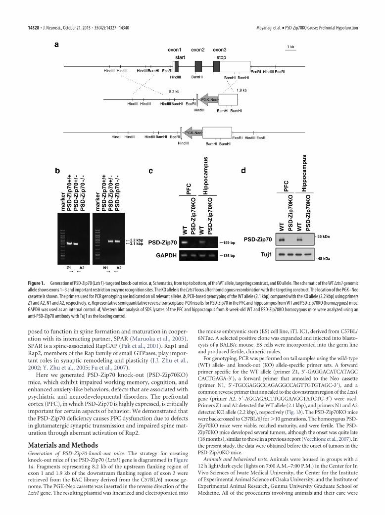
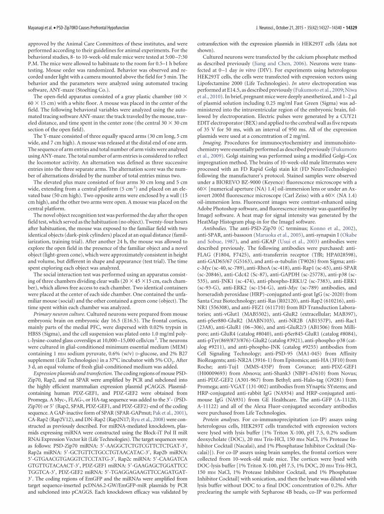
Materials and MethodsGeneration of PSD-Zip70-knock-out mice. The strategy for creatingknock-out mice of the PSD-Zip70 (Lzts1) gene is diagrammed in Figure1a. Fragments representing 8.2 kb of the upstream flanking region ofexon 1 and 1.9 kb of the downstream flanking region of exon 3 wereretrieved from the BAC library derived from the C57BL/6J mouse ge-nome. The PGK-Neo cassette was inserted in the reverse direction of theLzts1 gene. The resulting plasmid was linearized and electroporated into
the mouse embryonic stem (ES) cell line, iTL IC1, derived from C57BL/6NTac. A selected positive clone was expanded and injected into blasto-cysts of a BALB/c mouse. ES cells were incorporated into the germ lineand produced fertile, chimeric males.
For genotyping, PCR was performed on tail samples using the wild-type(WT) allele- and knock-out (KO) allele-specific primer sets. A forwardprimer specific for the WT allele (primer Z1, 5�-GAGGACATCATAGCCACTGAGA-3�), a forward primer that annealed to the Neo cassette(primer N1, 5�-TGCGAGGCCAGAGGCCAGTTGTGTAGC-3�), and acommon reverse primer that annealed to the downstream region of the Lzts1gene (primer A2, 5�-AGCAGACTTGGGAAGGTATCTG-3�) were used.Primers Z1 and A2 detected the WT allele (2.1 kbp), and primers N1 and A2detected KO allele (2.2 kbp), respectively (Fig. 1b). The PSD-Zip70KO micewere backcrossed to C57BL/6J for �10 generations. The homozygous PSD-Zip70KO mice were viable, reached maturity, and were fertile. The PSD-Zip70KO mice developed several tumors, although the onset was quite late(18 months), similar to those in a previous report (Vecchione et al., 2007). Inthe present study, the data were obtained before the onset of tumors in thePSD-Zip70KO mice.
Animals and behavioral tests. Animals were housed in groups with a12 h light/dark cycle (lights on 7:00 A.M.–7:00 P.M.) in the Center for InVivo Sciences of Iwate Medical University, the Center for the Instituteof Experimental Animal Science of Osaka University, and the Institute ofExperimental Animal Research, Gunma University Graduate School ofMedicine. All of the procedures involving animals and their care were
Figure 1. Generation of PSD-Zip70 (Lzts1)-targeted knock-out mice. a, Schematics, from top to bottom, of the WT allele, targeting construct, and KO allele. The schematic of the WT Lzts1 genomicallele shows exons 1–3 and important restriction enzyme recognition sites. The KO allele is the Lzts1 locus after homologous recombination with the targeting construct. The location of the PGK–Neocassette is shown. The primers used for PCR genotyping are indicated on all relevant alleles. b, PCR-based genotyping of the WT allele (2.1 kbp) compared with the KO allele (2.2 kbp) using primersZ1 and A2, N1 and A2, respectively. c, Representative semiquantitative reverse transcriptase-PCR results for PSD-Zip70 in the PFC and hippocampus from WT and PSD-Zip70KO (homozygous) mice.GAPDH was used as an internal control. d, Western blot analysis of SDS lysates of the PFC and hippocampus from 8-week-old WT and PSD-Zip70KO homozygous mice were analyzed using ananti-PSD-Zip70 antibody with Tuj1 as the loading control.
14328 • J. Neurosci., October 21, 2015 • 35(42):14327–14340 Mayanagi et al. • PSD-Zip70KO Causes Prefrontal Hypofunction
approved by the Animal Care Committees of these institutes, and wereperformed according to their guidelines for animal experiments. For thebehavioral studies, 8- to 10-week-old male mice were tested at 5:00 –7:30P.M. The mice were allowed to habituate to the room for 0.5–1 h beforetesting. Mouse order was randomized. Behavior was observed and re-corded under light with a camera mounted above the field for 5 min. Thebehavior and the parameters were analyzed using automated tracingsoftware, ANY-maze (Stoelting Co.).
The open-field apparatus consisted of a gray plastic chamber (60 �60 � 15 cm) with a white floor. A mouse was placed in the center of thefield. The following behavioral variables were analyzed using the auto-mated tracing software ANY-maze: the track traveled by the mouse, trav-eled distance, and time spent in the center zone (the central 30 � 30 cmsection of the open field).
The Y-maze consisted of three equally spaced arms (30 cm long, 5 cmwide, and 7 cm high). A mouse was released at the distal end of one arm.The sequence of arm entries and total number of arm visits were analyzedusing ANY-maze. The total number of arm entries is considered to reflectthe locomotor activity. An alternation was defined as three successiveentries into the three separate arms. The alternation score was the num-ber of alternations divided by the number of total entries minus two.
The elevated plus maze consisted of four arms 30 cm long and 5 cmwide, extending from a central platform (5 cm 2) and placed on an ele-vated base (50 cm high). Two opposite arms were enclosed by a wall (15cm high), and the other two arms were open. A mouse was placed on thecentral platform.
The novel object recognition test was performed the day after the openfield test, which served as the habituation (no object). Twenty-four hoursafter habituation, the mouse was exposed to the familiar field with twoidentical objects (dark-pink cylinders) placed at an equal distance (famil-iarization, training trial). After another 24 h, the mouse was allowed toexplore the open field in the presence of the familiar object and a novelobject (light-green cone), which were approximately consistent in heightand volume, but different in shape and appearance (test trial). The timespent exploring each object was analyzed.
The social interaction test was performed using an apparatus consist-ing of three chambers dividing clear walls (20 � 45 �15 cm, each cham-ber), which allows free access to each chamber. Two identical containerswere placed at the center of each side chamber. One contained the unfa-miliar mouse (social) and the other contained a green cone (object). Thetime spent within each chamber was analyzed.
Primary neuron culture. Cultured neurons were prepared from mouseembryonic brain on embryonic day 16.5 (E16.5). The frontal cortices,mainly parts of the medial PFC, were dispersed with 0.02% trypsin inHBSS (Sigma), and the cell suspension was plated onto 1.0 mg/ml poly-L-lysine-coated glass coverslips at 10,000 –15,000 cells/cm 2. The neuronswere cultured in glial-conditioned minimum essential medium (MEM)containing 1 mM sodium pyruvate, 0.6% (w/v) D-glucose, and 2% B27supplement (Life Technologies) in a 37°C incubator with 5% CO2. After3 d, an equal volume of fresh glial-conditioned medium was added.
Expression plasmids and transfection. The coding regions of mouse PSD-Zip70, Rap2, and rat SPAR were amplified by PCR and subcloned intothe highly efficient mammalian expression plasmid pCAGGS. Plasmid-containing human PDZ-GEF1, and PDZ-GEF2 were obtained fromPromega. A Myc-, FLAG-, or HA-tag sequence was added to the 3�- (PSD-Zip70) or 5� (Rap2, SPAR, PDZ-GEF1, and PDZ-GEF2)-end of the codingsequence. A GAP-inactive form of SPAR (SPAR-GAPmut; Pak et al., 2001),CA-Rap2 (Rap2V12), and DN-Rap2 (Rap2N17; Ryu et al., 2008) were con-structed as previously described. For miRNA-mediated knockdown, plas-mids expressing miRNA were constructed using the Block-iT Pol II miRRNAi Expression Vector kit (Life Technologies). The target sequences wereas follows: PSD-Zip70 miRNA: 5�-AAGGCTCTGTCGTTCTCTGAT-3�,Rap2a miRNA: 5�-GCTGTTCTGCCTGTAACATAC-3�, Rap2b miRNA:5�-GTGAACGTGAGGTCTCCTATG-3�, Rap2c miRNA: 5�-CAAGATCAGTGTTGTACAACT-3�, PDZ-GEF1 miRNA: 5�-GAAGAGCTGGATTCCTGGTCA-3�, PDZ-GEF2 miRNA: 5�-TGAGGAGAAGTTCCAGATGAT-3�. The coding regions of EmGFP and the miRNAs were amplified fromtarget sequence-inserted pcDNA6.2-GW/EmGFP-miR plasmids by PCRand subcloned into pCAGGS. Each knockdown efficacy was validated by
cotransfection with the expression plasmids in HEK293T cells (data notshown).
Cultured neurons were transfected by the calcium phosphate methodas described previously (Jiang and Chen, 2006). Neurons were trans-fected at 0 –1 day in vitro (DIV). For experiments using heterologousHEK293T cells, the cells were transfected with expression vectors usingLipofectamine 2000 (Life Technologies). In utero electroporation wasperformed at E14.5, as described previously (Fukumoto et al., 2009; Niwaet al., 2010). In brief, pregnant mice were deeply anesthetized, and 1–2 �lof plasmid solution including 0.25 mg/ml Fast Green (Sigma) was ad-ministered into the intraventricular region of the embryonic brain, fol-lowed by electroporation. Electric pulses were generated by a CUY21EDIT electroporator (BEX) and applied to the cerebral wall as five repeatsof 35 V for 50 ms, with an interval of 950 ms. All of the expressionplasmids were used at a concentration of 2 mg/ml.
Imaging. Procedures for immunocytochemistry and immunohisto-chemistry were essentially performed as described previously (Fukumotoet al., 2009). Golgi staining was performed using a modified Golgi–Coximpregnation method. The brains of 10-week-old male littermates wereprocessed with an FD Rapid Golgi stain kit (FD NeuroTechnologies)following the manufacturer’s protocol. Stained samples were observedunder a BIOREVO BZ-9000 (Keyence) fluorescence microscope with a60� [numerical aperture (NA) 1.4] oil-immersion lens or under an Ax-iovert 200M fluorescence microscope (Carl Zeiss) with a 60� (NA 1.4)oil-immersion lens. Fluorescent images were contrast-enhanced usingAdobe Photoshop software, and fluorescence intensity was quantified byImageJ software. A heat map for signal intensity was generated by theHeatMap Histogram plug-in for the ImageJ software.
Antibodies. The anti-PSD-Zip70 (C terminus; Konno et al., 2002),anti-SPAR, anti-bassoon (Maruoka et al., 2005), anti-synapsin I (Okabeand Sobue, 1987), and anti-GKAP (Usui et al., 2003) antibodies weredescribed previously. The following antibodies were purchased: anti-FLAG (F1804, F7425), anti-transferrin receptor (TfR; HPA028598),anti-GAD65/67 (G5163), and anti-�-tubulin (T9026) from Sigma; anti-c-Myc (sc-40, sc-789), anti-RhoA (sc-418), anti-Rap1 (sc-65), anti-SPAR(sc-20846), anti-Cdc42 (Sc-87), anti-GAPDH (sc-25778), anti-p38 (sc-535), anti-JNK1 (sc-474), anti-phospho-ERK1/2 (sc-7383), anti-ERK1(sc-93-G), anti-ERK2 (sc-154-G), anti-Myc (sc-789) antibodies, andhorseradish peroxidase (HRP)-conjugated anti-goat IgG (sc-2020) fromSanta Cruz Biotechnology; anti-Ras (R02120), anti-Rap2 (610216), anti-NR1 (556308), and anti-FEZ1 (611710) from BD Transduction Labora-tories; anti-vGlut1 (MAB5502), anti-GluR2 (extracellular; MAB397),anti-pSer880-GluR2 (MABN103), anti-NR2B (AB1557P), anti-Rac1(23A8), anti-GluR1 (06 –306), and anti-GluR2/3 (AB1506) from Milli-pore; anti-GluR4 (catalog #8040), anti-pSer845-GluR1 (catalog #8084),anti-pTyr(869/873/876)-GluR2 (catalog #3921), anti-phospho-p38 (cat-alog #9211), and anti-phospho-JNK (catalog #9255) antibodies fromCell Signaling Technology; anti-PSD-95 (MA1-045) from AffinityBioReagents; anti-NR2A (3916-1) from Epitomics; anti-HA (3F10) fromRoche; anti-Tuj1 (MMS-435P) from Covance; anti-PDZ-GEF1(H00009693) from Abnova; anti-Shank3 (NBP1-47610) from Novus;anti-PDZ-GEF2 (A301-967) from Bethyl; anti-Halo-tag (G9281) fromPromega; anti-VGAT (131-002) antibodies from SYnaptic SYstems; andHRP-conjugated anti-rabbit IgG (NA934) and HRP-conjugated anti-mouse IgG (NA931) from GE Healthcare. The anti-GFP (A-11120,A-11122) and all of the Alexa Fluor-conjugated secondary antibodieswere purchased from Life Technologies.
Protein analyses. For co-immunoprecipitation (co-IP) assays usingheterologous cells, HEK293T cells transfected with expression vectorswere lysed with lysis buffer [1% Triton X-100, pH 7.5, 0.2% sodiumdeoxycholate (DOC), 20 mM Tris-HCl, 150 mM NaCl, 1% Protease In-hibitor Cocktail (Nacalai), and 1% Phosphatase Inhibitor Cocktail (Na-calai)]). For co-IP assays using brain samples, the frontal cortices werecollected from 10-week-old male mice. The cortices were lysed withDOC-lysis buffer [1% Triton X-100, pH 7.5, 1% DOC, 20 mM Tris-HCl,150 mM NaCl, 1% Protease Inhibitor Cocktail, and 1% PhosphataseInhibitor Cocktail] with sonication, and then the lysate was diluted withlysis buffer without DOC to a final DOC concentration of 0.2%. Afterpreclearing the sample with Sepharose 4B beads, co-IP was performed
Mayanagi et al. • PSD-Zip70KO Causes Prefrontal Hypofunction J. Neurosci., October 21, 2015 • 35(42):14327–14340 • 14329
using the indicated antibodies and protein G-Sepharose (GE Health-care). The Sepharose beads were boiled in SDS-sample buffer to elute theimmunocomplexes.
The assay for GTP-bound small GTPase levels was based on the Rap1Activation Assay Kit (Millipore). Cultured neurons or individual frontalcortices from mice were used. Pull-down assays were then performed usingRalGDS-RBD beads for the Rap1, Rap2, and Ras activities, Rhotekin-RBDbeads (Millipore) for the Rho activity, and PAK-PBD beads (Millipore) forthe Cdc42 and Rac1 activities. As a positive control, GTP�S (Sigma) wasused. The proteins eluted from the beads were analyzed by Western blots.
For surface biotinylation assay, cortical slices (400 �m thick) wereprepared from 8- to 10-week-old male mice and the prefrontal corticeswere collected. The surface proteins were labeled with 0.3 mg/ml Sulfo-NHS-SS-biotin (Pierce) in HBSS for 15 min at 4°C. After incubation, theunbound biotin was quenched by Tris buffer (50 mM Tris-HCl, pH 8.0,120 mM NaCl). The tissues were then lysed with 1% Triton X-100, 0.1%SDS, 20 mM Tris-HCl, pH 7.5, 150 mM NaCl, 1% Protease InhibitorCocktail, and 1% Phosphatase Inhibitor Cocktail. The biotinylated pro-teins were isolated by Streptavidin-Agarose (Life Technologies) and an-alyzed by Western blots for each protein.
The procedure for Western analysis was essentially performed as de-scribed previously (Fukumoto et al., 2009). The detected band intensitieswere quantified using ImageJ software.
Slice electrophysiology. Coronal slices were prepared from postnatalday 59 – 88 PSD-Zip70KO mice or their WT littermates by using theprotective recovery method (Zhao et al., 2011; Tomioka et al., 2014).Mice were anesthetized with isoflurane and sodium pentobarbitaland transcardially perfused with a room-temperature carbogenated(95% O2 and 5% CO2) protective artificial CSF (ACSF) containing (inmM) 92 N-methyl-D-glucamine (NMDG), 2.5 KCl, 1.2 NaH2PO4, 30NaHCO3, 20 HEPES, 25 glucose, 5 ascorbate, 3 sodium pyruvate, 12N-acetyl-L-cysteine, 0.5 CaCl2, and 10 MgSO4. The mice were then de-capitated, the brains were removed, and coronal slices were cut using avibrating blade microtome (VT1200S, Leica) at room temperature. Sliceswere recovered in the carbogenated protective ACSF at 32°C for 12 minand then transferred into a holding chamber containing a room-temperature carbogenated incubating ACSF of the following composi-tion (in mM): 119 NaCl, 2.5 KCl, 1 NaH2PO4, 26.2 NaHCO3, 12.5glucose, 2 CaCl2�4H2O, 2 MgSO4�7H2O, 5 HEPES, and 12 N-acetyl-L-cysteine. After recovery for at least 2 h, the slices were transferred to arecording chamber mounted on an upright microscope (BX51WI,Olympus) equipped with IR-DIC optics. Slices were perfused with acarbogenated recording ACSF (at �32°C) containing (in mM) 119 NaCl,2.5 KCl, 4.0 CaCl2, 4.0 MgSO4, 1.0 NaH2PO4, 26.2 NaHCO3, 11 glucose,5 HEPES, and 0.1 picrotoxin. When measuring AMPAR/NMDAR ratiosin synapses on layer 2/3 pyramidal cells of the medial prefrontal cortexincluding the prelimbic, infralimbic, and anterior cingulate cortex, affer-ents to these cells were activated by a patch pipette that had a broken tip,filled with recording ACSF, and placed �80 –100 �m beneath the cell.These pyramidal cells were patched and EPSCs were recorded using aMultiClamp 700A (Molecular Devices) through a glass electrode filledwith an internal solution containing (in mM) 135 cesium methanesulfo-nate, 10 HEPES, 0.2 EGTA, 8 NaCl, 4 Mg-ATP, 0.3 Na3GTP (pH 7.2 withCsOH, osmolarity adjusted to 285 mOsm). The AMPAR/NMDAR ratiowas calculated as the ratio of the amplitude of EPSCs at �70 mV to thatof EPSCs at �40 mV. Decays of averaged EPSCs at �40 mV were calcu-lated as reported previously (Yasuda and Mukai, 2015). Miniature EPSCs(mEPSCs) were recorded in the presence of 1 �M TTX. Data acquisitionand analysis were performed using custom Igor Pro (WaveMetrics) soft-ware routines.
Semiquantitative reverse transcription-PCR. The total RNA was ex-tracted from brain tissues using TRIzol reagent (Life Technologies) andreverse-transcribed with SuperScript VILO Master Mix (Life Technolo-gies). The cDNA was amplified with gene-specific primer pairs usingSYBR GreenER qPCR SuperMix Universal reagent (Life Technologies).The primer sequences used in this study are as follows: PSD-Zip70-F:5�-CCATCGCCCAGACTACACAG-3�, PSD-Zip70-R: 5�-ACAGGCTTGAAGGCAGTGG-3�, GAPDH-F: 5�-CGTGCCGCCTGGAGAAAC-3�,GAPDH-R: 5�-TGGGAGTTGCTGTTGAAGTCG-3�.
Statistical analysis. All values represent means � SEM. All transfectionexperiments were performed at least three times. The described “n” in-dicates biological replicates. We used Excel Statistics add-in software(Social Survey Research Information Co., Ltd) for statistical processing.Statistical differences between pairs of values were analyzed by Student’st test. One-way ANOVA with Tukey-Kramer post hoc test was used forgroup comparisons. Fisher’s protected least significant difference test as apost hoc test was used to analyze the data (see Fig. 8b). p 0.05 wasconsidered significant.
ResultsGeneration of PSD-Zip70KO miceTo evaluate the neural functions of PSD-Zip70 in vivo, we newlygenerated PSD-Zip70-deficient mice (Fig. 1a,b). A human orthologof PSD-Zip70 is identical to Lzts1, which has been reported to have arole in carcinogenesis (Ishii et al., 1999; Vecchione et al., 2001). Lzts1KO mice develop various tumors with late onset (Vecchione et al.,2007), but no neuronal analysis of these mice has been performed. Ina previous report, KO mice were produced by introducing a stopcodon 123 bp downstream of the first start (ATG) codon. Our strat-egy for targeted KO of the PSD-Zip70 (Lzts1) gene was to delete all ofthe coding regions of the three exons starting upstream of the ORF,because the N terminus of PSD-Zip70 possesses a myristoylation sitethat is important for the protein’s membrane targeting (Konno et al.,2002). Homozygotes of PSD-Zip70KO mice were born in the ex-pected Mendelian ratio, and showed no differences from WT micein their growth, survival, or fertility for at least 12 months after birth(data not shown). The expression of PSD-Zip70 mRNA and proteinwas completely lacking in PSD-Zip70KO mice (Fig. 1c,d).
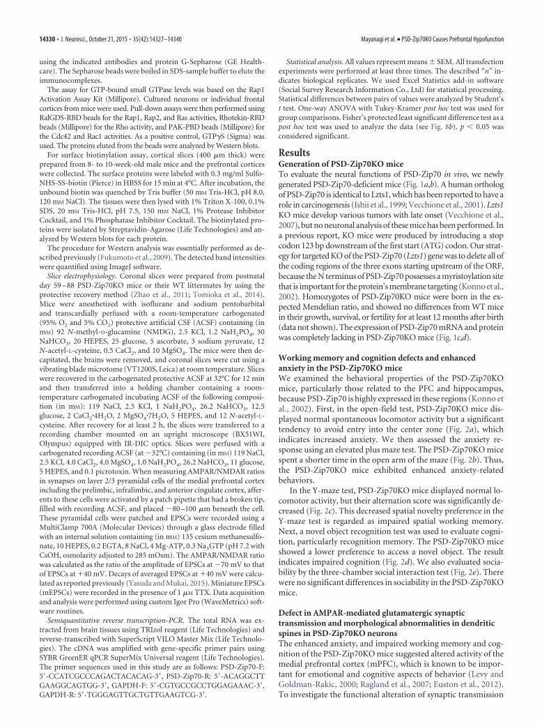
Working memory and cognition defects and enhancedanxiety in the PSD-Zip70KO miceWe examined the behavioral properties of the PSD-Zip70KOmice, particularly those related to the PFC and hippocampus,because PSD-Zip70 is highly expressed in these regions (Konno etal., 2002). First, in the open-field test, PSD-Zip70KO mice dis-played normal spontaneous locomotor activity but a significanttendency to avoid entry into the center zone (Fig. 2a), whichindicates increased anxiety. We then assessed the anxiety re-sponse using an elevated plus maze test. The PSD-Zip70KO micespent a shorter time in the open arm of the maze (Fig. 2b). Thus,the PSD-Zip70KO mice exhibited enhanced anxiety-relatedbehaviors.
In the Y-maze test, PSD-Zip70KO mice displayed normal lo-comotor activity, but their alternation score was significantly de-creased (Fig. 2c). This decreased spatial novelty preference in theY-maze test is regarded as impaired spatial working memory.Next, a novel object recognition test was used to evaluate cogni-tion, particularly recognition memory. The PSD-Zip70KO miceshowed a lower preference to access a novel object. The resultindicates impaired cognition (Fig. 2d). We also evaluated socia-bility by the three-chamber social interaction test (Fig. 2e). Therewere no significant differences in sociability in the PSD-Zip70KOmice.
Defect in AMPAR-mediated glutamatergic synaptictransmission and morphological abnormalities in dendriticspines in PSD-Zip70KO neuronsThe enhanced anxiety, and impaired working memory and cog-nition of the PSD-Zip70KO mice suggested altered activity of themedial prefrontal cortex (mPFC), which is known to be impor-tant for emotional and cognitive aspects of behavior (Levy andGoldman-Rakic, 2000; Ragland et al., 2007; Euston et al., 2012).To investigate the functional alteration of synaptic transmission
14330 • J. Neurosci., October 21, 2015 • 35(42):14327–14340 Mayanagi et al. • PSD-Zip70KO Causes Prefrontal Hypofunction
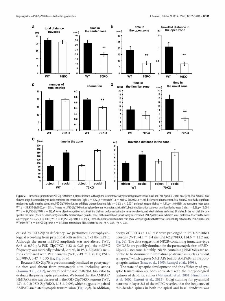
caused by PSD-Zip70 deficiency, we performed electrophysio-logical recording from pyramidal cells in layer 2/3 of the mPFC.Although the mean mEPSC amplitude was not altered (WT,6.48 � 0.30 pA; PSD-Zip70KO, 6.32 � 0.25 pA), the mEPSCfrequency was markedly reduced, �50%, in PSD-Zip70KO neu-rons compared with WT neurons (WT, 7.49 � 1.30 Hz; PSD-Zip70KO, 3.47 � 0.55 Hz; Fig. 3a,b).
Because PSD-Zip70 is predominantly localized to postsynap-tic sites and absent from presynaptic sites including axons(Konno et al., 2002), we examined the AMPAR/NMDAR ratio toevaluate the postsynaptic properties. We found that the AMPAR/NMDAR ratio was decreased in the PSD-Zip70KO neurons (WT,1.74 � 0.3; PSD-Zip70KO, 1.15 � 0.09), which suggests impairedAMPAR-mediated synaptic transmission (Fig. 3c,d). In addition,
decays of EPSCs at �40 mV were prolonged in PSD-Zip70KOneurons (WT, 94.1 � 8.4 ms; PSD-Zip70KO, 124.6 � 12.2 ms;Fig. 3e). The data suggest that NR2B-containing immature-typeNMDARs are possibly dominant in the postsynaptic sites of PSD-Zip70KO neurons. Notably, NR2B-containing NMDARs are re-ported to be dominant in immature postsynapses such as “silentsynapses,” which express NMDARs but not AMPARs, at the post-synaptic surface (Isaac et al., 1995; Rumpel et al., 1998).
The state of synaptic development and the efficiency of syn-aptic transmission are both correlated with the morphologicalfeatures of dendritic spines (Matsuzaki et al., 2001; Nimchinskyet al. 2002; Caroni et al., 2012). Golgi staining for pyramidalneurons in layer 2/3 of the mPFC revealed that the frequency ofthin-headed spines in both the apical and basal dendrites was
Figure 2. Behavioral properties of PSD-Zip70KO mice. a, Open-field test. Although the locomotor activity (track length) was similar in WT and PSD-Zip70KO (70KO) mice (left), PSD-Zip70KO miceshowed a significant tendency to avoid entry into the center zone (right; t 3.42, p 0.001; WT, n 21; PSD-Zip70KO, n 23). b, Elevated plus maze test. PSD-Zip70KO mice had a significanttendency to avoid entering open arms. PSD-Zip70KO mice also exhibited shorter durations (left; t 3.52, p 0.001) and track lengths (right; t 4.11, p 0.001) in the open arms (open zone;WT, n 33; PSD-Zip70KO, n 38). c, Y-maze test. PSD-Zip70KO mice displayed normal locomotor activity (left), but their alternation score was significantly decreased (right; t 5.23, p 0.001;WT, n 24; PSD-Zip70KO, n 29). d, Novel object recognition test. A training trial was performed using the same two objects, and a test trial was performed 24 h later. In the test trial, the timespent in the zones (20 cm � 20 cm each) around the familiar object (familiar zone) or the novel object (novel zone) was recorded. PSD-Zip70KO mice exhibited lower preference to access the novelobject (right; t 4.25, p 0.001; WT, n 19; PSD-Zip70KO, n 18). e, Three-chamber social interaction test. There were no significant differences in sociability between the PSD-Zip70KO andWT mice (WT, n 11; PSD-Zip70KO, n 11). Error bars indicate SEM. Student’s t test. *p 0.05, **p 0.01.
Mayanagi et al. • PSD-Zip70KO Causes Prefrontal Hypofunction J. Neurosci., October 21, 2015 • 35(42):14327–14340 • 14331
significantly increased, and the spine den-sity was also increased for PSD-Zip70KOcompared with WT neurons (Fig. 3f,g).This aberrant morphology of dendriticspines was also more prominent in pri-mary cultured neurons from the frontalcortex of PSD-Zip70KO mice (Fig. 3h), inwhich the frequency of thin spinesincreased, and that of mushroom-typespines decreased (Fig. 3i,j). These dataindicate that the PSD-Zip70 deficiencyresulted in the formation of tiny-headedthin spines. In accordance with theelectrophysiological abnormalities, thinspines of PSD-Zip70KO neurons appearto reflect a functionally immature state ofthe spine synapse.
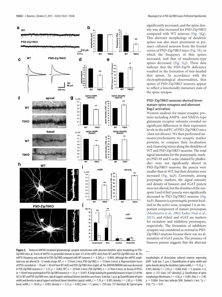
PSD-Zip70KO neurons showed fewermature spine synapses and aberrantRap2 activationWestern analyses for major synaptic pro-teins including AMPA- and NMDA-typeglutamate receptor subunits revealed nosignificant differences in their expressionlevels in the mPFC of PSD-Zip70KO mice(data not shown). We then performed im-munocytochemistry for synaptic markerproteins, to compare their localizationand clustering status along the dendrites ofWT and PSD-Zip70KO neurons. The totalsignal intensities for the postsynaptic mark-ers PSD-95 and F-actin (stained by phalloi-din) were not significantly altered inPSD-Zip70KO neurons; the puncta weresmaller than in WT, but their densities wereincreased (Fig. 4a,b). Conversely, amongpresynaptic markers, the signal intensityand density of bassoon and vGAT punctawere not altered, but the densities of the syn-apsin I and vGlut1 puncta were significantlydecreased in PSD-Zip70KO neurons (Fig.4a,b). Bassoon is a presynaptic protein local-ized to the active zone, synapsin I is an im-portant component of mature presynapses(Mozhayeva et al., 2002; Easley-Neal et al.,2013), and vGlut1 and vGAT are markersfor excitatory and inhibitory presynapses,respectively. The formation of inhibitorysynapses was considered as normal in PSD-Zip70KO neurons because there was no al-ternation of vGAT puncta. The presence ofbassoon puncta suggests that the aberrant
Figure 3. Reduced AMPAR-mediated glutamatergic synaptic transmission with aberrant dendritic spine morphology in PSD-Zip70KO mice. a, Traces of mEPSCs in pyramidal neurons in layer 2/3 of the mPFC slices from WT and PSD-Zip70KO mice. b, ThemEPSC frequency was reduced in PSD-Zip70KO compared with WT neurons (t 0.284, p 0.009), although the mEPSC ampli-tude was not altered (8 –12 weeks of age; WT, n 12 from 2 mice; PSD-Zip70KO, n 15 from 3 mice). c, Representative tracesof EPSCs recorded at �70 and �40 mV from WT (left) and PSD-Zip70KO slices (right). d. The AMPAR/NMDAR ratio was decreasedin PSD-Zip70KO neurons (t 3.31, p 0.002; WT, n 20 from 3 mice; PSD-Zip70KO, n 21 from 4 mice). e, Decays of EPSCsat�40 mV were prolonged in PSD-Zip70KO neurons (t 35, p 0.047). f, Golgi staining for pyramidal neurons in layer 2/3 of theACC in WT and PSD-Zip70KO mice. Apical (upper) and basal (lower) dendrites are shown. Scale bar, 5 �m. g, Quantification of spinewidth and density in apical (upper) and basal (lower) dendrites [apical: width, t 7.29, p 0.001; density, t 2.89, p 0.006;basal, width, t 10.69, p 0.001; density: t 3.27, p 0.002; mice 3, spines � 250 (size, 727 (density)]. h, Typical spine
4
morphologies of dissociation cultured neurons expressingEGFP. Scale bar, 5 �m. i, Quantification of spine width andspine density along the dendrites [spine width, t 9.15, p 0.001; density: t 2.93, p 0.006; trials 3, neurons � 6,spines � 251 (size), 547 (density)]. j, Classification of spinemorphology (Mushroom, t 4.56, p 0.001; thin, t 3.40,p 0.006). Error bars indicate SEM. Student’s t test. *p 0.05, **p 0.01.
14332 • J. Neurosci., October 21, 2015 • 35(42):14327–14340 Mayanagi et al. • PSD-Zip70KO Causes Prefrontal Hypofunction
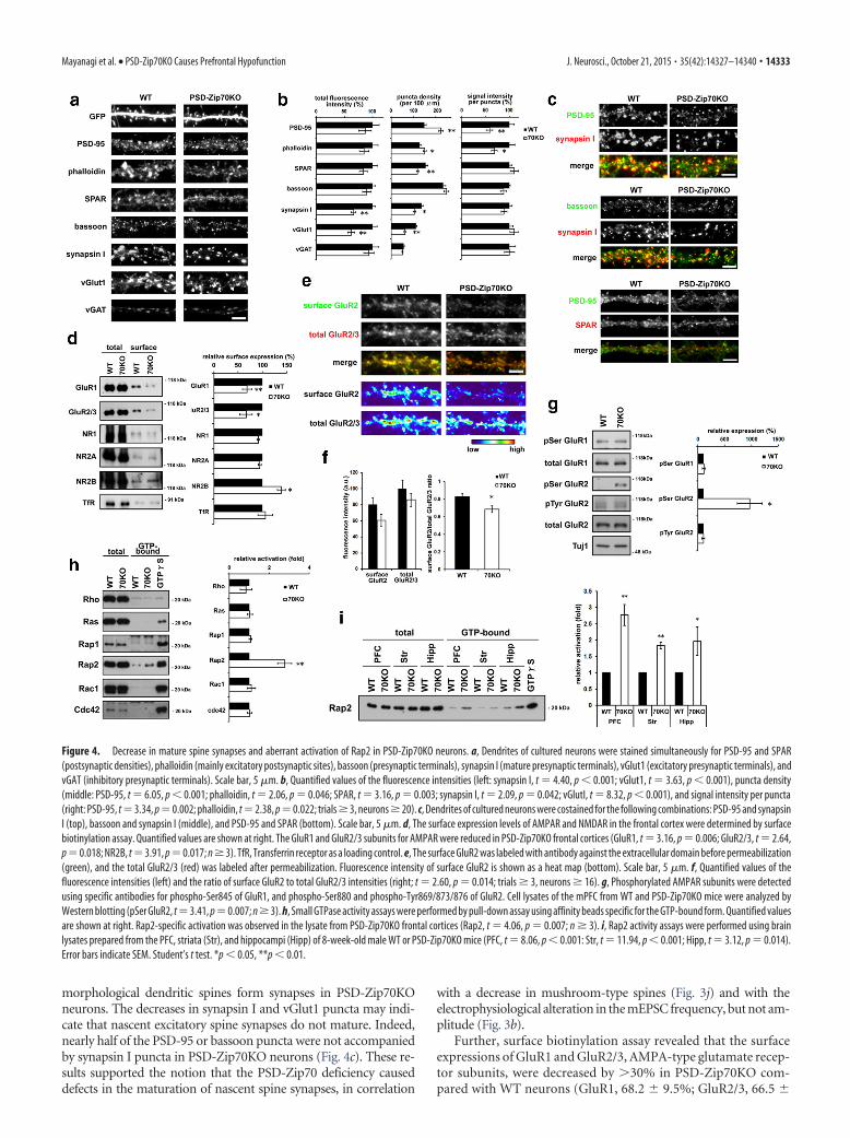
morphological dendritic spines form synapses in PSD-Zip70KOneurons. The decreases in synapsin I and vGlut1 puncta may indi-cate that nascent excitatory spine synapses do not mature. Indeed,nearly half of the PSD-95 or bassoon puncta were not accompaniedby synapsin I puncta in PSD-Zip70KO neurons (Fig. 4c). These re-sults supported the notion that the PSD-Zip70 deficiency causeddefects in the maturation of nascent spine synapses, in correlation
with a decrease in mushroom-type spines (Fig. 3j) and with theelectrophysiological alteration in the mEPSC frequency, but not am-plitude (Fig. 3b).
Further, surface biotinylation assay revealed that the surfaceexpressions of GluR1 and GluR2/3, AMPA-type glutamate recep-tor subunits, were decreased by �30% in PSD-Zip70KO com-pared with WT neurons (GluR1, 68.2 � 9.5%; GluR2/3, 66.5 �
Figure 4. Decrease in mature spine synapses and aberrant activation of Rap2 in PSD-Zip70KO neurons. a, Dendrites of cultured neurons were stained simultaneously for PSD-95 and SPAR(postsynaptic densities), phalloidin (mainly excitatory postsynaptic sites), bassoon (presynaptic terminals), synapsin I (mature presynaptic terminals), vGlut1 (excitatory presynaptic terminals), andvGAT (inhibitory presynaptic terminals). Scale bar, 5 �m. b, Quantified values of the fluorescence intensities (left: synapsin I, t 4.40, p 0.001; vGlut1, t 3.63, p 0.001), puncta density(middle: PSD-95, t 6.05, p 0.001; phalloidin, t 2.06, p 0.046; SPAR, t 3.16, p 0.003; synapsin I, t 2.09, p 0.042; vGlutI, t 8.32, p 0.001), and signal intensity per puncta(right: PSD-95, t3.34, p0.002; phalloidin, t2.38, p0.022; trials � 3, neurons � 20). c, Dendrites of cultured neurons were costained for the following combinations: PSD-95 and synapsinI (top), bassoon and synapsin I (middle), and PSD-95 and SPAR (bottom). Scale bar, 5 �m. d, The surface expression levels of AMPAR and NMDAR in the frontal cortex were determined by surfacebiotinylation assay. Quantified values are shown at right. The GluR1 and GluR2/3 subunits for AMPAR were reduced in PSD-Zip70KO frontal cortices (GluR1, t 3.16, p 0.006; GluR2/3, t 2.64,p0.018; NR2B, t3.91, p0.017; n � 3). TfR, Transferrin receptor as a loading control. e, The surface GluR2 was labeled with antibody against the extracellular domain before permeabilization(green), and the total GluR2/3 (red) was labeled after permeabilization. Fluorescence intensity of surface GluR2 is shown as a heat map (bottom). Scale bar, 5 �m. f, Quantified values of thefluorescence intensities (left) and the ratio of surface GluR2 to total GluR2/3 intensities (right; t 2.60, p 0.014; trials � 3, neurons � 16). g, Phosphorylated AMPAR subunits were detectedusing specific antibodies for phospho-Ser845 of GluR1, and phospho-Ser880 and phospho-Tyr869/873/876 of GluR2. Cell lysates of the mPFC from WT and PSD-Zip70KO mice were analyzed byWestern blotting (pSer GluR2, t3.41, p0.007; n � 3). h, Small GTPase activity assays were performed by pull-down assay using affinity beads specific for the GTP-bound form. Quantified valuesare shown at right. Rap2-specific activation was observed in the lysate from PSD-Zip70KO frontal cortices (Rap2, t 4.06, p 0.007; n � 3). i, Rap2 activity assays were performed using brainlysates prepared from the PFC, striata (Str), and hippocampi (Hipp) of 8-week-old male WT or PSD-Zip70KO mice (PFC, t 8.06, p 0.001: Str, t 11.94, p 0.001; Hipp, t 3.12, p 0.014).Error bars indicate SEM. Student’s t test. *p 0.05, **p 0.01.
Mayanagi et al. • PSD-Zip70KO Causes Prefrontal Hypofunction J. Neurosci., October 21, 2015 • 35(42):14327–14340 • 14333
12.7%; Fig. 4d). In contrast, the surface expression of NR2B wasincreased in PSD-Zip70KO neurons (138 � 8.10%), althoughthose of NR1 and NR2A were unchanged (Fig. 4d). The increasedsurface expression of NR2B appeared to be consistent with the
electrophysiological data shown in Figure 3e. We next examinedthe distribution of surface GluR2 along the dendrites. The surfaceGluR2/total GluR2/3 ratio was significantly decreased in PSD-Zip70KO neurons (Fig. 4e,f). Previous studies showed that the
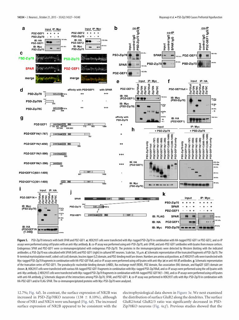
Figure 5. PSD-Zip70 interacts with both SPAR and PDZ-GEF1. a, HEK293T cells were transfected with Myc-tagged PSD-Zip70 in combination with HA-tagged PDZ-GEF1 or PDZ-GEF2, and co-IPassays were performed using cell lysates with an anti-Myc antibody. b, co-IP assay was performed using anti-PSD-Zip70, anti-SPAR, and anti-PDZ-GEF1 antibodies with lysates from mouse cortices.Endogenous SPAR and PDZ-GEF1 were co-immunoprecipitated with endogenous PSD-Zip70. The proteins in the immunoprecipitants were detected by Western blotting with the indicatedantibodies. c, PSD-Zip70 was colocalized with SPAR (left) and PDZ-GEF1 (right) in cultured WT neurons. Scale bar, 10 �m. d, Schematic representation of the truncated fragments of PSD-Zip70. TheN-terminal myristoylation motif, coiled-coil (coil) domain, leucine zipper (LZ) domain, and PDZ-binding motif are shown. Numbers are amino acid positions. e, f, HEK293T cells were transfected withMyc-tagged PSD-Zip70 fragments in combination with HA-PDZ-GEF1full, and co-IP assays were performed using cell lysates with anti-Myc (e) or anti-HA (f) antibodies. g, Schematic representationof the truncation series of PDZ-GEF1. The pseudocyclic-nucleotide binding domain (cNBD), Ras exchange motif (REM), PDZ domain, Ras-association (RA) domain, and RapGEF (GEF) domain areshown. h, HEK293T cells were transfected with various HA-tagged PDZ-GEF1 fragments in combination with Myc-tagged PSD-Zip70full, and co-IP assays were performed using the cell lysates withanti-Myc antibody. i, HEK293T cells were transfected with Myc-tagged PSD-Zip70 fragments in combination with HA-tagged PDZ-GEF1N(1–390), and co-IP assays were performed using cell lysateswith anti-HA antibody. j, Schematic diagram of the interactions among PSD-Zip70, SPAR, and PDZ-GEF1. k, co-IP assay was performed in HEK293T cells with Myc-PSD-Zip70 in combination withHA-PDZ-GEF1 and/or FLAG-SPAR. The co-immunoprecipitated proteins with Myc-PSD-Zip70 were analyzed.
14334 • J. Neurosci., October 21, 2015 • 35(42):14327–14340 Mayanagi et al. • PSD-Zip70KO Causes Prefrontal Hypofunction
phosphorylation of AMPARs at their cytoplasmic terminus iscorrelated with their surface expression at postsynaptic sites(Wang et al., 2005). We found that the phosphorylation of GluR2at S880, which is correlated with its internalization in response tolong-term depression (LTD) induction (Lu and Roche, 2012),was markedly elevated in PSD-Zip70KO neurons (Fig. 4g).
The Rho, Ras, and Rap small GTPases play important roles inspine formation and synaptic maturation (Bonhoeffer and Yuste,2002; Zhu et al., 2005; Lee et al., 2011). Among them, we foundthat the activity of Rap2 was specifically upregulated in the frontalcortex of PSD-Zip70KO mice (2.77 � 0.32-fold activation com-pared with WT; Fig. 4h,i). The Rap2 activation was also observedin the hippocampus (1.97 � 0.44-fold) and the striatum (1.83 �0.10-fold; Fig. 4i). A similar upregulation of Rap2 activity was
found in cultured PSD-Zip70KO neuronsprepared from the frontal cortex (2.56 �0.16-fold; data not shown). The Rap2 ac-tivity is reported to be a critical regulatorof AMPAR internalization in response toLTD-inducing stimuli (Zhu et al., 2005;Fu et al., 2007).
PSD-Zip70 interacts with both SPARand PDZ-GEF1, and modulates theRap2 activityThe activity of Rap2 is regulated by a balancebetween the GEF (guanine nucleotide ex-change factor) and GAP (GTPase-activatingprotein) activities. We previously found thatPSD-Zip70 interacts with SPAR, a spine-enriched RapGAP (Maruoka et al., 2005).We therefore presumed that the activities ofGEF and/or GAP for Rap2 would be dys-regulated in PSD-Zip70KO neurons. Thelocalization of SPAR along the dendriteswas diffuse along the dendrites of PSD-Zip70KO neurons (Fig. 4a–c), even thoughSPAR’s expression level was not altered (Fig.4a,b). These results suggested that the PSD-Zip70 deficiency weakened SPAR’s target-ing to the spines, and might have alteredSPAR’s RapGAP activity.
To elucidate the mechanism underly-ing the specific activation of Rap2, we nextexamined possible interactions betweenPSD-Zip70 and reported RapGEFs, be-cause SPAR possesses catalytic activity toboth Rap1 and Rap2. Among theRapGEFs examined, we identified PDZ-GEF1 and PDZ-GEF2 as novel PSD-Zip70-interacting proteins (Fig. 5a).PDZ-GEF1 is reported to be more highlyexpressed than PDZ-GEF2 in the brain(Lee et al., 2011). We confirmed that en-dogenous PSD-Zip70 interacted withboth SPAR and PDZ-GEF1 in lysates pre-pared from the frontal cortex (Fig. 5b).PSD-Zip70 showed the predominant ex-pression in excitatory pyramidal neurons,which possess numerous spines on thedendrites in cultured cortical as well ashippocampal neurons (Fig. 5c; Konno etal., 2002). PSD-Zip70 was colocalized
with SPAR and with PDZ-GEF1 as puncta at mainly dendriticspines in WT neurons (Fig. 5c).
Next, we analyzed the interaction sites for PSD-Zip70 andPDZ-GEF1 in heterologous HEK293T cells. The results re-vealed that the C-terminal region of PSD-Zip70 and theN-terminal region of PDZ-GEF1 were required for theirinteraction (Fig. 5d–j). PDZ-GEF1 lacking the C terminus ex-hibited a high affinity for PSD-Zip70 compared with the full-length form, suggesting that the C terminus of PDZ-GEF1suppresses its interaction with PSD-Zip70 (Fig. 5h). Further-more, PDZ-GEF1 did not compete with SPAR for the PSD-Zip70 binding (Fig. 5k). These results together suggested thatPSD-Zip70 forms a ternary complex with SPAR and PDZ-GEF1 at the postsynaptic site.
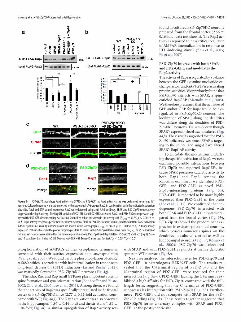
Figure 6. PSD-Zip70 modulates Rap2 activity via SPAR- and PDZ-GEF1. a, Rap2 activity assay was performed in cultured WTneurons. Cultured neurons were cotransfected with exogenous FLAG-tagged Rap2 in combination with the indicated expressionplasmids. Total and GTP-bound exogenous Rap2 were detected using anti-FLAG antibody. SPAR and PSD-Zip70 cooperativelysuppressed the Rap2 activity. The RapGEF activity of PDZ-GEF1 and PDZ-GEF2 activated Rap2, and PSD-Zip70 coexpression sup-pressed the PDZ-GEF-dependent Rap2 activation. Quantified values are shown in the lower graph (F(5,30) 31.35, p 0.001; n 6). b, Rap2 activity assay was performed in cultured neurons. SPAR or PSD-Zip70 expression rescued the aberrant Rap2 activationin PSD-Zip70KO neurons. Quantified values are shown in the lower graph (F(5,18) 68.28, p 0.001; n 4). c, Exogenouslyexpressed PSD-Zip70 rescued the proper targeting of SPAR to spines in the PSD-Zip70KO neurons. Scale bar, 5 �m. d, Dendrites ofcultured WT neurons were stained for the following combinations: PSD-Zip70 and Rap1 (left) or PSD-Zip70 and Rap2 (right). Scalebar, 10 �m. Error bars indicate SEM. One-way ANOVA with Tukey-Kramer post hoc test. *p 0.05, **p 0.01.
Mayanagi et al. • PSD-Zip70KO Causes Prefrontal Hypofunction J. Neurosci., October 21, 2015 • 35(42):14327–14340 • 14335
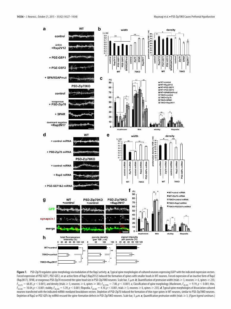
Figure 7. PSD-Zip70 regulates spine morphology via modulation of the Rap2 activity. a, Typical spine morphologies of cultured neurons expressing EGFP with the indicated expression vectors.Forced expression of PDZ-GEF1, PDZ-GEF2, or an active form of Rap2 (Rap2V12) induced the formation of spines with smaller heads in WT neurons. Forced expression of an inactive form of Rap2(Rap2N17), SPAR, or exogenous PSD-Zip70 recovered the spine head size in PSD-Zip70KO neurons. Scale bar, 5 �m. b, Quantification of protrusion width (trials � 3, neurons � 6, spines � 235;F(8,25) 68.85, p 0.001), and density (trials � 3, neurons � 6, spines � 383; F(8,194) 7.00, p 0.001). c, Classification of spine morphology (Mushroom, F(8,58) 9.19, p 0.001; thin,F(8,58) 10.04, p 0.001; stubby, F(8,58) 5.39, p 0.001; filopodia, F(8,58) 4.10, p 0.001, trials � 3, neurons � 6, spines � 235). d, Typical spine morphologies of dissociation culturedneurons transfected with the indicated miRNA-mediated knockdown vectors. Depletion of PSD-Zip70 induced the formation of thin-type spines in WT neurons, similar to PSD-Zip70KO neurons.Depletion of Rap2 or PDZ-GEFs by miRNA rescued the spine-formation defects in PSD-Zip70KO neurons. Scale bar, 5 �m. e, Quantification protrusion width (trials � 3, (Figure legend continues.)
14336 • J. Neurosci., October 21, 2015 • 35(42):14327–14340 Mayanagi et al. • PSD-Zip70KO Causes Prefrontal Hypofunction
We then examined the effect of PSD-Zip70’s interactions withSPAR and/or PDZ-GEF1 on the Rap2 activity in neurons. TheSPAR suppressed the coexpressed FLAG-Rap2 activity by its Rap-GAP activity, and the coexpression of SPAR and PSD-Zip70 co-operatively suppressed the Rap2 activity in WT neurons (Fig. 6a).Further, expression of PSD-Zip70 or SPAR could suppress theaberrant Rap2 activation in PSD-Zip70KO neurons (Fig. 6b).The impaired spine targeting of SPAR in PSD-Zip70KO neuronswas reversed by the moderate expression of exogenous PSD-Zip70 (Fig. 6c). These results suggested that PSD-Zip70 assists thespine targeting of SPAR and enhances the efficiency of SPAR’sRapGAP activity.
PDZ-GEF1/2 activated Rap2 by its RapGEF activity, and its co-expression with PSD-Zip70 suppressed the PDZ-GEF-dependentRap2 activation (Fig. 6a). The suppressive effect of PSD-Zip70 onthe GEF activity of PDZ-GEF1/2 for Rap1/2 was also observed inHEK293T cells (data not shown). Further, we found that PSD-Zip70and Rap2 were colocalized as puncta along the dendrites and spines,whereas Rap1 was localized as prominent puncta without PSD-Zip70 (Fig. 6d). Thus, the Rap2-specific activation in PSD-Zip70KOneurons may be related to the spatial association of PSD-Zip70 withSPAR and PDZ-GEF1 as a ternary complex close to Rap2 in thepostsynapse.
PSD-Zip70 regulates spine synapse maturation bymodulating the Rap2 activityWe then investigated the effect of Rap2 activity on the spinemorphology in WT and PSD-Zip70KO neurons. The forced ex-pression of PDZ-GEF1 or PDZ-GEF2 reduced the size of thespine head and altered the spine shape from mushroom to thin inWT neurons (Fig. 7a-c). The moderate expression of a constitu-tively active form of Rap2 (Rap2V12) using a low-dose expres-sion plasmid caused similar results when coexpressed withPDZ-GEFs (Fig. 7a– c). The Rap2-dependent effects on spinemorphology were confirmed by expressing a GAP-mutatedSPAR that functions as a dominant-negative form with respect toSPAR’s RapGAP activity. In contrast, the expression of Rap2N17(an inactive form of Rap2; Rap2DN), SPAR, or exogenous PSD-Zip70 recovered the spine morphology in PSD-Zip70KO neu-rons (Fig. 7a– c). We confirmed that depleting PSD-Zip70 bymiRNA-mediated knockdown in WT neurons mimicked thespine-morphology phenotype of PSD-Zip70KO neurons (Fig.7d–f). In contrast, depleting all of the Rap2 subtypes (Rap2a, b,and c) or both PDZ-GEF1 and PDZ-GEF2 could recover thespine-morphology defects of PSD-Zip70KO neurons (Fig. 7d–f).We further confirmed that the decrease in synapsin I punctaalong the dendrites of PSD-Zip70KO neurons was recovered byexpressing Rap2N17 (Fig. 7g,h). Thus, our results clearly suggestthat the Rap2 activation in PSD-Zip70KO neurons causes synap-tic dysfunction accompanied by impaired spine maturation.
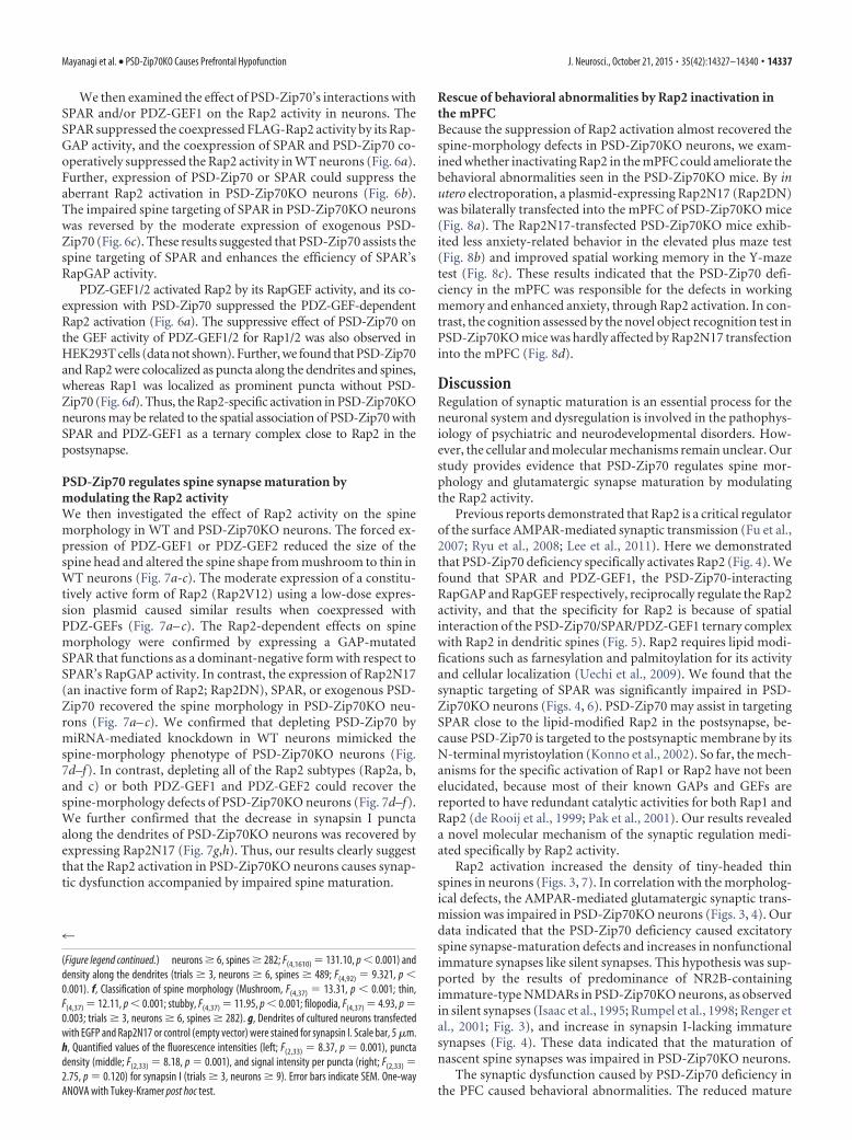
Rescue of behavioral abnormalities by Rap2 inactivation inthe mPFCBecause the suppression of Rap2 activation almost recovered thespine-morphology defects in PSD-Zip70KO neurons, we exam-ined whether inactivating Rap2 in the mPFC could ameliorate thebehavioral abnormalities seen in the PSD-Zip70KO mice. By inutero electroporation, a plasmid-expressing Rap2N17 (Rap2DN)was bilaterally transfected into the mPFC of PSD-Zip70KO mice(Fig. 8a). The Rap2N17-transfected PSD-Zip70KO mice exhib-ited less anxiety-related behavior in the elevated plus maze test(Fig. 8b) and improved spatial working memory in the Y-mazetest (Fig. 8c). These results indicated that the PSD-Zip70 defi-ciency in the mPFC was responsible for the defects in workingmemory and enhanced anxiety, through Rap2 activation. In con-trast, the cognition assessed by the novel object recognition test inPSD-Zip70KO mice was hardly affected by Rap2N17 transfectioninto the mPFC (Fig. 8d).
DiscussionRegulation of synaptic maturation is an essential process for theneuronal system and dysregulation is involved in the pathophys-iology of psychiatric and neurodevelopmental disorders. How-ever, the cellular and molecular mechanisms remain unclear. Ourstudy provides evidence that PSD-Zip70 regulates spine mor-phology and glutamatergic synapse maturation by modulatingthe Rap2 activity.
Previous reports demonstrated that Rap2 is a critical regulatorof the surface AMPAR-mediated synaptic transmission (Fu et al.,2007; Ryu et al., 2008; Lee et al., 2011). Here we demonstratedthat PSD-Zip70 deficiency specifically activates Rap2 (Fig. 4). Wefound that SPAR and PDZ-GEF1, the PSD-Zip70-interactingRapGAP and RapGEF respectively, reciprocally regulate the Rap2activity, and that the specificity for Rap2 is because of spatialinteraction of the PSD-Zip70/SPAR/PDZ-GEF1 ternary complexwith Rap2 in dendritic spines (Fig. 5). Rap2 requires lipid modi-fications such as farnesylation and palmitoylation for its activityand cellular localization (Uechi et al., 2009). We found that thesynaptic targeting of SPAR was significantly impaired in PSD-Zip70KO neurons (Figs. 4, 6). PSD-Zip70 may assist in targetingSPAR close to the lipid-modified Rap2 in the postsynapse, be-cause PSD-Zip70 is targeted to the postsynaptic membrane by itsN-terminal myristoylation (Konno et al., 2002). So far, the mech-anisms for the specific activation of Rap1 or Rap2 have not beenelucidated, because most of their known GAPs and GEFs arereported to have redundant catalytic activities for both Rap1 andRap2 (de Rooij et al., 1999; Pak et al., 2001). Our results revealeda novel molecular mechanism of the synaptic regulation medi-ated specifically by Rap2 activity.
Rap2 activation increased the density of tiny-headed thinspines in neurons (Figs. 3, 7). In correlation with the morpholog-ical defects, the AMPAR-mediated glutamatergic synaptic trans-mission was impaired in PSD-Zip70KO neurons (Figs. 3, 4). Ourdata indicated that the PSD-Zip70 deficiency caused excitatoryspine synapse-maturation defects and increases in nonfunctionalimmature synapses like silent synapses. This hypothesis was sup-ported by the results of predominance of NR2B-containingimmature-type NMDARs in PSD-Zip70KO neurons, as observedin silent synapses (Isaac et al., 1995; Rumpel et al., 1998; Renger etal., 2001; Fig. 3), and increase in synapsin I-lacking immaturesynapses (Fig. 4). These data indicated that the maturation ofnascent spine synapses was impaired in PSD-Zip70KO neurons.
The synaptic dysfunction caused by PSD-Zip70 deficiency inthe PFC caused behavioral abnormalities. The reduced mature
4
(Figure legend continued.) neurons � 6, spines � 282; F(4,1610) 131.10, p 0.001) anddensity along the dendrites (trials � 3, neurons � 6, spines � 489; F(4,92) 9.321, p 0.001). f, Classification of spine morphology (Mushroom, F(4,37) 13.31, p 0.001; thin,F(4,37) 12.11, p 0.001; stubby, F(4,37) 11.95, p 0.001; filopodia, F(4,37) 4.93, p 0.003; trials � 3, neurons � 6, spines � 282). g, Dendrites of cultured neurons transfectedwith EGFP and Rap2N17 or control (empty vector) were stained for synapsin I. Scale bar, 5 �m.h, Quantified values of the fluorescence intensities (left; F(2,33) 8.37, p 0.001), punctadensity (middle; F(2,33) 8.18, p 0.001), and signal intensity per puncta (right; F(2,33) 2.75, p 0.120) for synapsin I (trials � 3, neurons � 9). Error bars indicate SEM. One-wayANOVA with Tukey-Kramer post hoc test.
Mayanagi et al. • PSD-Zip70KO Causes Prefrontal Hypofunction J. Neurosci., October 21, 2015 • 35(42):14327–14340 • 14337
synapse numbers decreases synaptic transmission efficiency, andthe alteration of NMDAR subunit composition should also affectNMDAR-mediated synaptic plasticity, which is closely involvedin learning and memory. The dorsolateral prefrontal cortex(dlPFC) and mPFC in humans and the dorsal mPFC includingthe ACC and prelimbic cortex in rodents are known to playimportant roles in higher-order executive functions such asworking memory, cognition, and emotional control (Levy et al.,2000; Dumitriu et al., 2010; Etkin et al., 2011; Euston et al., 2012).Recent neuroimaging analyses implicate hypofrontality, whichmeans hypofunction of the PFC observed by decreased cerebralblood flow (CBF) during a test of executive function, as an im-portant factor underlying the pathophysiology of psychiatric andneurodevelopmental disorders (Zang et al., 2005; Ragland et al.,2007; Kinou et al., 2013). Working memory is thought to providethe temporal storage of information, the retrieval of temporalinformation from long-term memory, and the manipulation ofthe information in performing mental tasks (Wild-Wall et al.,2011). The recognition memory performance assessed by thenovel object recognition test also involves the PFC (Miller et al.,1996). These cognitive abilities are impaired in schizophrenia,depression, and ASD, and affect the pathophysiological traits ofthese disorders. Enhanced anxiety is frequently observed, notonly in anxiety disorders, but also in mood disorders includingdepression. One hypothesis is that impaired working memoryand/or cognition results in a failure to suppress the anxietyevoked by novel information (Holmes and Wellman, 2009). Ourresults indicated that PSD-Zip70 deficiency caused hypofunctionof the mPFC through defects in glutamatergic synaptic transmis-sion by aberrant Rap2 activation, which leads the behavioral de-fects (Figs. 2-4). This scenario was strongly supported by in vivoexperiments, in which Rap2 inactivation in the mPFC of PSD-Zip70KO mice ameliorated the behavioral defects in workingmemory and enhanced anxiety (Fig. 8). On the other hand, cog-nition assessed by the novel object recognition test was notrescued by the approach (Fig. 8). This task requires memoryconsolidation and retrieval as well as recognition of the differencein the objects, and the hippocampus is also important for theobject recognition memory paradigm (Antunes and Biala, 2012).Because PSD-Zip70 is highly expressed in the hippocampus inaddition to the PFC (Konno et al., 2002), it is not surprising thatthis behavioral defect was not affected by the Rap2 inactivation,which only occurred in the mPFC. It is also characterized bythe normal spontaneous locomotion and sociability in PSD-Zip70KO mice (Fig. 2). Recently, RapGEFs, EPAC2 and PDZ-GEF2, were reported as susceptibility candidates for autism andschizophrenia (Bacchelli et al., 2003; Chen et al., 2006). Accumu-lating reports indicate that regulation of Rap activity is involvedin psychiatric and neurodevelopmental disorders. Combinedwith our results, the PSD-Zip70KO mice may be a useful modelfor analyzing the mechanisms underlying Rap2-dependent syn-aptic dysfunction in the mPFC and its associated behavioraltraits, to improve our understanding of psychiatric and neurode-velopmental disorders.
It is noteworthy that PSD-Zip70 (Lzts1) was identified as atumor suppressor located on chromosome 8p22 (Ishii et al.,
Figure 8. Rescue of impaired spine maturation by the inactivation of Rap2 in PSD-Zip70KOneurons. The mPFC of PSD-Zip70KO mice was transfected with Rap2N17, and the behavioralproperties were compared with those of WT and PSD-Zip70KO mice. a, Representative coronalimages of a 10-week-old mouse brain. The mouse had been transfected with EGFP andRap2N17 expression plasmids in the mPFC using in utero electroporation at E14.5. Scale bar, 50�m. b, Elevated plus maze test. PSD-Zip70KO mice exhibited shorter durations and tracklengths into the open arms (open zone). The tendency to avoid entry to the open arms wasameliorated in the Rap2N17-transfected PSD-Zip70KO mice. (time, F(2,25) 3.75, p 0.038;distance, F(2,25) 5.11, p 0.014; WT, n 10; PSD-Zip70KO, n 9; PSD-Zip70KO�Rap2N17, n 9). One-way ANOVA and Fisher’s protected least significant differ-ence test was performed as post hoc test. c, Y-maze test. Rap2N17-transfected PSD-Zip70KOmice displayed normal locomotor activity (left) and a reversed alternation score from that of thePSD-Zip70KO mice (right; alternation, F(2,25) 9.81, p 0.001; WT, n 10; PSD-Zip70KO,n 9; PSD-Zip70KO�Rap2N17, n 9). One-way ANOVA with Tukey-Kramer post hoc test. d,
4
Novel object recognition (test. Rap2N17-transfected PSD-Zip70KO mice exhibited a lower pref-erence to access both novel and familiar objects, similar to PSD-Zip70KO mice (familiar,F(2,25) 5.81, p 0.007; novel, F(2,25) 7.11, p 0.004; WT, n 10; PSD-Zip70KO, n 9;PSD-Zip70KO�Rap2N17, n 9). Error bars indicate SEM. *p 0.05, **p 0.01.
14338 • J. Neurosci., October 21, 2015 • 35(42):14327–14340 Mayanagi et al. • PSD-Zip70KO Causes Prefrontal Hypofunction
1999; Vecchione et al., 2001), and that the 8p region is well knownto be related to susceptibility to psychiatric disorders as well ascancer (Tabares-Seisdedos and Rubenstein, 2009). Genome-wide linkage studies provided evidence that the 8p22 regioninfluences susceptibility to psychiatric phenotypes such as schi-zophrenia, bipolar disorder, depression, and autism (Kendler etal., 1999; Park et al., 2004; Zubenko et al., 2004; Cheng et al., 2006;Papanikolaou et al., 2006). Shyn et al. (2011) reported that anSNP (single-nucleotide polymorphism) in 8p21.3 flanking Lzts1gene is associated with depression. Although the association ofthe Lzts1 (PSD-Zip70) gene with psychiatric and neurodevelop-mental disorders is a subject for future study, the present resultsmay provide a fundamental mechanism for PSD-Zip70’s role inthe pathophysiology of these disorders.
ConclusionOur findings reveal essential roles of PSD-Zip70 in the mPFC inworking memory, cognition, and emotional control, throughmodulation of the Rap2 activity, which regulates glutamatergicspine synapse maturation.
ReferencesAntunes M, Biala G (2012) The novel object recognition memory: neurobi-
ology, test procedure, and its modifications. Cogn Process 13:93–110.CrossRef Medline
Bacchelli E, Blasi F, Biondolillo M, Lamb JA, Bonora E, Barnby G, Parr J,Beyer KS, Klauck SM, Poustka A, Bailey AJ, Monaco AP, Maestrini E;International Molecular Genetic Study of Autism Consortium (IMG-SAC) (2003) Screening of nine candidate genes for autism on chromo-some 2q reveals rare nonsynonymous variants in the cAMP-GEFII gene.Mol Psychiatry 8:916 –924. CrossRef Medline
Bayes A, van de Lagemaat LN, Collins MO, Croning MD, Whittle IR, Choud-hary JS, Grant SG (2011) Characterization of the proteome, diseases andevolution of the human postsynaptic density. Nat Neurosci 14:19 –21.CrossRef Medline
Bernardinelli Y, Nikonenko I, Muller D (2014) Structural plasticity: mech-anisms and contribution to developmental psychiatric disorders. FrontNeuroanat 8:123. CrossRef Medline
Bonhoeffer T, Yuste R (2002) Spine motility. Phenomenology, mecha-nisms, and function. Neuron 35:1019 –1027. CrossRef Medline
Caroni P, Donato F, Muller D (2012) Structural plasticity upon learning:regulation and functions. Nat Rev Neurosci 13:478 – 490. CrossRefMedline
Chen J, Yu S, Fu Y, Li X (2014) Synaptic proteins and receptors defects inautism spectrum disorders. Front Cell Neurosci 8:276. CrossRef Medline
Chen X, Wang X, Hossain S, O’Neill FA, Walsh D, Pless L, Chowdari KV,Nimgaonkar VL, Schwab SG, Wildenauer DB, Sullivan PF, van den OordE, Kendler KS (2006) Haplotypes spanning SPEC2, PDZ-GEF2 andACSL6 genes are associated with schizophrenia. Hum Mol Genet 15:3329 –3342. CrossRef Medline
Cheng R, Juo SH, Loth JE, Nee J, Iossifov I, Blumenthal R, Sharpe L, Kanyas K,Lerer B, Lilliston B, Smith M, Trautman K, Gilliam TC, Endicott J, BaronM (2006) Genome-wide linkage scan in a large bipolar disorder samplefrom the National Institute of Mental Health genetics initiative suggestsputative loci for bipolar disorder, psychosis, suicide, and panic disorder.Mol Psychiatry 11:252–260. CrossRef Medline
De Rubeis S, He X, Goldberg AP, Poultney CS, Samocha K, Cicek AE, Kou Y,Liu L, Fromer M, Walker S, Singh T, Klei L, Kosmicki J, Shih-Chen F,Aleksic B, Biscaldi M, Bolton PF, Brownfeld JM, Cai J, Campbell NG, et al.(2014) Synaptic, transcriptional and chromatin genes disrupted in au-tism. Nature 515:209 –215. CrossRef Medline
de Rooij J, Boenink NM, van Triest M, Cool RH, Wittinghofer A, Bos JL(1999) PDZ-GEF1, a guanine nucleotide exchange factor specific forRap1 and Rap2. J Biol Chem 274:38125–38130. CrossRef Medline
Dumitriu D, Hao J, Hara Y, Kaufmann J, Janssen WG, Lou W, Rapp PR,Morrison JH (2010) Selective changes in thin density and morphologyin monkey prefrontal cortex correlate with aging-related cognitive im-pairment. J Neurosci 30:7507–7515. CrossRef Medline
Easley-Neal C, Fierro J Jr, Buchanan J, Washbourne P (2013) Late recruit-
ment of synapsin to nascent synapses is regulated by Cdk5. Cell Rep3:1199 –1212. CrossRef Medline
Etkin A, Egner T, Kalisch R (2011) Emotional processing in anterior cingu-late and medial prefrontal cortex. Trends Cogn Sci 15: 85–93.1. Medline
Euston DR, Gruber AJ, McNaughton BL (2012) The role of medial prefron-tal cortex in memory and decision making. Neuron 76:1057–1070.CrossRef Medline
Fu M, Zuo Y (2011) Experience-dependent structural plasticity in the cor-tex. Trends Neurosci 34:177–187. CrossRef Medline
Fu Z, Lee SH, Simonetta A, Hansen J, Sheng M, Pak DT (2007) Differentialroles of Rap1 and Rap2 small GTPases in neurite retraction and synapseelimination in hippocampal spiny neurons. J Neurochem 100:118 –131.CrossRef Medline
Fukumoto K, Morita T, Mayanagi T, Tanokashira D, Yoshida T, Sakai A,Sobue K (2009) Detrimental effects of glucocorticoids on neuronal mi-gration during brain development. Mol Psychiatry 14:1119 –1131.CrossRef Medline
Holmes A, Wellman CL (2009) Stress-induced prefrontal reorganizationand executive dysfunction in rodents. Neurosci Biobehav Rev 33:773–783. CrossRef Medline
Hutsler JJ, Zhang H (2010) Increased dendritic spine densities on corticalprojection neurons in autism spectrum disorders. Brain Res 14:1309:83–13094. CrossRef
Irwin SA, Galvez R, Greenough WT (2000) Dendritic spine structuralanomalies in fragile-X mental retardation syndrome. Cereb Cortex 10:1038 –1044. CrossRef Medline
Isaac JT, Nicoll RA, Malenka RC (1995) Evidence for silent synapses: impli-cations for the expression of LTP. Neuron 15:427– 434. CrossRef Medline
Ishii H, Baffa R, Numata SI, Murakumo Y, Rattan S, Inoue H, Mori M,Fidanza V, Alder H, Croce CM (1999) The FEZ1 gene at chromosome8p22 encodes a leucine-zipper protein, and its expression is altered inmultiple human tumors. Proc Natl Acad Sci U S A 96:3928 –3933.CrossRef Medline
Jiang M, Chen G (2006) High Ca2�-phosphate transfection efficiency inlow-density neuronal cultures. Nat Protoc 1:695–700. CrossRef Medline
Kendler KS, MacLean CJ, Ma Y, O’Neill FA, Walsh D, Straub RE (1999)Marker-to-marker linkage disequilibrium on chromosomes 5q, 6p, and8p in Irish high-density schizophrenia pedigrees. Am J Med Genet 88:29 –33. CrossRef Medline
Kinou M, Takizawa R, Marumo K, Kawasaki S, Kawakubo Y, Fukuda M,Kasai K (2013) Differential spatiotemporal characteristics of the pre-frontal hemodynamic response and their association with functional im-pairment in schizophrenia and major depression. Schizophr Res 150:459 – 467. CrossRef Medline
Konno D, Ko JA, Usui S, Hori K, Maruoka H, Inui M, Fujikado T, Tano Y, Suzuki T,Tohyama K, Sobue K (2002) The postsynaptic density and dendritic raft local-izationofPSD-Zip70,whichcontainsanN-myristoylationsequenceandleucine-zipper motifs. J Cell Sci 115:4695–4706. CrossRef Medline
Lee KJ, Lee Y, Rozeboom A, Lee JY, Udagawa N, Hoe HS, Pak DT (2011)Requirement for Plk2 in orchestrated ras and rap signaling, homeostaticstructural plasticity, and memory. Neuron 69:957–973. CrossRef Medline
Levy R, Goldman-Rakic PS (2000) Segregation of working memory func-tions within the dorsolateral prefrontal cortex. Exp Brain Res 133:23–32.CrossRef Medline
Lu W, Roche KW (2012) Posttranslational regulation of AMPA receptortrafficking and function. Curr Opin Neurobiol 22:470 – 479. CrossRefMedline
Maruoka H, Konno D, Hori K, Sobue K (2005) Collaboration of PSD-Zip70with its binding partner, SPAR, in dendritic spine maturity. J Neurosci25:1421–1430. CrossRef Medline
Matsuzaki M, Ellis-Davies GC, Nemoto T, Miyashita Y, Iino M, Kasai H(2001) Dendritic spine geometry is critical for AMPA receptor expres-sion in hippocampal CA1 pyramidal neurons. Nat Neurosci 4:1086 –1092. CrossRef Medline
Miller EK, Erickson CA, Desimone R (1996) Neural mechanisms of visualworking memory in prefrontal cortex of the macaque. J Neurosci 16:5154 –5167. Medline
Mozhayeva MG, Sara Y, Liu X, Kavalali ET (2002) Development of vesiclepools during maturation of hippocampal synapses. J Neurosci 22:654 – 665. Medline
Nimchinsky EA, Sabatini BL, Svoboda K (2002) Structure and function ofdendritic spines. Annu Rev Physiol 64:313–353. Medline
Mayanagi et al. • PSD-Zip70KO Causes Prefrontal Hypofunction J. Neurosci., October 21, 2015 • 35(42):14327–14340 • 14339
Nishiyama J, Yasuda R (2015) Biochemical computation for spine struc-tural plasticity. Neuron 87:63–75. CrossRef Medline
Niwa M, Kamiya A, Murai R, Kubo K, Gruber AJ, Tomita K, Lu L, TomisatoS, Jaaro-Peled H, Seshadri S, Hiyama H, Huang B, Kohda K, Noda Y,O’Donnell P, Nakajima K, Sawa A, Nabeshima T (2010) Knockdown ofDISC1 by in utero gene transfer disturbs postnatal dopaminergic matu-ration in the frontal cortex and leads to adult behavioral deficits. Neuron65:480 – 489. CrossRef Medline
Okabe T, Sobue K (1987) Identification of a new 84/82 kDa calmodulin-binding protein, which also interacts with actin filaments, tubulin andspectrin, as synapsin I. FEBS Lett 213:184 –188. CrossRef Medline
Pak DT, Yang S, Rudolph-Correia S, Kim E, Sheng M (2001) Regulation ofdendritic spine morphology by SPAR, a PSD-95-associated RapGAP.Neuron 31:289 –303. CrossRef Medline
Papanikolaou K, Paliokosta E, Gyftodimou J, Kolaitis G, Vgenopoulou S,Sarri C, Tsiantis J (2006) A case of partial trisomy of chromosome 8passociated with autism. J Autism Dev Disord 36:705–709. CrossRefMedline
Park N, Juo SH, Cheng R, Liu J, Loth JE, Lilliston B, Nee J, Grunn A, KanyasK, Lerer B, Endicott J, Gilliam TC, Baron M (2004) Linkage analysis ofpsychosis in bipolar pedigrees suggests novel putative loci for bipolardisorder and shared susceptibility with schizophrenia. Mol Psychiatry9:1091–1099. CrossRef Medline
Penzes P, Cahill ME, Jones KA, VanLeeuwen JE, Woolfrey KM (2011) Den-dritic spine pathology in neuropsychiatric disorders. Nat Neurosci 14:285–293. CrossRef Medline
Ragland JD, Yoon J, Minzenberg MJ, Carter CS (2007) Neuroimaging ofcognitive disability in schizophrenia: search for a pathophysiologicalmechanism. Int Rev Psychiatry 19:417– 427. CrossRef Medline
Renger JJ, Egles C, Liu G (2001) Developmental switch in neurotransmitterflux enhances synaptic efficacy by affecting AMPA receptor activation.Neuron 29:469 – 484. CrossRef Medline
Rumpel S, Hatt H, Gottmann K (1998) Silent synapses in the developing ratvisual cortex: evidence for postsynaptic expression of synaptic plasticity.J Neurosci 18:8863– 8874. Medline
Ryu J, Futai K, Feliu M, Weinberg R, Sheng M (2008) Constitutively activeRap2 transgenic mice display fewer dendritic spines, reduced extracellularsignal-regulated kinase signaling, enhanced long-term depression, andimpaired spatial learning and fear extinction. J Neurosci 28:8178 – 8188.CrossRef Medline
Sala C, Segal M (2014) Dendritic spines: the locus of structural and func-tional plasticity. Physiol Rev 94:141–188. CrossRef Medline
Shyn SI, Shi J, Kraft JB, Potash JB, Knowles JA, Weissman MM, Garriock HA,Yokoyama JS, McGrath PJ, Peters EJ, Scheftner WA, Coryell W, LawsonWB, Jancic D, Gejman PV, Sanders AR, Holmans P, Slager SL, LevinsonDF, Hamilton SP (2011) Novel loci for major depression identified bygenome-wide association study of Sequenced Treatment Alternatives toRelieve Depression and meta-analysis of three studies. Mol Psychiatry16:202–215. CrossRef Medline
Tabares-Seisdedos R, Rubenstein JL (2009) Chromosome 8p as a potentialhub for developmental neuropsychiatric disorders: implications forschizophrenia, autism and cancer. Mol Psychiatry 14:563–589. CrossRefMedline
Tachibana T, Imanaka T, Ko JA, Tadokoro S, Nishida W, Sobue K (1999)Isolation of PSD-Zip70, a novel protein containing leucine zipper motif.Cell Struct Funct 24:582.
Tomioka NH, Yasuda H, Miyamoto H, Hatayama M, Morimura N, Matsu-moto Y, Suzuki T, Odagawa M, Odaka YS, Iwayama Y, Won Um J, Ko J,Inoue Y, Kaneko S, Hirose S, Yamada K, Yoshikawa T, Yamakawa K,Aruga J (2014) Elfn1 recruits presynaptic mGluR7 in trans and its lossresults in seizures. Nat Commun 5:4501. CrossRef Medline
Uechi Y, Bayarjargal M, Umikawa M, Oshiro M, Takei K, Yamashiro Y, AsatoT, Endo S, Misaki R, Taguchi T, Kariya K (2009) Rap2 function requirespalmitoylation and recycling endosome localization. Biochem BiophysRes Commun 378:732–737. CrossRef Medline
Usui S, Konno D, Hori K, Maruoka H, Okabe S, Fujikado T, Tano Y, Sobue K(2003) Synaptic targeting of PSD-Zip45 (Homer 1c) and its involvementin the synaptic accumulation of F-actin. J Biol Chem 278:10619 –10628.CrossRef Medline
Vecchione A, Ishii H, Shiao YH, Trapasso F, Rugge M, Tamburrino JF, Mu-rakumo Y, Alder H, Croce CM, Baffa R (2001) Altered expression ofFez1 protein in gastric carcinoma. Clin Cancer Res 7:1546 –1552. Medline
Vecchione A, Baldassarre G, Ishii H, Nicoloso MS, Belletti B, Petrocca F,Zanesi N, Fong LY, Battista S, Guarnieri D, Baffa R, Alder H, Farber JL,Donovan PJ, Croce CM (2007) Fez1/Lzts1 absence impairs Cdk1/Cdc25C interaction during mitosis and predisposes mice to cancer devel-opment. Cancer Cell 11:275–289. CrossRef Medline
Wang JQ, Arora A, Yang L, Parelkar NK, Zhang G, Liu X, Choe ES, Mao L(2005) Phosphorylation of AMPA receptors: mechanisms and synapticplasticity. Mol Neurobiol 32:237–249. CrossRef Medline
Wild-Wall N, Falkenstein M, Gajewski PD (2011) Age-related differences inworking memory performance in a 2-back task. Front Psychol 2:186 –198.CrossRef Medline
Yasuda H, Mukai H (2015) Turning off of GluN2B subunits and on of CICRin hippocampal LTD induction after developmental GluN2 subunitswitch. Hippocampus. Advance online publication. CrossRef Medline
Zang YF, Jin Z, Weng XC, Zhang L, Zeng YW, Yang L, Wang YF, Seidman LJ,Faraone SV (2005) Functional MRI in attention-deficit hyperactivitydisorder: evidence for hypofrontality. Brain Dev 27:544 –550. CrossRefMedline
Zhao S, Ting JT, Atallah HE, Qiu L, Tan J, Gloss B, Augustine GJ, Deisse-roth K, Luo M, Graybiel AM, Feng G (2011) Cell type-specificchannelrhodopsin-2 transgenic mice for optogenetic dissection of neuralcircuitry function. Nat Methods 8:745–752. CrossRef Medline
Zhu JJ, Qin Y, Zhao M, Van Aelst L, Malinow R (2002) Ras and Rap controlAMPA receptor trafficking during synaptic plasticity. Cell 110:443– 455.CrossRef Medline
Zhu Y, Pak D, Qin Y, McCormack SG, Kim MJ, Baumgart JP, Velamoor V,Auberson YP, Osten P, van Aelst L, Sheng M, Zhu JJ (2005) Rap2-JNKremoves synaptic AMPA receptors during depotentiation. Neuron 46:905–916. CrossRef Medline
Zubenko GS, Maher BS, Hughes HB 3rd, Zubenko WN, Scott Stiffler J, Mara-zita ML (2004) Genome-wide linkage survey for genetic loci that affectthe risk of suicide attempts in families with recurrent, early-onset, majordepression. Am J Med Genet B Neuropsychiatr Genet 129B:47–54.CrossRef Medline
14340 • J. Neurosci., October 21, 2015 • 35(42):14327–14340 Mayanagi et al. • PSD-Zip70KO Causes Prefrontal Hypofunction
Related Documents