- Final Draft - PS 3.6-2000 Digital Imaging and Communications in Medicine (DICOM) Part 6: Data Dictionary Warning: This copyrighted electronic document is a final draft document, and may differ from the official printed standard which is available from Global Engineering Documents at: http://global.ihs.com/ mailto:[email protected] 1-800-854-7179 phone +1-303-397-7956 phone +1-303-397-2740 fax Published by National Electrical Manufacturers Association 1300 N. 17th Street Rosslyn, Virginia 22209 USA © Copyright 2000 by the National Electrical Manufacturers Association. All rights including translation into other languages, reserved under the Universal Copyright Convention, the Berne Convention or the Protection of Literacy and Artistic Works, and the International and Pan American Copyright Conventions.

Welcome message from author
This document is posted to help you gain knowledge. Please leave a comment to let me know what you think about it! Share it to your friends and learn new things together.
Transcript

- Final Draft -
PS 3.6-2000
Digital Imaging and Communications in Medicine (DICOM)
Part 6: Data Dictionary
Warning: This copyrighted electronic document is a final draft document, and may differ from the official printed standard which is available from Global Engineering Documents at:
http://global.ihs.com/ mailto:[email protected] 1-800-854-7179 phone +1-303-397-7956 phone +1-303-397-2740 fax
Published by
National Electrical Manufacturers Association 1300 N. 17th Street Rosslyn, Virginia 22209 USA
© Copyright 2000 by the National Electrical Manufacturers Association. All rights including translation into other languages, reserved under the Universal Copyright Convention, the Berne Convention or the Protection of Literacy and Artistic Works, and the International and Pan American Copyright Conventions.


PS 3.6-2000 Page i
- Final Draft -
CONTENTS
CONTENTS.................................................................................................................................................... i FOREWORD ................................................................................................................................................. ii 1 Scope and field of application .................................................................................................................1 2 Normative references..............................................................................................................................1 3 Definitions ...............................................................................................................................................1
3.1 DICOM INTRODUCTION AND OVERVIEW DEFINITION........................................................1 3.2 DICOM INFORMATION OBJECT DEFINITION........................................................................1 3.3 DICOM DATA STRUCTURES AND ENCODING DEFINITIONS..............................................2 3.4 DICOM DATA DICTIONARY .....................................................................................................2
4 Symbols and abbreviations .....................................................................................................................2 5 Conventions ............................................................................................................................................2 6 Registry of DICOM data elements ..........................................................................................................5 7 Registry of DICOM File Meta Elements ................................................................................................59 8 Registry of DICOM directory structuring elements................................................................................61 Annex A Registry of DICOM unique identifiers (UID) (Normative) .........................................................63 Annex B – DICOM Terminology Mapping Resource Registry of Context Groups and Controlled Terminology.................................................................................................................................................70
Context Group ID 3001 Context Group Name ECG leads.................................................70 Context Group ID 3003 Context Group Name Hemodynamic waveform sources.............72 Context Group ID 3010 Context Group Name Cardiovascular anatomic locations ...........73 Context Group ID 3011 Context Group Name Electrophysiology anatomic locations .......76 Context Group ID 3014 Context Group Name Coronary artery segments ........................77 Context Group ID 3019 Context Group Name Cardiovascular Anatomic Location Modifiers78 Context Group ID 3082 Context Group Name Waveform Units of Measurement.............79 Context Group ID 3090 Context Group Name Time Synchronization channel types ........80 Context Group ID 3240 Context Group Name Electrophysiology measurement functions and techniques 80 Context Group ID 3241 Context Group Name Hemodynamic measurement techniques .80 Context Group ID 3250 Context Group Name Catheterization Procedure Phase .............81 Context Group ID 3254 Context Group Name Electrophysiology Procedure Phase .........81 Context Group ID 3261 Context Group Name Stress Protocols........................................81 Context Group ID 3262 Context Group Name ECG Patient State Values.........................82 Context Group ID 3263 Context Group Name Electrode Placement Values.....................82 Context Group ID 3264 Context Group Name XYZ Electrode Placement Values.............82 Context Group ID 3271 Context Group Name Hemodynamic Physiological Challenges ..83 Context Group ID 3335 Context Group Name ECG Annotations ......................................83 Context Group ID 3337 Context Group Name Hemodynamic Annotations.......................84 Context Group ID 3339 Context Group Name Electrophysiology Annotations ..................85
Annex C – Registry of Templates................................................................................................................86 Template ID 3401 Template Name ECG Acquisition Context...................................86 Template ID 3403 Template Name Catheterization Acquisition Context .................86 Template ID 3450 Template Name Cardiac Electrophysiology Acquisition Context .87

PS 3.6-2000 Page ii
- Final Draft -
FOREWORD
The American College of Radiology (ACR) and the National Electrical Manufacturers Association (NEMA) formed a joint committee to develop a standard for Digital Imaging and Communications in Medicine (DICOM). This DICOM Standard was developed according to the NEMA procedures.
This standard is developed in liaison with other standardization organizations including CEN TC251 in Europe and JIRA in Japan, with review also by other organizations including IEEE, HL7 and ANSI in the USA.
The DICOM Standard is structured as a multi-part document using the guidelines established in the following document:
ISO/IEC Directives, 1989 Part 3 : Drafting and Presentation of International Standards.
This document is one part of the DICOM Standard which consists of the following parts:
PS 3.1: Introduction and Overview PS 3.2: Conformance PS 3.3: Information Object Definitions PS 3.4: Service Class Specifications PS 3.5: Data Structures and Encoding PS 3.6: Data Dictionary PS 3.7: Message Exchange PS 3.8: Network Communication Support for Message Exchange PS 3.9: Point-to-Point Communication Support for Message Exchange PS 3.10: Media Storage and File Format for Media Interchange PS 3.11: Media Storage Application Profiles PS 3.12: Media Formats and Physical Media for Media Interchange PS 3.13: Print Management Point-to-Point Communication Support PS 3.14: Grayscale Standard Display Function PS 2.15: Security Profiles
These parts are related but independent documents. Their development level and approval status may differ. Additional parts may be added to this multi-part standard. PS 3.1 should be used as the base reference for the current parts of this standard.

PS 3.6-2000 Page 1
- Final Draft -
1 Scope and field of application
This part of the DICOM Standard is PS 3.6 of a multi-part standard produced to facilitate the interchange of information between digital imaging computer systems in medical environments. This interchange will enhance diagnostic imaging and potentially other clinical applications. The multi-part DICOM Standard covers the protocols and data that shall be supplied to achieve this interchange of information.
This part of the standard contains the registry of all DICOM Data Elements and all DICOM Unique Identifiers that are defined within the DICOM Standard.
2 Normative references
The following standards contain provisions that, through references in this text, constitute provisions of this standard. At the time of publication, the editions indicated were valid. All standards are subject to revision, and parties to agreements based on this standard are encouraged to investigate the possibilities of applying the most recent editions of the standards indicated below.
ACR-NEMA 300-1988 Digital Imaging and Communications ISO 8649:1988 Information processing systems - Open Systems Interconnection - Service definition
for the Association Control Service Element (ACSE). ISO 8822:1988 Information processing systems - Open Systems Interconnection - Connection
oriented presentation service definition. ISO/IEC Directives, 1989 Part 3 - Drafting and presentation of International Standards.
3 Definitions
For the purposes of this standard, the following definitions apply.
3.1 DICOM INTRODUCTION AND OVERVIEW DEFINITION
This part of the standard makes use of the following term defined in PS 3.1:
- Attribute
3.2 DICOM INFORMATION OBJECT DEFINITION
This part of the standard makes use of the following term defined in PS 3.3:
- Attribute Tag
PS 3.6-2000 Page 2
- Final Draft -
3.3 DICOM DATA STRUCTURES AND ENCODING DEFINITIONS
This part of the standard makes use of the following terms defined in PS 3.5:
a. Data Element b. Data Element Tag c. Element Number d. Group Number e. Repeating Group f. Retired Data Element g. Standard Data Element h. Value Multiplicity (VM) i. Value Representation (VR)
3.4 DICOM DATA DICTIONARY
The following definition is commonly used in this Standard:
Tag: A unique identifier for an element of information composed of an ordered pair of numbers (a Group Number followed by an Element Number), which is used to identify Attributes and corresponding Data Elements.
4 Symbols and abbreviations
The following symbols and abbreviations are used in this Standard.
ACR American College of Radiology DICOM Digital Imaging and Communications in Medicine IOD Information Object Definition ISO International Standards Organization NEMA National Electrical Manufacturers Association OSI Open Systems Interconnection TCP/IP Transmission Control Protocol/Internet Protocol UID Unique Identifier VM Value Multiplicity VR Value Representation
5 Conventions
Word(s) are capitalized in this document to help the reader understand that these word(s) have been previously defined in Section 3 and are to be interpreted with that meaning.

PS 3.6-2000 Page 3
- Final Draft -
A Data Element Tag is represented as (gggg,eeee), where gggg equates to the Group Number and eeee equates to the Element Number within that Group. Data Element Tags are represented in hexadecimal notation as specified for each named Data Element in this Standard.
“RET” is used to indicate that the corresponding Data Element, SOP Class, or Transfer Syntax has been retired. Retired items are shown italicized.
Note: The use of retired items is supported in this version of DICOM. However, new implementations are strongly encouraged to implement alternative Data Elements, SOP Classes or Transfer Syntaxes.


PS 3.6-2000 Page 5
- Final Draft -
6 Registry of DICOM data elements
Tag Name VR VM
(0008,0000) Group Length UL 1
(0008,0001) Length to End RET
(0008,0005) Specific Character Set CS 1-n
(0008,0008) Image Type CS 1-n
(0008,0010) Recognition Code RET
(0008,0012) Instance Creation Date DA 1
(0008,0013) Instance Creation Time TM 1
(0008,0014) Instance Creator UID UI 1
(0008,0016) SOP Class UID UI 1
(0008,0018) SOP Instance UID UI 1
(0008,0020) Study Date DA 1
(0008,0021) Series Date DA 1
(0008,0022) Acquisition Date DA 1
(0008,0023) Image Content Date DA 1
(0008,0024) Overlay Date DA 1
(0008,0025) Curve Date DA 1
(0008,002A) Acquisition Datetime DT 1
(0008,0030) Study Time TM 1
(0008,0031) Series Time TM 1
(0008,0032) Acquisition Time TM 1
(0008,0033) Image Content Time TM 1
(0008,0034) Overlay Time TM 1
(0008,0035) Curve Time TM 1
(0008,0040) Data Set Type RET
(0008,0041) Data Set Subtype RET
(0008,0042) Nuclear Medicine Series Type CS 1 RET
(0008,0050) Accession Number SH 1
(0008,0052) Query/Retrieve Level CS 1

PS 3.6-1999 Page 6
Tag Name VR VM
- Final Draft -
(0008,0054) Retrieve AE Title AE 1-n
(0008,0056) Instance Availability CS 1
(0008,0058) Failed SOP Instance UID List UI 1-n
(0008,0060) Modality CS 1
(0008,0061) Modalities in Study CS 1-n
(0008,0064) Conversion Type CS 1
(0008,0068) Presentation Intent Type CS 1
(0008,0070) Manufacturer LO 1
(0008,0080) Institution Name LO 1
(0008,0081) Institution Address ST 1
(0008,0082) Institution Code Sequence SQ 1
(0008,0090) Referring Physician’s Name PN 1
(0008,0092) Referring Physician's Address ST 1
(0008,0094) Referring Physician’s Telephone Numbers SH 1-n
(0008,0100) Code Value SH 1
(0008,0102) Coding Scheme Designator SH 1
(0008,0103) Coding Scheme Version SH 1
(0008,0104) Code Meaning LO 1
(0008,0105) Mapping Resource CS 1
(0008,0106) Context Group Version DT 1
(0008,0107) Context Group Local Version DT 1
(0008,010B) Code Set Extension Flag CS 1
(0008,010C) Private Coding Scheme Creator UID UI 1
(0008,010D) Code Set Extension Creator UID UI 1
(0008,010F) Context Identifier CS 1
(0008,0201) Timezone Offset From UTC SH 1
(0008,1000) Network ID RET
(0008,1010) Station Name SH 1
(0008,1030) Study Description LO 1
(0008,1032) Procedure Code Sequence SQ 1
(0008,103E) Series Description LO 1
PS 3.6-1999 Page 7
Tag Name VR VM
- Final Draft -
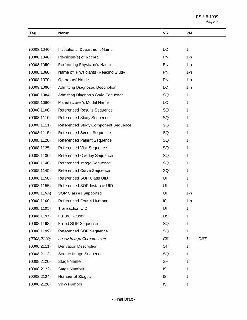
(0008,1040) Institutional Department Name LO 1
(0008,1048) Physician(s) of Record PN 1-n
(0008,1050) Performing Physician’s Name PN 1-n
(0008,1060) Name of Physician(s) Reading Study PN 1-n
(0008,1070) Operators’ Name PN 1-n
(0008,1080) Admitting Diagnoses Description LO 1-n
(0008,1084) Admitting Diagnosis Code Sequence SQ 1
(0008,1090) Manufacturer’s Model Name LO 1
(0008,1100) Referenced Results Sequence SQ 1
(0008,1110) Referenced Study Sequence SQ 1
(0008,1111) Referenced Study Component Sequence SQ 1
(0008,1115) Referenced Series Sequence SQ 1
(0008,1120) Referenced Patient Sequence SQ 1
(0008,1125) Referenced Visit Sequence SQ 1
(0008,1130) Referenced Overlay Sequence SQ 1
(0008,1140) Referenced Image Sequence SQ 1
(0008,1145) Referenced Curve Sequence SQ 1
(0008,1150) Referenced SOP Class UID UI 1
(0008,1155) Referenced SOP Instance UID UI 1
(0008,115A) SOP Classes Supported UI 1-n
(0008,1160) Referenced Frame Number IS 1-n
(0008,1195) Transaction UID UI 1
(0008,1197) Failure Reason US 1
(0008,1198) Failed SOP Sequence SQ 1
(0008,1199) Referenced SOP Sequence SQ 1
(0008,2110) Lossy Image Compression CS 1 RET
(0008,2111) Derivation Description ST 1
(0008,2112) Source Image Sequence SQ 1
(0008,2120) Stage Name SH 1
(0008,2122) Stage Number IS 1
(0008,2124) Number of Stages IS 1
(0008,2128) View Number IS 1

PS 3.6-1999 Page 8
Tag Name VR VM
- Final Draft -
(0008,2129) Number of Event Timers IS 1
(0008,212A) Number of Views in Stage IS 1
(0008,2130) Event Elapsed Time(s) DS 1-n
(0008,2132) Event Timer Name(s) LO 1-n
(0008,2142) Start Trim IS 1
(0008,2143) Stop Trim IS 1
(0008,2144) Recommended Display Frame Rate IS 1
(0008,2200) Transducer Position CS 1 RET
(0008,2204) Transducer Orientation CS 1 RET
(0008,2208) Anatomic Structure CS 1 RET
(0008,2218) Anatomic Region Sequence SQ 1
(0008,2220) Anatomic Region Modifier Sequence SQ 1
(0008,2228) Primary Anatomic Structure Sequence SQ 1
(0008,2229) Anatomic Structure, Space or Region Sequence SQ 1
(0008,2230) Primary Anatomic Structure Modifier Sequence SQ 1
(0008,2240) Transducer Position Sequence SQ 1
(0008,2242) Transducer Position Modifier Sequence SQ 1
(0008,2244) Transducer Orientation Sequence SQ 1
(0008,2246) Transducer Orientation Modifier Sequence SQ 1
(0008,4000) Comments RET
Tag Name VR VM
(0010,0000) Group Length UL 1
(0010,0010) Patient’s Name PN 1
(0010,0020) Patient ID LO 1
(0010,0021) Issuer of Patient ID LO 1
(0010,0030) Patient's Birth Date DA 1
(0010,0032) Patient's Birth Time TM 1
(0010,0040) Patient's Sex CS 1
(0010,0050) Patient's Insurance Plan Code Sequence SQ 1
(0010,1000) Other Patient IDs LO 1-n

PS 3.6-1999 Page 9
Tag Name VR VM
- Final Draft -
(0010,1001) Other Patient Names PN 1-n
(0010,1005) Patient's Birth Name PN 1
(0010,1010) Patient's Age AS 1
(0010,1020) Patient's Size DS 1
(0010,1030) Patient's Weight DS 1
(0010,1040) Patient's Address LO 1
(0010,1050) Insurance Plan Identification RET
(0010,1060) Patient's Mother's Birth Name PN 1
(0010,1080) Military Rank LO 1
(0010,1081) Branch of Service LO 1
(0010,1090) Medical Record Locator LO 1
(0010,2000) Medical Alerts LO 1-n
(0010,2110) Contrast Allergies LO 1-n
(0010,2150) Country of Residence LO 1
(0010,2152) Region of Residence LO 1
(0010,2154) Patient’s Telephone Numbers SH 1-n
(0010,2160) Ethnic Group SH 1
(0010,2180) Occupation SH 1
(0010,21A0) Smoking Status CS 1
(0010,21B0) Additional Patient History LT 1
(0010,21C0) Pregnancy Status US 1
(0010,21D0) Last Menstrual Date DA 1
(0010,21F0) Patient's Religious Preference LO 1
(0010,4000) Patient Comments LT 1
Tag Name VR VM
(0018,0000) Group Length UL 1
(0018,0010) Contrast/Bolus Agent LO 1
(0018,0012) Contrast/Bolus Agent Sequence SQ 1
(0018,0014) Contrast/Bolus Administration Route Sequence SQ 1
(0018,0015) Body Part Examined CS 1
(0018,0020) Scanning Sequence CS 1-n

PS 3.6-1999 Page 10
Tag Name VR VM
- Final Draft -
(0018,0021) Sequence Variant CS 1-n
(0018,0022) Scan Options CS 1-n
(0018,0023) MR Acquisition Type CS 1
(0018,0024) Sequence Name SH 1
(0018,0025) Angio Flag CS 1
(0018,0026) Intervention Drug Information Sequence SQ 1
(0018,0027) Intervention Drug Stop Time TM 1
(0018,0028) Intervention Drug Dose DS 1
(0018,0029) Intervention Drug Code Sequence SQ 1
(0018,002A) Additional Drug Sequence SQ 1
(0018,0030) Radionuclide LO 1-n RET
(0018,0031) Radiopharmaceutical LO 1
(0018,0032) Energy Window Centerline DS 1 RET
(0018,0033) Energy Window Total Width DS 1-n RET
(0018,0034) Intervention Drug Name LO 1
(0018,0035) Intervention Drug Start Time TM 1
(0018,0036) Interventional Therapy Sequence SQ 1
(0018,0037) Therapy Type CS 1
(0018,0038) Interventional Status CS 1
(0018,0039) Therapy Description CS 1
(0018,0040) Cine Rate IS 1
(0018,0050) Slice Thickness DS 1
(0018,0060) KVP DS 1
(0018,0070) Counts Accumulated IS 1
(0018,0071) Acquisition Termination Condition CS 1
(0018,0072) Effective Series Duration DS 1
(0018,0073) Acquisition Start Condition CS 1
(0018,0074) Acquisition Start Condition Data IS 1
(0018,0075) Acquisition Termination Condition Data IS 1
(0018,0080) Repetition Time DS 1
(0018,0081) Echo Time DS 1

PS 3.6-1999 Page 11
Tag Name VR VM
- Final Draft -
(0018,0082) Inversion Time DS 1
(0018,0083) Number of Averages DS 1
(0018,0084) Imaging Frequency DS 1
(0018,0085) Imaged Nucleus SH 1
(0018,0086) Echo Number(s) IS 1-n
(0018,0087) Magnetic Field Strength DS 1
(0018,0088) Spacing Between Slices DS 1
(0018,0089) Number of Phase Encoding Steps IS 1
(0018,0090) Data Collection Diameter DS 1
(0018,0091) Echo Train Length IS 1
(0018,0093) Percent Sampling DS 1
(0018,0094) Percent Phase Field of View DS 1
(0018,0095) Pixel Bandwidth DS 1
(0018,1000) Device Serial Number LO 1
(0018,1004) Plate ID LO 1
(0018,1010) Secondary Capture Device ID LO 1
(0018,1011) Hardcopy Creation Device ID LO 1
(0018,1012) Date of Secondary Capture DA 1
(0018,1014) Time of Secondary Capture TM 1
(0018,1016) Secondary Capture Device Manufacturer LO 1
(0018,1017) Hardcopy Device Manufacturer LO 1
(0018,1018) Secondary Capture Device Manufacturer’s Model Name LO 1
(0018,1019) Secondary Capture Device Software Version(s) LO 1-n
(0018,101A) Hardcopy Device Software Version LO 1-n
(0018,101B) Hardcopy Device Manfuacturer's Model Name LO 1
(0018,1020) Software Version(s) LO 1-n
(0018,1022) Video Image Format Acquired SH 1
(0018,1023) Digital Image Format Acquired LO 1
(0018,1030) Protocol Name LO 1
(0018,1040) Contrast/Bolus Route LO 1
(0018,1041) Contrast/Bolus Volume DS 1
(0018,1042) Contrast/Bolus Start Time TM 1

PS 3.6-1999 Page 12
Tag Name VR VM
- Final Draft -
(0018,1043) Contrast/Bolus Stop Time TM 1
(0018,1044) Contrast/Bolus Total Dose DS 1
(0018,1045) Syringe Counts IS 1
(0018,1046) Contrast Flow Rate(s) DS 1-n
(0018,1047) Contrast Flow Duration(s) DS 1-n
(0018,1048) Contrast/Bolus Ingredient CS 1
(0018,1049) Contrast/Bolus Ingredient Concentration DS 1
(0018,1050) Spatial Resolution DS 1
(0018,1060) Trigger Time DS 1
(0018,1061) Trigger Source or Type LO 1
(0018,1062) Nominal Interval IS 1
(0018,1063) Frame Time DS 1
(0018,1064) Framing Type LO 1
(0018,1065) Frame Time Vector DS 1-n
(0018,1066) Frame Delay DS 1
(0018,1067) Image Trigger Delay DS 1
(0018,1068) Multiplex Group Time Offset DS 1
(0018,1069) Trigger Time Offset DS 1
(0018,106A) Synchronization Trigger CS 1
(0018,106C) Synchronization Channel US 2
(0018,106E) Trigger Sample Position UL 1
(0018,1070) Radionuclide Route LO 1-n RET
(0018,1070) Radiopharmaceutical Route LO 1
(0018,1071) Radionuclide Volume DS 1-n RET
(0018,1071) Radiopharmaceutical Volume DS 1
(0018,1072) Radionuclide Start Time TM 1-n RET
(0018,1072) Radiopharmaceutical Start Time TM 1
(0018,1073) Radionuclide Stop Time TM 1-n RET
(0018,1073) Radiopharmaceutical Stop Time TM 1
(0018,1074) Radionuclide Total Dose DS 1-n RET
(0018,1074) Radionuclide Total Dose DS 1

PS 3.6-1999 Page 13
Tag Name VR VM
- Final Draft -
(0018,1075) Radionuclide Half Life DS 1
(0018,1076) Radionuclide Positron Fraction DS 1
(0018,1077) Radiopharmaceutical Specific Activity DS 1
(0018,1080) Beat Rejection Flag CS 1
(0018,1081) Low R-R Value IS 1
(0018,1082) High R-R Value IS 1
(0018,1083) Intervals Acquired IS 1
(0018,1084) Intervals Rejected IS 1
(0018,1085) PVC Rejection LO 1
(0018,1086) Skip Beats IS 1
(0018,1088) Heart Rate IS 1
(0018,1090) Cardiac Number of Images IS 1
(0018,1094) Trigger Window IS 1
(0018,1100) Reconstruction Diameter DS 1
(0018,1110) Distance Source to Detector DS 1
(0018,1111) Distance Source to Patient DS 1
(0018,1114) Estimated Radiographic Magnification Factor DS 1
(0018,1120) Gantry/Detector Tilt DS 1
(0018,1121) Gantry/Detector Slew DS 1
(0018,1130) Table Height DS 1
(0018,1131) Table Traverse DS 1
(0018,1134) Table Motion CS 1
(0018,1135) Table Vertical Increment DS 1-n
(0018,1136) Table Lateral Increment DS 1-n
(0018,1137) Table Longitudinal Increment DS 1-n
(0018,1138) Table Angle DS 1
(0018,113A) Table Type CS 1
(0018,1140) Rotation Direction CS 1
(0018,1141) Angular Position DS 1
(0018,1142) Radial Position DS 1-n
(0018,1143) Scan Arc DS 1
(0018,1144) Angular Step DS 1

PS 3.6-1999 Page 14
Tag Name VR VM
- Final Draft -
(0018,1145) Center of Rotation Offset DS 1
(0018,1146) Rotation Offset DS 1-n RET
(0018,1147) Field of View Shape CS 1
(0018,1149) Field of View Dimension(s) IS 1-2
(0018,1150) Exposure Time IS 1
(0018,1151) X-ray Tube Current IS 1
(0018,1152) Exposure IS 1
(0018,1153) Exposure in uAs IS 1
(0018,1154) Average Pulse Width DS 1
(0018,1155) Radiation Setting CS 1
(0018,1156) Rectification Type CS 1
(0018,115A) Radiation Mode CS 1
(0018,115E) Image Area Dose Product DS 1
(0018,1160) Filter Type SH 1
(0018,1161) Type of Filters LO 1-n
(0018,1162) Intensifier Size DS 1
(0018,1164) Imager Pixel Spacing DS 2
(0018,1166) Grid CS 1
(0018,1170) Generator Power IS 1
(0018,1180) Collimator/grid Name SH 1
(0018,1181) Collimator Type CS 1
(0018,1182) Focal Distance IS 1-2
(0018,1183) X Focus Center DS 1-2
(0018,1184) Y Focus Center DS 1-2
(0018,1190) Focal Spot(s) DS 1-n
(0018,1191) Anode Target Material CS 1
(0018,11A0) Body Part Thickness DS 1
(0018,11A2) Compression Force DS 1
(0018,1200) Date of Last Calibration DA 1-n
(0018,1201) Time of Last Calibration TM 1-n
(0018,1210) Convolution Kernel SH 1-n

PS 3.6-1999 Page 15
Tag Name VR VM
- Final Draft -
(0018,1240) Upper/Lower Pixel Values RET
(0018,1242) Actual Frame Duration IS 1
(0018,1243) Count Rate IS 1
(0018,1244) Preferred Playback Sequencing US 1
(0018,1250) Receiving Coil SH 1
(0018,1251) Transmitting Coil SH 1
(0018,1260) Plate Type SH 1
(0018,1261) Phosphor Type LO 1
(0018,1300) Scan Velocity DS 1
(0018,1301) Whole Body Technique CS 1-n
(0018,1302) Scan Length IS 1
(0018,1310) Acquisition Matrix US 4
(0018,1312) Phase Encoding Direction CS 1
(0018,1314) Flip Angle DS 1
(0018,1315) Variable Flip Angle Flag CS 1
(0018,1316) SAR DS 1
(0018,1318) dB/dt DS 1
(0018,1400) Acquisition Device Processing Description LO 1
(0018,1401) Acquisition Device Processing Code LO 1
(0018,1402) Cassette Orientation CS 1
(0018,1403) Cassette Size CS 1
(0018,1404) Exposures on Plate US 1
(0018,1405) Relative X-ray Exposure IS 1
(0018,1450) Column Angulation CS 1
(0018,1460) Tomo Layer Height DS 1
(0018,1470) Tomo Angle DS 1
(0018,1480) Tomo Time DS 1
(0018,1490) Tomo Type CS 1
(0018,1491) Tomo Class CS 1
(0018,1495) Number of Tomosynthesis Source Images IS 1
(0018,1500) Positioner Motion CS 1
(0018,1508) Positioner Type CS 1

PS 3.6-1999 Page 16
Tag Name VR VM
- Final Draft -
(0018,1510) Positioner Primary Angle DS 1
(0018,1511) Positioner Secondary Angle DS 1
(0018,1520) Positioner Primary Angle Increment DS 1-n
(0018,1521) Positioner Secondary Angle Increment DS 1-n
(0018,1530) Detector Primary Angle DS 1
(0018,1531) Detector Secondary Angle DS 1
(0018,1600) Shutter Shape CS 1-3
(0018,1602) Shutter Left Vertical Edge IS 1
(0018,1604) Shutter Right Vertical Edge IS 1
(0018,1606) Shutter Upper Horizontal Edge IS 1
(0018,1608) Shutter Lower Horizontal Edge IS 1
(0018,1610) Center of Circular Shutter IS 2
(0018,1612) Radius of Circular Shutter IS 1
(0018,1620) Vertices of the Polygonal Shutter IS 2-2n
(0018,1622) Shutter Presentation Value US 1
(0018,1623) Shutter Overlay Group US 1
(0018,1700) Collimator Shape CS 1-3
(0018,1702) Collimator Left Vertical Edge IS 1
(0018,1704) Collimator Right Vertical Edge IS 1
(0018,1706) Collimator Upper Horizontal Edge IS 1
(0018,1708) Collimator Lower Horizontal Edge IS 1
(0018,1710) Center of Circular Collimator IS 2
(0018,1712) Radius of Circular Collimator IS 1
(0018,1720) Vertices of the Polygonal Collimator IS 2-2n
(0018,1800) Acquisition Time Synchronized CS 1
(0018,1802) Time Distribution Protocol CS 1
(0018,1801) Time Source SH 1
(0018,4000) Comments RET
(0018,5000) Output Power SH 1-n
(0018,5010) Transducer Data LO 3
(0018,5012) Focus Depth DS 1

PS 3.6-1999 Page 17
Tag Name VR VM
- Final Draft -
(0018,5020) Processing Function LO 1
(0018,5021) Postprocessing Function LO 1
(0018,5022) Mechanical Index DS 1
(0018,5024) Thermal Index DS 1
(0018,5026) Cranial Thermal Index DS 1
(0018,5027) Soft Tissue Thermal Index DS 1
(0018,5028) Soft Tissue-focus Thermal Index DS 1
(0018,5029) Soft Tissue-surface Thermal Index DS 1
(0018,5030) Dynamic Range RET
(0018,5040) Total Gain RET
(0018,5050) Depth of Scan Field IS 1
(0018,5100) Patient Position CS 1
(0018,5101) View Position CS 1
(0018,5104) Projection Eponymous Name Code Sequence SQ 1
(0018,5210) Image Transformation Matrix DS 6
(0018,5212) Image Translation Vector DS 3
(0018,6000) Sensitivity DS 1
(0018,6011) Sequence of Ultrasound Regions SQ 1
(0018,6012) Region Spatial Format US 1
(0018,6014) Region Data Type US 1
(0018,6016) Region Flags UL 1
(0018,6018) Region Location Min X0 UL 1
(0018,601A) Region Location Min Y0 UL 1
(0018,601C) Region Location Max X1 UL 1
(0018,601E) Region Location Max Y1 UL 1
(0018,6020) Reference Pixel X0 SL 1
(0018,6022) Reference Pixel Y0 SL 1
(0018,6024) Physical Units X Direction US 1
(0018,6026) Physical Units Y Direction US 1
(0018,6028) Reference Pixel Physical Value X FD 1
(0018,602A) Reference Pixel Physical Value Y FD 1
(0018,602C) Physical Delta X FD 1

PS 3.6-1999 Page 18
Tag Name VR VM
- Final Draft -
(0018,602E) Physical Delta Y FD 1
(0018,6030) Transducer Frequency UL 1
(0018,6031) Transducer Type CS 1
(0018,6032) Pulse Repetition Frequency UL 1
(0018,6034) Doppler Correction Angle FD 1
(0018,6036) Steering Angle FD 1
(0018,6038) Doppler Sample Volume X Position UL 1
(0018,603A) Doppler Sample Volume Y Position UL 1
(0018,603C) TM-Line Position X0 UL 1
(0018,603E) TM-Line Position Y0 UL 1
(0018,6040) TM-Line Position X1 UL 1
(0018,6042) TM-Line Position Y1 UL 1
(0018,6044) Pixel Component Organization US 1
(0018,6046) Pixel Component Mask UL 1
(0018,6048) Pixel Component Range Start UL 1
(0018,604A) Pixel Component Range Stop UL 1
(0018,604C) Pixel Component Physical Units US 1
(0018,604E) Pixel Component Data Type US 1
(0018,6050) Number of Table Break Points UL 1
(0018,6052) Table of X Break Points UL 1-n
(0018,6054) Table of Y Break Points FD 1-n
(0018,6056) Number of Table Entries UL 1
(0018,6058) Table of Pixel Values UL 1-n
(0018,605A) Table of Parameter Values FL 1-n
(0018,7000) Detector Conditions Nominal Flag CS 1
(0018,7001) Detector Temperature DS 1
(0018,7004) Detector Type CS 1
(0018,7005) Detector Configuration CS 1
(0018,7006) Detector Description LT 1
(0018,7008) Detector Mode LT 1
(0018,700A) Detector ID SH 1

PS 3.6-1999 Page 19
Tag Name VR VM
- Final Draft -
(0018,700C) Date of Last Detector Calibration DA 1
(0018,700E) Time of Last Detector Calibration TM 1
(0018,7010) Exposures on Detector Since Last Calibration IS 1
(0018,7011) Exposures on Detector Since Manufactured IS 1
(0018,7012) Detector Time Since Last Exposure DS 1
(0018,7014) Detector Active Time DS 1
(0018,7016) Detector Activation Offset From Exposure DS 1
(0018,701A) Detector Binning DS 2
(0018,7020) Detector Element Physical Size DS 2
(0018,7022) Detector Element Spacing DS 2
(0018,7024) Detector Active Shape CS 1
(0018,7026) Detector Active Dimension(s) DS 1-2
(0018,7028) Detector Active Origin DS 2
(0018,7030) Field of View Origin DS 2
(0018,7032) Field of View Rotation DS 1
(0018,7034) Field of View Horizontal Flip CS 1
(0018,7040) Grid Absorbing Material LT 1
(0018,7041) Grid Spacing Material LT 1
(0018,7042) Grid Thickness DS 1
(0018,7044) Grid Pitch DS 1
(0018,7046) Grid Aspect Ratio IS 2
(0018,7048) Grid Period DS 1
(0018,704C) Grid Focal Distance DS 1
(0018,7050) Filter Material LTCS 1-n
(0018,7052) Filter Thickness Minimum DS 1-n
(0018,7054) Filter Thickness Maximum DS 1-n
(0018,7060) Exposure Control Mode CS 1
(0018,7062) Exposure Control Mode Description LT 1
(0018,7064) Exposure Status CS 1
(0018,7065) Phototimer Setting DS 1
(0018,8150) Exposure Time in µS DS 1
(0018,8151) X-Ray Tube Current in µA DS 1

PS 3.6-1999 Page 20
Tag Name VR VM
- Final Draft -
Tag Name VR VM
(0020,0000) Group Length UL 1
(0020,000D) Study Instance UID UI 1
(0020,000E) Series Instance UID UI 1
(0020,0010) Study ID SH 1
(0020,0011) Series Number IS 1
(0020,0012) Acquisition Number IS 1
(0020,0013) Instance Number IS 1
(0020,0014) Isotope Number IS 1 RET
(0020,0015) Phase Number IS 1 RET
(0020,0016) Interval Number IS 1 RET
(0020,0017) Time Slot Number IS 1 RET
(0020,0018) Angle Number IS 1 RET
(0020,0019) Item Number IS 1
(0020,0020) Patient Orientation CS 2
(0020,0022) Overlay Number IS 1
(0020,0024) Curve Number IS 1
(0020,0026) Lookup Table Number IS 1
(0020,0030) Image Position RET
(0020,0032) Image Position (Patient) DS 3
(0020,0035) Image Orientation RET
(0020,0037) Image Orientation (Patient) DS 6
(0020,0050) Location RET
(0020,0052) Frame of Reference UID UI 1
(0020,0060) Laterality CS 1
(0020,0062) Image Laterality CS 1
(0020,0070) Image Geometry Type RET
(0020,0080) Masking Image RET
(0020,0100) Temporal Position Identifier IS 1
(0020,0105) Number of Temporal Positions IS 1

PS 3.6-1999 Page 21
Tag Name VR VM
- Final Draft -
(0020,0110) Temporal Resolution DS 1
(0020,0200) Synchronization Frame of Reference UID UI 1
(0020,1000) Series in Study IS 1
(0020,1001) Acquisitions in Series RET
(0020,1002) Images in Acquisition IS 1
(0020,1004) Acquisitions in Study IS 1
(0020,1020) Reference RET
(0020,1040) Position Reference Indicator LO 1
(0020,1041) Slice Location DS 1
(0020,1070) Other Study Numbers IS 1-n
(0020,1200) Number of Patient Related Studies IS 1
(0020,1202) Number of Patient Related Series IS 1
(0020,1204) Number of Patient Related Instances IS 1
(0020,1206) Number of Study Related Series IS 1
(0020,1208) Number of Study Related Instances IS 1
(0020,1209) Number of Series Related Instances IS 1
(0020,3100 to 31FF)
Source Image IDs RET
(0020,3401) Modifying Device ID RET
(0020,3402) Modified Image ID RET
(0020,3403) Modified Image Date RET
(0020,3404) Modifying Device Manufacturer RET
(0020,3405) Modified Image Time RET
(0020,3406) Modified Image Description RET
(0020,4000) Image Comments LT 1
(0020,5000) Original Image Identification RET
(0020,5002) Original Image Identification Nomenclature RET
Tag Name VR VM
(0028,0000) Group Length UL 1
(0028,0002) Samples per Pixel US 1
(0028,0004) Photometric Interpretation CS 1

PS 3.6-1999 Page 22
Tag Name VR VM
- Final Draft -
(0028,0005) Image Dimensions RET
(0028,0006) Planar Configuration US 1
(0028,0008) Number of Frames IS 1
(0028,0009) Frame Increment Pointer AT 1-n
(0028,0010) Rows US 1
(0028,0011) Columns US 1
(0028,0012) Planes US 1
(0028,0014) Ultrasound Color Data Present US 1
(0028,0030) Pixel Spacing DS 2
(0028,0031) Zoom Factor DS 2
(0028,0032) Zoom Center DS 2
(0028,0034) Pixel Aspect Ratio IS 2
(0028,0040) Image Format RET
(0028,0050) Manipulated Image RET
(0028,0051) Corrected Image CS 1-n
(0028,0060) Compression Code RET
(0028,0100) Bits Allocated US 1
(0028,0101) Bits Stored US 1
(0028,0102) High Bit US 1
(0028,0103) Pixel Representation US 1
(0028,0104) Smallest Valid Pixel Value RET
(0028,0105) Largest Valid Pixel Value RET
(0028,0106) Smallest Image Pixel Value US or SS 1
(0028,0107) Largest Image Pixel Value US or SS 1
(0028,0108) Smallest Pixel Value in Series US or SS 1
(0028,0109) Largest Pixel Value in Series US or SS 1
(0028,0110) Smallest Image Pixel Value in Plane US or SS 1
(0028,0111) Largest Image Pixel Value in Plane US or SS 1
(0028,0120) Pixel Padding Value US or SS 1
(0028,0200) Image Location RET
(0028,0300) Quality Control Image CS 1

PS 3.6-1999 Page 23
Tag Name VR VM
- Final Draft -
(0028,0301) Burned In Annotation CS 1
(0028,1040) Pixel Intensity Relationship CS 1
(0028,1041) Pixel Intensity Relationship Sign SS 1
(0028,1050) Window Center DS 1-n
(0028,1051) Window Width DS 1-n
(0028,1052) Rescale Intercept DS 1
(0028,1053) Rescale Slope DS 1
(0028,1054) Rescale Type LO 1
(0028,1055) Window Center & Width Explanation LO 1-n
(0028,1080) Gray Scale RET
(0028,1090) Recommended Viewing Mode CS 1
(0028,1100) Gray Lookup Table Descriptor RET
(0028,1101) Red Palette Color Lookup Table Descriptor US\US or SS\US
3
(0028,1102) Green Palette Color Lookup Table Descriptor US\US or SS\US
3
(0028,1103) Blue Palette Color Lookup Table Descriptor US\US or SS\US
3
(0028,1199) Palette Color Lookup Table UID UI 1
(0028,1200) Gray Lookup Table Data RET
(0028,1201) Red Palette Color Lookup Table Data US or SS or OW
1-n 1
(0028,1202) Green Palette Color Lookup Table Data US or SS or OW
1-n 1
(0028,1203) Blue Palette Color Lookup Table Data US or SS or OW
1-n 1
(0028,1221) Segmented Red Palette Color Lookup Table Data OW 1
(0028,1222) Segmented Green Palette Color Lookup Table Data OW 1
(0028,1223) Segmented Blue Palette Color Lookup Table Data OW 1
(0028,1300) Implant Present CS 1
(0028,1350) Partial View CS 1
(0028,1351) Partial View Description ST 1
(0028,2110) Lossy Image Compression CS 1
(0028,2112) Lossy Image Compression Ratio DS 1-n

PS 3.6-1999 Page 24
Tag Name VR VM
- Final Draft -
(0028,3000) Modality LUT Sequence SQ 1
(0028,3002) LUT Descriptor US\US or SS\US
3
(0028,3003) LUT Explanation LO 1
(0028,3004) Modality LUT Type LO 1
(0028,3006) LUT Data US or SS or OW
1-n 1
(0028,3010) VOI LUT Sequence SQ 1
(0028,3110) Softcopy VOI LUT Sequence SQ 1
(0028,4000) Comments RET
(0028,5000) Bi-Plane Acquisition Sequence SQ 1
(0028,6010) Representative Frame Number US 1
(0028,6020) Frame Numbers of Interest (FOI) US 1-n
(0028,6022) Frame(s) of Interest Description LO 1-n
(0028,6030) Mask Pointer(s) US 1-n
(0028,6040) R Wave Pointer US 1-n
(0028,6100) Mask Subtraction Sequence SQ 1
(0028,6101) Mask Operation CS 1
(0028,6102) Applicable Frame Range US 2-2n
(0028,6110) Mask Frame Numbers US 1-n
(0028,6112) Contrast Frame Averaging US 1
(0028,6114) Mask Sub-pixel Shift FL 2
(0028,6120) TID Offset SS 1
(0028,6190) Mask Operation Explanation ST 1
Tag Name VR VM
(0032,0000) Group Length UL 1
(0032,000A) Study Status ID CS 1
(0032,000C) Study Priority ID CS 1
(0032,0012) Study ID Issuer LO 1
(0032,0032) Study Verified Date DA 1
(0032,0033) Study Verified Time TM 1

PS 3.6-1999 Page 25
Tag Name VR VM
- Final Draft -
(0032,0034) Study Read Date DA 1
(0032,0035) Study Read Time TM 1
(0032,1000) Scheduled Study Start Date DA 1
(0032,1001) Scheduled Study Start Time TM 1
(0032,1010) Scheduled Study Stop Date DA 1
(0032,1011) Scheduled Study Stop Time TM 1
(0032,1020) Scheduled Study Location LO 1
(0032,1021) Scheduled Study Location AE Title(s) AE 1-n
(0032,1030) Reason for Study LO 1
(0032,1032) Requesting Physician PN 1
(0032,1033) Requesting Service LO 1
(0032,1040) Study Arrival Date DA 1
(0032,1041) Study Arrival Time TM 1
(0032,1050) Study Completion Date DA 1
(0032,1051) Study Completion Time TM 1
(0032,1055) Study Component Status ID CS 1
(0032,1060) Requested Procedure Description LO 1
(0032,1064) Requested Procedure Code Sequence SQ 1
(0032,1070) Requested Contrast Agent LO 1
(0032,4000) Study Comments LT 1
Tag Name VR VM
(0038,0000) Group Length UL 1
(0038,0004) Referenced Patient Alias Sequence SQ 1
(0038,0008) Visit Status ID CS 1
(0038,0010) Admission ID LO 1
(0038,0011) Issuer of Admission ID LO 1
(0038,0016) Route of Admissions LO 1
(0038,001A) Scheduled Admission Date DA 1
(0038,001B) Scheduled Admission Time TM 1
(0038,001C) Scheduled Discharge Date DA 1
(0038,001D) Scheduled Discharge Time TM 1

PS 3.6-1999 Page 26
Tag Name VR VM
- Final Draft -
(0038,001E) Scheduled Patient Institution Residence LO 1
(0038,0020) Admitting Date DA 1
(0038,0021) Admitting Time TM 1
(0038,0030) Discharge Date DA 1
(0038,0032) Discharge Time TM 1
(0038,0040) Discharge Diagnosis Description LO 1
(0038,0044) Discharge Diagnosis Code Sequence SQ 1
(0038,0050) Special Needs LO 1
(0038,0300) Current Patient Location LO 1
(0038,0400) Patient's Institution Residence LO 1
(0038,0500) Patient State LO 1
(0038,4000) Visit Comments LT 1
Tag Name VR VM
(003A,0004) Waveform Originality CS 1
(003A,0005) Number of Waveform Channels US 1
(003A,0010) Number of Waveform Samples UL 1
(003A,001A) Sampling Frequency DS 1
(003A,0020) Multiplex Group Label SH 1
(003A,0200) Channel Definition Sequence SQ 1
(003A,0202) Waveform Channel Number IS 1
(003A,0203) Channel Label SH 1
(003A,0205) Channel Status CS 1-n
(003A,0208) Channel Source Sequence SQ 1
(003A,0209) Channel Source Modifiers Sequence SQ 1
(003A,020A) Source Waveform Sequence SQ 1
(003A,020C) Channel Derivation Description LO 1
(003A,0210) Channel Sensitivity DS 1
(003A,0211) Channel Sensitivity Units Sequence SQ 1
(003A,0212) Channel Sensitivity Correction Factor DS 1
(003A,0213) Channel Baseline DS 1

PS 3.6-1999 Page 27
Tag Name VR VM
- Final Draft -
(003A,0214) Channel Time Skew DS 1
(003A,0215) Channel Sample Skew DS 1
(003A,0218) Channel Offset DS 1
(003A,021A) Waveform Bits Stored US 1
(003A,0220) Filter Low Frequency DS 1
(003A,0221) Filter High Frequency DS 1
(003A,0222) Notch Filter Frequency DS 1
(003A,0223) Notch Filter Bandwidth DS 1
Tag Name VR VM
(0040,0000) Group Length UL 1
(0040,0001) Scheduled Station AE Title AE 1-n
(0040,0002) Scheduled Procedure Step Start Date DA 1
(0040,0003) Scheduled Procedure Step Start Time TM 1
(0040,0004) Scheduled Procedure Step End Date DA 1
(0040,0005) Scheduled Procedure Step End Time TM 1
(0040,0006) Scheduled Performing Physician's Name PN 1
(0040,0007) Scheduled Procedure Step Description LO 1
(0040,0008) Scheduled Action Item Code Sequence SQ 1
(0040,0009) Scheduled Procedure Step ID SH 1
(0040,0010) Scheduled Station Name SH 1-n
(0040,0011) Scheduled Procedure Step Location SH 1
(0040,0012) Pre-Medication LO 1
(0040,0020) Scheduled Procedure Step Status CS 1
(0040,0100) Scheduled Procedure Step Sequence SQ 1
(0040,0220) Referenced Standalone SOP Instance Sequence SQ 1
(0040,0241) Performed Station AE Title AE 1
(0040,0242) Performed Station Name SH 1
(0040,0243) Performed Location SH 1
(0040,0244) Performed Procedure Step Start Date DA 1
(0040,0245) Performed Procedure Step Start Time TM 1
(0040,0250) Performed Procedure Step End Date DA 1

PS 3.6-1999 Page 28
Tag Name VR VM
- Final Draft -
(0040,0251) Performed Procedure Step End Time TM 1
(0040,0252) Performed Procedure Step Status CS 1
(0040,0253) Performed Procedure Step ID SH 1
(0040,0254) Performed Procedure Step Description LO 1
(0040,0255) Performed Procedure Type Description LO 1
(0040,0260) Performed Action Item Sequence SQ 1
(0040,0270) Scheduled Step Attributes Sequence SQ 1
(0040,0275) Request Attributes Sequence SQ 1
(0040,0280) Comments on the Performed Procedure Steps ST 1
(0040,0293) Quantity Sequence SQ 1
(0040,0294) Quantity DS 1
(0040,0295) Measuring Units Sequence SQ 1
(0040,0296) Billing Item Sequence SQ 1
(0040,0300) Total Time of Fluoroscopy US 1
(0040,0301) Total Number of Exposures US 1
(0040,0302) Entrance Dose US 1
(0040,0303) Exposed Area US 1-2
(0040,0306) Distance Source to Entrance DS 1
(0040,0307) Distance Source to Support DS 1
(0040,0310) Comments on Radiation Dose ST 1
(0040,0312) X-Ray Output DS 1
(0040,0314) Half Value Layer DS 1
(0040,0316) Organ Dose DS 1
(0040,0318) Organ Exposed CS 1
(0040,0320) Billing Procedure Step Sequence SQ 1
(0040,0321) Film Consumption Sequence SQ 1
(0040,0324) Billing Supplies and Devices Sequence SQ 1
(0040,0330) Referenced Procedure Step Sequence SQ 1
(0040,0340) Performed Series Sequence SQ 1
(0040,0400) Comments on the Scheduled Procedure Step LT 1
(0040,050A) Specimen Accession Number LO 1
(0040,0550) Specimen Sequence SQ 1

PS 3.6-1999 Page 29
Tag Name VR VM
- Final Draft -
(0040,0551) Specimen Identifier LO 1
(0040,059A) Specimen Type Code Sequence SQ 1
(0040,0555) Acquisition Context Sequence SQ 1
(0040,0556) Acquisition Context Description ST 1
(0040,06FA) Slide Identifier LO 1
(0040,071A) Image Center Point Coordinates Sequence SQ 1
(0040,072A) X offset in Slide Coordinate System DS 1
(0040,073A) Y offset in Slide Coordinate System DS 1
(0040,074A) Z offset in Slide Coordinate System DS 1
(0040,08D8) Pixel Spacing Sequence SQ 1
(0040,08DA) Coordinate System Axis Code Sequence SQ 1
(0040,08EA) Measurement Units Code Sequence SQ 1
(0040,1001) Requested Procedure ID SH 1
(0040,1002) Reason for the Requested Procedure LO 1
(0040,1003) Requested Procedure Priority SH 1
(0040,1004) Patient Transport Arrangements LO 1
(0040,1005) Requested Procedure Location LO 1
(0040,1006) Placer Order Number / Procedure SH 1 RET
(0040,1007) Filler Order Number / Procedure SH 1 RET
(0040,1008) Confidentiality Code LO 1
(0040,1009) Reporting Priority SH 1
(0040,1010) Names of Intended Recipients of Results PN 1-n
(0040,1400) Requested Procedure Comments LT 1
(0040,2001) Reason for the Imaging Service Request LO 1
(0040,2004) Issue Date of Imaging Service Request DA 1
(0040,2005) Issue Time of Imaging Service Request TM 1
(0040,2006) Placer Order Number / Imaging Service Request SH 1 RET
(0040,2007) Filler Order Number / Imaging Service Request SH 1 RET
(0040,2008) Order Entered By PN 1
(0040,2009) Order Enterer’s Location SH 1
(0040,2010) Order Callback Phone Number SH 1
(0040,2016) Placer Order Number / Imaging Service Request LO 1

PS 3.6-1999 Page 30
Tag Name VR VM
- Final Draft -
(0040,2017) Filler Order Number / Imaging Service Request LO 1
(0040,2400) Imaging Service Request Comments LT 1
(0040,3001) Confidentiality Constraint on Patient Data Description LO 1
(0040,8302) Entrance Dose in mGy DS 1
(0040,A010) Relationship Type CS 1
(0040,A027) Verifying Organization LO 1
(0040,A030) Verification DateTime DT 1
(0040,A032) Observation DateTime DT 1
(0040,A040) Value Type CS 1
(0040,A043) Concept-name Code Sequence SQ 1
(0040,A050) Continuity Of Content CS 1
(0040,A073) Verifying Observer Sequence SQ 1
(0040,A075) Verifying Observer Name PN 1
(0040,A088) Verifying Observer Identification Code Sequence SQ 1
(0040,A0B0) Referenced Waveform Channels US 2-2n
(0040,A120) DateTime DT 1
(0040,A121) Date DA 1
(0040,A122) Time TM 1
(0040,A123) Person Name PN 1
(0040,A124) UID UI 1
(0040,A130) Temporal Range Type CS 1
(0040,A132) Referenced Sample Positions UL 1-n
(0040,A136) Referenced Frame Numbers US 1-n
(0040,A138) Referenced Time Offsets DS 1-n
(0040,A13A) Referenced Datetime DT 1-n
(0040,A160) Text Value UT 1
(0040,A168) Concept Code Sequence SQ 1
(0040,A180) Annotation Group Number US 1
(0040,A195) Modifier Code Sequence SQ 1
(0040,A300) Measured Value Sequence SQ 1
(0040,A30A) Numeric Value DS 1-n
(0040,A360) Predecessor Documents Sequence SQ 1

PS 3.6-1999 Page 31
Tag Name VR VM
- Final Draft -
(0040,A370) Referenced Request Sequence SQ 1
(0040,A372) Performed Procedure Code Sequence SQ 1
(0040,A375) Current Requested Procedure Evidence Sequence SQ 1
(0040,A385) Pertinent Other Evidence Sequence SQ 1
(0040,A491) Completion Flag CS 1
(0040,A492) Completion Flag Description LO 1
(0040,A493) Verification Flag CS 1
(0040,A504) Content Template Sequence SQ 1
(0040,A525) Identical Documents Sequence SQ 1
(0040,A730) Content Sequence SQ 1
(0040,B020) Annotation Sequence SQ 1
(0040,DB00) Template Identifier CS 1
(0040,DB06) Template Version DT 1
(0040,DB07) Template Local Version DT 1
(0040,DB0B) Template Extension Flag CS 1
(0040,DB0C) Template Extension Organization UID UI 1
(0040,DB0D) Template Extension Creator UID UI 1
(0040,DB73) Referenced Content Item Identifier UL 1-n
Tag Name VR VM
(0050,0000) Group Length UL 1
(0050,0004) Calibration Image CS 1
(0050,0010) Device Sequence SQ 1
(0050,0014) Device Length DS 1
(0050,0016) Device Diameter DS 1
(0050,0017) Device Diameter Units CS 1
(0050,0018) Device Volume DS 1
(0050,0019) Inter-marker Distance DS 1
(0050,0020) Device Description LO 1
Tag Name VR VM
PS 3.6-1999 Page 32
Tag Name VR VM
- Final Draft -
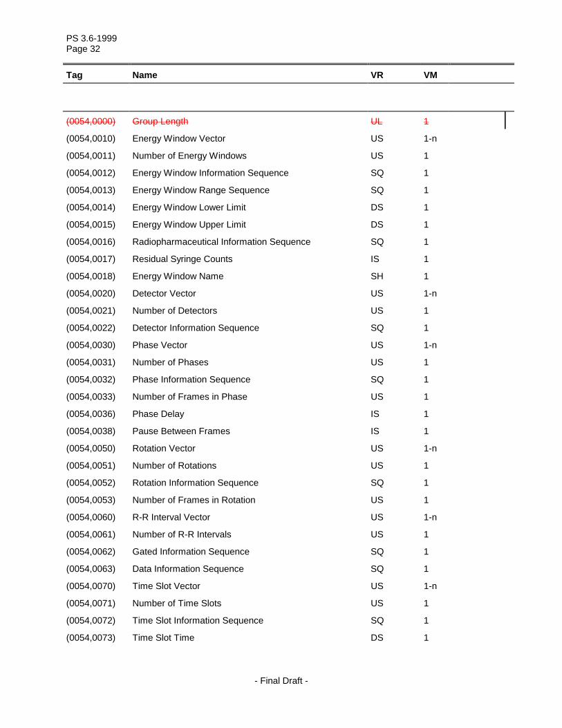
(0054,0000) Group Length UL 1
(0054,0010) Energy Window Vector US 1-n
(0054,0011) Number of Energy Windows US 1
(0054,0012) Energy Window Information Sequence SQ 1
(0054,0013) Energy Window Range Sequence SQ 1
(0054,0014) Energy Window Lower Limit DS 1
(0054,0015) Energy Window Upper Limit DS 1
(0054,0016) Radiopharmaceutical Information Sequence SQ 1
(0054,0017) Residual Syringe Counts IS 1
(0054,0018) Energy Window Name SH 1
(0054,0020) Detector Vector US 1-n
(0054,0021) Number of Detectors US 1
(0054,0022) Detector Information Sequence SQ 1
(0054,0030) Phase Vector US 1-n
(0054,0031) Number of Phases US 1
(0054,0032) Phase Information Sequence SQ 1
(0054,0033) Number of Frames in Phase US 1
(0054,0036) Phase Delay IS 1
(0054,0038) Pause Between Frames IS 1
(0054,0050) Rotation Vector US 1-n
(0054,0051) Number of Rotations US 1
(0054,0052) Rotation Information Sequence SQ 1
(0054,0053) Number of Frames in Rotation US 1
(0054,0060) R-R Interval Vector US 1-n
(0054,0061) Number of R-R Intervals US 1
(0054,0062) Gated Information Sequence SQ 1
(0054,0063) Data Information Sequence SQ 1
(0054,0070) Time Slot Vector US 1-n
(0054,0071) Number of Time Slots US 1
(0054,0072) Time Slot Information Sequence SQ 1
(0054,0073) Time Slot Time DS 1

PS 3.6-1999 Page 33
Tag Name VR VM
- Final Draft -
(0054,0080) Slice Vector US 1-n
(0054,0081) Number of Slices US 1
(0054,0090) Angular View Vector US 1-n
(0054,0100) Time Slice Vector US 1-n
(0054,0101) Number of Time Slices US 1
(0054,0200) Start Angle DS 1
(0054,0202) Type of Detector Motion CS 1
(0054,0210) Trigger Vector IS 1-n
(0054,0211) Number of Triggers in Phase US 1
(0054,0220) View Code Sequence SQ 1
(0054,0222) View Modifier Code Sequence SQ 1
(0054,0300) Radionuclide Code Sequence SQ 1
(0054,0302) Administration Route Code Sequence SQ 1
(0054,0304) Radiopharmaceutical Code Sequence SQ 1
(0054,0306) Calibration Data Sequence SQ 1
(0054,0308) Energy Window Number US 1
(0054,0400) Image ID SH 1
(0054,0410) Patient Orientation Code Sequence SQ 1
(0054,0412) Patient Orientation Modifier Code Sequence SQ 1
(0054,0414) Patient Gantry Relationship Code Sequence SQ 1
(0054,1000) Series Type CS 2
(0054,1001) Units CS 1
(0054,1002) Counts Source CS 1
(0054,1004) Reprojection Method CS 1
(0054,1100) Randoms Correction Method CS 1
(0054,1101) Attenuation Correction Method LO 1
(0054,1102) Decay Correction CS 1
(0054,1103) Reconstruction Method LO 1
(0054,1104) Detector Lines of Response Used LO 1
(0054,1105) Scatter Correction Method LO 1
(0054,1200) Axial Acceptance DS 1
(0054,1201) Axial Mash IS 2

PS 3.6-1999 Page 34
Tag Name VR VM
- Final Draft -
(0054,1202) Transverse Mash IS 1
(0054,1203) Detector Element Size DS 2
(0054,1210) Coincidence Window Width DS 1
(0054,1220) Secondary Counts Type CS 1-n
(0054,1300) Frame Reference Time DS 1
(0054,1310) Primary (Prompts) Counts Accumulated IS 1
(0054,1311) Secondary Counts Accumulated IS 1-n
(0054,1320) Slice Sensitivity Factor DS 1
(0054,1321) Decay Factor DS 1
(0054,1322) Dose Calibration Factor DS 1
(0054,1323) Scatter Fraction Factor DS 1
(0054,1324) Dead Time Factor DS 1
(0054,1330) Image Index US 1
(0054,1400) Counts Included CS 1-n
(0054,1401) Dead Time Correction Flag CS 1
Tag Name VR VM
(0060,3000) Histogram Sequence SQ 1
(0060,3002) Histogram Number of Bins US 1
(0060,3004) Histogram First Bin Value US or SS 1
(0060,3006) Histogram Last Bin Value US or SS 1
(0060,3008) Histogram Bin Width US 1
(0060,3010) Histogram Explanation LO 1
(0060,3020) Histogram Data UL 1-n
Tag Name VR VM
(0070,0001) Graphic Annotation Sequence SQ 1
(0070,0002) Graphic Layer CS 1
(0070,0003) Bounding Box Annotation Units CS 1
(0070,0004) Anchor Point Annotation Units CS 1
(0070,0005) Graphic Annotation Units CS 1

PS 3.6-1999 Page 35
Tag Name VR VM
- Final Draft -
(0070,0006) Unformatted Text Value ST 1
(0070,0008) Text Object Sequence SQ 1
(0070,0009) Graphic Object Sequence SQ 1
(0070,0010) Bounding Box Top Left Hand Corner FL 2
(0070,0011) Bounding Box Bottom Right Hand Corner FL 2
(0070,0012) Bounding Box Text Horizontal Justification CS 1
(0070,0014) Anchor Point FL 2
(0070,0015) Anchor Point Visibility CS 1
(0070,0020) Graphic Dimensions US 1
(0070,0021) Number of Graphic Points US 1
(0070,0022) Graphic Data FL 2-n
(0070,0023) Graphic Type CS 1
(0070,0024) Graphic Filled CS 1
(0070,0041) Image Horizontal Flip CS 1
(0070,0042) Image Rotation US 1
(0070,0052) Displayed Area Top Left Hand Corner SL 2
(0070,0053) Displayed Area Bottom Right Hand Corner SL 2
(0070,005A) Displayed Area Selection Sequence SQ 1
(0070,0060) Graphic Layer Sequence SQ 1
(0070,0062) Graphic Layer Order IS 1
(0070,0066) Graphic Layer Recommended Display Grayscale Value US 1
(0070,0067) Graphic Layer Recommended Display RGB Value US 3
(0070,0068) Graphic Layer Description LO 1
(0070,0080) Presentation Label CS 1
(0070,0081) Presentation Description LO 1
(0070,0082) Presentation Creation Date DA 1
(0070,0083) Presentation Creation Time TM 1
(0070,0084) Presentation Creator’s Name PN 1
(0070,0100) Presentation Size Mode CS 1
(0070,0101) Presentation Pixel Spacing DS 2
(0070,0102) Presentation Pixel Aspect Ratio IS 2
(0070,0103) Presentation Pixel Magnification Ratio FL 1

PS 3.6-1999 Page 36
Tag Name VR VM
- Final Draft -
Tag Name VR VM
(0088,0000) Group Length UL 1
(0088,0130) Storage Media File-set ID SH 1
(0088,0140) Storage Media File-set UID UI 1
(0088,0200) Icon Image Sequence SQ 1
(0088,0904) Topic Title LO 1
(0088,0906) Topic Subject ST 1
(0088,0910) Topic Author LO 1
(0088,0912) Topic Key Words LO 1-32
Tag Name VR VM
(0100,0410) SOP Instance Status CS 1
(0100,0420) SOP Authorization Date and Time DT 1
(0100,0424) SOP Authorization Comment LT 1
(0100,0426) Authorization Equipment Certification Number LO 1
Tag Name VR VM
(2000,0000) Group Length UL 1
(2000,0010) Number of Copies IS 1
(2000,001E) Printer Configuration Sequence SQ 1
(2000,0020) Print Priority CS 1
(2000,0030) Medium Type CS 1
(2000,0040) Film Destination CS 1
(2000,0050) Film Session Label LO 1
(2000,0060) Memory Allocation IS 1
(2000,0061) Maximum Memory Allocation IS 1
(2000,0062) Color Image Printing Flag CS 1
(2000,0063) Collation Flag CS 1
(2000,0065) Annotation Flag CS 1
(2000,0067) Image Overlay Flag CS 1

PS 3.6-1999 Page 37
Tag Name VR VM
- Final Draft -
(2000,0069) Presentation LUT Flag CS 1
(2000,006A) Image Box Presentation LUT Flag CS 1
(2000,00A0) Memory Bit Depth US 1
(2000,00A1) Printing Bit Depth US 1
(2000,00A2) Media Installed Sequence SQ 1
(2000,00A4) Other Media Available Sequence SQ 1
(2000,00A8) Supported Image Display Formats Sequence SQ 1
(2000,0500) Referenced Film Box Sequence SQ 1
(2000,0510) Referenced Stored Print Sequence SQ 1
Tag Name VR VM
(2010,0000) Group Length UL 1
(2010,0010) Image Display Format ST 1
(2010,0030) Annotation Display Format ID CS 1
(2010,0040) Film Orientation CS 1
(2010,0050) Film Size ID CS 1
(2010,0052) Printer Resolution ID CS 1
(2010,0054) Default Printer Resolution ID CS 1
(2010,0060) Magnification Type CS 1
(2010,0080) Smoothing Type CS 1
(2010,00A6) Default Magnification Type CS 1
(2010,00A7) Other Magnification Types Available CS 1-n
(2010,00A8) Default Smoothing Type CS 1
(2010,00A9) Other Smoothing Types Available CS 1-n
(2010,0100) Border Density CS 1
(2010,0110) Empty Image Density CS 1
(2010,0120) Min Density US 1
(2010,0130) Max Density US 1
(2010,0140) Trim CS 1
(2010,0150) Configuration Information ST 1
(2010,0152) Configuration Information Description LT 1
(2010,0154) Maximum Collated Films IS 1

PS 3.6-1999 Page 38
Tag Name VR VM
- Final Draft -
(2010,015E) Illumination US 1
(2010,0160) Reflected Ambient Light US 1
(2010,0376) Printer Pixel Spacing DS 2
(2010,0500) Referenced Film Session Sequence SQ 1
(2010,0510) Referenced Image Box Sequence SQ 1
(2010,0520) Referenced Basic Annotation Box Sequence SQ 1
Tag Name VR VM
(2020,0000) Group Length UL 1
(2020,0010) Image Position US 1
(2020,0020) Polarity CS 1
(2020,0030) Requested Image Size DS 1
(2020,0040) Requested Decimate/Crop Behavior CS 1
(2020,0050) Requested Resolution ID CS 1
(2020,00A0) Requested Image Size Flag CS 1
(2020,00A2) Decimate/Crop Result CS 1
(2020,0110) Basic Grayscale Image Sequence SQ 1
(2020,0111) Basic Color Image Sequence SQ 1
(2020,0130) Referenced Image Overlay Box Sequence SQ 1 RET
(2020,0140) Referenced VOI LUT Box Sequence SQ 1 RET
Tag Name VR VM
(2030,0000) Group Length UL 1
(2030,0010) Annotation Position US 1
(2030,0020) Text String LO 1
Tag Name VR VM
(2040,0000) Group Length UL 1
(2040,0010) Referenced Overlay Plane Sequence SQ 1
(2040,0011) Referenced Overlay Plane Groups US 1-99
(2040,0020) Overlay Pixel Data Sequence SQ 1

PS 3.6-1999 Page 39
Tag Name VR VM
- Final Draft -
(2040,0060) Overlay Magnification Type CS 1
(2040,0070) Overlay Smoothing Type CS 1
(2040,0072) Overlay or Image Magnification CS 1
(2040,0074) Magnify to Number of Columns US 1
(2040,0080) Overlay Foreground Density CS 1
(2040,0082) Overlay Background Density CS 1
(2040,0090) Overlay Mode CS 1 RET
(2040,0100) Threshold Density CS 1 RET
(2040,0500) Referenced Image Box Sequence SQ 1 RET
Tag Name VR VM
(2050,0010) Presentation LUT Sequence SQ 1
(2050,0020) Presentation LUT Shape CS 1
(2050,0500) Referenced Presentation LUT Sequence SQ 1
Tag Name VR VM
(2100,0000) Group Length UL 1
(2100,0010) Print Job ID SH 1
(2100,0020) Execution Status CS 1
(2100,0030) Execution Status Info CS 1
(2100,0040) Creation Date DA 1
(2100,0050) Creation Time TM 1
(2100,0070) Originator AE 1
(2100,0140) Destination AE AE 1
(2100,0160) Owner ID SH 1
(2100,0170) Number of Films IS 1
(2100,0500) Referenced Print Job Sequence SQ 1
Tag Name VR VM
(2110,0000) Group Length UL 1
(2110,0010) Printer Status CS 1

PS 3.6-1999 Page 40
Tag Name VR VM
- Final Draft -
(2110,0020) Printer Status Info CS 1
(2110,0030) Printer Name LO 1
(2110,0099) Print Queue ID SH 1
Tag Name VR VM
(2120,0010) Queue Status CS 1
(2120,0050) Print Job Description Sequence SQ 1
(2120,0070) Referenced Print Job Sequence SQ 1
Tag Name VR VM
(2130,0010) Print Management Capabilities Sequence SQ 1
(2130,0015) Printer Characteristics Sequence SQ 1
(2130,0030) Film Box Content Sequence SQ 1
(2130,0040) Image Box Content Sequence SQ 1
(2130,0050) Annotation Content Sequence SQ 1
(2130,0060) Image Overlay Box Content Sequence SQ 1
(2130,0080) Presentation LUT Content Sequence SQ 1
(2130,00A0) Proposed Study Sequence SQ 1
(2130,00C0) Original Image Sequence SQ 1
Tag Name VR VM
(3002,0002) RT Image Label SH 1
(3002,0003) RT Image Name LO 1
(3002,0004) RT Image Description ST 1
(3002,000A) Reported Values Origin CS 1
(3002,000C) RT Image Plane CS 1
(3002,000D) X-Ray Image Receptor Translation DS 3
(3002,000E) X-Ray Image Receptor Angle DS 1
(3002,0010) RT Image Orientation DS 6
(3002,0011) Image Plane Pixel Spacing DS 2
(3002,0012) RT Image Position DS 2
PS 3.6-1999 Page 41
Tag Name VR VM
- Final Draft -
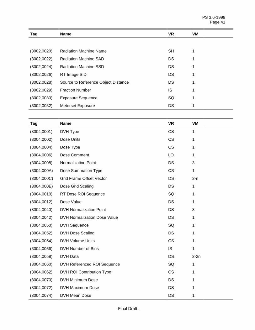
(3002,0020) Radiation Machine Name SH 1
(3002,0022) Radiation Machine SAD DS 1
(3002,0024) Radiation Machine SSD DS 1
(3002,0026) RT Image SID DS 1
(3002,0028) Source to Reference Object Distance DS 1
(3002,0029) Fraction Number IS 1
(3002,0030) Exposure Sequence SQ 1
(3002,0032) Meterset Exposure DS 1
Tag Name VR VM
(3004,0001) DVH Type CS 1
(3004,0002) Dose Units CS 1
(3004,0004) Dose Type CS 1
(3004,0006) Dose Comment LO 1
(3004,0008) Normalization Point DS 3
(3004,000A) Dose Summation Type CS 1
(3004,000C) Grid Frame Offset Vector DS 2-n
(3004,000E) Dose Grid Scaling DS 1
(3004,0010) RT Dose ROI Sequence SQ 1
(3004,0012) Dose Value DS 1
(3004,0040) DVH Normalization Point DS 3
(3004,0042) DVH Normalization Dose Value DS 1
(3004,0050) DVH Sequence SQ 1
(3004,0052) DVH Dose Scaling DS 1
(3004,0054) DVH Volume Units CS 1
(3004,0056) DVH Number of Bins IS 1
(3004,0058) DVH Data DS 2-2n
(3004,0060) DVH Referenced ROI Sequence SQ 1
(3004,0062) DVH ROI Contribution Type CS 1
(3004,0070) DVH Minimum Dose DS 1
(3004,0072) DVH Maximum Dose DS 1
(3004,0074) DVH Mean Dose DS 1

PS 3.6-1999 Page 42
Tag Name VR VM
- Final Draft -
Tag Name VR VM
(3006,0002) Structure Set Label SH 1
(3006,0004) Structure Set Name LO 1
(3006,0006) Structure Set Description ST 1
(3006,0008) Structure Set Date DA 1
(3006,0009) Structure Set Time TM 1
(3006,0010) Referenced Frame of Reference Sequence SQ 1
(3006,0012) RT Referenced Study Sequence SQ 1
(3006,0014) RT Referenced Series Sequence SQ 1
(3006,0016) Contour Image Sequence SQ 1
(3006,0020) Structure Set ROI Sequence SQ 1
(3006,0022) ROI Number IS 1
(3006,0024) Referenced Frame of Reference UID UI 1
(3006,0026) ROI Name LO 1
(3006,0028) ROI Description ST 1
(3006,002A) ROI Display Color IS 3
(3006,002C) ROI Volume DS 1
(3006,0030) RT Related ROI Sequence SQ 1
(3006,0033) RT ROI Relationship CS 1
(3006,0036) ROI Generation Algorithm CS 1
(3006,0038) ROI Generation Description LO 1
(3006,0039) ROI Contour Sequence SQ 1
(3006,0040) Contour Sequence SQ 1
(3006,0042) Contour Geometric Type CS 1
(3006,0044) Contour Slab Thickness DS 1
(3006,0045) Contour Offset Vector DS 3
(3006,0046) Number of Contour Points IS 1
(3006,0048) Contour Number IS 1
(3006,0049) Attached Contours IS 1-n
(3006,0050) Contour Data DS 3-3n

PS 3.6-1999 Page 43
Tag Name VR VM
- Final Draft -
(3006,0080) RT ROI Observations Sequence SQ 1
(3006,0082) Observation Number IS 1
(3006,0084) Referenced ROI Number IS 1
(3006,0085) ROI Observation Label SH 1
(3006,0086) RT ROI Identification Code Sequence SQ 1
(3006,0088) ROI Observation Description ST 1
(3006,00A0) Related RT ROI Observations Sequence SQ 1
(3006,00A4) RT ROI Interpreted Type CS 1
(3006,00A6) ROI Interpreter PN 1
(3006,00B0) ROI Physical Properties Sequence SQ 1
(3006,00B2) ROI Physical Property CS 1
(3006,00B4) ROI Physical Property Value DS 1
(3006,00C0) Frame of Reference Relationship Sequence SQ 1
(3006,00C2) Related Frame of Reference UID UI 1
(3006,00C4) Frame of Reference Transformation Type CS 1
(3006,00C6) Frame of Reference Transformation Matrix DS 16
(3006,00C8) Frame of Reference Transformation Comment LO 1
Tag Name VR VM
(3008,0010) Measured Dose Reference Sequence SQ 1
(3008,0012) Measured Dose Description ST 1
(3008,0014) Measured Dose Type CS 1
(3008,0016) Measured Dose Value DS 1
(3008,0020) Treatment Session Beam Sequence SQ 1
(3008,0022) Current Fraction Number IS 1
(3008,0024) Treatment Control Point Date DA 1
(3008,0025) Treatment Control Point Time TM 1
(3008,002A) Treatment Termination Status CS 1
(3008,002B) Treatment Termination Code SH 1
(3008,002C) Treatment Verification Status CS 1
(3008,0030) Referenced Treatment Record Sequence SQ 1
(3008,0032) Specified Primary Meterset DS 1

PS 3.6-1999 Page 44
Tag Name VR VM
- Final Draft -
(3008,0033) Specified Secondary Meterset DS 1
(3008,0036) Delivered Primary Meterset DS 1
(3008,0037) Delivered Secondary Meterset DS 1
(3008,003A) Specified Treatment Time DS 1
(3008,003B) Delivered Treatment Time DS 1
(3008,0040) Control Point Delivery Sequence SQ 1
(3008,0042) Specified Meterset DS 1
(3008,0044) Delivered Meterset DS 1
(3008,0048) Dose Rate Delivered DS 1
(3008,0050) Treatment Summary Calculated Dose Reference Sequence
SQ 1
(3008,0052) Cumulative Dose to Dose Reference DS 1
(3008,0054) First Treatment Date DA 1
(3008,0056) Most Recent Treatment Date DA 1
(3008,005A) Number of Fractions Delivered IS 1
(3008,0060) Override Sequence SQ 1
(3008,0062) Override Parameter Pointer AT 1
(3008,0064) Measured Dose Reference Number IS 1
(3008,0066) Override Reason ST 1
(3008,0070) Calculated Dose Reference Sequence SQ 1
(3008,0072) Calculated Dose Reference Number IS 1
(3008,0074) Calculated Dose Reference Description ST 1
(3008,0076) Calculated Dose Reference Dose Value DS 1
(3008,0078) Start Meterset DS 1
(3008,007A) End Meterset DS 1
(3008,0080) Referenced Measured Dose Reference Sequence SQ 1
(3008,0082) Referenced Measured Dose Reference Number IS 1
(3008,0090) Referenced Calculated Dose Reference Sequence SQ 1
(3008,0092) Referenced Calculated Dose Reference Number IS 1
(3008,00A0) Beam Limiting Device Leaf Pairs Sequence SQ 1
(3008,00B0) Recorded Wedge Sequence SQ 1
(3008,00C0) Recorded Compensator Sequence SQ 1

PS 3.6-1999 Page 45
Tag Name VR VM
- Final Draft -
(3008,00D0) Recorded Block Sequence SQ 1
(3008,00E0) Treatment Summary Measured Dose Reference Sequence
SQ 1
(3008,0100) Recorded Source Sequence SQ 1
(3008,0105) Source Serial Number LO 1
(3008,0110) Treatment Session Application Setup Sequence SQ 1
(3008,0116) Application Setup Check CS 1
(3008,0120) Recorded Brachy Accessory Device Sequence SQ 1
(3008,0122) Referenced Brachy Accessory Device Number IS 1
(3008,0130) Recorded Channel Sequence SQ 1
(3008,0132) Specified Channel Total Time DS 1
(3008,0134) Delivered Channel Total Time DS 1
(3008,0136) Specified Number of Pulses IS 1
(3008,0138) Delivered Number of Pulses IS 1
(3008,013A) Specified Pulse Repetition Interval DS 1
(3008,013C) Delivered Pulse Repetition Interval DS 1
(3008,0140) Recorded Source Applicator Sequence SQ 1
(3008,0142) Referenced Source Applicator Number IS 1
(3008,0150) Recorded Channel Shield Sequence SQ 1
(3008,0152) Referenced Channel Shield Number IS 1
(3008,0160) Brachy Control Point Delivered Sequence SQ 1
(3008,0162) Safe Position Exit Date DA 1
(3008,0164) Safe Position Exit Time TM 1
(3008,0166) Safe Position Return Date DA 1
(3008,0168) Safe Position Return Time TM 1
(3008,0200) Current Treatment Status CS 1
(3008,0202) Treatment Status Comment ST 1
(3008,0220) Fraction Group Summary Sequence SQ 1
(3008,0223) Referenced Fraction Number IS 1
(3008,0224) Fraction Group Type CS 1
(3008,0230) Beam Stopper Position CS 1
(3008,0240) Fraction Status Summary Sequence SQ 1

PS 3.6-1999 Page 46
Tag Name VR VM
- Final Draft -
(3008,0250) Treatment Date DA 1
(3008,0251) Treatment Time TM 1
Tag Name VR VM
(300A,0002) RT Plan Label SH 1
(300A,0003) RT Plan Name LO 1
(300A,0004) RT Plan Description ST 1
(300A,0006) RT Plan Date DA 1
(300A,0007) RT Plan Time TM 1
(300A,0009) Treatment Protocols LO 1-n
(300A,000A) Treatment Intent CS 1
(300A,000B) Treatment Sites LO 1-n
(300A,000C) RT Plan Geometry CS 1
(300A,000E) Prescription Description ST 1
(300A,0010) Dose Reference Sequence SQ 1
(300A,0012) Dose Reference Number IS 1
(300A,0014) Dose Reference Structure Type CS 1
(300A,0015) Nominal Beam Energy Unit CS 1
(300A,0016) Dose Reference Description LO 1
(300A,0018) Dose Reference Point Coordinates DS 3
(300A,001A) Nominal Prior Dose DS 1
(300A,0020) Dose Reference Type CS 1
(300A,0021) Constraint Weight DS 1
(300A,0022) Delivery Warning Dose DS 1
(300A,0023) Delivery Maximum Dose DS 1
(300A,0025) Target Minimum Dose DS 1
(300A,0026) Target Prescription Dose DS 1
(300A,0027) Target Maximum Dose DS 1
(300A,0028) Target Underdose Volume Fraction DS 1
(300A,002A) Organ at Risk Full-volume Dose DS 1
(300A,002B) Organ at Risk Limit Dose DS 1

PS 3.6-1999 Page 47
Tag Name VR VM
- Final Draft -
(300A,002C) Organ at Risk Maximum Dose DS 1
(300A,002D) Organ at Risk Overdose Volume Fraction DS 1
(300A,0040) Tolerance Table Sequence SQ 1
(300A,0042) Tolerance Table Number IS 1
(300A,0043) Tolerance Table Label SH 1
(300A,0044) Gantry Angle Tolerance DS 1
(300A,0046) Beam Limiting Device Angle Tolerance DS 1
(300A,0048) Beam Limiting Device Tolerance Sequence SQ 1
(300A,004A) Beam Limiting Device Position Tolerance DS 1
(300A,004C) Patient Support Angle Tolerance DS 1
(300A,004E) Table Top Eccentric Angle Tolerance DS 1
(300A,0051) Table Top Vertical Position Tolerance DS 1
(300A,0052) Table Top Longitudinal Position Tolerance DS 1
(300A,0053) Table Top Lateral Position Tolerance DS 1
(300A,0055) RT Plan Relationship CS 1
(300A,0070) Fraction Group Sequence SQ 1
(300A,0071) Fraction Group Number IS 1
(300A,0078) Number of Fractions Planned IS 1
(300A,0079) Number of Fractions Per Day IS 1
(300A,007A) Repeat Fraction Cycle Length IS 1
(300A,007B) Fraction Pattern LT 1
(300A,0080) Number of Beams IS 1
(300A,0082) Beam Dose Specification Point DS 3
(300A,0084) Beam Dose DS 1
(300A,0086) Beam Meterset DS 1
(300A,00A0) Number of Brachy Application Setups IS 1
(300A,00A2) Brachy Application Setup Dose Specification Point DS 3
(300A,00A4) Brachy Application Setup Dose DS 1
(300A,00B0) Beam Sequence SQ 1
(300A,00B2) Treatment Machine Name SH 1
(300A,00B3) Primary Dosimeter Unit CS 1
(300A,00B4) Source-Axis Distance DS 1

PS 3.6-1999 Page 48
Tag Name VR VM
- Final Draft -
(300A,00B6) Beam Limiting Device Sequence SQ 1
(300A,00B8) RT Beam Limiting Device Type CS 1
(300A,00BA) Source to Beam Limiting Device Distance DS 1
(300A,00BC) Number of Leaf/Jaw Pairs IS 1
(300A,00BE) Leaf Position Boundaries DS 3-n
(300A,00C0) Beam Number IS 1
(300A,00C2) Beam Name LO 1
(300A,00C3) Beam Description ST 1
(300A,00C4) Beam Type CS 1
(300A,00C6) Radiation Type CS 1
(300A,00C8) Reference Image Number IS 1
(300A,00CA) Planned Verification Image Sequence SQ 1
(300A,00CC) Imaging Device-Specific Acquisition Parameters LO 1-n
(300A,00CE) Treatment Delivery Type CS 1
(300A,00D0) Number of Wedges IS 1
(300A,00D1) Wedge Sequence SQ 1
(300A,00D2) Wedge Number IS 1
(300A,00D3) Wedge Type CS 1
(300A,00D4) Wedge ID SH 1
(300A,00D5) Wedge Angle IS 1
(300A,00D6) Wedge Factor DS 1
(300A,00D8) Wedge Orientation DS 1
(300A,00DA) Source to Wedge Tray Distance DS 1
(300A,00E0) Number of Compensators IS 1
(300A,00E1) Material ID SH 1
(300A,00E2) Total Compensator Tray Factor DS 1
(300A,00E3) Compensator Sequence SQ 1
(300A,00E4) Compensator Number IS 1
(300A,00E5) Compensator ID SH 1
(300A,00E6) Source to Compensator Tray Distance DS 1
(300A,00E7) Compensator Rows IS 1
(300A,00E8) Compensator Columns IS 1

PS 3.6-1999 Page 49
Tag Name VR VM
- Final Draft -
(300A,00E9) Compensator Pixel Spacing DS 2
(300A,00EA) Compensator Position DS 2
(300A,00EB) Compensator Transmission Data DS 1-n
(300A,00EC) Compensator Thickness Data DS 1-n
(300A,00ED) Number of Boli IS 1
(300A,00EE) Compensator Type CS 1
(300A,00F0) Number of Blocks IS 1
(300A,00F2) Total Block Tray Factor DS 1
(300A,00F4) Block Sequence SQ 1
(300A,00F5) Block Tray ID SH 1
(300A,00F6) Source to Block Tray Distance DS 1
(300A,00F8) Block Type CS 1
(300A,00FA) Block Divergence CS 1
(300A,00FC) Block Number IS 1
(300A,00FE) Block Name LO 1
(300A,0100) Block Thickness DS 1
(300A,0102) Block Transmission DS 1
(300A,0104) Block Number of Points IS 1
(300A,0106) Block Data DS 2-2n
(300A,0107) Applicator Sequence SQ 1
(300A,0108) Applicator ID SH 1
(300A,0109) Applicator Type CS 1
(300A,010A) Applicator Description LO 1
(300A,010C) Cumulative Dose Reference Coefficient DS 1
(300A,010E) Final Cumulative Meterset Weight DS 1
(300A,0110) Number of Control Points IS 1
(300A,0111) Control Point Sequence SQ 1
(300A,0112) Control Point Index IS 1
(300A,0114) Nominal Beam Energy DS 1
(300A,0115) Dose Rate Set DS 1
(300A,0116) Wedge Position Sequence SQ 1
(300A,0118) Wedge Position CS 1

PS 3.6-1999 Page 50
Tag Name VR VM
- Final Draft -
(300A,011A) Beam Limiting Device Position Sequence SQ 1
(300A,011C) Leaf/Jaw Positions DS 2-2n
(300A,011E) Gantry Angle DS 1
(300A,011F) Gantry Rotation Direction CS 1
(300A,0120) Beam Limiting Device Angle DS 1
(300A,0121) Beam Limiting Device Rotation Direction CS 1
(300A,0122) Patient Support Angle DS 1
(300A,0123) Patient Support Rotation Direction CS 1
(300A,0124) Table Top Eccentric Axis Distance DS 1
(300A,0125) Table Top Eccentric Angle DS 1
(300A,0126) Table Top Eccentric Rotation Direction CS 1
(300A,0128) Table Top Vertical Position DS 1
(300A,0129) Table Top Longitudinal Position DS 1
(300A,012A) Table Top Lateral Position DS 1
(300A,012C) Isocenter Position DS 3
(300A,012E) Surface Entry Point DS 3
(300A,0130) Source to Surface Distance DS 1
(300A,0134) Cumulative Meterset Weight DS 1
(300A,0180) Patient Setup Sequence SQ 1
(300A,0182) Patient Setup Number IS 1
(300A,0184) Patient Additional Position LO 1
(300A,0190) Fixation Device Sequence SQ 1
(300A,0192) Fixation Device Type CS 1
(300A,0194) Fixation Device Label SH 1
(300A,0196) Fixation Device Description ST 1
(300A,0198) Fixation Device Position SH 1
(300A,01A0) Shielding Device Sequence SQ 1
(300A,01A2) Shielding Device Type CS 1
(300A,01A4) Shielding Device Label SH 1
(300A,01A6) Shielding Device Description ST 1
(300A,01A8) Shielding Device Position SH 1
(300A,01B0) Setup Technique CS 1

PS 3.6-1999 Page 51
Tag Name VR VM
- Final Draft -
(300A,01B2) Setup Technique Description ST 1
(300A,01B4) Setup Device Sequence SQ 1
(300A,01B6) Setup Device Type CS 1
(300A,01B8) Setup Device Label SH 1
(300A,01BA) Setup Device Description ST 1
(300A,01BC) Setup Device Parameter DS 1
(300A,01D0) Setup Reference Description ST 1
(300A,01D2) Table Top Vertical Setup Displacement DS 1
(300A,01D4) Table Top Longitudinal Setup Displacement DS 1
(300A,01D6) Table Top Lateral Setup Displacement DS 1
(300A,0200) Brachy Treatment Technique CS 1
(300A,0202) Brachy Treatment Type CS 1
(300A,0206) Treatment Machine Sequence SQ 1
(300A,0210) Source Sequence SQ 1
(300A,0212) Source Number IS 1
(300A,0214) Source Type CS 1
(300A,0216) Source Manufacturer LO 1
(300A,0218) Active Source Diameter DS 1
(300A,021A) Active Source Length DS 1
(300A,0222) Source Encapsulation Nominal Thickness DS 1
(300A,0224) Source Encapsulation Nominal Transmission DS 1
(300A,0226) Source Isotope Name LO 1
(300A,0228) Source Isotope Half Life DS 1
(300A,022A) Reference Air Kerma Rate DS 1
(300A,022C) Air Kerma Rate Reference Date DA 1
(300A,022E) Air Kerma Rate Reference Time TM 1
(300A,0230) Application Setup Sequence SQ 1
(300A,0232) Application Setup Type CS 1
(300A,0234) Application Setup Number IS 1
(300A,0236) Application Setup Name LO 1
(300A,0238) Application Setup Manufacturer LO 1
(300A,0240) Template Number IS 1

PS 3.6-1999 Page 52
Tag Name VR VM
- Final Draft -
(300A,0242) Template Type SH 1
(300A,0244) Template Name LO 1
(300A,0250) Total Reference Air Kerma DS 1
(300A,0260) Brachy Accessory Device Sequence SQ 1
(300A,0262) Brachy Accessory Device Number IS 1
(300A,0263) Brachy Accessory Device ID SH 1
(300A,0264) Brachy Accessory Device Type CS 1
(300A,0266) Brachy Accessory Device Name LO 1
(300A,026A) Brachy Accessory Device Nominal Thickness DS 1
(300A,026C) Brachy Accessory Device Nominal Transmission DS 1
(300A,0280) Channel Sequence SQ 1
(300A,0282) Channel Number IS 1
(300A,0284) Channel Length DS 1
(300A,0286) Channel Total Time DS 1
(300A,0288) Source Movement Type CS 1
(300A,028A) Number of Pulses IS 1
(300A,028C) Pulse Repetition Interval DS 1
(300A,0290) Source Applicator Number IS 1
(300A,0291) Source Applicator ID SH 1
(300A,0292) Source Applicator Type CS 1
(300A,0294) Source Applicator Name LO 1
(300A,0296) Source Applicator Length DS 1
(300A,0298) Source Applicator Manufacturer LO 1
(300A,029C) Source Applicator Wall Nominal Thickness DS 1
(300A,029E) Source Applicator Wall Nominal Transmission DS 1
(300A,02A0) Source Applicator Step Size DS 1
(300A,02A2) Transfer Tube Number IS 1
(300A,02A4) Transfer Tube Length DS 1
(300A,02B0) Channel Shield Sequence SQ 1
(300A,02B2) Channel Shield Number IS 1
(300A,02B3) Channel Shield ID SH 1
(300A,02B4) Channel Shield Name LO 1

PS 3.6-1999 Page 53
Tag Name VR VM
- Final Draft -
(300A,02B8) Channel Shield Nominal Thickness DS 1
(300A,02BA) Channel Shield Nominal Transmission DS 1
(300A,02C8) Final Cumulative Time Weight DS 1
(300A,02D0) Brachy Control Point Sequence SQ 1
(300A,02D2) Control Point Relative Position DS 1
(300A,02D4) Control Point 3D Position DS 3
(300A,02D6) Cumulative Time Weight DS 1
Tag Name VR VM
(300C,0002) Referenced RT Plan Sequence SQ 1
(300C,0004) Referenced Beam Sequence SQ 1
(300C,0006) Referenced Beam Number IS 1
(300C,0007) Referenced Reference Image Number IS 1
(300C,0008) Start Cumulative Meterset Weight DS 1
(300C,0009) End Cumulative Meterset Weight DS 1
(300C,000A) Referenced Brachy Application Setup Sequence SQ 1
(300C,000C) Referenced Brachy Application Setup Number IS 1
(300C,000E) Referenced Source Number IS 1
(300C,0020) Referenced Fraction Group Sequence SQ 1
(300C,0022) Referenced Fraction Group Number IS 1
(300C,0040) Referenced Verification Image Sequence SQ 1
(300C,0042) Referenced Reference Image Sequence SQ 1
(300C,0050) Referenced Dose Reference Sequence SQ 1
(300C,0051) Referenced Dose Reference Number IS 1
(300C,0055) Brachy Referenced Dose Reference Sequence SQ 1
(300C,0060) Referenced Structure Set Sequence SQ 1
(300C,006A) Referenced Patient Setup Number IS 1
(300C,0080) Referenced Dose Sequence SQ 1
(300C,00A0) Referenced Tolerance Table Number IS 1
(300C,00B0) Referenced Bolus Sequence SQ 1
(300C,00C0) Referenced Wedge Number IS 1
(300C,00D0) Referenced Compensator Number IS 1

PS 3.6-1999 Page 54
Tag Name VR VM
- Final Draft -
(300C,00E0) Referenced Block Number IS 1
(300C,00F0) Referenced Control Point Index IS 1
Tag Name VR VM
(300E,0002) Approval Status CS 1
(300E,0004) Review Date DA 1
(300E,0005) Review Time TM 1
(300E,0008) Reviewer Name PN 1
Tag Name VR VM
(4000,0000) Group Length RET
(4000,0010) Arbitrary RET
(4000,4000) Comments RET
Tag Name VR VM
(4008,0000) Group Length UL 1
(4008,0040) Results ID SH 1
(4008,0042) Results ID Issuer LO 1
(4008,0050) Referenced Interpretation Sequence SQ 1
(4008,0100) Interpretation Recorded Date DA 1
(4008,0101) Interpretation Recorded Time TM 1
(4008,0102) Interpretation Recorder PN 1
(4008,0103) Reference to Recorded Sound LO 1
(4008,0108) Interpretation Transcription Date DA 1
(4008,0109) Interpretation Transcription Time TM 1
(4008,010A) Interpretation Transcriber PN 1
(4008,010B) Interpretation Text ST 1
(4008,010C) Interpretation Author PN 1
(4008,0111) Interpretation Approver Sequence SQ 1
(4008,0112) Interpretation Approval Date DA 1
(4008,0113) Interpretation Approval Time TM 1

PS 3.6-1999 Page 55
Tag Name VR VM
- Final Draft -
(4008,0114) Physician Approving Interpretation PN 1
(4008,0115) Interpretation Diagnosis Description LT 1
(4008,0117) Interpretation Diagnosis Code Sequence SQ 1
(4008,0118) Results Distribution List Sequence SQ 1
(4008,0119) Distribution Name PN 1
(4008,011A) Distribution Address LO 1
(4008,0200) Interpretation ID SH 1
(4008,0202) Interpretation ID Issuer LO 1
(4008,0210) Interpretation Type ID CS 1
(4008,0212) Interpretation Status ID CS 1
(4008,0300) Impressions ST 1
(4008,4000) Results Comments ST 1
Tag Name VR VM
(50xx,0000) Group Length UL 1
(50xx,0005) Curve Dimensions US 1
(50xx,0010) Number of Points US 1
(50xx,0020) Type of Data CS 1
(50xx,0022) Curve Description LO 1
(50xx,0030) Axis Units SH 1-n
(50xx,0040) Axis Labels SH 1-n
(50xx,0103) Data Value Representation US 1
(50xx,0104) Minimum Coordinate Value US 1-n
(50xx,0105) Maximum Coordinate Value US 1-n
(50xx,0106) Curve Range SH 1-n
(50xx,0110) Curve Data Descriptor US 1-n
(50xx,0112) Coordinate Start Value US 1
(50xx,0114) Coordinate Step Value US 1
(50xx,1001) Curve Activation Layer CS 1
(50xx,2000) Audio Type US 1
(50xx,2002) Audio Sample Format US 1
(50xx,2004) Number of Channels US 1

PS 3.6-1999 Page 56
Tag Name VR VM
- Final Draft -
(50xx,2006) Number of Samples UL 1
(50xx,2008) Sample Rate UL 1
(50xx,200A) Total Time UL 1
(50xx,200C) Audio Sample Data OW/ or OB 1
(50xx,200E) Audio Comments LT 1
(50xx,2500) Curve Label LO 1
(50xx,2600) Referenced Overlay Sequence SQ 1
(50xx,2610) Referenced Overlay Group US 1
(50xx,3000) Curve Data OW/ or OB 1
Tag Name VR VM
(5400,0100) Waveform Sequence SQ 1
(5400,0110) Channel Minimum Value OB or OW 1
(5400,0112) Channel Maximum Value OB or OW 1
(5400,1004) Waveform Bits Allocated US 1
(5400,1006) Waveform Sample Interpretation CS 1
(5400,100A) Waveform Padding Value OB or OW 1
(5400,1010) Waveform Data OB or OW 1
Tag Name VR VM
(60xx,0000) Group Length UL 1
(60xx,0010) Overlay Rows US 1
(60xx,0011) Overlay Columns US 1
(60xx,0012) Overlay Planes US 1
(60xx,0015) Number of Frames in Overlay IS 1
(60xx,0022) Overlay Description LO 1
(60xx,0040) Overlay Type CS 1
(60xx,0045) Overlay Subtype LO 1
(60xx,0050) Overlay Origin SS 2
(60xx,0051) Image Frame Origin US 1
(60xx,0052) Overlay Plane Origin US 1

PS 3.6-1999 Page 57
Tag Name VR VM
- Final Draft -
(60xx,0060) Compression Code RET
(60xx,0100) Overlay Bits Allocated US 1
(60xx,0102) Overlay Bit Position US 1
(60xx,0110) Overlay Format RET
(60xx,0200) Overlay Location RET
(60xx,1001) Overlay Activation Layer CS 1
(60xx,1100) Overlay Descriptor - Gray US 1 RET
(60xx,1101) Overlay Descriptor - Red US 1 RET
(60xx,1102) Overlay Descriptor - Green US 1 RET
(60xx,1103) Overlay Descriptor - Blue US 1 RET
(60xx,1200) Overlays- Gray US 1-n RET
(60xx,1201) Overlays - Red US 1-n RET
(60xx,1202) Overlays - Green US 1-n RET
(60xx,1203) Overlays- Blue US 1-n RET
(60xx,1301) ROI Area IS 1
(60xx,1302) ROI Mean DS 1
(60xx,1303) ROI Standard Deviation DS 1
(60xx,1500) Overlay Label LO 1
(60xx,3000) Overlay Data OB or OW 1
(60xx,4000) Comments RET
Tag Name VR VM
(7FE0,0000) Group Length UL 1
(7FE0,0010) Pixel Data OW/ or OB 1
Tag Name VR VM
(FFFC,FFFC) Data Set Trailing Padding OB 1
Tag Name VR VM
(FFFE,E000) Item see note 1
(FFFE,E00D) Item Delimitation Item see note 1

PS 3.6-1999 Page 58
Tag Name VR VM
- Final Draft -
(FFFE,E0DD) Sequence Delimitation Item see note 1 Note: The VR for Data Elements, Item (FFFE,E000), Item Delimitation Item (FFFE,E00D), and Sequence
Delimitation Item (FFFE,E0DD) do not exist. See PS 3.5 for explanation.

PS 3.6-2000 Page 59
- Final Draft -
7 Registry of DICOM File Meta Elements
This section specifies the File Meta Elements needed to support the formatting of the File Meta Information of the DICOM File Format (See PS 3.10).
Tag Name VR VM
(0002,0000) Group Length UL 1
(0002,0001) File Meta Information Version OB 1
(0002,0002) Media Storage SOP Class UID UI 1
(0002,0003) Media Storage SOP Instance UID UI 1
(0002,0010) Transfer Syntax UID UI 1
(0002,0012) Implementation Class UID UI 1
(0002,0013) Implementation Version Name SH 1
(0002,0016) Source Application Entity Title AE 1
(0002,0100) Private Information Creator UID UI 1
(0002,0102) Private Information OB 1


PS 3.6-2000 Page 61
- Final Draft -
8 Registry of DICOM directory structuring elements
Tag Name VR VM
(0004,0000) Group Length UL 1
(0004,1130) File-set ID CS 1
(0004,1141) File-set Descriptor File ID CS 1-8
(0004,1142) Specific Character Set of File-set Descriptor File CS 1
(0004,1200) Offset of the First Directory Record of the Root Directory Entity
UL 1
(0004,1202) Offset of the Last Directory Record of the Root Directory Entity
UL 1
(0004,1212) File-set Consistency Flag US 1
(0004,1220) Directory Record Sequence SQ 1
(0004,1400) Offset of the Next Directory Record UL 1
(0004,1410) Record In-use Flag US 1
(0004,1420) Offset of Referenced Lower-Level Directory Entity UL 1
(0004,1430) Directory Record Type CS 1
(0004,1432) Private Record UID UI 1
(0004,1500) Referenced File ID CS 1-8
(0004,1504) MRDR Directory Record Offset UL 1
(0004,1510) Referenced SOP Class UID in File UI 1
(0004,1511) Referenced SOP Instance UID in File UI 1
(0004,1512) Referenced Transfer Syntax UID in File UI 1
(0004,1600) Number of References UL 1


PS 3.6-1999 Page 63
- Final Draft -
Annex A Registry of DICOM unique identifiers (UID) (Normative)
Table A-1 lists the UID values which are registered and used throughout the Parts of the DICOM Standard. This central registry ensures that when additional UIDs are assigned, non duplicate values are assigned.
Table A-1 UID VALUES
UID Value UID NAME UID TYPE Part
1.2.840.10008.1.1 Verification SOP Class SOP Class PS 3.4
1.2.840.10008.1.2 Implicit VR Little Endian: Default Transfer Syntax for DICOM
Transfer Syntax PS 3.5
1.2.840.10008.1.2.1 Explicit VR Little Endian Transfer Syntax PS 3.5
1.2.840.10008.1.2.2 Explicit VR Big Endian Transfer Syntax PS 3.5
1.2.840.10008.1.2.4.50 JPEG Baseline (Process 1): Default Transfer Syntax for Lossy JPEG 8 Bit Image Compression
Transfer Syntax PS 3.5
1.2.840.10008.1.2.4.51 JPEG Extended (Process 2 & 4): Default Transfer Syntax for Lossy JPEG 12 Bit Image Compression (Process 4 only)
Transfer Syntax PS 3.5
1.2.840.10008.1.2.4.52 JPEG Extended (Process 3 & 5) Transfer Syntax PS 3.5
1.2.840.10008.1.2.4.53 JPEG Spectral Selection, Non-Hierarchical (Process 6 & 8)
Transfer Syntax PS 3.5
1.2.840.10008.1.2.4.54 JPEG Spectral Selection, Non-Hierarchical (Process 7 & 9)
Transfer Syntax PS 3.5
1.2.840.10008.1.2.4.55 JPEG Full Progression, Non-Hierarchical (Process 10 & 12)
Transfer Syntax PS 3.5
1.2.840.10008.1.2.4.56 JPEG Full Progression, Non-Hierarchical (Process 11 & 13)
Transfer Syntax PS 3.5
1.2.840.10008.1.2.4.57 JPEG Lossless, Non-Hierarchical (Process 14)
Transfer Syntax PS 3.5
1.2.840.10008.1.2.4.58 JPEG Lossless, Non-Hierarchical (Process 15)
Transfer Syntax PS 3.5
1.2.840.10008.1.2.4.59 JPEG Extended, Hierarchical (Process 16 & 18)
Transfer Syntax PS 3.5
1.2.840.10008.1.2.4.60 JPEG Extended, Hierarchical (Process 17 & 19)
Transfer Syntax PS 3.5

PS 3.6-1999 Page 64
- Final Draft -
1.2.840.10008.1.2.4.61 JPEG Spectral Selection, Hierarchical (Process 20 & 22)
Transfer Syntax PS 3.5
1.2.840.10008.1.2.4.62 JPEG Spectral Selection, Hierarchical (Process 21 & 23)
Transfer Syntax PS 3.5
1.2.840.10008.1.2.4.63 JPEG Full Progression, Hierarchical (Process 24 & 26)
Transfer Syntax PS 3.5
1.2.840.10008.1.2.4.64 JPEG Full Progression, Hierarchical (Process 25 & 27)
Transfer Syntax PS 3.5
1.2.840.10008.1.2.4.65 JPEG Lossless, Hierarchical (Process 28)
Transfer Syntax PS 3.5
1.2.840.10008.1.2.4.66 JPEG Lossless, Hierarchical (Process 29)
Transfer Syntax PS 3.5
1.2.840.10008.1.2.4.70 JPEG Lossless, Non-Hierarchical, First-Order Prediction (Process 14 [Selection Value 1]): Default Transfer Syntax for Lossless JPEG Image Compression
Transfer Syntax PS 3.5
1.2.840.10008.1.2.4.80 JPEG-LS Lossless Image Compression
Transfer Syntax PS 3.5
1.2.840.10008.1.2.4.81 JPEG-LS Lossy (Near-Lossless) Image Compression
Transfer Syntax PS 3.5
1.2.840.10008.1.2.5 RLE Lossless Transfer Syntax PS 3.5
1.2.840.10008.1.3.10 Media Storage Directory Storage SOP Class PS 3.4
1.2.840.10008.1.9 Basic Study Content Notification SOP Class
SOP Class PS 3.4
1.2.840.10008.1.20.1 Storage Commitment Push Model SOP Class
SOP Class PS 3.4
1.2.840.10008.1.20.1.1 Storage Commitment Push Model SOP Instance
Well-known SOP Instance
PS 3.4
1.2.840.10008.1.20.2 Storage Commitment Pull Model SOP Class
SOP Class PS 3.4
1.2.840.10008.1.20.2.1 Storage Commitment Pull Model SOP Instance
Well-known SOP Instance
PS 3.4
1.2.840.10008.3.1.1.1 DICOM Application Context Name Application Context Name
PS 3.7
1.2.840.10008.3.1.2.1.1 Detached Patient Management SOP Class
SOP Class PS 3.4
1.2.840.10008.3.1.2.1.4 Detached Patient Management Meta SOP Class
Meta SOP Class PS 3.4

PS 3.6-1999 Page 65
- Final Draft -
1.2.840.10008.3.1.2.2.1 Detached Visit Management SOP Class
SOP Class PS 3.4
1.2.840.10008.3.1.2.3.1 Detached Study Management SOP Class
SOP Class PS 3.4
1.2.840.10008.3.1.2.3.2 Study Component Management SOP Class
SOP Class PS 3.4
1.2.840.10008.3.1.2.3.3 Modality Performed Procedure Step SOP Class
SOP Class PS 3.4
1.2.840.10008.3.1.2.3.4 Modality Performed Procedure Step Retrieve SOP Class
SOP Class PS 3.4
1.2.840.10008.3.1.2.3.5 Modality Performed Procedure Step Notification SOP Class
SOP Class PS 3.4
1.2.840.10008.3.1.2.5.1 Detached Results Management SOP Class
SOP Class PS 3.4
1.2.840.10008.3.1.2.5.4 Detached Results Management Meta SOP Class
Meta SOP Class PS 3.4
1.2.840.10008.3.1.2.5.5 Detached Study Management Meta SOP Class
Meta SOP Class PS 3.4
1.2.840.10008.3.1.2.6.1 Detached Interpretation Management SOP Class
SOP Class PS 3.4
1.2.840.10008.5.1.1.1 Basic Film Session SOP Class SOP Class PS 3.4
1.2.840.10008.5.1.1.2 Basic Film Box SOP Class SOP Class PS 3.4
1.2.840.10008.5.1.1.4 Basic Grayscale Image Box SOP Class
SOP Class PS 3.4
1.2.840.10008.5.1.1.4.1 Basic Color Image Box SOP Class SOP Class PS 3.4
1.2.840.10008.5.1.1.4.2 Referenced Image Box SOP Class (Retired)
SOP Class PS 3.4
1.2.840.10008.5.1.1.9 Basic Grayscale Print Management Meta SOP Class
Meta SOP Class PS 3.4
1.2.840.10008.5.1.1.9.1 Referenced Grayscale Print Management Meta SOP Class (Retired)
Meta SOP Class PS 3.4
1.2.840.10008.5.1.1.14 Print Job SOP Class SOP Class PS 3.4
1.2.840.10008.5.1.1.15 Basic Annotation Box SOP Class SOP Class PS 3.4
1.2.840.10008.5.1.1.16 Printer SOP Class SOP Class PS 3.4
1.2.840.10008.5.1.1.16.376 Printer Configuration Retrieval SOP Class
Well-known Printer SOP Instance
PS 3.4
1.2.840.10008.5.1.1.17 Printer SOP Instance Well-known Printer SOP Instance
PS 3.4

PS 3.6-1999 Page 66
- Final Draft -
1.2.840.10008.5.1.1.17.376 Printer Configuration Retrieval SOP Instance
Well-known Printer SOP Instance
PS 3.4
1.2.840.10008.5.1.1.18 Basic Color Print Management Meta SOP Class
Meta SOP Class PS 3.4
1.2.840.10008.5.1.1.18.1 Referenced Color Print Management Meta SOP Class (Retired)
Meta SOP Class PS 3.4
1.2.840.10008.5.1.1.22 VOI LUT Box SOP Class SOP Class PS 3.4
1.2.840.10008.5.1.1.23 Presentation LUT SOP Class SOP Class PS 3.4 1.2.840.10008.5.1.1.24 Image Overlay Box SOP Class
(Retired) SOP Class PS 3.4
1.2.840.10008.5.1.1.24.1 Basic Print Image Overlay Box SOP Class
SOP Class PS 3.4
1.2.840.10008.5.1.1.25 Print Queue SOP Instance Well-known Print Queue SOP Instance
PS 3.4
1.2.840.10008.5.1.1.26 Print Queue Management SOP Class
SOP Class PS 3.4 1.2.840.10008.5.1.1.27 Stored Print Storage SOP Class SOP Class PS 3.4 1.2.840.10008.5.1.1.29 Hardcopy Grayscale Image
Storage SOP Class SOP Class PS 3.4
1.2.840.10008.5.1.1.30 Hardcopy Color Image Storage SOP Class
SOP Class PS 3.4
1.2.840.10008.5.1.1.31 Pull Print Request SOP Class SOP Class PS 3.4 1.2.840.10008.5.1.1.32 Pull Stored Print Management
Meta SOP Class Meta SOP Class PS 3.4
1.2.840.10008.5.1.4.1.1.1 Computed Radiography Image Storage
SOP Class PS 3.4
1.2.840.10008.5.1.4.1.1.1.1 Digital X-Ray Image Storage - For Presentation
SOP Class PS 3.4
1.2.840.10008.5.1.4.1.1.1.1.1 Digital X-Ray Image Storage - For Processing
SOP Class PS 3.4
1.2.840.10008.5.1.4.1.1.1.2 Digital Mammography X-Ray Image Storage - For Presentation
SOP Class PS 3.4
1.2.840.10008.5.1.4.1.1.1.2.1 Digital Mammography X-Ray Image Storage - For Processing
SOP Class PS 3.4
1.2.840.10008.5.1.4.1.1.1.3 Digital Intra-oral X-Ray Image Storage - For Presentation
SOP Class PS 3.4
1.2.840.10008.5.1.4.1.1.1.3.1 Digital Intra-oral X-Ray Image Storage - For Processing
SOP Class PS 3.4
1.2.840.10008.5.1.4.1.1.2 CT Image Storage SOP Class PS 3.4
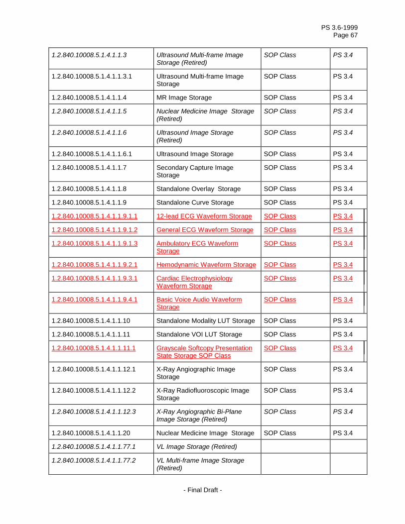
PS 3.6-1999 Page 67
- Final Draft -
1.2.840.10008.5.1.4.1.1.3 Ultrasound Multi-frame Image Storage (Retired)
SOP Class PS 3.4
1.2.840.10008.5.1.4.1.1.3.1 Ultrasound Multi-frame Image Storage
SOP Class PS 3.4
1.2.840.10008.5.1.4.1.1.4 MR Image Storage SOP Class PS 3.4
1.2.840.10008.5.1.4.1.1.5 Nuclear Medicine Image Storage (Retired)
SOP Class PS 3.4
1.2.840.10008.5.1.4.1.1.6 Ultrasound Image Storage (Retired)
SOP Class PS 3.4
1.2.840.10008.5.1.4.1.1.6.1 Ultrasound Image Storage SOP Class PS 3.4
1.2.840.10008.5.1.4.1.1.7 Secondary Capture Image Storage
SOP Class PS 3.4
1.2.840.10008.5.1.4.1.1.8 Standalone Overlay Storage SOP Class PS 3.4
1.2.840.10008.5.1.4.1.1.9 Standalone Curve Storage SOP Class PS 3.4
1.2.840.10008.5.1.4.1.1.9.1.1 12-lead ECG Waveform Storage SOP Class PS 3.4
1.2.840.10008.5.1.4.1.1.9.1.2 General ECG Waveform Storage SOP Class PS 3.4
1.2.840.10008.5.1.4.1.1.9.1.3 Ambulatory ECG Waveform Storage
SOP Class PS 3.4
1.2.840.10008.5.1.4.1.1.9.2.1 Hemodynamic Waveform Storage SOP Class PS 3.4
1.2.840.10008.5.1.4.1.1.9.3.1 Cardiac Electrophysiology Waveform Storage
SOP Class PS 3.4
1.2.840.10008.5.1.4.1.1.9.4.1 Basic Voice Audio Waveform Storage
SOP Class PS 3.4
1.2.840.10008.5.1.4.1.1.10 Standalone Modality LUT Storage SOP Class PS 3.4
1.2.840.10008.5.1.4.1.1.11 Standalone VOI LUT Storage SOP Class PS 3.4
1.2.840.10008.5.1.4.1.1.11.1 Grayscale Softcopy Presentation State Storage SOP Class
SOP Class PS 3.4
1.2.840.10008.5.1.4.1.1.12.1 X-Ray Angiographic Image Storage
SOP Class PS 3.4
1.2.840.10008.5.1.4.1.1.12.2 X-Ray Radiofluoroscopic Image Storage
SOP Class PS 3.4
1.2.840.10008.5.1.4.1.1.12.3 X-Ray Angiographic Bi-Plane Image Storage (Retired)
SOP Class PS 3.4
1.2.840.10008.5.1.4.1.1.20 Nuclear Medicine Image Storage SOP Class PS 3.4
1.2.840.10008.5.1.4.1.1.77.1 VL Image Storage (Retired)
1.2.840.10008.5.1.4.1.1.77.2 VL Multi-frame Image Storage (Retired)

PS 3.6-1999 Page 68
- Final Draft -
1.2.840.10008.5.1.4.1.1.77.1.1 VL Endoscopic Image Storage SOP Class PS3.4
1.2.840.10008.5.1.4.1.1.77.1.2 VL Microscopic Image Storage SOP Class PS3.4
1.2.840.10008.5.1.4.1.1.77.1.3 VL Slide-Coordinates Microscopic Image Storage
SOP Class PS3.4
1.2.840.10008.5.1.4.1.1.77.1.4 VL Photographic Image Storage SOP Class PS3.4
1.2.840.10008.5.1.4.1.1.88.11 Basic Text SR SOP Class 3.4
1.2.840.10008.5.1.4.1.1.88.22 Enhanced SR SOP Class 3.4
1.2.840.10008.5.1.4.1.1.88.33 Comprehensive SR SOP Class 3.4
1.2.840.10008.5.1.4.1.1.128 Positron Emission Tomography Image Storage
SOP Class PS 3.4
1.2.840.10008.5.1.4.1.1.129 Standalone PET Curve Storage SOP Class PS 3.4
1.2.840.10008.5.1.4.1.1.481.1 RT Image Storage SOP Class PS 3.4
1.2.840.10008.5.1.4.1.1.481.2 RT Dose Storage SOP Class PS 3.4
1.2.840.10008.5.1.4.1.1.481.3 RT Structure Set Storage SOP Class PS 3.4
1.2.840.10008.5.1.4.1.1.481.4 RT Beams Treatment Record Storage
SOP Class PS 3.4
1.2.840.10008.5.1.4.1.1.481.5 RT Plan Storage SOP Class PS 3.4
1.2.840.10008.5.1.4.1.1.481.6 RT Brachy Treatment Record Storage
SOP Class PS 3.4
1.2.840.10008.5.1.4.1.1.481.7 RT Treatment Summary Record Storage
SOP Class PS 3.4
1.2.840.10008.5.1.4.1.2.1.1 Patient Root Query/Retrieve Information Model - FIND
SOP Class PS 3.4
1.2.840.10008.5.1.4.1.2.1.2 Patient Root Query/Retrieve Information Model - MOVE
SOP Class PS 3.4
1.2.840.10008.5.1.4.1.2.1.3 Patient Root Query/Retrieve Information Model - GET
SOP Class PS 3.4
1.2.840.10008.5.1.4.1.2.2.1 Study Root Query/Retrieve Information Model - FIND
SOP Class PS 3.4
1.2.840.10008.5.1.4.1.2.2.2 Study Root Query/Retrieve Information Model - MOVE
SOP Class PS 3.4
1.2.840.10008.5.1.4.1.2.2.3 Study Root Query/Retrieve Information Model - GET
SOP Class PS 3.4
1.2.840.10008.5.1.4.1.2.3.1 Patient/Study Only Query/Retrieve Information Model - FIND
SOP Class PS 3.4
1.2.840.10008.5.1.4.1.2.3.2 Patient/Study Only Query/Retrieve Information Model - MOVE
SOP Class PS 3.4

PS 3.6-1999 Page 69
- Final Draft -
1.2.840.10008.5.1.4.1.2.3.3 Patient/Study Only Query/Retrieve Information Model - GET
SOP Class PS 3.4
1.2.840.10008.5.1.4.31 Modality Worklist Information Model - FIND
SOP Class PS 3.4

PS 3.6-1999 Page 70
- Final Draft -
Annex B – DICOM Terminology Mapping Resource Registry of Context Groups and Controlled Terminology
Table B-1 defines the Coding Schemes utilized in the controlled terminology specified in this Annex.
Table B-1 CODING SCHEME VALUES
SNM3 SNOMED Version 3 SRT SNOMED RT DCM DICOM SCPECG Standard Communications Protocol for Computer-Assisted Electrocardiography,
Draft proposal for ISO Standard, AAMI, Revision 1.3 BARI Bypass Angiography Revascularization Investigation, Alderman, EL and Stadius,
M, Coronary Artery Disease 1992,3:1189-1207; endorsed by ACC/AHA Guidelines for Coronary Angiography, J Am Coll Cardiol 1999,33:1791
UCUM The Unified Code for Units of Measure. Gunther Schadow Clement J. McDonald. The Regenstrief Institute For Health Care, Indianapolis
Context Group ID 3001 Context Group Name ECG leads
Coding Scheme
Coding Scheme Version
Code Value Code Meaning
SCPECG 1.3 5.6.3-9-73 Defibrillator lead: anterior-lateral SCPECG 1.3 5.6.3-9-74 External pacing lead: anterior-posterior SCPECG 1.3 5.6.3-9-27 Lead A SCPECG 1.3 5.6.3-9-71 Lead A (Nehb – Anterior) SCPECG 1.3 5.6.3-9-75 Lead A1 (Auxiliary unipolar lead 1) SCPECG 1.3 5.6.3-9-76 Lead A2 (Auxiliary unipolar lead 2) SCPECG 1.3 5.6.3-9-77 Lead A3 (Auxiliary unipolar lead 3) SCPECG 1.3 5.6.3-9-78 Lead A4 (Auxiliary unipolar lead 4) SCPECG 1.3 5.6.3-9-57 Lead A-cal SCPECG 1.3 5.6.3-9-84 Lead A-cal (cal for Nehb – Anterior) SCPECG 1.3 5.6.3-9-64 Lead aVF SCPECG 1.3 5.6.3-9-63 Lead aVL SCPECG 1.3 5.6.3-9-62 Lead aVR SCPECG 1.3 5.6.3-9-65 Lead -aVR SCPECG 1.3 5.6.3-9-26 Lead C SCPECG 1.3 5.6.3-9-19 Lead CC5 SCPECG 1.3 5.6.3-9-49 Lead CC5-cal

PS 3.6-1999 Page 71
- Final Draft -
SCPECG 1.3 5.6.3-9-56 Lead C-cal SCPECG 1.3 5.6.3-9-20 Lead CM5 SCPECG 1.3 5.6.3-9-50 Lead CM5-cal SCPECG 1.3 5.6.3-9-70 Lead D (Nehb – Dorsal) SCPECG 1.3 5.6.3-9-83 Lead D-cal (cal for Nehb – Dorsal) SCPECG 1.3 5.6.3-9-25 Lead E SCPECG 1.3 5.6.3-9-55 Lead E-cal SCPECG 1.3 5.6.3-9-29 Lead F SCPECG 1.3 5.6.3-9-59 Lead F-cal SCPECG 1.3 5.6.3-9-30 Lead H SCPECG 1.3 5.6.3-9-60 Lead H-cal SCPECG 1.3 5.6.3-9-1 Lead I (Einthoven) SCPECG 1.3 5.6.3-9-24 Lead I (Frank) SCPECG 1.3 5.6.3-9-31 Lead I-cal (Einthoven) SCPECG 1.3 5.6.3-9-54 Lead I-cal (Frank) SCPECG 1.3 5.6.3-9-2 Lead II SCPECG 1.3 5.6.3-9-32 Lead II-cal SCPECG 1.3 5.6.3-9-61 Lead III SCPECG 1.3 5.6.3-9-72 Lead J (Nehb – Inferior) SCPECG 1.3 5.6.3-9-85 Lead J-cal (cal for Nehb – Inferior) SCPECG 1.3 5.6.3-9-21 Lead Left Arm SCPECG 1.3 5.6.3-9-51 Lead Left Arm-cal SCPECG 1.3 5.6.3-9-23 Lead Left Leg SCPECG 1.3 5.6.3-9-53 Lead Left Leg-cal SCPECG 1.3 5.6.3-9-28 Lead M SCPECG 1.3 5.6.3-9-58 Lead M-cal SCPECG 1.3 5.6.3-9-22 Lead Right Arm SCPECG 1.3 5.6.3-9-52 Lead Right Arm-cal SCPECG 1.3 5.6.3-9-3 Lead V1 SCPECG 1.3 5.6.3-9-33 Lead V1-cal SCPECG 1.3 5.6.3-9-4 Lead V2 SCPECG 1.3 5.6.3-9-34 Lead V2-cal SCPECG 1.3 5.6.3-9-10 Lead V2R SCPECG 1.3 5.6.3-9-40 Lead V2R-cal SCPECG 1.3 5.6.3-9-5 Lead V3 SCPECG 1.3 5.6.3-9-35 Lead V3-cal SCPECG 1.3 5.6.3-9-11 Lead V3R SCPECG 1.3 5.6.3-9-41 Lead V3R-cal SCPECG 1.3 5.6.3-9-6 Lead V4

PS 3.6-1999 Page 72
- Final Draft -
SCPECG 1.3 5.6.3-9-36 Lead V4-cal SCPECG 1.3 5.6.3-9-12 Lead V4R SCPECG 1.3 5.6.3-9-42 Lead V4R-cal SCPECG 1.3 5.6.3-9-7 Lead V5 SCPECG 1.3 5.6.3-9-37 Lead V5-cal SCPECG 1.3 5.6.3-9-13 Lead V5R SCPECG 1.3 5.6.3-9-43 Lead V5R-cal SCPECG 1.3 5.6.3-9-8 Lead V6 SCPECG 1.3 5.6.3-9-38 Lead V6-cal SCPECG 1.3 5.6.3-9-14 Lead V6R SCPECG 1.3 5.6.3-9-44 Lead V6R-cal SCPECG 1.3 5.6.3-9-9 Lead V7 SCPECG 1.3 5.6.3-9-39 Lead V7-cal SCPECG 1.3 5.6.3-9-15 Lead V7R SCPECG 1.3 5.6.3-9-45 Lead V7R-cal SCPECG 1.3 5.6.3-9-66 Lead V8 SCPECG 1.3 5.6.3-9-79 Lead V8-cal SCPECG 1.3 5.6.3-9-68 Lead V8R SCPECG 1.3 5.6.3-9-81 Lead V8R-cal SCPECG 1.3 5.6.3-9-67 Lead V9 SCPECG 1.3 5.6.3-9-80 Lead V9-cal SCPECG 1.3 5.6.3-9-69 Lead V9R SCPECG 1.3 5.6.3-9-82 Lead V9R-cal SCPECG 1.3 5.6.3-9-16 Lead X SCPECG 1.3 5.6.3-9-46 Lead X-cal SCPECG 1.3 5.6.3-9-17 Lead Y SCPECG 1.3 5.6.3-9-47 Lead Y-cal SCPECG 1.3 5.6.3-9-18 Lead Z SCPECG 1.3 5.6.3-9-48 Lead Z-cal SCPECG 1.3 5.6.3-9-0 Unspecified lead
Context Group ID 3003 Context Group Name Hemodynamic waveform sources
Coding Scheme
Coding Scheme Version
Code Value Code Meaning
SRT V1 G-DB22 Aortic pressure waveform SRT V1 G-DB31 Aortic valve pullback pressure waveform SRT V1 G-DB24 Arterial pressure waveform SRT V1 G-DB23 Central venous pressure waveform SRT V1 G-DB33 Dye dilution cardiac output waveform

PS 3.6-1999 Page 73
- Final Draft -
SRT V1 G-DB20 Femoral artery pressure waveform SRT V1 G-DB34 Hemodynamic impedance waveform SRT V1 G-DB19 Left atrium pressure waveform SRT V1 G-DB16 Left ventricle pressure waveform SRT V1 G-DB28 Mitral valve pullback pressure waveform SRT V1 G-DB25 Pulmonary artery oxygen saturation waveform SRT V1 G-DB21 Pulmonary artery pressure waveform SRT V1 G-DB27 Pulmonary artery wedge pressure waveform SRT V1 G-DB26 Pulmonary capillary wedge pressure waveform SRT V1 G-DB30 Pulmonary valve pullback pressure waveform SRT V1 G-DB18 Right atrium pressure waveform SRT V1 G-DB17 Right ventricle pressure waveform SRT V1 G-DB32 Thermal cardiac output waveform SRT V1 G-DB29 Tricuspid valve pullback pressure waveform
Context Group ID 3010 Context Group Name Cardiovascular anatomic locations
Coding Scheme
Coding Scheme Version
Code Value Code Meaning
SNM3 3.4 T-42500 Abdominal aorta SRT V1 T-48503 Anomalous pulmonary vein SRT V1 T-49215 Antecubital vein SNM3 3.5 T-45530 Anterior communicating artery SNM3 3.5 T-45730 Anterior spinal artery SNM3 3.5 T-47700 Anterior tibial artery SNM3 3.4 T-42000 Aorta SNM3 3.5 T-42300 Aortic arch SRT V1 D3-81922 Aortic fistula SRT V1 T-32602 Apex of left ventricle SRT V1 T-32502 Apex of right ventricle SNM3 3.5 T-41000 Artery SNM3 3.5 T-42100 Ascending aorta SNM3 3.4 T-47100 Axillary Artery SNM3 3.5 T-49110 Axillary vein SNM3 3.4 T-48340 Azygos vein SRT V1 A-00203 Baffle SNM3 3.5 T-45800 Basilar artery SNM3 3.5 T-D00AB Body conduit SRT V1 T-49424 Boyd’s perforating vein SNM3 3.5 T-47160 Brachial artery

PS 3.6-1999 Page 74
- Final Draft -
SNM3 3.4 T-49350 Brachial vein SNM3 3.5 T-46010 Brachiocephalic artery SNM3 3.5 T-46010 Brachiocephalic trunk SNM3 3.5 T-48620 Brachiocephalic vein SNM3 3.4 T-45010 Carotid Artery SNM3 3.5 T-49240 Cephalic vein SNM3 3.5 T-45510 Cerebral artery SNM3 3.5 D4-31320 Common atrium SNM3 3.5 T-45100 Common carotid artery SNM3 3.5 D4-31120 Common ventricle SRT V1 D4-32504 Congenital coronary artery fistula to left atrium SRT V1 D4-32506 Congenital coronary artery fistula to left ventricle SRT V1 D4-32509 Congenital coronary artery fistula to right atrium SRT V1 D4-32510 Congenital coronary artery fistula to right ventricle SNM3 3.5 D3-40208 Congenital pulmonary arteriovenous fistula SRT V1 D4-33142 Congenital pulmonary artery conduit SRT V1 D4-33512 Congenital pulmonary vein confluence SRT V1 D4-33514 Congenital pulmonary venous atrium SRT V1 D4-33516 Congenital systemic venous atrium SNM3 3.5 T-43000 Coronary artery SNM3 3.5 T-48410 Coronary sinus SNM3 3.4 T-42400 Descending aorta SRT V1 T-49429 Dodd’s perforating vein SNM3 3.5 T-45240 Facial artery SNM3 3.5 T-47400 Femoral artery SNM3 3.4 T-49410 Femoral vein SNM3 3.5 T-48820 Gastric vein SRT V1 T-47490 Genicular artery SNM3 3.5 T-46420 Hepatic artery SNM3 3.5 T-48720 Hepatic vein SRT V1 T-4942A Hunterian perforating vein SNM3 3.5 T-46700 Iliac artery SNM3 3.4 T-48540 Inferior left pulmonary vein SNM3 3.5 T-48520 Inferior right pulmonary vein SNM3 3.5 T-48710 Inferior vena cava SNM3 3.5 T-46010 Innominate artery SNM3 3.4 T-48620 Innominate vein SNM3 3.5 T-45300 Internal carotid artery SNM3 3.5 T-48170 Internal jugular vein

PS 3.6-1999 Page 75
- Final Draft -
SNM3 3.5 T-46200 Internal mammary artery SRT V1 D4-31052 Juxtaposed atrial appendage SNM3 3.5 T-45410 Lacrimal artery SRT V1 T-45416 Lacrimal artery of right eye SNM3 3.5 T-32300 Left atrium SNM3 3.5 T-32310 Left auricular appendage SNM3 3.4 T-44400 Left pulmonary artery SNM3 3.5 T-32600 Left ventricle SNM3 3.5 T-32640 Left ventricle inflow SRT V1 D4-31022 Left ventricle outflow chamber SNM3 3.5 T-32640 Left ventricle outflow tract SNM3 3.5 T-45230 Lingual artery SNM3 3.5 T-46960 Lumbar artery SNM3 3.5 T-46500 Mesenteric artery SRT V1 T-4884A Mesenteric vein SNM3 3.5 T-45250 Occipital artery SNM3 3.5 T-48214 Occipital vein SNM3 3.5 T-45400 Ophthalmic artery SNM3 3.5 D4-32012 Patent ductus arteriosus SNM3 3.5 T-47630 Peroneal artery SNM3 3.5 T-47500 Popliteal artery SNM3 3.5 T-48810 Portal vein SNM3 3.5 T-45320 Posterior communication artery SRT V1 T-49535 Posterior medial tributary SNM3 3.5 T-47600 Posterior tibial artery SNM3 3.5 T-F7001 Primitive aorta SNM3 3.5 T-F7040 Primitive pulmonary artery SNM3 3.5 T-44000 Pulmonary artery SRT V1 T-32190 Pulmonary chamber of cor triatriatum SNM3 3.5 T-48500 Pulmonary vein SNM3 3.5 T-47300 Radial artery SNM3 3.5 T-46600 Renal artery SNM3 3.5 T-48740 Renal vein SNM3 3.5 T-32200 Right atrium SNM3 3.5 T-32210 Right auricular appendage SNM3 3.5 T-47410 Right femoral artery SNM3 3.5 T-44200 Right pulmonary artery SNM3 3.5 T-32500 Right ventricle SNM3 3.5 T-32540 Right ventricle inflow

PS 3.6-1999 Page 76
- Final Draft -
SRT V1 D4-31022 Right ventricle outflow chamber SNM3 3.5 T-32550 Right ventricle outflow tract SRT V1 T-D930A Saphenofemoral junction SNM3 3.5 T-49530 Saphenous vein SNM3 3.5 T-48890 Splenic vein SNM3 3.5 T-46100 Subclavian artery SNM3 3.5 T-48330 Subclavian vein SNM3 3.5 T-45270 Superficial temporal artery SNM3 3.5 T-48530 Superior left pulmonary vein SNM3 3.5 T-48510 Superior right pulmonary vein SNM3 3.5 T-45210 Superior thyroid artery SNM3 3.5 T-48610 Superior vena cava SRT V1 T-44007 Systemic collateral artery to lung SNM3 3.5 T-42070 Thoracic aorta SNM3 3.5 D4-31400 Truncus arteriosus communis SNM3 3.5 T-F1810 Umbilical artery SNM3 3.5 T-48817 Umbilical vein SNM3 3.5 T-48000 Vein SNM3 3.4 T-48170 Vena jugularis interna SNM3 3.5 T-48003 Venous network SNM3 3.5 T-45700 Vertebral artery
Context Group ID 3011 Context Group Name Electrophysiology anatomic locations
Coding Scheme
Coding Scheme Version
Code Value Code Meaning
SNM3 3.5 T-32850 Accessory atrioventricular bundle SRT V1 T-32602 Apex of left ventricle SRT V1 T-32502 Apex of right ventricle SNM3 3.5 T-32830 Atrioventricular bundle SNM3 3.5 T-32820 Atrioventricular node SNM3 3.5 T-32400 Common ventricle SNM3 3.5 T-48410 Coronary sinus SNM3 3.5 T-39010 Epicardium SNM3 3.5 T-48420 Great cardiac vein SRT V1 G-DE02 High right atrium SNM3 3.5 T-48540 Inferior left pulmonary vein SNM3 3.5 T-48520 Inferior right pulmonary vein SRT V1 G-DE04 Lateral high right atrium SNM3 3.5 T-32833 Left anterior division of left branch of left atrioventricular

PS 3.6-1999 Page 77
- Final Draft -
bundle SNM3 3.5 T-32300 Left Atrium SNM3 3.5 T-32310 Left auricular appendage SNM3 3.5 T-32832 Left branch of atrioventricular bundle SNM3 3.5 T-32834 Left posterior division of left branch of left atrioventricular
bundle SNM3 3.5 T-32600 Left ventricle SNM3 3.5 T-32640 Left ventricle inflow SNM3 3.5 T-32640 Left ventricle outflow tract SRT V1 G-DE08 Low right atrium SRT V1 G-DE06 Mid right atrium SNM3 3.5 T-48430 Middle cardiac vein SNM3 3.5 T-35310 Mitral ring SNM3 3.5 T-48411 Ostium of coronary sinus SNM3 3.5 T-48500 Pulmonary vein SNM3 3.5 T-35210 Pulmonic ring SNM3 3.5 T-32840 Purkinje fibers SNM3 3.5 T-35120 Right atrioventricular ostium SNM3 3.5 T-32200 Right Atrium SNM3 3.5 T-32210 Right auricular appendage SNM3 3.5 T-32831 Right branch of Atrioventricular bundle SNM3 3.5 T-32500 Right ventricle SNM3 3.5 T-32540 Right ventricle inflow SNM3 3.5 T-32550 Right ventricle outflow tract SNM3 3.5 T-32810 Sino-atrial node SNM3 3.5 T-48530 Superior left pulmonary vein SNM3 3.5 T-48510 Superior right pulmonary vein SRT V1 T-32202 Tendon of Todaro SNM3 3.5 T-35110 Tricuspid ring
Context Group ID 3014 Context Group Name Coronary artery segments
Coding Scheme
Coding Scheme Version
Code Value Code Meaning
BARI 1992 15 1st Diagonal Coronary Artery BARI 1992 24 1st Left Posterolateral Coronary Artery BARI 1992 20 1st Marginal Coronary Artery BARI 1992 6 1st Right posterolateral BARI 1992 17 1st Septal Coronary Artery BARI 1992 16 2nd Diagonal Coronary Artery

PS 3.6-1999 Page 78
- Final Draft -
BARI 1992 25 2nd Left Posterolateral Coronary Artery BARI 1992 21 2nd Marginal Coronary Artery BARI 1992 7 2nd Right posterolateral BARI 1992 29 3rd diagonal BARI 1992 26 3rd Left Posterolateral Coronary Artery BARI 1992 22 3rd Marginal Coronary Artery BARI 1992 8 3rd Right posterolateral BARI 1992 10 Acute Marginal BARI 1992 23 AV groove continuation BARI 1992 19A Distal Circumflex Coronary Artery BARI 1992 14 Distal Left Anterior Descending Coronary Artery BARI 1992 3 Distal Right Coronary Artery BARI 1992 15A Lateral 1st Diagonal Coronary Artery BARI 1992 20A Lateral 1st Marginal Coronary Artery BARI 1992 16A Lateral 2nd Diagonal Coronary Artery BARI 1992 21A Lateral 2nd Marginal Coronary Artery BARI 1992 29A Lateral 3rd Diagonal BARI 1992 22A Lateral 3rd Marginal Coronary Artery BARI 1992 28A Lateral Ramus BARI 1992 11 Left Main Coronary Artery BARI 1992 11A Left Main Coronary Artery Ostium BARI 1992 27 Left Posterior Descending Artery BARI 1992 19 Mid Circumflex Coronary Artery BARI 1992 13 Mid Left Anterior Descending Coronary Artery BARI 1992 2 Mid Right Coronary Artery BARI 1992 4 Posterior Descending Right Coronary Artery BARI 1992 9 Posterior descending septal perforators BARI 1992 18 Proximal Circumflex Coronary Artery BARI 1992 12 Proximal Left Anterior Descending Coronary Artery BARI 1992 1 Proximal Right Coronary Artery BARI 1992 28 Ramus BARI 1992 1A Right Coronary Artery Ostium BARI 1992 5 Right posterior AV Context Group ID 3019 Context Group Name Cardiovascular Anatomic Location Modifiers
Coding Scheme
Coding Scheme Version
Code Value Code Meaning
SNM3 3.5 G-A105 Anterior SRT V1 G-D873 Arterial graft to cited segment

PS 3.6-1999 Page 79
- Final Draft -
SNM3 3.5 GA110 Central SNM3 3.5 G-A119 Distal SRT V1 G-D870 Graft to cited body segment SRT V1 G-D872 Graft to distal anastomosis SRT V1 G-D871 Graft to proximal anastomosis SNM3 3.5 G-A115 Inferior SRT V1 G-A104 Lateral SNM3 3.5 G-A101 Left SRT V1 T-3215A Ostium SNM3 3.5 G-A106 Posterior SNM3 3.5 G-A118 Proximal SNM3 3.5 G-A100 Right SNM3 3.5 G-A116 Superior SRT V1 G-D874 Venous graft to cited segment
Context Group ID 3082 Context Group Name Waveform Units of Measurement
Coding Scheme
Coding Scheme Version
Code Value Code Meaning
UCUM 1.4 dB(A) A scale of loudness UCUM 1.4 [arb’U] arbitrary unit UCUM 1.4 d day UCUM 1.4 dB decibel UCUM 1.4 Cel degrees Celsius UCUM 1.4 {H.B.}/min Heart beat per minute UCUM 1.4 hr hour UCUM 1.4 J Joule UCUM 1.4 kOhm kilo Ohm UCUM 1.4 km/hr kilometer per hour UCUM 1.4 kPa kiloPascal UCUM 1.4 l/min liter per minute UCUM 1.5 [MET] Metabolic equivalent UCUM 1.4 uV microvolt UCUM 1.4 [mi_i]/hr mile per hour UCUM 1.4 ml/min milliliter per minute UCUM 1.4 ml/s milliliter per second UCUM 1.4 mm[Hg] millimeter of mercury UCUM 1.4 mV millivolt UCUM 1.4 min minute UCUM 1.4 mm/s mm/s

PS 3.6-1999 Page 80
- Final Draft -
UCUM 1.4 % percent UCUM 1.4 s second UCUM 1.4 1 unary, no units UCUM 1.4 V volt UCUM 1.4 W Watt
Context Group ID 3090 Context Group Name Time Synchronization channel types
Coding Scheme
Coding Scheme Version
Code Value Code Meaning
DCM 01 109001 Digital timecode (NOS) DCM 01 109002 ECG-based gating signal, processed DCM 01 109003 IRIG-B timecode DCM 01 109004 X-ray Fluoroscopy On Signal DCM 01 109005 X-ray On Trigger
Context Group ID 3240 Context Group Name Electrophysiology measurement functions and techniques
Coding Scheme
Coding Scheme Version
Code Value Code Meaning
DCM 01 109006 Differential signal DCM 01 109007 His bundle electrogram DCM 01 109008 Monopole signal DCM 01 109009 Pacing (electrical) stimulus, voltage DCM 01 109010 Radio frequency ablation, power DCM 01 109011 Voltage measurement by basket catheter DCM 01 109012 Voltage measurement by mapping catheter DCM 01 109013 Voltage measurement, NOS
Context Group ID 3241 Context Group Name Hemodynamic measurement techniques
Coding Scheme
Coding Scheme Version
Code Value Code Meaning
SRT V1 PA-50038 Averaged hemodynamic measurement method SRT V1 PA-50035 Composite hemodynamic measurement method SRT V1 PA-50034 Computed hemodynamic measurement method SRT V1 PA-50031 Dual catheter method SRT V1 PA-50039 Fluid filled catheter method SRT V1 PA-50033 Pullback method SRT V1 PA-50032 Pulmonary capillary wedge method

PS 3.6-1999 Page 81
- Final Draft -
SRT V1 PA-50036 Static catheter method SRT V1 PA-5003A Tip manometer method SRT V1 PA-50037 Wedge method
Context Group ID 3250 Context Group Name Catheterization Procedure Phase
Coding Scheme
Coding SchemeVersion
Code Value Code Meaning
SRT V1 G-7299 Cardiac catheterization bailout phase SRT V1 G-7293 Cardiac catheterization baseline phase SRT V1 G-7294 Cardiac catheterization image acquisition phase SRT V1 G-7295 Cardiac catheterization intervention phase SRT V1 G-729B Cardiac catheterization post contrast phase SRT V1 G-7298 Cardiac catheterization post-intervention phase SRT V1 G-7296 Cardiac catheterization pre-intervention phase SRT V1 G-7297 Cardiac catheterization therapy phase SRT V1 P1-3160A Catheterization of both left and right heart with graft SRT V1 P1-3160B Catheterization of both left and right heart without graft SNM3 3.5 P1-31604 Catheterization of left heart SNM3 3.5 P1-31602 Catheterization of right heart
Context Group ID 3254 Context Group Name Electrophysiology Procedure Phase
Coding Scheme
Coding SchemeVersion
Code Value Code Meaning
SRT V1 G-729D Atrial Effective Refractory Period, evaluation of SRT V1 G-7304 Carotid Sinus Massage procedure phase SRT V1 G-7306 Electrophysiology Mapping phase SRT V1 G-729A Electrophysiology procedure baseline phase SRT V1 G-7408 Post-ablation phase SRT V1 G-7305 Post-defibrillation procedure phase SRT V1 G-729F Radiofrequency Ablation procedure phase SRT V1 G-729C Sinus Node Recovery Time, evaluation of SRT V1 G-729E Ventricular Effective Refractory Period, evaluation of
Context Group ID 3261 Context Group Name Stress Protocols
Coding Scheme
Coding SchemeVersion
Code Value Code Meaning
SRT V1 P2-7131C Balke protocol

PS 3.6-1999 Page 82
- Final Draft -
SRT V1 P2-7131A Bruce protocol SRT V1 P2-7131D Ellestad protocol SRT V1 P2-7131B Modified Bruce protocol SRT V1 P2-713A1 Modified Naughton protocol SRT V1 P2-713A0 Naughton protocol SRT V1 P2-7131F Pepper protocol SRT V1 P2-7131E Ramp protocol
Context Group ID 3262 Context Group Name ECG Patient State Values
Coding Scheme
Coding SchemeVersion
Code Value Code Meaning
SRT V1 F-01602 Baseline state SRT V1 F-01606 Exercise state SRT V1 F-01608 Post-exercise state SRT V1 F-01604 Resting state SNM3 3.5 F-10340 Supine body position
Context Group ID 3263 Context Group Name Electrode Placement Values
Coding Scheme
Coding SchemeVersion
Code Value Code Meaning
SCPECG 1.3 5.4.5-33-1-5 12-lead ECG derived from Frank XYZ leads SCPECG 1.3 5.4.5-33-1-6 12-lead ECG derived from non-standard leads SCPECG 1.3 5.4.5-33-1-2 Mason-Likar positions: RA, RL, LA, and LL placed on the
torso. V1 to V6 placed at standard positions on the chest. All electrodes are placed individually.
SCPECG 1.3 5.4.5-33-1-3 Mason-Likar with V pad: RA, RL, LA, and LL individually placed on the torso. V1 to V6 placed on the chest as part of a single electrode pad (not placed individually).
SCPECG 1.3 5.4.5-33-1-4 Single electrode pad: All electrodes placed on the chest in a single electrode pad. None of the electrodes are placed individually
SCPECG 1.3 5.4.5-33-1-1 Standard 12-lead positions: RA, RL, LA, and LL placed at limb extremities; V1 to V6 placed at standard positions on the chest. All electrodes are placed individually.
SCPECG 1.3 5.4.5-33-1-0 Unspecified
Context Group ID 3264 Context Group Name XYZ Electrode Placement Values
Coding Scheme
Coding SchemeVersion
Code Value Code Meaning
SCPECG 1.3 5.4.5-33-2-4 Bipolar uncorrected XYZ lead system

PS 3.6-1999 Page 83
- Final Draft -
SCPECG 1.3 5.4.5-33-2-3 Cube lead system (Grishman et al, Amer Heart J 1951; 41:483).
SCPECG 1.3 5.4.5-33-2-1 Frank lead system (Frank, 1956; 13:737) SCPECG 1.3 5.4.5-33-2-2 McFee-Parungao lead system (Benchimol,
Vectorcardiography, Williams & Wilkins, Baltimore, 1973, Fig 1.6 on page 6)
SCPECG 1.3 5.4.5-33-2-5 Pseudo-orthogonal XYZ lead system (as used in Holter recording)
SCPECG 1.3 5.4.5-33-2-0 Unspecified SCPECG 1.3 5.4.5-33-2-6 XYZ leads derived from standard 12-lead ECG
Context Group ID 3271 Context Group Name Hemodynamic Physiological Challenges
Coding Scheme
Coding SchemeVersion
Code Value Code Meaning
SRT V1 P2-71310 Exercise challenge SRT V1 P2-71306 Handgrip SRT V1 P2-71302 Head up SRT V1 P2-71314 Held inspiration SRT V1 P2-71316 Held ventilation SRT V1 P2-71304 Leg up SRT V1 P2-71308 Negative lower body pressure SRT V1 P2-71318 Post volume challenge SRT V1 P2-71312 Vagal stimulation SNM3 3.5 F-F7102 Valsalva maneuver
Context Group ID 3335 Context Group Name ECG Annotations
Coding Scheme
Coding Scheme Version
Code Value Code Meaning
SCPECG 1.3 5.7.1-3 Fiducial point SCPECG 1.3 D.4.1-J J point SCPECG 1.3 D.4.1-ST20 J point + 20 msec SCPECG 1.3 D.4.1-ST60 J point + 60 msec SCPECG 1.3 D.4.1-ST80 J point +80 msec SCPECG 1.3 5.10.3-2 P wave end SCPECG 1.3 5.10.3-1 P wave onset SCPECG 1.3 D.4.1-P P wave peak SCPECG 1.3 5.10.1.2 Pacemaker spike, suppressed SCPECG 1.3 D.4.1-PR PR segment (isoelectric point)

PS 3.6-1999 Page 84
- Final Draft -
SCPECG 1.3 D.4.1-Q Q wave SCPECG 1.3 5.10.3-4 QRS end SCPECG 1.3 5.10.3-3 QRS onset SCPECG 1.3 D.4.1-R R wave peak SCPECG 1.3 D.4.1-R2 R’ peak SCPECG 1.3 D.4.1-S S wave SCPECG 1.3 D.4.1-S2 S’ wave SCPECG 1.3 5.10.3-5 T wave end SCPECG 1.3 D.4.1-STE T wave onset SCPECG 1.3 D.4.1-T T wave peak SCPECG 1.3 D.4.1-U U wave peak
Context Group ID 3337 Context Group Name Hemodynamic Annotations
Coding Scheme
Coding Scheme Version
Code Value Code Meaning
DCM 01 109014 35% of thermal CO DCM 01 109015 70% of thermal CO DCM 01 109016 A wave DCM 01 109017 A wave average DCM 01 109018 Beat detected (accepted) DCM 01 109019 Beat detected (rejected) DCM 01 109020 Diastolic average DCM 01 109021 Diastolic nadir DCM 01 109022 End diastole DCM 01 109023 End of expiration DCM 01 109024 End of inspiration DCM 01 109025 Max dp/dt DCM 01 109026 Max neg dp/dt DCM 01 109027 Mean pressure DCM 01 109028 Peak of thermal CO DCM 01 109029 Start of expiration DCM 01 109030 Start of inspiration DCM 01 109031 Start of thermal CO DCM 01 109032 Systolic average DCM 01 109033 Systolic peak DCM 01 109034 V wave DCM 01 109035 V wave average DCM 01 109036 Valve close DCM 01 109037 Valve open

PS 3.6-1999 Page 85
- Final Draft -
Context Group ID 3339 Context Group Name Electrophysiology Annotations
Coding Scheme
Coding Scheme Version
Code Value Code Meaning
DCM 01 109038 Ablation off DCM 01 109039 Ablation on DCM 01 109040 HIS bundle wave DCM 01 109041 P wave DCM 01 109042 Q wave DCM 01 109043 R wave DCM 01 109044 S wave DCM 01 109045 Start of atrial contraction DCM 01 109046 Start of atrial contraction (subsequent) DCM 01 109047 Stimulation at rate 1 interval DCM 01 109048 Stimulation at rate 2 interval DCM 01 109049 Stimulation at rate 3 interval DCM 01 109050 Stimulation at rate 4 interval DCM 01 109051 T wave DCM 01 109052 V wave DCM 01 109053 V wave of next beat
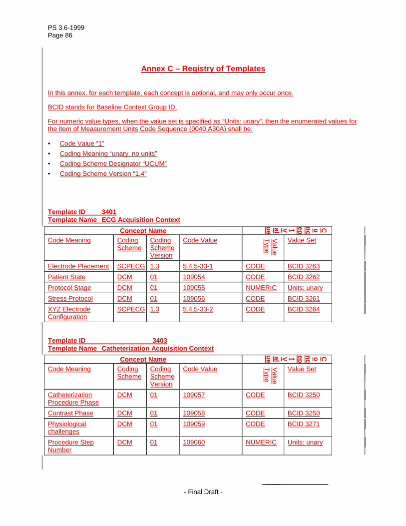
PS 3.6-1999 Page 86
- Final Draft -
Annex C – Registry of Templates
In this annex, for each template, each concept is optional, and may only occur once.
BCID stands for Baseline Context Group ID.
For numeric value types, when the value set is specified as “Units: unary”, then the enumerated values for the item of Measurement Units Code Sequence (0040,A30A) shall be:
• = Code Value “1” • = Coding Meaning “unary, no units” • = Coding Scheme Designator “UCUM” • = Coding Scheme Version “1.4”
Template ID 3401 Template Name ECG Acquisition Context
Concept Name
Concept Value
Code Meaning Coding Scheme
Coding Scheme Version
Code Value Value Type
Value Set
Electrode Placement SCPECG 1.3 5.4.5-33-1 CODE BCID 3263 Patient State DCM 01 109054 CODE BCID 3262 Protocol Stage DCM 01 109055 NUMERIC Units: unary Stress Protocol DCM 01 109056 CODE BCID 3261 XYZ Electrode Configuration
SCPECG 1.3 5.4.5-33-2 CODE BCID 3264
Template ID 3403 Template Name Catheterization Acquisition Context
Concept Name
Concept Value
Code Meaning Coding Scheme
Coding Scheme Version
Code Value Value Type
Value Set
Catheterization Procedure Phase
DCM 01 109057 CODE BCID 3250
Contrast Phase DCM 01 109058 CODE BCID 3250 Physiological challenges
DCM 01 109059 CODE BCID 3271
Procedure Step Number
DCM 01 109060 NUMERIC Units: unary

PS 3.6-1999 Page 87
- Final Draft -
Template ID 3450 Template Name Cardiac Electrophysiology Acquisition Context
Concept Name Concept Value
Code Meaning Coding Scheme
Coding Scheme Version
Code Value Value Type
Value Set
EP Procedure Phase DCM 01 109061 CODE BCID 3254 Procedure Step Number
DCM 01 109060 NUMERIC Units: unary
Pulse train definition DCM 01 109063 TEXT
Related Documents