protViz: Visualizing and Analyzing Mass Spectrometry Related Data in Proteomics Christian Panse Functional Genomics Center Zurich Jonas Grossmann Functional Genomics Center Zurich Abstract protViz is an R package to do quality checks, visualizations and analysis of mass spec- trometry data, coming from proteomics experiments. The package is developed, tested and used at the Functional Genomics Center Zurich. We use this package mainly for prototyping, teaching, and having fun with proteomics data. But it can also be used to do data analysis for small scale data sets. Nevertheless, if one is patient, it also handles large data sets. Keywords: proteomics, mass spectrometry, fragment-ion. 1. Related Work The method of choice in proteomics is mass spectrometry. There are already packages in R which deal with mass spec related data. Some of them are listed here: • MSnbase package (basic functions for mass spec data including quant aspect with iTRAQ data) http://www.bioconductor.org/packages/release/bioc/html/MSnbase.html • plgem – spectral counting quantification, applicable to MudPIT experiments http://www.bioconductor.org/packages/release/bioc/html/plgem.html • synapter – MSe (Hi3 = Top3 Quantification) for Waters Q-tof data aquired in MSe mode http://bioconductor.org/packages/synapter/ • mzR http://bioconductor.org/packages/mzR/ • isobar iTRAQ/TMT quantification package http://bioconductor.org/packages/isobar/ • readMzXmlData https://CRAN.R-project.org/package=readMzXmlData • rawDiag - an R package supporting rational LC-MS method optimization for bottom-up proteomics on multiple OS platforms (Trachsel, Panse, Kockmann, Wolski, Grossmann, and Schlapbach 2018)

Welcome message from author
This document is posted to help you gain knowledge. Please leave a comment to let me know what you think about it! Share it to your friends and learn new things together.
Transcript

protViz: Visualizing and Analyzing Mass
Spectrometry Related Data in Proteomics
Christian Panse
Functional Genomics Center ZurichJonas Grossmann
Functional Genomics Center Zurich
Abstract
protViz is an R package to do quality checks, visualizations and analysis of mass spec-trometry data, coming from proteomics experiments. The package is developed, testedand used at the Functional Genomics Center Zurich. We use this package mainly forprototyping, teaching, and having fun with proteomics data. But it can also be used todo data analysis for small scale data sets. Nevertheless, if one is patient, it also handleslarge data sets.
Keywords: proteomics, mass spectrometry, fragment-ion.
1. Related Work
The method of choice in proteomics is mass spectrometry. There are already packages in Rwhich deal with mass spec related data. Some of them are listed here:
• MSnbase package (basic functions for mass spec data including quant aspect withiTRAQ data)http://www.bioconductor.org/packages/release/bioc/html/MSnbase.html
• plgem – spectral counting quantification, applicable to MudPIT experimentshttp://www.bioconductor.org/packages/release/bioc/html/plgem.html
• synapter – MSe (Hi3 = Top3 Quantification) for Waters Q-tof data aquired in MSemodehttp://bioconductor.org/packages/synapter/
• mzR
http://bioconductor.org/packages/mzR/
• isobar iTRAQ/TMT quantification packagehttp://bioconductor.org/packages/isobar/
• readMzXmlData
https://CRAN.R-project.org/package=readMzXmlData
• rawDiag - an R package supporting rational LC-MS method optimization for bottom-upproteomics on multiple OS platforms (Trachsel, Panse, Kockmann, Wolski, Grossmann,and Schlapbach 2018)

2 protViz
2. Get Data In – Preprocessing
The most time consuming and challenging part of data analysis and visualization is shapingthe data the way that they can easily further process. In this package, we intentionally leftthis part away because it is very infrastructure dependent. Moreover, we use also commercialtools to analyze data and export the data into R accessible formats. We provide a differentkind of importers if these formats are available, but with little effort, one can bring otherexports in a similar format which will make it easy to use our package for a variety of tools.
2.1. Identification - In-silico from Proteins to Peptides
For demonstration, we use a sequence of peptides derived from a tryptic digest using theSwiss-Prot FETUA_BOVIN Alpha-2-HS-glycoprotein protein (P12763).
fcat and tryptic-digest are commandline programs which are included in the package.fcat removes the lines starting with > and all ’new line’ character within the protein sequencewhile tryptic-digest is doing the triptic digest of a protein sequence applying the rule:cleave after arginine (R) and lysine (K) except followed by proline(P).
Both programs can be used through the Fasta Rcpp module.
R> library(protViz)
R> fname <- system.file("extdata", name='P12763.fasta', package = "protViz")
R> F <- Fasta$new(fname)
print the first 60 characters of P12763.
R> substr(F$getSequences(), 1, 60)
[1] "MKSFVLLFCLAQLWGCHSIPLDPVAGYKEPACDDPDTEQAALAAVDYINKHLPRGYKHTL"
R> (fetuin <- F$getTrypticPeptides())
[1] "MK"
[2] "SFVLLFCLAQLWGCHSIPLDPVAGYK"
[3] "EPACDDPDTEQAALAAVDYINK"
[4] "HLPR"
[5] "GYK"
[6] "HTLNQIDSVK"
[7] "VWPR"
[8] "RPTGEVYDIEIDTLETTCHVLDPTPLANCSVR"
[9] "QQTQHAVEGDCDIHVLK"
[10] "QDGQFSVLFTK"
[11] "CDSSPDSAEDVR"
[12] "K"
[13] "LCPDCPLLAPLNDSR"
[14] "VVHAVEVALATFNAESNGSYLQLVEISR"
[15] "AQFVPLPVSVSVEFAVAATDCIAK"
[16] "EVVDPTK"

Christian Panse, Jonas Grossmann 3
[17] "CNLLAEK"
[18] "QYGFCK"
[19] "GSVIQK"
[20] "ALGGEDVR"
[21] "VTCTLFQTQPVIPQPQPDGAEAEAPSAVPDAAGPTPSAAGPPVASVVVGPSVVAVPLPLHR"
[22] "AHYDLR"
[23] "HTFSGVASVESSSGEAFHVGK"
[24] "TPIVGQPSIPGGPVR"
[25] "LCPGR"
[26] "IR"
[27] "YFK"
[28] "I"
3. Peptide Identification
The currency in proteomics are the peptides. In proteomics, proteins are digested to so-calledpeptides since peptides are much easier to handle biochemically than proteins. Proteins arevery different in nature some are very sticky while others are soluble in aqueous solutionswhile again are only sitting in membranes. Therefore, proteins are chopped up into peptidesbecause it is fair to assume, that for each protein, there will be many peptides behaving wellso that they can be measured with the mass spectrometer. This step introduces anotherproblem, the so-called protein inference problem. In this package here, we do not touch atall upon the protein inference.
3.1. Computing Mass and Hydrophobicity of a Peptide Sequence
parentIonMass computes the mass of an amino acid sequence.
R> (mass <- parentIonMass(fetuin))
[1] 278.1533 2991.5259 2406.0765 522.3147 367.1976 1154.6164
[7] 557.3194 3671.7679 1977.9447 1269.6474 1337.5274 147.1128
[13] 1740.8407 3016.5738 2519.3214 787.4196 847.4342 802.3552
[19] 631.3773 816.4210 6015.1323 774.3893 2120.0043 1474.8376
[25] 602.3079 288.2030 457.2445 132.1019
The ssrc function derives a measure for the hydrophobicity based on the method describedin (Krokhin, Craig, Spicer, Ens, Standing, Beavis, and Wilkins 2004).
R> (hydrophobicity <- ssrc(fetuin))
MK
NA
SFVLLFCLAQLWGCHSIPLDPVAGYK
71.74228
EPACDDPDTEQAALAAVDYINK

4 protViz
25.80645
HLPR
6.04805
GYK
2.15665
HTLNQIDSVK
18.36845
VWPR
9.54665
RPTGEVYDIEIDTLETTCHVLDPTPLANCSVR
46.68815
QQTQHAVEGDCDIHVLK
21.44645
QDGQFSVLFTK
32.22345
CDSSPDSAEDVR
2.07645
K
NA
LCPDCPLLAPLNDSR
31.62445
VVHAVEVALATFNAESNGSYLQLVEISR
54.51457
AQFVPLPVSVSVEFAVAATDCIAK
53.74569
EVVDPTK
7.77625
CNLLAEK
16.50635
QYGFCK
10.05345
GSVIQK
9.83325
ALGGEDVR
10.35025
VTCTLFQTQPVIPQPQPDGAEAEAPSAVPDAAGPTPSAAGPPVASVVVGPSVVAVPLPLHR
39.36853
AHYDLR
11.42005
HTFSGVASVESSSGEAFHVGK
27.94825
TPIVGQPSIPGGPVR
23.25845
LCPGR
3.60645
IR
NA

Christian Panse, Jonas Grossmann 5
YFK
7.91405
I
NA
A figure below shows a scatter plot graphing the parent ion mass versus the hydrophobicityvalue of each in-silico tryptic digested peptide of the FETUA BOVIN (P12763) protein.
R> op <- par(mfrow = c(1, 1))
R> plot(hydrophobicity ~ mass,
+ log = 'xy',
+ main = "sp|P12763|FETUA_BOVIN Alpha-2-HS-glycoprotein",
+ sub = 'tryptic peptides')
R> text(mass, hydrophobicity, fetuin, pos=3, cex=0.5)
●
●
●
●
●
●
●
●
●
●
●
●●
●
●
●●●
●
●
●
●
●
●
200 500 1000 2000 5000
25
10
20
50
sp|P12763|FETUA_BOVIN Alpha−2−HS−glycoprotein
tryptic peptides
mass
hyd
rop
ho
bic
ity
SFVLLFCLAQLWGCHSIPLDPVAGYK
EPACDDPDTEQAALAAVDYINK
HLPR
GYK
HTLNQIDSVK
VWPR
RPTGEVYDIEIDTLETTCHVLDPTPLANCSVR
QQTQHAVEGDCDIHVLK
QDGQFSVLFTK
CDSSPDSAEDVR
LCPDCPLLAPLNDSR
VVHAVEVALATFNAESNGSYLQLVEISRAQFVPLPVSVSVEFAVAATDCIAK
EVVDPTK
CNLLAEK
QYGFCKGSVIQKALGGEDVR
VTCTLFQTQPVIPQPQPDGAEAEAPSAVPDAAGPTPSAA
AHYDLR
HTFSGVASVESSSGEAFHVGK
TPIVGQPSIPGGPVR
LCPGR
YFK
3.2. In-silico Peptide Fragmentation
The fragment ions computation of a peptide follows the rules proposed in (Roepstorff andFohlman 1984). Beside the b and y ions the FUN argument of fragmentIon defines whichions are computed. the default ions beeing computed are defined in the function defaultIon.

6 protViz
The are no limits for defining other forms of fragment ions for ETD (c and z ions) CID (band y ions).
R> defaultIon
function (b, y)
{
Hydrogen <- 1.007825
Oxygen <- 15.994915
Nitrogen <- 14.003074
c <- b + (Nitrogen + (3 * Hydrogen))
z <- y - (Nitrogen + (3 * Hydrogen))
return(cbind(b, y, c, z))
}
<bytecode: 0x55a35923e220>
<environment: namespace:protViz>
R> peptides<-c('HTLNQIDSVK', 'ALGGEDVR', 'TPIVGQPSIPGGPVR')
R> pim<-parentIonMass(peptides)
R> fi<-fragmentIon(peptides)
R> par(mfrow=c(3,1));
R> for (i in 1:length(peptides)){
+ plot(0,0,
+ xlab='m/Z',
+ ylab='',
+ xlim=range(c(fi[i][[1]]$b,fi[i][[1]]$y)),
+ ylim=c(0,1),
+ type='n',
+ axes=FALSE,
+ sub=paste( pim[i], "Da"));
+ box()
+ axis(1,fi[i][[1]]$b,round(fi[i][[1]]$b,2))
+ pepSeq<-strsplit(peptides[i],"")
+ axis(3,fi[i][[1]]$b,pepSeq[[1]])
+
+ abline(v=fi[i][[1]]$b, col='red',lwd=2)
+ abline(v=fi[i][[1]]$c, col='orange')
+ abline(v=fi[i][[1]]$y, col='blue',lwd=2)
+ abline(v=fi[i][[1]]$z, col='cyan')
+ }

Christian Panse, Jonas Grossmann 7
1154.616401 Dam/Z
138.07 239.11 352.2 466.24 594.3 707.38 822.41 1008.51 1136.61
H T L N Q I D S V K
816.420981 Dam/Z
72.04 185.13 299.17 428.21 543.24 642.31 798.41
A L G G E D V R
1474.837601 Dam/Z
102.05 312.19 468.28 596.34 780.43 990.56 1201.66 1456.83
T P I V G Q P S I P G G P V R
The next lines compute the singly and doubly charged fragment ions of the HTLNQIDSVK
peptide. Which are usually the ones that can be used to make an identification.
R> Hydrogen<-1.007825
R> (fi.HTLNQIDSVK.1 <- fragmentIon('HTLNQIDSVK'))[[1]]
b y c z
1 138.0662 147.1128 155.0927 130.0863
2 239.1139 246.1812 256.1404 229.1547
3 352.1979 333.2132 369.2245 316.1867
4 466.2409 448.2402 483.2674 431.2136
5 594.2994 561.3242 611.3260 544.2977
6 707.3835 689.3828 724.4100 672.3563
7 822.4104 803.4258 839.4370 786.3992
8 909.4425 916.5098 926.4690 899.4833
9 1008.5109 1017.5575 1025.5374 1000.5309
10 1136.6058 1154.6164 1153.6324 1137.5899
R> (fi.HTLNQIDSVK.2 <-(fi.HTLNQIDSVK.1[[1]] + Hydrogen) / 2)
b y c z
1 69.53701 74.06031 78.05028 65.54704

8 protViz
2 120.06085 123.59452 128.57412 115.08124
3 176.60288 167.11053 185.11615 158.59726
4 233.62434 224.62400 242.13761 216.11073
5 297.65363 281.16603 306.16691 272.65276
6 354.19566 345.19532 362.70894 336.68205
7 411.70913 402.21679 420.22241 393.70351
8 455.22515 458.75882 463.73842 450.24554
9 504.75935 509.28266 513.27262 500.76938
10 568.80683 577.81211 577.32010 569.29884
3.3. Peptide Sequence – Fragment Ion Matching
Given a peptide sequence and a tandem mass spectrum. For the assignment of a candidatepeptide an in-silico fragment ion spectra fi is computed. The function findNN determines foreach fragment ion the closed peak in the MS2. If the difference between the in-silico mass andthe measured mass is inside the ’accuracy’ mass window of the mass spec device the in-silicofragment ion is considered as a potential hit.
R> peptideSequence<-'HTLNQIDSVK'
R> spec<-list(scans=1138,
+ title="178: (rt=22.3807) [20080816_23_fetuin_160.RAW]",
+ rtinseconds=1342.8402,
+ charge=2,
+ mZ=c(195.139940, 221.211970, 239.251780, 290.221750,
+ 316.300770, 333.300050, 352.258420, 448.384360, 466.348830,
+ 496.207570, 509.565910, 538.458310, 547.253380, 556.173940,
+ 560.358050, 569.122080, 594.435500, 689.536940, 707.624790,
+ 803.509240, 804.528220, 822.528020, 891.631250, 909.544400,
+ 916.631600, 973.702160, 990.594520, 999.430580, 1008.583600,
+ 1017.692500, 1027.605900),
+ intensity=c(931.8, 322.5, 5045, 733.9, 588.8, 9186, 604.6,
+ 1593, 531.8, 520.4, 976.4, 410.5, 2756, 2279, 5819, 2.679e+05,
+ 1267, 1542, 979.2, 9577, 3283, 9441, 1520, 1310, 1.8e+04,
+ 587.5, 2685, 671.7, 3734, 8266, 3309))
R> fi <- fragmentIon(peptideSequence)
R> n <- nchar(peptideSequence)
R> by.mZ<-c(fi[[1]]$b, fi[[1]]$y)
R> by.label<-c(paste("b",1:n,sep=''), paste("y",n:1,sep=''))
R> # should be a R-core function as findInterval!
R> idx <- findNN(by.mZ, spec$mZ)
R> mZ.error <- abs(spec$mZ[idx]-by.mZ)
R> plot(mZ.error[mZ.error.idx<-order(mZ.error)],
+ main="Error Plot",
+ pch='o',
+ cex=0.5,
+ sub='The error cut-off is 0.6Da (grey line).',

Christian Panse, Jonas Grossmann 9
+ log='y')
R> abline(h=0.6,col='grey')
R> text(1:length(by.label),
+ mZ.error[mZ.error.idx],
+ by.label[mZ.error.idx],
+ cex=0.75,pos=3)
oo
o oo o
o oo o o o o
o
o
o
oo
oo
5 10 15 20
5e
−0
25
e−
01
5e
+0
05
e+
01
Error Plot
The error cut−off is 0.6Da (grey line).
Index
mZ
.err
or[
mZ
.err
or.
idx <
− o
rde
r(m
Z.e
rro
r)]
b3b9
y4 y8b8 b4 b7 y3 y2 b5 b2 y7 y5
b6
y6
y9
y10b1
b10y1
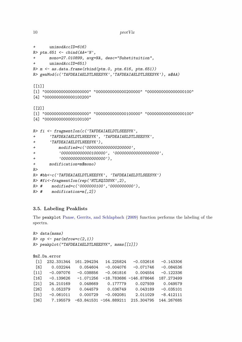
The graphic above is showing the mass error of the assignment between the MS2 spec and thesingly charged fragment ions of HTLNQIDSVK. The function psm is doing the peptide sequencematching. Of course, the more theoretical ions match (up to a small error tolerance, given bythe system) the measured ion chain, the more likely it is, that the measured spectrum indeedis from the inferred peptide (and therefore the protein is identified)
3.4. Modifications
R> library(protViz)
R> ptm.0 <- cbind(AA="-",
+ mono=0.0, avg=0.0, desc="unmodified", unimodAccID=NA)
R> ptm.616 <- cbind(AA='S',
+ mono=-27.010899, avg=NA, desc="Substituition",
10 protViz
+ unimodAccID=616)
R> ptm.651 <- cbind(AA='N',
+ mono=27.010899, avg=NA, desc="Substituition",
+ unimodAccID=651)
R> m <- as.data.frame(rbind(ptm.0, ptm.616, ptm.651))
R> genMod(c('TAFDEAIAELDTLNEESYK','TAFDEAIAELDTLSEESYK'), m$AA)
[[1]]
[1] "0000000000000000000" "0000000000000200000" "0000000000000000100"
[4] "0000000000000100200"
[[2]]
[1] "0000000000000000000" "0000000000000100000" "0000000000000000100"
[4] "0000000000000100100"
R> fi <- fragmentIon(c('TAFDEAIAELDTLSEESYK',
+ 'TAFDEAIAELDTLNEESYK', 'TAFDEAIAELDTLSEESYK',
+ 'TAFDEAIAELDTLNEESYK'),
+ modified=c('0000000000000200000',
+ '0000000000000100000', '0000000000000000000',
+ '0000000000000000000'),
+ modification=m$mono)
R>
R> #bh<-c('TAFDEAIAELDTLNEESYK', 'TAFDEAIAELDTLSEESYK')
R> #fi<-fragmentIon(rep('HTLNQIDSVK',2),
R> # modified=c('0000000100','0000000000'),
R> # modification=m[,2])
3.5. Labeling Peaklists
The peakplot Panse, Gerrits, and Schlapbach (2009) function performs the labeling of thespectra.
R> data(msms)
R> op <- par(mfrow=c(2,1))
R> peakplot("TAFDEAIAELDTLNEESYK", msms[[1]])
$mZ.Da.error
[1] 232.331344 161.294234 14.225824 -0.032616 -0.143306
[6] 0.032244 0.054604 -0.004076 -0.071746 -0.084536
[11] -0.097076 -0.038856 -0.061816 0.004554 -0.122336
[16] -0.139626 -1.071256 -18.783686 -146.878646 187.273499
[21] 24.210169 0.048669 0.177779 0.027939 0.049579
[26] 0.052379 0.044579 0.036749 0.043189 -0.035101
[31] -0.061011 0.000729 -0.092081 2.011029 -8.412111
[36] 7.195579 -63.841531 -164.889211 215.304795 144.267685

Christian Panse, Jonas Grossmann 11
[41] -2.800725 -17.059165 2.034875 2.264105 4.008125
[46] 1.292875 -0.003965 -13.612585 -0.060925 -17.065405
[51] 3.897535 3.000405 -17.148885 -17.166175 -18.097805
[56] -35.810235 -163.905195 204.300048 41.236718 17.075218
[61] -0.843372 -1.091812 0.129908 17.078928 -0.372162
[66] -16.539502 -1.044962 -1.000952 -1.409062 -2.995122
[71] 16.934468 19.037578 8.614438 24.222128 -46.814982
[76] -147.862662
$mZ.ppm.error
[1] 2.276532e+06 9.318407e+05 4.443342e+04 -7.494702e+01
[5] -2.539851e+02 5.075660e+01 7.296574e+01 -4.974443e+00
[9] -7.564705e+01 -7.963713e+01 -8.250960e+01 -3.041352e+01
[13] -4.445040e+01 3.026484e+00 -7.488007e+01 -7.920687e+01
[17] -5.791093e+02 -9.331667e+03 -6.860308e+04 1.272993e+06
[21] 7.805297e+04 1.225277e+02 3.378218e+02 4.263587e+01
[25] 6.444386e+01 5.935833e+01 4.532837e+01 3.345395e+01
[29] 3.564687e+01 -2.618263e+01 -4.321937e+01 4.781134e-01
[33] -5.770282e+01 1.165934e+03 -4.572174e+03 3.621478e+03
[37] -3.102183e+04 -7.637286e+04 1.808046e+06 7.588299e+05
[41] -8.306147e+03 -3.772366e+04 3.500821e+03 3.470990e+03
[45] 5.236793e+03 1.545734e+03 -4.106862e+00 -1.262129e+04
[49] -5.104441e+01 -1.318183e+04 2.768725e+03 1.971690e+03
[53] -1.038832e+04 -9.644849e+03 -9.694247e+03 -1.764117e+04
[57] -7.595171e+04 1.570497e+06 1.406678e+05 4.491332e+04
[61] -1.656190e+03 -1.710589e+03 1.726789e+02 1.973544e+04
[65] -3.850849e+02 -1.529356e+04 -8.747728e+02 -7.562373e+02
[69] -1.010347e+03 -1.986529e+03 1.072648e+04 1.114745e+04
[73] 4.725878e+03 1.229618e+04 -2.293808e+04 -6.903096e+04
$idx
[1] 1 1 1 3 14 21 38 49 64 87 91 97 102 106 110 113
[17] 115 116 116 1 1 2 12 25 41 53 70 89 94 99 104 107
[33] 108 111 114 116 116 116 1 1 1 3 16 24 41 52 67 88
[49] 93 97 104 107 110 113 115 116 116 1 1 2 11 22 40 53
[65] 68 88 93 98 103 106 108 111 114 116 116 116
$label
[1] "b1" "b2" "b3" "b4" "b5" "b6" "b7" "b8" "b9" "b10" "b11"
[12] "b12" "b13" "b14" "b15" "b16" "b17" "b18" "b19" "y1" "y2" "y3"
[23] "y4" "y5" "y6" "y7" "y8" "y9" "y10" "y11" "y12" "y13" "y14"
[34] "y15" "y16" "y17" "y18" "y19" "c1" "c2" "c3" "c4" "c5" "c6"
[45] "c7" "c8" "c9" "c10" "c11" "c12" "c13" "c14" "c15" "c16" "c17"
[56] "c18" "c19" "z1" "z2" "z3" "z4" "z5" "z6" "z7" "z8" "z9"
[67] "z10" "z11" "z12" "z13" "z14" "z15" "z16" "z17" "z18" "z19"
$score

12 protViz
[1] -1
$sequence
[1] "TAFDEAIAELDTLNEESYK"
$fragmentIon
b y c z
1 102.0550 147.1128 119.0815 130.0863
2 173.0921 310.1761 190.1186 293.1496
3 320.1605 397.2082 337.1870 380.1816
4 435.1874 526.2508 452.2140 509.2242
5 564.2300 655.2933 581.2566 638.2668
6 635.2671 769.3363 652.2937 752.3097
7 748.3512 882.4203 765.3777 865.3938
8 819.3883 983.4680 836.4148 966.4415
9 948.4309 1098.4950 965.4574 1081.4684
10 1061.5149 1211.5790 1078.5415 1194.5525
11 1176.5419 1340.6216 1193.5684 1323.5951
12 1277.5896 1411.6587 1294.6161 1394.6322
13 1390.6736 1524.7428 1407.7002 1507.7162
14 1504.7165 1595.7799 1521.7431 1578.7533
15 1633.7591 1724.8225 1650.7857 1707.7959
16 1762.8017 1839.8494 1779.8283 1822.8229
17 1849.8338 1986.9178 1866.8603 1969.8913
18 2012.8971 2057.9549 2029.9236 2040.9284
19 2140.9920 2159.0026 2158.0186 2141.9761
R> peakplot("TAFDEAIAELDTLSEESYK", msms[[2]])
$mZ.Da.error
[1] 245.264254 174.227144 27.158734 14.444434 0.021404
[6] -0.111266 -0.039926 -0.021626 -0.121916 -8.079236
[11] -0.158376 -0.153156 -0.094316 -0.022946 -0.186736
[16] -0.092226 -0.120456 -0.151686 -128.246646 200.206409
[21] 37.143079 0.078909 0.062269 0.129769 0.103729
[26] 0.060869 -0.051451 -18.048351 -0.027511 -0.025601
[31] -0.006211 0.020529 -0.048781 -0.024771 -9.166311
[36] 6.953579 -45.209531 -146.257211 228.237705 157.200595
[41] 10.132185 -2.582115 1.626855 2.722405 9.009025
[46] -1.130895 1.216385 13.347315 -3.671525 0.960295
[51] -17.120865 3.020205 -17.213285 -17.118775 -17.147005
[56] -17.178235 -145.273195 217.232958 54.169628 17.105458
[61] -0.833452 -1.260332 -0.899352 -3.098942 -1.173512
[66] -1.021802 -0.939162 -1.007752 -1.377062 -3.022622
[71] 16.977768 17.001778 7.860238 23.980128 -28.182982
[76] -129.230662

Christian Panse, Jonas Grossmann 13
$mZ.ppm.error
[1] 2.403257e+06 1.006558e+06 8.482850e+04 3.319130e+04
[5] 3.793488e+01 -1.751484e+02 -5.335196e+01 -2.639286e+01
[9] -1.285450e+02 -7.611043e+03 -1.346114e+02 -1.198789e+02
[13] -6.782037e+01 -1.552813e+01 -1.162198e+02 -5.313198e+01
[17] -6.608212e+01 -7.638202e+01 -6.066594e+04 1.360904e+06
[21] 1.197483e+05 1.986591e+02 1.183257e+02 1.980319e+02
[25] 1.397352e+02 7.115774e+01 -5.379332e+01 -1.684426e+04
[29] -2.322450e+01 -1.948903e+01 -4.485617e+00 1.370673e+01
[33] -3.109508e+01 -1.458996e+01 -5.056331e+03 3.547913e+03
[37] -2.226035e+04 -6.860121e+04 1.916651e+06 8.268554e+05
[41] 3.004915e+04 -5.709941e+03 2.798859e+03 4.173588e+03
[45] 1.177069e+04 -1.352074e+03 1.259905e+03 1.237534e+04
[49] -3.076091e+03 7.417604e+02 -1.216230e+04 2.020566e+03
[53] -1.060078e+04 -9.766434e+03 -9.319787e+03 -8.576627e+03
[57] -6.817113e+04 1.669915e+06 1.847849e+05 4.499286e+04
[61] -1.636709e+03 -1.974616e+03 -1.239974e+03 -3.696333e+03
[65] -1.249174e+03 -9.690310e+02 -8.043928e+02 -7.772361e+02
[69] -1.006903e+03 -2.041339e+03 1.094110e+04 1.011538e+04
[73] 4.376983e+03 1.234257e+04 -1.399411e+04 -6.110297e+04
$idx
[1] 1 1 1 3 11 20 39 45 64 90 96 106 116 121 126 129
[17] 131 133 133 1 1 2 7 24 38 49 65 90 97 110 115 122
[33] 123 127 130 132 133 133 1 1 1 3 13 23 40 47 67 91
[49] 98 108 116 122 126 129 131 133 133 1 1 2 6 21 36 47
[65] 62 90 95 108 113 121 123 127 130 132 133 133
$label
[1] "b1" "b2" "b3" "b4" "b5" "b6" "b7" "b8" "b9" "b10" "b11"
[12] "b12" "b13" "b14" "b15" "b16" "b17" "b18" "b19" "y1" "y2" "y3"
[23] "y4" "y5" "y6" "y7" "y8" "y9" "y10" "y11" "y12" "y13" "y14"
[34] "y15" "y16" "y17" "y18" "y19" "c1" "c2" "c3" "c4" "c5" "c6"
[45] "c7" "c8" "c9" "c10" "c11" "c12" "c13" "c14" "c15" "c16" "c17"
[56] "c18" "c19" "z1" "z2" "z3" "z4" "z5" "z6" "z7" "z8" "z9"
[67] "z10" "z11" "z12" "z13" "z14" "z15" "z16" "z17" "z18" "z19"
$score
[1] -1
$sequence
[1] "TAFDEAIAELDTLSEESYK"
$fragmentIon
b y c z
1 102.0550 147.1128 119.0815 130.0863
2 173.0921 310.1761 190.1186 293.1496

14 protViz
3 320.1605 397.2082 337.1870 380.1816
4 435.1874 526.2508 452.2140 509.2242
5 564.2300 655.2933 581.2566 638.2668
6 635.2671 742.3254 652.2937 725.2988
7 748.3512 855.4094 765.3777 838.3829
8 819.3883 956.4571 836.4148 939.4306
9 948.4309 1071.4841 965.4574 1054.4575
10 1061.5149 1184.5681 1078.5415 1167.5416
11 1176.5419 1313.6107 1193.5684 1296.5842
12 1277.5896 1384.6478 1294.6161 1367.6213
13 1390.6736 1497.7319 1407.7002 1480.7053
14 1477.7056 1568.7690 1494.7322 1551.7424
15 1606.7482 1697.8116 1623.7748 1680.7850
16 1735.7908 1812.8385 1752.8174 1795.8120
17 1822.8229 1959.9069 1839.8494 1942.8804
18 1985.8862 2030.9440 2002.9127 2013.9175
19 2113.9811 2131.9917 2131.0077 2114.9652
R> par(op)
AFDEAIAELDTLNEESYK / 1799: Scan 3246 (rt=67.4676) [/p474/Proteomics/ORBI_1/jonas_20080530_bhdaten_doro/20071028_bh_071029122357.RA
m/z
Inte
nsity
b4
b5
b6
b7
b8
b9
c9
b1
0
b1
1c1
1
b1
2
b1
3
b1
4
b1
5
b1
6
02
00
04
00
06
00
08
00
0
y3
y4
y5
z6
y6
y7
z8
y8
y9
y1
0
y1
1
y1
2
y1
3
y1
4
02
04
06
08
01
00
435.1
5
397.2
6
564.0
9
526.4
3
748.4
1
819.3
8
769.3
9752.4
4
948.3
6965.4
5
882.4
7
983.5
1966.0
7
1061.4
3
1098.5
3
1176.4
4
1277.5
5
1193.5
11211.6
2
1390.6
1
1340.5
9
1411.6
1504.7
21524.7
4 1762.6
6
AFDEAIAELDTLSEESYK / 1842: Scan 3353 (rt=70.3158) [/p474/Proteomics/ORBI_1/jonas_20080530_bhdaten_doro/20071028_bh_071029122357.RA
m/z
Inte
nsity
b5
b6
b7
b8
b9
b1
1
b1
2
b1
3
b1
4
b1
5
b1
6
b1
7
b1
8
02
00
04
00
06
00
08
00
01
00
00
y3
y4
y5
y6
y7
y8
y1
0
y1
1
y1
2
y1
3
y1
4
y1
5
02
04
06
08
01
00
397.2
9
564.2
5
526.3
1
748.3
1
819.3
7
742.4
3
948.3
1
855.4
7
956.4
1
1176.3
8
1277.4
4
1184.5
4
1313.5
9
1390.5
8
1477.6
8
1384.6
4
1606.5
6
1497.7
5
1735.7
1697.7
9
1822.7

Christian Panse, Jonas Grossmann 15
The following code snippet combine all the function to a simple peptide search engine. Asdefault arguments the mass spec measurement, a list of mZ and intensity arrays, and acharacter vector of peptide sequences is given.
R> peptideSearch <- function (x,
+ peptideSequence,
+ pimIdx = parentIonMass(peptideSequence),
+ peptideMassTolerancePPM = 5,
+ framentIonMassToleranceDa = 0.01,
+ FUN = .byIon)
+ {
+ query.mass <- ((x$pepmass * x$charge)) - (1.007825 * (x$charge -
+ 1))
+ eps <- query.mass * peptideMassTolerancePPM * 1e-06
+ lower <- findNN(query.mass - eps, pimIdx)
+ upper <- findNN(query.mass + eps, pimIdx)
+ rv <- lapply(peptideSequence[lower:upper], function(p) {
+ psm(p, x, plot = FALSE, FUN = FUN)
+ })
+ rv.error <- sapply(rv, function(p) {
+ sum(abs(p$mZ.Da.error) < framentIonMassToleranceDa)
+ })
+ idx.tophit <- which(rv.error == max(rv.error))[1]
+ data.frame(mass_error = eps,
+ idxDiff = upper - lower,
+ charge = x$charge,
+ pepmass = query.mass,
+ peptideSequence = rv[[idx.tophit]]$sequence,
+ groundTrue.peptideSequence = x$peptideSequence,
+ ms2hit = (rv[[idx.tophit]]$sequence ==
+ x$peptideSequence), hit = (x$peptideSequence %in%
+ peptideSequence[lower:upper]))
+ }
4. Quantification
For an overview on Quantitative Proteomics read Bantscheff, Lemeer, Savitski, and Kuster(2012); Cappadona, Baker, Cutillas, Heck, and van Breukelen (2012). The authors are awarethat meaningful statistics usually require a much higher number of biological replicates. Inalmost all cases there are not more than three to six repetitions. For the moment there arelimited options due to the availability of machine time and the limits of the technologies.
4.1. Label-free methods on protein level
The data set fetuinLFQ contains a subset of our results descriped in Grossmann, Roschitzki,Panse, Fortes, Barkow-Oesterreicher, Rutishauser, and Schlapbach (2010). The example be-

16 protViz
low shows a visualization using trellis plots. It graphs the abundance of four protein inde-pendency from the fetuin concentration spiked into the sample.
R> library(lattice)
R> data(fetuinLFQ)
R> cv<-1-1:7/10
R> t<-trellis.par.get("strip.background")
R> t$col<-(rgb(cv,cv,cv))
R> trellis.par.set("strip.background",t)
R> print(xyplot(abundance~conc|prot*method,
+ groups=prot,
+ xlab="Fetuin concentration spiked into experiment [fmol]",
+ ylab="Abundance",
+ aspect=1,
+ data=fetuinLFQ$t3pq[fetuinLFQ$t3pq$prot
+ %in% c('Fetuin', 'P15891', 'P32324', 'P34730'),],
+ panel = function(x, y, subscripts, groups) {
+ if (groups[subscripts][1] == "Fetuin") {
+ panel.fill(col="#ffcccc")
+ }
+ panel.grid(h=-1,v=-1)
+ panel.xyplot(x, y)
+ panel.loess(x,y, span=1)
+ if (groups[subscripts][1] == "Fetuin") {
+ panel.text(min(fetuinLFQ$t3pq$conc),
+ max(fetuinLFQ$t3pq$abundance),
+ paste("R-squared:",
+ round(summary(lm(x~y))$r.squared,2)),
+ cex=0.75,
+ pos=4)
+ }
+ }
+ ))

Christian Panse, Jonas Grossmann 17
Fetuin concentration spiked into experiment [fmol]
Abu
nd
an
ce
0e+00
2e+06
4e+06
6e+06
8e+06
0 50 150 250
●●●●●●●●●
●● ●●
●
●●
●
●●●
●
●
●●
●
●●
R−squared: 0.98
Fetuin
T3PQ
0 50 150 250
● ●●●●● ●● ●● ●●●● ● ●●● ●●● ●●● ●● ●● ●
P15891
T3PQ
0 50 150 250
●● ●● ●● ●●● ●● ● ●● ●●● ●● ●●●● ● ●●● ● ●●
P32324
T3PQ
0 50 150 250
●●●● ●●● ●● ●● ●●●●● ●●●●● ●●● ● ●●●●●
P34730
T3PQ
The plot shows the estimated concentration of the four proteins using the top three mostintense peptides. The Fetuin peptides are spiked in with increasing concentration while thethree other yeast proteins are kept stable in the background.
4.2. pgLFQ – LCMS based label-free quantification
LCMS based label-free quantification is a very popular method to extract relative quantitativeinformation from mass spectrometry experiments. At the FGCZ we use the software Pro-genesisLCMS for this workflow http://www.nonlinear.com/products/progenesis/lc-ms/
overview/. Progenesis is a graphical software which does the aligning between several LCMSexperiments, extracts signal intensities from LCMS maps and annotates the master map withpeptide and protein labels.
R> data(pgLFQfeature)
R> data(pgLFQprot)
R> featureDensityPlot<-function(data, n=ncol(data), nbins=30){
+ my.col<-rainbow(n);
+ mids<-numeric()
+ density<-numeric()
+ for (i in 1:n) {
+ h<-hist(data[,i],nbins, plot=FALSE)

18 protViz
+ mids<-c(mids, h$mids)
+ density<-c(density, h$density)
+ }
+ plot(mids,density, type='n')
+ for (i in 1:n) {
+ h<-hist(data[,i],nbins, plot=FALSE)
+ lines(h$mids,h$density, col=my.col[i])
+ }
+ legend("topleft", names(data), cex=0.5,
+ text.col=my.col
+ )
+ }
R> par(mfrow=c(1,1));
R> featureDensityPlot(asinh(pgLFQfeature$"Normalized abundance"),
+ nbins=25)
0 5 10 15 20
0.0
00
.05
0.1
00
.15
0.2
00
.25
0.3
0
mids
de
nsity
20120809_01_WT_NI_3_excl
20120809_02_WT_NI_4_excl
20120809_03_WT_NI_5_excl
20120809_05_WT_NI_1_excl
20120809_06_WT_NI_5_excl
20120809_07_WT_NI_6_excl
20120809_14_WT_Inf_1h_1_noLM_aexl
20120809_15_WT_Inf_1h_2_noLM_aexl
20120809_16_WT_Inf_1h_3_noLM_aexl
20120809_17_WT_Inf_1h_4_noLM_aexl
20120809_18_WT_Inf_1h_5_noLM_aexl
20120809_19_WT_Inf_1h_6_noLM_aexl
20120809_21_WT_Inf_2h_1_noLM_aexl
20120809_22_WT_Inf_2h_2_noLM_aexl
20120809_23_WT_Inf_2h_3_noLM_aexl
20120809_24_WT_Inf_2h_4_noLM_aexl
20120809_25_WT_Inf_2h_5_noLM_aexl
20120809_26_WT_Inf_2h_6_noLM_aexl
20120809_28_WT_Inf_3h_1_noLM_aexl
20120809_29_WT_Inf_3h_2_noLM_aexl
20120809_30_WT_Inf_3h_3_noLM_aexl
20120809_31_WT_Inf_3h_4_noLM_aexl
20120809_33_WT_Inf_3h_6_noLM_aexl
20120809_56_WT_Inf_3h_5_noLM_aexl
The featureDensityPlot shows the normalized signal intensity distribution (asinh trans-formed) over 24 LCMS runs which are aligned in this experiment.
R> op<-par(mfrow=c(1,1),mar=c(18,18,4,1),cex=0.5)
R> samples<-names(pgLFQfeature$"Normalized abundance")

Christian Panse, Jonas Grossmann 19
R> image(cor(asinh(pgLFQfeature$"Normalized abundance")),
+ col=gray(seq(0,1,length=20)),
+ main='pgLFQfeature correlation',
+ axes=FALSE)
R> axis(1,at=seq(from=0, to=1,
+ length.out=length(samples)),
+ labels=samples, las=2)
R> axis(2,at=seq(from=0, to=1,
+ length.out=length(samples)), labels=samples, las=2)
R> par(op)
pgLFQfeature correlation
20120809_01_W
T_N
I_3_excl
20120809_02_W
T_N
I_4_excl
20120809_03_W
T_N
I_5_excl
20120809_05_W
T_N
I_1_excl
20120809_06_W
T_N
I_5_excl
20120809_07_W
T_N
I_6_excl
20120809_14_W
T_In
f_1h_1_noLM
_aexl
20120809_15_W
T_In
f_1h_2_noLM
_aexl
20120809_16_W
T_In
f_1h_3_noLM
_aexl
20120809_17_W
T_In
f_1h_4_noLM
_aexl
20120809_18_W
T_In
f_1h_5_noLM
_aexl
20120809_19_W
T_In
f_1h_6_noLM
_aexl
20120809_21_W
T_In
f_2h_1_noLM
_aexl
20120809_22_W
T_In
f_2h_2_noLM
_aexl
20120809_23_W
T_In
f_2h_3_noLM
_aexl
20120809_24_W
T_In
f_2h_4_noLM
_aexl
20120809_25_W
T_In
f_2h_5_noLM
_aexl
20120809_26_W
T_In
f_2h_6_noLM
_aexl
20120809_28_W
T_In
f_3h_1_noLM
_aexl
20120809_29_W
T_In
f_3h_2_noLM
_aexl
20120809_30_W
T_In
f_3h_3_noLM
_aexl
20120809_31_W
T_In
f_3h_4_noLM
_aexl
20120809_33_W
T_In
f_3h_6_noLM
_aexl
20120809_56_W
T_In
f_3h_5_noLM
_aexl
20120809_01_WT_NI_3_excl
20120809_02_WT_NI_4_excl
20120809_03_WT_NI_5_excl
20120809_05_WT_NI_1_excl
20120809_06_WT_NI_5_excl
20120809_07_WT_NI_6_excl
20120809_14_WT_Inf_1h_1_noLM_aexl
20120809_15_WT_Inf_1h_2_noLM_aexl
20120809_16_WT_Inf_1h_3_noLM_aexl
20120809_17_WT_Inf_1h_4_noLM_aexl
20120809_18_WT_Inf_1h_5_noLM_aexl
20120809_19_WT_Inf_1h_6_noLM_aexl
20120809_21_WT_Inf_2h_1_noLM_aexl
20120809_22_WT_Inf_2h_2_noLM_aexl
20120809_23_WT_Inf_2h_3_noLM_aexl
20120809_24_WT_Inf_2h_4_noLM_aexl
20120809_25_WT_Inf_2h_5_noLM_aexl
20120809_26_WT_Inf_2h_6_noLM_aexl
20120809_28_WT_Inf_3h_1_noLM_aexl
20120809_29_WT_Inf_3h_2_noLM_aexl
20120809_30_WT_Inf_3h_3_noLM_aexl
20120809_31_WT_Inf_3h_4_noLM_aexl
20120809_33_WT_Inf_3h_6_noLM_aexl
20120809_56_WT_Inf_3h_5_noLM_aexl
This image plot shows the correlation between runs on feature level (values are asinh trans-formed). White is perfect correlation while black indicates a poor correlation.
R> op<-par(mfrow=c(1,1),mar=c(18,18,4,1),cex=0.5)
R> image(cor(asinh(pgLFQprot$"Normalized abundance")),
+ main='pgLFQprot correlation',
+ axes=FALSE,
+ col=gray(seq(0,1,length=20)))
R> axis(1,at=seq(from=0, to=1,
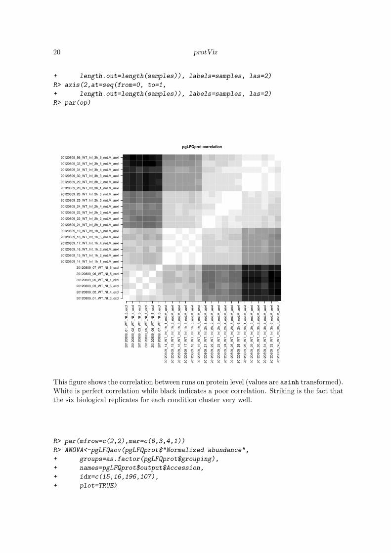
20 protViz
+ length.out=length(samples)), labels=samples, las=2)
R> axis(2,at=seq(from=0, to=1,
+ length.out=length(samples)), labels=samples, las=2)
R> par(op)
pgLFQprot correlation
20120809_01_W
T_N
I_3_excl
20120809_02_W
T_N
I_4_excl
20120809_03_W
T_N
I_5_excl
20120809_05_W
T_N
I_1_excl
20120809_06_W
T_N
I_5_excl
20120809_07_W
T_N
I_6_excl
20120809_14_W
T_In
f_1h_1_noLM
_aexl
20120809_15_W
T_In
f_1h_2_noLM
_aexl
20120809_16_W
T_In
f_1h_3_noLM
_aexl
20120809_17_W
T_In
f_1h_4_noLM
_aexl
20120809_18_W
T_In
f_1h_5_noLM
_aexl
20120809_19_W
T_In
f_1h_6_noLM
_aexl
20120809_21_W
T_In
f_2h_1_noLM
_aexl
20120809_22_W
T_In
f_2h_2_noLM
_aexl
20120809_23_W
T_In
f_2h_3_noLM
_aexl
20120809_24_W
T_In
f_2h_4_noLM
_aexl
20120809_25_W
T_In
f_2h_5_noLM
_aexl
20120809_26_W
T_In
f_2h_6_noLM
_aexl
20120809_28_W
T_In
f_3h_1_noLM
_aexl
20120809_29_W
T_In
f_3h_2_noLM
_aexl
20120809_30_W
T_In
f_3h_3_noLM
_aexl
20120809_31_W
T_In
f_3h_4_noLM
_aexl
20120809_33_W
T_In
f_3h_6_noLM
_aexl
20120809_56_W
T_In
f_3h_5_noLM
_aexl
20120809_01_WT_NI_3_excl
20120809_02_WT_NI_4_excl
20120809_03_WT_NI_5_excl
20120809_05_WT_NI_1_excl
20120809_06_WT_NI_5_excl
20120809_07_WT_NI_6_excl
20120809_14_WT_Inf_1h_1_noLM_aexl
20120809_15_WT_Inf_1h_2_noLM_aexl
20120809_16_WT_Inf_1h_3_noLM_aexl
20120809_17_WT_Inf_1h_4_noLM_aexl
20120809_18_WT_Inf_1h_5_noLM_aexl
20120809_19_WT_Inf_1h_6_noLM_aexl
20120809_21_WT_Inf_2h_1_noLM_aexl
20120809_22_WT_Inf_2h_2_noLM_aexl
20120809_23_WT_Inf_2h_3_noLM_aexl
20120809_24_WT_Inf_2h_4_noLM_aexl
20120809_25_WT_Inf_2h_5_noLM_aexl
20120809_26_WT_Inf_2h_6_noLM_aexl
20120809_28_WT_Inf_3h_1_noLM_aexl
20120809_29_WT_Inf_3h_2_noLM_aexl
20120809_30_WT_Inf_3h_3_noLM_aexl
20120809_31_WT_Inf_3h_4_noLM_aexl
20120809_33_WT_Inf_3h_6_noLM_aexl
20120809_56_WT_Inf_3h_5_noLM_aexl
This figure shows the correlation between runs on protein level (values are asinh transformed).White is perfect correlation while black indicates a poor correlation. Striking is the fact thatthe six biological replicates for each condition cluster very well.
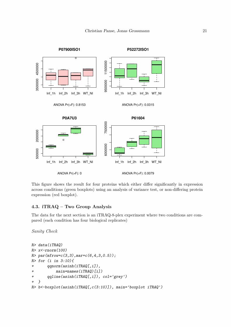
R> par(mfrow=c(2,2),mar=c(6,3,4,1))
R> ANOVA<-pgLFQaov(pgLFQprot$"Normalized abundance",
+ groups=as.factor(pgLFQprot$grouping),
+ names=pgLFQprot$output$Accession,
+ idx=c(15,16,196,107),
+ plot=TRUE)
Christian Panse, Jonas Grossmann 21
●
Inf_1h Inf_2h Inf_3h WT_NI
3500000
4500000
P07900ISO1
ANOVA Pr(>F): 0.8153
Inf_1h Inf_2h Inf_3h WT_NI
9500000
11500000
P52272ISO1
ANOVA Pr(>F): 0.0315
●
Inf_1h Inf_2h Inf_3h WT_NI
500000
2000000
P0A7U3
ANOVA Pr(>F): 0
Inf_1h Inf_2h Inf_3h WT_NI
6000000
7500000
P61604
ANOVA Pr(>F): 0.0079
This figure shows the result for four proteins which either differ significantly in expressionacross conditions (green boxplots) using an analysis of variance test, or non-differing proteinexpression (red boxplot).
4.3. iTRAQ – Two Group Analysis
The data for the next section is an iTRAQ-8-plex experiment where two conditions are com-pared (each condition has four biological replicates)
Sanity Check
R> data(iTRAQ)
R> x<-rnorm(100)
R> par(mfrow=c(3,3),mar=c(6,4,3,0.5));
R> for (i in 3:10){
+ qqnorm(asinh(iTRAQ[,i]),
+ main=names(iTRAQ)[i])
+ qqline(asinh(iTRAQ[,i]), col='grey')
+ }
R> b<-boxplot(asinh(iTRAQ[,c(3:10)]), main='boxplot iTRAQ')

22 protViz
●●
●
●
●
●
●
●●
●●●●●
●
●●
●●
●● ●
●
●
●●●
●●
●
●
●
●
●
●
●
●●
●●●
●
●
●
●●
●
●●●
●●
●
●●
●●
●
●
●
●●
●●
●
●●
●
●
●
●●
●●●
●
●●
●●●
●●
●●
●
●
●●
●
●
●
●
●
●●
●
●●
●●
●
●●
●
●●
●
●●●
●●●
●
●
●●
●
●●●
●●●
●
●●●●● ●●
●
●● ●●
●●●
●
● ●●●● ● ●●
●
●●
●
●
●●
●
●
●
●● ●
●● ●
●●
●●
●
●
●
●
●
● ●●
●
●
●
●
●
●
●
●
●●
●
●
●
●
●
●●●●
●●
●●●
●
●●
●
●●●
●
●●
●
●
●●
●
●
●●
●
●
●
●●
●
●●●
●●
●●
●●
●●
●●
●●
●●
●●
●●●
●●●
●●
●●
● ● ●●
●●
●●●
−3 −2 −1 0 1 2 3
04
812
area113
Theoretical Quantiles
Sam
ple
Quantile
s
●●
●●
●●
●
●●
●● ●●
●●
●●
●●
●● ●
●●
●●●
●●
●●
●
●
●
●
●
●●
●●●
●
●
●
●
●
●
●●
●
●●
●
●●
●●●
●●
●
●
●
●
●
●
●
●
●
●
●●●
●●
●
●●●
●●
●●
● ●●
●●●●●
●
●
●●●
●
●
●
●●
●
●
●●●●●
●●
●●●●
●●
●●
●
●●
●
●●●
●
●●
●●●●
●●
●●●
●●●●●
●●●
●●
●●
●
●
●●
●
●
●●
●
●
●●
●● ●● ●
●
●● ●
●
●
●
●
●
● ●●
●●●
●
●
●
●
●
●●
●
●●
●
●
● ●●
●●●
●●●
●
●●
●
●●●
●
●●●
●
●●
●
●
●●
●
●
●
●
●
●
●●●
●●
●●●●
●●
●●
●●
●
●●
●
●●
●
●●●
●
●●●
●● ●●
●●●●
●
−3 −2 −1 0 1 2 3
04
812
area114
Theoretical QuantilesS
am
ple
Quantile
s
●●
●
●
●
●
●
●●
●●●●
●● ●●
●●●●●
●●
●●●
●●
●●
●
●
●
●
●
●●
●●●
●
●
●
●
●
●
●
●
●
●●
●
●●
● ●●
●
●
●●●
●●
●
●
●
●
●
●
●
●●●
●
●●
●●●
●●
●●
●
●
●
●
●●
●
●
●
●
●
●
●
●
●●
●
●
●●●
●
●
●
●
●●
●
●
●
●
●●
●
●●●
●
●●
●
●●
●●●
●●
●
●●
●●
●●●
●
●●●●●
●
● ●
●
●●
●
●
●●●
●
●
●
●●●●●
●●
●●
●
●
●
●
●
●●●
●●
●
●
●
●
●●
●●
●
●
●
●
●
●●
● ●
●●
●
●●
●
●●
●
●●
●
●
●●
●
●
●●●
●
●●●
●
●
● ●
●
●●
●●
●
●●
●
●●
●
●●
●●
●
●
●●
●
●●
●●●●
●●●
● ● ●●
●●
●●
●
−3 −2 −1 0 1 2 3
04
812
area115
Theoretical Quantiles
Sam
ple
Quantile
s
●●
●
●
●
●
●
●●
●● ●
●●
●
●●
●●
●
●●
●
●
●●●
●●
●
●
●
●
●
●
●
●●
●●●
●
●
●
●●
●
●●
●
●●
●
●●
●●
●
●
●●
●
●●
●
●
●
●
●
●
●●
●●●●
●●●
●●●●
●●●
●●●
●
●●
●
●
●●
●
●●
●●
●●●●●●●
●●
●●●
●
●
●
●●
●
●●
●●●●
●
●
●
●●●
●
● ●
●●●
●●●●
●
● ●●
●●
●●
●
●
●●
●
●
●●
●
●
●●
●
●●● ●
●●
●●
●
●
●
●
●
● ●●
●●●
●●●
●
●
●●
●
●●
●
●
●●● ●
● ●
●
●●
●
●●
●
●●
●
●
●●
●
●
●●
●
●
●●
●
●
●
●●
●
●●●
●
●●
●●
●● ●
●●
●●
●
●
●
●
●●
●
●●●●●
●●
● ●●●
●●
●●●
−3 −2 −1 0 1 2 3
04
812
area116
Theoretical Quantiles
Sam
ple
Quantile
s
●●
●
●
●
●
●
●●
●● ●●●
●
●●
●●
●
●●
●●
●●●
● ●
●
●
●
●
●
●
●
●●
●●●
●
●
●
●
●
●
●●
●
●●
●
●●
●●
●
●●●
●
●●
●
●
●
●
●
●
●●
●●●
●
●●
●●●
●●
● ●●
●
●
●
●
●●
●●
●
●
●
●●
●●
●●●●●●●
●●
●●
●
●
●
●
●●
●
●●● ●
●●
●
●
●
●●●
●
●●
●●
●●
●●●
●
● ●●●
●●
●
●
●
●●
●
●
●●
●
●
●●
●
●●●●
●●
●●
●
●
●
●
●
●●●
●●
●●
●●
●●
●●
●
●●
●
●
●●● ●
●●
●●
●
●
●●
●
●●
●
●
●●●
●
●●
●
●
●
●●
●
●
●●
●
●●●
●
●
●●
●●
●●
●●
●●
●
●●
●●●●
●●●
●●
●●
●●●●●●
●●●
−3 −2 −1 0 1 2 3
04
812
area117
Theoretical Quantiles
Sam
ple
Quantile
s
●●
●
●●
●
●
●●
●●●●
●
●
●●
●●
●
● ●
●
●
●●●
●●
●
●
●
●
●
●
●
●●
●●●
●
●
●
●
●
●
●
●●
●●
●
●●
●●●
●●
●●●●
●
●
●
●
●
●
●●
●●●
●
● ●●●●
● ●
● ●●
●●
●
●
●●●
●
●●
●
●●
●●
●
●●
●
●●● ●●
●●
●
●●
●
●●●
●●
●●●●
●
●●
●●●
●●
●
●●●
● ●●●
●
● ●●
●●●●
●
●
●●
●
●
●●
●
●
●
●
●
●●● ●
●●●●
●
●
●
●
●
●●●
●●●●
●●
●
●
●●
●
●
●
●
●
●●●●
●●
●●●
●
●●
●
●●
●
●
●●
●
●
●
●
●
●
●●
●
●
●
●
●
●
●●
●●●
●●
●●
●●
●●
●●
●
●
●●
●●
●
●●●
●●
●●
● ●●●
●●
●●●
−3 −2 −1 0 1 2 3
04
812
area118
Theoretical QuantilesS
am
ple
Quantile
s
●●
●
●
●
●
●
●●
●●●●●
●
●●
●●
●
●●
●
●
●●●
● ●
●
●
●
●
●
●
●
●●
● ●●
●
●
●
●
●
●
●●
●
●●
●
●●
● ●
●
●
●
●●
●●
●
●
●
●
●
●
●●
●●●●
● ●●●●
● ●
● ●●
●●●●●
●
●●
●
●
●
●
●
●
●●●
●●
●●●●●
●
●●
●●
●
●●●
●●●●
●●
●
●
●
●●●●
●●
●●●
●●●●
●
● ●●●
●●
●
●
●
●●
●
●
●●
●
●
●●
●
●●● ●
●
●●●
●
●
●
●
●
●●●
●●
●
●●●
●
●
●●
●
●
●
●
●
●● ●●
●●
●●
●
●
●●
●
●●
●
●
●●●
●
●●
●
●
●●
●
●
●
●●
●
●●
●●
●
●●
●●
●●
●●
●●
●●
●
●
●●●
●●●
●●
●●
● ● ●●
●●
●●●
−3 −2 −1 0 1 2 3
04
812
area119
Theoretical Quantiles
Sam
ple
Quantile
s
●●
●●
●●
●
●●
●● ●●
●●
●●
●●●●●
●
●●
●●
●●
●
●
●
●
●
●
●
●●
●●●
●●
●
●
●
●
●●
●
●●
●
●●
●●●
●
●
●●
●●
●
●●
●
●
●
●●
●●●
●
● ●●●●
●●
● ●●
●●●●
●
●●
●
●
●
●
●●
●
●●
●
●●
●●●●●
●
●
●
●
●
●
●●
●
●●●
●●●
●
●●
●●●
●●
●
●●
●●●●●
●
● ●●●
●●
●●
●
●●
●
●
●●
●
●
●
●
●●● ●●●
●●●
●
●
●
●
●
●●●
●●
●
●
●●
●
●
●●
●
●
●
●
●
●● ●●●
●
●
●●
●
●●
●
●●●
●
●●
●
●
●●
●
●
●●
●
●
●
●●
●
●●●
●●
●
●●
●●
●
●●
●●
●●
●
●●●●
●
●●●●●
●
● ● ●●
●●
●●●
−3 −2 −1 0 1 2 3
04
812
area121
Theoretical Quantiles
Sam
ple
Quantile
s
●●●●●●●●●●●●●●●●●●●● ●●●●●●●●●●●●●●●●●●●●●●●●●●●●●●● ●●●●●●●●●●●●●●●●●●●●●●●●●●●●● ●●●●●●●●●●●●●●●●●●●●●●●●●●●●●●●●●●●● ●●●●●●●●●●●●●●●●●●●●●●●●● ●●●●●●●●●●●●●● ●●●●●●●●●●●●●●●●●●●●●● ●●●●●●●●●●●●●●
area113 area117
04
812
boxplot iTRAQ
A first quality check to see if all reporter ion channels are having the same distributions.Shown in the figure are Q-Q plots of the individual reporter channels against a normal dis-tribution. The last is a boxplot for all individual channels.
On Protein Level
R> data(iTRAQ)
R> group1Protein<-numeric()
R> group2Protein<-numeric()
R> for (i in c(3,4,5,6))
+ group1Protein<-cbind(group1Protein,
+ asinh(tapply(iTRAQ[,i], paste(iTRAQ$prot), sum, na.rm=TRUE)))
R> for (i in 7:10)
+ group2Protein<-cbind(group2Protein,
+ asinh(tapply(iTRAQ[,i], paste(iTRAQ$prot), sum, na.rm=TRUE)))
R> par(mfrow=c(2,3),mar=c(6,3,4,1))
R> for (i in 1:nrow(group1Protein)){
+ boxplot.color="#ffcccc"
+ tt.p_value<-t.test(as.numeric(group1Protein[i,]),
+ as.numeric(group2Protein[i,]))$p.value

Christian Panse, Jonas Grossmann 23
+
+ if (tt.p_value < 0.05)
+ boxplot.color='lightgreen'
+
+ b<-boxplot(as.numeric(group1Protein[i,]),
+ as.numeric(group2Protein[i,]),
+ main=row.names(group1Protein)[i],
+ sub=paste("t-Test: p-value =", round(tt.p_value,2)),
+ col=boxplot.color,
+ axes=FALSE)
+ axis(1, 1:2, c('group_1','group_2')); axis(2); box()
+
+ points(rep(1,b$n[1]), as.numeric(group1Protein[i,]), col='blue')
+ points(rep(2,b$n[2]), as.numeric(group2Protein[i,]), col='blue')
+ }
O95445
t−Test: p−value = 0.03
group_1 group_2
13.4
13.6
13.8
14.0
●
●●
●
●
●
●
●
P02652
t−Test: p−value = 0.04
group_1 group_2
14.4
14.6
14.8
15.0
15.2
●
●
●●
●
●
●
●
P02654
t−Test: p−value = 0.05
group_1 group_2
13.5
14.0
14.5
●
●
●
●
●
●
●
●
P02748
t−Test: p−value = 0.82
group_1 group_2
13.4
13.6
13.8
14.0
14.2
●
●
●
●
●
●
●
●
Q08554
t−Test: p−value = 0.49
group_1 group_2
8.9
9.0
9.1
9.2
9.3
●
●●
●
●
●
●
●
This figure shows five proteins which are tested if they differ across conditions using the fourbiological replicates with a t-test.

24 protViz
On Peptide Level
The same can be done on peptide level using the protViz function iTRAQ2GroupAnalysis.
R> data(iTRAQ)
R> q <- iTRAQ2GroupAnalysis(data=iTRAQ,
+ group1=c(3,4,5,6),
+ group2=7:10,
+ INDEX=paste(iTRAQ$prot,iTRAQ$peptide),
+ plot=FALSE)
R> q[1:10,]
name p_value Group1.area113
1 O95445 AFLLTPR 0.056 1705.43
2 O95445 DGLCVPR 0.161 2730.41
3 O95445 MKDGLCVPR 0.039 28726.38
4 O95445 NQEACELSNN 0.277 4221.31
5 O95445 SLTSCLDSK 0.036 20209.66
6 P02652 AGTELVNFLSYFVELGTQPA 0.640 4504.97
7 P02652 AGTELVNFLSYFVELGTQPAT 0.941 67308.30
8 P02652 AGTELVNFLSYFVELGTQPATQ 0.338 4661.54
9 P02652 EPCVESLVSQYFQTVTDYGK 0.115 4544.56
10 P02652 EQLTPLIK 0.053 24596.42
Group1.area114 Group1.area115 Group1.area116 Group2.area117
1 1459.10 770.65 3636.40 3063.48
2 1852.90 1467.65 2266.88 2269.57
3 15409.81 19050.13 58185.02 51416.05
4 4444.28 2559.23 6859.71 5545.12
5 14979.02 12164.94 37572.56 30687.57
6 4871.88 2760.53 9213.41 6728.62
7 46518.21 33027.14 111629.30 94531.76
8 3971.82 2564.39 8269.73 6045.30
9 4356.51 2950.48 6357.90 6819.99
10 22015.94 18424.56 49811.91 33197.47
Group2.area118 Group2.area119 Group2.area121
1 4046.73 2924.49 5767.87
2 3572.32 2064.82 2208.92
3 70721.05 38976.42 60359.72
4 11925.66 6371.50 15656.92
5 39176.99 34417.66 54439.22
6 14761.96 7796.29 18681.60
7 168775.00 83526.72 168032.50
8 13724.92 7426.84 17214.87
9 10265.84 7012.92 14279.22
10 67213.62 40030.86 87343.38

Christian Panse, Jonas Grossmann 25
5. Pressure Profiles QC
A common problem with mass spec setup is the pure reliability of the high-pressure pump.The following graphics provide visualizations for quality control.
An overview of the pressure profile data can be seen by using the ppp function.
R> data(pressureProfile)
R> ppp(pressureProfile)
The lines plots the pressure profiles data on a scatter plot “Pc” versus “time” grouped bytime range (no figure because of too many data items).
The Trellis xyplot shows the Pc development over each instrument run to a specified relativeruntime (25, 30, . . .).
R> pp.data<-pps(pressureProfile, time=seq(25,40,by=5))
R> print(xyplot(Pc ~ as.factor(file) | paste("time =",
+ as.character(time), "minutes"),
+ panel = function(x, y){
+ m<-sum(y)/length(y)
+ m5<-(max(y)-min(y))*0.05
+ panel.abline(h=c(m-m5,m,m+m5),
+ col=rep("#ffcccc",3),lwd=c(1,2,1))
+ panel.grid(h=-1, v=0)
+ panel.xyplot(x, y)
+ },
+ ylab='Pc [psi]',
+ layout=c(1,4),
+ sub='The three red lines indicate the average plus min 5%.',
+ scales = list(x = list(rot = 45)),
+ data=pp.data))

26 protViz
The three red lines indicate the average plus min 5%.
as.factor(file)
Pc [
psi]
1740
1760
1780
F01 F02 F03 F04 F05 F06 F07 F08 F09 F10 F11 F12 F13 F14 F16 F17 F18 F19 F20 F21 F22 F23 F24
●
●
●
●●
●
● ●●
●● ●
● ●
●● ●
● ● ●●
● ●
time = 25 minutes
1740
1760
1780
●
● ●●
●●
●●
●
● ●● ● ●
●
●● ● ●
● ●●
●
time = 30 minutes
1740
1760
1780
●
●● ●
●
●● ● ●
●●
●
● ●
●
● ●● ● ●
● ●●
time = 35 minutes
1740
1760
1780●
● ● ●●
● ● ●●
●● ●
●●
●
●● ● ● ● ●
●●
time = 40 minutes
While each panel in the xyplot above shows the data to a given point in time, we try to usethe levelplot to get an overview of the whole pressure profile data.
R> pp.data<-pps(pressureProfile, time=seq(0,140,length=128))
R> print(levelplot(Pc ~ time * as.factor(file),
+ main='Pc(psi)',
+ data=pp.data,
+ col.regions=rainbow(100)[1:80]))

Christian Panse, Jonas Grossmann 27
Pc(psi)
time
as.fa
cto
r(file
)
F01
F02
F03
F04
F05
F06
F07
F08
F09
F10
F11
F12
F13
F14
F16
F17
F18
F19
F20
F21
F22
F23
F24
50 100
1000
1200
1400
1600
1800
The protViz package has also been used in (Grossmann et al. 2010; Nanni, Panse, Gehrig,Mueller, Grossmann, and Schlapbach 2013; Panse, Trachsel, Grossmann, and Schlapbach2015; Kockmann, Trachsel, Panse, Wahlander, Selevsek, Grossmann, Wolski, and Schlapbach2016; Bilan, Leutert, Nanni, Panse, and Hottiger 2017; Egloff, Zimmermann, Arnold, Hutter,Morger, Opitz, Poveda, Keserue, Panse, Roschitzki, and Seeger 2018).
References
Bantscheff M, Lemeer S, Savitski MM, Kuster B (2012). “Quantitative mass spectrometry inproteomics: critical review update from 2007 to the present.” Anal Bioanal Chem, 404(4),939–965. doi:10.1007/s00216-012-6203-4.
Bilan V, Leutert M, Nanni P, Panse C, Hottiger MO (2017). “Combining Higher-Energy Col-lision Dissociation and Electron-Transfer/Higher-Energy Collision Dissociation Fragmen-tation in a Product-Dependent Manner Confidently Assigns Proteomewide ADP-RiboseAcceptor Sites.” Anal. Chem., 89(3), 1523–1530. doi:10.1021/acs.analchem.6b03365.
Cappadona S, Baker PR, Cutillas PR, Heck AJ, van Breukelen B (2012). “Current challengesin software solutions for mass spectrometry-based quantitative proteomics.” Amino Acids,43(3), 1087–1108. doi:10.1007/s00726-012-1289-8.

28 protViz
Egloff P, Zimmermann I, Arnold FM, Hutter CA, Morger D, Opitz L, Poveda L, KeserueHA, Panse C, Roschitzki B, Seeger M (2018). “Engineered Peptide Barcodes for In-DepthAnalyses of Binding Protein Ensembles.” doi:10.1101/287813. URL https://doi.org/
10.1101/287813.
Grossmann J, Roschitzki B, Panse C, Fortes C, Barkow-Oesterreicher S, Rutishauser D,Schlapbach R (2010). “Implementation and evaluation of relative and absolute quantifi-cation in shotgun proteomics with label-free methods.” J Proteomics, 73(9), 1740–1746.doi:10.1016/j.jprot.2010.05.011.
Kockmann T, Trachsel C, Panse C, Wahlander A, Selevsek N, Grossmann J, Wolski WE,Schlapbach R (2016). “Targeted proteomics coming of age - SRM, PRM and DIA per-formance evaluated from a core facility perspective.” Proteomics, 16(15-16), 2183–2192.doi:10.1002/pmic.201500502.
Krokhin OV, Craig R, Spicer V, Ens W, Standing KG, Beavis RC, Wilkins JA (2004). “Animproved model for prediction of retention times of tryptic peptides in ion pair reversed-phase HPLC: its application to protein peptide mapping by off-line HPLC-MALDI MS.”Mol. Cell Proteomics, 3(9), 908–919. doi:10.1074/mcp.M400031-MCP200.
Nanni P, Panse C, Gehrig P, Mueller S, Grossmann J, Schlapbach R (2013). “PTMMarkerFinder, a software tool to detect and validate spectra from peptides carryingpost-translational modifications.” Proteomics, 13(15), 2251–2255. doi:10.1002/pmic.
201300036.
Panse C, Gerrits B, Schlapbach R (2009). “PEAKPLOT: Visualizing Frag-mented Peptide Mass Spectra in Proteomics.” UseR!2009 conference, Rennes,F, URL https://www.r-project.org/conferences/useR-2009/abstracts/pdf/Panse+
Gerrits+Schlapbach.pdf.
Panse C, Trachsel C, Grossmann J, Schlapbach R (2015). “specL–an R/Bioconductor packageto prepare peptide spectrum matches for use in targeted proteomics.” Bioinformatics,31(13), 2228–2231. doi:10.1093/bioinformatics/btv105.
Roepstorff P, Fohlman J (1984). “Proposal for a common nomenclature for sequence ionsin mass spectra of peptides.” Biomed. Mass Spectrom., 11(11), 601. doi:10.1002/bms.
1200111109.
Trachsel C, Panse C, Kockmann T, Wolski WE, Grossmann J, Schlapbach R (2018). “rawDiag- an R package supporting rational LC-MS method optimization for bottom-up proteomics.”doi:10.1101/304485. URL https://doi.org/10.1101/304485.
A. Session information
An overview of the package versions used to produce this document are shown below.
• R version 3.5.0 (2018-04-23), x86_64-pc-linux-gnu

Christian Panse, Jonas Grossmann 29
• Locale: LC_CTYPE=en_US.UTF-8, LC_NUMERIC=C, LC_TIME=en_US.UTF-8,LC_COLLATE=en_US.UTF-8, LC_MONETARY=en_US.UTF-8, LC_MESSAGES=en_US.UTF-8,LC_PAPER=en_US.UTF-8, LC_NAME=C, LC_ADDRESS=C, LC_TELEPHONE=C,LC_MEASUREMENT=en_US.UTF-8, LC_IDENTIFICATION=C
• Running under: Debian GNU/Linux buster/sid
• Matrix products: default
• BLAS: /usr/lib/x86_64-linux-gnu/atlas/libblas.so.3.10.3
• LAPACK: /usr/lib/x86_64-linux-gnu/atlas/liblapack.so.3.10.3
• Base packages: base, datasets, graphics, grDevices, methods, stats, utils
• Other packages: lattice 0.20-35, protViz 0.3.1, xtable 1.8-2
• Loaded via a namespace (and not attached): codetools 0.2-15, compiler 3.5.0,grid 3.5.0, Rcpp 0.12.17, tools 3.5.0
Affiliation:
Jonas Grossmann and Christian PanseFunctional Genomics Center Zurich, UZH|ETHZWinterthurerstr. 190CH-8057, Zürich, SwitzerlandTelephone: +41-44-63-53912E-mail: [email protected]
URL: http://www.fgcz.ch
Related Documents


